The Microtubule-Associated Protein END BINDING1 Modulates Membrane Trafficking Pathways in Plant Root Cells by Saeid Shahidi B.Sc., University of Tehran, 1995 Thesis Submitted in Partial Fulfillment of the Requirements for the Degree of Master of Science in the Department of Biological Sciences Faculty of Science Saeid Shahidi 2013 SIMON FRASER UNIVERSITY Fall 2013

Welcome message from author
This document is posted to help you gain knowledge. Please leave a comment to let me know what you think about it! Share it to your friends and learn new things together.
Transcript
The Microtubule-Associated Protein END BINDING1 Modulates Membrane Trafficking Pathways in
Plant Root Cells
by Saeid Shahidi
B.Sc., University of Tehran, 1995
Thesis Submitted in Partial Fulfillment of the
Requirements for the Degree of
Master of Science
in the
Department of Biological Sciences
Faculty of Science
Saeid Shahidi 2013
SIMON FRASER UNIVERSITY Fall 2013
ii
Approval
Name: Saeid Shahidi
Degree: Master of Science
Title of Thesis: The Microtubule-Associated Protein END BINDING1 Modulates Membrane Trafficking Pathways in Plant Root Cells
Examining Committee:
Chair: Dr. Richard Routledge Professor Department of Statistics and Actuarial Science
Dr. Sherryl Bisgrove Senior Supervisor Associate Professor
Dr. Zamir K. Punja Supervisor Professor
Dr. Jim Mattsson Supervisor Associate Professor
Dr. Gordon Rintoul Internal Examiner Associate Professor
Date Defended/Approved: November 4, 2013
iii
Partial Copyright Licence
iv
Abstract
EB1 protein preferentially binds to the fast growing ends of microtubules where it
regulates microtubule dynamics. In addition to microtubules, EB1 interacts with several
additional proteins, and through these interactions modulates various cellular processes.
Arabidopsis thaliana eb1 mutants have roots that exhibit aberrant responses to
touch/gravity cues. Columella cells in the centre of the root cap are polarized and play
key roles in these responses by functioning as sensors.I examined the cytoarchitecture
of mutant columella cells to determine whether there were subcellular defects that might
be correlated with aberrant responses to touch/gravity. No structural differences
between mutant and wild type were found. However, by applying the lipophilic dye FM4-
64 and actin disturbing drugs, I found that EB1 modulates membrane trafficking
pathways possibly through an effect on the actin cytoskeleton. My results suggest that
EB1 may affect root responses to touch/gravity signals by modulating membrane
trafficking pathways in root cells.
Keywords: End Binding1 (EB1); microtubules; columella cells; membrane trafficking
pathways; actin cytoskeleton

v
Dedication
To My Dear Mom and Dad, Shamsi & Isa
Thanks for Your Help and Support, and
to My True Love, Negar
I Love You
vi
Acknowledgements
First and foremost, I would like to express my sincere gratitude to my supervisor Dr.
Sherryl Bisgrove for welcoming me into her group and for her unwavering support and
mentorship throughout my program. I would like to extend my all thanks to the members
of my supervisory committee, Dr. Zamir Punja and Dr. Jim Mattsson for their valuable
guidance and advice. My thanks go to my fellow labmates, Dr. Robin Young, Laura
Gleeson, Shannon Squires, Vita Lai, and the undergraduate students in our lab for all
their support and valuable comments on my research projects. I would like to
acknowledge all the staff in the Biological Sciences Dept. office, in particular Marlene
Nguyen, for their help and cooperation during my program.
I am grateful to Tim Heslip for patiently training me with confocal microscopy. I am also
grateful to Dr. Jacquelyn Bond and Dr. Adam Davison at Leeds Institute of Molecular
Medicine (LIMM) for providing me with the access to LIMM confocal facility to analyze
some part of my data with Nikon Advanced Research analysis software.
I would like to offer my gratitude to Dr. Andrei Igamberdiev and the School of Graduate
Studies at Memorial University of Newfoundland for giving me the opportunity to start my
graduate program in Canada.
I am deeply and proudly indebted to my parents and siblings, in particular my dear sister
Zahra, my nieces and nephews, especially Ali and Atiyeh, for their love, support and
encouragement throughout my entire life.
My sincere thanks and appreciation go to my best friend Zohreh Zekri for her continued
friendship, support, and inspiration. I am also thankful to Narges Hadjesfandiari and
Babak Talischi for their precious friendship.
I would like to offer my heartfelt gratitude to my love Negar for her extreme patience,
continued support, constant encouragement, inspiration, and her invaluable academic
advice. Without a doubt her academic experience, insight, and assistance provided me
the motivation that allowed me to pursue my studies.
vii
Table of Contents
Approval .......................................................................................................................... ii Partial Copyright Licence ............................................................................................... iii Abstract .......................................................................................................................... iv Dedication ....................................................................................................................... v Acknowledgements ........................................................................................................ vi Table of Contents .......................................................................................................... vii List of Figures................................................................................................................. ix List of Acronyms ............................................................................................................. xi
1. Introduction .......................................................................................................... 1 1.1. Root Responses to Gravity and Touch ................................................................... 4
1.1.1. Perception ................................................................................................... 4 1.1.2. Signal Transductions and Response ........................................................... 8
1.2. Membrane Trafficking Pathways and Root Responses to Touch and Gravity ......... 9 1.2.1. Tools to Study Membrane Trafficking ........................................................ 13
1.3. The Cytoskeleton and Root Responses to Touch and Gravity .............................. 14 1.3.1. The Actin Cytoskeleton ............................................................................. 15 1.3.2. Microtubules ............................................................................................. 18 1.3.3. EB1 ........................................................................................................... 22
1.4. Thesis Objectives ................................................................................................. 24
2. Materials and Experimental Approaches .......................................................... 25 2.1. Plant Material and Growth Conditions .................................................................. 25 2.2. Confocal Laser Scanning Microscopy ................................................................... 26 2.3. Quantification of Cell Polarity Parameters ............................................................ 26 2.4. FM4-64 Staining ................................................................................................... 26 2.5. Quantification of FM4-64 Uptake .......................................................................... 27 2.6. Pharmaceutical Treatments .................................................................................. 27 2.7. Amyloplast Staining and Visualization .................................................................. 28 2.8. Statistical Analysis ................................................................................................ 28
3. Results ................................................................................................................ 30 3.1. Cytoarchitechture of Root Cap Columella Cells .................................................... 30
3.1.1. Microtubule Organization .......................................................................... 30 3.1.2. The ER is Localized at the Distal Ends of eb1b Mutant Columella
Cells.......................................................................................................... 31 3.1.3. Nuclei Display the Same Position in Columella Cells in Wild Type
and eb1b Mutants ..................................................................................... 32 3.1.4. Amyloplast Distribution in Columella Cells ................................................ 33
2.5. EB1b has a Role in Membrane Trafficking in A. Thaliana Root Cells .................... 34 3.1.5. The Role of Eb1 in Membrane Trafficking Depends on Intact
Microtubules ............................................................................................. 37 3.1.6. The Effect of Actin Disturbing Drugs on FM4-64 Uptake ........................... 40 3.1.7. Reversible Effects of BFA on FM4-64 Uptake in Root Cells ...................... 43
viii
4. Discussion .......................................................................................................... 47 4.1. EB1 Modulates Membrane Trafficking in Root Cells ............................................. 47 4.2. How might the Role of EB1 in Membrane Trafficking be Correlated to Root
Responses to Mechanical Cues ........................................................................... 51
5. Conclusion and Future Prospects ..................................................................... 53
References ................................................................................................................... 57
ix
List of Figures
Figure 1.1. Structure of the Arabidopsis root and root cap ............................................ 6
Figure 1.2. Membrane trafficking pathways in plant cells ............................................ 10
Figure 1.3. Actin filament dynamics and organization in plant cells ............................. 15
Figure 1.4. Assembly, disassembly and dynamic instability of microtubules ............... 19
Figure 1.5. Schematic illustration of structural organization of homodimeric EB1 proteins ..................................................................................................... 23
Figure 3.1. Examination of microtubule arrays in A. thaliana wild type and eb1b mutant root cap cells ................................................................................. 31
Figure 3.2. Examination of the distribution of ER in wild type and eb1b mutant columella cells ........................................................................................... 32
Figure 3.3. Assessment of nuclear position in wild type and eb1b mutant columella cells ........................................................................................... 33
Figure 3.4. Examination of amyloplast distribution in wild type and eb1b mutant columella cells ........................................................................................... 34
Figure 3.5. FM4-64 uptake in wild type and eb1b mutant columella cells .................... 35
Figure 3.6. FM4-64 uptake in wild type and eb1b mutant transition zone cells ............ 37
Figure 3.7. Effects of different concentration of the microtubule depolymerizing drug oryzalin on cell expansion and MT arrays in the elongation zone of the root ......................................................................................... 39
Figure 3.8. Effects of the microtubule depolymerizing drug oryzalin on FM4-64 uptake in wild type and eb1b mutants ....................................................... 40
Figure 3.9. Effects of different concentration of the actin depolymerizing drug Lat B on FM4-64 uptake in wild type root cells .......................................... 41
Figure 3.10. Effects of actin disturbing drugs on FM4-64 uptake in wild type and eb1b mutant root cells ............................................................................... 42
Figure 3.11. Effects of BFA on FM4-64 uptake in wild type and eb1b mutant root cells .......................................................................................................... 44
Figure 3.12. The vacuolar morphology in wild type and eb1b mutant cells of the transition and elongation zones ................................................................. 45
x
Figure 3.13. Effects of long-term BFA-treatment/wash out on FM4-64 trafficking .......... 46
Figure 4.1. Potential mechanisms for EB1 in regulating membrane trafficking pathways in plant root cells ....................................................................... 49
Figure 4.2. Schematic diagram summarizing the putative role of EB1 on actin filaments and membrane trafficking pathways ........................................... 51
xi
List of Acronyms
MT Microtubule
EB1 End Binding 1
LCSM Laser Confocal Scanning Microscope
GFP-MBD Green Fluorescent Protein- Microtubule Binding Domain
AF Actin Filament
MAP Microtubule Associated Protein
FM4-64 N-(3- triethylammoniumpropyl)-4-(6-(4-(diethylamino)phenyl)hexatrienyl)pyridiniumdibromide
Lat B Latrunculin B
Jas Jasplankinolide
1
1. Introduction
Global warming has negatively impinged on agriculture yields through changes in
soil conditions, water availability, and vulnerability of plants to disease and pathogens
(Rosenzweig et al., 2001). These negative impacts together with an ever-increasing
global demand for food and other plant products have motivated scientists to enhance
plant yields by improving their tolerance to environmental stresses (Osakabe et al.,
2013). Producing transgenic plants with optimized tolerance to environmental stimuli
requires an understanding of mechanisms underlying plant responses to various
environmental cues (Smith and Smet, 2012; Osakabe et al., 2013).
As sessile organisms that cannot freely move from their ever-changing habitats,
plants have evolved an impressive repertoire of mechanisms that they use to rapidly
respond to a myriad of developmental and environmental stimuli. To display a fine-tuned
and punctual response to stimuli, plants are able to sense diverse stimuli, initiate and
synchronize signal transduction pathways, transmit the integrated signal to the site of
response, and display a coordinated response (Salisbury, 1993; Kiss, 2000). Each plant
organ displays distinct responses to environmental signals (Paul et al., 2013). It is
therefore important to understand the mechanisms underlying responses of each organ
to diverse stimuli to obtain a comprehensive knowledge on plant responses to
environmental stimuli.
Roots play an undeniably important role in plant growth and development as they
are primary sites of water and nutrient absorption and they anchor the plant in the soil.
The growing root continuously encounters a variety of environmental cues as it
penetrates through the soil. Roots are exquisitely sensitive to many different cues in the
environment. Roots can respond to environmental cues by altering their direction of
growth to take advantage of their surroundings or to bypass obstacles in their path of
growth. Directional growth responses of plants that result in curvature or bending of plant
organs towards or away from environmental stimuli are called positive and negative
2
tropisms respectively. Bending is accomplished by changing the rate of cell elongation
on one side of the root or stem compared to the other side (Esmon et al., 2005). Primary
roots predominantly grow down in response to gravity (positive gravitropism); however,
additional tropic signals may modulate this direction (Massa and Gilory, 2003). The
direction and trajectory of root growth through the soil is determined via integration of
root responses to gravity (gravitropism) and numerous other signals (tropisms) including
light (phototropism), touch (thigmotropism), water (hydrotropism), temperature
(thermotropism), and chemicals (chemiotropism; Esmon et al., 2005). In the past two
centuries, a significant amount of scientific literature on root responses to environmental
stimuli has been published. Many of these papers are focused on the mechanisms
regulating root gravitropism. The development of scientific tools in model plants such as
Arabidopsis thaliana in which advanced techniques in genetics, cell biology, and
biochemistry can be applied has facilitated investigation of root responses to gravity and
other environmental signals.
Arabidopsis thaliana, a small flowering plant in the family Brassicaceae, has
been used as a model plant to study the interaction between plant roots and their
surrounding environments (Scheres and Wolkenfelt, 1998). The A. thaliana root is small
and has simple cellular organization which makes it a well-suited model to study root
responses to stimuli. This small plant also has a rapid life cycle and produces abundant
seeds via self-fertilization. These natural features of A. thaliana coupled with its ease of
cultivation facilitate the rapid production of many generations in a short time. The
genome of A. thaliana is small and been completely sequenced. Transgenic plants can
be easily generated as A. thaliana can be effectively transformed with Agrobacterium
tumefaciens. Agrobacterium-mediated transformations as well as chemical and
irradiation-induced mutagenesis have provided a large number of A. thaliana mutant
lines which are available at The Arabidopsis Information Resource (TAIR). The biological
features of A. thaliana coupled with its accessible genetic and genomic resources make
it an excellent model plant to study root responses to environmental stimuli at both the
organ and cellular level (Somerville and Koornneef, 2002; Koornneef and Meinke, 2010).
Of the various environmental cues that the plant responds to, gravity is a
ubiquitous and constant signal that directs root growth down in the soil. When the root
encounters an obstacle in the soil, the root alters its direction of growth and is able to
3
manoeuvre around or bypass it (Massa and Gilory, 2003). To understand how root
growth is altered, a common assay is used. In this widely-accepted assay, plants are
grown on tilted hard-agar plates. In this case, the root constantly attempts to grow
downward (in the direction of the gravity vector), but it hits the impenetrable agar surface
and its growth is redirected (Okada and Shimura, 1990). This interaction between the
root and the impenetrable surface repeatedly occurs. These contacts and responses
result in various root growth patterns including skewing, waving and looping (Migliaccio
and Piconese, 2001). Phenotypic analyses of root growth patterns in A. thaliana wild
type and mutant plants growing on the surface of tilted hard-agar plates provide valuable
information about the genes that regulate root responses to touch and gravity (Okada
and Shimura, 1990).
Several mutants of A. thaliana with impaired root responses to touch and gravity
have been characterized. Many of these mutants have defects in the processes that
regulate auxin transport. In addition, mutations in signaling molecules or the cytoskeletal
system could result in aberrant root responses to touch and gravity. These possible
deficiencies that correlate with aberrant root responses to touch and gravity are
discussed here. Polar transport of the plant hormone auxin plays a key role in root
responses to environmental signals (see sub-section 1.1.2 for details). The direction of
auxin flow is mainly regulated by the polar localization of its cellular influx AUXIN
RESISTANT1 (AUX1) and efflux PIN FORMED (PIN) carrier proteins. The localization of
auxin transporters and their relative abundance at the plasma membrane is controlled
through their trafficking (see section 1.2 for details; Bennett et al., 1996; Peer et al.,
2011). In a group of mutants defective in root responses to touch and gravity, polar auxin
transport is disrupted. Roots of aux1 and pin2 mutant plants growing on tilted agar
plates form more loops compared to wild type, suggesting that auxin transporters are
required for proper root responses to touch and gravity (Mirza, 1987; Okada and
Shimura, 1990; Vaughn et al. 2011). In addition, mutations in signalling molecules that
regulate PIN trafficking such as the PINOID (PID) protein kinase, protein phosphatase
2A (PP2A), ROP GTPases, and ARF-GEF GNOM result in impaired responses to
gravity (reviewed in Yang, 2008) which could, in turn, affect root responses to
mechanical cues.
4
Mutations in some components of the cytoskeletal system also alter root
responses to touch and gravity. Mutations in the DISTORTED1 (DIS1) gene, which
encodes an actin-binding protein in A. thaliana, result in impaired root responses to
gravity (Reboulet et al., 2010). DIS1 protein binds to the existing actin filaments (AFs)
and stimulates the formation of new branch filaments (Amman and Pollard, 2001).
Mutations in tubulins, the subunits of microtubules (MTs), as well as some of microtubule
associated proteins (MAPs), which cause roots to twist, also affect root responses to
touch and gravity. Instead of cortical arrays, MTs in elongating root cells of twisted roots
have oblique helical arrays. It is thought that this arrangement of MTs causes roots to
twist (Liu and Hashimoto, 2011).
Many, but not all, skewing mutants have twisted roots with oblique helical MT
arrays in elongating root cells (Vaughn and Masson, 2011). For instance, A. thaliana
plants carrying T-DNA insertions in the gene encoding the microtubule-associated
protein END BINDING1 (EB1) have skewed roots, while MT in elongating root cells have
cortical arrays similar to wild type (Bisgrove et al., 2008; Gleeson et al., 2012). The
mechanism by which EB1 exerts its function in root responses to touch and gravity is still
unknown. In my thesis, I investigated possible functions of EB1 in roots at the cellular
level. In the next sections, I will first discuss what is known about how roots respond to
touch and gravity. Then in the second section I will concentrate on the role of membrane
trafficking in the regulation of root responses to touch and gravity. In the last section of
the introduction, I will focus on the cytoskeletal system and its role in membrane
trafficking and root responses to touch and gravity.
1.1. Root Responses to Gravity and Touch
1.1.1. Perception
Growing roots contain five zones; the root cap, the zone of cell division
(meristem), the transition zone, the elongation zone, and the zone of maturation
(differentiation zone) (Fig. 1; Verbelen et al., 2006).The first portion of the growing root
that meets the surrounding environment is called the root cap. The thimble-shaped root
cap consists of outer peripheral and inner columella cells (Blancaflor et al., 1998). The
5
columella cells are covered and protected by a layer of secretory peripheral cells. The
secretory cells produce and discharge a water-soluble polysaccharide mucilage. The
hydrated mucilage lubricates the root, which reduces friction between the growing root
and the soil thereby enhancing penetration of the root through the soil (Iijima et al.,
2004). The root cap has been identified as the most important part of the root in the
perception of environmental stimuli including gravity and touch. Removal of the root cap
temporarily reduces the root response to gravity until the apical meristem generates a
new root cap (Juniper et al., 1966).
The sensory function of the root cap is attributed to its centrally located columella
cells. This knowledge has come from studies in which ablating columella cells abolished
root curvature in response to a gravitropic stimulus (Blancaflor et al., 1998). The sensory
columella cells are positioned in three horizontal tiers with four cells in each tier. The
tiers are termed S1, S2, and S3 depending on their proximity to the meristem (Fig. 1- b).
The outer layer of columella cells, the S3 tier, occasionally slough off while cells derived
from the root apical meristem, the columella initials, expand and differentiate into new
columella cells (Dolan et al., 1993; Iijima et al., 2008). Out of the tiers, the ablation of the
S2 tier cells exerts the strongest inhibitory effect on root response to gravity; therefore,
the central S2 tier of columella cells are thought to be the main sensory cells of the root
(Blancaflor et al., 1998; Leitz et al., 2009).
6
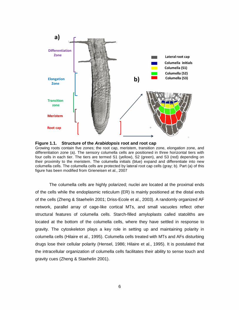
Figure 1.1. Structure of the Arabidopsis root and root cap Growing roots contain five zones; the root cap, meristem, transition zone, elongation zone, and differentiation zone (a). The sensory columella cells are positioned in three horizontal tiers with four cells in each tier. The tiers are termed S1 (yellow), S2 (green), and S3 (red) depending on their proximity to the meristem. The columella initials (blue) expand and differentiate into new columella cells. The columella cells are protected by lateral root cap cells (gray; b). Part (a) of this figure has been modified from Grieneisen et al., 2007
The columella cells are highly polarized; nuclei are located at the proximal ends
of the cells while the endoplasmic reticulum (ER) is mainly positioned at the distal ends
of the cells (Zheng & Staehelin 2001; Driss-Ecole et al., 2003). A randomly organized AF
network, parallel array of cage-like cortical MTs, and small vacuoles reflect other
structural features of columella cells. Starch-filled amyloplasts called statoliths are
located at the bottom of the columella cells, where they have settled in response to
gravity. The cytoskeleton plays a key role in setting up and maintaining polarity in
columella cells (Hilaire et al., 1995). Columella cells treated with MTs and AFs disturbing
drugs lose their cellular polarity (Hensel, 1986; Hilaire et al., 1995). It is postulated that
the intracellular organization of columella cells facilitates their ability to sense touch and
gravity cues (Zheng & Staehelin 2001).

7
How do columella cells perceive touch and gravity? Two hypotheses have been
put forward to explain the mechanism by which gravity is perceived by columella cells.
The starch-statolith hypothesis proposes that the starch-filled amyloplasts (statoliths) in
sensory columella cells (statocytes) are responsible for sensing gravity (Sack, 1997).
The gravity-induced sedimentation of amyloplasts within the sensory columella cells
triggers a signal transduction pathway through activating putative receptors located on
the ER or plasma membrane (Kiss, 2000). This hypothesis is supported by the analysis
of root responses to gravity in starchless or reduced-starch mutant plants. These plants
still can respond to gravity, albeit with a delay (Kiss et al., 1996; MacCleery and Kiss,
1999; Fitzelle and Kiss, 2001). However, in some cases, the hypothesis cannot explain
the behavior of roots in response to gravity. For instance, starchless mutant plants
display delayed gravitropic responses (Caspar and Pickard, 1989). Therefore, another
explanation has been proposed. The protoplast pressure hypothesis suggests that the
weight of entire cytoplasm acts as the gravity sensor (Chen et al., 1999). This hypothesis
was put forward to explain gravity-mediated polarity of cytoplasmic streaming in large
internodal cells of Chara algae. It proposes that gravity-mediated position of the
protoplast compresses the plasma membrane at the bottom of the cell against the wall
and exerts tension between the plasma membrane and the cell wall on the upper side.
This induced pressure differential could activate putative stretch-sensitive channels in
the plasma membrane and trigger signal transduction pathways within the cells
(reviewed in Staves, 1997).
Although many studies have focused on the mechanism of gravity perception in
sensory columella cells, the molecular mechanism of mechanosensing has received less
attention. As the growing root penetrates downward in the direction of gravity into the
soil, it encounters obstacles. To avoid the obstacles, the root detects and navigates
around the obstacles and follows a new trajectory. It has been hypothesized that
mechanical cues activate putative mechanosensors at the plasma membrane via
conformational changes in the cell wall and the plasma membrane. The activation of
mechanosensors in turn facilitates ion fluxes into the cell. The transitory changes in ion
concentrations in the cytoplasm initiates signal transduction cascades inside the cell
(Monshausen et al., 2009).
8
1.1.2. Signal Transductions and Response
Although the sensory role of columella cells in gravitropic responses has been
well established, the mechanism by which mechanical signals are translated to
biochemical signals is still unclear (Strohm et al., 2012). Both gravity and mechanical
stimuli trigger rapid changes in the cytosolic levels of several signaling molecules
including calcium ions (Ca2+), protons (H+) and reactive oxygen species (ROS) which are
correlated with root responses to touch and gravity (Joo et al., 2001; Perrin et al., 2005;
Strohm et al., 2012). The transitory elevation of cytosolic Ca2+ is thought to initiate a
cascade of signal transduction pathways, which would modulate transcriptional and
translational processes required for root responses to touch and gravity (Kimbrough et
al., 2004). In addition, the high concentration of cytoplasmic Ca2+ might stimulate influx of
H+ from the extracellular space into the cytoplasm as well as the production of ROS in
the apoplast (Monshausen et al., 2009). The high concentration of cytoplasmic H+
influences enzymatic activities as well as expression of regulatory genes that control root
responses to touch and gravity. The high concentration of apoplastic ROS strengthen
the cell wall and improve its tolerance against mechanical strain during bending
responses to touch/gravity (reviewed in Apel and Hirt, 2004; Monshausen et al., 2009).
Root bending in response to touch and gravity requires the propagation of signals from
the site of perception, the root cap, to the elongation zone where the response, root
bending, occurs.
As a biochemical transmitter, the plant hormone auxin (indole-3-acetic acid; IAA)
plays a key role in root responses to environmental signals (Schrader et al., 2003). Auxin
is polarly transported cell-to-cell from its main site of biosynthesis in the shoot apex
through the vascular tissue to the root tip (Müller and Leyser, 2011; Peer et al., 2011). In
columella cells, the central and basipetal direction of the auxin stream is redirected
laterally and acropetally through epidermis and cortex cells. In vertically-oriented roots,
auxin is uniformly distributed around the root in cells of the root cap and the outer cell
layers of elongation zone. In roots that have been reoriented horizontally, amyloplasts
are displaced to the new bottom side of columella cells. This displacement of
amyloplasts somehow triggers signaling events that result in an uneven distribution of
auxin across the root. Auxin flows more to the lover flank than to the upper flank of the
root (Yoder et al., 2001; Friml et al., 2002). This asymmetric distribution of auxin results
9
in downward curvature of the roots. The higher concentration of auxin in the lower flank
of the root reduces the cell growth in its elongation zone and induces the bending of the
root toward the new gravity vector (Rashotte et al., 2001; Ottenschläger et al., 2003).
The auxin flow in root cells depends on polar localization of auxin carrier proteins at the
plasma membrane. Endocytosis and subcellular trafficking of auxin transporters mediate
their asymmetric localization and as a result the differential distribution of auxin in root
cells. Therefore, membrane trafficking plays a key role in signal transduction pathways in
root responses to touch and gravity (Strohm et al., 2012).
1.2. Membrane Trafficking Pathways and Root Responses to Touch and Gravity
The flow of membrane between the plasma membrane and endomembrane
compartments coupled with intracellular membrane trafficking, herein called the
membrane trafficking pathways, facilitate transportation of macromolecules to their
intracellular and extracellular destinations (Cheung and Vries, 2008). Membrane
trafficking pathways connect the plasma membrane and the endomembrane system,
which comprises endosomes, Golgi apparatus, ER, and the vacuole (Fig. 1.2; Contento
and Bassham, 2012). As intermediaries of membrane trafficking, membrane-enclosed
vesicles are formed and pinched off from donor membranes, transported and fused to
acceptor membranes. Exchange of materials between the plasma membrane and
endomembrane compartments is mediated through different routes of membrane
trafficking pathways (Fig. 1.2): the endocytic pathway, the exocytic pathway, and
transcytosis.
10
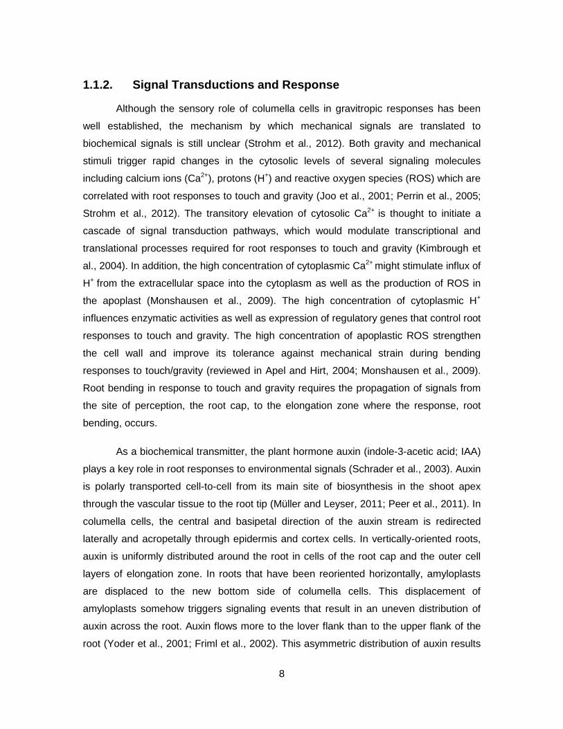
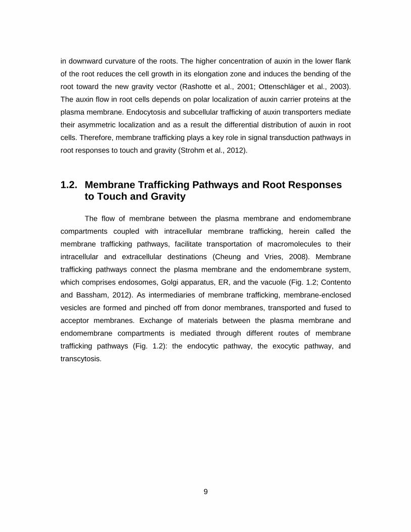
Figure 1.2. Membrane trafficking pathways in plant cells Membrane trafficking pathways connect the plasma membrane (PM) and the endomembrane system, which comprises endosomes (E; yellow), Golgi apparatus (GA; light green), the endoplasmic reticulum (ER; dark green), and the vacuolar apparatus (the prevacuolar compartment (PVC) and the vacuole)
The retrograde endocytic pathway internalizes vesicles containing extracellular
milieu and plasma membrane associated components. After fission from the plasma
membrane and internalization, vesicles fuse to early endosomes and are then either
recycled back to the plasma membrane or transported to the Trans Golgi (TNG) or
vacuoles for sorting or degradation (Robinson et al., 1998; Samaj et al., 2012b). Due to
the unique features of plant cells, i.e., rigid cell wall and high turgor pressure, the
existence of endocytosis in plant cells was questioned for a long time. However, a great
deal of studies in the past decade has repeatedly confirmed that this event occurs in
both aerial and under-ground plant cells (Samaj et al., 2005). Endocytosis is categorized
into two different pathways: clathrin-mediated and clathrin-independent endocytic
pathways (reviewed in Miaczynska and Stenmark et al., 2008). In the well-characterized
clathrin- mediated pathway, clathrin and its associated adaptor proteins mediate the
formation of receptor-containing coated pits at the plasma membrane. Accessory and
regulatory proteins facilitate the invagination of coated pits to form coated vesicles.
Clathrin- independent pathways do not use clathrin, instead, they exploit a variety of
11
mechanisms to select cargos and form endocytic vesicles. Similar to animal cells both
clathrin-mediated and clathrin-independent pathways have been reported in plant cells
(reviewed in Mayor and Pagano, 2007; Miaczynska and Stenmark et al., 2008).
However, the well-characterized clathrin-mediated pathway is most widely studied in
plants.
The anterograde secretory or exocytic pathway transports cargo from the ER to
Golgi apparatus, vacuoles and the plasma membrane for sorting, modification,
degradation, and recycling (Connerly, 2010). In plants, the role of this pathway in tip-
growth and in the final stage of cell division, cytokinesis, in plants has been widely
studied. In tip-growing plant cells and cytokinesis, exocytosis deliver Golgi-derived
vesicles containing new membrane and cell wall materials to the growing tips and the
new cell plate, respectively (Bednarek and Falbel, 2002). This pathway plays a role in
trafficking of auxin transporters in root cap cells (Kleine-Vehn et al., 2010).
In the third membrane trafficking pathway, transcytosis, the macromolecular
cargos are translocated from one side of the plasma membrane cell to other sides (Peer,
2011). Membrane trafficking performs an integral role in variety of cellular processes,
including signal transduction pathways and cell morphogenesis , which endow rooted
plants with the ability to monitor their surroundings and re-adjust their growth in
response to different environmental stresses (Peer, 2011).
The role of membrane trafficking in polar auxin transport is an interesting and
highly active area of plant research. Polar auxin transport is important in plant responses
to environmental signals (Friml et al., 2002; Nakayama et al., 2012). Polar auxin flow is
controlled mainly by the localization of auxin transporters to the ends or sides of the cells
through which auxin is transported. Auxin transporters are located in the plasma
membrane and facilitate the flow of auxin into and out of the cell. Auxin enters the cell by
diffusion and the AUX1 auxin influx carrier proteins (Zažímalová et al., 2010). In the
relatively acidic environment of the apoplast, auxin is mainly protonated (IAAH) in which
this neutral form of auxin easily enters the cell via passive diffusion. On the other hand,
the charged form of auxin (IAA-) is actively transported from the intracellular space into
the cell through AUX1 receptors. Auxin exits the cell only through the PIN auxin carrier
proteins. In the neutral environment of cytoplasm, auxin is deprotonated (IAA-) in which
12
this charged form of the auxin molecule is not able to cross the plasma membrane and
exits cells via the PIN efflux carrier proteins (Raven, 1975; Estelle, 1998; Michniewicz et
al., 2007).The polar localization of auxin transporters at the plasma membrane of
columella cells as well as epidermis and cortex cells direct the flow of auxin in the root.
Out of eight members of the PIN family in Arabidopsis, PIN3 and PIN2 play key roles in
the differential distribution of auxin during gravity stimulation (Friml et al., 2002; Kleine-
Vehn et al., 2010; Ambrose et al., 2013; Kakar et al., 2013). Auxin transporters are not
fixed at the plasma membrane. In response to different environmental signals, auxin
carrier proteins cycle between the plasma membrane and endomembrane
compartments by the endocytic and exocytic pathways. They are also translocated from
one side of the plasma membrane to other side by transcytosis (Friml et al., 2002;
Dhonukshe et al., 2008; Kleine-Vehn et al., 2010).
In vertically-oriented roots, PIN3 is symmetrically dispersed around the plasma
membrane of columella cells and directs auxin flow evenly in all directions through the
root cap cells. Also, PIN2 is mainly localized at the shootward side of the plasma
membrane in root epidermal cells and directs auxin flow equally towards the shoot. In
roots that have been reoriented horizontally, PIN3 relocates to the new basal side of
columella cells via transcytosis. This new localization of PIN3 redirects auxin flow
preferentially to the lower side of the root cap, leading to a high concentration of auxin
on this side. This higher concentration of auxin is needed to be directed from the root
cap towards the shoot. In the lower side of the root, PIN2 retains in the plasma
membrane (its endocytosis is inhibited) and this improves the movement of auxin flow on
this side. The low concentration of auxin in the upper side is also directed towards the
shoot. In this side, PIN2 is more degraded in vacuoles (fewer PIN2 is recycled back to
the plasma membrane) because it directs the low amount of auxin in this side.
Membrane trafficking pathways mediate the polar localization of auxin carriers via
recycling, degradation or translocation processes (reviewed in Kleine-Vehn and Friml,
2008).
Several signaling molecules such as ROP GTPases and ARFs as well as a
number of the cytoskeletal elements have been shown to be involved in regulation of
membrane trafficking pathways (Samaj et al., 2004). However, the exact regulatory
mechanism of these pathways is yet to be identified. Next, some of the tools used to
13
study membrane trafficking will be described and then the role of the cytoskeleton in root
responses to touch and gravity will be discussed.
1.2.1. Tools to Study Membrane Trafficking
Much of our knowledge on membrane trafficking has been obtained using state-
of-the-art live cell imaging techniques to monitor the trafficking of fluorescent probes in
combination with applying genetic and pharmaceutical approaches. The advent of the
membrane-selective fluorescent FM-dyes in the late 1980s has provided a unique
experimental probe to study membrane trafficking (Griffing, 2008). Among FM-dyes,
FM4-64 with distinct features has been wildly used for studying membrane trafficking in
plant cells. The dye fluoresces intensely and has a high photostability. In addition, its
emission spectrum does not overlap with GFP, which facilitates using the dye to label
cells expressing GFP tagged proteins (Bolte et al., 2004). Due to the amphiphilic nature
of FM-dyes, these dyes are not able to cross the plasma membrane bilayer. The dyes
are non-fluorescent in aqueous media and they become intensely fluorescent only after
inserting into the outer leaflet of the membrane (Bolte et al., 2004; Ivanov et al., 2008).
Upon incorporation into the plasma membrane, the dye molecules are internalized into
the cell via endocytosis, from where they either are recycled back to the membrane or
transported to different endosomes (Helling et al., 2006). The vacuolar membrane is the
final destination of FM4-64; about one hour after applying FM4-64 to A. thaliana roots,
the dye stains vacuolar membranes in root cells. Therefore, the dye has widely been
applied to study endocytosis as well as the vacuolar morphology in various eukaryotes
including plants (Vida and Emr, 1995; Ueda et al., 2001; Bolte et al., 2004; Tamura et
al., 2010).
Advanced imaging techniques such as confocal laser scanning microscopy
(CLSM) accompanied with subcellular targeting methods including fluorescent proteins
have enhanced the ability to visualize and examine vesicular transport in living cells.
There are several key features that make CLSM a valuable and efficient tool for a wide
range of studies in cell biology. The pinhole apparatus eliminates out-of-focus light and
provides high optical resolution and signal-to-noise images. This feature facilitates
acquisition of typical stack of optical sections or a Z-series without the need for physical
sectioning of thick samples. In addition, CLSM affords the ability to acquire
14
multidimensional images (x, y, z, and time). Furthermore, the versatile advanced image
analysis software enables data acquisition through quantitative image analyses (Claxton
et al., 2006; Zhang et al., 2013).
The pharmacological approach is a commonly used method to study membrane
trafficking pathways in plant cells. A variety of drugs that disturb proteins involved in
membrane trafficking pathways have been identified by chemical screening methods.
The advantage of the pharmacological approach is that drugs can be applied at different
times and with different doses. This can be an advantage in cases where mutations in
relevant genes are lethal (review in Samaj et al., 2012b). However, it should be noted
that a specific drug can have multiple targets in the cell. This can confound the
interpretation of results since membrane trafficking pathways are complex and
interconnected.
1.3. The Cytoskeleton and Root Responses to Touch and Gravity
MTs and AFs as well as their associated proteins comprise the major structural
components of the cytoskeletal system in plants. MTs and AFs are highly dynamic
biopolymers. The intrinsic dynamic properties of MTs and AFs enable them to undergo
rapid reorganization in response to developmental and environmental signals. The
arrangement of MTs and AFs are regulated by their binding proteins. Cytoskeletal
binding proteins also facilitate the interaction of MTs and AFs with other cellular
components. Through these interactions, the cytoskeleton could modulate a variety of
cellular processes. These biopolymers have been implicated in a myriad of cellular
activities including membrane trafficking pathways, cell division and elongation, and
generating / maintaining cell polarity. MTs and AFs fulfill their cellular functions
independently or in a coordinated manner (Petrasek et al., 2009). A role for the
cytoskeletal system in root responses to touch and gravity stimuli has been reported
(Bisgrove et al., 2008; Gleeson et al., 2012); however, the mechanisms underlying its
role and possible interactions between different cytoskeletal elements in these
responses have yet to be established.
15
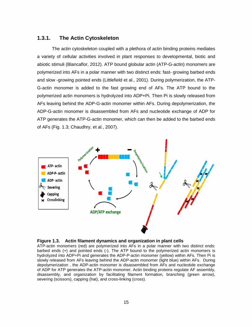
1.3.1. The Actin Cytoskeleton
The actin cytoskeleton coupled with a plethora of actin binding proteins mediates
a variety of cellular activities involved in plant responses to developmental, biotic and
abiotic stimuli (Blancaflor, 2012). ATP bound globular actin (ATP-G-actin) monomers are
polymerized into AFs in a polar manner with two distinct ends: fast- growing barbed ends
and slow -growing pointed ends (Littlefield et al., 2001). During polymerization, the ATP-
G-actin monomer is added to the fast growing end of AFs. The ATP bound to the
polymerized actin monomers is hydrolyzed into ADP+Pi. Then Pi is slowly released from
AFs leaving behind the ADP-G-actin monomer within AFs. During depolymerization, the
ADP-G-actin monomer is disassembled from AFs and nucleotide exchange of ADP for
ATP generates the ATP-G-actin monomer, which can then be added to the barbed ends
of AFs (Fig. 1.3; Chaudhry, et al., 2007).
Figure 1.3. Actin filament dynamics and organization in plant cells ATP-actin monomers (red) are polymerized into AFs in a polar manner with two distinct ends: barbed ends (+) and pointed ends (-). The ATP bound to the polymerized actin monomers is hydrolyzed into ADP+Pi and generates the ADP-P-actin monomer (yellow) within AFs. Then Pi is slowly released from AFs leaving behind the ADP-actin monomer (light blue) within AFs. During depolymerization , the ADP-actin monomer is disassembled from AFs and nucleotide exchange of ADP for ATP generates the ATP-actin monomer. Actin binding proteins regulate AF assembly, disassembly, and organization by facilitating filament formation, branching (green arrow), severing (scissors), capping (hat), and cross-linking (cross).
16
The ability of actin to polymerize and depolymerize endows AFs with the
capability to undergo reorganization in response to different signals (Henty-Ridilla et al.,
2013). Actin binding proteins regulate AF assembly, disassembly, and organization by
facilitating filament formation, stabilization, destabilization, severing (breaking AFs),
capping (preventing further polymerization), and cross-linking (bundling AFs) (Fig. 1.3;
reviewed in Gopinathan et al., 2007). Two distinct configurations of the actin
cytoskeleton have been reported in plant cells: extremely dynamic mesh-like actin
networks (or actin meshworks) and relatively more stable bundled actin networks (or
actin bundles) (Volkmann and Baluska, 1999; Ananthakrishnan and Ehrliche, 2007).
The dynamic mesh-like actin networks are usually located in the cortex of plant
and animal cells in close association with the plasma membrane (Thomas, 2012). In
yeast, animal, and plant cells, the cortical actin and its regulatory proteins such as the
actin-related protein (Arp) 2/3 complex are involved in endocytosis (reviewed in Samaj et
al., 2012a). In addition, the mesh-like actin networks are involved in the polar growth of
tip-growing plant cells as well as in the local growth of the puzzle-shaped leaf pavement
cells. AF bundles in plant cells are thought to function as passive highways. AF
associated motor proteins myosins travel along AFs during cytoplasmic streaming and
mediate long range movement of vesicles/endosomes and subcellular organelles
(Kuroda, 1990; Smertenko et al., 2010).
Distinctive AF organizations in tip-growing cells including pollen tubes and root
hairs have received much attention in studying the role of the actin cytoskeleton in plant
cells. In a tip-growing cell, the shank contains AF bundles, the sub-apex has less
bundled AFs and the apex has more dynamic mesh-like actin networks. These AF
arrangements are involved in movement of endosomes containing the plasma
membrane and cell wall materials to the growing tip as well as in retrieval of excess
secreted materials into the cell (reviewed in Ovecka et al., 2005). Endosomal
movements in the shank and the sup-apex area of root hairs are mediated by actin
motor proteins, myosins, which use AFs as tracks. In addition to actin meshworks, the tip
in a tip-growing cell contains a high population of exocytic and endocytic vesicles as well
as endosomes. Treatment with a low concentration of the actin destabilizing drug
Latrunculin B (Lat B) inhibited the movement of vesicles and endosomes in the apex of
the tip-growing cell; however, disrupting the ATPase activity of myosins failed to inhibit
17
the movement of endosomes in this region, suggesting a role for the dynamic actin
meshwork in membrane trafficking (Voigt et al., 2005; Wang et al. 2006).
In addition to tip-growing cells, the actin cytoskeleton has been implicated in
membrane trafficking in other aerial and underground plant cells. To date, most
experiments aimed at investigating the role played by the actin cytoskeleton in plant cells
have been based on pharmacological approaches. Treatment with high concentration of
Lat B that destabilizes both actin meshworks and actin bundles inhibits endocytosis in
root epidermal cells (Konopka et al. 2008). On the other hand, relatively low
concentration of Lat B induces the formation of small intracellular aggregates in these
cells (Samaj et al., 2012a). In addition, high concentration of the actin stabilizing drug
Jasplakinolide (Jas) inhibits endocytosis in root cells (Dhonukshe et al., 2008). Yet, the
exact role of the actin cytoskeleton in membrane trafficking pathways is unknown.
Nagawa et al. (2012) have proposed that the cortical AFs could act as an inhibitory
barrier to endocytosis in plant cells. This proposal was put forward based on their
observation that overexpression of ROP2 GTPase and its effector Rac4 increased the
accumulation of mesh-like cortical actin networks and inhibited endocytosis in leaf
epidermal pavement cells. Treatment ROP2 GFPase or Rac4 overexpressed lines with a
low concentration of Lat B, which reduces the accumulation of cortical actin and has no
effects on actin bundles, restored endocytosis in these cells. Also, stabilization of cortical
actin with a relatively low concentration of Jas in these cells inhibited endocytosis. On
the other hand, Bloch et al. (2005) have reported that treatment with a relatively low
concentration of Jas has no effect on endocytosis in root hairs.
The actin cytoskeleton also has been proposed to play a role in perception of
gravity in sensory columella cells (reviewed in Blancaflor, 2012). Columella cells contain
cortical mesh-like actin networks coupled with the organized cytoplasmic actin bundles
(Collings et al., 2001). Treatment with the actin destabilizing drug Lat B significantly
promotes the gravitropic responses (bending) in roots that have been reoriented
horizontally (Mancuso et al., 2006). This observation has led to two proposals about the
possible role of actin in gravitropsm. The first hypothesis proposes that the cytoplasmic
network of AFs in columella cells acts as barrier against the movement of amyloplsts in
these sensory cells. The destabilization of AFs may promote amyloplast sedimentation
to the new bottom of the columella cells in reoriented roots and enhance gravity
18
perception in root sensory cells. Another hypothesis suggests that destabilization of the
cortical actin in columella cells with Lat B could promote root responses to gravity by
affecting the trafficking of PIN3 in columella cells (Blancaflor, 2012). In vertically oriented
roots, PIN3 is equally distributed around the columella cells and directs auxin flow evenly
in all directions through the root cap cells. In roots that have been reoriented
horizontally, PIN3 relocates to the new lower side of columella cells. This new
localization of PIN3 redirects auxin flow preferentially to the lower side of the root cap
(Friml et al., 2002). This high concentration of auxin in the lower side is then directed to
the shoot via PIN2, which results in a higher concentration of auxin in the elongation
zone on the lower side of the root. The hypothesis suggests that destabilization of
cortical AFs in columella cells might promote relocalization of PIN3 and enhance root
responses to gravity (Blancaflor, 2012).
1.3.2. Microtubules
After more than half a century of studies on the MT cytoskeleton, our
understanding about the roles of MTs in plant cells is extending beyond cell division and
expansion (Wasteneys 2013). The hallmark of MTs is that they are intrinsically dynamic
polymers; they continuously undergo polymerization (growing), depolymerization
(shrinking), pausing (no significant growth or shrinkage), and rebuilding (rescue;
Mitchison and Kirschner, 1984). MTS are filamentous polymers of α/β-tubulin
heterodimers. These tubulin subunits are GTP-binding proteins; the GTP bound to α-
tubulin does not hydrolyze, while the GTP bound to β-tubulin can be hydrolyzed to GDP
(Tian et al., 1999). The longitudinal assembly of dimers in a head-to-tail fashion results
in the formation of linear protofilaments. The lateral interaction of these protofilaments
leads to the formation of a cylindrical MT with two distinctive ends: plus (fast -growing)
and minus (slow- growing) ends (Kirschner and Mandelkow, 1985; Wu et al., 2009). The
GTP bound states of tubulin dimers assemble onto the sheet-like structures at the fast
growing ends of MTs (polymerization). The sheet-like structure rapidly zips up in a
cylindrical MT. After GTP-bound heterodimers bind to the MT, GTP in β-tubulin is
hydrolyzed to GDP (depolymerization; Kirschner and Mandelkow, 1985). At the plus end
of the MT, GTP-bound heterodimers are added at a higher rate than the rate which the
hydrolysis of GTP occurs. This generates a GTP cap at this end. In a polymerizing MT,
19
the GTP cap stabilizes the MT and induces the association of GTP-bound subunits with
the growing ends of the MT. Further back in the MT lattice, GDP-bound subunits are less
tightly bound to MTs. During depolymerization, GDP-bound subunits are disassembled
from MTs. When the incorporation rate of GTP-bound subunits in the MT is slower than
the hydrolysis rate of GTP, the GTP cap is lost. This results in a rapid dissociation of
GDP tubulin subunits from the MT and a rapid shrinkage of MT. This transition of MTs
from growth to shrinkage is called catastrophe (Dhamodharan, 1995). When new GTP-
bound subunits are added to the shrinking MT, a GTP cap is formed and this switches
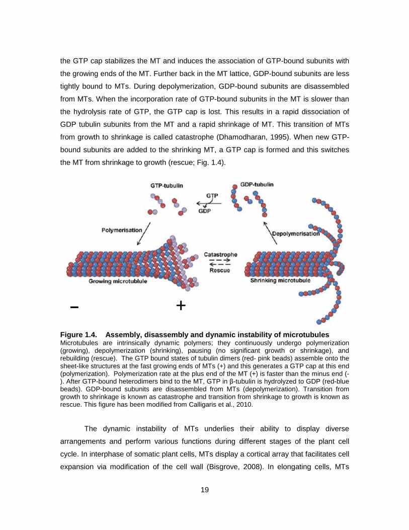
the MT from shrinkage to growth (rescue; Fig. 1.4).
Figure 1.4. Assembly, disassembly and dynamic instability of microtubules Microtubules are intrinsically dynamic polymers; they continuously undergo polymerization (growing), depolymerization (shrinking), pausing (no significant growth or shrinkage), and rebuilding (rescue). The GTP bound states of tubulin dimers (red- pink beads) assemble onto the sheet-like structures at the fast growing ends of MTs (+) and this generates a GTP cap at this end (polymerization). Polymerization rate at the plus end of the MT (+) is faster than the minus end (-). After GTP-bound heterodimers bind to the MT, GTP in β-tubulin is hydrolyzed to GDP (red-blue beads). GDP-bound subunits are disassembled from MTs (depolymerization). Transition from growth to shrinkage is known as catastrophe and transition from shrinkage to growth is known as rescue. This figure has been modified from Calligaris et al., 2010.
The dynamic instability of MTs underlies their ability to display diverse
arrangements and perform various functions during different stages of the plant cell
cycle. In interphase of somatic plant cells, MTs display a cortical array that facilitates cell
expansion via modification of the cell wall (Bisgrove, 2008). In elongating cells, MTs
20
beneath the plasma membrane guide the direction of cellulose synthetase and as a
result the orientation of cellulose microfibrils. This means that cellulose microfibrils are
oriented parallel to MTs and perpendicular to the long axis of the cell. Cellulose
microfibrils are resistant to stretching along their length and this feature constrains
direction of cell expansion. The isotropic outward turgor pressure supplies the force that
increases the distance between the adjacent cellulose microfibrils, resulting in
anisotropic cell expansion. Prior to mitosis, the unique MT arrays, the so- called
preprophase band (PPB), encircles the nucleus. The PPB somehow defines the future
division site. In metaphase of mitosis, the PPB is replaced by the mitotic spindle. MTs in
spindles are arranged in a bipolar manner; their minus ends are at the cell poles,
whereas their plus ends are towards the center of the cell. The mitotic spindle mediates
the alignment of chromatids at the center of the cell and their subsequent segregation to
the two daughter cells. As the cell enters cytokinesis, the spindle MTs rearrange into a
distinctive arrangement, the so-called phragmoplast, composed of two set of MTs with
opposite polarity between daughter nuclei (Wasteneys, 2002). The phragmoplast
contributes to the delivery of Golgi-derived vesicles towards the forming cell plate.
The dynamic properties and activities of MTs are modulated by a fleet of MT-
associated proteins (MAPs) that bind directly to MTs. Some MAPs preferentially
accumulate at the minus or the plus ends of MTs, while others associate with the MT
lattice (Liu et al., 2011). In animal cells, the minus ends of MTs are anchored at MT
organizing centers (MTOCs), centrosomes, which are comprised of centrioles and
pericentriolar material (PCM; Rusan and Rogers, 2009). The minus end of the MT is
embedded in PCM and it contains proteins such as γ-tubulin that are involved in MT
nucleation (Oakley, 1999). MTs grow outwards from centrosomes through the
cytoplasm into the cortex of the cell. Plant cells lack centrosome-like MT organizing
centers, but they have some of the proteins found in PCM of animal cells. For example,
γ-tubulin is dispersed in the cytoplasm and it binds to the minus ends of MTs in plant
cells. Several MAPs such as MICROTUBULE ORGANIZATION 1 bind to the MT lattice
and regulate MT organization and function (Kawamura et al., 2006). Furthermore,
Microtubule motor proteins, kinesins, use MTs as tracks to carry cargo within the cell in a
directional manner: towards plus ends (N-kinesins) or towards minus ends(C-kinesins).
Another group of MAPs preferentially bind and track the MT plus ends; these ones are
21
known as MT plus end-tracking proteins or +TIPs (Akhmanova and Steinmetz, 2008).
The localization of +TIPs at the active ends of MTs allows them to interact with other
cellular components and structures and, therefore, modulate a variety of cellular
activities including signal transduction pathways. Out of +TIPs, the End Binding 1(EB1)
family is thought to be a key regulator of MT functions, since it regulates MT dynamics
and recruits different types of cargo proteins to the plus ends of microtubules
(Akhmanova and Steinmetz, 2008).
The role of MTs in root responses to touch and gravity is not well understood.
Since MTs in interphase plant cells are associated with the plasma membrane, it has
been postulated that cortical MTs in roots might facilitate the perception of environmental
stimuli including touch and gravity through activation of putative microtubule-gated
membrane ion channels or interaction with potential stretch-activated membrane ion
channels (Nick, 2008). The fact that MTs regulate cell expansion by determining the
orientation of cellulose microfibrils raises another possibility about the role of MTs in root
responses to gravity and mechanical cues. The hypothesis proposes that gravity and
mechanical signals reorganize MT arrays in roots and this reorganization induces root
bending. In support of this idea is the observation that during root bending MTs on the
outer side of the root MTs rearrange from parallel to transverse arrays, whereas on the
inner side of the root MTs retains their parallel array. The different orientations of MTs on
the two sides of the root could alter the direction of cellulose microfibrils deposition and
the direction of root growth, resulting in root bending. However, whether the different MT
orientations on the two sides of roots are responsible for root bending or whether MT
rearrangements are induced by root bending is still controversial (reviewed in Bisgrove,
2008). There is evidence that supports the idea that MTs reorient after root bending. In
horizontally-oriented maize roots, the early stage of root bending occurred before
microtubule reorientation (Blancaflor and Hasenstein, 1997; Bisgrove, 2008). MT
dynamics and functions are regulated by MAPs; therefore, detailed studies of MAPs are
fundamental in our understanding about the role of MTs in root responses to touch and
gravity. In A. thaliana, EB1 plays a role in root responses to touch and gravity (Bisgrove
et al., 2008; Gleeson et al., 2012); however, the mechanism underlying its role in these
responses is unknown.
22
1.3.3. EB1
EB1 is an evolutionarily conserved protein with homologues found in eukaryotes
ranging from yeast to humans and plants. Studies on the protein in animals, fungi, and
plants have revealed that it binds directly to the fast growing ends of MTs where it
regulates growth and shrinkage rates of the MTs (Vitre et al., 2008). In addition to MTs,
EB1 in animal cells interacts with a large number of additional proteins in the cell.
Through these interactions, EB1 influences a variety of cellular processes (Akhmanova
and Stehbens, 2008). Studies aimed at disclosing the mechanism underlying EB1
interaction with other proteins have characterized two groups of proteins as putative EB1
binding partners: proteins containing the short polypeptide motif SxIP (serine-any amino
acid-isoleucine-proline; Honnappa et al., 2009), and proteins with Cytoskeleton-
Associated Protein-Glycin-rich (GAP-Gly) domains which bind to EEY motif of EB1
(Weisbrich et al., 2007). Plant EB1 proteins lack the EEY motif and proteins with the
CAP-Gly domains are not found in plant cells. This indicates that this mode of interaction
between EB1 and other proteins might not exist in plants (Akhmanova and Stehbens,
2008; Liu et al., 2011). Identifying putative EB1 binding proteins in plants would reveal
possible signaling pathways regulated by EB1 proteins. The A. thaliana genome
encodes a myriad of proteins with SxIP motif (personal communication with S. Squires).
Whether Arabidopsis EB1 proteins interact with the SxIP motif –containing proteins is
still unknown.
Since EB1 regulates MT dynamics and it interacts with other proteins, it has been
proposed to be a master regulator of MT plus end (Liu et al., 2011). The Arabidopsis
thaliana genome contains three EB1 genes: EB1a, EB1b, and EB1c (Chan et al., 2003;
Mathur et al., 2003). EB1 proteins are dimeric proteins with two conserved domains: a
calponin homology (CH) domain at the N-terminus and a dimerization domain at the C-
terminus (Fig. 1.5). The CH domain is responsible for the interaction of EB1 with the fast
growing ends of MTs (Slep et al., 2005). Several hypotheses have been put forward to
explain the mechanism by which EB1 recognizes the plus ends of MTs. One explanation
is that EB1 might preferentially bind to the extended tubulin sheet structure at the fast
growing ends of MTs rather than to the tubular wall of MTs. Another hypothesis
proposes that EB1 identifies the GTP bound state of β-tubulins confined to the plus ends
of MTs. Nevertheless, the exact mechanism of the plus end tracking behavior of EB1 is
23
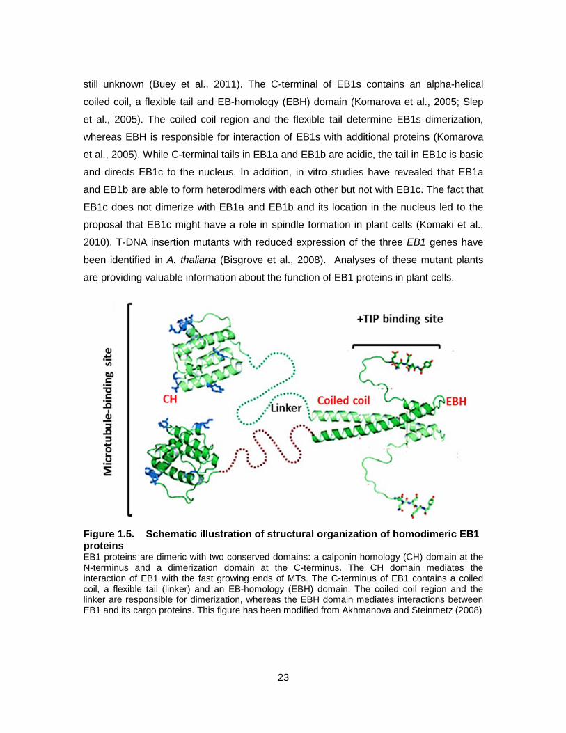
still unknown (Buey et al., 2011). The C-terminal of EB1s contains an alpha-helical
coiled coil, a flexible tail and EB-homology (EBH) domain (Komarova et al., 2005; Slep
et al., 2005). The coiled coil region and the flexible tail determine EB1s dimerization,
whereas EBH is responsible for interaction of EB1s with additional proteins (Komarova
et al., 2005). While C-terminal tails in EB1a and EB1b are acidic, the tail in EB1c is basic
and directs EB1c to the nucleus. In addition, in vitro studies have revealed that EB1a
and EB1b are able to form heterodimers with each other but not with EB1c. The fact that
EB1c does not dimerize with EB1a and EB1b and its location in the nucleus led to the
proposal that EB1c might have a role in spindle formation in plant cells (Komaki et al.,
2010). T-DNA insertion mutants with reduced expression of the three EB1 genes have
been identified in A. thaliana (Bisgrove et al., 2008). Analyses of these mutant plants
are providing valuable information about the function of EB1 proteins in plant cells.
Figure 1.5. Schematic illustration of structural organization of homodimeric EB1 proteins EB1 proteins are dimeric with two conserved domains: a calponin homology (CH) domain at the N-terminus and a dimerization domain at the C-terminus. The CH domain mediates the interaction of EB1 with the fast growing ends of MTs. The C-terminus of EB1 contains a coiled coil, a flexible tail (linker) and an EB-homology (EBH) domain. The coiled coil region and the linker are responsible for dimerization, whereas the EBH domain mediates interactions between EB1 and its cargo proteins. This figure has been modified from Akhmanova and Steinmetz (2008)
24
By studying the role of EB1 proteins in plant growth and development, our lab
has found that these proteins modulate root responses to touch and gravity (Bisgrove et
al., 2008; Gleeson et al., 2012; Squires et al., 2013). Plants carrying T-DNA insertions in
each of the EB1 genes have roots that exhibit exaggerated responses to touch and
gravity. When grown on the surface of reclined agar plates, roots of eb1 mutants skew
more to the left and form more loops than wild type. A. thaliana eb1b-1 and eb1 triple
mutants have the same phenotypic defects in their responses to touch/gravity cues,
suggesting that eb1b-1 has the main role in these responses (Bisgrove et al. 2008). The
fact that eb1b mutant roots skew more and form more loops than wild type suggests that
the wild type protein acts as a repressor of the response (Bisgrove et al., 2008; Gleeson
et al., 2012).
1.4. Thesis Objectives
In my thesis, I investigated whether EB1b, as a master regulator of +TIPs, could
alter root responses to touch/gravity cues through an effect on the subcellular
organization of columella cells or on membrane trafficking pathways in root cells. This
investigation involved two sets of experiments:
1. Mutant root caps were examined for possible subcellular defects that
might be correlated with an aberrant response to these stimuli. I examined the
arrangement of MTs, the distribution of the ER, and the position of nuclei in columella
cells.
2. Membrane trafficking pathways were studied in root cells. I applied a
membrane trafficking marker to investigate the role of EB1 in membrane trafficking
pathways in root cells of wild type and eb1 mutants.
25
2. Materials and Experimental Approaches
2.1. Plant Material and Growth Conditions
Wild type and the eb1b-1 mutant A. thaliana lines used in our study were of
Wassilewskija (Ws) ecotype. The eb1b-1 mutant, carrying a T-DNA insertion in the EB1b
gene, has been previously characterized (Bisgrove et al., 2008; Gleeson et al., 2012;
Squires and Bisgrove, 2013). Seeds were sterilized according to the vapour-phase
protocol (Clough and Bent, 1998). Seeds were put in eppendorf tubes with open lids and
the tubes were placed on an eppendorf tube rack in a glass jar. 100 ml bleach was
poured in a plastic beaker and then 3 ml concentrated hydrogen chloride was added to
the bleach afterwards. The beaker was placed inside of the glass jar and its lid was
tightly sealed. Seeds were incubated for 45 minutes (min) inside of the glass
jar.Sterilized seeds were placed on hallf- strength Murashige and Skoog medium (MS,
Sigma-Aldrich) supplemented with 1% (w/v) sucrose, 0.1% (w/v) 2-(N-morpholino)
ethanesulfonic acid (MES), 1% (w/v) agar (Phytablend, Caisson Laboratories Inc.), with
the final pH adjusted to 5.8. Plates containing sterilized seeds were incubated at 4ºC in
the dark for 3-4 days to break seed dormancy. Then the petri dishes were sealed with
micropore tape (3M Health Care) and placed in a growth chamber in a vertical position
for 3-5 days. Parameters for the growth chamber were 16 h light/ 8 h dark and 20ºC.
GFP-MBD (Columbia-0 (Col-0); Marc et al., 1998; Granger and Cyr, 2001) and
DR5rev::GFP (Col-0; Friml et al., 2003; Palme et al., 2006) seeds were obtained from
The Arabidopsis Information Resource (TAIR; http://www.arabidopsis.org/). GFP-MBD
and DR5rev::GFP plants were crossed to the eb1b-1 mutant plants and homozygous
lines were identified by analyzing the F3 generation (personal communication with Dr.
Bisgrove; Squires and Bisgrove, 2013). EB1 rescued and overexpressed lines were
generated and characterized by others in the lab. These lines were generated by
transforming A. thaliana eb1b-1 mutants with wild type EB1b gene under control of its
26
endogenous promoter pEB1b:EB1b (Gleeson et al., 2012; C. Chen, S. Squires and L.
Vita, unpublished data).
2.2. Confocal Laser Scanning Microscopy
Fluorescence images were captured by a Nikon Eclipse Ti inverted laser
scanning confocal microscope system (CLSM). Images were acquired using a 60x water
immersion objective lens (NA 1.2). GFP fluorophores were excited at 488nm and the
emission collected at 525-550nm. For FM4-64 detection, excitation was 561 nm and the
emission was collected at 575 nm. All image analyses were performed using Nikon
Advanced Research analysis software. All confocal setup parameters were kept
constant in all experiments in which the uptake of FM4-64 was monitored.
2.3. Quantification of Cell Polarity Parameters
3-4-day-old A. thaliana wild type and the eb1b mutant seedlings expressing
fluorescent reporter proteins were used to determine the spatial location, distribution,
and arrangement of subcellular compartments and structures. The distribution of ER was
determined by calculating the ratio of the circumference of the distal ER to the
circumference of the corresponding cell. To examine nuclear position, two distances
were measured: the distance of the center of nucleus to the proximal cell wall (Y), and
the distance of the center of nucleus to the outer cell wall (X). The nuclear position was
defined as the ratio of X to the cell length and Y to the cell width.
2.4. FM4-64 Staining
FM4-64 (N-(3-Triethylammoniumpropyl)-4-(6-(4-(Diethylamino) Phenyl)
Hexatrienyl) Pyridinium Dibromide; Molecular probes, Invitrogen Life technologies) was
stored as a 2mM stock solution in Dimethyl sulfoxide ( DMSO) at 4ºC and in the dark. To
monitor endocytosis, 3-4-day-old seedlings were incubated in liquid MS medium
containing 2µM FM4-64 for 5 min in the dark at room temperature, rinsed twice for 1min
each time in liquid MS medium. The dye was washed out at this step to remove all the
27
dye molecules that have not been incorporated into the plasma membrane. This allowed
monitoring of the amount of the dye that is lost from the plasma membrane during
endocytosis. Seedlings were then mounted on slides in liquid MS medium for 3 min. The
slides were covered with cover glasses and imaged by CLSM beginning 6 min
afterwards. Therefore, the cell had an overall time of 15 min to endocytose the dye.
FM4-64 was 15 min.
To stain the vacuolar membrane, 4-5-day-old seedlings were incubated in 5 µM
FM4-64 for 10 min in the dark at room temperature, rinsed twice for 1min each time in
liquid MS medium and then incubated in liquid MS medium for 2 hours.
2.5. Quantification of FM4-64 Uptake
To analyze the uptake of FM4-64 through endocytosis, confocal images
consisted of 6 sequential optical z sections taken at 0.5µm intervals were compressed.
To quantify FM4-64 uptake, the mean fluorescence intensity of the cytoplasm excluding
the plasma membrane and the nucleus (MFC) and the mean fluorescence intensity of
the corresponding plasma membrane (MFP) were measured. FM4-64 uptake was
defined as the ratio of MFC to MFP. Nikon Advanced Research analysis software was
used to create compressed images as well as measuring the fluorescence intensity.
2.6. Pharmaceutical Treatments
Brefeldin A (BFA) (Sigma-Aldrich) was prepared as a 1.5 mM stock solution in ethanol
and stored at 4ºC in the dark. For BFA treatment, 3-4-day-old seedlings were incubated
in liquid medium containing 25 µM BFA for 30 min. Then BFA treated seedlings were
stained with 5 µM FM4-64 containing 25 µM BFA for 5 min in the dark at room
temperature for 30 min and imaged with CLSM.
For BFA washout experiments, two sets of seedlings were used: one set as a
control line and the other as BFA washed-out set. Both sets of seedlings were placed in
liquid medium containing 25 µM BFA for 30 min. The seedlings were stained with 5 µM
FM4-64(with 25 µM BFA in the control lines) for 5 min in the dark at room temperature.
28
The seedlings then were incubated in liquid medium (with 25 µM BFA in the control set)
for 2 hours and then imaged with CLSM.
Oryzalin (Sigma-Aldrich) was dissolved in DMSO to yield a stock solution of
10mM and then stored at 4ºC in the dark. To examine the effects of oryzalin on FM4-64
uptake, 3-4-day-old wild type and eb1b mutants grown on agar medium containing 50
nM oryzalin were used. Latrunculin B (Sigma-Aldrich) was stored at -20ºC as a 5 mM
stock solution in DMSO. To assess the effects of Lat B on FM4-464 uptake, 3-day-old
wild type and eb1b mutants grown on agar medium supplemented with 10 nM Lat B
were used. A 100 µg/ml stock solution of Jasplakkinolide (Jas; Sigma-Aldrich) was
prepared in DMSO and stored at -20ºC in the dark. To investigate the effects of Jas on
FM4-64 uptake, 3-4-day-old seedlings grown on 100 nM Jas were used. In seedlings
treated with oryzalin, Lat B, and Jas, FM4-64 uptake was monitored based on the
processes described in section 2.4, except, liquid MS medium used in all the processes
(staining, rinsing, and mounting) contained the same concentration of drugs.
2.7. Amyloplast Staining and Visualization
Amyloplasts were visualized by staining their starch with Lugol’s iodine solution
(1% (w/v) iodine and 2% (w/v) potassium iodide). 3-4-day-old seedling were immersed
in 1% Lugol’s solution diluted with MS liquid for 2-3 min in the dark and rinsed in liquid
MS medium for 2 min. The stained seedlings were mounted on slides in a clearing
solution that contained 8:3:1(w/v/v) chloral hydrate, liquid MS medium, and glycerol
respectively. Slides were vertically positioned for 15 min and imaged immediately with
CLSM.
2.8. Statistical Analysis
In all experiments, a Student’s T-test (using Excel) was used for pair-wise
comparisons of means. Means, standard deviations, and P values were all calculated
using Excel. All FM4-64 uptake experiments were repeated at least three times and

29
each replicate contained at least one root for each genotype/treatment. In each root, at
least eight cells were analyzed.
Experiments aimed at investigating subcellular architecture of columella cells and
amyloplast distributions in these cells were repeated as follows: MT arrays were
examined in six independent seedlings for each genotype. Distal ER distribution was
examined in eight seedlings for each genotype from four independent experiments.
Nuclear positions were assessed in four independent experiments having two seedlings
for each genotype. Amyloplast distribution was visualized in two seedlings for each
genotype.
30
3. Results
In my thesis I studied the role of EB1 in root responses to touch and gravity at
the cellular level. The Arabidopsis thaliana genome has three EB1 genes: EB1a, EB1b,
and EB1c. Plants carrying T-DNA insertion with reduced expression of each of these
genes has been identified (Bisgrove et al., 2008). These mutants have defects in
responding to touch/gravity stimuli. Roots of the mutant plants grown on reclined agar
plates skew and form more loops than wild type seedlings. A. thaliana eb1b-1 and eb1
triple mutants demonstrate the same phenotypic defects in responding to touch/gravity
cues (Bisgrove et al. 2008; Gleeson et al., 2012). Consequently, my work is focused on
eb1b-1 mutants.
3.1. Cytoarchitechture of Root Cap Columella Cells
Columella cells in the center of the root cap function as sensory cells in roots.
These specialized cells are highly polarized, and it has been postulated that their
distinctive structure facilitates their function in mechano- and gravity-sensing (Sack,
1991; Blancaflor et al., 1998; Chen et al., 1999, Zheng and Staehlin 2001). Since roots
of eb1b mutant plants exhibit aberrant responses to touch/gravity stimuli (Bisgrove et al.,
2008; Gleeson et al., 2012), I investigated the possibility that the abnormal response
might be due to structural defects in columella cells. My first series of experiments was
aimed at examining the cytoarchitecture of columella cells in wild type and eb1b-1
mutant roots. Specifically, the arrangement of MTs, ER distribution, nuclear positioning
and the distribution of amyloplasts were assessed.
3.1.1. Microtubule Organization
To determine whether eb1b mutants display MT organizational defects in root-tip
cells, transgenic plants expressing a MT reporter, the MT binding domain (MBD) of the
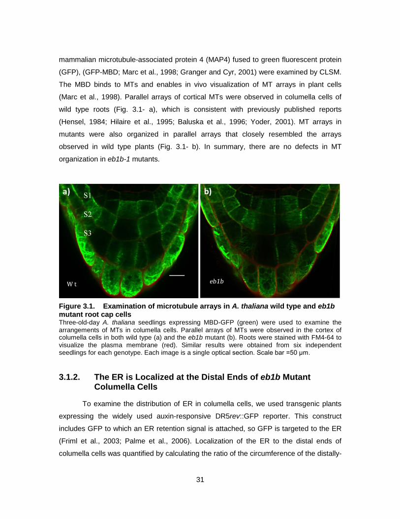
31
mammalian microtubule-associated protein 4 (MAP4) fused to green fluorescent protein
(GFP), (GFP-MBD; Marc et al., 1998; Granger and Cyr, 2001) were examined by CLSM.
The MBD binds to MTs and enables in vivo visualization of MT arrays in plant cells
(Marc et al., 1998). Parallel arrays of cortical MTs were observed in columella cells of
wild type roots (Fig. 3.1- a), which is consistent with previously published reports
(Hensel, 1984; Hilaire et al., 1995; Baluska et al., 1996; Yoder, 2001). MT arrays in
mutants were also organized in parallel arrays that closely resembled the arrays
observed in wild type plants (Fig. 3.1- b). In summary, there are no defects in MT
organization in eb1b-1 mutants.
Figure 3.1. Examination of microtubule arrays in A. thaliana wild type and eb1b mutant root cap cells Three-old-day A. thaliana seedlings expressing MBD-GFP (green) were used to examine the arrangements of MTs in columella cells. Parallel arrays of MTs were observed in the cortex of columella cells in both wild type (a) and the eb1b mutant (b). Roots were stained with FM4-64 to visualize the plasma membrane (red). Similar results were obtained from six independent seedlings for each genotype. Each image is a single optical section. Scale bar =50 μm.
3.1.2. The ER is Localized at the Distal Ends of eb1b Mutant Columella Cells
To examine the distribution of ER in columella cells, we used transgenic plants
expressing the widely used auxin-responsive DR5rev::GFP reporter. This construct
includes GFP to which an ER retention signal is attached, so GFP is targeted to the ER
(Friml et al., 2003; Palme et al., 2006). Localization of the ER to the distal ends of
columella cells was quantified by calculating the ratio of the circumference of the distally-
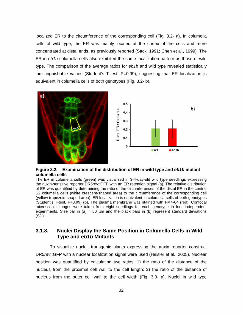
32
localized ER to the circumference of the corresponding cell (Fig. 3.2- a). In columella
cells of wild type, the ER was mainly located at the cortex of the cells and more
concentrated at distal ends, as previously reported (Sack, 1991; Chen et al., 1999). The
ER in eb1b columella cells also exhibited the same localization pattern as those of wild
type. The comparison of the average ratios for eb1b and wild type revealed statistically
indistinguishable values (Student’s T-test, P=0.99), suggesting that ER localization is
equivalent in columella cells of both genotypes (Fig. 3.2- b).
Figure 3.2. Examination of the distribution of ER in wild type and eb1b mutant columella cells The ER in columella cells (green) was visualized in 3-4-day-old wild type seedlings expressing the auxin-sensitive reporter DR5rev::GFP with an ER retention signal (a). The relative distribution of ER was quantified by determining the ratio of the circumferences of the distal ER in the central S2 columella cells (white crescent-shaped area) to the circumference of the corresponding cell (yellow trapezoid-shaped area). ER localization is equivalent in columella cells of both genotypes (Student’s T-test, P=0.99) (b). The plasma membrane was stained with FM4-64 (red). Confocal microscopic images were taken from eight seedlings for each genotype in four independent experiments. Size bar in (a) = 50 µm and the black bars in (b) represent standard deviations (SD).
3.1.3. Nuclei Display the Same Position in Columella Cells in Wild Type and eb1b Mutants
To visualize nuclei, transgenic plants expressing the auxin reporter construct
DR5rev::GFP with a nuclear localization signal were used (Heisler et al., 2005). Nuclear
position was quantified by calculating two ratios: 1) the ratio of the distance of the
nucleus from the proximal cell wall to the cell length: 2) the ratio of the distance of
nucleus from the outer cell wall to the cell width (Fig. 3.3- a). Nuclei in wild type
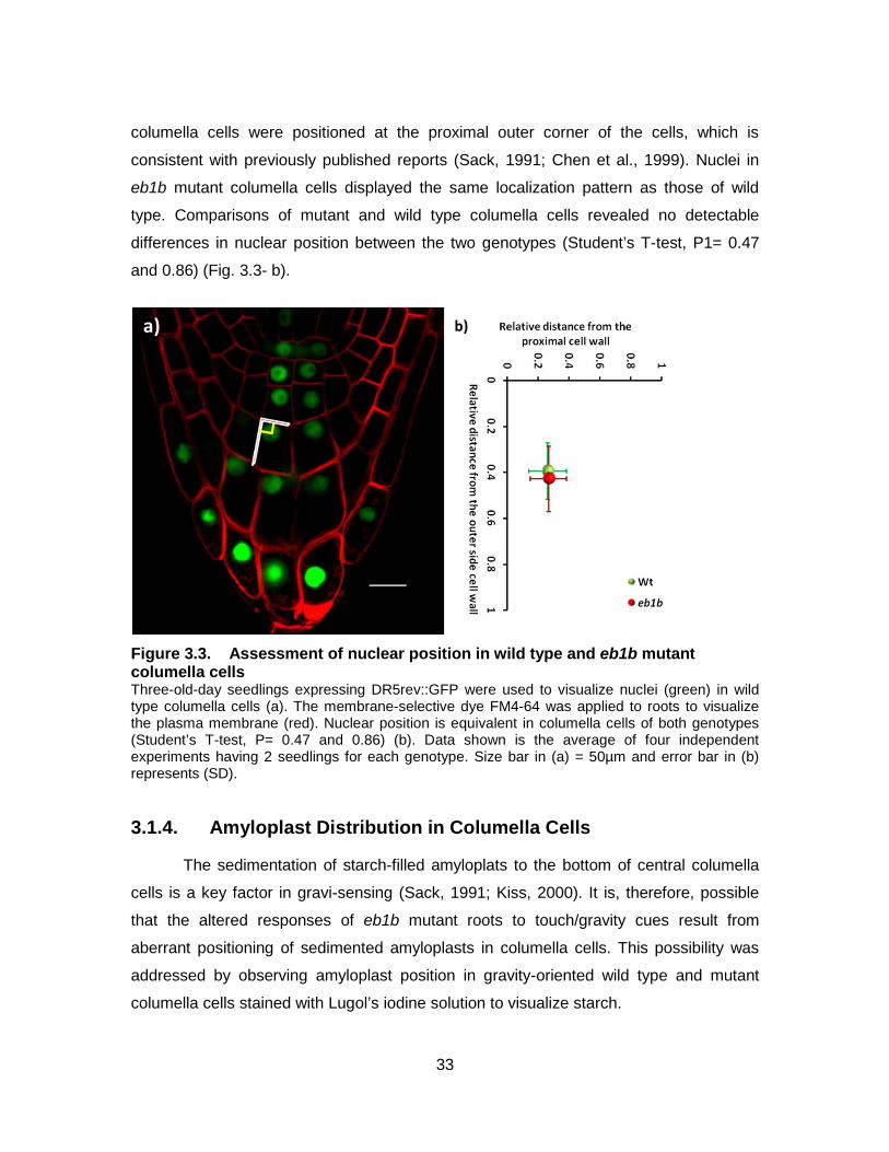
33
columella cells were positioned at the proximal outer corner of the cells, which is
consistent with previously published reports (Sack, 1991; Chen et al., 1999). Nuclei in
eb1b mutant columella cells displayed the same localization pattern as those of wild
type. Comparisons of mutant and wild type columella cells revealed no detectable
differences in nuclear position between the two genotypes (Student’s T-test, P1= 0.47
and 0.86) (Fig. 3.3- b).
Figure 3.3. Assessment of nuclear position in wild type and eb1b mutant columella cells Three-old-day seedlings expressing DR5rev::GFP were used to visualize nuclei (green) in wild type columella cells (a). The membrane-selective dye FM4-64 was applied to roots to visualize the plasma membrane (red). Nuclear position is equivalent in columella cells of both genotypes (Student’s T-test, P= 0.47 and 0.86) (b). Data shown is the average of four independent experiments having 2 seedlings for each genotype. Size bar in (a) = 50µm and error bar in (b) represents (SD).
3.1.4. Amyloplast Distribution in Columella Cells
The sedimentation of starch-filled amyloplats to the bottom of central columella
cells is a key factor in gravi-sensing (Sack, 1991; Kiss, 2000). It is, therefore, possible
that the altered responses of eb1b mutant roots to touch/gravity cues result from
aberrant positioning of sedimented amyloplasts in columella cells. This possibility was
addressed by observing amyloplast position in gravity-oriented wild type and mutant
columella cells stained with Lugol’s iodine solution to visualize starch.
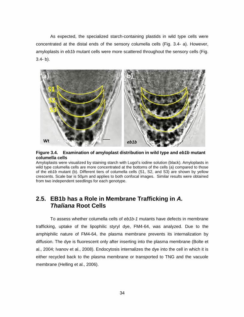
34
As expected, the specialized starch-containing plastids in wild type cells were
concentrated at the distal ends of the sensory columella cells (Fig. 3.4- a). However,
amyloplasts in eb1b mutant cells were more scattered throughout the sensory cells (Fig.
3.4- b).
Figure 3.4. Examination of amyloplast distribution in wild type and eb1b mutant columella cells Amyloplasts were visualized by staining starch with Lugol’s iodine solution (black). Amyloplasts in wild type columella cells are more concentrated at the bottoms of the cells (a) compared to those of the eb1b mutant (b). Different tiers of columella cells (S1, S2, and S3) are shown by yellow crescents. Scale bar is 50µm and applies to both confocal images. Similar results were obtained from two independent seedlings for each genotype.
2.5. EB1b has a Role in Membrane Trafficking in A. Thaliana Root Cells
To assess whether columella cells of eb1b-1 mutants have defects in membrane
trafficking, uptake of the lipophilic styryl dye, FM4-64, was analyzed. Due to the
amphiphilic nature of FM4-64, the plasma membrane prevents its internalization by
diffusion. The dye is fluorescent only after inserting into the plasma membrane (Bolte et
al., 2004; Ivanov et al., 2008). Endocytosis internalizes the dye into the cell in which it is
either recycled back to the plasma membrane or transported to TNG and the vacuole
membrane (Helling et al., 2006).
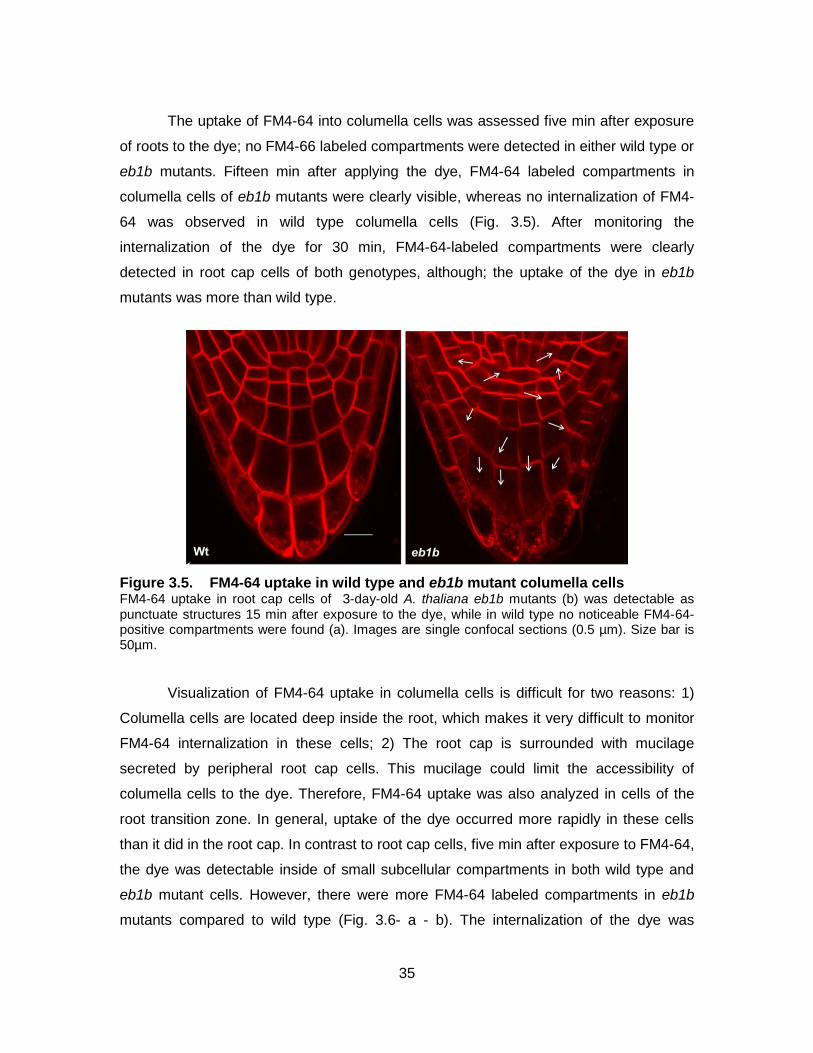
35
The uptake of FM4-64 into columella cells was assessed five min after exposure
of roots to the dye; no FM4-66 labeled compartments were detected in either wild type or
eb1b mutants. Fifteen min after applying the dye, FM4-64 labeled compartments in
columella cells of eb1b mutants were clearly visible, whereas no internalization of FM4-
64 was observed in wild type columella cells (Fig. 3.5). After monitoring the
internalization of the dye for 30 min, FM4-64-labeled compartments were clearly
detected in root cap cells of both genotypes, although; the uptake of the dye in eb1b
mutants was more than wild type.
Figure 3.5. FM4-64 uptake in wild type and eb1b mutant columella cells FM4-64 uptake in root cap cells of 3-day-old A. thaliana eb1b mutants (b) was detectable as punctuate structures 15 min after exposure to the dye, while in wild type no noticeable FM4-64-positive compartments were found (a). Images are single confocal sections (0.5 µm). Size bar is 50µm.
Visualization of FM4-64 uptake in columella cells is difficult for two reasons: 1)
Columella cells are located deep inside the root, which makes it very difficult to monitor
FM4-64 internalization in these cells; 2) The root cap is surrounded with mucilage
secreted by peripheral root cap cells. This mucilage could limit the accessibility of
columella cells to the dye. Therefore, FM4-64 uptake was also analyzed in cells of the
root transition zone. In general, uptake of the dye occurred more rapidly in these cells
than it did in the root cap. In contrast to root cap cells, five min after exposure to FM4-64,
the dye was detectable inside of small subcellular compartments in both wild type and
eb1b mutant cells. However, there were more FM4-64 labeled compartments in eb1b
mutants compared to wild type (Fig. 3.6- a - b). The internalization of the dye was
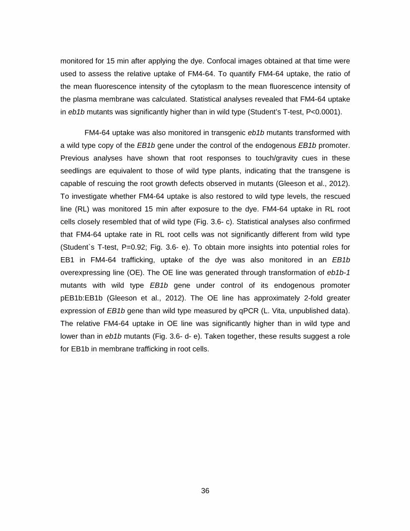
36
monitored for 15 min after applying the dye. Confocal images obtained at that time were
used to assess the relative uptake of FM4-64. To quantify FM4-64 uptake, the ratio of
the mean fluorescence intensity of the cytoplasm to the mean fluorescence intensity of
the plasma membrane was calculated. Statistical analyses revealed that FM4-64 uptake
in eb1b mutants was significantly higher than in wild type (Student’s T-test, P<0.0001).
FM4-64 uptake was also monitored in transgenic eb1b mutants transformed with
a wild type copy of the EB1b gene under the control of the endogenous EB1b promoter.
Previous analyses have shown that root responses to touch/gravity cues in these
seedlings are equivalent to those of wild type plants, indicating that the transgene is
capable of rescuing the root growth defects observed in mutants (Gleeson et al., 2012).
To investigate whether FM4-64 uptake is also restored to wild type levels, the rescued
line (RL) was monitored 15 min after exposure to the dye. FM4-64 uptake in RL root
cells closely resembled that of wild type (Fig. 3.6- c). Statistical analyses also confirmed
that FM4-64 uptake rate in RL root cells was not significantly different from wild type
(Student`s T-test, P=0.92; Fig. 3.6- e). To obtain more insights into potential roles for
EB1 in FM4-64 trafficking, uptake of the dye was also monitored in an EB1b
overexpressing line (OE). The OE line was generated through transformation of eb1b-1
mutants with wild type EB1b gene under control of its endogenous promoter
pEB1b:EB1b (Gleeson et al., 2012). The OE line has approximately 2-fold greater
expression of EB1b gene than wild type measured by qPCR (L. Vita, unpublished data).
The relative FM4-64 uptake in OE line was significantly higher than in wild type and
lower than in eb1b mutants (Fig. 3.6- d- e). Taken together, these results suggest a role
for EB1b in membrane trafficking in root cells.
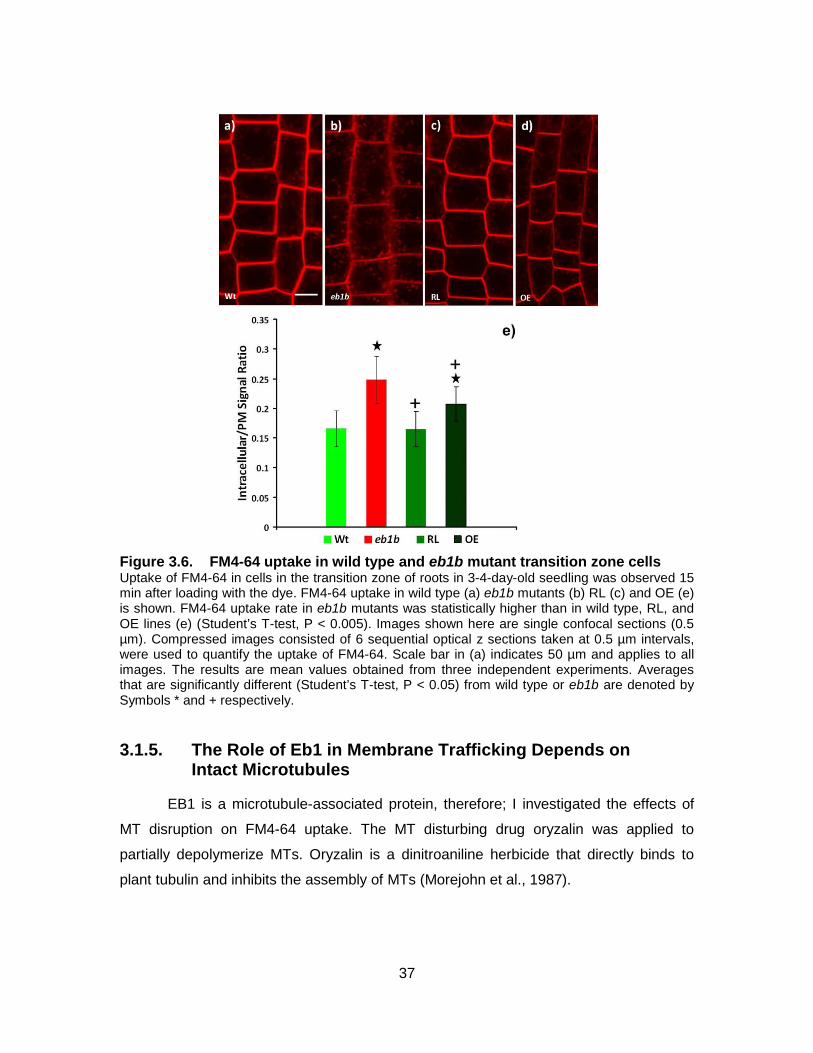
37
Figure 3.6. FM4-64 uptake in wild type and eb1b mutant transition zone cells Uptake of FM4-64 in cells in the transition zone of roots in 3-4-day-old seedling was observed 15 min after loading with the dye. FM4-64 uptake in wild type (a) eb1b mutants (b) RL (c) and OE (e) is shown. FM4-64 uptake rate in eb1b mutants was statistically higher than in wild type, RL, and OE lines (e) (Student’s T-test, P < 0.005). Images shown here are single confocal sections (0.5 µm). Compressed images consisted of 6 sequential optical z sections taken at 0.5 µm intervals, were used to quantify the uptake of FM4-64. Scale bar in (a) indicates 50 µm and applies to all images. The results are mean values obtained from three independent experiments. Averages that are significantly different (Student’s T-test, P < 0.05) from wild type or eb1b are denoted by Symbols * and + respectively.
3.1.5. The Role of Eb1 in Membrane Trafficking Depends on Intact Microtubules
EB1 is a microtubule-associated protein, therefore; I investigated the effects of
MT disruption on FM4-64 uptake. The MT disturbing drug oryzalin was applied to
partially depolymerize MTs. Oryzalin is a dinitroaniline herbicide that directly binds to
plant tubulin and inhibits the assembly of MTs (Morejohn et al., 1987).
38
I first established optimized concentrations of the drug that partially depolymerize
MTs. Based on previously published reports (Baskin et al., 2004; Nakamura et al., 2004)
two different concentrations of the drug were selected: 50 nM, and 200 nM. Root
morphology in 3-day-old wild type seedlings grown on agar medium containing
aforementioned concentrations of oryzalin as well as 0 nM oryzalin (control) were
examined. Roots in control seedlings show normal root morphology with slight radial
expansion in the meristem, as previously reported (Baskin et al., 1994; Fig. 3.7- a).
Seedlings grown on 50 nM oryzalin still display normal root morphology with slight radial
expansion in the meristem as well as the elongation zone (Fig. 3.7- b). However, in
seedlings grown on 200 nM oryzalin, both the meristem and the elongation zone of roots
undergo significant radial expansion. In addition, anisotropic expansion of cells in the
elongation zone was impaired (Fig. 3.7- c). I also assessed the effects of the
aforementioned concentrations of oryzalin (0 nM, 50 nM, and 200 nM) on MT arrays in
root cells of plants expressing GFP-MBD. MTs displayed parallel cortical arrays in
expanding root cells in A. thaliana control seedlings consistent with previously published
reports (Wasteneys, 2002; Fig. 3.7- d). MTs in expanding root cells of seedlings grown
on 50 nM oryzalin were partially depolymerized, but the intact MTs retain their ordered
cortical array (Fig. 3.7- e). MT depolymerization was more severe in 200 nM oryzalin
(Fig. 3.7- f). The degree of MT depolymerization and the impairments to anisotropic
expansion of root cells in seedlings treated with 200 nM oryzalin indicate that this
concentration has a severe effect on roots. Based on these results, I chose 50 nM
oryzalin to investigate the effects of partial disruption of MTs on FM4-64 uptake.
39
Figure 3.7. Effects of different concentration of the microtubule depolymerizing drug oryzalin on cell expansion and MT arrays in the elongation zone of the root Confocal microscopic images of root morphology in wild type seedlings treated with 0 nM (a) 50nM (b), and 200nM oryzalin (c). Mt arrays in the elongation zone of roots in wild type seedlings expressing GFP-MBD construct treated with 0 nM (e) 50 nM (e), and 200 nM oryzalin (f). Scale bars in (a) and (d) indicate 50µM and apply to all images.
To assess the effects of the MT disturbing drug oryzalin on FM4-64 uptake, 3-4-
day-old wild type and eb1b mutants grown on agar medium containing 0nM (control) and
50nM oryzalin were stained with FM4-64 and the uptake of the dye was monitored in
cells of the root transition zone 15 min later. FM4-64 uptake in untreated wild type and
eb1b mutants were in line with my previous observations; FM4-64 uptake in eb1b
mutants was significantly higher than wild type (Fig. 3.8- a) –b)- e). FM4-64 uptake in
wild type treated with 50 nM oryzalin was statistically higher than control cells (Student’s
T-test, P< 0.0001; Fig. 3.8). However, statistical analyses showed no significant
difference between FM4-64 uptake in eb1b mutants grown on 0 nM and 50 nM oryzalin
(Student’s T-test, P=0.35). These results suggest that the role of EB1 in membrane
trafficking depends on intact MTs.
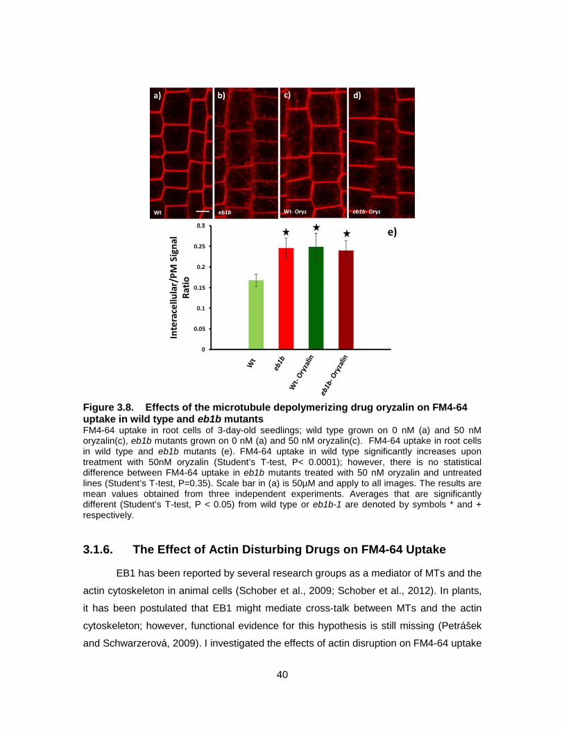
40
Figure 3.8. Effects of the microtubule depolymerizing drug oryzalin on FM4-64 uptake in wild type and eb1b mutants FM4-64 uptake in root cells of 3-day-old seedlings; wild type grown on 0 nM (a) and 50 nM oryzalin(c), eb1b mutants grown on 0 nM (a) and 50 nM oryzalin(c). FM4-64 uptake in root cells in wild type and eb1b mutants (e). FM4-64 uptake in wild type significantly increases upon treatment with 50nM oryzalin (Student’s T-test, P< 0.0001); however, there is no statistical difference between FM4-64 uptake in eb1b mutants treated with 50 nM oryzalin and untreated lines (Student’s T-test, P=0.35). Scale bar in (a) is 50µM and apply to all images. The results are mean values obtained from three independent experiments. Averages that are significantly different (Student’s T-test, P < 0.05) from wild type or eb1b-1 are denoted by symbols * and + respectively.
3.1.6. The Effect of Actin Disturbing Drugs on FM4-64 Uptake
EB1 has been reported by several research groups as a mediator of MTs and the
actin cytoskeleton in animal cells (Schober et al., 2009; Schober et al., 2012). In plants,
it has been postulated that EB1 might mediate cross-talk between MTs and the actin
cytoskeleton; however, functional evidence for this hypothesis is still missing (Petrášek
and Schwarzerová, 2009). I investigated the effects of actin disruption on FM4-64 uptake
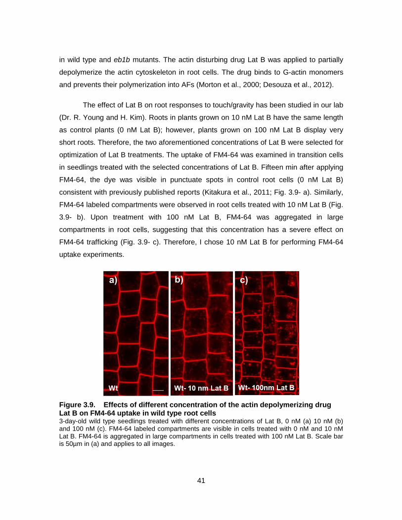
41
in wild type and eb1b mutants. The actin disturbing drug Lat B was applied to partially
depolymerize the actin cytoskeleton in root cells. The drug binds to G-actin monomers
and prevents their polymerization into AFs (Morton et al., 2000; Desouza et al., 2012).
The effect of Lat B on root responses to touch/gravity has been studied in our lab
(Dr. R. Young and H. Kim). Roots in plants grown on 10 nM Lat B have the same length
as control plants (0 nM Lat B); however, plants grown on 100 nM Lat B display very
short roots. Therefore, the two aforementioned concentrations of Lat B were selected for
optimization of Lat B treatments. The uptake of FM4-64 was examined in transition cells
in seedlings treated with the selected concentrations of Lat B. Fifteen min after applying
FM4-64, the dye was visible in punctuate spots in control root cells (0 nM Lat B)
consistent with previously published reports (Kitakura et al., 2011; Fig. 3.9- a). Similarly,
FM4-64 labeled compartments were observed in root cells treated with 10 nM Lat B (Fig.
3.9- b). Upon treatment with 100 nM Lat B, FM4-64 was aggregated in large
compartments in root cells, suggesting that this concentration has a severe effect on
FM4-64 trafficking (Fig. 3.9- c). Therefore, I chose 10 nM Lat B for performing FM4-64
uptake experiments.
Figure 3.9. Effects of different concentration of the actin depolymerizing drug Lat B on FM4-64 uptake in wild type root cells 3-day-old wild type seedlings treated with different concentrations of Lat B, 0 nM (a) 10 nM (b) and 100 nM (c). FM4-64 labeled compartments are visible in cells treated with 0 nM and 10 nM Lat B. FM4-64 is aggregated in large compartments in cells treated with 100 nM Lat B. Scale bar is 50µm in (a) and applies to all images.

42
To assess the effects of Lat B on FM4-64 uptake, 3-day-old wild type and eb1b
mutants grown on agar medium supplemented with 0 nM (control) and 10 nM Lat B were
stained with FM4-64 and uptake of the dye was monitored in transition zone root cells.
FM4-64 uptake in eb1b mutants was higher than wild type, which is in consistent with my
previous results (Fig. 3.10). FM4-64 uptake was significantly higher in wild type grown
on 10 nM Lat B compared to wild type controls. This result was in line with previously
published reports (Du et al., 2011; Nagawa et al., 2012). In contrast, FM4-64 uptake was
dramatically reduced in eb1b mutants treated with 10 nM Lat B (Fig. 3.10- e). Statistical
analyses of FM4-64 uptake revealed that uptake of FM4-64 in eb1b mutants treated with
10 nM Lat B closely resembled that of untreated wild type (Fig. 3.10- g), suggesting that
the actin cytoskeleton in wild type is more destabilized than in eb1b mutants.
Figure 3.10. Effects of actin disturbing drugs on FM4-64 uptake in wild type and eb1b mutant root cells FM4-64 uptake in root cells of 3--day-old seedlings treated with actin disturbing drugs, Wild type (a, b, c) and eb1b mutants (d,e,f). Wild type seedlings treated with 0 nM Lat B and 0 nM Jas (a), 10 nM Lat B (b), and 100 nM Jas (c). eb1b mutant seedlings treated with 0 nM Lat B and 0 nM Jas(d), 10 nM Lat B (e), and 100 nM Jas (f). Statistical analyses of FM4-64 uptake reveals that treatment of wild type with Lat B shows an increase in uptake of the dye, but FM4-64 uptake decreases in eb1b mutants treated with Lat B. Treatment with Jas give rises to an increase in FM4-64 uptake in wild type; however, it does not have any effect on FM4-64 uptake in eb1b mutants (h). The results are mean values obtained from three independent experiments. Error bars indicate (SD). Scale bar in (a) is 50µM and apply to all images. Averages that are significantly different (Student’s T-test, P < 0.05) from wild type or eb1b-1 are denoted by symbols * and + respectively.
43
I also investigated the effects of the actin stabilizing drug Jasplankinolide (Jas)
on FM4-64 uptake in wild type and eb1b mutants. Jas stabilizes and promotes the
polymerization of AFs by binding to F-actin (Bubb et al., 1994). Previous studies on the
effects of the drug on the actin cytoskeleton in live cells suggests that treatment of cells
with 100-150 nM Jas preferentially disrupts cortical AFs, but treatment with 400 nM and
higher results in bundling of AFs (Ou et al., 2002). Based on this report, I chose 100 nM
for partial disruption of AFs.
To investigate the effects of Jas on FM4-64 uptake, 3-4-day-old seedlings grown
with or without Jas were stained with FM4-64 and uptake of the dye in root cells was
observed 15 min after exposure to the dye. In consistent with our previous results, root
cells in eb1b mutants had higher FM4-64 uptake than wild type. Wild type cells treated
with 100 nM Jas showed a dramatic increase in FM4-64 uptake. FM4-64 uptake was not
statistically different in eb1b mutants treated with 0 nM and 100 nM Jas, suggesting that
EB1 in wild type counteracts with Jas and destabilizes the actin cytoskeleton. These
results are in line with our previous results performed with Lat B.
3.1.7. Reversible Effects of BFA on FM4-64 Uptake in Root Cells
To gain additional insights into the function of EB1 on membrane trafficking, I
disrupted membrane trafficking with brefeldin A (BFA) and monitored FM4-64 uptake in
cells of transition root zone. BFA is a drug that is commonly used to study
endomembrane trafficking pathways in plants. It is thought to primarily disrupt a GTPase
exchange factor (GEF) that regulates trafficking between the Golgi, recycling
endosomes, and the plasma membrane (Spano et al., 1999; Ritzenthaler et al., 2002;
Nie et al., 2003; Naramato et al., 2010). In BFA treated Arabidopsis root cells, disruption
of membrane trafficking results in the formation of large compartments, so called BFA-
compartments. I chose to apply 25 µM BFA because this concentration has been shown
to have only slight or no effect on Golgi/ER morphology (Grebe et al., 2003; Lam et al.,
2009). Higher concentrations of the drug (50-100 µM) disrupt the ER (Robinson et al.,
2008; Lam et al., 2009).
3-4-old-day seedlings were incubated in BFA for 30 min and stained with FM4-64
in the presence of BFA for 30 min then visualized with CLSM. BFA-induced FM4-64
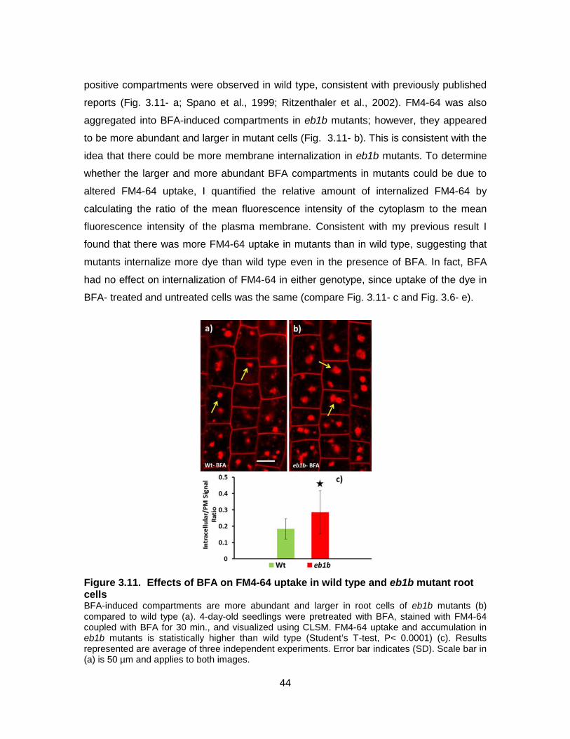
44
positive compartments were observed in wild type, consistent with previously published
reports (Fig. 3.11- a; Spano et al., 1999; Ritzenthaler et al., 2002). FM4-64 was also
aggregated into BFA-induced compartments in eb1b mutants; however, they appeared
to be more abundant and larger in mutant cells (Fig. 3.11- b). This is consistent with the
idea that there could be more membrane internalization in eb1b mutants. To determine
whether the larger and more abundant BFA compartments in mutants could be due to
altered FM4-64 uptake, I quantified the relative amount of internalized FM4-64 by
calculating the ratio of the mean fluorescence intensity of the cytoplasm to the mean
fluorescence intensity of the plasma membrane. Consistent with my previous result I
found that there was more FM4-64 uptake in mutants than in wild type, suggesting that
mutants internalize more dye than wild type even in the presence of BFA. In fact, BFA
had no effect on internalization of FM4-64 in either genotype, since uptake of the dye in
BFA- treated and untreated cells was the same (compare Fig. 3.11- c and Fig. 3.6- e).
Figure 3.11. Effects of BFA on FM4-64 uptake in wild type and eb1b mutant root cells BFA-induced compartments are more abundant and larger in root cells of eb1b mutants (b) compared to wild type (a). 4-day-old seedlings were pretreated with BFA, stained with FM4-64 coupled with BFA for 30 min., and visualized using CLSM. FM4-64 uptake and accumulation in eb1b mutants is statistically higher than wild type (Student’s T-test, P< 0.0001) (c). Results represented are average of three independent experiments. Error bar indicates (SD). Scale bar in (a) is 50 µm and applies to both images.
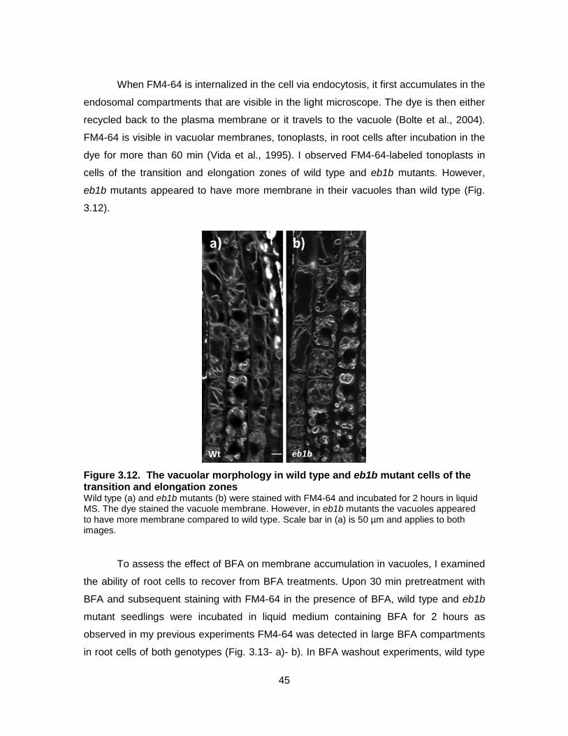
45
When FM4-64 is internalized in the cell via endocytosis, it first accumulates in the
endosomal compartments that are visible in the light microscope. The dye is then either
recycled back to the plasma membrane or it travels to the vacuole (Bolte et al., 2004).
FM4-64 is visible in vacuolar membranes, tonoplasts, in root cells after incubation in the
dye for more than 60 min (Vida et al., 1995). I observed FM4-64-labeled tonoplasts in
cells of the transition and elongation zones of wild type and eb1b mutants. However,
eb1b mutants appeared to have more membrane in their vacuoles than wild type (Fig.
3.12).
Figure 3.12. The vacuolar morphology in wild type and eb1b mutant cells of the transition and elongation zones Wild type (a) and eb1b mutants (b) were stained with FM4-64 and incubated for 2 hours in liquid MS. The dye stained the vacuole membrane. However, in eb1b mutants the vacuoles appeared to have more membrane compared to wild type. Scale bar in (a) is 50 µm and applies to both images.
To assess the effect of BFA on membrane accumulation in vacuoles, I examined
the ability of root cells to recover from BFA treatments. Upon 30 min pretreatment with
BFA and subsequent staining with FM4-64 in the presence of BFA, wild type and eb1b
mutant seedlings were incubated in liquid medium containing BFA for 2 hours as
observed in my previous experiments FM4-64 was detected in large BFA compartments
in root cells of both genotypes (Fig. 3.13- a)- b). In BFA washout experiments, wild type
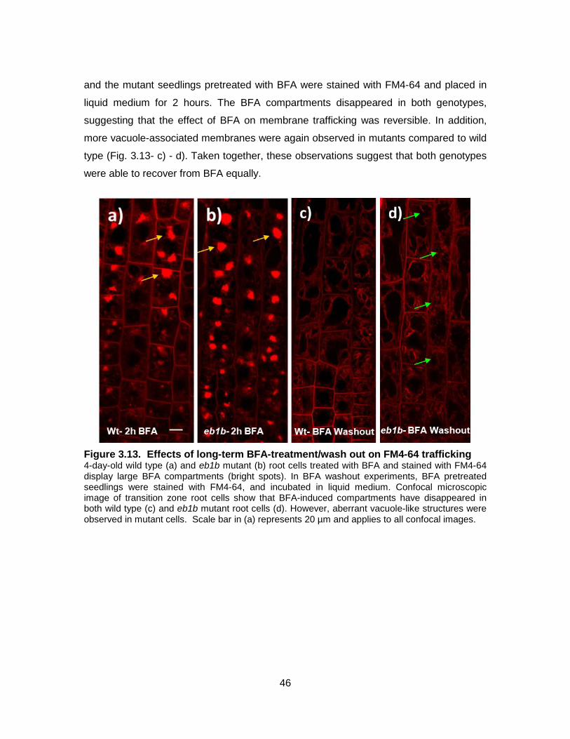
46
and the mutant seedlings pretreated with BFA were stained with FM4-64 and placed in
liquid medium for 2 hours. The BFA compartments disappeared in both genotypes,
suggesting that the effect of BFA on membrane trafficking was reversible. In addition,
more vacuole-associated membranes were again observed in mutants compared to wild
type (Fig. 3.13- c) - d). Taken together, these observations suggest that both genotypes
were able to recover from BFA equally.
Figure 3.13. Effects of long-term BFA-treatment/wash out on FM4-64 trafficking 4-day-old wild type (a) and eb1b mutant (b) root cells treated with BFA and stained with FM4-64 display large BFA compartments (bright spots). In BFA washout experiments, BFA pretreated seedlings were stained with FM4-64, and incubated in liquid medium. Confocal microscopic image of transition zone root cells show that BFA-induced compartments have disappeared in both wild type (c) and eb1b mutant root cells (d). However, aberrant vacuole-like structures were observed in mutant cells. Scale bar in (a) represents 20 µm and applies to all confocal images.
47
4. Discussion
Roots of the eb1b mutant plants exhibit defective responses to touch/gravity
stimuli (Bisgrove et al, 2008; Gleeson et al., 2012). However, the mechanism by which
EB1 contributes in these responses is still unclear. In my thesis, I studied the role of EB1
in root cells at the cellular levels to gain insights into how the function of this protein
inside of root cells could modulate root responses to touch and gravity. By examining the
subcellular architecture and membrane trafficking in the sensory columella cells, I found
that EB1 has a role in membrane trafficking in these cells. I extended my study to root
cells in the transition zone and found that the role of EB1 in membrane trafficking is not
limited to columella cells and, additionally, this role of EB1 depends on intact MTs. To
obtain further insights into the link between EB1 and membrane trafficking, I investigated
the effects of actin disturbing drugs on membrane trafficking in wild type and eb1b
mutants. The results show that EB1 modulates membrane trafficking through an effect
on the actin cytoskeleton. I propose that EB1 might affect root responses to touch and
gravity by regulating the trafficking of membrane transporters, receptors, and ion
channels which are involved in these responses.
4.1. EB1 Modulates Membrane Trafficking in Root Cells
FM4-64 labeled compartments in columella cells in eb1b mutants were detected
earlier than wild type upon staining with the lipophilic dye FM4-64, suggesting that the
internalization of FM4-64 in eb1b mutants is faster than wild type. In addition, the
observation of more FM 4-64 labeled puncta in eb1b mutants than wild type shows that
there is a higher uptake of the dye in eb1b mutants. These results support the
conclusion that EB1 has a role in FM4-64 trafficking in columella cells. FM4-64 is a
marker of the endocytic pathway and membrane trafficking in plant cells (Bolte et al.,
2004; Van Gisbergen et al., 2008). Therefore, these results indicate that EB1 modulates
membrane trafficking in columella cells. I also propose that this role of EB1 in columella
48
cells appears to be relatively specific, since I could not detect any structural or
architectural differences between mutant and wild type columella cells.
I also found that cells in the transition zone of eb1b mutant roots take up more
FM4-64 than transition zone cells in wild type roots. This result demonstrates that the
role of EB1b in membrane trafficking is not confined to columella cells. Another line of
evidence that indicates EB1b has a role in membrane trafficking comes from my
examination of transgenic eb1b mutant plants transformed with wild type copies of the
EB1b gene. When EB1b is expressed at levels equivalent to wild type (Gleeson et al.,
2012), I observed that the internalization of FM4-64 in transition cells was equivalent to
wild type. I also found that partial depolymerisation of MTs increased FM4-64 uptake in
wild type root cells but had no effect on eb1b mutants. This indicates that, as expected
for a MAP, EB1b depends on intact MTs to exert its function in membrane trafficking
pathways.
The finding that FM4–64 uptake was more in eb1b mutant root cells compared to
wild type raised three different possibilities about the role of EB1 in membrane trafficking
in wild type root cells (Fig. 1.4): EB1 might negatively regulate endocytosis (route 1 in
Fig. 1.4), EB1 might promote exocytosis (route 2 in Fig. 1.4), and EB1 might promote
vesicle trafficking from endosomes/TNG to vacuoles (route 3 in Fig. 1.4).
To examine these possibilities, I first treated wild type and eb1b mutants with a
low concentration of the endomembrane trafficking disturbing drug BFA. It has been
reported that the low concentration of this drug has a reversible inhibitory effect on
trafficking between the Golgi, recycling endosomes, and the plasma membrane (Spano
et al., 1999; Ritzenthaler et al., 2002; Nie et al., 2003; Naramato et al., 2010).
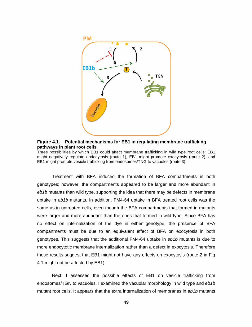
49
Figure 4.1. Potential mechanisms for EB1 in regulating membrane trafficking pathways in plant root cells Three possibilities by which EB1 could affect membrane trafficking in wild type root cells: EB1 might negatively regulate endocytosis (route 1), EB1 might promote exocytosis (route 2), and EB1 might promote vesicle trafficking from endosomes/TNG to vacuoles (route 3).
Treatment with BFA induced the formation of BFA compartments in both
genotypes; however, the compartments appeared to be larger and more abundant in
eb1b mutants than wild type, supporting the idea that there may be defects in membrane
uptake in eb1b mutants. In addition, FM4-64 uptake in BFA treated root cells was the
same as in untreated cells, even though the BFA compartments that formed in mutants
were larger and more abundant than the ones that formed in wild type. Since BFA has
no effect on internalization of the dye in either genotype, the presence of BFA
compartments must be due to an equivalent effect of BFA on exocytosis in both
genotypes. This suggests that the additional FM4-64 uptake in eb1b mutants is due to
more endocytotic membrane internalization rather than a defect in exocytosis. Therefore
these results suggest that EB1 might not have any effects on exocytosis (route 2 in Fig
4.1 might not be affected by EB1).
Next, I assessed the possible effects of EB1 on vesicle trafficking from
endosomes/TGN to vacuoles. I examined the vacuolar morphology in wild type and eb1b
mutant root cells. It appears that the extra internalization of membranes in eb1b mutants
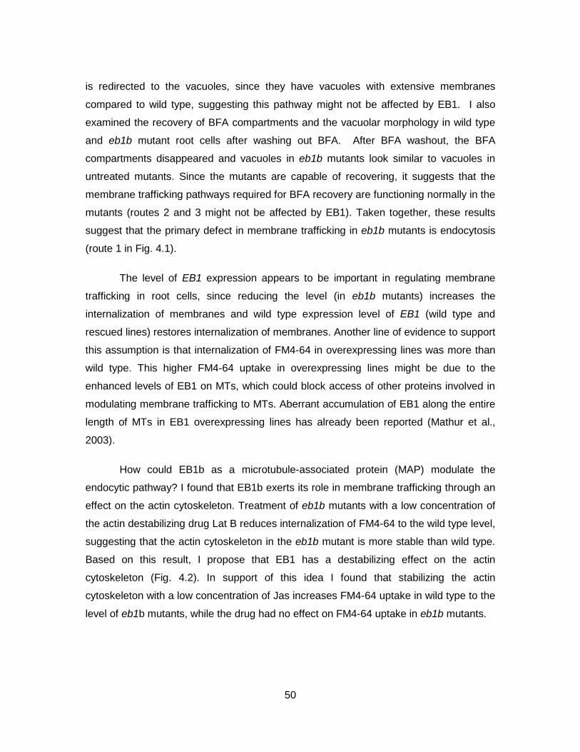
50
is redirected to the vacuoles, since they have vacuoles with extensive membranes
compared to wild type, suggesting this pathway might not be affected by EB1. I also
examined the recovery of BFA compartments and the vacuolar morphology in wild type
and eb1b mutant root cells after washing out BFA. After BFA washout, the BFA
compartments disappeared and vacuoles in eb1b mutants look similar to vacuoles in
untreated mutants. Since the mutants are capable of recovering, it suggests that the
membrane trafficking pathways required for BFA recovery are functioning normally in the
mutants (routes 2 and 3 might not be affected by EB1). Taken together, these results
suggest that the primary defect in membrane trafficking in eb1b mutants is endocytosis
(route 1 in Fig. 4.1).
The level of EB1 expression appears to be important in regulating membrane
trafficking in root cells, since reducing the level (in eb1b mutants) increases the
internalization of membranes and wild type expression level of EB1 (wild type and
rescued lines) restores internalization of membranes. Another line of evidence to support
this assumption is that internalization of FM4-64 in overexpressing lines was more than
wild type. This higher FM4-64 uptake in overexpressing lines might be due to the
enhanced levels of EB1 on MTs, which could block access of other proteins involved in
modulating membrane trafficking to MTs. Aberrant accumulation of EB1 along the entire
length of MTs in EB1 overexpressing lines has already been reported (Mathur et al.,
2003).
How could EB1b as a microtubule-associated protein (MAP) modulate the
endocytic pathway? I found that EB1b exerts its role in membrane trafficking through an
effect on the actin cytoskeleton. Treatment of eb1b mutants with a low concentration of
the actin destabilizing drug Lat B reduces internalization of FM4-64 to the wild type level,
suggesting that the actin cytoskeleton in the eb1b mutant is more stable than wild type.
Based on this result, I propose that EB1 has a destabilizing effect on the actin
cytoskeleton (Fig. 4.2). In support of this idea I found that stabilizing the actin
cytoskeleton with a low concentration of Jas increases FM4-64 uptake in wild type to the
level of eb1b mutants, while the drug had no effect on FM4-64 uptake in eb1b mutants.
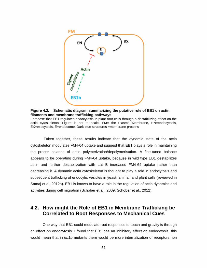
51
Figure 4.2. Schematic diagram summarizing the putative role of EB1 on actin filaments and membrane trafficking pathways I propose that EB1 regulates endocytosis in plant root cells through a destabilizing effect on the actin cytoskeleton. Figure is not to scale. PM= the Plasma Membrane, EN=endocytosis, EX=exocytosis, E=endosome, Dark blue structures =membrane proteins
Taken together, these results indicate that the dynamic state of the actin
cytoskeleton modulates FM4-64 uptake and suggest that EB1 plays a role in maintaining
the proper balance of actin polymerization/depolymerisation. A fine-tuned balance
appears to be operating during FM4-64 uptake, because in wild type EB1 destabilizes
actin and further destabilization with Lat B increases FM4-64 uptake rather than
decreasing it. A dynamic actin cytoskeleton is thought to play a role in endocytosis and
subsequent trafficking of endocytic vesicles in yeast, animal, and plant cells (reviewed in
Samaj et al, 2012a). EB1 is known to have a role in the regulation of actin dynamics and
activities during cell migration (Schober et al., 2009; Schober et al., 2012).
4.2. How might the Role of EB1 in Membrane Trafficking be Correlated to Root Responses to Mechanical Cues
One way that EB1 could modulate root responses to touch and gravity is through
an effect on endocytosis. I found that EB1 has an inhibitory effect on endocytosis, this
would mean that in eb1b mutants there would be more internalization of receptors, ion
52
channels, or transporters that are involved in root responses to touch and gravity. For
instance, PIN transporters are one of the major determinants in the polar transportation
of auxin, which is known to have a key role in root responses to environmental cues
(Zazimalova et al., 2010). In eb1b mutants, there would be more internalization of these
transporters and this is likely to affect their ability to relocalize in response to a cue.
Recent reports have shown that some MAPs are involved in polar PIN localization at the
plasma membrane (Nagawa et al., 2012; Ambrose et al., 2013; Kakar et al., 2013). I also
found that EB1 affects endocytosis through the actin cytoskeleton. Intact actin arrays are
important for PIN recycling and localization (Nagawa et al., 2012), supporting the idea
that EB1b might regulate PIN trafficking through an effect on the actin cytoskeleton.
Alternatively, EB1 could modulate root responses to gravity by affecting
amyloplasts sedimentation. My results suggest that EB1 has a destabilizing effect on the
actin cytoskeleton. This means that in eb1b columella cells actin filaments would be
more stable. Given that it has been reported that actin filaments form a network that
restricts amyloplast sedimentation through the cytoplasm of columella cells (Yamamoto
and Kiss, 2002; Hou et al., 2003), it is plausible that a more stable array of actin in eb1b
columella cells would be more restrictive than in wild type. Amyloplast sedimentation
would be reduced in eb1b mutants and this would slow down their responses to gravity.
This could in turn affect root responses to mechanical cues. The root bends away when
it encounters an obstacle and must be redirected towards gravity after bypassing the
obstacle; if response to gravity is delayed in eb1b mutants it could result in an
exaggerated response to the mechanical cue.
53
5. Conclusion and Future Prospects
The aim of this thesis was to investigate the role of EB1 in root cells that might be
correlated to root responses to touch and gravity. To sum up, my major finding is that
EB1b modulates membrane trafficking in root cells through destabilization of the actin
cytoskeleton. I also found that, surprisingly, there are no defects in the cytoarchitecture
of sensory columella cells in eb1b mutants, suggesting that the role of EB1 in columella
cells might be limited to endomembrane organization. I propose that the role of EB1 in
membrane trafficking is the mechanism by which it affects root responses to
touch/gravity cues.
I found that EB1 regulates endocytosis in root cells. Although both clathrin-
dependent and clathrin-independent endocytotic events have been reported in plant
cells, the clathrin mediated pathway is the prominent pathway for endocytosis in plant
cells (reviewed in Chen et al., 2011). This raises the question of whether EB1 affects the
clathrin-dependent pathway in root cells. One way to answer this question is by
monitoring FM4-64 uptake in plants expressing a clathrin marker in which clathrin light
chain is tagged with GFP (CLC-GFP; Konopka et al., 2008; Robert et al., 2010). FM4-64
uptake would be monitored in wild type and eb1b mutant plants expressing CLC-GFP.
Quantitative analysis of the co-localization between FM4-64 labeled compartments and
CLC-GFP in the cytoplasm in wild type and eb1b mutant root cells would provide
insights into the possible effects of EB1 on the clathrin-dependent pathway.
I propose that EB1 affects root responses to touch and gravity by modulating
trafficking of membrane proteins including transporters, receptors, and ion channels. The
importance of PIN trafficking on root responses to various environmental stimuli has
been well established (reviewed in Kleine-Vehn and Friml, 2008). PIN trafficking plays a
key role in directional cell-to-cell movement of auxin in root cells. Polar transport and
differential distribution of auxin is important in root responses to environmental cues
including gravity and mechanical cues (Raven, 1975; Estelle, 1998; Friml et al., 2002;
54
Michniewicz et al., 2007). In roots, PIN3 in columella cells and PIN2 in epidermal and
cortical cells facilitate the transport of auxin in and across root cells. An interesting
question that needs to be addressed is whether the role of EB1 in membrane trafficking
could affect PIN trafficking in root cells. One way to answer this question would be to
monitor PIN trafficking in wild type and eb1b mutant plants expressing PIN-GFP.
Quantitative analysis of the abundance of GFP-tagged PIN at the plasma membrane
and in the cytoplasm in wild type and eb1b mutant root cells would reveal whether EB1
affects PIN trafficking. These studies would assess the putative role of EB1 in auxin
movement in and across root cells which could be correlated to the function of EB1 in
root responses to touch and gravity.
Our results suggest that EB1 modulates membrane trafficking pathways.
Membrane trafficking has been implicated in plant responses to a variety of biotic and
abiotic stresses (Robatzek et al., 2006; Kleine-Vehn and Friml, 2008). Future research
could reveal whether EB1 has a role in root responses to other environmental cues
including pathogens. In plants, the membrane localized receptor-like kinase
FLAGELLIN-SENSING 2 (FLS2) recognizes and binds to a segment of a peptide called
flagellin secreted by pathogenic bacteria. After ligand binding, FLS2 is internalized and
degraded. This FLS2 trafficking is important for plant immunity responses (Robatzek et
al., 2006). It would be interesting to compare endocytosis of FLS2 in wild type and eb1b
mutant root cells as well as plant immunity to pathogens in both genotypes. GFP-FLS2
plants (Robatzek et al., 2006; Beck et al., 2012) would be used to monitor localization of
FLS2 at the plasma membrane and in the cytoplasm.
Another question that needs to be answered is whether EB1 activity is needed in
the root cap, the elongation zone or both during root responses to touch and gravity.
One way to answer this question would be to express EB1 protein in eb1b mutant root
cap cells and disturb expression from the EB1 gene in wild type root cap cells. A root
cap specific promoter, RCP1 (Tsugeki and Fedoroff, 1999) could be used to drive
expression from the EB1b gene solely in root caps of eb1b mutants. The same promoter
could be used to drive expression of an RNAi construct to reduce EB1 protein levels
specifically in root cap cells of wild type plants. Analyses of touch/gravity responses in
these plants could shed light on in which zone of the root EB1 is required for proper root
responses to touch and gravity. Measurement of FM4-64 uptake in root cap and cells in
55
the elongation zone of these plants would provide insights into a possible correlation
between the function of EB1 in endocytosis and its role in root responses to touch and
gravity.
I found that the role of EB1 in endocytosis depends on intact MTs. Another open
question that needs to be addressed is whether EB1 activities in root responses to touch
and gravity depend on intact MTs. It would be interesting to investigate the effects of the
MT depolymerizing drug oryzalin on root responses to touch and gravity in wild type and
eb1b mutants.
Another question that needs to be addressed is whether EB1 exerts its functions
in membrane trafficking and root responses to touch and gravity through interaction with
additional proteins or through regulation of MT dynamics. To answer this question, a
truncated version of EB1 that is missing the domain that mediates interactions with other
proteins (EBH) could be transformed into eb1b mutants. Also, a truncated EB1 gene that
is missing CH domain could be transformed into eb1b mutants. Assessment of FM4-64
uptake in root cells of the transformed plants as well as root responses to touch and
gravity in these plants would reveal whether EB1 activities in membrane trafficking and
root responses to touch and gravity require an interaction with MTs or additional
proteins.
Another line of investigation would be to identify proteins that interact with EB1 in
plant cells. Several approaches could be used to identify these proteins. This includes
yeast hybrid assay as well as protein pull-down assay followed by mass spectrometry.
Based on studies in other organisms (Jiang et al., 2012), there is likely to be a large
number of proteins capable of interacting with EB1 in plant cells. In this thesis I reported
that EB1 affects actin dynamics in root cells. It would be interesting to examine this list of
proteins that interact with the actin cytoskeleton. Indeed, in other organisms several
EB1-binding proteins have been found that regulate actin dynamics and activities (Liu et
al., 2011). Potential proteins that bind to EB1 and interact with the actin cytoskeleton in
plant cells could be further investigated by fluorescently tagging these proteins and EB1
and examining their interaction using live cell imaging approaches.

56
Finally, I propose that more stabilized AFs in eb1b columella cells could affect
amyloplast mobility and sedimentation in gravistimulated roots. In future investigations, it
might be possible to measure the velocity of amyloplast sedimentation in gravi-
stimulated roots in wild type and eb1b mutant.
57
References
Akhmanova A, Steinmetz MO (2008) Tracking the ends: a dynamic protein network controls the fate of microtubule tips. Nat Rev Mol Cell Biol. 9: 309-322
Amann KJ, Pollard TD (2001) Direct real-time observation of actin filament branching mediated by Arp2/3 complex using total internal reflection fluorescence microscopy. Proc Nat Acad Sci. 98: 15009-15013
Ambrose C, Ruan Y, Gardiner J, Tamblyn Laura M, Catching A, Kirik V, Marc J, Overall R, Wasteneys Geoffrey O (2013) CLASP interacts with Sorting Nexin 1 to link microtubules and auxin transport via PIN2 recycling in Arabidopsis thaliana. Devel Cell. 24: 649-659
Ananthakrishnan R EA (2007) The forces behind cell movement. Int J Biol Sci. 5: 303-317
Apel K, Hirt H (2004) Reactive oxygen species: metabolism, oxidative stress, and signal transduction. Annu Rev Plant Biol. 55: 373-399
Baluska F, Volkmann D, Barlow PW (1996) Specialized zones of development in roots: View from the cellular level. Plant Physiol. 112: 3-4
Baskin T I WJE, Cork A, Williamson R E (1994) Morphology and microtubule organization in Arabidopsis roots exposed to oryzalin or taxol. Plant Cell Physiol. 35: 935-942
Baskin TI BG, Judy-March JE, Marga F (2004) Disorganization of cortical microtubules stimulates tangential expansion and reduces the uniformity of cellulose microfibril alignment among cells in the root of Arabidopsis. Plant Physiol. 135: 2279-2290
Beck M, Zhou J, Faulkner C, MacLean D, Robatzek S (2012) Spatio-temporal cellular dynamics of the Arabidopsis flagellin receptor reveal activation status-dependent endosomal sorting. Plant Cell. 24: 4205-4219
Bednarek SY, Falbel TG (2002) Membrane trafficking during plant cytokinesis. Traffic 3: 621-629
Bennett M, Marchant A, Green H, May S, Ward S, Millner P, Walker A, Schulz B, Feldmann K (1996) Arabidopsis AUX1 gene: A permease-like regulator of root gravitropism. Science 273: 948-950
Bisgrove SR (2008) The roles of microtubules in tropisms. Plant Sci. 175: 747-755
58
Bisgrove SR, Lee Y-RJ, Liu B, Peters NT, Kropf DL (2008) The microtubule plus-end binding protein EB1 functions in root responses to touch and gravity signals in Arabidopsis. Plant Cell. 20: 396-410
Blancaflor EB (2012) Regulation of plant gravity sensing and signalling by the actin cytoskeleton. Am J Bot. 100: 143-152
Blancaflor EB, Fasano JM, Gilroy S (1998) Mapping the functional roles of cap cells in the response of Arabidopsis primary roots to gravity. Plant Physiol. 116: 213-222
Blancaflor EB, Hasenstein KH (1997) The organization of the actin cytoskeleton in vertical and graviresponding primary roots of maize. Plant Physiol. 113: 1447-1455
Bloch D, Lavy M, Efrat Y, Efroni I, Bracha-Drori K, Abu-Abied M, Sadot E, Yalovsky S (2005) Ectopic expression of an activated Rac in Arabidopsis disrupts membrane cycling. Mol Biol Cell. 16: 1913-1927
Bolte S, Talbot C, Boutte Y, Catrice O, Read ND, Satiat-Jeunemaitre B (2004) FM-dyes as experimental probes for dissecting vesicle trafficking in living plant cells. J Mic. 214: 159-173
Bubb MR SA, Sausville EA, Duncan KL, Korn ED. (1994) Jasplakinolide, a cytotoxic natural product, induces actin polymerization and competitively inhibits the binding of phalloidin to F-actin. J Biol Chem. 269: 14869-14871
Buey RM MR, Leslie K, Walzthoeni T, Missimer JH, Menzel A, Bjelic S, Bargsten K, Grigoriev I, Smal I, Meijering E, Aebersold R, Akhmanova A, Steinmetz MO (2011) Insights into EB1 structure and the role of its C-terminal domain for discriminating microtubule tips from the lattice. Mol Biol Cell. 22: 2912-2923
Calligaris D, Verdier-Pinard P, Devred F, Villard C, Braguer D, Lafitte D (2010) Microtubule targeting agents: from biophysics to proteomics. Cell Mol Life Sci. 67: 1089-1104
Caspar T PB (1989) Gravitropism in a starchless mutant of Arabidopsis : Implications for the starch-statolith theory of gravity sensing. Planta. 177: 185-197
Chan J, Calder GM, Doonan JH, Lloyd CW (2003) EB1 reveals mobile microtubule nucleation sites in Arabidopsis. Nat Cell Biol. 5: 967-971
Chaudhry F, Guérin C, von Witsch M, Blanchoin L, Staiger CJ (2007) Identification of Arabidopsis Cyclase-associated Protein 1 as the first nucleotide exchange factor for plant actin. Mol Biol Cell. 18: 3002-3014
Chen R, Rosen E, Masson PH (1999) Gravitropism in higher plants. Plant Physiol. 120: 343-350
Chen X, Irani NG, Friml J (2011) Clathrin-mediated endocytosis: The gateway into plant cells. Curr Opin Plant Biol. 14: 674-682
59
Cheung AY, de Vries SC (2008) Membrane trafficking: Intracellular highways and country roads. Plant Physiol. 147: 1451-1453
Claxton NS, Fellers, T. J., and Davidson, M. W. (2006) Laser scanning confocal microscopy; department of optical microscopy and digital imaging. National High Magnetic Field Laboratory, Florida State University 33
Clough SJ, Bent AF (1998) Floral dip: a simplified method for Agrobacterium-mediated transformation of Arabidopsis thaliana. Plant J. 16: 735-743
Collings DA, Zsuppan G, Allen NS, Blancaflor EB (2001) Demonstration of prominent actin filaments in the root columella. Planta 212: 392-403
Connerly PL (2010) How do proteins move through the Golgi apparatus? Nature Education 3: 60
Contento AL, Bassham DC (2012) Structure and function of endosomes in plant cells. J Cell Sci. 125: 3511-3518
Desouza M GPW, Stehn J R (2012) The actin cytoskeleton as a sensor and mediator of apoptosis. Bioarchitecture. 23: 75-87
Dhamodharan R JM, Thrower D, Wilson L, Wadsworth P (1995) Vinblastine suppresses dynamics of individual microtubules in living interphase cells. Mol Biol Cell. 6: 1215-1229
Dhonukshe P, Aniento F, Hwang I, Robinson DG, Mravec J, Stierhof Y-D, Friml J (2007) Clathrin-mediated constitutive endocytosis of PIN auxin efflux carriers in Arabidopsis. Curr Biol. 17: 520-527
Dhonukshe P, Tanaka H, Goh T, Ebine K, Mahonen AP, Prasad K, Blilou I, Geldner N, Xu J, Uemura T, Chory J, Ueda T, Nakano A, Scheres B, Friml J (2008) Generation of cell polarity in plants links endocytosis, auxin distribution and cell fate decisions. Nature 456: 962-966
Dhonukshe P GI, Fischer R, Tominaga M, Robinson DG, Hasek J, Paciorek T, Petrásek J, Seifertová D, Tejos R, Meisel LA, Zazímalová E, Gadella TW Jr, Stierhof YD, Ueda T, Oiwa K, Akhmanova A, Brock R, Spang A, Friml J. (2008) Auxin transport inhibitors impair vesicle motility and actin cytoskeleton dynamics in diverse eukaryotes. Proc Nat Acad Sci. 105: 4489-4494
Dolan L, Janmaat K, Willemsen V, Linstead P, Poethig S, Roberts K, Scheres B (1993) Cellular organisation of the Arabidopsis thaliana root. Development. 119: 71-84
Driss-Ecole D, Lefranc A, Perbal G (2003) A polarized cell: the root statocyte. Physiol Plant. 118: 305-312
Du C, Xu Y, Wang Y, Chong K (2011) Adenosine diphosphate ribosylation factor-GTPase-activating protein stimulates the transport of AUX1 endosome, which
60
relies on actin cytoskeletal organization in rice root development. J Integrat Plant Biol. 53: 698-709
Esmon C A PUV, Liscum E (2005) Plant tropisms: Providing the power of movement to a sessile organism. Int J Dev Biol. 49: 665-674
Estelle M (1998) Polar Auxin transport: new support for an old model. Plant Cell. 10: 1775-1778
Fitzelle KJ, & Kiss, J.Z (2001) Restoration of gravitropic sensitivity in starch-deficient mutants of Arabidopsis by hypergravity. J Expt Bot. 52: 265-275
Friml J, Vieten A, Sauer M, Weijers D, Schwarz H, Hamann T, Offringa R, Jurgens G (2003) Efflux-dependent auxin gradients establish the apical-basal axis of Arabidopsis. Nature 426: 147-153
Friml J, Wisniewska J, Benkova E, Mendgen K, Palme K (2002) Lateral relocation of auxin efflux regulator PIN3 mediates tropism in Arabidopsis. Nature 415: 806-809
Gleeson L, Squires S, Bisgrove SR (2012) The microtubule associated protein END BINDING1 represses root responses to mechanical cues. Plant Sci. 187: 1-9
Gopinathan A, Lee KC, Schwarz JM, Liu AJ (2007) Branching, capping, and severing in dynamic actin structures. Phys Rev Lett. 99: 58103
Granger CL, Cyr RJ (2001) Spatiotemporal relationships between growth and microtubule orientation as revealed in living root cells ofArabidopsis thaliana transformed with green-fluorescent-protein gene constructGFP-MBD. Protoplasma 216: 201-214
Grebe M, Xu J, Möbius W, Ueda T, Nakano A, Geuze HJ, Rook MB, Scheres B (2003) Arabidopsis sterol endocytosis involves actin-mediated trafficking via ARA6-positive early endosomes. Curr Biol. 13: 1378-1387
Grieneisen VA, Xu J, Maree AFM, Hogeweg P, Scheres B (2007) Auxin transport is sufficient to generate a maximum and gradient guiding root growth. Nature 449: 1008-1013
Griffing LR (2008) FRET analysis of transmembrane flipping of FM4–64 in plant cells: Is FM4–64 a robust marker for endocytosis? J Mic. 231: 291-298
Heisler MG, Ohno C, Das P, Sieber P, Reddy GV, Long JA, Meyerowitz EM (2005) Patterns of auxin transport and gene expression during primordium development revealed by live imaging of the Arabidopsis inflorescence meristem. Curr Biol. 15: 1899-1911
Helling D, Possart A, Cottier Sp, Klahre U, Kost B (2006) Pollen tube tip growth depends on plasma membrane polarization mediated by Tobacco PLC3 activity and endocytic membrane recycling. Plant Cell. 18: 3519-3534
61
Hensel W (1984) Microtubules in statocytes from roots of cress (Lepidium sativum L.). Protoplasma 119: 121-134
Hensel W (1986) Cytodifferentiation of polar plant cells: use of anti-microtubular agents during the differentiation of statocytes from cress roots (Lepidium sativum L.). Planta. 169: 293-303
Henty-Ridilla JL, Shimono M, Li J, Chang JH, Day B, Staiger CJ (2013) The plant actin cytoskeleton responds to signals from microbe-associated molecular patterns. PLoS Pathog. 9: e1003290
Hilaire E, Paulsen AQ, Brown CS, Guikema JA (1995) Microgravity and clinorotation cause redistribution of free calcium in sweet clover columella cells. Plant Cell Physiol. 36: 831-837
Honnappa S, Gouveia SM, Weisbrich A, Damberger FF, Bhavesh NS, Jawhari H, Grigoriev I, van Rijssel FJA, Buey RM, Lawera A, Jelesarov I, Winkler FK, Wüthrich K, Akhmanova A, Steinmetz MO (2009) An EB1-Binding motif acts as a microtubule tip localization signal. Cell. 138: 366-376
Hou G MD, Blancaflor EB (2003) Enhanced gravitropism of roots with a disrupted cap actin cytoskeleton. Plant Physiol. 131: 1360-1373
Iijima M, Higuchi T, Barlow PW (2004) Contribution of root cap mucilage and presence of an intact root cap in maize (Zea mays) to the reduction of soil mechanical impedance. Ann Bot. 94: 473-477
Iijima M MS, Barlow PW (2008) Structure and function of root cap. Plant Prod Sci. 11: 17-27
Ivanov A, Verstreken P, Ohyama T, Bellen H (2008) FM 1-43 Labeling of synaptic vesicle pools at the Drosophila neuromuscular junction. In Exocytosis and Endocytosis, Vol 440. Humana Press, pp 349-369
Jiang K TG, Montenegro Gouveia S, Davey NE, Hua S, van der Vaart B, Grigoriev I, Larsen J, Pedersen LB, Bezstarosti K, Lince-Faria M, Demmers J, Steinmetz MO, Gibson TJ, Akhmanova A (2012) A Proteome-wide screen for mammalian SxIP motif-containing microtubule plus-end tracking proteins. Curr Biol. 22: 1800-1807
Joo JH, Bae YS, Lee JS (2001) Role of auxin-induced reactive oxygen species in root gravitropism. Plant Physiol. 126: 1055-1060
Juniper BE, Groves S, Landau-Schachar B, Audus LJ (1966) Root cap and the perception of gravity. Nature 209: 93-94
Kuroda K (1990) Cytoplasmic streaming in plant cells. Int Rev Cytol. 121: 267-307
Kakar K, Zhang H, Scheres B, Dhonukshe P (2013) CLASP-mediated cortical microtubule organization guides PIN polarization axis. Nature 495: 529-533
62
Kawamura E, Himmelspach R, Rashbrooke MC, Whittington AT, Gale KR, Collings DA, Wasteneys GO (2006) MICROTUBULE ORGANIZATION 1 regulates structure and function of microtubule arrays during mitosis and cytokinesis in the Arabidopsis root. Plant Physiol. 140: 102-114
Kimbrough JM, Salinas-Mondragon R, Boss WF, Brown CS, Sederoff HW (2004) The fast and transient transcriptional network of gravity and mechanical stimulation in the Arabidopsis root apex. Plant Physiol. 136: 2790-2805
Kirchner K, Mandelkow EM (1985) Tubulin domains responsible for assembly of dimers and protofilaments. EMBO J. 4: 2397–2402
Kiss JZ (2000) Mechanisms of the early phases of plant gravitropism. Crit Rev Plant Sci. 19: 551-573
Kiss JZ, Wright JB, Caspar T (1996) Gravitropism in roots of intermediate-starch mutants of Arabidopsis. Physiol Plant. 97: 237-244
Kitakura S VS, Robert S, Löfke C, Teichmann T, Tanaka H, Friml J (2011) Clathrin mediates endocytosis and polar distribution of PIN auxin transporters in Arabidopsis. Plant Cell. 23: 1920-1931
Kleine-Vehn J, Friml J (2008) Polar targeting and endocytic recycling in auxin-dependent plant development. Ann Rev Cell Develop Biol. 24: 447-473
Kleine-Vehn J, Ding Z, Jones AR, Tasaka M, Morita MT, Friml J (2010) Gravity-induced PIN transcytosis for polarization of auxin fluxes in gravity-sensing root cells. Proc Nat Acad Sci. 107: 22344–22349.
Komaki S AT, Coutuer S, Inzé D, Russinova E, Hashimoto T (2010) Nuclear-localized subtype of end-binding 1 protein regulates spindle organization in Arabidopsis. J Cell Sci. 123: 451-459
Komarova Y LG, Galjart N, Grosveld F, Borisy GG, Akhmanova A (2005) EB1 and EB3 control CLIP dissociation from the ends of growing microtubules. Mol Biol Cell. 16: 5334-5345
Konopka CA, Backues SK, Bednarek SY (2008) Dynamics of Arabidopsis dynamin-related protein 1C and a clathrin light chain at the plasma membrane. Plant Cell 20: 1363-1380
Koornneef M, Meinke D (2010) The development of Arabidopsis as a model plant. Plant J. 61: 909-921
Kuroda K, Friedlander KWJaM (1990) Cytoplasmic streaming in plant cells. Intern Rev Cytology. 121: 267-307
Lam D, Levraud J-P, Luciani M-Fo, Golstein P (2007) Autophagic or necrotic cell death in the absence of caspase and bcl-2 family members. Biochem Biophys Res Commun. 363: 536-541
63
Lam SK, Cai Y, Tse YC, Wang J, Law AHY, Pimpl P, Chan HYE, Xia J, Jiang L (2009) BFA-induced compartments from the Golgi apparatus and trans-Golgi network/early endosome are distinct in plant cell. Plant J. 60: 865-881
Leitz G, Kang B-H, Schoenwaelder MEA, Staehelin LA (2009) Statolith sedimentation kinetics and force transduction to the cortical endoplasmic reticulum in gravity-sensing Arabidopsis columella cells. Plant Cell. 21: 843-860
Littlefield R, Almenar-Queralt A, Fowler VM (2001) Actin dynamics at pointed ends regulates thin filament length in striated muscle. Nat Cell Biol. 3: 544-551
Liu B, Hashimoto T (2011) Microtubule and cell shape determination. In The Plant Cytoskeleton, Vol 2. Springer New York, pp 245-257
Liu B, Young R, Bisgrove S (2011) Microtubule plus end-tracking proteins and their activities in plants. In The Plant Cytoskeleton, Vol 2. Springer New York, pp 95-117
MacCleery SA, Kiss JZ (1999) Plastid sedimentation kinetics in roots of wild-type and starch-deficient mutants of Arabidopsis. Plant Physiol. 120: 183-192
Mancuso S BP, Volkmann D, Baluska F (2006) Actin turnover-mediated gravity response in maize root apices: gravitropism of decapped roots implicates gravisensing outside of the root cap. Plant Signal Behav. 1: 52-58
Marc J GC, Brincat J, Fisher DD, Kao Th, McCubbin AG, Cyr RJ (1998) A GFP-MAP4 reporter gene for visualizing cortical microtubule rearrangements in living epidermal cells. Plant Cell. 10: 1927-1939
Massa GD, Gilroy S (2003) Touch modulates gravity sensing to regulate the growth of primary roots of Arabidopsis thaliana. Plant J. 33: 435 - 445
Mathur J MN, Kernebeck B, Srinivas BP, Hülskamp M (2003) A novel localization pattern for an EB1-like protein links microtubule dynamics to endomembrane organization. Curr Biol. 13: 1991-1997
Mayor S, Pagano RE (2007) Pathways of clathrin-independent endocytosis. Nat Rev Mol Cell Biol. 8: 603-612
Miaczynska M, Stenmark H (2008) Mechanisms and functions of endocytosis. J Cell Biol. 180: 7-11
Michniewicz M, Zago MK, Abas L, Weijers D, Schweighofer A, Meskiene I, Heisler MG, Ohno C, Zhang J, Huang F, Schwab R, Weigel D, Meyerowitz EM, Luschnig C, Offringa R, Friml JÃ (2007) Antagonistic regulation of PIN phosphorylation by PP2A and PINOID directs auxin flux. Cell 130: 1044-1056
Migliaccio F, Piconese S (2001) Spiralizations and tropisms in Arabidopsis roots. Trends Plant Sci. 6: 561-565
64
Mirza J, Maher EP (1987) Physiological characteristics of two auxin-resistant mutants of Arabidopsis thaliana, aux-2 and Dwf. Plant Growth Reg. 5: 41-49
Mitchison T, Kirschner M (1984) Microtubule assembly nucleated by isolated centrosomes. Nature 312: 232-237
Monshausen GB, Bibikova TN, Weisenseel MH, Gilroy S (2009) Ca2+ regulates reactive oxygen species production and pH during mechanosensing in Arabidopsis roots. Plant Cell. 21: 2341-2356
Moore R (1986) A morphometric analysis of the redistribution of organelles in columella cells of horizontally-oriented roots of Zea mays. Ann Bot. 57:119-131
Morejohn LC, Bureau TE, Molè-Bajer J, Bajer AS, Fosket DE (1987) Oryzalin, a dinitroaniline herbicide, binds to plant tubulin and inhibits microtubule polymerization in vitro. Planta 172: 252-264
Morton WM, Ayscough KR, McLaughlin PJ (2000) Latrunculin alters the actin-monomer subunit interface to prevent polymerization. Nat Cell Biol. 2: 376-378
Staves MP (1997) Cytoplasmic streaming and gravity sensing in Chara internodal cells. Planta Suppl. 203: S79–S84
Müller D, Leyser O (2011) Auxin, cytokinin and the control of shoot branching. Ann Bot. 107: 1203-1212
Nagawa S, Xu T, Lin D, Dhonukshe P, Zhang X, Friml J, Scheres B, Fu Y, Yang Z (2012) ROP GTPase-dependent actin microfilaments promote PIN1 polarization by localized inhibition of clathrin-dependent endocytosis. PLoS Biol. 10: e1001299
Nakamura M TM, Tasaka M, Morita MT (2004) An Arabidopsis E3 ligase, SHOOT GRAVITROPISM9, modulates the interaction between statoliths and F-actin in gravity sensing. Plant Cell. 23: 1830-1848
Nakayama N, Smith RS, Mandel T, Robinson S, Kimura S, Boudaoud A, Kuhlemeier C (2012) Mechanical regulation of auxin-mediated growth. Curr Biol. 22: 1468-1476
Naramoto S, Kleine-Vehn J, Robert S, Fujimoto M, Dainobu T, Paciorek T, Ueda T, Nakano A, Van Montagu MCE, Fukuda H, Friml J (2010) ADP-ribosylation factor machinery mediates endocytosis in plant cells. Proc Nat Acad Sci. 107: 21890-21895
Nick P, Nick P (2008) Microtubules as sensors for abiotic stimuli. In Plant Microtubules, Vol 11. Springer Berlin Heidelberg, pp 175-203
Nie Z, Boehm M, Boja ES, Vass WC, Bonifacino JS, Fales HM, Randazzo PA (2003) Specific regulation of the adaptor protein complex AP-3 by the ARF GAP AGAP1. Develop Cell. 5: 513-521
65
Oakley BR (1992) Gamma-tubulin: the microtubule organizer? Trends Cell Biol. 2: 1-5
Okada K, Shimura Y (1990) Reversible root tip rotation in Arabidopsis seedlings induced by obstacle-touching stimulus. Science 250: 274-276
Osakabe Y, Yamaguchi-Shinozaki K, Shinozaki K, Tran L-SP (2013) Sensing the environment: key roles of membrane-localized kinases in plant perception and response to abiotic stress. J Exp Bot. 64: 445-458
Ottenschläger I, Wolff P, Wolverton C, Bhalerao RP, Sandberg G, Ishikawa H, Evans M, Palme K (2003) Gravity-regulated differential auxin transport from columella to lateral root cap cells. Proc Nat Acad Sci. 100: 2987-2991
Ou GS, Chen ZL, Yuan M (2002) Jasplakinolide reversibly disrupts actin filaments in suspension-cultured tobacco BY-2 cells. Protoplasma 219: 168-175
Ovecka M LI, Baluska F, Ismail A, Illes P, Lichtscheidl IK (2005) Endocytosis and vesicle trafficking during tip growth of root hairs. Protoplasma 226: 39-54
Palme K, Dovzhenko A, Ditengou FA (2006) Auxin transport and gravitational research: perspectives. Protoplasma 229: 175-181
Paul LA, Zupanska KA, Schultz RE, Ferl JR (2013) Organ-specific remodeling of the Arabidopsis transcriptome in response to spaceflight. BMC Plant Biol. 13: 112
Peer WA, Blakeslee JJ, Yang H, Murphy AS (2011) Seven things we think we know about auxin transport. Mol Plant. 4: 487-504
Perrin RM, Young L-S, Narayana Murthy UM, Harrison BR, Wang YAN, Will JL, Masson PH (2005) Gravity signal transduction in primary roots. Ann Bot. 96: 737-743
Petrášek J, Schwarzerová, Kateřina (2009) Actin and microtubule cytoskeleton interactions. Curr Opin Plant Biol. 12: 728-734
Rashotte AM, DeLong A, Muday GK (2001) Genetic and chemical reductions in protein phosphatase activity alter auxin transport, gravity response, and lateral root growth. Plant Cell. 13: 1683-1697
Raven JA (1975) Transport of indoleacetic acid in plant cells in relation to pH and electrical potential gradients, and its significance for polar IAA transport. New Phytol. 74: 163-172
Reboulet JC, Kumar P, Kiss JZ (2010) DIS1 and DIS2 play a role in tropisms in Arabidopsis thaliana. Environ Expt Bot. 67: 474-478
Ritzenthaler C NA, Movafeghi A, Stussi-Garaud C, Behnia L, Pimpl P, Staehelin LA, Robinson DG (2002) Reevaluation of the effects of Brefeldin A on plant cells using Tobacco BRIGHT YELLOW 2 cells expressing Golgi-targeted green fluorescent protein and COPI antisera. Plant Cell. 14: 237-261
66
Robatzek S, Chinchilla D, Boller T (2006) Ligand-induced endocytosis of the pattern recognition receptor FLS2 in Arabidopsis. Genes Dev. 20: 537-542
Robert S, Kleine-Vehn J, Barbez E, Sauer M, Paciorek T, Baster P, Vanneste S, Zhang J, Simon S, Čovanová M, Hayashi K, Dhonukshe P, Yang Z, Bednarek SY, Jones AM, Luschnig C, Aniento F, Zažímalová E, Friml J (2010) ABP1 mediates auxin inhibition of clathrin-dependent endocytosis in Arabidopsis. Cell 143: 111-121
Robinson D, Hinz G, Holstein SH (1998) The molecular characterization of transport vesicles. Plant Mol Biol. 38: 49-76
Robinson DG, Langhans M, Saint-Jore-Dupas C, Hawes C (2008) BFA effects are tissue and not just plant specific. Trends Plant Sci. 13: 405-408
Rosenzweig C, Iglesias A, Yang XB, Epstein P, Chivian E (2001) Climate change and extreme weather events; implications for food production, plant diseases, and pests. Global Change Human Health 2: 90-104
Rusan NM, Rogers GC (2009) Centrosome function: Sometimes less is more. Traffic 10: 472-481
Sack FD(19991) Plant gravity sensing. Int Rev Cytol. 127: 193–252
Sack FD (1997) Plastids and gravitropic sensing. Planta 203: S63-S68
Sack FD, Kiss JZ (1989) Rootcap structure in wild type and in a starchless mutant of Arabidopsis. Am J Bot. 76: 454-464
Salisbury FB (1993) Gravitropism: changing ideas. In Horticultural Reviews. John Wiley & Sons, Inc., pp 233-278
Samaj J, Baluška F, Voigt B, Schlicht M, Volkmann D, Menzel D (2004) Endocytosis, actin cytoskeleton, and signaling. Plant Physiol. 135: 1150-1161
Samaj J, Baral A, Dhonukshe P (2012a) Endocytosis and cytoskeleton: dynamic encounters shaping the portals of cell entry. In Endocytosis in Plants. Springer Berlin Heidelberg, pp 313-332
Samaj J, Li R, Raikhel N, Hicks G (2012b) Chemical effectors of plant endocytosis and endomembrane trafficking. In J Samaj, ed, Endocytosis in Plants. Springer Berlin Heidelberg, pp 37-61
Samaj J, Read ND, Volkmann D, Menzel D, Baluska Fe (2005) The endocytic network in plants. Trends Cell Biol. 15: 425-433
Scheres B, Wolkenfelt H (1998) The Arabidopsis root as a model to study plant development. Plant Physiol Biochem. 36: 21-32
Schober JM, Cain JM, Komarova YA, Borisy GG (2009) Migration and actin
67
protrusion in melanoma cells are regulated by EB1 protein. Cancer lett. 284: 30-36
Schober JM, Kwon G, Jayne D, Cain JM (2012) The microtubule-associated protein EB1 maintains cell polarity through activation of protein kinase C. Bioc Biophys Res Commun. 417: 67-72
Schrader J, Baba K, May ST, Palme K, Bennett M, Bhalerao RP, Sandberg G (2003) Polar auxin transport in the wood-forming tissues of hybrid aspen is under simultaneous control of developmental and environmental signals. Proc Nat Acad Sci. 100: 10096-10101
Slep KC, Rogers SL, Elliott SL, Ohkura H, Kolodziej PA, Vale RD (2005) Structural determinants for EB1-mediated recruitment of APC and spectraplakins to the microtubule plus end. J Cell Biol. 168: 587-598
Smertenko AP, Deeks MJ, Hussey PJ (2010) Strategies of actin reorganisation in plant cells. J Cell Sci. 123: 3019-3028
Smith S, De Smet I (2012) Root system architecture: insights from Arabidopsis and cereal crops. Phil. Trans R Soc. B 367: 1441-1452
Somerville C, Koornneef M (2002) A fortunate choice: the history of Arabidopsis as a model plant. Nat Rev Genet. 3: 883-889
Spanò S SM, Colanzi A, Alberti S, Fiucci G, Valente C, Fusella A, Salmona M, Mironov A, Luini A, Corda D (1999) Molecular cloning and functional characterization of Brefeldin A-ADP-ribosylated substrate: A novel protein involved in the maintenance of the golgi structure. J Biol Chem. 274: 17705-17710
Squires S, Bisgrove S (2013) The microtubule-associated protein END BINDING1b, auxin, and root responses to mechanical cues. J Plant Growth Regul. 32: 1-11
Staves MP (1997) Cytoplasmic streaming and gravity sensing in Chara internodal cells. Planta 203: S79-S84
Strohm AK, Baldwin KL, Masson PH (2012) Multiple roles for membrane-associated protein trafficking and signaling in gravitropism. Front Plant Sci. 3: 1-12
Tamura N, Oku M, Sakai Y (2010) Atg8 regulates vacuolar membrane dynamics in a lipidation-independent manner in Pichia pastoris. J Cell Sci. 123: 4107-4116
Thomas C (2012) Bundling actin filaments from membranes: some novel players. Front Plant Sci. 3: 188
Tian G, Bhamidipati A, Cowan NJ, Lewis SA (1999) Tubulin folding cofactors as GTPase-activating proteins. GTP hydrolysis and the assembly of the alpha/beta-tubulin heterodimer. J Biol Chem. 274: 24054-24058
68
Tsugeki R, Fedoroff NV (1999) Genetic ablation of root cap cells in Arabidopsis. Proc Nat Acad Sci. 96: 12941-12946
Ueda T, Yamaguchi M, Uchimiya H, Nakano A (2001) Ara6, a plant-unique novel type Rab GTPase, functions in the endocytic pathway of Arabidopsis thaliana. EMBO J. 20: 4730-4741
Van Gisbergen PAC, Esseling-Ozdoba A, Vos JW (2008) Microinjecting FM4–64 validates it as a marker of the endocytic pathway in plants. J Mic. 231: 284-290
Vaughan KT (2005) TIP maker and TIP marker; EB1 as a master controller of microtubule plus ends. J Cell Biol. 171: 197-200
Vaughn LM, Masson PH (2011) A QTL study for regions contributing to Arabidopsis thaliana root skewing on tilted surfaces. G3 (Bethesda). 1: 105-115
Verbelen J-P, Cnodder TD, Le J, Vissenberg K, Baluska F (2006) The root apex of Arabidopsis thaliana consists of four distinct zones of growth activities: meristematic zone, transition zone, fast elongation zone and growth terminating zone. Plant Signal Behav. 1: 296-304
Vida TA, Emr SD (1995) A new vital stain for visualizing vacuolar membrane dynamics and endocytosis in yeast. J Cell Biol. 128: 779-792
Vitre B, Coquelle FM, Heichette C, Garnier C, Chretien D, Arnal I (2008) EB1 regulates microtubule dynamics and tubulin sheet closure in vitro. Nat Cell Biol. 10: 415-421
Voigt B, Timmers AC, Samaj J, Hlavacka A, Ueda T, Preuss M, Nielsen E, Mathur J, Emans N, Stenmark H, Nakano A, Baluska F, Menzel D (2005) Actin-based motility of endosomes is linked to the polar tip growth of root hairs. Eur J Cell Biol. 84: 609-621
Volkmann D, Baluška F (1999) Actin cytoskeleton in plants: From transport networks to signaling networks. Mic Res Techn. 47: 135-154
Wang X TY, Wang Q, Li X, Sheng X, Zheng M, Samaj J, Baluska F, Lin J (2006) Imaging of dynamic secretory vesicles in living pollen tubes of Picea meyeri using evanescent wave microscopy. Plant Physiol. 141: 1591-1603
Wasteneys GO (2002) Microtubule organization in the green kingdom: chaos or self-order? J Cell Sci. 115: 1345-1354
Wasteneys GO, Brandizzi F (2013) A glorious half-century of microtubules. Plant J. 75: 185-188
Weisbrich A, Honnappa S, Jaussi R, Okhrimenko O, Frey D, Jelesarov I, Akhmanova A, Steinmetz MO (2007) Structure-function relationship of CAP-Gly domains. Nat Struct Mol Biol. 14: 959-967
69
Wu Z, Wang H-W, Mu W, Ouyang Z, Nogales E, Xing J (2009) Simulations of tubulin sheet polymers as possible structural intermediates in microtubule assembly. PLoS ONE. 4: e7291
Yamamoto K, Kiss JZ (2002) Disruption of the actin cytoskeleton results in the promotion of gravitropism in inflorescence stems and hypocotyls of Arabidopsis. Plant Physiol. 128: 669-681
Yang Z (2008) Cell polarity signaling in Arabidopsis. Annu Rev Cell Dev Biol. 24: 551-575
Yoder TL, Zheng H-q, Todd P, Staehelin LA (2001) Amyloplast sedimentation dynamics in maize columella cells support a new model for the gravity-sensing apparatus of roots. Plant Physiol. 125: 1045-1060
Zazímalová E MA, Yang H, Hoyerová K, Hosek P (2012) Auxin transporters -why so many? Cold Spring Harbour Perspect Biol. 2: a001552
Zhang Y HB, Dai Y, Yang H, Huang W, Xue X, Li F, Zhang X, Jiang C, Gao F, Chang J (2013) A new multichannel spectral imaging laser scanning confocal microscope. Computat Math Meth Med. 2013: 8
Zheng HQ, Staehelin LA (2001) Nodal endoplasmic reticulum, a specialized form of endoplasmic reticulum found in gravity-sensing root tip columella cells. Plant Physiol. 125: 252-265
Related Documents