Paul C. Walker, Pharm.D. Paul C. Walker, Pharm.D. Manager, Clinical Pharmacy Services Manager, Clinical Pharmacy Services Detroit Medical Center Detroit Medical Center and and Clinical Assistant Professor Clinical Assistant Professor College of Pharmacy and School of Nursing College of Pharmacy and School of Nursing University of Michigan University of Michigan Antimicrobial Antimicrobial Pharmacotherapy in Pharmacotherapy in Children Children

Paul C. Walker, Pharm.D. Manager, Clinical Pharmacy Services Detroit Medical Center and Clinical Assistant Professor College of Pharmacy and School of.
Dec 30, 2015
Welcome message from author
This document is posted to help you gain knowledge. Please leave a comment to let me know what you think about it! Share it to your friends and learn new things together.
Transcript

Paul C. Walker, Pharm.D.Paul C. Walker, Pharm.D.Manager, Clinical Pharmacy ServicesManager, Clinical Pharmacy Services
Detroit Medical CenterDetroit Medical Centerandand
Clinical Assistant ProfessorClinical Assistant ProfessorCollege of Pharmacy and School of NursingCollege of Pharmacy and School of Nursing
University of MichiganUniversity of Michigan
Antimicrobial Antimicrobial Pharmacotherapy in Pharmacotherapy in ChildrenChildren

Inhibition of cell wall synthesisInhibition of cell wall synthesis Altering cell membrane permeabilityAltering cell membrane permeability Reversibly inhibiting protein synthesisReversibly inhibiting protein synthesis Irreversibly disrupting protein synthesisIrreversibly disrupting protein synthesis Disruption of nucleic acid metabolismDisruption of nucleic acid metabolism Blocking essential metabolic eventsBlocking essential metabolic events
Classifying Classifying Antimicrobial AgentsAntimicrobial Agents

Peptidoglycan Peptidoglycan SynthesisSynthesis
Peptidoglycan is composed of chains of peptidoglycan monomers (NAG-NAM-tetrapeptide). These monomers join together to form chains and the chains are then joined by cross-links between the tetrapeptides to provide strength.

Peptidoglycan Peptidoglycan SynthesisSynthesis
• New peptidoglycan synthesis occurs at the cell division plane by way of a collection of cell division machinery known as the divisome.
• Bacterial enzymes called autolysins, located in the divisome, break both the glycosidic bonds at the point of growth along the existing peptidoglycan, as well as the peptide cross-bridges that link the rows of sugars together.
• Transglycosidase enzymes then insert and link new peptidoglycan monomers into the breaks in the peptidoglycan.
• Finally, transpeptidase enzymes reform the peptide cross-links between the rows and layers of peptidoglycan to make the wall strong

Structure of Structure of Bacterial Cell Bacterial Cell WallsWalls
Comparison of the structure and composition of gram positive and gram negative bacterial cell walls
Peptidoglycan Cross-links

Beta Lactam Beta Lactam AntibioticsAntibiotics– PenicillinsPenicillins– CephalosporinsCephalosporins– CarbapenemsCarbapenems– MonobactamsMonobactams
VancomycinVancomycin
Inhibitors of Cell Wall Inhibitors of Cell Wall SynthesisSynthesis
The beta lactam ring of penicillin

How Penicillins Inhibit How Penicillins Inhibit Peptidoglycan Peptidoglycan SynthesisSynthesis During normal bacterial growth,
bacterial enzymes called autolysins put breaks in the peptidoglycan in order to allow for insertion of peptidoglycan building blocks (monomers of NAG-NAM-peptide). These monomers are then attached to the growing end of the bacterial cell wall with transglycosidase enzymes. Finally, transpeptidase enzymes join the peptide of one monomer with that of another in order to provide strength to the cell wall. Penicillins and other -lactam antibiotics bind to the transpeptidase enzyme and block the formation of the peptide cross-links. This results in a weak cell wall and osmotic lysis of the bacterium.

Natural PenicillinsNatural Penicillins– Penicillin GPenicillin G– Penicillin VPenicillin V
AminopenicillinsAminopenicillins– AmpicillinAmpicillin– AmoxicillinAmoxicillin
CarboxypenicillinsCarboxypenicillins– TicarcillinTicarcillin– CarbenicillinCarbenicillin
Penicillinase-Penicillinase-Resistant PenicillinsResistant Penicillins– CloxacillinCloxacillin– DicloxacillinDicloxacillin– MethicillinMethicillin– NafcillinNafcillin– OxacillinOxacillin
UreidopenicillinsUreidopenicillins– MezlocillinMezlocillin– PiperacillinPiperacillin
Beta Lactam Beta Lactam Antibiotics: Antibiotics: The PenicillinsThe Penicillins
First First GenerationGeneration– CephalothiCephalothi
nn– CefazolinCefazolin– CephalexinCephalexin– CephapirinCephapirin– CefadroxilCefadroxil– CephradinCephradin
ee
Second Second GenerationGeneration– CefaclorCefaclor– CefoxitinCefoxitin– CefuroximeCefuroxime– CefotetanCefotetan– CefpoxodimeCefpoxodime– CefprozilCefprozil– Cefonicid Cefonicid – CefmetazoleCefmetazole
Beta Lactam Beta Lactam Antibiotics: Antibiotics: The CephalosporinsThe Cephalosporins
Third Third Generation Generation – CefotaximeCefotaxime– CeftriaxoneCeftriaxone– CefoperazoneCefoperazone– Cefipime*Cefipime*– CefmenoximeCefmenoxime– Ceftizoxime – CeftazidimeCeftazidime– CefdinirCefdinir – Cefixime– Ceftibutin
*This is classified as a “fourth” generation agent; it has gram negative activity similar to other third generation agents, but better gram positive coverage.

CarbapenemsCarbapenems– Imipenem/CilastatinImipenem/Cilastatin– MeropenemMeropenem– ErtapenemErtapenem
MonobactamsMonobactams– AztreonamAztreonam
Beta Lactam Antibiotics: Beta Lactam Antibiotics: The Carbapenems and The Carbapenems and MonobactamsMonobactams

Beta lactam AntibioticsBeta lactam Antibiotics– Hepatic dysfunctionHepatic dysfunction– Acute interstitial nephritisAcute interstitial nephritis
azotemia, hematuria, proteinuria, fever, azotemia, hematuria, proteinuria, fever, rash, eosinophiliarash, eosinophilia
– NeurotoxicityNeurotoxicity– Transient blood dyscrasiasTransient blood dyscrasias– Allergic or hypersensitivity reactionsAllergic or hypersensitivity reactions– CoagulopathyCoagulopathy
Side Effects and Side Effects and Adverse Reactions Adverse Reactions

VancomycinVancomycin
Indications: serious gram positive Indications: serious gram positive infections where infections where -lactams are -lactams are inappropriate (MRSA, MRSE, allergy, inappropriate (MRSA, MRSE, allergy, etc.)etc.)
Toxicities and Side EffectsToxicities and Side Effects– NephrotoxicityNephrotoxicity– OtotoxicityOtotoxicity– Red Man SyndromeRed Man Syndrome
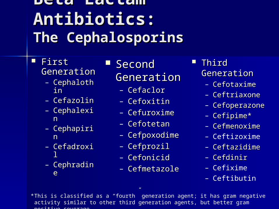
Prokaryotes vs. Prokaryotes vs. Eukaryotes: RibosomesEukaryotes: Ribosomes

Bind to the Bind to the ribosomal subunits ribosomal subunits to impair protein to impair protein synthesissynthesis– AminoglycosidesAminoglycosides– ChloramphenicolChloramphenicol– MacrolidesMacrolides
ErythromycinErythromycin ClarithromycinClarithromycin Azithromycin Azithromycin
– ClindamycinClindamycin
Disrupters of Protein Disrupters of Protein SynthesisSynthesis

KanamycinKanamycin GentamicinGentamicin TobramycinTobramycin AmikacinAmikacin NetilmicinNetilmicin SisomycinSisomycin Aminoglycosides bind to the 30s subunit to
impair protein synthesis.
The The AminoglycosidesAminoglycosides
Blocks initiation of protein synthesis
Blocks translation to cause premature termination
Causes incorporation of incorrect amino acid
Structure of the antibiotic gentamicin C1a bound to its RNA target. Aminoglycoside antibiotics cause misreading of the genetic code.

Agents that Bind to the Agents that Bind to the 50S Ribosome50S Ribosome ChloramphenicolChloramphenicol
– spectrum of activityspectrum of activity S. pneumoniaS. pneumonia H. influenzaH. influenza Neisseria spp.Neisseria spp. SalmonellaSalmonella BordetellaBordetella EnterobacteriaceaeEnterobacteriaceae some anaerobessome anaerobes

Agents that Bind to Agents that Bind to the 50S Ribosomethe 50S Ribosome MacrolidesMacrolides
– ErythromycinErythromycin SS. pneumonia, S. . pneumonia, S.
pyogenes, Legionella, pyogenes, Legionella, Chlamydia Chlamydia trachomatis, M. trachomatis, M. catarrhalis, H. catarrhalis, H. influenza, influenza, Mycoplasma Mycoplasma pneumoniapneumonia
– ClarithromycinClarithromycin MACMAC
– AzithromycinAzithromycin MACMAC
ClindamycinClindamycin– aerobic gram-aerobic gram-
positive bacteria positive bacteria – anaerobes, especially anaerobes, especially
B. fragilis B. fragilis – used in combination used in combination
with aminoglycosides with aminoglycosides to treat intra-to treat intra-abdominal and abdominal and gynecologic gynecologic infections infections

ChloramphenicolChloramphenicol– Gray syndromeGray syndrome– Dose-dependent Dose-dependent
bone marrow bone marrow suppressionsuppression
– Aplastic anemia, Aplastic anemia, pancytopeniapancytopenia
MacrolidesMacrolides– GI complaintsGI complaints– RashRash
ClindamycinClindamycin– DiarrheaDiarrhea– Pseudomembranous Pseudomembranous
colitiscolitis– Rash, urticariaRash, urticaria– Hypotension Hypotension
Side Effects and Side Effects and Adverse Reactions Adverse Reactions

Disrupters of Nucleic Disrupters of Nucleic Acid MetabolismAcid Metabolism MetronidazoleMetronidazole Quinolones: Quinolones:
– CiprofloxacinCiprofloxacin– LevfloxacinLevfloxacin– Moxifloxacin Moxifloxacin – NorfloxacinNorfloxacin– OfloxacinOfloxacin– TrovafloxacinTrovafloxacin– GatifloxacinGatifloxacin– GrepafloxacinGrepafloxacin

Disrupters of Nucleic Disrupters of Nucleic Acid MetabolismAcid Metabolism MetronidazoleMetronidazole
Participates in redox reactions; it is activated by a reduction of Participates in redox reactions; it is activated by a reduction of the nitro group to an anion radical. In the case of metronidazole, the nitro group to an anion radical. In the case of metronidazole, reduced ferredoxin appears to be the primary electron donor reduced ferredoxin appears to be the primary electron donor responsible for its reduction The anion radical is highly reactive responsible for its reduction The anion radical is highly reactive and will form adjuncts with proteins and DNA leading to a loss of and will form adjuncts with proteins and DNA leading to a loss of function. In particular, the reactions with DNA result in strand function. In particular, the reactions with DNA result in strand breakage and inhibition of replication and will lead to cell death.breakage and inhibition of replication and will lead to cell death.

Disrupters of Nucleic Disrupters of Nucleic Acid MetabolismAcid Metabolism Quinolones: inhibit Quinolones: inhibit
DNA-gyrase and DNA-gyrase and topoisomerase IItopoisomerase II– CiprofloxacinCiprofloxacin– LevfloxacinLevfloxacin– Moxifloxacin Moxifloxacin – NorfloxacinNorfloxacin– OfloxacinOfloxacin– TrovafloxacinTrovafloxacin– GatifloxacinGatifloxacin– GrepafloxacinGrepafloxacin

MetronidazoleMetronidazole– dizzinessdizziness– paresthesiasparesthesias– peripheral peripheral
neuropathyneuropathy– disulfiram-like disulfiram-like
reactionreaction– blood dyscrasiasblood dyscrasias
QuinolonesQuinolones– headacheheadache– rash, photosensitivityrash, photosensitivity– GI complaintsGI complaints– arthralgiasarthralgias– confusionconfusion– liver dysfunctionliver dysfunction
Side Effects and Side Effects and Adverse Reactions Adverse Reactions

AntimetabolitesAntimetabolites
TrimethoprimTrimethoprim SulfonamidesSulfonamides
– SulfamethoxazSulfamethoxazoleole
– SulfisoxazoleSulfisoxazole
Inhibition of folate metabolism by sulfonamides and trimethoprim

SulfonamidesSulfonamides– Dizziness, headacheDizziness, headache– RashRash– Blood dyscrasiasBlood dyscrasias– CrystalluriaCrystalluria– Acute nephropathyAcute nephropathy– Bilirubin displacementBilirubin displacement
Side Effects and Side Effects and Adverse Reactions Adverse Reactions

Identity of infecting Identity of infecting organismorganism
Susceptibility of infecting Susceptibility of infecting organismorganism
Host FactorsHost Factors
Proper Antimicrobial Proper Antimicrobial Selection: FSelection: Factors to actors to
ConsiderConsider

Major Mechanisms Major Mechanisms of Antimicrobial of Antimicrobial ResistanceResistance
BypassBypass(TMP/SMX)(TMP/SMX)
EffluxEfflux(macrolides, (macrolides, quinolones)quinolones)
DecreasedDecreased permeabilitypermeability
((-lactams)-lactams)
Target site modificationTarget site modification((intracellular or extracellular; intracellular or extracellular; -lactams, macrolides, -lactams, macrolides, quinolones, glycopeptides)quinolones, glycopeptides)
Enzymatic Enzymatic degradationdegradation(intracellular or (intracellular or extracellular; extracellular; -lactams, -lactams, aminoglycosidesaminoglycosides))
XX

Enzyme Inactivation of Enzyme Inactivation of PenicillinsPenicillins
Structure of penicillins and interaction with beta lactamase
1
2
1 = Site of action of penicillinase2 = Site of action of amidaseA = Thiazolidine ringB = -lactam ring

Resistance to Penicillin Resistance to Penicillin in in N. gonorrheaN. gonorrhea
Beta lactamase

Gram Negative Gram Negative OrganismsOrganisms– H. InfluenzaH. Influenza– M. CatarrhalisM. Catarrhalis– EnterobacterEnterobacter– KlebsiellaKlebsiella– CitrobacterCitrobacter– SerratiaSerratia
Gram PositiveGram Positive– Staphylococcus Staphylococcus
S. aureusS. aureus S. epidermidisS. epidermidis
– StreptococcusStreptococcus S. pneumoniaeS. pneumoniae
Vancomycin Vancomycin – EnterococciEnterococci
E. faecalisE. faecalis E. faeciumE. faecium
– S. aureusS. aureus
Bacterial Resistance: Bacterial Resistance: What Problems are We What Problems are We Seeing?Seeing?

Antimicrobial PharmacodynamicsAntimicrobial Pharmacodynamics– attempt to characterize the attempt to characterize the
relationship between relationship between ANTIMICROBIAL ANTIMICROBIAL EXPOSUREEXPOSURE (concentration, dose, AUC) (concentration, dose, AUC) and and ANTIMICROBIAL EFFECTANTIMICROBIAL EFFECT (eg., rate, (eg., rate, extent, and duration of antimicrobial extent, and duration of antimicrobial activity)activity)
Other Important Factors: Other Important Factors:
MICs and MBCs Fail to Tell the Whole MICs and MBCs Fail to Tell the Whole StoryStory

Antibiotic PharmacodynamicsAntibiotic Pharmacodynamics– Rate and Extent of Bactericidal Rate and Extent of Bactericidal
ActionAction– Post-antibiotic EffectPost-antibiotic Effect– Effects of Sub-inhibitory Effects of Sub-inhibitory
ConcentrationsConcentrations– Post-antibiotic Leukocyte EffectPost-antibiotic Leukocyte Effect– Inoculum EffectInoculum Effect
Other Important Factors: Other Important Factors:
MICs and MBCs Fail to Tell the Whole MICs and MBCs Fail to Tell the Whole StoryStory

– Concentration-Dependent AgentsConcentration-Dependent Agents Bactericidal activity is dependent on concentration above Bactericidal activity is dependent on concentration above
the MIC achieved, increasing with increasing concentrationthe MIC achieved, increasing with increasing concentration
– Time-Dependent AgentsTime-Dependent Agents Bactericidal activity is dependent on how long the Bactericidal activity is dependent on how long the
concentration exceeds the MICconcentration exceeds the MIC
– Bacteriostatic AgentsBacteriostatic Agents Abort bacterial growth and allow host defenses to Abort bacterial growth and allow host defenses to
eradicate organismseradicate organisms
Classification Based on Classification Based on Pharmacodynamic Pharmacodynamic CharacteristicsCharacteristics

Concentration-Dependent Killing of Pseudomonas aeruginosa with
Tobramycin
1 /
log
CF
U p
er m
L
1
2
3
4
5
6
7
8
0
Time (hours)
1 2 3 4 5 6
control
1/4 MIC
1 MIC
4 MIC
16 MIC
64 MIC
Antibiotic conc

NON-Concentration-Dependent Killing
9
1 /
log
CF
U p
er m
L
1
2
3
4
5
6
7
8
0
Time (hours)
1 2 3 4 5 6
control
1/4 MIC
1 MIC
4 MIC
16 MIC
64 MIC
Antibiotic conc

Pharmacodynamic Properties by Antibiotic
Class
CONCENTRATION dependent killing
TIME dependent killing
Aminoglycosides β-lactamsFluoroquinolones GlycopeptidesAzithromycin? Metronidazole
Macrolides (except Azithromycin) Chloramphenicol Rifampin Tetracyclines Clindamycin

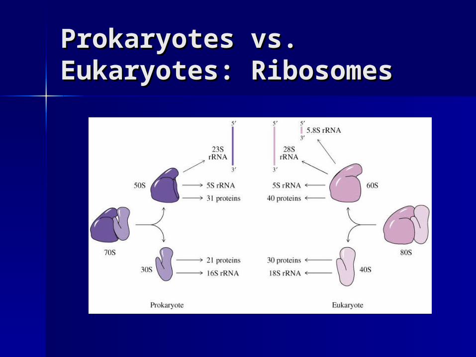
Pharmacodynamic Relationships between Antibiotic Concentration and Antibacterial Effect
Time
Plasma Conc
MBC
MIC
CIDAL activity PAE
Bacterial REGROWTH
STATIC activity
Site Conc
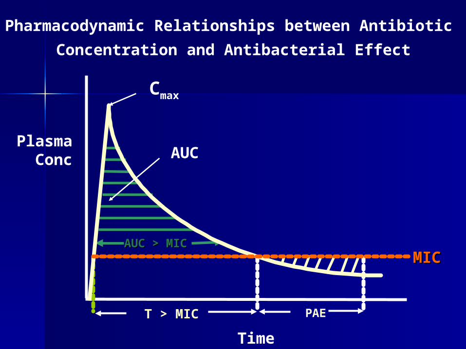
Pharmacodynamic Relationships between Antibiotic
Concentration and Antibacterial Effect
Time
Plasma Conc
T > MIC PAE
AUC > MICAUC > MIC
MICMIC
Cmax
AUC

Pharmacokinetics
Susceptibility
MIC / MBC Serum / Tissue Concentrations
Pharmacodynamics
Time > MIC
Peak / MIC
AUC > MIC
Eradication / Cure

Antibiotic Pharmacodynamics Antibiotic Pharmacodynamics in Otitis Media: T>MICin Otitis Media: T>MIC
Average percentage of time drug concentration exceeds the minimum inhibitory concentration (%T>MIC) for pediatric dosages of oral ß-lactam agents against penicillin-sensitive (black bars) and penicillin-intermediate (hatched bars) Streptococcus pneumoniae. Rodvold. Pharmacoatherapy. 2001; 21(11s) :319s-330s.

Antibiotic Pharmacodynamics: Antibiotic Pharmacodynamics: Ciprofloxacin AUCCiprofloxacin AUC0-240-24:MIC and Clinical :MIC and Clinical
Outcomes Outcomes
Percentage of bacteriologic (black bars) and clinical (hatched bars) cures as a function of AUC0-24:MIC in 68 patients with gram-negative infections treated with ciprofloxacin. Note that the bacteriologic and clinical outcomes are better with AUC > 125.

Clinical BreakpointsClinical Breakpoints
Clinical breakpoints are supposed to indicate at which MIC the chance of eradication or even clinical success of antimicrobial treatment prevails significantly over failure, given the dosing schedule of the drug. The breakpoint thus is not only dependent on the antimicrobial activity of the drugs itself, but also on its pharmacokinetics and pharmacodynamics.

Postantibiotic effectPostantibiotic effect
The period of time where there is The period of time where there is persistent suppression of bacterial persistent suppression of bacterial growthgrowth following exposure to an following exposure to an antimicrobial agent, despite antimicrobial agent, despite removal of the antimicrobial removal of the antimicrobial agent.agent.

Antibiotic Antibiotic PharmacodynamicsPharmacodynamics
MIC = minimum inhibitory concentration
MBC = minimum bactericidal concentration
From: Levinson ME. Infect Dis Clin North Amer. 1995; 483-95.
Antibiotic 1
Antibiotic 2

Additive EffectsAdditive Effects Synergistic EffectsSynergistic Effects Antagonistic EffectsAntagonistic Effects
Antibiotic Antibiotic Combinations:Combinations:Rationale and IndicationsRationale and Indications

Antibiotic Synergy Antibiotic Synergy and Antagonismand Antagonism

Prevent emergence of Prevent emergence of resistanceresistance
Polymicrobial infectionsPolymicrobial infections Empiric therapyEmpiric therapy Reduced drug toxicityReduced drug toxicity SynergismSynergism
Antibiotic Antibiotic Combinations:Combinations:Rationale and IndicationsRationale and Indications

AntagonismAntagonism Increased drug costsIncreased drug costs Adverse drug Adverse drug
reactionsreactions
Antibiotic Combinations:Antibiotic Combinations: Disadvantages of Inappropriate Disadvantages of Inappropriate Combination TherapyCombination Therapy
Related Documents