RESEARCH Open Access Patterns of locoregional failure following post-operative intensity-modulated radiotherapy to oral cavity cancer: quantitative spatial and dosimetric analysis using a deformable image registration workflow Abdallah S. R. Mohamed 1,3* , Andrew J. Wong 1 , Clifton D. Fuller 1 , Mona Kamal 1,4 , Gary B. Gunn 1 , Jack Phan 1 , William H. Morrison 1 , Beth M. Beadle 1 , Heath Skinner 1 , Stephen Y. Lai 2 , Sean R. Quinlan-Davidson 1,5 , Abdelaziz M. Belal 3 , Ahmed G. El-Gowily 3 , Steven J. Frank 1 , David I. Rosenthal 1 and Adam S. Garden 1* Abstract Background: We sought to identify spatial/dosimetric patterns of failure for oral cavity cancer patients receiving post-operative IMRT (PO-IMRT). Methods: Two hundred eighty-nine OCC patients receiving PO-IMRT were retrospectively reviewed from 2000 to 2012. Diagnostic CT documenting recurrence (rCT) was co-registered with planning CT (pCT) using a validated deformable image registration software. Manually segmented recurrent gross disease (rGTV) was deformed to co-registered pCTs. Mapped rGTVs were compared dosimetrically to planned dose and spatially to planning target volumes using centroid- based approaches. Failures types were classified using combined spatial/dosimetric criteria: A (central high-dose), B (peripheral high-dose), C (central intermediate/low-dose), D (peripheral intermediate/low-dose), and E (extraneous-dose). Results: Fifty-four patients with recurrence were analyzed; 26 local recurrence, 19 regional recurrence, and 9 both local and regional recurrence. Median time to recurrence was 4 months (range 0–71). Median rGTVs volume was 3.7 cm 3 (IQR 1.4–10.6). For spatial and dosimetric analysis of the patterns of failure, 30 patients (55.5%) were classified as type A (central high-dose). Non-central high dose failures were distributed as follows: 2 (3.7%) type B, 10 (18.5%) type C, 1 (1.8%) type D, and 9 (16.7%) type E. Non-IMRT failure in the matching low-neck field was seen in two patients. No failures were noted at the IMRT-supraclavicular field match-line. Conclusions: Approximately half of patients with local/regional failure had non-central high dose recurrence. Peripheral high dose misses were uncommon reflecting adequate delineation and dose delivery. Future strategies are needed to reduce types C and E failures. Keywords: Patterns of failure, Post-operative intensity modulated radiation therapy, Oral cavity cancer, Deformable image registration, Quantitative spatial and dosimetric analysis * Correspondence: [email protected]; [email protected] 1 Departments of Radiation Oncology, The University of Texas MD Anderson Cancer Center, Unit 97, 1515 Holcombe Boulevard, Houston, TX 77030, USA Full list of author information is available at the end of the article © The Author(s). 2017 Open Access This article is distributed under the terms of the Creative Commons Attribution 4.0 International License (http://creativecommons.org/licenses/by/4.0/), which permits unrestricted use, distribution, and reproduction in any medium, provided you give appropriate credit to the original author(s) and the source, provide a link to the Creative Commons license, and indicate if changes were made. The Creative Commons Public Domain Dedication waiver (http://creativecommons.org/publicdomain/zero/1.0/) applies to the data made available in this article, unless otherwise stated. Mohamed et al. Radiation Oncology (2017) 12:129 DOI 10.1186/s13014-017-0868-y

Welcome message from author
This document is posted to help you gain knowledge. Please leave a comment to let me know what you think about it! Share it to your friends and learn new things together.
Transcript
RESEARCH Open Access
Patterns of locoregional failure followingpost-operative intensity-modulatedradiotherapy to oral cavity cancer:quantitative spatial and dosimetric analysisusing a deformable image registrationworkflowAbdallah S. R. Mohamed1,3*, Andrew J. Wong1, Clifton D. Fuller1, Mona Kamal1,4, Gary B. Gunn1, Jack Phan1,William H. Morrison1, Beth M. Beadle1, Heath Skinner1, Stephen Y. Lai2, Sean R. Quinlan-Davidson1,5,Abdelaziz M. Belal3, Ahmed G. El-Gowily3, Steven J. Frank1, David I. Rosenthal1 and Adam S. Garden1*
Abstract
Background: We sought to identify spatial/dosimetric patterns of failure for oral cavity cancer patients receivingpost-operative IMRT (PO-IMRT).
Methods: Two hundred eighty-nine OCC patients receiving PO-IMRT were retrospectively reviewed from 2000 to 2012.Diagnostic CT documenting recurrence (rCT) was co-registered with planning CT (pCT) using a validated deformableimage registration software. Manually segmented recurrent gross disease (rGTV) was deformed to co-registered pCTs.Mapped rGTVs were compared dosimetrically to planned dose and spatially to planning target volumes using centroid-based approaches. Failures types were classified using combined spatial/dosimetric criteria: A (central high-dose), B(peripheral high-dose), C (central intermediate/low-dose), D (peripheral intermediate/low-dose), and E (extraneous-dose).
Results: Fifty-four patients with recurrence were analyzed; 26 local recurrence, 19 regional recurrence, and 9 both localand regional recurrence. Median time to recurrence was 4 months (range 0–71). Median rGTVs volume was 3.7 cm3
(IQR 1.4–10.6). For spatial and dosimetric analysis of the patterns of failure, 30 patients (55.5%) were classified as type A(central high-dose). Non-central high dose failures were distributed as follows: 2 (3.7%) type B, 10 (18.5%) type C, 1 (1.8%)type D, and 9 (16.7%) type E. Non-IMRT failure in the matching low-neck field was seen in two patients. No failures werenoted at the IMRT-supraclavicular field match-line.
Conclusions: Approximately half of patients with local/regional failure had non-central high dose recurrence. Peripheralhigh dose misses were uncommon reflecting adequate delineation and dose delivery. Future strategies are needed toreduce types C and E failures.
Keywords: Patterns of failure, Post-operative intensity modulated radiation therapy, Oral cavity cancer, Deformable imageregistration, Quantitative spatial and dosimetric analysis
* Correspondence: [email protected]; [email protected] of Radiation Oncology, The University of Texas MD AndersonCancer Center, Unit 97, 1515 Holcombe Boulevard, Houston, TX 77030, USAFull list of author information is available at the end of the article
© The Author(s). 2017 Open Access This article is distributed under the terms of the Creative Commons Attribution 4.0International License (http://creativecommons.org/licenses/by/4.0/), which permits unrestricted use, distribution, andreproduction in any medium, provided you give appropriate credit to the original author(s) and the source, provide a link tothe Creative Commons license, and indicate if changes were made. The Creative Commons Public Domain Dedication waiver(http://creativecommons.org/publicdomain/zero/1.0/) applies to the data made available in this article, unless otherwise stated.
Mohamed et al. Radiation Oncology (2017) 12:129 DOI 10.1186/s13014-017-0868-y
IntroductionSurgery is often the treatment of choice for oral cavitysquamous cell carcinoma (OCSCC). Post-operativeradiotherapy is indicated for OCSCC of advanced stagesor with adverse prognostic factors [1–3]. Intensity-modulated radiotherapy (IMRT) enables conformaltherapy and reduction of complications to surroundingnormal tissue, and for many centers has become thestandard radiation approach for head and neck cancer [4].Generally, OCSCC patients demonstrate relatively
worse loco-regional control compared to other head andneck subsites (e.g. oropharynx and larynx) [5–7]. Studiesthat have specifically examined cohorts of OCSCCpatients receiving postoperative IMRT (PO-IMRT)have consistently reported only fair locoregionalcontrol rates, as low as 53% at 3 years in someseries [8–13].Moreover, most report failures as “infield”, “marginal”,
or “outfield” based on percentage overlap between fail-ure volume and respective target volumes. However,these studies applied non-uniform spatial methods forfailure analysis, mainly utilizing non-validated rigid ormanual image registration tools and without includingthe dosimetric component in the analysis [8–13]. Wehave recently shown the potential impact of patterns offailure analysis methodology using a validated imageregistration software paired with combined spatial anddosimetric analysis of failure, in improving the accuracyof reporting the patterns of failure in the era of IMRT[14–16]. As a continuation of these efforts we sought toapply this unique analytic methodology to our institu-tional large scale oral cavity cancer dataset of patientsreceiving PO-IMRT with documented treatmentfailure to achieve the following specific aims: 1)characterize distinct spatial and dosimetric patterns offailure after PO-IMRT, 2) identify clinical risk featuresassociated with each failure type, 3) identify patternsof failure based target volume contouring recom-mendations, and 4) generate hypotheses for futureclinical trials.
Material and methodsPatient selectionTwo hundred eighty-nine patients with pathologicaldiagnosis of OCC who received PO-IMRT at the Universityof Texas MD Anderson Cancer Center from 2000 to 2012were retrospectively reviewed under an approved institu-tional review board protocol. Patients with distant metasta-ses or concurrent malignancies at the time of diagnosis, ortreatment with chemotherapy prior to staging at MDACCwere excluded. Patients with prove of recurrence afterPO-IMRT with available imaging documenting recurrencewere included in the current analysis.
Treatment planning and deliveryTreatment planning and delivery is described in detailsin previously published work, which examines the out-comes for this same patient cohort [17].
Clinical data collectionDiagnostic contrast-enhanced CT and/or PET/CT docu-menting the initial evidence of local and/or regionalrecurrence (rCT) was identified. Recurrences wereconfirmed via radiologic imaging (i.e. progression insubsequent CT imaging or high SUV on PET imaging)or pathology specimens (i.e. from surgical biopsy).Radiologically evident recurrent gross disease (rGTV)was manually segmented and reviewed by two experi-enced radiation oncologists (ASRM, CDF). Correspond-ing original planning CTs (pCT) were also identified andoriginal plans were restored. Patient, disease, and treat-ment characteristics were gathered during chart review.
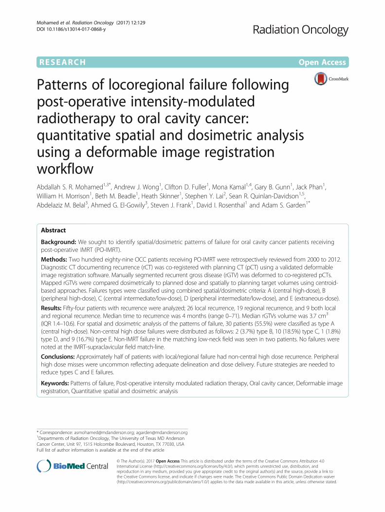
Image registration and dosimetric analysisrCT was co-registered with pCT using a previously vali-dated deformable image registration (DIR) methodology(VelocityAI 3.0.1, Velocity Medical Solutions, Atlanta,GA, 2004–2013) [14, 15]. rGTVs on the rCT weresubsequently deformed to co-registered pCT (Fig. 1).
Fig. 1 Workflow depicting how deformed rGTV is propagated tooriginal planning CT
Mohamed et al. Radiation Oncology (2017) 12:129 Page 2 of 12
The centroid, assumed as the origin of tumor recur-rence, is represented as the calculated center of mass ofthe deformed rGTV. Dosimetric and volumetric parame-ters were obtained from the dose-volume histogram.
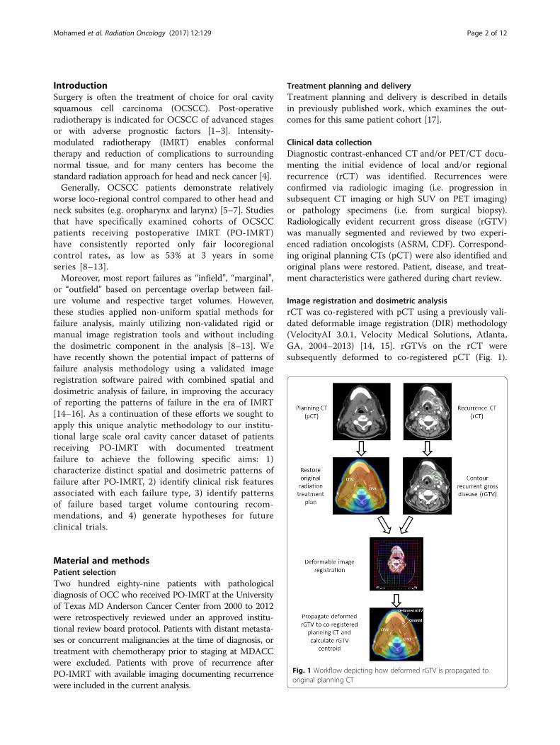
Patterns of failure classificationFailures are classified according to both spatial anddosimetric criteria as previously described. [16] Briefly, forspatial mapping of recurrence origin, the centroid of eachrGTV was mapped to the corresponding TV in the plan-ning CT. Subsequently, the dosimetric characteristics wereassessed by calculating the dose to 95% of the failure vol-ume (fD95%) then comparing it relative to the dose pre-scribed to the corresponding TV of origin as determinedby the spatial mapping. Finally, failures were classified intofive major types: Type A (central high dose where themapped failure centroid originates in high dose TV andfD95% is ≥95% dose prescribed to corresponding highdose TV of origin), Type B (peripheral high dose wherethe failure centroid originates from high dose TV but itsfD95% is <95% dose prescribed to corresponding highdose TV of origin), Type C (central elective dose wherethe failure centroid originates in lower dose TV andfD95% is ≥95% dose prescribed to corresponding lower
dose TV of origin), Type D (peripheral elective dose wherethe failure centroid originates in lower dose TV but thefD95% is <95% dose prescribed to corresponding lowerdose TV of origin), and Type E (extraneous dose whererGTV centroid originates outside all TVs). Type F describesjunctional failures at the IMRT/supraclavicular match line,and Type G describes low neck failures at the low-necksupraclavicular field. Type G is analogous to type C if thefD95% is ≥95% dose prescribed to the low-neck andanalogous to type D if the fD95% is <95%. Examples dem-onstrating failure type definitions are illustrated in Fig. 2.Patients were then classified according to the predom-
inant mode of failure. Patients with more than singlerecurrence lesion were classified as the following: 1) forpatients with type A recurrence and concurrent non-type A lesions, the overall pattern of failure was definedas type A because we believe type A for such patients isthe true recurrence rather than reseeding from the non-type A recurrence, 2) for patients whom exhibited morethan one failure type of non-type A simultaneously, pat-tern of failure of each patient was classified according tothe predominant type as determined by the most com-monly encountered failure type (i.e. higher number orhigher volume).
Fig. 2 Examples of Failure Types. 1) Type A (central high dose) failures. Centroid is mapped inside high dose TV and dose to 95% rGTV volume ≥ 95%dose prescribed to high dose TV. 2) Type B (peripheral high dose) failure. Centroid is mapped inside high dose TV, but dose to 95% rGTV volume < 95%dose prescribed to high dose TV. 3) Type C-int (central intermediate dose) failure. Centroid is mapped inside intermediate dose TV and dose to 95% rGTVvolume ≥ 95% dose prescribed to intermediate dose TV. 4) Type D-int (peripheral intermediate dose) failure. Centroid is mapped inside intermediate doseTV but dose to 95% rGTV volume < 95% dose prescribed to intermediate dose TV. 5) Type E (Extraneous dose failure) where rGTV centroid originatesoutside all TVs. 6) Type G (low neck failure) where rGTV centroid is located at the low-neck supraclavicular field
Mohamed et al. Radiation Oncology (2017) 12:129 Page 3 of 12
ResultsPatient and treatment characteristicsSixty-three patients (22%) developed locoregional recur-rences. Median time to recurrence was 4 months (range0–71). For spatial and dosimetric analysis of the patternsof failure, 9 patients were excluded: 4 with post-surgicalrecurrence prior to initiation of IMRT, 3 with no retriev-able IMRT plan, and 2 with no available imagingdocumenting recurrence. This left 54 patients for thecurrent analysis.Patient, disease, and treatment characteristics for the
analyzed 54 patients are summarized in Table 1. Themost common primary site was the oral tongue (39%).The most common pathologic T and N staging were T2(37%) and N2 (36%). Forty-seven (87%) patients hadStage III-IV disease.Surgical margins were positive and close (defined as
≤5 mm) in 4 (7%) and 9 (17%) patients, respectively.Perineural invasion or lymphovascular invasion waspresent in 22 (41%) and 14 (26%) patients, respectively.Depth of invasion was ≥1.5 cm in 18 patients (33%).Forty-seven patients (87%) had neck dissections, and ofthose 17 patients had nodal extracapsular extension.Mean RT dose was 60 ± 7 Gy and mean number of
fractions was 30 ± 3. One patient did not complete thefull course of IMRT, discontinuing therapy after 6 frac-tions. Thirteen (24%) and 41 (76%) patients receivedunilateral and bilateral neck irradiation, respectively.Mean overall treatment package time, defined as timeinterval from date of surgery to last day of irradiation,was 12.3 ± 1.7 weeks.
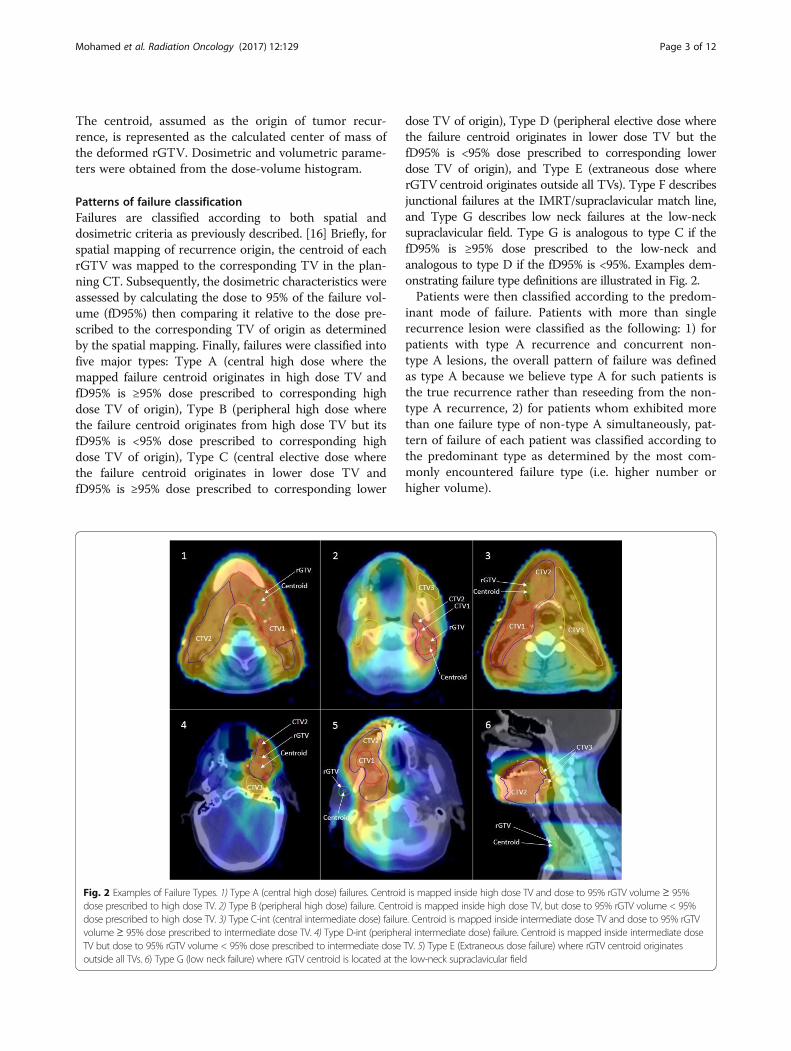
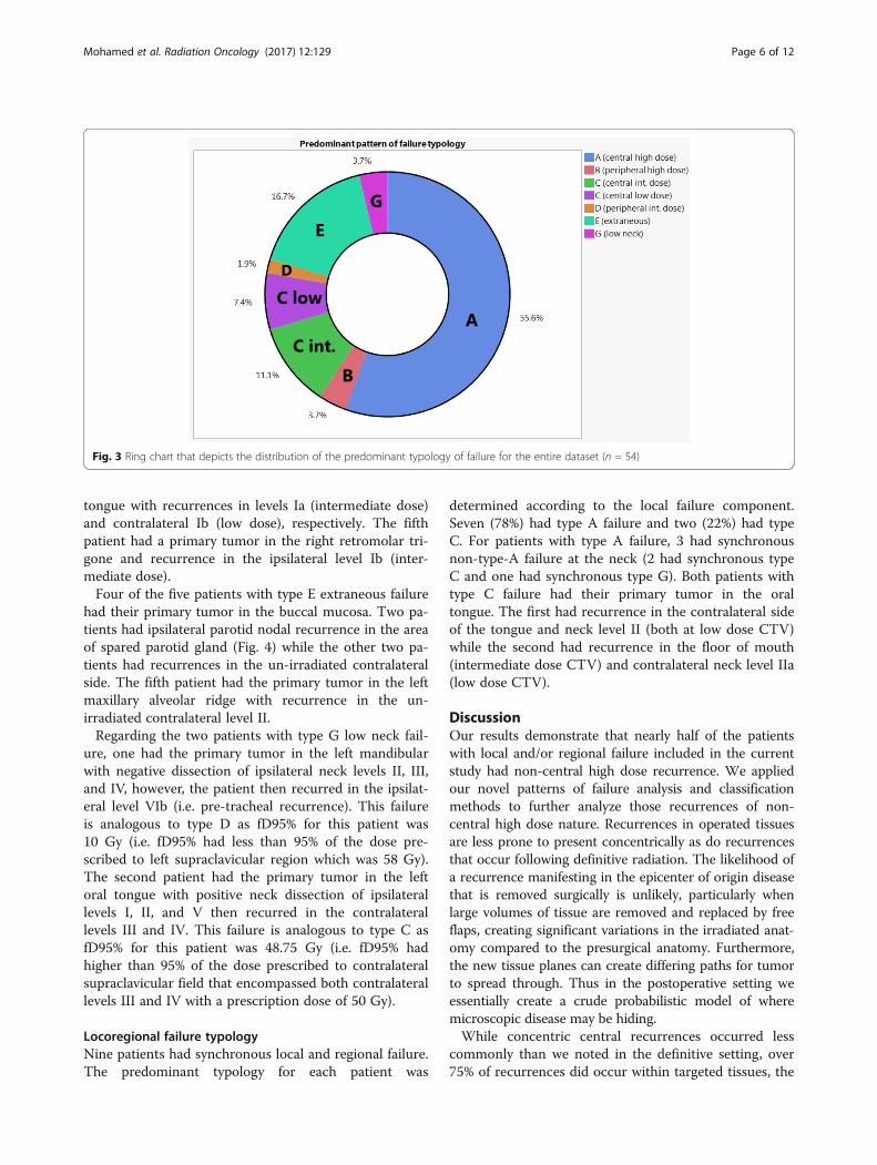
Recurrence characteristicsFor patients included in the current analysis; 26 (48%)had local recurrence, 19 (35%) had regional recurrence,and 9 (17%) had both local and regional recurrence. Atotal of 82 rGTVs were delineated. Median rGTVs vol-ume was 3.7 cm3 (IQR 1.4–10.6). Figure 3 shows the dis-tribution of the predominant type of failure for theentire dataset using the proposed classification schema.Thirty patients (55.5%) were classified as type A. Non-type A failures were distributed as follows: 2 (3.7%) typeB, 10 (18.5%) type C, 1 (1.8%) type D, 9 (16.7%) type E,and 2 (3.7%) type G. Because type A “central high dose”failures, are considered to resistance to maximal therapy,and thus could not conceivably be prevented bytechnical/operator dependent processes, however, non-type A failures represent a major goal for IMRT qualityassurance and improvement. Table 2 illustrates thedetails of the characteristics for all non-type A failures.
Local failure typologyOf the 26 patients with local disease failure without syn-chronous regional recurrence, 16 (62%) were type A
central high dose failures. Ten patients (38%) had non-type A local failure; two (8%) were type B, three (11%)were type C (intermediate dose), one (4%) was type D(intermediate dose), and four (15%) were extraneoustype E failure. Three of patients with type A failure hadmultifocal recurrence; two patients with two foci ofrecurrences (both within the central high dose region),and one had four foci of recurrences (three in the inter-mediate dose and one in the high dose).For the two patients with type B failure, one had a
primary tumor in the left mandibular gingiva and devel-oped recurrence involving the left maxillary sinus, witherosion of its lateral wall and extension along the buccalmucosa to the retromolar trigone. The second patienthad a primary tumor in the floor of mouth with rapiddisease progression subsequent to discontinuingradiation treatment after six fractions.Regarding patients with type C failure, one patient had
his primary tumor in the retromolar area and recurrencein the ipsilateral masticator space. The second patienthad the primary tumor in the floor of mouth with recur-rence involving the base of tongue, while the thirdpatient had a primary tumor in the oral tongue withrecurrence involving the floor of mouth.The single type D failure had the primary tumor in the
left mandibular gingiva and recurred along the leftmasticator space allowing the rGTV to partially growoutside the CTV2 boundaries, however the centroid wasstill located inside CTV2.For patients with type E failure, one had the primary
tumor in the right mandibular gingiva and the recur-rence in the contralateral mandibular gingiva approxi-mately 2 years following treatment. The second patienthad the primary tumor in the left buccal mucosa; therecurrence manifested as retrograde perineural spreadthat extended to the left pterygopalatine fossa, foramenrotundum, foramen ovale, cavernous sinus, and throughthe superior orbital fissure into the left orbit (Fig. 4).The third patient had T1 primary tumor in the centralfloor of mouth and the recurrence in the left alveolarmandibular ridge approximately 3 years following treat-ment. Lastly, the fourth patient had the primary tumorin the left mandibular gingiva and the recurrence in theipsilateral masticator space at the first follow up follow-ing treatment.
Regional failure typologyOf the 19 patients with regional disease failure withoutsynchronous local recurrence, only 7 patients (37%) weretype A central high dose failures. Twelve patients (63%)had non-type A local failure; 5 (26%) were type C (inter-mediate or low dose), five (26%) were extraneous type E,and 2 (11%) were type G low neck failure. Two of pa-tients with type A failure had multifocal recurrence. One
Mohamed et al. Radiation Oncology (2017) 12:129 Page 4 of 12
patient with a left oral tongue primary developed syn-chronous ipsilateral type A recurrence at level III andcontralateral type C (low dose) recurrence at level IIa.The second patient had the primary disease in the rightretromolar trigone with multi-nodal recurrence at ipsi-lateral neck level IIb and an ipsilateral retropharyngeallymph node (type D).For the five patients with type C failure, one patient
had a right hard palate primary with multifocal type C(low dose) failure with two foci of recurrence, both atcontralateral level IIa. The second patient had the pri-mary tumor in the left maxillary ridge and recurred inthe low dose region at the ipsilateral level Ib. The thirdand fourth patients had primary tumors of the oral
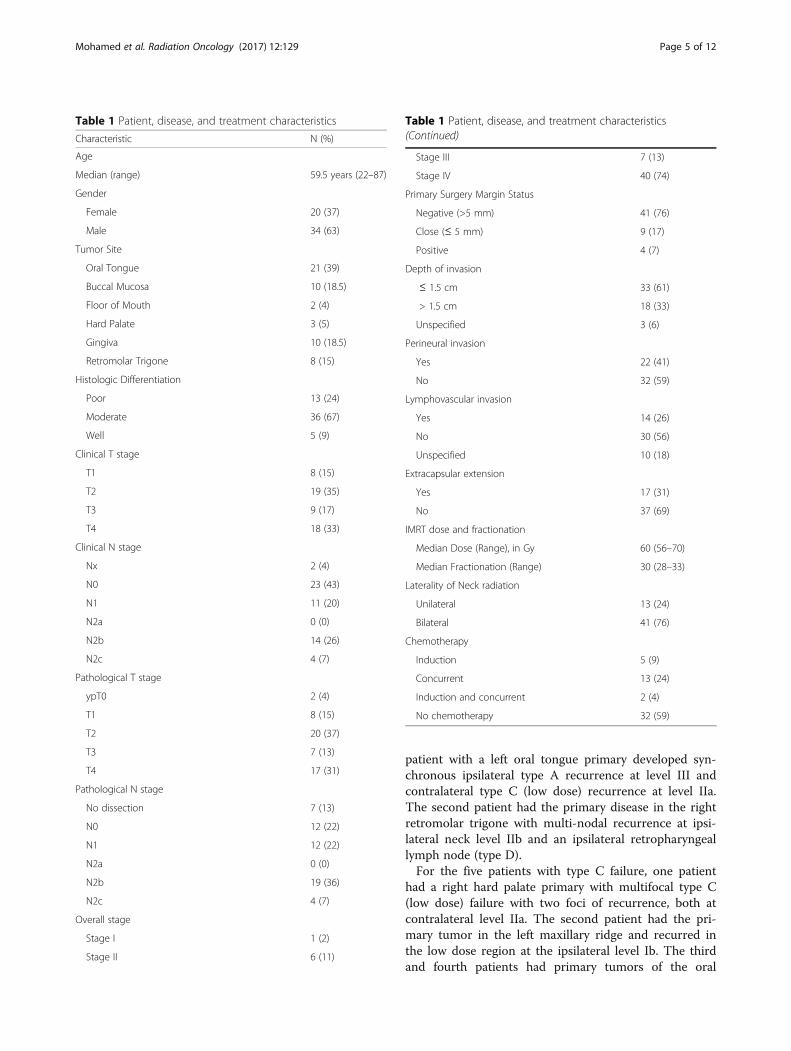
Table 1 Patient, disease, and treatment characteristics
Characteristic N (%)
Age
Median (range) 59.5 years (22–87)
Gender
Female 20 (37)
Male 34 (63)
Tumor Site
Oral Tongue 21 (39)
Buccal Mucosa 10 (18.5)
Floor of Mouth 2 (4)
Hard Palate 3 (5)
Gingiva 10 (18.5)
Retromolar Trigone 8 (15)
Histologic Differentiation
Poor 13 (24)
Moderate 36 (67)
Well 5 (9)
Clinical T stage
T1 8 (15)
T2 19 (35)
T3 9 (17)
T4 18 (33)
Clinical N stage
Nx 2 (4)
N0 23 (43)
N1 11 (20)
N2a 0 (0)
N2b 14 (26)
N2c 4 (7)
Pathological T stage
ypT0 2 (4)
T1 8 (15)
T2 20 (37)
T3 7 (13)
T4 17 (31)
Pathological N stage
No dissection 7 (13)
N0 12 (22)
N1 12 (22)
N2a 0 (0)
N2b 19 (36)
N2c 4 (7)
Overall stage
Stage I 1 (2)
Stage II 6 (11)
Table 1 Patient, disease, and treatment characteristics(Continued)
Stage III 7 (13)
Stage IV 40 (74)
Primary Surgery Margin Status
Negative (>5 mm) 41 (76)
Close (≤ 5 mm) 9 (17)
Positive 4 (7)
Depth of invasion
≤ 1.5 cm 33 (61)
> 1.5 cm 18 (33)
Unspecified 3 (6)
Perineural invasion
Yes 22 (41)
No 32 (59)
Lymphovascular invasion
Yes 14 (26)
No 30 (56)
Unspecified 10 (18)
Extracapsular extension
Yes 17 (31)
No 37 (69)
IMRT dose and fractionation
Median Dose (Range), in Gy 60 (56–70)
Median Fractionation (Range) 30 (28–33)
Laterality of Neck radiation
Unilateral 13 (24)
Bilateral 41 (76)
Chemotherapy
Induction 5 (9)
Concurrent 13 (24)
Induction and concurrent 2 (4)
No chemotherapy 32 (59)
Mohamed et al. Radiation Oncology (2017) 12:129 Page 5 of 12
tongue with recurrences in levels Ia (intermediate dose)and contralateral Ib (low dose), respectively. The fifthpatient had a primary tumor in the right retromolar tri-gone and recurrence in the ipsilateral level Ib (inter-mediate dose).Four of the five patients with type E extraneous failure
had their primary tumor in the buccal mucosa. Two pa-tients had ipsilateral parotid nodal recurrence in the areaof spared parotid gland (Fig. 4) while the other two pa-tients had recurrences in the un-irradiated contralateralside. The fifth patient had the primary tumor in the leftmaxillary alveolar ridge with recurrence in the un-irradiated contralateral level II.Regarding the two patients with type G low neck fail-
ure, one had the primary tumor in the left mandibularwith negative dissection of ipsilateral neck levels II, III,and IV, however, the patient then recurred in the ipsilat-eral level VIb (i.e. pre-tracheal recurrence). This failureis analogous to type D as fD95% for this patient was10 Gy (i.e. fD95% had less than 95% of the dose pre-scribed to left supraclavicular region which was 58 Gy).The second patient had the primary tumor in the leftoral tongue with positive neck dissection of ipsilaterallevels I, II, and V then recurred in the contralaterallevels III and IV. This failure is analogous to type C asfD95% for this patient was 48.75 Gy (i.e. fD95% hadhigher than 95% of the dose prescribed to contralateralsupraclavicular field that encompassed both contralaterallevels III and IV with a prescription dose of 50 Gy).
Locoregional failure typologyNine patients had synchronous local and regional failure.The predominant typology for each patient was
determined according to the local failure component.Seven (78%) had type A failure and two (22%) had typeC. For patients with type A failure, 3 had synchronousnon-type-A failure at the neck (2 had synchronous typeC and one had synchronous type G). Both patients withtype C failure had their primary tumor in the oraltongue. The first had recurrence in the contralateral sideof the tongue and neck level II (both at low dose CTV)while the second had recurrence in the floor of mouth(intermediate dose CTV) and contralateral neck level IIa(low dose CTV).
DiscussionOur results demonstrate that nearly half of the patientswith local and/or regional failure included in the currentstudy had non-central high dose recurrence. We appliedour novel patterns of failure analysis and classificationmethods to further analyze those recurrences of non-central high dose nature. Recurrences in operated tissuesare less prone to present concentrically as do recurrencesthat occur following definitive radiation. The likelihood ofa recurrence manifesting in the epicenter of origin diseasethat is removed surgically is unlikely, particularly whenlarge volumes of tissue are removed and replaced by freeflaps, creating significant variations in the irradiated anat-omy compared to the presurgical anatomy. Furthermore,the new tissue planes can create differing paths for tumorto spread through. Thus in the postoperative setting weessentially create a crude probabilistic model of wheremicroscopic disease may be hiding.While concentric central recurrences occurred less
commonly than we noted in the definitive setting, over75% of recurrences did occur within targeted tissues, the
Fig. 3 Ring chart that depicts the distribution of the predominant typology of failure for the entire dataset (n = 54)
Mohamed et al. Radiation Oncology (2017) 12:129 Page 6 of 12
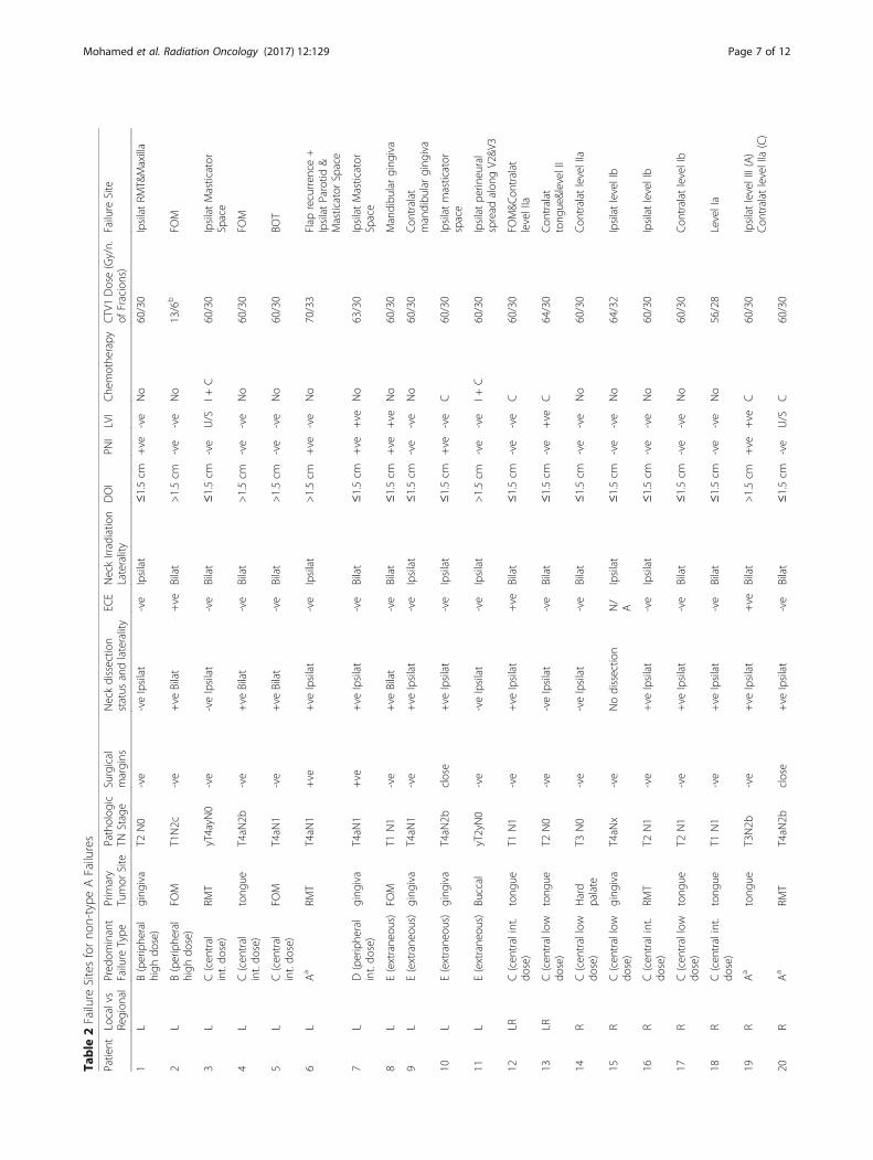
Table
2Failure
Sitesforno
n-type
AFailures
Patient
Localvs
Region
alPred
ominant
Failure
Type
Prim
ary
Tumor
Site
Patholog
icTN
Stage
Surgical
margins
Neckdissectio
nstatus
andlaterality
ECE
NeckIrradiatio
nLaterality
DOI
PNI
LVI
Che
mothe
rapy
CTV1Dose(Gy/n.
ofFracions)
Failure
Site
1L
B(periphe
ral
high
dose)
ging
iva
T2N0
-ve
-veIpsilat
-ve
Ipsilat
≤1.5cm
+ve
-ve
No
60/30
IpsilatRM
T&Maxilla
2L
B(periphe
ral
high
dose)
FOM
T1N2c
-ve
+ve
Bilat
+ve
Bilat
>1.5cm
-ve
-ve
No
13/6
bFO
M
3L
C(cen
tral
int.do
se)
RMT
yT4ayN
0-ve
-veIpsilat
-ve
Bilat
≤1.5cm
-ve
U/S
I+C
60/30
IpsilatMasticator
Space
4L
C(cen
tral
int.do
se)
tong
ueT4aN
2b-ve
+ve
Bilat
-ve
Bilat
>1.5cm
-ve
-ve
No
60/30
FOM
5L
C(cen
tral
int.do
se)
FOM
T4aN
1-ve
+ve
Bilat
-ve
Bilat
>1.5cm
-ve
-ve
No
60/30
BOT
6L
Aa
RMT
T4aN
1+ve
+ve
Ipsilat
-ve
Ipsilat
>1.5cm
+ve
-ve
No
70/33
Flap
recurren
ce+
IpsilatParotid
&Masticator
Space
7L
D(periphe
ral
int.do
se)
ging
iva
T4aN
1+ve
+ve
Ipsilat
-ve
Bilat
≤1.5cm
+ve
+ve
No
63/30
IpsilatMasticator
Space
8L
E(extrane
ous)
FOM
T1N1
-ve
+ve
Bilat
-ve
Bilat
≤1.5cm
+ve
+ve
No
60/30
Mandibu
larging
iva
9L
E(extrane
ous)
ging
iva
T4aN
1-ve
+ve
Ipsilat
-ve
Ipsilat
≤1.5cm
-ve
-ve
No
60/30
Con
tralat
mandibu
larging
iva
10L
E(extrane
ous)
ging
iva
T4aN
2bclose
+ve
Ipsilat
-ve
Ipsilat
≤1.5cm
+ve
-ve
C60/30
Ipsilatmasticator
space
11L
E(extrane
ous)
Buccal
yT2yN0
-ve
-veIpsilat
-ve
Ipsilat
>1.5cm
-ve
-ve
I+C
60/30
Ipsilatpe
rineural
spread
alon
gV2&V
3
12LR
C(cen
tralint.
dose)
tong
ueT1
N1
-ve
+ve
Ipsilat
+ve
Bilat
≤1.5cm
-ve
-ve
C60/30
FOM&C
ontralat
levelIIa
13LR
C(cen
trallow
dose)
tong
ueT2
N0
-ve
-veIpsilat
-ve
Bilat
≤1.5cm
-ve
+ve
C64/30
Con
tralat
tong
ue&levelII
14R
C(cen
trallow
dose)
Hard
palate
T3N0
-ve
-veIpsilat
-ve
Bilat
≤1.5cm
-ve
-ve
No
60/30
Con
tralat
levelIIa
15R
C(cen
trallow
dose)
ging
iva
T4aN
x-ve
Nodissectio
nN/
AIpsilat
≤1.5cm
-ve
-ve
No
64/32
IpsilatlevelIb
16R
C(cen
tralint.
dose)
RMT
T2N1
-ve
+ve
Ipsilat
-ve
Ipsilat
≤1.5cm
-ve
-ve
No
60/30
IpsilatlevelIb
17R
C(cen
trallow
dose)
tong
ueT2
N1
-ve
+ve
Ipsilat
-ve
Bilat
≤1.5cm
-ve
-ve
No
60/30
Con
tralat
levelIb
18R
C(cen
tralint.
dose)
tong
ueT1
N1
-ve
+ve
Ipsilat
-ve
Bilat
≤1.5cm
-ve
-ve
No
56/28
LevelIa
19R
Aa
tong
ueT3N2b
-ve
+ve
Ipsilat
+ve
Bilat
>1.5cm
+ve
+ve
C60/30
IpsilatlevelIII(A)
Con
tralat
levelIIa(C)
20R
Aa
RMT
T4aN
2bclose
+ve
Ipsilat
-ve
Bilat
≤1.5cm
-ve
U/S
C60/30
Mohamed et al. Radiation Oncology (2017) 12:129 Page 7 of 12
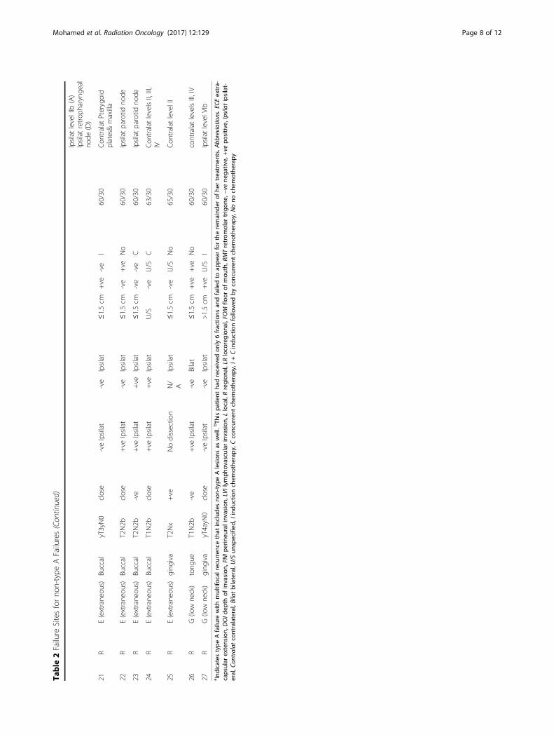
Table
2Failure
Sitesforno
n-type
AFailures(Con
tinued)
IpsilatlevelIIb
(A)
Ipsilatretrop
haryng
eal
node
(D)
21R
E(extrane
ous)
Buccal
yT3yN0
close
-veIpsilat
-ve
Ipsilat
≤1.5cm
+ve
-ve
I60/30
Con
tralat
Pterygoid
plates&maxilla
22R
E(extrane
ous)
Buccal
T2N2b
close
+ve
Ipsilat
-ve
Ipsilat
≤1.5cm
-ve
+ve
No
60/30
Ipsilatparotid
node
23R
E(extrane
ous)
Buccal
T2N2b
-ve
+ve
Ipsilat
+ve
Ipsilat
≤1.5cm
-ve
-ve
C60/30
Ipsilatparotid
node
24R
E(extrane
ous)
Buccal
T1N2b
close
+ve
Ipsilat
+ve
Ipsilat
U/S
-ve
U/S
C63/30
Con
tralat
levelsII,III,
IV
25R
E(extrane
ous)
ging
iva
T2Nx
+ve
Nodissectio
nN/
AIpsilat
≤1.5cm
-ve
U/S
No
65/30
Con
tralat
levelII
26R
G(low
neck)
tong
ueT1N2b
-ve
+ve
Ipsilat
-ve
Bilat
≤1.5cm
+ve
+ve
No
60/30
contralatlevelsIII,IV
27R
G(low
neck)
ging
iva
yT4ayN
0close
-veIpsilat
-ve
Ipsilat
>1.5cm
+ve
U/S
I60/30
IpsilatlevelV
Iba Ind
icates
type
Afailure
with
multifocal
recurren
cethat
includ
esno
n-type
Alesion
sas
well.bTh
ispa
tient
hadreceived
only6fractio
nsan
dfailedto
appe
arfortheremaind
erof
hertreatm
ents.A
bbreviations.ECE
extra-
capsular
extension,
DOId
epth
ofinvasion
,PNIp
erineu
ralinv
asion,
LVIlym
phov
ascu
larinvasion
,Llocal,Rregion
al,LRlocoregion
al,FOM
floor
ofmou
th,R
MTretrom
olar
trigon
e,−ve
nega
tive,
+ve
positiv
e,Ipsilatipsilat-
eral,C
ontralat
contralateral,Bilatbilateral,U/S
unspecified
,Iindu
ctionchem
othe
rapy
,Cconcurrent
chem
othe
rapy
,I+Cindu
ctionfollowed
byconcurrent
chem
othe
rapy
,Nono
chem
othe
rapy
Mohamed et al. Radiation Oncology (2017) 12:129 Page 8 of 12
majority of which were in the high dose tumor bed tar-get (Type A), while the remainder were in subclinical(Type C and G) targets. No technical or operator-dependent processes could conceivably prevent suchfailures. Whether dose intensification to Type A targetswould minimize these recurrences is unclear. To date,the benefits of treatment intensification seem small.There is a paucity of data demonstrating that increasingradiation dose is beneficial, and even concurrentchemoradiation only seems to benefit those at highest risk[5, 18]. Type C failures may be prevented by prescribinghigher doses (i.e. shifting to higher TV levels), but it isunclear if these relatively small dose increments (as the
differences between dose to each CTV was <10%) wouldbe beneficial, and also increasing dose to larger volumescan potentially increase the risk: benefit ratio. Non-IMRTfailure in the matching low-neck supraclavicular field wasvery uncommon and only seen in two patients. Also, nofailures were noted at the IMRT-supraclavicular fieldmatch-line confirming the safety of this technique.Types B (peripheral high dose), D (peripheral elective
dose) and E (extraneous) failure are potentiallydependent on technical or radiotherapy processes. TypeB and D recurrences are analogous to what has been de-scribed as “marginal miss”. Peripheral misses (type B)were seen only in 4 patients (of whom one didn’t
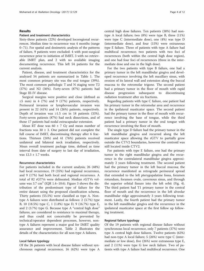
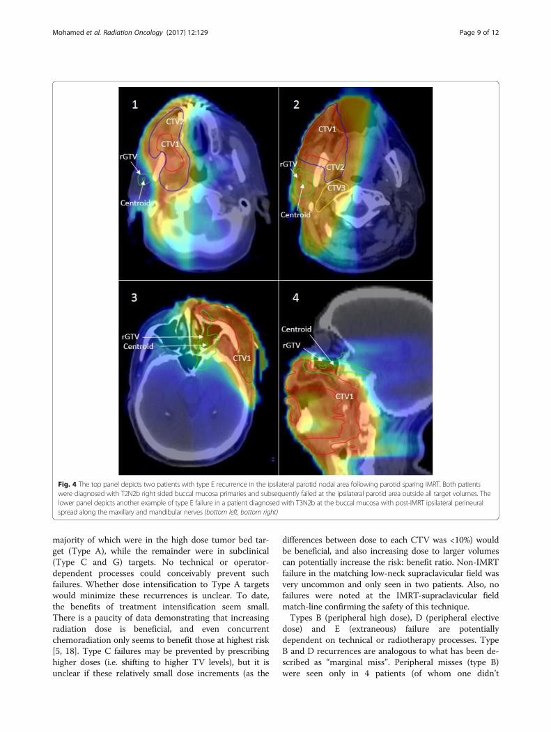
Fig. 4 The top panel depicts two patients with type E recurrence in the ipsilateral parotid nodal area following parotid sparing IMRT. Both patientswere diagnosed with T2N2b right sided buccal mucosa primaries and subsequently failed at the ipsilateral parotid area outside all target volumes. Thelower panel depicts another example of type E failure in a patient diagnosed with T3N2b at the buccal mucosa with post-IMRT ipsilateral perineuralspread along the maxillary and mandibular nerves (bottom left, bottom right)
Mohamed et al. Radiation Oncology (2017) 12:129 Page 9 of 12
complete the prescribed radiation dose). Three of thesewere in primary tumor sites, and more likely reflect thetumors finding pathways more amenable for spread, andgrowing out of the dose region rather than originating atthe periphery. Similarly, the one peripheral nodal failurewas in a retropharyngeal node that is typically not tar-geted, but fell into a D type failure rather than E due tothe proximity to the primary tumor bed. The paucity ofthese peripheral type recurrences reflects an adequateCTV delineation strategy, appropriate PTV margins, andprecise dose delivery.Type E extraneous recurrences were seen in approxi-
mately 17% of failures. Type E failures are analogous totraditionally defined “out of field” recurrences. This pat-tern of failure was mainly in patients who had primarytumors of the buccal mucosa or gingiva. Four patientshad primary type E recurrence, and 5 had nodal type Erecurrences. Two patients with type E failure at the pri-mary site had recurrences in sites relatively separatefrom the primary disease, and so these “recurrences”may represent second primary tumors.Retrograde ipsilateral perineural spread was the cause
of Type E recurrence in two patients (Fig. 4), and wasalso observed in two other patients (1 Type C and 1Type D) as seen in Table 2. Daly et al. had reported on apatient who had developed failure at the ipsilateralmasseter due to presence of perineural invasion andretrograde tracking along the mandibular nerve [11].Yao et al. had previously reported on two patients withextensive perineural invasion and retrograde trackingwho had developed failure within the infratemporal fossa[8]. We would also recommend that “nerves at risk inthe tumor bed should undergo biopsy and be covered ina retrograde fashion within the RT field” [11].Three patients had recurrence in the contralateral
undissected/unirradiated upper neck. Prior studies havereported that positive ipsilateral lymph nodes are apredictive factor for contralateral recurrence;conversely, contralateral lymph node metastases neveroccurred in patients without ipsilateral lymph node in-volvement [19, 20]. While these studies demonstratedthe association of ipsilateral lymph node involvementwith contralateral recurrence, the majority of patientsin these studies were predominantly patients with oraltongue cancer, and few patients had buccal cancer. Wecontinue to favor comprehensive bilateral radiation forpatients with tumors in central oral cavity sites, such asthe oral tongue and floor of mouth. Yao et al. recom-mended that patients with ipsilateral lymph node in-volvement in OCC should receive bilateral neckirradiation [8]. Moreover, Chan et al. suggested thatbilateral neck irradiation should be administered topatients with N2b disease [13]. However, again thesestudies were heavily weighted with patients with oral
tongue, and not buccal cancers. Thus our approach tobuccal and retromolar trigone tumors is individualized.Those patients with low nodal burden are still treatedto the ipsilateral neck, but those with bulky nodes, mul-tiple levels of nodes, or who have an epicentered lateralprimary site but the primary disease extends centrallyare treated to both sides of the neck.Two patients had almost identical pattern of recur-
rence in the ipsilateral parotid nodes as shown in Fig. 4.Strict dose constraints to the parotid have been recom-mended to avoid long-term risks of xerostomia [10]. Inour cohort, two patients with T2N2b buccal mucosa pri-maries had recurrences in the ipsilateral parotid glandwhich was spared during PO-IMRT. This phenomenahas been also reported in previous studies [13]. Theproximity of buccal and retromolar trigone tumors tothe parotid bed makes ipsilateral parotid avoidance chal-lenging. We therefore recommend limiting the extent ofradiation-induced xerostomia by focusing on sparing thecontralateral glands.To date, a limited number of studies have exclusively
investigated failure following PO-IMRT in OCC patients.Additional file 1: Tables S1 and S2 tabulate our study’spatient/treatment and failure characteristics comparedto extant literature. Loco-regional control of our studyare consistent with that of previous studies [8–13, 21].Although Sher et al. reported low loco-regional failurerates (7%), they acknowledged that it may have reflectedthe greater proportion of early T and N staging in theircohort [12]. Other disease characteristics are noted butare not directly comparable as the subset of reportedpatients varied from study to study.To classify failures using a spatial component, several
prior studies [8, 9, 11–13] used varying volume overlapapproaches [22–24]. Here we highlight two limitationsin these prior studies: 1) volume overlap methods forspatial characterization and 2) the lack of a dosimetriccomponent in failure analysis. Given enough time, recur-rence volumes can outgrow TV margins. Thus, thespatial characterization of “infield” vs. “outfield” is vol-ume dependent and biased by elapsed time. As thespatial component of our failure classification, we applya centroid-based approach. This approach has demon-strated to be more superior and accurate than volumet-ric overlap approaches, as the latter tends to assignfailures more peripherally [25, 26]. Moreover, the spatialcomponent alone is insufficient for accurate and specificreporting of failures. Without a dosimetric component,failures that are “infield” but in fact did not receive theprescribed dose (i.e. Types B and D in our classification)could be erroneously assumed to be biological failures.Subsequently, such “infield” failures are not investigatedfurther despite a potentially rectifiable technical orradiotherapy process.
Mohamed et al. Radiation Oncology (2017) 12:129 Page 10 of 12
As a retrospective series, the standard caveats apply. How-ever, although patients were not selected nor treated pro-spectively, all patients were reviewed by a multidisciplinaryteam. This data was collated as a secondary analysis of partof a larger programmatic evaluation of PO-IMRT outcomesfor OCC; the reader is encouraged to peruse the clinical/on-cologic report previously published [17]. Likewise, as a singleinstitute series from a high-volume tertiary center, thegeneralizability/scalability of our findings to facilities whichdo not utilize our systematic quality assurance methods (e.g.multi-physician direct physical examination and consensusreview of target delineation) would be suspect [27, 28].Nevertheless, despite these limitations, our study is the
largest, to our knowledge, systematic assessment of patternsof failure to OCC following PO-IMRT using a quality-controlled image-registration pipeline for methodologicallyrigorous pattern of failure investigation [16]. Similarly, ourstudy is the first to incorporate a dosimetric component infailure classification for OCC following PO-IMRT, inaddition to utilizing a centroid-based spatial componentand a validated DIR method which is critical for ac-curate failure analysis [14–16]. We hope that by util-izing a standardized typology for reporting patterns offailure in OCC following PO-IMRT, which can beadopted by multiple institutions, we can encourageother comparable reporting practices for PO-IMRT, ina manner that allows improved detection of possiblemodes of preventable error. This could allow forpooling of data to infer differences in treatment ap-proaches and subsequent outcomes amongst differentinstitutions.
ConclusionsPrior studies have assessed loco-regional control followingPO-IMRT to OCC in manner which elides the reality ofdosimetric gradients inherent in IMRT, and precludesidentification of systematic sources of modifiable errorwhich might impact these recurrences. A standardized typ-ology with both spatial and dosimetric components allowsfor more accurate and specific reporting of the patterns offailure over traditional “infield” vs. “marginal” vs. “outfield”failure classification schemes. Our study incorporates adosimetric component in addition to utilizing a centroid-based spatial component and a quantitatively validatedDIR method. Approximately half of the patients with localand/or regional failure included in the current study hadnon-central high dose recurrence. Thus, contrary to non-OCC sites, a substantial proportion of failures in our series,despite rigorous multiphysician quality assurance, are notdefinitive biological failures and, as potentially modifiablerisk-events, necessitate further investigation and potentialpractice modification. Other groups are encouraged toundertake similar efforts as single-site or pooled analysesfor OCC following PO-IMRT.
Additional file
Additional file 1:Tables S1 and S2. Showing our study’s patient/treatment and failure characteristics compared to extant literature. (PDF 99 kb)
AbbreviationsDIR: Deformable image registration; fD95%: dose to 95% of the failure volume;IRB: Institutional review board; pCT: planning CT; PO-IMRT: Postoperative intensitymodulated radiotherapy; rCT: recurrence CT; rGTV: recurrence gross tumorvolume; ROIs: Regions of interest; TVs: Target volumes
AcknowledgmentsNot applicable.
FundingDrs. Lai, Mohamed, and Fuller receive funding support from the National Institutesof Health (NIH)/National Institute for Dental and Craniofacial Research(1R01DE025248–01/R56DE025248–01). Dr. Fuller is a Sabin Family FoundationFellow and receives grant and/or salary support from the NIH/National CancerInstitute (NCI) Head and Neck Specialized Programs of Research Excellence (SPORE)Developmental Research Program Award (P50CA097007–10) and Paul CalabresiClinical Oncology Program Award (K12 CA088084–06); the National ScienceFoundation (NSF), Division of Mathematical Sciences, Joint NIH/NSF Initiative onQuantitative Approaches to Biomedical Big Data (QuBBD) Grant (NSF 1557679);the NIH Big Data to Knowledge (BD2K) Program of the National Cancer Institute(NCI) Early Stage Development of Technologies in Biomedical Computing,Informatics, and Big Data Science Award (1R01CA214825-01); NCI Early PhaseClinical Trials in Imaging and Image-Guided Interventions Program (1R01CA218148-01); a General Electric Healthcare/MD Anderson Center for Advanced BiomedicalImaging In-Kind Award; an Elekta AB/MD Anderson Department of RadiationOncology Seed Grant; the Center for Radiation Oncology Research (CROR) at MDAnderson Cancer Center Seed Grant; and the MD Anderson Institutional ResearchGrant (IRG) Program. Dr. Fuller has received speaker travel funding from Elekta AB.Supported in part by the National Institutes of Health (NIH)/National Cancer Institute(NCI) Cancer Center Support (Core) Grant CA016672 to The University of Texas MDAnderson Cancer Center.
Availability of data and materialsPlease contact the corresponding authors for data requests.
Authors’ contributionsAll listed co-authors performed the following: 1. Substantial contributions to theconception or design of the work; or the acquisition, analysis, or interpretation ofdata for the work; 2. Drafting the work or revising it critically for importantintellectual content; 3. Final approval of the version to be published; 4. Agreementto be accountable for all aspects of the work in ensuring that questions related tothe accuracy or integrity of any part of the work are appropriately investigatedand resolved. Specific additional individual cooperative effort contributions tostudy/manuscript design/execution/interpretation, in addition to all criteria aboveare listed as follows: ASRM and AJW- Undertook clinical and imaging data collec-tion; direct oversight of all image registration/segmentation, and data collectionworkflow, drafted initial manuscript, and participated in data analysis and interpret-ation of data. CDF- Co-primary investigator; with ASG conceived project and inter-preted study results, direct and final oversight of imaging and clinical datacollection. AJW, MK, GBG, JP, WHM, BMB, HS, SYL, SRQ, AMB, SJF, DIR, AGE - Directpatient care provision, direct imaging assessment and clinical data collection; inter-pretation and analytic support. ASG- Corresponding author; co-primary investiga-tor; conceived, coordinated, and directed all study activities, responsible for datacollection, project integrity, manuscript content and editorial oversight andcorrespondence. All authors read and approved the final manuscript.
Ethics approval and consent to participateThis study was initiated after the approval of the institutional review board at TheUniversity of Texas, MD Anderson Cancer Center of the study protocol #PA12–0168.
Consent for publicationNot applicable.
Competing interestsThe authors declare that they have no competing interest.
Mohamed et al. Radiation Oncology (2017) 12:129 Page 11 of 12
Publisher’s NoteSpringer Nature remains neutral with regard to jurisdictional claims inpublished maps and institutional affiliations.
Author details1Departments of Radiation Oncology, The University of Texas MD AndersonCancer Center, Unit 97, 1515 Holcombe Boulevard, Houston, TX 77030, USA.2Department of Head and Neck Surgery, The University of Texas MDAnderson Cancer Center, Unit 97, 1515 Holcombe Boulevard, Houston, TX77030, USA. 3Department of Clinical Oncology and Nuclear Medicine, Facultyof Medicine, University of Alexandria, Alexandria, Egypt. 4Department ofClinical Oncology and Nuclear Medicine, Faculty of Medicine, University ofAin-Shams, Cairo, Egypt. 5Department of Radiation Oncology, AllentownRadiation Oncology Associates, Allentown, PA, USA.
Received: 7 July 2017 Accepted: 9 August 2017
References1. Huang DT, Johnson CR, Schmidt-Ullrich R, Grimes M. Postoperative
radiotherapy in head and neck carcinoma with extracapsular lymph nodeextension and/or positive resection margins: a comparative study. Int JRadiat Oncol Biol Phys. 1992;23:737–42.
2. Ang KK, Trotti A, Brown BW, Garden AS, Foote RL, Morrison WH, et al.Randomized trial addressing risk features and time factors of surgery plusradiotherapy in advanced head-and-neck cancer. Int J Radiat Oncol BiolPhys. 2001;51:571–8.
3. Robertson AG, Soutar DS, Paul J, Webster M, Leonard AG, Moore KP, et al.Early closure of a randomized trial: surgery and postoperative radiotherapyversus radiotherapy in the management of intra-oral tumours. Clin Oncol.1998;10:155–60.
4. Boero IJ, Paravati AJ, Xu B, Cohen EEW, Mell LK, Le Q-T, et al. Importance ofradiation oncologist experience among patients with head-and-neck cancertreated with intensity-modulated radiation therapy. J Clin Oncol. 2016;34:684–90.
5. Blanchard P, Baujat B, Holostenco V, Bourredjem A, Baey C, Bourhis J, et al.Meta-analysis of chemotherapy in head and neck cancer (MACH-NC): acomprehensive analysis by tumour site. Radiother Oncol. 2011;100:33–40.
6. Daly ME, Le QT, Kozak MM, Maxim PG, Murphy JD, Hsu A, et al. Intensity-modulated radiotherapy for oral cavity Squamous cell carcinoma: patterns offailure and predictors of local control. Int J Radiat Oncol. 2011;80:1412–22.
7. Ooishi M, Motegi A, Kawashima M, Arahira S, Zenda S, Nakamura N, et al.Patterns of failure after postoperative intensity-modulated radiotherapy forlocally advanced and recurrent head and neck cancer. Jpn J Clin Oncol.2016;46:919–27.
8. Yao M, Chang K, Funk GF, Lu H, Tan H, Wacha J, et al. The failure patterns of oralcavity squamous cell carcinoma after intensity-modulated radiotherapy-theuniversity of iowa experience. Int J Radiat Oncol Biol Phys. 2007;67:1332–41.
9. Studer G, Zwahlen RA, Graetz KW, Davis BJ, Glanzmann C. IMRT in oralcavity cancer. Radiation Oncology (London, England). 2007; 2:16.
10. Gomez DR, Zhung JE, Gomez J, Chan K, Wu AJ, Wolden SL, et al. Intensity-modulated radiotherapy in postoperative treatment of oral cavity cancers.Int J Radiat Oncol Biol Phys. 2009;73:1096–103.
11. Daly ME, Le Q-T, Kozak MM, Maxim PG, Murphy JD, Hsu A, et al. Intensity-modulated radiotherapy for oral cavity Squamous cell carcinoma: patterns offailure and predictors of local control. Int J Radiat Oncol Biol Phys. 2011;80:1412–22.
12. Sher DJ, Thotakura V, Balboni TA, Norris CM Jr, Haddad RI, Posner MR, et al.Treatment of oral cavity Squamous cell carcinoma with adjuvant ordefinitive intensity-modulated radiation therapy. Int J Radiat Oncol BiolPhys. 2011;81:e215–e22.
13. Chan AK, Huang SH, Le LW, Yu E, Dawson LA, Kim JJ, et al. Postoperativeintensity-modulated radiotherapy following surgery for oral cavitysquamous cell carcinoma: patterns of failure. Oral Oncol. 2013;49:255–60.
14. Gunn GB, Blanchard P, Garden AS, Zhu XR, Fuller CD, Mohamed AS, et al.Clinical outcomes and patterns of disease recurrence after intensitymodulated proton therapy for Oropharyngeal Squamous carcinoma.Int J Radiat Oncol Biol Phys. 95:360–7.
15. Mohamed AS, Awan M, Kocak E, Beadle BM, Kantor ME, Gunn GB, et al.Methods for analysis and reporting the patterns of Locoregional failure in theera of IMRT for head and neck cancer: deformable image registration–basedquality assurance workflow. Int J Radiat Oncol Biol Phys. 2014;90:S569–S70.
16. Mohamed ASR, Rosenthal DI, Awan MJ, Garden AS, Kocak-Uzel E, Belal AM,et al. Methodology for analysis and reporting patterns of failure in the eraof IMRT: head and neck cancer applications. Radiat Oncol. 2016;11:1–10.
17. Quinlan-Davidson SR, Mohamed ASR, Myers JN, Gunn GB, Johnson FM,Skinner H, et al. Outcomes of oral cavity cancer patients treated withsurgery followed by postoperative intensity modulated radiation therapy.Oral Oncol. 2017;72:90–7.
18. Pignon JP, le Maitre A, Maillard E, Bourhis J, Group M-NC. Meta-analysis ofchemotherapy in head and neck cancer (MACH-NC): an update on 93randomised trials and 17,346 patients. Radiother Oncol: J Eur Soc TherapeutRadiol Oncol. 2009;92:4–14.
19. Chow TL, Chow TK, Chan TTF, Yu NF, Fung SC, Lam SH. Contralateral neckrecurrence of squamous cell carcinoma of oral cavity and oropharynx. J OralMaxillofac Surg. 2004;62:1225–8.
20. Kurita H, Koike T, Narikawa J-N, Sakai H, Nakatsuka A, Uehara S, et al. Clinicalpredictors for contralateral neck lymph node metastasis from unilateralsquamous cell carcinoma in the oral cavity. Oral Oncol. 2004;40:898–903.
21. Metcalfe E, Aspin L, Speight R, Ermis E, Ramasamy S, Cardale K, et al.Postoperative (chemo) radiotherapy for oral cavity Squamous cell carcinomas:outcomes and patterns of failure. Clin Oncol (R Coll Radiol). 2017;29:51–9.
22. Chao KSC, Ozyigit G, Tran BN, Cengiz M, Dempsey JF, Low DA. Patterns offailure in patients receiving definitive and postoperative IMRT for head-and-neck cancer. Int J Radiat Oncol Biol Phys. 2003;55:312–21.
23. Popovtzer A, Gluck I, Chepeha DB, Teknos TN, Moyer JS, Prince ME, et al.The pattern of failure after Reirradiation of recurrent Squamous cell headand neck cancer: implications for defining the targets. Int J Radiat OncolBiol Phys. 2009;74:1342–7.
24. Dawson LA, Anzai Y, Marsh L, Martel MK, Paulino A, Ship JA, et al. Patternsof local-regional recurrence following parotid-sparing conformal andsegmental intensity-modulated radiotherapy for head and neck cancer.Int J Radiat Oncol Biol Phys. 2000;46:1117–26.
25. Due AK, Vogelius IR, Aznar MC, Bentzen SM, Berthelsen AK, Korreman SS, et al.Methods for estimating the site of origin of locoregional recurrence in headand neck squamous cell carcinoma. Strahlenther Onkol. 2012;188:671–6.
26. Raktoe SAS, Dehnad H, Raaijmakers CPJ, Braunius W, Terhaard CHJ. Origin oftumor recurrence after intensity modulated radiation therapy for OropharyngealSquamous cell carcinoma. Int J Radiat Oncol Biol Phys. 2013;85:136–41.
27. Rosenthal DI, Asper JA, Barker JL, Garden AS, Chao KSC, Morrison WH, et al.Importance of patient examination to clinical quality assurance in head andneck radiation oncology. Head Neck. 2006;28:967–73.
28. Cardenas CE, Mohamed ASR, Tao R, Wong AJR, Awan MJ, Kuruvila S, et al.Prospective qualitative and quantitative analysis of real-time peer reviewquality assurance rounds incorporating direct physical examination for headand neck cancer radiation therapy. Int J Radiat Oncol Biol Phys. 2017;98:532–40.
• We accept pre-submission inquiries
• Our selector tool helps you to find the most relevant journal
• We provide round the clock customer support
• Convenient online submission
• Thorough peer review
• Inclusion in PubMed and all major indexing services
• Maximum visibility for your research
Submit your manuscript atwww.biomedcentral.com/submit
Submit your next manuscript to BioMed Central and we will help you at every step:
Mohamed et al. Radiation Oncology (2017) 12:129 Page 12 of 12
Related Documents