ABSTRACT: Matrix metalloproteases (MMPs) are key regulatory mole- cules in the formation, remodeling, and degradation of extracellular matrix components in both physiological and pathological processes. Skeletal mus- cles of mdx dystrophic mice show distinct patterns of inflammation and regeneration, suggesting that factors within the microenvironment influence the adaptive responses of muscles with predominantly slow-twitch or fast- twitch fibers. This study aimed to verify the pattern of MMP activity in gastrocnemius, soleus, and diaphragm muscles and correlate it with the regenerative capability at distinct stages of the mdx myopathy. Marked inflammation and myonecrosis was associated with increased MMP-9 ac- tivity and TNF- (tumor necrosis factor-alpha) production, whereas muscle regeneration, evidenced by NCAM (neural cell adhesion molecule) expres- sion and MMP-2 activity, varied at different stages of the disease. Soleus muscles showed a high percentage of NCAM-positive myofibers in the early stages (2 weeks) of the disease, but they appeared in the gastrocnemius muscles at 12 weeks and in the diaphragm at 24 weeks. Increased MMP-2 activity in the diaphragm throughout all stages of the disease suggests important tissue remodeling, which is probably associated with persistent inflammation. The results indicate that the microenvironment of distinct skeletal muscle may influence a particular kinetic pattern of MMP activity, which ultimately favors persistent inflammation and myofiber regeneration at different stages of the myopathy in mdx mice. Muscle Nerve 37: 583–592, 2008 PATTERN OF METALLOPROTEASE ACTIVITY AND MYOFIBER REGENERATION IN SKELETAL MUSCLES OF mdx MICE CRISTIANE BANI, MSc, 1 JUSSARA LAGROTA-CANDIDO, PhD, 2 DOUGLAS FLORINDO PINHEIRO, BSc, 1 PAULO EMI ´ LIO CORREA LEITE, MSc, 1 MARIA CRISTINA SALIMENA, MSc, 2 ANDREA HENRIQUES-PONS, PhD, 3 and THEREZA QUIRICO-SANTOS, PhD 1 1 Department of Cellular and Molecular Biology, Institute of Biology Federal Fluminense University, Nitero ´ i, RJ 24210-150, Brazil 2 Department of Immunobiology, Institute of Biology, Fluminense Federal University, Nitero ´i, Brazil 3 Department of Ultrastructure, Oswaldo Cruz Institute, Rio de Janeiro, Brazil Accepted 5 December 2007 Muscular dystrophy in the dystrophin-deficient mdx mouse, an animal model of Duchenne mus- cular dystrophy (DMD), is characterized by pro- gressive muscle wasting, which is usually associated with reactive fibrosis and increased deposition of connective tissue. 22 Dystrophin, a 427-kDa protein found in the inner sarcolemma as part of an inte- gral protein complex, is important for membrane stability, flexibility, and force during contrac- tion. 11 Lack of dystrophin results in a weakened cell membrane that renders muscle fibers fragile and susceptible to injury and necrosis. 2 The rela- tionship between lack of dystrophin and muscle fiber pathology is still not understood. The disease varies markedly among different skeletal muscles of the mdx mouse. In contrast to the relatively mild pathology in limb muscles, the diaphragm pre- sents extensive myonecrosis with progressive struc- tural and functional deterioration similar to that in DMD. 3,37 Such a difference may be partly attrib- uted to diversity in muscle-fiber–type expression due to levels of metabolic enzymes and the pattern of contractile and regulatory protein isoforms in the myofibril (16, 41). Abbreviations: ECM, extracellular matrix; DMD, Duchenne muscular dys- trophy; MMP, metalloprotease; NCAM, neural cell adhesion molecule; PBS, phosphate-buffered saline; PVDF, polyvinylidene fluoride; Sca, stem cell an- tigen; SP, side population; TACE, TNF-– converting enzyme; TNF-, tumor necrosis factor-alpha Key words: metalloprotease; muscle regeneration; muscular dystrophy; myofiber type Correspondence to: T. Quirico-Santos; e-mail: [email protected] © 2008 Wiley Periodicals, Inc. Published online 20 February 2008 in Wiley InterScience (www.interscience. wiley.com). DOI 10.1002/mus.20970 Metalloprotease Activity and mdx Muscle Regeneration MUSCLE & NERVE May 2008 583

Welcome message from author
This document is posted to help you gain knowledge. Please leave a comment to let me know what you think about it! Share it to your friends and learn new things together.
Transcript
ABSTRACT: Matrix metalloproteases (MMPs) are key regulatory mole-cules in the formation, remodeling, and degradation of extracellular matrixcomponents in both physiological and pathological processes. Skeletal mus-cles of mdx dystrophic mice show distinct patterns of inflammation andregeneration, suggesting that factors within the microenvironment influencethe adaptive responses of muscles with predominantly slow-twitch or fast-twitch fibers. This study aimed to verify the pattern of MMP activity ingastrocnemius, soleus, and diaphragm muscles and correlate it with theregenerative capability at distinct stages of the mdx myopathy. Markedinflammation and myonecrosis was associated with increased MMP-9 ac-tivity and TNF-� (tumor necrosis factor-alpha) production, whereas muscleregeneration, evidenced by NCAM (neural cell adhesion molecule) expres-sion and MMP-2 activity, varied at different stages of the disease. Soleusmuscles showed a high percentage of NCAM-positive myofibers in the earlystages (2 weeks) of the disease, but they appeared in the gastrocnemiusmuscles at 12 weeks and in the diaphragm at 24 weeks. Increased MMP-2activity in the diaphragm throughout all stages of the disease suggestsimportant tissue remodeling, which is probably associated with persistentinflammation. The results indicate that the microenvironment of distinctskeletal muscle may influence a particular kinetic pattern of MMP activity,which ultimately favors persistent inflammation and myofiber regeneration atdifferent stages of the myopathy in mdx mice.
Muscle Nerve 37: 583–592, 2008
PATTERN OF METALLOPROTEASE ACTIVITYAND MYOFIBER REGENERATION IN SKELETALMUSCLES OF mdx MICE
CRISTIANE BANI, MSc,1 JUSSARA LAGROTA-CANDIDO, PhD,2
DOUGLAS FLORINDO PINHEIRO, BSc,1 PAULO EMILIO CORREA LEITE, MSc,1
MARIA CRISTINA SALIMENA, MSc,2 ANDREA HENRIQUES-PONS, PhD,3
and THEREZA QUIRICO-SANTOS, PhD1
1 Department of Cellular and Molecular Biology, Institute of Biology Federal Fluminense University, Niteroi, RJ 24210-150, Brazil2 Department of Immunobiology, Institute of Biology, Fluminense Federal University, Niteroi, Brazil3 Department of Ultrastructure, Oswaldo Cruz Institute, Rio de Janeiro, Brazil
Accepted 5 December 2007
Muscular dystrophy in the dystrophin-deficientmdx mouse, an animal model of Duchenne mus-cular dystrophy (DMD), is characterized by pro-gressive muscle wasting, which is usually associatedwith reactive fibrosis and increased deposition ofconnective tissue.22 Dystrophin, a 427-kDa proteinfound in the inner sarcolemma as part of an inte-
gral protein complex, is important for membranestability, flexibility, and force during contrac-tion.11 Lack of dystrophin results in a weakenedcell membrane that renders muscle fibers fragileand susceptible to injury and necrosis.2 The rela-tionship between lack of dystrophin and musclefiber pathology is still not understood. The diseasevaries markedly among different skeletal musclesof the mdx mouse. In contrast to the relatively mildpathology in limb muscles, the diaphragm pre-sents extensive myonecrosis with progressive struc-tural and functional deterioration similar to thatin DMD.3,37 Such a difference may be partly attrib-uted to diversity in muscle-fiber–type expressiondue to levels of metabolic enzymes and the patternof contractile and regulatory protein isoforms inthe myofibril (16, 41).
Abbreviations: ECM, extracellular matrix; DMD, Duchenne muscular dys-trophy; MMP, metalloprotease; NCAM, neural cell adhesion molecule; PBS,phosphate-buffered saline; PVDF, polyvinylidene fluoride; Sca, stem cell an-tigen; SP, side population; TACE, TNF-�–converting enzyme; TNF-�, tumornecrosis factor-alphaKey words: metalloprotease; muscle regeneration; muscular dystrophy;myofiber typeCorrespondence to: T. Quirico-Santos; e-mail: [email protected]
© 2008 Wiley Periodicals, Inc.Published online 20 February 2008 in Wiley InterScience (www.interscience.wiley.com). DOI 10.1002/mus.20970
Metalloprotease Activity and mdx Muscle Regeneration MUSCLE & NERVE May 2008 583
LeitePec
Underline
Edited by Foxit Reader Copyright(C) by Foxit Software Company,2005-2007 For Evaluation Only.
Satellite cells are undifferentiated myogenic pre-cursors involved in the maintenance of normal mus-cle function.25 Following injury, muscle satellite cellsbecome activated and start to proliferate in order torepair the damaged area.13,32 Muscle satellite cellssustain the process of muscular regeneration ob-served in mdx mice. However, repeated cycles ofdegeneration exhaust the regenerative capacity ofsatellite cells so much that contractile muscle tissueis replaced by collagenous connective tissue.8 Myo-genic satellite cells reside beneath the basal laminaof adult skeletal muscles closely juxtaposed to skele-tal muscle fibers and account for 2%–5% of sublami-nal nuclei in adult muscle. Satellite cells expressspecific genes (M-cadherin, Pax7) and, following ac-tivation, give rise to a large number of daughtermyoblasts in addition to repopulating the satellitecell pool.1,33 Recent studies, however, have suggestedthat additional stem/progenitor cell populationsmay be recruited to participate in muscle regenera-tion and form functional myotubes.34 Muscle stemcells expressing the Sca-1 cell marker, located out-side the basal lamina, possess the ability to differen-tiate into hematopoietic cells and have the potentialto give rise, both in vitro and in vivo, to myogeniccells via a myocyte-mediated inductive interac-tion.1,10
Extracellular matrix (ECM) components and ma-trix metalloproteases (MMPs) play an important rolein the homeostasis and maintenance of myofiberfunctional integrity.4 MMPs influence cell motility,cell–cell-matrix interactions, matrix degradation,and the release of bioactive signaling molecules.40
The ECM surrounding myofibers differs, with slow-twitch muscles of rats containing more collagen thanfast-twitch muscles. Moreover, adaptive responses ofmuscles with predominantly slow-twitch fibers alsodiffer from those of fast-twitch fibers and this may bereflected by changes in MMP gene expression andcollagen metabolism.5,12 The aim of this study was toanalyze the pattern of metalloprotease activity andregenerative capability of different skeletal musclesat distinct stages of mdx myopathy.
METHODS
Animals. The study protocol and handling of ani-mals followed Brazilian institutional ethics guide-lines for animal studies. The mdx dystrophic andage-matched C57BL/10J (C57) control non-dystro-phic mice were maintained in animal housing atFluminense Federal University. They were kept atconstant temperature (20°C) with a light:dark cycleof 12:12 h and received acidified water and a com-
mercial rodent diet ad libitum. Male mdx mice wereselected at ages 6, 12, 24, and 48 weeks.
Histochemistry. Gastrocnemius, soleus, and dia-phragm muscles from mdx and control mice at dif-ferent stages of development were excised and fro-zen in liquid nitrogen. Frozen sections (10�m thick)were placed on poly-l-lysine (Sigma, St. Louis, Mis-souri) pre-coated slides and allowed to dry at roomtemperature for 4 h before staining. Fast green–Sirius red staining was used to analyze total collagenexpression.19 Myosin–ATPase fiber typing at pH 9.4was performed according to the method of Round etal.29
Immunohistochemistry. Cross-sections (5 �m) weremounted on poly-l-lysine pre-coated slides andblocked for endogenous peroxidase activity with3% hydrogen peroxide in phosphate-buffered sa-line (PBS) for 5 min. Sections were then rinsed for20 min in PBS and incubated for 20 min withdiluted normal blocking goat serum (Vector, Bur-lingame, California). Regenerating myofiberswere visualized using the optimal concentration ofmonoclonal rat IgG anti-CD56 (Clone 12F11; BD-Pharmingen Biosciences, San Diego, California).Anti-CD56 recognizes the neural cell adhesionmolecule (NCAM), a glycoprotein expressed dur-ing the early stages of myogenesis and in satellitecells. Stem cells were visualized using the optimaldilution of a monoclonal rat IgG-specific anti–Sca-1 (Clone E13-161.7; BD-Pharmingen Bio-sciences, San Diego, California). Sections werethen incubated in a moist chamber for 60 min atroom temperature with specific primary antibodydiluted in PBS with 1% bovine serum albuminfraction V (Sigma, St. Louis, Missouri). Slides werewashed in PBS for 15 min and incubated for 40min with peroxidase anti-rat secondary antibody(Southern Biotechnology Associates, Birmingham,Alabama). Peroxidase activity was revealed withaminoethylcarbazole (Sigma, St. Louis, Missouri)in the presence of H2O2. Sections were thenrinsed and counterstained with Mayer’s hematox-ylin. Myonuclei stained blue and cells brown.
Morphometric Analysis. Images from entire cross-sections of muscles from at least five mice at eachtime-point were acquired with a microdigital cameramounted on an Axioplan microscope (Zeiss;Oberkolchen, Germany). For quantification ofNCAM� cells, 400 myofibers were counted from twoindividual sections (at least 200 myofibers per sec-tion) and results expressed as the percentage of
584 Metalloprotease Activity and mdx Muscle Regeneration MUSCLE & NERVE May 2008
positive cells. The number of Sca-1� cells/mm2 inthe lesion area was determined by image-analysissoftware (Scion Image; National Institutes of Health,Bethesda, Maryland).
Tissue Extract Preparation. Muscles from mdx andcontrol muscles were immediately frozen and pre-served in liquid nitrogen (�165°C). Muscles wereweighed and homogenized (1/10 w/v) in extractionbuffer [100 mM Tris-HCl (pH 7.6), 200 mM NaCl,100 mM CaCl2, and 1% Triton X-100] at 4°C. Aftercentrifugation (15,000 g, 10°C, 10 min), the proteinconcentration in supernatant aliquots was deter-mined by the method of Lowry et al.,24 and equalamounts of total protein loaded for zymography (60�g/lane).
Gelatin Zymography. Zymogram gels consisted of7.5% (w/v) polyacrylamide impregnated with 2mg/ml type A gelatin from porcine skin (Sigma, St.Louis, Missouri) and 5% (w/v) polyacrylamide forstacking gels. Gels were further washed twice for 30min in 2.5% Triton X-100 solution at room temper-ature, then incubated at 37°C for 24 h in substratebuffer [10 mM Tris–HCl buffer (pH 7.5), with CaCl25 mM and ZnCl2 1 �M]. Thereafter, gels werestained with 30% methanol/10% acetic acid contain-ing 0.5% (w/v) brilliant blue R-250. Gelatinase activ-ity was visualized as unstained bands on a blue back-ground, representing areas of proteolysis of thesubstrate protein.
Metalloproteases are secreted in a latent formand require cleavage of amino-terminal peptide foractivation. The exposure of pro-enzymes of the tissueextracts to sodium dodecylsulfate during the gel sep-aration procedure leads to activation without proteo-lytic cleavage.38 Four bands can easily be seen corre-sponding to 100 kDa (MMP-9), 66 kDa (pre-proMMP-2), 60 kDa (pro MMP-2), and 55 kDa (activeMMP-2).21 Semi-quantitative analysis was performedusing image-analysis software (Scion Image; NIH,Bethesda, Maryland).
Western Blot. For the western blot assay, mdx andcontrol C57 skeletal muscles were homogenized in aprotease inhibitor solution (Sigma, St. Louis, Mis-souri) and protein concentrations determined bythe Bradford method. After establishing the sameconcentration of proteins for all samples, a 3� sam-ple buffer, pH 6.8 (0.173 M Tris, 30% glycerol, 3%sodium dodecylsulfate S, 3% �-mercaptoethanol,and 1 mg bromophenol blue), was added, and sam-ples denatured by boiling for 5 min.
Protein (20 �g per well) was loaded on 12.5%polyacrylamide gel for detection of tumor necrosisfactor-alpha (TNF-�). Samples were electrophoresedat 90 V for 120 min (Power Pac 200; Bio-Rad, Her-cules, California). The separated proteins weretransferred to polyvinylidene fluoride (PVDF) mem-branes (Hybond-P; Amersham Biosciences, Fairfield,Connecticut) and further blocked with 5% non-fatdry milk, 1% bovine serum albumin, and 1% normalgoat serum (Sigma, St. Louis, Missouri) in Tris-buff-ered saline 0.05% Tween-20 (TBST), pH 7.4, for 2 hat room temperature on a rocking platform. Mem-branes were then incubated with anti–TNF-� poly-clonal antibody (Sigma) in TBST at 4°C overnight.After washing three times for 10 min with TBST,blots were incubated with goat anti-rat peroxidase–conjugated secondary antibody (Sigma, St. Louis,Missouri) at 1:10,000 dilution for 2 h at room tem-perature. Finally, membranes were washed in TBST.Bands were identified using ECL Plus (AmershamBiosciences, Fairfield, Connecticut) for chemilumi-nescence detection and subsequent film exposurefor 5 min. The presence of TNF-� protein was veri-fied by comparing protein bands to the MolecularRainbow Weight Marker (Amersham Biosciences,Fairfield, Connecticut). As negative controls, sam-ples were incubated without primary antibodies.Equal loading of protein was assessed on strippedblots by immunodetection of actin with a horserad-ish peroxidase–conjugated goat polyclonal antibodydiluted at 1:1500 (Santa Cruz Biotechnology, SantaCruz, California).
Statistical Analysis. Excel software (Microsoft, Seat-tle, Washington) was used to calculate means andstandard deviations. Unpaired Student’s t-test wasapplied to assess the level of statistical significance.
RESULTS
Histological Alterations in mdx Skeletal Muscles.
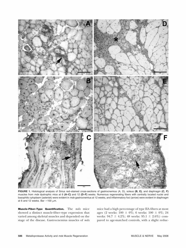
Compared with gastrocnemius and diaphragm, so-leus skeletal muscle of mdx mice showed limitedinflammation and necrosis at all stages of disease.Gastrocnemius muscles of mdx mice at 6 weeksshowed scattered foci of intense inflammatory infil-trate, whereas regenerating myofibers predominatedat 12 weeks. In contrast, intense inflammatory infil-trates were observed consistently in the diaphragmof mdx mice at 6 and 12 weeks (Fig. 1). Collagendeposition, evidenced by Sirius red stain, was oftenassociated with areas of inflammation and nearbyregenerating myoblasts in all muscles.
Metalloprotease Activity and mdx Muscle Regeneration MUSCLE & NERVE May 2008 585
Muscle-Fiber–Type Quantification. The mdx miceshowed a distinct muscle-fiber–type expression thatvaried among skeletal muscles and depended on thestage of the disease. Gastrocnemius muscles of mdx
mice had a high percentage of type IIA fibers at mostages (2 weeks: 100 � 0%; 6 weeks: 100 � 0%; 24weeks: 94.7 � 4.2%; 48 weeks: 95.1 � 2.6%) com-pared to age-matched controls, with a slight reduc-
FIGURE 1. Histological analysis of Sirius red–stained cross-sections of gastrocnemius (A, D), soleus (B, E), and diaphragm (C, F)muscles from mdx dystrophic mice at 6 (A–C) and 12 (D–F) weeks. Numerous regenerating fibers with centrally located nuclei andbasophilic cytoplasm (asterisk) were evident in mdx gastrocnemius at 12 weeks, and inflammatory foci (arrow) were evident in diaphragmat 6 and 12 weeks. Bar �100 �m.
586 Metalloprotease Activity and mdx Muscle Regeneration MUSCLE & NERVE May 2008
tion at 12 weeks (76.9 � 14.17%). At 2 weeks, C57and mdx mice showed a similar pattern of type IIAfibers in the soleus (C57: 31.8 � 4.5%; mdx: 27.6 �0.27%) and diaphragm (C57: 35.6 � 2.7%; mdx:30.0 � 5.5%) muscles. However, a marked increasein type IIA fibers was observed at 6 weeks (soleus:100 � 0%; diaphragm: 86.5 � 7.1%) in relation toC57 (soleus: 31.8 � 4.5%; diaphragm: 35.6 � 2.7%).Despite the muscle analyzed, clusters of type IIBmyofibers predominated, especially in areas with re-generating myofibers. High percentages (P �0.0001) of type IIB fibers were observed in the soleus(70.3 � 0.5%) and diaphragm (55.2 � 15.8%) ofmdx mice at 2 weeks. At the height of myonecrosis (6weeks), type IIB myofibers were not observed in anymuscle of mdx mice, but at 12 weeks gastrocnemiusmuscles (23.1 � 14.2%) showed a higher percentagethan soleus (8.16 � 4.9%) and diaphragm (6.8 �2.4%). Yet, only mdx diaphragm muscles presentedtype IIB fibers at ages corresponding to fibrosis (24weeks: 68.8 � 3.1%; 48 weeks: 46.2 � 14.6%).
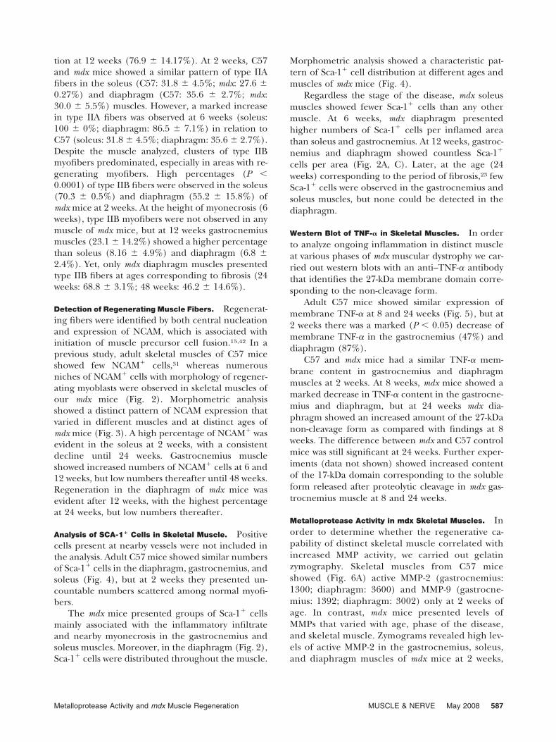
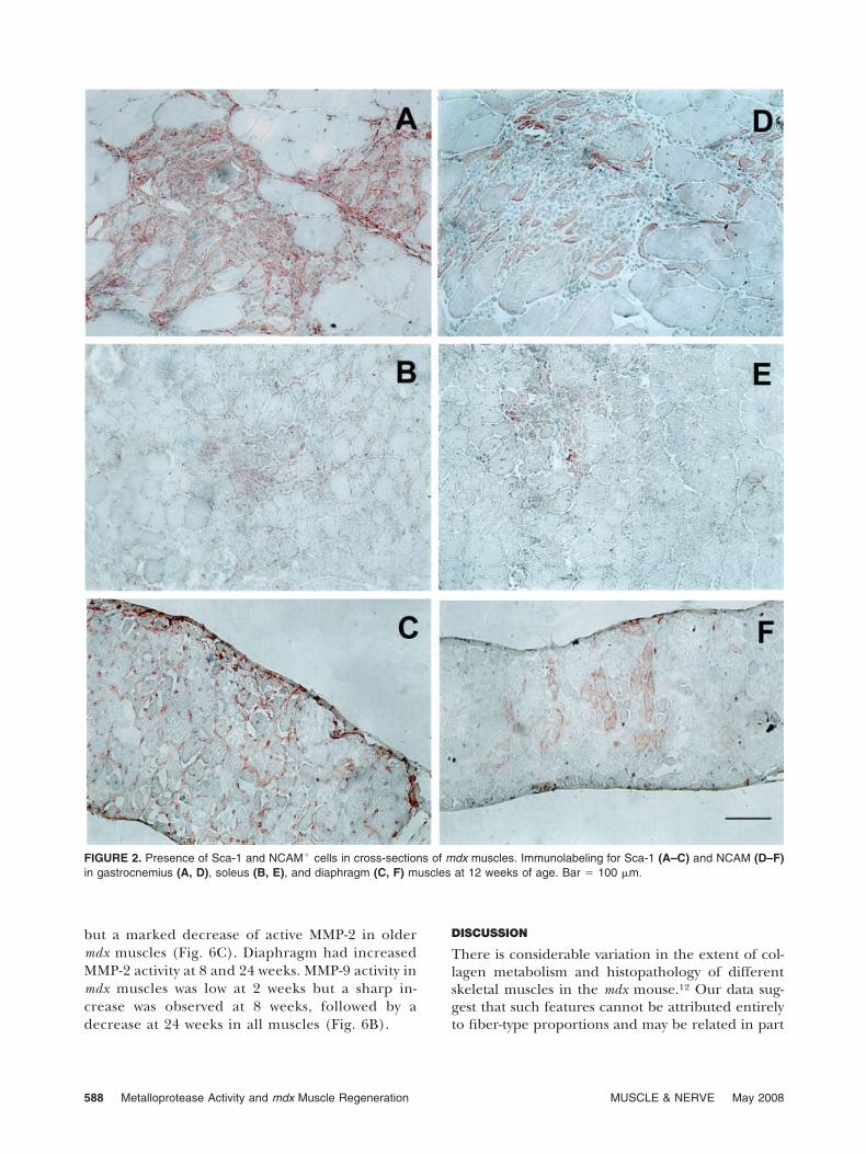
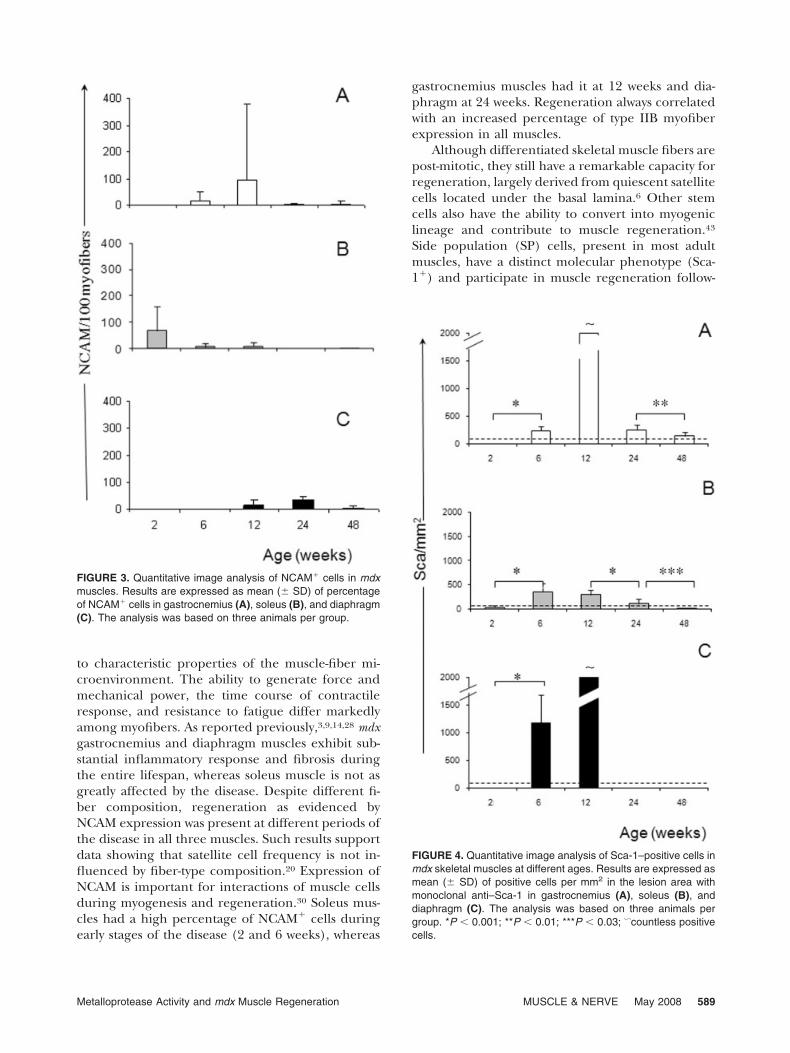
Detection of Regenerating Muscle Fibers. Regenerat-ing fibers were identified by both central nucleationand expression of NCAM, which is associated withinitiation of muscle precursor cell fusion.15,42 In aprevious study, adult skeletal muscles of C57 miceshowed few NCAM� cells,31 whereas numerousniches of NCAM� cells with morphology of regener-ating myoblasts were observed in skeletal muscles ofour mdx mice (Fig. 2). Morphometric analysisshowed a distinct pattern of NCAM expression thatvaried in different muscles and at distinct ages ofmdx mice (Fig. 3). A high percentage of NCAM� wasevident in the soleus at 2 weeks, with a consistentdecline until 24 weeks. Gastrocnemius muscleshowed increased numbers of NCAM� cells at 6 and12 weeks, but low numbers thereafter until 48 weeks.Regeneration in the diaphragm of mdx mice wasevident after 12 weeks, with the highest percentageat 24 weeks, but low numbers thereafter.
Analysis of SCA-1� Cells in Skeletal Muscle. Positivecells present at nearby vessels were not included inthe analysis. Adult C57 mice showed similar numbersof Sca-1� cells in the diaphragm, gastrocnemius, andsoleus (Fig. 4), but at 2 weeks they presented un-countable numbers scattered among normal myofi-bers.
The mdx mice presented groups of Sca-1� cellsmainly associated with the inflammatory infiltrateand nearby myonecrosis in the gastrocnemius andsoleus muscles. Moreover, in the diaphragm (Fig. 2),Sca-1� cells were distributed throughout the muscle.
Morphometric analysis showed a characteristic pat-tern of Sca-1� cell distribution at different ages andmuscles of mdx mice (Fig. 4).
Regardless the stage of the disease, mdx soleusmuscles showed fewer Sca-1� cells than any othermuscle. At 6 weeks, mdx diaphragm presentedhigher numbers of Sca-1� cells per inflamed areathan soleus and gastrocnemius. At 12 weeks, gastroc-nemius and diaphragm showed countless Sca-1�
cells per area (Fig. 2A, C). Later, at the age (24weeks) corresponding to the period of fibrosis,23 fewSca-1� cells were observed in the gastrocnemius andsoleus muscles, but none could be detected in thediaphragm.
Western Blot of TNF-� in Skeletal Muscles. In orderto analyze ongoing inflammation in distinct muscleat various phases of mdx muscular dystrophy we car-ried out western blots with an anti–TNF-� antibodythat identifies the 27-kDa membrane domain corre-sponding to the non-cleavage form.
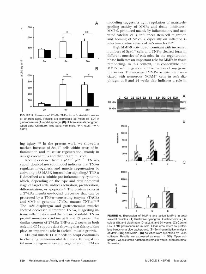
Adult C57 mice showed similar expression ofmembrane TNF-� at 8 and 24 weeks (Fig. 5), but at2 weeks there was a marked (P � 0.05) decrease ofmembrane TNF-� in the gastrocnemius (47%) anddiaphragm (87%).
C57 and mdx mice had a similar TNF-� mem-brane content in gastrocnemius and diaphragmmuscles at 2 weeks. At 8 weeks, mdx mice showed amarked decrease in TNF-� content in the gastrocne-mius and diaphragm, but at 24 weeks mdx dia-phragm showed an increased amount of the 27-kDanon-cleavage form as compared with findings at 8weeks. The difference between mdx and C57 controlmice was still significant at 24 weeks. Further exper-iments (data not shown) showed increased contentof the 17-kDa domain corresponding to the solubleform released after proteolytic cleavage in mdx gas-trocnemius muscle at 8 and 24 weeks.
Metalloprotease Activity in mdx Skeletal Muscles. Inorder to determine whether the regenerative ca-pability of distinct skeletal muscle correlated withincreased MMP activity, we carried out gelatinzymography. Skeletal muscles from C57 miceshowed (Fig. 6A) active MMP-2 (gastrocnemius:1300; diaphragm: 3600) and MMP-9 (gastrocne-mius: 1392; diaphragm: 3002) only at 2 weeks ofage. In contrast, mdx mice presented levels ofMMPs that varied with age, phase of the disease,and skeletal muscle. Zymograms revealed high lev-els of active MMP-2 in the gastrocnemius, soleus,and diaphragm muscles of mdx mice at 2 weeks,
Metalloprotease Activity and mdx Muscle Regeneration MUSCLE & NERVE May 2008 587
but a marked decrease of active MMP-2 in oldermdx muscles (Fig. 6C). Diaphragm had increasedMMP-2 activity at 8 and 24 weeks. MMP-9 activity inmdx muscles was low at 2 weeks but a sharp in-crease was observed at 8 weeks, followed by adecrease at 24 weeks in all muscles (Fig. 6B).
DISCUSSION
There is considerable variation in the extent of col-lagen metabolism and histopathology of differentskeletal muscles in the mdx mouse.12 Our data sug-gest that such features cannot be attributed entirelyto fiber-type proportions and may be related in part
FIGURE 2. Presence of Sca-1 and NCAM� cells in cross-sections of mdx muscles. Immunolabeling for Sca-1 (A–C) and NCAM (D–F)in gastrocnemius (A, D), soleus (B, E), and diaphragm (C, F) muscles at 12 weeks of age. Bar � 100 �m.
588 Metalloprotease Activity and mdx Muscle Regeneration MUSCLE & NERVE May 2008
to characteristic properties of the muscle-fiber mi-croenvironment. The ability to generate force andmechanical power, the time course of contractileresponse, and resistance to fatigue differ markedlyamong myofibers. As reported previously,3,9,14,28 mdxgastrocnemius and diaphragm muscles exhibit sub-stantial inflammatory response and fibrosis duringthe entire lifespan, whereas soleus muscle is not asgreatly affected by the disease. Despite different fi-ber composition, regeneration as evidenced byNCAM expression was present at different periods ofthe disease in all three muscles. Such results supportdata showing that satellite cell frequency is not in-fluenced by fiber-type composition.20 Expression ofNCAM is important for interactions of muscle cellsduring myogenesis and regeneration.30 Soleus mus-cles had a high percentage of NCAM� cells duringearly stages of the disease (2 and 6 weeks), whereas
gastrocnemius muscles had it at 12 weeks and dia-phragm at 24 weeks. Regeneration always correlatedwith an increased percentage of type IIB myofiberexpression in all muscles.
Although differentiated skeletal muscle fibers arepost-mitotic, they still have a remarkable capacity forregeneration, largely derived from quiescent satellitecells located under the basal lamina.6 Other stemcells also have the ability to convert into myogeniclineage and contribute to muscle regeneration.43
Side population (SP) cells, present in most adultmuscles, have a distinct molecular phenotype (Sca-1�) and participate in muscle regeneration follow-
FIGURE 4. Quantitative image analysis of Sca-1–positive cells inmdx skeletal muscles at different ages. Results are expressed asmean (� SD) of positive cells per mm2 in the lesion area withmonoclonal anti–Sca-1 in gastrocnemius (A), soleus (B), anddiaphragm (C). The analysis was based on three animals pergroup. *P � 0.001; **P � 0.01; ***P � 0.03; �countless positivecells.
FIGURE 3. Quantitative image analysis of NCAM� cells in mdxmuscles. Results are expressed as mean (� SD) of percentageof NCAM� cells in gastrocnemius (A), soleus (B), and diaphragm(C). The analysis was based on three animals per group.
Metalloprotease Activity and mdx Muscle Regeneration MUSCLE & NERVE May 2008 589
ing injury.1,26 In the present work, we showed amarked increase of Sca-1� cells within areas of in-flammation and muscular regeneration, mainly inmdx gastrocnemius and diaphragm muscles.
Recent evidence from a p55�/�p75�/� TNF-re-ceptor double-knockout model indicates that TNF-�regulates myogenesis and muscle regeneration byactivating p38 MAPK intracellular signaling.7 TNF-�is described as a soluble pro-inflammatory cytokine,which, depending on the type and developmentalstage of target cells, induces activation, proliferation,differentiation, or apoptosis.39 The protein exists asa 27-kDa membrane-bound precursor that can beprocessed by a TNF-�–converting enzyme (TACE)and MMP to generate 17-kDa, mature TNF-�.21,27
The mdx diaphragm and gastrocnemius musclesshowed decreased membrane TNF-�, suggesting in-tense inflammation and the release of soluble TNF-�pro-inflammatory cytokine at 8 and 24 weeks. Thesimilar content of 27-kDa TNF-� at 2 weeks in bothmdx and C57 support data showing that this cytokineplays an important role in skeletal muscle growth.
Skeletal muscle ECM needs to adapt continuallyto changing environmental demands. During skele-tal muscle degeneration and regeneration, ECM re-
modeling suggests a tight regulation of matrix-de-grading activity of MMPs and tissue inhibitors.4MMP-9, produced mainly by inflammatory and acti-vated satellite cells, influences stem-cell migrationand homing of SP cells, especially on inflamed l-selectin–positive vessels of mdx muscles.21,36
High MMP-9 activity, concomitant with increasednumbers of Sca-1� cells and TNF-� cleaved form indifferent muscles of mdx mice in the regenerationphase indicates an important role for MMPs in tissueremodeling. In this context, it is conceivable thatMMPs favor migration and activation of myogenicprecursors. The increased MMP-2 activity often asso-ciated with numerous NCAM� cells in mdx dia-phragm at 8 and 24 weeks also indicates a role in
FIGURE 6. Expression of MMP-9 and active MMP-2 in mdxskeletal muscles. (A) Illustrative zymogram. Gastrocnemius (G),soleus (S), and diaphragm (D) at 2, 8, and 24 weeks. (C) ControlC57BL/10 gastrocnemius muscle. Clear area refers to proteinlyse bands on a blue background. (B) Semi-quantitative analysisof MMP-9 (B) and MMP-2 (C) activities were quantified by Scionsoftware. Results are expressed as mean (� SD). Open col-umns: 2 weeks; cross-hatched columns: 8 weeks; filled columns:24 weeks.
FIGURE 5. Presence of 27-kDa TNF-� in mdx skeletal musclesat different ages. Results are expressed as mean (� SD) ingastrocnemius (A) and diaphragm (B) of three animals per group.Open bars: C57BL10; filled bars: mdx mice. *P � 0.05; **P �0.005.
590 Metalloprotease Activity and mdx Muscle Regeneration MUSCLE & NERVE May 2008
myofiber regeneration.21 During normal musclegrowth and myoblast fusion, MMP-2 is consideredimportant for degradation of type IV collagen andother basement membrane components such as en-tactin.35 MMPs are responsible for the processing of�-DG43 into �-DG30 (�-dystroglycan), which leads tothe disruption of the �-DG/�-DG interaction.17 In-deed, our data support the recent suggestion thatincreased MMP-2 activity may also reflect the extentof pathological injury in mdx muscles, especially inthe diaphragm. Taken together, the findings suggestthat the microenvironment of different skeletal mus-cles may influence a particular kinetic pattern ofMMP activity, which ultimately favors persistent in-flammation, membrane fragility, or myofiber regen-eration at different stages of the dystrophic pathol-ogy.
This study was supported by grants from CAPES (Coordenacao deAperfeicoamento de Pessoal de Nıvel Superior) and FAPERJ(Fundacao de Amparo a Pesquisa do Rio de Janeiro). The authorsthank Marcella d’Alincourt Salazar (Health Science Campus, Uni-versity of Toledo, Toledo, Ohio) and Rosangela Lawrence (Wash-tenaw Community College, Ann Arbor, Michigan) for Englishrevision of the manuscript.
REFERENCES
1. Asakura A, Seale P, Girgis-Gabardo A, Rudnicki MA. Myo-genic specification of side population cells in skeletal muscle.J Cell Biol 2002;159:123–134.
2. Bogdanovich S, Perkins KJ, Krag TO, Khurana TS. Therapeu-tics for Duchenne muscular dystrophy: current approachesand future directions. J Mol Med 2004;82:102–115.
3. Boland B, Himpens B, Denef JF, Gillis JM. Site-dependentpathological differences in smooth muscles and skeletal mus-cles of the adult mdx mouse. Muscle Nerve 1995;18:649–657.
4. Carmeli E, Moas M, Reznick AZ, Coleman R. Matrix metallo-proteinases and skeletal muscle: a brief review. Muscle Nerve2004;29:191–197.
5. Carmeli E, Moas M, Lennon S, Powers SK. High intensityexercise increases expression of matrix metalloproteinases infast skeletal muscle fibres. Exp Physiol 2005;90:613–619.
6. Charge SB, Rudnicki MA. Cellular and molecular regulationof muscle regeneration. Physiol Rev 2004;84:209–238.
7. Chen SE, Jin B, Li YP. TNF-alpha regulates myogenesis andmuscle regeneration by activating p38 MAPK. Am J PhysiolCell Physiol 2007;292:C1660–C1671.
8. Collins CA, Morgan JE. Duchenne’s muscular dystrophy: an-imal models used to investigate pathogenesis and developtherapeutic strategies. Int J Exp Pathol 2003;84:165–172.
9. Connolly AM, Keeling RM, Mehta S, Pestronk A, Sanes JR.Three mouse models of muscular dystrophy: the natural his-tory of strength and fatigue in dystrophin-, dystrophin/utro-phin-, and laminin a2-deficient mice. Neuromusc Dis 2001;11:703–712.
10. Dellavalle A, Sampaolesi M, Tonlorenzi R, Tagliafico E, Sac-chetti B, Perani L, et al. Pericytes of human skeletal muscleare myogenic precursors distinct from satellite cells. Nat CellBiol 2007;9:255–267.
11. Ehmsen J, Poon E, Davies K. The dystrophin-associated pro-tein complex. J Cell Sci 2002;115:2801–2803.
12. Gosselin LE, Williams JE, Personius K, Farkas GA. A compar-ison of factors associated with collagen metabolism in differ-
ent skeletal muscles from dystrophic (mdx) mice: impact ofpirfenidone. Muscle Nerve 2007;35:208–216.
13. Grounds MD. Age-associated changes in the response of skel-etal muscle cells to exercise and regeneration. Ann NY AcadSci 1998;854:78–91.
14. Haslett JN, Kang PB, Han M, Kho AT, Sanoudou D, VolinskiJM, et al. The influence of muscle type and dystrophin defi-ciency on murine expression profiles. Mamm Genome 2005;16:739–748.
15. Hawke TJ, Garry DJ. Myogenic satellite cells: physiology tomolecular biology. J Appl Physiol 2001;91:534–551.
16. Hitomi Y, Kizaki T, Watanabe S, Matsumura G, Fujioka Y,Haga S, et al. Seven skeletal muscles rich in slow muscle fibersmay function to sustain neutral position in the rodent hind-limb. Comp Biochem Physiol B Biochem Mol Biol 2005;140:45–50.
17. Hnia K, Hugon G, Rivier F, Masmoudi A, Mercier J, MornetD. Modulation of p38 mitogen-activated protein kinase cas-cade and metalloproteinase activity in diaphragm muscle inresponse to free radical scavenger administration in dystro-phin-deficient Mdx mice. Am J Pathol 2007;170:633–643.
18. Ibebunjo C. Type, diameter and distribution of fibres in somerespiratory and abdominal muscles of the goat. Vet Res Com-mun 1993;17:171–182.
19. Junqueira LC, Bignolas G, Brentani RR. Picrosirius stainingplus polarization microscopy, a specific method for collagendetection in tissue sections. Histochem J 1979;11:447–455.
20. Kadi F, Charifi N, Henriksson J. The number of satellite cellsin slow and fast fibres from human vastus lateralis muscle.Histochem Cell Biol 2006;126:83–87.
21. Kherif S, Lafuma C, Dehaupas M, Lachkar S, Fournier JG,Verdiere-Sahuque M, et al. Expression of matrix metallopro-teinases 2 and 9 in regenerating skeletal muscle: a study inexperimentally injured and mdx muscles. Dev Biol 1999;205:158–170.
22. Lagrota-Candido J, Canella I, Savino W, Quirico-Santos T.Expression of extracellular matrix ligands and receptors inthe muscular tissue and draining lymph nodes of mdx dystro-phic mice. Clin Immunol 1999;93:143–151.
23. Lagrota-Candido J, Vasconcellos R, Cavalcanti M, Bozza M,Savino WQ, Quirico-Santos T. Resolution of skeletal muscleinflammation in mdx dystrophic mouse is accompanied byincreased immunoglobulin and interferon-gamma produc-tion. Int J Exp Pathol 2002;83:121–132.
24. Lowry OH, Rosebrough NJ, Farr AL, Randall RJ. Proteinmeasurement with the Folin phenol reagent. J Biol Chem1951;193:265–275.
25. Mauro A. Satellite cell of skeletal muscle fibers. J BiophysBiochem Cytol 1961;9:493–495.
26. Meeson AP, Hawke TJ, Graham S, Jiang N, Elterman J,Hutcheson K, et al. Cellular and molecular regulation ofskeletal muscle side population cells. Stem Cells 2004;22:1305–1320.
27. Mullberg J, Althoff K, Jostock T, Rose-John S. The importanceof shedding of membrane proteins for cytokine biology. EurCytokine Netw 2000;11:27–38.
28. Porter JD, Merriam AP, Leahy P, Gong B, Feuerman J, ChengG, et al. Temporal gene expression profiling of dystrophin-deficient (mdx) mouse diaphragm identifies conserved andmuscle group-specific mechanisms in the pathogenesis ofmuscular dystrophy. Hum Mol Genet 2004;13:257–269.
29. Round JM, Matthews Y, Jones DA. A quick, simple and reliablehistochemical method for ATPase in human muscle prepara-tions. Histochem J 1980;12:707–710.
30. Salarieh A, Soler AP, Axiotis CA. Overexpression of neuralcell adhesion molecule in regenerative muscle fibers in 3-hy-droxy-3-methylglutaryl coenzyme: a reductase inhibitor-in-duced rhabdomyolysis. Appl Immunohistochem Mol Mor-phol 2004;12:234–239.
31. Salimena MC, Lagrota-Candido J, Quirico-Santos T. Genderdimorphism influences extracellular matrix expression and
Metalloprotease Activity and mdx Muscle Regeneration MUSCLE & NERVE May 2008 591
regeneration of muscular tissue in mdx dystrophic mice. His-tochem Cell Biol 2004;122:435–444.
32. Seale P, Rudnicki MA. A new look at the origin, function, and“stem-cell” status of muscle satellite cells. Dev Biol 2000;218:115–124.
33. Seale P, Sabourin LA, Girgis-Gabardo A, Mansouri A, Gruss P,Rudnicki MA. Pax7 is required for the specification of myo-genic satellite cells. Cell 2000;102:777–786.
34. Shi X, Garry DJ. Muscle stem cells in development, regener-ation, and disease. Genes Dev 2006;20:1692–1708.
35. Sires UI, Griffin GL, Broekelmann TJ, Mecham RP, MurphyG, Chung AE, et al. Degradation of entactin by matrix met-alloproteinases. Susceptibility to matrilysin and identificationof cleavage sites. J Biol Chem 1993;268:2069–2074.
36. Stahle-Backdahl M, Inoue M, Guidice GJ, Parks WC. 92-kDgelatinase is produced by eosinophils at the site of blisterformation in bullous pemphigoid and cleaves the extracellu-lar domain of recombinant 180-kD bullous pemphigoid au-toantigen. J Clin Invest 1994;93:2022–2030.
37. Stedman HH, Sweeney HL, Shrager JB, Maguire HC, Panet-tieri RA, Petrof B, et al. The mdx mouse diaphragm repro-
duces the degenerative changes of Duchenne muscular dys-trophy. Nature 1991;352:536–539.
38. Talhouk RS, Bissell MJ, Werb Z. Coordinated expression ofextracellular matrix-degrading proteinases and their inhibi-tors regulates mammary epithelial function during involu-tion. J Cell Biol 1992;118:1271–1282.
39. Tracey KJ. Physiology and immunology of the cholinergicantiinflammatory pathway. J Clin Invest 2007;117:289–296.
40. VanSaun MN, Matrisian LM. Matrix metalloproteinases andcellular motility in development and disease. Birth DefectsRes C Embryo Today 2006;78:69–79.
41. Wang LC, Kernell D. Fibre type regionalisation in lower hind-limb muscles of rabbit, rat and mouse: a comparative study. JAnat 2001;199:631–643.
42. Warren GL, Hulderman T, Mishra D, Gao X, Millecchia L,O’Farrell L, et al. Chemokine receptor CCR2 involvement inskeletal muscle regeneration. FASEB J 2005;19:413–415.
43. Zammit PS, Partridge TA, Yablonka-Reuveni Z. The skeletalmuscle satellite cell: the stem cell that came in from the cold.J Histochem Cytochem 2006;54:1177–1191.
592 Metalloprotease Activity and mdx Muscle Regeneration MUSCLE & NERVE May 2008
Related Documents


![MDX-2 Sintaxis MDX [Recordatorios]](https://static.cupdf.com/doc/110x72/55cf969a550346d0338c9b6f/mdx-2-sintaxis-mdx-recordatorios.jpg)