review article 115 Patients’ Expectations in Lens Extraction Surgery: a Systematic Review Eirini-Kanella Panagiotopoulou*, Panagiota Ntonti, Eleni Vlachou, Kimon Georgantzoglou, Georgios Labiris ABSTRACT It is common knowledge that there are patients who have an uncomplicated cataract surgery with an actual improvement of their visual acuity, but they are dissatisfied with their final visual capacity. It is hypothesized that patients’ preoperative expectations play asignificant role in their postoperative perceptions. A systematic review of the recent literature regarding preoperative expectations of patients before lens extraction surgery and their postoperative perceptions as regards the visual outcome was performed based on the PubMed, Medline, Google Scholar, American Academy of Ophthalmology, Nature and Springer databases in September 2017 and data from 14 descriptive and 7 comparative studies were included in this narrative review. e objective of this review is the determination of the relationship between preoperative expectations and postoperative perception of visual outcome, as well as the investigation of predictors of patient satisfaction by understanding the factors that determine preoperative patient expectations. A considerable number of studies evaluate patient expectations before cataract surgery and compare them with postoperative patient perceptions. In conclusion, the final patient’s postoperative perception could be affected both by the actual outcome of the operation and by patient preoperative expectations. Ocular and systemic comorbidity, unrealistic expectations, preoperative spectacle independence, the cost of surgery, and a previous cataract surgery as well as the level of health literacy and age could influence preoperative expectations and predict more accurately patient satisfaction. Taking these factors into consideration could allow surgeons to control the expectations with an extensive preoperative counseling. KEYWORDS satisfaction; cataract; expectations; visual outcomes AUTHOR AFFILIATIONS Department of Ophthalmology, University Hospital of Alexandroupolis, Dragana, Alexandroupolis, Greece * Ophthalmology Department, University Hospital of Alexandroupolis, 68100 Dragana, Alexandroupolis, Greece; e-mail: [email protected] Received: 2 July 2018 Accepted: 11 November 2018 Published online: 22 January 2019 Acta Medica (Hradec Králové) 2018; 61(4): 115–124 https://doi.org/10.14712/18059694.2018.129 © 2018 e Authors. is is an open-access article distributed under the terms of the Creative Commons Attribution License (http://creativecommons.org/licenses/by/4.0), which permits unrestricted use, distribution, and reproduction in any medium, provided the original author and source are credited.

Welcome message from author
This document is posted to help you gain knowledge. Please leave a comment to let me know what you think about it! Share it to your friends and learn new things together.
Transcript
review article 115
Patients’ Expectations in Lens Extraction Surgery: a Systematic Review
Eirini-Kanella Panagiotopoulou*, Panagiota Ntonti, Eleni Vlachou, Kimon Georgantzoglou, Georgios Labiris
A B S T R A C TIt is common knowledge that there are patients who have an uncomplicated cataract surgery with an actual improvement of their visual acuity, but they are dissatisfied with their final visual capacity. It is hypothesized that patients’ preoperative expectations play a significant role in their postoperative perceptions. A systematic review of the recent literature regarding preoperative expectations of patients before lens extraction surgery and their postoperative perceptions as regards the visual outcome was performed based on the PubMed, Medline, Google Scholar, American Academy of Ophthalmology, Nature and Springer databases in September 2017 and data from 14 descriptive and 7 comparative studies were included in this narrative review. The objective of this review is the determination of the relationship between preoperative expectations and postoperative perception of visual outcome, as well as the investigation of predictors of patient satisfaction by understanding the factors that determine preoperative patient expectations. A considerable number of studies evaluate patient expectations before cataract surgery and compare them with postoperative patient perceptions. In conclusion, the final patient’s postoperative perception could be affected both by the actual outcome of the operation and by patient preoperative expectations. Ocular and systemic comorbidity, unrealistic expectations, preoperative spectacle independence, the cost of surgery, and a previous cataract surgery as well as the level of health literacy and age could influence preoperative expectations and predict more accurately patient satisfaction. Taking these factors into consideration could allow surgeons to control the expectations with an extensive preoperative counseling.
K E Y W O R D S satisfaction; cataract; expectations; visual outcomes
A U T H O R A F F I L I AT I O N SDepartment of Ophthalmology, University Hospital of Alexandroupolis, Dragana, Alexandroupolis, Greece* Ophthalmology Department, University Hospital of Alexandroupolis, 68100 Dragana, Alexandroupolis, Greece;
e-mail: [email protected]
Received: 2 July 2018Accepted: 11 November 2018Published online: 22 January 2019
Acta Medica (Hradec Králové) 2018; 61(4): 115–124https://doi.org/10.14712/18059694.2018.129© 2018 The Authors. This is an open-access article distributed under the terms of the Creative Commons Attribution License (http://creativecommons.org/licenses/by/4.0), which permits unrestricted use, distribution, and reproduction in any medium, provided the original author and source are credited.
116 Eirini-Kanella Panagiotopoulou et al. Acta Medica (Hradec Králové)
INTRODUCTION
Cataract is formed by cloudy areas of accumulated protein on the crystalline lens of the eye. It is mainly an age-related eye disease that hinders clear vision. According to the World Health Organization, 20 million people are blind worldwide because of untreated cataract, namely 51% of world blindness arises from cataract (1). Currently, there is not any proven prophylactic medical treatment (2). However, reduction of smoking and exposure to ultraviolent radiation could prevent or delay the development of cataract. Other risk factors are diabetes and high BMI (body mass index). The only efficient treatment is surgical removal of the clouded lens and its re-placement with an artificial intraocular lens (IOL). Neverthe-less, in many developing countries people have no easy ac-cess to eye care. Hence cataract still remains the main cause of blindness (1).
Cataract surgery is the most common procedure per-formed worldwide (3). Today, the most popular technique of surgical removal of cataract in developed countries is phacoemulsification (2). However, when the relatively new technique of phacoemulsification is not easily available, the opaque lens is removed via extracapsular cataract extraction (ECCE). The great safety and efficacy of cataract surgery, the progress in the techniques of ocular biometry measurements and the existence of accurate IOL power calculation methods increase patients’ and surgeons’ expectations for a continu-ous improvement of outcomes (4).
The increasing patient expectations for the visual out-comes after a cataract surgery, intensify not only the need for evaluation of patient satisfaction, but also the need for a more detailed and targeted assessment of patient preop-erative expectations and postoperative perceptions for the visual outcome.
There is no doubt that a number of patients, who have an uncomplicated cataract surgery with an actual improvement of their visual acuity (VA), are dissatisfied with their final visual function. Probable causes might be insufficient commu-nication between patients and ophthalmologists, high expec-tations and demographic characteristics of each patient (5).
Regarding the expectations-satisfaction relationship, if the product/service performance is higher than the expect-ed, patients are highly satisfied. If the performance matches individuals’ expectations, they are satisfied. If it is lower than their expectations, individuals are dissatisfied (6, 7). In order to assess the success of a cataract surgery, the patient-re-ported outcome and patient satisfaction could be taken into account because they would demonstrate if postoper-ative visual outcome achieved preoperative expectations (8). Therefore, medical care professionals should control patients’ expectations for the outcome of the received treatment in order to affect their perception about the final treatment quality (6).
Aiming a more comprehensive description of preoper-ative expectations and self-reported level of difficulty and
satisfaction with vision after ocular procedures, a variety of questionnaires have been created. Among them are Visual Function 12-Item Scale (VF-12) (4), VF-14 (7–9), VF-15 (10) and Catquest-9SF (11).
Within this context, primary objective of this study is to review the published literature regarding patients’ expecta-tions before a lens extraction surgery and to determine the relationship between preoperative expectations and post-operative perception of visual outcome. A secondary objec-tive of the present study is the investigation of predictors of patient satisfaction by understanding the factors that de-termine preoperative patient expectations. Our intention is the best possible comprehension of the literature regarding patient expectations for a favorable cataract surgery visual outcome and the assistance of other researchers in further investigation in this domain of knowledge.
MATERIAL AND METHODS
A systematic search for relevant studies was performed based on the PubMed, Medline, Google Scholar, American Academy of Ophthalmology, Nature and Springer databases using the following search terms: preoperative expectations AND cataract surgery, patients expectations AND multifocal intraocular lenses, preoperative expectations AND cataract surgery AND postoperative outcomes. The search took place in September of 2017. Search filters and language restrictions were not used in this initial search. The results of this search were checked and only articles with a relative to the subject title were selected. Afterwards, the abstracts and full texts of these selected articles were reviewed thoroughly and the following data were extracted and assessed: patient selec-tion, demographic characteristics, questionnaires, preop-erative expectations, postoperative perceptions, spectacle independence, comorbidity, unrealistic expectations, cost of surgery, personality-psychology, use of spectacles preoper-atively and previous cataract surgery. Both comparative and descriptive studies in adult and adolescent patients were in-cluded in this review. Articles not available in English, German or Spanish language were excluded. When the eligible articles were not available in full text, abstracts were used as a source of information.
RESULTS
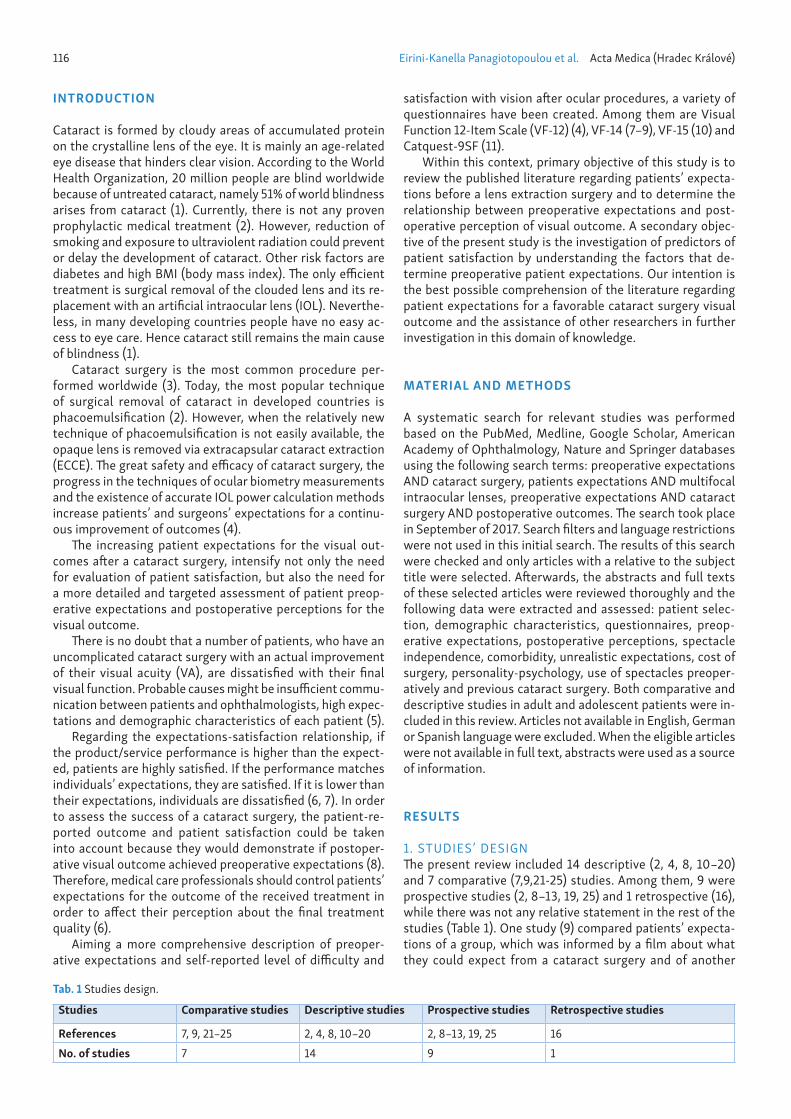
1. STUDIES’ DESIGNThe present review included 14 descriptive (2, 4, 8, 10–20) and 7 comparative (7,9,21-25) studies. Among them, 9 were prospective studies (2, 8–13, 19, 25) and 1 retrospective (16), while there was not any relative statement in the rest of the studies (Table 1). One study (9) compared patients’ expecta-tions of a group, which was informed by a film about what they could expect from a cataract surgery and of another
Tab. 1 Studies design.
Studies Comparative studies Descriptive studies Prospective studies Retrospective studies
References 7, 9, 21–25 2, 4, 8, 10–20 2, 8–13, 19, 25 16No. of studies 7 14 9 1
Patients’ Expectations in Lens Extraction Surgery 117
group, which was enlightened by video giving information on the anatomy and the pathophysiology of the cataract. Addi-tionally, expectations of patients who had already undergone lens exchange and patients who did not have any previous cataract operation were compared (9). One of the objectives in the study of Nijkamp et al. (7) was the comparison of preop-erative expectations of patients who were going to undergo monofocal and these ones who were going to undergo mul-tifocal lens exchange. Among the other objectives of Lowe et al. study (22) was the comparison of visual expectations of inpatients and day cases for cataract surgery.
2. PATIENTS’ SELECTION CRITERIAPatient selection was presented to be very crucial for the best possible assessment of patients’ preoperative expectations for their visual function after a cataract surgery and most studies dealt thoroughly with patients’ inclusion and exclusion criteria. The most common inclusion criterion was first-eye cataract surgery in order to avoid bias from another cataract surgery (4, 7, 11–13, 23). However, Pager et al. (8, 9) included also patients with a previous cataract surgery and examined how their expectations could be differentiated, compared to patients without any earlier cataract operation. Nijkamp et al. (7), who compared monofocal and multifocal IOLs, included only patients with astigmatism 1.5 diopters (D) or less, spec-tacle sphere power ≥ −6D and ≤ +4D and axial length between 19.5 and 26 mm. Additionally, patients of this study should not be professional night drivers, be able to answer question-naires in Dutch and should not have reduced mental capacity.
Among the exclusion criteria were the following: prior intraocular operation in the same eye, previous unilateral cataract surgery, single-eyed patients, corneal astigmatism, existence of psychiatric disorders and reduced mental ca-pacity to complete the questionnaire. Patients with visually impairing ocular comorbidities such as glaucoma, macular degeneration, amblyopia, macular hole, retinal detachment or other retinal abnormalities that may affect postoperative visual performance were excluded from many studies (2, 7, 13, 22). Nevertheless, there is a number of studies that did not exclude patients with ocular comorbidities (10–12). Ad-ditionally, difficulty in reading or understanding the language of questionnaires was a cause of rejection (2, 7).
Regarding patients’ age, one study conducted in Ethiopia (10) included patients aged 12 years old or older, while some other studies included patients of age ≥18 years old (11, 26). The majority of studies included older male and female indi-viduals, namely aged 50 years and older (14, 23) or ≥55 years old (22).
3. DEMOGRAPHIC CHARACTERISTICSIn some studies, sociodemographic factors of patients in-cluding age, gender and health literacy were examined and correlated with the preoperative expectations and postop-erative satisfaction. In this review, it was also examined if the level of education (low, middle or high / primary school, college, university) (4, 7, 11, 14, 23, 26, 27) and occupation (Oc-cupation level: I–VII) (14, 26) could serve as possible factors that could predict and affect patients’ expectations. Finally, nervousness and eagerness for spectacle independence were
investigated for their impact on the satisfaction of patients after cataract surgery (7).
4. QUESTIONNAIRESDifferent questionnaires were used as sources of informa-
tion about demographic variables and patients’ preoperative expectations as regards their present difficulties in daily life and the expected improvement in visual function after the cataract operation.
Tielsch et al. (4) assessed patients’ self-reported difficulty and satisfaction with their vision before and after the cata-ract procedure using VF-12. In addition, their preoperative expectations were measured thoroughly. Two studies (8, 9) which also examined the difference between expectations and final visual outcome used Visual Function Index (VF-14), which is a modification of VF-12 and one of the most common instruments used for patients who underwent cataract sur-gery (28). Addisu et al. (10) measured preoperative, expected postoperative and actual postoperative visual status with the use of VF-15 questionnaire (Visual Function-15, a 15-item slightly changed version of 14-item VF-14 questionnaire). Scores on the VF-12, VF-14 and VF-15 range from 0 (no visual ability) to 100 (no visual disability) (4, 8–10). Chen et al. (11) and Mollazadegan et al. (20) used Catquest-9SF question-naire, which contained questions about patients’ troubles in everyday-life activities, their satisfaction with vision before cataract extraction and their expectations for their postop-erative visual function. These expectations were compared with their self-assessed postoperative satisfaction. Nijkamp et al. (7), who conducted a study that evaluated the effec-tiveness of multifocal IOLs in the correction of presbyopia after cataract extraction, used Eagerness for Spectacle In-dependence (ESI) and the Neuroticism Scale of the Eysenck Personality Questionnaire – Revised Short Scale (EPQR-S). Other questionnaires which were used for the same purpose were the American Society of Cataract and Refractive Surgery (ASCRS) Cataract Data Collection Form (ACDCF) (16) and some self-administered questionnaires that were developed by researchers of each study (2, 12–14, 21).
There is a considerable number of variables which were assessed in the expectation questionnaires. Among them are the ability to read small prints such as newspaper or book, read subtitles on TV, large print, numbers on mobile phone and product prices during shopping, recognize people, do fine handworks like needlework, cook, dress and have a bath by themselves, take part in the same hobbies, sports and social activities like they had before surgery and drive at daytime and nighttime (8, 10, 11).
Regarding data collection, in order to estimate preoper-ative expectations and postoperative satisfaction, research-ers asked patients to answer “expected questionnaires” pre-operatively and other questionnaires usually 1 month (8, 9), 3 months (11, 12) or 4 months (4, 22) after the cataract pro-cedure to compare postoperative outcomes and satisfaction with the expectations that they had preoperatively. Addisu et al. (10) asked patients to complete the second questionnaire 5 weeks after the surgery, while Colin et al. (2) arranged the questionnaire completion between 14 and 30 days after sur-gery. Patients in Nijkamp et al. study (7) filled in the postop-erative questionnaires 3 months after the surgery of the first
118 Eirini-Kanella Panagiotopoulou et al. Acta Medica (Hradec Králové)
eye and 3 months after second-eye cataract operation. Finally, Berdeaux et al. (17) mentioned that all questionnaires in their study were filled postoperatively and recommended that it would be better if the questionnaire examining the benefits of spectacle independence was completed preoperatively in order to evaluate patients’ expectations. Subsequently, ques-tionnaire examining patient satisfaction could be answered postoperatively to evaluate their satisfaction.
5. PATIENT PREOPERATIVE EXPECTATIONSIn the past few decades, the revolution of cataract surgery techniques has contributed to the reduction of recovery time and the raise of expectations of patients and surgeons for postoperative visual outcome. The analysis of operation out-come could inform specialists about alterations in functional disability. For the best appreciation of the outcome of cata-ract surgery and, as a result, of functional ability changes, it is crucial to express this disability not only with a single vari-able like VA, but also in terms of visual function, quality of life and patient satisfaction (10). The desirable goal of surgeons is the best possible patients’ satisfaction. However, in order to increase satisfaction, the level of preoperative expectations should be examined thoroughly.
Consequently, the examination of preoperative expecta-tions requires the determination of some concrete terms: the definition of patients’ expectations, finding ways to measure the expectations, categories of patient expectations and fac-tors affecting patients’ expectations and/or satisfaction.
5.1 Definition of patient preoperative expectationsAccording to Kravitz et al. (29) the term “patients’ expecta-tions” can be used in two different ways. “Probability expec-tations” are “patients’ judgments about the likelihood that a set of events would occur”, while “Value expectations” are defined as “patients’ hopes, wishes or desires concerning clinical events”.
Among the above definitions, “probability expectations” is used to describe preoperative expectations in the present review.
5.2 Categories of patient preoperative expectationsAccording to the studies of this review, the main categories of expectations measured were expectations for what they considered as “normal” or expected vision and for spectacle independence.
5.2.1 Expectations for “normal” visionAs regards patient expectations for normal vision, there was a variety of ways in which different researchers expressed this parameter.
Addisu et al. (10) referred that 36% of patients expected to have a postoperative VF-15 score of 100/100, while Pag-er et al. (8) mentioned that 60% of participants had the ex-pectation for a VF-14 score of 100/100. In these two studies (8, 10), there is a number of patients who did not expect any improvement because they had already a preoperative VF-14 score 100 or >90.
90% of individuals in Hawker et al. study (13) expected a normal vision after cataract surgery.
Nijkamp et al. (7) reported that 90.6% of patients be-longing to monofocal group and 96.0% of patients belonging to multifocal group expected that their vision would be im-proved “much” to “very much”.
According to Kara-Júnior et al. study (14), 80% of individ-uals expected to find a total solution as regards the cataract.
In 1995, a study conducted by Tielsch et al. (4), demon-strated that 89% of subjects expected a postoperative VF-12 score higher than their preoperative VF-12 score. The other 11% of individuals did not expect any improvement because the majority of them had already a maximum VF-12 score of 100.
5.2.2 Expectations for spectacle independenceSome studies of this review examined if patients expected that they would not wear glasses after the cataract surgery and how important spectacle independence was for them.
Hawker et al. (13) observed that 73% and 87% of patients regarded spectacle independence for distant vision activities and reading respectively as an important outcome of the
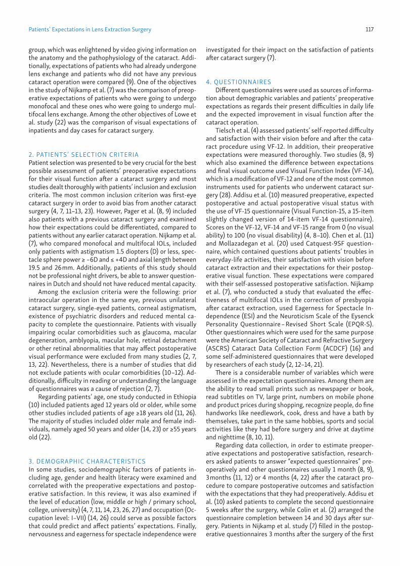
Tab. 2 Comparison between preoperative expectations and postoperative perceptions of patients.
Study Questionnaire Preoperative Perceptions (%)
Preoperative Expectations (%)
Postoperative Perceptions (Patient-reported outcome) (%)
Chen et al. (11) Catquest-9SF 56.07 (15.7/28) 93.9 (26.3/28) NA†
Addisu et al. (10) VF-15* 54.19 96.3 96.2
Pager et al. (9) VF-14* 84.7 Information video: 83.42Anatomy video: 86.03
First time surgery: 96.31Previous surgery: 95.68
89.8
Pager et al. (8) VF-14* 84.7 96.1 89.8
Nijkamp et al. (7) VF-14* Monofocal:74.1Multifocal:71.4
NA† Monofocal:96Multifocal:95.1
Tielsch et al. (4) VF-12* 80.2 98.7 NA†
*VF = Visual Function; †NA = Not applicable
Patients’ Expectations in Lens Extraction Surgery 119
cataract surgery. Nevertheless, 73% and 85% of individuals believed that they would need distance and near glasses re-spectively postoperatively.
In another study (7), ESI after the lens replacement with a mono- and multifocal implant was determined. The score of ESI questionnaire, which examined perceived advantages and disadvantages of wearing glasses, ranged from 1 (very eager to be spectacle independent) to 5 (not eager at all to be spectacle independent). No significant difference between the mono- and multifocal group was found in ESI (monofocal group: 3.2 ± 0.7, multifocal group: 3.1 ± 0.8, p = 0.35). Nijkamp et al. observed that subjects in this study believed that having good vision at more than one distance, even with comple-mentary reading or multifocal glasses, was more important than being spectacle independent.
6. PARAMETERS TO MEASURE EXPECTATION FULFILLMENTThere is no doubt that there is a need for developing some novel parameters in order to quantify the outcome of cat-aract surgery from a different perspective. In this way, the difference between preoperative expectations and actual outcomes, in other words the expectation fulfillment, could be assessed. These parameters could either be objective or subjective. Among the objective parameters are VA and spec-tacle independence, while among the subjective parameters are postoperative perceptions and expectation-outcome dis-crepancy. Patients’ preoperative perceptions regarding their visual function and preoperative expectations were compared with postoperative patient-reported outcome in Table 2.
6.1 Visual acuityThe measurement of VA is the most common method to esti-mate the postoperative outcome [Clinical Outcome Measure (COM)] (20).
Mollazadegan et al. (20) defined the improvement in VA as the increase of one line or more than one line on the Snellen chart after the surgery, while a negative COM was defined as a postoperative decline of VA.
Addisu et al. (10) described postoperative outcomes; un-corrected VA was ≥6/18 in 57% of cases (pinhole VA ≥6/18: 70%), while 29% of patients had VA<6/18. Seven patients who had VA<6/60 after 5 weeks were found to have ocular comorbidities.
Pager et al. (9) observed that there were no significant differences in postoperative VA between “expectations” and “anatomy” video group.
Pager et al. (8), in a second study, mentioned that the me-dian VA altered from 6/18 preoperatively to 6/6 after the lens extraction in the operative eye.
6.2 Spectacle independenceAmong the important parameters that could contribute to a favorable evaluation of the cataract surgery outcome is the option to offer spectacle independence.
Nijkamp et al. (7) mentioned that the monofocal group had worse Uncorrected Near Visual Acuity (UCNVA) and spec-tacle independence than the multifocal group. Nevertheless,
57.3% of patients who were implanted with multifocal lenses still used spectacles for reading.
6.3 Postoperative perceptions – Expectation fulfillmentThe majority of studies described a very good proportion of patients whose expectations were met (4, 7, 10, 11, 20).
Nijkamp et al. (7) observed that 62.5% from the monofo-cal group and 61.5% from the multifocal group achieved their expectations after the lens extraction of both eyes.
Tielsch et al. study (4) demonstrated that 61.2% of subjects had equal or better postoperative visual function compared to their preoperative expectations. However, when patients were questioned for the fulfillment of their expectations for specific daily activities, they performed better scores for each activity than for their visual function as a whole. Namely, 71% to 96% achieved their expectations of improvement for each of the VF-12 activities.
Mollazadegan et al. (20) mentioned that 10.1% of par-ticipants had a poor patient-reported outcome measure (PROM−) postoperatively, and 7.4% had a positive clinical outcome measure (COM+) but a negative patient-reported outcome (PROM−). They found that the latter group had dif-ficulties in near-vision and other daily activities postopera-tively. Researchers mentioned that PROM− generally was due to a good preoperative patient-reported visual function and poor VA in the best eye prior to surgery.
However, in Pager et al. study (8), where the expectations for the postoperative outcome were very high, 66% of individ-uals had lower perception 1 month after the cataract surgery in comparison with their preoperative expectations.
From the assessment of questionnaire answers, it was ob-served that there was a significant number of patients who did not achieve to meet their preoperative expectations in some daily activities, like reading small prints (eg. newspaper) (4, 8, 10), doing fine handwork (4, 8, 10), taking part in social activities (10), visiting friends (10) and driving at night (8).
6.4 Expectation-outcome discrepancyThe difference between preoperative expectations and actual outcomes is a subjective but effective way to estimate the degree of expectation fulfillment.
Chen et al. (11) noticed that the mean expectation-out-come difference was zero, namely the majority of initial expec-tations were met. Mainly patients implanted with aspherical IOL and patients with ocular comorbidity, very poor preoper-ative VA, poor health literacy, mild nuclear opalescence, and cortical cataract grading had a considerably greater discrep-ancy regarding expected and actual Catquest-9SF scores.
Pager et al. (8) observed that the mean discrepancy be-tween expectations and actual outcomes was 6.3 points.
Examining expectation-outcome discrepancy, Tielsch et al. (4) did not find any correlation between the number of pro-cedures performed by the surgeon per year, the period of time the ophthalmologist being in practice, the location, in which operations took place, and the discrepancy regarding expect-ed and actual outcomes. Mean expectations of visual function were slightly but significantly higher than actual postopera-tive visual function for each of the provider characteristics mentioned above.
120 Eirini-Kanella Panagiotopoulou et al. Acta Medica (Hradec Králové)
7. DETERMINING FACTORS OF PATIENTS’ EXPECTATIONS – PREDICTORS OF PATIENTS’ SATISFACTIONOne of the purposes of this review is the investigation of predictors of patient satisfaction by identifying the deter-minants of preoperative patient expectations. Patients’ sat-isfaction is affected not only by the measured postoperative VA but also by the degree of the expectations that they have before the operation (7, 10, 13, 23). It is observed that pa-tients with higher expectations tend to have a lower level of satisfaction compared to people who have lower expec-tations (30, 31).
7.1 Demographic characteristics7.1.1 AgeHawker et al. (13) observed that, although age and expec-tation of needing spectacles for distance was not correlat-ed significantly, the correlation between increasing age and expectation for near correction was weakly positive, namely older patients thought it more probable that they would need glasses for near vision than younger patients (range: between 41 to 97 years). Thus, younger patients should be treated with greater caution.
Nijkamp et al. (7) observed that older patients had a small-er ESI. This derives from the fact that 4 out of 5 patients in this study wore glasses for distance and near vision.
Tielsch et al. (4) did not find any correlation between ex-pected postoperative scores in the VF-12 questionnaire and patients’ age. However, patients >75 years old had a greater but not significant discrepancy between expected and post-operative scores in the VF-12 questionnaire.
7.1.2 GenderA Brazilian study by Kara-Júnior et al. (14) did not observe any significant difference in expectation between the two gen-ders as regards the possibility of full correction of cataract. Tielsch et al. (4) observed that expected postoperative VF-12 scores were not correlated with gender. Nevertheless, men seem to be satisfied more easily than women (2, 20).
7.1.3 EducationNijkamp et al. (23) highlighted the role of patients’ education in setting of realistic preoperative expectations, while Tielsch et al. (4) noticed that patient education was not correlated with expected postoperative mean VF-12 scores.
7.1.4 Occupation-IncomeAlthough occupation is one of the most important affecting factors of satisfaction (6), there was not any article analyz-ing the relationship between occupation and preoperative expectations.
7.1.5 Health literacyChen et al. (11) concluded that low patient health litera-cy was associated with a significant expectation-outcome discrepancy.
7.2 Medical information7.2.1 Patients’ counselingAccording to a considerable number of studies, a discussion between the cataract surgeon and patient before the opera-tion about the preoperative expectations for the outcome of the surgery could be very valuable. Specifically, health care professionals could inform patients about their prediction of the postoperative outcome in order to encourage more real-istic expectations (9, 12, 24).
Hawker et al. (13) emphasized the importance of discuss-ing about the possibilities of wearing spectacles for distance and/or reading after the surgery. Thus, patients would be more likely to have realistic expectations and a high level of satisfaction.
Chen et al. (11) concluded that extensive counseling could be remarkably helpful for patients with low health literacy and systemic or ocular comorbidities because it could di-minish the expectation-outcome difference and increase satisfaction.
Addisu et al. (10) resulted that patient understanding and counseling by health care professionals before surgery could help in the informing of patients about potential outcomes, decrease their expectations and, consequently, increase sat-isfaction even if the actual outcome is not the ideal.
Colin et al. (2) reported that 92.4% of patients included in the study said that health care professionals provided them preoperatively with enough to too much explanation regard-ing surgical procedure and 90.4% of patients mentioned they had received before surgery enough to too much explanation about risks of cataract surgery. Moreover, patients were ques-tioned to answer if they asked preoperatively their surgeon all their unanswered questions and only 7.9% gave a nega-tive answer. On the other hand, regarding the explanations they received during the surgery by their ophthalmologist, it was interesting that 67.2% of patients complained that the doctor did not give them enough explanation and only 10.3% were satisfied (received “enough” or “too much” explanation). Researchers explained that patients informed by their health care practitioners had a great level of satisfaction.
Chang-Godinich et al. (16) observed that some older patients, who had a lower level of satisfaction with their postoperative quality of life and the medical care that they received, complained of no enhancement in their function-al vision in spite of the objective improvement in their VA. It was concluded that a preoperative discussion of postop-erative expectations of the patients could be beneficial for them.
According to Tielsch et al. (4) patients who had been in-formed by ophthalmologists about the expectations that they should have during and after the surgery had a greater satisfaction. For example, a discussion of the possibility of wearing glasses for reading postoperatively or an explana-tion of operation risks, if patients had an ocular comorbidity, could reduce the difference between the expected and actual outcomes.
Gramer et al. (15) mentioned that patients could be in-formed by their physicians the day before the cataract sur-gery. They suggested that an informative session is less likely to address all the queries of the patient regarding his/her up-coming operation, and a more integrated approach including written material should be preferred.
Patients’ Expectations in Lens Extraction Surgery 121
7.2.2 Video presentationIn their study, Pager et al. (9) showed patients who were scheduled for cataract operation one of two different videos. The first videotape informed patients about the procedure of cataract surgery and its possible dangers (“expectations vid-eo group”), while the second video described only the way of cataract formation (“anatomy video group”). It is worth not-ing that although the expectations video group in comparison with the expectations of the second group feared preopera-tively that the surgery would have more risks and discomfort, patients who watched the first informational videotape, had a better understanding of the procedure of the extraction of their lens, less anxiety and a greater satisfaction, probably because they felt better that the procedure was not as painful and dangerous as they expected.
7.2.3 ComorbidityThe discrepancy regarding expected and actual outcomes after a cataract procedure is undoubtedly correlated with ocular and systemic comorbidities (11), which are the most important factors that can predict patient dissatisfaction (8, 16). For this reason, many surgeons hesitate to undertake the operation of patients with ocular or systemic comorbidities that may affect their visual capacity. Therefore, physicians try to explain the risks, give an estimate of the postoperative visual capacity and, overall attempt to manage preoperative expectations, accordingly (12).
Chen et al. (11) came to the conclusion that patients with systemic and ocular comorbidity should have an educative discussion with their ophthalmologist in order to reduce the expectation-outcome discrepancy.
Kuo et al. (12) mentioned that all patients with ocular or systemic comorbidity, including keratectasia after Laser In Situ Keratomileusis (LASIK), Age-Related Macular Degenera-tion (ARMD), cerebrovascular accident and retinitis pigmen-tosa, had a low possibility to meet their preoperative expec-tations of visual function after the cataract surgery.
Addisu et al. (10) observed that the existence of ocular comorbidities was one of the most important predictive fac-tors of dissatisfaction. On the contrary, they suggested poor correlation between patients’ satisfaction and enhancement of visual function.
7.2.4 Unrealistic ExpectationsSometimes patients have an unreasonably high level of ex-pectations. The most common expectations are reading small prints, doing fine handwork and driving at night (8, 10). In this case, it is very common that the majority of this patient group cannot meet their preoperative expectations (4, 8).
Tielsch et al. (4) were surprised by the fact that partici-pants with ocular comorbidities or with generally low prog-nosis had the same expectations in VF-12 scores as patients with favorable predictive factors. However, there was a sig-nificant difference between the actual and the expected outcomes. As a result, they had a greater expectation-out-come discrepancy compared with younger patients without comorbidity.
Pager et al. (8) emphasized that there was a number of patients who had no realistic expectations such as driving at
night, reading small prints and doing fine handwork. In ad-dition, it was demonstrated that the control of patient ex-pectations is more important than the actual improvement of postoperative outcomes for the best possible satisfaction.
Chang-Godinich et al. (16) reported that the fact that pa-tients did not perceive a better postoperative visual function probably derived from the absence of realistic expectations.
7.2.5 Spectacles preoperativelyOne of the major patients’ expectations is postoperative spectacle independence (32). According to Hawker et al. (13), who recommended the consideration of the patients’ preop-erative refractive status, patients who needed glasses before the cataract surgery generally expected to use them also after surgery. On the other hand, a small number of patients who did not wear spectacles preoperatively, had the expectation of complete spectacle independence. However, these high expectations increased the risk for dissatisfaction.
Nijkamp et al. (7) observed that older patients, who pre-operatively in their great majority needed spectacles both for distance and near vision, did not have very high expectations and were satisfied even if they should use glasses after the surgery, too. In addition, in this study, where the efficacy of multifocal intraocular lenses (IOLs) to correct presbyopia at the same procedure with cataract correction was examined, it was highlighted that patients having ESI would accept see-ing halos and having low contrast sensitivity, which are some side effects of multifocal IOLs, as long as they would not use spectacles again (7, 25).
7.2.6 Cost of surgeryWei et al. (18) described a cataract surgery payment model and assessed the satisfaction of patients according to the type of payment. The first type [“National Health Insurance (NHI) coverage”] was the total cost coverage of a “general IOL” by the NHI, while the second type (“balance billing”) was the partial payment depending on the kind and price of IOL (mon-ofocal/multifocal, yellow-tinted or not). It is worth to men-tion that there was not difference in satisfaction between the two groups. Writers supposed that this was due to the lower degree of expectations of patients using NHI coverage and, as a result, due to the low expectation-outcome discrepancy. On the contrary, patients who chose to pay for their proce-dure, expected to receive the best possible medical services. For this reason, they had a higher level of expectations and a lower level of satisfaction, although there was not any dif-ference in VA between the two groups. Actually, patients who were implanted with multifocal and yellow-tinted IOLs) had an easier looking at small objects and objects under strong lighting conditions.
7.2.7 Personality-PsychologyIn this review, studies did not reveal a concrete relationship between personality or emotional status and patients’ expec-tations. Patient’s personality and emotions were connected only with satisfaction.
Nijkamp et al. (7) examined the influence of patient per-sonality on their satisfaction and the possibility of taking the
122 Eirini-Kanella Panagiotopoulou et al. Acta Medica (Hradec Králové)
personality of patients into account in order to predict their satisfaction. It was hypothesized that it was more difficult for patients with obsessive personalities (eg. neuroticism) to be satisfied. Nevertheless, according to the results of this study, neuroticism did not seem to be related with satisfaction. Con-sequently, obsessiveness was found to be an inappropriate criterion for the inclusion of patients for cataract operation with multifocal IOLs.
Another study, which reveals how difficult the prediction of visual satisfaction is, is the study of Prakash et al. (19), who explained that expectations and mindset of patients can bias questionnaire outcomes because each patient perceives symptoms in a different personal way.
The study of Yucelt et al. (6) demonstrated that patients who were not satisfied with their life quality and are emotion-ally unstable (eg. having depression or other health problems) are more likely to be also dissatisfied with the received health care. For this reason, medical care professionals should always take patient expectations and emotions into consideration.
7.2.8 Previous cataract surgeryThe findings in Pager et al. (9) study revealed that there was a difference in the preoperative expectations and perceived postoperative outcomes between patients with and without a previous cataract surgery. Analytically, patients who had ex-perienced a previous cataract operation expected that they would be less anxious and the procedure would be more com-fortable in comparison with patients who did not have such a previous experience. Postoperatively, patients who had pre-viously experienced cataract surgery were more likely to find the surgery close to their expectations.
8. ADVANTAGES OF PATIENTS’ SATISFACTIONBeyond the improvement of patients’ vision and life quality, the effort to raise their satisfaction has some additional ad-vantages for health care providers. Patients whose preopera-tive expectations have been met, usually have a better com-pliance with medication, a consistent follow-up and generally enjoy the benefits of the procedure to the maximum degree (8, 9). Furthermore, satisfied patients do not lose their relation-ship with their physicians, recommend them to other people and do not blame them judicially for malpractice (8–10, 26).
DISCUSSION
Nowadays, higher life expectancy is likely to increase the number of patients having cataract, which is a significant cause of low vision not only in developing but also in devel-oped countries (1). For this reason, further knowledge of the factors that could affect the outcome of a cataract surgery would aid in the improvement of treatment quality.
Patient satisfaction depends both on the actual outcomes of VA and on the preoperative expectations of patients. Sat-isfaction and visual outcomes have a proportional relation-ship, while satisfaction is inversely proportional to preop-erative expectations. The objective of health care systems is to offer high quality services. Therefore, addressing this objective is possible either by the improvement of the actual
visual outcomes that patients attain or by the reduction of their preoperative expectations. However, cataract-related technological advances, cannot cope with the continuously increasing demand for perfect vision. Consequently, medi-cal care professionals should now focus on the influence of preoperative expectations, too. Namely, ophthalmologists should try to reduce their patients’ expectations to a realis-tic level that is compatible to their setting and expertize (12).
As regards the assessment of patients’ expectations be-fore cataract surgery, the majority of relevant studies used predefined psychometric tools and correlated their results with a series of visual capacity parameters.
Moreover, individuals were asked about their preoperative expectations both for expected visual capacity and for spec-tacle independence. Despite the fact that accurate assess-ment of both aforementioned expectations is essential to the care providers, no common methods of estimating them have been introduced. For instance, some studies measured the mean expected postoperative questionnaire score; the max-imum score of Catquest-9SF is 28 (11), while the maximum VF score is 100. Another study (8) measured the percentage of subjects that had a maximum expectation score. Other studies expressed the portion of participants who expected a normal vision (13), “much” to “very much” improvement (7) or a full recovery from the cataract (14). In an additional study (4), researchers assessed the number of patients who expect-ed better postoperative visual function scores in comparison to the preoperative ones. Regarding the estimation of ex-pectations for spectacle independence, there was one study (13) that examined this parameter too. A study examining ESI (7) found that individuals thought that having good vision at more than one distance, even with complementary reading or multifocal glasses, was more significant than not wearing glasses. However, the reliability of these results should be confirmed with further investigation. Therefore, it would be valuable if expectations about spectacle independence were examined on a regular basis. Consequently, it is obvious that there is no standard way of measurement and evaluation of patient expectations. As a result, the difficulty in comparison of patients’ expectations between the studies of this review of literature was considerable.
Postoperative VA and spectacle independence are useful for the comparison with the preoperative status. However, they are not enough for a total evaluation of the success of a cataract procedure. This review indicated that patients’ perception of their visual function should be asked after the surgery (4, 7, 8, 10, 11, 20). This would be necessary in order for ophthalmologists to know if patients’ expectations were fulfilled. The majority of studies (4, 7, 8, 10, 11, 20) mentioned the percentage of patients who addressed or surpassed their preoperative expectations for the minimal-required visual capacity to complete their daily activities. However, only a small number of studies described patient-reported out-comes for each daily activity (4, 8, 10). Within this context, the most difficult daily activities that usually were not ad-dressed following cataract-extraction surgery were revealed. Among them, reading small prints and doing fine handwork (4, 8, 10). Moreover, patients who had been implanted with multifocal lenses had higher expectations for specific activi-ties; among them driving at night (8). Undoubtedly, it would be very useful if all studies examining patient expectations
Patients’ Expectations in Lens Extraction Surgery 123
for cataract removal in the future evaluated expectations and postoperative perceptions for each activity separately with a special questionnaire. In this way, researchers could detect weaknesses of each type of lenses and try to improve them for the achievement of the best possible result.
We assumed that a possible reason that some studies showed a lower postoperative perception than preoperative expectations was ECCE instead of phacoemulsification. ECCE was the treatment for cataract in a study of the previous dec-ade (9) and in a study about cataract surgery in a developing country (10), where the surgical techniques and the equip-ment were not very advanced. However, the number of stud-ies using ECCE and the total number of studies examining the postoperative perceptions was limited.
Concerning the time when follow-up and questionnaire completion about patients’ postoperative expectation ful-fillment took place, this review indicated that there was no common method. Some of the studies collected data 1 month (8, 9) after cataract operation, other studies collected them 3 months (11, 12) and other studies 4 months (4, 22) post-operatively. Consequently, it is possible that this difference could complicate the objective comparison of postoperative perceptions because visual function is gradually improved over time postoperatively. Further investigation is required in order to clarify the most appropriate period of time after lens exchange for the most reliable assessment of vision im-provement compared with patients’ expectations.
A final parameter which was taken into account in a signif-icant number of studies (4, 7, 8) was the expectation-outcome discrepancy. Specifically, the difference between expected and actual outcome was estimated. This parameter should constitute a standard examination object of the studies con-cerning patient expectations as regards cataract surgery be-cause it is a simple way to evaluate the success of a cataract operation.
In summary, our review suggests that further investiga-tion is required in order to create a protocol that should be followed in every study concerning patients’ preoperative expectations. Namely, VA before and after cataract surgery should be measured. Preoperative expectations both for spectacle independence and normal vision at more than one distance, even with complementary reading or multifocal glasses, should be estimated prior to cataract surgery. Fur-thermore, an indispensable part of the postoperative ap-proach of patients should be the assessment of their postop-erative spectacle independence, patients’ perception of their visual function with the completion of a respective question-naire and the expectation-outcome discrepancy.
There are not many studies examining patients’ preop-erative expectations for the outcomes of a cataract surgery and ophthalmological operations in general, while there is a plethora of studies examining patients’ satisfaction as re-gards lens extraction. Additionally, nowadays, an increasing number of patients are implanted with multifocal IOLs. As a result, they have high expectations for their vision at all distances (distance, intermediate, near vision) and under all lighting conditions. However, there is an even greater defi-ciency in studies examining patients’ preoperative expecta-tions for cataract surgery with implantation of a multifocal lens. For this reason, further studies are needed in order to evaluate the relationship between preoperative expectations
and postoperative outcomes of patients implanted not only with monofocal but also with multifocal IOLs.
With regard to the factors that are able to influence pa-tient expectations, this systematical review of literature indi-cates that there is a variety of determinants of preoperative expectations. Ocular and systemic comorbidity, unrealistic expectations, preoperative spectacle independence, the cost of surgery, a previous cataract surgery and the level of health literacy are the major factors that can affect positively or neg-atively patient preoperative expectations. On the other hand, although it is known that demographic characteristics such as age, gender, education, occupation and income are associated with satisfaction (2, 26, 27), to our knowledge, there are not many studies which examine the correlation between age/gender/education and expectations, and there is no study regarding the occupation/income-expectations relationship.
It was hypothesized that patients’ personality and psy-chological state could play a determinant role in the devel-opment of patients’ initial expectations. Nevertheless, all studies that were found correlated emotional state only with satisfaction. Obsessiveness, neuroticism and depression could be factors that affect negatively patient satisfaction. Regarding preoperative expectations, we assume that pa-tients that are not emotionally stable have unpredictable pre-operative expectations. Further studies are needed to clarify the relationship between emotional instability and patients expectations.
CONCLUSIONS
Our intention was the best possible comprehension of the literature regarding patient expectations for a favorable cataract surgery visual outcome and the assistance of other researchers in further investigation in this domain of knowl-edge. Evaluation of patients’ preoperative expectations pro-vides essential information to the ophthalmologist. This re-view suggests that the final postoperative perception could be affected by the actual outcome of the operation and by pa-tient preoperative expectations. The higher the actual visual outcome and the lower the preoperative expectations are, the better the postoperative perception of visual outcome is. In addition, our review emphasizes the importance of the assessment of factors that determine patients’ preoperative expectations. Demographic characteristics such as age and the level of health literacy, as well as other factors including ocular and systemic comorbidity, unrealistic expectations, preoperative spectacle independence, the cost of surgery, and a previous cataract surgery could affect preoperative expectations and predict patients’ satisfaction. Taking these factors into consideration could lead to more accurate predic-tion of patient satisfaction and better control of expectations with an extensive preoperative counseling.
FINANCIAL DISCLOSURE
No financial support was received for this study. None of the authors has any proprietary interests or conflicts of inter-est related to this submission. It is not simultaneously being considered for publication at any other journal.
124 Eirini-Kanella Panagiotopoulou et al. Acta Medica (Hradec Králové)
REFERENCES 1. WHO. Cataract, Priority eye diseases, Prevention of Blindness and Visual
Impairment. (Accessed July 2, 2018 at http://www.who.int/blindness /causes/priority/en/index1.html).
2. Colin J, El Kebir S, Eydoux E, Hoang-Xuan T, Rozot P, Weiser M. Assess-ment of patient satisfaction with outcomes of and ophthalmic care for cataract surgery. J Cataract Refract Surg 2010; 36(8): 1373–9.
3. Apple DJ, Peng Q, Visessook N, et al. Surgical prevention of posterior capsule opacification. Part 1: Progress in eliminating this complication of cataract surgery. J Cataract Refract Surg 2000; 26: 180–7.
4. Tielsch JM, Steinberg EP, Cassard SD, et al. Preoperative functional ex-pectations and postoperative outcomes among patients undergoing first eye cataract surgery. Arch Ophthalmol 1995; 113(10): 1312–8.
5. McAlinden C. The importance of doctor-patient communication. Br J Hosp Med (Lond) 2014; 75(2): 64–5.
6. Yucelt U. An investigation of causes of patient satisfaction/dissatisfac-tion with physician services. Health Mark Q 1994; 12(2): 11–28.
7. Nijkamp MD, Dolders MG, de Brabander J, van den Borne B, Hendrikse F, Nuijts RM. Effectiveness of multifocal intraocular lenses to correct presbyopia after cataract surgery: a randomized controlled trial. Oph-thalmology 2004; 111(10): 1832–9.
8. Pager CK. Expectations and outcomes in cataract surgery: a prospec-tive test of 2 models of satisfaction. Arch Ophthalmol 2004; 122(12): 1788–92.
9. Pager CK. Randomised controlled trial of preoperative information to improve satisfaction with cataract surgery. Br J Ophthalmol 2005; 89(1): 10–3.
10. Addisu Z, Solomon B. Patients’ preoperative expectation and outcome of cataract surgery at jimma university specialized hospital-department of ophthalmology. Ethiop J Health Sci 2011; 21(1): 47–55.
11. Chen Z, Lin X, Qu B, et al. Preoperative Expectations and Postopera-tive Outcomes of Visual Functioning among Cataract Patients in Urban Southern China. PLoS One 2017; 12(1): e0169844.
12. Kuo IC, Broman AT, Massof RW, Park W. The impact of cataract surgery on patients from a low-vision clinic. Can J Ophthalmol 2011; 46(5): 391–8.
13. Hawker MJ, Madge SN, Baddeley PA, Perry SR. Refractive expectations of patients having cataract surgery. J Cataract Refract Surg 2005; 31(10): 1970–5.
14. Kara-Júnior N, Temporini ER, Kara-José N. Cataract surgery: expec-tations of patients assisted during a community project in São Paulo, state of São Paulo, Brazil. Rev Hosp Clin Fac Med Sao Paulo 2001; 56(6): 163–8.
15. Gramer E, Leydhecker W, Krieglstein GK. The physician’s obligation to educate patients – legal aspects – patients’ expectations. Klin Monbl Augenheilkd 1982; 181(1): 46–53.
16. Chang-Godinich A, Ou RJ, Koch DD. Functional improvement after phacoemulsification cataract surgery. J Cataract Refract Surg 1999; 25(9): 1226–31.
17. Berdeaux G, Meunier J, Arnould B, Viala-Danten M. Measuring benefits and patients’ satisfaction when glasses are not needed after cataract and presbyopia surgery: scoring an hometric validation of the Free-dom from Glasses Value Scale (FGVS). BMC Ophthalmol 2010; 10: 15.
18. Wei CK, Wang SM, Lin JC. A study of patient satisfaction after cataract surgery with implantation of different types of intraocular lenses. BMC Res Notes 2012; 5: 592.
19. Prakash G, Prakash DR, Agarwal A, Kumar DA, Agarwal A, Jacob S. Predic-tive factor and kappa angle analysis for visual satisfactions in patients with multifocal IOL implantation. Eye (Lond) 2011; 25(9): 1187-93.
20. Mollazadegan K, Lundström M. A study of the correlation between pa-tient-reported outcomes and clinical outcomes after cataract surgery in ophthalmic clinics. Acta Ophthalmol 2015; 93(3): 293–8.
21. Silveira JA, Hayashi L, Scarpi MJ. Identification of patients’ needs and expectations in a cataract clinic connected with a university public hos-pital. Arq Bras Oftalmol 2005; 68(5): 639–44.
22. Lowe KJ, Gregory DA, Jeffery RI, Easty DL. Patient perceptions and social impact. Preliminary results of the Bristol MRC Study. Eye (Lond) 1991; 5(Pt 3): 373–8.
23. Nijkamp MD, Nuijts RM, Borne B, Webers CA, van der Horst F, Hendrikse F. Determinants of patient satisfaction after cataract surgery in 3 set-tings. J Cataract Refract Surg 2000; 26(9): 1379–88.
24. Zuo L, Zou H, Fei X, Xu W, Zhang J. The impact of unilateral or bilater-al cataract surgery on visual acuity and life quality of elderly patients. J Ophthalmol 2015; 2015: 509049.
25. Dick HB, Krummenauer F, Schwenn O, Krist R, Pfeiffer N. Objective and subjective evaluation of photic phenomena after monofocal and mul-tifocal intraocular lens implantation. Ophthalmology 1999; 106(10): 1878–86.
26. Yucelt U. An investigation of causes of patient satisfaction/dissatisfac-tion with physician services. Health Mark Q 1994; 12(2): 11–28.
27. Baron-Epel O, Dushenat M, Friedman N. Evaluation of the consumer model: relationship between patients’ expectations, perceptions and satisfaction with care. Int J Qual Health Care 2001; 13(4): 317–23.
28. Labiris G, Giarmoukakis A, Patsiamanidi M, Papadopoulos Z, Kozobolis VP. Mini-monovision versus multifocal intraocular lens implantation. J Cataract Refract Surg 2015; 41(1): 53–7.
29. Kravitz RL. Patients’ expectations for medical care: an expanded formu-lation based on review of the literature. Med Care Res Rev 1996; 53(1): 3–27.
30. Berdeaux G, Viala M, Roborel de Climens A, Arnould B. Patient-reported benefit of ReSTOR multi-focal intraocular lenses after cataract surgery: results of principal component analysis on clinical trial data. Health Qual Life Outcomes 2008; 6: 10.
31. Unsal U, Baser G. Evaluation of Different Power of Near Addition in Two Different Multifocal Intraocular Lenses. J Ophthalmol 2016; 2016: 1395302.
32. Pager CK, McCluskey PJ, Retsas C. Cataract surgery in Australia: a profile of patient-centred outcomes. Clin Exp Ophthalmol 2004; 32(4): 388–92.
original article 125
Epidemiological Profile and Antimicrobial Resistance Pattern of Enteric Fever in a Tertiary Care Hospital of North India – a Seven Year Ambispective Study
Anuradha Makkar1, Shilpi Gupta2, Inam Danish Khan1,*, Rajiv Mohan Gupta3, KS Rajmohan1, Harleen Chopra1, Manisha Gupta4, Sachin Bansal5, Bindu Poonia1, Muqtadir Malik3, Pragyan Swagatika Panda1
A B S T R AC TIntroduction: Enteric-fever is a major public-health problem in developing countries emerging as multidrug-resistant, Nalidixic-acid resistant and extremely drug-resistant Salmonella (Pakistan, 2016), has intensified the use of WHO watch/reserve group antimicrobials such as azithromycin and meropenem.Methods: This ambispective-study was conducted on 782 non-repeat blood-culture isolates of S. Typhi, S. Paratyphi A and S. Paratyphi B obtained from 29,184 blood cultures received at a 1000-bedded tertiary-care hospital of North-India from 2011–2017. Identification and antibiograms were obtained by Vitek-2 compact and Kirby-Bauer’s disc diffusion with resistance to ampicillin, chloramphenicol and co-trimoxazole being labeled as multidrug-resistant. Decreased ciprofloxacin-susceptibility and ciprofloxacin-resistance were defined as MIC 0.125–0.5 and >1 µg/ml.Results: S. Typhi and S. Paratyphi A in a ratio of 3.9:1 were seen between July–September predominantly distributed between 6–45 year age group. Resistance to co-trimoxazole, chloramphenicol, ceftriaxone and azithromycin was 6.1%, 13.8%, 16.1 and 5.78% respectively. Multidrug-resistant S. typhi and S. paratyphi A were 2.73% and 1.91% respectively. Conclusion: Enteric-fever is a major public-health problem in India. Emergence of multidrug-resistant, Nalidixic-acid resistant and extremely-drug resistant Salmonella mandates ongoing surveillance for targeted empirical therapy and containment of spread. Repeated epidemics call for water, sanitation, hygiene and vaccination strategies to sustain herd-immunity.
K E Y WO R D Santimicrobial resistance; enteric fever; multidrug-resistant Salmonella
A U T H O R A F F I L I AT I O N S1 Army College of Medical Sciences and Base Hospital, New Delhi, India 2 Department of Microbiology, MH Bhopal, India3 Army Hospital Research and Referral, New Delhi 110010, India4 Sanjay Gandhi Post-Graduate Institute, Lucknow, India5 Dr Lal Path Labs, Tilak nagar, New Delhi, India* Corresponding author: Microbiology and Infectious Diseases, Army College of Medical Sciences and Base Hospital, New Delhi 110010,
India; e-mail: [email protected]
Received: 9 August 2018Accepted: 14 November 2018Published online: 22 January 2019
Acta Medica (Hradec Králové) 2018; 61(4): 125–130https://doi.org/10.14712/18059694.2018.130© 2018 The Authors. This is an open-access article distributed under the terms of the Creative Commons Attribution License (http://creativecommons.org/licenses/by/4.0), which permits unrestricted use, distribution, and reproduction in any medium, provided the original author and source are credited.
126 Anuradha Makkar et al. Acta Medica (Hradec Králové)
INTRODUCTION
Enteric-fever is one of the major public health problems in developing countries including India where safe drinking water and sanitation is not warranted (1). According to the estimates, worldwide there are 12–33 million new patients of enteric-fever annually with mortality reaching 600,000 (2). Typhoid is highly endemic in South Asia, South-East Asia, sub-Saharan Africa and West coast of Latin America. The in-cidence of enteric-fever range from 102 to 2,219 per 100,000 population in India and 9.8 cases per 1000 person-years in Delhi. The estimated annual incidence rate of 1% in India (3).
Enteric-fever constitutes typhoid fever caused by Salmo-nella enterica subsp enterica serotype Typhi and paratyphoid fever caused by Salmonella enterica subsp enterica serotypes Paratyphi A, Paratyphi B and Paratyphi C. Several studies from India have reported S. Typhi as the most common caus-ative agent, along with increasing number of patients due to S. Paratyphi A. Amongst the changing trends in epidemiology of enteric-fever one of the significant findings is increased incidence of S. Paratyphi A infections. Several studies from India have reported the increased incidence of S. Paratyphi A infections since 1996. However, the incidence of S. Para-typhi B and C are rarely reported from India (4–6). Usually majority of enteric-fever patients occur in children less than 15 years of age, but children younger than five years of age are more susceptible to infection in disease endemic areas. On the other hand, studies from North India have report-ed the incidence of paratyphoid fever more commonly in adults (4, 7). However, as far as seasonal distribution of en-teric-fever is concerned there is not much variation within the country. The studies from North India have encountered the occurrence of patients throughout the year with the peak of disease usually observed during summer and rainy season (8).
The case-fatality rate is 10–50% due to complications such as perforated typhoid ulcer, Myocarditis and shock; with 1–5 year children being at the highest risk. Mortality rates due to enteric-fever can be reduced from 30% to less than 1% by providing effective antimicrobial therapy which is facing challenges due to emerging antimicrobial resistance (9). Am-picillin, chloramphenicol and trimethoprim-sulphamethoxaz-ole were the conventional first line drugs for enteric-fever till mid-twentieth century. Since 1962, E. coli integron carrying resistance genes acquired through plasmids has appeared worldwide. After emergence of chloramphenicol resistant S. Typhi in India and Mexico in 1972, frequency of isolation of multidrug resistant (MDR) Salmonella is on increase world-wide (10, 11). Since 1989, there have been several outbreaks of MDR salmonella pathogens reported from Asian countries including India (12). With the increased prevalence of MDR isolates, third generation cephalosporins and fluoroquinolo-nes are recommended for MDR infections. Nalidixic acid re-sistant (NaR) Salmonella have emerged due to point muta-tions in gyrA gene leading to ten-folds higher MICs compared to fully susceptible strains. Recent emergence of extremely drug resistant Salmonella resistant to ceftriaxone and cipro-floxacin has intensified the use of WHO watch/reserve group antimicrobials such as azithromycin and meropenem (13). It is important to study the prevalence, epidemiological factors and antibiogram related to enteric-fever to enable effective
treatment and preventive measures in terms of vaccines and strengthening hygiene and sanitation measures. This ambi-spective study was undertaken to study the prevalence, ep-idemiology and antimicrobial resistance pattern of S. Typhi and S. Paratyphi species in a tertiary-care teaching hospital in North India.
MATERIALS AND METHODS
An ambispective study was conducted on 645 non-repeat blood-culture isolates of S. Typhi, S. Paratyphi A and S. Para-typhi B isolated at a 1000-bedded tertiary care hospital of North India from January 2011 to December 2017 after infer-ences from a pilot study conducted for the period covering Jul–Sep 2010 and due approval from the Hospital Ethics Com-mittee. The pilot study was conducted to improve identifica-tion of Salmonella using automated systems for blood culture, microbiological identification and susceptibility.
All samples were plated directly on blood and MacConkey agar after positive culture screen from BacT/ALERT® 3D (bio- Mérieux SA, F-69280 Marcy l’Etoile, France) blood culture system and incubated at 37 °C for 18–24 hrs. Identification and antimicrobial susceptibility testing of the isolates were done by Vitek 2 compact (bioMérieux SA, France). Inbuilt standards for identification comparison were utilized. Iden-tification percentage >85% and Vitek Advanced Expert Sys-tem flagging consistent were taken as cut-off for final vali-dation. Isolates were also tested by slide agglutination using specific sera.
In parallel manual antimicrobial susceptibility was put for azithromycin (15 µg) and chloramphenicol (30 µg) (HiMedia laboratories, India) using Kirby-Bauer’s disc diffusion meth-od as these antimicrobials are not covered in susceptibility panel for gram negative bacilli in Vitek 2 compact. All inter-pretations were done using CLSI 2016 guidelines. In 2012, the CLSI revised the breakpoints for ciprofloxacin susceptibility for typhoidal Salmonella with MIC ranging from ≤0.06 µg/ml (susceptible) to ≥1 µg/ml (resistant) as compared to earlier MIC range from ≤1 µg/ml (susceptible) to ≥4 µg/ml (resist-ant). Interpretation of ciprofloxacin for all the isolates includ-ing 2011 was done according to the revised CLSI guidelines of 2012 (14). Decreased ciprofloxacin susceptibility (DCS) was defined as isolates having MIC of ciprofloxacin within range of 0.125–0.5 µg/ml. Ciprofloxacin resistant strains were defined for isolates with MIC >1 µg/ml. Isolates resistant to ampicil-lin, chloramphenicol and co-trimoxazole (trimethoprim-sul-phamethoxazole were labeled as MDR.
RESULTS
A total of 780 (2.67%) isolates of typhoidal Salmonella were isolated from 29,184 blood cultures received during the study period. The number of S. Typhi isolated was 623 (79.67%) compared to 157 (20.1%) S. Paratyphi A and two S. Paratyphi B (0.26%) isolates (Table 1). Male : female ratio was 2.3 : 1. The highest number of culture positive enteric-fever patients were seen in the age groups of 6–15 years and 16–45 years with 345 (44.23%) and 346 (44.36%) isolates respectively. (Table 2)
Epidemiology and Antimicrobial Resistance in Enteric Fever Cases 127
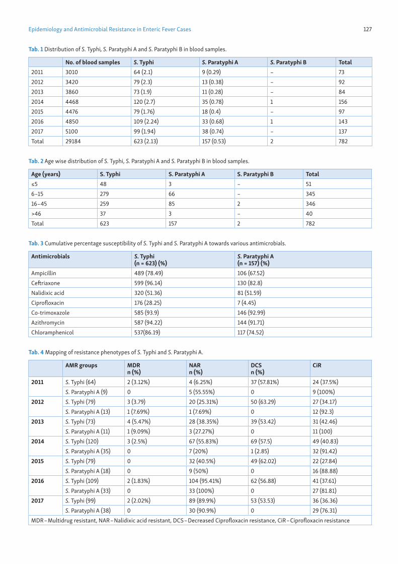
Tab. 1 Distribution of S. Typhi, S. Paratyphi A and S. Paratyphi B in blood samples.
No. of blood samples S. Typhi S. Paratyphi A S. Paratyphi B Total2011 3010 64 (2.1) 9 (0.29) – 732012 3420 79 (2.3) 13 (0.38) – 922013 3860 73 (1.9) 11 (0.28) – 842014 4468 120 (2.7) 35 (0.78) 1 1562015 4476 79 (1.76) 18 (0.4) – 972016 4850 109 (2.24) 33 (0.68) 1 1432017 5100 99 (1.94) 38 (0.74) – 137Total 29184 623 (2.13) 157 (0.53) 2 782
Tab. 2 Age wise distribution of S. Typhi, S. Paratyphi A and S. Paratyphi B in blood samples.
Age (years) S. Typhi S. Paratyphi A S. Paratyphi B Total≤5 48 3 – 516–15 279 66 – 34516–45 259 85 2 346>46 37 3 – 40Total 623 157 2 782
Tab. 3 Cumulative percentage susceptibility of S. Typhi and S. Paratyphi A towards various antimicrobials.
Antimicrobials S. Typhi (n = 623) (%)
S. Paratyphi A (n = 157) (%)
Ampicillin 489 (78.49) 106 (67.52)Ceftriaxone 599 (96.14) 130 (82.8)Nalidixic acid 320 (51.36) 81 (51.59)Ciprofloxacin 176 (28.25) 7 (4.45)Co-trimoxazole 585 (93.9) 146 (92.99)Azithromycin 587 (94.22) 144 (91.71)Chloramphenicol 537(86.19) 117 (74.52)
Tab. 4 Mapping of resistance phenotypes of S. Typhi and S. Paratyphi A.
AMR groups MDR n (%)
NARn (%)
DCSn (%)
CiR
2011 S. Typhi (64) 2 (3.12%) 4 (6.25%) 37 (57.81%) 24 (37.5%)S. Paratyphi A (9) 0 5 (55.55%) 0 9 (100%)
2012 S. Typhi (79) 3 (3.79) 20 (25.31%) 50 (63.29) 27 (34.17)S. Paratyphi A (13) 1 (7.69%) 1 (7.69%) 0 12 (92.3)
2013 S. Typhi (73) 4 (5.47%) 28 (38.35%) 39 (53.42) 31 (42.46)S. Paratyphi A (11) 1 (9.09%) 3 (27.27%) 0 11 (100)
2014 S. Typhi (120) 3 (2.5%) 67 (55.83%) 69 (57.5) 49 (40.83)S. Paratyphi A (35) 0 7 (20%) 1 (2.85) 32 (91.42)
2015 S. Typhi (79) 0 32 (40.5%) 49 (62.02) 22 (27.84)S. Paratyphi A (18) 0 9 (50%) 0 16 (88.88)
2016 S. Typhi (109) 2 (1.83%) 104 (95.41%) 62 (56.88) 41 (37.61)S. Paratyphi A (33) 0 33 (100%) 0 27 (81.81)
2017 S. Typhi (99) 2 (2.02%) 89 (89.9%) 53 (53.53) 36 (36.36)S. Paratyphi A (38) 0 30 (90.9%) 0 29 (76.31)
MDR – Multidrug resistant, NAR – Nalidixic acid resistant, DCS – Decreased Ciprofloxacin resistance, CiR – Ciprofloxacin resistance
128 Anuradha Makkar et al. Acta Medica (Hradec Králové)
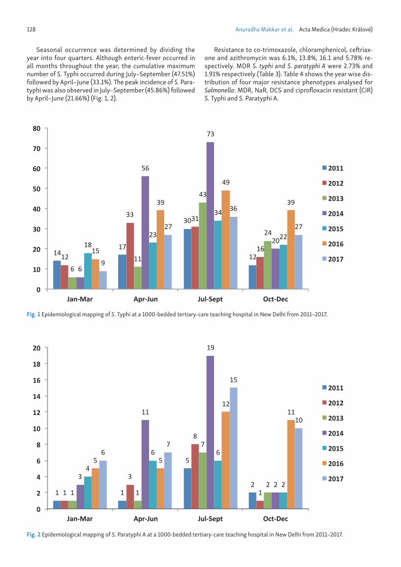
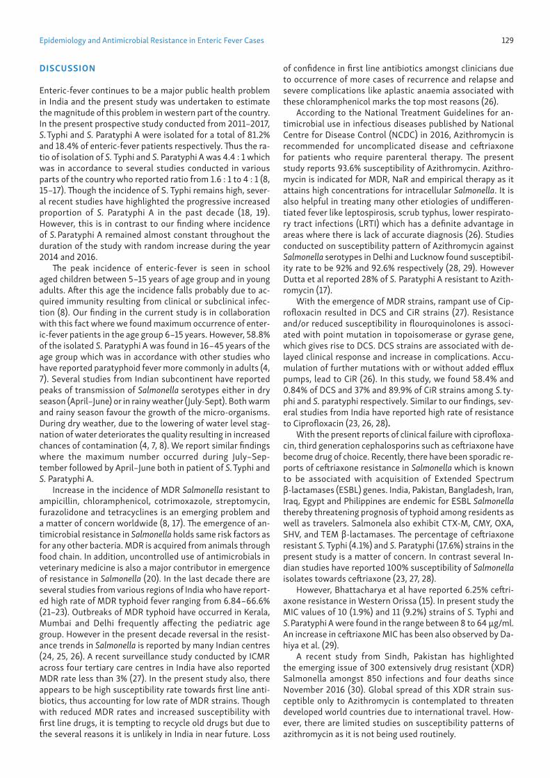
Seasonal occurrence was determined by dividing the year into four quarters. Although enteric-fever occurred in all months throughout the year, the cumulative maximum number of S. Typhi occurred during July–September (47.51%) followed by April–June (33.1%). The peak incidence of S. Para-typhi was also observed in July–September (45.86%) followed by April–June (21.66%) (Fig. 1, 2).
Resistance to co-trimoxazole, chloramphenicol, ceftriax-one and azithromycin was 6.1%, 13.8%, 16.1 and 5.78% re-spectively. MDR S. typhi and S. paratyphi A were 2.73% and 1.91% respectively (Table 3). Table 4 shows the year wise dis-tribution of four major resistance phenotypes analysed for Salmonella: MDR, NaR, DCS and ciprofloxacin resistant (CiR) S. Typhi and S. Paratyphi A.
1417
30
1212
33 31
16
611
43
24
6
56
73
201823
34
22
15
39
49
39
9
27
36
27
0
10
20
30
40
50
60
70
80
Jan-Mar Apr-Jun Jul-Sept Oct-Dec
2011
2012
2013
2014
2015
2016
2017
Fig. 1 Epidemiological mapping of S. Typhi at a 1000-bedded tertiary-care teaching hospital in New Delhi from 2011–2017.
1 1
5
21
3
8
11 1
7
23
11
19
2
4
6 6
2
5 5
1211
67
15
10
0
2
4
6
8
10
12
14
16
18
20
Jan-Mar Apr-Jun Jul-Sept Oct-Dec
2011
2012
2013
2014
2015
2016
2017
Fig. 2 Epidemiological mapping of S. Paratyphi A at a 1000-bedded tertiary-care teaching hospital in New Delhi from 2011–2017.
Epidemiology and Antimicrobial Resistance in Enteric Fever Cases 129
DISCUSSION
Enteric-fever continues to be a major public health problem in India and the present study was undertaken to estimate the magnitude of this problem in western part of the country. In the present prospective study conducted from 2011–2017, S. Typhi and S. Paratyphi A were isolated for a total of 81.2% and 18.4% of enteric-fever patients respectively. Thus the ra-tio of isolation of S. Typhi and S. Paratyphi A was 4.4 : 1 which was in accordance to several studies conducted in various parts of the country who reported ratio from 1.6 : 1 to 4 : 1 (8, 15–17). Though the incidence of S. Typhi remains high, sever-al recent studies have highlighted the progressive increased proportion of S. Paratyphi A in the past decade (18, 19). However, this is in contrast to our finding where incidence of S. Paratyphi A remained almost constant throughout the duration of the study with random increase during the year 2014 and 2016.
The peak incidence of enteric-fever is seen in school aged children between 5–15 years of age group and in young adults. After this age the incidence falls probably due to ac-quired immunity resulting from clinical or subclinical infec-tion (8). Our finding in the current study is in collaboration with this fact where we found maximum occurrence of enter-ic-fever patients in the age group 6–15 years. However, 58.8% of the isolated S. Paratyphi A was found in 16–45 years of the age group which was in accordance with other studies who have reported paratyphoid fever more commonly in adults (4, 7). Several studies from Indian subcontinent have reported peaks of transmission of Salmonella serotypes either in dry season (April–June) or in rainy weather (July-Sept). Both warm and rainy season favour the growth of the micro-organisms. During dry weather, due to the lowering of water level stag-nation of water deteriorates the quality resulting in increased chances of contamination (4, 7, 8). We report similar findings where the maximum number occurred during July–Sep- tember followed by April–June both in patient of S. Typhi and S. Paratyphi A.
Increase in the incidence of MDR Salmonella resistant to ampicillin, chloramphenicol, cotrimoxazole, streptomycin, furazolidone and tetracyclines is an emerging problem and a matter of concern worldwide (8, 17). The emergence of an-timicrobial resistance in Salmonella holds same risk factors as for any other bacteria. MDR is acquired from animals through food chain. In addition, uncontrolled use of antimicrobials in veterinary medicine is also a major contributor in emergence of resistance in Salmonella (20). In the last decade there are several studies from various regions of India who have report-ed high rate of MDR typhoid fever ranging from 6.84–66.6% (21–23). Outbreaks of MDR typhoid have occurred in Kerala, Mumbai and Delhi frequently affecting the pediatric age group. However in the present decade reversal in the resist-ance trends in Salmonella is reported by many Indian centres (24, 25, 26). A recent surveillance study conducted by ICMR across four tertiary care centres in India have also reported MDR rate less than 3% (27). In the present study also, there appears to be high susceptibility rate towards first line anti-biotics, thus accounting for low rate of MDR strains. Though with reduced MDR rates and increased susceptibility with first line drugs, it is tempting to recycle old drugs but due to the several reasons it is unlikely in India in near future. Loss
of confidence in first line antibiotics amongst clinicians due to occurrence of more cases of recurrence and relapse and severe complications like aplastic anaemia associated with these chloramphenicol marks the top most reasons (26).
According to the National Treatment Guidelines for an-timicrobial use in infectious diseases published by National Centre for Disease Control (NCDC) in 2016, Azithromycin is recommended for uncomplicated disease and ceftriaxone for patients who require parenteral therapy. The present study reports 93.6% susceptibility of Azithromycin. Azithro-mycin is indicated for MDR, NaR and empirical therapy as it attains high concentrations for intracellular Salmonella. It is also helpful in treating many other etiologies of undifferen-tiated fever like leptospirosis, scrub typhus, lower respirato-ry tract infections (LRTI) which has a definite advantage in areas where there is lack of accurate diagnosis (26). Studies conducted on susceptibility pattern of Azithromycin against Salmonella serotypes in Delhi and Lucknow found susceptibil-ity rate to be 92% and 92.6% respectively (28, 29). However Dutta et al reported 28% of S. Paratyphi A resistant to Azith-romycin (17).
With the emergence of MDR strains, rampant use of Cip-rofloxacin resulted in DCS and CiR strains (27). Resistance and/or reduced susceptibility in flouroquinolones is associ-ated with point mutation in topoisomerase or gyrase gene, which gives rise to DCS. DCS strains are associated with de-layed clinical response and increase in complications. Accu-mulation of further mutations with or without added efflux pumps, lead to CiR (26). In this study, we found 58.4% and 0.84% of DCS and 37% and 89.9% of CiR strains among S. ty-phi and S. paratyphi respectively. Similar to our findings, sev-eral studies from India have reported high rate of resistance to Ciprofloxacin (23, 26, 28).
With the present reports of clinical failure with ciprofloxa-cin, third generation cephalosporins such as ceftriaxone have become drug of choice. Recently, there have been sporadic re-ports of ceftriaxone resistance in Salmonella which is known to be associated with acquisition of Extended Spectrum β-lactamases (ESBL) genes. India, Pakistan, Bangladesh, Iran, Iraq, Egypt and Philippines are endemic for ESBL Salmonella thereby threatening prognosis of typhoid among residents as well as travelers. Salmonela also exhibit CTX-M, CMY, OXA, SHV, and TEM β-lactamases. The percentage of ceftriaxone resistant S. Typhi (4.1%) and S. Paratyphi (17.6%) strains in the present study is a matter of concern. In contrast several In-dian studies have reported 100% susceptibility of Salmonella isolates towards ceftriaxone (23, 27, 28).
However, Bhattacharya et al have reported 6.25% ceftri-axone resistance in Western Orissa (15). In present study the MIC values of 10 (1.9%) and 11 (9.2%) strains of S. Typhi and S. Paratyphi A were found in the range between 8 to 64 µg/ml. An increase in ceftriaxone MIC has been also observed by Da-hiya et al. (29).
A recent study from Sindh, Pakistan has highlighted the emerging issue of 300 extensively drug resistant (XDR) Salmonella amongst 850 infections and four deaths since November 2016 (30). Global spread of this XDR strain sus-ceptible only to Azithromycin is contemplated to threaten developed world countries due to international travel. How-ever, there are limited studies on susceptibility patterns of azithromycin as it is not being used routinely.
130 Anuradha Makkar et al. Acta Medica (Hradec Králové)
Infection prevention and control of Salmonella requires interventions at reservoir, transmission and host fronts. Reservoir elimination should be focused on provisioning of clean potable water, hygienic food. Chain of transmission can be broken by emphasis of hand-hygiene amongst chefs, food handlers and kitchen staff. Outbreak monitoring sys-tems should be instituted to implement swift anti-epidemic measures. Continued emergence of multidrug-resistant, Na-lidixic-acid resistant and extremely drug-resistant Salmonella mandates ongoing surveillance to type antibiogram patterns for empirical therapy and containment of spread.
CONCLUSION
Enteric-fever continues to be a major public-health problem in India. Emergence of multidrug-resistant and extremely drug-resistant Salmonella mandates ongoing surveillance to type antibiogram patterns for empirical therapy and contain-ment of spread. Repeated epidemics call for implementation of preventive measures like water, sanitation and hygiene (WASH) along with pre-emptive vaccination strategies to sustain herd immunity in susceptible communities.
REFERENCES 1. Crump JA, Luby SP, Mintz ED. The global burden of typhoid fever. Bull
World Health Org 2004; 82: 346–53. 2. Miller SI, Pegues DA. Salmonella species, including Salmonella typhi. In:
Mandell GL, Bennet JE, Dolin R, eds. Principles and Practice of Infectious Disease, 4th edn. New York: Churchill Livingstone, 1998: pp. 2344–73.
3. Chowta MN, Chowta NK. Study of clinical profile and antibiotic response in typhoid fever. Indian J Med Microbiol 2005; 23: 125–27.
4. Kanung OS, Dutta S, Sur D. Epidemiology of typhoid and paratyphoid fever in India. J Infect Developing Countries 2008; 2(6): 454–60.
5. Tankhiwale SS, Agrawal G, Jalgaonkar SV. An unusually high occurrence of Salmonella enterica serotype Paratyphi A in patients with enteric fe-ver. Indian Journal of Medical Research 2003; 117: 10–2.
6. Mahapatra A, Patro S, Choudhury S, Padhee A, Das R. Emerging enteric fever due to switching biotype of Salmonella (Paratyphi A) in Eastern Odisha. Indian J Pathol Microbiol 2016; 59: 327–9.
7. Sur D, Ali M, von Seidlein L, et al. Comparisons of predictors for typhoid and paratyphoid fever in Kolkata, India. BMC Public Health 2007; 7(147): 289.
8. Mohanty S, Renuka K, Sood S, Das BK, Kapil A. Antibiogram pattern and seasonality of Salmonella serotypes in a North Indian tertiary-care hos-pital. Epidemiol. Infect 2006; 134: 961–6.
9. Maskey AP, Basnyat B, Thwaltes GE, et al. Emerging trends in enteric fever in Nepal: 9124 cases confirmed by blood culture 1993–2003. Trans R Soc Trop Med Hyg 2008; 102: 91–5.
10. Panikar CKJ, Vimala KN. Transferable chloramphenicol resistance in Sal-monella typhi. Nature 1972; 239: 109–10.
11. Olarte J, Galind DE. Salmonella typhi resistant to chloramphenicol, ampi-cillin and other antimicrobial agents: Strains isolated during an extensive typhoid fever epidemic in Mexico. Antimicrob Agents Chemother 1973; 4: 597–601.
12. Chuang CH, Su LH, Perera J, et al. Surveillance of antimicrobial resistance of Salmonella enterica serotype typhi in seven Asian countries. Epidemi-ology and Infection 2009; 137: 266–9.
13. Sin L, Cariappa MP. Blood culture isolates and Antibiogram of Samonella: Experience of a tertiary care hospital. Med J Armed Forces India 2016; 72 (1): 281–4.
14. Clinical and Laboratory Standards Institute. Performance Standards for Antimicrobial Susceptibility Testing: Twenty Second Informational Sup-plement M100-22. Wayne, Pennsylvania: Clinical and Laboratory Stand-ards Institute; 2012.
15. Bhattacharya SS, Das U, Choudhury BK. Occurrence & Antibiogram Sal-monella Typhi & S. Paratyphi A isolated from Rourkela, Orissa. Ind J Med Microbiol 2011; 133: 431–3.
16. World Health Organization: Indian Network for Surveillance of Antimi-crobial Resistance. Antibiogram of Salmonella enterica serovar Typhi and Salmonella enterica serovar Paratyphi A: a multi-centre study from India. WHO South-East Asia J Public Health 2012; 1: 182–8.
17. Dutta S, Das S, Mitra U, et al. Antimicrobial Resistance, Virulence Pro-files and Molecular Subtypes of Salmonella enterica Serovars Typhi and Paratyphi A Blood Isolates from Kolkata, India during 2009–2013. PLoS ONE 2014; 9(8): e101347.
18. Gupta V, Kaur J, Chander J. An increase in enteric fever cases due to Sal-monella paratyphi A in and around Chandigarh. Indian J Med Res 2009; 129: 95–8.
19. Bhattacharya SS, Dash U. A sudden rise in occurrence of Salmonella paratyphi A infection in Rourkela Orissa. Indian J Med Microbiol 2007; 25: 78–9.
20. Zaki SA, Karande S. Multidrug resistant typhoid fever: a review. J Infect Dev Ctries 2011; 5(5): 324–37.
21. Shu-Kee E, Pusparajah P, Ab Mutalib NS, et al. Salmonella: A review on pathogenesis, epidemiology and antibiotic resistance. Frontiers in Life Science 2015; 8(3): 284–93.
22. Jog S, Soman R, Singhal T, et al. Enteric fever in Mumbai – clinical profile, sensitivity patterns and response to antimicrobials. J Assoc Physicians India 2008; 56: 237–40.
23. Sen B, Dutta S, Sur D, et al. Phage typing, biotyping & antimicrobial resistance profile of Salmonella enterica serotype Typhi from Kolkata. Indian J Med Res 2007; 125: 685–8.
24. Kumar Y, Sharma A, Mani KR. Antibiogram Profile of Salmonella enteri-ca Serovar Typhi in India – a two year study. Trop Life Sci Res 2013: 24: 45–54.
25. Badiyal A, Kumar Y, Sharma A, Mani KR. Re-emergence of chloramphen-icol sensitive isolates of Salmonella enterica serovar typhi isolates in India during 2013–14. Int J Current Microbiol Appl Sci 2015: 4: 1081–6.
26. Divyashree S , Nabarro LEB, Veeraraghavan B and Rupali P. Enteric fever in India: current scenario and future directions. Tropical Medicine and International Health 2016; 21: 1255–62.
27. Dahiya S, Sharma P, Kumari B, et al. Characterisation of antimicrobial resistance in Salmonellae during 2014–2015 from four centres across India: An ICMR antimicrobial resistance surveillance network report. In-dian J Med Microbiol 2017; 35: 61–8.
28. Saksena R, Nayyar C, Manchanda V. Six-year susceptibility trends and effect of revised Clinical Laboratory Standards Institute breakpoints on ciprofloxacin susceptibility reporting in typhoidal Salmonellae in a ter-tiary care paediatric hospital in Northern India. Indian J Med Microbiol 2016; 34: 520–5.
29. Misra R, Prasad KN. Antimicrobial susceptibility to Azithromycin among Salmonella enteric typhi and Paratyphi A isolates from India. J Med Mi-crobiol 2016; 65(12): 1536–9.
30. Klemm EJ, Shakoor S, Page AJ, et al. Emergence of an extensively drug re-sistant Salmonella enteric serovar Typhi clone harbouring a promiscuous plasmid encoding resistance to flouroquinolones and third generation cephalosporins. mBio 2018; 9: e00105–18.
original article 131
Prevalence of Musculoskeletal Disorders Symptoms among Czech Dental Students. Part 1: a Questionnaire Survey
Martin Kapitán1,*, Nela Pilbauerová1, Lenka Vavřičková1, Zdeňka Šustová1, Stanislav Machač2,3
A B S T R AC TMusculoskeletal disorders (MSDs) frequently occur among dentists and dental students. The first aim of this study was to gather and analyze information about the prevalence of symptoms of MSDs. The second aim was to determine a correlation between subjectively described complaints and the results of an objective examination of the spine using the Spinal Mouse® device; that part is described in the second section of this article. Dental students of the first, third and fifth years at Charles University, Faculty of Medicine in Hradec Králové were included. The participants filled out a questionnaire regarding the prevalence of symptoms of MSDs and the potential risk factors. From a total of 182 students that participated in this study, 71 reported musculoskeletal pain (prevalence 39%). The musculoskeletal pain was more frequent among women. During the studies its prevalence increased. The most frequent areas of pain were neck, lower back and upper back. The results indicate that musculoskeletal pain often begins and continues to develop throughout the dentistry studies.
K E Y WO R D Sdentistry students; musculoskeletal disorders; questionnaire survey
A U T H O R A F F I L I AT I O N S1 Department of Dentistry, Charles University, Faculty of Medicine in Hradec Králové, and University Hospital Hradec Králové,
Hradec Králové, Czech Republic2 Institute of Sports Medicine, Prague, Czech Republic3 Department of Rehabilitation and Sports Medicine, Charles University, 2nd Faculty of Medicine, and Motol University Hospital, Prague,
Czech Republic* Corresponding author: Department of Dentistry, University Hospital Hradec Králové, Sokolská 581, 500 05, Hradec Králové, Czech
Republic; [email protected]
Received: 5 August 2018Accepted: 29 November 2018Published online: 22 January 2019
Acta Medica (Hradec Králové) 2018; 61(4): 131–136https://doi.org/10.14712/18059694.2018.131© 2018 The Authors. This is an open-access article distributed under the terms of the Creative Commons Attribution License (http://creativecommons.org/licenses/by/4.0), which permits unrestricted use, distribution, and reproduction in any medium, provided the original author and source are credited.
132 Martin Kapitán et al. Acta Medica (Hradec Králové)
INTRODUCTION
Musculoskeletal disorders (MSDs) are diseases of the mus-culoskeletal apparatus affecting muscles, tendons, joints, nerves and supporting structures. The dominant symptoms are: frequently occurring pain, a failure of coordinated sta-bilization (as a function of the neuromuscular system), a re-stricted range of motion, and is often accompanied by a de-crease of muscular strength of certain muscle groups. Apart from pain, other subjective signs are stiffness of muscles and joints, tingling and feeling of warmth or cold. If work is assumed to initiate or develop these symptoms, then they are referred to as work-related musculoskeletal disorders (WRMSDs) (1).
The prevalence of MSDs among dentists was between 62% and 91% (2–10). The most frequent areas of pain are neck, shoulders, lower back, hands and wrists (3, 5, 7, 8, 10–16). In the dental profession there are many risk factors which combine and can contribute to initiation and devel-opment of MSDs. These risk factors include mainly a long-term static position, forced unnatural posture during work, doing tasks with small instruments using a large force, small working field with limited access, intensive lighting, noise and psychosocial stress (5, 12, 13, 17–19). Another factor which is considered negative is a lack of adequate physical activities (20).
MSDs begin from dental school studies with an preva-lence among students between 44% and 93% (2, 21–27). Stu-dents of the final two years suffer from musculoskeletal pain more often than the students of the first two years (28).
The aim of this study was to gather and analyze informa-tion about the prevalence of symptoms of MSDs and the role of potential risk factors among dental students. The second aim was to determine a correlation between subjectively described complaints and the results of an objective exam-ination of the spine using the Spinal Mouse® device; this is described in the second part of this article. The results could contribute to understand the early development of MSDs among dental students and to find out possible risk factors detectable during the objective examination.
MATERIAL AND METHODS
Students of the current first, third and fifth years of the study program Dentistry at Charles University, Faculty of Medicine in Hradec Králové, in academic years 2014/2015 and 2015/2016 were included in this study. The collection of data for this study started in October 2014 and was fin-ished in May 2016. The participation was voluntary. All the participants signed an informed consent. The students were examined at the beginning of their studies (the beginning of the first year), before the start of the clinical part of their studies (the beginning of the summer term of the third year) and at the end of their studies (the end of the fifth year). The only inclusion criterion was that the student was currently studying in the included year of studies as described above. No exclusion criteria were applied. There were no students
repeating the particular year, who would have participated twice in the study.
This study was approved by the Ethics committee of Uni-versity Hospital Hradec Králové (Ref. no. 201410 S04P) and by the dean of the Charles University, Faculty of Medicine in Hradec Králové.
A questionnaire survey was performed among the partic-ipants to gather the required information about the respond-ents and their complaints. The questionnaire was created by the authors and was based on other similar studies (5, 6, 8–10, 13, 29). The questionnaire consisted of the questions of the following areas:– Personal information of the respondents (i.e. gender, age,
weight and height).– General health status, medication and allergies.– Factors possibly influencing the development of MSDs
(i.e. dominant hand, musculoskeletal diseases in the fam-ily history, in the personal medical history and the sport activity).
– Currently experienced musculoskeletal pain and its consequences.
– Respondents’ opinions on dentistry studies (e.g. wheth-er the participant consider the dentistry studies as psy-chically demanding; if the level of ergonomic education is sufficient; and whether they had known before they were making the decision about their further profession, that 2 out of 3 dentists suffered from musculoskeletal disorders).The questionnaire was distributed to all possible par-
ticipants, i.e. to all students currently studying in the in-cluded years of studies. A total of 192 questionnaires were distributed.
The collected data were statistically analyzed in the NCSS 10 Statistical Software (2015; NCSS, LLC. Kaysville, Utah, USA, ncss.com/software/ncss) using methods of descriptive statistics, two-sample t-test, nonparametric Mann-Whitney test, Kolmogorov-Smirnov test, nonparametric Kruskal-Wal-lis analysis of variance with post hoc Dunn’s test with Bon-ferroni modification and Pearson’s χ2 test of independence in contingency tables or Fisher’s exact test. The level of statisti-cal significance was set to α = 0.05.
RESULTS
A total of 182 students participated in this study; there was a response rate of 94.8%. The student’s information is sum-marized in Table 1. As the data were not distributed normally, the median values along with the first and the third quartiles are presented. Table 2 presents the anamnestic data of the respondents’ and their influence on the prevalence of mus-culoskeletal pain. Pain of the musculoskeletal apparatus was declared by 39% of the respondents (n = 71). The differences of a prevalence of musculoskeletal pain between men and women and between the years of studies are summarized Table 3. The age, height, weight and BMI didn’t present any statistically significant influence on the prevalence of mus-culoskeletal pain.
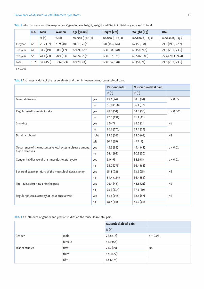
Prevalence of Musculoskeletal Disorders Symptoms 133
Tab. 1 Information about the respondents’ gender, age, height, weight and BMI in individual years and in total.
No. Men Women Age [years] Height [cm] Weight [kg] BMI
% (n) % (n) median (Q1; Q3) median (Q1; Q3) median (Q1; Q3) median (Q1; Q3)
1st year 65 26.2 (17) 73.9 (48) 20 (19; 20)* 170 (165; 176) 62 (56; 68) 21.3 (19.8; 22.7)
3rd year 61 31.2 (19) 68.9 (42) 22 (21; 22)* 173 (168; 178) 63 (57; 71,5) 21.6 (20.1; 23.5)
5th year 56 41.1 (23) 58.9 (33) 24 (24; 25)* 173 (167; 179) 65.5 (60; 80) 22.4 (20.3; 24.4)
Total 182 32.4 (59) 67.6 (123) 22 (20; 24) 173 (166; 178) 63 (57; 71) 21.6 (20.1; 23.5)
*p < 0.001
Tab. 2 Anamnestic data of the respondents and their influence on musculoskeletal pain.
Respondents Musculoskeletal pain
% (n) % (n)
General disease yes 13.2 (24) 58.3 (14) p < 0.05
no 86.8 (158) 36.1 (57)
Regular medicaments intake yes 28.0 (51) 58.8 (30) p < 0.001
no 72.0 (131) 31.3 (41)
Smoking yes 3.9 (7) 28.6 (2) NS
no 96.2 (175) 39.4 (69)
Dominant hand right 89.6 (163) 38.0 (62) NS
left 10.4 (19) 47.7 (9)
Occurrence of the musculoskeletal system disease among blood relatives
yes 45.6 (83) 49.4 (41) p < 0.01
no 54.4 (99) 30.3 (30)
Congenital disease of the musculoskeletal system yes 5.0 (9) 88.9 (8) p < 0.01
no 95.0 (173) 36.4 (63)
Severe disease or injury of the musculoskeletal system yes 15.4 (28) 53.6 (15) NS
no 84.4 (154) 36.4 (56)
Top-level sport now or in the past yes 26.4 (48) 43.8 (21) NS
no 73.6 (134) 37.3 (50)
Regular physical activity at least once a week yes 81.3 (148) 38.5 (57) NS
no 18.7 (34) 41.2 (14)
Tab. 3 An influence of gender and year of studies on the musculoskeletal pain.
Musculoskeletal pain
% (n)
Gender male 28.8 (17) p = 0.05
female 43.9 (54)
Year of studies first 23.2 (19) NS
third 44.3 (27)
fifth 44.6 (25)
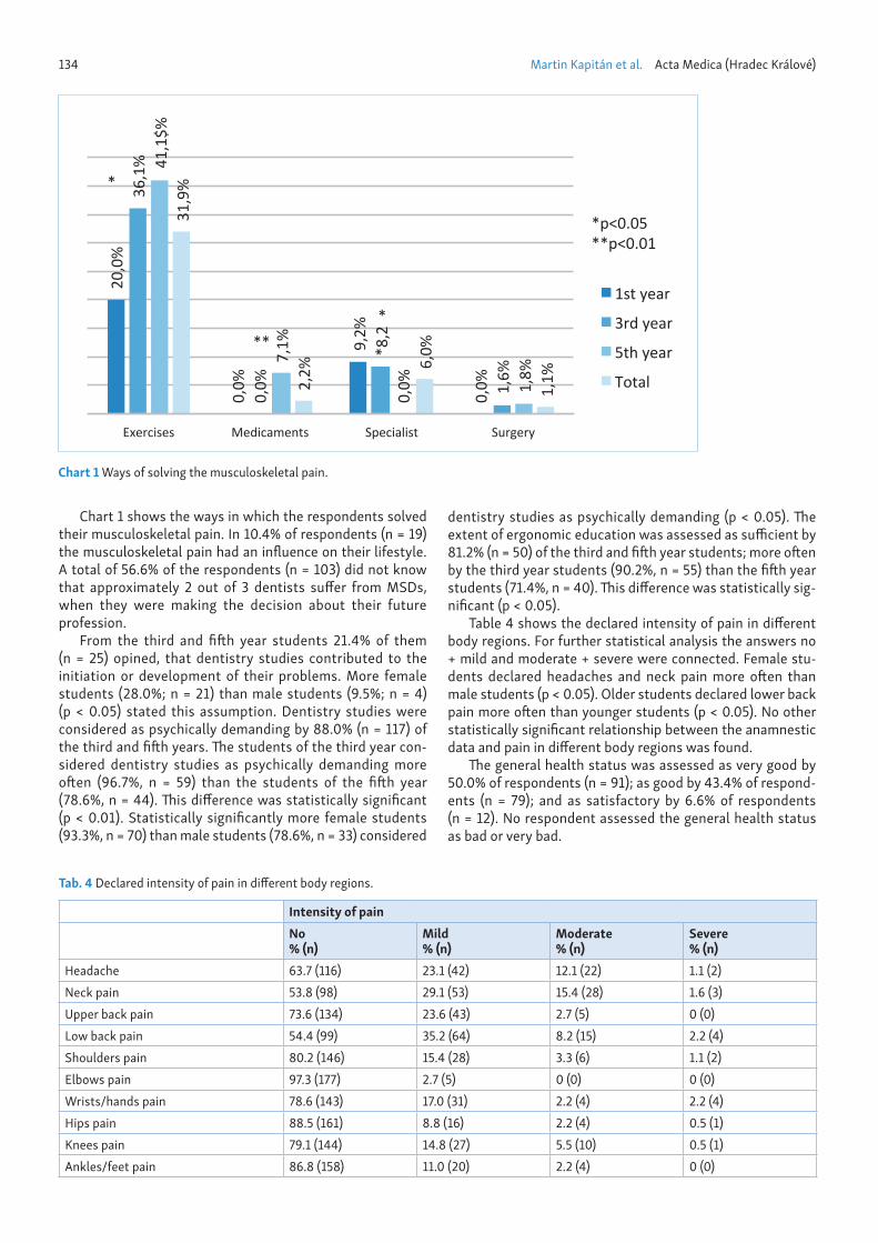
134 Martin Kapitán et al. Acta Medica (Hradec Králové)
Chart 1 shows the ways in which the respondents solved their musculoskeletal pain. In 10.4% of respondents (n = 19) the musculoskeletal pain had an influence on their lifestyle. A total of 56.6% of the respondents (n = 103) did not know that approximately 2 out of 3 dentists suffer from MSDs, when they were making the decision about their future profession.
From the third and fifth year students 21.4% of them (n = 25) opined, that dentistry studies contributed to the initiation or development of their problems. More female students (28.0%; n = 21) than male students (9.5%; n = 4) (p < 0.05) stated this assumption. Dentistry studies were considered as psychically demanding by 88.0% (n = 117) of the third and fifth years. The students of the third year con-sidered dentistry studies as psychically demanding more often (96.7%, n = 59) than the students of the fifth year (78.6%, n = 44). This difference was statistically significant (p < 0.01). Statistically significantly more female students (93.3%, n = 70) than male students (78.6%, n = 33) considered
dentistry studies as psychically demanding (p < 0.05). The extent of ergonomic education was assessed as sufficient by 81.2% (n = 50) of the third and fifth year students; more often by the third year students (90.2%, n = 55) than the fifth year students (71.4%, n = 40). This difference was statistically sig-nificant (p < 0.05).
Table 4 shows the declared intensity of pain in different body regions. For further statistical analysis the answers no + mild and moderate + severe were connected. Female stu-dents declared headaches and neck pain more often than male students (p < 0.05). Older students declared lower back pain more often than younger students (p < 0.05). No other statistically significant relationship between the anamnestic data and pain in different body regions was found.
The general health status was assessed as very good by 50.0% of respondents (n = 91); as good by 43.4% of respond-ents (n = 79); and as satisfactory by 6.6% of respondents (n = 12). No respondent assessed the general health status as bad or very bad.
20,0
%
0,0%
9,2%
0,0%
36,1
%
0,0%
*8,2
1,6%
41,1
$%
7,1%
0,0% 1,
8%
31,9
%
2,2% 6,
0%
1,1%
Exercises Medicaments Specialist Surgery
1st year
3rd year
5th year
Total
*p<0.05**p<0.01
*
***
Chart 1 Ways of solving the musculoskeletal pain.
Tab. 4 Declared intensity of pain in different body regions.
Intensity of pain No
% (n)Mild% (n)
Moderate% (n)
Severe% (n)
Headache 63.7 (116) 23.1 (42) 12.1 (22) 1.1 (2)Neck pain 53.8 (98) 29.1 (53) 15.4 (28) 1.6 (3)Upper back pain 73.6 (134) 23.6 (43) 2.7 (5) 0 (0)Low back pain 54.4 (99) 35.2 (64) 8.2 (15) 2.2 (4)Shoulders pain 80.2 (146) 15.4 (28) 3.3 (6) 1.1 (2)Elbows pain 97.3 (177) 2.7 (5) 0 (0) 0 (0)Wrists/hands pain 78.6 (143) 17.0 (31) 2.2 (4) 2.2 (4)Hips pain 88.5 (161) 8.8 (16) 2.2 (4) 0.5 (1)Knees pain 79.1 (144) 14.8 (27) 5.5 (10) 0.5 (1)Ankles/feet pain 86.8 (158) 11.0 (20) 2.2 (4) 0 (0)
Prevalence of Musculoskeletal Disorders Symptoms 135
DISCUSSION
A questionnaire survey was chosen as a study design to ac-complish the first aim of this study. A questionnaire survey is often used in similar studies, because it is a cheap and rela-tively fast method with a sufficient value of gathered infor-mation, however, it is not possible to verify its truthfulness and accuracy. The questionnaire for this study was created by a combination of questionnaires from other studies (5, 6, 9, 10, 13, 29) and from a pilot study performed by the authors among the dentists (8). It covered information about relevant personal information, possible risk factors, currently expe-rienced musculoskeletal pain and opinions of the respond-ents related to musculoskeletal pain, ergonomics and dental education.
The participants of this study were students in three im-portant moments of their studies: Students at the beginning of the first year represented the initial default status. These students were not subjected to any influences related to den-tistry studies/profession yet, thus, no control study was in-volved in this evaluation. Students in the middle of the third year represented the turning point between the preclinical and clinical phase of their studies. The students at the end of the fifth year represented the situation at the end of their studies. The study was performed in two subsequent ac-ademic years to gather a sufficient number of participants. About 1/3 of them were men and 2/3 were women. This cor-responds with the gender distribution among dentists in the Czech Republic (30) and also in the study of Diaz-Caballero et al. (22).
The prevalence of musculoskeletal pain among dental stu-dents in this study was lower than in other studies (2, 21–27). In accordance to other studies the prevalence of musculo-skeletal pain was higher in women than men (2, 6, 21, 23, 25, 28, 31), nevertheless, the difference was on the limit of statistical significance. A difference was found between the years of studies, however it was not statistically significant. An increase of musculoskeletal pain prevalence was seen be-tween the first and the third year. In the following years, the prevalence didn’t change more significantly. Thus, it can be assumed that dentistry studies could contribute to the initi-ation and development of musculoskeletal pain, particularly in the preclinical part. An important factor may also be a life-style with a lack of adequate physical activity. It is likely, that a long-term influence of the forced position on the prevalence of musculoskeletal pain manifests mainly after the end of the studies, i.e. after the beginning of daily dental work.
The most frequent areas of pain were neck, lower back and upper back. This finding is in accordance with other stud-ies (3, 5, 7, 10–16, 22, 25). Also, headaches were rather fre-quent; however, this is not classified as MSDs. An important factor for headache is the psychological stress, which was felt by the majority of the students. The third year students and female students considered dentistry studies as psychically demanding more often than fifth year students and male stu-dents. The reason may be that during their studies the stu-dents gradually become accustomed to deal with a lot of var-ious demands. The female students, for their generally higher conscientiousness, approach the task solving with higher re-sponsibility and mental engagement and they deal with over-loading and sectional failures worse than male students (17).
The presence of general disease and permanent medica-tion had a negative influence on the prevalence of musculo-skeletal pain. These factors were not analyzed in detail within the scope of this study. The musculoskeletal pain was more frequent in students with a prevalence of musculoskeletal disease in their blood relatives and in students with a congen-ital disease of the musculoskeletal system. Other followed factors didn’t present any influence on the prevalence of musculoskeletal pain. Hodačová et al. (6) found an influence of the severe disease or injury of the locomotive apparatus on the musculoskeletal pain among dental practitioners. In the study of De Carvalho et al. (21) regularly doing exercises didn’t have any influence on musculoskeletal pain, whereas Shirzaei et al. (28) proved a lower average intensity of pain in students, who did stretching after the patients treatment. In the study of Sharma et al. (20) 80% of responding dentists declared, that the cause of their pain was the lack of physical activity. Yi et al. (16) showed that regular exercise decreases the prevalence of musculoskeletal pain. In this study 81.3% of the respondents declared regular physical activity at least once a week. This factor didn’t have a statistically significant influence on the prevalence of musculoskeletal pain.
Only 1/5 of the students thought that dentistry studies had an influence on the initiation and development of their problems. Of the 1/5 of the students, more were women. The authors assume that the students had not realized the pos-sible relationships between their future profession and the potential difficulties.
More than 1/2 of the students were aware of the fact that two out of three dentists suffer from MSDs, when they were making the decision about their future profession. This is an important negative aspect of the dental profession and the authors believe that young people considering to choose dentistry as their profession have the right to be informed in advance. Thus, all the students should have been aware of that.
Although the third year students mostly considered the ergonomic education as sufficient, the prevalence of muscu-loskeletal pain increased between the first and the third year. The authors assume that even if the students have a sufficient knowledge of dental ergonomics, they are not able to apply all the recommendations during the preclinical practical les-sons. Several reasons may play a role, such as underestima-tion of MSDs in dentistry by students; stress, which was more frequently perceived by the third year students; and restrict-ed possibility of work in the correct position and in an indirect vision, because the students work on the dental simulators alone, i.e. without an assisting person. During the preclinical years the students go through 60 hours of practical lessons in the phantom labs in each semester. The working conditions change in the clinical years, when students work in pairs on the patients, they are able to follow the ergonomic principles and this phase of the studies is less stressful. This may be the reason, why there is no significant increase in MSDs preva-lence between the third and the fifth year.
When comparing pain intensity in different body regions with other factors, the answers no + mild and moderate + se-vere were combined. The reason for this was an effort to sim-plify the statistical analysis and a small number of answers in some categories, which was not sufficient to perform a sta-tistical analysis.
136 Martin Kapitán et al. Acta Medica (Hradec Králové)
Musculoskeletal pain was mostly solved by exercises, followed by visiting a specialist (rehabilitation physician or physiotherapeutist) and the usage of medicaments. The fre-quency of exercises and medicaments increased during the studies, conversely visiting specialists decreased.
A limitation of this study was that different students were involved in different years. For more precise evaluation of the evolution of musculoskeletal pain prevalence it would be use-ful to evaluate the same students gradually in the first, third and fifth year, followed by an evaluation of the same partici-pants after a few years of dental practice.
CONCLUSIONS
The prevalence of musculoskeletal pain among dental stu-dents in this study was 39%. The most frequent areas of pain were neck, lower back and upper back. Women suffered from musculoskeletal pain more often than men. The presence of a congenital disease of the musculoskeletal system increased the prevalence of the musculoskeletal pain, as well as an oc-currence of musculoskeletal system disease among blood relatives.
The prevalence of musculoskeletal pain increased dur-ing the running of the studies, mainly during the preclinical phase.
ACKNOWLEDGEMENTS
The authors declare that they have no competing interests. The authors declare that they are not associated in any way with the producer or distributor of the Spinal Mouse® device, and that the study was not supported by these companies.
The study was financially supported by the Charles Uni-versity’s programme PROGRES Q40/13.
The authors would like to thank Dr. Eva Čermáková from Computer Technology Center, Charles University, Faculty of Medicine in Hradec Králové, Czech Republic, for help with the statistical analysis, and Ms. Eva Vedralová, Ms. Sara Re-zazadeh Siaghi and Ms. Eliska Charlotte Wurfel for a language revision of the manuscript.
REFERENCES 1. European agency for safety and health at work. OSH in figures: Work-re-
lated musculoskeletal disorders in the EU-Facts and figures. Luxem-bourg: Publications Office of the European Union; 2010.
2. Abbas SB, Qazi SR, Iftikhar S, Iqbal MU. Musculoskeletal disorders among dentists and dental students. Pakistan Oral & Dental Journal 2015; 35: 461–5.
3. Abduljabbar TA. Musculoskeletal disorders among dentists in Saudi Ara-bia. Pakistan Oral & Dental Journal 2008; 28: 135–44.
4. Alexopoulos EC, Stathi IC, Charizani F. Prevalence of musculoskeletal disorders in dentists. BMC Musculoskelet Disord 2004; 9: 16.
5. Hayes MJ, Cockrell D, Smith DR. A systematic review of musculoskel-etal disorders among dental professionals. Int J Dent Hyg 2009; 7: 159–65.
6. Hodacova L, Sustova Z, Cermakova E, Kapitan M, Smejkalova J. Self-re-ported Risk Factors Related to the Most Frequent Musculoskeletal Com-plaints among Czech Dentists. Ind Health 2015; 53: 48–55.
7. Leggat PA, Smith DR. Musculoskeletal disorders self-reported by den-tists in Queensland, Australia. Aust Dent J 2006; 51: 324–7.
8. Pilbauerová N, Kapitán M, Šustová Z, Machač S. An evaluation of the oc-currence of musculoskeletal diders among dentists of the Department of Dentistry, Charles University, Faculty of Medicine in Hradec Králové, and University Hospital Hradec Králové – a pilot study. LKS 2017; 27: 8–12. [in Czech]
9. Puriene A, Aleksejuniene J, Petrauskiene J, Balciuniene I, Janulyte V. Self-reported occupation health issues among Lithuanian dentists. Ind Health 2008; 46: 369–74.
10. Šustová Z, Hodačová L, Kapitán M. The prevalence of musculoskeletal disorders among dentists in the Czech Republic. Acta Medica (Hradec Kralove) 2013; 56: 150–6.
11. Harutunian K, Gargallo-Albiol J, Figueiredo R, Gay-Escoda C. Ergonom-ics and musculoskeletal pain among postgraduate students and facul-ty members of the School of Dentistry of the University of Barcelona (Spain). A cross-sectional study. Med Oral Patol Oral Cir Bucal 2011; 16: e425–9.
12. Leggat PA, Kedjarune U, Smith DR. Occupational health problems in modern dentistry. A Review. Ind Health 2007; 45: 611–21.
13. Puriene A, Janulyte V, Musteikyte M, Bendinskaite R. General health of dentists. Literature review. Stomatologija 2007; 9: 10–20.
14. Shrestha BP, Singh GK, Niraula SR. Work related complaints among den-tist. JNMA J Nepal Med Assoc 2008; 47: 77–81.
15. Szymańska J. Disorders of the musculoskeletal system among dentists from the aspect of ergonomics and prophylaxis. Ann Agric Environ Med 2002; 9: 169–73.
16. Yi J, Hu X, Yan B, Zheng W, Li Y, Zhao Z. High and specialty-related mus-culoskeletal disorders afflict dental professionals even since early train-ing years. J Appl Oral Sci 2013; 21: 376–82.
17. Alzahem AM, van der Molen HT, Alaujan AH, Schmidt HG, Zamakhshary MH. Stress amongst dental students: a systematic review. Eur J Dent Educ 2011; 15: 8–18.
18. Myers HL, Myers LB. ‘It’s difficult being a dentist’: stress and health in the general dental practitioner. Br Dent J 2004; 197: 89–93.
19. Ohlendorf D, Erbe C, Nowak J, et al. Constrained posture in dentist-ry – a kinematic analysis of dentists. BMC Musculoskelet Disord 2017; 18(1): 291.
20. Sharma P, Golchha V. Awareness among Indian dentist regarding the role of physical activity in prevention of work related musculoskeletal disor-ders. Indian J dent Res 2011; 22: 381–4.
21. de Carvalho MV, Soriano EP, de França Caldas A Jr, Campello RI, de Miran-da HF, Cavalcanti FI. Work-related musculoskeletal disorders among Brazilian dental students. J Dent Educ 2009; 73: 624–30.
22. Diaz-Caballero AJ, Gómez-Palencia IP, Díaz-Cárdenas S. Ergonomic fac-tors that cause the presence of pain muscle in students of dentistry. Med Oral Patol Oral Cir Bucal 2010; 15: e906–11.
23. Khan SA, Chew KY. Effect of working characteristics and taught ergo-nomics on the prevalence of musculoskeletal disorders amongst dental students. BMC Musculoskelet Disord 2013; 14: 118.
24. Melis M, Abou-Atme YS, Cottogno L, Pittau R. Upper body musculoskel-etal symptoms in Sardinian dental students. J Can Dent Assoc 2004; 70: 306–10.
25. Ng A, Hayes MJ, Polster A. Musculoskeletal Disorders and Working Pos-ture among Dental and Oral Health Students. Healthcare (Basel) 2016; 4: E13.
26. Rising DW, Bennett BC, Hursh K, Plesh O. Reports of body pain in a den-tal student population. J Am Dent Assoc 2005; 136: 81–6.
27. Thornton LJ, Barr AE, Stuart-Buttle C, Gaughan JP, Wilson ER, Jackson AD, Wyszynski TC, Smarkola C. Perceived musculoskeletal symptoms among dental students in the clinic work environment. Ergonomics 2008; 51: 573–86.
28. Shirzaei M, Mirzaei R, Khaje-Alizade A, Mohammadi M. Evaluation of ergonomic factors and postures that cause muscle pains in dentistry students’ bodies. J Clin Exp Dent 2015; 7: e414–8.
29. Kuorinka I, Jonsson B, Kilbom A, et al. Standardised Nordic question-naires for the analysis of musculoskeletal symptoms. Appl Ergon 1987; 18: 233–7.
30. Czech Dental Chamber. Yearbook 2015. Prague: Czech Dental Chamber, 2016 Accessed May 7, 2017 at https://img.dent.cz/a/e/e/df/df/972941 /CSK_rocenka_2015_web.pdf).
31. Gorter R, Freeman R, Hammen S, Murtomaa H, Blinkhorn A, Humphris G. Psychological stress and health in undergraduate dental students: fifth year outcomes compared with first year baseline results from five Euro-pean dental schools. Eur J Dent Educ 2008; 12: 61–8.
original article 137
Histological Features of Oral Cavity Mucous Membrane Epithelium in Six-Month-Old Experimental Animals Born with Macrosomia
Olga Garmash1,*, Galina Gubina-Vakulik1, David Vondrášek2
A B S T R AC TWe examined the histological features of the mucobuccal fold of oral cavity mucous membrane from the area of the masticatory teeth roots’ projection in 6-month-old Wistar Albino Glaxo rats with fetal macrosomia. The animals were divided into groups according to the body weight, the body length, and the Quetelet index at birth. A morphological study was performed using the Leica SP8 AOBS laser scanning confocal microscope and a conventional light (Axiostar, Zeiss) microscopy. Morphometric parameters were used to estimate the degree of acanthosis development in the epithelium of the oral mucosa, which indicates the intensity of its proliferation.Numerous narrow and deep acanthotic outgrowths and densely located ‘juvenile’ epitheliocytes in the basal layer on the apex of the acanthotic protrusions were found in animals with fetal macrosomia that was due to intrauterine obesity. In these animals, the morphometric index, which we used, was maximally different from that in the control group. In animals with fetal macrosomia that was due to intrauterine growth acceleration of the body, the hyperproliferation of the mucous membrane epithelium of the oral cavity was absent or little pronounced. It can be assumed that fetal macrocosmia with obesity causes instability in the epithelium of the oral cavity mucosa, its rapid death, and therefore, a more active stimulation of proliferation.
K E Y WO R D Sfetal macrosomia; experimental animal; oral mucous membrane; confocal microscopy
A U T H O R A F F I L I AT I O N S1 Kharkiv National Medical University, Therapeutic Dentistry Department, Ukraine2 Institute of Physiology of the Czech Academy of Sciences, Department of Biomathematics, Czech Republic* Corresponding author: Kharkiv National Medical University, Therapeutic Dentistry Department , 61174, 51 Peremogy avenue,
The Stomatological Centre of KhNMU, Ukraine; e-mail: [email protected]
Received: 21 June 2018Accepted: 11 November 2018Published online: 22 January 2019
Acta Medica (Hradec Králové) 2018; 61(4): 137–143https://doi.org/10.14712/18059694.2018.132© 2018 The Authors. This is an open-access article distributed under the terms of the Creative Commons Attribution License (http://creativecommons.org/licenses/by/4.0), which permits unrestricted use, distribution, and reproduction in any medium, provided the original author and source are credited.
138 Olga Garmash et al. Acta Medica (Hradec Králové)
INTRODUCTION
During the last decade, a particular attention has been giv-en to studying the characteristic features of the intrauterine development of tissues and organs. The violation of these processes predisposes further development of a number of diseases.
The violation of adaptation processes and the aberration in physical, somatic, or neuropsychic states arise as a con-sequence of intrauterine sufferings. The hypothesis about fetal or intrauterine programming is termed the metabolic memory (1), when the changes that are downloaded during the intrauterine period are programming a predisposition towards certain diseases in the future ontogenesis has been confirmed in a few studies.
Fetal macrosomia or large birth weight is a well-studied scientific and practice clinical subject (2, 3, 4). It is well known that children with fetal macrosomia have high risk of obesity, high blood pressure, a high blood sugar level, and, as a con-sequence, the appearance of diabetes mellitus and cardiac diseases later in life (5, 6). Some scientists (7) emphasize an increased risk of the appearance of cancer diseases. Hermann GM and coauthors (8) studied the risk of metabolic syndrome formation in the adult life. The violations of the menstrual cy-cle, dyshormonal changes in mammary glands, sterility, and the formation of neoplastic diseases are characteristic of the females who born with macosomia (9).
The features of clinical, metabolic and immunological ad-aptations of such newborns have also been represented in numerous studies conducted by Kharkiv National Medical University scientists (10, 11, 12).
However, there is insufficient knowledge about the dental aspect of macrosomia (13, 14, 15). We also think that fetal macrosomia is a risk factor for the formation of certain oral cavity diseases, and we have already revealed dental viola-tions in people born with macrosomia in our previous studies (16, 17, 18, 19).
Morphological data analysis is a first step in investigat-ing the postnatal genesis features of oral cavity mucosa in species that were born macrosomic. The adequate evaluation of the functional and morphological condition of oral cavity mucosa is significant for successfully solving the diagnostic issues of diseases of the oral cavity at a pre-clinical manifes-tation stage. This evaluation also increases the efficiency of a clinical course prognosis.
One of the most informative methods that allow the study of all mucosa structures in detail is confocal microscopy (20, 21, 22, 23). Successively obtained images of the different lay-ers allow us both to reconstruct a 3-D structure and to per-form quantitative analysis of the sample (24). The 3-D recon-struction of a section of oral cavity mucosa in experimental animals is useful for evaluating the condition of the tissues.
Taking into account close coupling between the state of oral cavity mucosa and the appearance of dental violations, the experimental modeling of fetal macrosomia has been conducted. Analysis of the data obtained in this experiment elucidates some pathogenic links in the intrauterine forma-tion of dental diseases in the case of fetal macrosomia. The results obtained also form a basis for substantiating, devel-oping, approbating, and the further implementation in patho-genetically targeted treatment and prevention. Therefore,
experimental studies of oral cavity mucosa featured in spe-cies born with a body weight large for the gestational age are of interest for components of pathogenesis of pathological processes in their oral cavity.
The aim of this paper is to study the histological features of oral cavity mucosa, viz. mucobuccal fold epithelium, in rats with fetal macrosomia (groups under study) and in rats after normal intrauterine development (Control Group).
MATERIAL AND METHODS
The study was carried out by research fellows in the Depart-ments of Therapeutic Dentistry and Pathological Anatomy at Kharkiv National Medical University and by research fellows in the Department of Biomathematics, Institute of Physiology of the Czech Academy of Sciences.
Subjects. The macrosomia formation experiment was conducted to examine the histological features of oral cavity mucosa from the mucobuccal fold taken off the projection area of molar roots in 6-month old Wistar Albino Glaxo (WAG) rats with fetal macrosomia. In all experiments the WAG rats aged 180 days were used. The utilization of outbreed rats in these experiments permits the reproduction of the data being obtained in repeated studies. In addition, the reasons for uti-lizing WAG rats are rapid metabolism, short-term pregnancy and the offsprings reaching adulthood rapidly. The choice of WAG rats was due to the fact that this line is universal, not strictly specific, and it was possible to obtain an increased number of macrosomic pups due to the proposed models of macrosomia formation. We were not interested in genetically programmed animals for rapid intrauterine lengthening and rapid intrauterine body weight gain.
Caring and using the experimental animals meet the re-quirements of the National General Ethical Principles of Eth-ical Conduct in the Care and Use of Animals (Ukraine, 2001), which are in agreement with the provisions of “European Con-vention for the Protection of Vertebrate Animals used for Ex-perimental and other Scientific Purposes” (Strasbourg, 1986).
Experimental procedures. The standard clinical criterion (body weight in excess of the 90th percentile level) within a large number (500) of newborn rats was used to diagnose macrosomia. All groups had a roughly comparable ratio of male to female animals. The animals were divided in five groups (of 5–6 animals in each group) according to their body weight, the body length, and the Quetelet index at birth.
Four different models were used for macrosomia formation:
(1) Physical Development Acceleration Model: The over-sized (m ≥ 6.3 × 10⁻³ kg) pups of young (3–5 month-old) ani-mals with average weight-height parameters and the stand-ard nutrition ration (4 kcal/g, 5% fat) before zoogamy and during pregnancy were used (8). In this group, 10% offspring rats were born macrosomic.
(2) Young + Diet Model: The oversized pups of young (3–5 month-old) animals with average weight-height param-eters and the high-calorie diet (5 kcal/g) 45% fat, 35% carbo-hydrates and 20% protein before zoogamy and during preg-nancy were used (Gubina-Vakulik GI 1994) (25). The data on the content of nutritional substances and a diet energy value (D12451; Research Diets, New Brunswick, N.J., USA) were
Oral Mucous Membrane in Rats Born with Macrosomia 139
found in (8). Fetal macrosomia was obtained in 12% of rats when this model was used.
(3) Mature + Diet Model: The oversized pups of middle aged (8–9 month-old) animals with average weight-height parameters and the high-calorie diet (5 kcal/g) 45% fat, 35% carbohydrates and 20% protein before zoogamy and during pregnancy (Gubina-Vakulik GI 1994) were used (25). Fetal macrosomia was obtained in 17% of rats when this model was used.
(4) Hypokinetic + Diet Group Model: The oversized pups of young (3–5 month-old) animals with average weight-height parameters and the high-calorie diet (5 kcal/g: 45% fat, 35% carbohydrates and 20% protein) before zoogamy and during pregnancy, which were kept in mild hypokinetic conditions, were used (26). In the model used, the hypokinetic conditions were light, namely, to lower motion activity, an animal was place into a 20 cm × 20 cm × 25 cm cage, which was by a factor of 3 greater than the animal’s size. The rat specimen whose birth weight exceeded the 90th percentile level (m ≥ 6.3×10⁻³ kg) were ranged in Group 4. This way of modeling macrosomia resulted in 30% of macrosomic offsprings.
All offspring rats were maintained on standard balanced diet, had free access to tap water, and were held under the principles of Good Laboratory Practice.
Group 1 (n = 5) contained macrosomic animals (whose birth weight exceeded the 90th percentile level), with an in-creased intrauterine body outgrowth rate, with a relatively decreased body weight, and with a relatively decreased Que-telet index at birth (offsprings with intrauterine stimulation of growth processes);
Group 2 (n = 6) contained macrosomic animals with intra-uterine obesity (relatively small body length and large body weight);
Group 3 (n = 6) contained macrosomic animals whose in-trauterine weight gain rate and height rate are harmonious;
Group 4 (n = 6) included macrosomic animals with in-trauterine obesity, whose mothers were kept under hypoki-netic conditions during their pregnancy (Hypokinetic + Diet Group Model);
Group 5 (n = 5) (Control Group) contained animals with average weight-height parameters at birth (birth weight was within one standard deviation from the median body weight), whose mothers had the standard nutrition ration (4 kcal/g,
5% fats) and their pregnancy passed under common condi-tions. The offsprings from different litters were used to form each of the groups.
The somatometric examination of the rats (weighing, measurement of body length, and tail length) was conducted twice: at birth and immediately before withdrawing animals from the experiment (Table 1).
In Table 1, the primary examination refers to the data acquired for the one-day-old rats, and the secondary exam-ination refers to the data acquired for the same rats aged 180 days.
For the study, we used tissues of oral mucosa. In each an-imal, the samples were taken off the same topographic areas, namely the mucobuccal fold taken off the area of upper and lower jaw molar roots’ projection.
The histological sections of mucosa, 5–6 μm thick, were stained with hematoxylin and eosin, Einarson’s gallocya-nin-chrome alum, and Van Gieson’s stain. They were exam-ined with the PSA method.
One of the purposes of this study was to estimate the proliferation activity of the epithelium in the oral cavity mu-cosa by means of counting the average number of acanthotic outgrowths per microsection unit length, and measuring the depth of the acanthosis. To achieve this goal, the sections of oral cavity mucosa, 20–70 μm thick, stained by hematoxylin and eosin (H&E), were used.
The morphological study was performed using a con-ventional light (Axiostar, Zeiss) microscopy and confocal microscopy.
Confocal Microscopy. The Leica SP8 AOBS laser scanning confocal microscope (LSCM) at the inverted Leica DMi8 mi-croscope stand installed in the Department of Biomathe-matics, Institute of Physiology, Czech Academy of Sciences (Prague, Czech Republic) was used to acquire the image data.
The Argon (488 nm) laser, which is part of the microscope, was used in the single-photon excitation mode. The maximum power of the laser at the sample focal plane of ACS APON 40× 1.15 Oil CS lens was 0.27 mW. Leica power setting dur-ing sample acquisition was set to 2.5 % in order to avoid pho-tobleaching of the sample and the saturation of an image. We optimized the setting of excitation and detection wave-lengths for H&E imaging as follows. First, using an excitation wavelength of 488 nm, the detection range was optimized
Tab. 1 Results on morphometric studies of test animals.
Groups under study
Average body weight(kg × 10−3)
Average body length(m × 10−2)
Average weight-to- length ratio (g/cm²)
Average body weight(kg × 10−3)
Average body length(m × 10−2)
Average weight-to- length ratio (g/cm²)
Primary examination Secondary examination 180
Group 1 7.05*p = 0.004
6.117*p = 0.004
0.188*p = 0.007
220.000 18.750 0.625
Group 2 6.667*p = 0.004
5.000 0.268*p = 0.010
192.500 17.833 0.596
Group 3 7.183*p = 0.004
5.667*p = 0.012
0.224 198.333 17.750 0.618
Group 4 7.04*p = 0.006
5.140 0.266*p = 0.006
157.600 17.323 0.526
Group 5 5.633 5.017 0.225 188.333 17.520 0.595
* The difference between groups of large-weight rats and middleweight rats (control) is significant (within the 0.95 confidence interval).
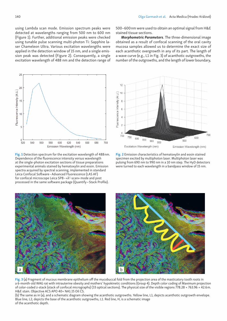
140 Olga Garmash et al. Acta Medica (Hradec Králové)
using Lambda scan mode. Emission spectrum peaks were detected at wavelengths ranging from 500 nm to 600 nm (Figure 1). Further, additional emission peaks were checked using tunable pulse scanning multi-photon Ti: Sapphire la-ser Chameleon Ultra. Various excitation wavelengths were applied in the detection window of 15 nm, and a single emis-sion peak was detected (Figure 2). Consequently, a single excitation wavelength of 488 nm and the detection range of
500–600 nm were used to obtain an optimal signal from H&E stained tissue sections.
Morphometric Parameters. Th e three-dimensional image obtained as a result of confocal scanning of the oral cavity mucosa samples allowed us to determine the exact size of each acanthotic overgrowth in any of its part. Th e length of a wave curve (e.g., L1 in Fig. 3) of acanthotic outgrowths, the number of the outgrowths, and the length of lower boundary,
Fig. 1 Detection spectrum for the excitation wavelength of 488 nm. Dependence of the fl uorescence intensity versus wavelength at the single-photon excitation sections of tissue preparations experimental animals stained by hematoxylin and eosin. Emission spectra acquired by spectral scanning, implemented in standard Leica Confocal Soft ware – Advanced Fluorescence (LAS AF) for confocal microscope Leica SP8 – «λ2-scan» mode and post processed in the same soft ware package (Quantify – Stack Profi le).
Fig. 2 Emission characteristics of hematoxylin and eosin stained specimen excited by multiphoton laser. Multiphoton laser was pulsing from 690 nm to 990 nm in a 10 nm step. Th e HyD detectors were turned to each wavelength in a bandpass window of 15 nm.
Fig. 3 (a) Fragment of mucous membrane epithelium off the mucobuccal fold from the projection area of the masticatory tooth roots in a 6-month-old WAG rat with intrauterine obesity and mothers’ hypokinetic conditions (Group 4). Depth color coding of Maximum projection of color-coded z-stack (stack of confocal micrographs) (33 optical sections). Th e physical size of the visible regions 778.28 × 763.96 × 42.6 m. H&E stain. Objective ACS APO 40× NA1.15 Oil CS. (b) Th e same as in (a), and a schematic diagram showing the acanthotic outgrowths. Yellow line, L1, depicts acanthotic outgrowth envelope. Blue line, L2, depicts the base of the acanthotic outgrowths, L1. Red line, H, is a schematic image of the acanthotic depth.
a b
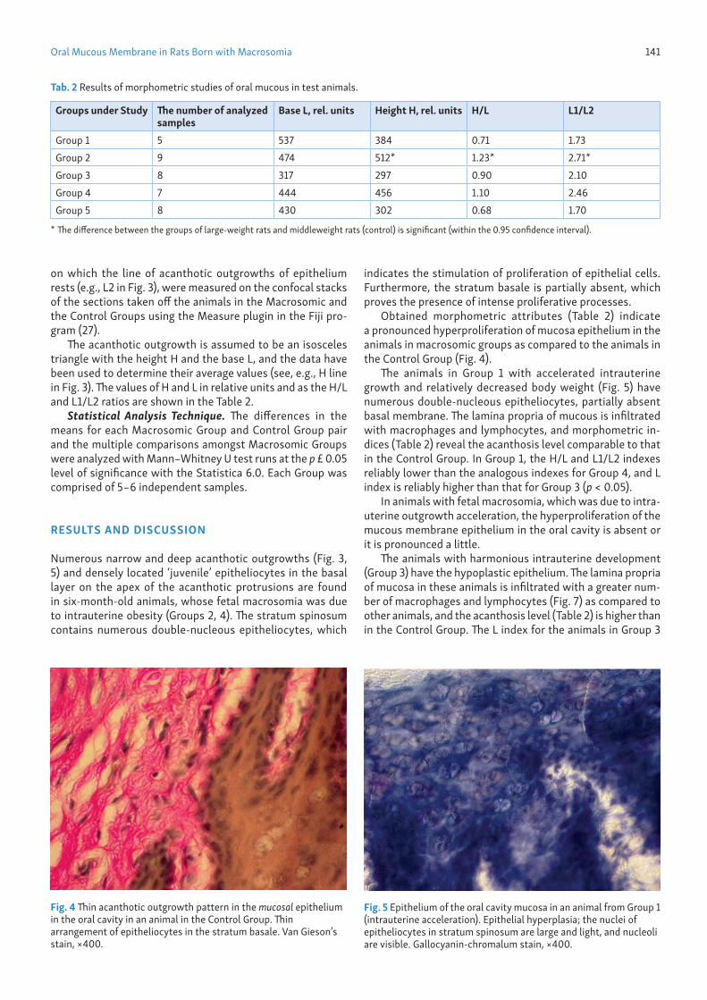
Oral Mucous Membrane in Rats Born with Macrosomia 141
on which the line of acanthotic outgrowths of epithelium rests (e.g., L2 in Fig. 3), were measured on the confocal stacks of the sections taken off the animals in the Macrosomic and the Control Groups using the Measure plugin in the Fiji pro-gram (27).
The acanthotic outgrowth is assumed to be an isosceles triangle with the height H and the base L, and the data have been used to determine their average values (see, e.g., H line in Fig. 3). The values of H and L in relative units and as the H/L and L1/L2 ratios are shown in the Table 2.
Statistical Analysis Technique. The differences in the means for each Macrosomic Group and Control Group pair and the multiple comparisons amongst Macrosomic Groups were analyzed with Mann–Whitney U test runs at the p £ 0.05 level of significance with the Statistica 6.0. Each Group was comprised of 5–6 independent samples.
RESULTS AND DISCUSSION
Numerous narrow and deep acanthotic outgrowths (Fig. 3, 5) and densely located ‘juvenile’ epitheliocytes in the basal layer on the apex of the acanthotic protrusions are found in six-month-old animals, whose fetal macrosomia was due to intrauterine obesity (Groups 2, 4). The stratum spinosum contains numerous double-nucleous epitheliocytes, which
indicates the stimulation of proliferation of epithelial cells. Furthermore, the stratum basale is partially absent, which proves the presence of intense proliferative processes.
Obtained morphometric attributes (Table 2) indicate a pronounced hyperproliferation of mucosa epithelium in the animals in macrosomic groups as compared to the animals in the Control Group (Fig. 4).
The animals in Group 1 with accelerated intrauterine growth and relatively decreased body weight (Fig. 5) have numerous double-nucleous epitheliocytes, partially absent basal membrane. The lamina propria of mucous is infiltrated with macrophages and lymphocytes, and morphometric in-dices (Table 2) reveal the acanthosis level comparable to that in the Control Group. In Group 1, the H/L and L1/L2 indexes reliably lower than the analogous indexes for Group 4, and L index is reliably higher than that for Group 3 (p < 0.05).
In animals with fetal macrosomia, which was due to intra-uterine outgrowth acceleration, the hyperproliferation of the mucous membrane epithelium in the oral cavity is absent or it is pronounced a little.
The animals with harmonious intrauterine development (Group 3) have the hypoplastic epithelium. The lamina propria of mucosa in these animals is infiltrated with a greater num-ber of macrophages and lymphocytes (Fig. 7) as compared to other animals, and the acanthosis level (Table 2) is higher than in the Control Group. The L index for the animals in Group 3
Tab. 2 Results of morphometric studies of oral mucous in test animals.
Groups under Study The number of analyzed samples
Base L, rel. units Height H, rel. units H/L L1/L2
Group 1 5 537 384 0.71 1.73Group 2 9 474 512* 1.23* 2.71*Group 3 8 317 297 0.90 2.10Group 4 7 444 456 1.10 2.46Group 5 8 430 302 0.68 1.70
* The difference between the groups of large-weight rats and middleweight rats (control) is significant (within the 0.95 confidence interval).
Fig. 4 Thin acanthotic outgrowth pattern in the mucosal epithelium in the oral cavity in an animal in the Control Group. Thin arrangement of epitheliocytes in the stratum basale. Van Gieson’s stain, ×400.
Fig. 5 Epithelium of the oral cavity mucosa in an animal from Group 1(intrauterine acceleration). Epithelial hyperplasia; the nuclei of epitheliocytes in stratum spinosum are large and light, and nucleoli are visible. Gallocyanin-chromalum stain, ×400.
142 Olga Garmash et al. Acta Medica (Hradec Králové)
is reliably lower than those for Group 1 and Group 2, and the H index is reliably lower than that for Group 2 (p < 0.05).
The intensity of epithelial proliferation equal to that in the Control Group is found in 6-month-old animals with a harmo-nious intrauterine gain in the both body weight and height. These animals also have symptoms of acanthosis, although they are far less expressed against the background of epithe-lial layer thinning, which can be explained by the accelerated aging of epitheliocytes.
At the present time, the diseases of oral cavity mucous are very common. Many factors contribute to the development of various lesions of the oral cavity mucosa. We can assume that fetal macrocosmia with obesity causes an instability in the epithelium of the oral cavity mucosa, its rapid death and therefore more active stimulation of proliferation. The high intensity of epitheliocyte proliferation in macrosomic animals can cause increased predisposition to the development of dif-ferent violations in oral cavity mucosa.
For instance, the susceptibility to epithelium hyperprolif-eration in oral cavity mucosa that we have revealed may be one of the causes of an increased predisposition to the de-velopment of acute herpetic stomatitis in children born with macrosomia (11).
We expect that the reproduction of this experiment on other linear animals will produce comparable results assum-ing that the maintenance of the standard conditions holds in repeated experiments, because the increased nutrition of a pregnant female promotes the weight gain of offspring in all animals. The scientific repeatability of this experiment is realized by the maintenance of the conditions for the each experiment and by the selection of macrosomic animals at birth, which have appeared to have a definite somatotype in our experiment.
CONCLUSION
The large body weight at birth could be an integral indica-tor of violations of the morphogenesis of oral cavity muco-sa. In addition, the prevalence of intrauterine obesity or an
accelerated body mass gain or the harmonious acceleration of body mass and height, have their own specific features in the microscopic picture of mucogingival fold of oral cavity mucosa epithelium in the 6-month-old experimental animals.
ACKNOWLEDGEMENTS
This work was supported by the BioImaging Facility, Institute of Physiology, supported by the Czech-BioImaging large RI project (LM2015062 funded by MEYS CR) and by the pro-ject Modernization and support of research activities of the national infrastructure for biological and medical imaging Czech-BioImaging (CZ.02.1.01/0.0/0.0/16_013/0001775 funded by OP RDE).
The authors wish to thank Dr. Lucie Kubínová and Dr. Jiří Janáček for their helpful advice and comments and Dr. Daniel Hadraba for his advice in various technical issues.
REFERENCES 1. Barker DF. The developmental origins of adult disease. J Am Coll Nutr
2004; 23 Suppl 6: 588S–595S. 2. Gu S, An X, Fang L, et al. Risk factors and long-term health consequences
of macrosomia: a prospective study in Jiangsu Province, China. J Biomed Res 2012 Jul; 26(4): 235–40.
3. Winter JD, Taylor Y, Mowrer L, Wintera KM. Dulinbc MF. BMI at birth and overweight at age four. Obes Res Clin Pract 2016; 11(2): 151–7.
4. Donma MM. Macrosomia, top of the iceberg: the charm of underlying factors. Pediatr Int 2011; 53(1): 78–84.
5. Boney CM, Verma A, Tucker R, Vohr BR. Metabolic syndrome in child-hood: association with birth weight, maternal obesity, and gestational diabetes mellitus. Pediatrics 2005; 115: e290–e296.
6. Godfrey KM, Inskip HM, Hanson, MA. The long-term effects of prenatal development on growth and metabolism. Semin Reprod Med. 2011 May; 29(3): 257–65.
7. Silva Idos S, De Stavola B, McCormack V. Birth size and breast cancer risk: re-analysis of individual participant data from 32 studies. PLoS Med 2008; 5: e193.
8. Hermann GM, Dallas LM, Haskell SE, Roghair RD. Neonatal macrosomia is an independent risk factor for adult metabolic syndrome. Neonatology 2010; 98(3): 238–44.
9. Huraseva, AB. Reproduktivnoe zdorove zhenschin, rodivshihsya s pol-yarnyimi znacheniyami massyi tela [Reproductive health of women born with polar body weight]. Mizhnarodnii endokrynolohichnii zhurnal 2010; 8: 106–15.
Fig. 7 Thick macrophage-lymphocytic infiltration of lamina propria in mucosa in an animal with fetal macrosomia with harmonious intrauterine development (Group 3). H&E stain, ×400.
Fig. 6 Thin and long acanthotic outgrowth pattern in the mucosal epithelium in the oral cavity in an animal with intrauterine obesity (Group 2). Van Gieson’s stain, ×100.
143
10. Grischenko VI. Krupnyiy plod (kliniko-morfologicheskoe issledovanie). Kiev: Zdorovya, 1991: 183.
11. Yakovtsova AF, Sorokina IV, Aleshchenko IE Immune system of the hu-man fetus in large and IUGR fetuses [in Russian]. Kharkov: BSF “Antiqua”, 2004: 218.
12. Sorokina, AV. Krupnyj plod: mify i real’nost’ (A large fetus: myths and realities). Rossiiskij vestnik akushera-ginekologa 2013; 13(4): 86–8. [in Russian].
13. Smolyar NI, Dubetska-Hrabous IS. Caries Risk Factors in Period of Initi-ation and Mineralization of Milk Teeth. [in Ukrainian] Profilaktychna ta Dytiacha Stomatologia. 2009; 1: 12–7.
14. Yokomichi H, Tanaka T, Suzuki K, Akiyama T, Okinawa Child Health Study Group, Yamagata Z. Macrosomic neonates carry increased risk of dental caries in early childhood: findings from a cohort study, the Okinawa child health study. Japan. PLoS One 2015; 10(7): e0133872.
15. Julihn A, Molund U, Drevsäter E, Modéer T. High birth weight is a risk factor of dental caries increment during adolescence in Sweden. Dent J 2014; 2(3): 118–33.
16. Ryabokon E, Garmash O, Nazarenko L, Babajanyan E. Dentists view on fetal macrosomia. Srodowisko a stan zdorowia jamy ustnej X Konfer-encja Naukowo-Szkoleniowa, Naleczow, Polska, 27.04.2016. Naleczow, 2016: 70.
17. Garmash O. An eruption pattern of deciduous teeth in children born with fetal macrosomia during the first year of life. Georgian Medical News 2017; 3(263): 14–23.
18. Garmash O, Ryabokon E. The Effect of Fetal Macrosomia on the Neo-nate and Infant Dental Health. International Journal of Clinical Dentistry 2017; 10(3): 200–10.
19. Analisis of oral health in newborns with macrosomia in Kharkiv city. Lik Sprava. 2017; 3–4: 122–6.
20. Kubínová L, Janáček J. Confocal stereology: an efficient tool for meas-urement of microscopic structures. Cell Tissue Res 2015; 360(1): 13–28.
21. Kubínová L, Mao XW, Janáček J. Blood capillary length estimation from three-dimensional microscopic data by image analysis and stereology. Microsc Microanal 2013; 19(4): 898–906.
22. Janáček J, Čapek M, Michálek J, Karen P, Kubínová L. 3D microscopic im-aging and evaluation of tubular tissue architecture. Physiol Res 2014; 63(1): S49–S55.
23. Eržen I, Janáček J, Kubínová L. Characterization of the capillary network in skeletal muscles from 3D data. Physiol Res 2011; 60(1): 1–13.
24. Capek M, Brůza P, Janácek J, Karen P, Kubínová L, Vagnerová R. Volume reconstruction of large tissue specimens from serial physical sections using confocal microscopy and correction of cutting deformations by elastic registration. Microsc Res Tech 2009; 72(2): 110–9.
25. Gubina-Vakulik GI. Patologichna anatomiia endokrynnyh zaloz plodu pry adaptatsii vagitnoi do dii okremyh seredovyshchnyh faktoriv (Pathologi-cal anatomy of the fetal endocrine glands in pregnant woman adaptation to the some environmental factors action). (Dissertation) [In Ukrainian] Ukraine, Kharkiv National Medical University1994.
26. Garmash OV, Ryabokon EN. Gubina-Vakulyk GI. Method of modeling fe-tal macrosomia in experiment [In Ukrainian]. Patent for utility model. No. 123084 IPC. (Ukraine). Published 02.22.2018.
27. Schindelin J, Arganda-Carreras I, Frise E, et al. Fiji: an open-source platform for biological-image analysis. Nature Methods 2012; 9(7): 676–82.
144 original article
Topiramate Reduces Aortic Cross-Clamping-Induced Lung Injury in Male Rats
Aysel Kurt1, Yildiray Kalkan2, Hasan Turut3, Medine Cumhur Cure4, Levent Tumkaya5, Erkan Cure6,*
A B S T R AC TBackground: Topiramate (TPM) decreases cytokine release and generation of reactive oxygen species (ROS). Cytokine and endothelin-1 (ET-1) secretion and ROS formation play an important role in ischemia-reperfusion (I/R) injury. We aimed to evaluate whether TPM prevents damage occurring in lung tissue during I/R.Materials and Methods: A total of 27 Wistar albino rats were divided into three groups of nine. To the I/R group, two hours of ischemia via infrarenal abdominal aorta cross-ligation and then two hours of reperfusion process were applied. TPM (100 mg/kg/day) orally for seven days was administered in the TPM treatment group. After the last dose of TPM treatment, respectively, two hours of ischemia and two hours of reperfusion were applied in this group.Results: Tumor necrosis factor-alpha (TNF-α) (p < 0.05), malondialdehyde (MDA) (p < 0.05), myeloperoxidase (MPO) (p < 0.05) and ET-1 (p < 0.05) levels of TPM treatment group’s lung tissue were significantly lower than for the I/R group. Caspase-3 and histopathological damage were rather lower than that of the I/R group. Conclusions: During I/R, lung damage occurs due to excessive TNF-α and ET-1 release and ROS generation. TPM could well reduce development of lung damage by decreasing cytokine and ET-1 release and levels of ROS produced.
K E Y WO R D STopiramate; ischemia-reperfusion; lung injury; tumor necrosis factor-alpha; endothelin-1
A U T H O R A F F I L I AT I O N S1 Private Practice, Department of Thoracic Surgery, Rize, Turkey2 Private Practice, Department of Histology and Embryology, Rize, Turkey3 Recep Tayyip Erdogan University, School of Medicine, Department of Thoracic Surgery, Rize, Turkey 4 Istanbul Laboratory, Department of Biochemistry, Istanbul, Turkey5 Recep Tayyip Erdogan University, School of Medicine, Department of Histology and Embryology, Rize, Turkey 6 Camlica Erdem Hospital, Department of Internal Medicine, Istanbul, Turkey* Corresponding author: Camlica Erdem Hospital, Department of Internal Medicine, Uskudar, Istanbul, Turkey; e-mail: [email protected]
Received: 10 February 2018Accepted: 2 November 2018Published online: 22 January 2019
Acta Medica (Hradec Králové) 2018; 61(4): 144–149https://doi.org/10.14712/18059694.2018.133© 2018 The Authors. This is an open-access article distributed under the terms of the Creative Commons Attribution License (http://creativecommons.org/licenses/by/4.0), which permits unrestricted use, distribution, and reproduction in any medium, provided the original author and source are credited.
TPM Protect Lung Injury 145
INTRODUCTION
Pulmonary dysfunction is one of the problematic conditions developing after ischemic reperfusion (I/R) on account of lung transplantation (1), extracorporeal circulation operations (2, 3) after resuscitation for circulatory arrest (4) or severe hem-orrhagic shock (5). Reperfusion of ischemic tissue causes mi-crovascular dysfunction and parenchymal cells, depending on local and systemic responses. Lung damage developing during the I/R process has been shown to be associated with physiological, biochemical and histopathological changes. Lung I/R injury is described by neutrophil extravasation, in-terstitial edema, disruption of epithelial integrity, and leakage of protein into the alveolar space, all of which are associated with severe alterations in gas exchange (6, 7). In its early stag-es, activated alveolar macrophages and pro-inflammatory cy-tokines such as tumor necrosis factor-alpha (TNF-α) increase, and reactive oxygen radicals (ROS) and later, in last stages of perfusion, activated neutrophils play crucial roles in this uncontrolled inflammation process (8, 9).
In showing the lung damage of the I/R process, malondi-aldehyde (MDA), a marker for lipid peroxidation and oxidative stress, and myeloperoxidase (MPO), a marker for activation of neutrophils located on the lung tissue, are both valuable and reliable parameters (10). Besides, endothelin-1 (ET-1), released from damaged endothelial cells, is another effective mediator causing vascular inflammation, cell proliferation, and fibrosis. Additionally, this ET-1 plays a crucial role in the development of acute lung injury (11).
Topiramate (TPM) is an agent used for epilepsy and mi-graine treatment. It inhibits voltage-gated sodium and calci-um channels, blocks glutamate a-amino-3-hydroxy-5-methyl- 4-isoxazole propionic acid (AMPA)/kainate receptors and enhances gamma-aminobutyric acid (GABA), a receptor-me-diated chloride (12). TPM has also been shown to have anti-oxidant effects, reducing TNF-α release and oxidative stress (13). In addition, decreases in the level of MDA enzyme has been reported in other studies (14). Another positive effect of TPM is diminished insulin resistance (15).
In this study, we aimed to evaluate the effects of TPM on ET-1, TNF-α, MDA and MPO enzyme activity which play cru-cial roles in producing ROS, to assess apoptosis via measuring caspase-3 enzyme activity and the effect of TPM on it, and to determine whether TPM has a preventive effect on I/R lung damage.
MATERIALS AND METHODS
Animals used in the experimental study were 12–15-week-old experimental design male Wistar albino rats (n = 27) weigh-ing 250–300 g. The study design was approved by license no: 2014/9 by the local ethical committee at the University and all protocols conformed to the Guide for the Care and Use of Laboratory Animals (NIH, 1985). Rats were kept under stand-ard temperature and environment with a 12-h light–dark cy-cle and were given food and water ad libitum. The rats were randomly divided into three groups as follows.
Group I: Control groups (n = 9), only received saline solu-tion intraperitoneally. They underwent a midline laparotomy, with the infrarenal abdominal aorta (IAA) being dissected and
crossed without obstruction. Group II: I/R groups (n = 9), af-ter the midline laparotomy of rats, the IAA was clamped for 120 mins, followed by 120 mins of reperfusion. Group III: For I/R+TPM groups (n = 9), TPM (100 mg/kg/day, Topamax®, Johnson and Johnson) was orally (via gastric gavage) given for seven sequential days prior to I/R. After seven days’ admin-istered TPM, this group underwent respectively 120 mins of ischemia and 120 mins of reperfusion.
In humans, oral dose of TPM has been found to be effective within 2 hours in plasma and at target dose within 4 weeks (16). We chose 100 mg/kg/day of TPM and for 1 week since the previous animal studies used this dose and duration (17–19).
PROCEDURE OF AORTIC ISCHEMIA-REPERFUSIONThe rats were anesthetized by intraperitoneal injection of 50 mg/kg ketamine hydrochloride (Ketalar; Eczacibasi, Is-tanbul, Turkey) and were maintained by intramuscular in-jections of 25 mg/kg ketamine hydrochloride when needed for all groups. The surgery was performed with rats placed in the supine position under a heating lamp. After a median laparotomy under sterile conditions, the IAA was explored by gentle dissection of the retroperitoneum, and then an atraumatic microvascular clamp was placed across the IAA for 120 mins of ischemia. After the abdomen incision was closed, the wound was covered with plastic wrap to prevent the loss of heat and fluid. Ten milliliters of normal saline was dribbled into the peritoneal cavity to maintain fluid balance. The traumatic microvascular clamp was removed on the IAA and lower extremities were reperfused for 120 mins. Aortic occlusion and reperfusion were verified by observing the loss and reappearance of the pulsation on the distal aorta. The I/R model was designed according to a method similar to previous studies (20). After ending the procedure, the rats were sacrificed under anesthesia and then lung tissues were obtained for biochemical and histological analyses.
TISSUE HOMOGENATESThe lung tissue samples were homogenized in Phosphate Buffer Saline (PBS) (50 mM, pH 7.4) and centrifuged at 10,000 g for 20 mins. The supernatant was removed to be aliquoted into tubes. It was kept frozen at −80 °C and bio-chemical parameters such as TNF-α, MDA, MPO, ET-1 were studied within a month.
MEASUREMENT OF PROTEINTissue homogenate protein assay is a Lowry protocol based on the Biuret reaction. The peptide bonds of proteins react with copper under alkaline conditions to produce Cu+, which reacts with the Folin reagent, and the Folin-Ciocalteau reac-tion (21).
TISSUE TNF-αThe TNF-α levels were measured using an enzyme-linked im-munosorbent assay (ELISA) method. We used a commercially available rat TNF-α ELISA kit (eBioscience, Vienna, Austria). The method was according to the manufacturer’s instruc-tions. Absorbance was measured at a wavelength of 450 ηm
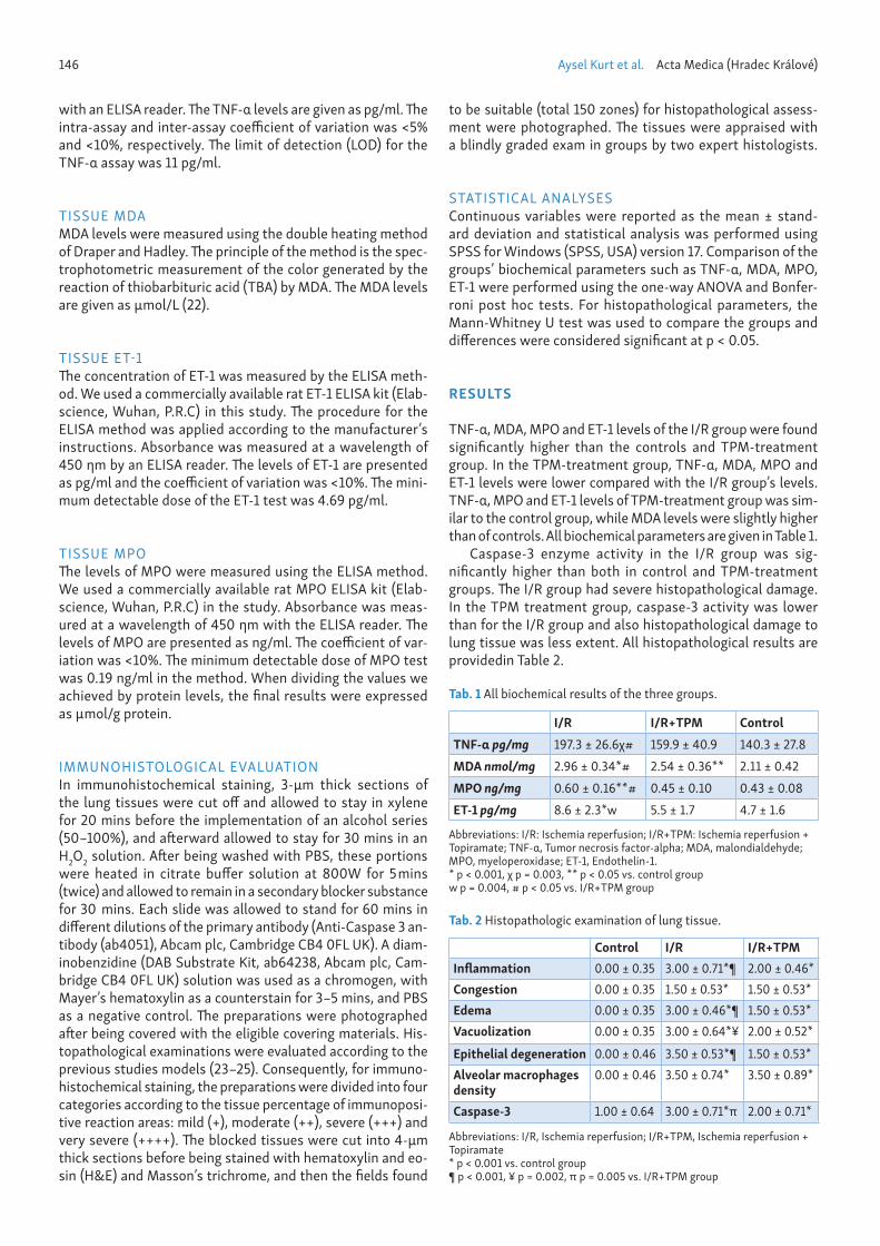
146 Aysel Kurt et al. Acta Medica (Hradec Králové)
with an ELISA reader. The TNF-α levels are given as pg/ml. The intra-assay and inter-assay coefficient of variation was <5% and <10%, respectively. The limit of detection (LOD) for the TNF-α assay was 11 pg/ml.
TISSUE MDAMDA levels were measured using the double heating method of Draper and Hadley. The principle of the method is the spec-trophotometric measurement of the color generated by the reaction of thiobarbituric acid (TBA) by MDA. The MDA levels are given as µmol/L (22).
TISSUE ET-1The concentration of ET-1 was measured by the ELISA meth-od. We used a commercially available rat ET-1 ELISA kit (Elab-science, Wuhan, P.R.C) in this study. The procedure for the ELISA method was applied according to the manufacturer’s instructions. Absorbance was measured at a wavelength of 450 ηm by an ELISA reader. The levels of ET-1 are presented as pg/ml and the coefficient of variation was <10%. The mini-mum detectable dose of the ET-1 test was 4.69 pg/ml.
TISSUE MPOThe levels of MPO were measured using the ELISA method. We used a commercially available rat MPO ELISA kit (Elab-science, Wuhan, P.R.C) in the study. Absorbance was meas-ured at a wavelength of 450 ηm with the ELISA reader. The levels of MPO are presented as ng/ml. The coefficient of var-iation was <10%. The minimum detectable dose of MPO test was 0.19 ng/ml in the method. When dividing the values we achieved by protein levels, the final results were expressed as µmol/g protein.
IMMUNOHISTOLOGICAL EVALUATIONIn immunohistochemical staining, 3-μm thick sections of the lung tissues were cut off and allowed to stay in xylene for 20 mins before the implementation of an alcohol series (50–100%), and afterward allowed to stay for 30 mins in an H2O2 solution. After being washed with PBS, these portions were heated in citrate buffer solution at 800W for 5 mins (twice) and allowed to remain in a secondary blocker substance for 30 mins. Each slide was allowed to stand for 60 mins in different dilutions of the primary antibody (Anti-Caspase 3 an-tibody (ab4051), Abcam plc, Cambridge CB4 0FL UK). A diam-inobenzidine (DAB Substrate Kit, ab64238, Abcam plc, Cam-bridge CB4 0FL UK) solution was used as a chromogen, with Mayer’s hematoxylin as a counterstain for 3–5 mins, and PBS as a negative control. The preparations were photographed after being covered with the eligible covering materials. His-topathological examinations were evaluated according to the previous studies models (23–25). Consequently, for immuno-histochemical staining, the preparations were divided into four categories according to the tissue percentage of immunoposi-tive reaction areas: mild (+), moderate (++), severe (+++) and very severe (++++). The blocked tissues were cut into 4-μm thick sections before being stained with hematoxylin and eo-sin (H&E) and Masson’s trichrome, and then the fields found
to be suitable (total 150 zones) for histopathological assess-ment were photographed. The tissues were appraised with a blindly graded exam in groups by two expert histologists.
STATISTICAL ANALYSESContinuous variables were reported as the mean ± stand-ard deviation and statistical analysis was performed using SPSS for Windows (SPSS, USA) version 17. Comparison of the groups’ biochemical parameters such as TNF-α, MDA, MPO, ET-1 were performed using the one-way ANOVA and Bonfer-roni post hoc tests. For histopathological parameters, the Mann-Whitney U test was used to compare the groups and differences were considered significant at p < 0.05.
RESULTS
TNF-α, MDA, MPO and ET-1 levels of the I/R group were found significantly higher than the controls and TPM-treatment group. In the TPM-treatment group, TNF-α, MDA, MPO and ET-1 levels were lower compared with the I/R group’s levels. TNF-α, MPO and ET-1 levels of TPM-treatment group was sim-ilar to the control group, while MDA levels were slightly higher than of controls. All biochemical parameters are given in Table 1.
Caspase-3 enzyme activity in the I/R group was sig-nificantly higher than both in control and TPM-treatment groups. The I/R group had severe histopathological damage. In the TPM treatment group, caspase-3 activity was lower than for the I/R group and also histopathological damage to lung tissue was less extent. All histopathological results are providedin Table 2.
Tab. 1 All biochemical results of the three groups.
I/R I/R+TPM ControlTNF-α pg/mg 197.3 ± 26.6χ# 159.9 ± 40.9 140.3 ± 27.8MDA nmol/mg 2.96 ± 0.34*# 2.54 ± 0.36** 2.11 ± 0.42MPO ng/mg 0.60 ± 0.16**# 0.45 ± 0.10 0.43 ± 0.08ET-1 pg/mg 8.6 ± 2.3*w 5.5 ± 1.7 4.7 ± 1.6
Abbreviations: I/R: Ischemia reperfusion; I/R+TPM: Ischemia reperfusion + Topiramate; TNF-α, Tumor necrosis factor-alpha; MDA, malondialdehyde; MPO, myeloperoxidase; ET-1, Endothelin-1.* p < 0.001, χ p = 0.003, ** p < 0.05 vs. control groupw p = 0.004, # p < 0.05 vs. I/R+TPM group
Tab. 2 Histopathologic examination of lung tissue.
Control I/R I/R+TPMInflammation 0.00 ± 0.35 3.00 ± 0.71*¶ 2.00 ± 0.46*Congestion 0.00 ± 0.35 1.50 ± 0.53* 1.50 ± 0.53*Edema 0.00 ± 0.35 3.00 ± 0.46*¶ 1.50 ± 0.53*Vacuolization 0.00 ± 0.35 3.00 ± 0.64*¥ 2.00 ± 0.52*
Epithelial degeneration 0.00 ± 0.46 3.50 ± 0.53*¶ 1.50 ± 0.53*Alveolar macrophagesdensity
0.00 ± 0.46 3.50 ± 0.74* 3.50 ± 0.89*
Caspase-3 1.00 ± 0.64 3.00 ± 0.71*π 2.00 ± 0.71*
Abbreviations: I/R, Ischemia reperfusion; I/R+TPM, Ischemia reperfusion + Topiramate* p < 0.001 vs. control group¶ p < 0.001, ¥ p = 0.002, π p = 0.005 vs. I/R+TPM group
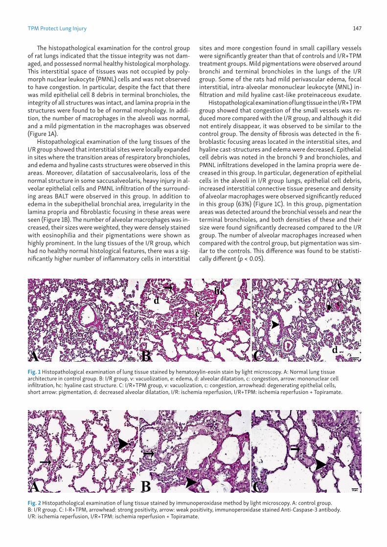
TPM Protect Lung Injury 147
The histopathological examination for the control group of rat lungs indicated that the tissue integrity was not dam-aged, and possessed normal healthy histological morphology. This interstitial space of tissues was not occupied by poly-morph nuclear leukocyte (PMNL) cells and was not observed to have congestion. In particular, despite the fact that there was mild epithelial cell 8 debris in terminal bronchioles, the integrity of all structures was intact, and lamina propria in the structures were found to be of normal morphology. In addi-tion, the number of macrophages in the alveoli was normal, and a mild pigmentation in the macrophages was observed (Figure 1A).
Histopathological examination of the lung tissues of the I/R group showed that interstitial sites were locally expanded in sites where the transition areas of respiratory bronchioles, and edema and hyaline casts structures were observed in this areas. Moreover, dilatation of saccusalveolaris, loss of the normal structure in some saccusalveolaris, heavy injury in al-veolar epithelial cells and PMNL infiltration of the surround-ing areas BALT were observed in this group. In addition to edema in the subepithelial bronchial area, irregularity in the lamina propria and fibroblastic focusing in these areas were seen (Figure 1B). The number of alveolar macrophages was in-creased, their sizes were weighted, they were densely stained with eosinophilia and their pigmentations were shown as highly prominent. In the lung tissues of the I/R group, which had no healthy normal histological features, there was a sig-nificantly higher number of inflammatory cells in interstitial
sites and more congestion found in small capillary vessels were significantly greater than that of controls and I/R+TPM treatment groups. Mild pigmentations were observed around bronchi and terminal bronchioles in the lungs of the I/R group. Some of the rats had mild perivascular edema, focal interstitial, intra-alveolar mononuclear leukocyte (MNL) in-filtration and mild hyaline cast-like proteinaceous exudate.
Histopathological examination of lung tissue in the I/R+TPM group showed that congestion of the small vessels was re-duced more compared with the I/R group, and although it did not entirely disappear, it was observed to be similar to the control group. The density of fibrosis was detected in the fi-broblastic focusing areas located in the interstitial sites, and hyaline cast-structures and edema were decreased. Epithelial cell debris was noted in the bronchi 9 and bronchioles, and PMNL infiltrations developed in the lamina propria were de-creased in this group. In particular, degeneration of epithelial cells in the alveoli in I/R group lungs, epithelial cell debris, increased interstitial connective tissue presence and density of alveolar macrophages were observed significantly reduced in this group (63%) (Figure 1C). In this group, pigmentation areas was detected around the bronchial vessels and near the terminal bronchioles, and both densities of these and their size were found significantly decreased compared to the I/R group. The number of alveolar macrophages increased when compared with the control group, but pigmentation was sim-ilar to the controls. This difference was found to be statisti-cally different (p < 0.05).
Fig. 1 Histopathological examination of lung tissue stained by hematoxylin-eosin stain by light microscopy. A: Normal lung tissue architecture in control group. B: I/R group, v: vacuolization, e: edema, d: alveolar dilatation, c: congestion, arrow: mononuclear cell infiltration, hc: hyaline cast structure. C: I/R+TPM group, v: vacuolization, c: congestion, arrowhead: degenerating epithelial cells, short arrow: pigmentation, d: decreased alveolar dilatation, I/R: ischemia reperfusion, I/R+TPM: ischemia reperfusion + Topiramate.
Fig. 2 Histopathological examination of lung tissue stained by immunoperoxidase method by light microscopy. A: control group. B: I/R group. C: I-R+TPM, arrowhead: strong positivity, arrow: weak positivity, immunoperoxidase stained Anti-Caspase-3 antibody. I/R: ischemia reperfusion, I/R+TPM: ischemia reperfusion + Topiramate.
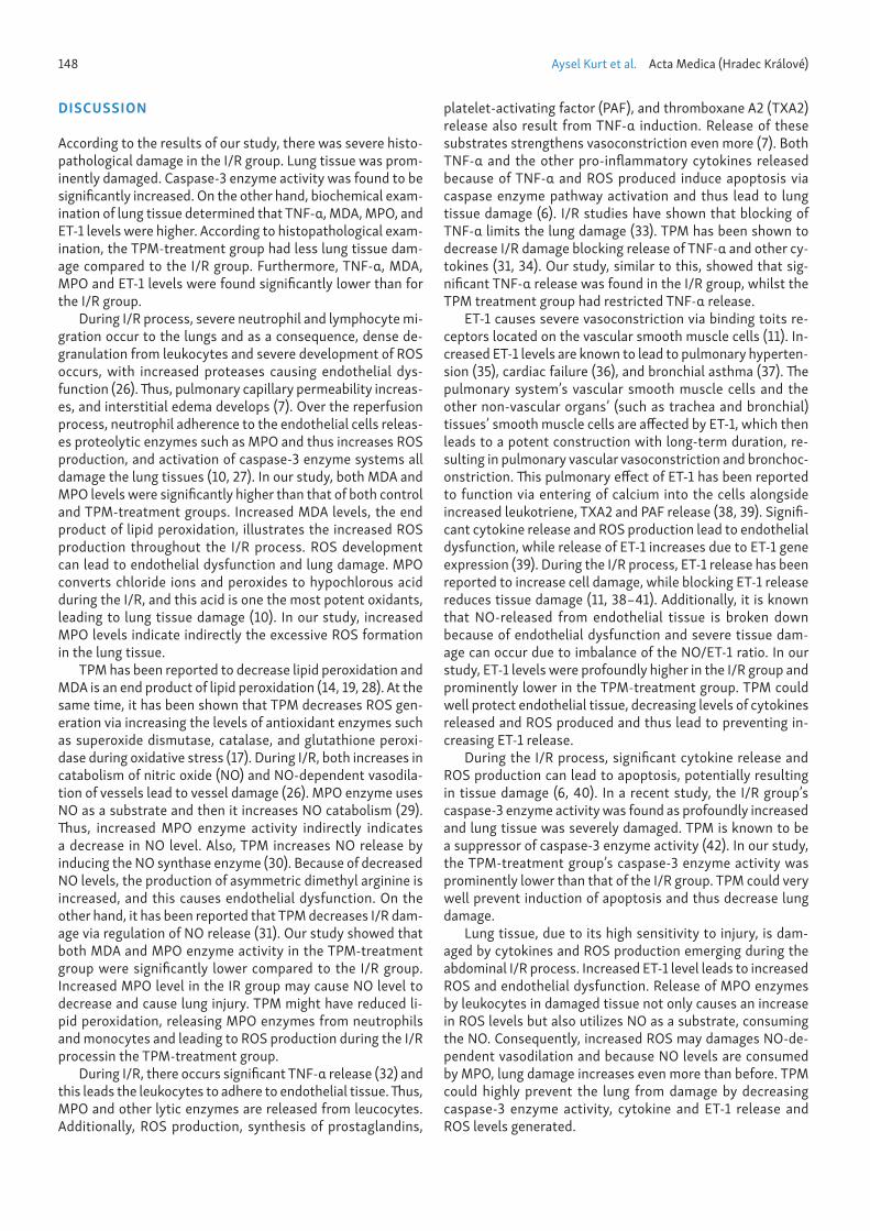
148 Aysel Kurt et al. Acta Medica (Hradec Králové)
DISCUSSION
According to the results of our study, there was severe histo-pathological damage in the I/R group. Lung tissue was prom-inently damaged. Caspase-3 enzyme activity was found to be significantly increased. On the other hand, biochemical exam-ination of lung tissue determined that TNF-α, MDA, MPO, and ET-1 levels were higher. According to histopathological exam-ination, the TPM-treatment group had less lung tissue dam-age compared to the I/R group. Furthermore, TNF-α, MDA, MPO and ET-1 levels were found significantly lower than for the I/R group.
During I/R process, severe neutrophil and lymphocyte mi-gration occur to the lungs and as a consequence, dense de-granulation from leukocytes and severe development of ROS occurs, with increased proteases causing endothelial dys-function (26). Thus, pulmonary capillary permeability increas-es, and interstitial edema develops (7). Over the reperfusion process, neutrophil adherence to the endothelial cells releas-es proteolytic enzymes such as MPO and thus increases ROS production, and activation of caspase-3 enzyme systems all damage the lung tissues (10, 27). In our study, both MDA and MPO levels were significantly higher than that of both control and TPM-treatment groups. Increased MDA levels, the end product of lipid peroxidation, illustrates the increased ROS production throughout the I/R process. ROS development can lead to endothelial dysfunction and lung damage. MPO converts chloride ions and peroxides to hypochlorous acid during the I/R, and this acid is one the most potent oxidants, leading to lung tissue damage (10). In our study, increased MPO levels indicate indirectly the excessive ROS formation in the lung tissue.
TPM has been reported to decrease lipid peroxidation and MDA is an end product of lipid peroxidation (14, 19, 28). At the same time, it has been shown that TPM decreases ROS gen-eration via increasing the levels of antioxidant enzymes such as superoxide dismutase, catalase, and glutathione peroxi-dase during oxidative stress (17). During I/R, both increases in catabolism of nitric oxide (NO) and NO-dependent vasodila-tion of vessels lead to vessel damage (26). MPO enzyme uses NO as a substrate and then it increases NO catabolism (29). Thus, increased MPO enzyme activity indirectly indicates a decrease in NO level. Also, TPM increases NO release by inducing the NO synthase enzyme (30). Because of decreased NO levels, the production of asymmetric dimethyl arginine is increased, and this causes endothelial dysfunction. On the other hand, it has been reported that TPM decreases I/R dam-age via regulation of NO release (31). Our study showed that both MDA and MPO enzyme activity in the TPM-treatment group were significantly lower compared to the I/R group. Increased MPO level in the IR group may cause NO level to decrease and cause lung injury. TPM might have reduced li-pid peroxidation, releasing MPO enzymes from neutrophils and monocytes and leading to ROS production during the I/R processin the TPM-treatment group.
During I/R, there occurs significant TNF-α release (32) and this leads the leukocytes to adhere to endothelial tissue. Thus, MPO and other lytic enzymes are released from leucocytes. Additionally, ROS production, synthesis of prostaglandins,
platelet-activating factor (PAF), and thromboxane A2 (TXA2) release also result from TNF-α induction. Release of these substrates strengthens vasoconstriction even more (7). Both TNF-α and the other pro-inflammatory cytokines released because of TNF-α and ROS produced induce apoptosis via caspase enzyme pathway activation and thus lead to lung tissue damage (6). I/R studies have shown that blocking of TNF-α limits the lung damage (33). TPM has been shown to decrease I/R damage blocking release of TNF-α and other cy-tokines (31, 34). Our study, similar to this, showed that sig-nificant TNF-α release was found in the I/R group, whilst the TPM treatment group had restricted TNF-α release.
ET-1 causes severe vasoconstriction via binding toits re-ceptors located on the vascular smooth muscle cells (11). In-creased ET-1 levels are known to lead to pulmonary hyperten-sion (35), cardiac failure (36), and bronchial asthma (37). The pulmonary system’s vascular smooth muscle cells and the other non-vascular organs’ (such as trachea and bronchial) tissues’ smooth muscle cells are affected by ET-1, which then leads to a potent construction with long-term duration, re-sulting in pulmonary vascular vasoconstriction and bronchoc-onstriction. This pulmonary effect of ET-1 has been reported to function via entering of calcium into the cells alongside increased leukotriene, TXA2 and PAF release (38, 39). Signifi-cant cytokine release and ROS production lead to endothelial dysfunction, while release of ET-1 increases due to ET-1 gene expression (39). During the I/R process, ET-1 release has been reported to increase cell damage, while blocking ET-1 release reduces tissue damage (11, 38–41). Additionally, it is known that NO-released from endothelial tissue is broken down because of endothelial dysfunction and severe tissue dam-age can occur due to imbalance of the NO/ET-1 ratio. In our study, ET-1 levels were profoundly higher in the I/R group and prominently lower in the TPM-treatment group. TPM could well protect endothelial tissue, decreasing levels of cytokines released and ROS produced and thus lead to preventing in-creasing ET-1 release.
During the I/R process, significant cytokine release and ROS production can lead to apoptosis, potentially resulting in tissue damage (6, 40). In a recent study, the I/R group’s caspase-3 enzyme activity was found as profoundly increased and lung tissue was severely damaged. TPM is known to be a suppressor of caspase-3 enzyme activity (42). In our study, the TPM-treatment group’s caspase-3 enzyme activity was prominently lower than that of the I/R group. TPM could very well prevent induction of apoptosis and thus decrease lung damage.
Lung tissue, due to its high sensitivity to injury, is dam-aged by cytokines and ROS production emerging during the abdominal I/R process. Increased ET-1 level leads to increased ROS and endothelial dysfunction. Release of MPO enzymes by leukocytes in damaged tissue not only causes an increase in ROS levels but also utilizes NO as a substrate, consuming the NO. Consequently, increased ROS may damages NO-de-pendent vasodilation and because NO levels are consumed by MPO, lung damage increases even more than before. TPM could highly prevent the lung from damage by decreasing caspase-3 enzyme activity, cytokine and ET-1 release and ROS levels generated.

TPM Protect Lung Injury 149
REFERENCES 1. Hatachi G, Tsuchiya T, Miyazaki T, et al. The poly (adenosine diphos-
phate-ribose) polymerase inhibitor PJ34 reduces pulmonary ischemia reperfusion injury in rats. Transplantation 2014; 98: 618–24.
2. Ng CS, Wan S, Yim AP, Arifi AA. Pulmonary dysfunction after cardiac sur-gery. Chest 2002; 121: 1269–77.
3. Cao QF, Qu MJ, Yang WQ, Wang DP, Zhang MH, Di SB. Ischemia postcon-ditioning preventing lung ischemia-reperfusion injury. Gene 2015; 554: 120–4.
4. Shimamoto A, Pohlman TH, Shomura S, Tarukawa T, Takao M, Shimpo H. Toll-like receptor 4 mediates lung ischemia-reperfusion injury. Ann Thorac Surg 2006; 82: 2017–23.
5. Reino DC, Pisarenko V, Palange D, et al. Trauma hemorrhagic shock-in-duced lung injury involves a gut-lymph-induced TLR4 pathway in mice. PLoS One 2011; 6: e14829.
6. Tang PS, Mura M, Seth R, Liu M. Acute lung injury and cell death: how many ways can cells die? Am J Physiol Lung Cell Mol Physiol 2008; 294: 632–41.
7. Ng CS, Wan S, Arifi AA, Yim AP. Inflammatory response to pulmonary ischemia reperfusion injury. Surg Today 2006; 36: 205–14.
8. Jiang L, Li L, Shen J, Qi Z, Guo L. Effect of dexmedetomidine on lung is-chemia-reperfusion injury. Mol Med Rep 2014; 9: 419–26.
9. Eppinger MJ, Deeb GM, Bolling SF, Ward PA. Mediators of ischemia-rep-erfusion injury of rat lung. Am J Pathol 1997; 150: 1773–84.
10. Türüt H, Kurutas EB, Bulbuloglu E, et al. Zinc aspartate alleviates lung in-jury induced by intestinal ischemia-reperfusion in rats. J Surg Res 2009; 151: 62–7.
11. Comellas AP, Briva A. Role of endothelin-1 in acute lung injury. Transl Res 2009; 153: 263–71.
12. White HS, Smith MD, Wilcox KS. Mechanisms of action of antiepileptic drugs. Int Rev Neurobiol 2007; 81: 85–110.
13. Nazıroğlu M, Yürekli VA. Effects of antiepileptic drugs on antioxidant and oxidant molecular pathways: focus on trace elements. Cell Mol Neurobi-ol 2013; 33: 589–99.
14. Sanchez-Villarejo MV, Lopez-Pedrajas R, Sanchez-Vallejo V, et al. Chronic cocaine effects in retinal metabolism and electrophysiology: treatment with topiramate. Curr Eye Res 2014; 39: 493–503.
15. Genc BO, Dogan EA, Dogan U, Genc E. Anthropometric indexes, insulin resistance, and serum leptin and lipid levels in women with cryptogen-ic epilepsy receiving topiramate treatment. J Clin Neurosci 2010; 17: 1256–9.
16. Minton GC, Miller AD, Bookstaver PB, Love BL. Topiramate: safety and efficacy of its use in the prevention and treatment of migraine. J Cent Nerv Syst Dis 2011; 3: 155–68.
17. Armagan A, Kutluhan S, Yilmaz M, et al. Topiramate and vitamin e mod-ulate antioxidant enzyme activities, nitric oxide and lipid peroxidation levels in pentylenetetrazol-induced nephrotoxicity in rats. Basic Clin Pharmacol Toxicol 2008; 103: 166–70.
18. Cure MC, Cure E, Tumkaya L, et al. Topiramate has protective effect on renal injury. Bratisl Lek Listy 2015; 116: 259–63.
19. Naziroğlu M, Kutluhan S, Yilmaz M. Selenium and topiramate modulates brain microsomal oxidative stress values, Ca2+-ATPase activity, and EEG records in pentylentetrazol-induced seizures in rats. J Membr Biol 2008; 225: 39–49.
20. Erer D, Dursun AD, Oktar GL, et al. The effects of iloprost on lung injury induced by skeletal muscle ischemia-reperfusion. Bratisl Lek Listy 2014; 115: 405–10.
21. Lowry Oh, Rosebrough Nj, Farr Al, Randall Rj. Protein measurement with the Folin phenol reagent. J Biol Chem 1951; 193: 265–75.
22. Draper HH, Hadley M. Malondialdehyde determination as index of lipid peroxidation. Methods Enzymol 1990; 186: 421–31.
23. Kurt A, Tumkaya L, Yuce S, et al. The protective effect of infliximab against carbon tetrachloride-induced acute lung injury. Iran J Basic Med Sci 2016; 19: 685–91.
24. Kurt A, Tumkaya L, Kalkan Y, et al. Is adalimumab protective in is-chemia-reperfusion injury in lung? Iran J Basic Med Sci 2015; 18: 1093–9.
25. Kurt A, Tumkaya L, Turut H, et al. Protective Effects of Infliximab on Lung Injury Induced by Methotrexate. Arch Bronconeumol 2015; 51: 551–7.
26. Pararajasingam R, Weight SC, Bell PR, Nicholson ML, Sayers RD. Pulmonary nitric oxide metabolism following infrarenal aortic cross-clamp-induced ischaemia-reperfusion injury. Eur J Vasc Endovasc Surg 2000; 19: 47–51.
27. Yip HK, Chang YC, Wallace CG, et al. Melatonin treatment improves ad-ipose-derived mesenchymal stem cell therapy for acute lung ischemi-areperfusion injury. J Pineal Res 2013; 54: 207–21.
28. Demirci S, Kutluhan S, Naziroğlu M, et al. Effects of selenium and topiramate on cytosolic Ca2+ influx and oxidative stress in neuronal PC12 cells. Neurochem Res 2013; 38: 90–7.
29. Baldus S, Heitzer T, Eiserich JP, et al. Myeloperoxidase enhances nitric oxide catabolism during myocardial ischemia and reperfusion. Free Radic Biol Med 2004; 37: 902–11.
30. Cardenas-Rodriguez N, Coballase-Urrutia E, Rivera-Espinosa L, et al. Modulation of antioxidant enzymatic activities by certain antiepileptic drugs (valproic acid, oxcarbazepine, and topiramate): evidence in humans and experimental models. Oxid Med Cell Longev 2013; 2013: 598493.
31. Cure E, Cure MC, Tumkaya L, et al. Topiramate ameliorates abdominal aorta cross-clamping induced liver injury in rats. Saudi J Gastroenterol 2014; 20: 297–303.
32. Cure E, Cumhur Cure M, Tumkaya L, et al. Adalimumab ameliorates ab-dominal aorta cross clamping which induced liver injury in rats. Biomed Res Int 2014; 2014: 907915.
33. Naidu BV, Woolley SM, Farivar AS, et al. Early tumor necrosis factor-al-pha release from the pulmonary macrophage in lung ischemia reperfu-sion injury. J Thorac Cardiovasc Surg 2004; 127: 1502–8.
34. Mao X, Ji C, Sun C, et al. Topiramate attenuates cerebral ischemia/reper-fusion injury in gerbils via activating GABAergic signaling and inhibiting astrogliosis. Neurochem Int 2012; 60: 39–46.
35. Chester AH, Yacoub MH. The role of endothelin-1 in pulmonary arterial hypertension. Glob Cardiol Sci Pract 2014; 2014: 62–78.
36. Gottlieb SS, Harris K, Todd J, et al. Prognostic significance of active and modified forms of endothelin 1 in patients with heart failure with re-duced ejection fraction. Clin Biochem 2015; 48: 292–6.
37. Iwata S, Ito S, Iwaki M, et al. Regulation of endothelin-1-induced inter-leukin-6 production by Ca2+ influx in human airway smooth muscle cells. Eur J Pharmacol 2009; 605: 15–22.
38. Kerem M, Bedirli A, Pasaoglu H, et al. Effect of adrenomedullin on hepatic damage in hepatic ischaemia/reperfusion injury in rats. Liver Int 2008; 28: 972–81.
39. Guo X, Cao W, Yao J, et al. Cardioprotective effects of tilianin in rat my-ocardial ischemia-reperfusion injury. Mol Med Rep 2015; 11: 2227–33.
40. Xing D, Zhang R, Li S, et al. Pivotal role of mast cell carboxypeptidase A in mediating protection against small intestinal ischemia-reperfusion inju-ry in rats after ischemic preconditioning. J Surg Res 2014; 192: 177–86.
41. Yang P, Yang N, Zhang X, Xu X. The significance and mechanism of propo-fol on treatment of ischemia reperfusion induced lung injury in rats. Cell Biochem Biophys 2014; 70: 1527–32.
42. Cheng XL, Li MK. Effect of topiramate on apoptosis-related protein ex-pression of hippocampus in model rats with Alzheimers disease. Eur Rev Med Pharmacol Sci 2014; 18: 761–8.
150 case report
Anaplastic Carcinoma Thyroid in a Young Child – an Extremely Rare Occurrence
Suresh Chandra Sharma1, Pirabu Sakthivel1,*, Sarath Raveendran1, Chirom Amit Singh1, Tripti Nakra2, Shipra Agarwal2
A B S T R AC TAnaplastic thyroid carcinoma (ATC), one of the most aggressive malignancies, is extremely rare in childhood. We present a case of 5-year-old child who presented with rapidly progressing thyroid swelling and stridor, for which she underwent emergency tracheostomy and biopsy. Histopathological features were suggestive of ATC and the patient died within two months after diagnosis. ATC, though very rare in childhood, should be kept in the differential diagnoses of rapidly enlarging neck masses in children. To the best of our knowledge, this is the youngest case of ATC reported in literature.
K E Y WO R D Sthyroid; pediatric thyroid cancer; anaplastic carcinoma
A U T H O R A F F I L I AT I O N S1 Department of Otorhinolaryngology & Head and Neck Surgery, All India Institute of Medical Sciences, New Delhi, India2 Department of Pathology, All India Institute of Medical Sciences, New Delhi, India* Corresponding author: Room no 4057, ENT office, Fourth floor, Department of ENT & Head and Neck surgery, All India Institute of Medical
Sciences, New Delhi – 110029, India; e- mail: [email protected]
Received: 26 August 2018Accepted: 12 November 2018Published online: 22 January 2019
Acta Medica (Hradec Králové) 2018; 61(4): 150–152https://doi.org/10.14712/18059694.2018.134© 2018 The Authors. This is an open-access article distributed under the terms of the Creative Commons Attribution License (http://creativecommons.org/licenses/by/4.0), which permits unrestricted use, distribution, and reproduction in any medium, provided the original author and source are credited.
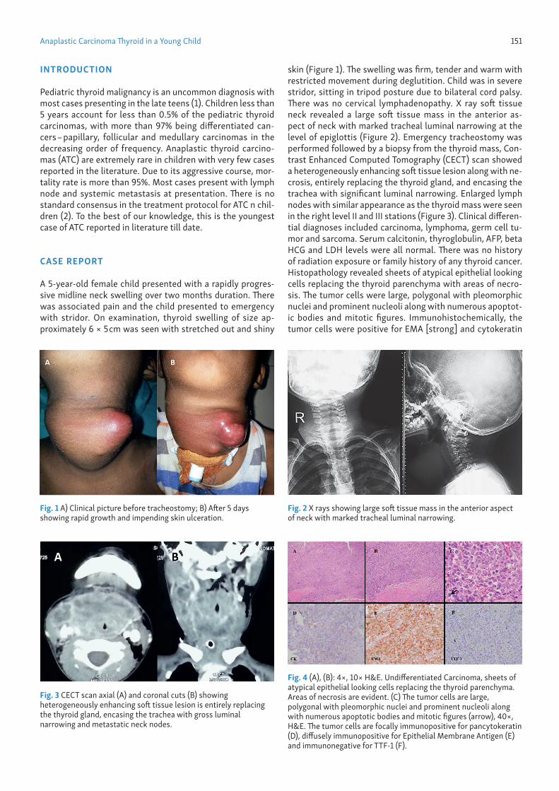
Anaplastic Carcinoma Thyroid in a Young Child 151
INTRODUCTION
Pediatric thyroid malignancy is an uncommon diagnosis with most cases presenting in the late teens (1). Children less than 5 years account for less than 0.5% of the pediatric thyroid carcinomas, with more than 97% being differentiated can-cers – papillary, follicular and medullary carcinomas in the decreasing order of frequency. Anaplastic thyroid carcino-mas (ATC) are extremely rare in children with very few cases reported in the literature. Due to its aggressive course, mor-tality rate is more than 95%. Most cases present with lymph node and systemic metastasis at presentation. There is no standard consensus in the treatment protocol for ATC n chil-dren (2). To the best of our knowledge, this is the youngest case of ATC reported in literature till date.
CASE REPORT
A 5-year-old female child presented with a rapidly progres-sive midline neck swelling over two months duration. There was associated pain and the child presented to emergency with stridor. On examination, thyroid swelling of size ap-proximately 6 × 5 cm was seen with stretched out and shiny
skin (Figure 1). The swelling was firm, tender and warm with restricted movement during deglutition. Child was in severe stridor, sitting in tripod posture due to bilateral cord palsy. There was no cervical lymphadenopathy. X ray soft tissue neck revealed a large soft tissue mass in the anterior as-pect of neck with marked tracheal luminal narrowing at the level of epiglottis (Figure 2). Emergency tracheostomy was performed followed by a biopsy from the thyroid mass, Con-trast Enhanced Computed Tomography (CECT) scan showed a heterogeneously enhancing soft tissue lesion along with ne-crosis, entirely replacing the thyroid gland, and encasing the trachea with significant luminal narrowing. Enlarged lymph nodes with similar appearance as the thyroid mass were seen in the right level II and III stations (Figure 3). Clinical differen-tial diagnoses included carcinoma, lymphoma, germ cell tu-mor and sarcoma. Serum calcitonin, thyroglobulin, AFP, beta HCG and LDH levels were all normal. There was no history of radiation exposure or family history of any thyroid cancer. Histopathology revealed sheets of atypical epithelial looking cells replacing the thyroid parenchyma with areas of necro-sis. The tumor cells were large, polygonal with pleomorphic nuclei and prominent nucleoli along with numerous apoptot-ic bodies and mitotic figures. Immunohistochemically, the tumor cells were positive for EMA [strong] and cytokeratin
Fig. 1 A) Clinical picture before tracheostomy; B) After 5 days showing rapid growth and impending skin ulceration.
Fig. 2 X rays showing large soft tissue mass in the anterior aspect of neck with marked tracheal luminal narrowing.
Fig. 3 CECT scan axial (A) and coronal cuts (B) showing heterogeneously enhancing soft tissue lesion is entirely replacing the thyroid gland, encasing the trachea with gross luminal narrowing and metastatic neck nodes.
Fig. 4 (A), (B): 4×, 10× H&E. Undifferentiated Carcinoma, sheets of atypical epithelial looking cells replacing the thyroid parenchyma. Areas of necrosis are evident. (C) The tumor cells are large, polygonal with pleomorphic nuclei and prominent nucleoli along with numerous apoptotic bodies and mitotic figures (arrow), 40×, H&E. The tumor cells are focally immunopositive for pancytokeratin (D), diffusely immunopositive for Epithelial Membrane Antigen (E) and immunonegative for TTF-1 (F).
152 Suresh Chandra Sharma et al. Acta Medica (Hradec Králové)
[focal], confirming their epithelial nature, while were nega-tive for LCA, EBV-LMP1, CD 3, CD 20, CD 30, ALK, CD 5, PLAP, CD117, TTF1 and thyroglobulin (Figure4). Overall features were suggestive of an undifferentiated ATC.
Due to extensive local disease, the child was planned for palliative chemotherapy and was started on docetaxel and doxorubicin. However, after 1st cycle of chemotherapy she succumbed to death due to disease progression.
DISCUSSION
Midline neck swellings in children have wide differentials like thyroid mass, dermoid cyst, lymph nodes, vascular malforma-tions, thymic mass, etc. (3). Careful history about the onset of the symptoms, its course and past history of any radiation should be obtained from caretakers. Clinical examination can narrow down the provisional diagnosis, but radiology is usually warranted in young children as accurate clinical ex-amination can often be difficult. However, thyroid origin was apparent on examination in our case.
ATC incidence typically peaks at the 6–7th decade of life with mean age at diagnosis of 55–65 years (4). Mon et al de-scribed a case of anaplastic carcinoma thyroid in a 12 year old female child which is the youngest reported case in the literature till date (5).The patients usually have an advanced disease at presentation and may have symptoms like hoarse-ness, stridor, dysphagia, or skin ulceration depending on the extent of invasion, and superior vena cava syndrome due to thoracic inlet obstruction. Evaluation for local and systemic metastasis should done as there is a 50% chance of lymph node metastasis. Lungs, bone and brain metastasis have been reported in decreasing order of frequency (6).
ATC usually arises by dedifferentiation of differentiated thyroid cancer, but de-novo development from multinodu-lar goiter has also been documented (4). It has been shown that they harbor the same BRAF mutation when they devel-op from BRAF mutated papillary carcinoma. More than 20% of anaplastic carcinomas have differentiated component in histological sections, typically in patients having a history of long standing thyroid nodule with recent rapid increase in size and symptoms (7). Elevated serum thyroglobulin can be a marker of the differentiated component within the tumor rather than the anaplastic variant itself (8). Our child had normal serum thyroglobulin levels, and neither had any his-tory of longstanding thyroid swelling nor any differentiated component in the biopsy, raising the possibility of de-novo genesis of anaplasia rather than dedifferentiation.
A CECT of neck with chest should be a minimum screen-ing protocol in anaplastic carcinoma cases. Lee et al found that tumor necrosis was the most valuable parameter in CECT for differentiating anaplastic carcinomas from other thyroid masses (9). Other imaging modalities include contrast en-hanced magnetic resonance imaging (CEMRI) and FDG-posi-tron enhanced tomography. CEMRI has been shown to have superior sensitivity and specificity compared to CECT in de-tecting local extension to soft tissue, esophagus and recur-rent laryngeal nerve (10). However, FDG-PET is a valuable tool to differentiate ATC metastases, which are highly FDG avid,
from more indolent metastases typically observed in differ-entiated thyroid cancers (11).
Treatment options in ATC remain controversial. Due to the aggressive course of this condition, median survival is less than 5 months and mortality is more than 95% (2, 12). Aggressive surgery followed by chemotherapy and external beam radiotherapy and have shown to increase the survival in loco regionally confined diseases (13). Neo-adjuvant ther-apy is also reported to de-bulk the disease which makes re-section possible. Pudney et al. reported a median survival of 13 months for five patients with stage IVB ATC treated with ei-ther radiation therapy concurrent with doxorubicin, or induc-tion with docetaxel, doxorubicin and cyclophosphamide (14).
CONCLUSION
ATC though very rare in childhood, should be kept in the differential diagnosis of rapidly enlarging neck masses in children. Early diagnosis and prompt therapy, especially in resectable cases, will help to improve the prognosis of this aggressive tumor.
CONSENT
Written informed consent was obtained from the parents for publication of this case report and accompanying images.
REFERENCES 1. Dermody S, Walls A, Harley EH Jr. Pediatric thyroid cancer: An update
from the SEER database 2007–2012. Int J Pediatr Otorhinolaryngol 2016; 89: 121–6.
2. Hogan AR, Zhuge Y, Perez EA, Koniaris LG, Lew JI, Sola JE. Pediatric thy-roid carcinoma: incidence and outcomes in 1753 patients. J Surg Res 2009; 156: 167–72.
3. Meier JD, Grimmer JF. Evaluation and management of neck masses in children. Am Fam Physician 2014; 89: 353–8.
4. Chiacchio S, Lorenzoni A, Boni G, Rubello D, Elisei R, Mariani G. Anaplas-tic thyroid cancer: prevalence, diagnosis and treatment. Minerva Endo-crinol 2008; 33: 341–57.
5. Mon R, Newlon J. Anaplastic carcinoma of the thyroid in a 12-year old girl. J Ped Surg Case Reports 2015; 3: 404–6.
6. Keutgen XM, Sadowski SM, Kebebew E. Management of anaplastic thy-roid cancer. Gland Surg 2015; 4: 44–51.
7. Moore JH Jr, Bacharach B, Choi HY. Anaplastic transformation of meta-static follicular carcinoma of the thyroid. J Surg Oncol 1985; 29: 216–21.
8. Herle AJ, Uller RP. Elevated serum thyroglobulin. A marker of metastases in differentiated thyroid carcinomas. J Clin Invest 1975; 56: 272–7.
9. Lee JW, Yoon DY, Choi CS, Chang SK, Yun EJ, Seo YL, Rho YS, Cho SJ, Kim KH. Anaplastic thyroid carcinoma: computed tomographic differentia-tion from other thyroid masses. Acta Radiol 2008; 49: 321–7.
10. Hoang JK, Branstetter BF 4th, Gafton AR, Lee WK, Glastonbury CM. Im-aging of thyroid carcinoma with CT and MRI: approaches to common scenarios. Cancer Imaging 2013; 13: 128–39.
11. Marcus C, Whitworth PW, Surasi DS, Pai SI, Subramaniam RM. PET/CT in the management of thyroid cancers. AJR Am J Roentgenol 2014; 202: 1316–29.
12. Perri F, Lorenzo GD, Scarpati GD, Buonerba C. Anaplastic thyroid carcino-ma: A comprehensive review of current and future therapeutic options. World J Clin Oncol 2011; 2: 150–7.
13. Käsmann L, Bolm L, Janssen S, Rades D. Prognostic Factors for Survival in Patients Treated with Multimodal Therapy for Anaplastic Thyroid Cancer. Anticancer Res 2016; 36: 4697–700.
14. Pudney D, Lau H, Ruether JD, Falck V. Clinical experience of the multimo-dality management of anaplastic thyroid cancer and literature review. Thyroid 2007; 17: 1243–50.
case report 153
Primary Hepatic Leiomyosarcoma: a Case Report and Review of the Literature
Themistoklis Feretis1, Ioannis D. Kostakis1, Christos Damaskos1,*, Nikolaos Garmpis1, Dimitrios Mantas1, Afroditi Nonni2, Gregory Kouraklis1, Dimitrios Dimitroulis1
A B S T R AC TBackground/Aim: Primary hepatic leiomyosarcoma is an extremely rare type of liver sarcoma with relatively poor prognosis, with about 50 cases having been reported in the literature. Potential origins of this tumor in the liver are the smooth muscle cells in the round ligament, intrahepatic blood vessels and intrahepatic bile ducts. There is no apparent sex predilection and there is a wide age range. The clinical presentation is not specific and the diagnosis depends on the expression of markers such as smooth muscle actin, desmin and vimentin by tumor cells.Patients and Methods: Herein, we present a case of a bulky primary hepatic leiomyosarcoma in a 68-year-old female patient. The patient underwent resection of the exophytic mass en block with the hepatic segments III and IVB.Conclusion: Surgical resection is the most effective among the treatment options.
K E Y WO R D Shepatic; liver; leiomyosarcoma, primary
A U T H O R A F F I L I AT I O N S1 Second Department of Propedeutic Surgery, “Laiko” General Hospital, National and Kapodistrian University of Athens, Medical School,
Athens, Greece2 First Department of Pathology, National and Kapodistrian University of Athens, Medical School, Athens, Greece* Second Department of Propedeutic Surgery, Laiko General Hospital, National and Kapodistrian University of Athens, Medical School;
17 Agiou Thoma Street, Athens, 11527, Greece; e-mail: [email protected]
Received: 26 May 2018Accepted: 5 November 2018Published online: 22 January 2019
Acta Medica (Hradec Králové) 2018; 61(4): 153–157https://doi.org/10.14712/18059694.2018.135© 2018 The Authors. This is an open-access article distributed under the terms of the Creative Commons Attribution License (http://creativecommons.org/licenses/by/4.0), which permits unrestricted use, distribution, and reproduction in any medium, provided the original author and source are credited.
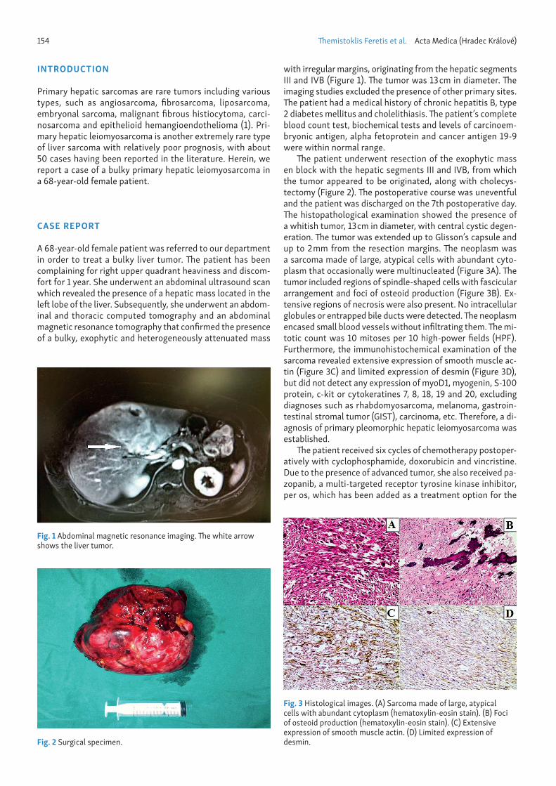
154 Themistoklis Feretis et al. Acta Medica (Hradec Králové)
INTRODUCTION
Primary hepatic sarcomas are rare tumors including various types, such as angiosarcoma, fibrosarcoma, liposarcoma, embryonal sarcoma, malignant fibrous histiocytoma, carci-nosarcoma and epithelioid hemangioendothelioma (1). Pri-mary hepatic leiomyosarcoma is another extremely rare type of liver sarcoma with relatively poor prognosis, with about 50 cases having been reported in the literature. Herein, we report a case of a bulky primary hepatic leiomyosarcoma in a 68-year-old female patient.
CASE REPORT
A 68-year-old female patient was referred to our department in order to treat a bulky liver tumor. The patient has been complaining for right upper quadrant heaviness and discom-fort for 1 year. She underwent an abdominal ultrasound scan which revealed the presence of a hepatic mass located in the left lobe of the liver. Subsequently, she underwent an abdom-inal and thoracic computed tomography and an abdominal magnetic resonance tomography that confirmed the presence of a bulky, exophytic and heterogeneously attenuated mass
with irregular margins, originating from the hepatic segments III and IVB (Figure 1). The tumor was 13 cm in diameter. The imaging studies excluded the presence of other primary sites. The patient had a medical history of chronic hepatitis B, type 2 diabetes mellitus and cholelithiasis. The patient’s complete blood count test, biochemical tests and levels of carcinoem-bryonic antigen, alpha fetoprotein and cancer antigen 19-9 were within normal range.
The patient underwent resection of the exophytic mass en block with the hepatic segments III and IVB, from which the tumor appeared to be originated, along with cholecys-tectomy (Figure 2). The postoperative course was uneventful and the patient was discharged on the 7th postoperative day. The histopathological examination showed the presence of a whitish tumor, 13 cm in diameter, with central cystic degen-eration. The tumor was extended up to Glisson’s capsule and up to 2 mm from the resection margins. The neoplasm was a sarcoma made of large, atypical cells with abundant cyto-plasm that occasionally were multinucleated (Figure 3A). The tumor included regions of spindle-shaped cells with fascicular arrangement and foci of osteoid production (Figure 3B). Ex-tensive regions of necrosis were also present. No intracellular globules or entrapped bile ducts were detected. The neoplasm encased small blood vessels without infiltrating them. The mi-totic count was 10 mitoses per 10 high-power fields (HPF). Furthermore, the immunohistochemical examination of the sarcoma revealed extensive expression of smooth muscle ac-tin (Figure 3C) and limited expression of desmin (Figure 3D), but did not detect any expression of myoD1, myogenin, S-100 protein, c-kit or cytokeratines 7, 8, 18, 19 and 20, excluding diagnoses such as rhabdomyosarcoma, melanoma, gastroin-testinal stromal tumor (GIST), carcinoma, etc. Therefore, a di-agnosis of primary pleomorphic hepatic leiomyosarcoma was established.
The patient received six cycles of chemotherapy postoper-atively with cyclophosphamide, doxorubicin and vincristine. Due to the presence of advanced tumor, she also received pa-zopanib, a multi-targeted receptor tyrosine kinase inhibitor, per os, which has been added as a treatment option for the
Fig. 1 Abdominal magnetic resonance imaging. The white arrow shows the liver tumor.
Fig. 2 Surgical specimen.
Fig. 3 Histological images. (A) Sarcoma made of large, atypical cells with abundant cytoplasm (hematoxylin-eosin stain). (B) Foci of osteoid production (hematoxylin-eosin stain). (C) Extensive expression of smooth muscle actin. (D) Limited expression of desmin.
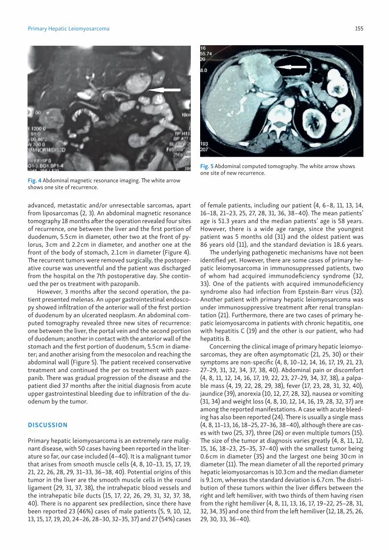
Primary Hepatic Leiomyosarcoma 155
advanced, metastatic and/or unresectable sarcomas, apart from liposarcomas (2, 3). An abdominal magnetic resonance tomography 18 months after the operation revealed four sites of recurrence, one between the liver and the first portion of duodenum, 5.5 cm in diameter, other two at the front of py-lorus, 3 cm and 2.2 cm in diameter, and another one at the front of the body of stomach, 2.1 cm in diameter (Figure 4). The recurrent tumors were removed surgically, the postoper-ative course was uneventful and the patient was discharged from the hospital on the 7th postoperative day. She contin-ued the per os treatment with pazopanib.
However, 3 months after the second operation, the pa-tient presented melenas. An upper gastrointestinal endosco-py showed infiltration of the anterior wall of the first portion of duodenum by an ulcerated neoplasm. An abdominal com-puted tomography revealed three new sites of recurrence: one between the liver, the portal vein and the second portion of duodenum; another in contact with the anterior wall of the stomach and the first portion of duodenum, 5.5 cm in diame-ter; and another arising from the mesocolon and reaching the abdominal wall (Figure 5). The patient received conservative treatment and continued the per os treatment with pazo-panib. There was gradual progression of the disease and the patient died 37 months after the initial diagnosis from acute upper gastrointestinal bleeding due to infiltration of the du-odenum by the tumor.
DISCUSSION
Primary hepatic leiomyosarcoma is an extremely rare malig-nant disease, with 50 cases having been reported in the liter-ature so far, our case included (4–40). It is a malignant tumor that arises from smooth muscle cells (4, 8, 10–13, 15, 17, 19, 21, 22, 26, 28, 29, 31–33, 36–38, 40). Potential origins of this tumor in the liver are the smooth muscle cells in the round ligament (29, 31, 37, 38), the intrahepatic blood vessels and the intrahepatic bile ducts (15, 17, 22, 26, 29, 31, 32, 37, 38, 40). There is no apparent sex predilection, since there have been reported 23 (46%) cases of male patients (5, 9, 10, 12, 13, 15, 17, 19, 20, 24–26, 28–30, 32–35, 37) and 27 (54%) cases
of female patients, including our patient (4, 6–8, 11, 13, 14, 16–18, 21–23, 25, 27, 28, 31, 36, 38–40). The mean patients’ age is 51.3 years and the median patients’ age is 58 years. However, there is a wide age range, since the youngest patient was 5 months old (31) and the oldest patient was 86 years old (11), and the standard deviation is 18.6 years.
The underlying pathogenetic mechanisms have not been identified yet. However, there are some cases of primary he-patic leiomyosarcoma in immunosuppressed patients, two of whom had acquired immunodeficiency syndrome (32, 33). One of the patients with acquired immunodeficiency syndrome also had infection from Epstein-Barr virus (32). Another patient with primary hepatic leiomyosarcoma was under immunosuppressive treatment after renal transplan-tation (21). Furthermore, there are two cases of primary he-patic leiomyosarcoma in patients with chronic hepatitis, one with hepatitis C (19) and the other is our patient, who had hepatitis B.
Concerning the clinical image of primary hepatic leiomyo-sarcomas, they are often asymptomatic (21, 25, 30) or their symptoms are non-specific (4, 8, 10–12, 14, 16, 17, 19, 21, 23, 27–29, 31, 32, 34, 37, 38, 40). Abdominal pain or discomfort (4, 8, 11, 12, 14, 16, 17, 19, 22, 23, 27–29, 34, 37, 38), a palpa-ble mass (4, 19, 22, 28, 29, 38), fever (17, 23, 28, 31, 32, 40), jaundice (39), anorexia (10, 12, 27, 28, 32), nausea or vomiting (31, 34) and weight loss (4, 8, 10, 12, 14, 16, 19, 28, 32, 37) are among the reported manifestations. A case with acute bleed-ing has also been reported (24). There is usually a single mass (4, 8, 11–13, 16, 18–25, 27–36, 38–40), although there are cas-es with two (25, 37), three (26) or even multiple tumors (15). The size of the tumor at diagnosis varies greatly (4, 8, 11, 12, 15, 16, 18–23, 25–35, 37–40) with the smallest tumor being 0.6 cm in diameter (35) and the largest one being 30 cm in diameter (11). The mean diameter of all the reported primary hepatic leiomyosarcomas is 10.3 cm and the median diameter is 9.1 cm, whereas the standard deviation is 6.7 cm. The distri-bution of these tumors within the liver differs between the right and left hemiliver, with two thirds of them having risen from the right hemiliver (4, 8, 11, 13, 16, 17, 19–22, 25–28, 31, 32, 34, 35) and one third from the left hemiliver (12, 18, 25, 26, 29, 30, 33, 36–40).
Fig. 4 Abdominal magnetic resonance imaging. The white arrow shows one site of recurrence.
Fig. 5 Abdominal computed tomography. The white arrow shows one site of new recurrence.
156 Themistoklis Feretis et al. Acta Medica (Hradec Králové)
There is a number of data about radiological and histo-logical findings of primary hepatic leiomyosarcoma in the lit-erature. Ultrasonography usually shows a hypoechoic mass (15, 17, 19, 29, 30, 33, 37) or a mass with heterogeneous echo-genicity (17, 22, 27, 30, 31, 37). Computed tomography usually reveals a hypodense (12, 17, 19, 21, 23, 27–33, 37, 39) and often heterogeneous mass (17, 21, 23, 31, 34) with inhomogeneous and often peripheral enhancement after administration of in-travenous contrast (15, 17, 19, 23, 27–33, 40), which may show regions of cystic degeneration (27, 28, 31, 32). Magnetic res-onance imaging displays on the other hand a usually hetero-geneous mass that is hypointense in T1-weighted images and hyperintense in T2-weighted images (16, 19, 21, 37, 39, 40). Histological examination of primary hepatic leiomyosarco-mas shows spindle-shaped cells with fascicular arrangement (4, 8, 10–12, 15–17, 19, 21, 22, 26–34, 36–38, 40). Immunohis-tochemistry reveals expression of smooth muscle actin (13, 15, 18, 19, 21, 22, 27–33, 36–38, 40), desmin (13, 15, 19, 21, 22, 27–34, 36–38) and vimentin (15, 17, 19, 22, 27–29, 31, 33, 37, 38, 40), whereas cytokeratins (22, 27–33, 37, 38), neuron-spe-cific enolase (27–29, 32) and S-100 protein (19, 21, 27–34, 37, 38, 40) are not expressed.
Regarding therapeutic options for primary hepatic leio-myosarcoma, hepatic resection, in the form of wedge resec-tion, segmentectomy, lobectomy or extended hepatectomy, was the preferred method for tumors without distant metas-tases (4, 6–8, 11, 12, 14, 16, 17, 20–23, 25, 26, 28–31, 33–36, 38). However, there were four patients with tumors confined within the liver who underwent liver transplantation (25, 27, 39). Some authors reported the addition of adjuvant chemo-therapy consisted of various combinations of drugs (6, 8, 10, 23, 28, 30, 31, 35, 37, 39). Some of the administrated chem-otherapeutic regimens were the following: 1. vincristine, cy-clophosphamide and dactinomycin (8); 2. cyclophosphamide, vincristine and actinomycin D (10); 3. doxorubicin (23); 4. fo-linic acid, fluorouracil, irinotecan (FOLFIRI) and bevacizumab (30); 5. mitoxantrone, cisplatin and fluorouracil (35); 6. ifosfa-mide and mesna (37). Radiotherapy has also been used as an adjuvant treatment along with chemotherapy in three cases (10, 23, 28). Finally, transarterial chemoembolization has also been used in one case (35) and transarterial infusion of epiru-bicin and carboplatin in another case (37). Survival rates are relatively low. On the basis of articles reporting survival data, median survival is 37.5 months and 5-year survival rate is 40% (4–12, 14, 17, 20–23, 25–38, 40).
In conclusion, primary hepatic leiomyosarcoma is a rare malignant disease with relatively poor prognosis. The most preferred type of treatment is surgical excision, which some-times is combined with adjuvant chemotherapy and/or radio-therapy. However, very little is known about the effectiveness of the current treatment because of the rarity of the disease. More in-depth studies are needed to investigate and shed light on this uncommon clinical entity.
CONFLICTS OF INTEREST
The authors declare that they have no conflicts of interest.
REFERENCES
1. Yu RS, Chen Y, Jiang B, et al. Primary hepatic sarcomas: CT findings. Eur Radiol. 2008; 18: 2196–205.
2. von Mehren M, Randall RL, Benjamin RS, et al. NCCN clinical practice guidelines in oncology. Soft tissue sarcoma. Version 2.2018. 2018; 1–49.
3. Casali PG, Abecassis N, Bauer S, et al. Soft tissue and visceral sarcomas: ESMO–EURACAN. Clinical Practice Guidelines for diagnosis, treatment and follow-up. Ann Oncol 2018; 29(Suppl. 4): iv51–iv67.
4. Wilson SE, Braitman H, Plested WG, et al. Primary leiomyosarcoma of the liver. Ann Surg 1971; 174: 232–7.
5. Fong JA, Ruebner BH. Primary leiomyosarcoma of the liver. Hum Pathol 1974; 5: 115–9.
6. Masur H, Sussman EB, Molander DW. Primary hepatic leiomyosarcoma: A report of two cases. Gastroenterology 1975; 69: 994–7.
7. Yoshikawa K, Satake K, Kinoshita H, et al. Primary leiomyosarcoma of the liver. Clin Oncol 1977; 3: 197–202.
8. Bloustein PA. Hepatic leiomyosarcoma: ultrastructural study and review of the differential diagnosis. Hum Pathol 1978; 9: 713–5.
9. O’Leary MR, Hill RB, Levine RA. Peritoneoscopic diagnosis of primary leiomyosarcoma of liver. Hum Pathol 1982; 13: 76–8.
10. Chen KT. Hepatic leiomyosarcoma. J Surg Oncol 1983; 24: 325–8.11. Maki HS, Hubert BC, Sajjad SM, et al. Primary hepatic leiomyosarcoma.
Arch Surg 1987; 122: 1193–6.12. Paraskevopoulos JA, Stephenson TJ, Dennison AR. Primary leiomyosar-
coma of the liver. HPB Surg 1991; 4: 157–62.13. Watanabe K, Saito A, Wakabayashi H, et al. Two autopsy cases of primary
leiomyosarcoma of the liver. Superiority of muscle-specific actin immu-noreactivity in diagnosis. Acta Pathol Jpn 1991; 41: 461–5.
14. Baur M1, Pötzi R, Lochs H, et al. Primary leiomyosarcoma of the liv-er – a case report. Z Gastroenterol 1993; 31: 20–3.
15. Holloway H, Walsh CB, Thomas R, et al. Primary hepatic leiomyosarcoma. J Clin Gastroenterol 1996; 23: 131–3.
16. Soyer P, Blanc F, Vissuzaine C, et al. Primary leiomyosarcoma of the liver MR findings. Clin Imaging 1996; 20: 273–5.
17. Ferrozzi F, Bova D, Zangrandi A, et al. Primary liver leiomyosarcoma: CT appearance. Abdom Imaging 1996; 21: 157–60.
18. Sato S, Hosoi K, Kagawa T. A primary leiomyosarcoma of the liver: an autopsy report. Electron J Pathol Histol 2000; 6: 8.
19. Tsuji M, Takenaka R, Kashihara T, et al. Primary hepatic leiomyosarcoma in a patient with hepatitis C virus-related liver cirrhosis. Pathol Int 2000; 50: 41–7.
20. Iordanidis F, Hytiroglou P, Drevelegas A, et al. A 25-year-old man with a large hepatic tumor and multiple nodular lesions. Semin Liver Dis 2002; 22: 97–102.
21. Fujita H, Kiriyama M, Kawamura T, et al. Primary hepatic leiomyosarco-ma in a woman after renal transplantation: report of a case. Surg Today 2002; 32: 446–9.
22. Lee HJ, Lee JL, Choi WH. A case of primary myxoid leiomyosarcoma of the liver. Korean J Intern Med 2002; 17: 278–82.
23. Almogy G, Lieberman S, Gips M, et al. Clinical outcomes of surgical resec-tions for primary liver sarcoma in adults: results from a single centre. Eur J Surg Oncol 2004; 30: 421–7.
24. Jeong TY, Kim YS, Park KJ, et al. A case of primary leiomyosarcoma of the liver presenting with acute bleeding. Korean J Gastroenterol 2008; 51: 194–8.
25. Matthaei H, Krieg A, Schmelzle M, et al. Long-term survival after surgery for primary hepatic sarcoma in adults. Arch Surg 2009; 144: 339–44.
26. Giuliante F, Sarno G, Ardito F, et al. Primary hepatic leiomyosarcoma in a young man after Hodgkin’s disease: diagnostic pitfalls and therapeutic challenge. Tumori 2009; 95: 374–7.
27. Liang X, Xiao-Min S, Jiang-Ping X, et al. Liver transplantation for primary hepatic leiomyosarcoma: a case report and review of the literatures. Med Oncol 2010; 27: 1269–72.
28. Shamseddine A, Faraj W, Mukherji D, et al. Unusually young age distri-bution of primary hepatic leiomyosarcoma: case series and review of the adult literature. World J Surg Oncol 2010; 8: 56.
29. Shivathirthan N, Kita J, Iso Y, et al. Primary hepatic leiomyosarcoma: Case report and literature review. World J Gastrointest Oncol 2011; 3: 148–52.
30. Takehara K, Aoki H, Takehara Y, et al. Primary hepatic leiomyosarcoma with liver metastasis of rectal cancer. World J Gastroenterol 2012; 18: 5479–84.
31. Tsai PS, Yeh TC, Shih SL. Primary hepatic leiomyosarcoma in a 5-month-old female infant. Acta Radiol Short Rep. 2013;2:2047981613498722.

Primary Hepatic Leiomyosarcoma 157
32. Chelimilla H, Badipatla K, Ihimoyan A, et al. A rare occurrence of primary hepatic leiomyosarcoma associated with epstein barr virus infection in an AIDS patient. Case Rep Gastrointest Med 2013; 2013: 691862.
33. Metta H, Corti M, Trione N, et al. Primary hepatic leiomyosarcoma – a rare neoplasm in an adult patient with AIDS: second case report and literature review. J Gastrointest Cancer 2014; 45: 36–9.
34. Majumder S, Dedania B, Rezaizadeh H, et al. Tumor rupture as the initial manifestation of primary hepatic leiomyosarcoma. Gastrointest Cancer Res 2014; 7: 33–4.
35. Lin YH, Lin CC, Concejero AM, et al. Surgical experience of adult primary hepatic sarcomas. World J Surg Oncol 2015; 13: 87.
36. Hamed MO, Roberts KJ, Merchant W, et al. Contemporary management and classification of hepatic leiomyosarcoma. HPB (Oxford) 2015; 17: 362–7.
37. Lv WF, Han JK, Cheng DL, et al. Imaging features of primary hepatic leio-myosarcoma: A case report and review of literature. Oncol Lett 2015; 9: 2256–60.
38. López-López V, Robles R, Ferri B, et al. Laparoscopically assisted resec-tion of a primary hepatic leiomyosarcoma: A safe approach in a rare ma-lignancy. Cir Esp 2017; 95(8): 478–80.
39. Xie P, Zhuang H. FDG PET/CT findings of primary hepatic leiomyosarco-ma in an immunocompetent pediatric patient. Clin Nucl Med 2017; 42: 323–4.
40. Iida T, Maeda T, Amari Y, et al. Primary hepatic leiomyosarcoma in a pa-tient with autosomal dominant polycystic kidney disease. CEN Case Rep 2017; 6: 74–8.
158 case report
Pharyngeal Papilloma: a Rare Non-Pulmonary Cause of Hemoptysis
Stamatis Katsenos*, Melita Nikolopoulou
A B S T R AC TThe most common benign neoplasm of the pharynx is papilloma. It is characterized by bulging brittle lesions, which are pedicled or sessile, whitish-grey or pinkish colour. Progressive hoarseness is the main clinical feature. When the papillomata spread throughout the tracheobronchial tree symptoms such as chronic cough, stridor, dyspnea or acute respiratory distress are mostly present. Hemoptysis as a presenting symptom is exceptionally rare in patients with pharyngeal papillomatosis. Herein, we report a case of pharyngeal papillomatosis in which hemoptysis was the primary clinical manifestation. The clinical and therapeutic aspects of the disease are briefly discussed.
K E Y WO R D Shemoptysis; pharynx; papilloma
A U T H O R A F F I L I AT I O N SDepartment of Pneumonology, Army General Hospital of Athens, Athens, Greece* Corresponding author: Department of Pneumonology, Army General Hospital of Athens, 158 Mesogion and Katehaki Avenue, 115 25 Athens, Greece; e-mail: [email protected]
Received: 19 March 2018Accepted: 25 November 2018Published online: 22 January 2019
Acta Medica (Hradec Králové) 2018; 61(4): 158–161https://doi.org/10.14712/18059694.2018.136© 2018 The Authors. This is an open-access article distributed under the terms of the Creative Commons Attribution License (http://creativecommons.org/licenses/by/4.0), which permits unrestricted use, distribution, and reproduction in any medium, provided the original author and source are credited.
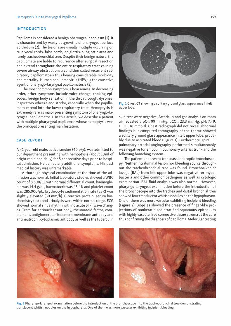
Hemoptysis Due to Pharyngeal Papilloma 159
INTRODUCTION
Papilloma is considered a benign pharyngeal neoplasm (1). It is characterized by warty outgrowths of pharyngeal surface epithelium (2). The lesions are usually multiple occurring on true vocal cords, false cords, epiglottis, subglottic area and rarely tracheobronchial tree. Despite their benign nature, the papillomata are liable to recurrence after surgical resection and extend throughout the entire respiratory tract causing severe airway obstruction; a condition called recurrent res-piratory papillomatosis thus bearing considerable morbidity and mortality. Human papilloma virus (HPV) is the causative agent of pharyngo-laryngeal papillomatosis (3).
The most common symptom is hoarseness. In decreasing order, other symptoms include voice change, choking epi-sodes, foreign body sensation in the throat, cough, dyspnea, inspiratory wheeze and stridor, especially when the papillo-mata extend into the lower respiratory tract. Hemoptysis is extremely rare as major presenting symptom of pharyngo-la-ryngeal papillomatosis. In this article, we describe a patient with multiple pharyngeal papillomas whose hemoptysis was the principal presenting manifestation.
CASE REPORT
A 41-year-old male, active smoker (40 p/y), was admitted to our department presenting with hemoptysis (about 10 ml of bright red blood daily) for 5 consecutive days prior to hospi-tal admission. He denied any additional symptoms. His past medical history was unremarkable.
A thorough physical examination at the time of the ad-mission was normal. Initial laboratory studies showed a WBC count of 8.500/μL with normal differential count, haemoglo-bin was 14.4 g/dL, haematocrit was 43.4% and platelet count was 285.000/μL. Erythrocyte sedimentation rate (ESR) was slightly elevated (30 mm/h). C-reactive protein, serum bio-chemistry tests and urinalysis were within normal range. ECG showed normal sinus rhythm with no acute ST-T wave chang-es. Tests for antinuclear antibody, rheumatoid factor, com-plement, antiglomerular basement membrane antibody and antineutrophil cytoplasmic antibody as well as the tuberculin
skin test were negative. Arterial blood gas analysis on room air revealed a pO2: 99 mmHg, pCO2: 23.3 mmHg, pH: 7.49, HCO3: 18 mmol/l. Chest radiograph did not reveal abnormal findings but computed tomography of the thorax showed a solitary ground glass appearance in left upper lobe, proba-bly due to aspirated blood (Figure 1). Furthermore, spiral CT pulmonary arterial angiography performed simultaneously was negative for emboli in pulmonary arterial trunk and the following branching system.
The patient underwent transnasal fiberoptic bronchosco-py. Neither intraluminal lesion nor bleeding source through-out the tracheobronchial tree was found. Bronchoalveolar lavage (BAL) from left upper lobe was negative for myco-bacteria and other common pathogens as well as cytologic examination. BAL fluid analysis was also normal. However, pharyngo-laryngeal examination before the introduction of the bronchoscope into the trachea and distal bronchial tree showed four translucent whitish nodules on the hypopharynx. One of them was more vascular exhibiting incipient bleeding (Figure 2). Biopsies showed the presence of finger-like pro-jections of nonkeratinized stratified squamous epithelium with highly vascularized connective tissue stroma at the core thus confirming the diagnosis of papilloma. Molecular testing
Fig. 1 Chest CT showing a solitary ground glass appearance in left upper lobe.
Fig. 2 Pharyngo-laryngeal examination before the introduction of the bronchoscope into the tracheobronchial tree demonstrating translucent whitish nodules on the hypopharynx. One of them was more vascular exhibiting incipient bleeding.
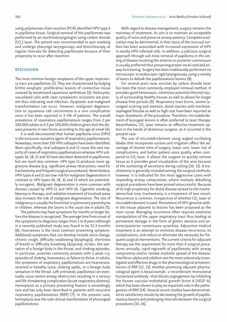
160 Stamatis Katsenos et al. Acta Medica (Hradec Králové)
using polymerase chain reaction (PCR) identified HPV type 6 in papilloma tissue. Surgical removal of the papillomata was performed by an otorhinolaryngologist using carbon dioxide (CO2) laser. The patient was recommended to quit smoking and undergo pharyngo-laryngoscopy and bronchoscopy at regular intervals for detecting papillomata because of their propensity to recur after resection.
DISCUSSION
The most common benign neoplasms of the upper respirato-ry tract are papillomas (1). They are characterized by bulging brittle exophytic proliferative lesions of connective tissue covered by keratinized squamous epithelium (2). Koilocytes, vacuolated cells with clear cytoplasmic inclusions are pres-ent thus indicating viral infection. Dysplastic and malignant transformation can occur. However, malignant degenera-tion to squamous cell carcininoma is a rare complication since it has been reported in 3–5% of patients. The overall prevalence of respiratory papillomatosis ranges from 2 per 100,000 adults to 4.5 per 100,000 children given that the dis-ease presents in two forms according to the age at onset (4).
It is well-documented that human papilloma virus (HPV) is the exclusive causative agent of respiratory papillomatosis. Nowadays, more than 150 HPV subtypes have been identified. More specifically, viral subtypes 6 and 11 cause the vast ma-jority of cases of respiratory papillomatosis whereas HPV sub-types 16, 18, 31 and 33 have also been detected in papillomas, but are much less common. HPV type 11 produces more ag-gressive disease (e.g. significant airway obstruction, need for tracheotomy and frequent surgical procedures). Nevertheless, HPV types 6 and 11 are low-risk for malignant degeneration in contrast to HPV types 16, 18, 31 and 33 which are potential-ly oncogenic. Malignant degeneration is more common with disease caused by HPV-11 and HPV-16. Cigarette smoking, bleomycin therapy, and radiation treatment of involved areas also increase the risk of malignant degeneration. The site of malignancy is usually the bronchial or pulmonary parenchyma in children, whereas the larynx is the usual site in adults (5).
The patients may have symptoms for months or longer be-fore the disease is recognized. The average time from onset of the symptoms to diagnosis ranges from 1 to 8 years although in a recently published study was found to be 52.3 months (6). Hoarseness is the most common presenting symptom. Additional symptoms that can develop include voice change, chronic cough, difficulty swallowing (dysphagia), shortness of breath or difficulty breathing (dyspnea), stridor, the sen-sation of a foreign body in the throat, and choking episodes. In particular, juveniles commonly present with a weak cry, episodes of choking, hoarseness, or failure to thrive. In adults, the symptoms of respiratory papillomatosis are hoarseness, strained or breathy voice, choking spells, or a foreign body sensation in the throat. Left untreated, papillomas can even-tually cause severe airway obstruction resulting in a serious and life-threatening complication (acute respiratory distress). Hemoptysis as a primary presenting feature is exceedingly rare and has only been described in patients with recurrent respiratory papillomatosis (RRP) (7). In the present case, hemoptysis was the sole clinical manifestation of pharyngeal papillomatosis.
With regard to disease management, surgery remains the mainstay of treatment. Its aim is to maintain an acceptable quality of voice and preserve airway patency. Complete erad-ication may be detrimental, in that injury of the mucosal sur-face has been associated with increased expression of HPV in nearby HPV-infected cells. In addition, a judicious surgical approach through sub-total removal of papilloma in the set-ting of disease involving the anterior or posterior commissure is usually preferred thus preserving proper vocal cord and air-way functioning. Surgery has been traditionally performed via microscopic or endoscopic rigid laryngoscopy using a variety of lasers to debulk the papillomatous lesions (8).
For several years now, excision by carbon dioxide laser has been the most commonly employed removal method. It provides good hemostasis, minimizes potential thermal inju-ry of surrounding healthy tissues as well as allows for longer disease-free periods (9). Respiratory tract burns, severe la-ryngeal scarring and stenosis, distal injuries with tracheoe-sophageal fistulae as well as high cost have been reported as major drawbacks of the procedure. Therefore, microdebride-ment of laryngeal lesions is often preferred to laser therapy. Nevertheless, CO2 laser remains an effective treatment op-tion in the hands of dexterous surgeon, as it occurred in the present case.
The use of microdebridement using angled oscillating blades that incorporate suction and irrigation offers the ad-vantage of shorter time of surgery, lower cost, lower risk of complications, and better patient voice quality when com-pared to CO2 laser. It allows the surgeon to quickly remove tissue as it provides good visualization of the area because of the suctioning of secretions during cutting (10, 11). A tra-cheotomy is generally included among the surgi cal methods; however, it is indicated for the most aggressive cases with impending airway compromise, where multiple debulking surgical procedures have been proved unsuccessful. Because of its high propensity for distal disease spread to the trache-obronchial tree, tracheotomy is a treatment of last resort. Recurrence is common, irrespective of whether CO2 laser or microdebridement is used. Persistence of HPV genome with-in the tissue adjacent to lesions has been proposed as the main cause. Managing recurrence often requires extensive manipulation of the upper respiratory tract thus leading to permanent damage in the form of glottic stenosis and an-terior/posterior commissure synechiae. Adjunctive medical treatment is an attempt to minimize disease recurrence, its complications, and reduce or eliminate the necessity for fre-quent surgical interventions. The current criteria for adjuvant therapy are the requirement for more than 4 surgical proce-dures annually, rapid regrowth of papillomata with airway compromise and/or remote multisite spread of the disease. Interferon-alpha and cidofovir are the most extensively inves-tigated and effective drugs in the pharmacological armamen-tarium of RRP (12, 13). Another promising adjuvant pharma-cological agent is bevacizumab – a recombinant monoclonal humanized antibody – that blocks angiogenesis by inhibiting the human vascular endothelial growth factor A (VEGF-A), which has been shown to play an important role in the patho-genesis of RRP (14). Several recent studies have demonstrat-ed its satisfactory results by decreasing the growth of papillo-matous lesions and prolonging intervals between the surgical procedures (15, 16).
Hemoptysis Due to Pharyngeal Papilloma 161
Moreover, the development of HPV vaccines represents a great medical breakthrough in preventing RRP. In par-ticular, the quadrivalent HPV vaccine, Gardasil, has activity against HPV types 6, 11, 16, 18 and its implementation as an adjunctive therapy has a positive effect on the course of RRP, since the recurrence rate seems to be significantly low-ered, according to two recently published studies (17, 18). The induction of sufficiently high antibody titers after HPV vaccination and subsequent immunoglobulin secretion on the mucous membranes of the aerodigestive tract could be a plausible explanation for its favourable therapeutic effect (19). Furthermore, the newly-developed nonavalent vaccine, Gardasil-9, achieved encouraging results in a descriptive study, as it abated the course of the disease, lengthened the inter-surgical interval, and decreased morbidity (20). Howev-er, large, multicentre randomized controlled trials are needed to adequately assess the efficacy of the HPV vaccination as a therapeutic vaccine in the RRP population.
CONCLUSIONS
In conclusion, the present case is unusual in that the patient manifested hemoptysis and bronchoscopic examination re-vealed isolated pharyngeal papillomatosis. Hemoptysis has only rarely been reported in patients with disease spread to the tracheobronchial tree. Bronchoscopy is strongly recom-mended as the primary method for diagnosing and localizing hemoptysis, especially if massive. Clinicians should be aware of the disease to make an early diagnosis. Because of its ten-dency for recurrences and malignant transformation, metic-ulous follow-up of patients including HPV typing is crucial for early detection and treatment.
REFERENCES 1. Bishop JA. Benign Neoplasms of the Larynx, Hypopharynx and Trachea.
In: Thompson LDR, Bishop JA, eds. Head and Neck Pathology. Elsevier 2019: 122–37 e2.
2. Abramson AL, Steinberg BM, Winkler B. Laryngeal papillomatosis: clin-ical, histopathologic and molecular studies. Laryngoscope 1987; 97: 678–85.
3. Donne AJ, Hampson L, Homer JJ, Hampson IN. The role of HPV type in re-current respiratory papillomatosis. Int J Pediatr Otorhinolaryngol 2010; 74: 7–14.
4. Reeves WC, Ruparelia SS, Swanson KI, Derkay CS, Marcus A, Unger ER. National registry for juvenile-onset recurrent respiratory papillomatosis. Arch Otolaryngol Head Neck Surg 2003; 129: 976-82.
5. Omland T, Akre H, Lie KA, Jebsen P, Sandvik L, Brøndbo K. Risk factors for aggressive recurrent respiratory papillomatosis in adults and juveniles. PLoS ONE 2014; 9: e113584.
6. Silva L, Gonçalves CP, Fernandes AM, Damrose EJ, Costa HO. Laryngeal papillomatosis in children: The impact of the late recognition over evo-lution. J Med Virol 2015; 87: 1413–7.
7. Zhang Z, Chang M, Moreta-Sainz CM. Case Report: Pulmonary Papil-lomatosis in a Patient Presenting with Cough and Hemoptysis. Perm J 2015; 19: e125–7.
8. Papaspyrou G, Schick B, Papaspyrou S, Eivazi B, Al Kadah B. Retrospec-tive analysis of laser vs other therapeutic modalities for laryngeal papil-lomatosis. European multicenter study. J BUON 2016; 21: 1274–8.
9. Castillo CG, Garcia EM, Duran MD, Mestre MS, Gras JR. Papillomas and laryngeal papillomatosis. Treatment with CO2 laser surgery. Our experi-ence over 15 years. Acta Otorrinolaringol Esp 2010; 61; 422–7.
10. Avelino MA, Zaiden TC, Gomes RO. Surgical treatment and adjuvant ther-apies of recurrent respiratory papillomatosis. Braz J Otorhinolaryngol 2013; 79: 636–42.
11. Mesolella M, Motta G, LaguardiaM, Galli V. Papillomatosis of the larynx: treatment with CO2 laser. B-ENT 2006; 2: 51–4.
12. Gerein V, Rastorguev E, Gerein J, Jecker P, Pfister H. Use of interferon-al-pha in recurrent respiratory papillomatosis: 20-year follow-up. Ann Otol Rhinol Laryngol 2005; 114: 463–71.
13. Ablanedo-Terrazas Y, Soda-Merhy A, Hernández-Palestina M, Ormsby CE, Reyes-Terán G. Intralesional cidofovir in severe juvenile respiratory papillomatosis. B-ENT 2012; 8: 197–202.
14. Rahbar R, Vargas SO, Folkman J, et al. Role of vascular endothelial growth factor-A in recurrent respiratory papillomatosis. Ann Otol Rhinol Laryn-gol 2005; 114: 289–95.
15. Zeitels SM, Barbu AM, Landau-Zemer T, et al. Local injection bevacizum-ab (Avastin) and angiolytic KTP laser treatment of recurrent respiratory papillomatosis of the vocal folds: a prospective study. Ann Otol Rhinol Laryngol 2011; 120: 627-34.
16. Best SR, Mohr M, Zur KB. Systemic bevacizumab for recurrent respirato-ry papillomatosis: A national survey. Laryngoscope 2017; 127: 2225–9.
17. Young DL, Moore MM, Halstead LA. The use of the quadrivalent human papilloma virus vaccine (Gardasil) as adjuvant therapy in the treatment of recurrent respiratory papilloma. J Voice 2015; 29: 223–9.
18. Mauz PS, Schäfer FA, Iftner T, Gonser P. HPV vaccination as preventive approach for recurrent respiratory papillomatosis – a 22-year retrospec-tive clinical analysis. BMC Infect Dis 2018; 18: 343.
19. Makiyama K, Hirai R, Matsuzaki H. Gardasil vaccination for recurrent laryngeal papillomatosis in adult men: first report: changes in HPV anti-body titer. J Voice 2017; 31: 104–6.
20. Sullivan C, Curtis S, Mouzakes J. Therapeutic use of the HPV vaccine in Recurrent Respiratory Papillomatosis: A case report. Int J Pediatr Otorhi-nolaryngol 2017; 93: 103–6.

Related Documents