PATIENT POSITIONING AND ANAESTHESIA

Welcome message from author
This document is posted to help you gain knowledge. Please leave a comment to let me know what you think about it! Share it to your friends and learn new things together.
Transcript
PATIENT POSITIONING AND ANAESTHESIA
INTRODUCTION
• Positioning is the joint responsibility of the surgeon & anesthesiologist.
• Ideal pt. positioning involves balancing surgical comfort, against the risks related to the pt. position.
• Pt. positioning & postural limitation should be considered during the PAC.
Overview • One must be aware of the anatomic and physiologic changes
associated with anesthesia, patient positioning, and the procedure.
• The following criteria should be met to prevent injury from pressure, obstruction, or stretching: – No interference with respiration – No interference with circulation – No pressure on peripheral nerves – Minimal skin pressure – Accessibility to operative site – Accessibility for anesthetic administration – No undue musculoskeletal discomfort – Maintenance of individual requirements
Assessment • The team should assess the following prior to positioning of
the patient: – Procedure length – Surgeon’s preference of position – Required position for procedure – Anesthesia to be administered – Patient’s risk factors
• age, weight, skin condition, mobility/limitations, pre‐existing conditions, airway etc.
– Patient’s privacy and medical needs
GENERAL PHYSIOLOGICAL CONCERNS
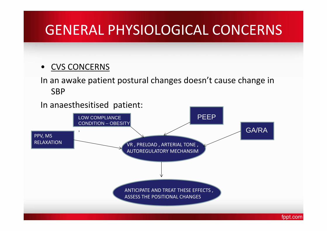
• CVS CONCERNS
In an awake patient postural changes doesn’t cause change in SBP
In anaesthesitised patient:
,
VR , PRELOAD , ARTERIAL TONE , AUTOREGULATORY MECHANSIM
LOW COMPLIANCE CONDITION – OBESITY
PEEP
GA/RAPPV, MS RELAXATION
ANTICIPATE AND TREAT THESE EFFECTS , ASSESS THE POSITIONAL CHANGES
• PULMONARY CONCERNS • Any position which limits movements of abdomen , chest wall
or diaphragm increase atelectasis and intrapulmonary shunt
• Change from standing to supine ‐ decrease FRC due to cephalad displacement of the diaphragm
Surgical Positions • Four basic surgical positions
include: – Supine
– Lateral – Prone
– Lithotomy
• Variations include: – Trendelenburg
– Reverse trendelenburg
– Fowler’s/semifowler – Beach chair position
– Wattson jone position
– Position for robotic surgeries
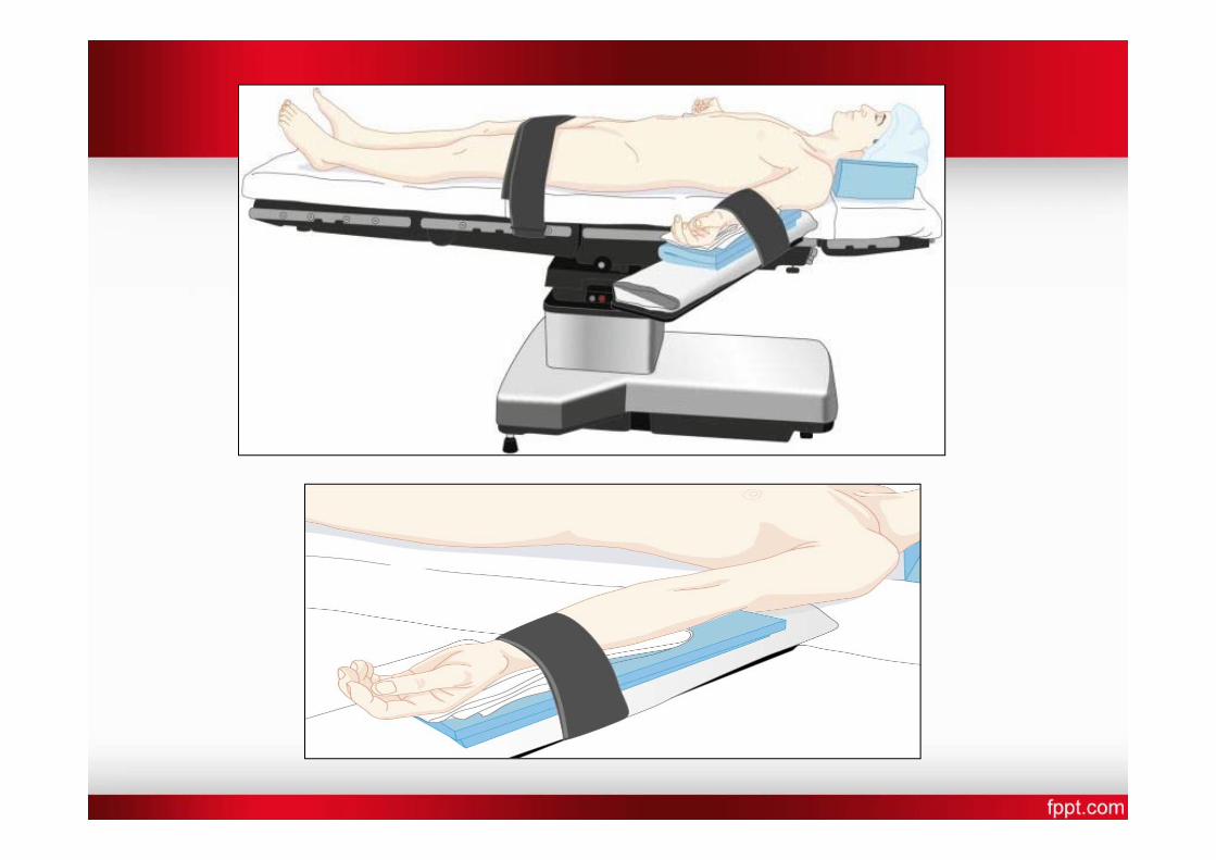
Supine • Most common with the least amount of harm • Placed on back with legs extended and uncrossed at the
ankles • Arms either on arm boards abducted <90* with palms up or
tucked (not touching metal or constricted) • Spinal column should be in alignment with legs parallel to the
OR bed – Head in line with the spine and the face is upward – Hips are parallel to the spine
• Padding is placed under the head, arms, and heels with a pillow placed under the knees
• Safety belt placed 2” above the knees while not impeding circulation
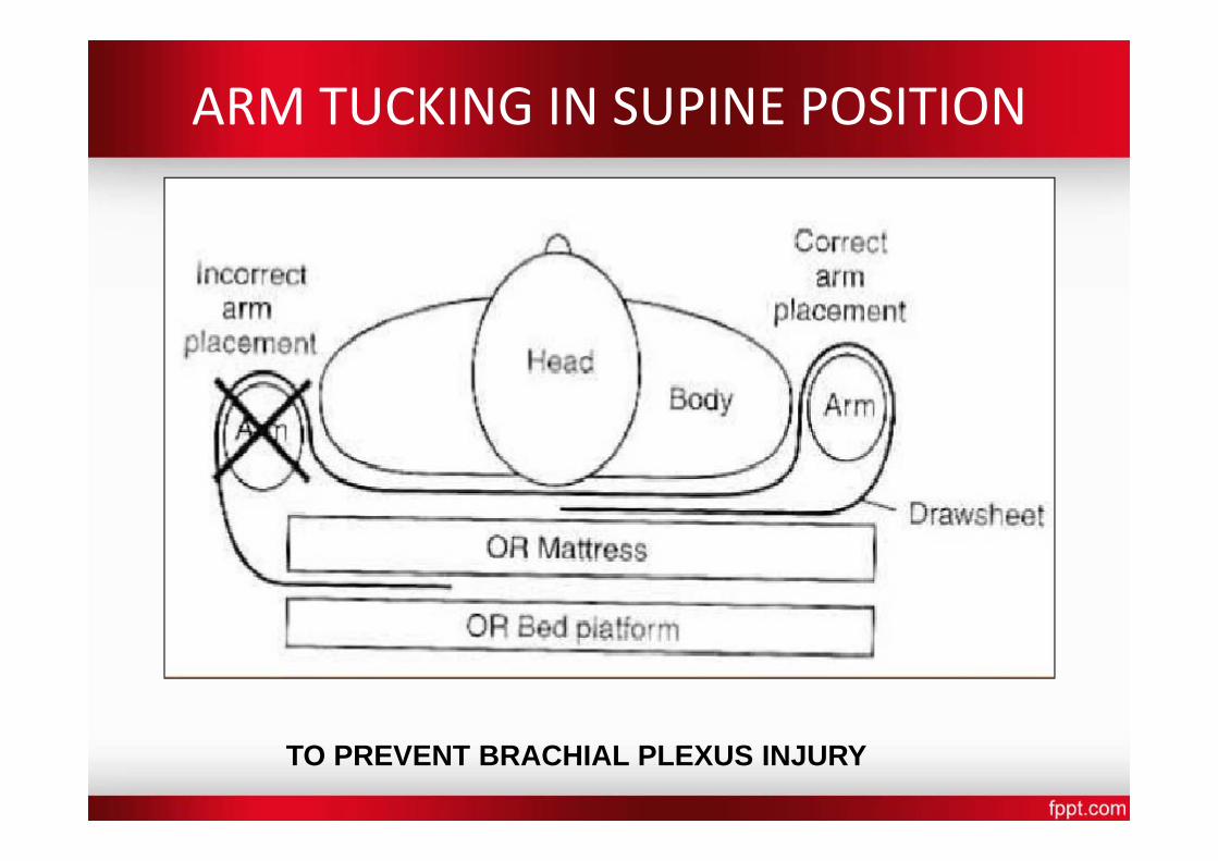
ARM TUCKING IN SUPINE POSITION
TO PREVENT BRACHIAL PLEXUS INJURY
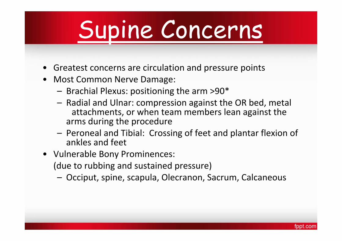
Supine Concerns • Greatest concerns are circulation and pressure points • Most Common Nerve Damage:
– Brachial Plexus: positioning the arm >90* – Radial and Ulnar: compression against the OR bed, metal
attachments, or when team members lean against thearms during the procedure
– Peroneal and Tibial: Crossing of feet and plantar flexion of ankles and feet
• Vulnerable Bony Prominences: (due to rubbing and sustained pressure) – Occiput, spine, scapula, Olecranon, Sacrum, Calcaneous
VARIATIONS
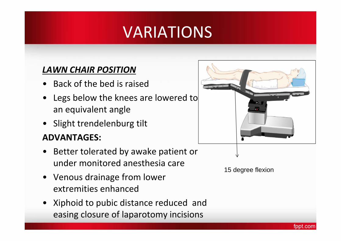
LAWN CHAIR POSITION
• Back of the bed is raised
• Legs below the knees are lowered to an equivalent angle
• Slight trendelenburg tilt ADVANTAGES: • Better tolerated by awake patient or
under monitored anesthesia care
• Venous drainage from lower extremities enhanced
• Xiphoid to pubic distance reduced and easing closure of laparotomy incisions
15 degree flexion
Trendelenburg • The patient is placed in the supine position while the OR bed
is modified to a head‐down tilt of 35 to 45 degrees resulting in the head being lower than the pelvis
• Arms are in a comfortable position – either at the side or on bilateral arm boards
• The foot of the OR bed is lowered to a desired angle
• ADVANTAGES
To increase V.R after spinal anesthesia
To increase central venous volume to facilitate central cannulation
To minimise aspiration during regurgitation
Trendelenburg Concerns
• ↑ CVP
• ↑ ICP
• ↑ IOP
• ↑ myocardial work
• ↑ pulmonary venous pressure
• ↓ pulmonary compliance
• ↓ FRC
• Swelling of face, eyelids, conjunctiva , tongue, laryngeal edema observed in long surgeries
Reverse Trendelenburg • The entire OR bed is tilted so the head is higher than the feet • Used for head and neck, laproscopic procedures • Facilitates exposure, aids in breathing and decreases blood
supply to the area
• A padded footboard is used to prevent the patient from sliding toward the foot
• Reduces venous return therefore hypotension
• Laproscopic cholecystectomy : reverse trendelenburg position with right up
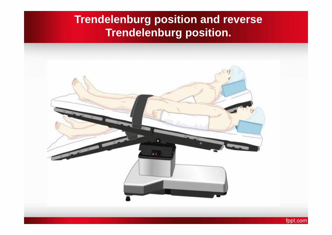
Trendelenburg position and reverse Trendelenburg position.
Hemodynamic and Ventilation.(supine)
• Every 2.5 cm change of vertical ht. from the reference point at level of the heart leads to a change of MAP by 2 mmHg in the opposite direction.
• V & Q are best in dependent lungs.
• Positive‐pressure ventilation provides the best ventilation to non‐dependent lung zones ‐V/Q mismatch.
Lithotomy • With the patient in the supine position, the hips are flexed to
80‐100 o from the torso so that legs are parallel to it and legs are abducted by 30‐45 o to expose the perineal region
• The patient’s buttocks are even with the lower break in theOR bed (to prevent lumbosacral strain)
• The legs and feet are placed in stirrups that support the lowerextremities
• The legs are raised, positioned, and lowered slowly andsimultaneously, with the permission of the anesthesia careprovider
• Adequate padding and support for the legs/feet shouldeliminate pressure on joints and nerve plexus
• The position must be symmetrical • The perineum should be in line with the longitudinal axis of
the OR bed
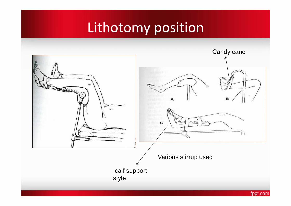
Lithotomy position
Various stirrup used
calf support style
Candy cane
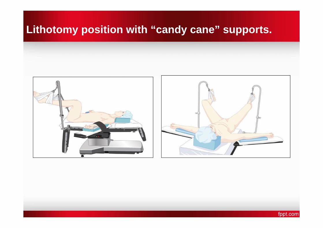
Lithotomy position with “candy cane” supports.
PHYSIOLOGICAL CHANGES
• Preload increases, causing a transient increase in CO , cerebral venous and intracranial pressure
• Reduce lung compliance
• If obesity or a large abdominal mass is present (tumor, gravid uterus)‐ VR to heart might decrease
• Normal lordotic curvature of the lumbar spine is lost potentially aggravating any previous lower back pain
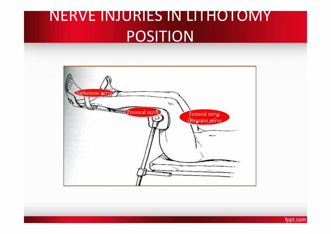
NERVE INJURIES IN LITHOTOMY POSITION
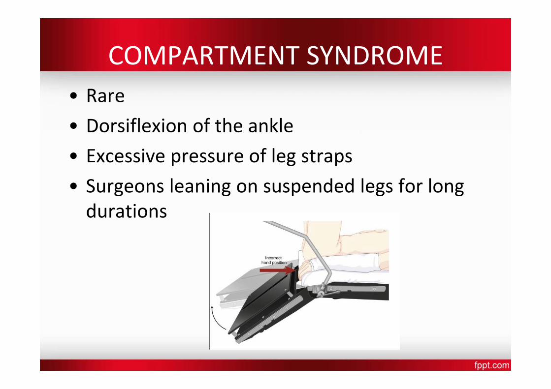
COMPARTMENT SYNDROME • Rare
• Dorsiflexion of the ankle
• Excessive pressure of leg straps • Surgeons leaning on suspended legs for long durations
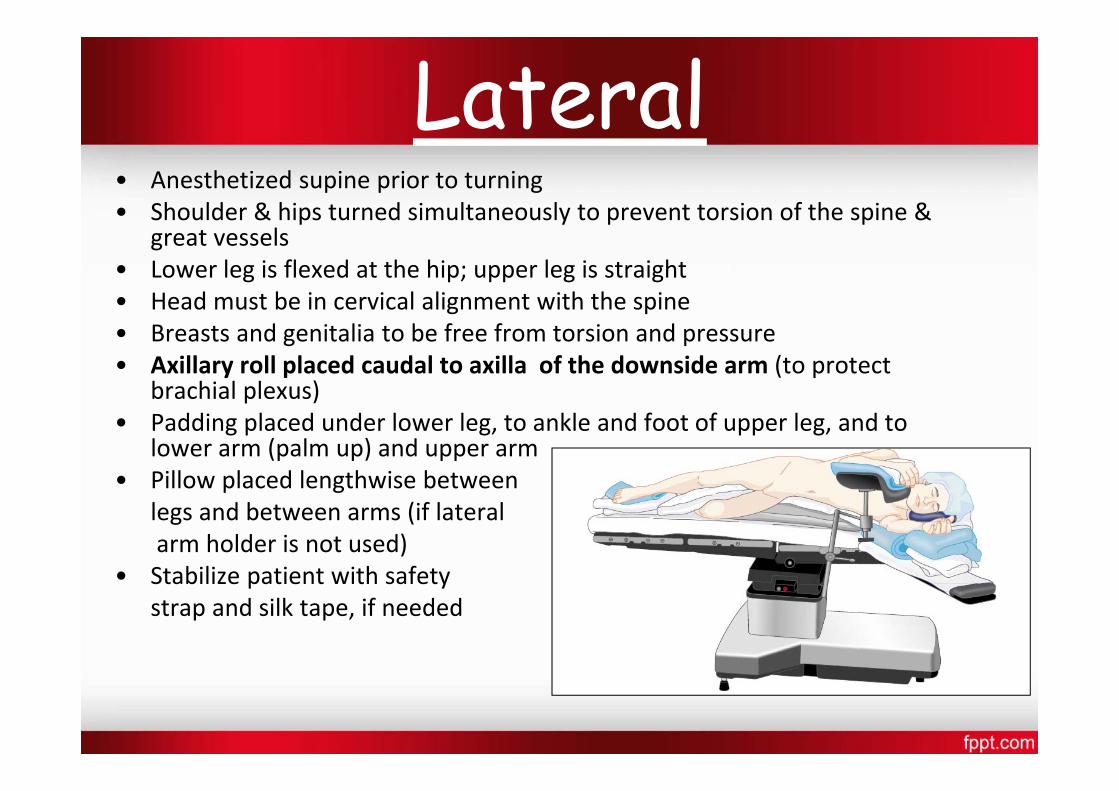
Lateral • Anesthetized supine prior to turning • Shoulder & hips turned simultaneously to prevent torsion of the spine &
great vessels • Lower leg is flexed at the hip; upper leg is straight • Head must be in cervical alignment with the spine • Breasts and genitalia to be free from torsion and pressure • Axillary roll placed caudal to axilla of the downside arm (to protect
brachial plexus) • Padding placed under lower leg, to ankle and foot of upper leg, and to
lower arm (palm up) and upper arm • Pillow placed lengthwise between
legs and between arms (if lateral arm holder is not used)
• Stabilize patient with safety strap and silk tape, if needed
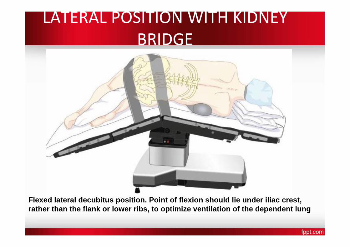
LATERAL POSITION WITH KIDNEY BRIDGE
Flexed lateral decubitus position. Point of flexion should lie under iliac crest, rather than the flank or lower ribs, to optimize ventilation of the dependent lung
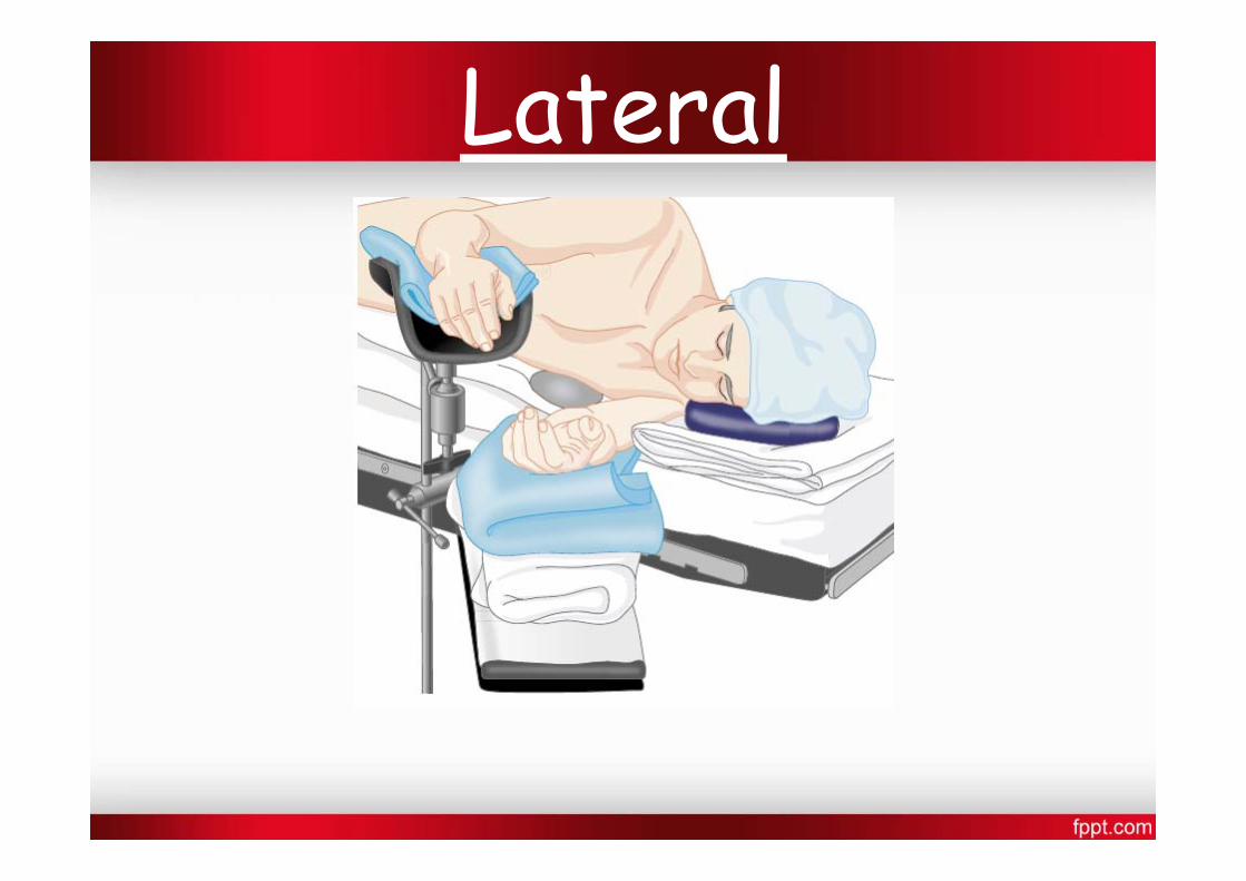
Lateral
• Pulse should be monitored in the dependent arm for early detection of compression to axillary neurovascular structures.
• Low saturation reading in pulse oximetry may be an early warning of compromised circulation.
• When a kidney rest is used, it must be properly placed under the dependent iliac crest to prevent inadvertent compression of the inferior vena cava

Park‐bench position: (SEMI‐PRONE POSITION) – Modification of lat. position. – Better access to posterior fossa. – Upper arm positioned along lateral trunk & uppershoulder is taped towards table.
Hemodynamic and Ventilation.
• In awake patient, Zone 3 West is occupying the dependent 18cm of lung tissue. Lung tissue above 18 cm from bed level isnot perfused.
• During GA & positive pressure ventilation, the non‐dependentlung zones are ventilated better ‐ worsening V/Q mismatch
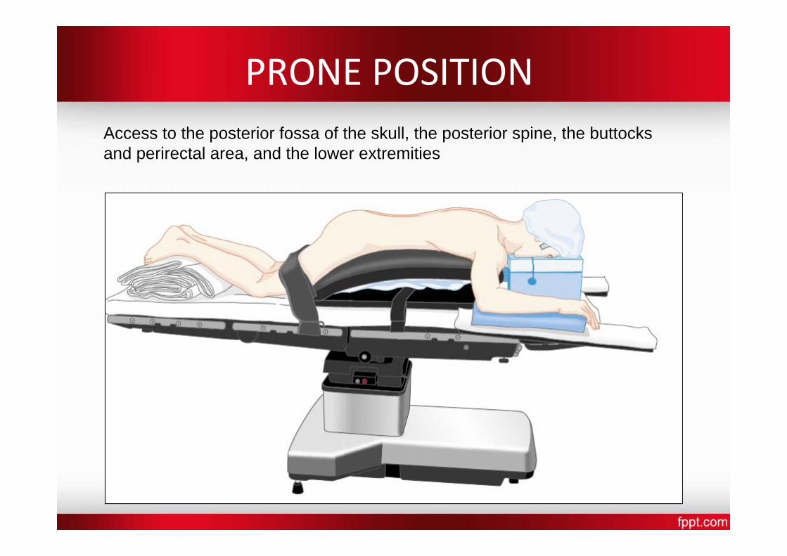
PRONE POSITION Access to the posterior fossa of the skull, the posterior spine, the buttocks and perirectal area, and the lower extremities
• Arms :tucked in the neutral position /placed next to the patient's head on arm boards—sometimes called the prone “superman” position/Extra padding under the elbow – prevent ulnar nerve
• When GA is planned, the patient is intubated on the stretcher/ i.v access is obtained/ETT is well secured/pt is turned prone onto the OT table/disconnect blood pressure cuffs and arterial and venous lines that are on the side to avoid dislodgment
• disconnection of pulse oximetry,arterial line, and tracheal tube, leading to hypoventilation, desaturation, hemodynamic instability, and altered anesthetic depth. Therefore its best to keep pulse oximetry and arterial line connected
• ETT position is reassessed immediately after the move
• Head position
Turned to the side(45 degrees) if neck mobility is fine. Check the dependent eye for external compression. Maintained by surgical pillow, horseshoe headrest, or
Mayfield head pins Mostly, including disposable foam versions, support the forehead, malar regions, and the chin, with a cutout for the eyes, nose, and mouth
Mirror systems are available to facilitate intermittent visual confirmation
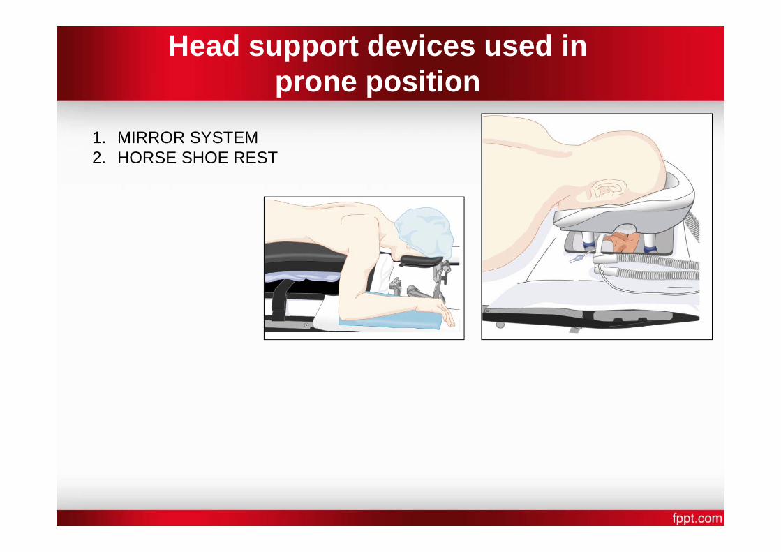
Head support devices used in prone position
1. MIRROR SYSTEM 2. HORSE SHOE REST
• Increased intra‐abdominal pressure decreases FRC, compliance and increased PAP and transmits elevated VP to the abdominal and spine vessels‐increase bleeding risk.
• Its imp that the abdomen hangs free and moves with respiration‐ space of atleast 6 cms!
• Thorax: firm rolls or bolsters placed each side from the clavicle to the iliac crest ( wilson frame, jackson table, relton frame)
• Pendulous structures (e.g., Male genitalia and female breasts) should be clear of compression
• Its essential to check the ETT position at a required degree of flexion
• Hemodynamics and Ventilation • increases intraabdominal pressure, decreases VR to the heart, and
increases systemic and pulmonary vascular resistance‐ HYPOTENSION
• Oxygenation and oxygen delivery, however, may improve as
1) Perfusion of the entire lungs improves 2) Increase in intraabdominal pressure decreases chest wall compliance, which under PPV, improves ventilation of the dependent zones of the lung, and
3) Previously atelectatic dorsal zones of lungs may open.
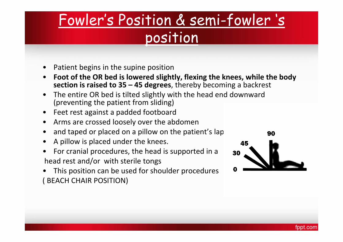
Fowler’s Position & semi-fowler ‘s position
• Patient begins in the supine position • Foot of the OR bed is lowered slightly, flexing the knees, while the body
section is raised to 35 – 45 degrees, thereby becoming a backrest • The entire OR bed is tilted slightly with the head end downward
(preventing the patient from sliding) • Feet rest against a padded footboard • Arms are crossed loosely over the abdomen • and taped or placed on a pillow on the patient’s lap • A pillow is placed under the knees. • For cranial procedures, the head is supported in a head rest and/or with sterile tongs • This position can be used for shoulder procedures ( BEACH CHAIR POSITION)
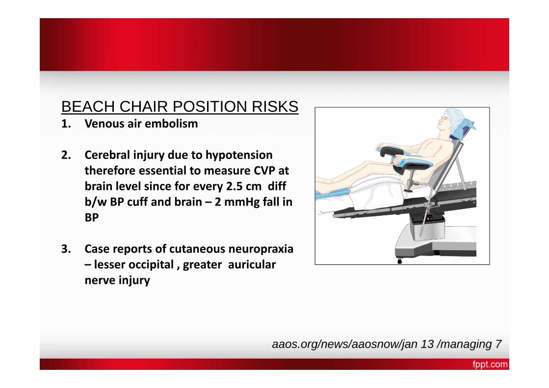
BEACH CHAIR POSITION RISKS 1. Venous air embolism
2. Cerebral injury due to hypotension therefore essential to measure CVP at brain level since for every 2.5 cm diff b/w BP cuff and brain – 2 mmHg fall in BP
3. Case reports of cutaneous neuropraxia – lesser occipital , greater auricular nerve injury
aaos.org/news/aaosnow/jan 13 /managing 7
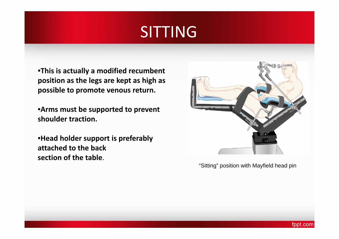
SITTING
•This is actually a modified recumbent position as the legs are kept as high as possible to promote venous return.
•Arms must be supported to prevent shoulder traction.
•Head holder support is preferably attached to the back section of the table.
“Sitting” position with Mayfield head pin
• ADVANTAGE: Excellent surgical exposure
Reduced perioperative blood loss Superior access to the airway
Reduced facial swelling
Improved ventilation, particularly in obese patients
Modern monitoring‐ early indication of air embolism
RELATIVE CONTRAINDICATIONS OF SITTING POSITION
• VP shunt • Cerebral ischemia upright awake
• Patent foramen ovale(detected preop by contrast ECHO)
• Cardiac instability /extreme ages
PROBLEMS
• Venous air embolism
• Hypotension (prevented by stockings) • Arms if not supported well‐ brachial plexus injury

VENOUS AIR EMBOLISM‐MONITOR WARNINGS
Mandatory monitoring: EKG, SPO2, ETCO2, BP,DOPPLER,CVP,PAP
SPECIAL CONCERNS in NEUROSURGICAL PATIENTS
Head Positioning
Ideal position of head for Craniotomies & spine procedures based on the 2 principles:
1) An imaginary trajectory from the highest point at skull surface to area of interest in brain should be the shortest distance between the 2 points.
2) The exposed surface of the skull & an imaginary perimeter of craniotomy should be parallel to the floor.
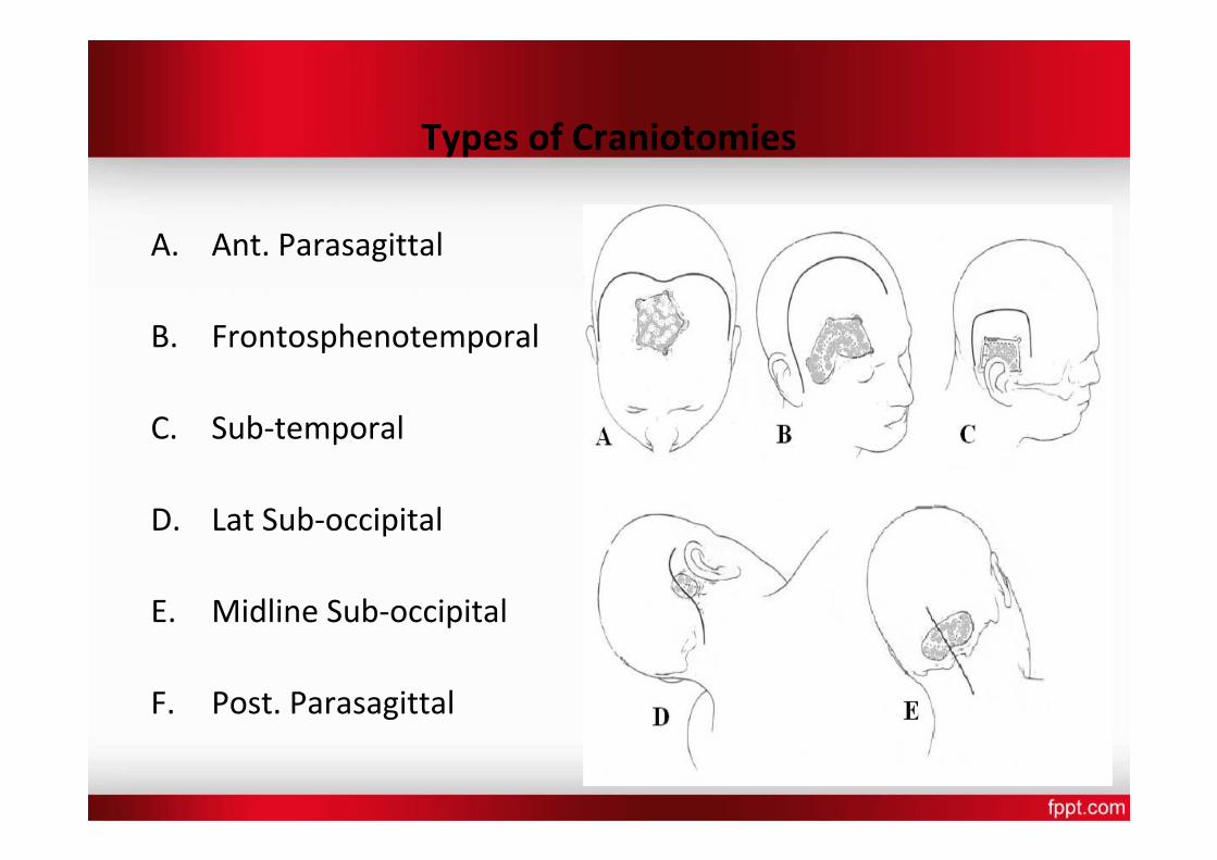
Types of Craniotomies
A. Ant. Parasagittal
B. Frontosphenotemporal
C. Sub‐temporal
D. Lat Sub‐occipital
E. Midline Sub‐occipital
F. Post. Parasagittal
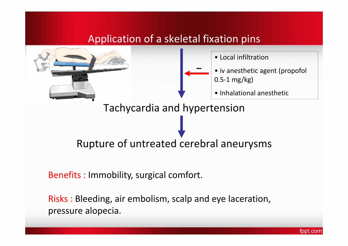
Application of a skeletal fixation pins • Local infiltration
• iv anesthetic agent (propofol 0.5‐1 mg/kg)
• Inhalational anesthetic
Tachycardia and hypertension
Rupture of untreated cerebral aneurysms
Benefits : Immobility, surgical comfort.
Risks : Bleeding, air embolism, scalp and eye laceration, pressure alopecia.
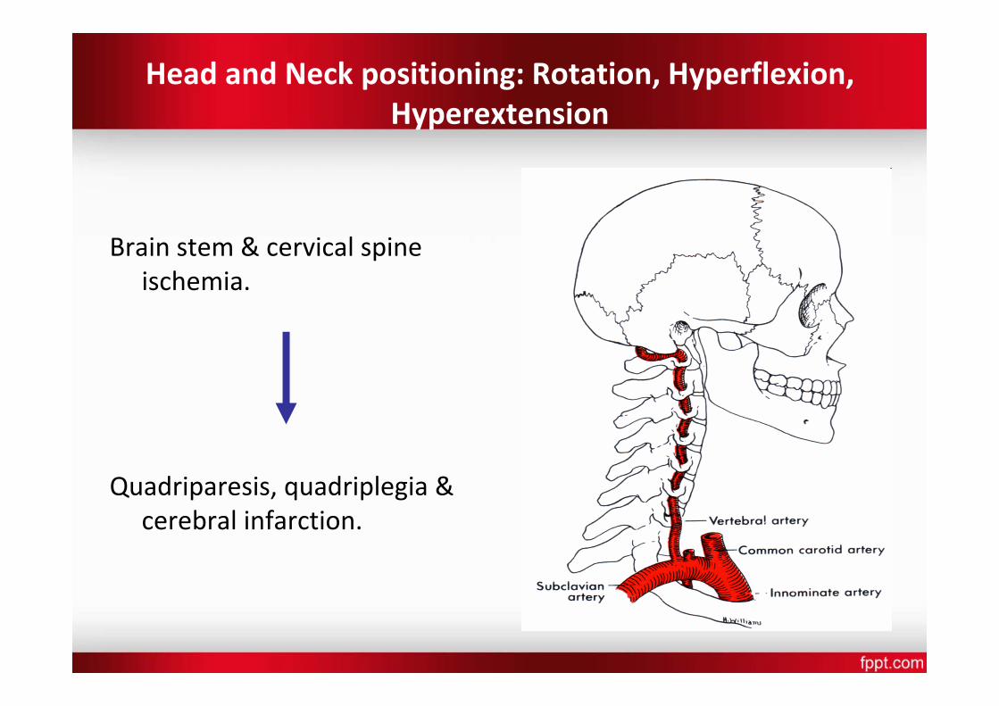
Head and Neck positioning: Rotation, Hyperflexion, Hyperextension
Brain stem & cervical spine ischemia.
Quadriparesis, quadriplegia & cerebral infarction.
• At risk Patients : – Osteophytes – Arthritis – Vascular atherosclerosis – Obesity – Hemodynamically unstable
• Head safely rotated b/w 0‐45°.
• For more rotation, a roll/pillow place under the opposite shoulder.
• Maintaining 2‐3 finger breadths thyromental distance during neck flexion.
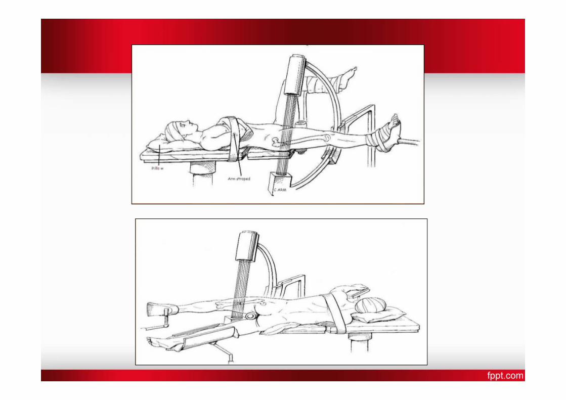
Orthopaedic surgery position
• Orthopedic fracture table – Wattson‐Jone’s Body section to support head & thorax Sacral plate for pelvis Perineal post Adjustable foot plates Table maintains traction of the extremity
Allows surgical & fluroscopic access Anesthesia induced & then the patients are positioned on
this table . Arm on side
Problems with jones position
• Brachial plexus injury due to > than 90* extension of the upper limb
• Lower extremity compartment syndrome due to long surgeries & compression
• Pudendal nerve injury Due to pressure of the perineal post
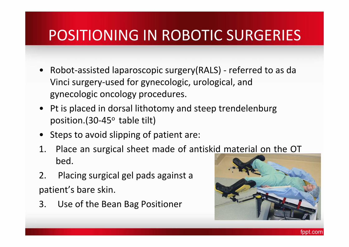
POSITIONING IN ROBOTIC SURGERIES
• Robot‐assisted laparoscopic surgery(RALS) ‐ referred to as da Vinci surgery‐used for gynecologic, urological, and gynecologic oncology procedures.
• Pt is placed in dorsal lithotomy and steep trendelenburg position.(30‐45o table tilt)
• Steps to avoid slipping of patient are: 1.
bed. 2. Placing surgical gel pads against a
patient’s bare skin. 3. Use of the Bean Bag Positioner
Place an surgical sheet made of antiskid material on the OT
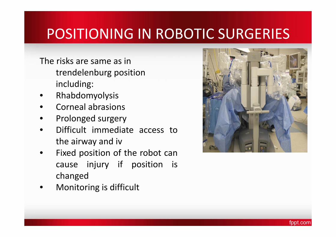
POSITIONING IN ROBOTIC SURGERIES
The risks are same as in trendelenburg position including:
• Rhabdomyolysis • Corneal abrasions • Prolonged surgery • Difficult immediate access to
the airway and iv • Fixed position of the robot can
cause injury if position is changed
• Monitoring is difficult
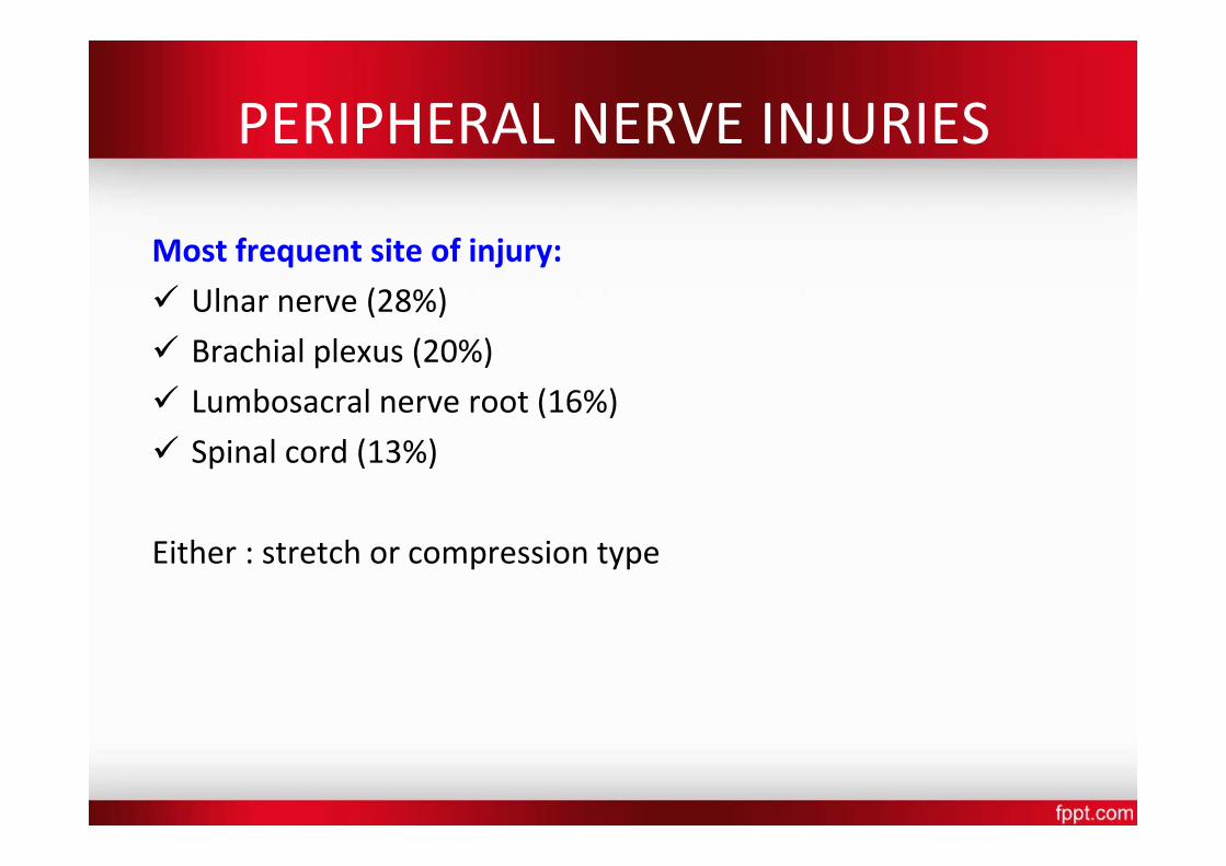
PERIPHERAL NERVE INJURIES
Most frequent site of injury: Ulnar nerve (28%) Brachial plexus (20%) Lumbosacral nerve root (16%) Spinal cord (13%)
Either : stretch or compression type
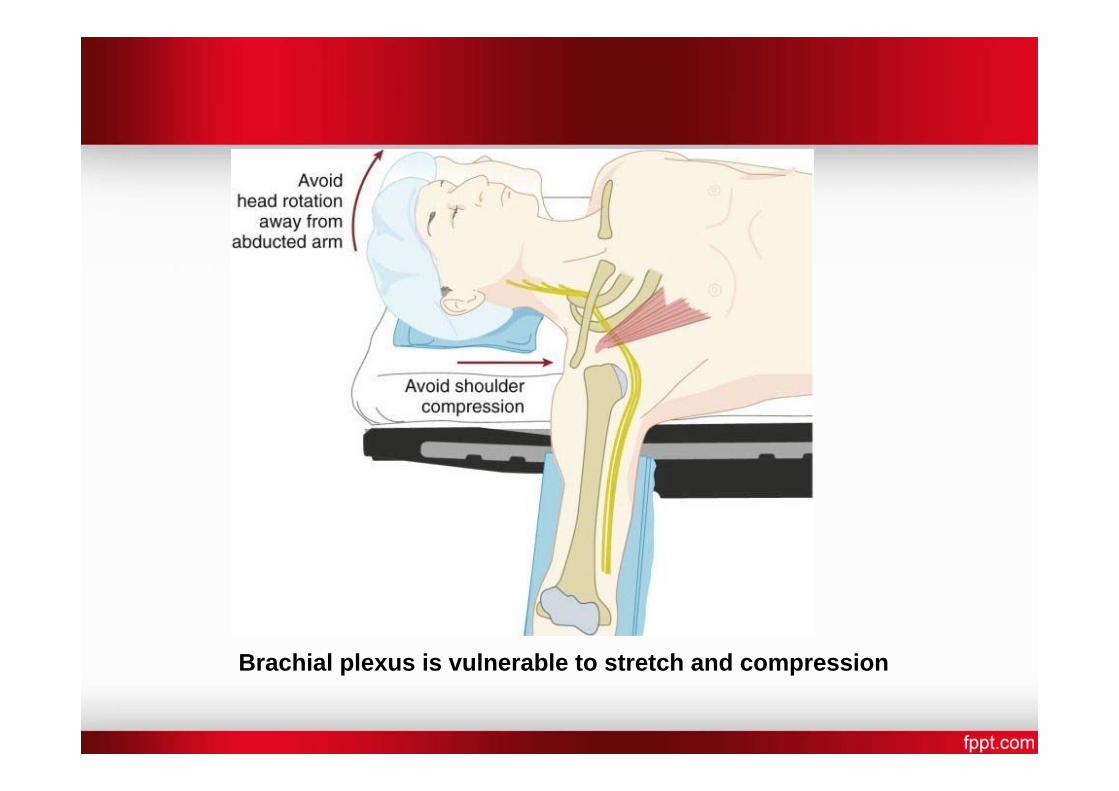
Brachial plexus is vulnerable to stretch and compression
CLASSIFICATION OF NERVE INJURY
Seddon’s classification
• Neurapraxia: damaged myelin with intact axon. Impulse conduction across the affected segment fails. Mild and reversible nerve injury. Recovery usually occurs in weeks to months and prognosis is good.
• Axonotmesis: axonal disruption. Endoneurium and other supporting connective tissue are preserved. Recovery and prognosis is variable.
• Neurotmesis: nerve is completely severed. There is complete destruction of all supporting connective tissue structures. Surgery may be required and prognosis is poor.
MANAGEMENT
• Pt Assessment‐ Findings must be well documented and neurologist review should occur.
• early Investigations include nerve conduction studies and electromyography(EMG).Progressive or severe pathology needs urgent neurologist assessment and immediate investigation.
• Electrophysiology can help distinguish between nerve dysfunction due to axonal degeneration (such as with PPNI) / nerve dysfunction due to demyelination ( like carpal tunnel syndrome). The electrophysiological diagnosis of degeneration is based on finding reduced numbers of functioning axons.
• Since 14 days required for nerve degeneration to occur so this study done only in few wks after the onset of symptoms
ASA GUIDELINES TO PREVENT PERIOPERATIVE PERIPHERAL
NEUROPATHY
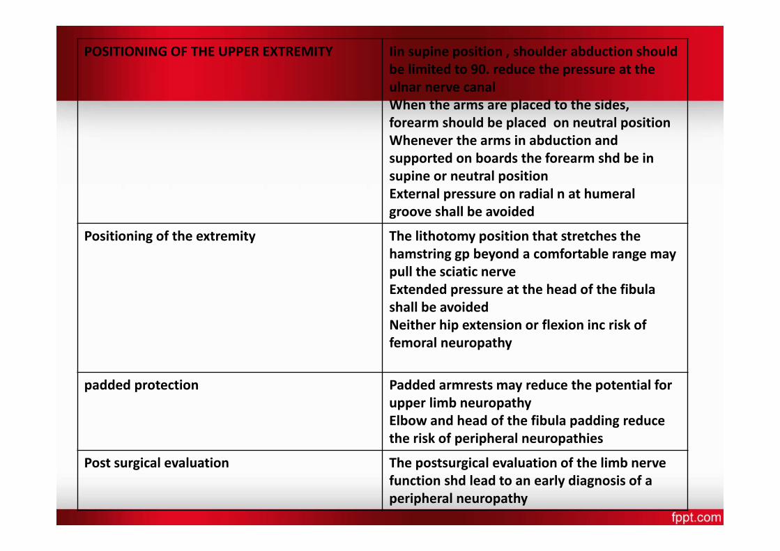
POSITIONING OF THE UPPER EXTREMITY Iin supine position , shoulder abduction should be limited to 90. reduce the pressure at the ulnar nerve canal When the arms are placed to the sides, forearm should be placed on neutral position Whenever the arms in abduction and supported on boards the forearm shd be in supine or neutral position External pressure on radial n at humeral groove shall be avoided
Positioning of the extremity The lithotomy position that stretches the hamstring gp beyond a comfortable range may pull the sciatic nerve Extended pressure at the head of the fibula shall be avoided Neither hip extension or flexion inc risk of femoral neuropathy
padded protection Padded armrests may reduce the potential for upper limb neuropathy Elbow and head of the fibula padding reduce the risk of peripheral neuropathies
Post surgical evaluation The postsurgical evaluation of the limb nerve function shd lead to an early diagnosis of a peripheral neuropathy
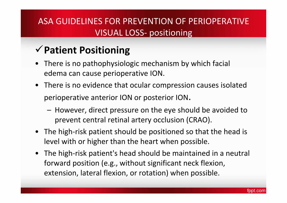
ASA GUIDELINES FOR PREVENTION OF PERIOPERATIVE VISUAL LOSS‐ positioning
Patient Positioning • There is no pathophysiologic mechanism by which facial
edema can cause perioperative ION. • There is no evidence that ocular compression causes isolated
perioperative anterior ION or posterior ION. – However, direct pressure on the eye should be avoided to prevent central retinal artery occlusion (CRAO).
• The high‐risk patient should be positioned so that the head is level with or higher than the heart when possible.
• The high‐risk patient's head should be maintained in a neutral forward position (e.g., without significant neck flexion, extension, lateral flexion, or rotation) when possible.
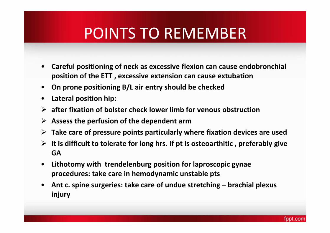
POINTS TO REMEMBER
• Careful positioning of neck as excessive flexion can cause endobronchial position of the ETT , excessive extension can cause extubation
• On prone positioning B/L air entry should be checked
• Lateral position hip: after fixation of bolster check lower limb for venous obstruction
Assess the perfusion of the dependent arm
Take care of pressure points particularly where fixation devices are used
It is difficult to tolerate for long hrs. If pt is osteoarthitic , preferably give GA
• Lithotomy with trendelenburg position for laproscopic gynae procedures: take care in hemodynamic unstable pts
• Ant c. spine surgeries: take care of undue stretching – brachial plexus injury
Related Documents