REVIEW ARTICLE published: 24 October 2012 doi: 10.3389/fmicb.2012.00378 Pathways of cell–cell transmission of HTLV-1 Claudine Pique 1 and Kathryn S. Jones 2 * 1 CNRS UMR 8104, INSERM U567, Université Paris-Descartes, Institut Cochin, Paris, France 2 Cancer and Inflammation Program, Basic Research Program, SAIC-Frederick, Inc., Center for Cancer Research, National Cancer Institute, Frederick National Laboratory for Cancer Research, Frederick, MD, USA Edited by: Renaud Mahieux, École Normale Supérieure de Lyon, France Reviewed by: Youichi Suzuki, National University of Singapore, Singapore Steven Jacobson, National Institutes of Health, USA David W. Brighty, University of Dundee, UK *Correspondence: Kathryn S. Jones, Cancer and Inflammation Program, Basic Research Program, SAIC-Frederick, Inc., Center for Cancer Research, National Cancer Institute, Frederick National Laboratory for Cancer Research, Building 567, Room 253, Frederick, MD 21702, USA. e-mail: [email protected] The deltaretroviruses human T cell lymphotropic virus type 1 (HTLV-1) and human T cell lymphotropic virus type 2 (HTLV-2) have long been believed to differ from retroviruses in other genera by their mode of transmission. While other retroviruses were thought to pri- marily spread by producing cell-free particles that diffuse through extracellular fluids prior to binding to and infecting target cells, HTLV-1 and HTLV-2 were believed to transmit the virus solely by cell–cell interactions.This difference in transmission was believed to reflect the fact that, relative to other retroviruses, the cell-free virions produced by HTLV-infected cells are very poorly infectious. Since HTLV-1 and HTLV-2 are primarily found inT cells in the peripheral blood, spread of these viruses was believed to occur between infected and uninfected T cells, although little was known about the cellular and viral proteins involved in this interaction. Recent studies have revealed that the method of transmission of HTLV is not unique: other retroviruses including human immunodeficiency virus (HIV) are also transmitted from cell-to-cell, and this method is dramatically more efficient than cell-free transmission. Moreover, cell–cell transmission of HTLV-1, as well as HIV, can occur follow- ing interactions between dendritic cells and T cells, as well as between T cells. Conversely, other studies have shown that cell-free HTLV-1 is not as poorly infectious as previously thought, since it is capable of infecting certain cell types. Here we summarize the recent insights about the mechanisms of cell–cell transmission of HTLV-1 and other retroviruses. We also review in vitro and in vivo studies of infection and discuss how these finding may relate to the spread of HTLV-1 between individuals. Keywords: retrovirus, HTLV-1, cell–cell transmission, HTLV-1 infectivity,T cell, antigen-presenting cells, HTLV-2, virological synapse INTRODUCTION Human T cell lymphotropic virus type 1 (HTLV-1) was the first pathogenic retrovirus discovered in humans (Poiesz et al., 1980). This virus is the only human retrovirus known to be the causal agent of a cancer, a neoplasia called adult T cell leukemia (ATL; Yoshida etal., 1982, 1984). HTLV-1 is also associated with several inflammatory disorders, primarily a progressive neurologi- cal disease named HTLV-1-associated myelopathy/tropical spastic paraparesis (HAM/TSP; Gessain et al., 1985; Osame et al., 1986). The closely related retrovirus human T cell leukemia virus type 2 (HTLV-2) does not cause leukemia, although some infected individuals develop mild lymphocytosis and, occasionally, neuro- logic symptoms (Hjelle et al., 1992; Dooneief et al., 1996; Feuer and Green, 2005; Biswas et al., 2009). In the peripheral blood of infected individuals, both HTLV-1 and HTLV-2 are primar- ily found in T cells, and both viruses can immortalize T cells in culture (reviewed in Feuer and Green, 2005). ATL is a malignancy of CD4 + T cells, and HTLV-1 primarily infects CD4 + T cells, while HTLV-2 primarily infects CD8 + T cells (Kira et al., 1991; Lal et al., 1995; Wu et al., 1996; Manns et al., 1999; Nagai et al., 2001; Mur- phy et al., 2004). Dendritic cells (DCs), B cells, and monocytes can also be infected in individuals with HTLV (Macatonia et al., 1992; Koyanagi et al., 1993). Like other retroviruses, HTLV-1 and HTLV-2 enter target cells following specific interactions between the viral envelope glycoprotein (Env) and cellular receptors. Efficient entry of HTLV- 1 has been shown to involve three distinct molecules: heparan sulfate proteoglycans (HSPGs) and Neuropilin 1 (NRP-1) for the initial binding to the cell, and glucose transporter 1 (GLUT1) for entry (Manel et al., 2003; Jones et al., 2005; Ghez et al., 2006; Lambert et al., 2009). HTLV-2 binding and entry also involves NRP-1 and GLUT1, but differs from HTLV-1 in that it does not require HSPG. The studies identifying the HTLV receptors have recently been extensively reviewed (Ghez et al., 2010; Ilinskaya et al., 2010; Jones et al., 2011; Hoshino, 2012) and thus will not be detailed here. For many years, HTLV-1 and HTLV-2 were believed to differ from retroviruses in other genera in their mode of transmission. For the other retroviruses, viral spread was thought to require the production of cell-free viral particles that diffuse through extra- cellular fluids and subsequently bind to, enter, and infect target cells. For HTLV-1 and HTLV-2, it was believed that the cell-free virus was poorly infectious, and that the viruses could only be effi- ciently spread by direct contact between infected cells and target cells (cell–cell transmission). The belief that HTLV-1 is poorly infectious as a cell-free particle originated soon after its discovery and was based on both in vivo and in vitro observations. Studies of transfusion suggested that cell–cell contact is required for HTLV-1 transmis- sion: although a high percentage of individuals receiving cellular www.frontiersin.org October 2012 | Volume 3 | Article 378 | 1

Welcome message from author
This document is posted to help you gain knowledge. Please leave a comment to let me know what you think about it! Share it to your friends and learn new things together.
Transcript
“fmicb-03-00378” — 2012/10/22 — 22:31 — page 1 — #1
REVIEW ARTICLEpublished: 24 October 2012
doi: 10.3389/fmicb.2012.00378
Pathways of cell–cell transmission of HTLV-1Claudine Pique1 and Kathryn S. Jones2*
1 CNRS UMR 8104, INSERM U567, Université Paris-Descartes, Institut Cochin, Paris, France2 Cancer and Inflammation Program, Basic Research Program, SAIC-Frederick, Inc., Center for Cancer Research, National Cancer Institute, Frederick National
Laboratory for Cancer Research, Frederick, MD, USA
Edited by:
Renaud Mahieux, École NormaleSupérieure de Lyon, France
Reviewed by:
Youichi Suzuki, National University ofSingapore, SingaporeSteven Jacobson, National Institutesof Health, USADavid W. Brighty, University ofDundee, UK
*Correspondence:
Kathryn S. Jones, Cancer andInflammation Program, BasicResearch Program, SAIC-Frederick,Inc., Center for Cancer Research,National Cancer Institute, FrederickNational Laboratory for CancerResearch, Building 567, Room 253,Frederick, MD 21702, USA.e-mail: [email protected]
The deltaretroviruses human T cell lymphotropic virus type 1 (HTLV-1) and human T celllymphotropic virus type 2 (HTLV-2) have long been believed to differ from retroviruses inother genera by their mode of transmission. While other retroviruses were thought to pri-marily spread by producing cell-free particles that diffuse through extracellular fluids priorto binding to and infecting target cells, HTLV-1 and HTLV-2 were believed to transmit thevirus solely by cell–cell interactions. This difference in transmission was believed to reflectthe fact that, relative to other retroviruses, the cell-free virions produced by HTLV-infectedcells are very poorly infectious. Since HTLV-1 and HTLV-2 are primarily found in T cells inthe peripheral blood, spread of these viruses was believed to occur between infected anduninfected T cells, although little was known about the cellular and viral proteins involvedin this interaction. Recent studies have revealed that the method of transmission of HTLVis not unique: other retroviruses including human immunodeficiency virus (HIV) are alsotransmitted from cell-to-cell, and this method is dramatically more efficient than cell-freetransmission. Moreover, cell–cell transmission of HTLV-1, as well as HIV, can occur follow-ing interactions between dendritic cells andT cells, as well as betweenT cells. Conversely,other studies have shown that cell-free HTLV-1 is not as poorly infectious as previouslythought, since it is capable of infecting certain cell types. Here we summarize the recentinsights about the mechanisms of cell–cell transmission of HTLV-1 and other retroviruses.We also review in vitro and in vivo studies of infection and discuss how these finding mayrelate to the spread of HTLV-1 between individuals.
Keywords: retrovirus, HTLV-1, cell–cell transmission, HTLV-1 infectivity, T cell, antigen-presenting cells, HTLV-2,
virological synapse
INTRODUCTIONHuman T cell lymphotropic virus type 1 (HTLV-1) was thefirst pathogenic retrovirus discovered in humans (Poiesz et al.,1980). This virus is the only human retrovirus known to be thecausal agent of a cancer, a neoplasia called adult T cell leukemia(ATL; Yoshida et al., 1982, 1984). HTLV-1 is also associated withseveral inflammatory disorders, primarily a progressive neurologi-cal disease named HTLV-1-associated myelopathy/tropical spasticparaparesis (HAM/TSP; Gessain et al., 1985; Osame et al., 1986).The closely related retrovirus human T cell leukemia virus type2 (HTLV-2) does not cause leukemia, although some infectedindividuals develop mild lymphocytosis and, occasionally, neuro-logic symptoms (Hjelle et al., 1992; Dooneief et al., 1996; Feuerand Green, 2005; Biswas et al., 2009). In the peripheral bloodof infected individuals, both HTLV-1 and HTLV-2 are primar-ily found in T cells, and both viruses can immortalize T cells inculture (reviewed in Feuer and Green, 2005). ATL is a malignancyof CD4+ T cells, and HTLV-1 primarily infects CD4+ T cells, whileHTLV-2 primarily infects CD8+T cells (Kira et al., 1991; Lal et al.,1995; Wu et al., 1996; Manns et al., 1999; Nagai et al., 2001; Mur-phy et al., 2004). Dendritic cells (DCs), B cells, and monocytes canalso be infected in individuals with HTLV (Macatonia et al., 1992;Koyanagi et al., 1993).
Like other retroviruses, HTLV-1 and HTLV-2 enter targetcells following specific interactions between the viral envelope
glycoprotein (Env) and cellular receptors. Efficient entry of HTLV-1 has been shown to involve three distinct molecules: heparansulfate proteoglycans (HSPGs) and Neuropilin 1 (NRP-1) for theinitial binding to the cell, and glucose transporter 1 (GLUT1)for entry (Manel et al., 2003; Jones et al., 2005; Ghez et al., 2006;Lambert et al., 2009). HTLV-2 binding and entry also involvesNRP-1 and GLUT1, but differs from HTLV-1 in that it does notrequire HSPG. The studies identifying the HTLV receptors haverecently been extensively reviewed (Ghez et al., 2010; Ilinskayaet al., 2010; Jones et al., 2011; Hoshino, 2012) and thus will not bedetailed here.
For many years, HTLV-1 and HTLV-2 were believed to differfrom retroviruses in other genera in their mode of transmission.For the other retroviruses, viral spread was thought to require theproduction of cell-free viral particles that diffuse through extra-cellular fluids and subsequently bind to, enter, and infect targetcells. For HTLV-1 and HTLV-2, it was believed that the cell-freevirus was poorly infectious, and that the viruses could only be effi-ciently spread by direct contact between infected cells and targetcells (cell–cell transmission).
The belief that HTLV-1 is poorly infectious as a cell-freeparticle originated soon after its discovery and was based onboth in vivo and in vitro observations. Studies of transfusionsuggested that cell–cell contact is required for HTLV-1 transmis-sion: although a high percentage of individuals receiving cellular
www.frontiersin.org October 2012 | Volume 3 | Article 378 | 1
“fmicb-03-00378” — 2012/10/22 — 22:31 — page 2 — #2
Pique and Jones Cell–cell transmission of HTLV-1
blood components (whole blood, red blood cells, or platelets)from HTLV-1- or HTLV-2-infected individuals become infectedwith the virus, the recipients of non-cellular blood products(plasma fraction or plasma derivatives) from infected individu-als do not become infected (Maeda et al., 1984; Miyamoto et al.,1984; Jason et al., 1985; Lairmore et al., 1989). In one studydirectly comparing transmission following transfusion of plasmafrom individuals with different human retroviruses, seroconver-sion occurred in 89% of the individuals who received plasmafrom HIV-1 infected individuals, but in none of the individualswho received plasma from individuals with HTLV-1 or HTLV-2(Donegan et al., 1994).
In vitro experiments supported the notion that the cell-freevirus is poorly infectious. Although in the peripheral blood thevirus is primarily found in T cells, early studies showed that cell-free HTLV-1 and HTLV-2 do not efficiently infect or transformprimary T cells isolated from the peripheral blood in vitro. Incontrast, primary T cells can become infected and transformed fol-lowing coculture with either HTLV-1-infected or HTLV-2-infectedcell lines (Yamamoto et al., 1982; Popovic et al., 1983; Green andChen, 1990).
However, other in vitro studies showed that cell-free HTLV-1 is not completely non-infectious. Early studies reported rareinfection of T cells (de Rossi et al., 1985) and non-lymphoid cells(Clapham et al., 1983) by cell-free virus. Later studies using moresensitive assays reported that a number of T and B cell lines (Fanet al., 1992; Agadjanyan et al., 1994; Jinno et al., 1999), as well ascell lines of non-lymphoid origin (Graziani et al., 1993; Haraguchiet al., 1994), could be infected following exposure to cell-free virus,although at a very low level. More recent studies with DCs haveconfirmed and extended the notion that cell-free HTLV-1 can beinfectious. Several groups have demonstrated that the primaryDCs, unlike T cells, are routinely infected after exposure to cell-free virus (Jones et al., 2008; Jain et al., 2009; Lambert et al., 2009;Valeri et al., 2010). In addition, in contrast to what occurs in cul-tures of purified T cells, T cells cocultured with DCs routinelybecome infected after the addition of cell-free virus. Further stud-ies showed that when DCs are exposed to cell-free virus, theyrapidly transmit the virus to T cells. These observations suggestthat there may be blocks to infection after the cell-free particlebinds to T cells that are not present when the virus is presented byDC cells to T cells, or that interacting with the DCs alters the virusin a way that allows it to infect T cells.
As described above, several early studies showed that the plasmafrom HTLV-1- and HTLV-2-infected individuals does not infectrecipients of transfusions. Originally, these studies were inter-preted as reflecting the fact that cell-free HTLV-1 and HTLV-2virions in the blood are far less infectious and/or more labilethan HIV-1 virions. However, later studies revealed that cell-freeviral particles are only rarely be detected in plasma or serum fromHTLV-1- or HTLV-2-infected individuals. Other studies revealedthat, even in individuals with a high percentage of PBMCs contain-ing integrated HTLV-1 genomic DNA, little or no viral mRNAs orproteins are detected in PBMC immediately after isolation (Ges-sain et al., 1991; Richardson et al., 1997; Moritoyo et al., 1999).These observations indicate that the lack of infection through acel-lular blood products is due to the lack of detectable levels of virus
in peripheral blood, and thus does not support the notion thatHTLV-1 particles are inherently non-infectious.
The lack of viremia in individuals with HTLV-1 is in sharp con-trast to the high levels of viremia and active replication found inHIV-1-infected individuals. Indeed, the method of persistence ofHTLV-1 in infected individuals appears to be very different fromthat of HIV-1. The peripheral blood of HTLV-1-infected individu-als contains clones of large numbers of infected cells with the sameintegration site (Wattel et al., 1995; Leclercq et al., 1998; Zane et al.,2009), indicating that they are derived from a single infected cell.Further studies revealed that specific clones can persist over yearsin a given individual (Cavrois et al., 1996; Etoh et al., 1997; Cavroiset al., 1998). A recent characterization of infected cells using high-throughput methods revealed that, in a typical HTLV-1-infectedindividual without ATL, there are between 500 and 5000 clonesand the majority of these clones are maintained over a period ofyears (Gillet et al., 2011).
The presence of these clones indicates that, rather than spread-ing from cell-to-cell, HTLV-1 persists in individuals primarily bymitotic replication of infected cells. Consistent with this, the per-centage of infected cells (referred to as the HTLV-1 proviral load)remains stable within an individual over time. Moreover, unlikeHIV-1, the HTLV-1 genome shows very little variation within anindividual, consistent with it being replicated by cellular DNApolymerase during division of infected cells rather than the moreerror-prone reverse transcriptase. Taken together, these observa-tions have lead to the belief that HTLV-1 persists in two stagesin an individual. Soon after an individual is exposed to the virus,HTLV-1 spreads from cell-to-cell. Later, during the chronic stageof infection, the virus persists via clonal expansion, through repli-cation of the provirus integrated into the host cell genome duringthe division of infected cells.
Ten years ago, little was known about the mechanism ofthe cell–cell transmission of HTLV-1. Since that time, imagingstudies along with in vitro studies of infection have providedinsight into the interactions between cells required for infectionof T cells by HTLV-1. During this time it has also become clearthat cell–cell transmission is not unique to deltaretroviruses: bothHIV and the gammaretrovirus murine leukemia viruses (MLV)can also be transmitted by cell–cell contacts, and this mode oftransmission is more efficient than cell-free virus. Here, we reviewwhat has recently been learned about transmission of HTLV-1, including observations that cell–cell transmission can occurbetween DC and T cells, as well as between T cells. We also reviewwhat has been learned about the precise interactions betweencells required for the infection of the target cells by HTLV-1 andby other retroviruses during cell–cell transmission, and discusshow these finding may relate to the spread of HTLV-1 betweenindividuals.
THE VIROLOGICAL SYNAPSEAlthough the observation that cell–cell contact is important forHTLV-1 transmission was made soon after the discovery of thevirus, for many years little was known about the mechanism ofcell-to-cell spread of this virus at the cellular level. In 2003, imag-ing techniques allowed the initial characterization of a specifictype of cell contact that allows virions from an HTLV-1-infected
Frontiers in Microbiology | Virology October 2012 | Volume 3 | Article 378 | 2
“fmicb-03-00378” — 2012/10/22 — 22:31 — page 3 — #3
Pique and Jones Cell–cell transmission of HTLV-1
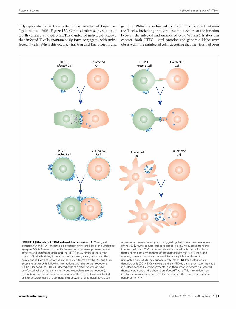
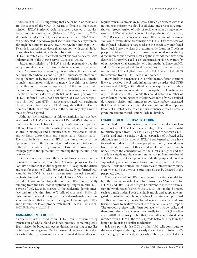
T lymphocyte to be transmitted to an uninfected target cell(Igakura et al., 2003; Figure 1A). Confocal microscopy studies ofT cells cultured ex vivo from HTLV-1-infected individuals showedthat infected T cells spontaneously form conjugates with unin-fected T cells. When this occurs, viral Gag and Env proteins and
genomic RNAs are redirected to the point of contact betweenthe T cells, indicating that viral assembly occurs at the junctionbetween the infected and uninfected cells. Within 2 h after thiscontact, both HTLV-1 viral proteins and genomic RNAs wereobserved in the uninfected cell, suggesting that the virus had been
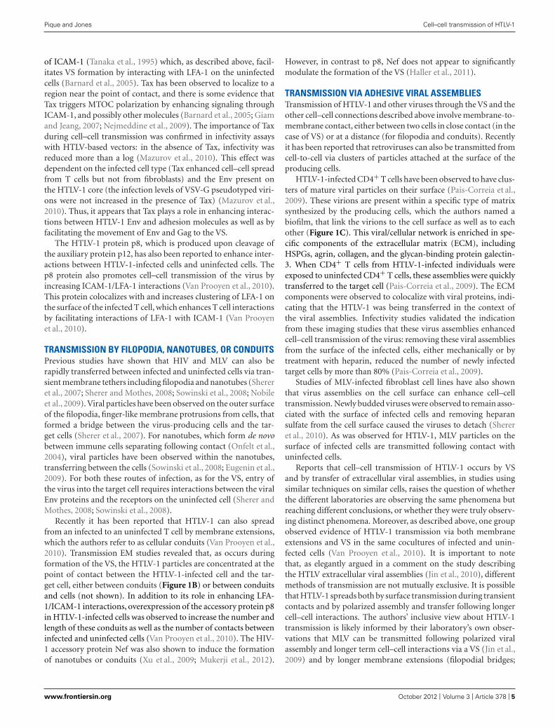
FIGURE 1 | Models of HTLV-1 cell–cell transmission. (A) Virologicalsynapse. When HTLV-1-infected cells contact uninfected cells, the virologicalsynapse (VS) is formed by specific interactions between proteins on theinfected and uninfected cells, and the MTOC (gray circle) is reorientedtoward VS. Viral budding is polarized to the virological synapse, and thenewly budded viruses enter the synaptic cleft formed by the VS, and thenenter the target cells following interactions with the cellular receptors.(B) Cellular conduits. HTLV-1-infected cells can also transfer virus touninfected cells by transient membrane extensions (cellular conduit).Interactions can occur between conduits on the infected and uninfectedcell, or between cells and conduits (not shown), and particles have been
observed at these contact points, suggesting that these may be a variantof the VS. (C) Extracellular viral assemblies. Following budding from theinfected cell, the HTLV-1 virus remains associated with the cell within amatrix containing components of the extracellular matrix (ECM). Uponcontact, these adhesive viral assemblies are rapidly transferred to anuninfected cell, which they subsequently infect. (D) Trans-infection viadendritic cells (DCs). DCs capture cell-free HTLV-1, transiently store the virusin surface-accessible compartments, and then, prior to becoming infectedthemselves, transfer the virus to uninfected T cells. This interaction mayinvolve membrane extensions of the DCs and/or the T cells, as has beenobserved for HIV.
www.frontiersin.org October 2012 | Volume 3 | Article 378 | 3
“fmicb-03-00378” — 2012/10/22 — 22:31 — page 4 — #4
Pique and Jones Cell–cell transmission of HTLV-1
transmitted to the target cell (Igakura et al., 2003). By analogy toother previously described specialized junctions, such as neuronaland immunological synapses, the structure at the junction betweenthe infected and uninfected cell was named the virological synapse(VS; Igakura et al., 2003).
Soon after the description of the VS in HTLV-1 transmission, itwas reported that HIV can also be transmitted from an uninfectedto infected T cell via a VS (Jolly et al., 2004) and, as detailed below,a number of subsequent studies have more fully characterized theHIV VS. Another retrovirus, MLV, has also been shown to form aVS between infected and uninfected cells (Sherer et al., 2007; Jinet al., 2009).
For both these viruses, interactions between the virally encodedEnv proteins and entry receptors are critical for the initial stage ofinfection, during which stable interactions are formed between thecells. For HIV, formation of the VS has been shown to be initiatedby interactions between the surface (SU) envelope glycoproteingp120 on the infected T cell and the receptor molecule CD4 onthe target T cell (Jolly et al., 2004; Felts et al., 2010). Similarly,MLV infection was observed to involve interactions between itsSU (gp70) on fibroblasts expressing viral proteins and the MLVreceptor mCAT-1 on the target cell (Jin et al., 2009). Althoughit has not yet been directly examined, there is some evidencethat supports the notion that Env/receptors interactions are alsorequired to trigger the formation of the HTLV-1 VS. It has beenshown that NRP-1 and Glut1 colocalize at the junction formedbetween an HTLV-1-infected T cell and a non-infected targetT cell (Ghez et al., 2006). Moreover, blocking interactions withHSPGs or NRP-1 on T cells has also been shown to block infec-tion following cell–cell transmission from HTLV-1-infected DC(Jones et al., 2008).
For the HIV-1 VS, it has been reported that the initialEnv/receptor interaction between the infected and uninfected cellsis reinforced by interactions mediated by adhesion molecules thatstabilize the junction. These include specific interactions betweenthe integrins intercellular adhesion molecule 1 (ICAM-1) andICAM-3 and their ligand lymphocyte function-associated anti-gen 1 (LFA-1; Jolly et al., 2007). Interactions between ICAM-1 andLFA-1 have also been shown to be critical for formation of theHTLV-1 VS (Barnard et al., 2005).
These stable contacts between the two cells delineate an intra-cellular space referred to as the synaptic cleft. Electron tomographystudies revealed the presence of HTLV-1 particles in the synap-tic cleft between infected and uninfected primary lymphocytes(Majorovits et al., 2008). Many of the viruses observed were touch-ing the membrane of the infected cell, the target cell, or both,indicating that HTLV-1 is released from the polarized cell intothe synaptic cleft, and is efficiently transferred to the target cell.This is similar to what has been observed for HIV and MLVduring formation of their VS: Gag and Env proteins of theseviruses localize to the point of cell–cell contact, and the virusesbud into the synaptic cleft (Jolly et al., 2004; Hubner et al., 2009;Jin et al., 2009).
It seems that the increased efficiency of cell–cell transmissionover cell-free transmission reflects at least in part a quantitativeadvantage, since it allows the polarized release of large numbersof concentrated particles in close proximity to the target cells.
Consistent with this notion, recent studies using labeled genomes(Del Portillo et al., 2011) and 3D video microscopy (Hubner et al.,2009) have shown that cell–cell transmission of HIV via the VS,but not exposure to cell-free virus, can result in the infection oftarget cells by multiple virus particles.
The observation of the redistribution of the Gag and Envproteins of HTLV and other viruses, followed by the bud-ding of virus into the VS, suggests that the viral proteins arebeing actively transported to the point of contact, which wouldlikely involve active transport by the cytoskeleton. Consistentwith this, the initial studies of the HTLV-1 VS reported thattalin, an actin-associated cytoskeletal protein involved in cell–cell interactions, accumulated at the point of contact betweenthe cells (Igakura et al., 2003). These initial studies also reportedthat the microtubule-organizing center (MTOC) was polarizedtoward the VS in the HTLV-1-infected cells, but not in the targetcells (Igakura et al., 2003). Disrupting formation of microtubuleswas shown to inhibit both the polarization of the MTOC andthe transmission of the HTLV-1 particle to the uninfected cell(Nejmeddine et al., 2005). Later infectivity studies using HTLV-1-based single-cycle vectors to quantify transmission between cellssupported the notion that the cytoskeleton plays an importantrole in cell–cell transmission: disrupting either actin function ormicrotubule formation dramatically reduced the level of infection(Mazurov et al., 2010).
Subsequent studies have shown that the MTOC also polarizesin HIV-1-infected cells during VS formation, and that the polar-ization of the viral proteins involves actin and tubulin (Jolly et al.,2004, 2007; Chen et al., 2007; Sol-Foulon et al., 2007; Vasiliver-Shamis et al., 2009). For HIV, it has recently been proposed thatthe delivery of viral proteins following the polarization of theinfected cell involves the regulated secretory pathway of CD4+T cells (Jolly et al., 2011). It was observed that the polarization ofthe MTOC is associated with the alignment of organelles involvedin the secretory pathway, that these organelles colocalize with HIVEnv proteins, and that cells with a genetic defect in this path-way were less efficient at cell–cell transmission of the virus (Jollyet al., 2011). Because of the other similarities between the HTLV-1and HIV VS, it seems possible that HTLV-1 could also hijack thissecretory pathway to facilitate cell–cell transmission.
Little is known about the fate of HTLV-1 following transmissionacross the VS. However, for HIV, a number of recent studies haveprovided insight into the steps required for productive infectionfollowing transmission via the VS. Several recent studies supporta model that involves two steps prior to productive infection ofCD4+ T cells. It has recently been observed that, following bud-ding from the polarized infected cell, immature HIV viral particlesenter an internal endocytic compartment of the target cells, a stepthat has been called cell-to-cell transfer (Bosch et al., 2008; Hubneret al., 2009; Puigdomenech et al., 2009). The virus then matures,which allows Env-mediated fusion of the viral and cellular mem-branes in the endocytic compartment (Dale et al., 2011). Futurework is needed to investigate whether a similar pathway is used byHTLV-1 following transmission via the VS.
For HTLV-1, the virus-encoded transactivating protein Taxhas been shown to contribute to VS formation and cytoskeletalpolarization in several related ways. Tax upregulates expression
Frontiers in Microbiology | Virology October 2012 | Volume 3 | Article 378 | 4
“fmicb-03-00378” — 2012/10/22 — 22:31 — page 5 — #5
Pique and Jones Cell–cell transmission of HTLV-1
of ICAM-1 (Tanaka et al., 1995) which, as described above, facil-itates VS formation by interacting with LFA-1 on the uninfectedcells (Barnard et al., 2005). Tax has been observed to localize to aregion near the point of contact, and there is some evidence thatTax triggers MTOC polarization by enhancing signaling throughICAM-1, and possibly other molecules (Barnard et al., 2005; Giamand Jeang, 2007; Nejmeddine et al., 2009). The importance of Taxduring cell–cell transmission was confirmed in infectivity assayswith HTLV-based vectors: in the absence of Tax, infectivity wasreduced more than a log (Mazurov et al., 2010). This effect wasdependent on the infected cell type (Tax enhanced cell–cell spreadfrom T cells but not from fibroblasts) and the Env present onthe HTLV-1 core (the infection levels of VSV-G pseudotyped viri-ons were not increased in the presence of Tax) (Mazurov et al.,2010). Thus, it appears that Tax plays a role in enhancing interac-tions between HTLV-1 Env and adhesion molecules as well as byfacilitating the movement of Env and Gag to the VS.
The HTLV-1 protein p8, which is produced upon cleavage ofthe auxiliary protein p12, has also been reported to enhance inter-actions between HTLV-1-infected cells and uninfected cells. Thep8 protein also promotes cell–cell transmission of the virus byincreasing ICAM-1/LFA-1 interactions (Van Prooyen et al., 2010).This protein colocalizes with and increases clustering of LFA-1 onthe surface of the infected T cell, which enhances T cell interactionsby facilitating interactions of LFA-1 with ICAM-1 (Van Prooyenet al., 2010).
TRANSMISSION BY FILOPODIA, NANOTUBES, OR CONDUITSPrevious studies have shown that HIV and MLV can also berapidly transferred between infected and uninfected cells via tran-sient membrane tethers including filopodia and nanotubes (Shereret al., 2007; Sherer and Mothes, 2008; Sowinski et al., 2008; Nobileet al., 2009). Viral particles have been observed on the outer surfaceof the filopodia, finger-like membrane protrusions from cells, thatformed a bridge between the virus-producing cells and the tar-get cells (Sherer et al., 2007). For nanotubes, which form de novobetween immune cells separating following contact (Onfelt et al.,2004), viral particles have been observed within the nanotubes,transferring between the cells (Sowinski et al., 2008; Eugenin et al.,2009). For both these routes of infection, as for the VS, entry ofthe virus into the target cell requires interactions between the viralEnv proteins and the receptors on the uninfected cell (Sherer andMothes, 2008; Sowinski et al., 2008).
Recently it has been reported that HTLV-1 can also spreadfrom an infected to an uninfected T cell by membrane extensions,which the authors refer to as cellular conduits (Van Prooyen et al.,2010). Transmission EM studies revealed that, as occurs duringformation of the VS, the HTLV-1 particles are concentrated at thepoint of contact between the HTLV-1-infected cell and the tar-get cell, either between conduits (Figure 1B) or between conduitsand cells (not shown). In addition to its role in enhancing LFA-1/ICAM-1 interactions, overexpression of the accessory protein p8in HTLV-1-infected cells was observed to increase the number andlength of these conduits as well as the number of contacts betweeninfected and uninfected cells (Van Prooyen et al., 2010). The HIV-1 accessory protein Nef was also shown to induce the formationof nanotubes or conduits (Xu et al., 2009; Mukerji et al., 2012).
However, in contrast to p8, Nef does not appear to significantlymodulate the formation of the VS (Haller et al., 2011).
TRANSMISSION VIA ADHESIVE VIRAL ASSEMBLIESTransmission of HTLV-1 and other viruses through the VS and theother cell–cell connections described above involve membrane-to-membrane contact, either between two cells in close contact (in thecase of VS) or at a distance (for filopodia and conduits). Recentlyit has been reported that retroviruses can also be transmitted fromcell-to-cell via clusters of particles attached at the surface of theproducing cells.
HTLV-1-infected CD4+ T cells have been observed to have clus-ters of mature viral particles on their surface (Pais-Correia et al.,2009). These virions are present within a specific type of matrixsynthesized by the producing cells, which the authors named abiofilm, that link the virions to the cell surface as well as to eachother (Figure 1C). This viral/cellular network is enriched in spe-cific components of the extracellular matrix (ECM), includingHSPGs, agrin, collagen, and the glycan-binding protein galectin-3. When CD4+ T cells from HTLV-1-infected individuals wereexposed to uninfected CD4+ T cells, these assemblies were quicklytransferred to the target cell (Pais-Correia et al., 2009). The ECMcomponents were observed to colocalize with viral proteins, indi-cating that the HTLV-1 was being transferred in the context ofthe viral assemblies. Infectivity studies validated the indicationfrom these imaging studies that these virus assemblies enhancedcell–cell transmission of the virus: removing these viral assembliesfrom the surface of the infected cells, either mechanically or bytreatment with heparin, reduced the number of newly infectedtarget cells by more than 80% (Pais-Correia et al., 2009).
Studies of MLV-infected fibroblast cell lines have also shownthat virus assemblies on the cell surface can enhance cell–celltransmission. Newly budded viruses were observed to remain asso-ciated with the surface of infected cells and removing heparansulfate from the cell surface caused the viruses to detach (Shereret al., 2010). As was observed for HTLV-1, MLV particles on thesurface of infected cells are transmitted following contact withuninfected cells.
Reports that cell–cell transmission of HTLV-1 occurs by VSand by transfer of extracellular viral assemblies, in studies usingsimilar techniques on similar cells, raises the question of whetherthe different laboratories are observing the same phenomena butreaching different conclusions, or whether they were truly observ-ing distinct phenomena. Moreover, as described above, one groupobserved evidence of HTLV-1 transmission via both membraneextensions and VS in the same cocultures of infected and unin-fected cells (Van Prooyen et al., 2010). It is important to notethat, as elegantly argued in a comment on the study describingthe HTLV extracellular viral assemblies (Jin et al., 2010), differentmethods of transmission are not mutually exclusive. It is possiblethat HTLV-1 spreads both by surface transmission during transientcontacts and by polarized assembly and transfer following longercell–cell interactions. The authors’ inclusive view about HTLV-1transmission is likely informed by their laboratory’s own obser-vations that MLV can be transmitted following polarized viralassembly and longer term cell–cell interactions via a VS (Jin et al.,2009) and by longer membrane extensions (filopodial bridges;
www.frontiersin.org October 2012 | Volume 3 | Article 378 | 5
“fmicb-03-00378” — 2012/10/22 — 22:31 — page 6 — #6
Pique and Jones Cell–cell transmission of HTLV-1
Sherer et al., 2007; Sherer et al., 2010), as well as by the transferof cell-surface bound virus during transient interactions (Shereret al., 2010).
HTLV-1 TRANSMISSION BY ANTIGEN-PRESENTING CELLSIn addition to spreading between T cells, HTLV-1 can be transmit-ted from DCs to CD4+ T cells. In vitro studies have shown thatDC-mediated HTLV-1 infection of T cells can occur in two dif-ferent ways. In one mode of transmission (cis-infection), the DCsare infected, and then the de novo produced HTLV-1 is transferredto the T cells (Jones et al., 2008). In the other (trans-infection),uninfected DCs capture virus produced by infected T cells, andtransmit the virus to T cells prior to becoming infected themselves(Figure 1D; Jones et al., 2008; Jain et al., 2009).
Both in vivo and in vitro studies support the notion that trans-mission to T cells via DCs plays a role in the spread of HTLVin infected individuals. The two major types of DCs in periph-eral blood and lymphoid tissues are plasmacytoid DC (pDCs)and myeloid DCs (myDCs). Studies examining pDCs and myDCsisolated from the peripheral blood of infected individuals haveshown that they are infected with HTLV-1 (Macatonia et al., 1992;Knight et al., 1993; Hishizawa et al., 2004). Moreover, pDCs iso-lated from HTLV-1-infected individuals and cultured ex vivo havebeen shown to be able to transmit virus to, and productively infect,CD4+ T cells (Jones et al., 2008). In addition, primary pDCs andmyDCs isolated from the peripheral blood of uninfected individ-uals can be infected by cell-free virus in vitro, and these cells caninfect autologous primary CD4+ T cells by the de novo producedvirus (Jones et al., 2008). In vitro generated monocyte-derived DCs(MDDCs) can also be infected by cell-free HTLV-1, and by cell–cell interactions with infected CD4+ T cells (Ceccaldi et al., 2006;Jones et al., 2008; Jain et al., 2009; Lambert et al., 2009; Valeri et al.,2010; Martin-Latil et al., 2012).
As mentioned above, DC-mediated infection can also occur intrans, by DCs that bind HTLV-1 and transfer the virus to CD4+T cells prior to becoming infected themselves. This has been shownfor pDCs and myDCs as well as MDDCs (Jones et al., 2008; Jainet al., 2009). One protein that has been identified as importantfor efficient transfer of HTLV-1 in trans from MDDCs to T cellsis DC-specific ICAM-3-grabbing non-integrin (DC-SIGN; Jainet al., 2009), a lectin previously shown to mediate transfer of HIVduring in trans infection of CD4+ T cells via MDDC. DC-SIGNexpression on uninfected DC has also been shown to promotetransmission of HTLV-1 from T to DC cells, by enhancing interac-tions with ICAMs on infected CD4+ T cells (Ceccaldi et al., 2006).As mentioned above, on the target CD4+ T cell, efficient in transtransmission of HTLV-1 from infected DCs requires both NRP-1and HSPG (Jones et al., 2008).
The cell–cell interactions that occur during transmission ofHTLV-1 from DC to T cells, either when transmitted in trans orfrom infected cells, have not been well characterized. To date, noimaging studies examining interactions during the DC-mediatedinfection of T cells by HTLV have been published. HIV, like HTLV-1, can be transmitted to CD4+ T cells via DCs either in transfollowing the capture of virus by uninfected DCs (McDonaldet al., 2003) or in cis during cell–cell transmission from infectedDCs to uninfected CD4+ T cells (Burleigh et al., 2006). For the
trans-infection of HIV via MDDCs, early imaging studies showedthat, similar to what occurs during T cell–T cell transmission,viral particles are concentrated on DC at the site of contact,CD4 and chemokine co-receptors are recruited to the contactsite on the T cell surface, and virions are transferred to T cells(McDonald et al., 2003; Arrighi et al., 2004; Turville et al., 2004;Garcia et al., 2005; Wang et al., 2007). More recent studies usingsuper resolution light microscopy, ion abrasion scanning electronmicroscopy, and electron tomography have revealed that HIV istransferred from surface-accessible compartments within the DCsto the T cells, and that these interactions can involve membraneextensions of the DCs and T cells (Yu et al., 2008; Felts et al., 2010;Nikolic et al., 2011). Less is known about interactions betweeninfected DC and CD4+ T cells during the transfer of de novo pro-duced HIV. One recent study of infected MDDCs observed thepresence of HIV particles on the tips of the majority of the filopo-dia. Using real-time imaging, these viral-containing filopodia wereobserved to contact and tether to the CD4+ T cells, and thenreposition and converge to become the DC-T cell viral synapse(Aggarwal et al., 2012).
Several lines of evidence suggest that cells of the mono-cyte/macrophage lineage may also be involved in cell–cell trans-mission of HTLV-1. Like DC, cells of this lineage isolated fromHTLV-1-infected individuals have been reported to be infectedwith the virus (Koyanagi et al., 1993) and primary cultures ofmonocytes, macrophages, and microglial can be productivelyinfected in vitro following exposure to cell-free HTLV-1 (Hoff-man et al., 1992; Koralnik et al., 1992). Macrophages isolated frombreast milk as well as from peripheral blood can also be infectedin vitro by coculture with HTLV-1-infected T cells (de Revel et al.,1993; Takeuchi et al., 2010). Moreover, breast milk macrophagescan be persistently infected and transformed with HTLV-1,and these cells can transmit virus to activated T cells (Takeuchiet al., 2010).
These observations raise the possibility that macrophages playa role in the persistence of HTLV-1. This has been well docu-mented for HIV: macrophages can be productively infected withHIV (Gendelman et al., 1988), and HIV-infected macrophages arebelieved to be an important part of the viral reservoir (reviewedin Le Douce et al., 2010). In contrast to T cells, where the virusis assembled and released from the plasma membrane, infectedmacrophages accumulate HIV in large virus-containing compart-ment (VCC) inside the cells (Raposo et al., 2002; Nydegger et al.,2003; Pelchen-Matthews et al., 2003; Sherer et al., 2003; Ono andFreed, 2004; Deneka et al., 2007). Imaging studies have shownthat HIV released from the VCC can be transmitted to CD4+T cells via a VS (Gousset et al., 2008; Groot et al., 2008; Ben-nett et al., 2009). Further studies are needed to determine whethermacrophages play a similar role in the infection and persistenceof HTLV-1.
CELL–CELL INTERACTIONS DURING HTLV-1 TRANSMISSIONBETWEEN INDIVIDUALSHTLV-1 is transmitted in three ways: from mother to infant, bysexual contact, and through HTLV-1-infected blood or cellularblood products (reviewed in Goncalves et al., 2010). This has beenassumed to involve the transfer of HTLV-1-containing T cells in the
Frontiers in Microbiology | Virology October 2012 | Volume 3 | Article 378 | 6
“fmicb-03-00378” — 2012/10/22 — 22:31 — page 7 — #7
Pique and Jones Cell–cell transmission of HTLV-1
bodily fluids of the infected individuals followed by interactionswith, and infection of, T cells in the recipient. However, the specificcell types from either the infected or the uninfected individualsinvolved in this transmission have not yet been characterized. Inaddition to T cells, a significant percentage of pDCs and myDCsin peripheral blood are infected with HTLV-1 in some individuals(Hishizawa et al., 2004; Azakami et al., 2009). One study reportedthat the HTLV-1 proviral load in pDCs was higher than in PBMCsfrom the same individual (Hishizawa et al., 2004). Along with stud-ies demonstrating that HTLV-1 is efficiently transmitted to CD4+T cells via DCs in vitro (Jones et al., 2008) and recent insights intothe cells involved in transmission of HIV and simian immunod-eficiency virus (SIV) described below, these observations supportthe notion that cells other than T cells are involved in HTLV-1transmission between individuals.
MOTHER-TO-CHILD TRANSMISSIONIn endemic areas, the primary route of transmission of HTLV-1is from infected mother to child. The rate of vertical transmis-sion in endemic populations has been estimated to be between 10and 25% (Tsuji et al., 1990; Hirata et al., 1992; Nyambi et al., 1996;Takezaki et al., 1997; Wiktor et al., 1997; Biggar et al., 2006). In thevast majority of these cases, HTLV-1 infection occurs by ingestionof breast milk: mother-to-child transmission occurs in less than5% of women who do not breastfeed their children, indicating thattransplacental and perinatal infection are uncommon (Kinoshitaet al., 1987; Caterino-de-Araujo and de los Santos-Fortuna, 1999;Kashiwagi et al., 2004). Moreover, the risk of HTLV-1 infectionincreases with the duration of breastfeeding (Hirata et al., 1992;Takezaki et al., 1997; Wiktor et al., 1997) and public health poli-cies in Japan encouraging infected women to avoid breastfeedinghave dramatically reduced the number of mother-to-child infec-tions (Kashiwagi et al., 2004). Vertical transmission of HTLV-2 alsooccurs in this manner: two independent studies saw no evidenceof mother-to-child transmission in women who did not breastfeedtheir children (Kaplan et al., 1992; Gallo et al., 1993).
Several lines of evidence indicate that HTLV-1 transmissionvia breast milk, like transmission from blood products, involvesinfected cells. Cells isolated from the breast milk have beenshown to contain HTLV-1 proviral DNA and epidemiologicalstudies have shown that the risk of infection in children cor-relates with the provirus load in these cells (Li et al., 2004).The fact that cells are sufficient for viral transmission has beenshown in an animal model system: HTLV-1 was transmitted tonon-human primates following oral infection with cells isolatedfrom the breast milk of HTLV-1-infected women (Kinoshita et al.,1985). No studies performed to date have examined whether cell-free HTLV-1 virions are present in the breast milk of infectedwomen.
The majority of HTLV-1 infections occur in children who havebeen breastfed for more than 6 months (Takezaki et al., 1997).At that time, the majority of the cells in breast milk (>80%)are T cells; monocytes, macrophages, B cells, and epithelialcells are also present (Southern and Southern, 1998; Kelly andCoutts, 2000). Studies with ex vivo cultures of luminal epithelialcells isolated from the milk of uninfected women revealed thatthese epithelial cells can become infected following exposure to
HTLV-1-infected T cells. The HTLV-1-infected epithelial cells canbecome transformed, and these transformed cells spontaneouslyproduce an ECM (Southern and Southern, 1998). Moreover, bothT lymphocytes isolated from peripheral blood and epithelial cellsisolated from the gut were infected following coculture with thesetransformed, HTLV-1-infected cells (LeVasseur et al., 1998; South-ern and Southern, 1998). In light of the more recent studiescharacterizing the extracellular viral assemblies described above,it is interesting to note that the level of infection in this studywas reported to be higher for the T cells cultured with HTLV-1-infected epithelial cells attached to the ECM than those culturedwith non-adherent aggregates of the same cells.
In addition to T cells and epithelial cells, breast milk macro-phages also may play a role in transmission: it has recently beenreported that these cells can be infected with HTLV-1 and that acell line generated from HTLV-1-infected breast milk macrophagescan transmit virus to primary T cells (Takeuchi et al., 2010).
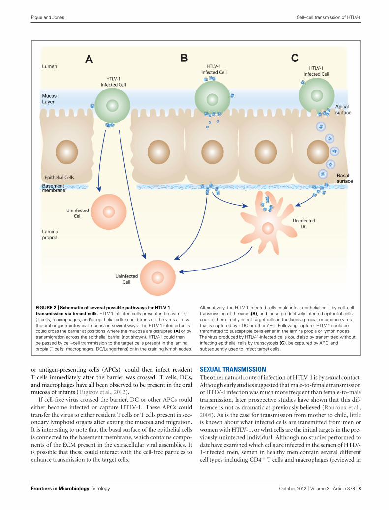
After the HTLV-1-infected cells enter the digestive tract, infec-tion would likely involve transfer of HTLV-1-infected cells and/orcell-free HTLV-1 produced by those infected cells across the epithe-lium in the oral or gastrointestinal mucosa. This could occur in anumber of different ways, some of which are shown in Figure 2.HTLV-1-infected cells could be transmitted at places where theintegrity of mucosa is disrupted or where there are gaps in theepithelium (Figure 2A). It is also possible that HTLV-1-infectedcells could cross intact epithelium: HIV-infected macrophageshave recently been shown to be able to transmigrate across fetaloral epithelia (Tugizov et al., 2012). Alternatively, the HTLV-1-infected cells could attach to the apical side of the epithelial cells,infect the cells by cell–cell transmission of the virus, and thenewly produced viral particles could bud from the basal surface(Figure 2B). This model is supported by in vitro studies show-ing that HTLV-1-infected T cells can adhere to the microvilli ofa monolayer formed by an intestine-derived epithelial cell line,that a large number of viral particles are released into the spacebetween the cells, and that the epithelial cells became infected(Zacharopoulos et al., 1992). Another way that the cell-free HTLV-1 virions produced by infected cells could be transmitted throughan intact epithelial barrier is by being taken up by a vesicle onthe apical surface of an epithelial cell and transported to andreleased from the basal side (Figure 2C). HTLV-1 has recentlybeen shown to be capable of crossing an epithelial barrier by thisprocess, which is referred to as transcytosis: virus produced frominfected T cells was transported across a tight epithelial barrierin an in vitro model of human intestinal cells (Martin-Latil et al.,2012). Moreover, the transmitted virus was able to infect humanDCs located immediately below the epithelial barrier (Martin-Latilet al., 2012). HIV-1 has also been shown to be capable of crossingintact oral and intestinal epithelial barriers by transcytosis and tobe capable of infecting DCs, macrophages, and CD4+ T cells onthe other side of those barriers in both ex vivo studies using tis-sue explants and in vitro studies (Bomsel, 1997; Alfsen et al., 2005;Tugizov et al., 2012).
In the model of infection just described, the cells in the childwould become infected after the HTLV-1 crossed the oral or gas-trointestinal mucosa, either as an infected cell or as a cell-freeparticle. It is possible that HTLV-1-infected cells, either T cells
www.frontiersin.org October 2012 | Volume 3 | Article 378 | 7
“fmicb-03-00378” — 2012/10/22 — 22:31 — page 8 — #8
Pique and Jones Cell–cell transmission of HTLV-1
FIGURE 2 | Schematic of several possible pathways for HTLV-1
transmission via breast milk. HTLV-1-infected cells present in breast milk(T cells, macrophages, and/or epithelial cells) could transmit the virus acrossthe oral or gastrointestinal mucosa in several ways. The HTLV-1-infected cellscould cross the barrier at positions where the mucosa are disrupted (A) or bytransmigration across the epithelial barrier (not shown). HTLV-1 could thenbe passed by cell–cell transmission to the target cells present in the laminapropia (T cells, macrophages, DC/Langerhans) or in the draining lymph nodes.
Alternatively, the HTLV-1-infected cells could infect epithelial cells by cell–celltransmission of the virus (B), and these productively infected epithelial cellscould either directly infect target cells in the lamina propia, or produce virusthat is captured by a DC or other APC. Following capture, HTLV-1 could betransmitted to susceptible cells either in the lamina propia or lymph nodes.The virus produced by HTLV-1-infected cells could also by transmitted withoutinfecting epithelial cells by transcytosis (C), be captured by APC, andsubsequently used to infect target cells.
or antigen-presenting cells (APCs), could then infect residentT cells immediately after the barrier was crossed. T cells, DCs,and macrophages have all been observed to be present in the oralmucosa of infants (Tugizov et al., 2012).
If cell-free virus crossed the barrier, DC or other APCs couldeither become infected or capture HTLV-1. These APCs couldtransfer the virus to either resident T cells or T cells present in sec-ondary lymphoid organs after exiting the mucosa and migration.It is interesting to note that the basal surface of the epithelial cellsis connected to the basement membrane, which contains compo-nents of the ECM present in the extracellular viral assemblies. Itis possible that these could interact with the cell-free particles toenhance transmission to the target cells.
SEXUAL TRANSMISSIONThe other natural route of infection of HTLV-1 is by sexual contact.Although early studies suggested that male-to-female transmissionof HTLV-I infection was much more frequent than female-to-maletransmission, later prospective studies have shown that this dif-ference is not as dramatic as previously believed (Roucoux et al.,2005). As is the case for transmission from mother to child, littleis known about what infected cells are transmitted from men orwomen with HTLV-1, or what cells are the initial targets in the pre-viously uninfected individual. Although no studies performed todate have examined which cells are infected in the semen of HTLV-1-infected men, semen in healthy men contain several differentcell types including CD4+ T cells and macrophages (reviewed in
Frontiers in Microbiology | Virology October 2012 | Volume 3 | Article 378 | 8
“fmicb-03-00378” — 2012/10/22 — 22:31 — page 9 — #9
Pique and Jones Cell–cell transmission of HTLV-1
Anderson et al., 2010), suggesting that one or both of these cellsare the source of the virus. In regard to female-to-male trans-mission, HTLV-1-infected cells have been detected in cervicalsecretions of infected women (Belec et al., 1996; Zunt et al., 2002),although the infected cell types were not identified. CD4+ T cellscan be detected in cervicovaginal secretions from healthy women,although the numbers are very low. However, the number of CD4+T cells is increased in cervicovaginal secretions with certain infec-tions; this is consistent with the observation that detection ofHTLV-1-infected cells in cervical secretions was associated withinflammation of the uterine cervix (Zunt et al., 2002).
Sexual transmission of HTLV-1 would presumably requireentry through mucosal barriers in the female and male genitaltracts. As during transmission by breastfeeding, the virus couldbe transmitted where lesions disrupt the mucosa, by infection ofthe epithelium or by transcytosis across epithelial cells. Female-to-male transmission is higher in men with syphilis or a historyof penile sores or ulcers (Murphy et al., 1989), consistent withthe notion that disrupting the epithelium increases transmission.Infection of a cervix-derived epithelial line following exposure toHTLV-1-infected T cells has been shown in vitro (Zacharopou-los et al., 1992), and HTLV-1 has been associated with carcinomaof the cervix (Strickler et al., 1995), suggesting that viral infec-tion of epithelium or other cells in the cervix may play a role inmale-to-female spread.
Although the mechanism of this transmission has not beenexamined for HTLV, mucosal entry of HIV and SIV in the genitaltracts have been well characterized by in vitro studies of purifiedcell populations, ex vivo studies using human explants, and in vivostudies in macaques and humanized mice (reviewed in Hladikand McElrath, 2008; Ganor and Bomsel, 2011; Kaushic, 2011).These studies have shown that HIV can be transmitted across theepithelium by all of the methods described above: infected seminalcells, or virus produced by those cells, have been shown to crossthrough gaps in the epithelium, by infecting the epithelium, or bytranscytosis.
Once viruses have crossed the mucosal barriers, as with infec-tion via breast milk, they can infect DCs, macrophages, or T cells.For HIV, a number of studies suggest that APCs capture the virusesand transfer them to T cells. For example, study performed witha model for HIV-1 female-to-male transmission using foreskinexplants observed that virus-infected cells form a VS with the api-cal side of foreskin keratinocytes and that HIV-1 subsequentlybudding from the basal side is captured by Langerhan cells (LC),a type of DC. LC then migrate to the epidermis–dermis inter-face and transfer the virus to T cells (Ganor et al., 2010). Exvivo human organ culture systems for male-to-female transmis-sion have shown that intraepithelial vaginal LCs can capture HIVand that these cells can productively infect T cells (Hladik et al.,2007; Ballweber et al., 2011).
TRANSMISSION BY BLOODAs discussed in the introduction, HTLV-1 can be transmitted bytransfusion of whole blood or blood products containing cells.Transmission by blood also occurs during the sharing of needlesby intravenous drug users. Unlike the natural methods of infectiondescribed above, transmission via contaminated blood does not
require transmission across a mucosal barrier. Consistent with thatnotion, transmission via blood is efficient: one prospective studyshowed seroconversion of 44% of recipients after a single expo-sure to HTLV-1-infected cellular blood products (Manns et al.,1992). Because of the lack of a barrier, this method of transmis-sion could involve direct transmission of HTLV-1 from the cells ofthe infected individual to target cells in the previously uninfectedindividual. Since the virus is predominantly found in T cells inperipheral blood, this type of transmission could occur duringdirect interactions between T cells by the methods that have beendescribed for in vitro T cell–T cell transmission: via VS, by transferof extracellular viral assemblies, or other methods. Since myDCsand pDCs from peripheral blood of asymptomatic individuals areinfected with HTLV-1 (Hishizawa et al., 2004; Azakami et al., 2009)transmission from DC to T cells may also occur.
Individuals who acquire HTLV-1 by blood transfusion are morelikely to develop the chronic inflammatory disorder HAM/TSP(Osame et al., 1990), while individuals who acquire the virus dur-ing breast feeding are more likely to develop the T cell malignancyATL (Kakuda et al., 2002). While this could reflect a number ofother factors (including age of infection, amount of virus acquiredduring transmission, and immune response), it has been suggestedthat these different methods of infection result in different popu-lations of infected cells, which in turn influences which disease agiven infected individual is more likely to develop.
ESTABLISHMENT OF HTLV-1 INFECTIONAs described in the introduction, it is believed that infection of anindividual with HTLV-1 occurs in two stages. The virus is thoughtto initially spread from T cell to T cell, primarily between CD4+T cells, and later to persist by clonal expansion of infected cells.Although nearly all studies of HTLV-1 performed to date havefocused on studies of T cells from peripheral blood, it would seemlikely that at least some of this spread would occur in the lymphnodes, where the concentration of the T cells is higher and theT cells are highly motile. The notion that a significant number ofHTLV-1-infected cells are present outside the peripheral blood issupported by observations of a strong immune response (HTLV-1-specific T cells and antibodies) in chronically infected individuals,even when no virus or virus-expressing cells can be detected in theperipheral blood.
One recent study of HIV transmission provides a model forhow the observations of cell–cell transmission via VS observed forHTLV-1 and HIV-1 in vitro might be relevant to in vivo transmis-sion in lymph nodes (Llewellyn et al., 2010). In lymphoid organs,such as lymph nodes, T cells are highly motile and adopt an elon-gated or polarized morphology. When HIV-1-infected polarizedT cells were examined, Gag was found to localize to a rear end pro-trusion known to mediate contact with other cells called a uropod.The uropods preferentially form contacts with target cells, andthese uropod-mediated contacts eventually form a VS (Llewellynet al., 2010). It seems possible that, soon after an individual isinfected with HTLV-1, the virus spreads between T cells in thelymph nodes using a similar mechanism.
It is also possible that DCs or other APC cells contribute tothe cell–cell spread during this early stage of transmission. DCscan be highly mobile and, as described above, are infected at a
www.frontiersin.org October 2012 | Volume 3 | Article 378 | 9
“fmicb-03-00378” — 2012/10/22 — 22:31 — page 10 — #10
Pique and Jones Cell–cell transmission of HTLV-1
significant level in many individuals with HTLV-1. This notionthat virus may be passed from T cells to DC and then back toT cells is also supported by the in vitro observations that bothDCs can be infected by HTLV-1-infected T cells and T cells can beinfected by HTLV-1-infected DC.
Another possible reservoir for HTLV-1 is human CD34+hematopoietic progenitor/stem cells (HP/HSCs). HTLV-1 caninfect these cells when cultured ex vivo, and the proviral genomeis maintained when the cells are differentiated down severallineages, including T cells and monocyte/macrophage lineages(Feuer et al., 1996). Moreover, a significant number of SCID micein which hematopoiesis is reconstituted with HTLV-1-infectedCD34+ HP/HSCs develop lymphoma similar to ATL (Banerjeeet al., 2010). CD34+ HP/HSCs isolated from infected individu-als have been reported to contain HTLV-1 sequences (Banerjeeet al., 2010), and, in one case, transmission of HTLV-1 occurredfollowing allogeneic BM transplantation (Kikuchi et al., 2000).
CONCLUSIONS AND PERSPECTIVESInsights gained in the last 10 years have dramatically changedour understanding about how HTLV is transmitted between cells.The long held beliefs that efficient transmission of HTLV-1 andHTLV-2 requires interactions between an infected and an unin-fected T cell, and that this mode of transmission distinguishesthese viruses from other retroviruses, no longer appears to bethe case. While a decade ago little was known about the inter-actions between cells required for HTLV-1 transmission, recentimaging studies have identified some of the components of theinfected cell, the uninfected cell, and the virus that facilitate dif-ferent types of cell–cell transmission. The more detailed studies ofHIV-1 cell–cell transmission and other retroviruses have providedadditional clues about how HTLV-1 may be transmitted. Thesestudies, along with in vitro studies of infectivity, have revealed thatT cells can be infected by cell–cell transmission from infected non-T cells as well as T cells. They have also demonstrated that cell–celltransmission of HTLV-1 and HIV to a target cell can occur via anuninfected APC that has captured viral particles, as well as from aninfected cell.
These studies suggest that the mechanism of transmissionbetween individuals, and within newly infected individuals priorto the clonal expansion that characterizes chronic infection byHTLV-1, differs from the current dogma that cell–cell transmissionof HTLV-1 only involves interactions between T cells. Observa-tions that macrophages and epithelial cells isolated from breastmilk can be infected and transformed with HTLV-1 suggest that
transmission via breast milk might involve these cells as well asT cells. The fact that HTLV-1-infected DCs are present in theperipheral blood of infected individuals, and that T cells are effi-ciently infected by DC in vitro, suggests that DCs or other APC playa role in HTLV-1 transmission between individuals, and/or duringthe initial spread of infection in an individual. This is supportedby the large number of studies indicating that DC and other APCplay a role during mucosal entry of HIV and SIV.
Despite our recent advances, there are many aspects of HTLV-1transmission that remain poorly understood. Nothing is knownabout whether productive infection following cell–cell transmis-sion of HTLV-1 occurs following endocytosis of the virus by thetarget cell and/or by fusion at the cellular surface. Since cell-freeHTLV has been shown to efficiently bind and enter primary T cells,it is unclear whether the block to stable infection reflects uptakeby a non-productive pathway, restriction by intracellular antiviralfactors, a negative effect of viral expression in the cells, or otherfactors. Although one can speculate (as we have in the previoussection) about what cells are involved during transmission betweenindividuals by mucosal and parenteral routes based on studies oflentiviral transmission, only a few, limited studies have been per-formed on fluids from HTLV-1-infected individuals or with exvivo cultured cells and tissues to address this directly. In addition,once an individual is infected with the virus, it is unclear where,or between what cell types, the virus initially spreads prior toclonal expansion of the infected T cells, and whether HTLV-1 per-sists in reservoirs outside the peripheral blood during the chronicstage of infection. Future studies investigating these and otheraspects of HTLV transmission and persistence will be importantfor developing therapeutics to block initial infection with the virus,as well as to reduce the level of cells containing the virus in infectedindividuals.
ACKNOWLEDGMENTSThe authors wish to thank Drs Vineet KewalRamani and NickLlewellyn for their helpful comments regarding the manuscript.This project has been funded in whole or in part with federalfunds from the National Cancer Institute, National Institutes ofHealth, under contract HHSN26120080001E. The content of thispublication does not necessarily reflect the views or policies ofthe Department of Health and Human Services, nor does men-tion of trade names, commercial products, or organizations implyendorsement by the U.S. Government. The authors would alsolike to thank the Cent pour sang la vie foundation and the InstitutNational du Cancer (InCA) for their support.
REFERENCESAgadjanyan, M. G., Ugen, K. E., Wang,
B., Williams, W. V., and Weiner, D.B. (1994). Identification of an 80-kilodalton membrane glycoproteinimportant for human T-cell leukemiavirus type I and type II syncytiumformation and infection. J. Virol. 68,485–493.
Aggarwal, A., Iemma, T. L., Shih,I., Newsome, T. P., Mcallery, S.,Cunningham, A. L., et al. (2012).Mobilization of HIV spread by
diaphanous 2 dependent filopodia ininfected dendritic cells. PLoS Pathog.8, e1002762. doi: 10.1371/journal.ppat.1002762
Alfsen, A., Yu, H., Magerus-Chatinet, A.,Schmitt, A., and Bomsel, M. (2005).HIV-1-infected blood mononu-clear cells form an integrin- andagrin-dependent viral synapse toinduce efficient HIV-1 transcytosisacross epithelial cell mono-layer. Mol. Biol. Cell 16, 4267–4279.
Anderson, D. J., Politch, J. A., Nadol-ski, A. M., Blaskewicz, C. D., Pud-ney, J., and Mayer, K. H. (2010).Targeting Trojan Horse leukocytesfor HIV prevention. AIDS 24,163–187.
Arrighi, J. F., Pion, M., Garcia, E.,Escola, J. M., Van Kooyk, Y., Geijten-beek, T. B., et al. (2004). DC-SIGN-mediated infectious synapse forma-tion enhances X4 HIV-1 transmissionfrom dendritic cells to T cells. J. Exp.Med. 200, 1279–1288.
Azakami, K., Sato, T., Araya, N.,Utsunomiya, A., Kubota, R., Suzuki,K., et al. (2009). Severe loss ofinvariant NKT cells exhibiting anti-HTLV-1 activity in patients withHTLV-1-associated disorders. Blood114, 3208–3215.
Ballweber, L., Robinson, B., Kreger, A.,Fialkow, M., Lentz, G., Mcelrath,M. J., et al. (2011). Vaginal Langer-hans cells nonproductively transport-ing HIV-1 mediate infection of Tcells. J. Virol. 85, 13443–13447.
Frontiers in Microbiology | Virology October 2012 | Volume 3 | Article 378 | 10
“fmicb-03-00378” — 2012/10/22 — 22:31 — page 11 — #11
Pique and Jones Cell–cell transmission of HTLV-1
Banerjee, P., Tripp, A., Lairmore, M. D.,Crawford, L., Sieburg, M., Ramos,J. C., et al. (2010). Adult T-cellleukemia/lymphoma development inHTLV-1-infected humanized SCIDmice. Blood 115, 2640–2648.
Barnard, A. L., Igakura, T., Tanaka,Y., Taylor, G. P., and Bangham, C.R. (2005). Engagement of specificT-cell surface molecules regulatescytoskeletal polarization in HTLV-1-infected lymphocytes. Blood 106,988–995.
Belec, L., Georges-Courbot, M. C.,Georges, A., Mohamed, A. S.,Londos-Gagliardi, D., Hallouin, M.C., et al. (1996). Cervicovaginalsynthesis of IgG antibodies to theimmunodominant 175-199 domainof the surface glycoprotein gp46 ofhuman T-cell leukemia virus type I.J. Med. Virol. 50, 42–49.
Bennett, A. E., Narayan, K., Shi, D.,Hartnell, L. M., Gousset, K., He, H.,et al. (2009). Ion-abrasion scanningelectron microscopy reveals surface-connected tubular conduits in HIV-infected macrophages. PLoS Pathog.5, e1000591. doi: 10.1371/journal.ppat.1000591
Biggar, R. J., Ng, J., Kim, N., Hisada, M.,Li, H. C., Cranston, B., et al. (2006).Human leukocyte antigen concor-dance and the transmission risk viabreast-feeding of human T cell lym-photropic virus type I. J. Infect. Dis.193, 277–282.
Biswas, H. H., Engstrom, J. W.,Kaidarova, Z., Garratty, G., Gibble,J. W., Newman, B. H., et al. (2009).Neurologic abnormalities in HTLV-I- and HTLV-II-infected individualswithout overt myelopathy. Neurology73, 781–789.
Bomsel, M. (1997). Transcytosis ofinfectious human immunodeficiencyvirus across a tight human epithe-lial cell line barrier. Nat. Med. 3,42–47.
Bosch, B., Grigorov, B., Senserrich, J.,Clotet, B., Darlix, J. L., Muriaux,D., et al. (2008). A clathrin-dynamin-dependent endocytic pathway for theuptake of HIV-1 by direct T cell–T cell transmission. Antiviral Res. 80,185–193.
Burleigh, L., Lozach, P. Y., Schiffer, C.,Staropoli, I., Pezo, V., Porrot, F.,et al. (2006). Infection of dendriticcells (DCs), not DC-SIGN-mediatedinternalization of human immunod-eficiency virus, is required for long-term transfer of virus to T cells. J.Virol. 80, 2949–2957.
Caterino-Caterino-de-Araujo, A., andde los Santos-Fortuna, E. (1999). Noevidence of vertical transmission ofHTLV-I and HTLV-II in children at
high risk for HIV-1 infection fromSao Paulo, Brazil. J. Trop. Pediatr. 45,42–47.
Cavrois, M., Leclercq, I., Gout,O., Gessain, A., Wain-Hobson, S.,and Wattel, E. (1998). Persistentoligoclonal expansion of human T-cell leukemia virus type 1-infectedcirculating cells in patients withTropical spastic paraparesis/HTLV-1associated myelopathy. Oncogene 17,77–82.
Cavrois, M., Wain-Hobson, S., Gessain,A., Plumelle, Y., and Wattel, E. (1996).Adult T-cell leukemia/lymphoma ona background of clonally expandinghuman T-cell leukemia virus type-1-positive cells. Blood 88, 4646–4650.
Ceccaldi, P. E., Delebecque, F., Prevost,M. C., Moris, A., Abastado, J. P.,Gessain, A., et al. (2006). DC-SIGNfacilitates fusion of dendritic cellswith human T-cell leukemia virustype 1-infected cells. J. Virol. 80,4771–4780.
Chen, P., Hubner, W., Spinelli, M. A.,and Chen, B. K. (2007). Predominantmode of human immunodeficiencyvirus transfer between T cells is medi-ated by sustained Env-dependentneutralization-resistant virologicalsynapses. J. Virol. 81, 12582–12595.
Clapham, P., Nagy, K., Cheingsong-Popov, R., Exley, M., and Weiss, R.A. (1983). Productive infection andcell-free transmission of human T-cell leukemia virus in a nonlymphoidcell line. Science 222, 1125–1127.
Dale, B. M., Mcnerney, G. P., Thomp-son, D. L., Hubner, W., De Los Reyes,K., Chuang, F. Y., et al. (2011). Cell-to-cell transfer of HIV-1 via viro-logical synapses leads to endosomalvirion maturation that activates viralmembrane fusion. Cell Host Microbe10, 551–562.
de Revel, T., Mabondzo, A., Gras, G.,Delord, B., Roques, P., Boussin, F.,et al. (1993). In vitro infection ofhuman macrophages with human T-cell leukemia virus type 1. Blood 81,1598–1606.
de Rossi, A., Aldovini, A., Franchini, G.,Mann, D., Gallo, R. C., and Wong-Staal, F. (1985). Clonal selection ofT lymphocytes infected by cell-freehuman T-cell leukemia/lymphomavirus type I: parameters of virus inte-gration and expression. Virology 143,640–645.
Del Portillo, A., Tripodi, J., Najfeld, V.,Wodarz, D., Levy, D. N., and Chen, B.K. (2011). Multiploid inheritance ofHIV-1 during cell-to-cell infection. J.Virol. 85, 7169–7176.
Deneka, M., Pelchen-Matthews, A.,Byland, R., Ruiz-Mateos, E., andMarsh, M. (2007). In macrophages,
HIV-1 assembles into an intracellularplasma membrane domain contain-ing the tetraspanins CD81, CD9, andCD53. J. Cell Biol. 177, 329–341.
Donegan, E., Lee, H., Operskalski, E. A.,Shaw, G. M., Kleinman, S. H., Busch,M. P., et al. (1994). Transfusion trans-mission of retroviruses: human T-lymphotropic virus types I and IIcompared with human immunodefi-ciency virus type 1. Transfusion 34,478–483.
Dooneief, G., Marlink, R., Bell, K.,Marder, K., Renjifo, B., Stern, Y., et al.(1996). Neurologic consequences ofHTLV-II infection in injection-drugusers. Neurology 46, 1556–1560.
Etoh, K., Tamiya, S., Yamaguchi, K.,Okayama, A., Tsubouchi, H., Ideta,T., et al. (1997). Persistent clonal pro-liferation of human T-lymphotropicvirus type I-infected cells in vivo.Cancer Res. 57, 4862–4867.
Eugenin, E. A., Gaskill, P. J., andBerman, J. W. (2009). Tunneling nan-otubes (TNT) are induced by HIV-infection of macrophages: a potentialmechanism for intercellular HIV traf-ficking. Cell. Immunol. 254, 142–148.
Fan, N., Gavalchin, J., Paul, B., Wells,K. H., Lane, M. J., and Poiesz,B. J. (1992). Infection of periph-eral blood mononuclear cells and celllines by cell-free human T-cell lym-phoma/leukemia virus type I. J. Clin.Microbiol. 30, 905–910.
Felts, R. L., Narayan, K., Estes, J. D., Shi,D., Trubey, C. M., Fu, J., et al. (2010).3D visualization of HIV transfer atthe virological synapse between den-dritic cells and T cells. Proc. Natl.Acad. Sci. U.S.A. 107, 13336–13341.
Feuer, G., Fraser, J. K., Zack, J. A.,Lee, F., Feuer, R., and Chen, I.S. (1996). Human T-cell leukemiavirus infection of human hematopoi-etic progenitor cells: maintenance ofvirus infection during differentiationin vitro and in vivo. J. Virol. 70,4038–4044.
Feuer, G., and Green, P. L. (2005).Comparative biology of human T-celllymphotropic virus type 1 (HTLV-1)and HTLV-2. Oncogene 24, 5996–6004.
Gallo, D., Petru, A., Yeh, E. T., andHanson, C. V. (1993). No evidenceof perinatal transmission of HTLV-II. J. Acquir. Immune Defic. Syndr. 6,1168–1170.
Ganor, Y., and Bomsel, M. (2011). HIV-1 transmission in the male genitaltract. Am. J. Reprod. Immunol. 65,284–291.
Ganor, Y., Zhou, Z., Tudor, D., Schmitt,A., Vacher-Lavenu, M. C., Gibault,L., et al. (2010). Within 1 h, HIV-1uses viral synapses to enter efficiently
the inner, but not outer, foreskinmucosa and engages Langerhans-Tcell conjugates. Mucosal Immunol. 3,506–522.
Garcia, E., Pion, M., Pelchen-Matthews,A., Collinson, L., Arrighi, J. F.,Blot, G., et al. (2005). HIV-1 traf-ficking to the dendritic cell-T-cellinfectious synapse uses a pathway oftetraspanin sorting to the immuno-logical synapse. Traffic 6, 488–501.
Gendelman, H. E., Orenstein, J. M.,Martin, M. A., Ferrua, C., Mitra,R., Phipps, T., et al. (1988). Effi-cient isolation and propagation ofhuman immunodeficiency virus onrecombinant colony-stimulating fac-tor 1-treated monocytes. J. Exp. Med.167, 1428–1441.
Gessain, A., Barin, F., Vernant, J.C., Gout, O., Maurs, L., Calen-der, A., et al. (1985). Antibodies tohuman T-lymphotropic virus type-I in patients with tropical spasticparaparesis. Lancet 2, 407–410.
Gessain, A., Louie, A., Gout, O.,Gallo, R. C., and Franchini, G.(1991). Human T-cell leukemia-lymphoma virus type I (HTLV-I)expression in fresh peripheral bloodmononuclear cells from patients withtropical spastic paraparesis/HTLV-I-associated myelopathy. J. Virol. 65,1628–1633.
Ghez, D., Lepelletier, Y., Jones, K.S., Pique, C., and Hermine, O.(2010). Current concepts regardingthe HTLV-1 receptor complex. Retro-virology 7, 99.
Ghez, D., Lepelletier, Y., Lambert, S.,Fourneau, J. M., Blot, V., Janvier, S.,et al. (2006). Neuropilin-1 is involvedin human T-cell lymphotropic virustype 1 entry. J. Virol. 80, 6844–6854.
Giam, C. Z., and Jeang, K. T.(2007). HTLV-1 Tax and adult T-cellleukemia. Front. Biosci. 12, 1496–1507.
Gillet, N. A., Malani, N., Melamed, A.,Gormley, N., Carter, R., Bentley, D.,et al. (2011). The host genomic envi-ronment of the provirus determinesthe abundance of HTLV-1-infectedT-cell clones. Blood 117, 3113–3122.
Goncalves, D. U., Proietti, F. A., Ribas,J. G., Araujo, M. G., Pinheiro, S. R.,Guedes, A. C., et al. (2010). Epidemi-ology, treatment, and prevention ofhuman T-cell leukemia virus type 1-associated diseases. Clin. Microbiol.Rev. 23, 577–589.
Gousset, K., Ablan, S. D., Coren, L. V.,Ono, A., Soheilian, F., Nagashima,K., et al. (2008). Real-time visual-ization of HIV-1 GAG trafficking ininfected macrophages. PLoS Pathog.4, e1000015. doi: 10.1371/journal.ppat.1000015
www.frontiersin.org October 2012 | Volume 3 | Article 378 | 11
“fmicb-03-00378” — 2012/10/22 — 22:31 — page 12 — #12
Pique and Jones Cell–cell transmission of HTLV-1
Graziani, G., Faraoni, I., Zhang, J.,Caronti, B., Lauro, G., Bonmassar,E., et al. (1993). Transient HTLV-Iinfection of a human glioma cell linefollowing cell-free exposure. Virology197, 767–769.
Green, P. L., and Chen, I. S. (1990).Regulation of human T cell leukemiavirus expression. FASEB J. 4,169–175.
Groot, F., Welsch, S., and Sattentau, Q. J.(2008). Efficient HIV-1 transmissionfrom macrophages to T cells acrosstransient virological synapses. Blood111, 4660–4663.
Haller, C., Tibroni, N., Rudolph, J. M.,Grosse, R., and Fackler, O. T. (2011).Nef does not inhibit F-actin remod-elling and HIV-1 cell–cell transmis-sion at the T lymphocyte virolog-ical synapse. Eur. J. Cell Biol. 90,913–921.
Haraguchi, Y., Yang, D. W., Handa, A.,Shimizu, N., Tanaka, Y., and Hoshino,H. (1994). Detection of neutraliz-ing antibodies against human T-cellleukemia virus type 1 using a cell-free infection system and polymerasechain reaction. Int. J. Cancer 59,416–421.
Hirata, M., Hayashi, J., Noguchi,A., Nakashima, K., Kajiyama, W.,Kashiwagi, S., et al. (1992). Theeffects of breastfeeding and pres-ence of antibody to p40tax pro-tein of human T cell lymphotropicvirus type-I on mother to childtransmission. Int. J. Epidemiol. 21,989–994.
Hishizawa, M., Imada, K., Kitawaki,T., Ueda, M., Kadowaki, N.,and Uchiyama, T. (2004). Deple-tion and impaired interferon-alpha-producing capacity of blood plas-macytoid dendritic cells in humanT-cell leukaemia virus type I-infectedindividuals. Br. J. Haematol. 125,568–575.
Hjelle, B., Appenzeller, O., Mills, R.,Alexander, S., Torrez-Martinez, N.,Jahnke, R., et al. (1992). Chronicneurodegenerative disease associatedwith HTLV-II infection. Lancet 339,645–646.
Hladik, F., and McElrath, M. J.(2008). Setting the stage: host inva-sion by HIV. Nat. Rev. Immunol. 8,447–457.
Hladik, F., Sakchalathorn, P., Ball-weber, L., Lentz, G., Fialkow, M.,Eschenbach, D., et al. (2007). Initialevents in establishing vaginal entryand infection by human immunod-eficiency virus type-1. Immunity 26,257–270.
Hoffman, P. M., Dhib-Jalbut, S.,Mikovits, J. A., Robbins, D. S.,Wolf, A. L., Bergey, G. K., et al.
(1992). Human T-cell leukemia virustype I infection of monocytes andmicroglial cells in primary humancultures. Proc. Natl. Acad. Sci. U.S.A.89, 11784–11788.
Hoshino, H. (2012). Cellular fac-tors involved in HTLV-1 entryand pathogenicit. Front. Micro-biol. 3:222. doi: 10.3389/fmicb.2012.00222
Hubner, W., Mcnerney, G. P., Chen, P.,Dale, B. M., Gordon, R. E., Chuang,F. Y., et al. (2009). Quantitative 3Dvideo microscopy of HIV transferacross T cell virological synapses.Science 323, 1743–1747.
Igakura, T., Stinchcombe, J. C., Goon,P. K., Taylor, G. P., Weber, J.N., Griffiths, G. M., et al. (2003).Spread of HTLV-I between lympho-cytes by virus-induced polarizationof the cytoskeleton. Science 299,1713–1716.
Ilinskaya, A., Heidecker, G., and Jones,K. (2010). Interaction between theHTLV-1 envelope and cellular pro-teins: impact on virus infection andrestriction. Future Med. Chem. 2,1651–1668.
Jain, P., Manuel, S. L., Khan, Z. K.,Ahuja, J., Quann, K., and Wig-dahl, B. (2009). DC-SIGN mediatescell-free infection and transmissionof human T-cell lymphotropic virustype 1 by dendritic cells. J. Virol. 83,10908–10921.
Jason, J. M., Mcdougal, J. S., Cabradilla,C., Kalyanaraman, V. S., andEvatt, B. L. (1985). Human T-cellleukemia virus (HTLV-I) p24 anti-body in New York City blood prod-uct recipients. Am. J. Hematol. 20,129–137.
Jin, J., Sherer, N., and Mothes,W. (2010). Surface transmission orpolarized egress? Lessons learnedfrom HTLV cell-to-cell transmission.Viruses 2, 601–605.
Jin, J., Sherer, N. M., Heidecker,G., Derse, D., and Mothes, W.(2009). Assembly of the murineleukemia virus is directed towardssites of cell–cell contact. PLoS Biol. 7,e1000163. doi: 10.1371/journal.pbio.1000163
Jinno, A., Haraguchi, Y., Shiraki, H., andHoshino, H. (1999). Inhibition ofcell-free human T-cell leukemia virustype 1 infection at a postbinding stepby the synthetic peptide derived froman ectodomain of the gp21 trans-membrane glycoprotein. J. Virol. 73,9683–9689.
Jolly, C., Kashefi, K., Hollinshead, M.,and Sattentau, Q. J. (2004). HIV-1cell to cell transfer across an Env-induced, actin-dependent synapse. J.Exp. Med. 199, 283–293.
Jolly, C., Mitar, I., and Sattentau, Q.J. (2007). Adhesion molecule inter-actions facilitate human immunod-eficiency virus type 1-induced viro-logical synapse formation between Tcells. J. Virol. 81, 13916–13921.
Jolly, C., Welsch, S., Michor, S., andSattentau, Q. J. (2011). The regu-lated secretory pathway in CD4(+) Tcells contributes to human immun-odeficiency virus type-1 cell-to-cellspread at the virological synapse.PLoS Pathog. 7, e1002226. doi:10.1371/journal.ppat.1002226
Jones, K. S., Lambert, S., Bouttier, M.,Benit, L., Ruscetti, F. W., Hermine,O., et al. (2011). Molecular aspectsof HTLV-1 entry: functional domainsof the HTLV-1 surface subunit (SU)and their relationships to the entryreceptors. Viruses 3, 794–810.
Jones, K. S., Petrow-Sadowski, C.,Bertolette, D. C., Huang, Y., andRuscetti, F. W. (2005). Heparan sul-fate proteoglycans mediate attach-ment and entry of human T-cellleukemia virus type 1 virions intoCD4+ T cells. J. Virol. 79, 12692–12702.
Jones, K. S., Petrow-Sadowski, C.,Huang, Y.K., Bertolette, D. C., andRuscetti, F. W. (2008). Cell-freeHTLV-1 infects dendritic cells leadingto transmission and transformationof CD4(+) T cells. Nat. Med. 14,429–436.
Kakuda, K., Ikematsu, H., Chong, W. L.,Hayashi, J., and Kashiwagi, S. (2002).Molecular epidemiology of human Tlymphotropic virus type 1 transmis-sion in Okinawa, Japan. Am. J. Trop.Med. Hyg. 66, 404–408.
Kaplan, J. E., Abrams, E., Shaffer, N.,Cannon, R. O., Kaul, A., Krasinski,K., et al. (1992). Low risk of mother-to-child transmission of human Tlymphotropic virus type II in non-breast-fed infants. The NYC Perina-tal HIV Transmission CollaborativeStudy. J. Infect. Dis. 166, 892–895.
Kashiwagi, K., Furusyo, N., Nakashima,H., Kubo, N., Kinukawa, N., Kashi-wagi, S., et al. (2004). A decreasein mother-to-child transmission ofhuman T lymphotropic virus type I(HTLV-I) in Okinawa, Japan. Am. J.Trop. Med. Hyg. 70, 158–163.
Kaushic, C. (2011). HIV-1 infection inthe female reproductive tract: role ofinteractions between HIV-1 and gen-ital epithelial cells. Am. J. Reprod.Immunol. 65, 253–260.
Kelly, D., and Coutts, A. G. (2000).Early nutrition and the developmentof immune function in the neonate.Proc. Nutr. Soc. 59, 177–185.
Kikuchi, H., Ohtsuka, E., Ono, K.,Nakayama, T., Saburi, Y., Tezono, K.,
et al. (2000). Allogeneic bone marrowtransplantation-related transmissionof human T lymphotropic virus typeI (HTLV-I). Bone Marrow Transplant.26, 1235–1237.
Kinoshita, K., Amagasaki, T., Hino, S.,Doi, H., Yamanouchi, K., Ban, N.,et al. (1987). Milk-borne transmis-sion of HTLV-I from carrier mothersto their children. Jpn. J. Cancer Res.78, 674–680.
Kinoshita, K., Yamanouchi, K., Ikeda,S., Momita, S., Amagasaki, T., Soda,H., et al. (1985). Oral infection ofa common marmoset with humanT-cell leukemia virus type-I (HTLV-I) by inoculating fresh human milkof HTLV-I carrier mothers. Jpn. J.Cancer Res. 76, 1147–1153.
Kira, J., Koyanagi, Y., Yamada, T.,Itoyama, Y., Goto, I., Yamamoto,N., et al. (1991). Increased HTLV-Iproviral DNA in HTLV-I-associatedmyelopathy: a quantitative poly-merase chain reaction study. Ann.Neurol. 29, 194–201.
Knight, S. C., Macatonia, S. E., Cruick-shank, K., Rudge, P., and Patterson, S.(1993). Dendritic cells in HIV-1 andHTLV-1 infection. Adv. Exp. Med.Biol. 329, 545–549.
Koralnik, I. J., Lemp, J. F. Jr.,Gallo, R. C., and Franchini,G. (1992). In vitro infection ofhuman macrophages by humanT-cell leukemia/lymphotropic virustype I (HTLV-I). AIDS Res. Hum.Retroviruses 8, 1845–1849.
Koyanagi, Y., Itoyama, Y., Nakamura,N., Takamatsu, K., Kira, J., Iwa-masa, T., et al. (1993). In vivo infec-tion of human T-cell leukemia virustype I in non-T cells. Virology 196,25–33.
Lairmore, M. D., Jason, J. M., Hartley, T.M., Khabbaz, R. F., De, B., and Evatt,B. L. (1989). Absence of human T-celllymphotropic virus type I coinfectionin human immunodeficiency virus-infected hemophilic men. Blood 74,2596–2599.
Lal, R. B., Owen, S. M., Rudolph, D. L.,Dawson, C., and Prince, H. (1995).In vivo cellular tropism of humanT-lymphotropic virus type II is notrestricted to CD8+ cells. Virology210, 441–447.
Lambert, S., Bouttier, M., Vassy, R.,Seigneuret, M., Petrow-Sadowski, C.,Janvier, S., et al. (2009). HTLV-1 usesHSPG and neuropilin-1 for entryby molecular mimicry of VEGF165.Blood 113, 5176–5185.
Le Douce, V., Herbein, G., Rohr, O.,and Schwartz, C. (2010). Molecularmechanisms of HIV-1 persistence inthe monocyte-macrophage lineage.Retrovirology 7, 32.
Frontiers in Microbiology | Virology October 2012 | Volume 3 | Article 378 | 12
“fmicb-03-00378” — 2012/10/22 — 22:31 — page 13 — #13
Pique and Jones Cell–cell transmission of HTLV-1
Leclercq, I., Cavrois, M., Mortreux,F., Hermine, O., Gessain, A.,Morschhauser, F., et al. (1998). Oligo-clonal proliferation of human T-cellleukaemia virus type 1 bearing T cellsin adult T-cell leukaemia/lymphomawithout deletion of the 3′ provirusintegration sites. Br. J. Haematol. 101,500–506.
LeVasseur, R. J., Southern, S. O., andSouthern, P. J. (1998). Mammaryepithelial cells support and trans-fer productive human T-cell lym-photropic virus infections. J. Hum.Virol. 1, 214–223.
Li, H. C., Biggar, R. J., Miley, W. J.,Maloney, E. M., Cranston, B., Han-chard, B., et al. (2004). Provirus loadin breast milk and risk of mother-to-child transmission of human Tlymphotropic virus type I. J. Infect.Dis. 190, 1275–1278.
Llewellyn, G. N., Hogue, I. B., Grover,J. R., and Ono, A. (2010). Nucleo-capsid promotes localization of HIV-1 gag to uropods that participatein virological synapses between Tcells. PLoS Pathog. 6, e1001167. doi:10.1371/journal.ppat.1001167
Macatonia, S. E., Cruickshank, J.K., Rudge, P., and Knight, S. C.(1992). Dendritic cells from patientswith tropical spastic paraparesis areinfected with HTLV-1 and stimulateautologous lymphocyte proliferation.AIDS Res. Hum. Retroviruses 8, 1699–1706.
Maeda, Y., Furukawa, M., Takehara,Y., Yoshimura, K., Miyamoto, K.,Matsuura, T., et al. (1984). Preva-lence of possible adult T-cell leukemiavirus-carriers among volunteer blooddonors in Japan: a nation-wide study.Int. J. Cancer 33, 717–720.
Majorovits, E., Nejmeddine, M.,Tanaka, Y., Taylor, G. P., Fuller, S. D.,and Bangham, C. R. (2008). HumanT-lymphotropic virus-1 visualized atthe virological synapse by electrontomography. PLoS ONE 3, e2251. doi:10.1371/journal.pone.0002251
Manel, N., Kim, F. J., Kinet, S., Tay-lor, N., Sitbon, M., and Battini, J.L. (2003). The ubiquitous glucosetransporter GLUT-1 is a receptor forHTLV. Cell 115, 449–459.
Manns, A., Miley, W. J., Wilks, R.J., Morgan, O. S., Hanchard, B.,Wharfe, G., et al. (1999). Quanti-tative proviral DNA and antibodylevels in the natural history of HTLV-I infection. J. Infect. Dis. 180,1487–1493.
Manns, A., Wilks, R. J., Murphy, E.L., Haynes, G., Figueroa, J. P., Bar-nett, M., et al. (1992). A prospectivestudy of transmission by transfusionof HTLV-I and risk factors associated
with seroconversion. Int. J. Cancer 51,886–891.
Martin-Latil, S., Gnadig, N. F.,Mallet, A., Desdouits, M., Guivel-Benhassine, F., Jeannin, P., et al.(2012). Transcytosis of HTLV-1across a tight human epithelialbarrier and infection of sub-epithelial dendritic cells. Blood 120,572–580.
Mazurov, D., Ilinskaya, A., Heidecker,G., Lloyd, P., and Derse, D. (2010).Quantitative comparison of HTLV-1 and HIV-1 cell-to-cell infectionwith new replication dependent vec-tors. PLoS Pathog. 6, e1000788. doi:10.1371/journal.ppat.1000788
McDonald, D., Wu, L., Bohks, S. M.,Kewalramani, V. N., Unutmaz, D.,and Hope, T. J. (2003). Recruitmentof HIV and its receptors to den-dritic cell-T cell junctions. Science300, 1295–1297.
Miyamoto, K., Tomita, N., Ishii,A., Nishizaki, T., Kitajima, K.,Tanaka, T., et al. (1984). Transforma-tion of ATLA-negative leukocytes byblood components from anti-ATLA-positive donors in vitro. Int. J. Cancer33, 721–725.
Moritoyo, T., Izumo, S., Moritoyo, H.,Tanaka, Y., Kiyomatsu, Y., Nagai,M., et al. (1999). Detection ofhuman T-lymphotropic virus typeI p40tax protein in cerebrospinalfluid cells from patients withhuman T-lymphotropic virus typeI-associated myelopathy/tropicalspastic paraparesis. J. Neurovirol. 5,241–248.
Mukerji, J., Olivieri, K. C., Misra, V.,Agopian, K. A., and Gabuzda, D.(2012). Proteomic analysis of HIV-1Nef cellular binding partners revealsa role for exocyst complex proteinsin mediating enhancement of inter-cellular nanotube formation. Retro-virology 9, 33.
Murphy, E. L., Figueroa, J. P., Gibbs,W. N., Brathwaite, A., Holding-Cobham, M., Waters, D., et al. (1989).Sexual transmission of human T-lymphotropic virus type I (HTLV-I).Ann. Intern. Med. 111, 555–560.
Murphy, E. L., Lee, T. H., Chafets,D., Nass, C. C., Wang, B., Lough-lin, K., et al. (2004). Higher human Tlymphotropic virus (HTLV) provirusload is associated with HTLV-I ver-sus HTLV-II, with HTLV-II subtypeA versus B, and with male sex and ahistory of blood transfusion. J. Infect.Dis. 190, 504–510.
Nagai, M., Brennan, M. B., Sakai, J. A.,Mora, C. A., and Jacobson, S. (2001).CD8(+) T cells are an in vivo reser-voir for human T-cell lymphotropicvirus type I. Blood 98, 1858–1861.
Nejmeddine, M., Barnard, A. L., Tanaka,Y., Taylor, G. P., and Bangham, C.R. (2005). Human T-lymphotropicvirus, type 1, tax protein trig-gers microtubule reorientation in thevirological synapse. J. Biol. Chem.280, 29653–29660.
Nejmeddine, M., Negi, V. S., Mukher-jee, S., Tanaka, Y., Orth, K., Taylor,G. P., et al. (2009). HTLV-1-Tax andICAM-1 act on T-cell signal path-ways to polarize the microtubule-organizing center at the virologicalsynapse. Blood 114, 1016–1025.
Nikolic, D. S., Lehmann, M., Felts, R.,Garcia, E., Blanchet, F. P., Subrama-niam, S., et al. (2011). HIV-1 activatesCdc42 and induces membrane exten-sions in immature dendritic cells tofacilitate cell-to-cell virus propaga-tion. Blood 118, 4841–4852.
Nobile, C., Rudnicka, D., Hasan, M.,Aulner, N., Porrot, F., Machu, C.,et al. (2009). HIV-1 Nef inhibits ruf-fles, induces filopodia, and modulatesmigration of infected lymphocytes. J.Virol. 84, 2282–2293.
Nyambi, P. N., Ville, Y., Louwagie,J., Bedjabaga, I., Glowaczower, E.,Peeters, M., et al. (1996). Mother-to-child transmission of human T-celllymphotropic virus types I and II(HTLV-I/II) in Gabon: a prospec-tive follow-up of 4 years. J. Acquir.Immune Defic. Syndr. Hum. Retrovi-rol. 12, 187–192.
Nydegger, S., Foti, M., Derdowski,A., Spearman, P., and Thali, M.(2003). HIV-1 egress is gated throughlate endosomal membranes. Traffic 4,902–910.
Onfelt, B., Nedvetzki, S., Yanagi, K.,and Davis, D. M. (2004). Cuttingedge: membrane nanotubes connectimmune cells. J. Immunol. 173, 1511–1513.
Ono, A., and Freed, E. O. (2004). Cell-type-dependent targeting of humanimmunodeficiency virus type 1assembly to the plasma membraneand the multivesicular body. J. Virol.78, 1552–1563.
Osame, M., Janssen, R., Kubota, H.,Nishitani, H., Igata, A., Nagataki,S., et al. (1990). Nationwide sur-vey of HTLV-I-associated myelopa-thy in Japan: association withblood transfusion. Ann. Neurol. 28,50–56.
Osame, M., Usuku, K., Izumo, S.,Ijichi, N., Amitani, H., Igata, A., et al.(1986). HTLV-I associated myelopa-thy, a new clinical entity. Lancet 1,1031–1032.
Pais-Correia, A. M., Sachse, M.,Guadagnini, S., Robbiati, V., Lasserre,R., Gessain, A., et al. (2009). Biofilm-like extracellular viral assemblies
mediate HTLV-1 cell-to-cell trans-mission at virological synapses. Nat.Med. 16, 83–89.
Pelchen-Matthews, A., Kramer, B., andMarsh, M. (2003). Infectious HIV-1assembles in late endosomes in pri-mary macrophages. J. Cell Biol. 162,443–455.
Poiesz, B. J., Ruscetti, F. W., Gazdar, A. F.,Bunn, P. A., Minna, J. D., and Gallo,R. C. (1980). Detection and isolationof type C retrovirus particles fromfresh and cultured lymphocytes of apatient with cutaneous T-cell lym-phoma. Proc. Natl. Acad. Sci. U.S.A.77, 7415–7419.
Popovic, M., Sarin, P. S., Robert-Gurroff, M., Kalyanaraman, V. S.,Mann, D., Minowada, J., et al. (1983).Isolation and transmission of humanretrovirus (human T-cell leukemiavirus). Science 219, 856–859.
Puigdomenech, I., Massanella, M.,Cabrera, C., Clotet, B., and Blanco,J. (2009). On the steps of cell-to-cell HIV transmission between CD4T cells. Retrovirology 6, 89.
Raposo, G., Moore, M., Innes, D.,Leijendekker, R., Leigh-Brown, A.,Benaroch, P., et al. (2002). Humanmacrophages accumulate HIV-1 par-ticles in MHC II compartments. Traf-fic 3, 718–729.
Richardson, J. H., Hollsberg, P., Wind-hagen, A., Child, L. A., Hafler, D.A., and Lever, A. M. (1997). Vari-able immortalizing potential and fre-quent virus latency in blood-derivedT-cell clones infected with human T-cell leukemia virus type I. Blood 89,3303–3314.
Roucoux, D. F., Wang, B., Smith, D.,Nass, C. C., Smith, J., Hutching, S.T., et al. (2005). A prospective studyof sexual transmission of human Tlymphotropic virus (HTLV)-I andHTLV-II. J. Infect. Dis. 191, 1490–1497.
Sherer, N. M., Jin, J., and Mothes, W.(2010). Directional spread of surface-associated retroviruses regulated bydifferential virus-cell interactions. J.Virol. 84, 3248–3258.
Sherer, N. M., Lehmann, M. J.,Jimenez-Soto, L. F., Horensavitz, C.,Pypaert, M., and Mothes, W. (2007).Retroviruses can establish filopodialbridges for efficient cell-to-cell trans-mission. Nat. Cell Biol. 9, 310–315.
Sherer, N. M., Lehmann, M. J., Jimenez-Soto, L. F., Ingmundson, A., Horner,S. M., Cicchetti, G., et al. (2003).Visualization of retroviral replica-tion in living cells reveals buddinginto multivesicular bodies. Traffic 4,785–801.
Sherer, N. M., and Mothes, W.(2008). Cytonemes and tunneling
www.frontiersin.org October 2012 | Volume 3 | Article 378 | 13
“fmicb-03-00378” — 2012/10/22 — 22:31 — page 14 — #14
Pique and Jones Cell–cell transmission of HTLV-1
nanotubules in cell–cell communica-tion and viral pathogenesis. TrendsCell Biol. 18, 414–420.
Sol-Foulon, N., Sourisseau, M.,Porrot, F., Thoulouze, M. I.,Trouillet, C., Nobile, C., et al.(2007). ZAP-70 kinase regulates HIVcell-to-cell spread and virologicalsynapse formation. EMBO J. 26,516–526.
Southern, S. O., and Southern, P. J.(1998). Persistent HTLV-I infectionof breast luminal epithelial cells: arole in HTLV transmission? Virology241, 200–214.
Sowinski, S., Jolly, C., Berninghausen,O., Purbhoo, M. A., Chauveau, A.,Kohler, K., et al. (2008). Membranenanotubes physically connect T cellsover long distances presenting a novelroute for HIV-1 transmission. Nat.Cell Biol. 10, 211–219.
Strickler, H. D., Rattray, C., Escoffery,C., Manns, A., Schiffman, M. H.,Brown, C., et al. (1995). Human T-cell lymphotropic virus type I andsevere neoplasia of the cervix inJamaica. Int. J. Cancer 61, 23–26.
Takeuchi, H., Takahashi, M., Norose, Y.,Takeshita, T., Fukunaga, Y., and Taka-hashi, H. (2010). Transformation ofbreast milk macrophages by HTLV-I:implications for HTLV-I transmis-sion via breastfeeding. Biomed. Res.31, 53–61.
Takezaki, T., Tajima, K., Ito, M.,Ito, S., Kinoshita, K., Tachibana,K., et al. (1997). Short-term breast-feeding may reduce the risk ofvertical transmission of HTLV-I.The Tsushima ATL Study Group.Leukemia 11(Suppl. 3), 60–62.
Tanaka, Y., Fukudome, K., Hayashi, M.,Takagi, S., and Yoshie, O. (1995).Induction of ICAM-1 and LFA-3 byTax1 of human T-cell leukemia virustype 1 and mechanism of down-regulation of ICAM-1 or LFA-1 inadult-T-cell-leukemia cell lines. Int.J. Cancer 60, 554–561.
Tsuji, Y., Doi, H., Yamabe, T., Ishimaru,T., Miyamoto, T., and Hino, S. (1990).Prevention of mother-to-child trans-mission of human T-lymphotropicvirus type-I. Pediatrics 86, 11–17.
Tugizov, S. M., Herrera, R., Veluppillai,P., Greenspan, D., Soros, V., Greene,W. C., et al. (2012). Differentialtransmission of HIV traversing fetaloral/intestinal epithelia and adult oralepithelia. J. Virol. 86, 2556–2570.
Turville, S. G., Santos, J. J., Frank,I., Cameron, P. U., Wilkinson,J., Miranda-Saksena, M., et al.(2004). Immunodeficiency virusuptake, turnover, and 2-phase trans-fer in human dendritic cells. Blood103, 2170–2179.
Valeri, V. W., Hryniewicz, A., Andresen,V., Jones, K., Fenizia, C., Bialuk,I., et al. (2010). Requirement of thehuman T-cell leukemia virus p12 andp30 products for infectivity of humandendritic cells and macaques but notrabbits. Blood 116, 3809–3817.
Van Prooyen, N., Gold, H., Andresen, V.,Schwartz, O., Jones, K., Ruscetti, F.,et al. (2010). Human T-cell leukemiavirus type 1 p8 protein increasescellular conduits and virus transmis-sion. Proc. Natl. Acad. Sci. U.S.A. 107,20738–20743.
Vasiliver-Shamis, G., Cho, M. W.,Hioe, C. E., and Dustin, M. L.(2009). Human immunodeficiencyvirus type 1 envelope gp120-inducedpartial T-cell receptor signaling cre-ates an F-actin-depleted zone in thevirological synapse. J. Virol. 83,11341–11355.
Wang, J. H., Janas, A. M., Olson, W. J.,and Wu, L. (2007). Functionally dis-tinct transmission of human immun-odeficiency virus type 1 mediated byimmature and mature dendritic cells.J. Virol. 81, 8933–8943.
Wattel, E., Vartanian, J. P., Pannetier,C., and Wain-Hobson, S. (1995).Clonal expansion of human T-cellleukemia virus type I-infected cells in
asymptomatic and symptomatic car-riers without malignancy. J. Virol. 69,2863–2868.
Wiktor, S. Z., Pate, E. J., Rosenberg, P.S., Barnett, M., Palmer, P., Medeiros,D., et al. (1997). Mother-to-childtransmission of human T-cell lym-photropic virus type I associated withprolonged breast-feeding. J. Hum.Virol. 1, 37–44.
Wu, L., Gerard, N. P., Wyatt, R., Choe,H., Parolin, C., Ruffing, N., et al.(1996). CD4-induced interaction ofprimary HIV-1 gp120 glycoproteinswith the chemokine receptor CCR-5.Nature 384, 179–183.
Xu, W., Santini, P. A., Sullivan, J. S., He,B., Shan, M., Ball, S. C., et al. (2009).HIV-1 evades virus-specific IgG2 andIgA responses by targeting systemicand intestinal B cells via long-rangeintercellular conduits. Nat. Immunol.10, 1008–1017.
Yamamoto, N., Okada, M., Koyanagi,Y., Kannagi, M., and Hinuma, Y.(1982). Transformation of humanleukocytes by cocultivation with anadult T cell leukemia virus producercell line. Science 217, 737–739.
Yoshida, M., Miyoshi, I., and Hinuma,Y. (1982). Isolation and characteri-zation of retrovirus from cell linesof human adult T-cell leukemia andits implication in the disease. Proc.Natl. Acad. Sci. U.S.A. 79, 2031–2035.
Yoshida, M., Seiki, M., Yamaguchi, K.,and Takatsuki, K. (1984). Mono-clonal integration of human T-cellleukemia provirus in all primarytumors of adult T-cell leukemia sug-gests causative role of human T-cellleukemia virus in the disease. Proc.Natl. Acad. Sci. U.S.A. 81, 2534–2537.
Yu, H. J., Reuter, M. A., and McDon-ald, D. (2008). HIV traffics through aspecialized, surface-accessible intra-cellular compartment during trans-infection of T cells by mature
dendritic cells. PLoS Pathog. 4,e1000134. doi: 10.1371/journal.ppat.1000134
Zacharopoulos, V. R., Perotti, M. E., andPhillips, D. M. (1992). Lymphocyte-facilitated infection of epithelia byhuman T-cell lymphotropic virustype I. J. Virol. 66, 4601–4605.
Zane, L., Sibon, D., Mortreux, F., andWattel, E. (2009). Clonal expansionof HTLV-1 infected cells dependson the CD4 versus CD8 phenotype.Front. Biosci. 14, 3935–3941.
Zunt, J. R., Dezzutti, C. S., Montano,S. M., Thomas, K. K., Alarcon, J.O., Quijano, E., et al. (2002). Cervi-cal shedding of human T cell lym-photropic virus type I is associatedwith cervicitis. J. Infect. Dis. 186,1669–1672.
Conflict of Interest Statement: Theauthors declare that the research wasconducted in the absence of any com-mercial or financial relationships thatcould be construed as a potential con-flict of interest.
Received: 24 July 2012; paper pendingpublished: 22 August 2012; accepted:03 October 2012; published online: 24October 2012.Citation: Pique C and Jones KS (2012)Pathways of cell–cell transmission ofHTLV-1. Front. Microbio. 3:378. doi:10.3389/fmicb.2012.00378This article was submitted to Frontiersin Virology, a specialty of Frontiers inMicrobiology.Copyright © 2012 Pique and Jones. Thisis an open-access article distributed underthe terms of the Creative CommonsAttribution License, which permits use,distribution and reproduction in otherforums, provided the original authors andsource are credited and subject to anycopyright notices concerning any third-party graphics etc.
Frontiers in Microbiology | Virology October 2012 | Volume 3 | Article 378 | 14
Related Documents