PATHOPHYSIOLOGY OF AUTO- AND DRUG-INDUCED IMMUNE HEMOLYTIC ANEMIA George Garratty, PhD, FRCPath. Scientific Director American Red Cross Blood Services Southern California Region and Clinical Professor of Pathology University of California, Los Angeles [email protected]

Welcome message from author
This document is posted to help you gain knowledge. Please leave a comment to let me know what you think about it! Share it to your friends and learn new things together.
Transcript
PATHOPHYSIOLOGY OF AUTO- AND DRUG-INDUCED
IMMUNE HEMOLYTIC ANEMIA
George Garratty, PhD, FRCPath. Scientific Director
American Red Cross Blood Services Southern California Region
and Clinical Professor of Pathology
University of California, Los Angeles
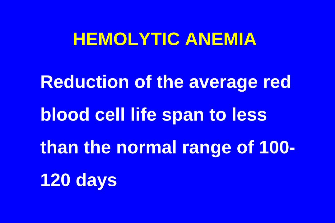
HEMOLYTIC ANEMIA
Reduction of the average red
blood cell life span to less
than the normal range of 100-
120 days
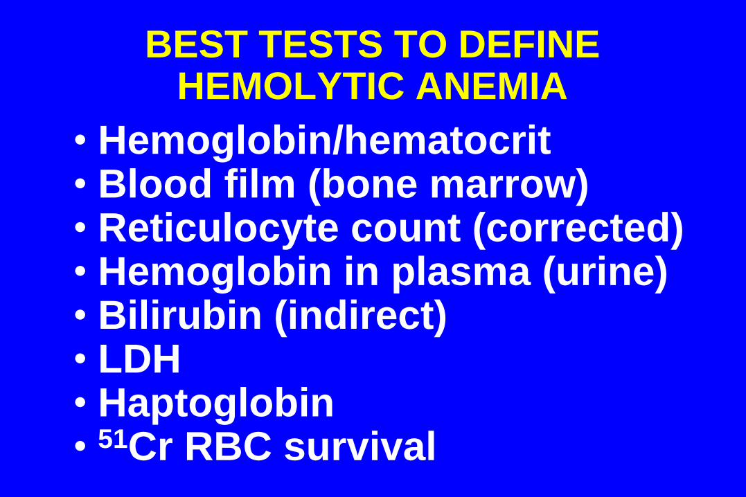
BEST TESTS TO DEFINE HEMOLYTIC ANEMIA
• Hemoglobin/hematocrit • Blood film (bone marrow) • Reticulocyte count (corrected) • Hemoglobin in plasma (urine) • Bilirubin (indirect) • LDH • Haptoglobin • 51Cr RBC survival
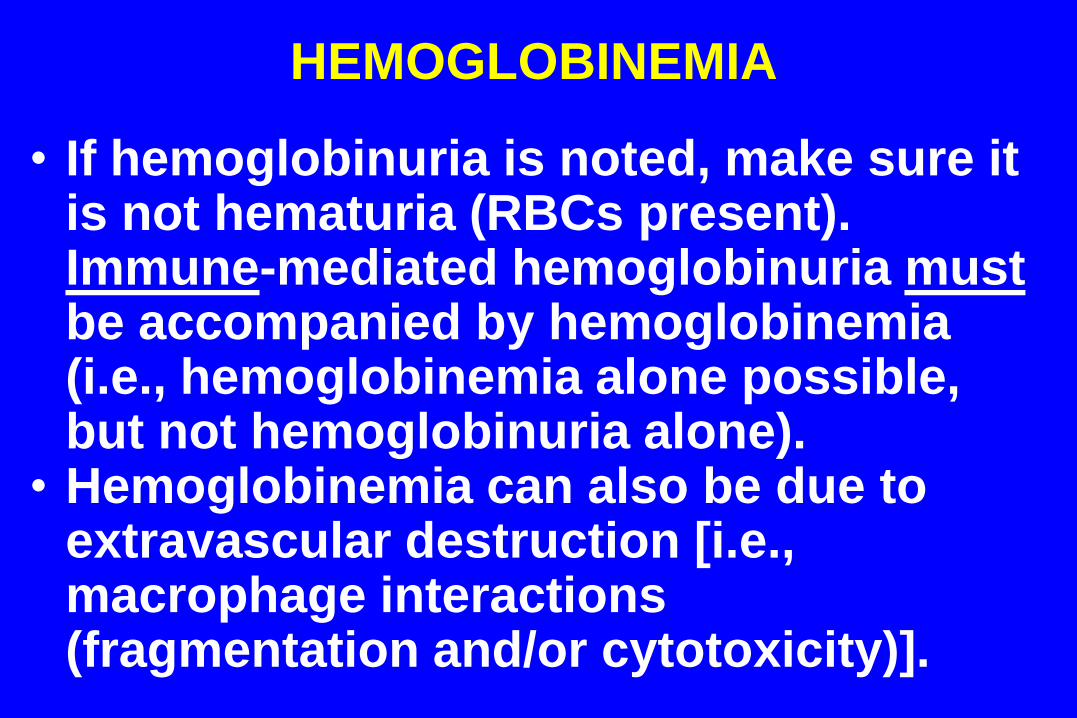
HEMOGLOBINEMIA
• If hemoglobinuria is noted, make sure it is not hematuria (RBCs present). Immune-mediated hemoglobinuria must be accompanied by hemoglobinemia (i.e., hemoglobinemia alone possible, but not hemoglobinuria alone).
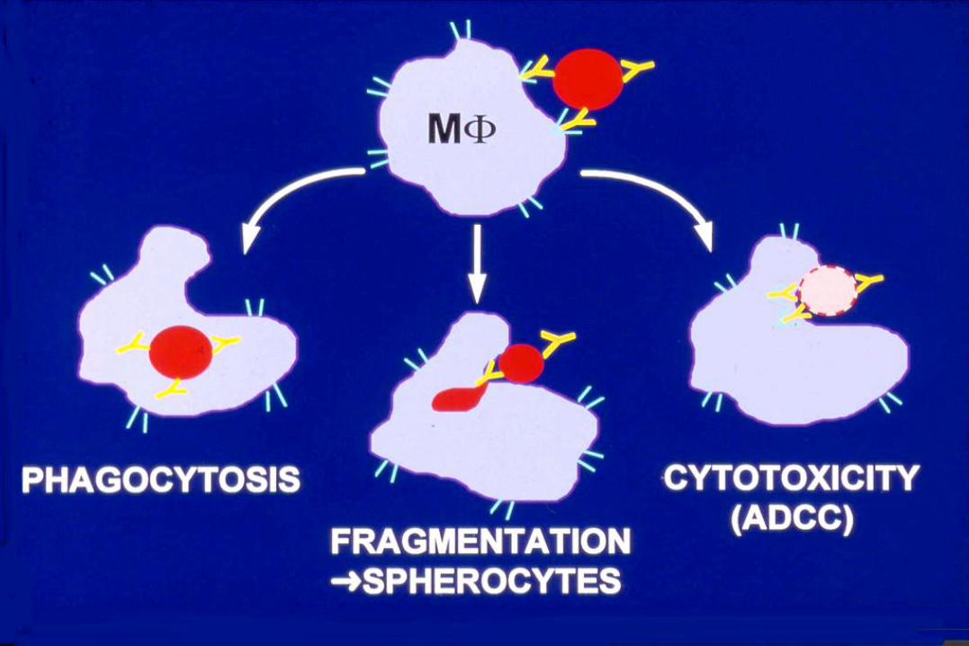
• Hemoglobinemia can also be due to extravascular destruction [i.e., macrophage interactions (fragmentation and/or cytotoxicity)].
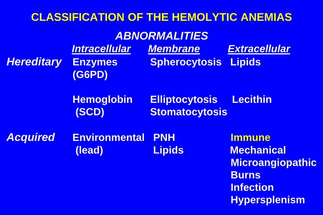
CLASSIFICATION OF THE HEMOLYTIC ANEMIAS
ABNORMALITIES
Intracellular Membrane Extracellular
Hereditary Enzymes Spherocytosis Lipids
(G6PD)
Hemoglobin Elliptocytosis Lecithin
(SCD) Stomatocytosis
Acquired Environmental PNH Immune
(lead) Lipids Mechanical
Microangiopathic
Burns
Infection
Hypersplenism
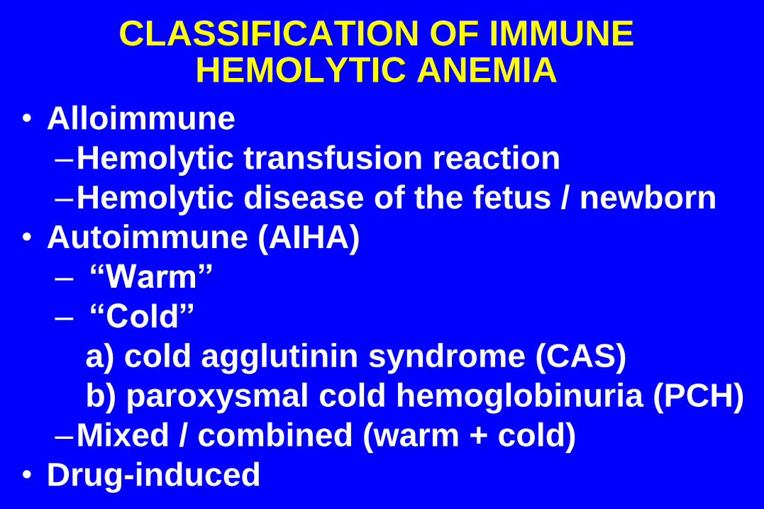
CLASSIFICATION OF IMMUNE HEMOLYTIC ANEMIA
• Alloimmune
–Hemolytic transfusion reaction
–Hemolytic disease of the fetus / newborn
• Autoimmune (AIHA)
– “Warm”
– “Cold”
a) cold agglutinin syndrome (CAS)
b) paroxysmal cold hemoglobinuria (PCH)
–Mixed / combined (warm + cold)
• Drug-induced
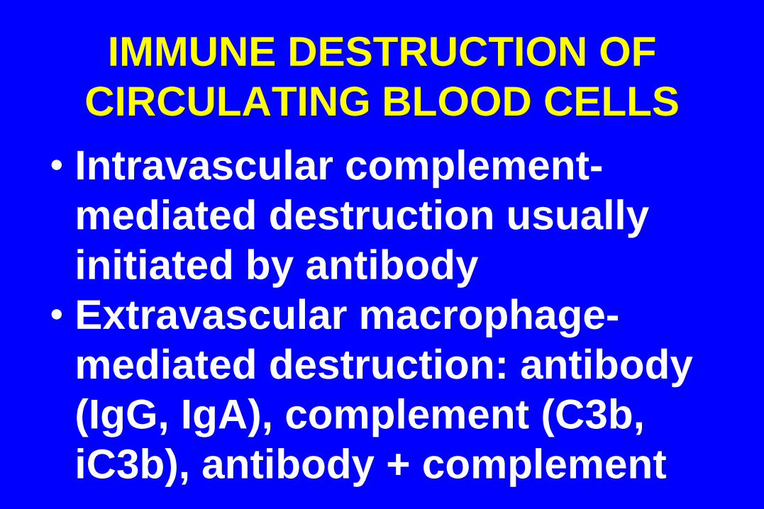
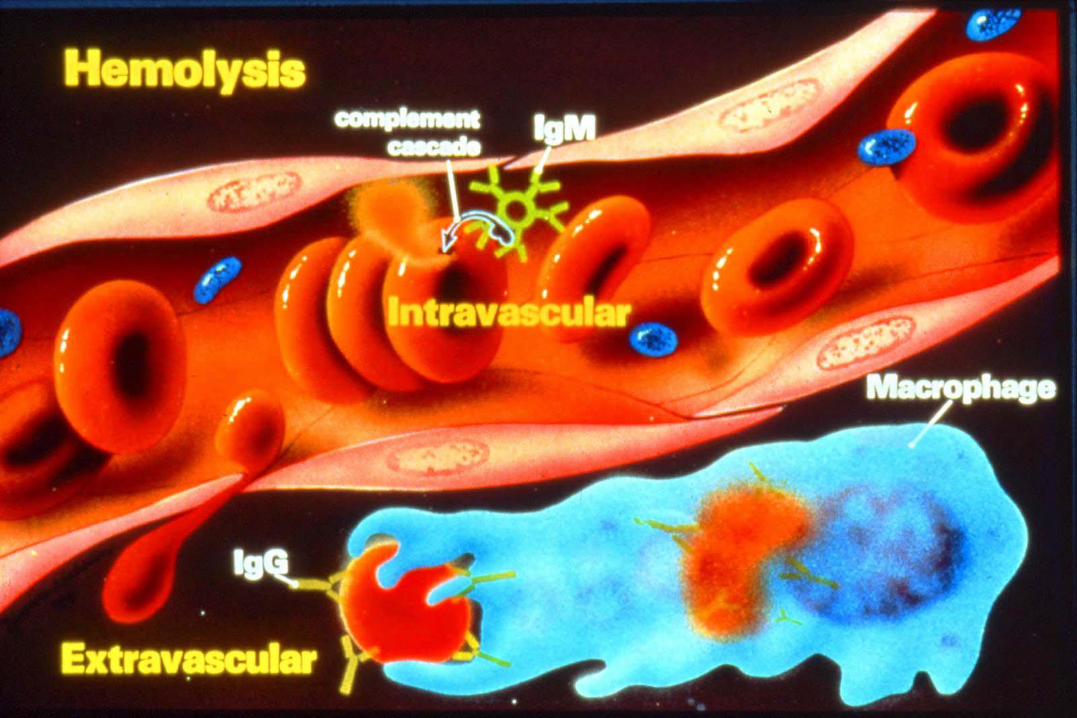
IMMUNE DESTRUCTION OF
CIRCULATING BLOOD CELLS
• Intravascular complement-
mediated destruction usually
initiated by antibody
• Extravascular macrophage-
mediated destruction: antibody
(IgG, IgA), complement (C3b,
iC3b), antibody + complement
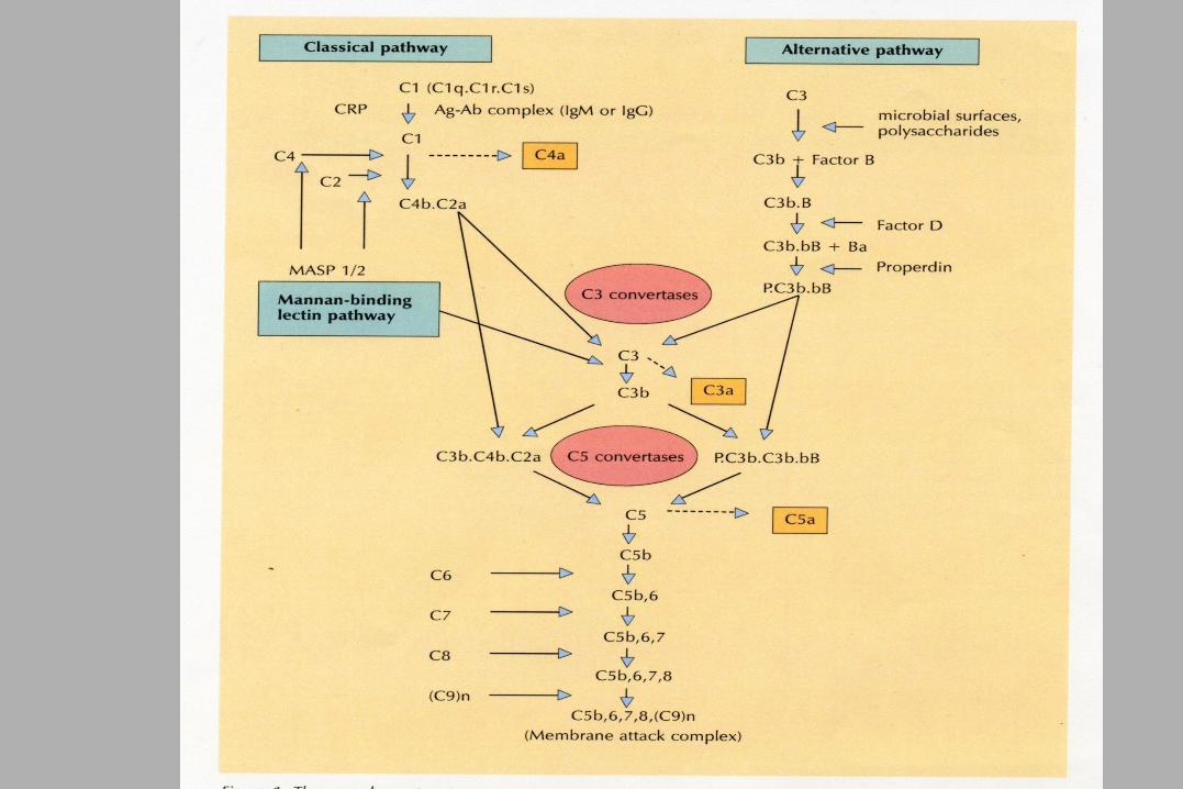
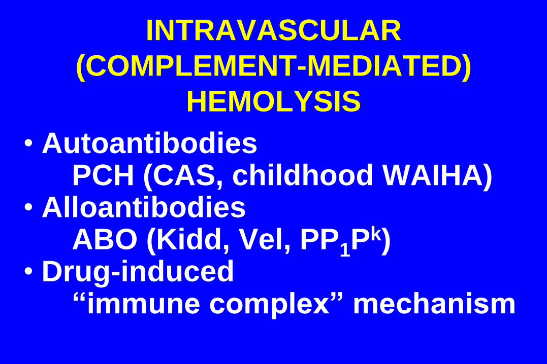
INTRAVASCULAR
(COMPLEMENT-MEDIATED)
HEMOLYSIS
• Autoantibodies PCH (CAS, childhood WAIHA)
• Alloantibodies ABO (Kidd, Vel, PP1P
k) • Drug-induced
“immune complex” mechanism
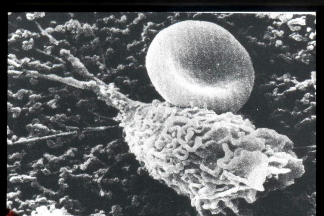
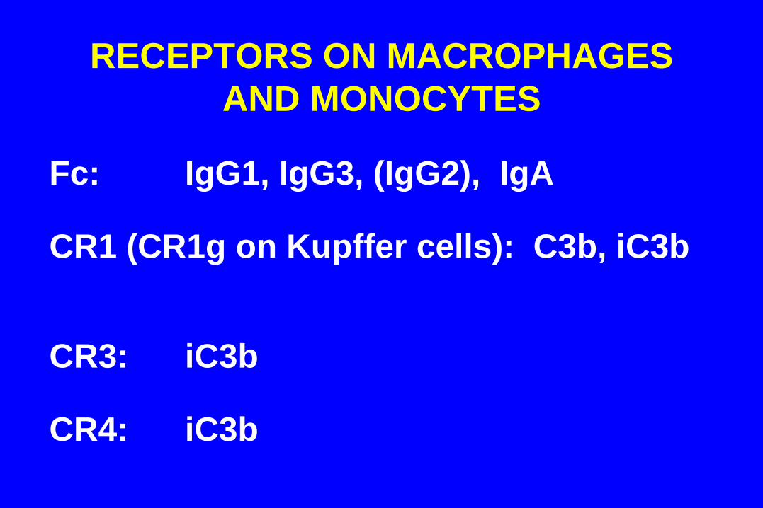
RECEPTORS ON MACROPHAGES
AND MONOCYTES
Fc: IgG1, IgG3, (IgG2), IgA CR1 (CR1g on Kupffer cells): C3b, iC3b CR3: iC3b CR4: iC3b
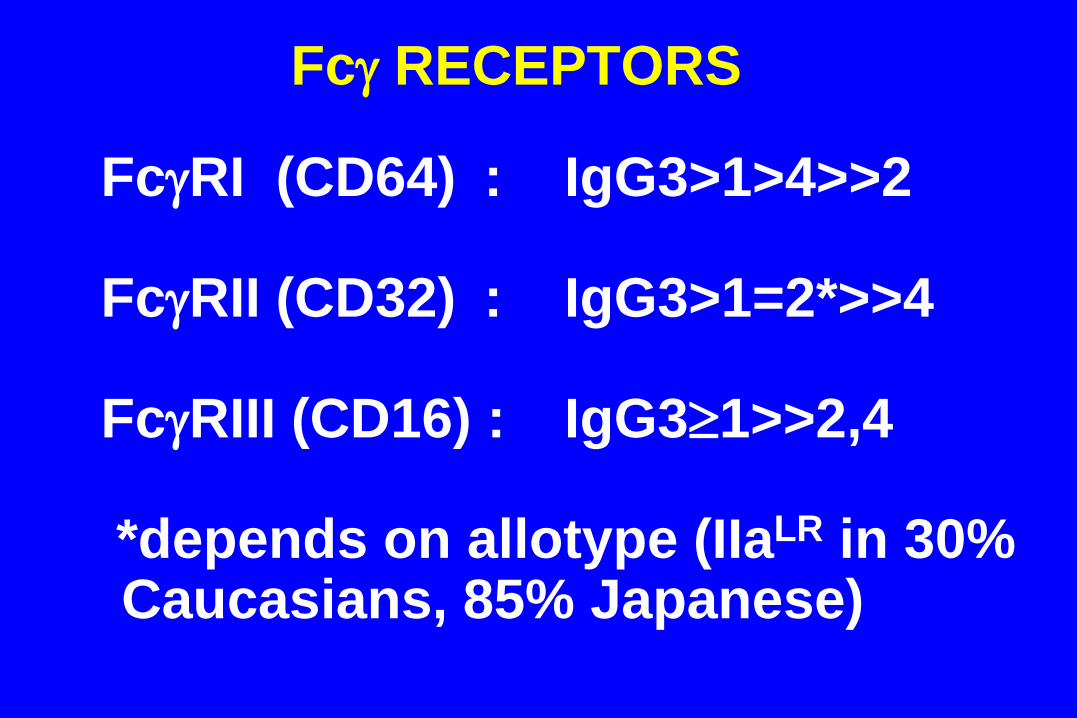
Fc RECEPTORS
FcRI (CD64) : IgG3>1>4>>2 FcRII (CD32) : IgG3>1=2*>>4 FcRIII (CD16) : IgG31>>2,4 *depends on allotype (IIaLR in 30%
Caucasians, 85% Japanese)
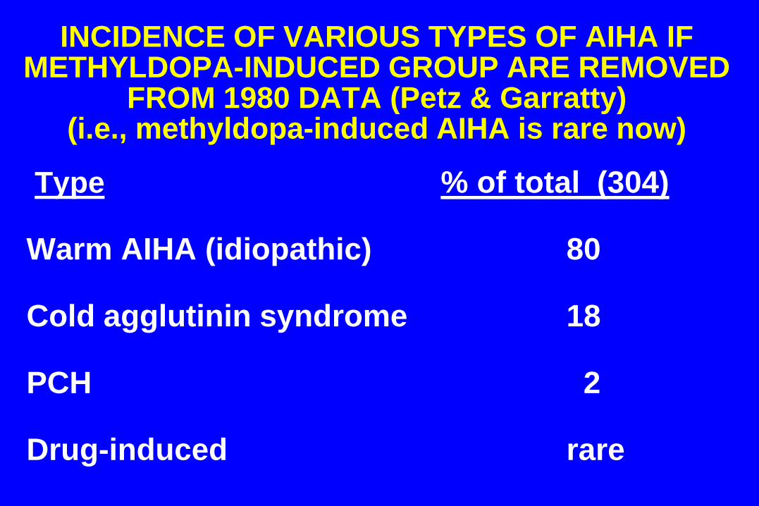
INCIDENCE OF VARIOUS TYPES OF AIHA IF METHYLDOPA-INDUCED GROUP ARE REMOVED
FROM 1980 DATA (Petz & Garratty) (i.e., methyldopa-induced AIHA is rare now)
Type % of total (304) Warm AIHA (idiopathic) 80 Cold agglutinin syndrome 18 PCH 2 Drug-induced rare
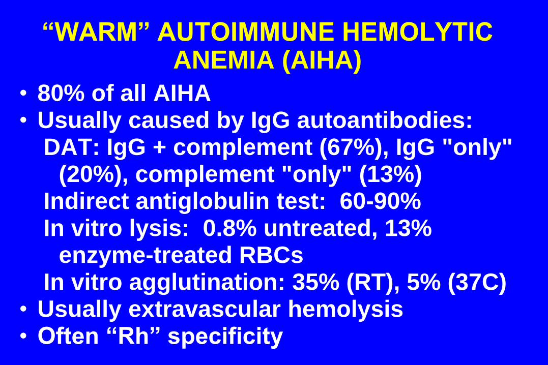
“WARM” AUTOIMMUNE HEMOLYTIC ANEMIA (AIHA)
• 80% of all AIHA • Usually caused by IgG autoantibodies:
DAT: IgG + complement (67%), IgG "only" (20%), complement "only" (13%)
Indirect antiglobulin test: 60-90% In vitro lysis: 0.8% untreated, 13%
enzyme-treated RBCs In vitro agglutination: 35% (RT), 5% (37C)
• Usually extravascular hemolysis • Often “Rh” specificity
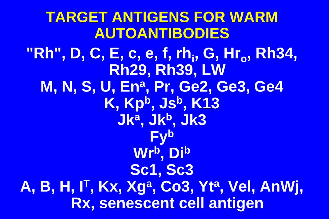
TARGET ANTIGENS FOR WARM AUTOANTIBODIES
"Rh", D, C, E, c, e, f, rhi, G, Hro, Rh34, Rh29, Rh39, LW
M, N, S, U, Ena, Pr, Ge2, Ge3, Ge4 K, Kpb, Jsb, K13
Jka, Jkb, Jk3 Fyb
Wrb, Dib
Sc1, Sc3 A, B, H, IT, Kx, Xga, Co3, Yta, Vel, AnWj,
Rx, senescent cell antigen
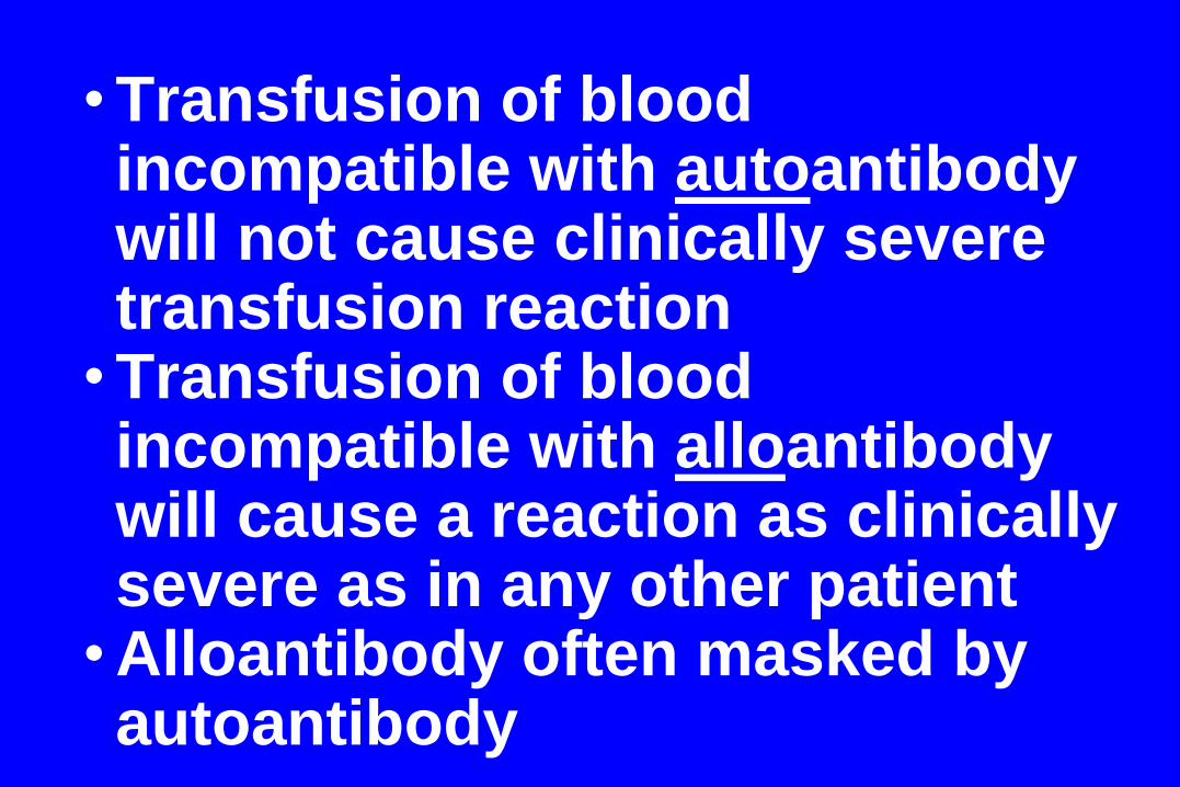
•Transfusion of blood incompatible with autoantibody will not cause clinically severe transfusion reaction
•Transfusion of blood incompatible with alloantibody will cause a reaction as clinically severe as in any other patient
•Alloantibody often masked by autoantibody
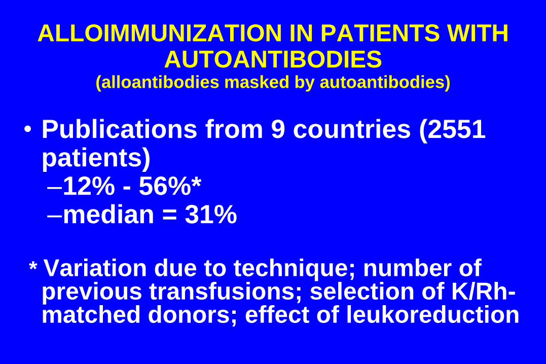
ALLOIMMUNIZATION IN PATIENTS WITH
AUTOANTIBODIES (alloantibodies masked by autoantibodies)
• Publications from 9 countries (2551 patients) –12% - 56%* –median = 31%
* Variation due to technique; number of
previous transfusions; selection of K/Rh-matched donors; effect of leukoreduction
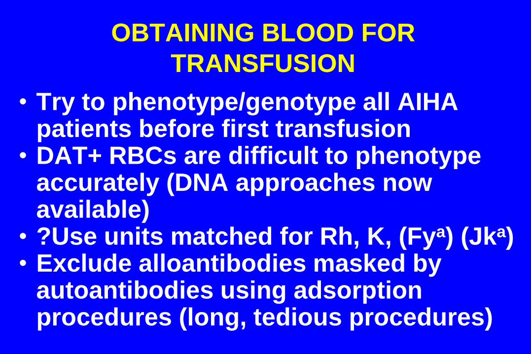
OBTAINING BLOOD FOR
TRANSFUSION
• Try to phenotype/genotype all AIHA patients before first transfusion
• DAT+ RBCs are difficult to phenotype accurately (DNA approaches now available)
• ?Use units matched for Rh, K, (Fya) (Jka) • Exclude alloantibodies masked by
autoantibodies using adsorption procedures (long, tedious procedures)
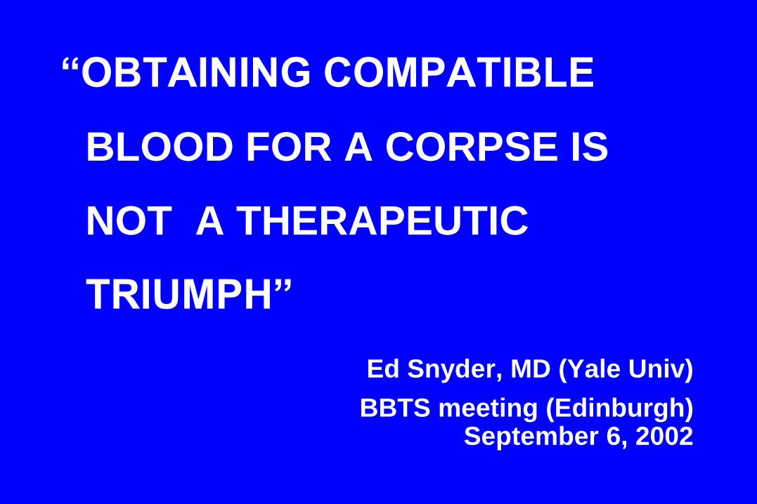
“OBTAINING COMPATIBLE
BLOOD FOR A CORPSE IS
NOT A THERAPEUTIC
TRIUMPH”
Ed Snyder, MD (Yale Univ)
BBTS meeting (Edinburgh) September 6, 2002
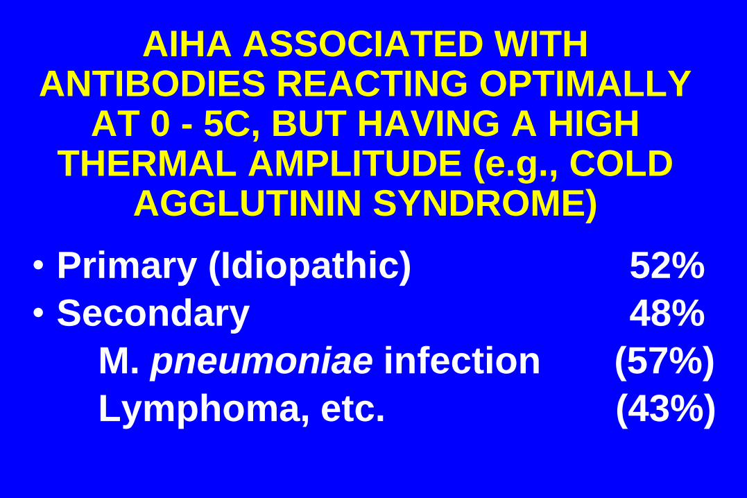
AIHA ASSOCIATED WITH ANTIBODIES REACTING OPTIMALLY
AT 0 - 5C, BUT HAVING A HIGH THERMAL AMPLITUDE (e.g., COLD
AGGLUTININ SYNDROME)
• Primary (Idiopathic) 52%
• Secondary 48%
M. pneumoniae infection (57%)
Lymphoma, etc. (43%)
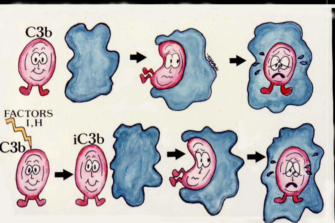
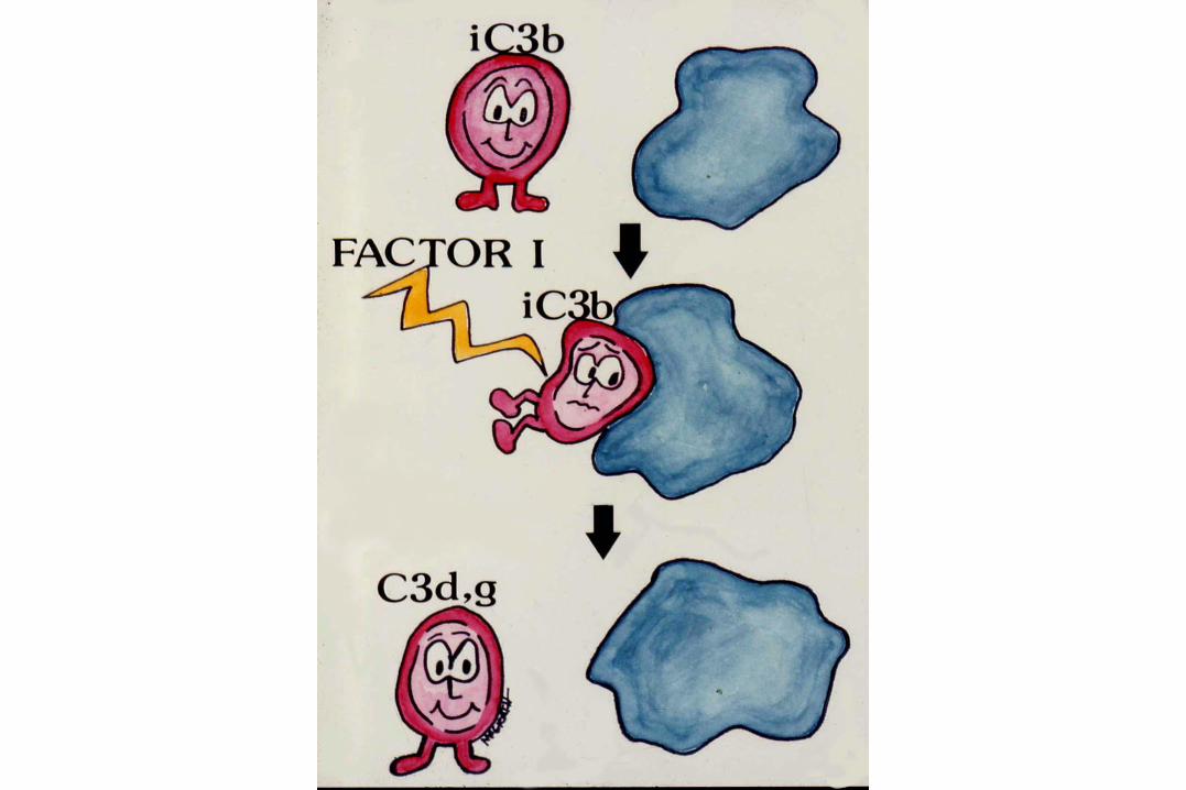
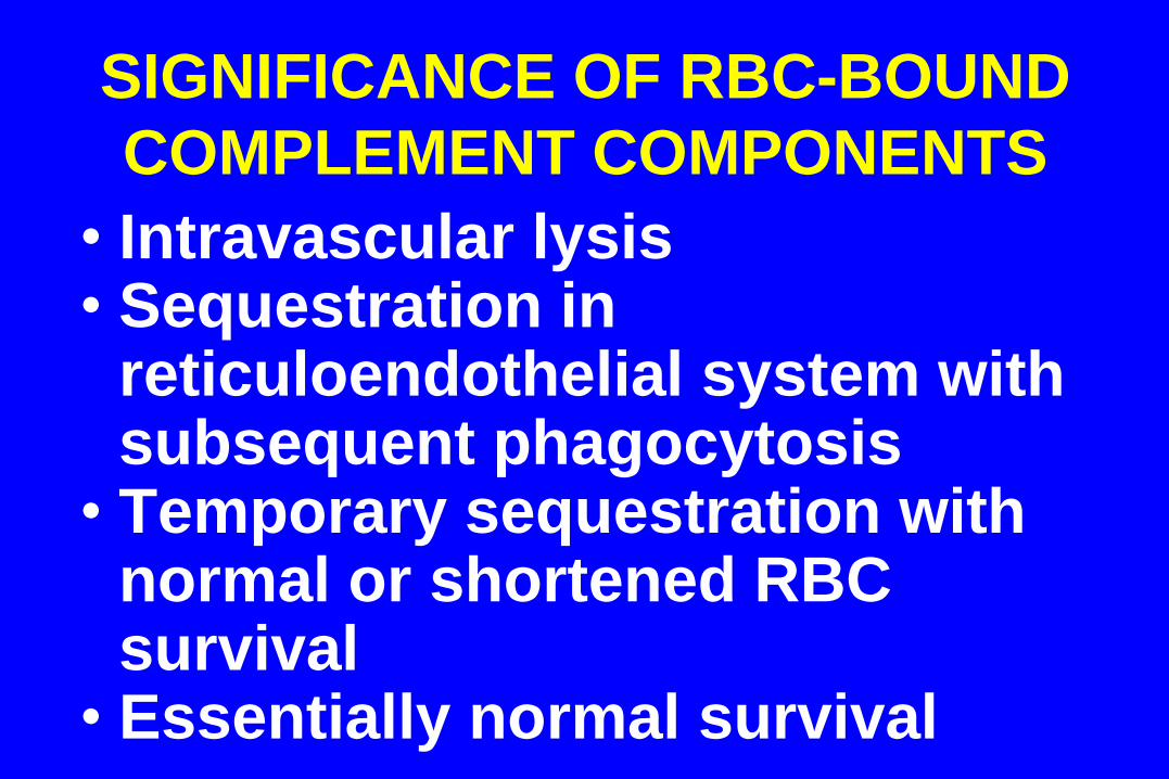
SIGNIFICANCE OF RBC-BOUND
COMPLEMENT COMPONENTS
• Intravascular lysis • Sequestration in reticuloendothelial system with
subsequent phagocytosis • Temporary sequestration with normal or shortened RBC survival • Essentially normal survival
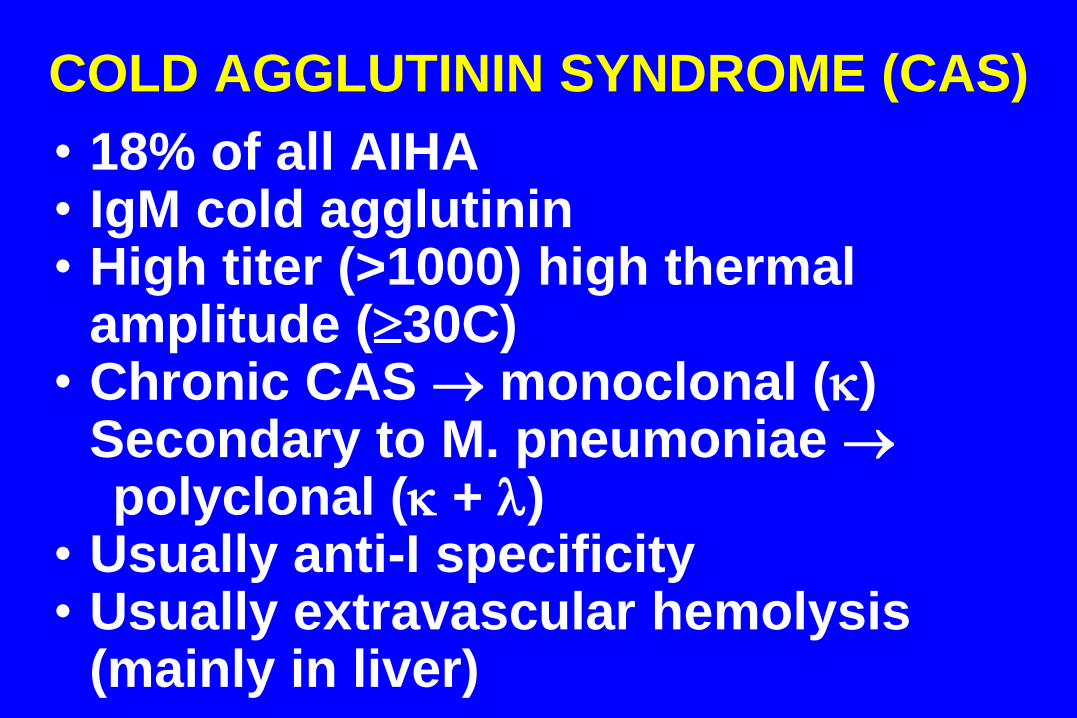
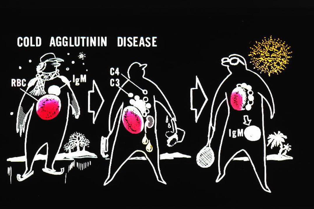
COLD AGGLUTININ SYNDROME (CAS)
• 18% of all AIHA • IgM cold agglutinin • High titer (>1000) high thermal
amplitude (30C) • Chronic CAS monoclonal ()
Secondary to M. pneumoniae polyclonal ( + ) • Usually anti-I specificity • Usually extravascular hemolysis
(mainly in liver)
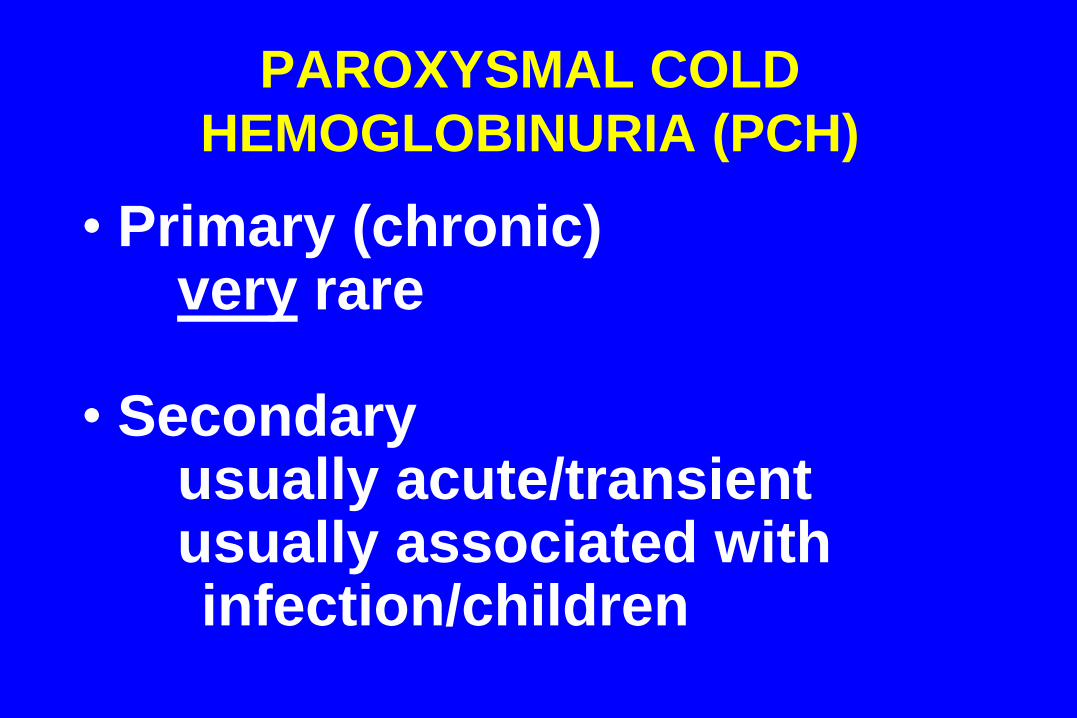
PAROXYSMAL COLD
HEMOGLOBINURIA (PCH)
• Primary (chronic) very rare • Secondary
usually acute/transient usually associated with infection/children
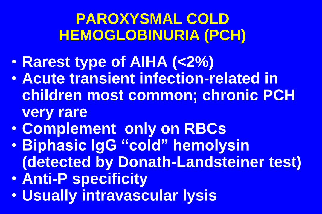
PAROXYSMAL COLD HEMOGLOBINURIA (PCH)
• Rarest type of AIHA (<2%) • Acute transient infection-related in
children most common; chronic PCH very rare
• Complement only on RBCs • Biphasic IgG “cold” hemolysin
(detected by Donath-Landsteiner test) • Anti-P specificity • Usually intravascular lysis
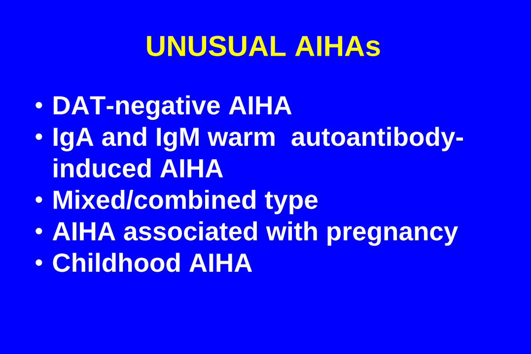
UNUSUAL AIHAs
• DAT-negative AIHA
• IgA and IgM warm autoantibody-
induced AIHA
• Mixed/combined type
• AIHA associated with pregnancy
• Childhood AIHA
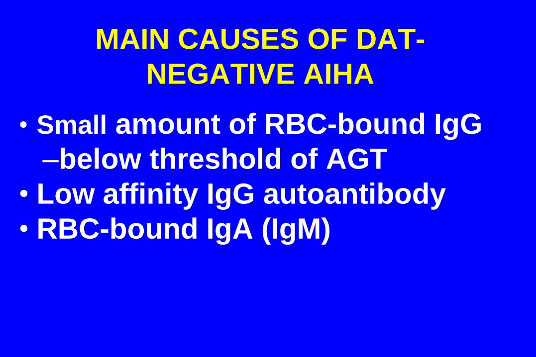
MAIN CAUSES OF DAT-
NEGATIVE AIHA
• Small amount of RBC-bound IgG
–below threshold of AGT
• Low affinity IgG autoantibody
• RBC-bound IgA (IgM)
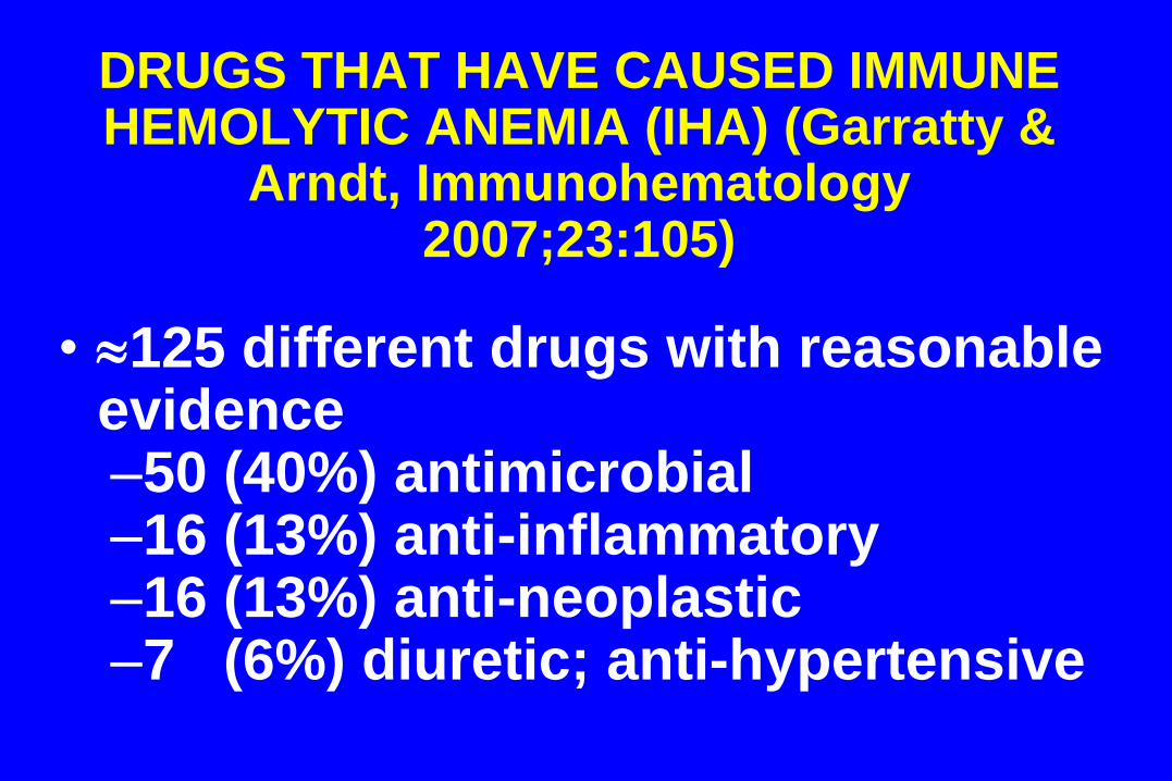
DRUGS THAT HAVE CAUSED IMMUNE HEMOLYTIC ANEMIA (IHA) (Garratty &
Arndt, Immunohematology 2007;23:105)
• 125 different drugs with reasonable evidence –50 (40%) antimicrobial –16 (13%) anti-inflammatory –16 (13%) anti-neoplastic –7 (6%) diuretic; anti-hypertensive
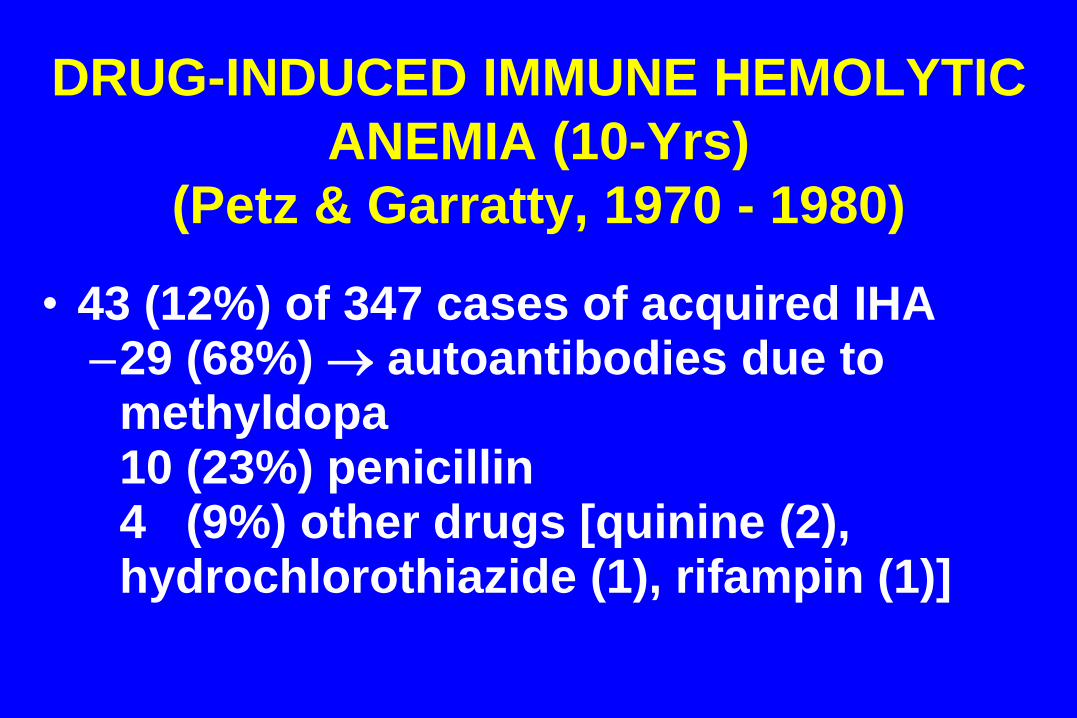
DRUG-INDUCED IMMUNE HEMOLYTIC
ANEMIA (10-Yrs)
(Petz & Garratty, 1970 - 1980)
• 43 (12%) of 347 cases of acquired IHA –29 (68%) autoantibodies due to
methyldopa 10 (23%) penicillin 4 (9%) other drugs [quinine (2),
hydrochlorothiazide (1), rifampin (1)]
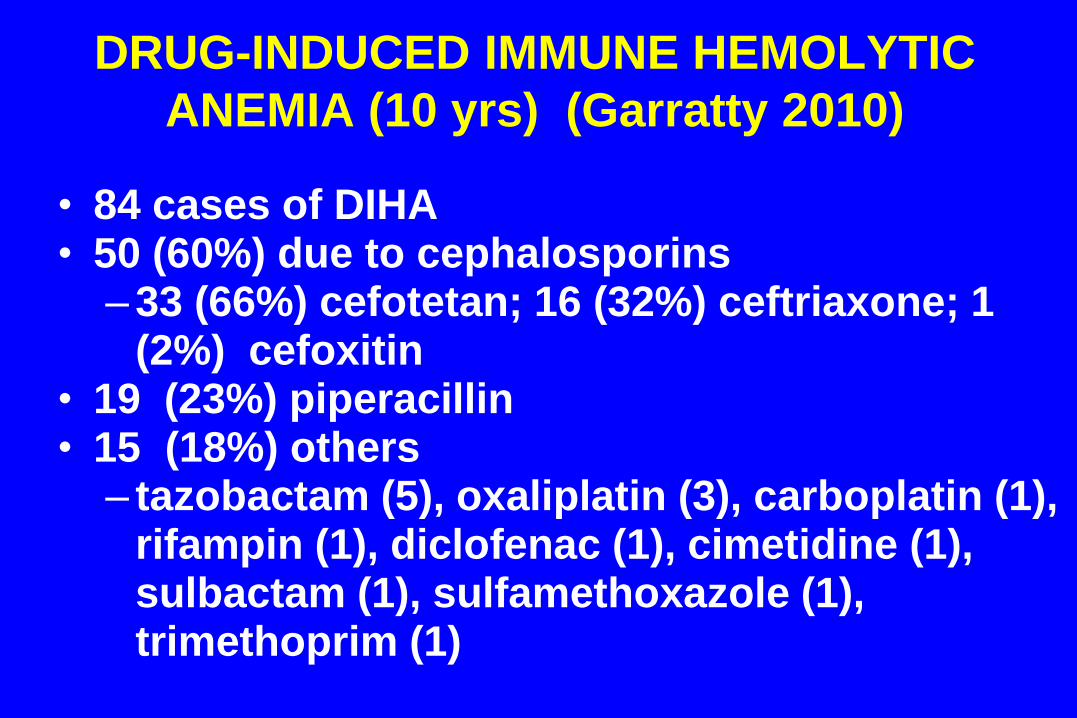
DRUG-INDUCED IMMUNE HEMOLYTIC
ANEMIA (10 yrs) (Garratty 2010)
• 84 cases of DIHA • 50 (60%) due to cephalosporins
–33 (66%) cefotetan; 16 (32%) ceftriaxone; 1 (2%) cefoxitin
• 19 (23%) piperacillin • 15 (18%) others
– tazobactam (5), oxaliplatin (3), carboplatin (1), rifampin (1), diclofenac (1), cimetidine (1), sulbactam (1), sulfamethoxazole (1), trimethoprim (1)

TOP 3 DRUGS CAUSING DIIHA
2006 – 2010 2011 (so far)
Piperacillin 17 9 Ceftriaxone 11 1 Cefotetan 8 1
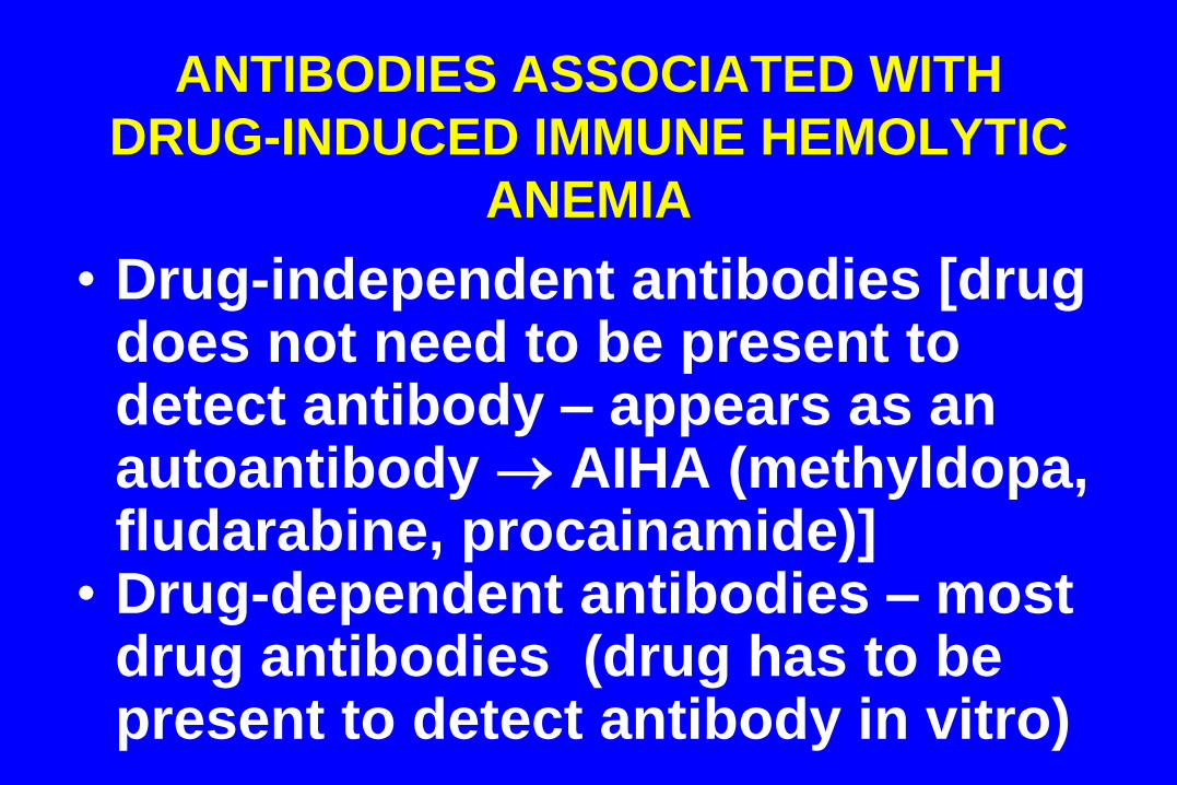
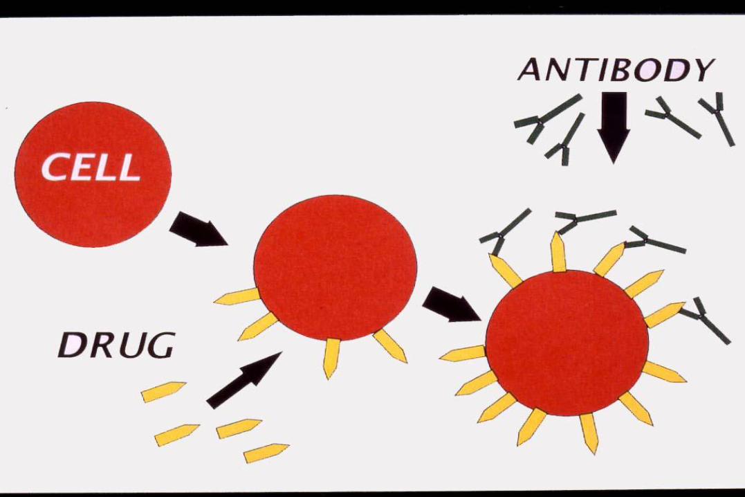
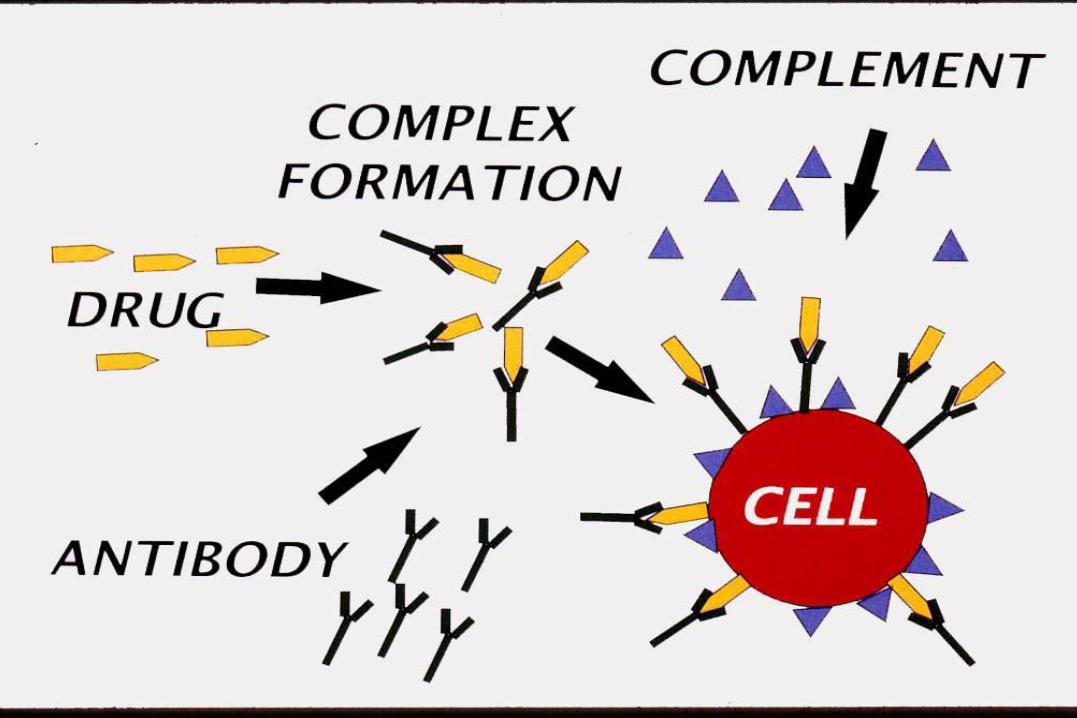
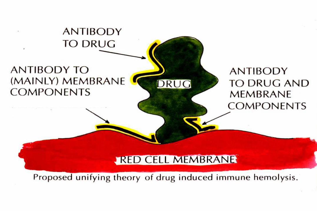
ANTIBODIES ASSOCIATED WITH
DRUG-INDUCED IMMUNE HEMOLYTIC
ANEMIA
• Drug-independent antibodies [drug does not need to be present to detect antibody – appears as an autoantibody AIHA (methyldopa, fludarabine, procainamide)]
• Drug-dependent antibodies – most drug antibodies (drug has to be present to detect antibody in vitro)
DIFFICULT TO PROVE THAT DRUG HAS CAUSED AUTOIMMUNE
HEMOLYTIC ANEMIA (AIHA)
• Many reports only describe a HA and/or +DAT following drug therapy and improvement when drug stopped
• Cannot be proven in laboratory as antibody reacts without presence of drug
• Idiopathic AIHA is far more common than drug-induced IHA, thus is first suspect
• Best proof (but not often possible) is to give drug again when autoantibody disappears
ALERT
•Cephalosporin- and piperacillin-induced hemolytic anemia and/or +DAT can mimic: –delayed hemolytic transfusion
reaction –autoimmune hemolytic anemia
• Antibodies sometimes react without adding drug in vitro as circulating drug can be present in vivo up to 48 hrs.
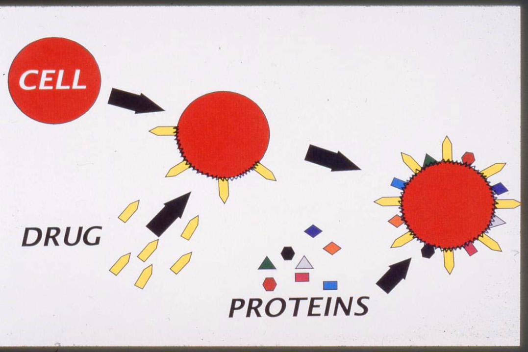
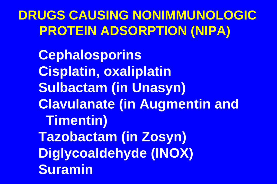
DRUGS CAUSING NONIMMUNOLOGIC
PROTEIN ADSORPTION (NIPA)
Cephalosporins
Cisplatin, oxaliplatin
Sulbactam (in Unasyn)
Clavulanate (in Augmentin and
Timentin)
Tazobactam (in Zosyn)
Diglycoaldehyde (INOX)
Suramin
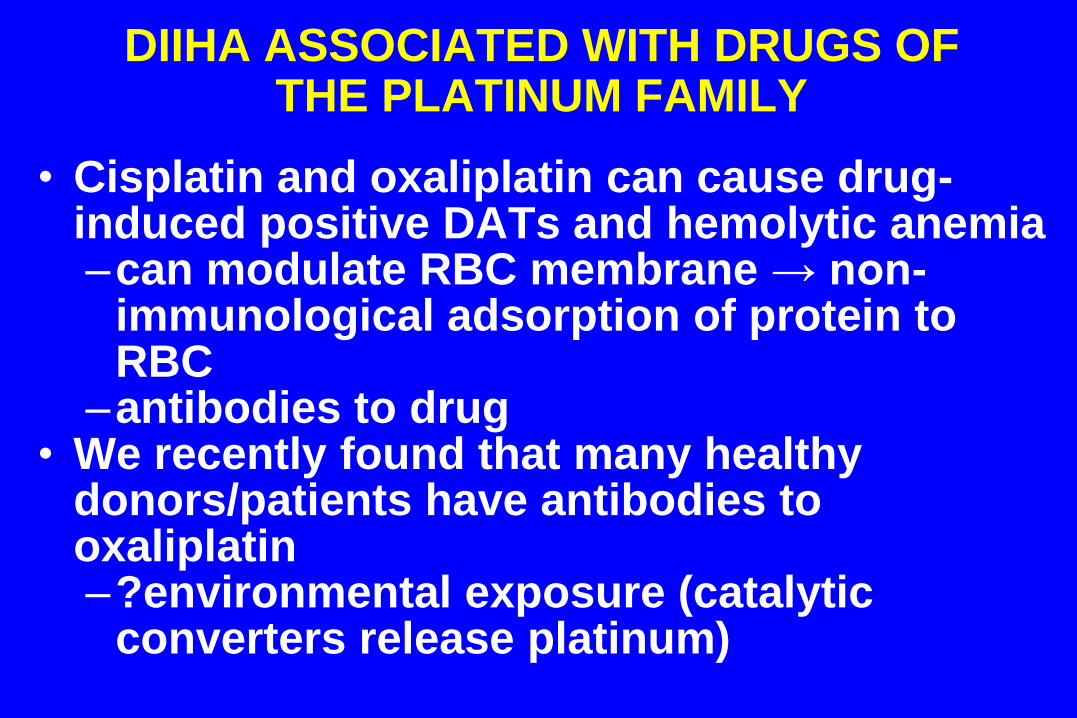
DIIHA ASSOCIATED WITH DRUGS OF THE PLATINUM FAMILY
• Cisplatin and oxaliplatin can cause drug-induced positive DATs and hemolytic anemia –can modulate RBC membrane → non-
immunological adsorption of protein to RBC
–antibodies to drug • We recently found that many healthy
donors/patients have antibodies to oxaliplatin –?environmental exposure (catalytic
converters release platinum)
Related Documents