CHAPTER 5 Pathophysiology of asthma P.J. Barnes Correspondence: P.J. Barnes, Dept of Thoracic Medicine, National Heart and Lung Institute, Dovehouse St, London SW3 6LY, UK. Asthma is characterised by a specific pattern of inflammation that is largely driven via immunoglobulin (Ig)E-dependent mechanisms. Genetic factors have an important influence on whether atopy develops and several genes have now been identified [1]. Most of the genetic linkages reported for asthma are common to all allergic diseases [2]. However, environmental factors appear to be more important in determining whether an atopic individual develops asthma, although genetic factors may exert an influence on how severely the disease is expressed and the amplification of the inflammatory response. Inflammation It had been recognised for many years that patients who die from acute asthma attacks have grossly inflamed airways. The airway lumen is occluded by a tenacious mucus plug composed of plasma proteins exuded from airway vessels and mucus glycoproteins secreted from surface epithelial cells. The airway wall is oedematous and infiltrated with inflammatory cells, which are predominantly eosinophils and lymphocytes. The airway epithelium is invariably shed in a patchy manner and clumps of epithelial cells are found in the airway lumen. Occasionally there have been opportunities to examine the airways of asthmatic patients who die accidentally and similar though less marked inflammatory changes have been observed [3]. More recently it has been possible to examine the airways of asthmatic patients by fibreoptic and rigid bronchoscopy, by bronchial biopsy and by bronchoalveolar lavage (BAL). Direct bronchoscopy reveals that the airways of asthmatic patients are often reddened and swollen, indicating acute inflammation. Lavage has revealed an increase in the numbers of lymphocytes, mast cells and eosino- phils and evidence for activation of macrophages in comparison with nonasthmatic controls. Biopsies have revealed evidence for increased numbers and activation of mast cells, macrophages, eosinophils and T-lymphocytes [4, 5]. These changes are found even in patients with mild asthma who have few symptoms, and this suggests that asthma is an inflammatory condition of the airways. Inflammation is classically characterised by four cardinal signs: calor and rubor (due to vasodilatation), tumour (due to plasma exudation and oedema) and dolor (due to sensi- tisation and activation of sensory nerves. It is now recognised that inflammation is also characterised by an infiltration with inflammatory cells and that these will differ depending on the type of inflammatory process. Inflammation is an important defence response that defends the body against invasion from microorganisms and against the effects of external toxins. The inflammation in allergic asthma is characterised by the fact that it is driven by exposure to allergens through IgE-dependent mechanisms, resulting in a characteristic pattern of inflammation. This allergic inflammatory response is characterised by an infiltration with eosinophils and resembles the inflammatory process mounted in response to parasitic and worm infections. The inflammatory response not Eur Respir Mon, 2003, 23, 84–113. Printed in UK - all rights reserved. Copyright ERS Journals Ltd 2003; European Respiratory Monograph; ISSN 1025-448x. ISBN 1-904097-26-x. 84

Welcome message from author
This document is posted to help you gain knowledge. Please leave a comment to let me know what you think about it! Share it to your friends and learn new things together.
Transcript
CHAPTER 5
Pathophysiology of asthma
P.J. Barnes
Correspondence: P.J. Barnes, Dept of Thoracic Medicine, National Heart and Lung Institute, DovehouseSt, London SW3 6LY, UK.
Asthma is characterised by a specific pattern of inflammation that is largely driven viaimmunoglobulin (Ig)E-dependent mechanisms. Genetic factors have an importantinfluence on whether atopy develops and several genes have now been identified [1]. Mostof the genetic linkages reported for asthma are common to all allergic diseases [2].However, environmental factors appear to be more important in determining whether anatopic individual develops asthma, although genetic factors may exert an influence onhow severely the disease is expressed and the amplification of the inflammatory response.
Inflammation
It had been recognised for many years that patients who die from acute asthma attackshave grossly inflamed airways. The airway lumen is occluded by a tenacious mucus plugcomposed of plasma proteins exuded from airway vessels and mucus glycoproteinssecreted from surface epithelial cells. The airway wall is oedematous and infiltrated withinflammatory cells, which are predominantly eosinophils and lymphocytes. The airwayepithelium is invariably shed in a patchy manner and clumps of epithelial cells are foundin the airway lumen. Occasionally there have been opportunities to examine the airwaysof asthmatic patients who die accidentally and similar though less marked inflammatorychanges have been observed [3]. More recently it has been possible to examine theairways of asthmatic patients by fibreoptic and rigid bronchoscopy, by bronchial biopsyand by bronchoalveolar lavage (BAL). Direct bronchoscopy reveals that the airways ofasthmatic patients are often reddened and swollen, indicating acute inflammation.Lavage has revealed an increase in the numbers of lymphocytes, mast cells and eosino-phils and evidence for activation of macrophages in comparison with nonasthmaticcontrols. Biopsies have revealed evidence for increased numbers and activation of mastcells, macrophages, eosinophils and T-lymphocytes [4, 5]. These changes are found evenin patients with mild asthma who have few symptoms, and this suggests that asthma is aninflammatory condition of the airways.
Inflammation is classically characterised by four cardinal signs: calor and rubor (dueto vasodilatation), tumour (due to plasma exudation and oedema) and dolor (due to sensi-tisation and activation of sensory nerves. It is now recognised that inflammation is alsocharacterised by an infiltration with inflammatory cells and that these will differdepending on the type of inflammatory process. Inflammation is an important defenceresponse that defends the body against invasion from microorganisms and against theeffects of external toxins. The inflammation in allergic asthma is characterised by the factthat it is driven by exposure to allergens through IgE-dependent mechanisms, resultingin a characteristic pattern of inflammation. This allergic inflammatory response ischaracterised by an infiltration with eosinophils and resembles the inflammatory processmounted in response to parasitic and worm infections. The inflammatory response not
Eur Respir Mon, 2003, 23, 84–113. Printed in UK - all rights reserved. Copyright ERS Journals Ltd 2003; European Respiratory Monograph;ISSN 1025-448x. ISBN 1-904097-26-x.
84
only provides an acute defence against injury, but is also involved in healing andrestoration of normal function after tissue damage as a result of infection of toxins. Inasthma, the inflammatory response is activated inappropriately and is harmful ratherthan beneficial. For some reason allergens, such as house dust mite and pollen proteins,induce an eosinophilic inflammation. Normally such an inflammatory response wouldkill the invading parasite (or vice versa) and would therefore be self-limiting, but inallergic disease the inciting stimulus persists and the normally acute inflammatoryresponse becomes converted into a chronic inflammation which may have structuralconsequences in the airways and skin.
Intrinsic asthma
Although the majority of patients with asthma have atopy, in a proportion of patientswith asthma there is no evidence of atopy with normal total and specific IgE and negativeskin tests. This so-called "intrinsic" asthma usually comes on later in life and tends to bemore severe than allergic asthma [6]. The pathophysiology is very similar to that ofallergic asthma and there is increasing evidence for local IgE production, possiblydirected at bacterial or viral antigens [7].
Inflammation and airway hyperresponsiveness
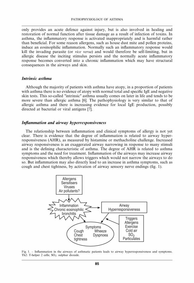
The relationship between inflammation and clinical symptoms of allergy is not yetclear. There is evidence that the degree of inflammation is related to airway hyper-responsiveness (AHR), as measured by histamine or methacholine challenge. Increasedairway responsiveness is an exaggerated airway narrowing in response to many stimuliand is the defining characteristic of asthma. The degree of AHR is related to asthmasymptoms and the need for treatment. Inflammation of the airways may increase airwayresponsiveness which thereby allows triggers which would not narrow the airways to doso. But inflammation may also directly lead to an increase in asthma symptoms, such ascough and chest tightness, by activation of airway sensory nerve endings (fig. 1).
�����������������������
�� ���������
�����������������������
���������������� ���������
�������
������������ ���������� ��������������
���������������!"��������# ���$%
&��������
Fig. 1. – Inflammation in the airways of asthmatic patients leads to airway hyperresponsiveness and symptoms.Th2: T-helper 2 cells; SO2: sulphur dioxide.
PATHOPHYSIOLOGY OF ASTHMA
85
Persistence of inflammation
Although most attention has focused on the acute inflammatory changes seen inasthma, this is a chronic condition, with inflammation persisting over many years in mostpatients. The mechanisms involved in persistence of inflammation in asthma are stillpoorly understood. Superimposed on this chronic inflammatory state are acute inflam-matory episodes which correspond to exacerbations of asthma.
Inflammatory cells
Many different inflammatory cells are involved in asthma, although the precise role ofeach cell type is not yet certain [4]. It is evident that no single inflammatory cell is able toaccount for the complex pathophysiology of allergic disease, but some cells predominatein asthmatic inflammation.
Mast cells
Mast cells are important in initiating the acute bronchoconstrictor responses toallergen and probably to other indirect stimuli, such as exercise and hyperventilation (viaosmolality or thermal changes) and fog. Patients with asthma are characterised by amarked increase in mast cell numbers in airway smooth muscle [8]. Treatment ofasthmatic patients with prednisone results in a decrease in the number of tryptasepositive mast cells [9]. Furthermore, mast cell tryptase appears to play a role in airwayremodelling, as this mast cell product stimulates human lung fibroblast proliferation [10].Mast cells also secrete certain cytokines, such as interleukin (IL)-4 that may be involvedin maintaining the allergic inflammatory response and tumour necrosis factor (TNF)-a[11].
However, there are questions about the role of mast cells in more chronic allergicinflammatory events and it seems more probable that other cells, such as macrophages,eosinophils and T-lymphocytes are more important in the chronic inflammatory process,including AHR. Classically mast cells are activated by allergens through an IgE-dependent mechanism. The importance of IgE in the pathophysiology of asthma hasbeen highlighted by recent clinical studies with humanised anti-IgE antibodies, whichinhibit IgE-mediated effects [12, 13]. Although anti-IgE antibody results in a reductionin circulating IgE to undetectable levels, this treatment results in minimal clinicalimprovement in patients with severe steroid-dependent asthma [14]. Interestingly, treat-ment with the anti-IgE monoclonal, did allow reduction of the dose of steroids requiresfor asthma control. This observation suggests that the mechanisms whereby IgE leads toairway obstruction are steroid sensitive, although corticosteroids do not reduce and mayeven increase, circulating levels of IgE [15, 16].
It is now increasingly recognised that mast cells may also release several othermediators that may play a role in the pathophysiology of asthma, including neuro-trophins, proinflammatory cytokines, chemokines and growth factors. This has led to are-evaluation of the role of mast cells, particularly during exacerbations [17].
Macrophages
Macrophages, which are derived from blood monocytes, may traffic into the airways inasthma and may be activated by allergen via low affinity IgE receptors (FceRII) [18, 19].The enormous immunological repertoire of macrophages allows these cells to produce
P.J. BARNES
86
many different products, including a large variety of cytokines that may orchestrate theinflammatory response. Macrophages have the capacity to initiate a particular type ofinflammatory response via the release of a certain pattern of cytokines. Macrophagesmay both increase and decrease inflammation, depending on the stimulus. Alveolarmacrophages normally have a suppressive effect on lymphocyte function, but this may beimpaired in asthma after allergen exposure [20]. One anti-inflammatory protein secretedby macrophages is IL-10 and its secretion is reduced in alveolar macrophages frompatients with asthma [21]. Macrophages from normal subjects also inhibit the secretionof IL-5 from T-lymphocytes, probably via the release of IL-12, but this is defective inpatients with allergic asthma [22]. Macrophages may therefore play an important anti-inflammatory role, by preventing the development of allergic inflammation. Macro-phages may also act as antigen-presenting cells which process allergen for presentation toT-lymphocytes, although alveolar macrophages are far less effective in this respect thanmacrophages from other sites, such as the peritoneum [23].
There may be subtypes of macrophages that perform different inflammatory, anti-inflammatory or phagocytic roles in allergic disease. Immunological markers that candistinguish these subpopulations are beginning to emerge [24]. However, no differencesin the macrophage population in induced sputum of allergic asthmatic compared tonormal subjects have been detected [25].
Dendritic cells
Dendritic cells are specialised macrophage-like cells that have a unique ability toinduce a T-lymphocyte mediated immune response and therefore play a critical role inthe development of asthma [26]. Dendritic cells in the respiratory tract form a networkthat is localised to the epithelium and act as very effective antigen-presenting cells [27]. Itis likely that dendritic cells play an important role in the initiation of allergen-inducedresponses in asthma [28]. Dendritic cells take up allergens, process them to peptides andmigrate to local lymph nodes where they present the allergenic peptides to uncommittedT-lymphocytes and with the aid of co-stimulatory molecules, such as B7.1, B7.2 andCD40 they programme the production of allergen-specific T-cells. Granulocyte-macrophage colony-stimulating factor (GM-CSF), which is expressed in abundanceby epithelial cells and macrophages in asthma, leads to differentiation and activation ofdendritic cells. This leads to production of myeloid dendritic cells which favour thedifferentiation of T-helper (Th)2 cells. [29]. Animal studies have demonstrated thatmyeloid dendritic cells are critical to the development of Th2 cells and eosinophilia [30].Immature dendritic cells in the respiratory tract promote Th2 cell differentiation andrequire cytokines such as IL-12 and TNF-a to promote the normally preponderant Th1response [31]. Dendritic cell based immunotherapy may be developed in the future for theprevention and control of allergic diseases.
Eosinophils
Eosinophil infiltration is a characteristic feature of allergic inflammation. Asthmamight more accurately be termed "chronic eosinophilic bronchitis" (a term first coined asearly as 1916). Allergen inhalation results in a marked increase in eosinophils in BALfluid at the time of the late reaction and there is a correlation between eosinophil countsin peripheral blood or bronchial lavage and AHR. Eosinophils are linked to thedevelopment of AHR through the release of basic proteins and oxygen-derived freeradicals [32, 33]. Experimentally activated eosinophils have been shown to induce airwayepithelial damage, which is a characteristic of patients with asthma [34].
PATHOPHYSIOLOGY OF ASTHMA
87
Several mechanisms involved in recruitment of eosinophils into the airways [35].Eosinophils are derived from bone marrow precursors. After allergen challenge eosinophilsappear in BAL fluid during the late response and this is associated with a decrease inperipheral eosinophil counts and with the appearance of eosinophil progenitors in thecirculation [36]. The signal for increased eosinophil production is presumably derivedfrom the inflamed airway. Eosinophil recruitment initially involves adhesion of eosinophilsto vascular endothelial cells in the airway circulation, their migration into the submucosaand their subsequent activation. The role of individual adhesion molecules, cytokinesandmediators in orchestrating these responses has been extensively investigated. Adhesionof eosinophils involves the expression of specific glycoprotein molecules on the surface ofeosinophils (integrins) and their expression of such molecules as intercellular adhesionmolecule (ICAM)-1 on vascular endothelial cells [37, 38]. An antibody directed at ICAM-1markedly inhibits eosinophil accumulation in the airways after allergen exposure andalso blocks the accompanying hyperresponsiveness [39], although results in other speciesare less impressive [40]. However, ICAM-1 is not selective for eosinophils and cannotaccount for the selective recruitment of eosinophils in allergic inflammation. The adhesionmolecule very late antigen- (VLA)4 expressed on eosinophils which interacts with vascularcell adhesion molecule (VCAM)-1 appears to be more selective for eosinophils [41] andIL-4 increases the expression of VCAM-1 on endothelial cells [42]. GM-CSF and IL-5may be important for the survival of eosinophils in the airways and for "priming"eosinophils to exhibit enhanced responsiveness.
Eosinophils from asthmatic patients show exaggerated responses to platelet-activatingfactor (PAF) and phorbol esters, compared to eosinophils from atopic nonasthmaticindividuals [43] and this is further increased by allergen challenge [44], suggesting thatthey may have been primed by exposure to cytokines in the circulation. There are severalmediators involved in the migration of eosinophils from the circulation to the surface ofthe airway. The most potent and selective agents appear to be chemokines, such as RANTES(regulated on activation T-cell expressed and secreted), eotaxins 1–3 and macrophagechemotactic protein (MCP)-4, that are expressed in epithelial cells [45]. There appears tobe a cooperative interaction between IL-5 and chemokines, so that both cytokines arenecessary for the eosinophilic response in airways [46]. Once recruited to the airwayseosinophils require the presence of various growth factors, of which GM-CSF and IL-5appear to be the most important [47]. In the absence of these growth factors eosinophilsmay undergo programmed cell death (apoptosis) [48, 49].
Recently a humanised monoclonal antibody to IL-5 has been administered toasthmatic patients [50] and as in animal studies, there is a profound and prolongedreduction in circulating eosinophils. Although the infiltration of eosinophils into theairway after inhaled allergen challenge is completely blocked, there is no effect on theresponse to inhaled allergen and no reduction in AHR. A clinical study with anti-IL-5blocking antibody showed a similar profound reduction in circulating eosinophils, but noimprovement in clinical parameters of asthma control [51]. These data question thepivotal role of eosinophils in AHR and asthma, but it is possible that eosinophils may beplaying an important role in the structural changes that occur in chronic asthma throughthe secretion of growth factors, such as transforming growth factor-b [52].
Neutrophils
While considerable attention has focused on eosinophils in allergic disease, therehas been much less attention paid to neutrophils. Although neutrophils are not apredominant cell type observed in the airways of patients with mild-to-moderate chronicasthma, they appear to be a more prominent cell type in airways and induced sputum of
P.J. BARNES
88
patients with more severe asthma [53–55]. Also in patients who die suddenly of asthmalarge numbers of neutrophils are found in the airways [56], although this may reflect therapid kinetics of neutrophil recruitment compared to eosinophil inflammation. Thepresence of neutrophils in severe asthma may reflect treatment with high doses ofcorticosteroids as steroids prolong neutrophil survival by inhibition of apoptosis [48, 57,58]. However, it is possible that neutrophils are actively recruited in severe asthma.Neutrophils may be recruited to the airways in severe asthma and the concentrations ofIL-8 are increased in induced sputum of these patients [54]. This in turn may be due tothe increased levels of oxidative stress in severe asthma [59]. The role of neutrophils inasthma is also unknown and whether it pays a role in the pathophysiology of severeasthma needs to be determined, when selective inhibitors of IL-8 become available. Thefact that patients with even higher degrees of neutrophilic inflammation, such as inchronic obstructive pulmonary disease (COPD) and cystic fibrosis, do not have thepronounced AHR seen in asthma makes it unlikely that neutrophils are linked toincreased airway responsiveness. However, it is possible that they may be associated withreduced responsiveness to corticosteroids that is found in patients with severe asthma.Neutrophils may also play a role in acute exacerbations of asthma.
T-Lymphocytes
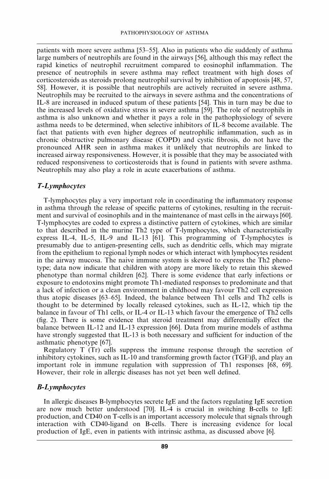
T-lymphocytes play a very important role in coordinating the inflammatory responsein asthma through the release of specific patterns of cytokines, resulting in the recruit-ment and survival of eosinophils and in the maintenance of mast cells in the airways [60].T-lymphocytes are coded to express a distinctive pattern of cytokines, which are similarto that described in the murine Th2 type of T-lymphocytes, which characteristicallyexpress IL-4, IL-5, IL-9 and IL-13 [61]. This programming of T-lymphocytes ispresumably due to antigen-presenting cells, such as dendritic cells, which may migratefrom the epithelium to regional lymph nodes or which interact with lymphocytes residentin the airway mucosa. The naive immune system is skewed to express the Th2 pheno-type; data now indicate that children with atopy are more likely to retain this skewedphenotype than normal children [62]. There is some evidence that early infections orexposure to endotoxins might promote Th1-mediated responses to predominate and thata lack of infection or a clean environment in childhood may favour Th2 cell expressionthus atopic diseases [63–65]. Indeed, the balance between Th1 cells and Th2 cells isthought to be determined by locally released cytokines, such as IL-12, which tip thebalance in favour of Th1 cells, or IL-4 or IL-13 which favour the emergence of Th2 cells(fig. 2). There is some evidence that steroid treatment may differentially effect thebalance between IL-12 and IL-13 expression [66]. Data from murine models of asthmahave strongly suggested that IL-13 is both necessary and sufficient for induction of theasthmatic phenotype [67].
Regulatory T (Tr) cells suppress the immune response through the secretion ofinhibitory cytokines, such as IL-10 and transforming growth factor (TGF)b, and play animportant role in immune regulation with suppression of Th1 responses [68, 69].However, their role in allergic diseases has not yet been well defined.
B-Lymphocytes
In allergic diseases B-lymphocytes secrete IgE and the factors regulating IgE secretionare now much better understood [70]. IL-4 is crucial in switching B-cells to IgEproduction, and CD40 on T-cells is an important accessory molecule that signals throughinteraction with CD40-ligand on B-cells. There is increasing evidence for localproduction of IgE, even in patients with intrinsic asthma, as discussed above [6].
PATHOPHYSIOLOGY OF ASTHMA
89
Basophils
The role of basophils in asthma is uncertain, as these cells have previously beendifficult to detect by immunocytochemistry [71]. Using a basophil-specific marker a smallincrease in basophils has been documented in the airways of asthmatic patients, with anincreased number after allergen challenge [72, 73]. However, these cells are far outnumberedby eosinophils (approximately 10:1 ratio) and their functional role is unknown [72].There is also an increase in the numbers of basophils, as well as mast cells, in inducedsputum after allergen challenge [74]. The role of basophils, as opposed to mast cells, issomewhat uncertain in asthma [75].
Platelets
There is some evidence for the involvement of platelets in the pathophysiology ofallergic diseases, since platelet activation may be observed and there is evidence forplatelets in bronchial biopsies of asthmatic patients [76]. After allergen challenge there isa significant fall in circulating platelets [77] and circulating platelets from patients withasthma show evidence of increased activation and release the chemokine RANTES [78].Chemokines associated with Th2-mediated inflammation have recently been shown toactivate and aggregate platelets [79].
Structural cells
Structural cells of the airways, including epithelial cells, endothelial cells, fibroblastsand even airway smooth muscle cells may also be an important source of inflammatorymediators, such as cytokines and lipid mediators in asthma [80–83]. Indeed, because
��������
'���������������#� �(
��
�)*+%�)*+,
*��
�+
�)*%�-.*�
�)*+/
�-.*�
0-�
�
�)*1
*
* *�)*2
�)*1�)*+3 ��!
0� �*3 �%
!�������
'�� ����
45*%��%,
�����*�������� �������#�� ����6 ����������
Fig. 2. – Asthmatic inflammation is characterised by a preponderance of T-helper (Th) 2 lymphocytes over Th1cells. The transcription factors T-bet and GATA-3 may regulate the balance between Th1 and Th2 cells.Regulatory T-cells (Tr) have an inhibitory effect. IL: interleukin; IFN: interferon; TGF: tumour growth factor;IG: immunoglobulin.
P.J. BARNES
90
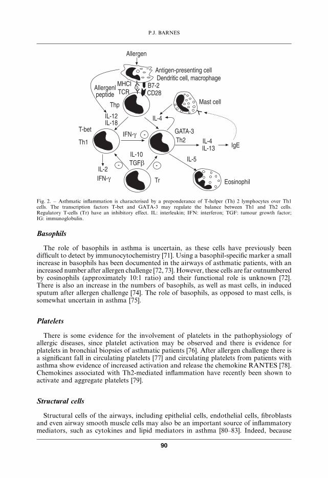
structural cells far outnumber inflammatory cells in the airway, they may become themajor source of mediators driving chronic inflammation in asthma. Epithelial cells mayhave a key role in translating inhaled environmental signals into an airway inflammatoryresponse and are probably the major target cell for inhaled glucocorticoids (fig. 3).
Inflammatory mediators
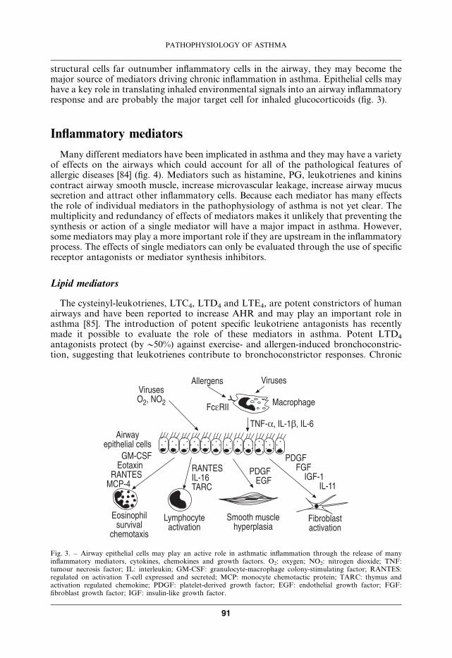
Many different mediators have been implicated in asthma and they may have a varietyof effects on the airways which could account for all of the pathological features ofallergic diseases [84] (fig. 4). Mediators such as histamine, PG, leukotrienes and kininscontract airway smooth muscle, increase microvascular leakage, increase airway mucussecretion and attract other inflammatory cells. Because each mediator has many effectsthe role of individual mediators in the pathophysiology of asthma is not yet clear. Themultiplicity and redundancy of effects of mediators makes it unlikely that preventing thesynthesis or action of a single mediator will have a major impact in asthma. However,some mediators may play a more important role if they are upstream in the inflammatoryprocess. The effects of single mediators can only be evaluated through the use of specificreceptor antagonists or mediator synthesis inhibitors.
Lipid mediators
The cysteinyl-leukotrienes, LTC4, LTD4 and LTE4, are potent constrictors of humanairways and have been reported to increase AHR and may play an important role inasthma [85]. The introduction of potent specific leukotriene antagonists has recentlymade it possible to evaluate the role of these mediators in asthma. Potent LTD4
antagonists protect (byy50%) against exercise- and allergen-induced bronchoconstric-tion, suggesting that leukotrienes contribute to bronchoconstrictor responses. Chronic
������$%6 .$%
���������
-��(��
������
'���������
.-*�6 �)*+�6 �)*7
&�0- -0- �0-*+ �)*++
&�0- !0-
(�. !��)*+7 �(�
-�������������
����� ����������������
)��������������
!��������������
������"�
0'*��- !��"� (�. !�'�&*1
������������ �����
Fig. 3. – Airway epithelial cells may play an active role in asthmatic inflammation through the release of manyinflammatory mediators, cytokines, chemokines and growth factors. O2: oxygen; NO2: nitrogen dioxide; TNF:tumour necrosis factor; IL: interleukin; GM-CSF: granulocyte-macrophage colony-stimulating factor; RANTES:regulated on activation T-cell expressed and secreted; MCP: monocyte chemotactic protein; TARC: thymus andactivation regulated chemokine; PDGF: platelet-derived growth factor; EGF: endothelial growth factor; FGF:fibroblast growth factor; IGF: insulin-like growth factor.
PATHOPHYSIOLOGY OF ASTHMA
91
treatment with antileukotrienes improves lung function and symptoms in asthmaticpatients, although the degree of lung function improvement is not as great as withinhaled corticosteroids which have a much broader spectrum of effects [86, 87]. Inaddition to their effects of airway smooth muscle and vessels, cys-LTs have weakinflammatory effects, with an increase in eosinophils in induced sputum [88], but the anti-inflammatory effects of antileukotrienes are small [89].
PAF is a potent inflammatory mediator that mimics many of the features of asthma,including eosinophil recruitment and activation and induction of AHR [90], yet evenpotent PAF antagonists, such as modipafant, do not control asthma symptoms, at leastin chronic asthma [91–93]. A genetic mutation that results in impaired function of thePAF metabolising enzyme, PAF acetyl hydrolase, is associated with presence severeasthma in Japan [94], suggesting that PAF may play a role in some forms of asthma.
PG have potent effects on airway function and there is increased expression of theinducible form of cyclooxygenase (COX-2) in asthmatic airways [95], but inhibition oftheir synthesis with COX inhibitors, such as aspirin or ibuprofen, does not have anyeffect in most patients with asthma. Some patients have aspirin-sensitive asthma, which ismore common in some ethnic groups, such as eastern Europeans and Japanese [96]. It isassociated with increased expression of LTC4 synthase, resulting in increased formationof cys-LTs, possibly because of genetic polymorphisms [97]. PGD2 is a bronchocon-strictor PG produced predominantly by mast cells. Deletion of the PGD2 receptors inmice significantly inhibits inflammatory responses to allergen and inhibits AHR,suggesting that this mediator may be important in asthma [98]. Recently it has also beendiscovered that PGD2 activates a novel chemoattractant receptor termed chemoattrac-tant receptor of Th2 cells (CRTH2), which is expressed on Th2 cells, eosinophils andbasophils and mediates chemotaxis of these cell types and may provide a link betweenmast cell activation and allergic inflammation [96].
Cytokines
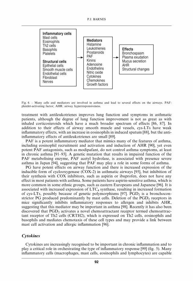
Cytokines are increasingly recognised to be important in chronic inflammation and toplay a critical role in orchestrating the type of inflammatory response [99] (fig. 5). Manyinflammatory cells (macrophages, mast cells, eosinophils and lymphocytes) are capable
������������ ���'�� �����!�������� �% �����4�������&������
���������� ���!������ ���������� ������ �����!�#������ �����-�������.�����
� ������������)��8������&������#�&�-9����#������!�#������.�� �"#����8��������8���0���� ������
��� ���4�����������&����� �"�#���'���� ���������(�������� �������
Fig. 4. – Many cells and mediators are involved in asthma and lead to several effects on the airways. PAF:platelet-activating factor; AHR: airway hyperresponsiveness.
P.J. BARNES
92
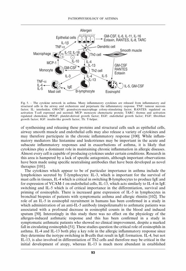
of synthesising and releasing these proteins and structural cells such as epithelial cells,airway smooth muscle and endothelial cells may also release a variety of cytokines andmay therefore participate in the chronic inflammatory response [100]. While inflam-matory mediators like histamine and leukotrienes may be important in the acute andsubacute inflammatory responses and in exacerbations of asthma, it is likely thatcytokines play a dominant role in maintaining chronic inflammation in allergic diseases.Almost every cell is capable of producing cytokines under certain conditions. Research inthis area is hampered by a lack of specific antagonists, although important observationshave been made using specific neutralising antibodies that have been developed as noveltherapies [101].
The cytokines which appear to be of particular importance in asthma include thelymphokines secreted by T-lymphocytes: IL-3, which is important for the survival ofmast cells in tissues, IL-4 which is critical in switching B-lymphocytes to produce IgE andfor expression of VCAM-1 on endothelial cells, IL-13, which acts similarly to IL-4 in IgEswitching and IL-5 which is of critical importance in the differentiation, survival andpriming of eosinophils. There is increased gene expression of IL-5 in lymphocytes inbronchial biopsies of patients with symptomatic asthma and allergic rhinitis [102]. Therole of an IL-5 in eosinophil recruitment in humans has been confirmed in a study inwhich administration of an anti-IL-5 antibody (mepolizumab) to asthmatic patients wasassociated with a profound decrease in eosinophil counts in the blood and inducedsputum [50]. Interestingly in this study there was no effect on the physiology of theallergen-induced asthmatic response and this has been confirmed in a study insymptomatic asthmatic patients who showed no clinical improvement, despite a markedfall in circulating eosinophils [51]. These studies question the critical role of eosinophils inasthma. IL-4 and IL-13 both play a key role in the allergic inflammatory response sincethey determine the isotype switching in B-cells that result in IgE formation. IL-4, but notIL-13, is also involved in differentiation of Th2 cells and therefore may be critical in theinitial development of atopy, whereas IL-13 is much more abundant in established
��������
!������ ������)*+�
.-*�
'��������� �)*+%6 �)*+,
'�&*+ �)*+/
0'*��-'������
�)*3 .-*��)*1�)*2
'�� ������!
4*��������� !��������)*26 0'*��-
0'*��-(�. !�!��"�
����� ���������#�� ����
0'*��-6 �)*76 �)*++6 �)*+7!��"�6 (�. !�6 �)*,6 �(�
�)*2�)*1�)*+3
�% ����
�)*1�)*+3
�/ ����*
Fig. 5. – The cytokine network in asthma. Many inflammatory cytokines are released from inflammatory andstructural cells in the airway and orchestrate and perpetuate the inflammatory response. TNF: tumour necrosisfactor; IL: interleukin; GM-CSF: granulocyte-macrophage colony-stimulating factor; RANTES: regulated onactivation T-cell expressed and secreted; MCP: monocyte chemotactic protein; TARC: thymus and activationregulated chemokine; PDGF: platelet-derived growth factor; EGF: endothelial growth factor; FGF: fibroblastgrowth factor; IGF: insulin-like growth factor; Th: T-helper.
PATHOPHYSIOLOGY OF ASTHMA
93
disease and may therefore be more important in maintaining the inflammatory process[67, 103]. Another Th2 cytokine IL-9 may play a critical role in sensitising responses tothe cytokines IL-4 and IL-5 [104, 105].
Other cytokines, such as IL-1b, IL-6, TNF-a and GM-CSF are released from a varietyof cells, including macrophages and epithelial cells and may be important in amplifyingthe inflammatory response. TNF-a may be an amplifying mediator in asthma and isproduced in increased amounts in asthmatic airways [106]. Inhalation of TNF-aincreased airway responsiveness in normal individuals [107]. TNF-a and IL-1b bothactivate the proinflammatory transcription factors, nuclear factor-kB (NF-kB) andactivator protein-1 (AP-1) which then switch on many inflammatory genes in theasthmatic airway.
Other cytokines, such as interferon (IFN)-c, IL-10, IL-12 and IL-18, play a regulatoryrole and inhibit the allergic inflammatory process (see below).
Chemokines
Many chemokines are involved in the recruitment of inflammatory cells in asthma [45].Over 50 different chemokines are now recognised and they activatew20 different surfacereceptors [108]. Chemokine receptors belong to the seven transmembrane-receptorsuperfamily of G-protein-coupled receptors and this makes it possible to find smallmolecule inhibitors, which has not been possible for classical cytokine receptor [109].Some chemokines appear to be selective for single chemokines, whereas others arepromiscuous and mediate the effects of several related chemokines. Chemokines appearto act in sequence in determining the final inflammatory response and so inhibitors maybe more or less effective depending on the kinetics of the response [110].
Several chemokines, including eotaxin, eotaxin-2, eotaxin-3, RANTES and MCP-4,activate a common receptor on eosinophils termed CCR3 [111]. A neutralising antibodyagainst eotaxin reduces eosinophil recruitment in to the lung after allergen and theassociated AHR in mice [112]. There is increased expression of eotaxin, eotaxin-2, MCP-3, MCP-4 and CCR3 in the airways of asthmatic patients and this is correlated withincreased AHR [113, 114]. Several small molecule inhibitors of CCR3, includingUCB35625, SB-297006 and SB-328437 are effective in inhibiting eosinophil recruitmentin allergen models of asthma [115, 116] and drugs in this class are currently undergoingclinical trials in asthma. Although it was thought that CCR3 were restricted toeosinophils, there is some evidence for their expression on Th2 cells and mast cells, sothat these inhibitors may have a more widespread effect than on eosinophils alone,making them potentially more valuable in asthma treatment. RANTES, which showsincreased expression in asthmatic airways [117] also activates CCR3, but also has effectson CCR1 and CCR5, which may play a role in T-cell recruitment.
MCP-1 activates CCR2 on monocytes and T-lymphocytes. Blocking MCP-1 withneutralising antibodies reduces recruitment of both T-cells and eosinophils in a murinemodel of ovalbumin-induced airway inflammation, with a marked reduction in AHR[112]. MCP-1 also recruits and activates mast cells, an effect that is mediated via CCR2[118]. MCP-1 instilled into the airways induces marked and prolonged AHR in mice,associated with mast cell degranulation. A neutralising antibody to MCP-1 blocks thedevelopment of AHR in response to allergen [118]. MCP-1 levels are increased in BALfluid of patients with asthma [119]. This has led to a search for small molecule inhibitorsof CCR2.
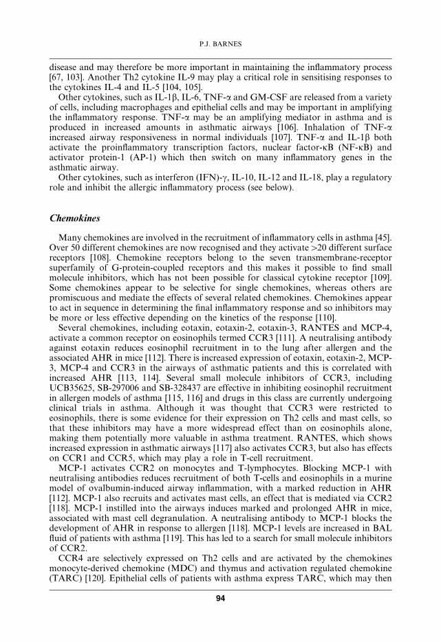
CCR4 are selectively expressed on Th2 cells and are activated by the chemokinesmonocyte-derived chemokine (MDC) and thymus and activation regulated chemokine(TARC) [120]. Epithelial cells of patients with asthma express TARC, which may then
P.J. BARNES
94
recruit Th2 cells [121]. Increased concentrations of TARC are also found in BAL fluid ofasthmatic patients, whereas MDC is only weakly expressed in the airways [122]. TARCmay thus induce a sequence of responses resulting in coordinated eosinophilic inflam-mation (fig. 6). Inhibitors of CCR4 may therefore inhibit the recruitment of Th2 cells andthus persistent eosinophilic inflammation in the airways.
Oxidative stress
As in all inflammatory diseases, there is increased oxidative stress in allergic inflam-mation, as activated inflammatory cells, such as macrophages and eosinophils, producereactive oxygen species. Evidence for increased oxidative stress in asthma is provided bythe increased concentrations of 8-isoprostane (a product of oxidised arachidonic acid) inexhaled breath condensates [59] and increased ethane (a product of oxidative lipidperoxidation) in exhaled breath of asthmatic patients [123]. There is also persuasiveepidemiological evidence that a low dietary intake of antioxidants is linked to anincreased prevalence of asthma [124]. Increased oxidative stress is related to diseaseseverity and may amplify the inflammatory response and reduce responsiveness tocorticosteroids, particularly in severe disease and during exacerbations. One of themechanisms whereby oxidative stress may be detrimental in asthma is through thereaction of superoxide anions with nitric oxide (NO) to form the reactive radicalperoxynitrite, that may then modify several target proteins.
Endothelins
Endothelins are potent peptide mediators that are vasoconstrictors and bronchocon-strictors [125, 126]. Endothelin-1 levels are increased in the sputum of patients withasthma; these levels are modulated by allergen exposure and steroid treatment [127, 128].
.-*�
������������
!��"��
��(3
!�������
�)*2
�)*16 �)*+3
�(�
��(1
�% ����
Fig. 6. – Chemokines in asthma. Tumour necrosis factor (TNF)-a releases thymus and activation regulatedchemokine (TARC) from epithelial cells which attracts T-helper (Th)2 cells via activation of CCR4 receptors.These promote eosinophilic inflammation directly through the release of interleukin (IL)-5 and indirectly via therelease of IL-4 and IL-13 which induce eotaxin formation in airway epithelial cells.
PATHOPHYSIOLOGY OF ASTHMA
95
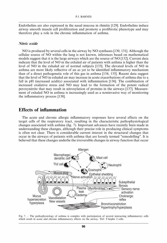
Endothelins are also expressed in the nasal mucosa in rhinitis [129]. Endothelins induceairway smooth muscle cell proliferation and promote a profibrotic phenotype and maytherefore play a role in the chronic inflammation of asthma.
Nitric oxide
NO is produced by several cells in the airway by NO synthases [130, 131]. Although thecellular source of NO within the lung is not known, inferences based on mathematicalmodels suggest that it is the large airways which are the source of NO [132]. Current dataindicate that the level of NO in the exhaled air of patients with asthma is higher than thelevel of NO in the exhaled air of normal subjects [133]. The elevated levels of NO inasthma are more likely reflective of an as yet to be identified inflammatory mechanismthan of a direct pathogenetic role of this gas in asthma [134, 135]. Recent data suggestthat the level of NO in exhaled air may increase in acute exacerbations of asthma due to afall in pH (increased acidity) associated with inflammation [136]. The combination ofincreased oxidative stress and NO may lead to the formation of the potent radicalperoxynitrite that may result in nitrosylation of proteins in the airways [137]. Measure-ment of exhaled NO in asthma is increasingly used as a noninvasive way of monitoringthe inflammatory process [138].
Effects of inflammation
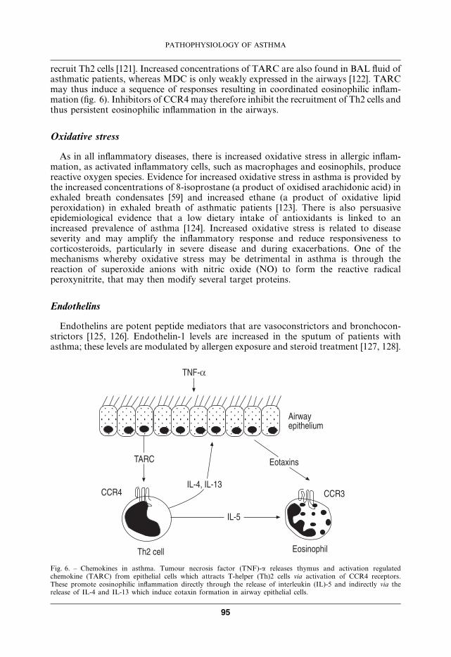
The acute and chronic allergic inflammatory responses have several effects on thetarget cells of the respiratory tract, resulting in the characteristic pathophysiologicalchanges associated with asthma (fig. 7). Important advances have recently been made inunderstanding these changes, although their precise role in producing clinical symptomsis often not clear. There is considerable current interest in the structural changes thatoccur in the airways of patients with asthma that are loosely termed "remodelling". It isbelieved that these changes underlie the irreversible changes in airway function that occur
��������
'���������:#��#�� ����
�% ����
'��������������������������
����#�����.�� �������
&��������8
$�#���
4������������������������:����������
��������������"
������� �����������
����������������
'����������
!������ ���##��
.�������!�������
'�� ����
.���� ������'���� ����
Fig. 7. – The pathophysiology of asthma is complex with participation of several interacting inflammatory cellswhich result in acute and chronic inflammatory effects on the airway. Th2: T-hepler 2 cells.
P.J. BARNES
96
in some patients with asthma [139, 140]. However, many patients with asthma continueto have normal lung function throughout life, so it is likely that genetic factors maydetermine which patients develop these structural changes in the airways.
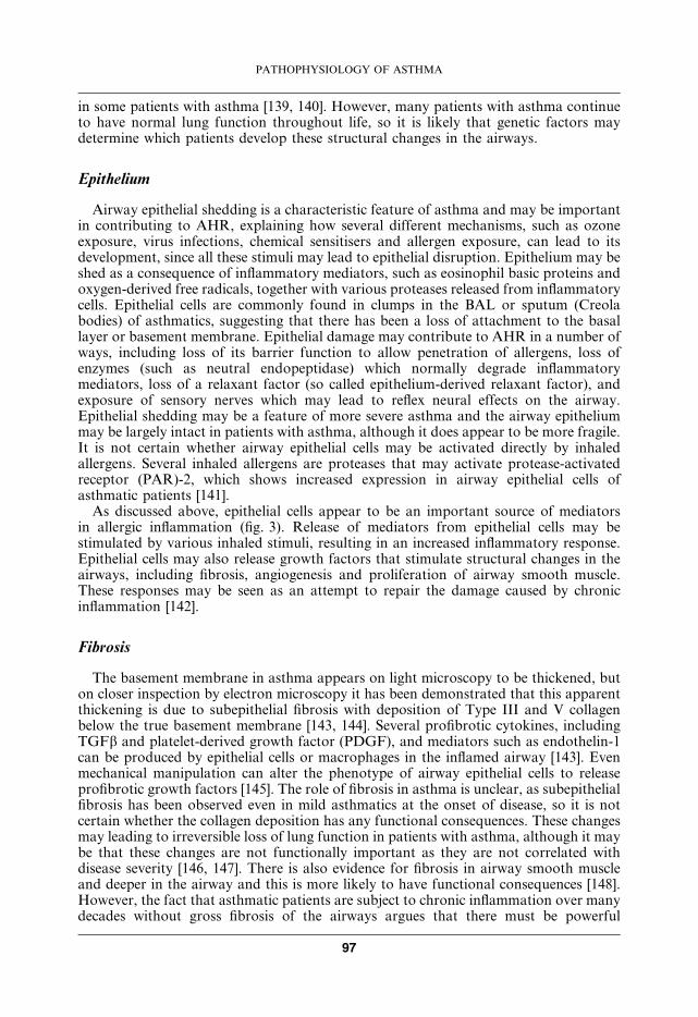
Epithelium
Airway epithelial shedding is a characteristic feature of asthma and may be importantin contributing to AHR, explaining how several different mechanisms, such as ozoneexposure, virus infections, chemical sensitisers and allergen exposure, can lead to itsdevelopment, since all these stimuli may lead to epithelial disruption. Epithelium may beshed as a consequence of inflammatory mediators, such as eosinophil basic proteins andoxygen-derived free radicals, together with various proteases released from inflammatorycells. Epithelial cells are commonly found in clumps in the BAL or sputum (Creolabodies) of asthmatics, suggesting that there has been a loss of attachment to the basallayer or basement membrane. Epithelial damage may contribute to AHR in a number ofways, including loss of its barrier function to allow penetration of allergens, loss ofenzymes (such as neutral endopeptidase) which normally degrade inflammatorymediators, loss of a relaxant factor (so called epithelium-derived relaxant factor), andexposure of sensory nerves which may lead to reflex neural effects on the airway.Epithelial shedding may be a feature of more severe asthma and the airway epitheliummay be largely intact in patients with asthma, although it does appear to be more fragile.It is not certain whether airway epithelial cells may be activated directly by inhaledallergens. Several inhaled allergens are proteases that may activate protease-activatedreceptor (PAR)-2, which shows increased expression in airway epithelial cells ofasthmatic patients [141].
As discussed above, epithelial cells appear to be an important source of mediatorsin allergic inflammation (fig. 3). Release of mediators from epithelial cells may bestimulated by various inhaled stimuli, resulting in an increased inflammatory response.Epithelial cells may also release growth factors that stimulate structural changes in theairways, including fibrosis, angiogenesis and proliferation of airway smooth muscle.These responses may be seen as an attempt to repair the damage caused by chronicinflammation [142].
Fibrosis
The basement membrane in asthma appears on light microscopy to be thickened, buton closer inspection by electron microscopy it has been demonstrated that this apparentthickening is due to subepithelial fibrosis with deposition of Type III and V collagenbelow the true basement membrane [143, 144]. Several profibrotic cytokines, includingTGFb and platelet-derived growth factor (PDGF), and mediators such as endothelin-1can be produced by epithelial cells or macrophages in the inflamed airway [143]. Evenmechanical manipulation can alter the phenotype of airway epithelial cells to releaseprofibrotic growth factors [145]. The role of fibrosis in asthma is unclear, as subepithelialfibrosis has been observed even in mild asthmatics at the onset of disease, so it is notcertain whether the collagen deposition has any functional consequences. These changesmay leading to irreversible loss of lung function in patients with asthma, although it maybe that these changes are not functionally important as they are not correlated withdisease severity [146, 147]. There is also evidence for fibrosis in airway smooth muscleand deeper in the airway and this is more likely to have functional consequences [148].However, the fact that asthmatic patients are subject to chronic inflammation over manydecades without gross fibrosis of the airways argues that there must be powerful
PATHOPHYSIOLOGY OF ASTHMA
97
inhibitory mechanisms that prevent a fibrotic reaction to the multiple profibroticmediators produced.
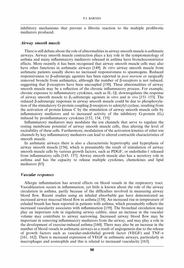
Airway smooth muscle
There is still debate about the role of abnormalities in airway smoothmuscle is asthmaticairways. Airway smooth muscle contraction plays a key role in the symptomatology ofasthma and many inflammatory mediators released in asthma have bronchoconstrictoreffects. More recently it has been recognised that airway smooth muscle cells may alsohave other functions in asthmatic airways [149]. In vitro airway smooth muscle fromasthmatic patients usually shows no increased responsiveness to spasmogens. Reducedresponsiveness to b-adrenergic agonists has been reported in post mortem or surgicallyremoved bronchi from asthmatics, although the number of b-receptors is not reduced,suggesting that b-receptors have been uncoupled [150]. These abnormalities of airwaysmooth muscle may be a reflection of the chronic inflammatory process. For example,chronic exposure to inflammatory cytokines, such as IL-1b, downregulates the responseof airway smooth muscle to b2-adrenergic agonists in vitro and in vivo [151–153]. Thereduced b-adrenergic responses in airway smooth muscle could be due to phosphoryla-tion of the stimulatory G-protein coupling b-receptors to adenylyl cyclase, resulting fromthe activation of protein kinase C by the stimulation of airway smooth muscle cells byinflammatory mediators and to increased activity of the inhibitory G-protein (Gi)induced by proinflammatory cytokines [152, 154, 155].
Inflammatory mediators may modulate the ion channels that serve to regulate theresting membrane potential of airway smooth muscle cells, thus altering the level ofexcitability of these cells. Furthermore, modulation of the activation kinetics of other ionchannels by key inflammatory mediators can lead to altered contractile characteristics ofsmooth muscle.
In asthmatic airways there is also a characteristic hypertrophy and hyperplasia ofairway smooth muscle [156], which is presumably the result of stimulation of airwaysmooth muscle cells by various growth factors, such as PDGF, or endothelin-1 releasedfrom inflammatory cells [143, 157]. Airway smooth muscle also has a secretory role inasthma and has the capacity to release multiple cytokines, chemokines and lipidmediators [83].
Vascular responses
Allergic inflammation has several effects on blood vessels in the respiratory tract.Vasodilatation occurs in inflammation, yet little is known about the role of the airwaycirculation in asthma, partly because of the difficulties involved in measuring airwayblood flow. Recent studies using an inhaled absorbable gas have demonstrated anincreased airway mucosal blood flow in asthma [158]. An increased rise in temperature ofexhaled breath has been reported in patients with asthma, which presumably reflects theincreased vascularity associates with inflammation [159]. The bronchial circulation mayplay an important role in regulating airway calibre, since an increase in the vascularvolume may contribute to airway narrowing. Increased airway blood flow may beimportant in removing inflammatory mediators from the airway, and may play a role inthe development of exercise-induced asthma [160]. There may also be an increase in thenumber of blood vessels in asthmatic airways as a result of angiogenesis due to the releaseof growth factors such as vascular-endothelial growth factor (VEGF) and TNF-a[161, 162]. There is increased expression of VEGF in asthmatic airways, particularly inmacrophages and eosinophils and this is related to increased vascularity [163].
P.J. BARNES
98
Microvascular leakage is an essential component of the inflammatory response andmany of the inflammatory mediators implicated in asthma produce this leakage [164,165]. There is good evidence for microvascular leakage in asthma and it may have severalconsequences on airway function, including increased airway secretions, impairedmucociliary clearance, formation of new mediators from plasma precursors (such askinins) and mucosal oedema which may contribute to airway narrowing and increasedAHR [166, 167].
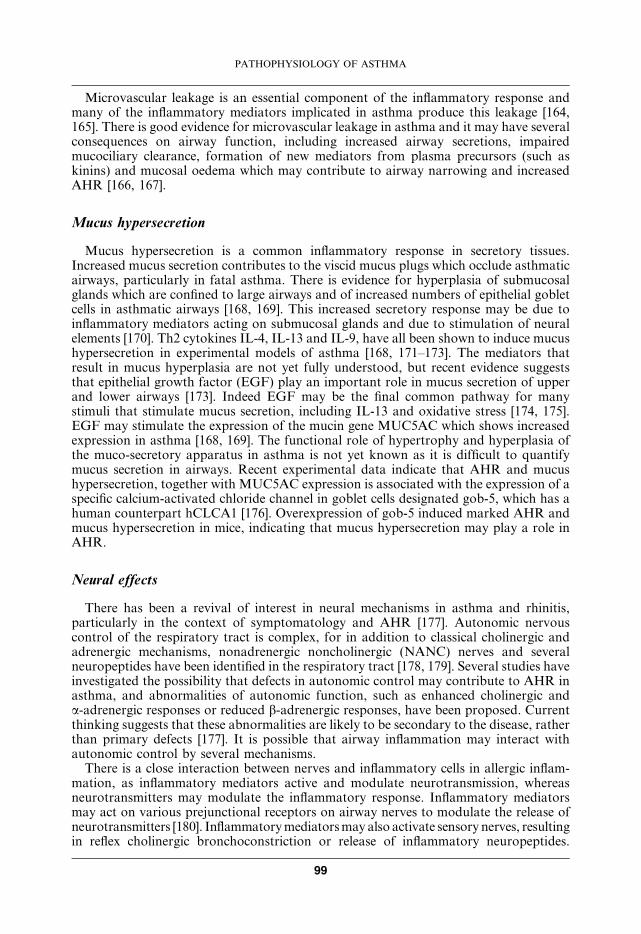
Mucus hypersecretion
Mucus hypersecretion is a common inflammatory response in secretory tissues.Increased mucus secretion contributes to the viscid mucus plugs which occlude asthmaticairways, particularly in fatal asthma. There is evidence for hyperplasia of submucosalglands which are confined to large airways and of increased numbers of epithelial gobletcells in asthmatic airways [168, 169]. This increased secretory response may be due toinflammatory mediators acting on submucosal glands and due to stimulation of neuralelements [170]. Th2 cytokines IL-4, IL-13 and IL-9, have all been shown to induce mucushypersecretion in experimental models of asthma [168, 171–173]. The mediators thatresult in mucus hyperplasia are not yet fully understood, but recent evidence suggeststhat epithelial growth factor (EGF) play an important role in mucus secretion of upperand lower airways [173]. Indeed EGF may be the final common pathway for manystimuli that stimulate mucus secretion, including IL-13 and oxidative stress [174, 175].EGF may stimulate the expression of the mucin gene MUC5AC which shows increasedexpression in asthma [168, 169]. The functional role of hypertrophy and hyperplasia ofthe muco-secretory apparatus in asthma is not yet known as it is difficult to quantifymucus secretion in airways. Recent experimental data indicate that AHR and mucushypersecretion, together with MUC5AC expression is associated with the expression of aspecific calcium-activated chloride channel in goblet cells designated gob-5, which has ahuman counterpart hCLCA1 [176]. Overexpression of gob-5 induced marked AHR andmucus hypersecretion in mice, indicating that mucus hypersecretion may play a role inAHR.
Neural effects
There has been a revival of interest in neural mechanisms in asthma and rhinitis,particularly in the context of symptomatology and AHR [177]. Autonomic nervouscontrol of the respiratory tract is complex, for in addition to classical cholinergic andadrenergic mechanisms, nonadrenergic noncholinergic (NANC) nerves and severalneuropeptides have been identified in the respiratory tract [178, 179]. Several studies haveinvestigated the possibility that defects in autonomic control may contribute to AHR inasthma, and abnormalities of autonomic function, such as enhanced cholinergic anda-adrenergic responses or reduced b-adrenergic responses, have been proposed. Currentthinking suggests that these abnormalities are likely to be secondary to the disease, ratherthan primary defects [177]. It is possible that airway inflammation may interact withautonomic control by several mechanisms.
There is a close interaction between nerves and inflammatory cells in allergic inflam-mation, as inflammatory mediators active and modulate neurotransmission, whereasneurotransmitters may modulate the inflammatory response. Inflammatory mediatorsmay act on various prejunctional receptors on airway nerves to modulate the release ofneurotransmitters [180]. Inflammatorymediatorsmay also activate sensory nerves, resultingin reflex cholinergic bronchoconstriction or release of inflammatory neuropeptides.
PATHOPHYSIOLOGY OF ASTHMA
99
Bradykinin is a potent activator of unmyelinated sensory nerves (C-fibres) [181], but alsosensitises these nerves to other stimuli [182].
Inflammatory products may also sensitise sensory nerve endings in the airwayepithelium, so that the nerves become hyperalgesic. Hyperalgesia and pain (dolor) arecardinal signs of inflammation, and in the asthmatic airway may mediate cough and chesttightness, which are such characteristic symptoms of asthma. The precise mechanismsof hyperalgesia are not yet certain, but mediators such as PG, certain cytokines andneurotrophins may be important. Neurotrophins, which may be released from variouscell types in peripheral tissues, may cause proliferation and sensitisation of airwaysensory nerves [183, 184]. Neurotrophins, such as nerve growth factor (NGF), may bereleased from inflammatory and structural cells in asthmatic airways and then stimulatethe increased synthesis of neuropeptides, such as substance P (SP), in airway sensorynerves, as well as sensitising nerve endings in the airways [185]. Thus, NGF is releasedfrom human airway epithelial cells after exposure to inflammatory stimuli [186].Neurotrophins may play an important role in mediating AHR in asthma [187].
Bronchodilator nerves which are nonadrenergic are prominent in human airways andit has been suggested that these nerves may be defective in asthma [188]. In animalairways vasoactive intestinal peptide (VIP) has been shown to be a neurotransmitter ofthese nerves and a striking absence of VIP-immunoreactive nerves has been reportedin the lungs from patients with severe fatal asthma [189]. However, no difference inexpression of VIP has been reported in bronchial biopsies from asthmatic patients [190].It is likely that this loss of VIP-immunoreactivity in severe asthma is explained bydegradation by tryptase released from degranulating mast cells in the airways ofasthmatics. In human airways the single bronchodilator neurotransmitter appears to beNO [191].
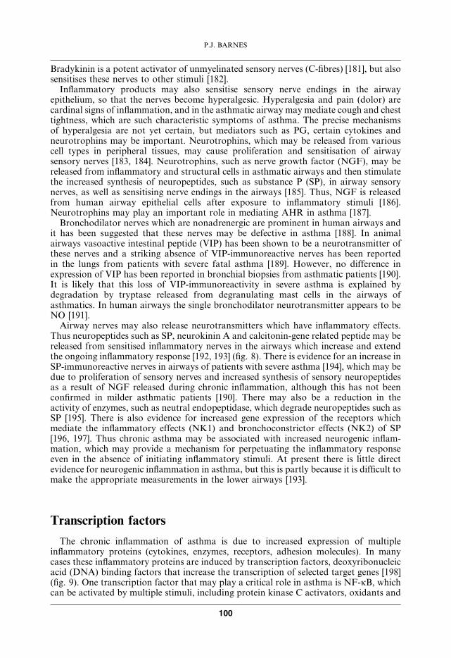
Airway nerves may also release neurotransmitters which have inflammatory effects.Thus neuropeptides such as SP, neurokinin A and calcitonin-gene related peptide may bereleased from sensitised inflammatory nerves in the airways which increase and extendthe ongoing inflammatory response [192, 193] (fig. 8). There is evidence for an increase inSP-immunoreactive nerves in airways of patients with severe asthma [194], which may bedue to proliferation of sensory nerves and increased synthesis of sensory neuropeptidesas a result of NGF released during chronic inflammation, although this has not beenconfirmed in milder asthmatic patients [190]. There may also be a reduction in theactivity of enzymes, such as neutral endopeptidase, which degrade neuropeptides such asSP [195]. There is also evidence for increased gene expression of the receptors whichmediate the inflammatory effects (NK1) and bronchoconstrictor effects (NK2) of SP[196, 197]. Thus chronic asthma may be associated with increased neurogenic inflam-mation, which may provide a mechanism for perpetuating the inflammatory responseeven in the absence of initiating inflammatory stimuli. At present there is little directevidence for neurogenic inflammation in asthma, but this is partly because it is difficult tomake the appropriate measurements in the lower airways [193].
Transcription factors
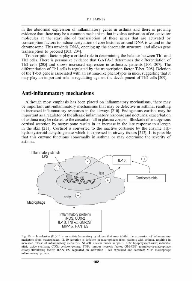
The chronic inflammation of asthma is due to increased expression of multipleinflammatory proteins (cytokines, enzymes, receptors, adhesion molecules). In manycases these inflammatory proteins are induced by transcription factors, deoxyribonucleicacid (DNA) binding factors that increase the transcription of selected target genes [198](fig. 9). One transcription factor that may play a critical role in asthma is NF-kB, whichcan be activated by multiple stimuli, including protein kinase C activators, oxidants and
P.J. BARNES
100
proinflammatory cytokines (such as IL-1b and TNF-a) [199]. There is evidence forincreased activation of NF-kB in asthmatic airways, particularly in epithelial cells andmacrophages [200, 201]. NF-kB regulates the expression of several key genes that areoverexpressed in asthmatic airways, including proinflammatory cytokines (IL-1b, TNF-a,GM-CSF), chemokines (RANTES, MIP-1a, eotaxin), adhesion molecules (ICAM-1,VCAM-1) and inflammatory enzymes (cyclooxygenase-2 and iNOS). The c-Fos com-ponent of AP-1 is also activated in asthmatic airways and often cooperates with NF-kBin switching on inflammatory genes [202]. Many other transcription factors are involved
������� �����������
!������ ���##�� !��������
����#�����
'�����������
&������"�#���
.�������#� ��������&6 .9�6 �0(& ����
4��������������
������������������������ �������
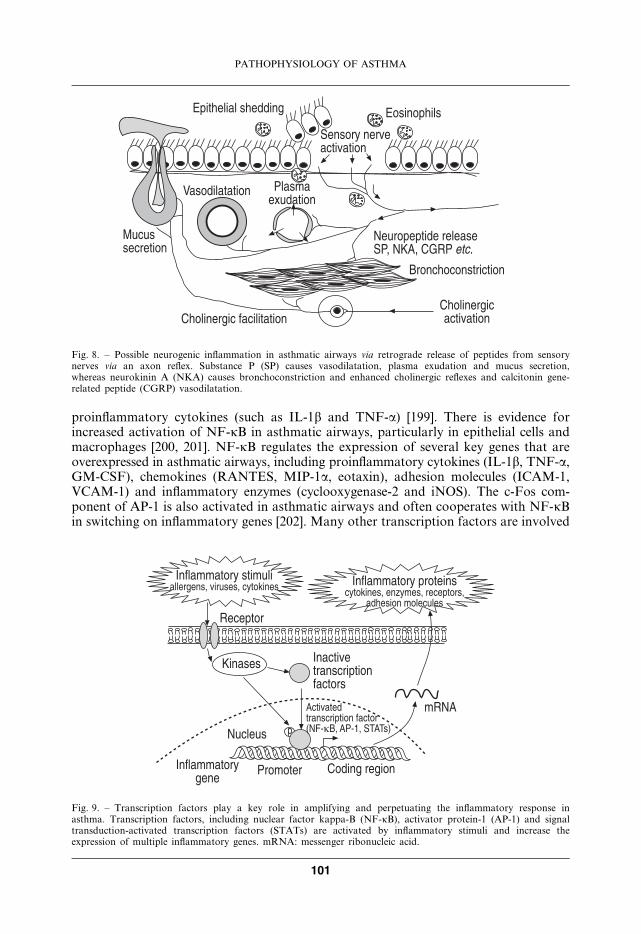
Fig. 8. – Possible neurogenic inflammation in asthmatic airways via retrograde release of peptides from sensorynerves via an axon reflex. Substance P (SP) causes vasodilatation, plasma exudation and mucus secretion,whereas neurokinin A (NKA) causes bronchoconstriction and enhanced cholinergic reflexes and calcitonin gene-related peptide (CGRP) vasodilatation.
����������� �������������6 ������6 ���8��� ����������� ������
���8���6 �������6 ��������6�#����� ���������
�(.�
���������������������
9�����
(������
.������
��������������� &������ ��#�� �����
�����#��������� �����;.-*�46 �&*+6 � � �<�
Fig. 9. – Transcription factors play a key role in amplifying and perpetuating the inflammatory response inasthma. Transcription factors, including nuclear factor kappa-B (NF-kB), activator protein-1 (AP-1) and signaltransduction-activated transcription factors (STATs) are activated by inflammatory stimuli and increase theexpression of multiple inflammatory genes. mRNA: messenger ribonucleic acid.
PATHOPHYSIOLOGY OF ASTHMA
101
in the abnormal expression of inflammatory genes in asthma and there is growingevidence that there may be a common mechanism that involves activation of co-activatormolecules at the start site of transcription of these genes that are activated bytranscription factors to induce acetylation of core histones around DNA is wound in thechromosome. This unwinds DNA, opening up the chromatin structure, and allows genetranscription to proceed [203, 204].
Transcription factors play a critical role in determining the balance between Th1 andTh2 cells. There is persuasive evidence that GATA-3 determines the differentiation ofTh2 cells [205] and shows increased expression in asthmatic patients [206, 207]. Thedifferentiation of Th1 cells is regulated by the transcription factor T-bet [208]. Deletionof the T-bet gene is associated with an asthma-like phenotypes in mice, suggesting that itmay play an important role in regulating against the development of Th2 cells [209].
Anti-inflammatory mechanisms
Although most emphasis has been placed on inflammatory mechanisms, there maybe important anti-inflammatory mechanisms that may be defective in asthma, resultingin increased inflammatory responses in the airways [210]. Endogenous cortisol may beimportant as a regulator of the allergic inflammatory response and nocturnal exacerbationof asthma may be related to the circadian fall in plasma cortisol. Blockade of endogenouscortisol secretion by metyrapone results in an increase in the late response to allergenin the skin [211]. Cortisol is converted to the inactive cortisone by the enzyme 11b-hydroxysteroid dehydrogenase which is expressed in airway tissues [212]. It is possiblethat this enzyme functions abnormally in asthma or may determine the severity ofasthma.
����������� ����)&�
'��������������
���
�)*+/ =
���������#�=
=
=
*
.-*�4
����������� ������.$�6 �$>*%
�)*+�6 .-*�6 0'*��-'�&*+�6 (�. !�
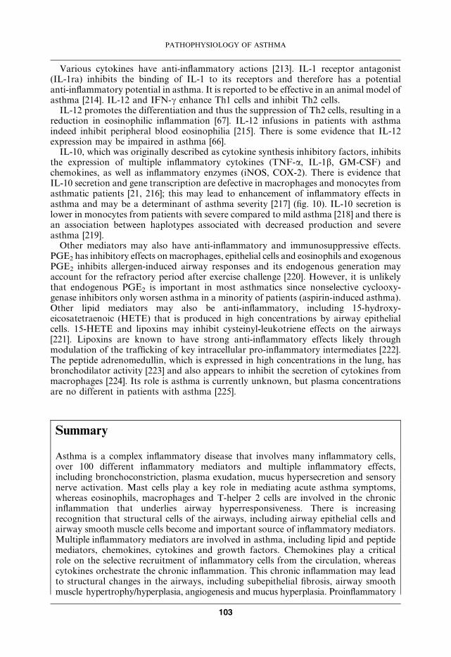
Fig. 10. – Interleukin (IL)-10 is an anti-inflammatory cytokines that may inhibit the expression of inflammatorymediators from macrophages. IL-10 secretion is deficient in macrophages from patients with asthma, resulting inincreased release of inflammatory mediators. NF-kB: nuclear factor kappa-B; LPS: lipopolysaccharide; induciblenitric oxide synthase; COX: cyclooxygenase; TNF: tumour necrosis factor; GM-CSF: granulocyte-macrophagecolony-stimulating factor; RANTES: regulated on activation T-cell expressed and secreted; MIP: macrophageinflammatory protein.
P.J. BARNES
102
Various cytokines have anti-inflammatory actions [213]. IL-1 receptor antagonist(IL-1ra) inhibits the binding of IL-1 to its receptors and therefore has a potentialanti-inflammatory potential in asthma. It is reported to be effective in an animal model ofasthma [214]. IL-12 and IFN-c enhance Th1 cells and inhibit Th2 cells.
IL-12 promotes the differentiation and thus the suppression of Th2 cells, resulting in areduction in eosinophilic inflammation [67]. IL-12 infusions in patients with asthmaindeed inhibit peripheral blood eosinophilia [215]. There is some evidence that IL-12expression may be impaired in asthma [66].
IL-10, which was originally described as cytokine synthesis inhibitory factors, inhibitsthe expression of multiple inflammatory cytokines (TNF-a, IL-1b, GM-CSF) andchemokines, as well as inflammatory enzymes (iNOS, COX-2). There is evidence thatIL-10 secretion and gene transcription are defective in macrophages and monocytes fromasthmatic patients [21, 216]; this may lead to enhancement of inflammatory effects inasthma and may be a determinant of asthma severity [217] (fig. 10). IL-10 secretion islower in monocytes from patients with severe compared to mild asthma [218] and there isan association between haplotypes associated with decreased production and severeasthma [219].
Other mediators may also have anti-inflammatory and immunosuppressive effects.PGE2 has inhibitory effects onmacrophages, epithelial cells and eosinophils and exogenousPGE2 inhibits allergen-induced airway responses and its endogenous generation mayaccount for the refractory period after exercise challenge [220]. However, it is unlikelythat endogenous PGE2 is important in most asthmatics since nonselective cyclooxy-genase inhibitors only worsen asthma in a minority of patients (aspirin-induced asthma).Other lipid mediators may also be anti-inflammatory, including 15-hydroxy-eicosatetraenoic (HETE) that is produced in high concentrations by airway epithelialcells. 15-HETE and lipoxins may inhibit cysteinyl-leukotriene effects on the airways[221]. Lipoxins are known to have strong anti-inflammatory effects likely throughmodulation of the trafficking of key intracellular pro-inflammatory intermediates [222].The peptide adrenomedullin, which is expressed in high concentrations in the lung, hasbronchodilator activity [223] and also appears to inhibit the secretion of cytokines frommacrophages [224]. Its role is asthma is currently unknown, but plasma concentrationsare no different in patients with asthma [225].
Summary
Asthma is a complex inflammatory disease that involves many inflammatory cells,over 100 different inflammatory mediators and multiple inflammatory effects,including bronchoconstriction, plasma exudation, mucus hypersecretion and sensorynerve activation. Mast cells play a key role in mediating acute asthma symptoms,whereas eosinophils, macrophages and T-helper 2 cells are involved in the chronicinflammation that underlies airway hyperresponsiveness. There is increasingrecognition that structural cells of the airways, including airway epithelial cells andairway smooth muscle cells become and important source of inflammatory mediators.Multiple inflammatory mediators are involved in asthma, including lipid and peptidemediators, chemokines, cytokines and growth factors. Chemokines play a criticalrole on the selective recruitment of inflammatory cells from the circulation, whereascytokines orchestrate the chronic inflammation. This chronic inflammation may leadto structural changes in the airways, including subepithelial fibrosis, airway smoothmuscle hypertrophy/hyperplasia, angiogenesis and mucus hyperplasia. Proinflammatory
PATHOPHYSIOLOGY OF ASTHMA
103
transcription factors, such as nuclear factor-kB and activating protein-1 andGATA-3 play a key role in orchestrating the expression of inflammatory genes.There are several endogenous mechanisms that may counteract the inflammation ofasthma and some evidence that these may be deficient in asthma. Because of thecomplexity of asthma drugs that target a since cell or mediator are unlikely toprovide significant clinical benefit; the most effective drugs are those that targetmany mechanisms. b2-Agonists are not only the most effective bronchodilators, butthey also inhibit mast cells and plasma leakage, whereas corticosteroids inhibitmultiple inflammatory effects and the production of cytokines and chemokines.
Keywords: Airway hyperresponsiveness, eosinophil, epithelial cell, mast cell, sensorynerve, T-helper 2 cells.
References
1. Cookson WO, Moffatt MF. Genetics of asthma and allergic disease. Hum Mol Genet 2000;
9: 2359–2364.
2. Barnes KC. Evidence for common genetic elements in allergic disease. J Allergy Clin Immunol
2000; 106: S192–S200.
3. Dunnill MS. The pathology of asthma, with special reference to the changes in the bronchial
mucosa. J Clin Pathol 1960; 13: 27–33.
4. Busse WW, Lemanske RF. Asthma. N Engl J Med 2001; 344: 350–362.
5. Bousquet J, Jeffery PK, Busse WW, JohnsonM, Vignola AM. Asthma. From bronchoconstriction
to airways inflammation and remodeling. Am J Respir Crit Care Med 2000; 161: 1720–1745.
6. Humbert M, Menz G, Ying S, et al. The immunopathology of extrinsic (atopic) and intrinsic
(non-atopic) asthma: more similarities than differences. Immunol Today 1999; 20: 528–533.
7. Ying S, Humbert M, Meng Q, et al. Local expression of epsilon germline gene transcripts and
RNA for the epsilon heavy chain of IgE in the bronchial mucosa in atopic and nonatopic asthma.
J Allergy Clin Immunol 2001; 107: 686–692.
8. Brightling CE, Bradding P, Symon FA, Holgate ST, Wardlaw AJ, Pavord ID. Mast-cell
infiltration of airway smooth muscle in asthma. N Engl J Med 2002; 346: 1699–1705.
9. Bentley AM, Hamid Q, Robinson DS, et al. Prednisolone treatment in asthma. Reduction in the
numbers of eosinophils, T cells, tryptase-only positive mast cells, and modulation of IL-4, IL-5,
and interferon-gamma cytokine gene expression within the bronchial mucosa. Am J Respir Crit
Care Med 1996; 153: 551–556.
10. Akers IA, Parsons M, Hill MR, et al. Mast cell tryptase stimulates human lung fibroblast
proliferation via protease-activated receptor-2. Am J Physiol Lung Cell Mol Physiol 2000;
278: L193–L201.
11. Williams CM, Galli SJ. The diverse potential effector and immunoregulatory roles of mast cells in
allergic disease. J Allergy Clin Immunol 2000; 105: 847–859.
12. Fahy JV. Reducing IgE levels as a strategy for the treatment of asthma. Clin Exp Allergy 2000;
30: Suppl. 1, 16–21.
13. Barnes PJ. Anti-IgE therapy in asthma: rationale and therapeutic potential. Int Arch Allergy
Immunol 2000; 123: 196–204.
14. Milgrom H, Fick RB Jr, Su JQ, et al. Treatment of allergic asthma with monoclonal anti-IgE
antibody. New Engl J Med 1999; 341: 1966–1973.
15. Zieg G, Lack G, Harbeck RJ, Gelfand EW, Leung DY. In vivo effects of glucocorticoids on IgE
production. J Allergy Clin Immunol 1994; 94: 222–230.
16. Barnes PJ. Corticosteroids, IgE, and atopy. J Clin Invest 2001; 107: 265–266.
17. Barnes PJ. Are mast cells still important in asthma? Rev Fr Allergol Immunol Clin 2002; 42: 20–27.
P.J. BARNES
104

18. Lee TH, Lane SJ. The role of macrophages in the mechanisms of airway inflammation in asthma.
Am Rev Respir Dis 1992; 145: S27–S30.
19. Poulter LW, Burke CM.Macrophages and allergic lung disease. Immunobiology 1996; 195: 574–587.
20. Spiteri MA, Knight RA, Jeremy JY, Barnes PJ, Chung KF. Alveolar macrophage-induced
suppression of peripheral blood mononuclear cell responsiveness is reversed by in vitro allergen
exposure in bronchial asthma. Eur Respir J 1994; 7: 1431–1438.
21. John M, Lim S, Seybold J, et al. Inhaled corticosteroids increase IL-10 but reduce MIP-1a,
GM-CSF and IFN-g release from alveolar macrophages in asthma. Am J Respir Crit Care Med
1998; 157: 256–262.
22. Tang C, Ward C, Reid D, Bish R, O’Byrne PM, Walters EH. Normally suppressing CD40
coregulatory signals delivered by airway macrophages to TH2 lymphocytes are defective in
patients with atopic asthma. J Allergy Clin Immunol 2001; 107: 863–870.
23. Holt PG, McMenamin C. Defence against allergic sensitization in the healthy lung: the role of
inhalation tolerance. Clin Exp Allergy 1989; 19: 255–262.
24. Taams LS, Poulter LW, Rustin MH, Akbar AN. Phenotypic analysis of IL-10-treated
macrophages using the monoclonal antibodies RFD1 and RFD7. Pathobiology 1999; 67: 249–252.
25. Zeibecoglou K, Ying S, Meng Q, Poulter LW, Robinson DS, Kay AB. Macrophage subpopulations
and macrophage-derived cytokines in sputum of atopic and nonatopic asthmatic subjects and
atopic and normal control subjects. J Allergy Clin Immunol 2000; 106: 697–704.
26. Banchereau J, Briere F, Caux C, et al. Immunobiology of dendritic cells. Annu Rev Immunol 2000;
18: 767–811.
27. Holt PG, Stumbles PA. Regulation of immunologic homeostasis in peripheral tissues by dendritic
cells: the respiratory tract as a paradigm. J Allergy Clin Immunol 2000; 105: 421–429.
28. Lambrecht BN. The dendritic cell in allergic airway diseases: a new player to the game. Clin Exp
Allergy 2001; 31: 206–218.
29. Moser M, Murphy KM. Dendritic cell regulation of TH1-TH2 development. Nat Immunol 2000;
1: 199–205.
30. Lambrecht BN, De Veerman M, Coyle AJ, Gutierrez-Ramos JC, Thielemans K, Pauwels RA.
Myeloid dendritic cells induce Th2 responses to inhaled antigen, leading to eosinophilic airway
inflammation. J Clin Invest 2000; 106: 551–559.
31. Stumbles PA, Thomas JA, Pimm CL, et al. Resting respiratory tract dendritic cells preferentially
stimulate T helper cell type 2 (Th2) responses and require obligatory cytokine signals for induction
of Th1 immunity. J Exp Med 1998; 188: 2019–2031.
32. Gleich GJ. Mechanisms of eosinophil-associated inflammation. J Allergy Clin Immunol 2000;
105: 651–663.
33. Robinson DS, Kay AB, Wardlaw AJ. Eosinophils. Clin Allergy Immunol 2002; 16: 43–75.
34. Yukawa T, Read RC, Kroegel C, et al. The effects of activated eosinophils and neutrophils on
guinea pig airway epithelium in vitro. Am J Respir Cell Mol Biol 1990; 2: 341–354.
35. Adamko D, Lacy P, Moqbel R. Mechanisms of eosinophil recruitment and activation. Curr
Allergy Asthma Rep 2002; 2: 107–116.
36. Woolley MJ, Denburg JA, Ellis R, Dahlback M, O’Byrne PM. Allergen-induced changes in bone
marrow progenitors and airway responsiveness in dogs and the effect of inhaled budesonide on
these parameters. Am J Resp Cell Mol Biol 1994; 11: 600–606.
37. Tachimoto H, Bochner BS. The surface phenotype of human eosinophils. Chem Immunol 2000;
76: 45–62.
38. Wardlaw AJ. Molecular basis for selective eosinophil trafficking in asthma: A multistep paradigm.
J Allergy Clin Immunol 1999; 104: 917–926.
39. Wegner CD, Gundel L, Reilly P, Haynes N, Letts LG, Rothlein R. Intracellular adhesion
molecule-1 (ICAM-1) in the pathogenesis of asthma. Science 1990; 247: 456–459.
40. Sun J, Elwood W, Haczku A, Barnes PJ, Hellewell PG, Chung KF. Contribution of intracellular
adhesion molecule-1 in allergen-induced airway hyperesponsiveness and inflammation in sensitised
Brown-Norway rats. Int Arch Allergy Immunol 1994; 104: 291–295.
PATHOPHYSIOLOGY OF ASTHMA
105
41. Pilewski JM, Albelda SM. Cell adhesion molecules in asthma: homing activation and airway
remodelling. Am J Respir Cell Mol Biol 1995; 12: 1–3.
42. Lamas AM, Mulroney CM, Schleimer RP. Studies of the adhesive interaction between purified
human eosinophils and cultured vascular endothelial cells. J Immunol 1988; 140: 1500–1510.
43. Chanez P, Dent G, Yukawa T, Barnes PJ, Chung KF. Generation of oxygen free radicals from
blood eosinophils from asthma patients after stimulation with PAF or phorbol ester. Eur Respir J
1990; 3: 1002–1007.
44. Evans DJ, Lindsay MA, O’Connor BJ, Barnes PJ. Priming of circulating human eosinophils
following exposure to allergen challenge. Eur Respir J 1996; 9: 703–708.
45. Blease K, Lukacs NW, Hogaboam CM, Kunkel SL. Chemokines and their role in airway
hyper-reactivity. Respir Res 2000; 1: 54–61.
46. Collins PD, Marleau S, Griffiths-Johnson DA, Jose PJ, Williams TJ. Cooperation between
interleukin-5 and the chemokine eotaxin to induce eosinophil accumulation in vivo. J Exp Med
1995; 182: 1169–1174.
47. Park CS, Choi YS, Ki SY, et al. Granulocyte macrophage colony-stimulating factor is the main
cytokine enhancing the survival of eosinophils in asthmatic airways. Eur Respir J 1998; 12: 872–878.
48. Simon HU. Regulation of eosinophil and neutrophil apoptosis–similarities and differences.
Immunol Rev 2001; 179: 156–162.
49. De Souza PM, Kankaanranta H, Michael A, Barnes PJ, Giembycz MA, Lindsay MA. Caspase-
catalyzed cleavage and activation of Mst1 correlates with eosinophil but not neutrophil apoptosis.
Blood 2002; 99: 3432–3438.
50. Leckie MJ, ten Brincke A, Khan J, et al. Effects of an interleukin-5 blocking monoclonal
antibody on eosinophils, airway hyperresponsiveness and the late asthmatic response. Lancet 2000;
356: 2144–2148.
51. Kips JC, O’Connor BJ, Langley SJ, et al. Results of a phase I trial with SCH55700, a humanized
anti-IL-5 antibody in severe persistent asthma. Am J Resp Crit Care Med 2000; 161: A505.
52. Minshall EM, Leung DY, Martin RJ, et al. Eosinophil-associated TGF-beta1 mRNA expression
and airways fibrosis in bronchial asthma. Am J Respir Cell Mol Biol 1997; 17: 326–333.
53. Wenzel SE, Szefler SJ, Leung DY, Sloan SI, Rex MD, Martin RJ. Bronchoscopic evaluation of
severe asthma. Persistent inflammation associated with high dose glucocorticoids. Am J Respir
Crit Care Med 1997; 156: 737–743.
54. Jatakanon A, Uasaf C, Maziak W, Lim S, Chung KF, Barnes PJ. Neutrophilic inflammation in
severe persistent asthma. Am J Respir Crit Care Med 1999; 160: 1532–1539.
55. Gibson PG, Simpson JL, Saltos N. Heterogeneity of airway inflammation in persistent
asthma: evidence of neutrophilic inflammation and increased sputum interleukin-8. Chest 2001;
119: 1329–1336.
56. Sur S, Crotty TB, Kephart GM, et al. Sudden onset fatal asthma: a distinct entity with few
eosinophils and relatively more neutrophils in the airway submucosa. Am Rev Respir Dis 1993;
148: 713–719.
57. Cox G. Glucocorticoid treatment inhibits apoptosis in human neutrophils. J Immunol 1995;
193: 4719–4725.
58. Meagher LC, Cousin JM, Seckl JR, Haslett C. Opposing effects of glucocorticoids on the rate of
apoptosis in neutrophilic and eosinophilic granulocytes. J Immunol 1996; 156: 4422–4428.
59. Montuschi P, Ciabattoni G, Corradi M, et al. Increased 8-Isoprostane, a marker of oxidative
stress, in exhaled condensates of asthmatic patients. Am J Respir Crit CareMed 1999; 160: 216–220.
60. Kay AB. Allergy and allergic diseases. N Engl J Med 2001; 344: 109–113.
61. Mosmann TR, Sad S. The expanding universe of T-cell subsets: Th1, Th2 and more. Immunol
Today 1996; 17: 138–146.
62. Prescott SL, Macaubas C, Smallacombe T, Holt BJ, Sly PD, Holt PG. Development of
allergen-specific T-cell memory in atopic and normal children. Lancet 1999; 353: 196–200.
63. Holt PG, Sly PD. Prevention of adult asthma by early intervention during childhood: potential
value of new generation immunomodulatory drugs. Thorax 2000; 55: 700–703.
P.J. BARNES
106
64. Ball TM, Castro-Rodriguez JA, Griffith KA, Holberg CJ, Martinez FD, Wright AL. Siblings,
day-care attendance, and the risk of asthma and wheezing during childhood. N Engl J Med 2000;
343: 538–543.
65. Christiansen SC. Day care, siblings, and asthma–please, sneeze on my child. N Engl J Med 2000;
343: 574–575.
66. Naseer T, Minshall EM, Leung DY, et al. Expression of IL-12 and IL-13 mRNA in asthma and
their modulation in response to steroids. Am J Resp Crit Care Med 1997; 155: 845–851.
67. Wills-Karp M. IL-12/IL-13 axis in allergic asthma. J Allergy Clin Immunol 2001; 107: 9–18.
68. Levings MK, Sangregorio R, Roncarolo MG. Human cd25(z)cd4(z) t regulatory cells suppress
naive and memory T cell proliferation and can be expanded in vitro without loss of function. J Exp
Med 2001; 193: 1295–1302.
69. Roncarolo MG, Bacchetta R, Bordignon C, Narula S, Levings MK. Type 1 T regulatory cells.
Immunol Rev 2001; 182: 68–79.
70. Gould HJ, Beavil RL, Vercelli D. IgE isotype determination: epsilon-germline gene transcription,
DNA recombination and B-cell differentiation. Br Med Bull 2000; 56: 908–924.
71. Costa JJ, Weller PF, Galli SJ. The cells of the allergic response: mast cells, basophils, and
eosinophils. JAMA 1997; 278: 1815–1822.
72. Macfarlane AJ, Kon OM, Smith SJ, et al. Basophils, eosinophils, and mast cells in atopic and
nonatopic asthma and in late-phase allergic reactions in the lung and skin. J Allergy Clin Immunol
2000; 105: 99–107.
73. Braunstahl GJ, Overbeek SE, Fokkens WJ, et al. Segmental bronchoprovocation in allergic
rhinitis patients affects mast cell and basophil numbers in nasal and bronchial mucosa. Am J
Respir Crit Care Med 2001; 164: 858–865.
74. Gauvreau GM, Lee JM, Watson RM, Irani AM, Schwartz LB, O’Byrne PM. Increased numbers
of both airway basophils and mast cells in sputum after allergen inhalation challenge of atopic
asthmatics. Am J Respir Crit Care Med 2000; 161: 1473–1478.
75. Holgate ST. The role of mast cells and basophils in inflammation. Clin Exp Allergy 2000;
30: Suppl. 1, 28–32.
76. Herd CM, Page CP. Pulmonary immune cells in health and disease: platelets. Eur Respir J 1994;
7: 1145–1160.
77. Sullivan PJ, Jafar ZH, Harbinson PL, Restrick LJ, Costello JF, Page CP. Platelet dynamics
following allergen challenge in allergic asthmatics. Respiration 2000; 67: 514–517.
78. Moritani C, Ishioka S, Haruta Y, Kambe M, Yamakido M. Activation of platelets in bronchial
asthma. Chest 1998; 113: 452–458.
79. Abi-Younes S, Si-Tahar M, Luster AD. The CC chemokines MDC and TARC induce platelet
activation via CCR4. Thromb Res 2001; 101: 279–289.
80. Levine SJ. Bronchial epithelial cell-cytokine interactions in airway epithelium. J Invest Med 1995;
43: 241–249.
81. Saunders MA, Mitchell JA, Seldon PM, Barnes PJ, Giembycz MA, Belvisis MG. Release of
granulocyte-macrophage colony-stimulating factor by human cultured airway smooth muscle
cells: suppression by dexamethasone. Br J Pharmacol 1997; 120: 545–546.
82. Johnson SR, Knox AJ. Synthetic functions of airway smooth muscle in asthma. Trends Pharmacol
Sci 1997; 18: 288–292.
83. Chung KF. Airway smooth muscle cells: contributing to and regulating airway mucosal
inflammation? Eur Respir J 2000; 15: 961–968.
84. Barnes PJ, Chung KF, Page CP. Inflammatory mediators of asthma: an update. Pharmacol Rev
1998; 50: 515–596.
85. Drazen JM, Israel E, O’Byrne PM. Treatment of asthma with drugs modifying the leukotriene
pathway. N Engl J Med 1999; 340: 197–206.
86. Bleecker ER, Welch MJ, Weinstein SF, et al. Low-dose inhaled fluticasone propionate versus oral
zafirlukast in the treatment of persistent asthma. J Allergy Clin Immunol 2000; 105: 1123–1129.
87. Kim KT, Ginchansky EJ, Friedman BF, et al. Fluticasone propionate versus zafirlukast: effect in
PATHOPHYSIOLOGY OF ASTHMA
107
patients previously receiving inhaled corticosteroid therapy. Ann Allergy Asthma Immunol 2000;
85: 398–406.
88. Diamant Z, Hiltermann JT, van Rensen EL, et al. The effect of inhaled leukotriene D4 and
methacholine on sputum cell differentials in asthma. Am JRespir Crit CareMed 1997; 155: 1247–1253.
89. Pizzichini E, Leff JA, Reiss TF, et al. Montelukast reduces airway eosinophilic inflammation in
asthma: a randomized, controlled trial. Eur Respir J 1999; 14: 12–18.
90. Chung KF. Platelet-activating factor in inflammation and pulmonary disorders. Clin Sci (Colch)
1992; 83: 127–138.
91. Freitag A, Watson RM, Mabos G, Eastwood C, O’Byrne PM. Effect of a platelet activating factor
antagonist, WEB 2086, on allergen induced asthmatic responses. Thorax 1993; 48: 594–598.
92. Kuitert LM, Angus RM, Barnes NC, et al. The effect of a novel potent PAF antagonist,
modipafant, in chronic asthma. Am J Respir Crit Care Med 1995; 151: 1331–1335.
93. Spence DPS, Johnston SL, Calverley PMA, et al. The effect of the orally active platelet-activating
factor antagonist WEB 2086 in the treatment of asthma. Am J Resp Crit Care Med 1994;
149: 1142–1148.
94. Stafforini DM, Numao T, Tsodikov A, et al. Deficiency of platelet-activating factor acetyl-
hydrolase is a severity factor for asthma. J Clin Invest 1999; 103: 989–997.
95. Taha R, Olivenstein R, Utsumi T, et al. Prostaglandin H synthase 2 expression in airway cells from
patients with asthma and chronic obstructive pulmonary disease. Am J Respir Crit Care Med 2000;
161: 636–640.
96. Hirai H, Tanaka K, Yoshie O, et al. Prostaglandin D2 selectively induces chemotaxis in T helper
type 2 cells, eosinophils, and basophils via seven-transmembrane receptor CRTH2. J Exp Med
2001; 193: 255–261.
97. Cowburn AS, Sladek K, Soja J, et al. Overexpression of leukotriene C4 synthase in bronchial
biopsies from patients with aspirin-intolerant asthma. J Clin Invest 1998; 101: 834–846.
98. Matsuoka T, Hirata M, Tanaka H, et al. Prostaglandin D2 as a mediator of allergic asthma.
Science 2000; 287: 2013–2017.
99. Chung KF, Barnes PJ. Cytokines in asthma. Thorax 1999; 54: 825–857.
100. Barnes PJ. Cytokines as mediators of chronic asthma. Am J Resp Crit CareMed 1994; 150: S42–S49.
101. Barnes PJ. Cytokine modulators as novel therapies for asthma. Ann Rev Pharmacol Toxicol 2002;
42: 81–98.
102. Greenfeder S, Umland SP, Cuss FM, Chapman RW, Egan RW. The role of interleukin-5 in
allergic eosinophilic disease. Respir Res 2001; 2: 71–79.
103. Borish LC, Nelson HS, Corren J, et al. Efficacy of soluble IL-4 receptor for the treatment of adults
with asthma. J Allergy Clin Immunol. 2001; 107: 963–970.
104. Levitt RC, McLane MP, MacDonald D, et al. IL-9 pathway in asthma: new therapeutic targets for
allergic inflammatory disorders. J Allergy Clin Immunol 1999; 103: S485–S491.
105. Zhou Y, McLane M, Levitt RC. Interleukin-9 as a therapeutic target for asthma. Respir Res 2001;
2: 80–84.
106. Kips JC, Tavernier JH, Joos GF, Peleman RA, Pauwels RA. The potential role of tumor necrosis
factor a in asthma. Clin Exp Allergy 1993; 23: 247–250.
107. Thomas PS, Yates DH, Barnes PJ. Tumor necrosis factor-a increases airway responsiveness and
sputum neutrophils in normal human subjects. Am J Respir Crit Care Med 1995; 152: 76–80.
108. Rossi D, Zlotnik A. The biology of chemokines and their receptors. Annu Rev Immunol 2000;
18: 217–242.
109. Proudfoot AE, Power CA, Wells TN. The strategy of blocking the chemokine system to combat
disease. Immunol Rev 2000; 177: 246–256.
110. Gutierrez-Ramos JC, Lloyd C, Kapsenberg ML, Gonzalo JA, Coyle AJ. Non-redundant
functional groups of chemokines operate in a coordinate manner during the inflammatory
response in the lung. Immunol Rev 2000; 177: 31–42.
111. Gutierrez-Ramos JC, Lloyd C, Gonzalo JA. Eotaxin: from an eosinophilic chemokine to a major
regulator of allergic reactions. Immunol Today 1999; 20: 500–504.
P.J. BARNES
108

112. Gonzalo JA, Lloyd CM, Kremer L, et al. Eosinophil recruitment to the lung in a murine model of
allergic inflammation. The role of T cells, chemokines, and adhesion receptors. J Clin Invest 1996;
98: 2332–2345.
113. Ying S, Robinson DS, Meng Q, et al. Enhanced expression of eotaxin and CCR3 mRNA
and protein in atopic asthma. Association with airway hyperresponsiveness and predominant
co-localization of eotaxin mRNA to bronchial epithelial and endothelial cells. Eur J Immunol 1997;
27: 3507–3516.
114. Ying S, Meng Q, Zeibecoglou K, et al. Eosinophil chemotactic chemokines (eotaxin, eotaxin-2,
RANTES, monocyte chemoattractant protein-3 (MCP-3), and MCP-4), and C-C chemokine
receptor 3 expression in bronchial biopsies from atopic and nonatopic (Intrinsic) asthmatics.
J Immunol 1999; 163: 6321–6329.
115. Sabroe I, Peck MJ, Van Keulen BJ, et al. A small molecule antagonist of chemokine receptors
CCR1 and CCR3. Potent inhibition of eosinophil function and CCR3-mediated HIV-1 entry.
J Biol Chem 2000; 275: 25985–25992.
116. White JR, Lee JM, Dede K, et al. Identification of potent, selective non-peptide CC chemokine
receptor-3 antagonist that inhibits eotaxin-, eotaxin-2-, and monocyte chemotactic protein-4-
induced eosinophil migration. J Biol Chem 2000; 275: 36626–36631.
117. Berkman N, Krishnan VL, Gilbey T, O’Connor BJ, Barnes PJ. Expression of RANTES mRNA
and protein in airways of patients with mild asthma. Am J Respir Crit Care Med 1996; 15: 382–389.
118. Campbell EM, Charo IF, Kunkel SL, et al. Monocyte chemoattractant protein-1 mediates
cockroach allergen-induced bronchial hyperreactivity in normal but not CCR2-/- mice: the role of
mast cells. J Immunol 1999; 163: 2160–2167.
119. Holgate ST. Cellular and mediator basis of asthma in relationship to natural history. Lancet 1997;
35: Suppl. 2, S115–S119.
120. Lloyd CM, Delaney T, Nguyen T, et al. CC chemokine receptor (CCR)3/eotaxin is followed
by CCR4/monocyte-derived chemokine in mediating pulmonary T helper lymphocyte type 2
recruitment after serial antigen challenge in vivo. J Exp Med 2000; 191: 265–274.
121. Berin MC, Eckmann L, Broide DH, Kagnoff MF. Regulated production of the T helper 2-type
T-cell chemoattractant TARC by human bronchial epithelial cells in vitro and in human lung
xenografts. Am J Respir Cell Mol Biol 2001; 24: 382–389.
122. Sekiya T, Miyamasu M, Imanishi M, et al. Inducible expression of a Th2-type CC chemokine
thymus- and activation-regulated chemokine by human bronchial epithelial cells. J Immunol 2000;
165: 2205–2213.
123. Paredi P, Kharitonov SA, Barnes PJ. Elevation of exhaled ethane concentration in asthma. Am J
Respir Crit Care Med 2000; 162: 1450–1454.
124. Barnes PJ. Reactive oxygen species in asthma. Eur Respir Rev 2000; 10: 240–243.
125. Hay DW, Henry PJ, Goldie RG. Is endothelin-1 a mediator in asthma? Am J Respir Crit Care Med
1996; 154: 1594–1597.
126. Goldie RG, Henry PJ. Endothelins and asthma. Life Sci 1999; 65: 1–15.
127. Chalmers GW, Little SA, Patel KR, Thomson NC. Endothelin-1-induced bronchoconstriction in
asthma. Am J Respir Crit Care Med 1997; 156: 382–388.
128. Redington AE, Springall DR, Ghatei MA, et al. Airway endothelin levels in asthma: influence
of endobronchial allergen challenge and maintenance corticosteroid therapy. Eur Respir J 1997;
10: 1026–1032.
129. Mullol J, Picado C. Endothelin in nasal mucosa: role in nasal function and inflammation. Clin Exp
Allergy 2000; 30: 172–177.
130. Barnes PJ, Liew FY. Nitric oxide and asthmatic inflammation. Immunol Today 1995; 16: 128–130.
131. Gaston B, Drazen JM, Loscalzo J, Stamler JS. The biology of nitrogen oxides in the airways. Am J
Respir Crit Care Med 1994; 149: 538–551.
132. Silkoff PE, Sylvester JT, Zamel N, Permutt S. Airway nitric oxide diffusion in asthma: Role
in pulmonary function and bronchial responsiveness. Am J Respir Crit Care Med 2000;
161: 1218–1228.
PATHOPHYSIOLOGY OF ASTHMA
109
133. Kharitonov SA, Barnes PJ. Exhaled markers of pulmonary disease. Am J Respir Crit Care Med
2001; 163: 1693–1772.
134. Jatakanon A, Lim S, Kharitonov SA, Chung KF, Barnes PJ. Correlation between exhaled nitric
oxide, sputum eosinophils and methacholine responsiveness. Thorax 1998; 53: 91–95.
135. Lim S, Jatakanon A, Meah S, Oates T, Chung KF, Barnes PJ. Relationship between exhaled nitric
oxide and mucosal eosinophilic inflammation in mild to moderately severe asthma. Thorax 2000;
55: 184–188.
136. Hunt JF, Fang K, Malik R, et al. Endogenous airway acidification. Implications for asthma
pathophysiology. Am J Respir Crit Care Med 2000; 161: 694–699.
137. Saleh D, Ernst P, Lim S, Barnes PJ, Giaid A. Increased formation of the potent oxidant
peroxynitrite in the airways of asthmatic patients is associated with induction of nitric oxide
synthase: effect of inhaled glucocorticoid. FASEB J 1998; 12: 929–937.
138. Kharitonov SA, Barnes PJ. Clinical aspects of exhaled nitric oxide. Eur Respir J 2000; 16: 781–792.
139. Lange P, Parner J, Vestbo J, Schnohr P, Jensen G. A 15-year follow-up study of ventilatory
function in adults with asthma. N Engl J Med 1998; 339: 1194–1200.
140. Ulrik CS, Lange P. Decline of lung function in adults with bronchial asthma. Am J Respir Crit
Care Med 1994; 150: 629–634.
141. Knight DA, Lim S, Scaffidi AK, et al. Protease-activated receptors in human airways:
upregulation of PAR-2 in respiratory epithelium from patients with asthma. J Allergy Clin
Immunol 2001; 108: 797–803.
142. Holgate ST, Davies DE, Lackie PM, Wilson SJ, Puddicombe SM, Lordan JL. Epithelial-
mesenchymal interactions in the pathogenesis of asthma. J Allergy Clin Immunol 2000; 105: 193–204.
143. Redington AE. Fibrosis and airway remodelling. Clin Exp Allergy 2000; 30: Suppl. 1, 42–45.
144. Chetta A, Foresi A, Del Donno M, Bertorelli G, Pesci A, Olivieri D. Airways remodeling is a
distinctive feature of asthma and is related to severity of disease. Chest 1997; 111: 852–857.
145. Ressler B, Lee RT, Randell SH, Drazen JM, Kamm RD. Molecular responses of rat
tracheal epithelial cells to transmembrane pressure. Am J Physiol Lung Cell Mol Physiol 2000;
278: L1264–L1272.
146. Kips JC, Pauwels RA. Airway wall remodelling: does it occur and what does it mean? Clin Exp
Allergy 1999; 29: 1457–1466.
147. Fish JE, Peters SP. Airway remodeling and persistent airway obstruction in asthma. J Allergy Clin
Immunol 1999; 104: 509–516.
148. Wilson JW, Li X. The measurement of reticular basement membrane and submucosal collagen in
the asthmatic airway. Clin Exp Allergy 1997; 27: 363–371.
149. Barnes PJ. Pharmacology of airway smooth muscle. Am J Respir Crit Care Med 1998; 158: S123–
S132.
150. Bai TR, Mak JCW, Barnes PJ. A comparison of beta-adrenergic receptors and in vitro relaxant
responses to isoproterenol in asthmatic airway smooth muscle. Am J Respir Cell Mol Biol 1992;
6: 647–651.
151. Hakonarson H, Herrick DJ, Serrano PG, Grunstein MM. Mechanism of cytokine-induced
modulation of b-adrenoceptor responsiveness in airway smooth muscle. J Clin Invest 1996;
97: 2593–2600.
152. Koto H, Mak JCW, Haddad E-B, et al. Mechanisms of impaired b-adrenergic receptor relaxation
by interleukin-1b in vivo in rat. J Clin Invest 1996; 98: 1780–1787.
153. Laporte JD, Moore PE, Panettieri RA, Moeller W, Heyder J, Shore SA. Prostanoids mediate
IL-1b-induced beta-adrenergic hyporesponsiveness in human airway smooth muscle cells. Am J
Physiol 1998; 275: L491–L501.
154. Grandordy BM, Mak JCW, Barnes PJ. Modulation of airway smooth muscle b-receptor function
by a muscarinic agonist. Life Sci 1994; 54: 185–191.
155. Mak JC, Hisada T, Salmon M, Barnes PJ, Chung KF. Glucocorticoids reverse IL-1b-induced
impairment of b-adrenoceptor-mediated relaxation and up-regulation of G-protein-coupled
receptor kinases. Br J Pharmacol 2002; 135: 987–996.
P.J. BARNES
110
156. Ebina M, Yaegashi H, Chiba R, Takahashi T, Motomiya M, Tanemura M. Hyperreactive site in
the airway tree of asthmatic patients recoded by thickening of bronchial muscles: a morphometric
study. Am Rev Respir Dis 1990; 141: 1327–1332.
157. Hirst SJ, Walker TR, Chilvers ER. Phenotypic diversity and molecular mechanisms of airway
smooth muscle proliferation in asthma. Eur Respir J 2000; 16: 159–177.
158. Kumar SD, Emery MJ, Atkins ND, Danta I, Wanner A. Airway mucosal blood flow in bronchial
asthma. Am J Respir Crit Care Med 1998; 158: 153–156.
159. Paredi P, Kharitonov SA, Barnes PJ. Faster rise of exhaled breath temperature in asthma. A novel
marker of airway inflammation? Am J Respir Crit Care Med 2002; 165: 181–184.
160. McFadden ER. Hypothesis: exercise-induced asthma as a vascular phenomenon. Lancet 1990;
335: 880–883.
161. Kuwano K, Boskev CH, Pare PD, Bai TR, Wiggs BR, Hogg JC. Small airways dimensions in
asthma and chronic obstructive pulmonary disease. Am Rev Respir Dis 1993; 148: 1220–1225.
162. Wilson J. The bronchial microcirculation in asthma. Clin Exp Allergy 2000; 30: Suppl. 1, 51–53.
163. Hoshino M, Takahashi M, Aoike N. Expression of vascular endothelial growth factor, basic
fibroblast growth factor, and angiogenin immunoreactivity in asthmatic airways and its
relationship to angiogenesis. J Allergy Clin Immunol 2001; 107: 295–301.
164. Persson CGA. Plasma exudation and asthma. Lung 1988; 166: 1–23.
165. Chung KF, Rogers DF, Barnes PJ, Evans TW. The role of increased airway microvascular
permeability and plasma exudation in asthma. Eur Respir J 1990; 3: 329–337.
166. Persson CG, AnderssonM,Greiff L, et al.Airway permeability. Clin Exp Allergy 1995; 25: 807–814.
167. Yager D, Martins MA, Feldman H, Kamm RD, Drazen JM. Acute histamine-induced flux of
airway liquid: role of neuropeptides. J Appl Physiol 1996; 80: 1285–1295.
168. Longphre M, Li D, Gallup M, et al. Allergen-induced IL-9 directly stimulates mucin transcription
in respiratory epithelial cells. J Clin Invest 1999; 104: 1375–1382.
169. Ordonez CL, Khashayar R, Wong HH, et al.Mild and moderate asthma is associated with airway
goblet cell hyperplasia and abnormalities in mucin gene expression. Am J Respir Crit Care Med
2001; 163: 517–523.
170. Rogers DF. Motor control of airway goblet cells and glands. Respir Physiol 2001; 125: 129–144.
171. Zhu Z, Homer RJ, Wang Z, et al. Pulmonary expression of interleukin-13 causes inflammation,
mucus hypersecretion, subepithelial fibrosis, physiologic abnormalities, and eotaxin production.
J Clin Invest 1999; 103: 779–788.
172. Temann UA, Prasad B, Gallup MW, et al. A novel role for murine IL-4 in vivo: induction
of MUC5AC gene expression and mucin hypersecretion. Am J Respir Cell Mol Biol 1997;
16: 471–478.
173. Takeyama K, Dabbagh K, Lee HM, et al. Epidermal growth factor system regulates mucin
production in airways. Proc Natl Acad Sci USA 1999; 96: 3081–3086.
174. Takeyama K, Dabbagh K, Jeong SJ, Dao-Pick T, Ueki IF, Nadel JA. Oxidative stress causes
mucin synthesis via transactivation of epidermal growth factor receptor: role of neutrophils.
J Immunol 2000; 164: 1546–1552.
175. Shim JJ, Dabbagh K, Ueki IF, et al. IL-13 induces mucin production by stimulating epidermal
growth factor receptors and by activating neutrophils. Am J Physiol Lung Cell Mol Physiol 2001;
280: L134–L140.
176. Nakanishi A, Morita S, Iwashita H, et al. Role of gob-5 in mucus overproduction and airway
hyperresponsiveness in asthma. Proc Natl Acad Sci USA 2001; 98: 5175–5180.
177. Barnes PJ. Is asthma a nervous disease? Chest 1995; 107: 119S–124S.
178. Barnes PJ, Baraniuk J, Belvisi MG. Neuropeptides in the respiratory tract. Am Rev Respir Dis
1991; 144: 1187–1198, 1391–1399.
179. Joos GF, Germonpre PR, Pauwels RA. Role of tachykinins in asthma. Allergy 2000; 55: 321–337.
180. Barnes PJ. Modulation of neurotransmission in airways. Physiol Rev 1992; 72: 699–729.
181. Fox AJ, Barnes PJ, Urban L, Dray A. An in vitro study of the properties of single vagal afferents
innervating guinea-pig airways. J Physiol 1993; 469: 21–35.
PATHOPHYSIOLOGY OF ASTHMA
111
182. Fox AJ, Lalloo UG, Belvisi MG, Bernareggi M, Chung KF, Barnes PJ. Bradykinin-evoked
sensitization of airway sensory nerves: a mechanism for ACE-inhibitor cough. Nature Med 1996;
2: 814–817.
183. Colucci WS, Wright RF, Braunwald E. New positive inotropic agents in the treatment of
congestive heart failure. Mechanisms of action and recent clinical developments. N Engl J Med
1986; 314: 349–358.
184. Braun A, Lommatzsch M, Renz H. The role of neurotrophins in allergic bronchial asthma. Clin
Exp Allergy 2000; 30: 178–186.
185. Carr MJ, Hunter DD, Undem BJ. Neurotrophins and asthma. Curr Opin Pulm Med 2001; 7: 1–7.
186. Fox AJ, Patel HJ, Barnes PJ, Belvisi MG. Release of nerve growth factor by human pulmonary
epithelial cells: role in airway inflammatory diseases. Eur J Pharmacol 2001; 424: 159–162.
187. Renz H. Neurotrophins in bronchial asthma. Respir Res 2001; 2: 265–268.
188. Lammers JWJ, Barnes PJ, Chung KF. Non-adrenergic, non-cholineergic airway inhibitory nerves.
Eur Respir J 1992; 5: 239–246.
189. Ollerenshaw S, Jarvis D, Woolcock A, Sullivan C, Scheibner T. Absence of immunoreactive
vasoactive intestinal polypeptide in tissue from the lungs of patients with asthma. N Engl J Med
1989; 320: 1244–1248.
190. Howarth PH, Springall DR, Redington AE, Djukanovic R, Holgate ST, Polak JM. Neuropeptide-
containing nerves in bronchial biopsies from asthmatic and non-asthmatic subjects. Am J Respir
Cell Mol Biol 1995; 13: 288–296.
191. Belvisi MG, Stretton CD, Barnes PJ. Nitric oxide is the endogenous neurotransmitter of
bronchodilator nerves in human airways. Eur J Pharmacol 1992; 210: 221–222.
192. Barnes PJ. Sensory nerves, neuropeptides and asthma. Ann NY Acad Sci 1991; 629: 359–370.
193. Barnes PJ. Neurogenic inflammation in the airways. Respir Physiol 2001; 125: 145–154.
194. Ollerenshaw SL, Jarvis D, Sullivan CE, Woolcock AJ. Substance P immunoreactive nerves in
airways from asthmatics and non-asthmatics. Eur Respir J 1991; 4: 673–682.
195. Nadel JA.Neutral endopeptidasemodulates neurogenic inflammation. Eur Respir J 1991; 4: 745–754.
196. Adcock IM, Peters M, Gelder C, Shirasaki H, Brown CR, Barnes PJ. Increased tachykinin
receptor gene expression in asthmatic lung and its modulation by steroids. J Mol Endocrinol 1993;
11: 1–7.
197. Bai TR, Zhou D, Weir T, et al. Substance P (NK1)- and neurokinin A (NK2)-receptor gene
expression in inflammatory airway diseases. Am J Physiol 1995; 269: L309–L317.
198. Barnes PJ, Adcock IM. Transcription factors and asthma. Eur Respir J 1998; 12: 221–234.
199. Barnes PJ, Karin M. Nuclear factor-kB: a pivotal transcription factor in chronic inflammatory
diseases. New Engl J Med 1997; 336: 1066–1071.
200. Hart LA, Krishnan VL, Adcock IM, Barnes PJ, Chung KF. Activation and localization of
transcription factor, nuclear factor-kB, in asthma. Am J Respir Crit CareMed 1998; 158: 1585–1592.
201. Hart L, Lim S, Adcock I, Barnes PJ, Chung KF. Effects of inhaled corticosteroid therapy on
expression and DNA-binding activity of nuclear factor-kB in asthma. Am J Respir Crit Care Med
2000; 161: 224–231.
202. Demoly P, Basset-Seguin N, Chanez P, et al. c-Fos proto-oncogene expression in bronchial
biopsies of asthmatics. Am J Respir Cell Mol Biol 1992; 7: 128–133.
203. Urnov FD, Wolffe AP. Chromatin remodeling and transcriptional activation: the cast (in order of
appearance). Oncogene 2001; 20: 2991–3006.
204. Ito K, Barnes PJ, Adcock IM. Glucocorticoid receptor recruitment of histone deacetylase 2
inhibits IL-1b-induced histoneH4 acetylation on lysines 8 and 12.Mol Cell Biol 2000; 20: 6891–6903.
205. Zheng W, Flavell RA. The transcription factor GATA-3 is necessary and sufficient for Th2
cytokine gene expression in CD4 T cells. Cell 1997; 89: 587–596.
206. Nakamura Y, Ghaffar O, Olivenstein R, et al. Gene expression of the GATA-3 transcription
factor is increased in atopic asthma. J Allergy Clin Immunol 1999; 103: 215–222.
207. Caramori G, Lim S, Ito K, et al. Expression of GATA family of transcription factors in T-cells,
monocytes and bronchial biopsies. Eur Respir J 2001; 18: 466–473.
P.J. BARNES
112
208. Szabo SJ, Sullivan BM, Stemmann C, Satoskar AR, Sleckman BP, Glimcher LH. Distinct effects
of T-bet in TH1 lineage commitment and IFN-gamma production in CD4 and CD8 T cells.
Science 2002; 295: 338–342.
209. Finotto S, Neurath MF, Glickman JN, et al. Development of spontaneous airway changes
consistent with human asthma in mice lacking T-bet. Science 2002; 295: 336–338.
210. Barnes PJ. Endogenous inhibitory mechanisms in asthma. Am J Respir Crit Care Med 2000;
161: S176–S181.
211. Herrscher RF, Kasper C, Sullivan TJ. Endogenous cortisol regulates immunoglobulin E-dependent
late phase reactions. J Clin Invest 1992; 90: 593–603.
212. Schleimer RP. Potential regulation of inflammation in the lung by local metabolism of hydro-
cortisone. Am J Respir Cell Mol Biol 1991; 4: 166–173.
213. Barnes PJ, Lim S. Inhibitory cytokines in asthma. Mol Medicine Today 1998; 4: 452–458.
214. Selig W, Tocker J. Effect of interleukin-1 receptor antagonist on antigen-induced pulmonary
responses in guinea-pigs. Eur J Pharmacol 1992; 213: 331–336.
215. Bryan S, O’Connor BJ, Matti S, et al. Effects of recombinant human interleukin-12 on eosinophils,
airway hyperreactivity and the late asthmatic response. Lancet 2000; 356: 2149–2153.
216. Borish L, Aarons A, Rumbyrt J, Cvietusa P, Negri J, Wenzel S. Interleukin-10 regulation in
normal subjects and patients with asthma. J Allergy Clin Immunol 1996; 97: 1288–1296.
217. Barnes PJ. IL-10: a key regulator of allergic disease. Clin Exp Allergy 2001; 31: 667–669.
218. Tomita K, Lim S, Hanazawa T, et al. Attenuated production of intracellular IL-10 and IL-12 in
monocytes from patients with severe asthma. Clin Immunol 2002; 102: 258–266.
219. Lim S, Crawley E, Woo P, Barnes PJ. Haplotype associated with low interleukin-10 production in
patients with severe asthma. Lancet 1998; 352: 113.
220. Pavord ID, Tattersfield AE. Bronchoprotective role for endogenous prostaglandin E2. Lancet
1995; 344: 436–438.
221. Lee HJ, Masuda ES, Arai N, Arai K, Yokota T. Deffinition of cis-regulatory elements of the
mouse interleukin-5 gene promoter. Involvement of nuclear factor of activated T cell-related
factors in interleukin-5 expression. J Biol Chem 1995; 270: 17541–17550.
222. Levy BD, Fokin VV, Clark JM, Wakelam MJ, Petasis NA, Serhan CN. Polyisoprenyl phosphate
(PIPP) signaling regulates phospholipase D activity: a ’stop’ signaling switch for aspirin-triggered
lipoxin A4. FASEB J 1999; 13: 903–911.
223. Kanazawa H, Kurihara N, Hirata K, Kudo S, Kawaguchi T, Takeda T. Adrenomedullin, a newly
discovered hypotensive peptide, is a potent bronchodilator. Biochem Biophys Res Commun 1994;
205: 251–254.
224. Kamoi H, Kanazawa H, Hirata K, Kurihara N, Yano Y, Otani S. Adrenomedullin inhibits the
secretion of cytokine-induced neutrophil chemoattractant, a memeber of the interleukin-8 family,
from rat alveolar macrophages. Biochem Biophy Res Commun 1995; 211: 1031–1035.
225. Ceyhan BB, Karakurt S, Hekim N. Plasma adrenomedullin levels in asthmatic patients. J Asthma
2001; 38: 221–227.
PATHOPHYSIOLOGY OF ASTHMA
113
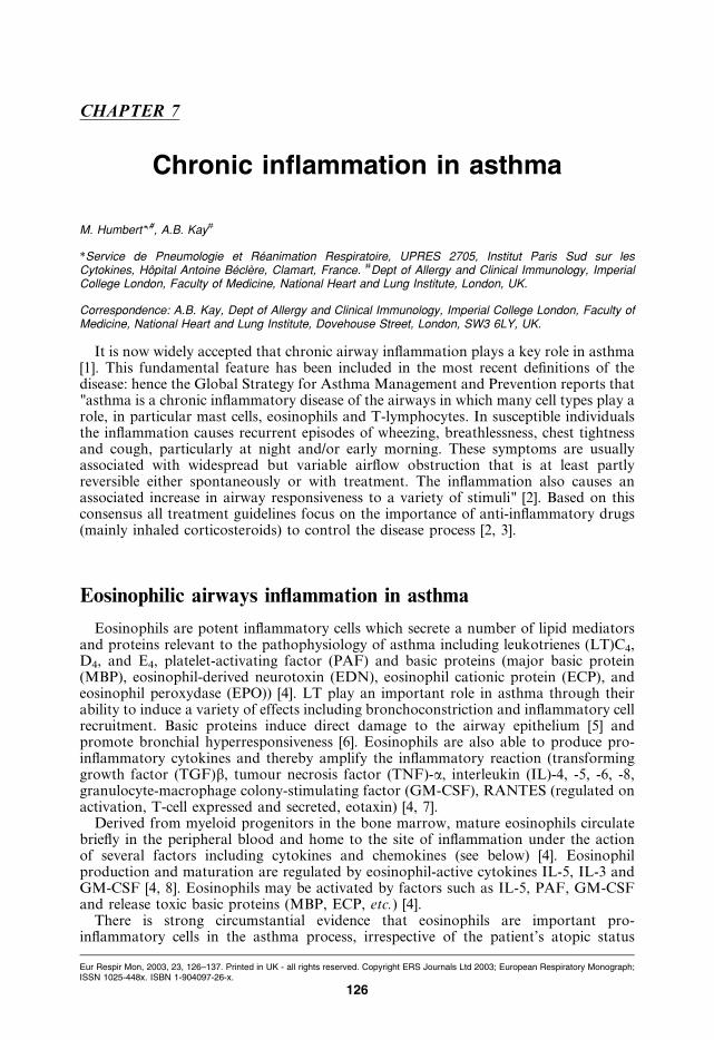
CHAPTER 7
Chronic inflammation in asthma
M. Humbert*,#, A.B. Kay#
*Service de Pneumologie et Reanimation Respiratoire, UPRES 2705, Institut Paris Sud sur lesCytokines, Hopital Antoine Beclere, Clamart, France. #Dept of Allergy and Clinical Immunology, ImperialCollege London, Faculty of Medicine, National Heart and Lung Institute, London, UK.
Correspondence: A.B. Kay, Dept of Allergy and Clinical Immunology, Imperial College London, Faculty ofMedicine, National Heart and Lung Institute, Dovehouse Street, London, SW3 6LY, UK.
It is now widely accepted that chronic airway inflammation plays a key role in asthma[1]. This fundamental feature has been included in the most recent definitions of thedisease: hence the Global Strategy for Asthma Management and Prevention reports that"asthma is a chronic inflammatory disease of the airways in which many cell types play arole, in particular mast cells, eosinophils and T-lymphocytes. In susceptible individualsthe inflammation causes recurrent episodes of wheezing, breathlessness, chest tightnessand cough, particularly at night and/or early morning. These symptoms are usuallyassociated with widespread but variable airflow obstruction that is at least partlyreversible either spontaneously or with treatment. The inflammation also causes anassociated increase in airway responsiveness to a variety of stimuli" [2]. Based on thisconsensus all treatment guidelines focus on the importance of anti-inflammatory drugs(mainly inhaled corticosteroids) to control the disease process [2, 3].
Eosinophilic airways inflammation in asthma
Eosinophils are potent inflammatory cells which secrete a number of lipid mediatorsand proteins relevant to the pathophysiology of asthma including leukotrienes (LT)C4,D4, and E4, platelet-activating factor (PAF) and basic proteins (major basic protein(MBP), eosinophil-derived neurotoxin (EDN), eosinophil cationic protein (ECP), andeosinophil peroxydase (EPO)) [4]. LT play an important role in asthma through theirability to induce a variety of effects including bronchoconstriction and inflammatory cellrecruitment. Basic proteins induce direct damage to the airway epithelium [5] andpromote bronchial hyperresponsiveness [6]. Eosinophils are also able to produce pro-inflammatory cytokines and thereby amplify the inflammatory reaction (transforminggrowth factor (TGF)b, tumour necrosis factor (TNF)-a, interleukin (IL)-4, -5, -6, -8,granulocyte-macrophage colony-stimulating factor (GM-CSF), RANTES (regulated onactivation, T-cell expressed and secreted, eotaxin) [4, 7].
Derived from myeloid progenitors in the bone marrow, mature eosinophils circulatebriefly in the peripheral blood and home to the site of inflammation under the actionof several factors including cytokines and chemokines (see below) [4]. Eosinophilproduction and maturation are regulated by eosinophil-active cytokines IL-5, IL-3 andGM-CSF [4, 8]. Eosinophils may be activated by factors such as IL-5, PAF, GM-CSFand release toxic basic proteins (MBP, ECP, etc.) [4].
There is strong circumstantial evidence that eosinophils are important pro-inflammatory cells in the asthma process, irrespective of the patient’s atopic status
Eur Respir Mon, 2003, 23, 126–137. Printed in UK - all rights reserved. Copyright ERS Journals Ltd 2003; European Respiratory Monograph;ISSN 1025-448x. ISBN 1-904097-26-x.
126
[9, 10]. It is well known that blood and sputum eosinophilia are commonly associatedwith asthma. Moreover, the numbers of eosinophils in peripheral blood, bronchoalveolar(BAL) fluid and bronchial biopsies in a group of asthmatics were elevated whencompared to normal controls and it was possible to demonstrate an increasing degreeof eosinophilia with clinical severity [9]. Furthermore, immunostaining of the bronchialmucosa of patients who had died from severe asthma revealed the presence of largenumbers of activated eosinophils and considerable amounts of MBP deposited in theairways [11]. Increased concentrations of MBP were found in BAL fluid from atopicasthmatics when compared to normal controls and correlations were found between theconcentrations of MBP and the numbers of denuded epithelial cells in BAL fluid [12]. Inatopic asthmatics, late-phase bronchoconstriction was accompanied by an influx ofeosinophils in BAL fluid [13]. This was not observed in individuals developing an isolatedearly-phase response. Lastly, airway eosinophils are very sensitive to corticosteroidtherapy, and their disappearance is associated with an improvement in bronchialhyperresponsiveness [14].
T-lymphocytes in asthma
T-cells have a central role to play in an antigen-driven inflammatory process, since theyare the only cells capable of recognising antigenic material after processing by antigen-presenting cells [15]. CD4zand CD8zT-lymphocytes activated in this manner elaboratea wide variety of protein mediators including cytokines which have the capacity toorchestrate the differentiation, recruitment, accumulation and activation of specificgranulocytes at mucosal surfaces. T-cell derived products can also influence immuno-globulin production by plasma cells. There now exists considerable support for thehypothesis that allergic diseases and asthma represent specialised forms of cell-mediatedimmunity, in which cytokines secreted predominantly by activated T-cells (but also byother leukocytes such as mast cells and eosinophils) bring about the specific accumu-lation and activation of eosinophils [10].
Antigenic peptides are presented to T-cell receptors as a high-affinity complex ofpeptide and major histocompatibility complex (MHC) molecules. T-cells can be broadlydivided into two groups based on their recognition of peptide in the context of eitherMHC class I gene or MHC II gene products. T-cells recognising endogenously generatedpeptides, presented with class I molecules, express the CD8 molecule which binds to classI molecules thus increasing the avidity of the interaction. Most cytotoxic T-cells have theCD8zphenotype. The expression of CD4 by T-cells indicates recognition of peptides inthe context of class II molecules to which CD4 is able to bind. Peptides presented in thecontext of class II MHC proteins generally elicit a T-helper (CD4z T-lymphocyte)response. T-helper cells can be further subdivided according to the pattern of cytokineselaborated following activation [16, 17]. Great progress in the knowledge of phenotypicand functional activities of different T-cell subsets in mice and humans has been maderecently. T-cells secreting cytokines such as IL-2, interferon (IFN)-c and TNF-b arereferred to as T-helper (Th)1 cells. The cytokines produced by these cells promotecytotoxic T-cell and delayed-type responses and inhibit allergic reactions. T-cells pro-ducing IL-4, IL-5, and IL-10 but not IFN-c are referred to as Th2 cells. These cells arecritical to allergic diseases and asthma, as they provide B-cell help for isotype switchingto immunoglobulin (Ig)E and eosinophil maturation, survival and activation.T-lymphocyte activation and expression of Th2-type cytokines is believed to contributeto tissue eosinophilia and local IgE-dependent events in allergic diseases and asthma [10].
The demonstration of primed circulating blood T-lymphocytes in acute severe asthma
CHRONIC INFLAMMATION IN ASTHMA
127
is interesting as it presumably reflects the presence of activated cells in the bronchialmucosa, the major site of the asthmatic inflammatory process [18]. In allergic individuals,circulating blood CD4z T-lymphocytes produce high levels of Th2-type cytokinesincluding IL-5, GM-CSF and IL-3 and may therefore promote eosinophilic inflamma-tion [10, 19]. Moreover, elevated numbers of CD4z T-lymphocytes expressing IL-5messenger ribonucleic acid (mRNA) have been demonstrated in the airways fromasthmatics compared with nonasthmatic controls [20]. More precisely a Th2-like cyto-kine profile has been identified in bronchial samples from atopic and nonatopic asthma[21, 22]. This is in agreement with the demonstration that CD4z and to a lesser extentCD8z T-cell lines grown from BAL cells from atopic asthmatics produce more IL-5protein than in control subjects [23].
Activated T-lymphocytes are usually sensitive to corticosteroid therapy and improve-ment of bronchial hyperresponsiveness and reduction in airway eosinophils parallelsreduction of activated CD25z T-lymphocytes and Th2-type cytokines including IL-4and IL-5 [14]. However some patients with chronic severe asthma are refractory tocorticosteroids [24]. Pharmacological targeting of T-cells has been proposed as a novelapproach to the treatment of corticosteroid-dependent or corticosteroid-resistant asthma.A 12-week randomised, double-blind, placebo-controlled, crossover trial established thatcyclosporin A improves lung function in patients with corticosteroid-dependent chronicsevere asthma [25]. Compared with placebo, a 36-week treatment with cyclosporin Aresulted in a significant reduction in median daily prednisolone dosage and totalprednisolone intake [26]. In addition morning peak expiratory flow rate improvedsignificantly in the active treatment group but not in the placebo group [26]. Usingplacebo-controlled, double-blind conditions Sihra et al. [27] showed that cycloporin Ainhibited the late, but not the early, bronchoconstrictor response to inhaled allergenchallenge of sensitised mild atopic asthmatics. These data support the concept thatT-cells play a crucial role in asthma, as cyclosporin A exerts its immunosuppressiveaction primarily by inhibition of antigen-induced T-lymphocyte activation and thetranscription and translation of mRNA for several cytokines including IL-2, IL-5 andGM-CSF [28]. More recently a single intravenous infusion of a chimeric monoclonalantibody that binds specifically to human CD4 antigen has been evaluated in severecorticosteroid-dependent asthmatics. This randomised double-blind, placebo-controlledtrial demonstrated significant increases in morning and evening peak flow rates in thehighest dose cohort [29]. Additional experiments showed that keliximab infusion induceda rapid and effective binding to all CD4zT-cells with a transient reduction in numbers ofcirculating CD4z T-cells and modulation of CD4 expression, further suggesting thattherapy aimed at the CD4zT-cell may be useful in asthma [30].
Other inflammatory cells
B-cells
Th2-type cytokine-induced B-cell activation and subsequent IgE production isbelieved to be a critical characteristic of patients with atopy, a disorder characterised bysustained, inappropriate IgE responses to common environmental antigens ("allergens")encountered at mucosal surfaces [31, 32]. Interaction of environmental allergens withcells sensitised by binding of surface Fc receptors to allergen-specific IgE is assumed toplay a role in the pathogenesis of atopic asthma. Stimulation of IgE synthesis by B-cellsis mainly driven by IL-4 [31]. It has recently been shown that in both atopic andnonatopic asthmatics, airways CD20zB-cells have the potential to switch in favour of
M. HUMBERT, A.B. KAY
128

IgE heavy-chain production, supporting the concept that local IgE production mayoccur in these patients [33]. These changes may be at least in part under the regulation ofIL-4.
Mast cells and basophils
Mast cells and basophils have long been recognised as major effector cells of allergicreactions by virtue of their high affinity surface receptors for IgE (FceRI) [1]. The earlyphase bronchoconstrictor response to allergen challenge of sensitised atopic asthmaticscan probably be accounted for by mast cell and basophil products, mostly histamine.Mast cells can produce and store several cytokines which may play a role in the chronicasthmatic process, including TNF-a, IL-4, IL-5, and IL-6 [34]. Mast cells are alsobelieved to be responsible at least in part of airways remodelling through fibroblastsactivation. Mast cells have been described in the airways of asthmatics, and, although notnecessarily increased in number, are in the activated state (degranulated) [35]. BB1zbasophils were identified in baseline bronchial biopsies of asthmatics, althougheosinophils and mast cells were 10-fold higher. Similarly basophils increased afterallergen inhalation in atopic asthma, but again basophils werev10% of eosinophils [36].
Macrophages
Macrophages are phagocytic cells derived from bone marrow precursors. They playa fundamental role as accessory cells and they also produce several mediators andcytokines promoting chronic inflammation. Macrophages infiltrate the asthmatic air-ways, especially in nonatopic patients, but also in atopics where allergen challengeactivates macrophages [32, 37, 38]. This cell type may also play a role in airwayremodelling through the production of growth factors such as platelet-derived growthfactor (PDGF), bFGF and TGF-b [1].
Dendritic cells
Dendritic cells are specialised in antigen processing and presentation. IgE presumablyplays a role in their function as they express high numbers of FceRI. These cells areessential in the induction of immune responses within the airways and their numbersare increased in asthma. Their role in human asthma is still a matter of debate [1].
Fibroblasts
Fibroblasts are responsible for the production of collagen and reticular and elasticfibres. Myofibroblasts numbers in the submucosa correlate with subepithelial collagendeposition, supporting a role in airway remodelling [1].
Neutrophils
These cells are recruited in the airways after allergen challenge and are found inelevated numbers in cases of fatal asthma [39]. Their role in asthma however remainsunclear.
CHRONIC INFLAMMATION IN ASTHMA
129
Cytokines in asthma
Pro-eosinophilic cytokines
Accumulating evidence tends to show that the combined effects of a wide array ofcytokines produced by different cell types including activated T-lymphocytes could playa major part in regulating the successive steps leading to a characteristic eosinophil-richairways inflammation [10]. It is well established that tissue recruitment of eosinophilsfrom the bloodstream requires rolling and firm adhesion of circulating cells under thecontrol of cytokine-induced adhesion molecules (mostly of selectin and integrin families)and migration following a gradient of chemotactic substances in which the newly des-cribed family of chemokines are of utmost importance [40, 41]. In addition eosinophilscan be activated by several environmental factors including eosinophil-active cytokines(IL-5, GM-CSF and IL-3) [42–44]. As a result, tissue damage is due at least in part to therelease of toxic granule proteins from activated infiltrating eosinophils [5, 6].
Eosinophil active cytokines (IL-5, GM-CSF, IL-3). T-lymphocytes are thoughtto orchestrate eosinophilic inflammation in asthma through the release of cytokinesincluding "eosinophil-active" cytokines (IL-5, GM-CSF and IL-3) which promoteeosinophil maturation, activation, hyperadhesion and survival. The relevance of IL-5to asthma has been highlighted by the demonstration of elevated numbers of bronchialmucosal activated (EG2z) eosinophils expressing the IL-5 receptor a-chain mRNA inasthmatics and by positive correlations between the numbers of cells expressing IL-5mRNA and markers of asthma severity such as bronchial hyperresponsiveness andasthma symptom (Aas) score [9, 45, 46]. Moreover aerosolised Ascaris suum extract-induced airways inflammation and bronchial hyperresponsiveness in nonhuman primatesare dramatically reduced by the intravenous infusion of an anti-IL-5 monoclonalantibody (TRFK-5) prior to parasite extract inhalation [47]. However, preliminary datain human asthma indicate that anti-IL-5 dramatically reduces allergen-inducedeosinophilia although no significant effect was observed on the magnitude of the late-phase reaction and bronchial hyperresponsiveness [48].
Using double immunohistochemistry and in situ hybridisation, 70% of IL-5 mRNAzsignals co-localized to CD3zT-cells, the majority of which (w70%) were CD4z, althoughCD8z cells also expressed IL-5 [20]. The remaining signals co-localized to mast cellsand eosinophils [20]. In contrast double immunohistochemistry showed that IL-5 immuno-reactivity was predominantly associated with eosinophils and mast cells. However,numbers of IL-5z cells detected by immunohistochemistry were relatively low, raisingthe possibility that insufficient protein accumulated within T-cells to enable detection byimmunohistochemistry [20].
GM-CSF and IL-3 are also thought to participate to the bronchial pro-eosinophiliccytokine network in asthma [43, 44]. T-cell lines grown from BAL cells in patients withatopic asthma have the capacity of producing elevated quantities of GM-CSF [23].Recently, IL-5, IL-8 and GM-CSF immunostaining of sputum cells in bronchial asthmaand chronic bronchitis has shown that the numbers of IL-5 and GM-CSF immuno-stained cells was increased in asthma, a condition characterised by elevated sputumeosinophila, compared to chronic bronchitis where elevated IL-8 expression paralleledsputum neutrophilia [49]. Others have shown that bronchial epithelial cells are also ableto participate to the production of GM-CSF in asthma, emphasising that noninflam-matory cells can participate actively to the local inflammatory process [50]. Interestingly,inhaled corticosteroid attenuates both epithelial cell GM-CSF expression and thenumbers of epithelial activated eosinophils, suggesting that inhaled corticosteroids could
M. HUMBERT, A.B. KAY
130
attenuate airways inflammation partly by down-regulating epithelial cell cytokineexpression [51]. Lastly, GM-CSF could also act on macrophages, as suggested by elevatedaGM-CSF receptor expression on CD68zmacrophages in nonatopic asthmatics [32, 52].
Cytokine-induced upregulation of adhesion molecules. To migrate from the blood-stream to the bronchial mucosa, eosinophils must adhere to vascular endothelial cells,extracellular matrix components and tissue cells. Cell recruitment to the inflamed tissueconsists of at least three events: rolling, firm adhesion and transendothelial migration [40].Granulocyte margination and diapedesis at sites of inflammation seem to be principallyunder the control of the cytokine-induced upregulated expression of several endothelialadhesion molecules including P-selectin, E-selectin (ELAM-1), intercellular adhesionmolecule (ICAM)-1 (CD54) and vascular cell adhesion molecule (VCAM)-1. Theleukocyte receptors for the P- and E-selectins exist on most leukocytes [53]. The leukocytereceptors for ICAM-1 are LFA-1a (CD11a/CD18) and Mac-1 (CD11b/CD18) and thosefor VCAM-1 are VLA-4.
In the asthmatic airways, "pro-inflammatory" cytokines such as TNF-a can upregulateICAM-1 and E-selectin expression and therefore granulocyte recruitment [40, 54]. Morespecifically, the expression of VLA-4 on lymphocytes and eosinophils but not onneutrophils, has led to the hypothesis that VCAM-1 may be the predominant endothelialregulator of the chronic asthmatic bronchial mucosal inflammation. VCAM-1 is upre-gulated by several cytokines including IL-4 and IL-13 [55, 56]. IL-4 and IL-13 have beendetected in the asthmatic airways [46, 57], emphasising the fact that specific eosinophilicrecruitment through an IL-4 (and/or IL-13) induced upregulation of VCAM-1endothelial expression could participate to chronic bronchial mucosal inflammation.Animal models of asthma and IL-4-deficient mice have shown that this cytokine might becritical to the development of an allergic eosinophilic response [58].
Eosinophil chemokines. Classical chemoattractants such as C5a act broadly onneutrophils, eosinophils, basophils and monocytes. The past few years have seen thediscovery of a group of chemoattractive cytokines (termed chemokines) with similaritiesin structure whose principal activities appear to include chemoattraction and activation ofleukocytes including granulocytes, monocytes and T-lymphocytes [41]. Chemokines arepolypeptides of relatively small molecular weight (8–14kDa) which have been assigned todifferent subgroups by structural criteria. The a- and b-chemokines, which contain fourcysteines, are the largest families. The a-chemokines have their first two cysteinesseparated by one additional amino acid ("CXC chemokines": IL-8, etc.), whereas thesecysteines are adjacent to each other in the b-chemokine subgroup ("CC chemokines":eotaxins, monocyte chemotactic proteins (MCPs), RANTES). Interestingly chemokinesare distinguished from classical chemoattractants by a certain cell-target specificity: theCXC chemokines tend to act more on neutrophils, whereas the CC chemokines tend to actmore onmonocytes and in some cases basophils, lymphocytes and eosinophils [59]. Owingto the effects of some CC-chemokines on basophils and eosinophils, their ability to attractand activate monocytes, and their potential role in lymphocyte recruitment, thesemolecules have emerged as the most potent stimulators of effector-cell accumulation andactivation in allergic inflammation [41]. The CC chemokines interacting with the "eotaxinreceptor" CCR3 (eotaxin-1, eotaxin-2, RANTES, MCP-3, MCP-4) are potent pro-eosinophilic cytokines which are believed to play an important role in asthma [60]. Sinceeosinophil chemokines all stimulate eosinophils via CCR3, this receptor is potentially aprime therapeutic target in asthma and other diseases involving eosinophil-mediatedtissue damage. Antagonising CCR3 may be particularly relevant to asthma, as this
CHRONIC INFLAMMATION IN ASTHMA
131
receptor is also expressed by several cell types playing a pivotal role in this condition,including Th2-type cells, basophils and mast cells.
Eotaxin mediates eosinophil (but not neutrophil) accumulation in vivo. Recently,eotaxin and CCR3 mRNA and protein product have been identified in the bronchialsubmucosa of atopic and nonatopic asthmatics [61]. Moreover eotaxin and CCR3expression correlate with airway responsiveness. Cytokeratine-positive epithelial cellsand CD31z endothelial cells were the major source of eotaxin mRNA whereas CCR3co-localized predominantly to eosinophils [61]. These data are consistent with thehypothesis that damage to the bronchial mucosa in asthma involves secretion of eotaxinby epithelial and endothelial cells resulting in eosinophil infiltration mediated via CCR3.
RANTES, MCP-3, and MCP-4 have all the properties that are needed to mobilise andactivate basophils and eosinophils and currently available evidence suggests a primaryrole for them in allergic inflammation [60]. A combined expression of eosinophilchemokines (eotaxins, MCPs and RANTES) together with eosinophil active cytokines(IL-5, GM-CSF and IL-3), has been demonstrated in asthma, indicating that thesecytokines could act in synergy to promote the elaboration of an eosinophil-rich bronchialmucosal infiltrate [61, 62]. Indeed, priming eosinophils with IL-5 increases the chemo-tactic properties of RANTES on eosinophils [63]. The cell sources of RANTES andMCPs in asthma also include primarily epithelial and endothelial cells, as well asmacrophages, T-lymphocytes and eosinophils [61]. Interestingly, bronchial epithelial cellproduction of RANTES is downregulated by inhaled corticosteroids [64].
Due to their cell-target specificity favouring neutrophil chemoattraction, a role forCXC chemokines in asthma is less likely although IL-8 has been shown to be a chemo-tactic factor for eosinophils [65]. Although eosinophils are most prominent in the airwaysof asthmatics, fewer eosinophils and more neutrophils have been identified in the airwaysof sudden-onset fatal asthma [39]. Elevated IL-8 expression has been reported in asthma[65]. In that setting, IL-8 could promote not only neutrophil accumulation but alsoeosinophil migration in synergy with IL-5.
Pro-atopic cytokines in asthma
Asthma is often, though not invariably, associated with atopy [66]. Since the clinicalclassification of asthma by Rackerman [67], it has been widely accepted that a subgroupof asthmatics are not demonstrably atopic, the so-called "intrinsic" variant of the disease[67]. Intrinsic asthmatics show negative skin tests and there is no clinical history ofallergy. Furthermore, serum total IgE concentrations are within the normal range andthere is no evidence of specific IgE antibodies directed against common aeroallergens.These patients are usually older than their allergic counterparts and have onset of theirsymptoms in later life, often with a more severe clinical course. There is a preponderanceof females and the association of nasal polyps and aspirin sensitivity occurs morefrequently in the nonatopic form of the disease. Whereas some authors suggest that onlyy10% of asthmatics are intrinsic, the Swiss SAPALDIA survey (8,357 adults, aged 18–60 yrs) found that one-third of total asthmatics were nonallergic [68].
Ever since the first description of intrinsic asthma, there has been debate about therelationship of this variant of the disease to atopy [32, 66]. One suggestion is that intrinsicasthma represents a form of autoimmunity, or auto-allergy, triggered by infection as arespiratory influenza-like illness often precedes onset. Other authors have suggested thatintrinsic asthmatics are allergic to an as yet undetected allergen. The present authors viewis that although intrinsic asthma has a different clinical profile from extrinsic asthma itdoes not appear to be a distinct immunopathological entity [32]. This concept issupported by the demonstration of elevated numbers of activated eosinophils [37],
M. HUMBERT, A.B. KAY
132

Th2-type lymphocytes [69], and cells expressing FceRI [70] in bronchial biopsies fromatopic and nonatopic asthmatics, together with epidemiological evidence indicating thatserum IgE concentrations relate closely to asthma prevalence regardless of atopic status[66]. IL-4 expression is a feature of asthma, irrespective of its atopic status, providingfurther evidence for similarities in the immunopathogenesis of atopic and nonatopicasthma [32]. IL-4 mRNA is mainly CD4z T- cell derived [20]. Expression of aIL-4receptor mRNA and protein is significantly elevated in the epithelium and subepitheliumof biopsies from atopic and nonatopic asthmatics compared to atopic controls [71].Recent evidence of the effectiveness of nebulised soluble IL-4 receptors in atopic asthmafurther support the relevance of this cytokine in this disease [72].
In addition, IL-13 is a cytokine very close to IL-4 which exhibits activities possiblyrelevant to asthma: promotion of IgE synthesis, eosinophil vascular adhesion by VLA-4/VCAM-1 interaction and promotion of Th2-type cell responses [56, 57, 73]. ImportantlyIL-4 or IL-13 are absolutely required for IgE-switching in B-cells, a prerequisite forelevated IgE synthesis. The present authors have reported elevated expression of IL-13mRNA in the bronchial mucosa of so-called atopic and nonatopic asthma [57].Therefore, although intrinsic asthma have no demonstrable atopy, they have a biologicalpattern of airway inflammation strongly suggesting a possible "atopic-like" status whichmay be restricted to the bronchial submucosa [32]. As discussed above, local IgEsynthesis in CD20z B-cells has been demonstrated in the bronchial submucosa of
�������������� ��������
��� ��������� ������������
�������� ����� ������� �������������
������� ������������
��������������
��� ����
��� �� � ��������� ���
������!������"�#��� ����!������
���$
���%&'(�)(�'
��������� �� ������* � ��
�+���
� �,�-� ��
.���� ��
"+#����.�
����
���
/� ��
0 ����"�#�����0 �� �����
Fig. 1. – Proposed scheme for the progression of asthma in relation to pathogenesis. The progression of asthmawith emphasis on the cells and mediators involved is shown diagrammatically. The early asthmatic reactionoccurs within minutes and is largely due to the release of histamine and lipid mediators from mast cellsfollowing interaction of allergen with cell bound immunoglobulin (Ig)E. The late asthmatic reaction, whichpeaks 6–13 h after allergen challenge, is believed to be partially T-cell dependent. For example, the late-phase,but not the early-phase, was inhibited by cyclosporin A [27] and challenge with T-cell peptide epitopes inducedan isolated late asthmatic reaction. CD4 cells may interact directly with smooth muscle to produce airwaynarrowing through the release of neurotrophins (NT) (which in turn activate neuropeptides (NP)). Interleukin(IL)-13 may also play a role in late-phase asthmatic reactions [74]. The role of the eosinophil remains uncertain.On the one hand there is much circumstantial evidence to incriminate eosinophils as pro-inflammatory cells inthe asthma process. However, depletion of the eosinophils with anti-IL-5 did not influence the late-phasereaction or bronchial hyperresponsiveness in mild asthmatics [48]. Airway hyperresponsiveness is a feature ofchronic persistent asthma and is due in part to airway thickening due to remodelling, fibrosis and other repairprocesses.These changes are brought about by the elaboration of growth factors and fibrogenic factors fromvarious cell types including eosinophils (E’phil), fibroblasts (F’blast), monocytes (Mw), epithelial (Epi) cells andendothelial (Endo) cells.
CHRONIC INFLAMMATION IN ASTHMA
133
patients with atopic and nonatopic asthma (detection of elevated expression of e germ-line gene transcripts and mRNA encoding the e heavy chain of IgE) [33]. This along withthe demonstration of elevated numbers of cells expressing the high affinity IgE receptorin intrinsic asthma suggests the possibility of local IgE-mediated processes in the absenceof detectable systemic IgE production.
Summary
There now exists considerable support for the hypothesis that asthma represents aspecialised form of cell-mediated immunity, in which cytokines, chemokines and othermediators such as leukotrienes secreted by a wide range of inflammatory cells bringabout the specific accumulation and activation of eosinophils in the bronchial mucosa(the progression of asthma is diagrammatically depicted in figure 1). Theseobservations have important implications for future therapies, since it suggests thatmore selective drugs than corticosteroids should be of interest in asthma.
Keywords: Cell-mediated immunity, chemokines, cytokines, eosinophils, leukotrienes.
References
1. Bousquet J, Jeffery PK, Busse WW, Johnson M, Vignola AM. Asthma: from bronchoconstriction
to airways inflammation and remodeling. Am J Respir Crit Care Med 2000; 161: 1720–1745.
2. WHO/NHLBI Workshop Report. Global Strategy for asthma management and prevention.
National Institutes of Health, National Heart, Lung, and Blood Institute, Bethesda, MD.
Publication No 95-3959.
3. Barnes PJ. Inhaled glucocorticoids for asthma. N Engl J Med 1995; 332: 868–875.
4. Wardlaw AJ, Moqbel R, Kay AB. Eosinophils: biology and role in disease. Adv Immunol 1995;
60: 151–266.
5. Motojima S, Frigas E, Loegering DA, Gleich GJ. Toxicity of eosinophil cationic protein for
guinea pig tracheal epithelium. Am Rev Respir Dis 1989; 139: 801–805.
6. Gundel RH, Letts LG, Gleich GJ. Human eosinophil major basic protein induces airway
constriction and airway responsiveness in primates. J Clin Invest 1991; 87: 1470–1473.
7. Broide DH, Paine MM, Firestein GS. Eosinophils express interleukin-5 and granulocyte
macrophage colony-stimulating factor mRNA at sites of allergic inflammation in asthmatics.
J Clin Invest 1992; 90: 1414–1424.
8. Lopez AF, Sanderson CJ, Gamble JR, Campbell HD, Young IG, Vadas MA. Recombinant
interleukin 5 is a selective activator of human eosinophil function. J Exp Med 1988; 167: 219–224.
9. Bousquet J, Chanez P, Lacoste J-Y, et al. Eosinophilic inflammation in asthma. N Engl J Med
1990; 323: 1033–1039.
10. Corrigan CJ, Kay AB. T cells and eosinophils in the pathogenesis of asthma. Immunol Today 1992;
13: 501–507.
11. Azzawi M, Johnston PW, Majumbar S, et al. T lymphocytes and activated eosinophils in airway
mucosa in fatal asthma and cystic fibrosis. Am Rev Respir Dis 1992; 145: 1477–1482.
12. Wardlaw AJ, Dunnette S, Gleich GJ, et al. Eosinophils and mast cells in bronchoalveolar lavage in
mild asthma: relationship to bronchial hyperreactivity. Am Rev Respir Dis 1988; 137: 62–69.
13. Till S, Durham SR, Rajakulasingam K, et al. Allergen-induced proliferation and interleukin-5
M. HUMBERT, A.B. KAY
134
production by bronchoalveolar lavage and blood T cells after segmental allergen challenge. Am J
Respir Crit Care Med 1998; 158: 404–411.
14. Robinson D, Hamid Q, Ying S, et al. Prednisolone treatment in asthma is associated with
modulation of bronchoalveolar lavage cell interleukin-4, interleukin-5, and interferon-c cytokine
gene expression. Am Rev Respir Dis 1993; 148: 401–406.
15. Germain RN. Antigen processing and presentation. In: Paul WE, ed. Fundamental Immunology,
3rd Edn. New York, Raven Press Ltd, 1993; pp. 629–676.
16. Mossmann TR, Coffman RL. Two types of mouse helper T cell clone: implication for immune
regulation. Immunol Today 1987; 8: 223–227.
17. Mossmann TR, Sad S. The expanding universe of T-cell subsets: Th1, Th2 and more. Immunol
Today 1996; 17: 138–146.
18. Corrigan CJ, Kay AB. CD4 T-lymphocyte activation in acute severe asthma: relationship to
disease activity and atopic status. Am Rev Respir Dis 1990; 141: 970–977.
19. Romagnani S. Lymphokine production by human T cells in disease states. Annu Rev Immunol
1994; 12: 227–257.
20. Ying S, Humbert M, Barkans J, et al. Expression of IL-4 and IL-5 mRNA and protein product by
CD4zand CD8zT cells, eosinophils, and mast cells in bronchial biopsies obtained from atopic and
nonatopic (intrinsic) asthmatics. J Immunol 1997; 158: 3539–3544.
21. Robinson DS, Hamid Q, Ying S, et al. Predominant TH2-like bronchoalveolar T-lymphocyte
population in atopic asthma. N Engl J Med 1992; 326: 298–304.
22. Ying S, Durham SR, Corrigan C, Hamid Q, Kay AB. Phenotype of cells expressing mRNA for
TH2 type (IL-4 and IL-5) and TH1 type (IL-2 and IFN-gamma) cytokines in bronchoalveolar
lavage and bronchial biopsies from atopic asthmatics. Am J Respir Cell Mol Biol 1995; 12: 477–487.
23. Till S, Li B, Durham S, et al. Secretion of the eosinophil-active cytokines (IL-5, GM-CSF and
IL-3) by bronchoalveolar lavage CD4zand CD8zT cell lines in atopic asthmatics and atopic and
non-atopic controls. Eur J Immunol 1995; 25: 2727–2731.
24. Lane SJ, Lee TH. Glucocorticoid-resistant asthma. In: Holgate ST, Boushey HA, Fabbri LM, eds.
Difficult Asthma. London, Martin Dunitz Ltd, 1999; pp. 389–411.
25. Alexander AG, Barnes NC, Kay AB. Trial of ciclosporin on corticosteroid-dependent asthma.
Lancet 1992; 339: 324–328.
26. Lock S, Kay AB, Barnes NC. Double-blind, placebo-controlled study of cyclosporin A as a
corticosteroid-sparing agent in corticosteroid-dependent asthma. Am J Respir Crit Care Med 1996;
153: 509–514.
27. Sihra BS, Kon OM, Durham SR, Walker S, Barnes NC, Kay AB. Effect of cyclosporin A on the
allergen-induced late asthmatic reaction. Thorax 1997; 52: 447–452.
28. Kahan BD. Cyclosporin. N Engl J Med 1989; 321: 1725–1738.
29. Kon OM, Sihra BS, Compton CH, Leonerd TB, Kay AB, Barnes NC. Randomized, dose-ranging,
placebo-controlled study of chimeric antibody to CD4 (Keliximlab) in chronic severe asthma.
Lancet 1998; 35: 1109–1113.
30. Kon OM, Sihra BS, Loh LC, et al. The effects of anti-CD4 monoclonal antibody, keliximab, on
peripheral blood CD4zT-cells in asthma. Eur Respir J 2001; 18: 45–52.
31. Del Prete GF, Maggi E, Parronchi P, et al. IL-4 is an essential co-factor for the IgE synthesis
induced in vitro by human T cell clones and their supernatants. J Immunol 1988; 140: 4193–
4198.
32. Humbert M, Menz G, Ying S, et al. Extrinsinc (atopic) and intrinsic (non-atopic) asthma: more
similarities than differences. Immunol Today 1999; 20: 528–533.
33. Durham SR, Ying S, Meng Q, Humbert M, Gould H, Kay AB. Local expression of germ-line gene
transcripts (Ie) and mRNA for the heavy chain of IgE (Ce) in the bronchial mucosa in atopic and
non-atopic asthma. J Allergy Clin Immunol 1998; 101: S162.
34. Bradding P, Roberts JA, Britten KM, et al. Interleukin-4, -5, and -6 and tumor necrosis factor-a in
normal and asthmatic airways: evidence for the human mast cell as a source of these cytokines.
Am J Respir Cell Mol Biol 1995; 10: 471–480.
CHRONIC INFLAMMATION IN ASTHMA
135
35. Djukanovic R, Wilson JW, Britten KM. Mucosal inflammation in asthma. Am Rev Respir Dis
1990; 142: 434–457.
36. Macfarlane AJ, Kon OM, Smith SJ, et al. Basophils in atopic and non-atopic asthma and late
allergic reactions. J Allergy Clin Immunol 2000; 105: 99–107.
37. Bentley AM, Menz G, Storz C, et al. Identification of T lymphocytes, macrophages, and activated
eosinophils in the bronchial mucosa of intrinsic asthma: relationship to symptoms and bronchial
responsiveness. Am Rev Respir Dis 1992; 146: 500–506.
38. Gosset P, Tsicopoulos A, Wallaert B, et al. Increased secretion of tumor necrosis factor-alpha and
interleukin-6 by alveolar macrophages consecutive to the development of the late asthmatic
reaction. J Allergy Clin Immunol 1991; 88: 561–571.
39. Sur S, Crotty TB, Kephart GM, et al. Sudden-onset fatal asthma: a distinct entity with few
eosinophils and relatively more neutrophils in the airway submucosa? Am Rev Respir Dis 1993;
148: 713–719.
40. Springer TA. Traffic signals for lymphocyte recirculation and leukocyte emigration: the multistep
paradigm. Cell 1994; 76: 301–314.
41. Baggiolini M, Dahinden CA. CC chemokines in allergic inflammation. Immunol Today 1994;
15: 127–133.
42. Walsh GM, Hartnell A, Wardlaw AJ, Kurihara K, Sanderson CJ, Kay AB. IL-5 enhances the in
vitro adhesion of human eosinophils, but not neutrophils, in a leucocyte integrin (CD11/18)-
dependent manner. Immunology 1990; 71: 258–265.
43. Warringa RAJ, Koenderman L, Kok PTM, Krekniet J, Bruijneel PLB. Modulation and induction
of eosinophil chemotaxis by granulocyte-monocyte colony stimulating factor and interleukin 3.
Blood 1991; 77: 2694–2700.
44. Rothenberg ME, Owen Jr WF, Silberstein DS, et al. Human eosinophils have prolonged survival,
enhanced functional properties and become hypodense when exposed to human interleukin 3.
J Clin Invest 1988; 81: 1986–1992.
45. Yasruel Z, Humbert M, Kotsimbos ATC, et al. Expression of membrane-bound and soluble
interleukin-5 alpha receptor mRNA in the bronchial mucosa of atopic and non-atopic asthmatics.
Am J Respir Crit Care Med 1997; 155: 1413–1418.
46. Humbert M, Corrigan CJ, Durham SR, Kimmitt P, Till SJ, Kay AB. Relationship between
bronchial mucosal interleukin-4 and interleukin-5 mRNA expression and disease severity in atopic
asthma. Am J Respir Crit Care Med 1997; 156: 704–708.
47. Mauser PJ, Pitman AM, Fernandez X, et al. Effects of an antibody to interleukin-5 in a monkey
model of asthma. Am J Respir Crit Care Med 1995; 152: 467–472.
48. Leckie MJ, ten Brinke A, Khan J, et al. Effects of an interleukin-5 blocking monoclonal antibody
on eosinophils, airway hyper-responsiveness, and the late asthmatic response. Lancet 2000;
356: 2144–2148.
49. Hoshi H, Ohno I, Honma M, et al. IL-5, IL-8 and GM-CSF immunostaining of sputum cells in
bronchial asthma and chronic bronchitis. Clin Exp Allergy 1995; 25: 720–728.
50. Sousa AR, Poston RN, Lane SJ, Nakhosteen JA, Lee TH. Detection of GM-CSF in asthmatic
bronchial epithelium and decrease by inhaled corticosteroids. Am Rev Respir Dis 1993; 147: 1557–
1561.
51. Davies RJ, Wang JH, Trigg CJ, Devalia JL. Expression of granulocyte/macrophage-
colony-stimulating factor, interleukin-8 and RANTES in the bronchial epithelium of mild
asthmatics is down-regulated by inhaled beclomethasone dipropionate. Int Arch Allergy Immunol
1995; 107: 428–429.
52. Kotsimbos ATC, Humbert M, Minshall E, et al. Upregulation of aGM-CSF-receptor in non-
atopic but not in atopic asthma. J Allergy Clin Immunology 1997; 99: 666–672.
53. Springer TA. Adhesion receptors of the immune system. Nature 1990; 346: 425–434.
54. Ying S, Robinson DS, Varney V, et al. TNF alpha mRNA expression in allergic inflammation.
Clin Exp Allergy 1991; 21: 745–750.
55. Schleimer RP, Sterbinsky SA, Kaiser J, et al. IL-4 induces adherence of human eosinophils and
M. HUMBERT, A.B. KAY
136
basophils but not neutrophils to endothelium: association with expression of VCAM-1. J Immunol
1992; 148: 1086–1092.
56. Bochner BS, Klunk DA, Sterbinsky SA, Coffman RL, Schleimer RP. IL-13 selectively induces
vascular cell adhesion molecule-1 expression in human endothelial cells. J Immunol 1995; 154: 799–
803.
57. Humbert M, Durham SR, Kimmitt P, et al. Elevated expression of mRNA encoding interleukin-
13 in the bronchial mucosa of atopic and non-atopic asthmatics. J Allergy Clin Immunology 1997;
99: 657–665.
58. Brusselle G, Kips J, Joos G, Bluethmann H, Pauwels R. Allergen-induced airway inflammation
and bronchial responsiveness in wild-type and interleukin-4-deficient mice. Am J Respir Cell Mol
Biol 1995; 12: 254–259.
59. Schall TJ, Bacon KB. Chemokines, leukocyte trafficking, and inflammation. Cur Opin Immunol
1994; 6: 865–873.
60. Menzies-Gow A, Robinson DS. Eosinophil chemokines and their receptors: an attractive target in
asthma. Lancet 2000; 355: 1741–1743.
61. Ying S, Meng Q, Zeibecoglou K, et al. Eosinophil chemotactic chemokines (eotaxin, eotaxin-2,
RANTES, monocyte chemotactic protein-3 (MCP-3), andMCP-4), and C-C chemokine receptor 3
expression in bronchial biopsies from atopic and nonatopic (intrinsic) asthmatics. J Immunol 1999;
163: 6321–6329.
62. Humbert M, Ying S, Corrigan C, et al. Bronchial mucosal gene expression of the CC chemokines
RANTES and MCP-3 in symptomatic atopic and non-atopic asthmatics: relationship to the
eosinophil-active cytokines IL-5, GM-CSF and IL-3. Am J Respir Cell Mol Biol 1997; 16: 1–8.
63. Collins PD, Marleau S, Griffiths-Johnson DA, Jose PJ, Williams TJ. Cooperation between
interleukin-5 and the chemokine eotaxin to induce eosinophil accumulation in vivo. J Exp Med
1995; 182: 1169–1174.
64. Jung Kwon O, Jose PJ, Robbins RA, Schall TJ, Williams TJ, Barnes PJ. Glucocorticoid inhibition
of RANTES expression in human lung epithelial cells. Am J Respir Cell Mol Biol 1995; 12: 488–
496.
65. Yousefi S, Hemmann S, Weber M, et al. IL-8 is expressed by human peripheral blood eosinophils:
evidence for increased secretion in asthma. J Immunol 1995; 154: 5481–5490.
66. Burrows B, Martinez FD, Halonen M, Barbee RA, Cline MG. Association of asthma with serum
IgE levels and skin-test reactivity to allergens. N Engl J Med 1989; 320: 271–277.
67. Rackeman FM. A working classification of asthma. Am J Med 1947; 3: 601–606.
68. Wurthrich B, Schindler C, Leuenberger P, Ackermann-Liebrich U. Prevalence of atopy and
pollinosis in the adult population of Switzerland (SAPALDIA study). Int Arch Allergy Immunol
1995; 106: 149–156.
69. Humbert M, Durham SR, Ying S, et al. IL-4 and IL-5 mRNA and protein in bronchial biopsies
from atopic and non-atopic asthmatics: evidence against "intrinsic" asthma being a distinct
immunopathological entity. Am J Respir Crit Care Med 1996; 154: 1497–1504.
70. Humbert M, Grant JA, Taborda-Barata L, et al. High affinity IgE receptor (FceRI)-bearing cells
in bronchial biopsies from atopic and non-atopic asthmaAm J Respir Crit Care Med 1996;
153: 1931–1937.
71. Kotsimbos TC, Ghaffar O, Minshall E, et al. Expression of interleukin-4 receptor a-subunit is
increased in bronchial biopsies from atopic and non-atopic asthmatics. J Allergy Clin Immunol
1998; 102: 859–866.
72. Borish LC, Nelson HS, Lanz MJ, et al. Interleukin-4 receptor in moderate atopic asthma. A phase
I/II randomized, placebo-controlled trial. Am J Respir Crit Care Med 1999; 160: 1816–1823.
73. Wills-Karp M. IL-13 as a target for modulation of the inflammatory response in asthma. Allergy
1998; 53: 113–119.
74. Haselden BM, Kay AB, Larche M. IgE-independent MHC-restricted T cell peptide epitope-
induced late asthmatic reactions. J Exp Med 1999; 189: 1885–1894.
CHRONIC INFLAMMATION IN ASTHMA
137
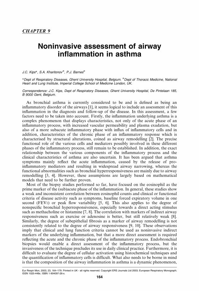
CHAPTER 9
Noninvasive assessment of airwayinflammation in asthma
J.C. Kips*, S.A. Kharitonov#, P.J. Barnes#
*Dept of Respiratory Diseases, Ghent University Hospital, Belgium. #Dept of Thoracic Medicine, NationalHeart and Lung Institute, Imperial College School of Medicine London, UK.
Correspondence: J.C. Kips, Dept of Respiratory Diseases, Ghent University Hospital, De Pintelaan 185,B 9000 Gent, Belgium.
As bronchial asthma is currently considered to be and is defined as being aninflammatory disorder of the airways [1], it seems logical to include an assessment of thisinflammation in the diagnosis and follow-up of the disease. In this assessment, a fewfactors need to be taken into account. Firstly, the inflammation underlying asthma is acomplex phenomenon that displays characteristics, not only of the acute phase of aninflammatory process, with increased vascular permeability and plasma exudation, butalso of a more subacute inflammatory phase with influx of inflammatory cells and inaddition, characteristics of the chronic phase of an inflammatory response which ischaracterised by structural alterations, coined as airway remodelling [2]. The precisefunctional role of the various cells and mediators possibly involved in these differentphases of the inflammatory process, still remain to be established. In addition, the exactrelationship between the various components of the inflammatory process and theclinical characteristics of asthma are also uncertain. It has been argued that asthmasymptoms mainly reflect the acute inflammation, caused by the release of pro-inflammatory mediators and resulting in widespread airway narrowing, whereas thefunctional abnormalities such as bronchial hyperresponsiveness are mainly due to airwayremodelling [3, 4]. However, these assumptions are largely based on mathematicalmodels that need to be further proven.
Most of the biopsy studies performed so far, have focused on the eosinophil as theprime marker of the (sub)acute phase of the inflammation. In general, these studies showa weak and inconsistent correlation between eosinophil counts and clinical or functionalcriteria of disease activity such as symptoms, baseline forced expiratory volume in onesecond (FEV1) or peak flow variability [5, 6]. This also applies to the degree ofnonspecific bronchial hyperresponsiveness, especially towards a direct acting stimulussuch as methacholine or histamine [7, 8]. The correlation with markers of indirect airwayresponsiveness such as exercise or adenosine is better, but still relatively weak [8].Similarly, the degree of subepithelial fibrosis as a marker of airway remodelling is notconsistently related to the degree of airway responsiveness [9, 10]. These observationsimply that clinical and lung function criteria cannot be used as noninvasive indirectmarkers of the underlying inflammation, but that a more direct assessment is requiredreflecting the acute and the chronic phase of the inflammatory process. Endobronchialbiopsies would enable a direct assessment of the inflammatory process, but theinvasiveness of the technique precludes its use in daily clinical practice. Furthermore, it isdifficult to evaluate the degree of cellular activation using histochemical techniques andthe quantification of inflammatory cells is difficult. What also needs to be borne in mindis that the composition of the airway inflammation in asthma is a dynamic phenomenon,
Eur Respir Mon, 2003, 23, 164–179. Printed in UK - all rights reserved. Copyright ERS Journals Ltd 2003; European Respiratory Monograph;ISSN 1025-448x. ISBN 1-904097-26-x.
164
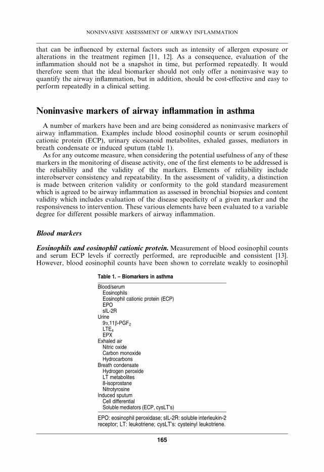
that can be influenced by external factors such as intensity of allergen exposure oralterations in the treatment regimen [11, 12]. As a consequence, evaluation of theinflammation should not be a snapshot in time, but performed repeatedly. It wouldtherefore seem that the ideal biomarker should not only offer a noninvasive way toquantify the airway inflammation, but in addition, should be cost-effective and easy toperform repeatedly in a clinical setting.
Noninvasive markers of airway inflammation in asthma
A number of markers have been and are being considered as noninvasive markers ofairway inflammation. Examples include blood eosinophil counts or serum eosinophilcationic protein (ECP), urinary eicosanoid metabolites, exhaled gasses, mediators inbreath condensate or induced sputum (table 1).
As for any outcome measure, when considering the potential usefulness of any of thesemarkers in the monitoring of disease activity, one of the first elements to be addressed isthe reliability and the validity of the markers. Elements of reliability includeinterobserver consistency and repeatability. In the assessment of validity, a distinctionis made between criterion validity or conformity to the gold standard measurementwhich is agreed to be airway inflammation as assessed in bronchial biopsies and contentvalidity which includes evaluation of the disease specificity of a given marker and theresponsiveness to intervention. These various elements have been evaluated to a variabledegree for different possible markers of airway inflammation.
Blood markers
Eosinophils and eosinophil cationic protein.Measurement of blood eosinophil countsand serum ECP levels if correctly performed, are reproducible and consistent [13].However, blood eosinophil counts have been shown to correlate weakly to eosinophil
Table 1. – Biomarkers in asthma
Blood/serumEosinophilsEosinophil cationic protein (ECP)EPOsIL-2R
Urine9a,11b-PGF2
LTE4
EPXExhaled air
Nitric oxideCarbon monoxideHydrocarbons
Breath condensateHydrogen peroxideLT metabolites8-isoprostaneNitrotyrosine
Induced sputumCell differentialSoluble mediators (ECP, cysLT’s)
EPO: eosinophil peroxidase; sIL-2R: soluble interleukin-2receptor; LT: leukotriene; cysLT’s: cysteinyl leukotriene.
NONINVASIVE ASSESSMENT OF AIRWAY INFLAMMATION
165
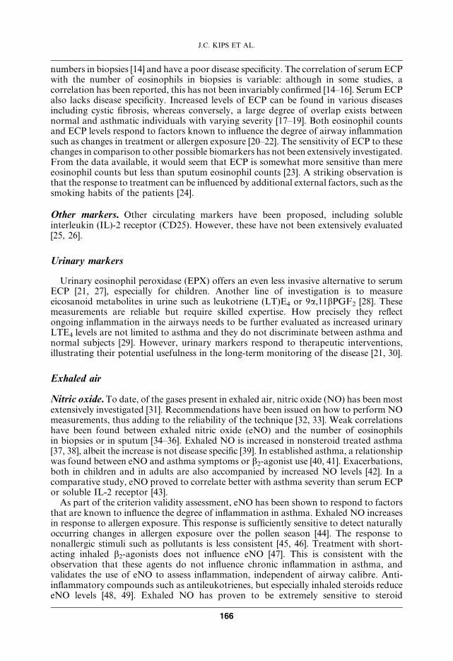
numbers in biopsies [14] and have a poor disease specificity. The correlation of serum ECPwith the number of eosinophils in biopsies is variable: although in some studies, acorrelation has been reported, this has not been invariably confirmed [14–16]. Serum ECPalso lacks disease specificity. Increased levels of ECP can be found in various diseasesincluding cystic fibrosis, whereas conversely, a large degree of overlap exists betweennormal and asthmatic individuals with varying severity [17–19]. Both eosinophil countsand ECP levels respond to factors known to influence the degree of airway inflammationsuch as changes in treatment or allergen exposure [20–22]. The sensitivity of ECP to thesechanges in comparison to other possible biomarkers has not been extensively investigated.From the data available, it would seem that ECP is somewhat more sensitive than mereeosinophil counts but less than sputum eosinophil counts [23]. A striking observation isthat the response to treatment can be influenced by additional external factors, such as thesmoking habits of the patients [24].
Other markers. Other circulating markers have been proposed, including solubleinterleukin (IL)-2 receptor (CD25). However, these have not been extensively evaluated[25, 26].
Urinary markers
Urinary eosinophil peroxidase (EPX) offers an even less invasive alternative to serumECP [21, 27], especially for children. Another line of investigation is to measureeicosanoid metabolites in urine such as leukotriene (LT)E4 or 9a,11bPGF2 [28]. Thesemeasurements are reliable but require skilled expertise. How precisely they reflectongoing inflammation in the airways needs to be further evaluated as increased urinaryLTE4 levels are not limited to asthma and they do not discriminate between asthma andnormal subjects [29]. However, urinary markers respond to therapeutic interventions,illustrating their potential usefulness in the long-term monitoring of the disease [21, 30].
Exhaled air
Nitric oxide.To date, of the gases present in exhaled air, nitric oxide (NO) has been mostextensively investigated [31]. Recommendations have been issued on how to perform NOmeasurements, thus adding to the reliability of the technique [32, 33]. Weak correlationshave been found between exhaled nitric oxide (eNO) and the number of eosinophilsin biopsies or in sputum [34–36]. Exhaled NO is increased in nonsteroid treated asthma[37, 38], albeit the increase is not disease specific [39]. In established asthma, a relationshipwas found between eNO and asthma symptoms or b2-agonist use [40, 41]. Exacerbations,both in children and in adults are also accompanied by increased NO levels [42]. In acomparative study, eNO proved to correlate better with asthma severity than serum ECPor soluble IL-2 receptor [43].
As part of the criterion validity assessment, eNO has been shown to respond to factorsthat are known to influence the degree of inflammation in asthma. Exhaled NO increasesin response to allergen exposure. This response is sufficiently sensitive to detect naturallyoccurring changes in allergen exposure over the pollen season [44]. The response tononallergic stimuli such as pollutants is less consistent [45, 46]. Treatment with short-acting inhaled b2-agonists does not influence eNO [47]. This is consistent with theobservation that these agents do not influence chronic inflammation in asthma, andvalidates the use of eNO to assess inflammation, independent of airway calibre. Anti-inflammatory compounds such as antileukotrienes, but especially inhaled steroids reduceeNO levels [48, 49]. Exhaled NO has proven to be extremely sensitive to steroid
J.C. KIPS ET AL.
166
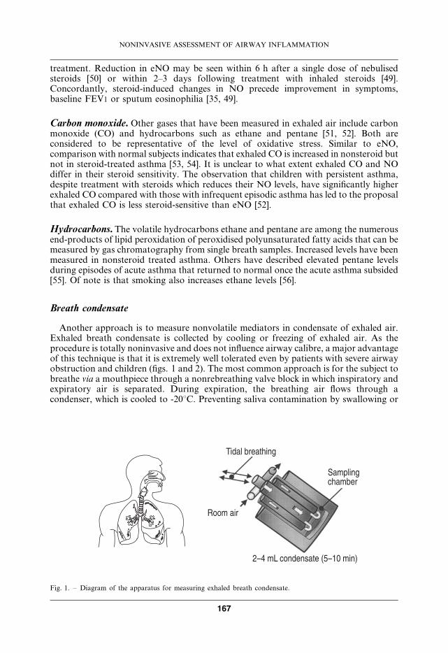
treatment. Reduction in eNO may be seen within 6 h after a single dose of nebulisedsteroids [50] or within 2–3 days following treatment with inhaled steroids [49].Concordantly, steroid-induced changes in NO precede improvement in symptoms,baseline FEV1 or sputum eosinophilia [35, 49].
Carbon monoxide. Other gases that have been measured in exhaled air include carbonmonoxide (CO) and hydrocarbons such as ethane and pentane [51, 52]. Both areconsidered to be representative of the level of oxidative stress. Similar to eNO,comparison with normal subjects indicates that exhaled CO is increased in nonsteroid butnot in steroid-treated asthma [53, 54]. It is unclear to what extent exhaled CO and NOdiffer in their steroid sensitivity. The observation that children with persistent asthma,despite treatment with steroids which reduces their NO levels, have significantly higherexhaled CO compared with those with infrequent episodic asthma has led to the proposalthat exhaled CO is less steroid-sensitive than eNO [52].
Hydrocarbons.The volatile hydrocarbons ethane and pentane are among the numerousend-products of lipid peroxidation of peroxidised polyunsaturated fatty acids that can bemeasured by gas chromatography from single breath samples. Increased levels have beenmeasured in nonsteroid treated asthma. Others have described elevated pentane levelsduring episodes of acute asthma that returned to normal once the acute asthma subsided[55]. Of note is that smoking also increases ethane levels [56].
Breath condensate
Another approach is to measure nonvolatile mediators in condensate of exhaled air.Exhaled breath condensate is collected by cooling or freezing of exhaled air. As theprocedure is totally noninvasive and does not influence airway calibre, a major advantageof this technique is that it is extremely well tolerated even by patients with severe airwayobstruction and children (figs. 1 and 2). The most common approach is for the subject tobreathe via a mouthpiece through a nonrebreathing valve block in which inspiratory andexpiratory air is separated. During expiration, the breathing air flows through acondenser, which is cooled to -20uC. Preventing saliva contamination by swallowing or
������������
��������
������� ������
�������������������������
Fig. 1. – Diagram of the apparatus for measuring exhaled breath condensate.
NONINVASIVE ASSESSMENT OF AIRWAY INFLAMMATION
167
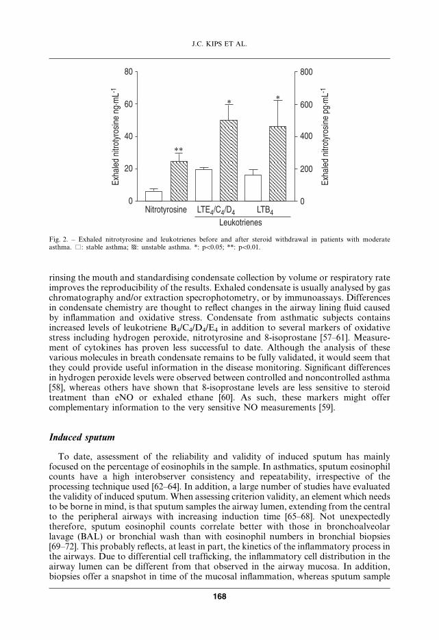
rinsing the mouth and standardising condensate collection by volume or respiratory rateimproves the reproducibility of the results. Exhaled condensate is usually analysed by gaschromatography and/or extraction specrophotometry, or by immunoassays. Differencesin condensate chemistry are thought to reflect changes in the airway lining fluid causedby inflammation and oxidative stress. Condensate from asthmatic subjects containsincreased levels of leukotriene B4/C4/D4/E4 in addition to several markers of oxidativestress including hydrogen peroxide, nitrotyrosine and 8-isoprostane [57–61]. Measure-ment of cytokines has proven less successful to date. Although the analysis of thesevarious molecules in breath condensate remains to be fully validated, it would seem thatthey could provide useful information in the disease monitoring. Significant differencesin hydrogen peroxide levels were observed between controlled and noncontrolled asthma[58], whereas others have shown that 8-isoprostane levels are less sensitive to steroidtreatment than eNO or exhaled ethane [60]. As such, these markers might offercomplementary information to the very sensitive NO measurements [59].
Induced sputum
To date, assessment of the reliability and validity of induced sputum has mainlyfocused on the percentage of eosinophils in the sample. In asthmatics, sputum eosinophilcounts have a high interobserver consistency and repeatability, irrespective of theprocessing technique used [62–64]. In addition, a large number of studies have evaluatedthe validity of induced sputum. When assessing criterion validity, an element which needsto be borne in mind, is that sputum samples the airway lumen, extending from the centralto the peripheral airways with increasing induction time [65–68]. Not unexpectedlytherefore, sputum eosinophil counts correlate better with those in bronchoalveolarlavage (BAL) or bronchial wash than with eosinophil numbers in bronchial biopsies[69–72]. This probably reflects, at least in part, the kinetics of the inflammatory process inthe airways. Due to differential cell trafficking, the inflammatory cell distribution in theairway lumen can be different from that observed in the airway mucosa. In addition,biopsies offer a snapshot in time of the mucosal inflammation, whereas sputum sample
�
��
��
��
��
!���������"������� #��$�
���
���
���
���
�
!���������"������� #��$�
%���"������&'�����
�� �()�(*� ��+�
��
� �
Fig. 2. – Exhaled nitrotyrosine and leukotrienes before and after steroid withdrawal in patients with moderateasthma. %: stable asthma; p: unstable asthma. *: pv0.05; **: pv0.01.
J.C. KIPS ET AL.
168
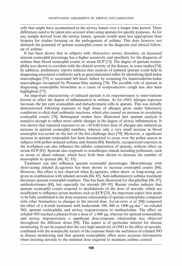
cells that might have accumulated in the airway lumen over a longer time period. Thesedifferences need to be taken into account when using sputum for specific purposes. As forany sample derived from the airway lumen, sputum would seem less appropriate thanbiopsies for studies focusing on the pathogenesis of asthma. This does however notdiminish the potential of sputum eosinophil counts in the diagnosis and clinical follow-up of asthma.
It has been shown that in subjects with obstructive airway disorders, an increasedsputum eosinophil percentage has a higher sensitivity and specificity for the diagnosis ofasthma than blood eosinophil counts or serum ECP [73]. The degree of sputum eosino-philia was shown to correlate with the clinical severity of the disease, in some studies [74].In addition, preliminary reports indicate that analysis of induced sputum could help indiagnosing associated conditions such as gastrointestinal reflux by identifying lipid-ladenmacrophages [75] or associated left heart failure by screening for haemosiderine-ladenmacrophages recognised by Prussian-blue staining [76]. The possible role of sputum indiagnosing eosinophilic bronchitis as a cause of nonproductive cough has also beenhighlighted [77].
An important characteristic of induced sputum is its responsiveness to interventionsknown to affect the degree of inflammation in asthma. As for eNO, allergen exposureincreases the per cent eosinophils and metachromatic cells in sputum. This was initiallydemonstrated following exposure to high doses of allergen given under laboratoryconditions to elicit dual asthmatic reactions, which also caused an increase in circulatingeosinophil counts [78]. Subsequent studies have illustrated that sputum analysis issensitive enough to reflect more subtle changes in the degree of airway inflammation. Itwas shown that repeated exposure to any10-fold lower dose of allergen also induced anincrease in sputum eosinophil numbers, whereas only a very small increase in bloodeosinophil was noted on the last of the five challenge days [79]. Moreover, a significantincrease in sputum eosinophils has been documented to occur over the pollen season insubjects with pollen-induced asthma and rhinitis [80]. Similarly, occupational exposure inthe workplace can also influence the cellular composition of sputum, without effect onserum ECP [81]. Sputum also responds to nonallergen stimuli including pollutants, suchas ozone or diesel exhaust, which have both been shown to increase the number ofneutrophils in sputum [46, 82, 83].
Treatment can also influence sputum eosinophil percentages. Monotherapy withshort-acting inhaled b2-agonists has been shown to increase eosinophil counts [84].However, this effect is not observed when b2-agonists, either short- or long-acting, aregiven in combination with inhaled steroids [84, 85]. Anti-inflammatory asthma treatmentdecreases sputum eosinophil numbers. This has been illustrated for theophylline [86, 87],antileukotrienes [88], but especially for steroids [89–93]. Recent studies indicate thatsputum eosinophil counts respond to modulations of the dose of steroids, which areinsufficient to influence serum markers such as ECP [23]. An important aspect that needsto be fully established is the dose-response relationship of sputum eosinophilia comparedwith other biomarkers to changes in the steroid dose. Jatakanon et al. [94] comparedthe effect of a 4-week treatment with budesonide 100, 400 or 1600 mg?day-1 on exhaledNO, sputum eosinophilia and airway responsiveness to methacholine. The effect onexhaled NO reached a plateau from a dose ofi400 mg, whereas for sputum eosinophiliaand airway responsiveness a significant dose-response relationship was observedthroughout the different doses [94]. This aspect is of particular interest for diseasemonitoring. It can be argued that the very high sensitivity of eNO to the effect of steroids,combined with the nonspecific nature of the response limits the usefulness of exhaled NOin disease monitoring and that sputum eosinophilia offers more accurate informationwhen titrating steroids to the minimal dose required to maintain asthma control.
NONINVASIVE ASSESSMENT OF AIRWAY INFLAMMATION
169
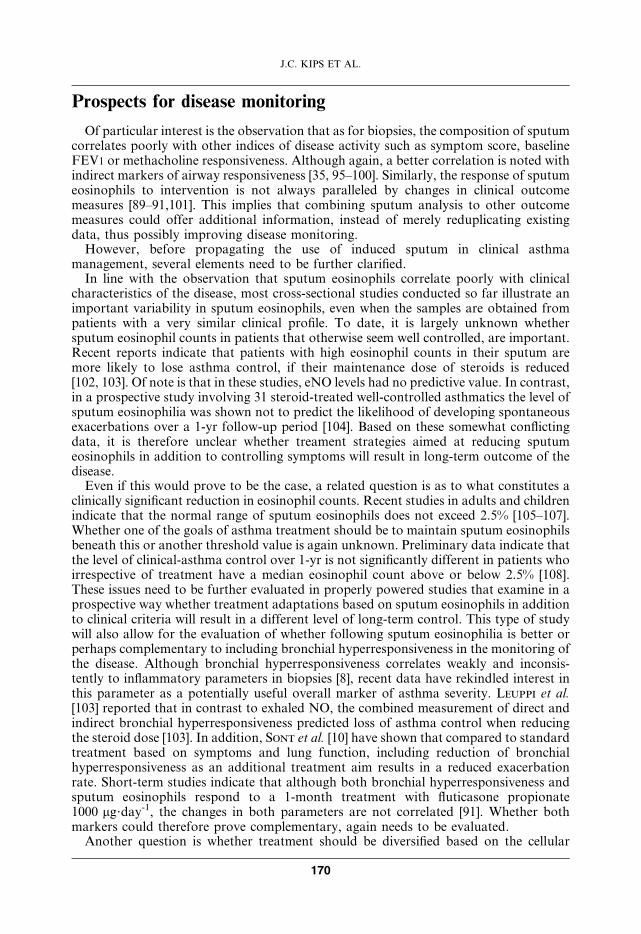
Prospects for disease monitoring
Of particular interest is the observation that as for biopsies, the composition of sputumcorrelates poorly with other indices of disease activity such as symptom score, baselineFEV1 or methacholine responsiveness. Although again, a better correlation is noted withindirect markers of airway responsiveness [35, 95–100]. Similarly, the response of sputumeosinophils to intervention is not always paralleled by changes in clinical outcomemeasures [89–91,101]. This implies that combining sputum analysis to other outcomemeasures could offer additional information, instead of merely reduplicating existingdata, thus possibly improving disease monitoring.
However, before propagating the use of induced sputum in clinical asthmamanagement, several elements need to be further clarified.
In line with the observation that sputum eosinophils correlate poorly with clinicalcharacteristics of the disease, most cross-sectional studies conducted so far illustrate animportant variability in sputum eosinophils, even when the samples are obtained frompatients with a very similar clinical profile. To date, it is largely unknown whethersputum eosinophil counts in patients that otherwise seem well controlled, are important.Recent reports indicate that patients with high eosinophil counts in their sputum aremore likely to lose asthma control, if their maintenance dose of steroids is reduced[102, 103]. Of note is that in these studies, eNO levels had no predictive value. In contrast,in a prospective study involving 31 steroid-treated well-controlled asthmatics the level ofsputum eosinophilia was shown not to predict the likelihood of developing spontaneousexacerbations over a 1-yr follow-up period [104]. Based on these somewhat conflictingdata, it is therefore unclear whether treament strategies aimed at reducing sputumeosinophils in addition to controlling symptoms will result in long-term outcome of thedisease.
Even if this would prove to be the case, a related question is as to what constitutes aclinically significant reduction in eosinophil counts. Recent studies in adults and childrenindicate that the normal range of sputum eosinophils does not exceed 2.5% [105–107].Whether one of the goals of asthma treatment should be to maintain sputum eosinophilsbeneath this or another threshold value is again unknown. Preliminary data indicate thatthe level of clinical-asthma control over 1-yr is not significantly different in patients whoirrespective of treatment have a median eosinophil count above or below 2.5% [108].These issues need to be further evaluated in properly powered studies that examine in aprospective way whether treatment adaptations based on sputum eosinophils in additionto clinical criteria will result in a different level of long-term control. This type of studywill also allow for the evaluation of whether following sputum eosinophilia is better orperhaps complementary to including bronchial hyperresponsiveness in the monitoring ofthe disease. Although bronchial hyperresponsiveness correlates weakly and inconsis-tently to inflammatory parameters in biopsies [8], recent data have rekindled interest inthis parameter as a potentially useful overall marker of asthma severity. Leuppi et al.[103] reported that in contrast to exhaled NO, the combined measurement of direct andindirect bronchial hyperresponsiveness predicted loss of asthma control when reducingthe steroid dose [103]. In addition, Sont et al. [10] have shown that compared to standardtreatment based on symptoms and lung function, including reduction of bronchialhyperresponsiveness as an additional treatment aim results in a reduced exacerbationrate. Short-term studies indicate that although both bronchial hyperresponsiveness andsputum eosinophils respond to a 1-month treatment with fluticasone propionate1000 mg?day-1, the changes in both parameters are not correlated [91]. Whether bothmarkers could therefore prove complementary, again needs to be evaluated.
Another question is whether treatment should be diversified based on the cellular
J.C. KIPS ET AL.
170
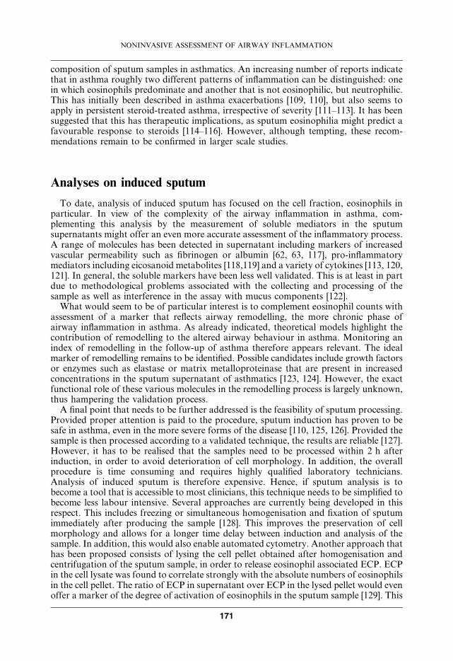
composition of sputum samples in asthmatics. An increasing number of reports indicatethat in asthma roughly two different patterns of inflammation can be distinguished: onein which eosinophils predominate and another that is not eosinophilic, but neutrophilic.This has initially been described in asthma exacerbations [109, 110], but also seems toapply in persistent steroid-treated asthma, irrespective of severity [111–113]. It has beensuggested that this has therapeutic implications, as sputum eosinophilia might predict afavourable response to steroids [114–116]. However, although tempting, these recom-mendations remain to be confirmed in larger scale studies.
Analyses on induced sputum
To date, analysis of induced sputum has focused on the cell fraction, eosinophils inparticular. In view of the complexity of the airway inflammation in asthma, com-plementing this analysis by the measurement of soluble mediators in the sputumsupernatants might offer an even more accurate assessment of the inflammatory process.A range of molecules has been detected in supernatant including markers of increasedvascular permeability such as fibrinogen or albumin [62, 63, 117], pro-inflammatorymediators including eicosanoid metabolites [118,119] and a variety of cytokines [113, 120,121]. In general, the soluble markers have been less well validated. This is at least in partdue to methodological problems associated with the collecting and processing of thesample as well as interference in the assay with mucus components [122].
What would seem to be of particular interest is to complement eosinophil counts withassessment of a marker that reflects airway remodelling, the more chronic phase ofairway inflammation in asthma. As already indicated, theoretical models highlight thecontribution of remodelling to the altered airway behaviour in asthma. Monitoring anindex of remodelling in the follow-up of asthma therefore appears relevant. The idealmarker of remodelling remains to be identified. Possible candidates include growth factorsor enzymes such as elastase or matrix metalloproteinase that are present in increasedconcentrations in the sputum supernatant of asthmatics [123, 124]. However, the exactfunctional role of these various molecules in the remodelling process is largely unknown,thus hampering the validation process.
A final point that needs to be further addressed is the feasibility of sputum processing.Provided proper attention is paid to the procedure, sputum induction has proven to besafe in asthma, even in the more severe forms of the disease [110, 125, 126]. Provided thesample is then processed according to a validated technique, the results are reliable [127].However, it has to be realised that the samples need to be processed within 2 h afterinduction, in order to avoid deterioration of cell morphology. In addition, the overallprocedure is time consuming and requires highly qualified laboratory technicians.Analysis of induced sputum is therefore expensive. Hence, if sputum analysis is tobecome a tool that is accessible to most clinicians, this technique needs to be simplified tobecome less labour intensive. Several approaches are currently being developed in thisrespect. This includes freezing or simultaneous homogenisation and fixation of sputumimmediately after producing the sample [128]. This improves the preservation of cellmorphology and allows for a longer time delay between induction and analysis of thesample. In addition, this would also enable automated cytometry. Another approach thathas been proposed consists of lysing the cell pellet obtained after homogenisation andcentrifugation of the sputum sample, in order to release eosinophil associated ECP. ECPin the cell lysate was found to correlate strongly with the absolute numbers of eosinophilsin the cell pellet. The ratio of ECP in supernatant over ECP in the lysed pellet would evenoffer a marker of the degree of activation of eosinophils in the sputum sample [129]. This
NONINVASIVE ASSESSMENT OF AIRWAY INFLAMMATION
171

technique has the advantage that the measurements can be automated, saving costs.Albeit theoretically appealing, these various approaches need further validation. Anotherpotential disadvantage of induced sputum is that the induction process with nebulisedhypertonic saline induces an inflammatory response, so that it is not advisable to makerepeated measurements in less than 24 h [128]. While this may not be a limitation in long-term disease monitoring, it limits research studies of kinetic factors.
Conclusion
Analysis of induced sputum offers a relatively noninvasive direct marker of airwayinflammation in asthma. Including sputum analysis in the diagnosis but especially in thefollow-up of the disease, could offer additional information to clinical-outcomemeasures. Measurement of exhaled biomarkers is also very promising and is feasiblein children and patients with severe disease. Whether incorporating these newmeasurements into disease monitoring will result in improved long-term clinical controlof asthma, or allow for the diversification of treatments now needs to be addressed incarefully designed studies.
Summary
Bronchial asthma is currently considered and defined as an inflammatory disorder ofthe airways. It therefore seems logical to include a direct marker of airwayinflammation in the diagnosis and follow-up of the disease. Several relativelynoninvasive biomarkers have been and are being considered in this respect. Examplesthat have been most extensively investigated, as to their reliability and validity for theassessment of airway inflammation in asthma, include blood eosinophil counts orserum eosinophil cationic protein, urinary eicosanoid metabolites, exhaled gasses,mediators in breath condensate or induced sputum. From the studies performed todate, it would appear that biomarkers correlate poorly with other indices of diseaseactivity, such as symptoms, baseline forced expiratory volume in one second ormethacholine responsiveneness. As such, including biomarkers in the diagnosis, butespecially follow-up of the disease, could offer additional information to clinical-outcome measures. Whether this attitude in disease monitoring will result in improvedlong-term clinical control of asthma or allow for the diversification of treatments, nowneeds to be addressed. In addition, the routine use of these techniques in daily practicerequires simplification of the methodology involved.
Keywords: Asthma, biomarker, breath condensate, exhaled nitric oxide, inducedsputum.
References
1. Global initiative for asthma. Global strategy for asthma management and prevention. Global
initiative for asthma. National Heart, Lung, and Blood Institute. 1995 NIH Publication
No. 95–3659.
J.C. KIPS ET AL.
172
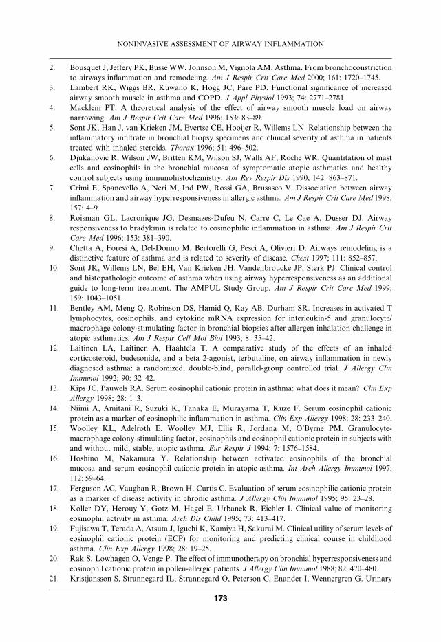
2. Bousquet J, Jeffery PK, Busse WW, JohnsonM, Vignola AM. Asthma. From bronchoconstriction
to airways inflammation and remodeling. Am J Respir Crit Care Med 2000; 161: 1720–1745.
3. Lambert RK, Wiggs BR, Kuwano K, Hogg JC, Pare PD. Functional significance of increased
airway smooth muscle in asthma and COPD. J Appl Physiol 1993; 74: 2771–2781.
4. Macklem PT. A theoretical analysis of the effect of airway smooth muscle load on airway
narrowing. Am J Respir Crit Care Med 1996; 153: 83–89.
5. Sont JK, Han J, van Krieken JM, Evertse CE, Hooijer R, Willems LN. Relationship between the
inflammatory infiltrate in bronchial biopsy specimens and clinical severity of asthma in patients
treated with inhaled steroids. Thorax 1996; 51: 496–502.
6. Djukanovic R, Wilson JW, Britten KM, Wilson SJ, Walls AF, Roche WR. Quantitation of mast
cells and eosinophils in the bronchial mucosa of symptomatic atopic asthmatics and healthy
control subjects using immunohistochemistry. Am Rev Respir Dis 1990; 142: 863–871.
7. Crimi E, Spanevello A, Neri M, Ind PW, Rossi GA, Brusasco V. Dissociation between airway
inflammation and airway hyperresponsiveness in allergic asthma. Am J Respir Crit Care Med 1998;
157: 4–9.
8. Roisman GL, Lacronique JG, Desmazes-Dufeu N, Carre C, Le Cae A, Dusser DJ. Airway
responsiveness to bradykinin is related to eosinophilic inflammation in asthma. Am J Respir Crit
Care Med 1996; 153: 381–390.
9. Chetta A, Foresi A, Del-Donno M, Bertorelli G, Pesci A, Olivieri D. Airways remodeling is a
distinctive feature of asthma and is related to severity of disease. Chest 1997; 111: 852–857.
10. Sont JK, Willems LN, Bel EH, Van Krieken JH, Vandenbroucke JP, Sterk PJ. Clinical control
and histopathologic outcome of asthma when using airway hyperresponsiveness as an additional
guide to long-term treatment. The AMPUL Study Group. Am J Respir Crit Care Med 1999;
159: 1043–1051.
11. Bentley AM, Meng Q, Robinson DS, Hamid Q, Kay AB, Durham SR. Increases in activated T
lymphocytes, eosinophils, and cytokine mRNA expression for interleukin-5 and granulocyte/
macrophage colony-stimulating factor in bronchial biopsies after allergen inhalation challenge in
atopic asthmatics. Am J Respir Cell Mol Biol 1993; 8: 35–42.
12. Laitinen LA, Laitinen A, Haahtela T. A comparative study of the effects of an inhaled
corticosteroid, budesonide, and a beta 2-agonist, terbutaline, on airway inflammation in newly
diagnosed asthma: a randomized, double-blind, parallel-group controlled trial. J Allergy Clin
Immunol 1992; 90: 32–42.
13. Kips JC, Pauwels RA. Serum eosinophil cationic protein in asthma: what does it mean? Clin Exp
Allergy 1998; 28: 1–3.
14. Niimi A, Amitani R, Suzuki K, Tanaka E, Murayama T, Kuze F. Serum eosinophil cationic
protein as a marker of eosinophilic inflammation in asthma. Clin Exp Allergy 1998; 28: 233–240.
15. Woolley KL, Adelroth E, Woolley MJ, Ellis R, Jordana M, O’Byrne PM. Granulocyte-
macrophage colony-stimulating factor, eosinophils and eosinophil cationic protein in subjects with
and without mild, stable, atopic asthma. Eur Respir J 1994; 7: 1576–1584.
16. Hoshino M, Nakamura Y. Relationship between activated eosinophils of the bronchial
mucosa and serum eosinophil cationic protein in atopic asthma. Int Arch Allergy Immunol 1997;
112: 59–64.
17. Ferguson AC, Vaughan R, Brown H, Curtis C. Evaluation of serum eosinophilic cationic protein
as a marker of disease activity in chronic asthma. J Allergy Clin Immunol 1995; 95: 23–28.
18. Koller DY, Herouy Y, Gotz M, Hagel E, Urbanek R, Eichler I. Clinical value of monitoring
eosinophil activity in asthma. Arch Dis Child 1995; 73: 413–417.
19. Fujisawa T, Terada A, Atsuta J, Iguchi K, Kamiya H, Sakurai M. Clinical utility of serum levels of
eosinophil cationic protein (ECP) for monitoring and predicting clinical course in childhood
asthma. Clin Exp Allergy 1998; 28: 19–25.
20. Rak S, Lowhagen O, Venge P. The effect of immunotherapy on bronchial hyperresponsiveness and
eosinophil cationic protein in pollen-allergic patients. J Allergy Clin Immunol 1988; 82: 470–480.
21. Kristjansson S, Strannegard IL, Strannegard O, Peterson C, Enander I, Wennergren G. Urinary
NONINVASIVE ASSESSMENT OF AIRWAY INFLAMMATION
173
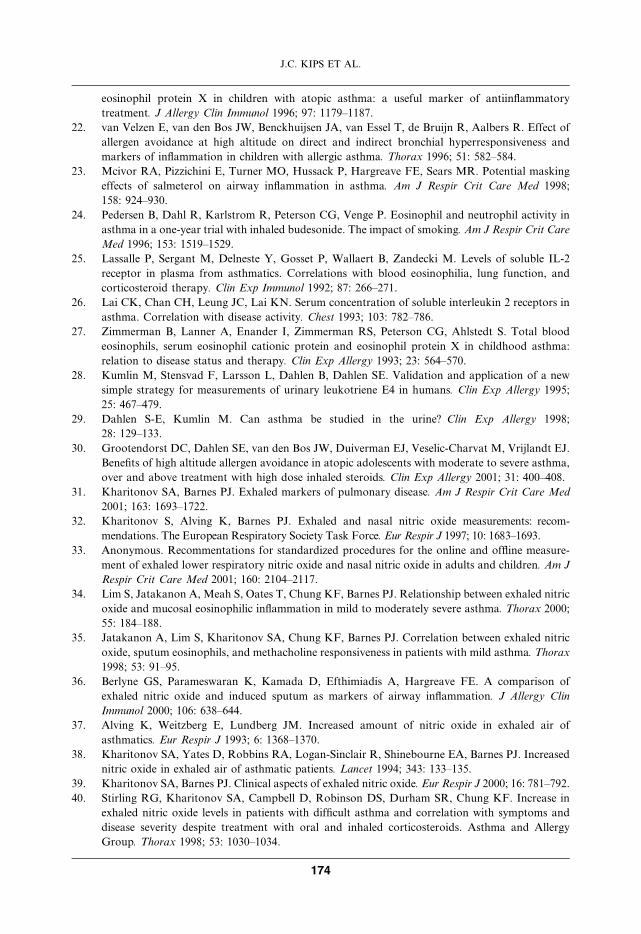
eosinophil protein X in children with atopic asthma: a useful marker of antiinflammatory
treatment. J Allergy Clin Immunol 1996; 97: 1179–1187.
22. van Velzen E, van den Bos JW, Benckhuijsen JA, van Essel T, de Bruijn R, Aalbers R. Effect of
allergen avoidance at high altitude on direct and indirect bronchial hyperresponsiveness and
markers of inflammation in children with allergic asthma. Thorax 1996; 51: 582–584.
23. Mcivor RA, Pizzichini E, Turner MO, Hussack P, Hargreave FE, Sears MR. Potential masking
effects of salmeterol on airway inflammation in asthma. Am J Respir Crit Care Med 1998;
158: 924–930.
24. Pedersen B, Dahl R, Karlstrom R, Peterson CG, Venge P. Eosinophil and neutrophil activity in
asthma in a one-year trial with inhaled budesonide. The impact of smoking. Am J Respir Crit Care
Med 1996; 153: 1519–1529.
25. Lassalle P, Sergant M, Delneste Y, Gosset P, Wallaert B, Zandecki M. Levels of soluble IL-2
receptor in plasma from asthmatics. Correlations with blood eosinophilia, lung function, and
corticosteroid therapy. Clin Exp Immunol 1992; 87: 266–271.
26. Lai CK, Chan CH, Leung JC, Lai KN. Serum concentration of soluble interleukin 2 receptors in
asthma. Correlation with disease activity. Chest 1993; 103: 782–786.
27. Zimmerman B, Lanner A, Enander I, Zimmerman RS, Peterson CG, Ahlstedt S. Total blood
eosinophils, serum eosinophil cationic protein and eosinophil protein X in childhood asthma:
relation to disease status and therapy. Clin Exp Allergy 1993; 23: 564–570.
28. Kumlin M, Stensvad F, Larsson L, Dahlen B, Dahlen SE. Validation and application of a new
simple strategy for measurements of urinary leukotriene E4 in humans. Clin Exp Allergy 1995;
25: 467–479.
29. Dahlen S-E, Kumlin M. Can asthma be studied in the urine? Clin Exp Allergy 1998;
28: 129–133.
30. Grootendorst DC, Dahlen SE, van den Bos JW, Duiverman EJ, Veselic-Charvat M, Vrijlandt EJ.
Benefits of high altitude allergen avoidance in atopic adolescents with moderate to severe asthma,
over and above treatment with high dose inhaled steroids. Clin Exp Allergy 2001; 31: 400–408.
31. Kharitonov SA, Barnes PJ. Exhaled markers of pulmonary disease. Am J Respir Crit Care Med
2001; 163: 1693–1722.
32. Kharitonov S, Alving K, Barnes PJ. Exhaled and nasal nitric oxide measurements: recom-
mendations. The European Respiratory Society Task Force. Eur Respir J 1997; 10: 1683–1693.
33. Anonymous. Recommentations for standardized procedures for the online and offline measure-
ment of exhaled lower respiratory nitric oxide and nasal nitric oxide in adults and children. Am J
Respir Crit Care Med 2001; 160: 2104–2117.
34. Lim S, Jatakanon A, Meah S, Oates T, Chung KF, Barnes PJ. Relationship between exhaled nitric
oxide and mucosal eosinophilic inflammation in mild to moderately severe asthma. Thorax 2000;
55: 184–188.
35. Jatakanon A, Lim S, Kharitonov SA, Chung KF, Barnes PJ. Correlation between exhaled nitric
oxide, sputum eosinophils, and methacholine responsiveness in patients with mild asthma. Thorax
1998; 53: 91–95.
36. Berlyne GS, Parameswaran K, Kamada D, Efthimiadis A, Hargreave FE. A comparison of
exhaled nitric oxide and induced sputum as markers of airway inflammation. J Allergy Clin
Immunol 2000; 106: 638–644.
37. Alving K, Weitzberg E, Lundberg JM. Increased amount of nitric oxide in exhaled air of
asthmatics. Eur Respir J 1993; 6: 1368–1370.
38. Kharitonov SA, Yates D, Robbins RA, Logan-Sinclair R, Shinebourne EA, Barnes PJ. Increased
nitric oxide in exhaled air of asthmatic patients. Lancet 1994; 343: 133–135.
39. Kharitonov SA, Barnes PJ. Clinical aspects of exhaled nitric oxide. Eur Respir J 2000; 16: 781–792.
40. Stirling RG, Kharitonov SA, Campbell D, Robinson DS, Durham SR, Chung KF. Increase in
exhaled nitric oxide levels in patients with difficult asthma and correlation with symptoms and
disease severity despite treatment with oral and inhaled corticosteroids. Asthma and Allergy
Group. Thorax 1998; 53: 1030–1034.
J.C. KIPS ET AL.
174
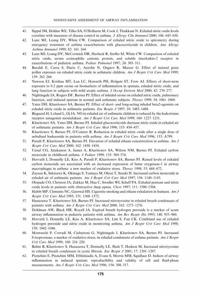
41. Sippel JM, Holden WE, Tilles SA, O’Hollaren M, Cook J, Thukkani N. Exhaled nitric oxide levels
correlate with measures of disease control in asthma. J Allergy Clin Immunol 2000; 106: 645–650.
42. Lanz MJ, Leung DY, White CW. Comparison of exhaled nitric oxide to spirometry during
emergency treatment of asthma exacerbations with glucocorticoids in children. Ann Allergy
Asthma Immunol 1999; 82: 161–164.
43. Lanz MJ, Leung DY, McCormick DR, Harbeck R, Szefler SJ, White CW. Comparison of exhaled
nitric oxide, serum eosinophilic cationic protein, and soluble interleukin-2 receptor in
exacerbations of pediatric asthma. Pediatr Pulmonol 1997; 24: 305–311.
44. Baraldi E, Carra S, Dario C, Azzolin N, Ongaro R, Marcer G. Effect of natural grass
pollen exposure on exhaled nitric oxide in asthmatic children. Am J Respir Crit Care Med 1999;
159: 262–266.
45. Newson EJ, Krishna MT, Lau LC, Howarth PH, Holgate ST, Frew AJ. Effects of short-term
exposure to 0.2 ppm ozone on biomarkers of inflammation in sputum, exhaled nitric oxide, and
lung function in subjects with mild atopic asthma. J Occup Environ Med 2000; 42: 270–277.
46. Nightingale JA, Rogers DF, Barnes PJ. Effect of inhaled ozone on exhaled nitric oxide, pulmonary
function, and induced sputum in normal and asthmatic subjects. Thorax 1999; 54: 1061–1069.
47. Yates DH, Kharitonov SA, Barnes PJ. Effect of short- and long-acting inhaled beta2-agonists on
exhaled nitric oxide in asthmatic patients. Eur Respir J 1997; 10: 1483–1488.
48. Bisgaard H, Loland L, Oj JA. NO in exhaled air of asthmatic children is reduced by the leukotriene
receptor antagonist montelukast. Am J Respir Crit Care Med 1999; 160: 1227–1231.
49. Kharitonov SA, Yates DH, Barnes PJ. Inhaled glucocorticoids decrease nitric oxide in exhaled air
of asthmatic patients. Am J Respir Crit Care Med 1996; 153: 454–457.
50. Kharitonov S, Barnes PJ, O’Connor B. Reduction in exhaled nitric oxide after a single dose of
nebulised budesonide in patients with asthma. Am J Respir Crit Care Med 1996; 153: A799.
51. Paredi P, Kharitonov SA, Barnes PJ. Elevation of exhaled ethane concentration in asthma. Am J
Respir Crit Care Med 2000; 162: 1450–1454.
52. Uasuf CG, Jatakanon A, James A, Kharitonov SA, Wilson NM, Barnes PJ. Exhaled carbon
monoxide in childhood asthma. J Pediatr 1999; 135: 569–574.
53. Horvath I, Donnelly LE, Kiss A, Paredi P, Kharitonov SA, Barnes PJ. Raised levels of exhaled
carbon monoxide are associated with an increased expression of heme oxygenase-1 in airway
macrophages in asthma: a new marker of oxidative stress. Thorax 1998; 53: 668–672.
54. Zayasu K, Sekizawa K, Okinaga S, Yamaya M, Ohrui T, Sasaki H. Increased carbon monoxide in
exhaled air of asthmatic patients. Am J Respir Crit Care Med 1997; 156: 1140–1143.
55. Olopade CO, Christon JA, Zakkar M, Hua C, Swedler WI, Scheff PA. Exhaled pentane and nitric
oxide levels in patients with obstructive sleep apnea. Chest 1997; 111: 1500–1504.
56. Habib MP, Clements NC, Garewal HS. Cigarette smoking and ethane exhalation in humans. Am J
Respir Crit Care Med 1995; 151: 1368–1372.
57. Hanazawa T, Kharitonov SA, Barnes PJ. Increased nitrotyrosine in exhaled breath condensate of
patients with asthma. Am J Respir Crit Care Med 2000; 162: 1273–1276.
58. Dohlman AW, Black HR, Royall JA. Expired breath hydrogen peroxide is a marker of acute
airway inflammation in pediatric patients with asthma. Am Rev Respir Dis 1993; 148: 955–960.
59. Horvath I, Donnelly LE, Kiss A, Kharitonov SA, Lim S, Fan CK. Combined use of exhaled
hydrogen peroxide and nitric oxide in monitoring asthma. Am J Respir Crit Care Med 1998;
158: 1042–1046.
60. Montuschi P, Corradi M, Ciabattoni G, Nightingale J, Kharitonov SA, Barnes PJ. Increased
8-isoprostane, a marker of oxidative stress, in exhaled condensate of asthma patients. Am J Respir
Crit Care Med 1999; 160: 216–220.
61. Balint B, Kharitonov S, Hanazawa T, Donnelly LE, Shah T, Hodson M. Increased nitrotyrosine
in exhaled breath condensate in cystic fibrosis. Eur Respir J 2001; 17: 1201–1207.
62. Pizzichini E, Pizzichini MM, Efthimiadis A, Evans S, Morris MM, Squillace D. Indices of airway
inflammation in induced sputum: reproducibility and validity of cell and fluid-phase
measurements. Am J Respir Crit Care Med 1996; 154: 308–317.
NONINVASIVE ASSESSMENT OF AIRWAY INFLAMMATION
175
63. in’t Veen JC, de Gouw HW, Smits HH, Sont JK, Hiemstra PS, Sterk PJ. Repeatability of cellular
and soluble markers of inflammation in induced sputum from patients with asthma. Eur Respir J
1996; 9: 2441–2447.
64. Fahy JV, Boushey HA, Lazarus SC, Mauger EA, Cherniack RM, Chinchilli VM. Safety and
reproducibility of sputum induction in asthmatic subjects in a multicenter study. Am J Respir Crit
Care Med 2001; 163: 1470–1475.
65. Moodley YP, Krishnan V, Lalloo UG. Neutrophils in induced sputum arise from central airways.
Eur Respir J 2000; 15: 36–40.
66. Gershman NH, Liu H, Wong HH, Liu JT, Fahy JV. Fractional analysis of sequential induced
sputum samples during sputum induction: evidence that different lung compartments are sampled
at different time points. J Allergy Clin Immunol 1999; 104: 322–328.
67. Holz O, Richter K, Jorres RA, Speckin P, Mucke M, Magnussen H. Changes in sputum
composition between two inductions performed on consecutive days. Thorax 1998; 53: 83–86.
68. Richter K, Holz O, Jorres RA, Mucke M, Magnussen H. Sequentially induced sputum in patients
with asthma or chronic obstructive pulmonary disease. Eur Respir J 1999; 14: 697–701.
69. Keatings VM, Evans DJ, O’connor BJ, Barnes PJ. Cellular profiles in asthmatic airways: a
comparison of induced sputum, bronchial washings, and bronchoalveolar lavage fluid. Thorax
1997; 52: 372–374.
70. Maestrelli P, Saetta M, Di Stefano A, Calcagni PG, Turato G, Ruggieri MP. Comparison of
leukocyte counts in sputum, bronchial biopsies, and bronchoalveolar lavage. Am J Respir Crit
Care Med 1995; 152: 1926–1931.
71. Grootendorst DC, Sont JK, Willems LN, Kluin-Nelemans JC, Van Krieken JH, Veselic-Charvat M.
Comparison of inflammatory cell counts in asthma: induced sputum vs bronchoalveolar lavage
and bronchial biopsies. Clin Exp Allergy 1997; 27: 769–779.
72. Fahy JV, Wong H, Liu J, Boushey HA. Comparison of samples collected by sputum induction and
bronchoscopy from asthmatic and healthy subjects. Am J Respir Crit Care Med 1995; 152: 53–58.
73. Pizzichini E, Pizzichini MM, Efthimiadis A, Dolovich J, Hargreave FE. Measuring airway
inflammation in asthma: eosinophils and eosinophilic cationic protein in induced sputum
compared with peripheral blood. J Allergy Clin Immunol 1997; 99: 539–544.
74. Louis R, Lau LC, Bron AO, Roldaan AC, Radermecker M, Djukanovic R. The relationship
between airways inflammation and asthma severity. Am J Respir Crit Care Med 2000; 161: 9–16.
75. Parameswaran K, Anvari M, Efthimiadis A, Kamada D, Hargreave FE, Allen CJ. Lipid-laden
macrophages in induced sputum are a marker of oropharyngeal reflux and possible gastric
aspiration. Eur Respir J 2000; 16: 1119–1122.
76. Leigh R, Sharon RF, Efthimiadis A, Hargreave FE, Kitching AD. Diagnosis of left-ventricular
dysfunction from induced sputum examination (letter). Lancet 1999; 354: 833–834.
77. Brightling CE, Pavord ID. Eosinophilic bronchitis - what is it and why is it important? (editorial;
comment). Clin Exp Allergy 2000; 30: 4–6.
78. Gauvreau GM, Doctor J, Watson RM, JordanaM, O’Byrne PM. Effects of inhaled budesonide on
allergen-induced airway responses and airway inflammation. Am J Respir Crit Care Med 1996;
154: 1267–1271.
79. Sulakvelidze I, Inman MD, Rerecich T, O’Byrne PM. Increases in airway eosinophils and
interleukin-5 with minimal bronchoconstriction during repeated low-dose allergen challenge in
atopic asthmatics. Eur Respir J 1998; 11: 821–827.
80. Kips J, Baker AJ. The effect of intranasal and/or inhaled fluticasone propionate (FP) on sputum
markers in seasonal rhinitis and asthma. Results of the SPIRA study (FNP 40001). Am J Respir
Crit Care Med 2001; 163: A604.
81. Lemiere C, Pizzichini MM, Balkissoon R, Clelland L, Efthimiadis A, O’Shaughnessy D.
Diagnosing occupational asthma: use of induced sputum. Eur Respir J 1999; 13: 482–488.
82. Holz O, Jorres RA, Timm P, Mucke M, Richter K, Koschyk S. Ozone-induced airway
inflammatory changes differ between individuals and are reproducible. Am J Respir Crit Care Med
1999; 159: 776–784.
J.C. KIPS ET AL.
176
83. Nordenhall C, Pourazar J, Blomberg A, Levin JO, Sandstrom T, Adelroth E. Airway
inflammation following exposure to diesel exhaust: a study of time kinetics using induced
sputum. Eur Respir J 2000; 15: 1046–1051.
84. Aldridge RE, Hancox RJ, Robin TD, Cowan JO, WinnMC, Frampton CM. Effects of terbutaline
and budesonide on sputum cells and bronchial hyperresponsiveness in asthma. Am J Respir Crit
Care Med 2000; 161: 1459–1464.
85. Kips JC, O’Connor BJ, Inman MD, Svensson K, Pauwels RA, O’Byrne PM. A long-term study of
the antiinflammatory effect of low-dose budesonide plus formoterol versus high-dose budesonide
in asthma. Am J Respir Crit Care Med 2000; 161: 996–1001.
86. Aizawa H, Iwanaga T, Inoue H, Takata S, Matsumoto K, Takahashi N. Once-daily theophylline
reduces serum eosinophil cationic protein and eosinophil levels in induced sputum of asthmatics.
Int Arch Allergy Immunol 2000; 121: 123–128.
87. Tohda Y, Muraki M, Iwanaga T, Kubo H, FukuokaM, Nakajima S. The effect of theophylline on
blood and sputum eosinophils and ECP in patients with bronchial asthma. Int J Immunopharmacol
1998; 20: 173–181.
88. Pizzichini E, Leff JA, Reiss TF, Hendeles L, Boulet LP, Wei LX. Montelukast reduces airway
eosinophilic inflammation in asthma: a randomized, controlled trial. Eur Respir J 1999; 14: 12–18.
89. Gershman NH, Wong HH, Liu JT, Fahy JV. Low- and high-dose fluticasone propionate in
asthma; effects during and after treatment. Eur Respir J 2000; 15: 11–18.
90. Fahy JV, Boushey HA. Effect of low-dose beclomethasone dipropionate on asthma control and
airway inflammation. Eur Respir J 1998; 11: 1240–1247.
91. van Rensen EL, Straathof KC, Veselic-Charvat MA, Zwinderman AH, Bel EH, Sterk PJ. Effect of
inhaled steroids on airway hyperresponsiveness, sputum eosinophils, and exhaled nitric oxide
levels in patients with asthma. Thorax 1999; 54: 403–408.
92. Jatakanon A, Lim S, Chung KF, Barnes PJ. An inhaled steroid improves markers of airway
inflammation in patients with mild asthma. Eur Respir J 1998; 12: 1084–1088.
93. in’t Veen JC, Smits HH, Hiemstra PS, Zwinderman AE, Sterk PJ, Bel EH. Lung function and
sputum characteristics of patients with severe asthma during an induced exacerbation by double-
blind steroid withdrawal. Am J Respir Crit Care Med 1999; 160: 93–99.
94. Jatakanon A, Kharitonov S, Lim S, Barnes PJ. Effect of differing doses of inhaled budesonide on
markers of airway inflammation in patients with mild asthma. Thorax 1999; 54: 108–114.
95. Wilson NM, Bridge P, Spanevello A, Silverman M. Induced sputum in children: feasibility,
repeatability, and relation of findings to asthma severity. Thorax 2000; 55: 768–774.
96. Polosa R, Renaud L, Cacciola R, Prosperini G, Crimi N, Djukanovic R. Sputum eosinophilia is
more closely associated with airway responsiveness to bradykinin than methacholine in asthma.
Eur Respir J 1998; 12: 551–556.
97. Yoshikawa T, Shoji S, Fujii T, Kanazawa H, Kudoh S, Hirata K. Severity of exercise-induced
bronchoconstriction is related to airway eosinophilic inflammation in patients with asthma. Eur
Respir J 1998; 12: 879–884.
98. Rosi E, Scano G. Association of sputum parameters with clinical and functional measurements in
asthma. Thorax 2000; 55: 235–238.
99. Gibson PG, Saltos N, Borgas T. Airway mast cells and eosinophils correlate with clinical severity
and airway hyperresponsiveness in corticosteroid-treated asthma. J Allergy Clin Immunol 2000;
105: 752–759.
100. Piacentini GL, Bodini A, Costella S, Vicentini L, Mazzi P, Sperandio S. Exhaled nitric oxide
and sputum eosinophil markers of inflammation in asthmatic children. Eur Respir J 1999;
13: 1386–1390.
101. Lim S, Jatakanon A, John M, Gilbey T, O’connor BJ, Chung KF. Effect of inhaled budesonide on
lung function and airway inflammation. Assessment by various inflammatory markers in mild
asthma. Am J Respir Crit Care Med 1999; 159: 22–30.
102. Jatakanon A, Lim S, Barnes PJ. Changes in sputum eosinophils predict loss of asthma control. Am
J Respir Crit Care Med 2000; 161: 64–72.
NONINVASIVE ASSESSMENT OF AIRWAY INFLAMMATION
177
103. Leuppi JD, Salome CM, Jenkins CR, Anderson SD, Xuan W, Marks GB. Predictive markers of
asthma exacerbation during stepwise dose reduction of inhaled corticosteroids. Am J Respir Crit
Care Med 2001; 163: 406–412.
104. Belda J, Giner J, Casan P, Sanchis J. Mild exacerbations and eosinophilic inflammation in patients
with stable, well-controlled asthma after 1 year of follow-up. Chest 2001; 119: 1011–1017.
105. Belda J, Leigh R, Parameswaran K, O’Byrne PM, Sears MR, Hargreave FE. Induced sputum cell
counts in healthy adults. Am J Respir Crit Care Med 2000; 161: 475–478.
106. Spanevello A, Confalonieri M, Sulotto F, Romano F, Balzano G, Migliori GB. Induced sputum
cellularity. Reference values and distribution in normal volunteers. Am J Respir Crit Care Med
2000; 162: 1172–1174.
107. Gibson PG, Henry RL, Thomas P. Noninvasive assessment of airway inflammation in children:
induced sputum, exhaled nitric oxide, and breath condensate. Eur Respir J 2000; 16: 1008–1015.
108. Kips J, O’Connor B, InmanM, Svenson K, Pauwels R, O’Byrne P. Is sputum eosinophilia a useful
outcome measure in the follow-up of asthma? Am J Respir Crit Care Med 2000; 161: A314.
109. Fahy JV, Kim KW, Liu J, Boushey HA. Prominent neutrophilic inflammation in sputum from
subjects with asthma exacerbation. J Allergy Clin Immunol 1995; 95: 843–852.
110. Pizzichini MM, Pizzichini E, Clelland L, Efthimiadis A, Mahony J, Dolovich J. Sputum in severe
exacerbations of asthma: kinetics of inflammatory indices after prednisone treatment. Am J Respir
Crit Care Med 1997; 155: 1501–1508.
111. Jatakanon A, Uasuf C, Maziak W, Lim S, Chung KF, Barnes PJ. Neutrophilic inflammation in
severe persistent asthma. Am J Respir Crit Care Med 1999; 160: 1532–1539.
112. Kips J, Cobot ESG. Cellular composition of induced sputum and peripheral blood in severe
persistent asthma. Eur Respir J 1999; 14: P2248.
113. Gibson PG, Simpson JL, Saltos N. Heterogeneity of airway inflammation in persistent asthma:
evidence of neutrophilic inflammationand increased sputum interleukin-8. Chest2001; 119: 1329–1336.
114. Pavord ID, Brightling CE, Woltmann G, Wardlaw AJ. Non-eosinophilic corticosteroid
unresponsive asthma (letter). Lancet 1999; 353: 2213–2214.
115. Pizzichini E, Pizzichini MM, Gibson P, Parameswaran K, Gleich GJ, Berman L. Sputum
eosinophilia predicts benefit from prednisone in smokers with chronic obstructive bronchitis. Am J
Respir Crit Care Med 1998; 158: 1511–1517.
116. Little SA, Chalmers GW, MacLeod KJ, McSharry C, Thomson NC. Non-invasive markers
of airway inflammation as predictors of oral steroid responsiveness in asthma. Thorax 2000;
55: 232–234.
117. Fahy JV, Liu J, Wong H, Boushey HA. Cellular and biochemical analysis of induced sputum from
asthmatic and from healthy subjects. Am Rev Respir Dis 1993; 147: 1126–1131.
118. Macfarlane AJ, Dworski R, Sheller JR, Pavord ID, Barry KA, Barnes NC. Sputum cysteinyl
leukotrienes increase 24 hours after allergen inhalation in atopic asthmatics. Am J Respir Crit Care
Med 2000; 161: 1553–1558.
119. Profita M, Sala A, Riccobono L, Paterno A, Mirabella A, Bonanno A. 15-Lipoxygenase
expression and 15(S)-hydroxyeicoisatetraenoic acid release and reincorporation in induced sputum
of asthmatic subjects. J Allergy Clin Immunol 2000; 105: 711–716.
120. Keatings VM, Collins PD, Scott DM, Barnes PJ. Differences in interleukin-8 and tumor necrosis
factor-alpha in induced sputum from patients with chronic obstructive pulmonary disease or
asthma. Am J Respir Crit Care Med 1996; 153: 530–534.
121. Norzila MZ, Fakes K, Henry RL, Simpson J, Gibson PG. Interleukin-8 secretion and neutrophil
recruitment accompanies induced sputum eosinophil activation in children with acute asthma. Am
J Respir Crit Care Med 2000; 161: 769–774.
122. Kelly MM, Leigh R, McKenzie R, Kamada D, Ramsdale EH, Hargreave FE. Induced sputum
examination: diagnosis of pulmonary involvement in Fabry’s disease. Thorax 2000; 55: 720–721.
123. Vignola AM, Riccobono L,Mirabella A, ProfitaM, Chanez P, Bellia V. Sputummetalloproteinase-9/
tissue inhibitor of metalloproteinase-1 ratio correlates with airflow obstruction in asthma and
chronic bronchitis. Am J Respir Crit Care Med 1998; 158: 1945–1950.
J.C. KIPS ET AL.
178
124. Vignola AM, Bonanno A, Mirabella A, Riccobono L, Mirabella F, Profita M. Increased levels of
elastase and alpha1-antitrypsin in sputum of asthmatic patients. Am J Respir Crit Care Med 1998;
157: 505–511.
125. de la Fuente PT, Romagnoli M, Godard P, Bousquet J, Chanez P. Safety of inducing sputum in
patients with asthma of varying severity. Am J Respir Crit Care Med 1998; 157: 1127–1130.
126. Vlachos-Mayer H, Leigh R, Sharon RF, Hussack P, Hargreave FE. Success and safety of sputum
induction in the clinical setting. Eur Respir J 2000; 16: 997–1000.
127. Kips JC, Peleman RA, Pauwels RA. Methods of examining induced sputum: do differences
matter? Eur Respir J 1998; 11: 529–533.
128. Holz O, Kips J, Magnussen H. Update on sputum methodology. Eur Respir J 2000; 16: 355–359.
129. Gibson PG,Woolley KL, Carty K, Murree-Allen K, Saltos N. Induced sputum eosinophil cationic
protein (ECP) measurement in asthma and chronic obstructive airway disease (COAD). Clin Exp
Allergy 1998; 28: 1081–1088.
NONINVASIVE ASSESSMENT OF AIRWAY INFLAMMATION
179
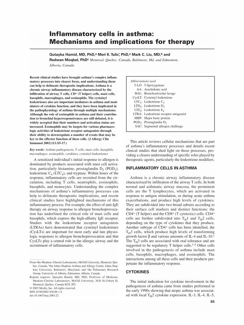
Recent clinical studies have brought asthma’s complex inflam-matory processes into clearer focus, and understanding themcan help to delineate therapeutic implications. Asthma is achronic airway inflammatory disease characterized by theinfiltration of airway T cells, CD+ (T helper) cells, mast cells,basophils, macrophages, and eosinophils. The cysteinylleukotrienes also are important mediators in asthma and mod-ulators of cytokine function, and they have been implicated inthe pathophysiology of asthma through multiple mechanisms.Although the role of eosinophils in asthma and their contribu-tion to bronchial hyperresponsiveness are still debated, it iswidely accepted that their numbers and activation status areincreased. Eosinophils may be targets for various pharmaco-logic activities of leukotriene receptor antagonists throughtheir ability to downregulate a number of events that may bekey to the effector function of these cells. (J Allergy ClinImmunol 2003;111:S5-17.)
Key words: Asthma pathogenesis, T cells, mast cells, basophils,macrophages, eosinophils, cytokines, cysteinyl leukotrienes
A sensitized individual’s initial response to allergen isdominated by products associated with mast cell activa-tion, particularly histamine, prostaglandin D2 (PGD2),leukotriene C4 (LTC4), and tryptase. Within hours of theresponse, inflammatory cells are recruited from the cir-culation, including T cells, neutrophils, eosinophils,basophils, and monocytes. Understanding the complexmechanisms of asthma’s inflammatory processes canhelp to delineate therapeutic implications, and recentclinical studies have highlighted mechanisms of thisinflammatory process. For example, the effect of anti-IgEtherapy on airway response to allergen bronchoprovoca-tion has underlined the critical role of mast cells andbasophils, which express the high-affinity IgE receptor.Studies with the leukotriene receptor antagonists(LTRAs) have demonstrated that cysteinyl leukotrienes(CysLTs) are important for most early and late physio-logic responses to allergen bronchoprovocation and thatCysLTs play a central role in the allergic airway and therecruitment of inflammatory cells.
This article reviews cellular mechanisms that are partof asthma’s inflammatory processes and details recentclinical studies that shed light on those processes, pro-viding a clearer understanding of specific roles played bytherapeutic agents, particularly the leukotriene modifiers.
INFLAMMATORY CELLS IN ASTHMA
Asthma is a chronic airway inflammatory diseasecharacterized by infiltration of the airway T cells. In bothnormal and asthmatic airway mucosa, the prominentcells are the T lymphocytes, which are activated inresponse to antigen stimulation, or during acute asthmaexacerbations, and produce high levels of cytokines.They are subdivided into two broad subsets according totheir surface cell markers and distinct functions: theCD4+ (T helper) and the CD8+ (T cytotoxic) cells. CD4+
cells are further subdivided into TH1 and TH2 cells,depending on the type of cytokines that they produce.Another subtype of CD4+ cells has been identified, theTH3 cells, which produce high levels of transforminggrowth factor β and various amounts of IL-4 and IL-10.1
The TH3 cells are associated with oral tolerance and aresuggested to be regulatory T helper cells.1-5 Other cellsinvolved in the pathogenesis of asthma include mastcells, basophils, macrophages, and eosinophils. Theinteractions among all these cells and their products per-petuate the inflammatory response.
CYTOKINES
The initial indication for cytokine involvement in thepathogenesis of asthma came from studies performed inthe early 1990s showing that atopic asthma was associat-ed with local TH2 cytokine expression. IL-3, IL-4, IL-5,
Inflammatory cells in asthma:Mechanisms and implications for therapy
Qutayba Hamid, MD, PhD,a Meri K.Tulic’, PhD,a Mark C. Liu, MD,b and
Redwan Moqbel, PhDc Montreal, Quebec, Canada, Baltimore, Md, and Edmonton,
Alberta, Canada
S5
From athe Meakins–Christie Laboratories, McGill University, Montreal, Que-bec, Canada, bthe Johns Hopkins Asthma and Allergy Center, Johns Hop-kins University, Baltimore, Maryland, and cthe Pulmonary ResearchGroup, University of Alberta, Edmonton, Alberta, Canada.
Reprint requests: Qutayba Hamid, MD, PhD, Professor of Medicine,Meakins-Christie Laboratories, McGill University, 3626 St-Urbain St,Montreal, Quebec, Canada H2X 2P2.
© 2003 Mosby, Inc. All rights reserved.0091-6749/2003 $30.00 + 0doi:10.1067/mai.2003.22
Abbreviations used5-LO: 5-lipoxygenase
AA: Arachidonic acidBAL: Bronchoalveolar lavage
CysLT: Cysteinyl leukotrieneLTC4: Leukotriene C4LTD4: Leukotriene D4LTE4: Leukotriene E4
LTRA: Leukotriene receptor antagonistMBP: Major basic protein
PGD2: Prostaglandin D2SAC: Segmental allergen challenge

S6 Hamid et al J ALLERGY CLIN IMMUNOL
JANUARY 2003
and GM-CSF were upregulated in asthmatic patients rel-ative to control subjects. These cytokines were signifi-cantly upregulated after antigen challenge, and theirreceptors were identified locally on the surface of inflam-matory cells.6 Studies have confirmed the existence ofthe prominent TH2-type cytokine profile not only in asth-ma but also in allergic rhinitis and atopic dermatitis.
Many of these cytokines have been found in humanbeings and have been shown to be associated with patho-logic changes of asthma.6 For example, IL-13 is associ-ated not only with IgE synthesis and chemoattraction ofeosinophils but also with mucus hypersecretion, fibro-blast activation, and the regulation of airway smoothmuscle function.7 Another TH2 cytokine, IL-9, is upreg-ulated preferentially and is associated with airway hyper-responsiveness, mucus hypersecretion, eosinophil func-tion, IgE regulation, and the upregulation ofcalcium-activated chloride channel.8-10
The list of chemokines associated with asthma hasexpanded and includes eotaxin, monocyte chemoattractantprotein 4, and RANTES. Although transforming growthfactor β has long been considered the major profibroticcytokine associated with subepithelial fibrosis and produc-tion of extracellular matrix proteins, other cytokines suchas IL-11 and IL-17 have also demonstrated profibroticactivity in association with severe asthma.11-13
LEUKOTRIENES
Although not cytokines, the CysLTs have emerged asimportant mediators in asthma and as modulators ofcytokine function. Leukotrienes are lipid mediatorsresulting from the catabolism of the arachidonic acid(AA) released from the cell membrane by phospholipaseA2 after cell activation. After its release, AA is metabo-lized either by the cyclooxygenase pathway, generatingprostaglandins and thromboxanes, or by the 5-lipoxy-
genase (5-LO) pathway, which in association with 5-LO–activating protein as a helper protein produces theleukotrienes: leukotriene B4, LTC4, leukotriene D4(LTD4), and leukotriene E4 (LTE4), with the last threeforming the CysLT group. LTC4 is metabolized enzymat-ically to LTD4 and subsequently to LTE4, which is excret-ed in the urine. The CysLTs are produced in eosinophils,monocytes, macrophages, mast cells, basophils, and, to alesser extent, endothelial cells and T lymphocytes.14
Increased production of CysLTs has been detected inbronchoalveolar lavage (BAL)15,16 and urine17 samplesfrom patients with asthma, especially after allergen chal-lenge18 or during an acute asthma attack.19 In allergicairway inflammation, the expressions of 5-LO and 5-LO–activating protein enzymes are increased; theirmRNA is present in endothelial and inflammatory cellsafter allergen challenge in mice.20 Furthermore, an over-expression of LTC4 synthase has been demonstrated inbronchial biopsy specimens from asthmatic patients.21-23
The CysLTs also have been implicated in the patho-physiology of asthma by way of multiple mechanisms,including mucus hypersecretion, increased microvascu-lar permeability, ciliary activity impairment, inflammato-ry cell recruitment, edema, and neuronal dysfunction(Fig 1).24-29 The CysLTs also induce eosinophil recruit-ment into the airways of guinea pigs in vitro30 as well asin patients with asthma in vivo.31,32 Most important,these molecules increase airway hyperresponsivenessand cause smooth muscle hypertrophy in both healthysubjects and asthmatic patients.32,33
MAST CELLS
Mast cells make up a small proportion of cells recov-ered by BAL, but within the airway tissue as many as20% of inflammatory cells are mast cells.34,35 In BALspecimens, normal mast cell numbers range from 0.02%
FIG 1. Potential sites and effects of cysteinyl leukotrienes relevant to asthma pathophysiology. Adaptedfrom Hay DW, Torphy TJ, Undem BJ. Cysteinyl leukotrienes in asthma: old mediators up to new tricks.Trends Pharmacol Sci 1995;16:304-9.
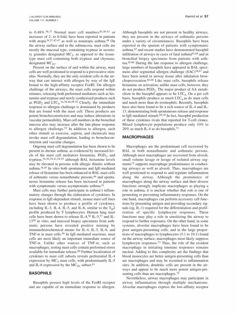
J ALLERGY CLIN IMMUNOL
VOLUME 111, NUMBER 1
Hamid et al S7
to 0.48%.36,37 Normal mast cell numbers36,38-41 orincreases of 2- to 6-fold, have been reported in patientswith atopic34,37,42-47 as well as nonatopic asthma.48 Onthe airway surface and in the submucosa, mast cells aremostly the mucosal type, containing tryptase in secreto-ry granules designated MCT, as opposed to the tissue-type mast cell containing both tryptase and chymase,designated MCTC.
Present on the surface of and within the airway, mastcells are well positioned to respond to a provocative stim-ulus. Normally, they are the only resident cells in the air-way that can interact with allergen by way of the IgEbound to the high-affinity receptor FcεRI. On allergenchallenge of the airways, the mast cells respond withinminutes, releasing both preformed mediators such as his-tamine and tryptase and newly synthesized products suchas PGD2 and LTC4.16,38,40,49-52 Clearly, the immediateresponse to allergen challenge is dominated by productsthat are found with the mast cell. These products arepotent bronchoconstrictors and may induce alterations invascular permeability. Mast cell numbers in the bronchialmucosa also may increase after the late-phase responseto allergen challenge.53 In addition to allergen, suchother stimuli as exercise, aspirin, and chemicals mayinvoke mast cell degranulation, leading to bronchocon-striction and vascular changes.
Ongoing mast cell degranulation has been shown to bepresent in chronic asthma, as evidenced by increased lev-els of the mast cell mediators histamine, PGD2, andtryptase,34,36,39,41,54,55 although BAL histamine levelsmay be elevated in persons with allergic rhinitis withoutasthma.36,45 In vitro both spontaneous and IgE-mediatedrelease of histamine has been enhanced in BAL mast cellsof asthmatic versus nonasthmatic persons,46 and sponta-neous histamine release has been increased in patientswith symptomatic versus asymptomatic asthma.41
Mast cells may further participate in asthma’s inflam-matory changes through the elaboration of cytokines. Inresponse to IgE-dependent stimuli, mouse mast cell lineshave been shown to produce a profile of cytokines,including IL-3, IL-4, IL-5, and IL-6, similar to the TH2profile produced by T lymphocytes. Human lung mastcells have been shown to release IL-4,56 IL-5,57 and IL-1358 in vitro, and mucosal biopsy specimens from asth-matic persons have revealed positive staining byimmunohistochemical means for IL-4, IL-5, IL-6, andTNF-α in mast cells.59 In IgE-mediated reactions, mastcells are most likely an important immediate source ofTNF-α. Unlike other sources of TNF-α, such asmacrophages, resting mast cells contain preformed storesavailable for immediate release.60 Further localization ofcytokines to mast cell subsets reveals preferential IL-4expression by MCT mast cells, with predominantly IL-5and IL-6 expression by the MCTC subset.61
BASOPHILS
Basophils possess high levels of the FcεRI receptorand are capable of an immediate response to allergen.
Although basophils are not present in healthy airways,they are present in the airways of asthmatic personsunder a variety of circumstances. Basophils have beenreported in the sputum of patients with symptomaticasthma,62 and recent studies have demonstrated basophilinfiltration of airways in cases of fatal asthma63,64 and inbronchial biopsy specimens from patients with asth-ma.65,66 During the late response to allergen challenge,large numbers of basophils have appeared in BAL speci-mens after segmental allergen challenge (SAC)38,67 andhave been noted in airway tissue after inhalation bron-choprovocation.66,68 Like mast cells, basophils releasehistamine on activation; unlike mast cells, however, theydo not produce PGD2. The major product of AA metab-olism in the basophil appears to be LTC4. On a per cellbasis, basophils produce as much LTC4 as do mast cellsand much more than do eosinophils. Recently, basophilshave also been found to be a rich source of IL-4 and IL-13, demonstrating both spontaneous release and responseto IgE-mediated stimuli.69,70 In fact, basophil productionof these cytokines rivals that reported for T-cell clones.Mixed lymphocyte populations produce only 10% to20% as much IL-4 as do basophils.71
MACROPHAGES
Macrophages are the predominant cell recovered byBAL in both nonasthmatic and asthmatic persons.Although most macrophages are recovered from alveoli,small volume lavage or lavage of isolated airway seg-ments72 supports macrophage predominance in conduct-ing airways as well as alveoli. Thus, macrophages arewell positioned to respond to and regulate inflammationalong the airway. Although the prominence ofmacrophages along the airway surface and their diversefunctions strongly implicate macrophages as playing arole in asthma, it is unclear whether that role is one ofpromoting or preventing inflammatory responses. On theone hand, macrophages can perform accessory cell func-tions by presenting antigen and providing secondary sig-nals (eg, IL-1) required for the differentiation and prolif-eration of specific lymphocyte responses. Thesefunctions may play a role in sensitizing the airway torespond to further exposures. On the other hand, in somesystems, alveolar macrophages have been found to bepoor antigen-presenting cells, and in the large propor-tions of macrophages to lymphocytes (5:1 to 10:1) foundon the airway surface, macrophages most likely suppresslymphocyte responses.73 Thus, the role of the residentmacrophage in initiating immune responses remainsunclear. Adding to this complexity are the findings thatblood monocytes are better antigen-presenting cells thanare macrophages and may be recruited to inflammationsites. In addition, dendritic cells are present in the air-ways and appear to be much more potent antigen-pre-senting cells than are macrophages.74
Nevertheless, airway macrophages may participate inairway inflammation through multiple mechanisms.Alveolar macrophages express the low-affinity receptor
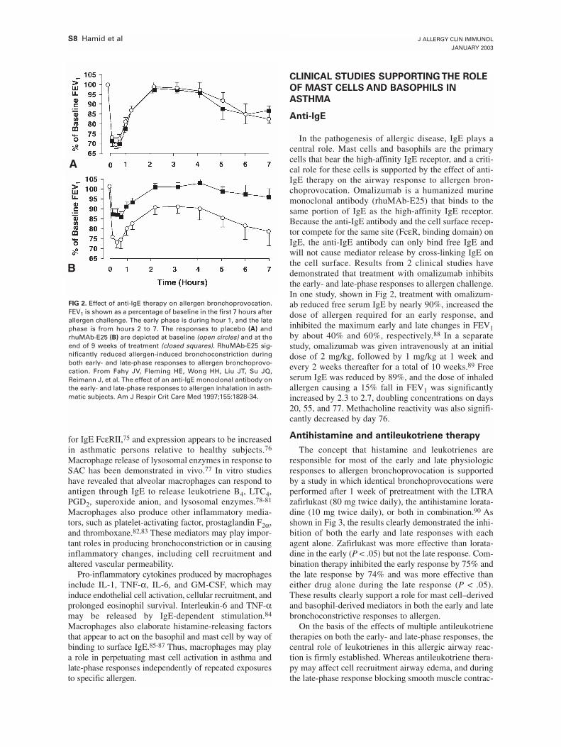
S8 Hamid et al J ALLERGY CLIN IMMUNOL
JANUARY 2003
for IgE FcεRII,75 and expression appears to be increasedin asthmatic persons relative to healthy subjects.76
Macrophage release of lysosomal enzymes in response toSAC has been demonstrated in vivo.77 In vitro studieshave revealed that alveolar macrophages can respond toantigen through IgE to release leukotriene B4, LTC4,PGD2, superoxide anion, and lysosomal enzymes.78-81
Macrophages also produce other inflammatory media-tors, such as platelet-activating factor, prostaglandin F2α,and thromboxane.82,83 These mediators may play impor-tant roles in producing bronchoconstriction or in causinginflammatory changes, including cell recruitment andaltered vascular permeability.
Pro-inflammatory cytokines produced by macrophagesinclude IL-1, TNF-α, IL-6, and GM-CSF, which mayinduce endothelial cell activation, cellular recruitment, andprolonged eosinophil survival. Interleukin-6 and TNF-αmay be released by IgE-dependent stimulation.84
Macrophages also elaborate histamine-releasing factorsthat appear to act on the basophil and mast cell by way ofbinding to surface IgE.85-87 Thus, macrophages may playa role in perpetuating mast cell activation in asthma andlate-phase responses independently of repeated exposuresto specific allergen.
CLINICAL STUDIES SUPPORTING THE ROLE
OF MAST CELLS AND BASOPHILS IN
ASTHMA
Anti-IgE
In the pathogenesis of allergic disease, IgE plays acentral role. Mast cells and basophils are the primarycells that bear the high-affinity IgE receptor, and a criti-cal role for these cells is supported by the effect of anti-IgE therapy on the airway response to allergen bron-choprovocation. Omalizumab is a humanized murinemonoclonal antibody (rhuMAb-E25) that binds to thesame portion of IgE as the high-affinity IgE receptor.Because the anti-IgE antibody and the cell surface recep-tor compete for the same site (FcεR, binding domain) onIgE, the anti-IgE antibody can only bind free IgE andwill not cause mediator release by cross-linking IgE onthe cell surface. Results from 2 clinical studies havedemonstrated that treatment with omalizumab inhibitsthe early- and late-phase responses to allergen challenge.In one study, shown in Fig 2, treatment with omalizum-ab reduced free serum IgE by nearly 90%, increased thedose of allergen required for an early response, andinhibited the maximum early and late changes in FEV1by about 40% and 60%, respectively.88 In a separatestudy, omalizumab was given intravenously at an initialdose of 2 mg/kg, followed by 1 mg/kg at 1 week andevery 2 weeks thereafter for a total of 10 weeks.89 Freeserum IgE was reduced by 89%, and the dose of inhaledallergen causing a 15% fall in FEV1 was significantlyincreased by 2.3 to 2.7, doubling concentrations on days20, 55, and 77. Methacholine reactivity was also signifi-cantly decreased by day 76.
Antihistamine and antileukotriene therapy
The concept that histamine and leukotrienes areresponsible for most of the early and late physiologicresponses to allergen bronchoprovocation is supportedby a study in which identical bronchoprovocations wereperformed after 1 week of pretreatment with the LTRAzafirlukast (80 mg twice daily), the antihistamine lorata-dine (10 mg twice daily), or both in combination.90 Asshown in Fig 3, the results clearly demonstrated the inhi-bition of both the early and late responses with eachagent alone. Zafirlukast was more effective than lorata-dine in the early (P < .05) but not the late response. Com-bination therapy inhibited the early response by 75% andthe late response by 74% and was more effective thaneither drug alone during the late response (P < .05).These results clearly support a role for mast cell–derivedand basophil-derived mediators in both the early and latebronchoconstrictive responses to allergen.
On the basis of the effects of multiple antileukotrienetherapies on both the early- and late-phase responses, thecentral role of leukotrienes in this allergic airway reac-tion is firmly established. Whereas antileukotriene thera-py may affect cell recruitment airway edema, and duringthe late-phase response blocking smooth muscle contrac-
FIG 2. Effect of anti-IgE therapy on allergen bronchoprovocation.FEV1 is shown as a percentage of baseline in the first 7 hours afterallergen challenge. The early phase is during hour 1, and the latephase is from hours 2 to 7. The responses to placebo (A) andrhuMAb-E25 (B) are depicted at baseline (open circles) and at theend of 9 weeks of treatment (closed squares). RhuMAb-E25 sig-nificantly reduced allergen-induced bronchoconstriction duringboth early- and late-phase responses to allergen bronchoprovo-cation. From Fahy JV, Fleming HE, Wong HH, Liu JT, Su JQ,Reimann J, et al. The effect of an anti-IgE monoclonal antibody onthe early- and late-phase responses to allergen inhalation in asth-matic subjects. Am J Respir Crit Care Med 1997;155:1828-34.
A
B
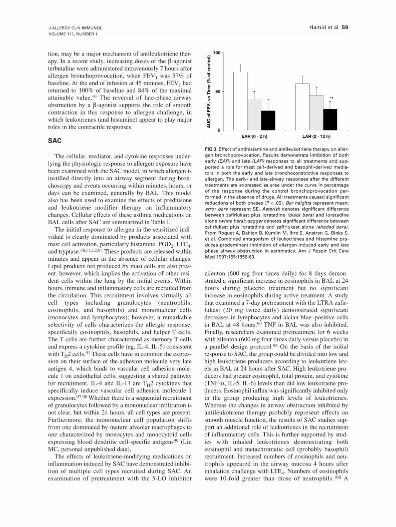
J ALLERGY CLIN IMMUNOL
VOLUME 111, NUMBER 1
Hamid et al S9
tion, may be a major mechanism of antileukotriene ther-apy. In a recent study, increasing doses of the β-agonistterbutaline were administered intravenously 7 hours afterallergen bronchoprovocation, when FEV1 was 57% ofbaseline. At the end of infusion at 45 minutes, FEV1 hadreturned to 100% of baseline and 84% of the maximalattainable value.91 The reversal of late-phase airwayobstruction by a β-agonist supports the role of smoothcontraction in this response to allergen challenge, inwhich leukotrienes (and histamine) appear to play majorroles in the contractile responses.
SAC
The cellular, mediator, and cytokine responses under-lying the physiologic response to allergen exposure havebeen examined with the SAC model, in which allergen isinstilled directly into an airway segment during bron-choscopy and events occurring within minutes, hours, ordays can be examined, generally by BAL. This modelalso has been used to examine the effects of prednisoneand leukotriene modifier therapy on inflammatorychanges. Cellular effects of these asthma medications onBAL cells after SAC are summarized in Table I.
The initial response to allergen in the sensitized indi-vidual is clearly dominated by products associated withmast cell activation, particularly histamine, PGD2, LTC4,and tryptase.38,51,52,97 These products are released withinminutes and appear in the absence of cellular changes.Lipid products not produced by mast cells are also pres-ent, however, which implies the activation of other resi-dent cells within the lung by the initial events. Withinhours, immune and inflammatory cells are recruited fromthe circulation. This recruitment involves virtually allcell types including granulocytes (neutrophils,eosinophils, and basophils) and mononuclear cells(monocytes and lymphocytes); however, a remarkableselectivity of cells characterizes the allergic response,specifically eosinophils, basophils, and helper T cells.The T cells are further characterized as memory T cellsand express a cytokine profile (eg, IL-4, IL-5) consistentwith TH2 cells.91 These cells have in common the expres-sion on their surface of the adhesion molecule very lateantigen 4, which binds to vascular cell adhesion mole-cule 1 on endothelial cells, suggesting a shared pathwayfor recruitment. IL-4 and IL-13 are TH2 cytokines thatspecifically induce vascular cell adhesion molecule 1expression.97,98 Whether there is a sequential recruitmentof granulocytes followed by a mononuclear infiltration isnot clear, but within 24 hours, all cell types are present.Furthermore, the mononuclear cell population shiftsfrom one dominated by mature alveolar macrophages toone characterized by monocytes and monocytoid cellsexpressing blood dendritic cell-specific antigens99 (LiuMC, personal unpublished data).
The effects of leukotriene-modifying medications oninflammation induced by SAC have demonstrated inhibi-tion of multiple cell types recruited during SAC. Anexamination of pretreatment with the 5-LO inhibitor
zileuton (600 mg four times daily) for 8 days demon-strated a significant increase in eosinophils in BAL at 24hours during placebo treatment but no significantincrease in eosinophils during active treatment. A studythat examined a 7-day pretreatment with the LTRA zafir-lukast (20 mg twice daily) demonstrated significantdecreases in lymphocytes and alcian blue–positive cellsin BAL at 48 hours.93 TNF in BAL was also inhibited.Finally, researchers examined pretreatment for 6 weekswith zileuton (600 mg four times daily versus placebo) ina parallel design protocol.94 On the basis of the initialresponse to SAC, the group could be divided into low andhigh leukotriene producers according to leukotriene lev-els in BAL at 24 hours after SAC. High leukotriene pro-ducers had greater eosinophil, total protein, and cytokine(TNF-α, IL-5, IL-6) levels than did low leukotriene pro-ducers. Eosinophil influx was significantly inhibited onlyin the group producing high levels of leukotrienes.Whereas the changes in airway obstruction inhibited byantileukotriene therapy probably represent effects onsmooth muscle function, the results of SAC studies sup-port an additional role of leukotrienes in the recruitmentof inflammatory cells. This is further supported by stud-ies with inhaled leukotrienes demonstrating botheosinophil and metachromatic cell (probably basophil)recruitment. Increased numbers of eosinophils and neu-trophils appeared in the airway mucosa 4 hours afterinhalation challenge with LTE4. Numbers of eosinophilswere 10-fold greater than those of neutrophils.100 A
FIG 3. Effect of antihistamine and antileukotriene therapy on aller-gen bronchoprovocation. Results demonstrate inhibition of bothearly (EAR) and late (LAR) responses to all treatments and sup-ported a role for mast cell–derived and basophil-derived media-tors in both the early and late bronchoconstrictive responses toallergen. The early- and late-airway responses after the differenttreatments are expressed as area under the curve in percentageof the response during the control bronchoprovocation per-formed in the absence of drugs. All treatments caused significantreductions of both phases (P < .05). Bar heights represent mean;error bars represent SE. Asterisk denotes significant differencebetween zafirlukast plus loratadine (black bars) and loratadinealone (white bars); dagger denotes significant difference betweenzafirlukast plus loratadine and zafirlukast alone (shaded bars).From Roquet A, Dahlen B, Kumlin M, Ihre E, Anstren G, Binks S,et al. Combined antagonism of leukotrienes and histamine pro-duces predominant inhibition of allergen-induced early and latephase airway obstruction in asthmatics. Am J Respir Crit CareMed 1997;155:1856-63.
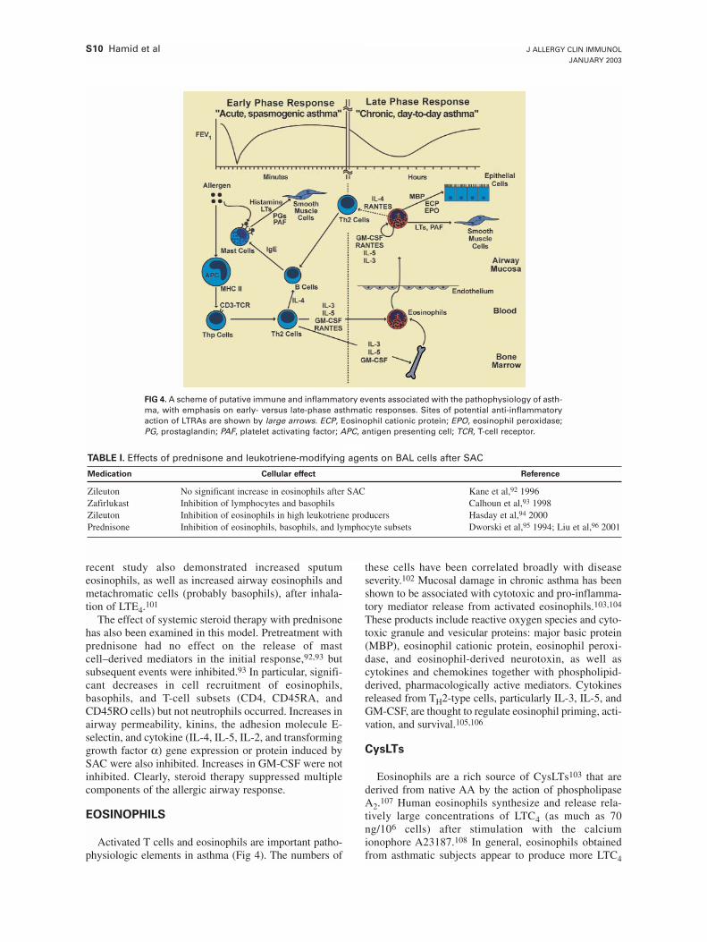
S10 Hamid et al J ALLERGY CLIN IMMUNOL
JANUARY 2003
recent study also demonstrated increased sputumeosinophils, as well as increased airway eosinophils andmetachromatic cells (probably basophils), after inhala-tion of LTE4.101
The effect of systemic steroid therapy with prednisonehas also been examined in this model. Pretreatment withprednisone had no effect on the release of mastcell–derived mediators in the initial response,92,93 butsubsequent events were inhibited.93 In particular, signifi-cant decreases in cell recruitment of eosinophils,basophils, and T-cell subsets (CD4, CD45RA, andCD45RO cells) but not neutrophils occurred. Increases inairway permeability, kinins, the adhesion molecule E-selectin, and cytokine (IL-4, IL-5, IL-2, and transforminggrowth factor α) gene expression or protein induced bySAC were also inhibited. Increases in GM-CSF were notinhibited. Clearly, steroid therapy suppressed multiplecomponents of the allergic airway response.
EOSINOPHILS
Activated T cells and eosinophils are important patho-physiologic elements in asthma (Fig 4). The numbers of
these cells have been correlated broadly with diseaseseverity.102 Mucosal damage in chronic asthma has beenshown to be associated with cytotoxic and pro-inflamma-tory mediator release from activated eosinophils.103,104
These products include reactive oxygen species and cyto-toxic granule and vesicular proteins: major basic protein(MBP), eosinophil cationic protein, eosinophil peroxi-dase, and eosinophil-derived neurotoxin, as well ascytokines and chemokines together with phospholipid-derived, pharmacologically active mediators. Cytokinesreleased from TH2-type cells, particularly IL-3, IL-5, andGM-CSF, are thought to regulate eosinophil priming, acti-vation, and survival.105,106
CysLTs
Eosinophils are a rich source of CysLTs103 that arederived from native AA by the action of phospholipaseA2.107 Human eosinophils synthesize and release rela-tively large concentrations of LTC4 (as much as 70ng/106 cells) after stimulation with the calciumionophore A23187.108 In general, eosinophils obtainedfrom asthmatic subjects appear to produce more LTC4
TABLE I. Effects of prednisone and leukotriene-modifying agents on BAL cells after SAC
Medication Cellular effect Reference
Zileuton No significant increase in eosinophils after SAC Kane et al,92 1996Zafirlukast Inhibition of lymphocytes and basophils Calhoun et al,93 1998Zileuton Inhibition of eosinophils in high leukotriene producers Hasday et al,94 2000Prednisone Inhibition of eosinophils, basophils, and lymphocyte subsets Dworski et al,95 1994; Liu et al,96 2001
FIG 4. A scheme of putative immune and inflammatory events associated with the pathophysiology of asth-ma, with emphasis on early- versus late-phase asthmatic responses. Sites of potential anti-inflammatoryaction of LTRAs are shown by large arrows. ECP, Eosinophil cationic protein; EPO, eosinophil peroxidase;PG, prostaglandin; PAF, platelet activating factor; APC, antigen presenting cell; TCR, T-cell receptor.
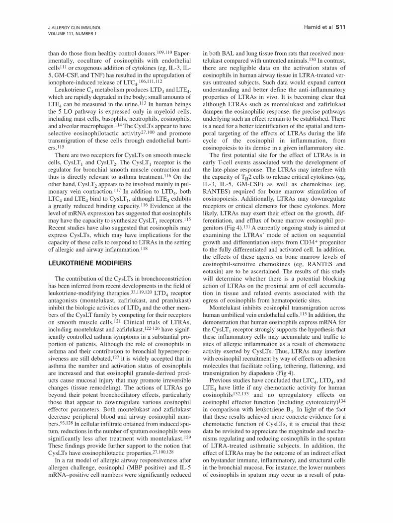
J ALLERGY CLIN IMMUNOL
VOLUME 111, NUMBER 1
Hamid et al S11
than do those from healthy control donors.109,110 Exper-imentally, coculture of eosinophils with endothelialcells111 or exogenous addition of cytokines (eg, IL-3, IL-5, GM-CSF, and TNF) has resulted in the upregulation ofionophore-induced release of LTC4.106,111,112
Leukotriene C4 metabolism produces LTD4 and LTE4,which are rapidly degraded in the body; small amounts ofLTE4 can be measured in the urine.113 In human beingsthe 5-LO pathway is expressed only in myeloid cells,including mast cells, basophils, neutrophils, eosinophils,and alveolar macrophages.114 The CysLTs appear to haveselective eosinophilotactic activity27,100 and promotetransmigration of these cells through endothelial barri-ers.115
There are two receptors for CysLTs on smooth musclecells, CysLT1 and CysLT2. The CysLT1 receptor is theregulator for bronchial smooth muscle contraction andthus is directly relevant to asthma treatment.116 On theother hand, CysLT2 appears to be involved mainly in pul-monary vein contraction.117 In addition to LTD4, bothLTC4 and LTE4 bind to CysLT1, although LTE4 exhibitsa greatly reduced binding capacity.116 Evidence at thelevel of mRNA expression has suggested that eosinophilsmay have the capacity to synthesize CysLT1 receptors.115
Recent studies have also suggested that eosinophils mayexpress CysLTs, which may have implications for thecapacity of these cells to respond to LTRAs in the settingof allergic and airway inflammation.118
LEUKOTRIENE MODIFIERS
The contribution of the CysLTs in bronchoconstrictionhas been inferred from recent developments in the field ofleukotriene-modifying therapies.33,119,120 LTD4 receptorantagonists (montelukast, zafirlukast, and pranlukast)inhibit the biologic activities of LTD4 and the other mem-bers of the CysLT family by competing for their receptorson smooth muscle cells.121 Clinical trials of LTRAs,including montelukast and zafirlukast,122-126 have signif-icantly controlled asthma symptoms in a substantial pro-portion of patients. Although the role of eosinophils inasthma and their contribution to bronchial hyperrespon-siveness are still debated,127 it is widely accepted that inasthma the number and activation status of eosinophilsare increased and that eosinophil granule-derived prod-ucts cause mucosal injury that may promote irreversiblechanges (tissue remodeling). The actions of LTRAs gobeyond their potent bronchodilatory effects, particularlythose that appear to downregulate various eosinophileffector parameters. Both montelukast and zafirlukastdecrease peripheral blood and airway eosinophil num-bers.93,128 In cellular infiltrate obtained from induced spu-tum, reductions in the number of sputum eosinophils weresignificantly less after treatment with montelukast.129
These findings provide further support to the notion thatCysLTs have eosinophilotactic properties.27,100,128
In a rat model of allergic airway responsiveness afterallergen challenge, eosinophil (MBP positive) and IL-5mRNA–positive cell numbers were significantly reduced
in both BAL and lung tissue from rats that received mon-telukast compared with untreated animals.130 In contrast,there are negligible data on the activation status ofeosinophils in human airway tissue in LTRA-treated ver-sus untreated subjects. Such data would expand currentunderstanding and better define the anti-inflammatoryproperties of LTRAs in vivo. It is becoming clear thatalthough LTRAs such as montelukast and zafirlukastdampen the eosinophilic response, the precise pathwaysunderlying such an effect remain to be established. Thereis a need for a better identification of the spatial and tem-poral targeting of the effects of LTRAs during the lifecycle of the eosinophil in inflammation, fromeosinopoiesis to its demise in a given inflammatory site.
The first potential site for the effect of LTRAs is inearly T-cell events associated with the development ofthe late-phase response. The LTRAs may interfere withthe capacity of TH2 cells to release critical cytokines (eg,IL-3, IL-5, GM-CSF) as well as chemokines (eg,RANTES) required for bone marrow stimulation ofeosinopoiesis. Additionally, LTRAs may downregulatereceptors or critical elements for these cytokines. Morelikely, LTRAs may exert their effect on the growth, dif-ferentiation, and efflux of bone marrow eosinophil pro-genitors (Fig 4).131 A currently ongoing study is aimed atexamining the LTRAs’ mode of action on sequentialgrowth and differentiation steps from CD34+ progenitorto the fully differentiated and activated cell. In addition,the effects of these agents on bone marrow levels ofeosinophil-sensitive chemokines (eg, RANTES andeotaxin) are to be ascertained. The results of this studywill determine whether there is a potential blockingaction of LTRAs on the proximal arm of cell accumula-tion in tissue and related events associated with theegress of eosinophils from hematopoietic sites.
Montelukast inhibits eosinophil transmigration acrosshuman umbilical vein endothelial cells.115 In addition, thedemonstration that human eosinophils express mRNA forthe CysLT1 receptor strongly supports the hypothesis thatthese inflammatory cells may accumulate and traffic tosites of allergic inflammation as a result of chemotacticactivity exerted by CysLTs. Thus, LTRAs may interferewith eosinophil recruitment by way of effects on adhesionmolecules that facilitate rolling, tethering, flattening, andtransmigration by diapedesis (Fig 4).
Previous studies have concluded that LTC4, LTD4, andLTE4 have little if any chemotactic activity for humaneosinophils132,133 and no upregulatory effects oneosinophil effector function (including cytotoxicity)134
in comparison with leukotriene B4. In light of the factthat these results achieved more concrete evidence for achemotactic function of CysLTs, it is crucial that thesedata be revisited to appreciate the magnitude and mecha-nisms regulating and reducing eosinophils in the sputumof LTRA-treated asthmatic subjects. In addition, theeffect of LTRAs may be the outcome of an indirect effecton bystander immune, inflammatory, and structural cellsin the bronchial mucosa. For instance, the lower numbersof eosinophils in sputum may occur as a result of puta-
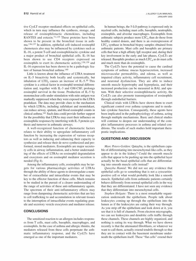
S12 Hamid et al J ALLERGY CLIN IMMUNOL
JANUARY 2003
tive CysLT receptor–mediated effects on epithelial cells,which in turn may influence the synthesis, storage, andrelease of eosinophilotactic chemokines, includingRANTES and eotaxin.71,135 These proteins have beenshown to be present in the bronchial tissue in asth-ma.136,137 In addition, epithelial cell–induced eosinophilchemotaxis also may be influenced by cytokines such asIL-16, a potent T-cell and eosinophilotactic cytokine anda major product of bronchial epithelial cells. IL-16 hasbeen shown to use CD4 receptors expressed oneosinophils to exert its chemotactic activity,138,139 andIL-16 expression has been shown to be a pathologic fea-ture of human bronchial asthma.140
Little is known about the influence of LTRA treatmenton IL-5 bioactivity both locally and systemically, butinhalation of LTD4 causes an increase of IL-5.30 Thiscytokine is a critical factor in eosinophil terminal differen-tiation and, together with IL-3 and GM-CSF, prolongseosinophil survival in the tissue. Production of IL-5 bymononuclear cells under stimulation with mite antigen wasmarkedly suppressed when they were exposed to the LTRApranlukast. The data may provide clues to the mechanismby which LTRAs, including zafirlukast and montelukast,can reduce airway, sputum, and blood eosinophil counts inclinical asthma.141 Such data could provide further supportfor the possibility that LTRAs may exert their influence oneosinophilic responses by interfering with IL-5 protein syn-thesis and turnover in asthmatic airways.
A well-recognized function of chemotactic factorsrelates to their ability to upregulate inflammatory cellfunction by increasing the expression of various recep-tors as well as inducing and enhancing their capacity tosynthesize and release their de novo synthesized and pre-formed, stored mediators. Eosinophils are major secreto-ry cells in airway inflammation, and a better understand-ing of the effects of LTRAs on eosinophil degranulationand exocytosis and on eosinophil mediator secretion isneeded (Fig 4).
Among the inflammatory cells, eosinophils may be tar-gets for various pharmacologic activities of LTRAsthrough the ability of these agents to downregulate a num-ber of extracellular and intracellular events that may bekey to the effector function of these cells. Much remainsto be studied in the pursuit of a clearer understanding ofthe range of activities of these anti-inflammatory agents.The spectrum of their anti-inflammatory effects mayrange from dampening chemotactic activities that are keyto cell trafficking to and accumulation in relevant tissuesto the interruption of intracellular events regulating gran-ule and secretory vesicle exocytosis and mediator release.
CONCLUSIONS
The sensitized reaction to an allergen includes respons-es from T cells, mast cells, basophils, macrophages, andeosinophils. In the case of asthma and allergic rhinitis, themediators released from these cells perpetuate the asth-matic inflammatory response, and the CysLTs haveemerged as one of the important mediators.
In human beings, the 5-LO pathway is expressed only inmyeloid cells, including mast cells, basophils, neutrophils,eosinophils, and alveolar macrophages. Eosinophils fromasthmatic subjects produce more LTC4 than do those fromhealthy control donors, and there is an overexpression ofLTC4 synthase in bronchial biopsy samples obtained fromasthmatic patients. Mast cells and basophils are primarycells that bear a high affinity IgE receptor and, during theirkey involvement in the early and late phases, CysLTs arereleased. Basophils produce as much LTC4 as do mast cellsand much more than do eosinophils.
The CysLTs have the potential to cause the cardinalsymptoms of asthma: mucus hypersecretion, increasedmicrovascular permeability, and edema, as well asimpaired ciliary activity, inflammatory cell recruitment,and neuronal dysfunction. They are able to inducesmooth muscle hypertrophy and hyperplasia, and theirincreased production can be measured in BAL and spu-tum. With their selective eosinophilotactic activity, theCysLTs can also promote transmigration of eosinophilsthrough endothelial barriers.
Clinical trials with LTRAs have shown them to exertsignificant control over asthma symptoms and to modu-late cytokine function. Consequently, these agents havebeen implicated in the pathophysiology of asthma, actingthrough multiple mechanisms. Basic and clinical studieswill continue to deepen our understanding of the com-plex inflammatory processes in asthma and related con-ditions. The results of such studies hold important thera-peutic implications.
QUESTIONS AND DISCUSSION
Marc Peters-Golden: Qutayba, is the epithelium capa-ble of differentiating into mesenchymal-like cells, as theyappear to do in the kidney? Could the smooth musclecells that appear to be pushing up into the epithelial layeractually be the basal epithelial cells that are differentiat-ing into smooth muscle cells instead?
Qutayba Hamid: We did not see any evidence thatepithelial cells go to something that is not a cytocuritin-positive cell or what would probably look like a smoothmuscle. Epithelial cells from asthmatic patients certainlybehave differently from normal epithelial cells in the waythat they are differentiated. I have not seen any evidencethat they differentiate into mesenchymal cells.
Stephen Holgate: There is a most remarkable organi-zation underneath the epithelium. People talk aboutleukocytes coming up through the epithelium into thelumen as if the leukocytes are eating their way through.As you strip off the epithelium and look down at it, yousee that it is full of channels. From electron micrographs,we can see leukocytes and dendritic cells traffic throughthese channels. These channels are highly organized, andnothing is eating its way through. What we have shownrecently is that the attenuated fibroblasts, or whatever wewant to call them, actually extend tendrils through so thatthey are in contact with the basement membrane under-neath the epithelium itself. These “flat cells” extend their
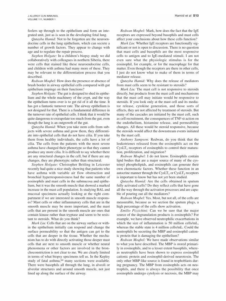
J ALLERGY CLIN IMMUNOL
VOLUME 111, NUMBER 1
Hamid et al S13
feelers up through to the epithelium and form an inte-grated unit, just as is seen in the developing fetal lung.
Qutayba Hamid: Not to be forgotten are the neuroen-docrine cells in the lung epithelium, which can secrete anumber of growth factors. They appear to change withage and to regulate the repair process.
Stephen Holgate: In a children’s biopsy study we didcollaboratively with colleagues in northern Siberia, therewere cells that stained like these neuroendocrine cells,and children with asthma had many more of these. Theymay be relevant to the differentiation process that youdescribed.
Redwan Moqbel: How does the presence or absence ofbrush border in airway epithelial cells compared with gutepithelium impinge on their functions?
Stephen Holgate: The gut is designed to shed its epithe-lium and the whole machinery of the crypt, and the waythe epithelium turns over is to get rid of it all the time. Ithas got a fantastic turnover rate. The airway epithelium isnot designed for that. There is a fundamental difference inthe turnover rate of epithelial cells. I think that it would bequite dangerous to extrapolate too much from the gut, eventhough the lung is an outgrowth of the gut.
Qutayba Hamid: When you take stem cells from sub-jects with severe asthma and grow them, they differenti-ate into epithelial cells that do not have cilia. If you takethem from healthy individuals, the cells have a lot ofcilia. The cells from the patients with the most severeasthma have changed their phenotype so that they cannotproduce any more cilia. It is difficult to say whether thereare any structural changes in the cell, but if there are anychanges, they are phenotypic rather than structural.
Stephen Holgate: Christopher Britling in Leicesterrecently had quite a nice study showing that patients whohave asthma with variable air flow obstruction andbronchial hyperresponsiveness had the same number ofeosinophils and mast cells in the submucosa and epithe-lium, but it was the smooth muscle that showed a markedincrease in the mast cell population. Is studying BAL andmucosal specimens actually looking at the right com-partment if we are interested in smooth muscle respons-es? Mast cells or other inflammatory cells that are in thesmooth muscle may be more important, and the mastcells that are present in the smooth muscle are ones thatcontain kinase rather than tryptase and seem to be resis-tant to steroids. What do you think?
Mark Liu: Cells that are on the airway surface or with-in the epithelium initially can respond and change thesurface permeability so that the antigen can get to thecells that are deeper in the tissue. Whether the mecha-nism has to do with directly activating those deeper mastcells that are next to smooth muscle or whether neuralphenomena or other factors are involved in the bron-choconstriction is not clear to me. We are clearly limitedin terms of what biopsy specimens tell us. In the Kepleystudy of fatal asthma,64 many sections were available.There were basophils all through the lung, in alveoli oralveolar structures and around smooth muscle, not justlined up along the surface of the airway.
Redwan Moqbel: Mark, how does the fact that the IgEreceptors are expressed beyond basophils and mast cellsaffect your conclusions about how these cells function?
Mark Liu: Whether IgE receptors are functionally sig-nificant or not is open to discussion. There is no questionthat mast cells and basophils are the most responsivecells to antigen and to IgE-mediated stimuli. I am noteven sure what the physiologic stimulus is for theeosinophil, for example, or for the macrophage for thatmatter. Even though the receptors are there in other cells,I just do not know what to make of them in terms ofmediator release.
Qutayba Hamid: Why does the release of mediatorsfrom mast cells seem to be resistant to steroids?
Mark Liu: The mast cell is not responsive to steroidsdirectly, but products from the mast cell and mechanismsthat the mast cell may initiate would be responsive tosteroids. If you look only at the mast cell and its media-tor release, cytokine generation, and those sorts ofeffects, they are not affected by treatment of steroids. Butmany of the cascades are initiated by the mast cell, suchas cell recruitment, the consequences of TNF-α action onthe endothelium, histamine release, and permeabilitychanges. All these would be steroid responsive, becausethe steroids would affect the downstream events initiatedby the mast cell.
Anthony Sampson: Redwan, do you think that theleukotrienes released from the eosinophils act on theCysLT1 receptors of eosinophils to control their matura-tion, proliferation, and migration?
Redwan Moqbel: I do not know. Eosinophils containlipid bodies that are a major source of many of the cys-teinyl phospholipids, and eosinophils can generate theirown chemotactic factors. Whether these function in anautocrine manner through the CysLT1 or CysLT2 receptorsis important to know but has not yet been studied.
Qutayba Hamid: Are the cells obtained in sputumfully activated cells? Do they reflect cells that have goneall the way through the activation processes and are capa-ble of pouring out all the mediators?
Redwan Moqbel: Yes. Most, but not all, of the cells aremeasurable, because as we section the sputum plugs, ahigh percentage of the cells show activation.
Emilio Pizzichini: Can we be sure that the majorsource of the degranulation products is eosinophils? Forexample, we have observed neutrophilic exacerbations inwhich the size of inflammation is 50 million cells/mL,whereas the stable state is 4 million cells/mL. Could theneutrophils be secreting the MBP and eosinophil cation-ic protein that is damaging the epithelium?
Redwan Moqbel: We have made observations similarto what you have described. The MBP is stored primari-ly in eosinophils, and to a lesser extent basophils, where-as neutrophils have been shown to express eosinophilcationic protein and eosinophil-derived neurotoxin. Theonly other MBP-like source is found in trophoblasts dur-ing pregnancy. The MBP from eosinophils acts on neu-trophils, and there is always the possibility that onceeosinophils undergo cytolysis or necrosis, the MBP may
S14 Hamid et al J ALLERGY CLIN IMMUNOL
JANUARY 2003
become sequestered in neutrophils. We use MAb BMK-13 to detect the human MBP.
Qutayba Hamid: What are the half-lives of theseeosinophil products in the tissue?
Redwan Moqbel: These products have long-termeffects, and because of their cationic nature they bindstrongly to the target cells, which makes it difficult to getrid of them. These basic proteins are extremely “sticky”and rich in arginine. Peroxidase and MBP are the twothat are most cytotoxic. Eosinophil cationic protein andeosinophil-derived neurotoxin have potent ribonucleaseactivity and lesser cytotoxic capacity. I am not aware ofany studies on the half-lives of these proteins in the tis-sue, but I assume that they may be long.
REFERENCES
1. Asano M, Toda M, Sakaguchi N, Sakaguchi S. Autoimmune disease asa consequence of developmental abnormality of a T cell subpopulation.J Exp Med 1996;184:387-96.
2. Turner H, Kinet JP. Signalling through the high-affinity IgE receptor FcepsilonRI. Nature 1999;402:B24-30.
3. Suto A, Nakajima H, Kagami SI, Suzuki K, Saito Y, Iwamoto I. Role ofCD4(+) CD25(+) regulatory T cells in T helper 2 cell-mediated allergicinflammation in the airways. Am J Respir Crit Care Med 2001;164:680-7.
4. Thornton AM, Shevach EM. CD4+CD25+ immunoregulatory T cellssuppress polyclonal T cell activation in vitro by inhibiting interleukin 2production. J Exp Med 1998;188:287-96.
5. Chen J, Huoam C, Plain K, He XY, Hodgkinson SJ, Hall BM. CD4(+),CD25(+) T cells as regulators of alloimmune responses. Transplant Proc2001;33:163-4.
6. Chung KF, Barnes PJ. Cytokines in asthma. Thorax 1999;54:825-57.7. de Vries JE. The role of IL-13 and its receptor in allergy and inflamma-
tory responses. J Allergy Clin Immunol 1998;102:165-9.8. Louahed J, Toda M, Jen J, Hamid Q, Renauld JC, Levitt RC, et al. Inter-
leukin-9 upregulates mucus expression in the airways. Am J Respir CellMol Biol 2000;22:649-56.
9. Louahed J, Zhou Y, Maloy WL, Rani PU, Weiss C, Tomer Y, et al. Inter-leukin 9 promotes influx and local maturation of eosinophils. Blood2001;97:1035-42.
10. Shimbara A, Christodoulopoulos P, Soussi-Gounni A, Olivenstein R,Nakamura Y, Levitt RC, et al. IL-9 and its receptor in allergic and non-allergic lung disease: increased expression in asthma. J Allergy ClinImmunol 2000;105:108-15.
11. Molet S, Hamid Q, Davoine F, Nutku E, Taha R, Page N, et al. IL-17 isincreased in asthmatic airways and induces human bronchial fibroblaststo produce cytokines. J Allergy Clin Immunol 2001;108:430-8.
12. Einarsson O, Geba GP, Zhou Z, Landry ML, Panettieri RA Jr, TristramD, et al. Interleukin-11 in respiratory inflammation. Ann N Y Acad Sci1995;762:89-100.
13. Minshall E, Chakir J, Laviolette M, Molet S, Zhu Z, Olivenstein R, et al.IL-11 expression is increased in severe asthma: association with epithe-lial cells and eosinophils. J Allergy Clin Immunol 2000;105:232-8.
14. Salvi SS, Krishna MT, Sampson AP, Holgate ST. The anti-inflammato-ry effects of leukotriene-modifying drugs and their use in asthma. Chest2001;119:1533-46.
15. Lam S, Chan H, LeRiche JC, Chan-Yeung M, Salari H. Release ofleukotrienes in patients with bronchial asthma. J Allergy Clin Immunol1988;81:711-7.
16. Wenzel SE, Larsen GL, Johnston K, Voelkel NF, Westcott JY. Elevatedlevels of leukotriene C4 in bronchoalveolar lavage fluid from atopicasthmatics after endobronchial allergen challenge. Am Rev Respir Dis1990;142:112-9.
17. Kikawa Y, Hosoi S, Inoue Y, Saito M, Nakai A, Shigematsu Y, et al.Exercise-induced urinary excretion of leukotriene E4 in children withatopic asthma. Pediatr Res 1991;29:455-9.
18. Creticos PS, Peters SP, Adkinson NF Jr, Naclerio RM, Hayes EC, Nor-man PS, et al. Peptide leukotriene release after antigen challenge inpatients sensitive to ragweed. N Engl J Med 1984;310:1626-30.
19. Shindo K, Fukumura M, Miyakawa K. Plasma levels of leukotriene E4during clinical course of bronchial asthma and the effect of oral pred-nisolone. Chest 1994;105:1038-41.
20. Chu SJ, Tang LO, Watney E, Chi EY, Henderson WR Jr. In situ ampli-fication of 5-lipoxygenase and 5-lipoxygenase–activating protein inallergic airway inflammation and inhibition by leukotriene blockade. JImmunol 2000;165:4640-8.
21. Cowburn AS, Sladek K, Soja J, Adamek L, Nizankowska E, Szczeklik A,et al. Overexpression of leukotriene C4 synthase in bronchial biopsies frompatients with aspirin-intolerant asthma. J Clin Invest 1998;101:834-46.
22. Sampson AP, Cowburn AS, Sladek K, Adamek L, Nizankowska E,Szczeklik A, et al. Profound overexpression of leukotriene C4 synthasein bronchial biopsies from aspirin-intolerant asthmatic patients. Int ArchAllergy Immunol 1997;113:355-7.
23. Nasser SM, Pfister R, Christie PE, Sousa AR, Barker J, Schmitz-Schu-mann M, et al. Inflammatory cell populations in bronchial biopsies fromaspirin- sensitive asthmatic subjects. Am J Respir Crit Care Med1996;153:90-6.
24. Drazen JM, Austen KF. Leukotrienes and airway responses. Am RevRespir Dis 1987;136:985-98.
25. Piacentini GL, Kaliner MA. The potential roles of leukotrienes inbronchial asthma. Am Rev Respir Dis 1991;143(5 Pt 2):S96-9.
26. Henderson WR Jr. The role of leukotrienes in inflammation. Ann InternMed 1994;121:684-97.
27. Hay DW, Torphy TJ, Undem BJ. Cysteinyl leukotrienes in asthma: oldmediators up to new tricks. Trends Pharmacol Sci 1995;16:304-9.
28. Wanner A, Salathe M, O’Riordan TG. Mucociliary clearance in the air-ways. Am J Respir Crit Care Med 1996;154:1868-902.
29. Ahmed T, Greenblatt DW, Birch S, Marchette B, Wanner A. Abnormalmucociliary transport in allergic patients with antigen-induced bron-chospasm: role of slow reacting substance of anaphylaxis. Am RevRespir Dis 1981;124:110-4.
30. Underwood DC, Osborn RR, Newsholme SJ, Torphy TJ, Hay DW. Per-sistent airway eosinophilia after leukotriene (LT) D4 administration inthe guinea pig: modulation by the LTD4 receptor antagonist, pranlukast,or an interleukin-5 monoclonal antibody. Am J Respir Crit Care Med1996;154:850-7.
31. Fonteh AN, LaPorte T, Swan D, McAlexander MA. A decrease inremodeling accounts for the accumulation of arachidonic acid in murinemast cells undergoing apoptosis. J Biol Chem 2001;276:1439-49.
32. O’Hickey SP, Hawksworth RJ, Fong CY, Arm JP, Spur BW, Lee TH.Leukotrienes C4, D4, and E4 enhance histamine responsiveness in asth-matic airways. Am Rev Respir Dis 1991;144:1053-7.
33. Kurosawa M, Yodonawa S, Tsukagoshi H, Miyachi Y. Inhibition by anovel peptide leukotriene receptor antagonist ONO-1078 of airway wallthickening and airway hyperresponsiveness to histamine induced byleukotriene C4 or leukotriene D4 in guinea-pigs. Clin Exp Allergy1994;24:960-8.
34. Foresi A, Bertorelli G, Pesci A, Chetta A, Olivieri D. Inflammatorymarkers in bronchoalveolar lavage and in bronchial biopsy in asthmaduring remission. Chest 1990;98:528-35.
35. Dunhill MS. The pathology of asthma, with special reference to changesin the bronchial mucosa. J Clin Pathol 1960;13:27-33.
36. Liu MC, Bleecker ER, Lichtenstein LM, Kagey-Sobotka A, Niv Y,McLemore TL, et al. Evidence for elevated levels of histamine,prostaglandin D2, and other bronchoconstricting prostaglandins in the air-ways of subjects with mild asthma. Am Rev Respir Dis 1990;142:126-32.
37. Adelroth E, Rosenhall L, Johansson SA, Linden M, Venge P. Inflam-matory cells and eosinophilic activity in asthmatics investigated bybronchoalveolar lavage: the effects of antiasthmatic treatment withbudesonide or terbutaline. Am Rev Respir Dis 1990;142:91-9.
38. Liu MC, Hubbard WC, Proud D, Stealey BA, Galli SJ, Kagey-SobotkaA, et al. Immediate and late inflammatory responses to ragweed antigenchallenge of the peripheral airways in allergic asthmatics: cellular, medi-ator, and permeability changes. Am Rev Respir Dis 1991;144:51-8.
39. Gravelyn TR, Pan PM, Eschenbacher WL. Mediator release in an iso-lated airway segment in subjects with asthma. Am Rev Respir Dis1988;137:641-6.
40. Wenzel SE, Fowler AA, Schwartz LB. Activation of pulmonary mastcells by bronchoalveolar allergen challenge. In vivo release of histamineand tryptase in atopic subjects with and without asthma. Am Rev RespirDis 1988;137:1002-8.
J ALLERGY CLIN IMMUNOL
VOLUME 111, NUMBER 1
Hamid et al S15
41. Broide DH, Gleich GJ, Cuomo AJ, Coburn DA, Federman EC,Schwartz LB, et al. Evidence of ongoing mast cell and eosinophildegranulation in symptomatic asthma airway. J Allergy Clin Immunol1991;88:637-48.
42. Rennard SI, Basset G, Lecossier D, O’Donnell KM, Pinkston P, MartinPG, et al. Estimation of volume of epithelial lining fluid recovered bylavage using urea as marker of dilution. J Appl Physiol 1986;60:532-8.
43. Tomioka M, Ida S, Shindoh Y, Ishihara T, Takishima T. Mast cells inbronchoalveolar lumen of patients with bronchial asthma. Am RevRespir Dis 1984;129:1000-5.
44. Kirby JG, Hargreave FE, Gleich GJ, O’Byrne PM. Bronchoalveolar cellprofiles of asthmatic and nonasthmatic subjects. Am Rev Respir Dis1987;136:379-83.
45. Casale TB, Wood D, Richerson HB, Trapp S, Metzger WJ, Zavala D, etal. Elevated bronchoalveolar lavage fluid histamine levels in allergicasthmatics are associated with methacholine bronchial hyperresponsive-ness. J Clin Invest 1987;79:1197-203.
46. Flint KC, Leung KB, Hudspith BN, Brostoff J, Pearce FL, Johnson NM.Bronchoalveolar mast cells in extrinsic asthma: a mechanism for the ini-tiation of antigen specific bronchoconstriction. Br Med J (Clin Res Ed)1985;291:923-6.
47. Wardlaw AJ, Dunnette S, Gleich GJ, Collins JV, Kay AB. Eosinophilsand mast cells in bronchoalveolar lavage in subjects with mild asthma:relationship to bronchial hyperreactivity. Am Rev Respir Dis1988;137:62-9.
48. Mattoli S, Mattoso VL, Soloperto M, Allegra L, Fasoli A. Cellular andbiochemical characteristics of bronchoalveolar lavage fluid in sympto-matic nonallergic asthma. J Allergy Clin Immunol 1991;87:794-802.
49. Sedgwick JB, Calhoun WJ, Gleich GJ, Kita H, Abrams JS, SchwartzLB, et al. Immediate and late airway response of allergic rhinitispatients to segmental antigen challenge: characterization of eosinophiland mast cell mediators. Am Rev Respir Dis 1991;144:1274-81.
50. Wenzel SE, Westcott JY, Larsen GL. Bronchoalveolar lavage fluidmediator levels 5 minutes after allergen challenge in atopic subjectswith asthma: relationship to the development of late asthmatic respons-es. J Allergy Clin Immunol 1991;87:540-8.
51. Wenzel SE, Westcott JY, Smith HR, Larsen GL. Spectrum of prostanoidrelease after bronchoalveolar allergen challenge in atopic asthmaticsand in control groups: an alteration in the ratio of bronchoconstrictive tobronchoprotective mediators. Am Rev Respir Dis 1989;139:450-7.
52. Murray JJ, Tonnel AB, Brash AR, Roberts LJ 2nd, Gosset P, WorkmanR, et al. Release of prostaglandin D2 into human airways during acuteantigen challenge. N Engl J Med 1986;315:800-4.
53. Crimi E, Chiaramondia M, Milanese M, Rossi GA, Brusasco V.Increased numbers of mast cells in bronchial mucosa after the late-phaseasthmatic response to allergen. Am Rev Respir Dis 1991;144:1282-6.
54. Zehr BB, Casale TB, Wood D, Floerchinger C, Richerson HB, Hun-ninghake GW. Use of segmental airway lavage to obtain relevant medi-ators from the lungs of asthmatic and control subjects. Chest1989;95:1059-63.
55. Van Vyve T, Chanez P, Bernard A, Bousquet J, Godard P, Lauwerijs R,et al. Protein content in bronchoalveolar lavage fluid of patients withasthma and control subjects. J Allergy Clin Immunol 1995;95:60-8.
56. Bradding P, Feather IH, Howarth PH, Mueller R, Roberts JA, Britten K,et al. Interleukin 4 is localized to and released by human mast cells. JExp Med 1992;176:1381-6.
57. Jaffe JS, Glaum MC, Raible DG, Post TJ, Dimitry E, Govindarao D, etal. Human lung mast cell IL-5 gene and protein expression: temporalanalysis of upregulation following IgE-mediated activation. Am JRespir Cell Mol Biol 1995;13:665-75.
58. Jaffe JS, Raible DG, Post TJ, Wang Y, Glaum MC, Butterfield JH, et al.Human lung mast cell activation leads to IL-13 mRNA expression andprotein release. Am J Respir Cell Mol Biol 1996;15:473-81.
59. Bradding P, Roberts JA, Britten KM, Montefort S, Djukanovic R,Mueller R, et al. Interleukin-4, -5, and -6 and tumor necrosis factor-alphain normal and asthmatic airways: evidence for the human mast cell as asource of these cytokines. Am J Respir Cell Mol Biol 1994;10:471-80.
60. Gordon JR, Burd PR, Galli SJ. Mast cells as a source of multifunction-al cytokines. Immunol Today 1990;11:458-64.
61. Bradding P, Okayama Y, Howarth PH, Church MK, Holgate ST. Het-erogeneity of human mast cells based on cytokine content. J Immunol1995;155:297-307.
62. Kimura I, Tanizaki Y, Saito K, Takahashi K, Ueda N, Sato S. Appear-ance of basophils in the sputum of patients with bronchial asthma. ClinAllergy 1975;5:95-8.
63. Koshino T, Teshima S, Fukushima N, Takaishi T, Hirai K, Miyamoto Y,et al. Identification of basophils by immunohistochemistry in the air-ways of post-mortem cases of fatal asthma. Clin Exp Allergy1993;23:919-25.
64. Kepley CL, McFeeley PJ, Oliver JM, Lipscomb MF. Immunohisto-chemical detection of human basophils in postmortem cases of fatalasthma. Am J Respir Crit Care Med 2001;164:1053-8.
65. Koshino T, Arai Y, Miyamoto Y, Sano Y, Itami M, Teshima S, et al. Air-way basophil and mast cell density in patients with bronchial asthma:relationship to bronchial hyperresponsiveness. J Asthma 1996;33:89-95.
66. Macfarlane AJ, Kon OM, Smith SJ, Zeibecoglou K, Khan LN, BarataLT, et al. Basophils, eosinophils, and mast cells in atopic and nonatopicasthma and in late-phase allergic reactions in the lung and skin. J Aller-gy Clin Immunol 2000;105:99-107.
67. Guo CB, Liu MC, Galli SJ, Bochner BS, Kagey-Sobotka A, Lichten-stein LM. Identification of IgE-bearing cells in the late-phase responseto antigen in the lung as basophils. Am J Respir Cell Mol Biol1994;10:384-90.
68. Beasley R, Roche WR, Roberts JA, Holgate ST. Cellular events in thebronchi in mild asthma and after bronchial provocation. Am Rev RespirDis 1989;139:806-17.
69. MacGlashan DJ, White JM, Huang SK, Ono SJ, Schroeder JT, Lichten-stein LM. Secretion of IL-4 from human basophils: the relationshipbetween IL-4 mRNA and protein in resting and stimulated basophils. JImmunol 1994;152:3006-16.
70. Li H, Sim TC, Alam R. IL-13 released by and localized in humanbasophils. J Immunol 1996;156:4833-8.
71. Schroeder JT, Kagey-Sobotka A, MacGlashan DW, Lichtenstein LM.The interaction of cytokines with human basophils and mast cells. IntArch Allergy Immunol 1995;107:79-81.
72. Eschenbacher WL, Gravelyn TR. A technique for isolated airway seg-ment lavage. Chest 1987;92:105-9.
73. Liu MC, Proud D, Schleimer RP, Plaut M. Human lung macrophagesenhance and inhibit lymphocyte proliferation. J Immunol1984;132:2895-903.
74. Langhoff E, Steinman RM. Clonal expansion of human T lymphocytesinitiated by dendritic cells. J Exp Med 1989;169:315-20.
75. Melewicz FM, Kline LE, Cohen AB, Spiegelberg HL. Characterizationof Fc receptors for IgE on human alveolar macrophages. Clin ExpImmunol 1982;49:364-70.
76. Williams J, Johnson S, Mascali JJ, Smith H, Rosenwasser LJ, Borish L.Regulation of low affinity IgE receptor (CD23) expression on mononu-clear phagocytes in normal and asthmatic subjects. J Immunol1992;149:2823-9.
77. Tonnel AB, Joseph M, Gosset P, Fournier E, Capron A. Stimulation ofalveolar macrophages in asthmatic patients after local provocation test.Lancet 1983;1:1406-8.
78. Fuller RW, Morris PK, Richmond R, Sykes D, Varndell IM, KemenyDM, et al. Immunoglobulin E–dependent stimulation of human alveolarmacrophages: significance in type 1 hypersensitivity. Clin Exp Immunol1986;65:416-26.
79. Rankin JA. The contribution of alveolar macrophages to hyperreactiveairway disease. J Allergy Clin Immunol 1989;83:722-9.
80. Fuller RW. The role of the alveolar macrophage in asthma. Respir Med1989;83:177-8.
81. Joseph M, Tonnel AB, Capron A, Voisin C. Enzyme release and super-oxide anion production by human alveolar macrophages stimulated withimmunoglobulin E. Clin Exp Immunol 1980;40:416-22.
82. MacDermot J, Kelsey CR, Waddell KA, Richmond R, Knight RK, ColePJ, et al. Synthesis of leukotriene B4, and prostanoids by human alveo-lar macrophages: analysis by gas chromatography/mass spectrometry.Prostaglandins 1984;27:163-79.
83. Arnoux B, Duval D, Benveniste J. Release of platelet-activating factor(PAF-acether) from alveolar macrophages by the calcium ionophoreA23187 and phagocytosis. Eur J Clin Invest 1980;10:437-41.
84. Gosset P, Tsicopoulos A, Wallaert B, Joseph M, Capron A, Tonnel AB.Tumor necrosis factor alpha and interleukin-6 production by humanmononuclear phagocytes from allergic asthmatics after IgE-dependentstimulation. Am Rev Respir Dis 1992;146:768-74.
S16 Hamid et al J ALLERGY CLIN IMMUNOL
JANUARY 2003
85. Liu MC, Proud D, Lichtenstein LM, MacGlashan DW, Schleimer RP,Adkinson NF, et al. Human lung macrophage-derived histamine-releasingactivity is due to IgE-dependent factors. J Immunol 1986;136:2588-95.
86. MacDonald SM, Lichtenstein LM, Proud D, Plaut M, Naclerio RM,MacGlashan DW, et al. Studies of IgE-dependent histamine releasingfactors: heterogeneity of IgE. J Immunol 1987;139:506-12.
87. MacDonald SM, Rafnar T, Langdon J, Lichtenstein LM. Molecularidentification of an IgE-dependent histamine-releasing factor. Science1995;269:688-90.
88. Fahy JV, Fleming HE, Wong HH, Liu JT, Su JQ, Reimann J, et al. Theeffect of an anti-IgE monoclonal antibody on the early- and late-phaseresponses to allergen inhalation in asthmatic subjects. Am J Respir CritCare Med 1997;155:1828-34.
89. Boulet LP, Chapman KR, Cote J, Kalra S, Bhagat R, Swystun VA, et al.Inhibitory effects of an anti-IgE antibody E25 on allergen-induced earlyasthmatic response. Am J Respir Crit Care Med 1997;155:1835-40.
90. Roquet A, Dahlen B, Kumlin M, Ihre E, Anstren G, Binks S, et al. Com-bined antagonism of leukotrienes and histamine produces predominantinhibition of allergen-induced early and late phase airway obstruction inasthmatics. Am J Respir Crit Care Med 1997;155:1856-63.
91. Peebles RS, Permutt S, Togias A. Rapid reversibility of the allergen-induced pulmonary late-phase reaction by an intravenous beta2-agonist.J Appl Physiol 1998;84:1500-5.
92. Kane GC, Pollice M, Kim CJ, Cohn J, Dworski RT, Murray JJ, et al. Acontrolled trial of the effect of the 5-lipoxygenase inhibitor, zileuton, onlung inflammation produced by segmental antigen challenge in humanbeings. J Allergy Clin Immunol 1996;97:646-54.
93. Calhoun WJ, Lavins BJ, Minkwitz MC, Evans R, Gleich GJ, Cohn J.Effect of zafirlukast (Accolate) on cellular mediators of inflammation:bronchoalveolar lavage fluid findings after segmental antigen challenge.Am J Respir Crit Care Med 1998;157:1381-9.
94. Hasday JD, Meltzer SS, Moore WC, Wisniewski P, Hebel JR, Lanni C,et al. Anti-inflammatory effects of zileuton in a subpopulation of aller-gic asthmatics. Am J Respir Crit Care Med 2000;161:1229-36.
95. Dworski R, Fitzgerald GA, Oates JA, Sheller JR. Effect of oral pred-nisone on airway inflammatory mediators in atopic asthma. Am J RespirCrit Care Med 1994;149:953-9.
96. Liu MC, Proud D, Lichtenstein LM, Hubbard WC, Bochner BS, StealeyBA, et al. Effects of prednisone on the cellular responses and release ofcytokines and mediators after segmental allergen challenge of asthmat-ic subjects. J Allergy Clin Immunol 2001;108:29-38.
97. Schleimer RP, Sterbinsky SA, Kaiser J, Bickel CA, Klunk DA, Tomio-ka K, et al. IL-4 induces adherence of human eosinophils and basophilsbut not neutrophils to endothelium: association with expression ofVCAM-1. J Immunol 1992;148:1086-92.
98. Bochner BS, Klunk DA, Sterbinsky SA, Coffman RL, Schleimer RP.IL-13 selectively induces vascular cell adhesion molecule-1 expressionin human endothelial cells. J Immunol 1995;154:799-803.
99. Dzionek A, Fuchs A, Schmidt P, Cremer S, Zysk M, Miltenyi S, et al.BDCA-2, BDCA-3, and BDCA-4: three markers for distinct subsets ofdendritic cells in human peripheral blood. J Immunol 2000;165:6037-46.
100. Laitinen LA, Laitinen A, Haahtela T, Vilkka V, Spur BW, Lee TH.Leukotriene E4 and granulocytic infiltration into asthmatic airways.Lancet 1993;341:989-90.
101. Gauvreau GM, Parameswaran KN, Watson RM, O’Byrne PM. Inhaledleukotriene E(4), but not leukotriene D(4), increased airway inflamma-tory cells in subjects with atopic asthma. Am J Respir Crit Care Med2001;164:1495-500.
102. Corrigan CJ, Kay AB. T cells and eosinophils in the pathogenesis ofasthma. Immunol Today 1992;13:501-7.
103. Gleich GJ, Adolphson CR. The eosinophilic leukocyte: structure andfunction. Adv Immunol 1986;39:177-253.
104. Wardlaw AJ, Moqbel R, Kay AB. Eosinophils: biology and role in dis-ease. Adv Immunol 1995;60:151-266.
105. Lopez AF, Sanderson CJ, Gamble JR, Campbell HD, Young IG, VadasMA. Recombinant human interleukin 5 is a selective activator of humaneosinophil function. J Exp Med 1988;167:219-24.
106. Rothenberg ME, Owen WF, Silberstein DS, Woods J, Soberman RJ,Austen KF, et al. Human eosinophils have prolonged survival, enhancedfunctional properties, and become hypodense when exposed to humaninterleukin 3. J Clin Invest 1988;81:1986-92.
107. Samuelsson B, Dahlen SE, Lindgren JA, Rouzer CA, Serhan CN.
Leukotrienes and lipoxins: structures, biosynthesis, and biologicaleffects. Science 1987;237:1171-6.
108. Weller PF, Lee CW, Foster DW, Corey EJ, Austen KF, Lewis RA. Gen-eration and metabolism of 5-lipoxygenase pathway leukotrienes byhuman eosinophils: predominant production of leukotriene C4. ProcNatl Acad Sci U S A 1983;80:7626-30.
109. Aizawa T, Tamura G, Ohtsu H, Takishima T. Eosinophil and neutrophilproduction of leukotriene C4 and B4: comparison of cells from asth-matic subjects and healthy donors. Ann Allergy 1990;64:287-92.
110. Kohi F, Miyagawa H, Agrawal DK, Bewtra AK, Townley RG. Genera-tion of leukotriene B4 and C4 from granulocytes of normal controls,allergic rhinitis, and asthmatic subjects. Ann Allergy 1990;65:228-32.
111. Rothenberg ME, Owen WF, Silberstein DS, Soberman RJ, Austen KF,Stevens RL. Eosinophils cocultured with endothelial cells haveincreased survival and functional properties. Science 1987;237:645-7.
112. Roubin R, Elsas PP, Fiers W, Dessein AJ. Recombinant human tumournecrosis factor (rTNF)2 enhances leukotriene biosynthesis in neu-trophils and eosinophils stimulated with the Ca2+ ionophore A23187.Clin Exp Immunol 1987;70:484-90.
113. Drazen JM, O’Brien J, Sparrow D, Weiss ST, Martins MA, Israel E, etal. Recovery of leukotriene E4 from the urine of patients with airwayobstruction. Am Rev Respir Dis 1992;146:104-8.
114. Lewis RA, Austen KF, Soberman RJ. Leukotrienes and other productsof the 5-lipoxygenase pathway: biochemistry and relation to pathobiol-ogy in human diseases. N Engl J Med 1990;323:645-55.
115. Virchow JC, Faehndrich S, Nassenstein C, Bock S, Matthys H,Luttmann W. Effect of a specific cysteinyl leukotriene-receptor 1-antag-onist (montelukast) on the transmigration of eosinophils across humanumbilical vein endothelial cells. Clin Exp Allergy 2001;31:836-44.
116. Coleman RA, Eglen RM, Jones RL, Narumiya S, Shimizu T, Smith WL,et al. Prostanoid and leukotriene receptors: a progress report from theIUPHAR working parties on classification and nomenclature. AdvProstaglandin Thromboxane Leukot Res 1995;23:283-5.
117. Labat C, Ortiz JL, Norel X, Gorenne I, Verley J, Abram TS, et al. A sec-ond cysteinyl leukotriene receptor in human lung. J Pharmacol Exp Ther1992;263:800-5.
118. Heise CE, O’Dowd BF, Figueroa DJ, Sawyer N, Nguyen T, Im DS, etal. Characterization of the human cysteinyl leukotriene 2 receptor. J BiolChem 2000;275:30531-6.
119. Busse WW. The role of leukotrienes in asthma and allergic rhinitis. ClinExp Allergy 1996;26:868-79.
120. Sorkness CA. The use of 5-lipoxygenase inhibitors and leukotrienereceptor antagonists in the treatment of chronic asthma. Pharmacother-apy 1997;17(1 Pt 2):50S-4S.
121. Gorenne I, Norel X, Brink C. Cysteinyl leukotriene receptors in thehuman lung: what’s new? Trends Pharmacol Sci 1996;17:342-5.
122. Leff JA, Busse WW, Pearlman D, Bronsky EA, Kemp J, Hendeles L, etal. Montelukast, a leukotriene-receptor antagonist, for the treatment ofmild asthma and exercise-induced bronchoconstriction. N Engl J Med1998;339:147-52.
123. Noonan MJ, Chervinsky P, Brandon M, Zhang J, Kundu S, McBurneyJ, et al. Montelukast, a potent leukotriene receptor antagonist, causesdose-related improvements in chronic asthma. Montelukast AsthmaStudy Group. Eur Respir J 1998;11:1232-9.
124. Adkins JC, Brogden RN. Zafirlukast: a review of its pharmacology andtherapeutic potential in the management of asthma. Drugs 1998;55:121-44.
125. Barnes NC, de Jong B, Miyamoto T. Worldwide clinical experience withthe first marketed leukotriene receptor antagonist. Chest 1997;111(2Suppl):52S-60S.
126. O’Byrne PM. Asthma treatment: antileukotriene drugs. Can Respir J1998;5 Suppl A:64A-70A.
127. Leckie MJ, ten Brinke A, Khan J, Diamant Z, O’Connor BJ, Walls CM,et al. Effects of an interleukin-5 blocking monoclonal antibody oneosinophils, airway hyper-responsiveness, and the late asthmaticresponse. Lancet 2000;356:2144-8.
128. Reiss TF, Chervinsky P, Dockhorn RJ, Shingo S, Seidenberg B,Edwards TB. Montelukast, a once-daily leukotriene receptor antagonist,in the treatment of chronic asthma: a multicenter, randomized, double-blind trial. Montelukast Clinical Research Study Group. Arch InternMed 1998;158:1213-20.
129. Leff J, Bouler LB, Weinland DB, Hendeles L, Hargreave FE. Effect of
J ALLERGY CLIN IMMUNOL
VOLUME 111, NUMBER 1
Hamid et al S17
montelukast (MK-0476) on airway eosinophilic inflammation in mildlyuncontrolled asthma: a randomized placebo-controlled trial [abstract].Am J Respir Crit Care Med 1997;155:A977.
130. Ihaku D, Cameron L, Suzuki M, Molet S, Martin J, Hamid Q. Mon-telukast, a leukotriene receptor antagonist, inhibits the late airwayresponse to antigen, airway eosinophilia, and IL-5–expressing cells inbrown Norway rats. J Allergy Clin Immunol 1999;104:1147-54.
131. Denburg JA, Wood L, Gauvreau G, Sehmi R, Inman MD, O’Byrne PM.Bone marrow contribution to eosinophilic inflammation. Mem InstOswaldo Cruz 1997;92:33-5.
132. Nagy L, Lee TH, Goetzl EJ, Pickett WC, Kay AB. Complement recep-tor enhancement and chemotaxis of human neutrophils and eosinophilsby leukotrienes and other lipoxygenase products. Clin Exp Immunol1982;47:541-7.
133. Payan DG, Goodman DW, Goetzl EJ. Biochemical and cellular charac-terization of the regulation of human leukocyte function by lipoxygen-ase products of arachidonic acid. In: Charkin LW, Bailey DM, editors.The leukotrienes: chemistry and biology. New York: Academic Press;1984. p. 231-45.
134. Moqbel R, Sass-Kuhn SP, Goetzl EJ, Kay AB. Enhancement of neu-trophil- and eosinophil-mediated complement-dependent killing ofschistosomula of Schistosoma mansoni in vitro by leukotriene B4. ClinExp Immunol 1983;52:519-27.
135. Luster AD, Rothenberg ME. Role of the monocyte chemoattractant pro-tein and eotaxin subfamily of chemokines in allergic inflammation. JLeukoc Biol 1997;62:620-33.
136. Humbert M, Ying S, Corrigan C, Menz G, Barkans J, Pfister R, et al.Bronchial mucosal expression of the genes encoding chemokinesRANTES and MCP-3 in symptomatic atopic and nonatopic asthmatics:relationship to the eosinophil-active cytokines interleukin (IL)-5, gran-ulocyte macrophage-colony-stimulating factor, and IL-3. Am J RespirCell Mol Biol 1997;16:1-8.
137. Taha RA, Minshall EM, Miotto D, Shimbara A, Luster A, Hogg JC, etal. Eotaxin and monocyte chemotactic protein-4 mRNA expression insmall airways of asthmatic and nonasthmatic individuals. J Allergy ClinImmunol 1999;103:476-83.
138. Center DM, Kornfeld H, Cruikshank WW. Interleukin-16. Int JBiochem Cell Biol 1997;29:1231-4.
139. Lim KG, Wan HC, Bozza PT, Resnick MB, Wong DT, Cruikshank WW,et al. Human eosinophils elaborate the lymphocyte chemoattractants.IL-16 (lymphocyte chemoattractant factor) and RANTES. J Immunol1996;156:2566-70.
140. Laberge S, Ernst P, Ghaffar O, Cruikshank WW, Kornfeld H, Center DM,et al. Increased expression of interleukin-16 in bronchial mucosa of sub-jects with atopic asthma. Am J Respir Cell Mol Biol 1997;17:193-202.
141. Tohda Y, Nakahara H, Kubo H, Haraguchi R, Fukuoka M, Nakajima S.Effects of ONO-1078 (pranlukast) on cytokine production in peripheralblood mononuclear cells of patients with bronchial asthma. Clin ExpAllergy 1999;29:1532-6.
Related Documents