Diabetic Nephropathy pathophysiology ..pathology… therapy Dr. Muhamed Al Rohani,MD

pathophysiology and therapy of diabetic nephropathy
Aug 20, 2015
Welcome message from author
This document is posted to help you gain knowledge. Please leave a comment to let me know what you think about it! Share it to your friends and learn new things together.
Transcript

Diabetic Nephropathypathophysiology ..pathology… therapy
Dr. Muhamed Al Rohani,MD
Harris MI. Clin Invest Med. 1995;18:231-239. Nelson RG, et al. Adv Nephrol Necker Hosp. 1995;24:145-156.
World Health Organization. Diabetes Mellitus Fact Sheet 138. 2002.ADA. National diabetes fact sheet. Available at:
http://www.diabetes.org/diabetes-statistics/national-diabetes-fact-sheet.jsp.
Microvascular Complications Macrovascular Complications
Complications of Type 2 Diabetes
PeripheralVascular Disease
HeartDisease
Diabetic RetinopathyLeading cause of blindness in working-age adults
Diabetic Nephropathy Leading cause of end-stage renal disease
Diabetic Neuropathy Leading cause of nontraumatic lower extremity amputations
Stroke 2- to 4-fold increase in cardiovascular mortality and stroke
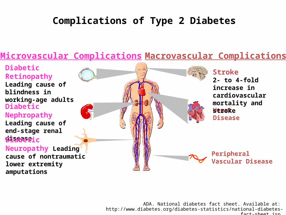
Natural history of DN

Definition
progressive rise in urine albumin excretion coupled with increasing BP and leading to declining GFR and CKD
Abnormal urine albumin excretion• >30 mg/24 hours
and/or
diabetic glomerular lesions
and/or
loss of glomerular filtration rateADA recommendations, Diabetes Care, January 2012

EpidemiologyDiabetic nephropathy affects
approximately one third of people with type 1 or type 2 diabetes mellitus.
Increase prevalence of DMUSA4% 1995 – 5.4% 2025
Now: USA 7% (20.8 million)Worldwide:2.8 % 171 million 2000 – 4.4% 366 million 2030
DN prevalence In India: 5.5% and 8.9%Asian Indians in UK 22.3%
Incident ESRD patients. Adj: age/gender/race; ref: 2010 ESRD patients.

04/18/2023 6
Epidemiology
Type 1 Diabetic 25 - 45% will develop diabetic nephropathy 80- 90% with microalbuminuria will progress to overt diabetic
nephropathy in 5 - 10 years nearly 100% with gross proteinuria will progress to ESRD in 7 - 10 yrs
Type 2 Diabetic 50% will have microalbuminuria at the time of presentation with
hypertension 10-20% with microalbuminuria will progress to overt nephropathy.
Risk factors for DN: Family historyHypertensionDyslipidemiaObese Male smokers

Pathology and pathophysiology
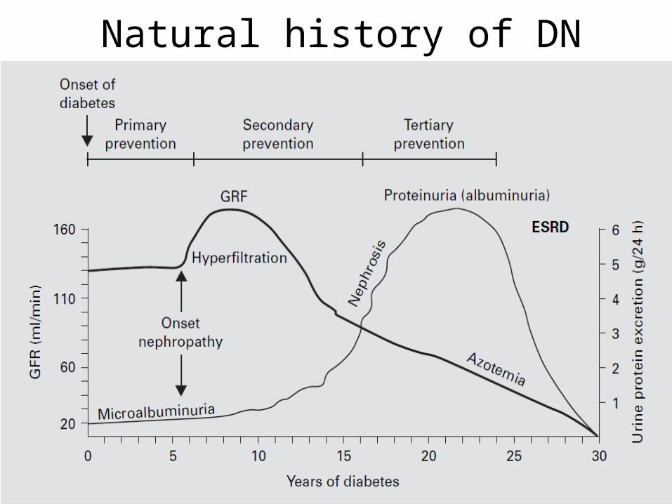
Pathophysiological stages
Stage increased GFR Increased filtration pressure as result of increased intraglolerular pressure Increased UOP and low s.cra, ureaPathological change but no clinically evident disease
Proteinuria Mesangial expansion and increased matrix change in pore sizes leading to leakage of proteinStarling Foces: increased plasma flow, increased glomerular capillary hydrostatic pressure
Microhematuria Ischemic injury of tubules due to construction and stenosis of efferent arteriole
Decreased GFR Atrophy and death of nephrons
CKD and ESKD Loss of compensation mechanisms of nephrons

Endothelium Fenestration (60 – 100 nm)Glycocalyx (network of proteoglycans with neg. charge
Endothelial cell injury: Increased permeability Impaired nitric oxide productionUpregulation of adhesion molecules
Defects of the glycocalyx: Decrease of negativity associated with increased albumin clearance
GBM: It is 300 – 400 nm tick gel like structure and 90% water It contains: collagen IV, heparan sulfate proteoglucans, laminin, nidogen
Heparan sulfate reduction correlates with degree of proteinuriaIts degradation is mediated by heparanase This theory approved in Type 1 and 2 DM but in advanced human cases but not in the early stage where there is also proteinuria.
Thickening of GBM:Accumulation of extracellular matrix Reduction in matrix degradation due to decreased metalloproteinase

Intraglomerular mesangial cells:Axis holding the edothelial and epithelial cellsConstruction and dilatation leading to fenestration change the filtration
Cytokines
inflammation
Mesangial cell expansion
Mesangial matrix expansionFibrin collagens deposition in GBM
Systemic HTN, RAS and hyperinsuliemia Efferent vasocostruction intraglomerular HTN High blood glucose
Free radicals
Atherosclerosis of efferent arteriole

Mesangial expansion By collagens : IV (α1,α2) V, VI
GBM thickening by collagens : IV (α3,α4,α5)
Hyaline subendothelial deposition

Pores Barrier sizeElectric charge Molecular weight
MW: < 40 kDa free to pass > 100 kDa totally restricted
Albumin mw 69 kDa
Microalbuminuria:Change in elecric charge
Macroalbuminuria: Change in elecric chargeIncreased pores size
Electric charge: Anionic ferritin restricted Cationic ferritin pass to podocyte Positively charged Dextran permeable more than neg. or neutral charged dextran

Synthesis and maintenance of the GBMCounteraction of hydrostatic pressureCritical membrane of the filtration barrier (last frontier) The narrow gaps 30 – 40 nm premeable for water and solutes It contains cytoskeleton The apical membrane contains podocalyxin, podoplanin and podoendin which are responable of the negative charge
Podocytopenia:Loss of negative charge (loss of podocalyxin)Change in the pores size due to damage in the diagram integrity
Causes of reduced number of podocytes: Podocyte detacementPodocyte apoptosisInability to proliferate and restore podocyte number
Silt diaphragm abnormalities:Abnormalities of nephrinFoot process widening and effacement

Pathological classification of DNClass Description Inclusion Criteria
IMild or nonspecific LM changes and EM-proven GBM thickening
Biopsy does not meet any of the criteria mentioned below for class II, III, or IV
GBM > 395 nm in female and >430 nm in male individuals 9 years of age and older, Podocyte hypertrophy
IIa
Mild mesangial expansion
Biopsy does not meet criteria for class III or IV
Mild mesangial expansion in >25% of the observed mesangium
IIb
Severe mesangial expansion
Biopsy does not meet criteria for class III or IV
Severe mesangial expansion in >25% of the observed mesangium
III Nodular sclerosis (Kimmelstiel–Wilson lesion)
Biopsy does not meet criteria for class IV
At least one convincing Kimmelstiel–Wilson lesion
IV Advanced diabetic glomerulosclerosis
Global glomerular sclerosis in >50% of glomeruli
Lesions from classes I through III


Mechanisms of proteinuria Site of injury
Glomerular hemodynamics Glomerular hyperfiltrationAfferent arteriole vasodilatation Efferent arteriole vasoconstriction glomerular capillary pressure
glomerular endothelial cellEndothelial cell injury Diminished endothelial glycocalyxAltered VEGF signaling
Hyperglycemia< AGE, ROS Endothelial cell injury or enzymatic cleavagePodocyte injury or loss
GBM Irregular thickeningDecreased negative charge
production and/or degradation of extracellular matrix proteins production and/or degradation of HSPG
podocyte
proximal tubule
PodocytopeniaLoss of slit diaphragm integrity Foot process widening and effacement
Loss negative charge
Decrease protein reabsorption
Detachment, apoptosis, lack of proliferation Decrease or changes in subcellular localization of nephrin Disrupted actin cytoskeletonLoss of slit diaphragm integrityImpaired podocyte GBM interaction Podocalyxin
Tubular injury and interstitial fibrosis
AGE, advanced glycosylation end products; HSPG, heparan sulfate proteoglycan; ROS, reactive oxygen species; VEGF, vascular endothelial growth factor.

Proteinuria Is an Independent Risk Factorfor Mortality in Type 2 Diabetes
1.0
0.9
0.8
0.7
0.6
0.5
0 1 2 3 4 5 6
Years
Su
rviv
al(a
ll-c
ause
mo
rtal
ity)
Normoalbuminuria(n=191)
Microalbuminuria(n=86)
Macroalbuminuria(n=51)
Gall et al. Diabetes. 1995;44:1303.


Increases AER Decreases AER
Strenuous exercise Poorly controlled DM Heart failure UTI Acute febrile illness Uncontrolled HPT Haematuria Menstruation Pregnancy
NSAIDs ACE inhibitors
Factors affecting urinary albumin excretion


04/18/2023 21
SCREENING FOR NEPHROPATHYWHEN: Type 1 - annually after puberty and 5 years of DM
Type 2 - at diagnosis and then annually
WHAT: random urine ACR;
and random urine dipstick
Normal< 2.0 mg/mmol men
< 2.8 mg/mmol womenRescreen in 1 year
Microalbuminuria2.0 - 20 mg/mmol men
2.8 - 28 mg/mmol women
Macroalbuminuria> 20 mg/mmol men
> 28 mg/mmol womenDiabetic nephropathy
diagnosed
Up to 2 repeat random urine ACRs performed 1 week to 2
months apart
Suspicion of nondiabetic
renal disease?
Yes
Workup or referral fornondiabetic renal
diseaseNo
Check ACR results
Only 1 abnormal ACR: Repeat screen
in 1 year
Any 2 abnormal out of 3 ACRs: Diabetic
nephropathy diagnosed

Albumin Excretion
SPECIMEN COLLECTED
24hr collection (mg/24h)
Timed collection (μg/min)
First voided morning specimen
Urine Albumin concentration
(mg/l)
Urine Albumin:Creatinine
ratio* (mg/mmol)
Normoalbuminuria <30 <20 <20 <3.5 (F)<2.5 (M)
Microalbuminuria 30-300 20-200 20-200 3.5 to 35 (F) 2.5 to 25 (M)
Overt proteinuria >300 >200 >200 >35 (F)>25 (M)
Stages of Renal Involvement According to the Urinary Albumin Level

04/18/2023 23
TREATMENT OF NEPHROPATHY
Already on ACE inhibitor?
Choose 2nd line therapy: ACE or
ARB and add non-DHP CCB
NO
On first-line nephropathydrug?
NO
First line drug atmaximum dose?
YES
Add first-line drug;Recheck ACR in 2 weeks to 2 months
ACR normal?
First line drugs:Type 1- ACE inhibitorType 2 with Cr Cl > 60 mL/min - ACE inhibitor or ARBType 2 with Cr Cl 60 mL/min - ARB
Titrate up; recheck ACR in
2 weeks to 2 months
YES
Yes Remeasure ACR in 1 year
NONO
YES

Table 2. Recommendations for the Comprehensive Management of T2DM Patients with CKD
Factor Recommendations Lifestyle factors diet, exercise, smoking,and alcohol intake
Blood glucose Treatment goal: HbA1c <6.5%Preprandial plasma glucose 90-130 mg/dlPostprandial plasma glucose <180 mg/dl
Blood pressure Goal ≤130/80 mm HgUse maximal tolerated dose of ACE inhibitor or ARB before adding a second agent
Cholesterol Goal <4.0 mmol/L for total cholesterol and <2.0 mmol/L for LDL-C Consider use of a statin irrespective of baseline lipid values for the secondary prevention of cardiovascular disease
Platelets Consider use of low dose aspirin for the secondary prevention of cardiovascular disease
Monitoring Annual monitoring of eGFR and ACR

A1c Target :
Outcome
UKPDS 10 yrs
> 7% The clinical lesson from the UKPDS follow-up studies is that, although the risks of complications of hypertension might be mitigated with initiation of treatment even after a prolonged elevation of blood pressure, it is particularly necessary to treat hyperglycemia appropriately from the outset of type 2 diabetes.
ADVANCE studyIntensive Blood Glucose Control and VascularOutcomes in Patients with Type 2 Diabetes
6.5% A strategy of intensive glucose control, involving gliclazide (modified release) andother drugs as required, that lowered the glycated hemoglobin value to 6.5% yielded a 10% relative reduction in the combined outcome of major macrovascular and microvascular events, primarily as a consequence of a 21% relative reduction in nephropathy.
ACCORD Study: Long-Term Effects of Intensive Glucose Lowering on Cardiovascular Outcomes
<6% As compared with standard therapy, the use of intensive therapy for 3.7 years to target a glycated hemoglobin level below 6% reduced 5-year nonfatal myocardial infarctions but increased 5-year mortality. Such a strategy cannot be recommended for high-risk patients with advanced type 2 diabetes.
The glycemic control studies

KDIGO recommendation for proteinuria
• ARBs are more effective than other antihypertensive classes in slowing progression of kidney disease characterized by macroalbuminuria in hypertensive patients with type 2 diabetes. (Strong)
• ACE inhibitors are more effective than other antihypertensive classes in slowing progression of kidney disease characterized by macroalbuminuria in hypertensive patients with type 1 diabetes. (Strong)
• ACE inhibitors may be more effective than other antihypertensive classes in slowing the progression of kidney disease characterized by macroalbuminuria in hypertensive patients with type 2 diabetes. (Weak)
ACE inhibitors and ARBs are effective in slowing progression of kidney disease characterized by microalbuminuria in hypertensive patients with type 1 or type 2 diabetes. (Moderate)
ACE inhibitors, ARBs, and nondihydropyridine calcium channel blockers have a greater antiproteinuric effect than other antihypertensive classes in hypertensive patients with DKD. (Strong)
Dihydropyridine calcium channel blockers, when used to treat hypertension in the absence of ACE inhibitors or ARBs, are less effective than other agents in slowing progression of DKD. (Strong)

ACE Inhibitors can prevent progression of renal failure
120
160
200
240
280
320
350
400
800 1 2 3 4 5 6
Years
Ann Intern Med 118 577-581.1993J Am Soc Nephrol 2006
Placebo
Enalapril 85
90
95
100
105
110
800 1 2 3 4 5 6
Years
Placebo
Enalapril
Normotensive Type 2 Diabetics
Proteinuria
(mg/day)
% Initial GFR
Risk reduction is 51%Reduce microalbuminuria All causes of mortality

Incidence of Progression to Diabetic Nephropathy during Treatment with 150 mg of Irbesartan Daily, 300 mg of Irbesartan Daily, or Placebo in Hypertensive
Patients with Type 2 Diabetes and Persistent Microalbuminuria.
Parving H et al. N Engl J Med 2001;345:870-878.

Olmesartan for the Delay or Prevention of Microalbuminuria in Type 2 Diabetes in 4447 patients
Conclusion:• Olmesartan was associated with a delayed onset of microalbuminuria, even though
blood-pressure control• The higher rate of fatal cardiovascular events with olmesartan among patients with
preexisting coronary heart disease is of concern.
Combined Angiotensin Inhibition for the Treatment of Diabetic Nephropathy in 1448 patients
Combination therapy with an ACE inhibitor and an ARB was associated with an increased risk of adverse events among patients with diabetic nephropathy.
There was no benefit with respect to mortality or cardiovascular events. Combination therapy increased the risk of hyperkalemia and acute kidney injury.

Metformin in Patients with T2DM and CKD
• The recommendation of the ADA/EASD metformin can be used – down to an eGFR of 30 mL/min/1.73 m2, – the dose of metformin should be reduced when
eGFR is less than 45 mL/min/1.73 m2. – Kidney function should be checked regularly (every 6
months)– discontinued if eGFR falls below 30 mL/min/1.73 m2. – prescribed with caution in patients with an eGFR less
than 45 mL/min/1.73 m2, which is rapidly deteriorating.
– All patients should be warned that if they develop a condition that can lead to dehydration.


KDIGO lipid control in DM
• Target LDL-C in people with diabetes and CKD stages 1-4 should be < 100 mg/dL; <70 mg/dL is a therapeutic option. (B)
• People with diabetes, CKD stages 1-4, and LDL-C > 100 mg/dL should be treated with a statin. (B)
• Treatment with a statin should not be initiated in patients with type 2 diabetes on maintenance hemodialysis who do not have a specific cardiovascular indication for treatment. (A)
• Atorvastatin treatment in patients with type 2 diabetes on maintenance treatment does not improve cardiovascular outcomes. (Strong)

Other oral antidiabetic drugs :
DPP-4 inhibitors:Choices of antidiabetic agents for patients
with type 2 diabetes mellitus (T2DM) and chronic kidney disease (CKD) are limited. Available data suggest that the use of dipeptidyl peptidase-4 (DPP-4) inhibitors may be safe in patients at various stages of renal insufficiency. However, except for linagliptin, dosage adjustment is necessary.
patients with moderate renal impairment (defined in the label as a creatinine clearance ≥ 30 to < 50 ml/min). In severe renal impairment (creatinine clearance < 30 ml/min) or end-stage renal disease requiring dialysis, the dose is further reduced to 25 mg once daily.
Gliclazide In patients with mild to moderate
renal insufficiency the same dosing regimen can be used as in patients with normal renal function with careful patient monitoring. These data have been confirmed in clinical trials.
glimepirideA multiple-dose titration using
doses ranging from 1 mg to 8 mg daily for 3 months. Baseline creatinine clearance ranged from 10–60 mL/min.

Insulin
Metabolism of insulin: 30–80% of systemic insulin in the kidney
40–50% of the endogenous insulin metabolized by the liver
Insulin effect on the kidney : Na reabsorption Increase
glucose and phosphate higher risk of hypoglycaemiaIntensive glucose control with
HbA1c around 7% is associated with:
Reduction of microalbuminuria by 39% and marcroalbuminuria by 54%

(SHARP; Lancet. 2011;377:2181-2192).
Study of Heart and Renal Protection (SHARP): randomized trial to assess the effects of lowering low-density lipoprotein cholesterol among 9,438 patients with chronic kidney disease.
CONCLUSIONS: SHARP should provide evidence about the efficacy and safety of lowering LDL cholesterol with the combination of ezetimibe and simvastatin among a wide range of patients with CKD
RESULTS: A total of 9,438 CKD patients were randomized, of whom 3,056 were on dialysis. Mean age was 61 years, two thirds were male, one fifth had diabetes mellitus, and one sixth had vascular disease. Compared with either placebo or simvastatin alone, allocation to ezetimibe plus simvastatin was not associated with any excess of myopathy, hepatic toxicity, or biliary complications during the first year of follow-up. Compared with placebo, allocation to ezetimibe 10 mg plus simvastatin 20 mg daily yielded average LDL cholesterol differences of 43 mg/dL (1.10 mmol/L) at 1 year and 33 mg/dL (0.85 mmol/L) at 2.5 years. Follow-up is scheduled to continue until August 2010, when all patients will have been followed for at least 4 years.
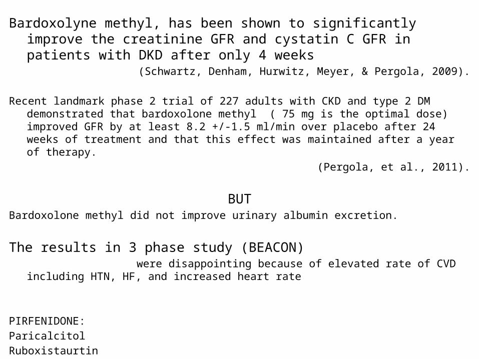
Bardoxolyne methyl, has been shown to significantly improve the creatinine GFR and cystatin C GFR in patients with DKD after only 4 weeks
(Schwartz, Denham, Hurwitz, Meyer, & Pergola, 2009).
Recent landmark phase 2 trial of 227 adults with CKD and type 2 DM demonstrated that bardoxolone methyl ( 75 mg is the optimal dose) improved GFR by at least 8.2 +/-1.5 ml/min over placebo after 24 weeks of treatment and that this effect was maintained after a year of therapy.
(Pergola, et al., 2011).
BUTBardoxolone methyl did not improve urinary albumin excretion.
The results in 3 phase study (BEACON) were disappointing because of elevated rate of CVD including HTN, HF, and increased heart rate
PIRFENIDONE: Paricalcitol RuboxistaurtinAllopurinol

Thank you
The End
Related Documents