From DEPARTMENT OF MEDICINE, UNIT OF EXPERIMENTAL RHEUMATOLOGY Karolinska Institutet, Stockholm, Sweden PATHOPHYSIOLOGICAL MECHANISMS AND CLINICAL MANIFESTATIONS IN PRIMARY SJÖGREN’S SYNDROME AND SYSTEMIC LUPUS ERYTHEMATOSUS Marika Kvarnström Stockholm 2014

Welcome message from author
This document is posted to help you gain knowledge. Please leave a comment to let me know what you think about it! Share it to your friends and learn new things together.
Transcript
From DEPARTMENT OF MEDICINE, UNIT OF EXPERIMENTAL RHEUMATOLOGY Karolinska Institutet, Stockholm, Sweden
PATHOPHYSIOLOGICAL MECHANISMS AND CLINICAL MANIFESTATIONS IN
PRIMARY SJÖGREN’S SYNDROME AND SYSTEMIC LUPUS ERYTHEMATOSUS
Marika Kvarnström
Stockholm 2014

All previously published papers were reproduced with permission from the publisher. Published by Karolinska Institutet. Printed by ÅTTA.45 TRYCKERI AB © Marika Kvarnström, 2014 ISBN 978-91-7549-571-2
Department of Medicine, Unit of Experimental Rheumatology
Pathophysiological Mechanisms and Clinical Manifestations in Primary Sjögren´s Syndrome and Systemic Lupus ErythematosusThesis for Doctoral Degree (Ph.D.)
by
Marika Kvarnström MD
Defense of the thesis will occur Friday 9th May at 9.00 in the CMM Lecture Hall, CMM L8:00, Karolinska University Hospital, Solna
Principal Supervisor:Professor Marie Wahren-HerleniusKarolinska InstitutetDepartment of Medicine, SolnaUnit of Experimental Rheumatology
Co-supervisor(s):Assoc. Prof. Elisabet SvenungssonKarolinska InstitutetDepartment of Medicine, SolnaRheumatology Unit
Opponent:Assoc. Prof. Thomas MandlLund UniversityDepartment of RheumatologyMalmö University Hospital
Examination Board:Assoc. Prof. Ann-Charlotte WikströmKarolinska InstitutetDepartment of Clinical Immunology and Transfusion MedicineKarolinska University Hospital
Assoc. Prof. Johan BrattKarolinska InstitutetDepartment of MedicineRheumatology Unit
Professor Kristina ÅkessonLund UniversityDepartment of Clinical SciencesClinical and Molecular Osteoporosis Research Unit


Till Carl

ABSTRACT Primary Sjögren’s syndrome (pSS) is a systemic inflammatory autoimmune condition. The etiology of pSS is mainly unknown, but it has been suggested that environmental factors trigger an immune response in genetically susceptible individuals. The disease is characterized by chronic inflammation which results in progressive destruction of exocrine glands, primarily the salivary and lacrimal glands, leading to symptoms of dryness, mainly in the mouth and eyes. Patients may also experience symptoms of exocrine dysfunction and dryness in other parts of the body. Autoantibodies against the antigens Ro/SSA (Ro52 and Ro60) and La/SSB, are a well-known sign of dysregulation of the immune system. Extraglandular manifestations (EGM) are present in a subset of patients with pSS.
In a population based prospective study we included all patients referred to the Department of Rheumatology for suspected pSS during a five-year period. The patients were evaluated using a structured procedure according to the 2002 Revised American-European Consensus Criteria for Sjögren’s syndrome. Of referred individuals, 199 of 781 patients were diagnosed with pSS. We found an annual incidence rate of pSS in the Karolinska University Hospital catchment area of 3.1 (95% CI 2.3-4.3) cases per 100,000 adult inhabitants. In this cohort, we noted lower figures for severe EGM such as lung and neurological involvement than previously reported for prevalent pSS. Also, the frequency of autoantibodies including ANA, anti-Ro/SSA and anti-La/SSB was lower compared to other cohorts. Subsets of these patients were recruited and included in genetic studies.
We contributed with 79 pSS patients in a candidate-gene association case control study in 540 patients with pSS and 532 controls, from Sweden and Norway. Three novel gene loci, not previously associated with pSS, were identified: the early B-cell factor 1 (EBF1) gene, the family with sequence similarity 167 member A–B-lymphoid tyrosine kinase (FAM167A–BLK) locus, and the tumor necrosis factor superfamily (TNFSF4=Ox40L) gene. Variations in the gene loci for interferon regulation factor 5 (IRF5) and signal transducer and activator of transcription 4 (STAT4) were confirmed. The three novel gene variants are involved in B-cell development and activation.
In an international multicenter case control genome-wide association study (GWAS)/ large-scale association study of pSS, we contributed with 99 patients. The results confirmed associations with variations in the gene loci of the human leukocyte antigen (HLA) region, IRF5, STAT4, FAM167A-BLK, chemokine C-X-C motif receptor 5 (DX6-CXCR), and TNFAIP3 interacting
protein 1 (TNIP1) at a genome-wide significance level. In addition, 29 further suggested associations (p meta < 5 × 10⁻⁵) were observed. The major part of these genes is involved in both innate and adaptive immune responses.
We observed that the autoantigen Ro52 was expressed and upregulated in salivary glands of patients with primary Sjögren’s syndrome. This was noted in biopsies from 28 pSS patients and 19 non-pSS controls from Sweden and Norway. The degree of expression was correlated with the level of inflammation in the tissue.
Systemic lupus erythematosus (SLE) is a chronic autoimmune disease characterized by multiple organ manifestations and immunological dysregulation including production of a large set of autoantibodies targeting different epitopes. We showed that antibodies to the RING domain of Ro52, which is the functionally active domain with E3 ligase activity, were significantly correlated with disease activity as measured by the SLAM score in a well-characterized cohort of SLE patients.
In conclusion, the results of these studies have highlighted the role of the immune system in the pathogenesis in pSS and SLE.
SVENSK SAMMANFATTNING Primärt Sjögren’s syndrom (pSS) är en systemisk autoimmun inflammatorisk sjukdom. Etiologin till pSS är huvudsakligen okänd, men hypotesen är att miljöfaktorer utlöser en immunologisk reaktion i genetiskt predisponerade individer. Sjukdomen karakteriseras av kronisk inflammation som progressivt destruerar exokrina körtlar. Mest drabbade är saliv- och tårkörtlar, vilket leder till symtom med torrhet, s.k. sicca symtom, i främst mun och ögon. Patienterna kan uppleva sicca symtom även i andra delar av kroppen orsakad av exokin dysfunktion. Autoantikroppar mot antigenerna Ro/SSA (Ro52 och Ro60) och La/SSB, är ett tecken på förändrad reglering av immunförsvaret. Extraglandular manifestationer (EGM), förekommer hos en del patienter med pSS.
I en populations baserad prospektiv studie, inkluderade vi alla patienter som remitterades till Reumatologiska kliniken vid Karolinska Universitetssjukhuset för utredning av pSS under fem års tid. Patienterna utreddes enligt 2002 års reviderade Amerikansk-Europeiska Konsensus Kriterierna för Sjögrens syndrom. Av 781 remitterade patienter uppfyllde 199 kriterierna för pSS. Incidensen beräknades till 3.1 (95% CI 2.3-4.3) fall per 100 000 vuxna invånare per år i Karolinska Universitetssjukhusets upptagningsområde. I denna kohort noterade vi lägre prevalence siffror för EGM som interstitiell lungsjukdom och polyneuropati jämfört med tidigare studier. Frekvensen av autoantikroppar såsom ANA, anti-Ro/SSA- och anti-La/SSB var också lägre vid jämförelse
Vi bidrog med 79 pSS-patienter i en kandidatgen fall-kontrollstudie med totalt 540 patienter med pSS och 532 kontroller från Sverige och Norge. Tre nya gen-lokus, som inte tidigare förknippats med pSS, identifierades: the early B-cell factor 1 (EBF1) genen, the family with sequence similarity 167 member A–B-lymphoid tyrosine kinase (FAM167A–BLK) gen-lokus och the tumor necrosis factor superfamily (TNFSF4=Ox40L) genen. Genvariation i interferon regulation factor 5 (IRF5) och signal transducer and activator of transcription 4 (STAT4) generna bekräftades. De tre nya genvariationerna ligger i gener som deltar i B-cells utmognad och aktivering.
I en internationell multicenter fall-kontroll studie, en sk genome-wide association studie (GWAS), bidrog vi med 99 patienter. Resultaten bekräftade tidigare fynd från andra studier, men med en genome-wide signifikansnivå. Förändringarna var belägna i HLA regionen, IRF5, STAT4, FAM167A-BLK, chemokine C-X-C motiv receptor 5 (DX6-CXCR), och TNFAIP3 interacting protein 1 (TNIP1). Dessutom observerades ytterligare 29 genvarianter med lägre signifikansnivå (p meta < 5 × 10⁻⁵). De flesta av generna är inblandade i det medfödda och adaptiva immunförsvaret.
I en laborativ studie kunde vi konstatera att autoantigenet Ro52 uttryckts och uppregleras i spottkörtelvävnad hos patienter med primärt Sjögrens syndrom. Detta undersöktes i biopsier från 28 patienter med pSS och 19 kontrollerfrån Sverige och Norge. Graden av uppreglering korrelerade med graden av inflammation i vävnaden.
Systemisk lupus erythematosus (SLE) är en kronisk autoimmun sjukdom som kännetecknas av multipla organ manifestationer och hög produktion av autoantikroppar riktade mot olika epitoper. Vi visade att antikroppar riktade mot den s.k. RING domänen av Ro52 proteinet var signifikant korrelerat med ökad sjukdomsaktivitet mätt med SLAM (SLE activity measure). Undersökningen genomfördes i en välkaraktäriserad kohort av SLE patienter. RING domänen är den funktionellt aktiva domänen av Ro52 proteinet med E3 ligase aktivitet vilket indikeras en inverkan som hör ihop med Ro52:s funktion.
Sammanfattningsvis har resultaten av dessa studier belyst immunförsvarets roll i patogenesen vid sjukdomarna pSS och SLE.
LIST OF SCIENTIFIC PAPERS This thesis is based on the following papers, which will be referred to in the text by their Roman numerals.
I. Marika Kvarnström, Vijole Ottosson, Birgitta Nordmark, Marie Wahren-Herlenius. Incident cases of primary Sjögren´s syndrome during a five-year period in Stockholm County: A descriptive study of the patients and their characteristics. Submitted manuscript.
II. Gunnel Nordmark, Gulla Kristjansdottir, Elke Theander, Silke Appel, Per Eriksson, Lilian Vasaitis, Marika Kvarnström, Nicolas Delaleu, P Lundmark, A Lundmark, Christopher Sjöwall, Johan G Brun, Malin V Jonsson, E Harboe, L G Göransson, Svein Joar Johnsen, P Söderkvist, Maija-Leena Eloranta, Gunnar Alm, Eva Baecklund, Marie Wahren-Herlenius, Roald Omdal, Lars Rönnblom, Roland Jonsson, Ann-Christine Syvänen. Association of EBF1, FAM167A(C8orf13)-BLK and TNFSF4 gene variants with primary Sjogren's syndrome. Genes and immunity, 2011, 12, 100-109.
III. Christopher J Lessard, He Li, Indra Adrianto, John A Ice, Astrid Rasmussen, Kiely M Grundahl, Jennifer A Kelly, Mikhail G Dozmorov, Corinne Miceli-Richard, Simon Bowman, Sue Lester, Per Eriksson, Maija-Leena Eloranta, Johan G Brun, Lasse G Goransson, Erna Harboe, Joel M Guthridge, Kenneth M Kaufman, Marika Kvarnström, Helmi Jazebi, Deborah S Graham Cunninghame, Martha E Grandits, Abu N M Nazmul-Hossain, Ketan Patel, Adam J Adler, Jacen S Maier-Moore, A Darise Farris, Michael T Brennan, James A Lessard, James Chodosh, Rajaram Gopalakrishnan, Kimberly S Hefner, Glen D Houston, Andrew J W Huang, Pamela J Hughes, David M Lewis, Lida Radfar, Michael D Rohrer, Donald U Stone, Jonathan D Wren, Timothy J Vyse, Patrick M Gaffney, Judith A James, Roald Omdal, Marie Wahren-Herlenius, Gabor G Illei, Torsten Witte, Roland Jonsson, Maureen Rischmueller, Lars Rönnblom, Gunnel Nordmark, Wan-Fai Ng, for UK Primary Sjogren's Syndrome Registry, Xavier Mariette, Juan-Manuel Anaya, Nelson L Rhodus, Barbara M Segal, R Hal Scofield, Courtney G Montgomery, John B Harley, Kathy L Sivils. Variants at multiple loci implicated in both innate and adaptive immune responses are associated with Sjögren’s syndrome. Nature Genetics, 2013, 45, 1284-1294.
IV. Marika Kvarnström, Vijole Dzikaite-Ottosson, Lars Ottosson, Johanna Gustafsson, Iva Gunnarsson, Elisabet Svenungsson, Marie Wahren-Herlenius. Autoantibodies to the functionally active RING-domain of Ro52/SSA are associated with disease activity in patients with lupus. Lupus, 2013, 22, 477-485.
V. Lara A. Aqrawi, Marika Kvarnström, Karl A. Brokstad, Roland Jonsson, Kathrine Skarstein, Marie Wahren-Herlenius. Ductal epithelial expression of Ro52 correlates with inflammation in salivary glands of patients with primary Sjögren’s syndrome. Clinical and Experimental Immunology, 2014, Mar 28. doi: 10.1111/cei.12341. PMID:24673429
CONTENTS 1 SYSTEMIC AUTOIMMUNE RHEUMATIC DISEASES ....................... 1
1.1 Introduction ........................................................................................ 1 2 ETIOLOGIC FACTORS IN SYSTEMIC AUTOIMMUNITY ................ 2
2.1 GENETICS ......................................................................................... 2 2.1.1 Genetics in Sjögren’s syndrome ............................................ 3 2.1.2 Genetics in SLE ..................................................................... 4
2.2 GENDER ............................................................................................ 4 2.2.1 Gender and Sjögren’s syndrome ........................................... 4 2.2.2 Gender and SLE ..................................................................... 5
2.3 ENVIRONMENTAL FACTORS ..................................................... 5 2.3.1 Environmental factors and Sjögren’s syndrome ................... 5 2.3.2 Environmental factors and SLE ............................................. 5
3 IMMUNOPATHOLOGY IN SYSTEMIC AUTOIMMUNITY ............... 6 3.1 INNATE IMMUNE REACTIONS ................................................... 6 3.2 THE ADAPTIVE IMMUNE SYSTEM ............................................ 7 3.3 AUTOANTIGENS AND AUTOANTIBODIES .............................. 9
3.3.1 ANA ..................................................................................... 10 3.3.2 ENA ...................................................................................... 10 3.3.3 Ro/SSA ................................................................................. 10 3.3.4 Ro60 ..................................................................................... 10 3.3.5 Ro52 ..................................................................................... 11 3.3.6 La .......................................................................................... 11
4 CLINCIAL FEATURES OF SYSTEMIC AUTOIMMUNE DISEASES12 4.1 SJÖGREN’S SYNDROME ............................................................. 12
4.1.1 Background/Summary ......................................................... 12 4.1.2 Immunopathogenesis ........................................................... 13 4.1.3 Prevalence ............................................................................ 14 4.1.4 Incidence .............................................................................. 14 4.1.5 Organ manifestations ........................................................... 15 4.1.6 Classification and diagnosis................................................. 18 4.1.7 Disease activity index .......................................................... 19 4.1.8 Treatment ............................................................................. 21 4.1.9 Prognosis .............................................................................. 23
4.2 SYSTEMIC LUPUS ERYTHEMATOSUS (SLE) ........................ 23 4.2.1 Background/Summary ......................................................... 23 4.2.2 Immunopathogenesis ........................................................... 24 4.2.3 Incidence .............................................................................. 26 4.2.4 Prevalence ............................................................................ 26 4.2.5 Organ manifestations ........................................................... 26 4.2.6 Classification and diagnosis................................................. 29
4.2.7 Disease activity and damage ................................................ 31 4.2.8 Treatment ............................................................................. 35 4.2.9 Prognosis .............................................................................. 35
5 AIMS .......................................................................................................... 37 6 PATIENTS AND METHODS .................................................................. 38
6.1 Patients .............................................................................................. 38 6.2 Study designs .................................................................................... 39 6.3 Diagnosis of Sjögren’s syndrome .................................................... 39 6.4 Extraglandular manifestations, organ involvements and indices measuring disease activity and damage ..................................................... 40 6.5 General health and anamnestic data ................................................ 40 6.6 Laboratory parameters ..................................................................... 40 6.7 Genotyping ....................................................................................... 41 6.8 Minor saliva gland biopsies ............................................................. 41 6.9 Statistical analyses ............................................................................ 42
7 RESULTS ................................................................................................... 43 7.1 Paper I ............................................................................................... 43 7.2 Paper II ............................................................................................. 46 7.3 Paper III ............................................................................................ 46 7.4 Paper IV ............................................................................................ 47 7.5 Paper V ............................................................................................. 47
8 DISCUSSION ............................................................................................ 48 8.1 Paper I ............................................................................................... 48 8.2 Paper II ............................................................................................. 49 8.3 Paper III ............................................................................................ 50 8.4 Paper IV ............................................................................................ 51 8.5 Paper V ............................................................................................. 52
9 CONCLUDING REMARKS AND FUTURE PERSPECTIVES ........... 54 10 ACKNOWLEDGEMENTS ...................................................................... 55 11 REFERENCES ........................................................................................... 58
LIST OF ABBREVIATIONS ACR American College of Rheumatology AECC 2002 Revised American-European Consensus Criteria for
Sjögren’s syndrome AIS Adaptive Immune System ANA Antinuclear Antibodies ANCA Anti-Neutrophil Cytoplasmic Antibodies APC Antigen Presenting Cell ANCA Anti-neutrophil Cytoplasmic Antibodies Anti-dsDNA Antibodies to double stranded DNA BAFF B-cell Activating Factor CNS Central Nervous System CRP C-Reactive Protein CVD Cardiovascular Diseases DC Dendritic Cell DLBC lymphoma Diffuse Large B-cell lymphoma DMARD Disease Modifying Anti Rheumatic Drugs dRTA distal Renal Tubular Acidosis EBV Epstein-Barr Virus EGM Extraglandular Manifestations ELISA Electron-Linked-Immunosorbent Assay ENA Extractable Nuclear Antigen ESR Erythrocyte Sedimentation Rate ESSDAI EULAR Sjögren’s Syndrome Disease Activity Index EULAR European League Against Rheumatism FDA Food and Drug Administration GWAS Genome Wide Association Studies HCV Hepatitis C viruse HLA Human Leukocyte Antigen IFL Indirect Immunofluorescence Technique IFN Interferon Ig Immunoglobulin IL Interleukin IRF Interferon Regulatory Factor IVIg Intravenous Immunoglobulins LD Linkage Disequilibrium MALT lymphoma Mucosa-Associated Lymphoid Tissue lymphoma MHC Major Histocompatibility Complex NK cells Natural Killer cells NSAID Non Steroid Anti Inflammatory Drugs
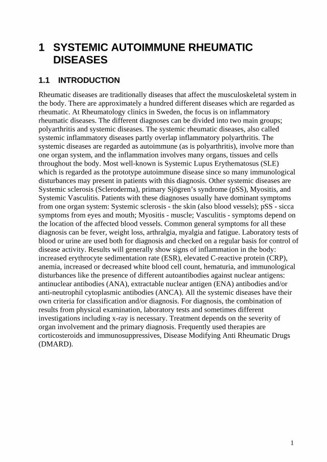
NET Neutrophil Extracellular Traps OD Optical Density OR Odds Ratio pDC plasmacytoid Dendritic Cell PNS Peripheral Nervous System pSS Primary Sjögren’s Syndrome QC Quality Control RING Really Interesting New Gene SCLE Subacute Cutaneous Lupus Erythematosus SLAM Systemic Lupus Activity Measure SLE Systemic Lupus Erythematosus SLEDAI Systemic Lupus Erythematosus Disease Activity Index SLICC Systemic Lupus Erythematosus Collaborating Clinics SNP Single Nucleotide Polymorphism SSA Sjögren’s Syndrome Antigen A=Ro SSB Sjögren’s Syndrome Antigen B=La sSS Secondary Sjögren’s Syndrome STAT4 Signal Transducer and Activator of Transcription TLR Toll Like Receptors TNFα Tumor Necrosis Factor α UWSF Unstimulated Whole Salivary Flow WHO World Health Organization
1
1 SYSTEMIC AUTOIMMUNE RHEUMATIC DISEASES
1.1 INTRODUCTION Rheumatic diseases are traditionally diseases that affect the musculoskeletal system in the body. There are approximately a hundred different diseases which are regarded as rheumatic. At Rheumatology clinics in Sweden, the focus is on inflammatory rheumatic diseases. The different diagnoses can be divided into two main groups; polyarthritis and systemic diseases. The systemic rheumatic diseases, also called systemic inflammatory diseases partly overlap inflammatory polyarthritis. The systemic diseases are regarded as autoimmune (as is polyarthritis), involve more than one organ system, and the inflammation involves many organs, tissues and cells throughout the body. Most well-known is Systemic Lupus Erythematosus (SLE) which is regarded as the prototype autoimmune disease since so many immunological disturbances may present in patients with this diagnosis. Other systemic diseases are Systemic sclerosis (Scleroderma), primary Sjögren’s syndrome (pSS), Myositis, and Systemic Vasculitis. Patients with these diagnoses usually have dominant symptoms from one organ system: Systemic sclerosis - the skin (also blood vessels); pSS - sicca symptoms from eyes and mouth; Myositis - muscle; Vasculitis - symptoms depend on the location of the affected blood vessels. Common general symptoms for all these diagnosis can be fever, weight loss, arthralgia, myalgia and fatigue. Laboratory tests of blood or urine are used both for diagnosis and checked on a regular basis for control of disease activity. Results will generally show signs of inflammation in the body: increased erythrocyte sedimentation rate (ESR), elevated C-reactive protein (CRP), anemia, increased or decreased white blood cell count, hematuria, and immunological disturbances like the presence of different autoantibodies against nuclear antigens: antinuclear antibodies (ANA), extractable nuclear antigen (ENA) antibodies and/or anti-neutrophil cytoplasmic antibodies (ANCA). All the systemic diseases have their own criteria for classification and/or diagnosis. For diagnosis, the combination of results from physical examination, laboratory tests and sometimes different investigations including x-ray is necessary. Treatment depends on the severity of organ involvement and the primary diagnosis. Frequently used therapies are corticosteroids and immunosuppressives, Disease Modifying Anti Rheumatic Drugs (DMARD).
2
2 ETIOLOGIC FACTORS IN SYSTEMIC AUTOIMMUNITY
The different systemic autoimmune rheumatic diseases have many clinical features in common as described above. In recent years, research has found similar etiologic mechanisms for these diseases in areas such as genetics, immunology and environmental factors. Hence, the two diseases included in this thesis, pSS and SLE, are as far as possible presented integrated in the text.
2.1 GENETICS During the 20th century science has revealed how genetic information works and is inherited. The cellular basis of heredity is chromosomes (46XYin humans) consisting of the DNA (Deoxyribonucleic acid) double helix molecule and histones. DNA is built of nucleotides with four different nucleobases: adenine, cytosine, guanine and thymine (A, C, G and T). The nucleobases form pairs, either A-T or C-G. This sequence of the DNA strands provides all genetic information. The biological mechanisms by which cells read the information contained in DNA and eventually produce peptides include transcription of DNA to messenger-RNA (a single stranded polynucleotide) and translation to peptides where the amino acid sequence depends on the nucleotide-sequence (Strachan and Read 2011c). The human genome consists of over three billion base-pairs. Scientists have been working on the task to decipher first genes, and then the entire human genome. The invention of the recombinant DNA technologies with cloning and sequencing was used to reveal the human complete DNA sequence i.e. genome (Lander, et al. 2001; Venter, et al. 2001). The human genome differs by less than a half percent person to person (Kidd, et al. 2008).
A gene is a molecular unit of DNA, which is inherited, usually coding for a protein. The number of protein coding genes is currently 18,683 (http://www.ncbi.nlm.nih.gov/CCDS, Jan 29, 2013). The genes consist of coding and non-coding sequences of DNA. The non-coding parts, which are removed from the transcript, are called introns, and can have regulatory functions such as providing binding sites for regulatory factors, or acting as promoters where the transcription starts (Strachan and Read 2011c).
Single nucleotide polymorphisms (SNPs) are the most common genetic variations, and occur at 1 out of every 1,900 bases in the human genome (Sachidanandam, et al. 2001). They are mutations which originate from the transcription of DNA. There are millions of SNPs registered in the main SNP database, dbSNP (http://www.ncbi.nlm.nih.gov/projects/SNP/). SNPs are defined as: a single base-pair at a specific position, which has been changed from A-T to C-G, or the opposite. Other examples of genetic variations are insertion/deletion or structural alterations of base-pairs or nucleobases. SNPs of importance are those located in a coding region, resulting
3
in an altered amino acid sequence of the peptide eventually produced, or in an impact on the regulation of the gene.
The human haplotype mapping project, the HapMap project, catalogues all human genetic variations and is freely accessible, via the internet. Haplotype is a particular combination of alleles along a chromosome, and an allele is one in the pair of alternative forms of the same gene locus on a specific chromosome (Strachan and Read 2011b) (2005). The variation of alleles at a locus is measurable as the number of alleles present in the population. The frequency of the minor allele must be greater than 1% to be regarded as a polymorphism (Feero, et al. 2010).
In diseases caused by a combination of genetic and environmental factors, complex diseases, both common and rare alleles can be involved (Feero, et al. 2010; Schork, et al. 2009). Association studies have been performed to identify gene loci that underlie complex diseases. New genotyping technique methods have made large-scale analysis of SNPs possible (Syvanen 2001).
Association studies of SNPs are either in the form of Genome Wide Association Studies (GWAS) which measure the whole genome, or candidate gene studies which measure selected genes. These studies in large patient cohorts have made it possible to measure the associations between mapped SNPs and complex diseases (Manolio, et al. 2008).
When an association study is planned, the decision on which SNPs to analyze is based on linkage disequilibrium (LD). LD is the correlation between SNP alleles; a person with a specific allele at one site often carries specific alleles at other nearby variant sites. Tag-SNPs are the variant chosen for analysis. Nearby SNPs are not analyzed if in high LD (>80%). The results in the form of different frequencies in patients and controls are compared. Odds Ratio (OR) is calculated and the p-value needs to have a stringent value to compensate for multiple testing (Strachan and Read 2011a).
Today, with commercially available gene chips, it is possible to detect over 1 million different SNPs in a sample from a person in less than a day.
2.1.1 Genetics in Sjögren’s syndrome Few studies on genetic variation had been published at the beginning of the work for this thesis, except for the MHC/HLA region. Several specific genetic polymorphisms have since been significantly associated with pSS in large scale candidate-gene studies (Nordmark, et al. 2011; Nordmark, et al. 2013; Reksten, et al. 2013) and a recently reported GWAS (Lessard, et al. 2013). The SNPs identified to associate with pSS are located in the region of MHC/HLA, in genes affecting innate immune mechanisms such as interferon- regulating and signaling pathways, as well as in genes of the adaptive immune response.
4
2.1.2 Genetics in SLE Familial aggregation, in the form of an affected first-degree relative, occurs in ten to twenty percent of SLE patients (Arnett and Shulman 1976). This indicates the importance of genetic factors.
Several SLE genes have been discovered using candidate gene studies. In a meta-analysis, 17 well-validated SNPs (P<10-5), including five with genome-wide level significance (P < 5 x 10⁻⁸), confirmed earlier candidate-gene studies: HLA class II haplotypes; interferon regulatory factor 5 (IRF5); signal transducer and activator of transcription 4 (STAT4); ITGAM; and B lymphoid tyrosine kinase (BLK). The genes are involved in B-cell signaling and development, signaling through toll-like receptors 7 and 9, and neutrophil function (Graham, et al. 2009; Kaiser and Criswell 2010).
GWAS have resulted in numerous SLE associated or confirmed risk genes including alleles in the MHC region (multiple genes), IRF5, ITGAM, STAT4, BLK, B cell scaffold protein with ankyrin repeats (BANK1), Programmed Cell Death 1 gene (PDCD1), Protein Tyrosine Phosphatase 22 (PTPN22), Tumor Necrosis Factor Super Family member 4 (TNFSF4), Tumor Necrosis Factor Alpha-induced Protein 3 (TNFAIP3), osteopontin (SPP1), ATG5, XKR6, PXK, some of the Fcγ- receptors, and deficiencies in several complement components, including C1q, C4, and C2. These genes are involved in pathways of both innate and adaptive immune responses (Kaiser and Criswell 2010; Moser, et al. 2009).
2.2 GENDER Autoimmune rheumatic diseases are generally more common in women than in men and if present during the reproductive years, require special considerations (Schuna 2002). This strong female dominance indicates that gender plays an important role in the pathogenesis of the diseases (Shoenfeld, et al. 2012). The mechanisms are not fully understood. It has been suggested that hormones play an important role. Estrogen enhances humoral immunity and androgens and progesterone function as imunosuppressors (Cutolo, et al. 2004). Other possibilities could be a protective role of male hormones or an impact of genes on the X and/or Y-chromosomes. Other factors which have different effects on women and men are socio-economic factors, life-style, and occupation.
2.2.1 Gender and Sjögren’s syndrome More than nine out of ten patients with pSS are women (Pillemer, et al. 2001). The onset of symptoms and an incident peak occurs around the time of the menopause suggesting that sex hormones have an effect. Obvious correlations such as increased disease activity in situations where female sex hormones have changed titers have not been noted in pSS, as in subgroups of patients with SLE.
5
2.2.2 Gender and SLE The female dominance is obvious also in SLE patients, nine out of ten are women (Pons-Estel, et al. 2010). Increased disease activity and disease onset (Masi and Kaslow 1978) correlate, at least in a subset of SLE patients, with the first menstruation, pregnancy and hormone therapy involving estrogens. The theory that there is an impact on genes on the X chromosome is supported by the increased risk of SLE in patients with an extra X chromosome, Klinefelter’s syndrome (French and Hughes 1983).
2.3 ENVIRONMENTAL FACTORS
2.3.1 Environmental factors and Sjögren’s syndrome When the genesis of pSS is discussed, it is stated that multiple environmental factors affecting an individual with a genetic susceptibility can lead to pSS. It has been suspected that infections caused by viruses, especially retroviruses are involved in the disease process either as triggers of the immune response or as factors in the cellular processes. The Hepatitis C viruses (HCV) can mimic histological changes in the salivary glands, which is one reason why HCV infection was added to the list of exclusion criteria in the current AECC. There is no convincing data to support the theory that viruses cause the disease, but an immune response (IgG) to Epstein-Barr Virus (EBV)has been demonstrated (Kivity, et al. 2014).
No other interesting environmental factors have been noted.
2.3.2 Environmental factors and SLE Several environmental factors have been noted to associate with SLE onset or flare (D'Cruz, et al. 2007; Simard and Costenbader 2007). Exposure to sunlight-ultraviolet radiation can lead to flare (Bengtsson, et al. 2002; Meller, et al. 2005; Rahman and Isenberg 2008). Cigarette smoking can increase the risk of SLE (Bengtsson, et al. 2002; Costenbader, et al. 2004). Other associated environmental factors noted in the literature are; environmental chemicals including silica, pesticides, mercury; infectious exposures especially EBV have attracted interest since patients have had serologic evidence of EBV infection (Harley, et al. 2006); dietary factors, one study has reported increased risk from ingestion of alfalfa sprout (L-canavanine is the substance responsible) (Akaogi, et al. 2006; Montanaro and Bardana 1991); low levels of vitamin D, observed in subjects with SLE (avoidance of sun exposure is a possible cause) (Kamen, et al. 2006).
Many drugs cause a reversible variant of SLE called drug-induced lupus; procainamide, hydralazine, quinidine, anti-TNFα therapy, and interferons (Araujo-Fernandez, et al. 2014; Rahman and Isenberg 2008; Ronnblom, et al. 1991).

6
3 IMMUNOPATHOLOGY IN SYSTEMIC AUTOIMMUNITY
The immune system is a defence system developed in multicellular organisms for protection against infections. In humans it consists of several parts which together can defend us against microbes such as bacteria, fungi, viruses and foreign substances. The immune system is divided into two major parts: the innate immune system and the adaptive immune system. Autoimmune diseases can develop in genetically susceptible individuals when exposed to specific environmental factors triggering immune responses. The central parts of the immune system are described below.
3.1 INNATE IMMUNE REACTIONS The innate immune system, also called natural or native, is the defence we are born with. It is rapid, functions within seconds, minutes or hours after time of infection but is generally unspecific in its character and has no memory function. It consists principally of epithelial barriers, phagocyting cells and blood proteins.
The first line of defence consists of the barriers towards our surroundings: skin and mucosal epithelia which prevent antigens and other harmful substances from entering the body. To enhance this part of our defence we have enzymes in tears and saliva, mucus in the airways mucosal epithelia, the cough reflex, low pH in the vagina and stomach acid.(Abbas 2007d)
If microbes have entered through the epithelia, this will be recognized by the phagocytes: macrophages and other phagocyting leukocytes. The macrophages will bind the microbes to their surface receptors and molecules and engulf them. This leads to the macrophages becoming activated, which means that they start production of substances which will kill and degrade the microbes, and to the production and secretion of cytokines. Cytokines are proteins with a number of different effects. Initially their presence will attract other cells involved in the immune response, especially neutrophil granulocytes. The neutrophils will participate in the inflammatory process. They phagocytose and secrete substances which will attract even more cells to the site of inflammation.(Abbas 2007c)
In viral infections cytokines called interferons are produced and, together with T lymphocytes and Natural Killer (NK) cells, they can kill cells infected by viruses.
If the microbes enter the blood stream, the innate immune system has several blood proteins used for defence, for example the complement system through the alternative way. The complement factors can be activated directly by the surface of the microbes, bind, and binding leads to cleavage. The cleavage products coat the microbe which
7
enhances the phagocytosis, and also creates holes in the microbes, and finally lysis.(Elias 2008)
3.2 THE ADAPTIVE IMMUNE SYSTEM The name adaptive immune system (AIS) comes from the mechanism of response and adaptation to invasion of microbes and other antigens. An antigen is a substance that causes an immune reaction. Another name for AIS is the Specific Immune System which also indicates how it works. AIS consists principally of lymphocytes and their secreted antibodies. It is characterized by reaction to specific antigens (specificity), the ability to react to a large amount of different types of antigens (diversity), and its memory function at repeated exposure. (Abbas 2007d)
Specificity means that the immune response is specific for an antigen, or rather for a part of an antigen called the epitope. Innumerable T-cells and B cells exist with different structured receptors, the T- and B cell receptors. This is the basis for the diversity, defined as the total number of antigen specificities in the B- and T-cell repertoire. During an immune response, only lymphocytes which can bind to the antigen, in solution for B cells or presented by the Major Histocompatibility Complex (MHC) on the antigen presenting cells (APC) for T cells, will be activated. The lymphocytes will proliferate and differentiate. This increase in number of specific clones is called clonal expansion. The memory function consists of several components. After elimination of the antigen, memory cells will remain, and if new exposure occurs, the AIS will react much faster (secondary immune response) compared to the primary immune response. There are specific long-lived memory cells. These T-cells act more effectively and faster, and the B-cells produce antibodies with higher affinity. Also, when the antigen is eliminated and no activating stimuli are present the major part of the lymphocytes will undergo apoptosis.
The initiation of an immune response of AIS starts with the antigen presentation by an APC that has engulfed and digested the antigen. Professional APCs are usually dendritic cells, but B-cells and macrophages can also present antigens. The APC encounters and engulfs the antigen at the site of infection or inflammation, and then migrates to regional lymph nodes and presents peptides of the antigen on its surface on the MHC molecule for naive T-cells. T-cells recognizing the complex of the MHC molecule and the peptide are activated. To start an immune response, co-stimulatory signals must also occur between molecules on the surfaces of the APC and T-cell (Abbas 2007a).
Two main classes of T cells have been described; T helper and T cytotoxic cells. The T helper cells provide "help" for activation of many other cell-types of the immune system for initiating effector mechanisms, while cytotoxic T cells directly kill infected cells themselves. The T helper and T cytotoxic cells are distinguished by their respective expression of specific co-receptors for the T cell receptor; T helper cells express CD4 -
8
which is specific for binding MHC class II (present on APCs), while T cytotoxic cells express CD8 - which is specific for MHC class I, present on all our cells except red blood cells. Thus, activation of T helper cells is obtained and regulated through antigen presentation of immune cells, while cytotoxic T cells are activated when foreign peptides are presented on the cell surface of regular non-immune tissue cells. The latter is important for fighting intracellular pathogens such as viruses by killing the infected cells.
T-helper lymphocytes become active after antigen presentation has occurred via MHC class II of an APC and start producing cytokines which have several effects: activation of themselves, activation of macrophages, and proliferation and differentiation of other T-and B-cells. The activated macrophages can kill and eliminate ingested microbes using enzymes. Several subclasses of T helper cells with different immune-regulatory properties have been described; Th1 and Th2, important for our defence against intracellular bacteria and prozoa, and helminthes respectively, as well as Th17 and T-reg cells. Th17 cells mediate host immunity against extracellular bacteria and fungi, while T-reg cells, as the name indicates, can regulate the activity of other T cells. In autoimmune disorders, mainly Th1 and Th17 cells have been implicated (Peters, et al. 2011).
While the T cell receptor remains bound on the cell surface, the B cell receptor may be produced in a secreted form, denoted antibodies. Antibodies can directly bind antigens without any presenting molecules, and thereby neutralize the antigens. This leads to destruction by effector mechanisms, such as phagocytosis and the release of inflammatory mediators. The secreted antibodies, part of our humoral immunity, are one of two types of responses by the AIS. Humoral immunity has been developed for defence against extra cellular antigens. The B-cells maturate by different steps of differentiation after exposure to an antigen as unmature B-cells (Carter 2006). This starts the differentiation into plasma cells with a high capacity of producing and secreting antibodies with specificity against the antigen the unmature B-cells once met. B-cells can bind and respond not only to protein antigens like T-cells, they can also bind lipids and polysaccharide antigens. Production of all classes of antibodies, Immunoglobulins ( Ig)M, IgG, IgA, and IgE, can only occur after exposure to an antigen and activating signals from T-helper-cells (Elias 2008). During the development into mature B-cells i.e. differentiation, mechanisms that mediate B-cell tolerance are clonal deletion and the negative selection and elimination of B cells that express autoantibodies with strong binding to autoantigens (King, et al. 1999).
The antibodies function in several ways: They neutralize the microbes and thereby prevent them from entering cells; they coat (opsonize) microbes to enhance phagocytosis; and they bind to complement factors which will mediate lysis of the bound cell or pathogen.
9
3.3 AUTOANTIGENS AND AUTOANTIBODIES Our immune system developed to protect us from microorganisms. To defend us against the endless variants of pathogens, the immune system needs to be able to recognize all these variants (Abbas 2007a). At the same time, the immune system should not recognize and react against components of our own bodies ("self"). The concept of not generating immune responses against self-tissue is denoted "tolerance", and is an important feature of the immune system. In autoimmune diseases, such as most rheumatic diseases, tolerance is broken and the immune system starts to react to self-tissue and/or cells. The proteins targeted are called "autoantigens".
Why some proteins become autoantigens is not known, but it is known that tolerance can be broken for neo-antigens in malignancies, or when cross-reactions occur after infections (Levin, et al. 2002; Winter, et al. 1992). Some proteins can function as autoantigens both in humans and animal models, with spontaneously developing autoimmunity. Most autoantigens are intracellular proteins. It is probable that characteristics of the proteins via structural, immunological and biochemical properties affect the antigen presentation. Structural features found in several autoantigens are high flexibility, loops, and protrusion from the antigen surface (Fenalti and Rowley 2008; Plotz 2003). Other examples of features of autoantigens are protein modifications such as glycosylation and citrullination (Reynisdottir, et al. 2014; Szodoray, et al. 2010).
Antibodies against self, autoantibodies, may occur in healthy individuals at low titers and are often of IgM isotype, while autoantibodies of high titers and IgG isotype rather occur in autoimmune diseases as a sign that breaking of tolerance has occurred.
The role of autoantibodies in the pathogenesis is often unclear, as they commonly bind intracellular antigens not accessible for the antibodies. However, an active role in pathogenesis has been shown for anti-Ro52 in congenital heart block, as they bind cell surface structures in the fetal heart after passing the placenta of the pregnant mother.
However, for clinicians measurement of autoantibodies has been a useful tool in diagnosis and the presence of autoantibodies has been an item in classification criteria and disease activity indices (Bombardier, et al. 1992; Gladman, et al. 1996; Tan, et al. 1982; Vitali, et al. 2002).
SLE is characterized by the production of a large set of autoantibodies targeting different epitopes, many of which overlap with other autoimmune diseases. Autoantibodies to Ro/SSA and La/SSB are commonly found in patients with primary Sjögren´s syndrome (pSS) and patients with SLE. Longitudinal studies have demonstrated that antibodies to Ro/SSA appear early, before disease onset, and prior to the occurrence of antibodies to double stranded DNA in SLE patients (Arbuckle, et al. 2003; Eriksson, et al. 2011). The most frequent antibodies in SLE are ANA (96%), thereafter comes anti-dsDNA (78%), antiphospholipid antibodies, rheumatoid factor, and different anti-ENA including Ro/SSA and La/SSB (Cervera, et al. 1993). In pSS the most frequent antibodies are ANA anti Ro/SSA (about 50-70%), followed by La/SSB
10
(about 30-50%) and rheumatoid factor (Botsios, et al. 2011; Friedman, et al. 2006; Malladi, et al. 2012; Ter Borg, et al. 2010). The variation in presented figures from the different surveys depends on if the study is of population-based cohorts or hospital-based case series. In the former case, it is more likely skewed towards the most severe forms and also more subjected to selection bias.
3.3.1 ANA Anti-nuclear antibodies (ANA) are autoantibodies directed against antigens present in the nucleus and/or in the cytoplasm (von Muhlen and Tan 1995).
ANA has high sensitivity but low specificity for both SLE and pSS since the autoantibodies are present in many different autoimmune diseases. Indirect Immunofluorescence Technique (IFL) has been used for analyses since the 1950s for detection of ANA, and is still a golden standard for analyses of ANA.
3.3.2 ENA ENA is the abbreviation for the term Extractable Nuclear Antigens. The term refers to the chemical properties of the antigens. ENA consisted originally of Ro/SSA (including Ro52 and Ro 60), La/SSB, Sm and RNP. ELISA technique is often used today for analyses of antibodies against these antigens.
3.3.3 Ro/SSA Both Ro/SSA and La/SSB antigens were identified by two research groups, which resulted in two names for each of the antigen and corresponding autoantibody which we use today collaterally (Aslpaugh and Tan 1976; Sestak, et al. 1987). It was later discovered that the Ro antigen includes a 60kD and a 52kD protein, denoted Ro60 and Ro52 respectively. In the 80s, it was revealed that Ro60 and La are ribonucleoproteins binding RNA (Wahren-Herlenius, et al. 1999), while the sequencing of Ro52 reveal that it shared no homologies with Ro60, and in fact was later defined as an E3 ligase (Espinosa, et al. 2006). All three protein are conserved i.e. present in both humans and animals.
3.3.4 Ro60 Ro60 has been described to function in a discard pathway of defect 5S RNA. Noncoding RNAs, called Y RNAs, and single-stranded ends of misfolded RNAs bind to the protein. After recognition and binding of the RNAs, they are targeted for degradation (Wolin and Reinisch 2006; Wolin and Steitz 1984).
11
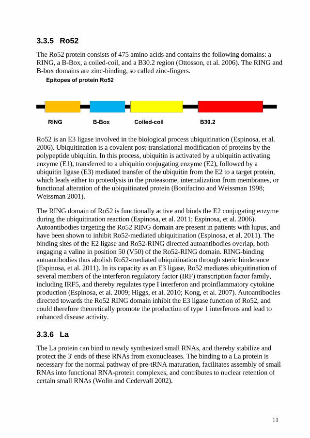
3.3.5 Ro52 The Ro52 protein consists of 475 amino acids and contains the following domains: a RING, a B-Box, a coiled-coil, and a B30.2 region (Ottosson, et al. 2006). The RING and B-box domains are zinc-binding, so called zinc-fingers.
Ro52 is an E3 ligase involved in the biological process ubiquitination (Espinosa, et al. 2006). Ubiquitination is a covalent post-translational modification of proteins by the polypeptide ubiquitin. In this process, ubiquitin is activated by a ubiquitin activating enzyme (E1), transferred to a ubiquitin conjugating enzyme (E2), followed by a ubiquitin ligase (E3) mediated transfer of the ubiquitin from the E2 to a target protein, which leads either to proteolysis in the proteasome, internalization from membranes, or functional alteration of the ubiquitinated protein (Bonifacino and Weissman 1998; Weissman 2001).
The RING domain of Ro52 is functionally active and binds the E2 conjugating enzyme during the ubiquitination reaction (Espinosa, et al. 2011; Espinosa, et al. 2006). Autoantibodies targeting the Ro52 RING domain are present in patients with lupus, and have been shown to inhibit Ro52-mediated ubiquitination (Espinosa, et al. 2011). The binding sites of the E2 ligase and Ro52-RING directed autoantibodies overlap, both engaging a valine in position 50 (V50) of the Ro52-RING domain. RING-binding autoantibodies thus abolish Ro52-mediated ubiquitination through steric hinderance (Espinosa, et al. 2011). In its capacity as an E3 ligase, Ro52 mediates ubiquitination of several members of the interferon regulatory factor (IRF) transcription factor family, including IRF5, and thereby regulates type I interferon and proinflammatory cytokine production (Espinosa, et al. 2009; Higgs, et al. 2010; Kong, et al. 2007). Autoantibodies directed towards the Ro52 RING domain inhibit the E3 ligase function of Ro52, and could therefore theoretically promote the production of type 1 interferons and lead to enhanced disease activity.
3.3.6 La The La protein can bind to newly synthesized small RNAs, and thereby stabilize and protect the 3' ends of these RNAs from exonucleases. The binding to a La protein is necessary for the normal pathway of pre-tRNA maturation, facilitates assembly of small RNAs into functional RNA-protein complexes, and contributes to nuclear retention of certain small RNAs (Wolin and Cedervall 2002).
12
4 CLINCIAL FEATURES OF SYSTEMIC AUTOIMMUNE DISEASES
The first section of this text, Systemic autoimmune rheumatic diseases, summarized the systemic diseases. SLE and pSS are the two diseases which resemble each other most. To differentiate between SLE with low disease activity and pSS, or pSS with high disease activity and SLE can be difficult. There are patients who, according to classification criteria, can be regarded as having either diagnosis.
4.1 SJÖGREN’S SYNDROME
4.1.1 Background/Summary Primary Sjögren’s syndrome is a systemic inflammatory autoimmune condition. It is named after the Swedish ophthalmologist Henrik Sjögren who defended his doctoral thesis in1933 with the title: ”ZUR Kenntnis der Keratokonjunktivitis sicca”. Historically other terms have been used, e.g. Mikulicz’s disease.
When Sjögren’s syndrome exists as a disorder without any other rheumatic disease it is called primary Sjögren’s syndrome, and when it develops in patients with another primary diagnosis like Rheumatoid Arthritis, SLE or Systemic Sclerosis it is called secondary Sjögren’s syndrome.
Prevalence figures vary around half a percent with a high female dominance. The etiology is mainly unknown, but it has been suggested that multiple environmental factors trigger an immune response in genetically susceptible individuals (Wahren-Herlenius and Dorner 2013). In genetic studies, several single nucleotide polymorphisms have been significantly associated with pSS. They are located in chromosomal regions with loci containing genes such as MHC/HLA, Toll-like receptor (TLR) and type 1 IFN system genes and B-lymphocyte differentiation related genes. Speculations about environmental factors like viruses have long been present because of the similarities with pathological changes in the salivary gland tissue in hepatitis C and pSS.
In pSS, inflammation results in progressive destruction of the exocrine glands, primarily the salivary and lacrimal glands. This leads to symptoms of dryness, mainly in the mouth and eyes. Patients may also experience symptoms of exocrine dysfunction and dryness in other areas of the body such as the skin, nose, vagina, bronchial tubes and gastrointestinal tract. Further, arthralgia and fatigue are prominent features of the disease. Signs of dysregulation of the immune system include formation of ectopic lymphoid tissue with lymphocyte proliferation and germinal centers in the salivary glands, a changed pattern in B-cell maturation and proliferation,
13
hypergammaglobulinemia and autoantibodies against the antigens Ro/SSA (Ro52 and Ro60) and La/SSB (Salomonsson, et al. 2003; Salomonsson, et al. 2002; Tengner, et al. 1998).
Extraglandular manifestations (EGM) are present in a subset of patients with pSS. This subset of patients is characterized by a more systemic phenotype. Examples of extraglandular organ involvement include hypergammaglobulinaemic purpura, skin vasculitis, arthritis, sensory polyneuropathy, interstitial lung disease and interstitial renal disease with secondary distal Renal Tubular Acidosis (dRTA).
For classification and diagnosis the Revised American European Consensus Group Criteria from 2002 (AECC) (Vitali, et al. 2002) are used. A disease activity index has been developed for patients with EGM: the European League Against Rheumatism (EULAR) Sjögren’s Syndrome Disease Activity Index (ESSDAI) (Seror, et al. 2010).
Treatment is symptomatic for sicca symptoms. Moisture replacement therapies and products that stimulate production of tears and saliva are available. In the case of severe EGM corticosteroids, DMARD and biologic treatment have been used.
Lymphoma is a severe complication which a subset of patients develops. Otherwise the prognosis is good. Severe life threatening or life shortening organ manifestations are rare. The disease is mainly characterized by subjective symptoms which impair quality of life.
4.1.2 Immunopathogenesis Focal infiltration of mononuclear cells, especially T- and B-lymphocytes in exocrine glands, is a hallmark of pSS. Focal inflammation starts around epithelial ductal structures and spreads to the deeper layer of the gland resulting in progressive destruction of the exocrine glands, primarily the salivary and lacrimal glands. This leads to symptoms of dryness, mainly in the mouth and eyes.
The pathogenic process is complex and involves different parts of the immune system during the disease progression (Halse, et al. 1999; Jonsson, et al. 2011). Hypotheses linked together and supported by results from many investigations have formed a picture of the pathogenic process that leads to pSS. The current understanding indicates a process where environmental factors initiate disease in genetically susceptible individuals. The genetically dysregulated cellular pathways lead to changed activity in processes which increases the risk of autoimmunity.
It is not known what initiates the process of infiltration and accumulation of mononuclear cells, but viral infections have been suggested. The mononuclear cells consist of T lymphocytes, macrophages, B-lymphocytes (plasma cells), NK cells and dendritic cells (DC). Epithelial adhesion molecules enable lymphocytes to adhere and then infiltrate the tissue. This leads to the formation of ectopic lymphoid tissue with lymphocyte proliferation and germinal center-like structures in the salivary glands.
14
Inappropriate apoptosis in epithelial cells of the ducts of the exocrine glands creates exposure of intracellular proteins which become autoantigens. DCs play a role in the maintenance of tolerance to self-antigens. Loss of self-tolerance occurs and the DCs can be involved in several ways, for example production of IFN and antigen presentation of autoantigens. Patients with pSS have an IFN signature, that is, genes induced by IFN are activated. The presented autoantigens attract lymphocytes; some of which are autoreactive T-lymphocytes which can result in B-cell activation and production of autoantibodies.
The increased Ig production results in hypergammaglobulinemia with a high sedimentation rate and the presence of the well-known autoantibodies against the antigens Ro/SSA (Ro52 and Ro60) and La/SSB (Salomonsson, et al. 2003; Salomonsson, et al. 2002; Tengner, et al. 1998). These autoantibodies are known to be present as signs of disease in the systemic rheumatic syndromes pSS and SLE. Recently, autoantibodies against the Ro52 antigen have been shown to functionally impair Ro52 activity indicating a role in disease mechanism and activity (Ambrosi, et al. 2012; Espinosa, et al. 2011; Kvarnstrom, et al. 2013; Salomonsson, et al. 2005).
The immunologic process becomes chronic and leads to inflammation involving larger parts of the glands and sometimes other organs. Organs containing epithelial structures are at risk. Increased levels of pro-inflammatory cytokines such as IL-1B, TNF-α, IL-6, IFN, B-cell Activating Factor (BAFF) and APRIL are present in pSS. Production of cytokines and kemokines leads to a positive feed-back loop that increases the inflammation further.
4.1.3 Prevalence The reported prevalence in different populations varies from 0.01-0.6% (Bowman, et al. 2004; Goransson, et al. 2011; Maldini, et al. 2014)when the AECC (Vitali, et al. 2002) was used. Relating to previous use of other classification criteria, varying prevalence figures over 5% have been reported. This is due to sicca symptoms which are common in the elderly with no autoimmune cause.
4.1.4 Incidence Published studies on incidence are rare and have reported 1.1–5.3 cases per 100.000 person-years (Alamanos, et al. 2006; Pillemer, et al. 2001; Plesivcnik Novljan, et al. 2004; Yu, et al. 2013). The surveys have used different classification criteria and study designs, which aggravates comparisons.
15
4.1.5 Organ manifestations In pSS the chronic inflammation of systemic character that affects the exocrine glands and other internal organs, results in three types of symptoms and organ manifestations: general symptoms, glandular, and extraglandular manifestations.
4.1.5.1 General symptoms
General symptoms are common for all inflammatory rheumatic diseases particularly systemic diseases. They are often present long before diagnosis which can make it difficult to differentiate between different rheumatic diagnoses early in the disease process. Fatigue, fever, arthralgia, myalgia and sicca symptoms are included in this group.
4.1.5.2 Glandular manifestations
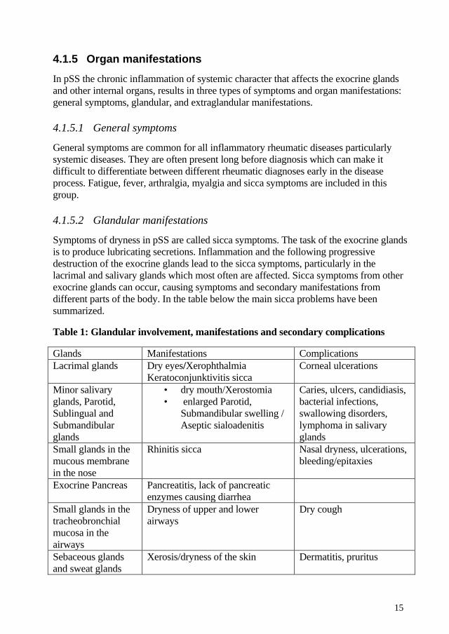
Symptoms of dryness in pSS are called sicca symptoms. The task of the exocrine glands is to produce lubricating secretions. Inflammation and the following progressive destruction of the exocrine glands lead to the sicca symptoms, particularly in the lacrimal and salivary glands which most often are affected. Sicca symptoms from other exocrine glands can occur, causing symptoms and secondary manifestations from different parts of the body. In the table below the main sicca problems have been summarized.
Table 1: Glandular involvement, manifestations and secondary complications
Glands Manifestations Complications Lacrimal glands Dry eyes/Xerophthalmia
Keratoconjunktivitis sicca Corneal ulcerations
Minor salivary glands, Parotid, Sublingual and Submandibular glands
• dry mouth/Xerostomia • enlarged Parotid,
Submandibular swelling / Aseptic sialoadenitis
Caries, ulcers, candidiasis, bacterial infections, swallowing disorders, lymphoma in salivary glands
Small glands in the mucous membrane in the nose
Rhinitis sicca Nasal dryness, ulcerations, bleeding/epitaxies
Exocrine Pancreas Pancreatitis, lack of pancreatic enzymes causing diarrhea
Small glands in the tracheobronchial mucosa in the airways
Dryness of upper and lower airways
Dry cough
Sebaceous glands and sweat glands
Xerosis/dryness of the skin Dermatitis, pruritus
16
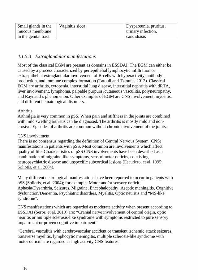
Small glands in the mucous membrane in the genital tract
Vaginitis sicca Dyspareunia, pruritus, urinary infection, candidiasis
4.1.5.3 Extraglandular manifestations
Most of the classical EGM are present as domains in ESSDAI. The EGM can either be caused by a process characterized by periepithelial lymphocytic infiltration or extraepithelial extraglandular involvement of B-cells with hyperactivity, antibody production, and immune complex formation (Tatouli and Tzioufas 2012). Classical EGM are arthritis, cytopenia, interstitial lung disease, interstitial nephritis with dRTA, liver involvement, lymphoma, palpable purpura /cutaneous vasculitis, polyneuropathy, and Raynaud´s phenomenon. Other examples of EGM are CNS involvement, myositis, and different hematological disorders.
Arthritis Arthralgia is very common in pSS. When pain and stiffness in the joints are combined with mild swelling arthritis can be diagnosed. The arthritis is mostly mild and non-erosive. Episodes of arthritis are common without chronic involvement of the joints.
CNS involvement There is no consensus regarding the definition of Central Nervous System (CNS) manifestations in patients with pSS. Most common are involvements which affect quality of life. Characteristics of pSS CNS involvements have been described as a combination of migraine-like symptoms, sensorimotor deficits, coexisting neuropsychiatric disease and unspecific subcortical lesions (Escudero, et al. 1995; Soliotis, et al. 2004). Many different neurological manifestations have been reported to occur in patients with pSS (Soliotis, et al. 2004); for example: Motor and/or sensory deficit, Aphasia/Dysarthria, Seizures, Migraine, Encephalopathy, Aseptic meningitis, Cognitive dysfunction/Dementia, Psychiatric disorders, Myelitis, Optic neuritis and “MS-like syndrome”.
CNS manifestations which are regarded as moderate activity when present according to ESSDAI (Seror, et al. 2010) are: “Cranial nerve involvement of central origin, optic neuritis or multiple sclerosis-like syndrome with symptoms restricted to pure sensory impairment or proven cognitive impairment.”
“Cerebral vasculitis with cerebrovascular accident or transient ischemic attack seizures, transverse myelitis, lymphocytic meningitis, multiple sclerosis-like syndrome with motor deficit” are regarded as high activity CNS features.
17
Cutaneous manifestations More than half of patients with pSS have different kind of skin disorders. Most common is xerosis (dry skin). In patients with hypergammaglobulinemia, palpable purpura emerges on the front of the lower legs after physical activity. Palpable and non-palpable purpura are manifestations of vasculitis in small vessels. Urticarial vasculitis and necrotizing vasculitis of medium-sized vessels are other examples of different types of vasculitis in pSS. Other skin manifestations include eyelid dermatitis, pruritus, usually due to the xerosis, vitiligo and alopecia. Photosensitivity can develop in the subset of patients with anti-Ro antibodies when exposed to the sun, and this results in lesions of subacute cutaneous lupus erythematosus or erythema annulare (Fox 2005; Soy and Piskin 2006).
Cytopenia Cytopenia in pSS includes anemia, leucopenia, and thrombocytopenia. Neutropenia and lymphopenia are the most common type of leucopenia. The severity of the cytopenia is based on the grade of reduction in the number of blood cells according to ESSDAI (Seror, et al. 2010).
Liver involvement Autoimmune hepatitis and Primary biliary cirrhosis are present at higher rates in pSS patients than in the general population. Liver diseases are not included as a domain in ESSDAI (Seror, et al. 2010). Instead, these liver diseases are often regarded as co-morbidities. Lung manifestations Dryness of the tracheobronchial mucosa can manifest as a dry cough. The symptoms can be mistaken for hypersensitivity and asthma. Inflammation in the lungs is often situated in the interstitium. Treatment with corticosteroids is often effective and the infiltrate seen on chest-x-rays looks more severe than the patient’s status.
Lymphoma Lymphoma is a hematological malignancy. It is the most serious complication in pSS. Non-Hodgkin lymphomas develop from B-cells and the dominating types are Diffuse Large B-cell (DLBC) lymphoma and mucosa-associated lymphoid tissue (MALT) lymphoma. They are usually located in the salivary glands. Lifetime risk is estimated to be 5 to 15%.Tumors in the salivary glands should lead to investigation of suspected lymphoma. (Jonsson, et al. 2012)
Myositis Inflammatory myopathy is rare but exists. A diagnosis of overlap syndrome or myositis with secondary SS should be considered when appropriate. Myalgia is very common and can be accompanied by weakness due to the pain. Another differential diagnosis is polyneuropathy affecting the muscles.
18
Polyneuropathy The subtypes of polyneuropathy that dominate among pSS patients are axonal sensorimotor polyneuropathy, pure sensory neuronopathy and mononeuritis multiplex. The conditions are slowly progressive and can affect survival, especially mononeuritis multiplex caused by necrotizing vasculitis in medium size vessels (Brito-Zeron, et al. 2013).
Renal involvement The most common form of renal involvement in pSS is interstitial nephritis which affects the tubular part of the nefrome leading to impaired ion exchange and systemic acidosis. This is known as distal renal tubular acidosis (dRTA). Patients can present with hypokalemic paralysis, renal calculi, or osteomalacia (Fox 2005)
Raynaud’s phenomenon In patients with pSS the objective and subjective symptoms of Raynaud’s phenomenon are milder compared to the combination with the primary diagnosis Systemic sclerosis. Complications such as ulcers should result in re-evaluation of the diagnosis.
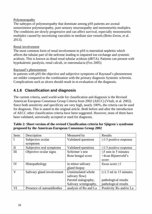
4.1.6 Classification and diagnosis The current criteria, used world-wide for classification and diagnosis is the Revised American European Consensus Group Criteria from 2002 (AECC) (Vitali, et al. 2002). Since both sensitivity and specificity are very high, nearly 100%, the criteria can be used for diagnosis. This is stated in the original article. Both before and after the introduction of AECC other classification criteria have been suggested. However, none of them have been validated, universally accepted or used for diagnosis.
Table 2: Short version of the revised Classification criteria for Sjögren´s syndrome proposed by the American-European Consensus Group 2002
Item: Description Measured by Results I Subjective ocular
symptoms Validated questions ≥1/3 positive response
II Subjective oral symptoms Validated questions ≥1/3 positive response III Objective ocular signs Schirmer´s test
Rose bengal score ≤5 mm in 5 minutes >4van Bijsterveld’s score
IV Histopathology in minor salivary gland biopsy
focus score ≥1
V Salivary gland involvement Unstimulated whole salivary flow) Parotid sialography, Salivary scintigraphy,
(≤1.5 ml in 15 minutes pathological results pathological results
VI Presence of autoantibodies analysis of Ro and La Positivity Ro and/or La
19
Primary SS is defined as: the presence of any 4 of the 6 items and either item IV (Histopathology) or VI (Serology) must be positive or the presence of any 3 of the 4 objective criteria items (that is, items III, IV, V, VI). Exclusion criteria: head and neck radiation treatment, Hepatitis C, AIDS, pre-existing Lymphoma, Sarcoidosis, Graft versus host disease and use of anticholinergic drugs.
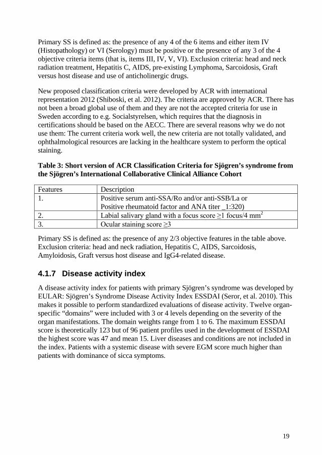
New proposed classification criteria were developed by ACR with international representation 2012 (Shiboski, et al. 2012). The criteria are approved by ACR. There has not been a broad global use of them and they are not the accepted criteria for use in Sweden according to e.g. Socialstyrelsen, which requires that the diagnosis in certifications should be based on the AECC. There are several reasons why we do not use them: The current criteria work well, the new criteria are not totally validated, and ophthalmological resources are lacking in the healthcare system to perform the optical staining.
Table 3: Short version of ACR Classification Criteria for Sjögren’s syndrome from the Sjögren’s International Collaborative Clinical Alliance Cohort
Features Description 1. Positive serum anti-SSA/Ro and/or anti-SSB/La or
Positive rheumatoid factor and ANA titer _1:320) 2. Labial salivary gland with a focus score ≥1 focus/4 mm2
3. Ocular staining score ≥3
Primary SS is defined as: the presence of any 2/3 objective features in the table above. Exclusion criteria: head and neck radiation, Hepatitis C, AIDS, Sarcoidosis, Amyloidosis, Graft versus host disease and IgG4-related disease.
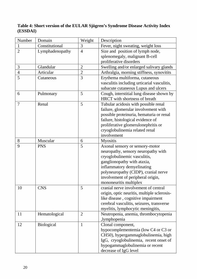
4.1.7 Disease activity index A disease activity index for patients with primary Sjögren’s syndrome was developed by EULAR: Sjögren’s Syndrome Disease Activity Index ESSDAI (Seror, et al. 2010). This makes it possible to perform standardized evaluations of disease activity. Twelve organ-specific “domains” were included with 3 or 4 levels depending on the severity of the organ manifestations. The domain weights range from 1 to 6. The maximum ESSDAI score is theoretically 123 but of 96 patient profiles used in the development of ESSDAI the highest score was 47 and mean 15. Liver diseases and conditions are not included in the index. Patients with a systemic disease with severe EGM score much higher than patients with dominance of sicca symptoms.
20
Table 4: Short version of the EULAR Sjögren’s Syndrome Disease Activity Index (ESSDAI)
Number Domain Weight Description 1 Constitutional 3 Fever, night sweating, weight loss 2 Lymphadenopathy 4 Size and position of lymph node,
splenomegaly, malignant B-cell proliferative disorders
3 Glandular 2 Swelling and/or enlarged salivary glands 4 Articular 2 Arthralgia, morning stiffness, synovitits 5 Cutaneous 3 Erythema multiforma, cutaneous
vasculitis including urticarial vasculitis, subacute cutaneous Lupus and ulcers
6 Pulmonary 5 Cough, interstitial lung disease shown by HRCT with shortness of breath
7 Renal 5 Tubular acidosis with possible renal failure, glomerular involvement with possible proteinuria, hematuria or renal failure, histological evidence of proliferative glomerulonephritis or cryoglobulinemia related renal involvement
8 Muscular 6 Myositis 9 PNS 5 Axonal sensory or sensory-motor
neuropathy, sensory neuropathy with cryoglobulinemic vasculitis, ganglionopathy with ataxia, inflammatory demyelinating polyneuropathy (CIDP), cranial nerve involvement of peripheral origin, mononeuritis multiplex
10 CNS 5 cranial nerve involvement of central origin, optic neuritis, multiple sclerosis-like disease , cognitive impairment cerebral vasculitis, seizures, transverse myelitis, lymphocytic meningitis,
11 Hematological 2 Neutropenia, anemia, thrombocytopenia ,lymphopenia
12 Biological 1 Clonal component, hypocomplementemia (low C4 or C3 or CH50), hypergammaglobulinemia, high IgG, cryoglobulinemia, recent onset of hypogammaglobulinemia or recent decrease of IgG level
21
4.1.8 Treatment Treatment today is symptomatic for sicca symptoms caused by the glandular manifestations resulting in dryness of different surfaces of the body. As yet, no medication that can stop disease progression of the sicca symptoms exists. Moisture replacement therapies and products that stimulate production of tears and saliva are available. Muscarinic agonists can have an effect on both eyes and mouth, but limitations due to side effects impair their usefulness. In the case of EGM, corticosteroids, DMARD and biologic treatment can be used. No evidence based guidelines exist for the treatment of pSS (Ramos-Casals, et al. 2012; Ramos-Casals, et al. 2010).
4.1.8.1 Xerostomia
The goal of treatment is both to minimize the dryness symptoms and to prevent long-term oral complications such as caries, candidiasis and ulcers. Saliva substitutes in the form of oral sprays and gels or mouth rinsing with water or oil ease the symptoms of dryness in the mouth and throat. More effective but only possible for patients who have residual salivary gland function is the stimulation of saliva flow with a secretagogue. Sugar-free gums and candies have an effect through mechanical and gustatory saliva stimulation. The muscarinic acetylcholine receptor agonist pilocarpine (Salagen®) can be prescribed by the patients´ physicians. It works through the stimulation of the muscarinic acetylcholine receptors M1 and M3 present on salivary glands, leading to increased secretory function Side effects associated with pilocarpine use, including sweating, palpitations, increased urinary frequency and flushing, limit the use of the agent. Several things can be done to protect the teeth. Avoidance of sugar is essential. Dental care with regular visits to the dentist and dental hygienists is important. Fluoride treatment in the form of fluoride toothpaste and extra fluoride rinses strengthens the tooth enamel and thereby prevents caries. Chlorhexidine mouth rinses decrease the amount of bacteria and can be recommended for the prevention of infections.
4.1.8.2 Xerophthalmia/ Keratoconjunctivitis sicca
The goal of treatment is both to minimize the dryness symptoms and to prevent complications such as infections and ulcers. Basic treatment is topical eye drops with artificial tears containing different agents (e.g. hyaluronate or methylcellulose) which influence the lubricating effect and how long they last. They should always be preservative-free. For night use lubricating ointments are used. Ophthalmic formulation of cyclosporine A is approved by the FDA in the USA and several other countries for the treatment of dry eye disease. These eye drops decrease the inflammation and thereby make the tears more long lasting. In Sweden this treatment is available by license. The
22
muscarinic acetylcholine receptor agonist pilocarpine (Salagen®) can also have some effect on the eyes. In severe forms, plug insertion or surgery of the tear duct so that tears cover the eye longer can be performed by an ophthalmologist.
4.1.8.3 Exocrine dysfunction with sicca symptoms of other parts of the body
Hair and scalp: Restriction of hair washing and the amount of shampoo used to not more than what is necessary.
Nose: Saltwater (saline) rinse and nasal sprays followed by lubrication with oil or ointment.
Skin: Restriction of showering and the amount of soap used, moisturizing cream containing a high proportion of lipids and low proportion (max 5%) of urea.
Genital: No use of soap on mucous membrane, washing with water and oil, lubricating products are available to buy over the counter but oil works well.
Gastro-Intestinal tract: Lack of pancreatic enzymes from exocrine pancreas results in diarrhea especially if the food contains a high proportion of fat. Capsules containing pancreatic enzymes can be prescribed by the patients’ physicians.
4.1.8.4 Non-inflammatory muscle and joint involvement
These symptoms vary over time, and Non Steroid Anti Inflammatory Drugs (NSAID) have a good analgesic effect and can be used when needed. Examination by a physician should have been performed to exclude synovitis and fibromyalgia which are treated differently. If NSAID are not good enough, especially if the symptoms include fatigue, hydroxychloroquine (Plaquenil®) has often been used, despite lack of evidence.
4.1.8.5 Extra Glandular Manifestations
The grade of evidence is low to non-existent when it comes to therapies for EGM in pSS. The use is based on small studies, case reports and on experience of treatment for clinically similar conditions in other autoimmune diseases, usually SLE or vasculitis. In general, corticosteroids in combination with immunosuppressive agents are the first line of medication. If the manifestations are refractory to standard therapies, rituximab has often been tried.
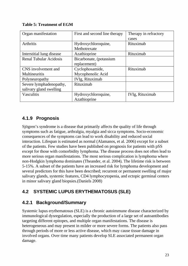
23
Table 5: Treatment of EGM
Organ manifestation First and second line therapy Therapy in refractory cases
Arthritis Hydroxychloroquine, Methotrexate
Rituximab
Interstitial lung disease Azathioprine Rituximab Renal Tubular Acidosis Bicarbonate, (potassium
replacement)
CNS involvement and Multineuritis
Cyclophosamide, Mycophenolic Acid
Rituximab
Polyneuropathy IVIg, Rituximab Severe lymphadenopathy, salivary gland swelling
Rituximab
Vasculitis Hydroxychloroquine, Azathioprine
IVIg, Rituximab
4.1.9 Prognosis Sjögren’s syndrome is a disease that primarily affects the quality of life through symptoms such as fatigue, arthralgia, myalgia and sicca symptoms. Socio-economic consequences of the symptoms can lead to work disability and reduced social interaction. Lifespan is estimated as normal (Alamanos, et al. 2006) except for a subset of the patients. Few studies have been published on prognosis for patients with pSS except for those with comorbidity lymphoma. The disease process does not often lead to more serious organ manifestations. The most serious complication is lymphoma where non-Hodgkin lymphoma dominates (Theander, et al. 2004). The lifetime risk is between 5-15%. A subset of the patients have an increased risk for lymphoma development and several predictors for this have been described; recurrent or permanent swelling of major salivary glands, systemic features, CD4 lymphocytopenia, and ectopic germinal centers in minor salivary gland biopsies.(Daniels 2008)
4.2 SYSTEMIC LUPUS ERYTHEMATOSUS (SLE)
4.2.1 Background/Summary Systemic lupus erythematosus (SLE) is a chronic autoimmune disease characterized by immunological dysregulation, especially the production of a large set of autoantibodies targeting different epitopes, and multiple organ manifestations. The disease is heterogeneous and may present in milder or more severe forms. The patients also pass through periods of more or less active disease, which may cause tissue damage in involved organs. Over time many patients develop SLE associated permanent organ damage.

24
The word lupus comes from Latin and means wolf. Since the Middle Ages the word lupus has been used to describe severe skin ulcers which today we know could have had different causes such as TBC, malignancies or SLE. The word Erythematosus refer to the colour of the skin disorders. During the 20th century immunological changes were discovered and the disease could be separated from other conditions. During the same period of time treatment became available which dramatically improved the prognosis. Even so, the disease still has an effect on survival but today this is also due to co-morbidities, in particular cardiovascular diseases(Gustafsson, et al. 2012).
The female dominance is substantial: 9 out of 10 are women and prevalence varies with ethnicity. In Sweden the incidence is 5 cases per 100,000 persons-years. (Stahl-Hallengren, et al. 2000).
Diagnosis is based on involvement of two organ systems in combination with an immunological disorder. The best known classification criteria is theAmerican College of Rheumatology (ACR) 1982 revised classification criteria for SLE(Tan, et al. 1982). The different items reflect possible and most frequent organ manifestations. . Manifestations from the musculoskeletal, hematological and mucocutaneous systems, heart- lung-, and kidneys are common. Validated indices that measure disease activity and organ damage have been developed for SLE. Most well-known are Systemic Lupus Erythematosus Disease Activity Index (SLEDAI), Systemic Lupus Activity Measure (SLAM), and index of clinical disease activity in systemic lupus erythematosus-British Isles Lupus Assessment Group (BILAG) (Bombardier, et al. 1992; Gladman, et al. 1996; Griffiths, et al. 2005; Hay, et al. 1993; Liang, et al. 1989; Symmons, et al. 1988). Treatments depend on involved organs and severity of the flare/disease activity. Today corticosteroids, immunosuppressives and biologics are available.(D'Cruz, et al. 2007; Rahman and Isenberg 2008)
4.2.2 Immunopathogenesis The exact process and the different mechanisms that lead to SLE are not fully understood. Type I IFNs (a group of cytokines) has been suggested to play a key role based on the observation that treatment with IFN can result in autoimmune diseases, including a lupus-like-syndrome (Ronnblom, et al. 1991). Further, SLE patients have an overproduction of Typ I IFN resulting in an activation of IFN-dependent genes, a so called IFN signature (Ronnblom and Eloranta 2013). This overproduction has been suggested to rely on activation of plasmacytoid dendritic cells (pDCs) by complexes of nucleoprotein autoantibodies and nucleosomes through TLRs (Finke, et al. 2009; Lovgren, et al. 2004; Ronnblom 2011). The nucleosomes are thought to originate from apoptotic and necrotic cells, of which SLE patients have a defective uptake and clearance.
25
Another described way of activating the pDCs is by NETs, Neutrophil Extracellular Traps. Dying neutrophils release NETs containing multiple components including histones and DNA, which can induce IFN-a production in pDCs (Garcia-Romo, et al. 2011). The pDCs have an extremely high capacity for IFN production and can be found in affected tissue rather than the blood in SLE patients.
The complement system has an impact on SLE in several ways. If activated, it can cause tissue damage by the formation of immune complexes, which can deposit in tissue especially in the kidneys. Complement factors, for example C1q, bind to the apoptotic material to enhance phagocytosis by macrophages. In complement deficiencies, patients become predisposed to infections, and a decreased capacity for clearage of apoptotic material develops (Sturfelt and Truedsson 2005; Truedsson, et al. 2007). C1q also functions as a regulator of IFN production by inhibiting pDCs (Lood, et al. 2009). Notably, C1q deficiency is the strongest risk factor known for the development of SLE. C1q functions as a regulator of IFN production by inhibiting pDCs
Multiple changes in B-cell function and repertoire are present in SLE. The B cell population in SLE consists of more pre-immune B cells, memory cells, and plasma cells (Hostmann, et al. 2008), and the balance of plasma blasts and plasma cells is skewed (Odendahl, et al. 2000). The presence of autoantibodies produced by the B cells is one of the main characteristics of SLE. Anti-dsDNA antibodies are specific and correlate to disease activity. In renal biopsies the deposition of a number of other antibodies, including anti-Ro, anti-La, anti-C1q (a subunit of the C1 complement component), and anti-Sm, is known. The depositions can be in the form of immune-complexes with autoantigens. The deposit of immune complexes is responsible for the organ manifestations glomerulonephritis, arthritis and vasculitis in small arteries, followed by complement activation and tissue damage. Cross-reaction with proteins in the kidneys has also been mentioned. Hemolytic anemia and thrombocytopenia are caused by the binding of autoantibodies to erythrocytes and platelets, resulting in lysis (Abbas 2007b; D'Cruz, et al. 2007; Rahman and Isenberg 2008). In all, this loss of B-cell tolerance and the production of autoantibodies lead to the concept that SLE is a B-cell dependent disease.
Although B cells may have a prominent role in SLE, also the T cell compartment is affected. Increased levels of Th17 cells with a capacity to induce inflammation, tissue damage and production of autoantibodies has been shown (Shin, et al. 2011), and T regulatory cells in SLE are fewer and have a reduced capacity of suppressing T-cell proliferation and cytokine production. Also, indirectly, T-cells have an impact through the production of cytokines TNF-α, IFN-γ, IL2, and IL-10 which stimulate B cells, facilitate immunoglobulin class switching, and promote production of high-affinity autoantibodies (Azevedo, et al. 2014).
A number of cytokines are up-regulated in SLE, resulting in enhanced activity in some of their normal functions. In addition to immunomodulation, they are involved in systemic inflammation and end-organ tissue damage. IFNα levels are increased and the
26
cytokine acts as an immune adjuvant, i.e. increases the expression of MHC class I molecules which enhance the antigen presentation; it increases the expression of several chemokines, chemokine receptors, and co-stimulatory molecules; activates NK cells and cytotoxic cells; promotes the differentiation/maturation of DCs; prolongs B and T-cell survival; induces immunoglobulin class switch; increases the production of antibodies; and enhances the phagocytic capacity of macrophages (Ronnblom and Pascual 2008). Several B cell promoting cytokines are found in increased levels; IL-6 which enhances maturation of B-cells into plasma cells and increases antibody secretion; IL-10 which promotes antibody class-switch and B-cell activating factor (BAFF) which promotes B-cell survival and autoantibody production. Further, SLE patients have increased levels of TNF, contributing organ inflammation (Ronnblom and Elkon 2010), and IL-17 which induces inflammation, tissue damage and again promotes production of autoantibodies (Shin, et al. 2011).
4.2.3 Incidence
4.2.4 Prevalence Incidence rates range from 1 to 10 per 100,000 person-years generally; higher figures have been reported from ethnic groups other than Caucasian, even an extreme figure from an Afro-Caribbean cohort.
Prevalence rates generally range from 20 to 70 per 100,000 people (Pons-Estel, et al. 2010)
Several causes are known for the different reported figures: study design (population based cohorts or hospital based case series/cohorts), classification criteria, patient recruitment, health care system, and ethnicity, which is correlated to both genetic differences and socio-economic factors (Pons-Estel, et al. 2010).
An incidence rate of 5 per 100,000 person-years has been reported from Sweden (Stahl-Hallengren, et al. 2000).
4.2.5 Organ manifestations
4.2.5.1 General symptoms
General symptoms such as fatigue, fever, arthralgia, myalgia, weight loss and sicca symptoms are often the first to occur. Laboratory tests show signs of general inflammation in the body; increased erythrocyte sedimentation rate (ESR), anemia and increased or decreased white blood cell count. (Bertsias 2012; Buyon 2008)
27
4.2.5.2 Mucocutaneous manifestations
Manifestations in the skin and mucus membranes are common; more than 80% of the patients have experienced one or several of the associated involvements of this system. The most well-known are the four included in ACR 1982 revised classification criteria for SLE(items1-4) (Tan, et al. 1982) ; malar rash, discoid rash, photosensitivity and oral ulcers. The lesions can be divided into lupus specific and non-lupus specific. Non-lupus-specific lesions can present separately or concordantly with other autoimmune conditions. Well-known are cutaneous vasculitis, livedo reticulitis, Raynaud’s phenomenon, erythema multiforme, urticaria and alopecia. The lupus-specific lesions are divided into three groups; acute, subacute and chronic lesions. The malar rash is an acute rash. It has a butterfly form, is situated over the nose and cheeks, sparing the nasolabial folds, and is characterized by the absence of papules and pustules. Another example is generalized erythema. Acute rashes can heal without scarring. Subacute cutaneous lupus erythematosus (SCLE) is an example of subacute rash but is not always associated with SLE. It usually occurs in patients with Ro/SSA autoantibodies and is associated with sun exposure. The lesions start as small papules and develop into psoriasiform or annular form lesions. They are often located on the arms and upper torso. Discoid Lupus Erythematosus (DLE) is a chronic lesion. It is characterized by erythematous, plaque and scales, heals with scarring and is located on the head and upper torso. Photosensitivity means the development of rash after exposure to sunlight. Alopecia, loss of hair, can occur anywhere on the body. Scarring alopecia is a complication of DLE when it affects the hair follicles. Ulcers in the mucus membranes can be present in the mouth, nose and throat. They can be painless which is why it is important to perform an inspection. (Bertsias 2012; Buyon 2008)
4.2.5.3 Musculoskeletal system
Arthralgia is the most common symptom in patients with SLE. Arthritis often affects the small joints in the hands, but it can also occur in large joints. The arthritis is non-erosive in most patients. Tenosynovitis is common. Deformities have many similar features to those of patients with rheumatoid arthritis, such as ulnar drift and swan neck deformities. Inflammatory myositis can occur and sampling for creatine kinas should be performed, especially in cases with muscular weakness. (Bertsias 2012; Buyon 2008)
4.2.5.4 Renal involvement
Renal involvement is a severe organ manifestation. All patients should be checked on a regular basis with urine analysis for microscopic hematuria and proteinuria, since the manifestation does not give any subjective symptoms. If present, further analysis of urine sedimentation, including analysis of different types of casts, which are signs of renal involvement, should be done. Renal biopsy provides a histopathological diagnosis and classification. World Health Organizations (WHO) have classified lupus nephritis; Type I-normal glomerulus, Type II-mesangial disease, Type III-focal and segmental proliferative nephritis (less than 50% of the glomeruli are involved) Type IV-diffuse
28
proliferative nephritis (more than 50% of glomeruli are involved) and Type V-membranous nephropathy (Churg J 1982). The classification is based on light microscopy. Electron microscopy can reveal minor changes and immune complex deposits. In 2004 the International Society of Nephrology/Renal Pathology Society published revised classification criteria. To summarize the most important changes, Class 1 is defined as minimal mesangial lupus nephritis, Class III has been divided into subgroups depending on whether active (Class III A) or chronic (Class III C) lesions are to be found. Class IV has been divided into subgroups depending on whether the proliferation is situated segmentally or globally (Class IV: -S(A), -G(A), etc.)(1999; Bertsias 2012; Buyon 2008; Weening, et al. 2004)
4.2.5.5 Nervous system
Both CNS and PNS can be involved. Neuropsychiatric SLE (NPSLE) was defined and classified by the ACR. The 19 different syndromes and symptoms included vary in frequency and severity. Probability of association to activity in SLE can be difficult to prove, especially for headaches and mood disorders. The included syndromes and conditions are: aseptic meningitis, cerebrovascular disease, demyelinating syndrome, headache, chorea, myelopathy, seizure, confusion, anxiety disorder, cognitive dysfunction, mood disorders, psychosis(CNS), Guillan Barré syndrome, autonomic disorder, mononeuropathy (single, multiplex), myasthenia gravis, cranial neuropathy, plexopathy and polyneuropathy(PNS)”. (Bertsias 2012; Buyon 2008)nnn
4.2.5.6 Serositis
Pleuritis is often painful and can be both with and without effusion. Diagnosis is based on typical symptoms as reported by the patient, clinical examination with rub sounds at auscultation by the physician, and/or signs of pleural effusion at clinical examination or chest-x-ray. Pericarditis has mild symptoms unless at high grade of effusion which affects the heart´s hemodynamic function. ECG is used to detect the condition. Echo can reveal the presence of effusion. If the inflammation also includes the muscular part of the heart, it is a more severe condition termed perimyocarditis. (Bertsias 2012; Buyon 2008)
4.2.5.7 Cardiovascular Disease
Patients with SLE have a higher rate of cardiovascular morbidity and mortality. Cardiovascular disease is the main reason for premature mortality due to myocardial infarction and stroke. Smoking is the only traditional risk factor associated to cardiovascular events and mortality in SLE patients (Gustafsson, et al. 2009; Gustafsson, et al. 2012). Disease related risk factors such as antiphospholipid antibodies and inflammatory factors are believed to be of greater importance. Medications can modulate the risk, both drugs that increase (NSAID) and decrease (hydrochloroquine) the risk exist among commonly used medications for SLE. (Bertsias 2012; Buyon 2008)
29
4.2.6 Classification and diagnosis Diagnosis based on ≥2 typical organ manifestations and positive ANA or another typical immunological disorder at the time of diagnosis (Fries' criteria) is practical and has high sensitivity. No validated diagnostic criteria exist. Several classification criteria have been presented through the years. The most used and well- known classification criteria are theAmerican College of Rheumatology (ACR) 1982 revised classification criteria for SLE (Tan, et al. 1982). The classification is based on 11 criteria; 9 clinical and 2 immunological. They are constructed for research purposes. A patient is classified as having SLE if any 4 or more of the 11 criteria are present. These criteria are cumulative.
Table 6: Edited version of ACR 1982 revised classification criteria for SLE
Criterion: Definition 1. Malar rash Fixed erythema over the malar eminences 2. Discoid rash Erythematous patches 3. Photosensitivity Skin rash as a result of unusual reaction to sunlight 4. Oral ulcers Oral or nasopharyngeal ulceration 5. Arthritis Nonerosive arthritis 6. Serositis Pleuritis
Pericarditis 7. Renal disorder Persistent Cellular casts-may be red cell, hemoglobin,
granular, tubular, or mixed 8.Neurologic disorder Seizures
Psychosis 9. Hematologic disorder Hemolytic anemia
Leukopenia Lymphopenia Thrombocytopenia
10. Immunologic disorder Positive LE cell Anti-DNA Anti-Sm antigen OR False positive test for syphilis
11. Antinuclear antibody abnormal titer of ANA
In 2012 the SLICC group (Systemic Lupus International Collaborating Clinics) proposed new validated classification criteria (Petri, et al. 2012). The classification is based on 17 criteria; 11 clinical and 6 immunological. They are also constructed for research purposes. A patient is classified as having SLE if any 4 or more of the 17 criteria are present, including at least one clinical and one immunologic criterion. Alternatively, a kidney biopsy with histopathological changes comparable with SLE
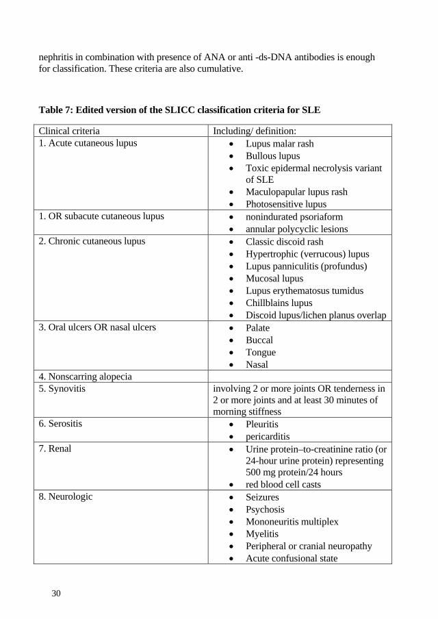
30
nephritis in combination with presence of ANA or anti -ds-DNA antibodies is enough for classification. These criteria are also cumulative.
Table 7: Edited version of the SLICC classification criteria for SLE
Clinical criteria Including/ definition: 1. Acute cutaneous lupus • Lupus malar rash
• Bullous lupus • Toxic epidermal necrolysis variant
of SLE • Maculopapular lupus rash • Photosensitive lupus
1. OR subacute cutaneous lupus • nonindurated psoriaform • annular polycyclic lesions
2. Chronic cutaneous lupus • Classic discoid rash • Hypertrophic (verrucous) lupus • Lupus panniculitis (profundus) • Mucosal lupus • Lupus erythematosus tumidus • Chillblains lupus • Discoid lupus/lichen planus overlap
3. Oral ulcers OR nasal ulcers • Palate • Buccal • Tongue • Nasal
4. Nonscarring alopecia 5. Synovitis involving 2 or more joints OR tenderness in
2 or more joints and at least 30 minutes of morning stiffness
6. Serositis • Pleuritis • pericarditis
7. Renal • Urine protein–to-creatinine ratio (or 24-hour urine protein) representing 500 mg protein/24 hours
• red blood cell casts 8. Neurologic • Seizures
• Psychosis • Mononeuritis multiplex • Myelitis • Peripheral or cranial neuropathy • Acute confusional state
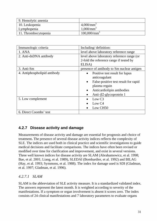
31
9. Hemolytic anemia 10. Leukopenia Lymphopenia
4,000/mm3 1,000/mm3
11. Thrombocytopenia 100,000/mm3
Immunologic criteria Including/ definition: 1. ANA level above laboratory reference range 2. Anti-dsDNA antibody level above laboratory reference range (or
2-fold the reference range if tested by ELISA)
3. Anti-Sm presence of antibody to Sm nuclear antigen 4. Antiphospholipid antibody • Positive test result for lupus
anticoagulant • False-positive test result for rapid
plasma regain • Anticardiolipin antibodies • Anti–β2-glycoprotein 1
5. Low complement • Low C3 • Low C4 • Low CH50
6. Direct Coombs’ test
4.2.7 Disease activity and damage Measurements of disease activity and damage are essential for prognosis and choice of treatment. The presence of several disease activity indices reflects the complexity of SLE. The indices are used both in clinical practice and scientific investigations to guide medical decisions and facilitate comparisons. The indices have often been revised or modified over time for clarification and improvement, and exist in several versions. Three well known indices for disease activity are SLAM (Abrahamowicz, et al. 1998; Bae, et al. 2001; Liang, et al. 1989), SLEDAI (Bombardier, et al. 1992) and BILAG (Hay, et al. 1993; Symmons, et al. 1988). The index for damage used is SDI (Gladman, et al. 1997; Gladman, et al. 1996).
4.2.7.1 SLAM
SLAM is the abbreviation of SLE activity measure. It is a standardized validated index. The answers represent the latest month. It is weighted according to severity of the manifestations. If a symptom or organ involvement is absent it scores zero. The index consists of 24 clinical manifestations and 7 laboratory parameters to evaluate organs
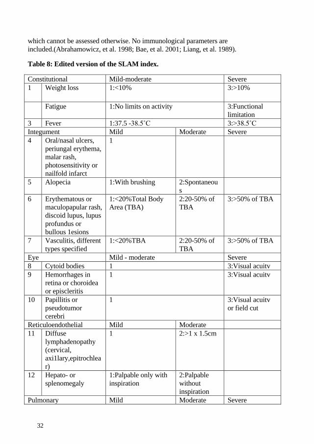
32
which cannot be assessed otherwise. No immunological parameters are included.(Abrahamowicz, et al. 1998; Bae, et al. 2001; Liang, et al. 1989).
Table 8: Edited version of the SLAM index.
Constitutional Mild-moderate Severe 1 Weight loss 1:<10% 3:>10%
Fatigue 1:No limits on activity 3:Functional limitation
3 Fever 1:37.5 -38.5˚C 3:>38.5˚C Integument Mild Moderate Severe 4 Oral/nasal ulcers,
periungal erythema, malar rash, photosensitivity or nailfold infarct
1
5 Alopecia 1:With brushing 2:Spontaneous
6 Erythematous or maculopapular rash, discoid lupus, lupus profundus or bullous 1esions
1:<20%Total Body Area (TBA)
2:20-50% of TBA
3:>50% of TBA
7 Vasculitis, different types specified
1:<20%TBA 2:20-50% of TBA
3:>50% of TBA
Eye Mild - moderate Severe 8 Cytoid bodies 1 3:Visual acuitv 9 Hemorrhages in
retina or choroidea or episcleritis
1 3:Visual acuitv
10 Papillitis or pseudotumor cerebri
1 3:Visual acuitv or field cut
Reticuloendothelial Mild Moderate 11 Diffuse
lymphadenopathy (cervical, axi1lary,epitrochlear)
1
2:>1 x 1.5cm
12 Hepato- or splenomegaly
1:Palpable only with inspiration
2:Palpable without inspiration
Pulmonary Mild Moderate Severe
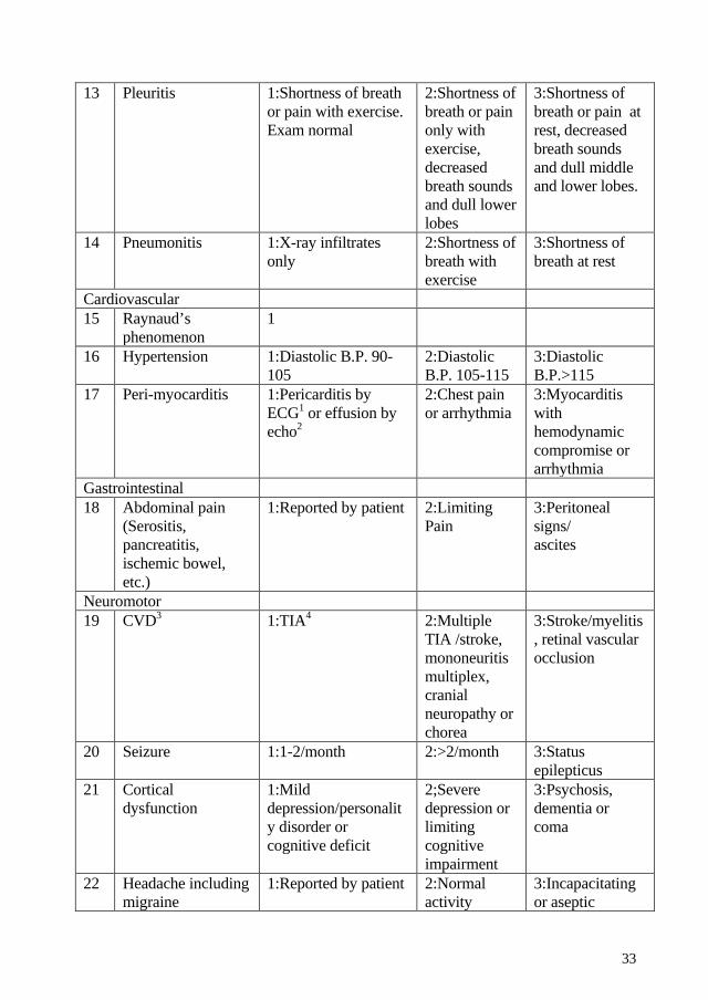
33
13 Pleuritis 1:Shortness of breath or pain with exercise. Exam normal
2:Shortness of breath or pain only with exercise, decreased breath sounds and dull lower lobes
3:Shortness of breath or pain at rest, decreased breath sounds and dull middle and lower lobes.
14 Pneumonitis 1:X-ray infiltrates only
2:Shortness of breath with exercise
3:Shortness of breath at rest
Cardiovascular 15 Raynaud’s
phenomenon 1
16 Hypertension 1:Diastolic B.P. 90-105
2:Diastolic B.P. 105-115
3:Diastolic B.P.>115
17 Peri-myocarditis 1:Pericarditis by ECG1 or effusion by echo2
2:Chest pain or arrhythmia
3:Myocarditis with hemodynamic compromise or arrhythmia
Gastrointestinal 18 Abdominal pain
(Serositis, pancreatitis, ischemic bowel, etc.)
1:Reported by patient 2:Limiting Pain
3:Peritoneal signs/ ascites
Neuromotor 19 CVD3 1:TIA4 2:Multiple
TIA /stroke, mononeuritis multiplex, cranial neuropathy or chorea
3:Stroke/myelitis, retinal vascular occlusion
20 Seizure 1:1-2/month 2:>2/month 3:Status epilepticus
21 Cortical dysfunction
1:Mild depression/personality disorder or cognitive deficit
2;Severe depression or limiting cognitive impairment
3:Psychosis, dementia or coma
22 Headache including migraine
1:Reported by patient 2:Normal activity
3:Incapacitating or aseptic
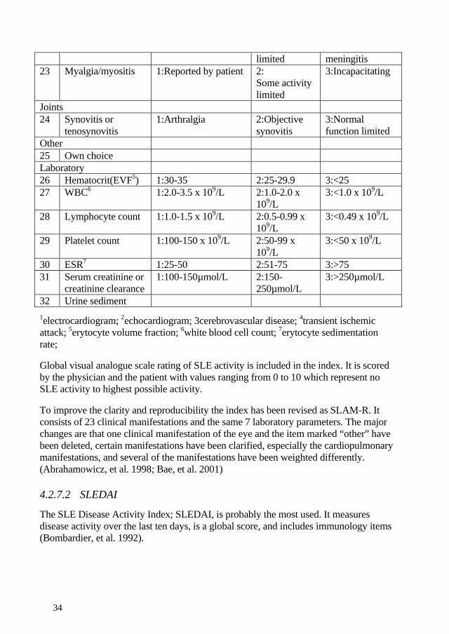
34
limited meningitis 23 Myalgia/myositis 1:Reported by patient 2:
Some activity limited
3:Incapacitating
Joints 24 Synovitis or
tenosynovitis 1:Arthralgia 2:Objective
synovitis 3:Normal function limited
Other 25 Own choice Laboratory 26 Hematocrit(EVF5) 1:30-35 2:25-29.9 3:<25 27 WBC6 1:2.0-3.5 x 10⁹/L 2:1.0-2.0 x
109/L 3:<1.0 x 10⁹/L
28 Lymphocyte count 1:1.0-1.5 x 10⁹/L 2:0.5-0.99 x 10⁹/L
3:<0.49 x 10⁹/L
29 Platelet count 1:100-150 x 109/L 2:50-99 x 109/L
3:<50 x 109/L
30 ESR7 1:25-50 2:51-75 3:>75 31 Serum creatinine or
creatinine clearance 1:100-150µmol/L 2:150-
250µmol/L 3:>250µmol/L
32 Urine sediment 1electrocardiogram; 2echocardiogram; 3cerebrovascular disease; 4transient ischemic attack; 5erytocyte volume fraction; 6white blood cell count; 7erytocyte sedimentation rate;
Global visual analogue scale rating of SLE activity is included in the index. It is scored by the physician and the patient with values ranging from 0 to 10 which represent no SLE activity to highest possible activity.
To improve the clarity and reproducibility the index has been revised as SLAM-R. It consists of 23 clinical manifestations and the same 7 laboratory parameters. The major changes are that one clinical manifestation of the eye and the item marked “other” have been deleted, certain manifestations have been clarified, especially the cardiopulmonary manifestations, and several of the manifestations have been weighted differently. (Abrahamowicz, et al. 1998; Bae, et al. 2001)
4.2.7.2 SLEDAI
The SLE Disease Activity Index; SLEDAI, is probably the most used. It measures disease activity over the last ten days, is a global score, and includes immunology items (Bombardier, et al. 1992).

35
4.2.7.3 SDI
The SLE Damage Index, SDI, measures the cumulative damage and has a prognostic value (Gladman, et al. 1997; Gladman, et al. 1996).
4.2.8 Treatment Treatment of SLE depends on the severity of the disease activity/flare i.e. grade of inflammation and type of organ involvement. Mild activity when subjective symptoms dominate without objective findings in clinical and laboratory tests can be managed without immunosuppressive drugs.
Musculoskeletal- and joint pain are often treated with NSAID or analgesics. Topical corticosteroids are used for different limited mucocutaneous diseases or in combination with other medical treatments. Antimalarial agents are used for both skin and joint involvement. Hydroxychloroquine is the most frequently prescribed and used medication in SLE. It prevents new flares, is used for skin and joint involvements and generally prolongs life. Today it is recommended that all patients with SLE should be treated with hydroxychloroquine if there are no contraindications. Systemic corticosteroids are very effective in easing symptoms and decreasing disease activity. The dose used is based on the severity of the organ involvement. Use of corticosteroids is limited by side effects such as osteoporosis, diabetes, peptic ulcers, cataract, and development of Cushingoid features. Today there are medical treatments that can prevent or at least decrease the risk of several of these side effects. Systemic treatment with corticosteroids should always be given using the smallest possible dose for a limited time. Several immunosuppressive agents are used in the treatment of SLE. The traditional immunosuppressives most commonly used today are azathioprine, methotrexate, cyclosporine, cyclophosphamide, and mycophenolate mofetil. Both the drug effects and side effects are taken into consideration when the choice is made as to which treatment the patient should have. Treatment with biologics has been successful for patients with rheumatoid arthritis but a similar positive effect has not been achieved for patients with SLE. Biologics used for treatment of SLE are agents that affect the B-cells: rituximab Mabthera® and belilumab Benlysta®. Rituximab is a B-cell depleting therapy used off label for refractory lupus nephritis or severe CNS manifestations. Belilumab is the first approved medication for SLE by the authorities in about fifty years It is an anti BLyS-protein (B Lymphocyte Stimulator) also known as BAFF (B-cell Activating Factor).
4.2.9 Prognosis Prognosis has improved dramatically. In the 1950s the estimated five years survival was less than 50% (Merrell and Shulman 1955). In 2006 Kasitano et al published figures on a 95% five years survival. Prognostic factors included socio-economic factors, clinical
36
manifestations, disease activity, and male gender (Kasitanon, et al. 2006). Today many factors and possibilities have improved: diagnostic tools, possibility for early diagnosis and treatment, new medication alternatives, and better social-economic status in the western world. In spite of this, the risk for cardiovascular disease (CVD) has not decreased. Gustafsson summarized the results of fourteen surveys from 1974 to 2009 on mortality causes. Death from CVD accounted for 6% to 76%. Death from lupus disease activity and infections has decreased, but not from CVD (Gustafsson 2012). In a Swedish study, the figure for CVD mortality between 1964 and 2004 was stable and accounted for about 42% (Bjornadal, et al. 2004).
37
5 AIMS
General aim
To study pathophysiological mechanisms and clinical manifestations in primary Sjögren’s Syndrome and Systemic Lupus Erythematosus
Specific aims
• To define the incidence rate of primary Sjögren’s syndrome (pSS) and the prevalence of extraglandular manifestations (EGM) at the time of diagnosis of pSS in a prospective, population-based manner.
• To identify genetic polymorphisms of candidate genes with a role in the immune pathogenesis of pSS.
• To identify new risk loci and single nucleotide polymorphisms associated with pSS in a Genome Wide Association Study (GWAS).
• To investigate the expression of the target autoantigen Ro52 in salivary glands of patients with pSS.
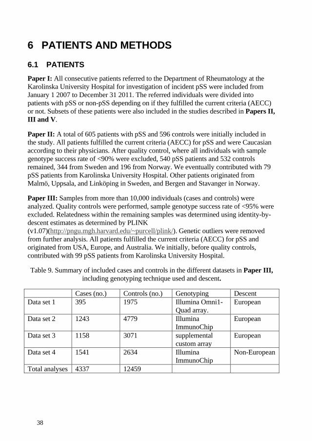
38
6 PATIENTS AND METHODS
6.1 PATIENTS Paper I: All consecutive patients referred to the Department of Rheumatology at the Karolinska University Hospital for investigation of incident pSS were included from January 1 2007 to December 31 2011. The referred individuals were divided into patients with pSS or non-pSS depending on if they fulfilled the current criteria (AECC) or not. Subsets of these patients were also included in the studies described in Papers II, III and V.
Paper II: A total of 605 patients with pSS and 596 controls were initially included in the study. All patients fulfilled the current criteria (AECC) for pSS and were Caucasian according to their physicians. After quality control, where all individuals with sample genotype success rate of <90% were excluded, 540 pSS patients and 532 controls remained, 344 from Sweden and 196 from Norway. We eventually contributed with 79 pSS patients from Karolinska University Hospital. Other patients originated from Malmö, Uppsala, and Linköping in Sweden, and Bergen and Stavanger in Norway.
Paper III: Samples from more than 10,000 individuals (cases and controls) were analyzed. Quality controls were performed, sample genotype success rate of <95% were excluded. Relatedness within the remaining samples was determined using identity-by-descent estimates as determined by PLINK (v1.07)(http://pngu.mgh.harvard.edu/~purcell/plink/). Genetic outliers were removed from further analysis. All patients fulfilled the current criteria (AECC) for pSS and originated from USA, Europe, and Australia. We initially, before quality controls, contributed with 99 pSS patients from Karolinska University Hospital.
Table 9. Summary of included cases and controls in the different datasets in Paper III, including genotyping technique used and descent.
Cases (no.) Controls (no.) Genotyping Descent Data set 1 395 1975 Illumina Omni1-
Quad array. European
Data set 2 1243 4779 Illumina ImmunoChip
European
Data set 3 1158 3071 supplemental custom array
European
Data set 4 1541 2634 Illumina ImmunoChip
Non-European
Total analyses 4337 12459
39
Paper IV: In this paper, 232 of the SLE patients managed at the Department of Rheumatology, Karolinska University Hospital were included February 2004 to October 2006. All patients fulfilled four or more of the American College of Rheumatology (ACR) 1982 revised classification criteria for SLE (Tan, et al. 1982).
Paper V: Twenty-eight patients from the Department of Rheumatology at Karolinska University Hospital, Stockholm, Sweden and the Department of Otolaryngology/Head and Neck Surgery at Haukeland University Hospital, Bergen, Norway diagnosed with pSS between the years 1992 and 2013 were included. All patients fulfilled the current criteria (AECC) for pSS. Twenty controls were included who originated from patients with sicca-symptoms who had not fulfilled the AECC critera, when evaluated for pSS. This was a case-control-study of 28 cases and 20 controls.
6.2 STUDY DESIGNS Paper I: Population-based prospective cohort study: The patients were included consecutively and data was recorded at time of diagnosis. Additional data was retrospectively collected to the database from the patients’ records.
Paper II and III: Multicenter case-control studies: The controls were matched for age, gender and geographical area. Paper V was a case-control study from two centers. The controls were matched for geographical area and time-period for the evaluation of suspected pSS.
Paper IV: Cross-sectional study.
6.3 DIAGNOSIS OF SJÖGREN’S SYNDROME In Papers I, II, III and V patients were evaluated for pSS, and in Paper IV patients were evaluated for sSS.
To confirm if patients fulfilled the current criteria, the 2002 Revised American-European Consensus Criteria (AECC), an examination in accordance with the items was performed. This included a questionnaire with validated questions from the defined AECC regarding sicca symptoms and exclusion criteria, a Schirmer’s test, Unstimulated Whole Salivary Flow (UWSF) during 15 minutes, and serological analysis of autoantibodies against Ro/SSA and La/SSB (this item is not included for sSS). A minor salivary gland biopsy was performed, if required for a complete diagnostic investigation of pSS, in accordance with the criteria.
40
6.4 EXTRAGLANDULAR MANIFESTATIONS, ORGAN INVOLVEMENTS AND INDICES MEASURING DISEASE ACTIVITY AND DAMAGE
Papers I, II, III and V: No general definition of extraglandular manifestations (EGM) exists. In Paper I we defined EGM as typical organ manifestations from our clinical experience with pSS patients, and also required that they should have been described previously in the literature. These EGM correspond to the EGM included in the Sjögren’s Syndrome Disease Activity Index (ESSDAI). (Seror, et al. 2010) published in 2010, with the addition of the liver diseases Primary Biliary Cirrhosis (PBC) and autoimmune hepatitis. We regarded the symptoms and organ manifestations in each individual as EGM of pSS if no other explanation was present. In Papers II and III a decision on which organ manifestations we should analyze for possible associations with genetic polymorphisms was made within multicenter collaborations and a study protocol was followed.
To detect EGM, a clinical exam at the out patients’ clinic was performed, information was collected from the patients’ records, and the patients were asked anamnestic questions on this issue if they had an appointment, which all patients who fulfilled the criteria or had undergone a minor salivary biopsy were offered.
In Paper IV features of SLE, including all items according to the ACR 1982 revised classification criteria (Tan, et al. 1982) and the presence of thyroid disease, were recorded. Disease activity was measured by SLAM (Liang, et al. 1989) and cumulative organ damage by the SLICC groups index SDI (Gladman, et al. 1996).
6.5 GENERAL HEALTH AND ANAMNESTIC DATA Paper I, II, III: Data on general health and anamnestic information was retrieved from questionnaires, patients’ records and a clinical exam of those who attended an appointment at the out patients’ clinic. In Paper IV the same procedure was performed, but following a specific structured protocol.
6.6 LABORATORY PARAMETERS In Paper IV, sampling followed the study protocol: Fasting blood samples were drawn at inclusion and routine laboratory tests and analyses of complement factors, Ig, and autoantibodies were performed at the SWEDAC (www.swedac.se) accredited Clinical Chemistry and Immunology Laboratories at the Karolinska University Hospital. IFL or ELISA was used for analyses of ANA, anti-ds-DNA-antibodies, anti Sm, and anti-RNP. In Papers I, II, III and V, in addition to autoantibodies according to item 6 in AECC for diagnosis, samples were taken following a decision by the physician, or retrieved from the patients’ records. The analysis procedures were performed as in Paper IV. In Paper IV, analysis of autoantibodies against the antigen Ro 52, including the epitopes RING and B-Box, Ro60 and La in serum, and in Paper V, autoantibodies against the
41
antigen Ro 52 in serum and urine, was performed at the Center of Molecular Medicine, Karolinska Institutet, at the research laboratory by ELISA (capture-ELISA in Paper V) technique using purified recombinant antigens.
6.7 GENOTYPING Paper II: We selected 84 candidate genes: Genes in the type I IFN system; genes involved in the inflammatory process in minor salivary glands; genes involved in lymphoma development; genes associated with systemic inflammation; and genes identified in GWAS to associate with SLE.
Tag-SNPs (r2=0.8 was considered as tagged) were selected by the following criteria: genotypes from the HapMap Caucasian (CEU) samples for SNPs with a minor allele frequency >5%; SNPs with an Illumina design score of >0.4; and >60 base pairs between SNPs. DNA samples were genotyped by the Illumina GoldenGate assay (Illumina Inc.).
Usual quality controls (QC)were performed: Twenty-eight samples were genotyped twice by the same technique to assure high consistency, which was confirmed (100%); the genotype quality control threshold used in the study was a SNP call rate of >90%; a minor allele frequency of >1%; and a Hardy–Weinberg equilibrium in the control samples (P>0.001). All SNPs which did not fulfill the criteria were excluded. 1139 SNPs out of 1261 (90.33%) remained.
In Paper III the QC procedures were stricter, additional requirements compared with Paper II were: well-defined cluster scatter plots; SNP call rate >95%; sample call rate >95%; and p > 0.001 for differential missing between cases and controls. All SNPs which did not fulfill the criteria were excluded. DNA samples were genotyped by the Illumina Omni1-Quad array (GWAS chip), Illumina ImmunoChip, or for a subset supplemental custom array. Table 9 summarizes data on number of cases and controls for the performed data-sets analyses, used genotyping technique, and the descent of the included individuals. Note that subsets of cases and controls were included in more than one data-set.
6.8 MINOR SALIVA GLAND BIOPSIES Paper V: When the biopsies were done for diagnostic purposes, the patients were offered to leave samples for research purposes to the bio-bank. The tissue samples were stored at -70°C in aliquots. The biopsy samples were prepared as described in the original manuscript and stained with Mayer’s Haematoxylin. Evaluation of staining was performed by three investigators (scored blindly by two) regarding: Focal infiltrates, whether these focal infiltrates were positively stained for Ro52 protein, and Ro52 staining of ductal epithelium. Depending on the degree of positivity number 0, 1 or 2 was assigned for each category, where 0 was considered negative, 1 was regarded
42
positive and 2 represented strongly positive. Focus scoring accounts focal infiltrates comprising of >50 mononuclear cells/4mm2 of tissue. Morphometry was added to score the sections by calculating the ratio-index in each gland. The ratio-index is defined as the total inflammatory area of focal infiltrates in the section divided by the total glandular tissue area (Manz, et al. 1997).
6.9 STATISTICAL ANALYSES Characteristics of the cohort and/or included patients are described using descriptive statistics: For continuous variables mean ±standard deviation (SD) is used; categorical variables are presented as percentages. Normality of data distribution was analysed by the Shapiro-Wilk test. For statistical comparisons between groups the following tests were used: for continuous variables, Mann-Whitney U test; for categorical variables, the Chi-square test for 2x2 tables or Fishers exact test; and for means, the unpaired t-test. The Spearman correlation test was used for continuous variables. The Kruskal-Wallis test was used for correlation comparing several groups.
In Paper 1 demographic characteristics such as age and gender specific information of the background population were retrieved from Statistics Sweden (SCB www.scb.se). For calculation of the 95% confidence intervals of incidence rates, the Bernoulli Model for categorical populations with two categories was used (Lindgren 1976).
Calculations in Papers I and IV were performed using Graph Pad Prism version 5.01 for Windows (Graph Pad Software, San Diego, California, USA (www.graphpad.com)) and Microsoft Excel 2010 (www.microsoft.com). A p-value <0.05 was considered significant.
Calculations in Paper II and III were performed using: the PLINK software (http://pngu.mgh.harvard.edu/~purcell/plink/) for estimation of LD, calculation of ORs, (In Paper III for, logistic regression models) and the Cochran–Mantel–Haenszel test for combining the Swedish and Norwegian cohorts; the Haploview v.4.1 software (http://www.broadinstitute.org/haploview/) for construction of LD plots between the genotyped polymorphisms in the combined dataset from the controls, and to determine the pairwise correlation between SNPs in the combined control data and in the HapMap data; and the genetic power calculator Quanto for calculation of the allelic association power of the study using the matched case–control module for discrete traits and an additive inheritance mode (http://hydra.usc.edu/gxe). A p-value <0.001 was considered a significant association, and a p-value <0.01 was considered a suggestive association.
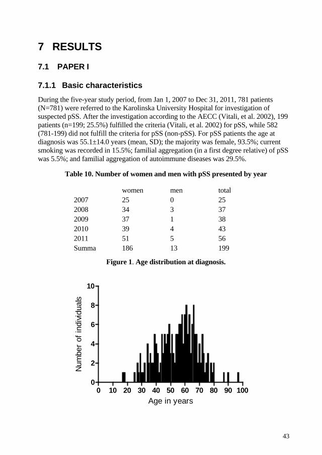
43
7 RESULTS
7.1 PAPER I
7.1.1 Basic characteristics During the five-year study period, from Jan 1, 2007 to Dec 31, 2011, 781 patients (N=781) were referred to the Karolinska University Hospital for investigation of suspected pSS. After the investigation according to the AECC (Vitali, et al. 2002), 199 patients (n=199; 25.5%) fulfilled the criteria (Vitali, et al. 2002) for pSS, while 582 (781-199) did not fulfill the criteria for pSS (non-pSS). For pSS patients the age at diagnosis was 55.1±14.0 years (mean, SD); the majority was female, 93.5%; current smoking was recorded in 15.5%; familial aggregation (in a first degree relative) of pSS was 5.5%; and familial aggregation of autoimmune diseases was 29.5%.
Table 10. Number of women and men with pSS presented by year
women men total
2007 25 0 25 2008 34 3 37 2009 37 1 38 2010 39 4 43 2011 51 5 56 Summa 186 13 199
Figure 1. Age distribution at diagnosis.
0 10 20 30 40 50 60 70 80 90 1000
2
4
6
8
10
Age in years
Num
ber o
f ind
ividu
als
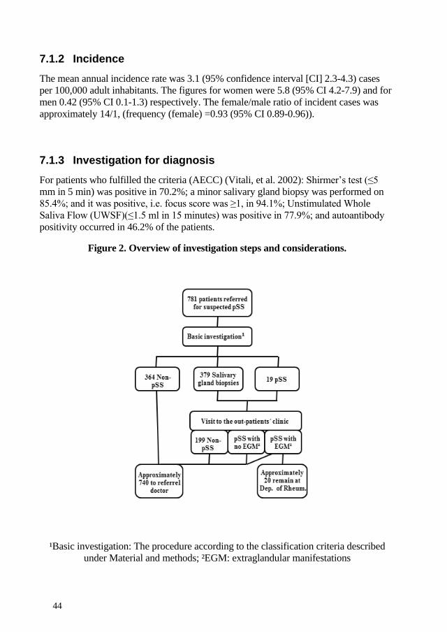
44
7.1.2 Incidence The mean annual incidence rate was 3.1 (95% confidence interval [CI] 2.3-4.3) cases per 100,000 adult inhabitants. The figures for women were 5.8 (95% CI 4.2-7.9) and for men 0.42 (95% CI 0.1-1.3) respectively. The female/male ratio of incident cases was approximately 14/1, (frequency (female) =0.93 (95% CI 0.89-0.96)).
7.1.3 Investigation for diagnosis For patients who fulfilled the criteria (AECC) (Vitali, et al. 2002): Shirmer’s test (≤5 mm in 5 min) was positive in 70.2%; a minor salivary gland biopsy was performed on 85.4%; and it was positive, i.e. focus score was ≥1, in 94.1%; Unstimulated Whole Saliva Flow (UWSF)(≤1.5 ml in 15 minutes) was positive in 77.9%; and autoantibody positivity occurred in 46.2% of the patients.
Figure 2. Overview of investigation steps and considerations.
¹Basic investigation: The procedure according to the classification criteria described under Material and methods; ²EGM: extraglandular manifestations
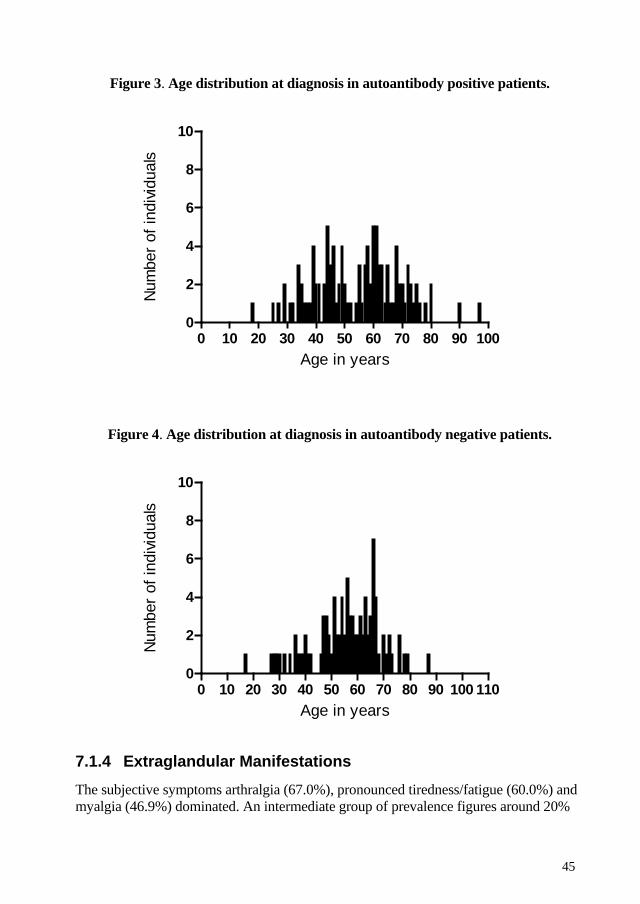
45
Figure 3. Age distribution at diagnosis in autoantibody positive patients.
0 10 20 30 40 50 60 70 80 90 1000
2
4
6
8
10
Age in years
Num
ber o
f ind
ividu
als
Figure 4. Age distribution at diagnosis in autoantibody negative patients.
0 10 20 30 40 50 60 70 80 90 100 1100
2
4
6
8
10
Age in years
Num
ber o
f ind
ividu
als
7.1.4 Extraglandular Manifestations The subjective symptoms arthralgia (67.0%), pronounced tiredness/fatigue (60.0%) and myalgia (46.9%) dominated. An intermediate group of prevalence figures around 20%
46
included Raynaud´s phenomenon (20.4%), depression (19.8%) and hypothyroidism (18.0%). Classical EGM associated with systemic pSS were present with the following prevalence figures: Arthritis (13.9%), leucopenia (11.0%), lymphopenia (10.2%), recurrent fever episodes (7.7%), PBC (4.6%), polyneuropathy (4.1%), cutaneous vasculitis (3.6%), lymphadenopathy (2.1%), interstitial lung disease (1.0%) and lymphoma (0%).
To identify factors that predicted EGM, we first compared autoantibody positive and negative patients. Autoantibody positive patients had significantly more leucopenia (p=0.008) and lymphopenia (p=0.05). The presence of lymphadenopathy and atrophic gastritis showed a tendency towards being more common in pSS patients with autoantibodies (p=0.07 for both).
In the subgroups with polyneuropathy and PBC the mean age was significantly higher. Patients with polyneuropathy were 11.0 years older, with a mean age of 65.5 (p=0.05), and patients with PBC were 11.1 years older, with a mean age of 65.6 (p=0.009).
7.2 PAPER II The Swedish and Norwegian cohorts were analyzed both separately and combined. The combined analysis (meta-analysis) identified associated SNPs with p-values <0.001 in three gene loci with no previously reported association with pSS: The early B-cell factor 1 (EBF1) gene on chromosome 5q34; the family with sequence similarity 167, member A (previously denoted C8 open reading frame 13)–B-lymphoid tyrosine kinase (FAM167A–BLK) locus on chromosome 8p23.1; and the tumor necrosis factor superfamily member 4 (TNFSF4=Ox40L) gene on chromosome 1q25.1. Confirmation of previous association of SNP loci to pSS for IRF5/ TNPO3 gene on chromosome 7q32.1and STAT4 gene on chromosome 2q32.3. A further eight suggested associated gene variants were identified.
7.3 PAPER III The meta analysis identified associated SNPs with p-values < 5 x 10⁻⁸ i.e. at a genome-wide level of significance in gene loci previously identified to associate with pSS though not with this grade of significance (polymorphisms in the HLA region had been identified previously). The gene variants were: HLA-region on chromosome 6p21 (p meta = 7.65 × 10−114); IRF5-TNPO3 (p meta = 2.73 × 10⁻¹⁹); STAT4 (p meta = 6.80 × 10⁻¹⁵); IL12A (p meta = 1.17 × 10⁻¹⁰); FAM167A-BLK (p meta = 4.97 × 10⁻¹⁰); DDX6-CXCR5 (p meta = 1.10 × 10⁻⁸); and TNIP1 (p meta = 3.30 × 10⁻⁸). A further 29 suggested associated gene variants were identified (p meta < 5 × 10⁻⁵).
47
7.4 PAPER IV
7.4.1 Basic characteristics At inclusion, patients were 48.1±14.5 (mean±standard deviation (SD)) years old and with a disease duration of 16.5±11.1 years (mean, SD). The age at diagnosis was 31.6±13.0 years (mean, SD). The majority were female, 91.8%.
7.4.2 Organ involvements and indices measuring disease activity and damage
The most common organ manifestation was arthritis, observed in 86.1% of the patients, followed by photosensitivity (68.5%), and malar rash (53.4%). The frequency of autoantibody positivity for ANA was 99.1%, and for anti-ds-DNA 68.5%. The mean number of fulfilled ACR classification criteria in the cohort was 6, the mean for the SLAM activity score was 7, and the mean of the cumulative organ damage index SDI was 1.
7.4.3 Correlation between autoantibodies and disease activity Ro52-RING antibody levels were associated with higher disease activity measured by SLAM. The analysis was made by categorizing the patients into four groups (quartiles) depending on their autoantibody level. The difference in clinical activity observed for Ro52-RING antibody levels had a p value of 0.04 (Kruskal-Wallis). No other association between any of the autoantibodies and disease activity was found.
7.4.4 Correlation between autoantibodies and clinical/laboratory manifestations
Anti-Ro52 antibody levels correlated with more of the investigated variables than did Ro60 and La autoantibodies. The condition of sSS and the items for diagnosis, as well as the degree of decreased production of saliva and tears, associated with Ro52 and to some extent with Ro60 and La antibodies. Ro52 antibody levels also correlated positively with the sedimentation rate, Ig levels, and with decreased count of both leucocytes and lymphocytes. We found no correlation between the autoantibodies and any individual severe form of organ manifestation.
7.5 PAPER V Ro52 was highly expressed in all the focal infiltrates in pSS patients. Significantly higher degree of Ro52 expression in ductal epithelium was observed in the patients compared to the controls (p<0.03). The degree of ductal epithelium expression of Ro52 correlated to the level of inflammation (Spearman r=0.48, p<0.0120). No secreted Ro52 protein could be detected in serum or saliva samples of these individuals.
48
8 DISCUSSION
8.1 PAPER I
8.1.1 Incidence In this prospective study we found an annual incidence rate of pSS in the Karolinska University Hospital catchment area of 3.1 cases per 100,000 person-years. To the best of our knowledge, no other population based prospective study of incidence using the current criteria exists. Three other publications on this topic have published similar figures (1.1–5.3 cases per 100.000 person-years), but the surveys have other study designs and criteria. Plesivcnik et al (Plesivcnik Novljan, et al. 2004) observed an incidence rate in Slovenia of 3.9 cases per 100,000 inhabitants (95% CI 1.1-10.2) using the European classification criteria. Pillemer et al (Pillemer, et al. 2001) observed an incidence of 3.9 (95% CI 2.8-4.9) per 100,000 in a retrospective study of physician-diagnosed pSS in Olmsted County, Minnesota, and Yu et al (Yu, et al. 2013) published figures of 10.6 (95% CI 9.9–11.4) from Taiwan using the present AECC. We believe the true incidence may be slightly higher than our findings. The difference, in addition to other criteria and study designs, may be explained by potential selection bias, and possibly that private rheumatologists have seen and diagnosed pSS patients: We believe their patients represent less than five percent of the total number, based on private rheumatologists limited capacity to accept new patients, in combination with the fact that the Department of Rheumatology at Karolinska has accepted to perform investigations for all referred patients with suspected pSS. In addition to study design and used classification criteria, other factors which influence the pattern of referral will have an effect on the calculated figures of incidence rate we have presented. People with mild symptoms can remain undiagnosed, some individuals may never contact their doctor regarding their symptoms, and for some patients the doctor performing the primary investigation may have chosen not to refer a patient that would have fulfilled the criteria. We believe there is a growing awareness of pSS in the general population in Sweden due to more easily available information from patient organizations and via the internet. As a result, more patients probably contact their doctors, even with milder symptoms. The examples can have both increasing and decreasing impact on incidence.
8.1.2 Extraglandular Manifestations The presence of EGM in our cohort is comparable to other recently published studies, although figures for the presence of autoantibodies and more severe EGM such as lung and neurological diseases are lower in our cohort. We attribute the differences to the following: The fact that our cohort consists of incident cases; we expect development of more EGM over time and a follow-up in our cohort may result in EGM in range with other cohorts of established pSS; and a different study-design, i.e. population-based or hospital case-series, prospective or retrospective. In hospital case-series, the patients are skewed towards the most severe forms of the disease. We wanted to compare our results
49
with those from other population based surveys. We performed several data base searches in PubMed and found several published population based studies on pSS but few on EGM generally that would match for comparison. Friedman et al. (Friedman, et al. 2006) was the only matching survey with the criterion of population-based surveys of EGM fulfilled. Their published figures were generally lower for all EGM except polyneuropathy, but data were missing for liver diseases.
Notably, lymphoma is an EGM which is typically known to debut several years after the pSS diagnosis, explaining why no patient in our cohort had lymphoma, since they were included at the time of diagnosis, and preexisting lymphoma is an exclusion criterion for pSS. In our cohort, five individuals were excluded from pSS diagnosis due to preexisting lymphoma.
We believe our low figures of EGMs represent a low grade of selection bias, and that they are truly representative for pSS in the population as defined by the AECC criteria.
8.2 PAPER II The three new identified gene loci with SNPs associated with pSS, EBF1, FAM167A-BLK and TNFSF4, are all involved in B-cell development and activation. Two of them have previously been associated with other autoimmune diseases, FAM167A-BLK and TNFSF4. EBF1 had not previously been associated with several autoimmune conditions, but with acute leukemia of B cell origin. However, despite no previous association for EBF1 with autoimmune diseases, it is obvious that the gene variation relates to the other novel discovered gene variations in FAM167A-BLK and TNFSF4, via the common factor involvement in one of the steps of the B-cells development from immature forms to effector cells, plasma-cells. The possibility that other gene variations affect cells in the immune system, but not previously have been associated with autoimmunity, should be considered in the future search for associated gene variations.
The novel results of association with gene variations in EBF1, FAM167A-BLK and TNFSF4 support the opinion that pSS is a B-cell driven disease, to an even higher degree than in SLE, in view of the additional association with EBF1. The involvement of associated gene variants with functions in the immune system, including previous findings of associations with IRF5 gene variants, strengthens the importance of autoimmune mechanisms in the pathogenesis of pSS and the close relationship with other autoimmune diseases, in particular SLE. All associated novel gene variants, except EBF1, and all confirmed gene variants, IRF5 and STAT4, had previously been associated with SLE. Patients with SLE and pSS have similar and different features in their disease phenotypes. For physicians it can be difficult to differentiate between the diagnoses, especially at disease onset. In other situations the difference is obvious, especially in patients with severe organ manifestations and presence of specific autoantibodies. To further investigate the genetic background to these similarities and
50
differences in the diseases phenotypes, functional studies must be done. Association studies can be the first step in the process of understanding pathogenesis and disease mechanisms. Though, the actual effect of the gene variation in form of increased/ decreased production of peptides from the protein-coding gene or altered effect of the peptides needs to be investigated by functional studies. This future perspective describes the next step in how we must form the coming study designs for our work.
The chosen threshold level for associated gene variants was p<0.001 and for suggested associated gene variants p<0.01, was decided in comparison with other publications. At that time other candidate-gene studies had been published with the same association significance level. The identified gene variants have later been confirmed for pSS by GWAS using the genome-wide level significance (p < 5 x 10⁻⁸). We chose to not use Bonferroni correction in our candidate gene study, which often is used to counteract the problem of multiple comparisons. The method is regarded as the simplest and most conservative of available alternative tests for correction in multiple testing. However, we still found it too strict for our candidate-gene study. If the Bonferroni correlation had been used, several of the associated gene variants had not been regarded as significantly associated. To perform a Bonferroni correction, the calculated p value is divided by the number of comparisons (i.e. tests/SNPs). Bonferroni assumes that all analyses are independent of each other. This is not the case of SNP analysis, since several SNPs have high LD. Additionally; the selected genes for analyses are not independent on each other since they have common biologic involvement in a specific disease, in this case pSS.
8.3 PAPER III This is the first GWAS of pSS, and both novel gene loci associated with pSS were identified and previously associated gene loci were confirmed. More than 10,000 cases and controls were genotyped. The different used technique for genotyping is summarized in Table 10. Note that subsets of both cases and controls were included in more than one analysis. This large numbers of patients and controls, well matched for descent, secured the possibility to achieve a valid result. The associated genes with variations have their normal function within the immune system: STAT4, IRF5, and GTF21 are transcription factors; IL12A is a cytokine; BLK is a B-cell kinase; and CXCR5 a chemokine. Again, this highlights the importance of a normal function of the immune system to prevent pSS development. It is also very interesting that no association with tissue specific gene variants were identified. The question why exocrine glands are the target organs, when no genetic association with these organs can be identified, must lead us to investigate more environmental factors, as triggering factors for the pathogenesis.
In patients with rheumatoid arthritis, autoantibody (ACPA) positive and negative patients, have different associated gene variants, mainly in the HLA region (Padyukov, et al. 2011). In line with this finding, we should consider possibility of similar gene association pattern in pSS. In future analyses, stratification by autoantibody status would be interesting in analysis of genetic associations in pSS.
51
Another GWAS in pSS originating from China was published at the same time as ours (Li, et al. 2013). In the study, 597 pSS cases and 1,090 healthy controls were genotyped using Affymetrix® genome wide arrays. The results were substantially different. A novel gene loci was identified, not involved in immune response (GTF2I at 7q11.23 p meta = 1.31 × 10-53)). They also confirmed some previously reported associations in our GWAS with pSS cases of European descent, STAT4, TNFAIP3 and the MHC. Possible explanations for the differences can be: Other genetic variations in the analyzed Chinese cohort compared to the European descent cohort and the difference in number of included cases and controls, thereby less power to identify associated gene variants in the Chinese cohort.
In our GWAS >60% of the analyses were performed by the Illumina Immunochip, which is not a GWAS technique chip. However, it includes most genes involved in autoimmune diseases. The survey may be regarded as a GWAS due to made comparisons between the subsets of the analyzed data sets, the well-characterized patients, and the European descent secured by the PLINK technique. We believe the result would have been essentially the same if all patients had been genotyped with the GWAS technique, and that identification of additional loci will depend mostly on expanding the patient and control cohorts.
8.4 PAPER IV
8.4.1 Correlation between autoantibodies and disease activity The novel finding in this study is the observation that only those antibodies targeting the RING domain of Ro52 were associated with higher disease activity in patients with SLE. None of the other autoantibodies we focused on in this study: Ro, La, and the more recently discovered subset of autoantibodies against the B-box epitopes of Ro52 showed this association. The RING domain is the enzymatically active part of the Ro52 protein, and contains the E3 ligase activity. This is interesting since anti-RING antibodies have been shown to bind, and inhibit, this function, and thereby inhibit the degradation of transcription factors for type 1 interferons (Espinosa, et al. 2011)known to mediate higher disease activity in SLE. Ro52 ubiquitinates and down-regulates the activity of IRF5 and IRF3 transcription factors. Anti-RING antibodies could thus directly contribute to disease pathogenesis in SLE through inhibition of Ro52 biologic activity in its capacity as a negative regulator of type 1 interferon production, leading to increased IFN-a and -b levels. Increased levels of type 1 interferons are present in a substantial number of lupus patients, and they increase during flares.
Our choice of SLAM, instead of SLEDAI which is more commonly used for measurements of disease activity in SLE, is based on the fact that SLAM does not include items with autoantibodies. Thus, a bias when analyzing correlations with autoantibody levels is avoided. SLAM is also a well-validated index for measurement of
52
disease activity in SLE and takes both subjectivity and severity into consideration. Studies in other, similarly well-characterized cohorts are desirable for confirmation of our observation that SLE disease activity correlates with Ro52-RING antibodies. It would also be desirable to perform a study of RING- autoantibodies in a cohort of pSS patients. Presence of Ro-antibodies and/or La antibodies is one of the items in the diagnosis AECC for pSS. Today a validated disease activity index for pSS is available, the EULAR Sjögren’s Syndrome Disease Activity Index (ESSDAI) (Seror, et al. 2010). However, the index is not yet well established in clinical use and it measures more severe form of EGM in pSS. A multi-center study could be a future possibility to have enough patients for such an investigation.
8.4.2 Correlation between autoantibodies and clinical/laboratory manifestations
We confirm that SLE patients with La and/or Ro autoantibodies have less severe disease where skin and joint manifestations dominate, a clinical feature also present in pSS with EGM. This finding adds another example of similarities between pSS and the subset of SLE patients with sSS. We noted the correlation between Ro52 autoantibodies with sSS and the items for diagnosis of sSS in the current diagnostic and classification criteria. Ro autoantibodies correlated with more variables than any other autoantibody. We could not confirm that the process of epitope spreading would lead to correlation with other symptoms of sSS and other of the studies autoantibodies. To further analyze this we would need to do a follow-up study and re-measure autoantibodies and re-evaluate the different items included in the classification criteria AECC. Ro autoantibodies are one of the first autoantibodies to appear in the SLE development, often before onset (Eriksson, et al. 2011). The very presence of these autoantibodies can contradict a more severe disease. This could mean that it is possible early after diagnosis of SLE in absence of negative predictive factors chose less strong medications. This is an important matter since side-effects of medication have previously been an important problem for patients with SLE. Even if we have more treatment alternatives to offer today, even biologics, severe side-effect will be a matter for worry among patients for a long time.
8.5 PAPER V The expression of Ro52 was observed in ductal epithelium in saliva gland tissue deriving from minor saliva gland biopsies. These biopsies had been performed if required for a complete diagnostic process in accordance with the present classification criteria for investigation of suspected pSS. The expression was present in both cases and controls but in a significantly higher degree in the cases (patients) compared to the non-pSS controls (p<0.03).
53
The cases consisted of patients who fulfilled the classification criteria and the controls did not. The controls were thus not healthy individuals but patients with sicca symptoms of a degree that made a physician decide to perform a minor salivary gland biopsy. The impact of this background information is not known. It could be possible that expression of Ro52 in ductal epithelium in saliva gland tissue, at least in a low degree is a found present in subsets of the population over time. An investigation of healthy controls, without sicca symptoms, is required for more information in this matter.
That the degree of ductal epithelium expression of Ro52 correlated to the level of inflammation (Spearman r=0.48, p<0.0120) in the tissue, supports that a high degree of expression of Ro52 is a part of the disease pathogenesis. Correlation of Ro52 expression has earlier been demonstrated in SLE patients from skin lesions (Oke, et al. 2009). Expression and upregulation of Ro52 have been demonstrated in both SLE and pSS and in both diseases a correlation with inflammation. This supports Ro52 as a general target in autoimmunity. It also is an example of similarities of the diseases.
We believe that the upregulation of Ro52 occurs early in the pathogenic autoimmune disease and that it can lead to breaking of tolerance in genetic susceptible individuals. Autoantibodies can be a consequence of this expression. If so, the pathogenic process is ongoing for a very long time. The presence of autoantibodies has been shown to occur many years before diagnosis, and if the expression of Ro52 appears even earlier, the pathogenic process is expected to start decades before diagnosis.
The ELISA analyses could not detect Ro52 in saliva or serum from neither cases nor controls. This suggests that Ro52 is not a secreted protein, and that the antigen exposure takes place in the tissue of the target organ.
If possible it would be interesting to analyze Ro52 in salivary gland tissue from SLE patients, healthy controls and sSS to other autoimmune diagnosis in addition to SLE. This could further reveal the role of Ro52 in pathogenesis of autoimmunity.
54
9 CONCLUDING REMARKS AND FUTURE PERSPECTIVES
This thesis has focused on pathogenesis and descriptive studies of pSS and SLE. For pSS the studies have included epidemiology, genetics, and histopathological mechanisms. For SLE we focused on immunological mechanisms in the form of autoantibodies. The main observations include:
• The annual incidence rate of pSS in the Karolinska University Hospital catchment area was 3.1 (95% CI 2.3-4.3) cases per 100,000 adult inhabitants. The female/male ratio of incident cases was 14/1, (frequency (female) =0.93 (95% CI 0.89-0.96)).
• Lower figures for severe EGM for incident pSS, and lower frequency of autoantibodies including ANA, anti-Ro/SSA and anti-La/SSB that previously described for pSS was observed in this population based cohort.
• Candidate-gene and GWAS studies clearly indicate a similar background in genetic mechanisms in autoimmune diseases, in particular for pSS and SLE. The genetic variances demonstrate the importance of B-lymphocytes in the mechanisms of pSS pathogenesis.
• RING-autoantibodies are correlated to higher disease activity in SLE patients which support the importance of Ro52s function as an E3ligase and its clinical implications.
Future perspectives
More epidemiological studies on incidence rate in population-based prospective studies of pSS are desirable to confirm our figures.
A follow-up study of EGM frequencies in the incident pSS cohort to confirm the expected development is planned.
Functional studies of the consequences of SNPs in associated genes are necessary to further reveal how these gene variants enhance the risk for pSS development.
Further studies on the RING-autoantibodies should be performed in patients with other autoimmune diseases.
55
10 ACKNOWLEDGEMENTS I wish to thank all who have contributed to this thesis and supported me in different ways. In particular I would like to acknowledge:
Marie Wahren-Herlenius, my main supervisor, for being the best main supervisor I could wish for. She has introduced me to research, and taught me a lot about basal mechanisms in pSS and about autoimmunity in general. She is knowledgeable, positive and encouraging. She has become more than a supervisor to me. I value her as a friend I can talk to, and who is able to give me advice on everything from science to how it is to be a mother.
Elisabet Svenungsson, my co-supervisor and co-author, who means so much to me in so many ways: as a co-supervisor, as a colleague, and through our involvement in SRF’s group for the Prevention of Cardiovascular Diseases. She has introduced me at the clinic, helped me with statistics, and with difficult SLE cases.
Elisabet Welin-Henriksson, my mentor, for our enjoyable lunch meetings which made me feel that you would always be there to support me had I needed it.
Ingrid Lundberg, current head of the Rheumatology Unit, KI, Solna, and Lars Klareskog, former head of the Rheumatology Unit, KI, Solna, for creating a good scientific atmosphere at the clinic, and for creating possibilities for so many people to be introduced to science and doctoral studies. I would also like to thank you both for being there to give clinical advice concerning particularly difficult cases within your fields of expertise.
Cecilia Carlens, current head of the Department of Rheumatology, Karolinska University Hospital, and Johan Bratt, former head of the Department of Rheumatology, Karolinska University Hospital, for giving me the opportunity to start and complete my doctoral studies.
Iva Gunnarsson, my current director at the Department of Rheumatology, Karolinska University Hospital, and co-author. I value her for always being so encouraging and supportive.
I would like to thank all my co-authors at the clinic: Birgitta Nordmark, Johanna Gustafsson, and Iva Gunnarsson; co-authors in the Wahren group Viijole Ottosson, Lars Ottosson, and Lara Aqrawi; co-authors in the Sjögren-network; and co-authors in the international collaboration for the GWAS study.of Sjögren’s syndrome.
I would like to thank all present, former and temporary members of the Wahren group for your friendship and for sharing your knowledge: Aurelie Ambrosi, Susanna Brauner, Åse Elfving, Alexander Espinosa, Sabrina Görgen, Vilija Oke, Viijole Ottosson, Maria Sjöstrand, Amanda Skog, Joanna Tingström, Marie Wahren-Herlenius, and Therese Östberg. Many thanks, too, for all the enjoyable dinners and
56
trips. I especially would like to thank Nånnis for always helping me to understand difficult things (not defined) without ever complaining and also along with all in the group for friendship.
I would like to thank all members of the Sjögren-network for their cooperation in the genetic studies and for sharing their knowledge at the regular meetings. In particular, I would like to thank Gunnel Nordmark as the initiator of the Sjögren network, and also, along with Elke Theander and Lilian Vasaitis, for substantial collaboration on these studies and other questions concerning Sjögren’s syndrome.
Eva Jemseby and Gull-Britt Almgren for excellent technical assistance.
Julia F Simard for insightful statistical advice.
Gunnel Bremerfeldt, Susanne Karlfeldt, Stina Norman, and Veronica Ebbersten Lindholm for always giving excellent support, including technical support with computers and photocopier.
Karin Bohjort, Märta Tötterman, Ljiljana Eshete, Viktoria Bila and Miliete Ghebremicael for assistance with sampling for the Sjögren bio-bank.
Rebecca Preuveneers for language correction and always being so helpful.
I would like to thank all colleagues and friends at the Department of Rheumatology, Karolinska University Hospital Huddinge and Solna, that we are able to work together for common goals in rheumatology, both in the clinic and in research and education. Thank you for making the clinic a pleasant workplace where one feels welcome. Erik af Klint, Ola Börjesson, Christina Dorph, Iva Gunnarsson, Jon Lampa, Annika Nordin, Bo Ringertz, Elisabet Svenungsson, Ralph Nisell, Tomas Zweig, Lena Björnådal, Anders Harju, Ronald van Vollenhoven, Johanna Gustafsson, Saedis Saevarsdottir, Maryam Dastmalchi, Hamed Rezaei, Louise Ekholm, Johan Askling, Ingrid.lundberg, Dimitrios Makrygiannakis, Aikaterini Chatzidionysiou, Karina Gheorghe, Lara Dani, Per-Johan Jakobsson, Reem Altawil, Sara Wedrén, Vilija Oke, Anca Catrina, Ioannis Parodis, Liselotte Tidblad, Cecilia Carlens, Saedis Saevarsdottir, Agneta Zickert, Aleksandra Antovic, Anna Vikerfors, Aune Avik, Bernhard Grewin, Christina Stranger, Christoffer Romland, Cristina Anania, Gudrun Reynisdottir, Helene Bolinder, Inga-Lill Engvall, Ingiäld Hafström, Johan Bratt, Karin Hellgren, Per Larsson, Sofia Ajeganova, Sofia Ernestam, Yvonne Dellmark, Zsuzsanna Fabianne, Anita Domargård, and Aikaterini Chatzidionysiou. Wiveca Roth for their fantastic collaboration with patients.
I would like to thank all who participated as controls in the research study on effect of vaccinations against influenza: Petra Amoudraz, Aikaterini Chatzidionysiou, Lara Dani, Anita Domargård, Ljiljana Eshete, Eleanor Gullström, Seija Johansson, Lena Jonsson, Christa Liewendahl, Inga Lodin, Vivianne Malmström, Carola
57
Nilsson, Annica Owetz, Jenny Riazolli, Wiveca Roth, Rosmari Rönnqvist, and Majken Svensson.
Seija Johansson and Lena Jonsson for assistance and collaboration in research studies.
Christina Ingermansson and Anette Hjärne for administrational assistance.
I would like to thank all patients who have contributed to the research studies through their participation.
Slutligen men viktigast, vill jag tacka vänner, släkt och familj för stöd och hjälp. Carl! min son, som jag älskar över allt och varje dag påminner mig om att mirakel finns. Rikard, utan dig skulle inget av detta varit möjligt.
Jag vill tacka Susanne, Yvonne, Kristina, Vincent.
Och 5F
58
11 REFERENCES (1999). "The American College of Rheumatology nomenclature and case definitions for
neuropsychiatric lupus syndromes." Arthritis Rheum 42(4): 599-608. (2005). "A haplotype map of the human genome." Nature 437(7063): 1299-1320. Abbas (2007). Cells and Tissues of the Adaptive Immune System. Cellular and
MolecularImmunology: 47-74. Abbas (2007). Diseases caused by immune respones: hypersensitivity and autoimmunity. Cellular
and Molecular Immunology 427.
Abbas (2007). Innate Immunity. Cellular and Molecular Immunology: 19-46. Abbas (2007). Properties and Overview of immune Responses. Cellular and Molecular
Immunology: 3-18. Abrahamowicz, M., P. R. Fortin, et al. (1998). "The relationship between disease activity and
expert physician's decision to start major treatment in active systemic lupus erythematosus: a decision aid for development of entry criteria for clinical trials." J Rheumatol 25(2): 277-284.
Akaogi, J., T. Barker, et al. (2006). "Role of non-protein amino acid L-canavanine in autoimmunity." Autoimmun Rev 5(6): 429-435.
Alamanos, Y., N. Tsifetaki, et al. (2006). "Epidemiology of primary Sjogren's syndrome in north-west Greece, 1982-2003." Rheumatology (Oxford) 45(2): 187-191.
Ambrosi, A., V. Dzikaite, et al. (2012). "Anti-Ro52 monoclonal antibodies specific for amino acid 200-239, but not other Ro52 epitopes, induce congenital heart block in a rat model." Ann Rheum Dis 71(3): 448-454.
Araujo-Fernandez, S., M. Ahijon-Lana, et al. (2014). "Drug-induced lupus: Including anti-tumour necrosis factor and interferon induced." Lupus.
Arbuckle, M. R., M. T. McClain, et al. (2003). "Development of autoantibodies before the clinical onset of systemic lupus erythematosus." N Engl J Med 349(16): 1526-1533.
Arnett, F. C. and L. E. Shulman (1976). "Studies in familial systemic lupus erythematosus." Medicine (Baltimore) 55(4): 313-322.
Aslpaugh, M. A. and E. M. Tan (1976). "Serum antibody in rheumatoid arthritis reactive with a cell-associated antigen. Demonstration by precipitation and immunofluorescence." Arthritis Rheum 19(4): 711-719.
Azevedo, P. C., G. Murphy, et al. (2014). "Pathology of systemic lupus erythematosus: the challenges ahead." Methods Mol Biol 1134: 1-16.
Bae, S. C., H. K. Koh, et al. (2001). "Reliability and validity of systemic lupus activity measure-revised (SLAM-R) for measuring clinical disease activity in systemic lupus erythematosus." Lupus 10(6): 405-409.
Bengtsson, A. A., L. Rylander, et al. (2002). "Risk factors for developing systemic lupus erythematosus: a case-control study in southern Sweden." Rheumatology (Oxford) 41(5): 563-571.
Bertsias, G. C., R.; Boumpas, D. T. (2012). Systemic Lupus Erythematosus: Pathogenesis and Clinical Featurs. EULAR Textbook on Rheumatic Diseases. J. W. Bijlsma: 476-505.
Bjornadal, L., L. Yin, et al. (2004). "Cardiovascular disease a hazard despite improved prognosis in patients with systemic lupus erythematosus: results from a Swedish population based study 1964-95." J Rheumatol 31(4): 713-719.
Bombardier, C., D. D. Gladman, et al. (1992). "Derivation of the SLEDAI. A disease activity index for lupus patients. The Committee on Prognosis Studies in SLE." Arthritis Rheum 35(6): 630-640.
59
Bonifacino, J. S. and A. M. Weissman (1998). "Ubiquitin and the control of protein fate in the secretory and endocytic pathways." Annu Rev Cell Dev Biol 14: 19-57.
Botsios, C., A. Furlan, et al. (2011). "Elderly onset of primary Sjogren's syndrome: clinical manifestations, serological features and oral/ocular diagnostic tests. Comparison with adult and young onset of the disease in a cohort of 336 Italian patients." Joint Bone Spine 78(2): 171-174.
Bowman, S. J., G. H. Ibrahim, et al. (2004). "Estimating the prevalence among Caucasian women of primary Sjogren's syndrome in two general practices in Birmingham, UK." Scand J Rheumatol 33(1): 39-43.
Brito-Zeron, P., M. Akasbi, et al. (2013). "Classification and characterisation of peripheral neuropathies in 102 patients with primary Sjogren's syndrome." Clin Exp Rheumatol 31(1): 103-110.
Buyon, J. P. (2008). Systemic Lupus Erythematosus A. Clinical and Laboratory Features. Primar on the Rheumatic Diseases. J. H. Klippel: 303-318.
Carter, R. H. (2006). "B cells in health and disease." Mayo Clin Proc 81(3): 377-384. Cervera, R., M. A. Khamashta, et al. (1993). "Systemic lupus erythematosus: clinical and
immunologic patterns of disease expression in a cohort of 1,000 patients. The European Working Party on Systemic Lupus Erythematosus." Medicine (Baltimore) 72(2): 113-124.
Churg J, S. L. (1982). "Renal Disease: Classification and Atlas of Glomerular Disease." Costenbader, K. H., D. J. Kim, et al. (2004). "Cigarette smoking and the risk of systemic lupus
erythematosus: a meta-analysis." Arthritis Rheum 50(3): 849-857. Cutolo, M., A. Sulli, et al. (2004). "Sex hormones influence on the immune system: basic and
clinical aspects in autoimmunity." Lupus 13(9): 635-638. D'Cruz, D. P., M. A. Khamashta, et al. (2007). "Systemic lupus erythematosus." Lancet
369(9561): 587-596. Daniels, T. (2008). Sjögren´s syndrome. Primer on the Rheumatic Diseases. J. H. Klippel: 389-
397. Elias, K. (2008). Molecular and CellularBasis of Immunity and ImmunologicalDiseases. Primer
on the Rheumatic Diseases. J. H. Klippel: 94-107. Eriksson, C., H. Kokkonen, et al. (2011). "Autoantibodies predate the onset of systemic lupus
erythematosus in northern Sweden." Arthritis Res Ther 13(1): R30. Escudero, D., P. Latorre, et al. (1995). "Central nervous system disease in Sjogren's syndrome."
Ann Med Interne (Paris) 146(4): 239-242. Espinosa, A., V. Dardalhon, et al. (2009). "Loss of the lupus autoantigen Ro52/Trim21 induces
tissue inflammation and systemic autoimmunity by disregulating the IL-23-Th17 pathway." J Exp Med 206(8): 1661-1671.
Espinosa, A., J. Hennig, et al. (2011). "Anti-Ro52 autoantibodies from patients with Sjogren's syndrome inhibit the Ro52 E3 ligase activity by blocking the E3/E2 interface." J Biol Chem 286(42): 36478-36491.
Espinosa, A., W. Zhou, et al. (2006). "The Sjogren's syndrome-associated autoantigen Ro52 is an E3 ligase that regulates proliferation and cell death." J Immunol 176(10): 6277-6285.
Feero, W. G., A. E. Guttmacher, et al. (2010). "Genomic medicine--an updated primer." N Engl J Med 362(21): 2001-2011.
Fenalti, G. and M. J. Rowley (2008). "GAD65 as a prototypic autoantigen." Journal of Autoimmunity 31(3): 228-232.
Finke, D., M. L. Eloranta, et al. (2009). "Endogenous type I interferon inducers in autoimmune diseases." Autoimmunity 42(4): 349-352.
Fox, R. I. (2005). "Sjogren's syndrome." Lancet 366(9482): 321-331.
60
French, M. A. and P. Hughes (1983). "Systemic lupus erythematosus and Klinefelter's syndrome." Ann Rheum Dis 42(4): 471-473.
Friedman, J. A., E. B. Miller, et al. (2006). "A community-based cohort of 201 consecutive patients with primary Sjogren's syndrome in Israel: Ashkenazi patients compared with those of Sephardic descent." Clin Exp Rheumatol 24(3): 274-280.
Garcia-Romo, G. S., S. Caielli, et al. (2011). "Netting neutrophils are major inducers of type I IFN production in pediatric systemic lupus erythematosus." Sci Transl Med 3(73): 73ra20.
Gladman, D., E. Ginzler, et al. (1996). "The development and initial validation of the Systemic Lupus International Collaborating Clinics/American College of Rheumatology damage index for systemic lupus erythematosus." Arthritis Rheum 39(3): 363-369.
Gladman, D. D., M. B. Urowitz, et al. (1997). "The reliability of the Systemic Lupus International Collaborating Clinics/American College of Rheumatology Damage Index in patients with systemic lupus erythematosus." Arthritis Rheum 40(5): 809-813.
Goransson, L. G., K. Haldorsen, et al. (2011). "The point prevalence of clinically relevant primary Sjogren's syndrome in two Norwegian counties." Scand J Rheumatol 40(3): 221-224.
Graham, R. R., G. Hom, et al. (2009). "Review of recent genome-wide association scans in lupus." J Intern Med 265(6): 680-688.
Griffiths, B., M. Mosca, et al. (2005). "Assessment of patients with systemic lupus erythematosus and the use of lupus disease activity indices." Best Pract Res Clin Rheumatol 19(5): 685-708.
Gustafsson, J. (2012). Studies on Cardiovascular Risk Factors in Systemic Lupus Erythematosus. Department of Medicin-Solna, Rheumatology Unit. Stockholm, Karolinska institutet. Doctoral: 14.
Gustafsson, J., I. Gunnarsson, et al. (2009). "Predictors of the first cardiovascular event in patients with systemic lupus erythematosus - a prospective cohort study." Arthritis Res Ther 11(6): R186.
Gustafsson, J., J. F. Simard, et al. (2012). "Risk factors for cardiovascular mortality in patients with systemic lupus erythematosus, a prospective cohort study." Arthritis Res Ther 14(2): R46.
Halse, A., P. Tengner, et al. (1999). "Increased frequency of cells secreting interleukin-6 and interleukin-10 in peripheral blood of patients with primary Sjogren's syndrome." Scand J Immunol 49(5): 533-538.
Harley, J. B., I. T. Harley, et al. (2006). "The curiously suspicious: a role for Epstein-Barr virus in lupus." Lupus 15(11): 768-777.
Hay, E. M., P. A. Bacon, et al. (1993). "The BILAG index: a reliable and valid instrument for measuring clinical disease activity in systemic lupus erythematosus." Q J Med 86(7): 447-458.
Higgs, R., E. Lazzari, et al. (2010). "Self protection from anti-viral responses--Ro52 promotes degradation of the transcription factor IRF7 downstream of the viral Toll-Like receptors." PLoS One 5(7): e11776.
Hostmann, A., A. M. Jacobi, et al. (2008). "Peripheral B cell abnormalities and disease activity in systemic lupus erythematosus." Lupus 17(12): 1064-1069.
Jonsson, M. V., E. Theander, et al. (2012). "Predictors for the development of non-Hodgkin lymphoma in primary Sjogren's syndrome." Presse Med 41(9 Pt 2): e511-516.
Jonsson, R., P. Vogelsang, et al. (2011). "The complexity of Sjogren's syndrome: novel aspects on pathogenesis." Immunol Lett 141(1): 1-9.
61
Kaiser, R. and L. A. Criswell (2010). "Genetics research in systemic lupus erythematosus for clinicians: methodology, progress, and controversies." Curr Opin Rheumatol 22(2): 119-125.
Kamen, D. L., G. S. Cooper, et al. (2006). "Vitamin D deficiency in systemic lupus erythematosus." Autoimmun Rev 5(2): 114-117.
Kasitanon, N., L. S. Magder, et al. (2006). "Predictors of survival in systemic lupus erythematosus." Medicine (Baltimore) 85(3): 147-156.
Kidd, J. M., G. M. Cooper, et al. (2008). "Mapping and sequencing of structural variation from eight human genomes." Nature 453(7191): 56-64.
King, L. B., A. Norvell, et al. (1999). "Antigen receptor-induced signal transduction imbalances associated with the negative selection of immature B cells." J Immunol 162(5): 2655-2662.
Kivity, S., M. T. Arango, et al. (2014). "Infection and autoimmunity in Sjogren's syndrome: A clinical study and comprehensive review." J Autoimmun.
Kong, H. J., D. E. Anderson, et al. (2007). "Cutting edge: autoantigen Ro52 is an interferon inducible E3 ligase that ubiquitinates IRF-8 and enhances cytokine expression in macrophages." J Immunol 179(1): 26-30.
Kvarnstrom, M., V. Dzikaite-Ottosson, et al. (2013). "Autoantibodies to the functionally active RING-domain of Ro52/SSA are associated with disease activity in patients with lupus." Lupus 22(5): 477-485.
Lander, E. S., L. M. Linton, et al. (2001). "Initial sequencing and analysis of the human genome." Nature 409(6822): 860-921.
Lessard, C. J., H. Li, et al. (2013). "Variants at multiple loci implicated in both innate and adaptive immune responses are associated with Sjogren's syndrome." Nat Genet 45(11): 1284-1292.
Levin, M. C., S. M. Lee, et al. (2002). "Autoimmunity due to molecular mimicry as a cause of neurological disease." Nat Med 8(5): 509-513.
Li, Y., K. Zhang, et al. (2013). "A genome-wide association study in Han Chinese identifies a susceptibility locus for primary Sjogren's syndrome at 7q11.23." Nat Genet 45(11): 1361-1365.
Liang, M. H., S. A. Socher, et al. (1989). "Reliability and validity of six systems for the clinical assessment of disease activity in systemic lupus erythematosus." Arthritis Rheum 32(9): 1107-1118.
Lindgren, B. (1976). Analysis of categorical data. In Statistical theory, MacMillian Publishing Co. Inc: 423-452.
Lood, C., B. Gullstrand, et al. (2009). "C1q inhibits immune complex-induced interferon-alpha production in plasmacytoid dendritic cells: a novel link between C1q deficiency and systemic lupus erythematosus pathogenesis." Arthritis Rheum 60(10): 3081-3090.
Lovgren, T., M. L. Eloranta, et al. (2004). "Induction of interferon-alpha production in plasmacytoid dendritic cells by immune complexes containing nucleic acid released by necrotic or late apoptotic cells and lupus IgG." Arthritis Rheum 50(6): 1861-1872.
Maldini, C., R. Seror, et al. (2014). "Epidemiology of Primary Sjogren's Syndrome in a French Multiracial/Multiethnic Area." Arthritis Care Res (Hoboken) 66(3): 454-463.
Malladi, A. S., K. E. Sack, et al. (2012). "Primary Sjogren's syndrome as a systemic disease: a study of participants enrolled in an international Sjogren's syndrome registry." Arthritis Care Res (Hoboken) 64(6): 911-918.
Manolio, T. A., L. D. Brooks, et al. (2008). "A HapMap harvest of insights into the genetics of common disease." J Clin Invest 118(5): 1590-1605.
62
Manz, R. A., A. Thiel, et al. (1997). "Lifetime of plasma cells in the bone marrow." Nature 388(6638): 133-134.
Masi, A. T. and R. A. Kaslow (1978). "Sex effects in systemic lupus erythematosus: a clue to pathogenesis." Arthritis Rheum 21(4): 480-484.
Meller, S., F. Winterberg, et al. (2005). "Ultraviolet radiation-induced injury, chemokines, and leukocyte recruitment: An amplification cycle triggering cutaneous lupus erythematosus." Arthritis Rheum 52(5): 1504-1516.
Merrell, M. and L. E. Shulman (1955). "Determination of prognosis in chronic disease, illustrated by systemic lupus erythematosus." J Chronic Dis 1(1): 12-32.
Montanaro, A. and E. J. Bardana, Jr. (1991). "Dietary amino acid-induced systemic lupus erythematosus." Rheum Dis Clin North Am 17(2): 323-332.
Moser, K. L., J. A. Kelly, et al. (2009). "Recent insights into the genetic basis of systemic lupus erythematosus." Genes Immun 10(5): 373-379.
Nordmark, G., G. Kristjansdottir, et al. (2011). "Association of EBF1, FAM167A(C8orf13)-BLK and TNFSF4 gene variants with primary Sjogren's syndrome." Genes Immun 12(2): 100-109.
Nordmark, G., C. Wang, et al. (2013). "Association of genes in the NF-kappaB pathway with antibody positive primary Sjogren's syndrome." Scand J Immunol.
Odendahl, M., A. Jacobi, et al. (2000). "Disturbed peripheral B lymphocyte homeostasis in systemic lupus erythematosus." J Immunol 165(10): 5970-5979.
Oke, V., I. Vassilaki, et al. (2009). "High Ro52 expression in spontaneous and UV-induced cutaneous inflammation." J Invest Dermatol 129(8): 2000-2010.
Ottosson, L., J. Hennig, et al. (2006). "Structural, functional and immunologic characterization of folded subdomains in the Ro52 protein targeted in Sjogren's syndrome." Mol Immunol 43(6): 588-598.
Padyukov, L., M. Seielstad, et al. (2011). "A genome-wide association study suggests contrasting associations in ACPA-positive versus ACPA-negative rheumatoid arthritis." Ann Rheum Dis 70(2): 259-265.
Peters, A., Y. Lee, et al. (2011). "The many faces of Th17 cells." Curr Opin Immunol 23(6): 702-706.
Petri, M., A. M. Orbai, et al. (2012). "Derivation and validation of the Systemic Lupus International Collaborating Clinics classification criteria for systemic lupus erythematosus." Arthritis Rheum 64(8): 2677-2686.
Pillemer, S. R., E. L. Matteson, et al. (2001). "Incidence of physician-diagnosed primary Sjogren syndrome in residents of Olmsted County, Minnesota." Mayo Clin Proc 76(6): 593-599.
Plesivcnik Novljan, M., B. Rozman, et al. (2004). "Incidence of primary Sjogren's syndrome in Slovenia." Ann Rheum Dis 63(7): 874-876.
Plotz, P. H. (2003). "The autoantibody repertoire: searching for order." Nat Rev Immunol 3(1): 73-78.
Pons-Estel, G. J., G. S. Alarcon, et al. (2010). "Understanding the epidemiology and progression of systemic lupus erythematosus." Semin Arthritis Rheum 39(4): 257-268.
Rahman, A. and D. A. Isenberg (2008). "Systemic lupus erythematosus." N Engl J Med 358(9): 929-939.
Ramos-Casals, M., P. Brito-Zeron, et al. (2012). "Topical and systemic medications for the treatment of primary Sjogren's syndrome." Nat Rev Rheumatol 8(7): 399-411.
Ramos-Casals, M., A. G. Tzioufas, et al. (2010). "Treatment of primary Sjogren syndrome: a systematic review." JAMA 304(4): 452-460.
Reksten, T. R., S. J. Johnsen, et al. (2013). "Genetic associations to germinal centre formation in primary Sjogren's syndrome." Ann Rheum Dis.
63
Reynisdottir, G., R. Karimi, et al. (2014). "Structural changes and antibody enrichment in the lungs are early features of anti-citrullinated protein antibody-positive rheumatoid arthritis." Arthritis Rheumatol 66(1): 31-39.
Ronnblom, L. (2011). "The type I interferon system in the etiopathogenesis of autoimmune diseases." Ups J Med Sci 116(4): 227-237.
Ronnblom, L. and K. B. Elkon (2010). "Cytokines as therapeutic targets in SLE." Nat Rev Rheumatol 6(6): 339-347.
Ronnblom, L. and M. L. Eloranta (2013). "The interferon signature in autoimmune diseases." Curr Opin Rheumatol 25(2): 248-253.
Ronnblom, L. and V. Pascual (2008). "The innate immune system in SLE: type I interferons and dendritic cells." Lupus 17(5): 394-399.
Ronnblom, L. E., G. V. Alm, et al. (1991). "Autoimmunity after alpha-interferon therapy for malignant carcinoid tumors." Ann Intern Med 115(3): 178-183.
Sachidanandam, R., D. Weissman, et al. (2001). "A map of human genome sequence variation containing 1.42 million single nucleotide polymorphisms." Nature 409(6822): 928-933.
Salomonsson, S., M. V. Jonsson, et al. (2003). "Cellular basis of ectopic germinal center formation and autoantibody production in the target organ of patients with Sjogren's syndrome." Arthritis Rheum 48(11): 3187-3201.
Salomonsson, S., P. Larsson, et al. (2002). "Expression of the B cell-attracting chemokine CXCL13 in the target organ and autoantibody production in ectopic lymphoid tissue in the chronic inflammatory disease Sjogren's syndrome." Scand J Immunol 55(4): 336-342.
Salomonsson, S., S. E. Sonesson, et al. (2005). "Ro/SSA autoantibodies directly bind cardiomyocytes, disturb calcium homeostasis, and mediate congenital heart block." J Exp Med 201(1): 11-17.
Schork, N. J., S. S. Murray, et al. (2009). "Common vs. rare allele hypotheses for complex diseases." Curr Opin Genet Dev 19(3): 212-219.
Schuna, A. A. (2002). "Autoimmune rheumatic diseases in women." J Am Pharm Assoc (Wash) 42(4): 612-623; quiz 623-614.
Seror, R., P. Ravaud, et al. (2010). "EULAR Sjogren's syndrome disease activity index: development of a consensus systemic disease activity index for primary Sjogren's syndrome." Ann Rheum Dis 69(6): 1103-1109.
Sestak, A. L., J. B. Harley, et al. (1987). "Lupus/Sjogren's autoantibody specificities in sera with paraproteins." J Clin Invest 80(1): 138-144.
Shiboski, S. C., C. H. Shiboski, et al. (2012). "American College of Rheumatology classification criteria for Sjögren's syndrome: A data-driven, expert consensus approach in the Sjögren's International Collaborative Clinical Alliance Cohort." Arthritis Care & Research 64(4): 475-487.
Shin, M. S., N. Lee, et al. (2011). "Effector T-cell subsets in systemic lupus erythematosus: update focusing on Th17 cells." Curr Opin Rheumatol 23(5): 444-448.
Shoenfeld, Y., A. Tincani, et al. (2012). "Sex gender and autoimmunity." J Autoimmun 38(2-3): J71-73.
Simard, J. F. and K. H. Costenbader (2007). "What can epidemiology tell us about systemic lupus erythematosus?" Int J Clin Pract 61(7): 1170-1180.
Soliotis, F. C., C. P. Mavragani, et al. (2004). "Central nervous system involvement in Sjogren's syndrome." Ann Rheum Dis 63(6): 616-620.
Soy, M. and S. Piskin (2006). "Cutaneous findings in patients with primary Sjogren’s syndrome." Clinical Rheumatology 26(8): 1350-1352.
64
Stahl-Hallengren, C., A. Jonsen, et al. (2000). "Incidence studies of systemic lupus erythematosus in Southern Sweden: increasing age, decreasing frequency of renal manifestations and good prognosis." J Rheumatol 27(3): 685-691.
Strachan, T. and A. Read (2011). Identifying Human Disease Genes and Susceptibility Faktors. Human Molecular Genetics: 497-535.
Strachan, T. and A. Read (2011). Mapping Genes ConferringSuseptibilityto Complex Diseases. Human Molecular Genetics: 467-495.
Strachan, T. and A. Read (2011). Nucleic Acid Structure and Gene Expression. Human Molecular Genetics: 1-28.
Sturfelt, G. and L. Truedsson (2005). "Complement and its breakdown products in SLE." Rheumatology (Oxford) 44(10): 1227-1232.
Symmons, D. P., J. S. Coppock, et al. (1988). "Development and assessment of a computerized index of clinical disease activity in systemic lupus erythematosus. Members of the British Isles Lupus Assessment Group (BILAG)." Q J Med 69(259): 927-937.
Syvanen, A. C. (2001). "Accessing genetic variation: genotyping single nucleotide polymorphisms." Nat Rev Genet 2(12): 930-942.
Szodoray, P., Z. Szabo, et al. (2010). "Anti-citrullinated protein/peptide autoantibodies in association with genetic and environmental factors as indicators of disease outcome in rheumatoid arthritis." Autoimmun Rev 9(3): 140-143.
Tan, E. M., A. S. Cohen, et al. (1982). "The 1982 revised criteria for the classification of systemic lupus erythematosus." Arthritis Rheum 25(11): 1271-1277.
Tatouli, I. and A. Tzioufas (2012). "Pathogenetic aspects of humoral autoimmunity in Sjogren's syndrome." Lupus 21(11): 1151-1154.
Tengner, P., A. K. Halse, et al. (1998). "Detection of anti-Ro/SSA and anti-La/SSB autoantibody-producing cells in salivary glands from patients with Sjogren's syndrome." Arthritis Rheum 41(12): 2238-2248.
Ter Borg, E. J., A. P. Risselada, et al. (2010). "Relation of Systemic Autoantibodies to the Number of Extraglandular Manifestations in Primary Sjogren's Syndrome: A Retrospective Analysis of 65 Patients in the Netherlands." Semin Arthritis Rheum.
Theander, E., R. Manthorpe, et al. (2004). "Mortality and causes of death in primary Sjogren's syndrome: a prospective cohort study." Arthritis Rheum 50(4): 1262-1269.
Truedsson, L., A. A. Bengtsson, et al. (2007). "Complement deficiencies and systemic lupus erythematosus." Autoimmunity 40(8): 560-566.
Wahren-Herlenius, M. and T. Dorner (2013). "Immunopathogenic mechanisms of systemic autoimmune disease." Lancet 382(9894): 819-831.
Wahren-Herlenius, M., S. Muller, et al. (1999). "Analysis of B-cell epitopes of the Ro/SS-A autoantigen." Immunol Today 20(5): 234-240.
Weening, J. J., V. D. D'Agati, et al. (2004). "The classification of glomerulonephritis in systemic lupus erythematosus revisited." Kidney Int 65(2): 521-530.
Weissman, A. M. (2001). "Themes and variations on ubiquitylation." Nat Rev Mol Cell Biol 2(3): 169-178.
Venter, J. C., M. D. Adams, et al. (2001). "The sequence of the human genome." Science 291(5507): 1304-1351.
Winter, S. F., J. D. Minna, et al. (1992). "Development of antibodies against p53 in lung cancer patients appears to be dependent on the type of p53 mutation." Cancer Res 52(15): 4168-4174.
Vitali, C., S. Bombardieri, et al. (2002). "Classification criteria for Sjogren's syndrome: a revised version of the European criteria proposed by the American-European Consensus Group." Ann Rheum Dis 61(6): 554-558.
65
Wolin, S. L. and T. Cedervall (2002). "The La protein." Annu Rev Biochem 71: 375-403. Wolin, S. L. and K. M. Reinisch (2006). "The Ro 60 kDa autoantigen comes into focus:
interpreting epitope mapping experiments on the basis of structure." Autoimmun Rev 5(6): 367-372.
Wolin, S. L. and J. A. Steitz (1984). "The Ro small cytoplasmic ribonucleoproteins: identification of the antigenic protein and its binding site on the Ro RNAs." Proc Natl Acad Sci U S A 81(7): 1996-2000.
von Muhlen, C. A. and E. M. Tan (1995). "Autoantibodies in the diagnosis of systemic rheumatic diseases." Semin Arthritis Rheum 24(5): 323-358.
Yu, K. H., L. C. See, et al. (2013). "Prevalence and incidence in patients with autoimmune rheumatic diseases: a nationwide population-based study in Taiwan." Arthritis Care Res (Hoboken) 65(2): 244-250.

Related Documents