5/3/2009 1 Pathology Review Flash Cards General Pathology Spring 2009 Cell Adaptation • Causes – Increased/decreased demand or workload – trophic stimulation (ex: hormones, growth factors) – decreased nutrients/ischemia/denervation – chronic irritation/inflammation • Types – hyperplasia – hypertrophy – atrophy – metaplasia Cell Adaptation • Hypertrophy= (+) cell volume – Due to increased synthesis of structural components – Caused by increased functional demand (ex: skeletal muscle) or hormonal stimulation (ex: breast tissue during lactation) • Hyperplasia= (+) cell number – Occurs if cell population is capable of synthesizing DNA – Physiologic – Ex: female breast at puberty (hormonal) or liver regrowth after partial hepactectomy (compensatory) – Pathologic – excessive hormones/growth factors (Ex: endometrium) • Can lead to cancerous proliferation • Both – Triggered by same mechanism – Ex: Estrogen-induced growth of pregnant uterus Cell Adaptation • Atrophy= shrinkage due to loss of cell substance – Physiologic – Ex: fetal developmental atrophy of notochord or thyroglossal duct – Pathologic – Can be local or generalized – Causes: Causes: • decreased workload (broken limb in cast) • decreased nutrition (cachexia) • aging (senile atrophy) of brain/heart • pressure/ischemia (benign tumors) • loss of nerve or endocrine stimulation (menopause shrinks the breasts) Cell Adaptation • Metaplasia – reversible change in which one adult cell type is replaced by another adult cell type – Caused by changes in cytokines, growth factors, and ECM components in surrounding environment – Ex: Columnar to squamous- occurs in trachea and bronchioles of smokers or in Vit A deficiency bronchioles of smokers or in Vit A deficiency – Squamous to columnar- Barrett’s esophagus, due to chronic acid exposure • Influences that predispose to metaplasia may induce cancer formation if the stimulus persists Cell Injury and Necrosis • Common Biochemical Mechanisms of Cell Injury – ATP depletion: loss of ATP-dependent processes -> inability to maintain ion gradients due to loss of Na+/K+ pump function; • increased Na+ in cell leads to cell swelling and dilation of endoplasmic reticulum • cells switch to anaerobic glycolysis, resulting in intracellular acidosis – Mitochondrial damage: will ultimately kill cell; increased Ca 2+ in Mitochondrial damage: will ultimately kill cell; increased Ca in cytosol causes formation of high conductance channels (“mitochondrial permeability transition”) • non-selective pores form, interfering with membrane function – Oxidative phosphorylation lost • leakage of cytochrome C into the cytosol & apoptosis – Disturbance of Ca 2+ homeostasis: both influx and release from intracellular stores (loss of sequestration in mitochondria and ER) • activation of enzymes (phospholipases, endonucleases, etc.) • increased mitochondrial permeability leading to apoptosis

Welcome message from author
This document is posted to help you gain knowledge. Please leave a comment to let me know what you think about it! Share it to your friends and learn new things together.
Transcript
5/3/2009
1
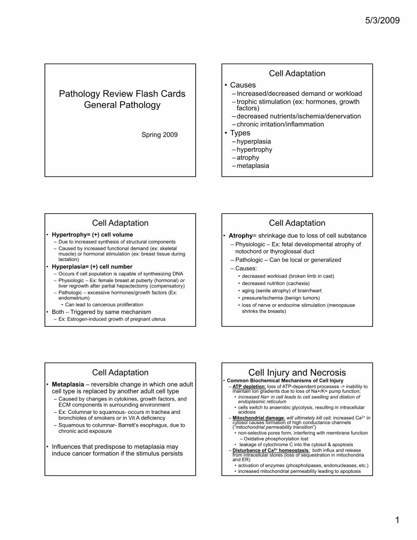
Pathology Review Flash CardsGeneral Pathology
Spring 2009
Cell Adaptation• Causes
– Increased/decreased demand or workload– trophic stimulation (ex: hormones, growth
factors)– decreased nutrients/ischemia/denervation– chronic irritation/inflammation
• Types– hyperplasia– hypertrophy– atrophy– metaplasia
Cell Adaptation• Hypertrophy= (+) cell volume
– Due to increased synthesis of structural components– Caused by increased functional demand (ex: skeletal
muscle) or hormonal stimulation (ex: breast tissue during lactation)
• Hyperplasia= (+) cell number– Occurs if cell population is capable of synthesizing DNA– Physiologic – Ex: female breast at puberty (hormonal) or
liver regrowth after partial hepactectomy (compensatory)– Pathologic – excessive hormones/growth factors (Ex:
endometrium)• Can lead to cancerous proliferation
• Both – Triggered by same mechanism– Ex: Estrogen-induced growth of pregnant uterus
Cell Adaptation• Atrophy= shrinkage due to loss of cell substance
– Physiologic – Ex: fetal developmental atrophy of notochord or thyroglossal duct
– Pathologic – Can be local or generalized– Causes:Causes:
• decreased workload (broken limb in cast)• decreased nutrition (cachexia)• aging (senile atrophy) of brain/heart• pressure/ischemia (benign tumors) • loss of nerve or endocrine stimulation (menopause
shrinks the breasts)
Cell Adaptation• Metaplasia – reversible change in which one adult
cell type is replaced by another adult cell type– Caused by changes in cytokines, growth factors, and
ECM components in surrounding environment– Ex: Columnar to squamous- occurs in trachea and
bronchioles of smokers or in Vit A deficiencybronchioles of smokers or in Vit A deficiency– Squamous to columnar- Barrett’s esophagus, due to
chronic acid exposure
• Influences that predispose to metaplasia may induce cancer formation if the stimulus persists
Cell Injury and Necrosis• Common Biochemical Mechanisms of Cell Injury
– ATP depletion: loss of ATP-dependent processes -> inability to maintain ion gradients due to loss of Na+/K+ pump function; • increased Na+ in cell leads to cell swelling and dilation of
endoplasmic reticulum• cells switch to anaerobic glycolysis, resulting in intracellular
acidosis– Mitochondrial damage: will ultimately kill cell; increased Ca2+ inMitochondrial damage: will ultimately kill cell; increased Ca in
cytosol causes formation of high conductance channels (“mitochondrial permeability transition”) • non-selective pores form, interfering with membrane function
– Oxidative phosphorylation lost• leakage of cytochrome C into the cytosol & apoptosis
– Disturbance of Ca2+ homeostasis: both influx and release from intracellular stores (loss of sequestration in mitochondria and ER) • activation of enzymes (phospholipases, endonucleases, etc.)• increased mitochondrial permeability leading to apoptosis
5/3/2009
2
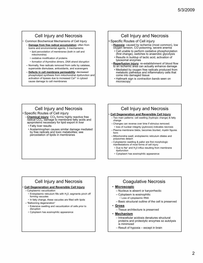
Cell Injury and Necrosis• Common Biochemical Mechanisms of Cell Injury
– Damage from free radical accumulation: often from toxins and environmental agents; 3 mechanisms:
• lipid peroxidation of membranes (both in cell and mitochondria)
• oxidative modification of proteinsoxidative modification of proteins • formation of thymidine dimers, DNA strand disruption
– Normally, free radicals removed from cells by catalase, superoxide dismutase, antioxidants, and scavengers
– Defects in cell membrane permeability: decreased phospholipid synthesis from mitochondrial dysfunction and activation of lipases due to increased Ca2+ in cytosol cause damage to cell membranes
Cell Injury and Necrosis• Specific Routes of Cell injury
–Hypoxia: caused by ischemia (most common), low oxygen tension, CO poisoning, severe anemia• Cell unable to perform oxidative phosphorylation
(first change), switches to anaerobic glycolysis• Results in buildup of lactic acid, activation of p ,
lysosomal enzymes–Reperfusion injury: re-establishment of blood flow
to an ischemic area can actually enhance damage• Mediated by oxygen free radicals produced from
metabolic pathways and inflammatory cells that come into damaged tissue
• Hallmark sign is contraction bands seen on microscopy
Cell Injury and Necrosis• Specific Routes of Cell injury
–Chemical injury: CCl4 forms highly reactive free radical CCl3; damage to membrane fatty acids and apoproteins necessary for lipid export in liver• Fatty liver results• Acetaminophen causes similar damage mediated
b f di l d t i t b litby free radicals and toxic metabolites; see peroxidation of lipids in membranes
Cell Injury and Necrosis• Cell Degeneration and Reversible Cell Injury
– Two main patterns: cell swelling (hydropic change) & fatty change
– Changes can reverse over time if stimulus removed; • loss of nuclear integrity (pyknosis) indicates necrosis
– Plasma membrane blebs, becomes blunted, myelin figures y gform
– Mitochondria swell, endoplasmic reticulum dilates and polysomes detach
– Cytoplasmic swelling & pallor are first morphologic manifestations of most forms of cell injury• Due to Na+ and H20 influx resulting from membrane
dysfunction• Cytoplasm has eosinophilic appearance
Cell Injury and Necrosis• Cell Degeneration and Reversible Cell Injury
– Cytoplasmic vacuolization • Endoplasmic reticulum fills with H20, segments pinch off
forming vacuoles• In fatty change, these vacuoles are filled with lipids
– “Ballooning degeneration”g g• Extensive swelling and vacuolization of cells prior to
disruption• Cytoplasm has eosinophilic appearance
Coagulative Necrosis• Microscopic
– Nucleus is absent or karyorrhectic– Cytoplasm is eosinophilic
• Loss of cytoplasmic RNA– Basic structural outline of the cell is preserved
• Gross– Tissue architecture is preserved
• Mechanism– Intracellular acidosis denatures structural
proteins and proteolytic enzymes so autolysis is minimized
– Result of hypoxia – except in brain
5/3/2009
3
Liquefactive Necrosis• Microscopic
– Infiltration by neutrophils– Fibrous connective tissue surround older lesions– Tissue architecture destroyed
• Gross– Soft to liquefied viscous mass– Inspissated material
• Mechanism– Pyogenic bacteria stimulate inflammatory
response– Neutrophils release proteolytic enzymes– Hypoxia in CNS
Calcification• Dystrophic
– Calcium deposited locally in necrotic tissue• Basophilic, amorphous granular or
clumpedclumped• Can be intracellular, extracellular or both
– Normal serum levels and metabolism– Found in advanced atherosclerotic
plaques– Psammoma body formation
Calcification• Metastatic
– Deposition of calcium phosphate in normal tissue
– hypercalcemia– Increased parathyroid hormone secretion– vit. D toxicosis– tumors associated with increased bone
catabolism• multiple myeloma
– Renal osteodystrophy – secondary hyperparathyroidism
Lipofuscin• Insoluble, wear and tear pigment
– Does not harm cell or cellular functions• End product of membrane lipid peroxidation• Commonly accumulates in the elderly
– Most often in hepatocytes and myocardium• Combination of lipofuscin accumulation and
atrophy of organs is brown atrophy
Apoptosis - Intro• Process of programmed cell death• Several different initiating events but each ultimately
results in activation of caspases that degrade nuclear and cytoskeletal elements– Caspases exist as zymogens that must undergo cleavage
to be activatedCaspases degrade nuclear and cytoskeletal scaffold– Caspases degrade nuclear and cytoskeletal scaffold
– Caspases activate DNAses which degrade nuclear DNA • Plasma membrane remains intact and cellular
contents do not leak out• Apoptotic cells recognized and phagocytosed• No inflammatory response
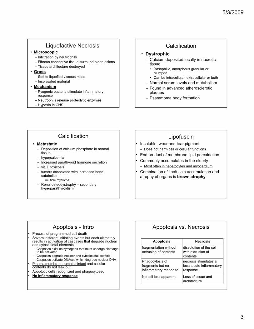
Apoptosis vs. Necrosis
Apoptosis Necrosisfragmentation without extrusion of contents
dissolution of the cell with extrusion of contentscontents
Phagocytosis of fragments but no inflammatory response
necrosis stimulates a local acute inflammatory response
No cell loss apparent Loss of tissue and architecture
5/3/2009
4
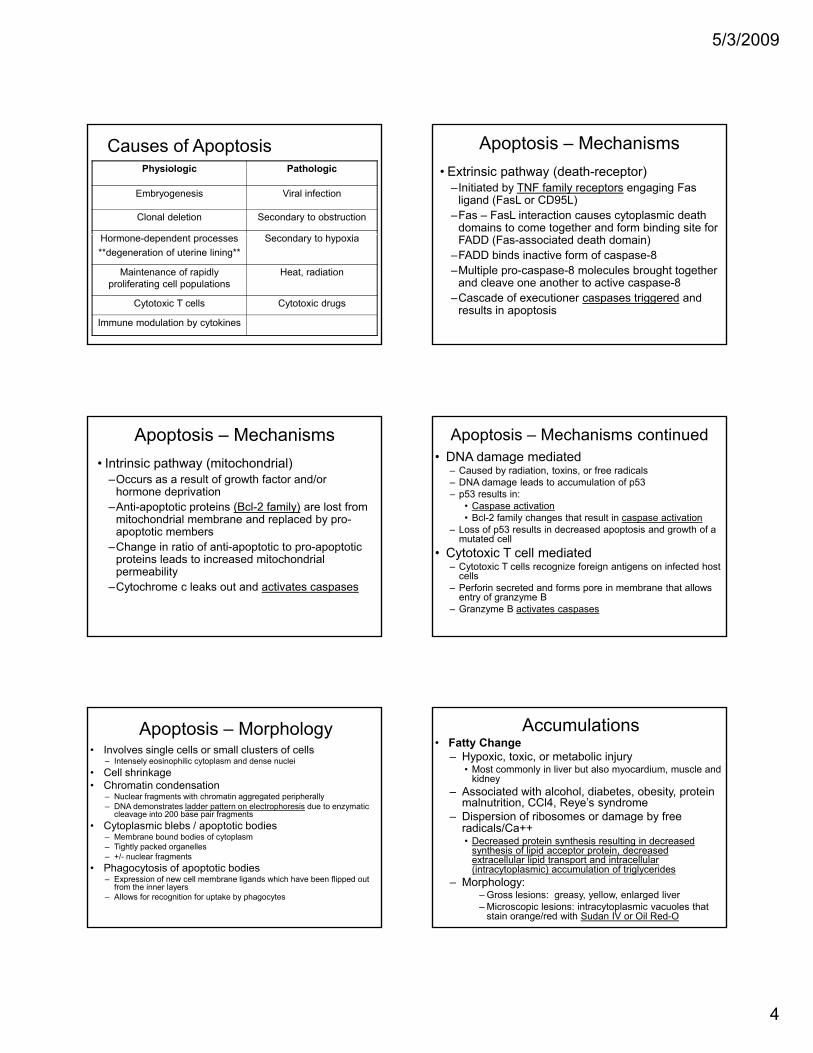
Causes of ApoptosisPhysiologic Pathologic
Embryogenesis Viral infection
Clonal deletion Secondary to obstruction
H d d S d h iHormone-dependent processes**degeneration of uterine lining**
Secondary to hypoxia
Maintenance of rapidly proliferating cell populations
Heat, radiation
Cytotoxic T cells Cytotoxic drugs
Immune modulation by cytokines
Apoptosis – Mechanisms• Extrinsic pathway (death-receptor)
–Initiated by TNF family receptors engaging Fas ligand (FasL or CD95L)
–Fas – FasL interaction causes cytoplasmic death domains to come together and form binding site for FADD (Fas-associated death domain)
–FADD binds inactive form of caspase-8–Multiple pro-caspase-8 molecules brought together
and cleave one another to active caspase-8–Cascade of executioner caspases triggered and
results in apoptosis
Apoptosis – Mechanisms• Intrinsic pathway (mitochondrial)
–Occurs as a result of growth factor and/or hormone deprivation
–Anti-apoptotic proteins (Bcl-2 family) are lost from mitochondrial membrane and replaced by pro-apoptotic members
–Change in ratio of anti-apoptotic to pro-apoptotic proteins leads to increased mitochondrial permeability
–Cytochrome c leaks out and activates caspases
Apoptosis – Mechanisms continued• DNA damage mediated
– Caused by radiation, toxins, or free radicals– DNA damage leads to accumulation of p53– p53 results in:
• Caspase activation• Bcl-2 family changes that result in caspase activation
L f 53 lt i d d t i d th f– Loss of p53 results in decreased apoptosis and growth of a mutated cell
• Cytotoxic T cell mediated– Cytotoxic T cells recognize foreign antigens on infected host
cells– Perforin secreted and forms pore in membrane that allows
entry of granzyme B– Granzyme B activates caspases
Apoptosis – Morphology• Involves single cells or small clusters of cells
– Intensely eosinophilic cytoplasm and dense nuclei• Cell shrinkage• Chromatin condensation
– Nuclear fragments with chromatin aggregated peripherally– DNA demonstrates ladder pattern on electrophoresis due to enzymatic
cleavage into 200 base pair fragmentsg p g• Cytoplasmic blebs / apoptotic bodies
– Membrane bound bodies of cytoplasm– Tightly packed organelles– +/- nuclear fragments
• Phagocytosis of apoptotic bodies– Expression of new cell membrane ligands which have been flipped out
from the inner layers– Allows for recognition for uptake by phagocytes
Accumulations• Fatty Change
– Hypoxic, toxic, or metabolic injury• Most commonly in liver but also myocardium, muscle and
kidney– Associated with alcohol, diabetes, obesity, protein
malnutrition, CCl4, Reye’s syndrome– Dispersion of ribosomes or damage by freeDispersion of ribosomes or damage by free
radicals/Ca++• Decreased protein synthesis resulting in decreased
synthesis of lipid acceptor protein, decreased extracellular lipid transport and intracellular (intracytoplasmic) accumulation of triglycerides
– Morphology:– Gross lesions: greasy, yellow, enlarged liver– Microscopic lesions: intracytoplasmic vacuoles that
stain orange/red with Sudan IV or Oil Red-O
5/3/2009
5
Accumulations• Protein accumulations (non-specific, eosinophilic)
– eosinophilic intracell deposits = hyaline change– examples: proximal renal tubules (proteinuria),
Russell bodies (accumulation of Ig in ER of plasma cells), Mallory bodies (cirrhosis), α-1 antitrypsin deficiency, α-synuclein/Lewey Bodies (familial/sporadic Alzheimer's, PD, dementia)
• Glycogen– Non-staining cytoplasmic vacuoles assoc. w/ abnl
glucose/glycogen metabolism (DM: hepatocytes/renal tubules; glycogen storage disease)
– Pigments (anthracosis – carbon; lipofuscin –gold/brown aging pigment; melanin; hemosiderin; bile)
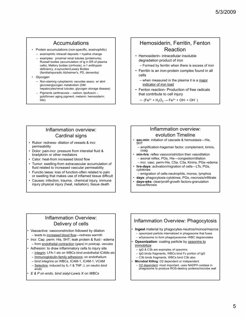
Hemosiderin, Ferritin, Fenton Reaction
• Hemosiderin- intracellular insoluble degradation product of iron – Formed by ferritin when there is excess of iron
• Ferritin is an iron-protein complex found in all• Ferritin is an iron-protein complex found in all cells– when measured in the plasma it is a major
indicator of iron load• Fenton reaction- Production of free radicals
that contribute to cell injury– (Fe2+ + H2O2 → Fe3+ + OH· + OH− )
Inflammation overview: Cardinal signs
• Rubor: redness- dilation of vessels & incr. permeability
• Dolor: pain-incr. pressure from interstial fluid & bradykinin or other mediators
• Calor: heat from increased blood flow• Calor: heat-from increased blood flow• Tumor: swelling-from extravascular accumulation of
fluid related to increased vascular permeability • Functio laesa: loss of function-often related to pain
or swelling that makes use of inflamed tissue difficult• Causes: infection, trauma, chemical injury, immune
injury physical injury (heat, radiation), tissue death
Inflammation overview: evolution Timeline
• sec-min: initiation of cascade & hemostasis—His, 5HT– amplification-hageman factor, complement, kinins,
coag• min-hrs: reflex vasoconstriction then vasodilation
– axonal reflex PGs His—congestion/dilationaxonal reflex, PGs, His congestion/dilation– incr. vasc. perm-His, C5a, C3a, Kinins, PGs--edema
• hrs-days: activation/migration of cells—LTs, PGs, cytokines– emigration of cells-neutrophils, monos, lymphos
• days: phagocytosis-cytokines, PGs,-necrosis/infiltrate• days-wks: clear/prolif-growth factors-granulation
tissue/fibrosis
Inflammation Overview: Delivery of cells
• Vasoactive: vasoconstriction followed by dilation– leads to increased blood flow---redness warmth
• Incr. Cap. perm: His, 5HT; leak protein & fluid - edema– from endothelial contraction (gaps) in postcap. venules
f• Adhesion: to draw inflammatory cells to injury site– Integrin: LFA-1 etc on WBCs bind endothelial ICAMs etc– Immunoglobulin-family adhesions: on endothelium– bind integrins on WBCs, ICAM-1, ICAM-1, VCAM– Selectins: induced by IL-1 & TNF; L on neutro bind
endo• E & P on endo, bind sialyl-Lewis X on WBCs
Inflammation Overview: Phagocytosis• Ingest material by phagocytes-neutros/monos/macros
– opsonized particle internalized in phagosome that fuses– w/lysosome to form phagolysosome--WBC degranulates
• Opsonization: coating particle by opsonins to immobilize– IgG & C3b are examples of opsonins – IgG binds fragments, WBCs bind Fc portion of IgG– C3b binds fragments, WBCs bind C3b also
• Microbial Killing: O2 dependent or independent– O2 dependent: most important, uses NADPH oxidase in
phagosome to produce ROS-destroy proteins/microbe wall
5/3/2009
6
Types of Inflammation• Classification by Duration• Chronic- weeks to years
– Usually from persistence of injury-causing agent• Infection, autoimmune disease, sterile agent
– Monocytes and macrophagesMonocytes and macrophages• Also lymphocytes, plasma cells, eosinophils
– Necrosis NOT as prominent as in acute inflammation• Loss of parenchyma due to fibrosis• Granulation tissue converted to scar tissue• Blood vessel proliferation
– Granuloma- type of chronic inflammation
Types of Inflammation• Classification by Morphologic Type• Serous
– lack of cellular infiltrate– Accumulation of fluid from blood serum due to increased
vascular permeability– from mesothelium- pleural, peritoneal, pericardial
• Fibrinous– Increased vascular permeability allows for passage of fibrin
exudate– Gives a “shaggy” appearance– resolves via lysis- degradation by plasmin and macrophages– organization- fibrin remains, involved in fibrosis and scarring
Types of Inflammation• Classification by Morphologic Type• Suppurative• Granulomatous
Types of Inflammation• Classification by Duration
– Peracute (0-6 hrs)• no inflammatory cells yet present• Vasodilation, incr’d vascular permeability edema
– Acute (6-48 hrs)i filt ti f t hil• infiltration of neutrophils
– Subacute (24-72 hrs)• neutrophils begin undergoing apoptosis• emigration of monocytes & activation of macrophages
– Chronic (weeks-months)• Lymphocytes predominate• Also monocytes, fibroblasts
Inflammation overview: WBC emigration
Emigration: process of WBC migration from post capillary venule, between endothelial cells, and into tissue
• Margination: blood slowing, movement of WBCs to vessel periphery
• Adhesion: mediated by sequential expression of specific surface moleculessurface molecules– Weak adhesion: between endothelial selectins and WBC surface
carbohydrates, results in “rolling” – Firm Adhesion: between endothelial ICAM/VCAM and WBC
integrins– Sequential expression of different CAMs determines what type of
WBC migrates at different phases of inflammation (PMN, mono, etc)
• Transmigration: WBC “pseudopod” formation, diapedesis by “crawling” along ECM
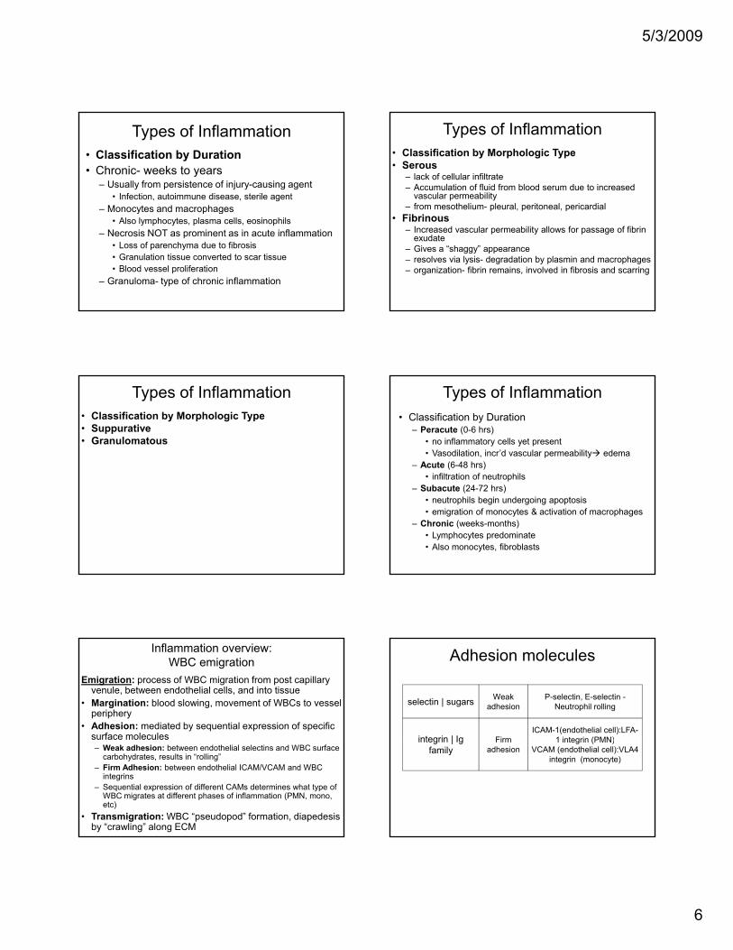
Adhesion molecules
selectin | sugars Weak adhesion
P-selectin, E-selectin -Neutrophil rolling
integrin | Ig FiICAM-1(endothelial cell):LFA-
1 i t i (PMN)integrin | Ig family
Firm adhesion
1 integrin (PMN)VCAM (endothelial cell):VLA4
integrin (monocyte)
5/3/2009
7
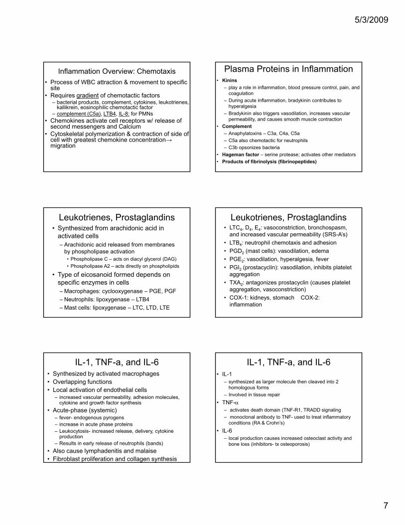
Inflammation Overview: Chemotaxis• Process of WBC attraction & movement to specific
site• Requires gradient of chemotactic factors
– bacterial products, complement, cytokines, leukotrienes, kallikrein, eosinophilic chemotactic factor
– complement (C5a), LTB4, IL-8: for PMNs• Chemokines activate cell receptors w/ release of
second messengers and Calcium• Cytoskeletal polymerization & contraction of side of
cell with greatest chemokine concentration→ migration
Plasma Proteins in Inflammation• Kinins
– play a role in inflammation, blood pressure control, pain, and coagulation
– During acute inflammation, bradykinin contributes to hyperalgesia
– Bradykinin also triggers vasodilation, increases vascular permeability, and causes smooth muscle contraction
• Complement– Anaphylatoxins – C3a, C4a, C5a– C5a also chemotactic for neutrophils– C3b opsonizes bacteria
• Hageman factor – serine protease; activates other mediators• Products of fibrinolysis (fibrinopeptides)
Leukotrienes, Prostaglandins• Synthesized from arachidonic acid in
activated cells– Arachidonic acid released from membranes
by phospholipase activation• Phospholipase C – acts on diacyl glycerol (DAG)• Phospholipase A2 – acts directly on phospholipids
• Type of eicosanoid formed depends on specific enzymes in cells– Macrophages: cyclooxygenase – PGE, PGF– Neutrophils: lipoxygenase – LTB4– Mast cells: lipoxygenase – LTC, LTD, LTE
Leukotrienes, Prostaglandins• LTC4, D4, E4: vasoconstriction, bronchospasm,
and increased vascular permeability (SRS-A’s)• LTB4: neutrophil chemotaxis and adhesion• PGD2 (mast cells): vasodilation, edema• PGE2: vasodilation, hyperalgesia, fever2
• PGI2 (prostacyclin): vasodilation, inhibits platelet aggregation
• TXA2: antagonizes prostacyclin (causes platelet aggregation, vasoconstriction)
• COX-1: kidneys, stomach COX-2: inflammation
IL-1, TNF-a, and IL-6• Synthesized by activated macrophages• Overlapping functions• Local activation of endothelial cells
– increased vascular permeability, adhesion molecules, cytokine and growth factor synthesis
• Acute-phase (systemic)– fever- endogenous pyrogens– increase in acute phase proteins– Leukocytosis- increased release, delivery, cytokine
production– Results in early release of neutrophils (bands)
• Also cause lymphadenitis and malaise• Fibroblast proliferation and collagen synthesis
IL-1, TNF-a, and IL-6• IL-1
– synthesized as larger molecule then cleaved into 2 homologous forms
– Involved in tissue repair• TNF-α
– activates death domain (TNF-R1, TRADD signaling– monoclonal antibody to TNF- used to treat inflammatory
conditions (RA & Crohn’s)• IL-6
– local production causes increased osteoclast activity and bone loss (inhibitors- tx osteoporosis)
5/3/2009
8
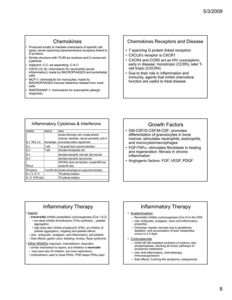
Chemokines• Produced locally to mediate chemotaxis of specific cell
types; seven-spanning transmembrane receptors linked to G proteins
• Similar structure with 70-80 aa residues and 2 conserved cysteines
• Adjacent- C-C; aa separating- C-X-C• CXC8 (=IL-8): chemotactic for neutrophils (acute
inflammation); made by MACROPHAGES and endothelial cells
• MCP-1- chemotactic for monocytes; made by MACROPHAGES induces histamine release from mast cells
• RANTES/MIP-1- chemotactic for eosinophils (allergic response)
Chemokines Receptors and Disease
• 7 spanning G protein linked receptors• CXCL8’s receptor is CXCR1• CXCR4 and CCR5 act as HIV coreceptors-
early in disease: monotropic (CCR5), later T-ll i (CXCR4)cell tropic (CXCR4)
• Due to their role in inflammation and immunity, agents that inhibit chemokine function are useful to treat disease
Inflammatory Cytokines & Interferonsmediator made by action
IL-1, TNFa, IL-6 macrophages
activate inflammatory cells, increase adhesion molecules, vasodilation, vascular permeability, synth of acute phase proteins, regulate fever
IL-2 T cells T cell growth factor; autocrine stimulationIL 3 T cells stimulates hematopoietic cellsIL-3 T cells stimulates hematopoietic cellsIL-4 stimulates eosinophils, mast cells, IgE productionIL-5 stimulates eosinophils, IgA production
IFN a,bANTIVIRAL (block viral replication, increase MHCI exp, activate NK cells)
IFN gamma T and NK cells activate macrophages (ie in granuloma formation)IL- 4, 5, 10, 13 TH2 pathway mediatorsIL- 12, 18 IFN (a/b) TH1 pathway mediators
Growth Factors• GM-CSF/G-CSF/M-CSF- promotes
differentiation of granulocytes in bone marrow; stimulates neutrophils, eosinophils, and monocytes/macrophages
• FGF/TNFα- stimulates fibroblasts in healing and regeneration; fibrosis in chronic inflammation
• Angiogenic factors- FGF, VEGF, PDGF
Inflammatory Therapy• Aspirin
– Irreversibly inhibits (acetylates) cyclooxygenase (Cox 1 & 2)• low dose inhibits thromboxane (TXA) synthesis, ↓ platelet
aggregation• high dose also inhibits prostacyclin (PGI), an inhibitor of
platelet aggregation, negating anti-platelet effects– Use: antipyretic, analgesic, anti-inflammatory, anti-platelet– Side effects: gastric ulcer, bleeding, tinnitus, Reye syndrome
• Other NSAIDs (naproxen, indomethacin, ibuprofen)– similar mechanism to aspirin, but inhibition is reversible– may have less GI irritation, but more nephrotoxic– Indomethacin used to close PDAs / PGE keeps PDAs open
Inflammatory Therapy• Acetaminophen
– Reversibly inhibits cyclooxygenase (Cox 3) in the CNS– Use: antipyretic, analgesic, lacks anti-inflammatory
properties– Overdose: hepatic necrosis due to glutathione
depletion and accumulation of toxic metabolites, i 2 3 doccurs in 2-3 days
• Corticosteroids– inhibit NF-kB-mediated synthesis of cytokins; also
phospholipases, blocking all known pathways of eicosanoid metabolism
– Use: Anti-inflammatory, chemotherapy, immunosuppression
– Side effects: Cushing-like symptoms, osteoporosis
5/3/2009
9
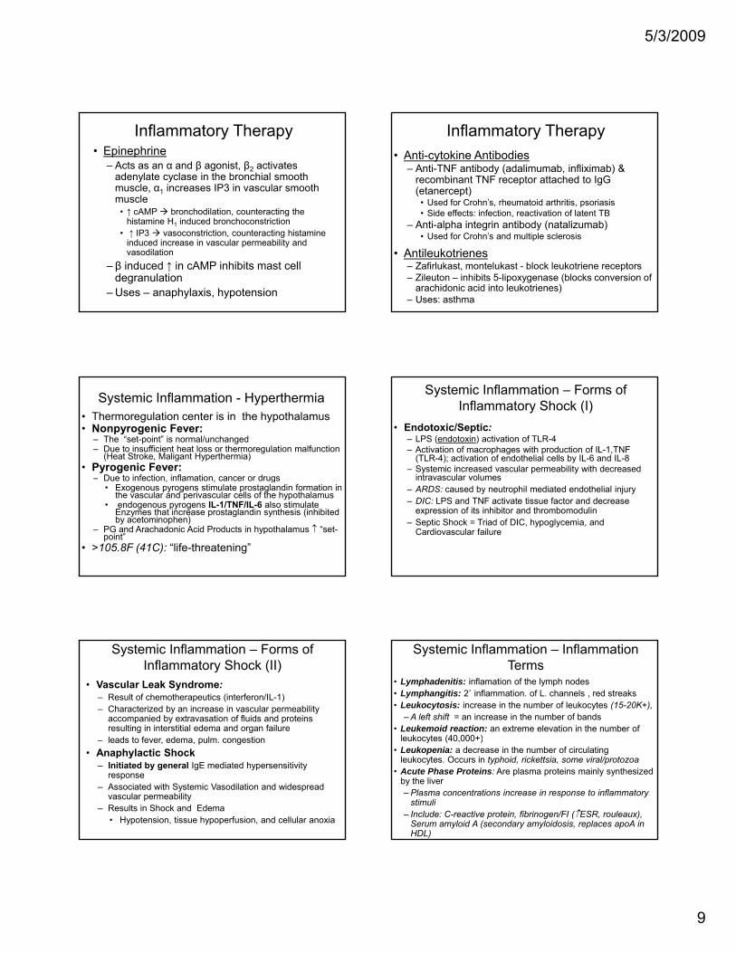
Inflammatory Therapy• Epinephrine
– Acts as an α and β agonist, β2 activates adenylate cyclase in the bronchial smooth muscle, α1 increases IP3 in vascular smooth muscle
• ↑ cAMP bronchodilation counteracting the• ↑ cAMP bronchodilation, counteracting the histamine H1 induced bronchoconstriction
• ↑ IP3 vasoconstriction, counteracting histamine induced increase in vascular permeability and vasodilation
– β induced ↑ in cAMP inhibits mast cell degranulation
– Uses – anaphylaxis, hypotension
Inflammatory Therapy• Anti-cytokine Antibodies
– Anti-TNF antibody (adalimumab, infliximab) & recombinant TNF receptor attached to IgG (etanercept)
• Used for Crohn’s, rheumatoid arthritis, psoriasis• Side effects: infection reactivation of latent TB• Side effects: infection, reactivation of latent TB
– Anti-alpha integrin antibody (natalizumab)• Used for Crohn’s and multiple sclerosis
• Antileukotrienes– Zafirlukast, montelukast - block leukotriene receptors– Zileuton – inhibits 5-lipoxygenase (blocks conversion of
arachidonic acid into leukotrienes)– Uses: asthma
Systemic Inflammation - Hyperthermia• Thermoregulation center is in the hypothalamus• Nonpyrogenic Fever:
– The “set-point” is normal/unchanged– Due to insufficient heat loss or thermoregulation malfunction
(Heat Stroke, Maligant Hyperthermia)• Pyrogenic Fever:
D e to infection inflamation cancer or dr gs– Due to infection, inflamation, cancer or drugs• Exogenous pyrogens stimulate prostaglandin formation in
the vascular and perivascular cells of the hypothalamus• endogenous pyrogens IL-1/TNF/IL-6 also stimulate
Enzymes that increase prostaglandin synthesis (inhibited by acetominophen)
– PG and Arachadonic Acid Products in hypothalamus ↑ “set-point”
• >105.8F (41C): “life-threatening”
Systemic Inflammation – Forms of Inflammatory Shock (I)
• Endotoxic/Septic:– LPS (endotoxin) activation of TLR-4– Activation of macrophages with production of IL-1,TNF
(TLR-4); activation of endothelial cells by IL-6 and IL-8– Systemic increased vascular permeability with decreased
intravascular volumesintravascular volumes– ARDS: caused by neutrophil mediated endothelial injury – DIC: LPS and TNF activate tissue factor and decrease
expression of its inhibitor and thrombomodulin– Septic Shock = Triad of DIC, hypoglycemia, and
Cardiovascular failure
Systemic Inflammation – Forms of Inflammatory Shock (II)
• Vascular Leak Syndrome:– Result of chemotherapeutics (interferon/IL-1) – Characterized by an increase in vascular permeability
accompanied by extravasation of fluids and proteins resulting in interstitial edema and organ failureleads to fever edema pulm congestion– leads to fever, edema, pulm. congestion
• Anaphylactic Shock– Initiated by general IgE mediated hypersensitivity
response– Associated with Systemic Vasodilation and widespread
vascular permeability– Results in Shock and Edema
• Hypotension, tissue hypoperfusion, and cellular anoxia
Systemic Inflammation – Inflammation Terms
• Lymphadenitis: inflamation of the lymph nodes• Lymphangitis: 2˚ inflammation. of L. channels , red streaks• Leukocytosis: increase in the number of leukocytes (15-20K+),
– A left shift = an increase in the number of bands• Leukemoid reaction: an extreme elevation in the number of
leukocytes (40 000+)leukocytes (40,000+)• Leukopenia: a decrease in the number of circulating
leukocytes. Occurs in typhoid, rickettsia, some viral/protozoa• Acute Phase Proteins: Are plasma proteins mainly synthesized
by the liver– Plasma concentrations increase in response to inflammatory
stimuli– Include: C-reactive protein, fibrinogen/FI (↑ESR, rouleaux),
Serum amyloid A (secondary amyloidosis, replaces apoA in HDL)
5/3/2009
10
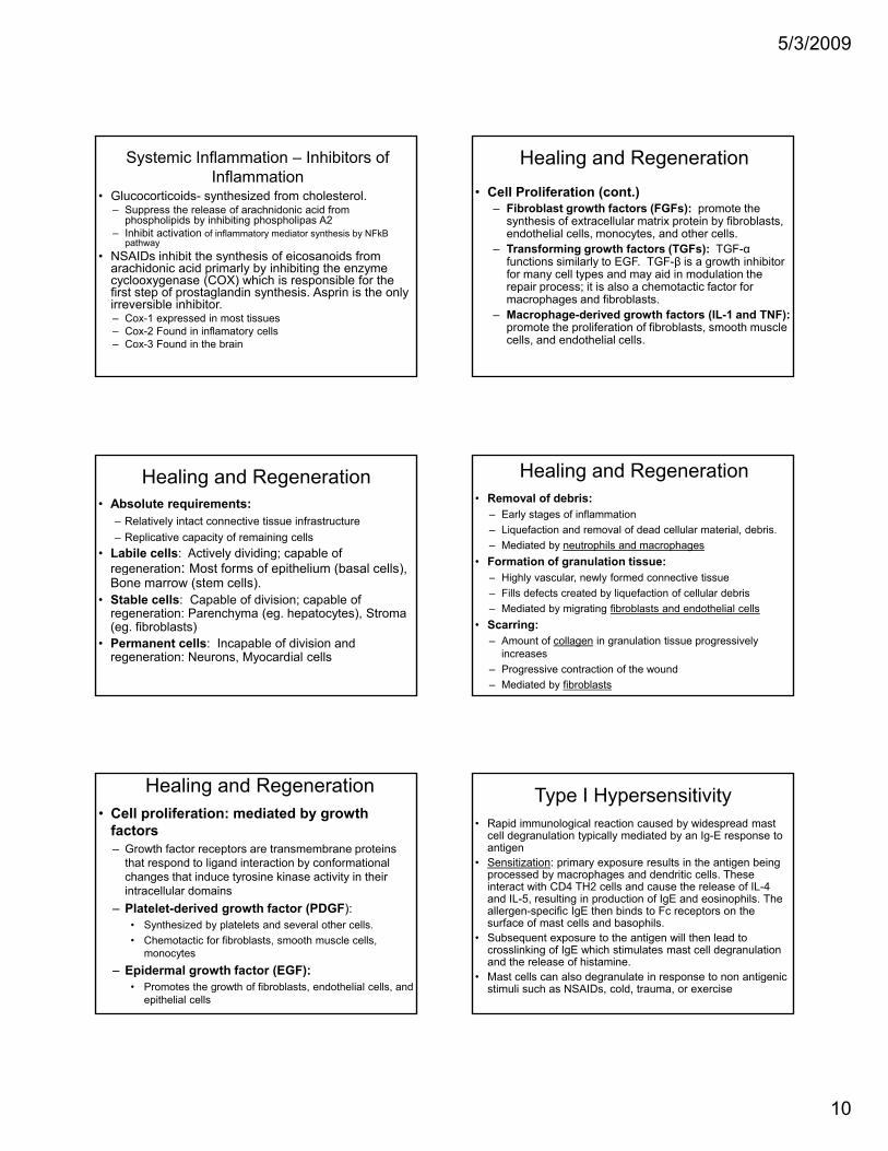
Systemic Inflammation – Inhibitors of Inflammation
• Glucocorticoids- synthesized from cholesterol. – Suppress the release of arachnidonic acid from
phospholipids by inhibiting phospholipas A2– Inhibit activation of inflammatory mediator synthesis by NFkB
pathway• NSAIDs inhibit the synthesis of eicosanoids from• NSAIDs inhibit the synthesis of eicosanoids from
arachidonic acid primarly by inhibiting the enzyme cyclooxygenase (COX) which is responsible for the first step of prostaglandin synthesis. Asprin is the only irreversible inhibitor. – Cox-1 expressed in most tissues– Cox-2 Found in inflamatory cells– Cox-3 Found in the brain
Healing and Regeneration• Cell Proliferation (cont.)
– Fibroblast growth factors (FGFs): promote the synthesis of extracellular matrix protein by fibroblasts, endothelial cells, monocytes, and other cells.
– Transforming growth factors (TGFs): TGF-α functions similarly to EGF. TGF-β is a growth inhibitor for many cell types and may aid in modulation the repair process; it is also a chemotactic factor for macrophages and fibroblasts.
– Macrophage-derived growth factors (IL-1 and TNF):promote the proliferation of fibroblasts, smooth muscle cells, and endothelial cells.
Healing and Regeneration• Absolute requirements:
– Relatively intact connective tissue infrastructure– Replicative capacity of remaining cells
• Labile cells: Actively dividing; capable of regeneration: Most forms of epithelium (basal cells), g p ( )Bone marrow (stem cells).
• Stable cells: Capable of division; capable of regeneration: Parenchyma (eg. hepatocytes), Stroma (eg. fibroblasts)
• Permanent cells: Incapable of division and regeneration: Neurons, Myocardial cells
Healing and Regeneration• Removal of debris:
– Early stages of inflammation– Liquefaction and removal of dead cellular material, debris.– Mediated by neutrophils and macrophages
• Formation of granulation tissue:Hi hl l l f d ti ti– Highly vascular, newly formed connective tissue
– Fills defects created by liquefaction of cellular debris– Mediated by migrating fibroblasts and endothelial cells
• Scarring: – Amount of collagen in granulation tissue progressively
increases– Progressive contraction of the wound– Mediated by fibroblasts
Healing and Regeneration• Cell proliferation: mediated by growth
factors– Growth factor receptors are transmembrane proteins
that respond to ligand interaction by conformational changes that induce tyrosine kinase activity in their intracellular domainsintracellular domains
– Platelet-derived growth factor (PDGF): • Synthesized by platelets and several other cells.• Chemotactic for fibroblasts, smooth muscle cells,
monocytes – Epidermal growth factor (EGF):
• Promotes the growth of fibroblasts, endothelial cells, and epithelial cells
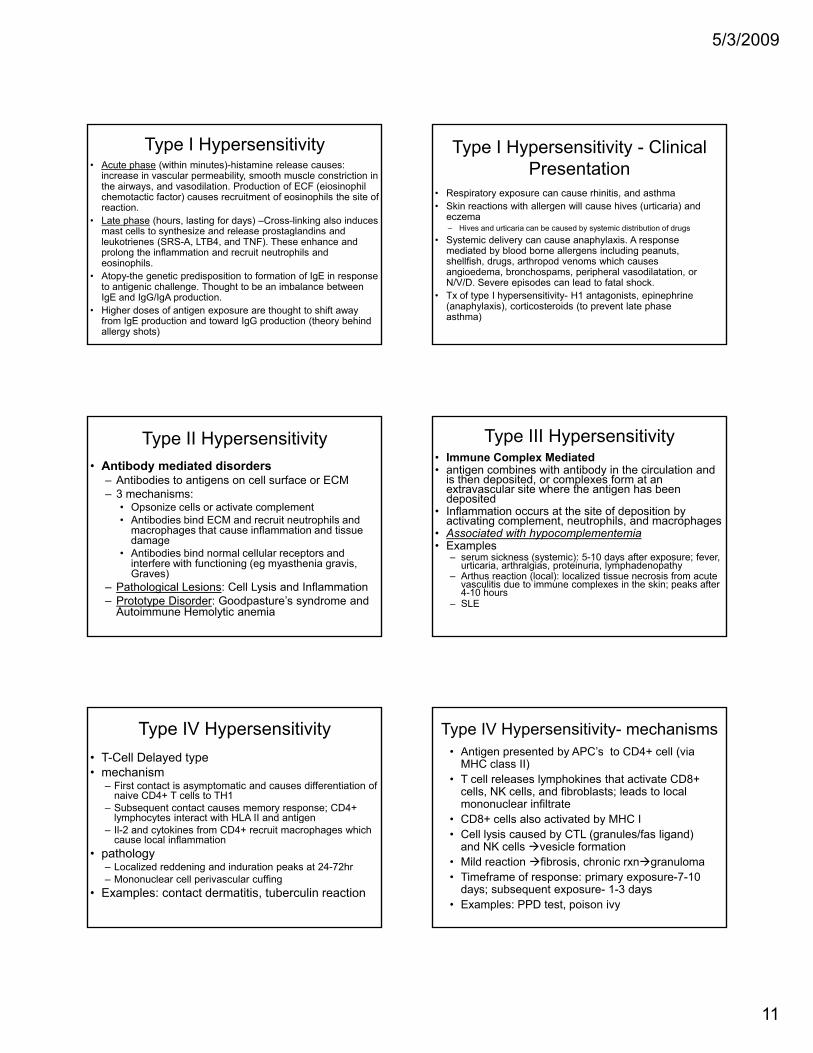
Type I Hypersensitivity• Rapid immunological reaction caused by widespread mast
cell degranulation typically mediated by an Ig-E response to antigen
• Sensitization: primary exposure results in the antigen being processed by macrophages and dendritic cells. These interact with CD4 TH2 cells and cause the release of IL-4 and IL-5, resulting in production of IgE and eosinophils. The allergen-specific IgE then binds to Fc receptors on the surface of mast cells and basophils.
• Subsequent exposure to the antigen will then lead to crosslinking of IgE which stimulates mast cell degranulation and the release of histamine.
• Mast cells can also degranulate in response to non antigenic stimuli such as NSAIDs, cold, trauma, or exercise
5/3/2009
11
Type I Hypersensitivity• Acute phase (within minutes)-histamine release causes:
increase in vascular permeability, smooth muscle constriction in the airways, and vasodilation. Production of ECF (eiosinophil chemotactic factor) causes recruitment of eosinophils the site of reaction.
• Late phase (hours, lasting for days) –Cross-linking also induces mast cells to synthesize and release prostaglandins and y p gleukotrienes (SRS-A, LTB4, and TNF). These enhance and prolong the inflammation and recruit neutrophils and eosinophils.
• Atopy-the genetic predisposition to formation of IgE in response to antigenic challenge. Thought to be an imbalance between IgE and IgG/IgA production.
• Higher doses of antigen exposure are thought to shift away from IgE production and toward IgG production (theory behind allergy shots)
Type I Hypersensitivity - Clinical Presentation
• Respiratory exposure can cause rhinitis, and asthma• Skin reactions with allergen will cause hives (urticaria) and
eczema– Hives and urticaria can be caused by systemic distribution of drugs
• Systemic delivery can cause anaphylaxis. A response mediated by blood borne allergens including peanuts, shellfish, drugs, arthropod venoms which causes angioedema, bronchospams, peripheral vasodilatation, or N/V/D. Severe episodes can lead to fatal shock.
• Tx of type I hypersensitivity- H1 antagonists, epinephrine (anaphylaxis), corticosteroids (to prevent late phase asthma)
Type II Hypersensitivity• Antibody mediated disorders
– Antibodies to antigens on cell surface or ECM– 3 mechanisms:
• Opsonize cells or activate complement• Antibodies bind ECM and recruit neutrophils and
h h i fl i d imacrophages that cause inflammation and tissue damage
• Antibodies bind normal cellular receptors and interfere with functioning (eg myasthenia gravis, Graves)
– Pathological Lesions: Cell Lysis and Inflammation– Prototype Disorder: Goodpasture’s syndrome and
Autoimmune Hemolytic anemia
Type III Hypersensitivity• Immune Complex Mediated• antigen combines with antibody in the circulation and
is then deposited, or complexes form at an extravascular site where the antigen has been deposited
• Inflammation occurs at the site of deposition by activating complement, neutrophils, and macrophages g p , p , p g
• Associated with hypocomplementemia• Examples
– serum sickness (systemic): 5-10 days after exposure; fever, urticaria, arthralgias, proteinuria, lymphadenopathy
– Arthus reaction (local): localized tissue necrosis from acute vasculitis due to immune complexes in the skin; peaks after 4-10 hours
– SLE
Type IV Hypersensitivity• T-Cell Delayed type• mechanism
– First contact is asymptomatic and causes differentiation of naive CD4+ T cells to TH1
– Subsequent contact causes memory response; CD4+ lymphocytes interact with HLA II and antigenlymphocytes interact with HLA II and antigen
– Il-2 and cytokines from CD4+ recruit macrophages which cause local inflammation
• pathology– Localized reddening and induration peaks at 24-72hr– Mononuclear cell perivascular cuffing
• Examples: contact dermatitis, tuberculin reaction
Type IV Hypersensitivity- mechanisms• Antigen presented by APC’s to CD4+ cell (via
MHC class II)• T cell releases lymphokines that activate CD8+
cells, NK cells, and fibroblasts; leads to local mononuclear infiltrate
• CD8+ cells also activated by MHC I• CD8+ cells also activated by MHC I • Cell lysis caused by CTL (granules/fas ligand)
and NK cells vesicle formation• Mild reaction fibrosis, chronic rxn granuloma• Timeframe of response: primary exposure-7-10
days; subsequent exposure- 1-3 days• Examples: PPD test, poison ivy
5/3/2009
12
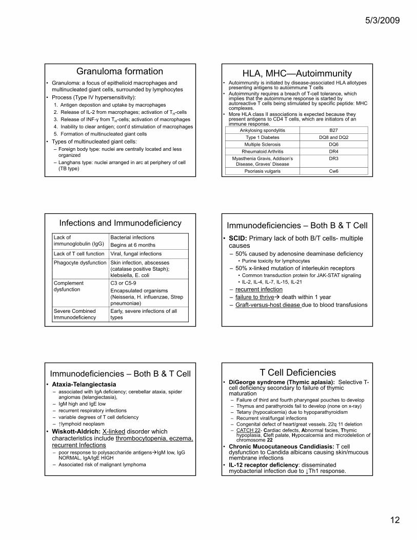
Granuloma formation• Granuloma: a focus of epithelioid macrophages and
multinucleated giant cells, surrounded by lymphocytes• Process (Type IV hypersensitivity):
1. Antigen depostion and uptake by macrophages2. Release of IL-2 from macrophages; activation of TH-cells3 Release of INF-γ from TH-cells; activation of macrophages3. Release of INF γ from TH cells; activation of macrophages4. Inability to clear antigen; cont’d stimulation of macrophages5. Formation of multinucleated giant cells
• Types of multinucleated giant cells:– Foreign body type: nuclei are centrally located and less
organized– Langhans type: nuclei arranged in arc at periphery of cell
(TB type)
HLA, MHC—Autoimmunity • Autoimmunity is initiated by disease-associated HLA allotypes
presenting antigens to autoimmune T cells• Autoimmunity requires a breach of T-cell tolerance, which
implies that the autoimmune response is started by autoreactive T cells being stimulated by specific peptide: MHC complexes.
• More HLA class II associations is expected because they present antigens to CD4 T cells, which are initiators of anpresent antigens to CD4 T cells, which are initiators of an immune response.
Ankylosing spondylitis B27Type 1 Diabetes DQ8 and DQ2
Multiple Sclerosis DQ6Rheumatoid Arthritis DR4
Myasthenia Gravis, Addison’s Disease, Graves’ Disease
DR3
Psoriasis vulgaris Cw6
Infections and ImmunodeficiencyLack of immunoglobulin (IgG)
Bacterial infectionsBegins at 6 months
Lack of T cell function Viral, fungal infections
Phagocyte dysfunction Skin infection, abscesses (catalase positive Staph);(catalase positive Staph); klebsiella, E. coli
Complement dysfunction
C3 or C5-9Encapsulated organisms (Neisseria, H. influenzae, Strep pneumoniae)
Severe Combined Immunodeficiency
Early, severe infections of all types
Immunodeficiencies – Both B & T Cell• SCID: Primary lack of both B/T cells- multiple
causes– 50% caused by adenosine deaminase deficiency
• Purine toxicity for lymphocytes– 50% x-linked mutation of interleukin receptors50% x linked mutation of interleukin receptors
• Common transduction protein for JAK-STAT signaling• IL-2, IL-4, IL-7, IL-15, IL-21
– recurrent infection – failure to thrive death within 1 year – Graft-versus-host diease due to blood transfusions
Immunodeficiencies – Both B & T Cell• Ataxia-Telangiectasia
– associated with IgA deficiency; cerebellar ataxia, spider angiomas (telangiectasia),
– IgM high and IgE low– recurrent respiratory infections– variable degrees of T cell deficiencyg y– ↑lymphoid neoplasm
• Wiskott-Aldrich: X-linked disorder which characteristics include thrombocytopenia, eczema, recurrent Infections– poor response to polysaccharide antigens IgM low, IgG
NORMAL, IgA/IgE HIGH – Associated risk of malignant lymphoma
T Cell Deficiencies• DiGeorge syndrome (Thymic aplasia): Selective T-
cell deficiency secondary to failure of thymic maturation– Failure of third and fourth pharyngeal pouches to develop– Thymus and parathyroids fail to develop (none on x-ray)– Tetany (hypocalcemia) due to hypoparathyroidism– Recurrent viral/fungal infectionsRecurrent viral/fungal infections– Congenital defect of heart/great vessels. 22q 11 deletion– CATCH 22- Cardiac defects, Abnormal facies, Thymic
hypoplasia, Cleft palate, Hypocalcemia and microdeletion of chromosome 22
• Chronic Mucocutaneous Candidiasis: T cell dysfunction to Candida albicans causing skin/mucous membrane infections
• IL-12 receptor deficiency: disseminated myobacterial infection due to ↓Th1 response.
5/3/2009
13
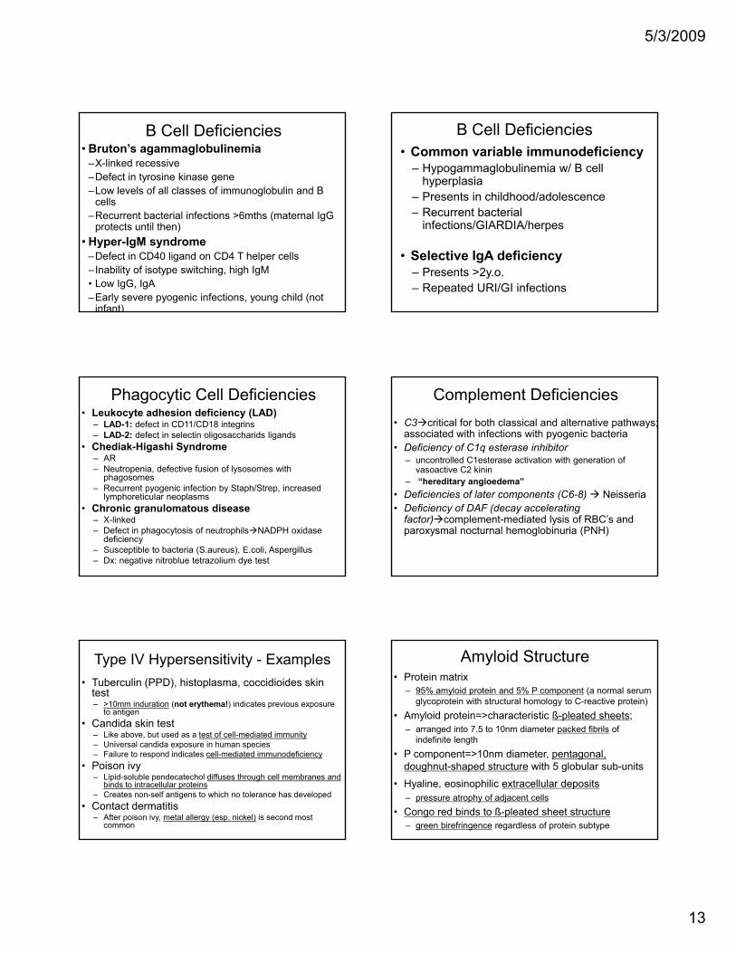
B Cell Deficiencies• Bruton’s agammaglobulinemia
–X-linked recessive–Defect in tyrosine kinase gene–Low levels of all classes of immunoglobulin and B
cells R t b t i l i f ti 6 th ( t l I G–Recurrent bacterial infections >6mths (maternal IgG protects until then)
• Hyper-IgM syndrome –Defect in CD40 ligand on CD4 T helper cells–Inability of isotype switching, high IgM• Low IgG, IgA–Early severe pyogenic infections, young child (not
infant)
B Cell Deficiencies• Common variable immunodeficiency
– Hypogammaglobulinemia w/ B cell hyperplasia
– Presents in childhood/adolescenceRecurrent bacterial– Recurrent bacterial infections/GIARDIA/herpes
• Selective IgA deficiency – Presents >2y.o. – Repeated URI/GI infections
Phagocytic Cell Deficiencies• Leukocyte adhesion deficiency (LAD)
– LAD-1: defect in CD11/CD18 integrins– LAD-2: defect in selectin oligosaccharids ligands
• Chediak-Higashi Syndrome– AR– Neutropenia, defective fusion of lysosomes with
phagosomesphagosomes– Recurrent pyogenic infection by Staph/Strep, increased
lymphoreticular neoplasms• Chronic granulomatous disease
– X-linked– Defect in phagocytosis of neutrophils NADPH oxidase
deficiency– Susceptible to bacteria (S.aureus), E.coli, Aspergillus– Dx: negative nitroblue tetrazolium dye test
Complement Deficiencies• C3 critical for both classical and alternative pathways;
associated with infections with pyogenic bacteria• Deficiency of C1q esterase inhibitor
– uncontrolled C1esterase activation with generation of vasoactive C2 kinin
– “hereditary angioedema”• Deficiencies of later components (C6-8) Neisseria• Deficiency of DAF (decay accelerating
factor) complement-mediated lysis of RBC’s and paroxysmal nocturnal hemoglobinuria (PNH)
Type IV Hypersensitivity - Examples• Tuberculin (PPD), histoplasma, coccidioides skin
test– >10mm induration (not erythema!) indicates previous exposure
to antigen• Candida skin test
– Like above, but used as a test of cell-mediated immunity– Universal candida exposure in human species– Failure to respond indicates cell-mediated immunodeficiency
• Poison ivy– Lipid-soluble pendecatechol diffuses through cell membranes and
binds to intracellular proteins– Creates non-self antigens to which no tolerance has developed
• Contact dermatitis– After poison ivy, metal allergy (esp. nickel) is second most
common
Amyloid Structure• Protein matrix
– 95% amyloid protein and 5% P component (a normal serum glycoprotein with structural homology to C-reactive protein)
• Amyloid protein=>characteristic ß-pleated sheets; – arranged into 7.5 to 10nm diameter packed fibrils of
indefinite lengthindefinite length• P component=>10nm diameter, pentagonal,
doughnut-shaped structure with 5 globular sub-units• Hyaline, eosinophilic extracellular deposits
– pressure atrophy of adjacent cells• Congo red binds to ß-pleated sheet structure
– green birefringence regardless of protein subtype
5/3/2009
14
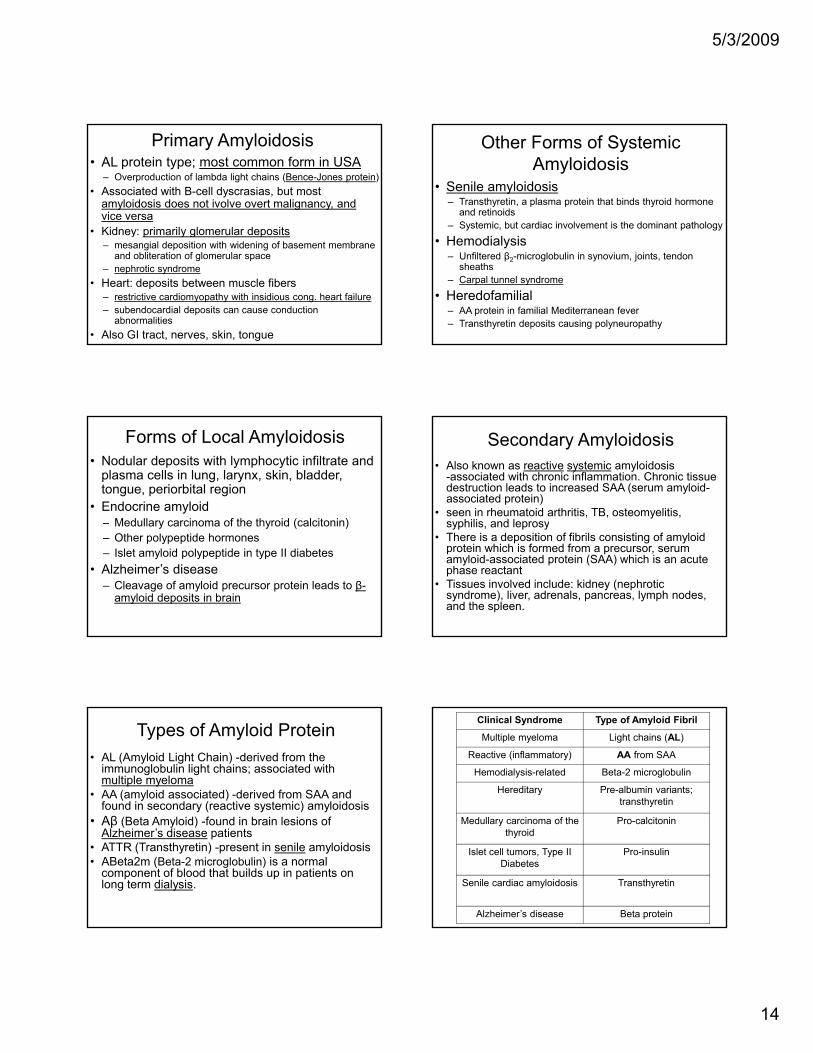
Primary Amyloidosis• AL protein type; most common form in USA
– Overproduction of lambda light chains (Bence-Jones protein)• Associated with B-cell dyscrasias, but most
amyloidosis does not ivolve overt malignancy, and vice versa
• Kidney: primarily glomerular depositsKidney: primarily glomerular deposits– mesangial deposition with widening of basement membrane
and obliteration of glomerular space– nephrotic syndrome
• Heart: deposits between muscle fibers– restrictive cardiomyopathy with insidious cong. heart failure– subendocardial deposits can cause conduction
abnormalities• Also GI tract, nerves, skin, tongue
Other Forms of Systemic Amyloidosis
• Senile amyloidosis– Transthyretin, a plasma protein that binds thyroid hormone
and retinoids– Systemic, but cardiac involvement is the dominant pathology
• Hemodialysis– Unfiltered β2-microglobulin in synovium, joints, tendon
sheaths– Carpal tunnel syndrome
• Heredofamilial– AA protein in familial Mediterranean fever– Transthyretin deposits causing polyneuropathy
Forms of Local Amyloidosis• Nodular deposits with lymphocytic infiltrate and
plasma cells in lung, larynx, skin, bladder, tongue, periorbital region
• Endocrine amyloid– Medullary carcinoma of the thyroid (calcitonin)Medullary carcinoma of the thyroid (calcitonin)– Other polypeptide hormones– Islet amyloid polypeptide in type II diabetes
• Alzheimer’s disease– Cleavage of amyloid precursor protein leads to β-
amyloid deposits in brain
Secondary Amyloidosis• Also known as reactive systemic amyloidosis
-associated with chronic inflammation. Chronic tissue destruction leads to increased SAA (serum amyloid-associated protein)
• seen in rheumatoid arthritis, TB, osteomyelitis, syphilis, and leprosysyphilis, and leprosy
• There is a deposition of fibrils consisting of amyloid protein which is formed from a precursor, serum amyloid-associated protein (SAA) which is an acute phase reactant
• Tissues involved include: kidney (nephrotic syndrome), liver, adrenals, pancreas, lymph nodes, and the spleen.
Types of Amyloid Protein• AL (Amyloid Light Chain) -derived from the
immunoglobulin light chains; associated with multiple myeloma
• AA (amyloid associated) -derived from SAA and found in secondary (reactive systemic) amyloidosisAβ (B t A l id) f d i b i l i f• Aβ (Beta Amyloid) -found in brain lesions of Alzheimer’s disease patients
• ATTR (Transthyretin) -present in senile amyloidosis• ABeta2m (Beta-2 microglobulin) is a normal
component of blood that builds up in patients on long term dialysis.
Clinical Syndrome Type of Amyloid Fibril
Multiple myeloma Light chains (AL)
Reactive (inflammatory) AA from SAA
Hemodialysis-related Beta-2 microglobulin
Hereditary Pre-albumin variants; transthyretin
M d ll i f th P l it iMedullary carcinoma of the thyroid
Pro-calcitonin
Islet cell tumors, Type II Diabetes
Pro-insulin
Senile cardiac amyloidosis Transthyretin
Alzheimer’s disease Beta protein
5/3/2009
15
Transplantation Reactions• Hyperacute Rejection:
– Irreversible, occurs within minutes of organ transplant
– Pre-formed antibody reacts with vascular endothelium of grafted organ.
A tib di b i t ABO bl d• Antibodies may be against ABO blood groups or anti-HLA antibodies (increased in multiparous women and recipients of multiple blood transfusions)
– Complement fixation leads to vessel damage -> vessel thrombosis -> ischemia of the graft.
Transplantation Reactions Cont’d• Acute Rejection
– Reversible, occurs within days to months– CD4+ cells stimulated by foreign MHC on donor or recipient
antigen presenting cells• Cellular response leads to interstitial lymphocytic infiltrate
of macrophages and CD8+ cells which damage graft tissue
• Humoral response leads to plasma cell production of anti-HLA antibodies.
– Immune complexes cause Vasculitis and thrombosis– Vascular damage and cytokines lead to intimal thickening
with narrowing of vascular lumen -> graft ischemia• Chronic Rejection
– Occurs within months to years, less well understood– Continued vascular injury to tissue leads to obliterative
intimal fibrosis of blood vessels -> ischemia of graft
Fluid and Hemodynamics• Non-inflammatory edema –due to
– ↑d hydrostatic pressure (local venous obstruction,↑d venous pressure/congestion, ↑d intravascular volume);
– ↓d plasma oncotic pressure – loss of albumin (nephrotic syndrome, protein-losing(nephrotic syndrome, protein losing gastroenteropathy), ↓d albumin synthesis (cirrhosis, malnutrition, lymphatic obstruction, Na+ retention)
– Lymphatic blockage– Transudate – low protein, low cells, specific
gravity <1.012• **see next slide for Up-to-Date guidelines for dif.
Fluid and Hemodynamics• Inflammatory edema – due to ↑d vascular
permeability (cytokines, trauma to endothelial cells, angiogenesis)– Exudate – high cells, low glucose, specific gravity >1.020– Three-Test Rule (Pleural Fluid)
• protein >2.9 g/dL• cholesterol >45 mg/dL• LDH >0.45 times the upper limit of the laboratory's
normal serum LDH• Hyperemia (active hyperemia) – inflammatory
cytokines arterial/arteriolar dilatation increased flow into capillary beds; *RED*/flushed– Ex: heat dissipation (fever, exercise), blushing, inflammation
Fluid and Hemodynamics -Congestion
• Congestion (passive hyperemia) – impaired venous drainage blood accumulation in capillaries; *BLUE-RED*– Ex: heart failure– Acute – shock, acute inflammation, sudden right CHF– Chronic – usually left CHF or mitral stenosis
• Right CHF – nutmeg liver (centrilobular necrosis)– Chronic congestion necrosis and fibrosis (cardiac
cirrhosis)• Left CHF – causes pulmonary edema; alveolar
macrophages phagocytose RBCs ”brown induration” & “heart failure cells”
Fluid and Hemodynamics• Hemorrhage
– Accumulation in a tissue – hematoma– Minute 1-2 mm into skin, mucous membranes, serosa-
petechiae (associated with thrombocytopenia)– >3mm hemorrhages – purpura associated with
petechia vasculitispetechia, vasculitis– >1 to 2 cm subcutaneous hematomas- ecchymoses – Large accumulations named by location – ie
hemopericardium, hemothorax• Significance depends on volume and rate of
bleeding– Rapid (up to 20% loss) – hypovolemic shock– Chronic, slow loss – iron deficiency
5/3/2009
16
Virchow’s Triad• 3 factors that predispose to venous thrombosis
– Hypercoagulable State: dehydration (EtOH, caffeine), hormones (estrogen), hyperlipidemia, malignancy, inherited clotting disorders, pregnancy, hyper-homocysteinemiaStasis: inactivity varicose veins heart failure– Stasis: inactivity, varicose veins, heart failure, hyperviscosity
– Endothelial Injury: smoking, surgery, trauma
Thrombosis General• Intravascular mass attached to the vessel wall
composed of platelets, coagulation factors, RBCs• Formation Virchow’s Triad-
– endothelial cell injury (MOST IMPORTANT)• NOTE- does NOT have to be denudation, can be any disruption
in the balance of pro- and antithrombotic effect of the endotheliumendothelium
– Stasis/turbulence- important in venous thrombi– Hypercoagulable state
• Types: arterial, venous (antemortem ONLY)• Fate: Propagation, embolization, dissolution,
organization and recanalization
Thrombosis Morphology• Arterial Thrombus:
– Grow retrograde from point of attachment– Most common in coronary>cerebral>femoral artery– Usually gray/white friable superimposed on
atherosclerotic plaqueLi f Z h lt ti l f l– Lines of Zahn- alternating layers of pale platelets and fibrin with darker layers of red cells
Thrombosis Morphology• Venous Thrombus:
– Extend from point of attachment in direction of blood flow
– Deep veins of lower extremities below the knee– Adherent, occlusive dark red- RBC and fibrin
• Post-mortem Clot:– Not attached to vessel wall – NOT a true thrombus – Upper chicken fat layer (supernatant) & lower
currant jelly layer (contains RBCs).
Ischemia
• Ischemia – reduced arterial blood flow– Occurs in response to significant drop in
blood pressure or occlusion of artery– Most common cause of cell injury
coagulative necrosis (except in brain)coagulative necrosis (except in brain)• Different from hypoxia- any state of reduced
oxygen availablity – Ischemia tends to injure cells faster
because it compromises the delivery of glycolytic enzymes and removal of wastes
Coagulation and Hemostasis• Thrombosis=formation of clots in non-interrupted
vasculature• Intact endothelial cells resist thrombosis by:
1. Heparin-like molecules activate antithrombin IIIneutralize thrombin & factor Xa (XII, IX, XI too)
2. Synthesize prostacyclin (PGI2) & NO inhibit platelet activation and vasodilate
3. Secrete tPA activates prothrombin4. Degrade ADP (ADP is pro-thrombotic)5. Synthesize thrombomodulin which binds thrombin to
activate Protein C which, with Protein S, cleaves factors Va, VIIIa
5/3/2009
17
Platelet Aggregation• ADP - highly potent mediator of platelet aggregation• TxA2 - prod. by platelets; also causes
vasoconstriction• Thrombin - formed by activation of coagulation
cascade; binds to thrombin receptors on platelets• GpIIb IIIa complexes binds activated platelets to• GpIIb-IIIa complexes - binds activated platelets to
fibrinogen (deficiency - Glanzmann thrombasthenia)• VonWillebrand factor – mediates binding of platelets to
collagen (via GpIb; deficiency - Bernard-Soulier syndrome)
• Platelet Factor 3 – cell surface membranes of platelets that allow assembly of coagulation proteins
• Calcium
Coagulation and Hemostasis• Hemostasis=formation of blood clots at the site of
vascular injury• Damaged blood vessels initiate hemostasis by:
1. Endothelial cells produce vWF (alpha granules platelets also) binds GpIb on platelets to exposed collagen
2. Tissue factor (aka thromboplastin or factor III) release activates extrinsic path (factor VII)
3. Platelets synthesize thromboxane A2vasoconstriction and platelet aggregation
4. Fibrinogen links platelets via gpIIb-IIIa (1°hemostatic plug)
Coagulation and Hemostasis• Extrinsic pathway
–Initiated by tissue factor (thromboplastin)–Tissue factor activates factor VII factor VII
activates factor X–Prothrombin time (PT) measures VII and factorsProthrombin time (PT), measures VII and factors
of common pathway (PT for war (warfarin) at 7am)
• Common Pathway–Xa + Va + platelet factor 3 + Ca++ (prothrombin
complex, on platelet membrane) converts prothrombin to thrombin converts fibrinogen to fibrin stabilized by XIII (XIII activated by thrombin)
Coagulation and Hemostasis• Intrinsic Pathway
– Factor XII (Hageman) activated by exposed collagen or HMWK
– XIIa activates 1) factor XI 2) plasminogen 3) kininogen system (bradykinin and kallikrein)Factor XIa activates factor IX factor IXa + factor VIIIa +– Factor XIa activates factor IX factor IXa + factor VIIIa + PF3 + Ca++ complex to activate factor X of the common pathway
– Partial Thromboplastin Time (PTT) measures, VIII, IX, XI, XII, and factors of the common pathway (for monitoring heparin)
– Hageman Factor XIIa links the fibrinolytic system, coagulation system, complement system, and kinin system.
Coagulation and Hemostasis• Fibrinolysis (thrombus dissolution)
– Plasminogen plasmin by tPA (alteplase, reteplase) or XIIa
– Plasmin cleaves fibrin (D-dimers) and fibrinogen (FDPs)– Plasmin also degrades factors V and VII
• Anticoagulants– Antithrombin III: inhibits thrombin & factors IXa, Xa, XIa,
XIIa, heparin modulates activity of ATIII– Protein C & S: Vit K dependent; inactivate Va, VIIIa.
Coagulopathy: Vascular damage• Petechia, epistaxis, prolonged bleed time, normal PT/PTT• Scurvy: vit C def causing weak capillaries and venules
--low hydroxylation K + P = low tropocollogen crosslinks--gingival/subQ bleed, poor wound healing, ecchymosis
• Henoch-Scholein purpura: hypersensitivity vasculitis w/ immune damage endothelium fever arthralgia renal/GI--immune damage endothelium, fever, arthralgia, renal/GI
--hemorrhagic urticaria (palpable purpura)• Waldenstrom’s Macroglobinemia: hyperviscosity • CT disorders:abnormal collagen/elastin-vascular bleeding• RMSF/Meningiococcus:necrosis/rupture of small vessels
5/3/2009
18
Coagulopathy: thrombocytopenia• Epixtaxis, petechia, GI/intracranial bleed, prolonged
bleed time• ITP: IgG antibodies against GpIIb:IIa
--kids: acute, self-limited after viral URI --adults: chronic idiopathic, associated with HIV and
SLESLE--no lymphadenopathy or splenomegaly
• TTP: aquired or genetic deficiency in vWF- cleaving metalloproteinase excess vWF increased platelet adhesion--pentad:microangiopathic hemolytic
anemia(schistocytes), fever, thrombocytopenia, renal insufficiency, neurologic abnormalities
Blood Groups• Determined by glycoproteins attached to RBC
surface• Blood Group O: no antigens on surface, anti-A and
anti-B IgM, most common blood group.• Blood Group A: A antigen, anti-B IgM, increased ood G oup a ge , a g , c eased
gastric carcinoma.• Blood Group B: B antigen, anti-A IgM.• Blood Group AB: both A and B antigens, no
antibodies, least frequent blood group• Rh group: 5 different antigens, either Rh + or Rh -
Blood Type Abs/Bombay Type• Transferase adds carbohydrate moities onto H
substance– Bombay type has NO H substance have anti-A, anti-B Abs– A - N-acetylgalactosamine added have anti-B Abs– B - D-galactose added have anti-A Abs
O t t f h ti A ti B Ab– O – most common, no transferase have anti-A, anti-B Abs• ABO Abs are naturally occurring, usually IgM
– Activate complement cause intravascular hemolysis• Rh Abs – not naturally occurring, mostly IgG
– Cross placenta cause extravascular hemolysis• ABO and Rh status determined by indirect Coombs
ABO Incompatibility in Transfusions
• Antibodies to A and/or B antigens bind the transfused erythrocytes leading to complement fixation and removal from the circulation by the spleen y p
• Pathogenesis is identical to that seen in type II hypersensitivity reactions
• Symptoms include hemolytic anemia, chills, shock, renal failure and possible death
ABO Incompatibility in Transplant• Hyperacute graft rejection• Antibodies react with antigens on the
vascular endothelial cells of the graft and initiate complement and clotting cascades
• Vessels become blocked with clots leading to death of the graft
• Gross pathology: graft is engorged and purple colored from hemorrhaged deoxygenated blood
Immune Hydrops• Results from immunization of the mother by blood
group antigens on fetal red cells usually during the 3rd trimester
• 1st exposure leads to production of IgM which cannot pass through the placenta (immune hydrops is not p g p ( y pseen in 1st pregnancies)
• A second exposure produces IgG antibodies to the fetal RBC antigen and crosses the placenta
• Complement fixation is induced and coated RBCs are cleared by the spleen (extravascular)
5/3/2009
19
Immune Hydrops, Cont’d• Hemolysis leads to anemia and/or jaundice• If hemolysis is mild, extramedullary hematopoiesis
will prevent anemia • If severe, anemia causes hypoxic injury to heart
d li lb i d th t i th i iand liver- albumin and other protein synthesis is impaired; along with heart injury leads to edema
• Increased unconjugated bilirubin from hemolysis binds lipids creating a poorly developed BBB and kernicterus
Rh Factor Immune Hydrops
• Rh system incompatibility is the most common cause of immune hydrops
• D antigen is the major cause Rh i tibilit h d i t d b• Rh incompatibility hydrops is prevented by maternal injection of RhIg (Rhogam) at 28 weeks and within 72 hours of the delivery of the 1st child and all subsequent children in a women that is Rh- and does not yet have anti-D antibodies
ABO Immune Hydrops• ABO incompatibility seen in 20-25% of
pregnancies, only 1 in 10 of these has hemolysis and 1 in 200 requires treatment
• Most ab is IgM; neonatal cells express AMost ab is IgM; neonatal cells express A & B antigens weakly; other cells also have blood group antigens and sequester the antibody
• Seen most often in A or B infants born to type O mothers who make some IgG to A & B antigens
Type and Screen/Crossmatch• Type and screen- determines recipient
blood type and presence of serum anti-RBC antibodies; screen for ab to RBCs; no precipitation of RBCs = no antibodies present, no blood actually set asidep y
• Type and Cross- units intended for patient are incubated with patient serum and an Indirect Coombs test is preformed; negative Indirect Coombs indicates the blood is ABO compatible, not reusable after cross.
Shock• SHOCK = circulatory collapse → impaired tissue
perfusion → systemic hypoxia• Brain is the first organ affected• Medical emergency! Need to reverse cause of
hypoxia– Some types require aggressive volume replacement
• Stages ends w/ irreversible end organ damageNonprogressive compensatory mechanisms– Nonprogressive- compensatory mechanisms• HR, TPR; perfusion maintenance of vital organs
– Progressive- onset of tissue hyperperfusion & circ/metabolic imbalance• Ex. metabolic acidosis due to lactic acidemia• Compensatory mechanisms no longer adequate
– Irreversible – damage too severe – survival impossible• Signs: acute tubular necrosis, GI mucosal
hemorrhages, pulmonary edema, fatty change
Shock Types - Hypovolemic• Circulatory collapse b/c fluid loss
– Normal = 9 units or 4-5 liters– Loss of 10-15% without clinical sequelae– Loss of 15-30% - tachycardia– Loss of 30-40% - worsening of mental status– >40% - limit of compensation and risk of deathp
• Hemorrhage, severe trauma, fluid loss via skin (ex. 3rd degree burns), diarrhea, vomiting
• pulmo capillarty wedge pressure (PCWP) b/c LV EDV• mixed venous oxygen content (tissues have time to extract
more oxygen than nL)• cold skin b/c of peripheral vasoconstriction (sympathetic)• if due to blood loss, IV crystalloid solutions will reveal RBC • Therapy - replace volume w/ whole blood
5/3/2009
20
Shock Types – Cardiogenic• Circulatory collapse b/c of pump failure of the LV• MCC= acute MI• other causes: PE, arrythmias, cardiac tamponade,
pulmonary saddle embolus (↓↓ blood return to LA)• PCWP (b/c fluid back-up into pulmonary vv.)
l i• normovolemic• other signs are similar to hypovolemic shock• *NEUROGENIC-loss of ANS (brain stem or cord
damage)• HR, TPR (b/c loss of tonic sympathetic stim.)• warm, dry skin, venous pooling• normovolemic
Shock Types - Neurogenic• Due to loss of vascular tone
– Tone loss secondary to loss of ANS (brain stem or cord damage)
• HR, TPR (b/c of loss of tonic sympathetic stim.)• warm, dry skin (can’t vasoconstrict), venous pooling, y ( ), p g• normovolemic
Septic Shock/Sepsis• Sepsis = blood infection + systemic
inflammatory response• Most associated w/gram negative infection (bug
expressing LPS or LOS)– Causes gram-negative endotoxemiag g– Same result can happed from injecting LPS alone• Septic shock results from sepsis• Septic shock also seen w/gram positive and other
infections
Septic Shock/Sepsis• Endotoxins (LPS, LOS - lipid part of cell wall) cause
release of IL-1, IL-6, TNF by monocytes– Activated complement and kinin systems → direct toxic
injury to cell• Endothelial cell damage releases nitric oxide –
vasodilates & can activate coagulation cascade (+/-g (DIC)– CO may initially increase due to vasodilation
• Systemic in vascular permeability hypovolemia• Warm, pink skin, organ hypoxia• organ dysfunction is due both to hypoxia and systemic
cytokine release
DIC• Activation of DIC• Pathogenesis• Clinical associations
– Sepsis, Neisseria meningitidis• Clinical measures – D-dimer; fibrinolytic
peptides• Pathologic findings
Acid-BaseHenderson-Hasselbach
– pH = 6.1 + log(HCO3)/pCO2* 0.03
• General consideratoins– pH rises with ↑HCO3 or ↓pCO2
– pH falls with dec HCO3 or inc pCO2p 3 p 2
– dec pH w/inc CO2 = respiratory acidosis (HCO3 >30)**– dec pH w/dec HCO3 = metabolic acidosis (HC03 <22)– inc pH w/dec CO2 = respiratory alkalosis (HCO3 <18)**– inc pH w/inc HCO3 = metabolic alkalosis (HCO3 > 28)
• ** if compensated metabolically
5/3/2009
21
Acid-Base• Clinical considerations
– CO2 changes reflect respiratory function– HCO3 changes reflect renal/metabolic function– Compensatory mechanisms: renal function altered
f i di hilto compensate for respiratory disease while respiratory function is altered to compensate for metabolic or renal disease
– The resulting attempt to compensate is never complete (pH never gets back to 7.4).
Acid-Base• Total CO2
– Total CO2(mEq/L) = HCO3 + pCO2*0.03• Serum potassium is often increased with acidosis and
decreased in alkalosis• Anion Gap may increase with metabolic acidosisAnion Gap may increase with metabolic acidosis
– AG= Na-(Cl + HCO3) THINK MULEPAK
• Acidosis can be treated with bicarb to neutralize acid or hyperventilation to breathe off excess CO2
• Alkalosis can be treated by hypoventilation, retention of H+, or excretion of HCO3
-
Control of Growth – Tissue Proliferation
• Labile tissues – Continuously dividing tissues (i.e. skin, surface epithelia, mucosa of glands and GI)
• Quiescent tissues – Normally have a low level of replication but can regenerate if needed (i.e. liver, kid fib bl t d th l )kidneys, pancreas, fibroblasts and smooth muscle)
• Permanent tissues - Terminally differentiated cells with little to no regenerative capability (i.e. neurons, skeletal muscle, and cardiac muscle)
Control of Growth – Growth Factors• EGF & TGFα – Similar factors that stimulate
keratinoctye migration and granulation tissue formation• VEGF – Induces angiogenesis and increases vascular
permeability is important in tumor growth• PDGF – Causes migration and proliferation of
fibroblasts and smooth muscle and is important infibroblasts and smooth muscle and is important in wound healing
• FGF – Angiogenesis, wound repair, skeletal muscle development and lung maturation, and hematopoiesis.
• TGFβ – Growth inhibitor for epithelial cells and leukocytes, stimulates fibroblasts and smooth muscle cells, strong anti-inflammatory effect, and potent promoter or fibrosis
Control of Growth – Control Points• Cyclin-dependent kinase (CDK) – Proteins that serve
as checkpoints between cell cycle phases by phosphorylating proteins (ie: RB) vital to cycle transition
• Cyclin – Proteins that are synthesized during specific phases and then rapidly decline after their function isphases and then rapidly decline after their function is complete. – Function phosphorylate inactive CDKs rendering them
active• CDK inhibitors – Prevent the movement from one cell
cycle point to the next by inhibiting CDK. – Cip/Kip and INK4/ARF are examples – Serve as tumor suppressors and frequently altered in tumors
Control of Growth• Resting cells are in G0 and are recruited into G1• Orderly progression through phases is regulated
by cyclins and CDKs:– CyclinD/CDK4 phosphorylates RB allowing passage
through the G1 restriction point.– CyclinE/CDK2 permits DNA replication– CyclinA/CDK2 regulates mitotic prophase– CyclinB/CDK1 regulates nuclear division
• Cell cycle has 2 check- points– Between G1/S and G2/M– If DNA damage present- DNA duplication is arrested– If DNA damage is reparable- repaired, if not undergoes
apoptosis
5/3/2009
22
Neoplasia - Definitions• Hyperplasia – physiologic or pathologic increase in
number of cells in a normal arrangment (reversible)• Metaplasia – replacement of one fully differentiated
cell type by another fully differentiated cell type (reversible)
• Dysplasia – pre-neoplastic pleomorphic cells (change in cell size, shape and organization (reversible)
• Anaplasia –lack of differentiation marked by: pleomorphism, hyperchromatism, mitosis
• Neoplasia – uncontrolled clonal cell proliferation• Grade – degree of cellular differentiation (I to IV)• Stage – degree of spread from primary lesion (TNM)
Neoplasia – Definitions cont.• Adenoma- benign neoplasm of parenchyma derived from
glands or forming glandular patterns– Sebaceous gland adenoma– Ovarian cystadenoma
• Papilloma – benign neoplasms that form microscopic papilla• Adenocarcinoma malignant neoplasm of parenchyma derived
from glands or forming glandular patternsfrom glands or forming glandular patterns– Sebaceous gland adenocarcinoma– Ovarian cysadenocarcinoma
• Leukemia- malignant lymphoid neoplasm with widespread involvement of the bone marrow and tumor cells often in peripheral blood
• Lymphoma- malignant lymphoid neoplasm that arise in discrete tissue masses outside of the bone marrow
Neoplasia - Definitions• Hemangioma – benign accumulation of blood vessels• Hemangiosarcoma – malignant blood vessel tumor• Leiomyoma – benign smooth muscle tumor• Leiomyosarcoma – malignant smooth muscle tumor• Rhabdomyoma – benign skeletal muscle tumor• Rhabdomyosarcoma – malignant skeletal muscle tumor• Osteoma – benign bone tumor• Osteosarcoma – malignant bone tumor• Lipoma – benign fat tumor• Liposarcoma – malignant fat tumor• Carcinoma in situ – no surrounding tissue invasion
Neoplasia - Definitions• Mature teratoma – benign, from the 3 germ cell layers• Immature teratoma – malignant, same derivation • Carcinoma – malignant tumor of epithelial origin• Sarcoma – malignant tumor of mesenchymal origin• Invasion – spread to adjacent tissuesInvasion spread to adjacent tissues• Metastasis – spread to nonadjacent tissues• Desmoplasia – non-neoplastic, tumor-induced fibrous tissue• Choristoma – normal tissue in another organ (benign)• Hamartoma – benign disorganized overgrowth in • appropriate organ• Proto-oncogene – regulate cell growth & differentiation
Neoplasia - Definitions• Tumor suppressor genes – gene inactivation promotes
cellular proliferation• Clonal – all cells originated from a single cell• Oncogene – altered gene frequently found in cancer• Recessive oncogene – loss of both alleles required to g q
remove inhibition—tumor suppressor genes• Dominant oncogene – a single allele unregulated
proliferation; promote growth—oncogenes• Transformation – autonomous growth capability begins• Carcinogenesis - oncogenic changes by environmental
agent• Complete carcinogen – induces initiation and promotion
Oncogenes• genes from the normal genome which are now
altered in structure or expressed in abnormal amounts.
• Dominant Oncogenes- are elements that promote growth only need expression of a single allele to cause unregulated proliferation (RAS, growth factors, growth factor receptors)
• Recessive Oncogenes- are elements that inhibit growth require loss of both alleles to eliminate the inhibitory signal (Tumor suppressor genes, DNA repair genes).
5/3/2009
23
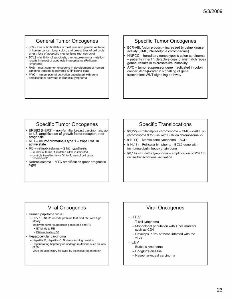
General Tumor Oncogenes• p53 – loss of both alleles is most common genetic mutation
in human cancer; lung, colon, and breast; loss of cell cycle arrest; loss of apoptotic mechanisms (not necrosis)
• BCL2 – inhibitor of apoptosis; over-expression or mutation results in arrest of apoptosis in neoplasms (Follicular lymphoma)RAS t i d l t f h• RAS – most common oncogene in development of human cancers; trapped in activated GTP-bound state
• MYC – transcriptional activation associated with gene amplification; activated in Burkitt’s lymphoma
Specific Tumor Oncogenes• BCR-ABL fusion product – increased tyrosine kinase
activity (CML, Philadelphia chromosome)• HNPCC – hereditary nonpolyposis colon carcinoma
– patients inherit 1 defective copy of mismatch repair genes; results in microsatellite instability
• APC tumor suppressor gene inactivated in colon• APC – tumor suppressor gene inactivated in colon cancer; APC-β-catenin signalling of gene trascription; WNT signaling pathway
Specific Tumor Oncogenes• ERBB2 (HER2) – non-familial breast carcinomas; up
to 1/3; amplification of growth factor receptor; poor prognosis
• NF1 – neurofibromatosis type 1 – traps RAS in active state
• RB – retinoblastoma – 2 hit hypothesis – In familial forms, 1 mutated allele is inherited– controls transition from G1 to S; loss of cell cycle
“checkpoint”• Neuroblastoma – MYC amplification (poor prognostic
sign)
Specific Translocations• t(9;22) – Philadelphia chromosome – CML – c-ABL on
chromosome 9 to fuse with BCR on chromosome 22• t(11;14) – Mantle zone lymphoma – BCL1 • t(14;18) – Follicular lymphoma - BCL2 gene with
immunoglobulin heavy chain gene• t(8;14) – Burkitt’s lymphoma – amplification of MYC to
cause transcriptional activation
Viral Oncogenes• Human papilloma virus
– HPV 16, 18, 31 encode proteins that bind p53 with high affinity
– Inactivate tumor suppressor genes p53 and RB• E7 binds to RB• E6 inactivates p53p
• Hepatocellular carcinoma– Hepatitis B, Hepatitis C; No transforming proteins– Regenerating hepatocytes undergo mutations such as loss
of p53– Virus-induced injury followed by extensive regeneration
Viral Oncogenes
• HTLV– T cell lymphoma– Monoclonal population with T cell markers
such as CD4Develops in 1% of those infected with the– Develops in 1% of those infected with the virus
• EBV– Burkitt’s lymphoma– Hodgkin’s disease– Nasopharyngeal carcinoma
5/3/2009
24
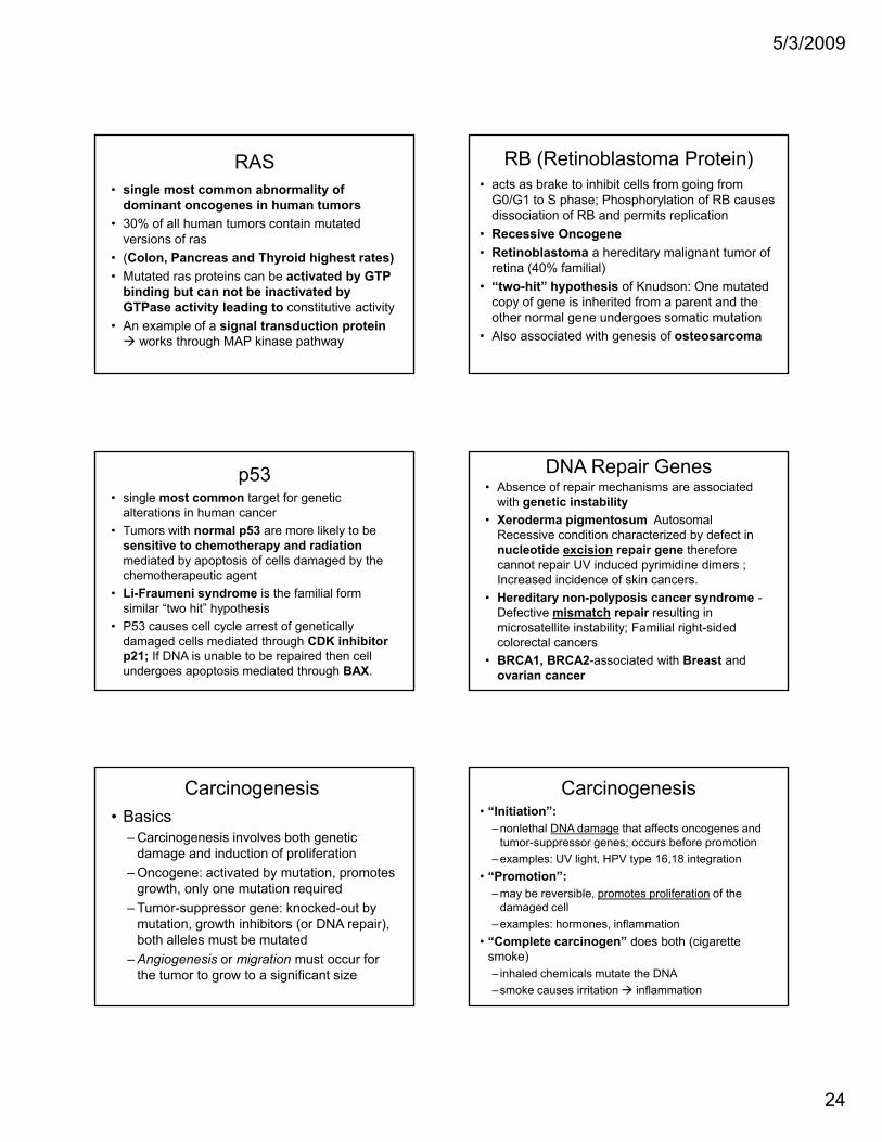
RAS• single most common abnormality of
dominant oncogenes in human tumors• 30% of all human tumors contain mutated
versions of ras(C l P d Th id hi h t t )• (Colon, Pancreas and Thyroid highest rates)
• Mutated ras proteins can be activated by GTP binding but can not be inactivated by GTPase activity leading to constitutive activity
• An example of a signal transduction proteinworks through MAP kinase pathway
RB (Retinoblastoma Protein)• acts as brake to inhibit cells from going from
G0/G1 to S phase; Phosphorylation of RB causes dissociation of RB and permits replication
• Recessive Oncogene• Retinoblastoma a hereditary malignant tumor ofRetinoblastoma a hereditary malignant tumor of
retina (40% familial) • “two-hit” hypothesis of Knudson: One mutated
copy of gene is inherited from a parent and the other normal gene undergoes somatic mutation
• Also associated with genesis of osteosarcoma
p53• single most common target for genetic
alterations in human cancer • Tumors with normal p53 are more likely to be
sensitive to chemotherapy and radiationmediated by apoptosis of cells damaged by the h h ichemotherapeutic agent
• Li-Fraumeni syndrome is the familial form similar “two hit” hypothesis
• P53 causes cell cycle arrest of genetically damaged cells mediated through CDK inhibitorp21; If DNA is unable to be repaired then cell undergoes apoptosis mediated through BAX.
DNA Repair Genes• Absence of repair mechanisms are associated
with genetic instability• Xeroderma pigmentosum Autosomal
Recessive condition characterized by defect in nucleotide excision repair gene therefore cannot repair UV induced pyrimidine dimers ; p pyIncreased incidence of skin cancers.
• Hereditary non-polyposis cancer syndrome -Defective mismatch repair resulting in microsatellite instability; Familial right-sided colorectal cancers
• BRCA1, BRCA2-associated with Breast and ovarian cancer
Carcinogenesis• Basics
– Carcinogenesis involves both genetic damage and induction of proliferation
– Oncogene: activated by mutation, promotes growth only one mutation requiredgrowth, only one mutation required
– Tumor-suppressor gene: knocked-out by mutation, growth inhibitors (or DNA repair), both alleles must be mutated
– Angiogenesis or migration must occur for the tumor to grow to a significant size
Carcinogenesis• “Initiation”:
–nonlethal DNA damage that affects oncogenes and tumor-suppressor genes; occurs before promotion
–examples: UV light, HPV type 16,18 integration• “Promotion”:
–may be reversible, promotes proliferation of the damaged cell
–examples: hormones, inflammation• “Complete carcinogen” does both (cigarette
smoke)– inhaled chemicals mutate the DNA–smoke causes irritation inflammation
5/3/2009
25
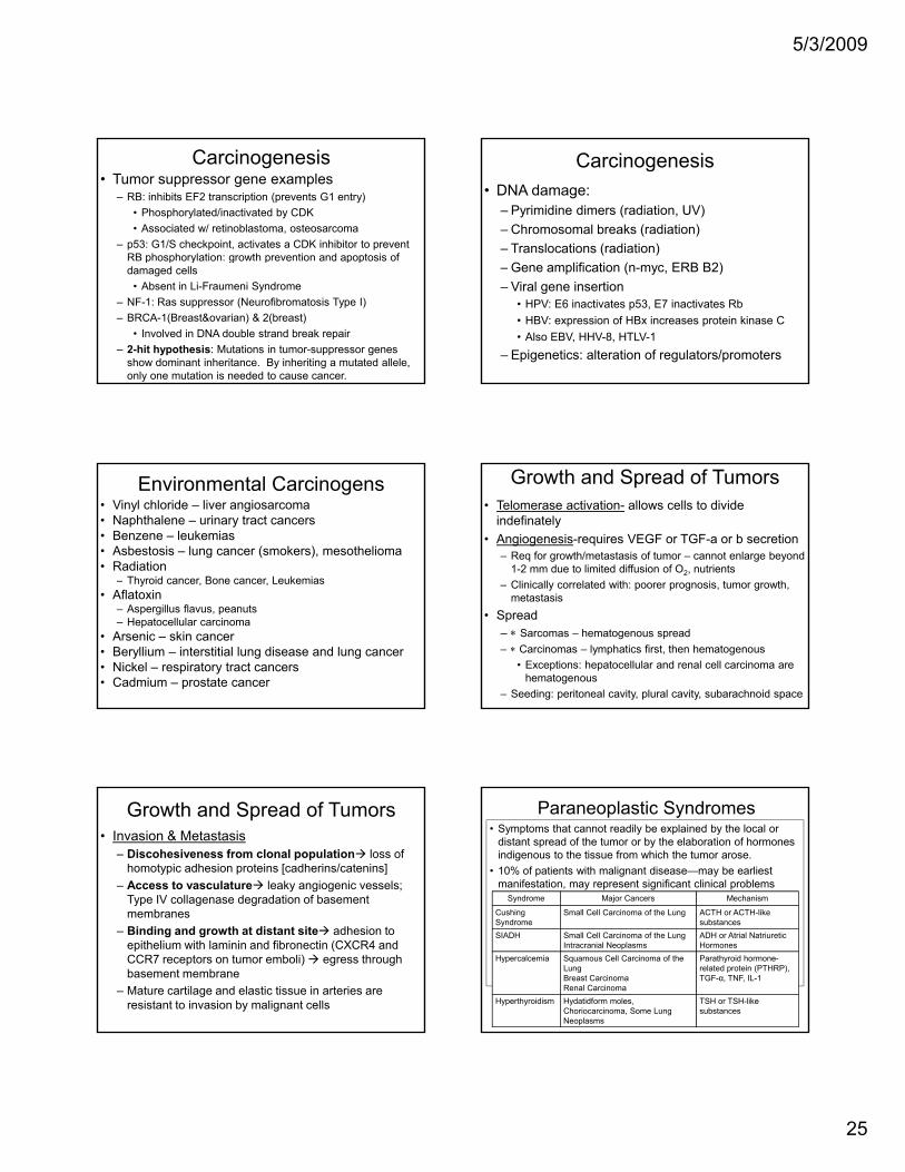
Carcinogenesis• Tumor suppressor gene examples
– RB: inhibits EF2 transcription (prevents G1 entry)• Phosphorylated/inactivated by CDK• Associated w/ retinoblastoma, osteosarcoma
– p53: G1/S checkpoint, activates a CDK inhibitor to prevent RB phosphorylation: growth prevention and apoptosis of p osp o y a o g o p e e o a d apop os s odamaged cells
• Absent in Li-Fraumeni Syndrome– NF-1: Ras suppressor (Neurofibromatosis Type I)– BRCA-1(Breast&ovarian) & 2(breast)
• Involved in DNA double strand break repair– 2-hit hypothesis: Mutations in tumor-suppressor genes
show dominant inheritance. By inheriting a mutated allele, only one mutation is needed to cause cancer.
Carcinogenesis • DNA damage:
– Pyrimidine dimers (radiation, UV)– Chromosomal breaks (radiation)– Translocations (radiation)– Gene amplification (n-myc, ERB B2)– Viral gene insertion
• HPV: E6 inactivates p53, E7 inactivates Rb• HBV: expression of HBx increases protein kinase C• Also EBV, HHV-8, HTLV-1
– Epigenetics: alteration of regulators/promoters
Environmental Carcinogens• Vinyl chloride – liver angiosarcoma• Naphthalene – urinary tract cancers• Benzene – leukemias• Asbestosis – lung cancer (smokers), mesothelioma• Radiation
– Thyroid cancer, Bone cancer, Leukemiasy• Aflatoxin
– Aspergillus flavus, peanuts– Hepatocellular carcinoma
• Arsenic – skin cancer• Beryllium – interstitial lung disease and lung cancer• Nickel – respiratory tract cancers• Cadmium – prostate cancer
Growth and Spread of Tumors• Telomerase activation- allows cells to divide
indefinately• Angiogenesis-requires VEGF or TGF-a or b secretion
– Req for growth/metastasis of tumor – cannot enlarge beyond 1-2 mm due to limited diffusion of O2, nutrients
– Clinically correlated with: poorer prognosis tumor growth– Clinically correlated with: poorer prognosis, tumor growth, metastasis
• Spread– ∗ Sarcomas – hematogenous spread– ∗ Carcinomas – lymphatics first, then hematogenous
• Exceptions: hepatocellular and renal cell carcinoma are hematogenous
– Seeding: peritoneal cavity, plural cavity, subarachnoid space
Growth and Spread of Tumors• Invasion & Metastasis
– Discohesiveness from clonal population loss of homotypic adhesion proteins [cadherins/catenins]
– Access to vasculature leaky angiogenic vessels; Type IV collagenase degradation of basement
bmembranes– Binding and growth at distant site adhesion to
epithelium with laminin and fibronectin (CXCR4 and CCR7 receptors on tumor emboli) egress through basement membrane
– Mature cartilage and elastic tissue in arteries are resistant to invasion by malignant cells
Paraneoplastic Syndromes• Symptoms that cannot readily be explained by the local or
distant spread of the tumor or by the elaboration of hormones indigenous to the tissue from which the tumor arose.
• 10% of patients with malignant disease—may be earliest manifestation, may represent significant clinical problems (possibly lethal), may mimic metastatic diseaseSyndrome Major Cancers Mechanism
C hi S ll C ll C i f th L ACTH ACTH likCushing Syndrome
Small Cell Carcinoma of the Lung ACTH or ACTH-like substances
SIADH Small Cell Carcinoma of the LungIntracranial Neoplasms
ADH or Atrial NatriureticHormones
Hypercalcemia Squamous Cell Carcinoma of the LungBreast CarcinomaRenal Carcinoma
Parathyroid hormone-related protein (PTHRP), TGF-α, TNF, IL-1
Hyperthyroidism Hydatidform moles, Choriocarcinoma, Some Lung Neoplasms
TSH or TSH-like substances
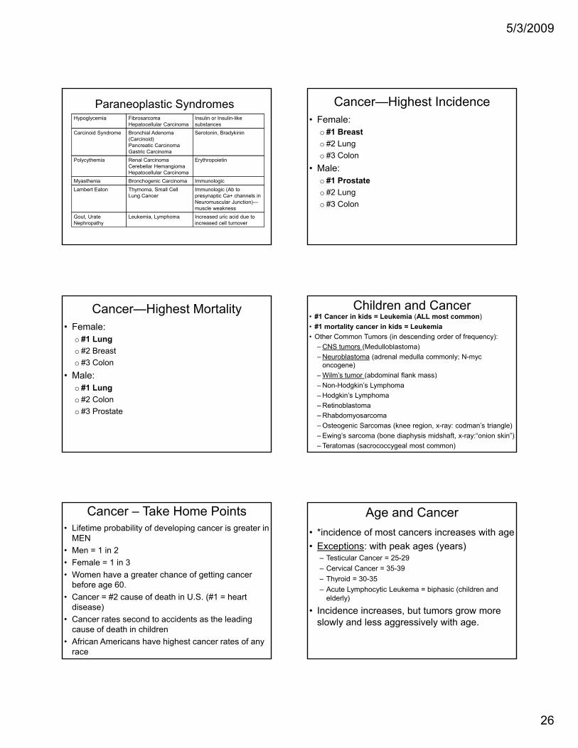
5/3/2009
26
Paraneoplastic SyndromesHypoglycemia Fibrosarcoma
Hepatocellular CarcinomaInsulin or Insulin-like substances
Carcinoid Syndrome Bronchial Adenoma (Carcinoid)Pancreatic CarcinomaGastric Carcinoma
Serotonin, Bradykinin
Polycythemia Renal CarcinomaCerebellar Hemangioma
ErythropoietinCerebellar HemangiomaHepatocellular Carcinoma
Myasthenia Bronchogenic Carcinoma ImmunologicLambert Eaton Thymoma, Small Cell
Lung CancerImmunologic (Ab to presynaptic Ca+ channels in Neuromuscular Junction)—muscle weakness
Gout, UrateNephropathy
Leukemia, Lymphoma Increased uric acid due to increased cell turnover
Cancer—Highest Incidence• Female:
o #1 Breasto #2 Lungo #3 Colon
Male:• Male:o #1 Prostateo #2 Lungo #3 Colon
Cancer—Highest Mortality• Female:
o #1 Lungo #2 Breasto #3 Colon
Male:• Male:o #1 Lungo #2 Colono #3 Prostate
Children and Cancer• #1 Cancer in kids = Leukemia (ALL most common)• #1 mortality cancer in kids = Leukemia• Other Common Tumors (in descending order of frequency):
– CNS tumors (Medulloblastoma)– Neuroblastoma (adrenal medulla commonly; N-myc
oncogene)Wil ’ t ( bd i l fl k )– Wilm’s tumor (abdominal flank mass)
– Non-Hodgkin’s Lymphoma– Hodgkin’s Lymphoma– Retinoblastoma– Rhabdomyosarcoma– Osteogenic Sarcomas (knee region, x-ray: codman’s triangle)– Ewing’s sarcoma (bone diaphysis midshaft, x-ray:“onion skin”)– Teratomas (sacrococcygeal most common)
Cancer – Take Home Points• Lifetime probability of developing cancer is greater in
MEN• Men = 1 in 2• Female = 1 in 3• Women have a greater chance of getting cancer
before age 60.• Cancer = #2 cause of death in U.S. (#1 = heart
disease)• Cancer rates second to accidents as the leading
cause of death in children• African Americans have highest cancer rates of any
race
Age and Cancer• *incidence of most cancers increases with age• Exceptions: with peak ages (years)
– Testicular Cancer = 25-29– Cervical Cancer = 35-39– Thyroid = 30-35Thyroid 30 35– Acute Lymphocytic Leukema = biphasic (children and
elderly)
• Incidence increases, but tumors grow more slowly and less aggressively with age.
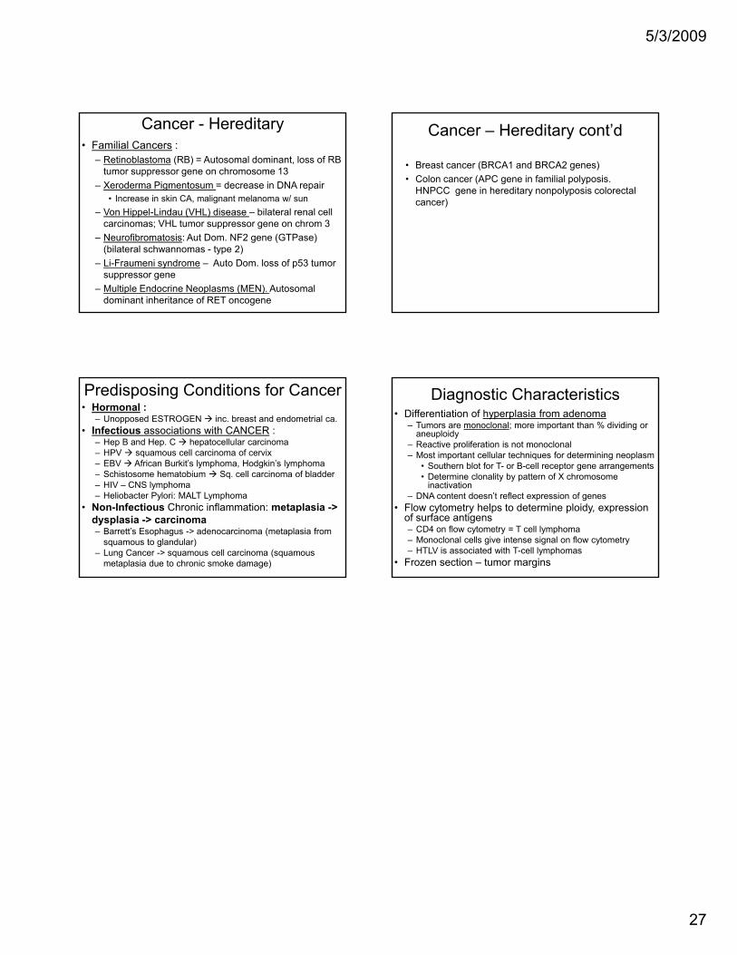
5/3/2009
27
Cancer - Hereditary• Familial Cancers :
– Retinoblastoma (RB) = Autosomal dominant, loss of RB tumor suppressor gene on chromosome 13
– Xeroderma Pigmentosum = decrease in DNA repair • Increase in skin CA, malignant melanoma w/ sun
– Von Hippel-Lindau (VHL) disease – bilateral renal cell– Von Hippel-Lindau (VHL) disease – bilateral renal cell carcinomas; VHL tumor suppressor gene on chrom 3
– Neurofibromatosis: Aut Dom. NF2 gene (GTPase) (bilateral schwannomas - type 2)
– Li-Fraumeni syndrome – Auto Dom. loss of p53 tumor suppressor gene
– Multiple Endocrine Neoplasms (MEN). Autosomal dominant inheritance of RET oncogene
Cancer – Hereditary cont’d
• Breast cancer (BRCA1 and BRCA2 genes)• Colon cancer (APC gene in familial polyposis.
HNPCC gene in hereditary nonpolyposis colorectal cancer)
Predisposing Conditions for Cancer• Hormonal :
– Unopposed ESTROGEN inc. breast and endometrial ca.• Infectious associations with CANCER :
– Hep B and Hep. C hepatocellular carcinoma– HPV squamous cell carcinoma of cervix– EBV African Burkit’s lymphoma, Hodgkin’s lymphoma– Schistosome hematobium Sq. cell carcinoma of bladderq– HIV – CNS lymphoma– Heliobacter Pylori: MALT Lymphoma
• Non-Infectious Chronic inflammation: metaplasia -> dysplasia -> carcinoma– Barrett’s Esophagus -> adenocarcinoma (metaplasia from
squamous to glandular)– Lung Cancer -> squamous cell carcinoma (squamous
metaplasia due to chronic smoke damage)
Diagnostic Characteristics• Differentiation of hyperplasia from adenoma
– Tumors are monoclonal; more important than % dividing or aneuploidy
– Reactive proliferation is not monoclonal– Most important cellular techniques for determining neoplasm
• Southern blot for T- or B-cell receptor gene arrangements• Determine clonality by pattern of X chromosome• Determine clonality by pattern of X chromosome
inactivation– DNA content doesn’t reflect expression of genes
• Flow cytometry helps to determine ploidy, expression of surface antigens– CD4 on flow cytometry = T cell lymphoma– Monoclonal cells give intense signal on flow cytometry– HTLV is associated with T-cell lymphomas
• Frozen section – tumor margins
Related Documents