Pathology Lap Pictures and Explanation POD II BLOCK Abdullah Al Garni

Pathology Lap Pictures and Explanation POD II BLOCK Abdullah Al Garni.
Jan 03, 2016
Welcome message from author
This document is posted to help you gain knowledge. Please leave a comment to let me know what you think about it! Share it to your friends and learn new things together.
Transcript

Pathology Lap Pictures and Explanation POD II BLOCK
Abdullah Al Garni
Prostate gland, nodular hyperplasia) الموثة ) الـــبروســـتاتا غـــدة تــــضخــم

well-circumscribed nodules
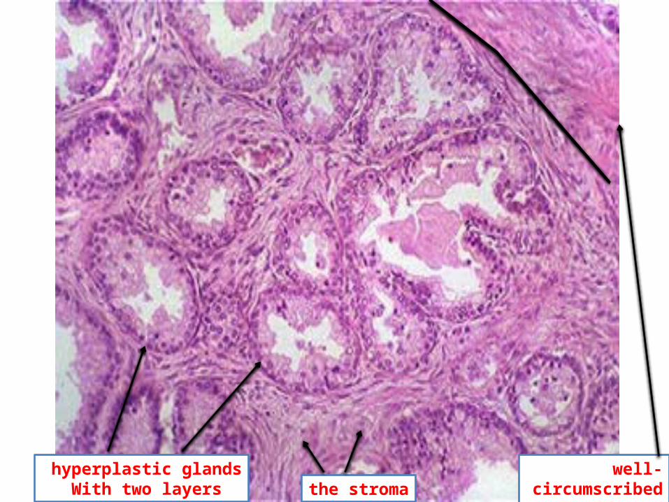
hyperplastic glands With two layers
well-circumscribed nodules margin the stroma
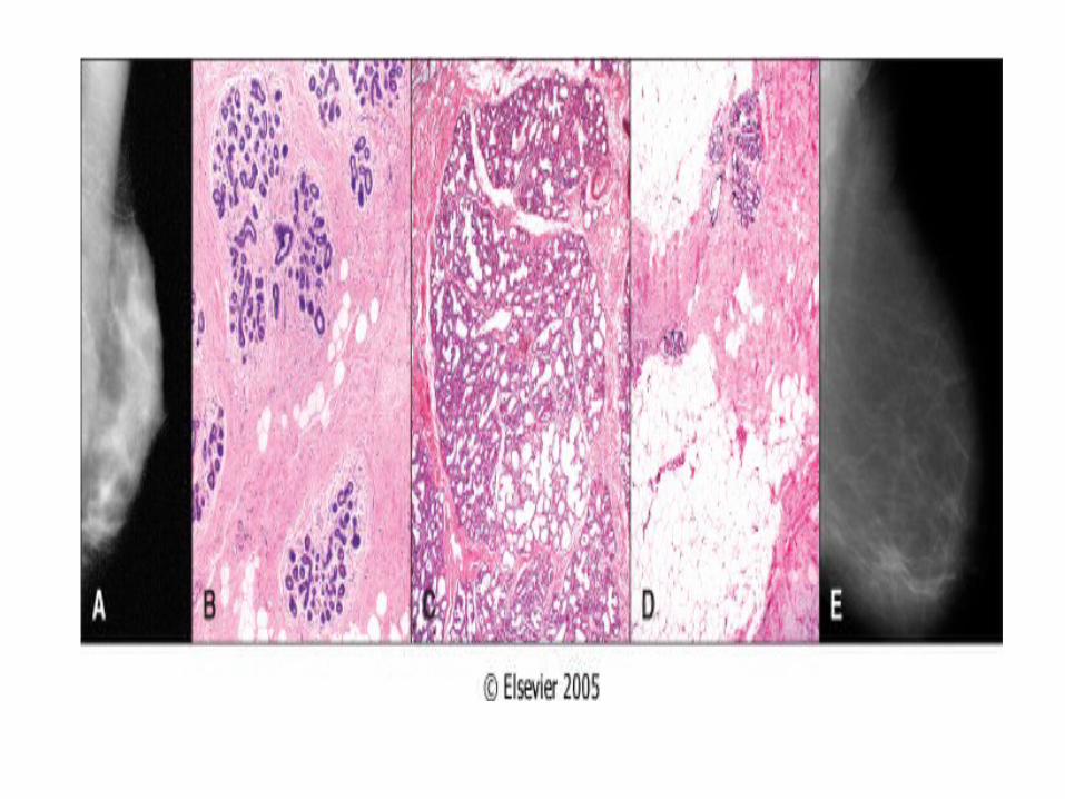
Breast, Fibroadenomaالثــــــدي ســــرطــــان

fibroadenoma normal breast tissue well-demarcated border

Large irregularly shaped glandular spaces fibrous stroma

columnar epithelium
Delicate stroma

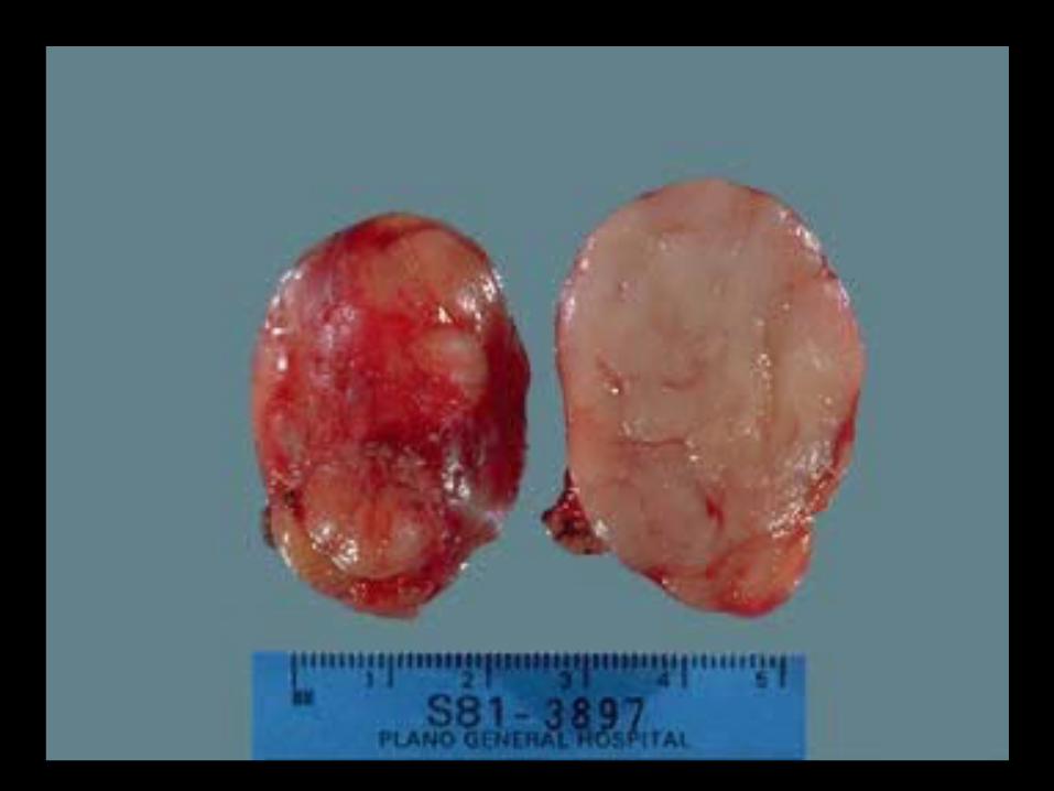
Breast, Invasive ductal carcinomaالـــــثدي ســـــرطان
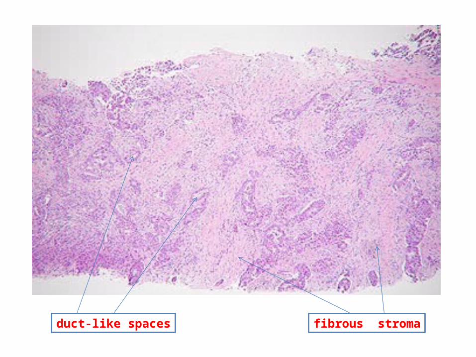
duct-like spaces fibrous stroma

duct-like spaces fibrous stroma

Non-neoplastic ducts Invading tumor cells
Fat
Stroma

Tumor cells
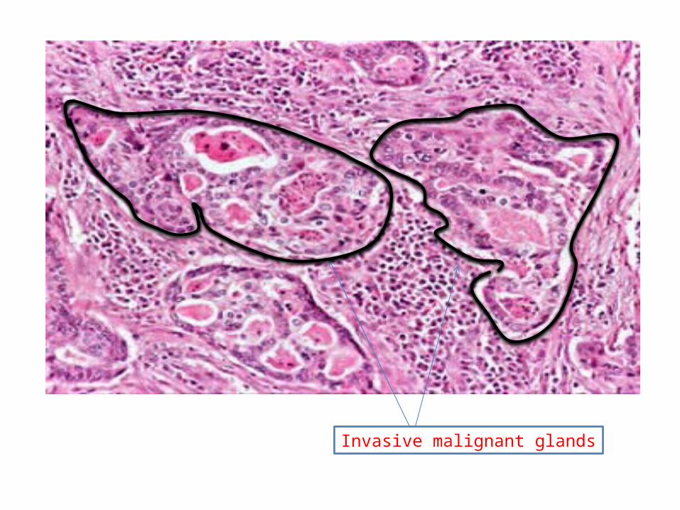
Invasive malignant glands
Invasive squamous cell carcinomaEXAMPLE: Bronchousالهوائية القصبات ســـرطـــان
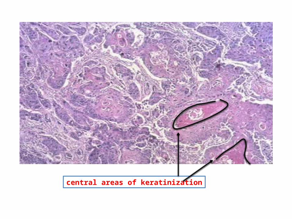
central areas of keratinization
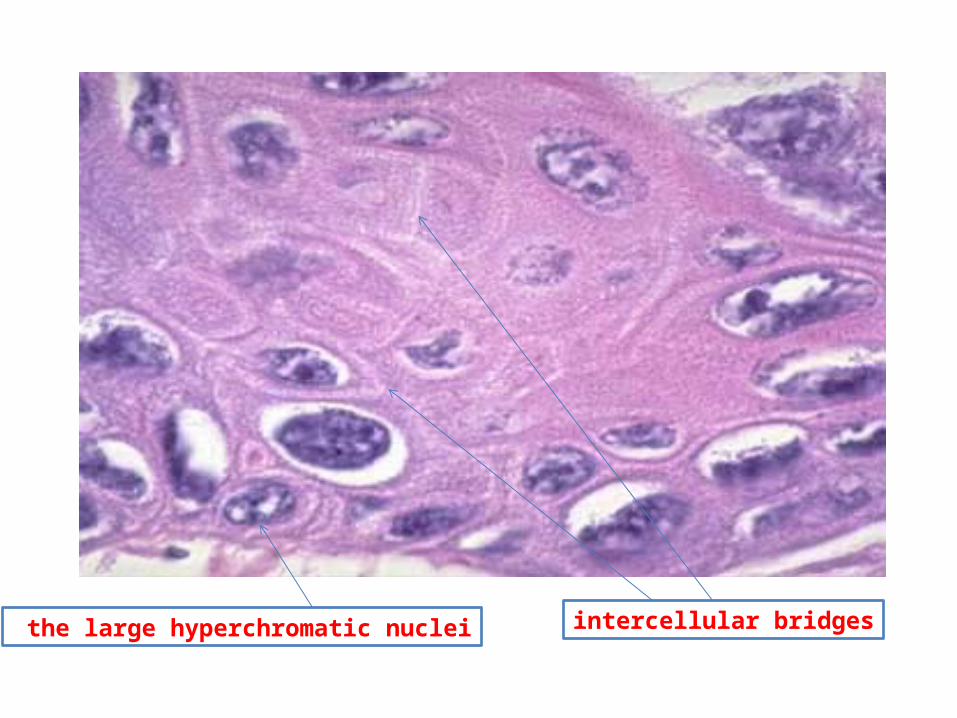
intercellular bridgesthe large hyperchromatic nuclei

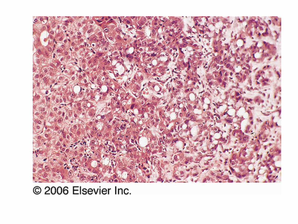
Lung, Metastatic Carcinomaالـرئة ســـــرطان

The lung tissue has been replaced by large sheets of malignant cellsThe sheets is formed of pleomorphic cells with hyperchromatic nucleiThere is also extensive necrosisThe characteristic architecture of the residual normal lung can be seen.

Lipoma
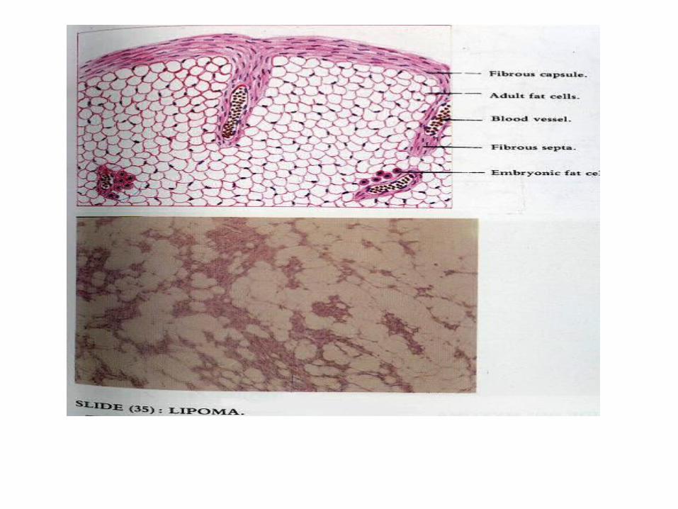

mature white fat cells The nucleus

Schwannoma
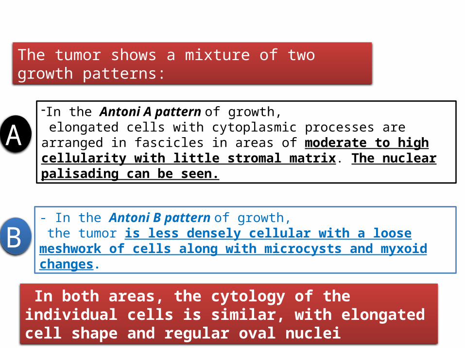
The tumor shows a mixture of two growth patterns:
-In the Antoni A pattern of growth, elongated cells with cytoplasmic processes are arranged in fascicles in areas of moderate to high cellularity with little stromal matrix. The nuclear palisading can be seen.
- In the Antoni B pattern of growth, the tumor is less densely cellular with a loose meshwork of cells along with microcysts and myxoid changes.
In both areas, the cytology of the individual cells is similar, with elongated cell shape and regular oval nuclei
A
B
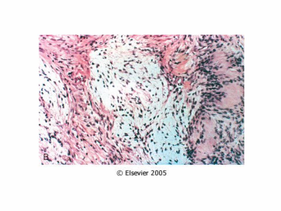

Black arrow: Antoni A ; blue arrow: Antoni B
External picture
Bone, osteosarcomaســـــرطان العـــظـــم

Spicules of tumor bonemalignant osteoblasts that have produced spicules of bone surrounded by osteoid matrix (( characteristic of osteosarcomas))
External information

Mitosis

eosinophilic osteoid

Uterus, leiomyosarcomaالــــرحم ســـــرطان

high cellularity

High (N/C ratio) enlarged nuclei

Heart, acute myocardial infarctionالقلب عضلة احتشاء

coagulative necrosis(SCAR )

neutrophils

Coagulative necrosis
Granulation tissue
Myocardial infarct, oldعضلة احتشاء
السن , كبار القلب
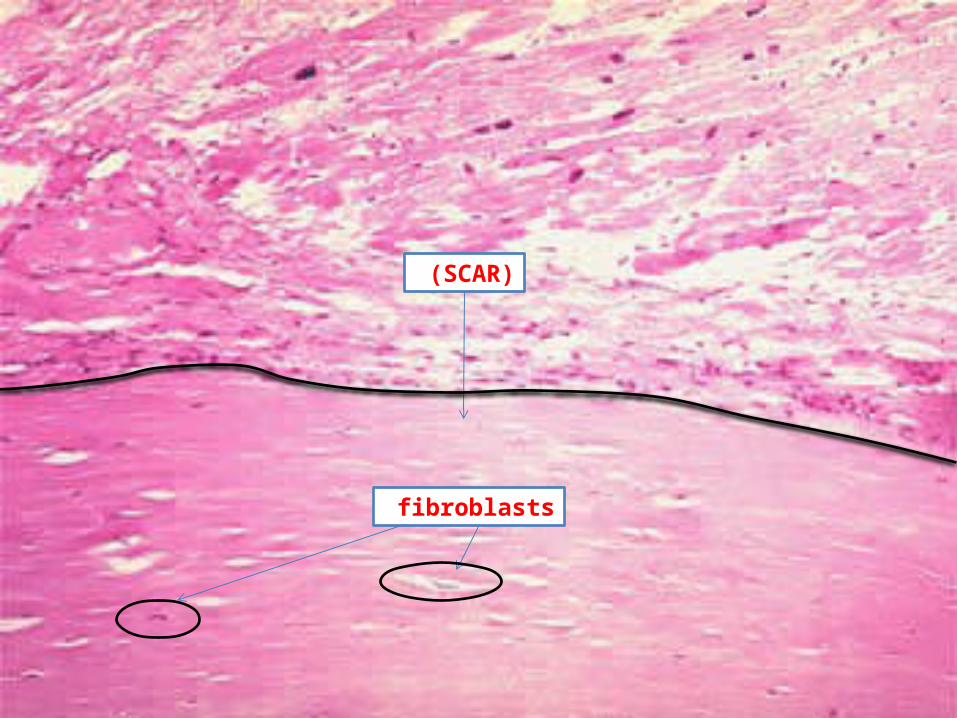
(SCAR )
fibroblasts

Pulmonary edemaالرئة تورم

Congested alveolar septa
Intra-alveolar transudate


الله بــحمد تـــمبالتوفيـــق للجميع الرجاء كل مــــع
Related Documents