INFECTION AND IMMUNITY, Apr. 2010, p. 1437–1446 Vol. 78, No. 4 0019-9567/10/$12.00 doi:10.1128/IAI.00621-09 Copyright © 2010, American Society for Microbiology. All Rights Reserved. Pathogenicity Islands PAPI-1 and PAPI-2 Contribute Individually and Synergistically to the Virulence of Pseudomonas aeruginosa Strain PA14 † Ewan M. Harrison, 1 ‡ Melissa E. K. Carter, 1 ‡ Shelley Luck, 3 Hong-Yu Ou, 4 Xinyi He, 4 Zixin Deng, 4 Chris O’Callaghan, 1,5 Aras Kadioglu, 1 *§ and Kumar Rajakumar 1,2 *§ Department of Infection, Immunity, and Inflammation, University of Leicester, Leicester LE1 9HN, 1 Department of Clinical Microbiology, University Hospitals of Leicester NHS Trust, Leicester LE1 5WW, 2 and Division of Child Health, University of Leicester, Robert Kilpatrick Clinical Sciences Building, Leicester Royal Infirmary, Leicester LE2 7LX, 5 United Kingdom; Department of Microbiology, Monash University, Melbourne, Australia 3 ; and Laboratory of Microbial Metabolism and School of Life Sciences and Biotechnology, Shanghai Jiaotong University, Shanghai 200240, People’s Republic of China 4 Received 1 June 2009/Returned for modification 7 July 2009/Accepted 20 January 2010 Pseudomonas aeruginosa is a leading cause of hospital-acquired pneumonia and severe chronic lung infections in cystic fibrosis patients. The reference strains PA14 and PAO1 have been studied extensively, revealing that PA14 is more virulent than PAO1 in diverse infection models. Among other factors, this may be due to two pathogenicity islands, PAPI-1 and PAPI-2, both present in PA14 but not in PAO1. We compared the global contributions to virulence of PAPI-1 and PAPI-2, rather than that of individual island-borne genes, using murine models of acute pneumonia and bacteremia. Three isogenic island-minus mutants (PAPI-1-minus, PAPI-2-minus, and PAPI-1- minus, PAPI-2-minus mutants) were compared with the wild-type parent strain PA14 and with PAO1. Our results showed that both islands contributed significantly to the virulence of PA14 in acute pneumonia and bacteremia models. However, in contrast to the results for the bacteremia model, where each island was found to contribute individually, loss of the 108-kb PAPI-1 island alone was insufficient to measurably attenuate the mutant in the acute pneumonia model. Nevertheless, the double mutant was substantially more attenuated, and exhibited a lesser degree of virulence, than even PAO1 in the acute pneumonia model. In particular, its ability to disseminate from the lungs to the bloodstream was markedly inhibited. We conclude that both PAPI-1 and PAPI-2 contribute directly and synergistically in a major way to the virulence of PA14, and we suggest that analysis of island-minus strains may be a more appropriate way than individual gene knockouts to assess the contributions to virulence of large, horizontally acquired segments of DNA. Pseudomonas aeruginosa is an environmentally ubiquitous opportunistic pathogen that causes a wide range of acute life- threatening infections, especially in immunocompromised pa- tients. Hospitalized patients with damaged airways due to me- chanical ventilation, injury, or viral infections are at significant risk of acute pneumonia caused by P. aeruginosa (51). Result- ing cases of ventilator-associated pneumonia are associated with very high rates of mortality (12). In addition, this bacte- rium is a common cause of chronic respiratory infection in patients with cystic fibrosis (CF), chronic obstructive pulmo- nary disease (COPD), and non-CF bronchiectasis (43, 50). A large variety of extracellular virulence factors, including pro- teases, hemolysins, pyocyanin, pili, lipopolysaccharide, algi- nate, and the type III secretion system (T3SS) effector pro- teins, ExoS, ExoT, ExoU, and ExoY, are associated with P. aeruginosa (52, 62). P. aeruginosa PA14 is a fully sequenced, highly virulent wild- type (WT) reference strain that has been used extensively to study the contribution of putative virulence factors to disease (31). Furthermore, as defined through study of a large panel of strains representative of this species, PA14 was found to be- long to the most common P. aeruginosa global lineage (61). Another fully sequenced strain, PAO1, is widely regarded as a laboratory strain of P. aeruginosa. PA14 has been shown to be much more virulent than PAO1 in a number of diverse models of infection, leading to the hypothesis that PA14 is a multihost pathogen capable of infecting invertebrate and vertebrate an- imal species and plant species (29). The genomes of individual P. aeruginosa strains have a highly mosaic structure made up of conserved cores and variable accessory genomes (36, 61), re- sulting in composite genomes of 5.2 to 7 Mb (36). The majority of large-scale genomic differences arise from the presence or absence of an expanding list of genomic islands within con- served “hot spots” throughout the genome (36). PA14 carries two well-characterized pathogenicity islands: PAPI-1, a 108-kb island integrated within a lysine tRNA gene (PA4541.1), and PAPI-2, a much smaller, 11-kb island inserted into a sequence- identical but distinct tRNA Lys gene (PA0976.1) (19). In addi- * Corresponding author. Mailing address: Department of Infection, Immunity, and Inflammation, Maurice Shock Building, University of Leicester, Leicester LE1 9HN, United Kingdom. Fax: 44-116-252- 5030. Phone for Aras Kadioglu: 44-116-252-2947. E-mail: [email protected] .uk. Phone for Kumar Rajakumar: 44-116-223-1498. E-mail: kr46@le .ac.uk. † Supplemental material for this article may be found at http://iai .asm.org/. ‡ E.M.H. and M.E.K.C. contributed equally to this work. § A.K. and K.R. are both senior authors. Published ahead of print on 1 February 2010. 1437 on April 6, 2020 by guest http://iai.asm.org/ Downloaded from

Welcome message from author
This document is posted to help you gain knowledge. Please leave a comment to let me know what you think about it! Share it to your friends and learn new things together.
Transcript

INFECTION AND IMMUNITY, Apr. 2010, p. 1437–1446 Vol. 78, No. 40019-9567/10/$12.00 doi:10.1128/IAI.00621-09Copyright © 2010, American Society for Microbiology. All Rights Reserved.
Pathogenicity Islands PAPI-1 and PAPI-2 ContributeIndividually and Synergistically to the Virulence of
Pseudomonas aeruginosa Strain PA14�†Ewan M. Harrison,1‡ Melissa E. K. Carter,1‡ Shelley Luck,3 Hong-Yu Ou,4 Xinyi He,4 Zixin Deng,4
Chris O’Callaghan,1,5 Aras Kadioglu,1*§ and Kumar Rajakumar1,2*§Department of Infection, Immunity, and Inflammation, University of Leicester, Leicester LE1 9HN,1 Department of Clinical Microbiology,
University Hospitals of Leicester NHS Trust, Leicester LE1 5WW,2 and Division of Child Health, University of Leicester,Robert Kilpatrick Clinical Sciences Building, Leicester Royal Infirmary, Leicester LE2 7LX,5 United Kingdom;
Department of Microbiology, Monash University, Melbourne, Australia3; and Laboratory ofMicrobial Metabolism and School of Life Sciences and Biotechnology,
Shanghai Jiaotong University, Shanghai 200240,People’s Republic of China4
Received 1 June 2009/Returned for modification 7 July 2009/Accepted 20 January 2010
Pseudomonas aeruginosa is a leading cause of hospital-acquired pneumonia and severe chronic lung infections incystic fibrosis patients. The reference strains PA14 and PAO1 have been studied extensively, revealing that PA14 ismore virulent than PAO1 in diverse infection models. Among other factors, this may be due to two pathogenicityislands, PAPI-1 and PAPI-2, both present in PA14 but not in PAO1. We compared the global contributions tovirulence of PAPI-1 and PAPI-2, rather than that of individual island-borne genes, using murine models of acutepneumonia and bacteremia. Three isogenic island-minus mutants (PAPI-1-minus, PAPI-2-minus, and PAPI-1-minus, PAPI-2-minus mutants) were compared with the wild-type parent strain PA14 and with PAO1. Our resultsshowed that both islands contributed significantly to the virulence of PA14 in acute pneumonia and bacteremiamodels. However, in contrast to the results for the bacteremia model, where each island was found to contributeindividually, loss of the 108-kb PAPI-1 island alone was insufficient to measurably attenuate the mutant in the acutepneumonia model. Nevertheless, the double mutant was substantially more attenuated, and exhibited a lesserdegree of virulence, than even PAO1 in the acute pneumonia model. In particular, its ability to disseminate fromthe lungs to the bloodstream was markedly inhibited. We conclude that both PAPI-1 and PAPI-2 contribute directlyand synergistically in a major way to the virulence of PA14, and we suggest that analysis of island-minus strains maybe a more appropriate way than individual gene knockouts to assess the contributions to virulence of large,horizontally acquired segments of DNA.
Pseudomonas aeruginosa is an environmentally ubiquitousopportunistic pathogen that causes a wide range of acute life-threatening infections, especially in immunocompromised pa-tients. Hospitalized patients with damaged airways due to me-chanical ventilation, injury, or viral infections are at significantrisk of acute pneumonia caused by P. aeruginosa (51). Result-ing cases of ventilator-associated pneumonia are associatedwith very high rates of mortality (12). In addition, this bacte-rium is a common cause of chronic respiratory infection inpatients with cystic fibrosis (CF), chronic obstructive pulmo-nary disease (COPD), and non-CF bronchiectasis (43, 50). Alarge variety of extracellular virulence factors, including pro-teases, hemolysins, pyocyanin, pili, lipopolysaccharide, algi-nate, and the type III secretion system (T3SS) effector pro-
teins, ExoS, ExoT, ExoU, and ExoY, are associated with P.aeruginosa (52, 62).
P. aeruginosa PA14 is a fully sequenced, highly virulent wild-type (WT) reference strain that has been used extensively tostudy the contribution of putative virulence factors to disease(31). Furthermore, as defined through study of a large panel ofstrains representative of this species, PA14 was found to be-long to the most common P. aeruginosa global lineage (61).Another fully sequenced strain, PAO1, is widely regarded as alaboratory strain of P. aeruginosa. PA14 has been shown to bemuch more virulent than PAO1 in a number of diverse modelsof infection, leading to the hypothesis that PA14 is a multihostpathogen capable of infecting invertebrate and vertebrate an-imal species and plant species (29). The genomes of individualP. aeruginosa strains have a highly mosaic structure made up ofconserved cores and variable accessory genomes (36, 61), re-sulting in composite genomes of 5.2 to 7 Mb (36). The majorityof large-scale genomic differences arise from the presence orabsence of an expanding list of genomic islands within con-served “hot spots” throughout the genome (36). PA14 carriestwo well-characterized pathogenicity islands: PAPI-1, a 108-kbisland integrated within a lysine tRNA gene (PA4541.1), andPAPI-2, a much smaller, 11-kb island inserted into a sequence-identical but distinct tRNALys gene (PA0976.1) (19). In addi-
* Corresponding author. Mailing address: Department of Infection,Immunity, and Inflammation, Maurice Shock Building, University ofLeicester, Leicester LE1 9HN, United Kingdom. Fax: 44-116-252-5030. Phone for Aras Kadioglu: 44-116-252-2947. E-mail: [email protected]. Phone for Kumar Rajakumar: 44-116-223-1498. E-mail: [email protected].
† Supplemental material for this article may be found at http://iai.asm.org/.
‡ E.M.H. and M.E.K.C. contributed equally to this work.§ A.K. and K.R. are both senior authors.� Published ahead of print on 1 February 2010.
1437
on April 6, 2020 by guest
http://iai.asm.org/
Dow
nloaded from
tion to targeting integration sites bearing identical sequences,these two islands share a limited but recognizable degree ofsynteny and code for factors that have been implicated inpathogenesis in murine, plant, and nematode models of infec-tion (19, 31, 37, 52). Interestingly, islands with significant ho-mology to PAPI-1 and/or PAPI-2 have been identified in manyP. aeruginosa strains and several other bacterial species (5, 6,19, 23, 27, 30, 61, 63) (see Tables S2 and S3 and Fig. S2 in thesupplemental material).
PAPI-1 is a member of the increasingly populated pKLC102/PAGI-2 island family (26). PAPI-1 encodes a number of likelyvirulence factors, including type IVB pili, the cupD1 to cupD5fimbrial biogenesis gene cluster (39), and PvrR, a two-compo-nent response regulator involved in biofilm synthesis and an-tibiotic resistance (11, 38). However, the large majority ofPAPI-1 predicted proteins have no assigned function, andmany of these are encoded by homologues on related islands inother P. aeruginosa strains (27, 63). PAGI-5, a hybrid islandthat shares 79 of 121 predicted open reading frames (ORFs)with PAPI-1, has recently been implicated in acute murinepneumonia (5). Isogenic mutants harboring deletions withinNR-I or NR-II, PAGI-5-specific novel regions, were shown tobe attenuated in a model of acute murine pneumonia, dem-onstrating that an island in the pKLC102/PAGI-2 familyplayed a role in this infection.
PAPI-2 is a stably integrated island belonging to the ExoUisland family (30). This island encodes the cytotoxin ExoU, apotent phospholipase, and its cognate chaperone SpcU. ExoUhas been studied extensively and has been shown to be themost potent of the known T3SS effector proteins in P. aerugi-nosa (54). The introduction of exoU into strains that lacked thisgene resulted in marked increases in virulence in a murineacute pneumonia model (1). Recent work has suggested thatExoU impairs host response in the lung through targeted kill-ing of phagocytes, which play a crucial role during the earlyphase of infection (10). Additionally, the secretion of ExoU byan infecting strain has been shown to be a marker of highvirulence and poor clinical outcome in hospital-acquired pneu-monia (52).
Since genomic islands are widely believed to be acquiredand/or lost as whole units, and since PAPI-1-like islands areknown to be widespread and possibly highly mobile (46), weinvestigated the contributions of PAPI-1 and PAPI-2 to viru-lence as single unitary modules rather than defining the rolesof individual island-borne genes (5, 19). We postulated thatthis en bloc deletion approach would reveal combinatorialand/or synergistic effects that would be missed by single-geneknockout approaches. To pursue this goal, we created threeisogenic whole-island deletion mutants that lacked eitherPAPI-1 (PA14�PAPI-1), PAPI-2 (PA14�PAPI-2), or both(PA14�1�2) in a PA14 background and examined these rela-tive to their wild-type parent and the laboratory strain PAO1,which naturally lacks both islands. Our results revealed thatboth PAPI-1 and PAPI-2 contributed individually and syner-gistically to the virulence of P. aeruginosa strain PA14 in mu-rine models of acute pneumonia and bacteremia.
MATERIALS AND METHODS
Bacterial strains, plasmids, and growth media. The bacterial strains andplasmids used in this study are described in Table 1. P. aeruginosa and Esche-
richia coli strains were grown routinely at 37°C on LB medium plus 100 �g/mlampicillin, 30 �g/ml kanamycin, and/or 15 �g/ml gentamicin for E. coli and 200�g/ml carbenicillin and/or 30 �g/ml gentamicin for P. aeruginosa, as required.The minimal medium used for conjugative transfer was VBMM (57), supple-mented when appropriate with antibiotics and sucrose at 5% (wt/vol).
DNA manipulations. Restriction enzymes and T4 DNA ligase were purchasedfrom Promega. Genomic DNA and plasmid DNA were extracted by followingthe manufacturer’s instructions using the DNeasy blood and tissue kit and theQIAspin miniprep kit (Qiagen, United Kingdom). Standard PCR amplificationwas carried out using GoTaq (Promega, United Kingdom), while KOD Hotstart(Merck, United Kingdom) was used for high-fidelity amplification. DNA se-quencing was performed by MWG Biotech.
Construction of defined PA14 mutants harboring deletions of PAPI-1 and/orPAPI-2. PAPI-1 was deleted from PA14 using an “island-probing” approach(47). Qiu et al. (46) had reported that mutation of the PAPI-1-encoded soj geneincreased the instability of the island. A 498-bp internal fragment of soj wasamplified by PCR using primers SOJ-F and SOJ-R (see Table S1 in the supple-mental material), cloned into pCR4-TOPO (Invitrogen, United Kingdom), ex-cised as a fractionally larger EcoRI fragment, and ligated into the EcoRI site ofpEX18Ap (21) to create pEX�SOJ. This plasmid was conjugally transferredfrom E. coli SM10 �-pir into PA14 (21), and a single-crossover plasmid integrantwas identified by the resistance phenotype and PCR. The �PAPI-1 mutant wasgenerated from this intermediate strain by counterselection on LB agar mediumwith 5% sucrose to select for the loss of the sacB-tagged PAPI-1 island. Loss ofPAPI-1 was confirmed by reversion to carbenicillin sensitivity, the absence of fivePAPI-1-specific regions by PCR using primers PP-1-1 to PP-1-5 (see Table S1 inthe supplemental material), and sequencing across the newly vacated attB site.
PAPI-2 was deleted by following the method described by Choi and Schweizer(9). Briefly, upstream and downstream flanking regions of PAPI-2 were amplifiedby PCR using the primer pairs P2UFFW-Gm–P2UFRS-GWR and P2DFFW-GWL–P2DFRS-Gm, respectively (see Table S1 in the supplemental material).These fragments were joined to a FLP recombination target (FRT)-flankedgentamicin resistance (Gmr) cassette amplified from pPS856 (9) using splicingoverlap extension (SOE) PCR. The composite fragment was transferred viapDONR221 into the pEX18ApGW suicide vector using the Gateway site-specificrecombination system (Invitrogen, United Kingdom). The resulting plasmid,pEX�PAPI-2, was then transferred by conjugation into PA14 (21). A merodip-loid intermediate strain was resolved by sucrose counterselection, and the Gmr
cassette was removed using FLP-mediated excision (9) to generate a marker-freePAPI-2 deletant. The PAPI-2 deletion was confirmed by reversion to gentamicinsensitivity, loss of the exoU gene, and sequencing across the deletion site. Adouble PAPI-1–PAPI-2 deletion mutant was constructed by deleting PAPI-1, asdescribed above, from the PA14�PAPI-2 mutant. The �exoU mutants wereconstructed like the �PAPI-2 mutant, with the modification that the SOE PCRfragment was cloned directly into pEX18Ap by inclusion of KpnI restriction sitesin primers GW-KPN1-F and GW-KPN1-R, used for amplification of the SOEfragment.
Growth, biofilm, motility, and pyocyanin assays. Strains were grown for 16 hat 37°C at 200 rpm in LB broth and were subcultured 1/100 into LB broth; M63medium with glucose (0.2%), MgSO4 (1 mM), and Casamino Acids (0. 5%); andM9 medium with glucose (0.2%), MgSO4 (1 mM), and CaCl3 (1 mM). Twohundred microliters of each was placed in a Honeywell plate, and the opticaldensity (OD) was measured using a Bioscreen apparatus (Life Sciences, UnitedKingdom) at 37°C with continuous shaking for 24 or 48 h. The sizes of the initialinocula were checked by serial dilutions and plating for CFU. Biofilm assays wereperformed using the microtiter plate-based biofilm assay described by O’Tooleand Kolter (45). Twitching motility was evaluated at the agar-plate interface after24 h at 37°C on LB medium with 1.5% agar (48). Swimming motility wasmeasured after 20 h at 30°C on LB medium with 0.3% agar. Pyocyanin produc-tion was measured at 520 nm in a chloroform acidic-extraction solution using amethod described previously (20).
Murine acute pneumonia model. The murine acute pneumonia model usedwas adapted from previous work with pneumococci (24) and with P. aeruginosa(54). Six hundred microliters of a 37°C shaken P. aeruginosa culture was removedand centrifuged at 13,000 rpm for 1 min. The pellet was added to 20 ml oftryptone soy broth supplemented with 20% fetal calf serum (FCS) (Sigma,United Kingdom). The culture was then incubated at 37°C and 200 rpm for 4 hbefore being stored at �70°C as 1-ml aliquots. Stock cultures were left at �70°Cfor at least 3 days. Eight- to 10-week-old female BALB/c mice (Harlan Olac,Bicester, United Kingdom) were anesthetized with 5% (vol/vol) isoflurane overoxygen at 1.8 liter/min. Mice were intranasally challenged with 2 � 106 CFU ofP. aeruginosa and were then allowed to recover. Mouse survival, disease signs,and levels of bacteria in the lungs, nasopharynx, and/or blood were measured.
1438 HARRISON ET AL. INFECT. IMMUN.
on April 6, 2020 by guest
http://iai.asm.org/
Dow
nloaded from
Disease severity was monitored at each time point by one of four trained indi-viduals, two of whom were blinded, using the scheme of Morton and Griffiths(42), which scored the following clinical signs: hunching, a starry coat, andlethargy. Mice were scored as follows: 0, no sign detectable; 1, sign just evident;2, sign obviously present. The individual scores for the three signs were combinedto give a maximum clinical sign score of 6. Mice with a score of 6 were culledimmediately, because this was the experimental endpoint defined by our HomeOffice project license. The remaining mice were culled at 18 h postinfection, andsamples were taken for analysis. Blood was taken via cardiac puncture underterminal anesthesia, and the nasopharynx and lungs were removed by dissection,weighed, and homogenized in phosphate-buffered saline (PBS) using an Ultra-Turrax tissue homogenizer. Organ homogenates were serially diluted and cul-tured on LB agar for the determination of CFU counts. Ten mice were used perstrain. For survival experiments, mice were culled when they reached a score of2 for lethargy. Ten mice were used for each strain, and survival was monitored for96 h.
Murine bacteremia model. Mice were inoculated via their dorsal tail veins with2 � 106 CFU. Blood was collected by tail-bleeding at 2 h and 6 h, and finally viacardiac puncture at 24 h postinfection. Five mice were infected per strain.
Differential leukocyte counts and lung histopathology. Trypan blue was addedto lung homogenates at a 1:1 volume ratio. Mouse leukocyte viability followinghomogenization and purification of cells had previously been shown to be above90%, as determined by trypan blue exclusion (24; A. Kadioglu, unpublisheddata). Five milliliters of stained lung homogenate was filtered through 40-�m-pore size nylon Falcon cell strainers (Becton Dickinson [BD], United Kingdom)into a 50-ml Falcon tube. The homogenate was centrifuged at 300 � g for 5 min.The supernatant was discarded, and the pellet was resuspended in 10 ml BDPharm Lyse lysing buffer (BD Biosciences, United Kingdom) diluted 1:10 withnanopure water and was left for 10 min at room temperature in the dark. Thesuspension was recentrifuged as before, and the cells were resuspended in 5 mlPBS. Three cytospin slides were prepared per individual mouse. Fifty microlitersof the final cell suspension was centrifuged onto a cytospin slide (Shandon,United Kingdom) using a Cytospin 2 centrifuge (Shandon, United Kingdom) at1,500 rpm for 3 min. The slides were allowed to dry overnight. The slides were
stained using the Reastain Quick-Diff kit (Reagena, Finland). Slides werecounted using the 40� objective lens on a light microscope to differentiallyidentify monocytes, macrophages, lymphocytes, and polymorphonuclear leuko-cytes. Cytospin slides prepared from four mice per P. aeruginosa strain investi-gated were analyzed. For histopathology, lungs were removed, placed in 5 mlphosphate-buffered saline, fixed in OCT tissue embedding compound using dryice and isopentane as a heat buffer, and then stored at �70°C. The day beforesectioning, lungs were moved to �20°C. Lungs were cut into 15-�m-thick sec-tions at �18°C using a Bright microtome and were subsequently stained usinghematoxylin and eosin; photographs were taken at a magnification of �10. Twolungs were examined per strain.
Galleria mellonella killing assay. The Galleria mellonella assay used was basedon assays described by Miyata et al. and Inglis et al. (22, 41). Test strains weregrown overnight in 5 ml LB broth at 37°C with shaking at 200 rpm. The next day,a 1/100 subculture grown under identical conditions for 4 h was centrifuged at3,000 rpm for 10 min; the supernatant was removed; and the cell pellet wasresuspended in 10 mM MgSO4 to an OD at 600 nm (OD600) of 0.2. Bacterialsuspensions were then serially diluted to 10�5 using 10 mM MgSO4 supple-mented with 0.5 mg/ml rifampin (Sigma, United Kingdom) as the diluent. G.mellonella larvae were inoculated between two rear thoracic segments with 10 �lof the 10�5 suspension containing �10 CFU of P. aeruginosa by using a 0.3-mlMyjector U-100 insulin syringe (VWR, United Kingdom). Ten larvae wereinoculated with each bacterial strain, and three biological replicates originatingfrom independent bacterial colonies were performed for each strain, resulting inan overall number of 30. Larvae were incubated at 37°C and were scored fordeath every half hour after 14 h. Larvae were scored dead when caterpillars didnot respond to direct mechanical stimulation applied to the head.
Invasion assay. The invasion assay was based on that described by Luck et al.(34) with modifications for P. aeruginosa. Quantitative assessment of bacterialinvasion was performed using A549 human lung carcinoma cells. Cell monolay-ers were obtained by seeding 500 �l of a solution consisting of 1 � 105/ml A549cells resuspended in RPMI 1640 plus glutamine supplemented with 10% FCSand 100 �g/ml penicillin-streptomycin in a 24-well tissue culture tray and incu-bating overnight at 37°C under 5% CO2. Semiconfluent cell monolayers were
TABLE 1. Bacterial strains and plasmids used in this study
Strain or plasmid Genotype or relevant characteristics Source orreference
StrainsP. aeruginosa
PA14 Wild-type, hypervirulent strain from burn infection 31PA14�PAPI-1 PA14 with PAPI-1 deleted This studyPA14�PAPI-2 PA14 with PAPI-2 deleted This studyPA14�1�2 PA14 with both PAPI-1 and PAPI-2 deleted This studyPA14�exoU PA14 with exoU deleted This studyPA14�PAPI-1�exoU PA14 with PAPI-1 and exoU deleted This studyPAO1 Laboratory strain, originally from wound infection 56
E. coliSM10 �-pir Kmr (thi-1 thr leu tonA lacY supE recA::RP4-2Tc::Mu) 55DH5� F� �80dlacZ�M15 �(lacZYA-argF)U169 deoR recA1 endA1 hsdR17(rK
� mK) phoA supE44 ��
thi-1 gyrA96 relA117
One Shot T1-R F proAB lacIq lacZ�M15 Tn10(Tetr) �(ccdAB) mcrA �(mrr hsdRMS-mcrBC) �80(lacZ)�M15�(lacZYA-argF)U169 endA1 recA1 supE44 thi-1 gyrA96 relA1 tonA panD
Invitrogen
PlasmidspFLP2 FLP recombinase-expressing plasmid; Ampr 21pPS856 Source of Gmr cassette; Gmr 21pEX18Ap Suicide vector; Ampr 53pUCP24 E. coli–P. aeruginosa shuttle vector; Gmr 60pCR4-Topo Topo PCR cloning vector; Ampr Kmr; ccdB gene InvitrogenpEX18ApGW Gateway suicide destination vector; Ampr 9pDONR221 Gateway cloning entry vector; Kmr Cmr attP1 attP2; ccdB gene InvitrogenpDONR221-�PAPI-2 Gateway entry vector with SOE fragment for deletion of PAPI-2; Kmr Gmr This studypEX�PAPI-2 pEX18ApGW with SOE fragment for deletion of PAPI-2; Ampr Gmr This studypCR4-�SOJ pCR4-Topo with PCR-generated 49-bp fragment of soj; Kmr This studypEX�ExoU Suicide vector with SOE fragment for deletion of exoU; Ampr Gmr This studypEX�SOJ Suicide vector for PAPI-1 deletion containing 498-bp insert into EcoRI; Ampr This study
VOL. 78, 2010 PAPI-1 AND PAPI-2 IN P. AERUGINOSA PA14 VIRULENCE 1439
on April 6, 2020 by guest
http://iai.asm.org/
Dow
nloaded from
washed three times in warm PBS, and fresh medium was added. A549 cellswere inoculated with approximately 3 � 106 total bacteria per well, and trayswere centrifuged at room temperature prior to incubation for 2 h at 37°C under5% CO2. To eliminate extracellular bacteria, A549 cells were washed three timeswith PBS, and fresh medium containing 400 �g/ml amikacin was added, beforea final 1 h of incubation at 37°C under 5% CO2. Finally, A549 cells were washedthree times with PBS and lysed in 0.1% digitonin. Following cell lysis, bacteriawere resuspended in LB broth and quantified by plating serial dilutions. Assayswere carried out in duplicate, and the results from at least three independentexperiments were expressed as the percentage of the total number of inoculatedbacteria that was intracellular (mean � standard deviation). PA14 and its threeisogenic mutants were shown to be fully susceptible to 200 �g/ml amikacin whenincubated in the absence of A549 cells under conditions identical to those usedin the invasion assay.
Cytotoxicity assay. The cytotoxicity assay was based on that described by Leeet al. (32). A549 cells and P. aeruginosa were grown, prepared, and/or inoculatedas described for the invasion assay. After 2 h of incubation, cytotoxicity towardA549 cells was quantified by measuring the amount of lactate dehydrogenase(LDH) released into the culture supernatant by using the CytoTox-ONE kit(Promega, United Kingdom). Assays were carried out in duplicate with threeindependent biological replicates for each strain. The percentage of cytotoxicitywas calculated relative to the value for the maximum-LDH-release control (A549cells lysed by the addition of a 9% [wt/vol] solution of Triton X-100).
Statistics. Statistical analysis was performed with GraphPad Prism software(version 5; GraphPad Software, Inc.) and SPSS software. CFU and differentialleukocyte counts were analyzed, and cytotoxicity assays were performed, byanalysis of variance (ANOVA) with Tukey posthoc analysis. Symptom scoreswere analyzed using the nonparametric Kruskal-Wallis test and Dunn’s posthocanalysis. Survival data were analyzed using the Mantel-Cox log rank test. For the
invasion assay, differences in invasion were assessed for significance by using anunpaired, two-tailed t test.
RESULTS
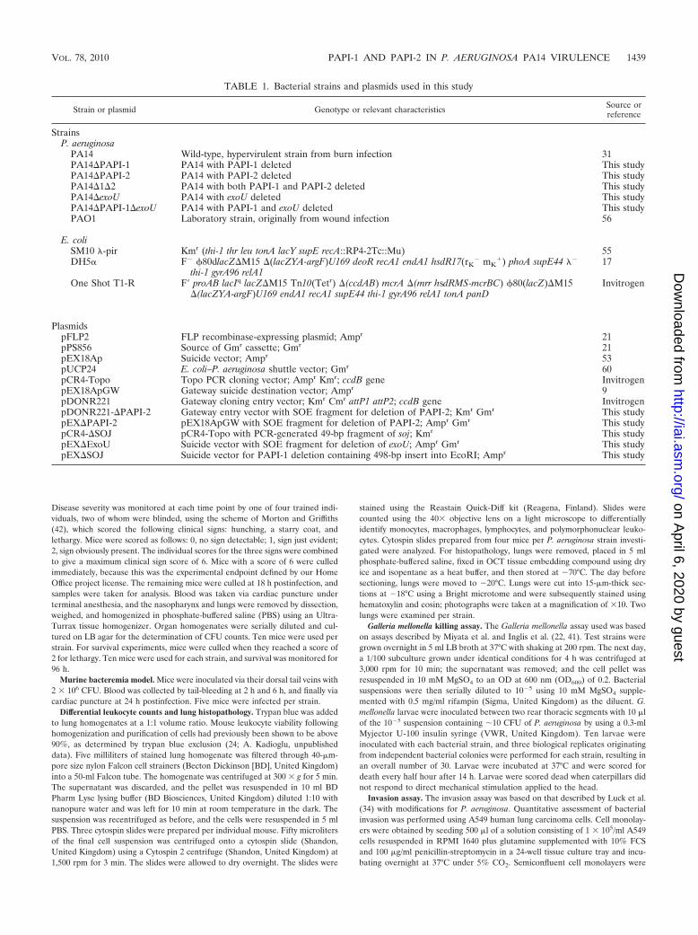
PA14 is more virulent than PAO1 in a murine model ofacute pneumonia. As had been reported previously for a widerange of P. aeruginosa infection models (29), PA14 was foundto be more virulent than PAO1 in a mouse model of acutepneumonia. Following intranasal infection with equivalent in-ocula (2 � 106 CFU), PA14-infected mice had to be culled dueto the onset of fulminant symptoms at a significantly earliertime point (18 h) than mice infected with PAO1 (27 h) (P �0.02) (Fig. 1A). Furthermore, PA14 was found in much highernumbers than PAO1 in nasopharynx, lung, and blood speci-mens obtained at 18 h postinfection (Fig. 2). Consistent withthese data, mean qualitative clinical sign scores at 18 h follow-ing infection with PA14 and PAO1 were 5.7 (range, 5 to 6) and4 (range, 2 to 5), respectively; higher scores denoted greaterseverity of disease (Fig. 1B).
Loss of PAPI-1 and/or PAPI-2 caused no change in in vitrofitness, motility, biofilm formation, or pyocyanin production.We constructed three island-minus isogenic mutants of PA14that lacked either PAPI-1 or PAPI-2, or both PAPI-1 and
FIG. 1. (A) Survival graph for the acute murine pneumonia model. Mice were monitored for 96 h. Ten mice were used for each strain. Datawere generated from two independent experiments for each strain, except for the �exoU and �PAPI-1�exoU strains, for which data were generatedfrom a single experiment only. Horizontal lines represent the percentage of mice surviving postinfection for each strain. (B) Acute murinepneumonia symptom scores at 18 h postinfection. Ten mice were used for each strain. Each data point represents a single mouse. All data weregenerated from two independent experiments for each strain. The asterisk indicates a significant difference from wild-type PA14 (P � 0.05). Thebars represent mean symptom scores � standard deviations.
FIG. 2. P. aeruginosa bacterial burdens in the nasopharynx, lungs, and blood at 18 h postinfection in the acute murine pneumonia model. Tenmice were used for each strain. All data were generated from two independent experiments for each strain. Asterisks and number symbols indicatesignificant differences (P � 0.05) from wild-type PA14 and PA14�PAPI-2, respectively. The error bars represent the standard errors of the means.
1440 HARRISON ET AL. INFECT. IMMUN.
on April 6, 2020 by guest
http://iai.asm.org/
Dow
nloaded from
PAPI-2 (PA14�PAPI-1, PA14�PAPI-2, and PA14�1�2), asdescribed in Materials and Methods. None of these mutantsshowed significant growth defects in either rich (LB) or mini-mal (M9 or M63) medium compared to the growth of WTPA14 (data not shown), suggesting that the deletions had nosignificant effect on growth kinetics in vitro. In the biofilmassays on two different surfaces (polyvinyl chloride [PVC] andpolystyrene), none of the mutants showed consistent differ-ences from wild-type PA14 (data not shown). Similarly, nosignificant difference in pyocyanin production was detected(data not shown). Furthermore, the three mutants showed nochange in swimming or twitching motility (data not shown),even though twitching motility is known to be dependent ontype IV pili, and PAPI-1 codes for type IV pilus chaperonegenes. Consistent with these findings, He et al. (19) reportedno significant difference in biofilm growth, motility, or pyocy-anin production in the 23 PAPI-1 and PAPI-2 single-genemutants screened in their study.
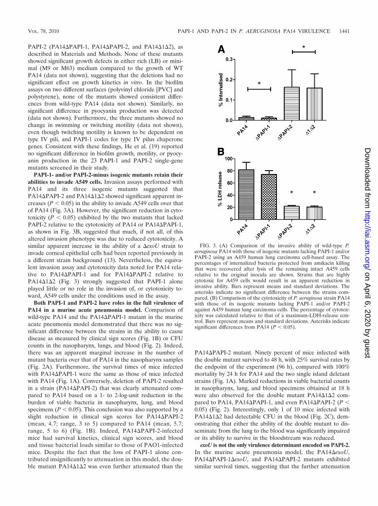
PAPI-1- and/or PAPI-2-minus isogenic mutants retain theirabilities to invade A549 cells. Invasion assays performed withPA14 and its three isogenic mutants suggested thatPA14�PAPI-2 and PA14�1�2 showed significant apparent in-creases (P � 0.05) in the ability to invade A549 cells over thatof PA14 (Fig. 3A). However, the significant reduction in cyto-toxicity (P � 0.05) exhibited by the two mutants that lackedPAPI-2 relative to the cytotoxicity of PA14 or PA14�PAPI-1,as shown in Fig. 3B, suggested that much, if not all, of thisaltered invasion phenotype was due to reduced cytotoxicity. Asimilar apparent increase in the ability of a �exoU strain toinvade corneal epithelial cells had been reported previously ina different strain background (13). Nevertheless, the equiva-lent invasion assay and cytotoxicity data noted for PA14 rela-tive to PA14�PAPI-1 and for PA14�PAPI-2 relative toPA14�1�2 (Fig. 3) strongly suggested that PAPI-1 aloneplayed little or no role in the invasion of, or cytotoxicity to-ward, A549 cells under the conditions used in the assay.
Both PAPI-1 and PAPI-2 have roles in the full virulence ofPA14 in a murine acute pneumonia model. Comparison ofwild-type PA14 and the PA14�PAPI-1 mutant in the murineacute pneumonia model demonstrated that there was no sig-nificant difference between the strains in the ability to causedisease as measured by clinical sign scores (Fig. 1B) or CFUcounts in the nasopharynx, lungs, and blood (Fig. 2). Indeed,there was an apparent marginal increase in the number ofmutant bacteria over that of PA14 in the nasopharynx samples(Fig. 2A). Furthermore, the survival times of mice infectedwith PA14�PAPI-1 were the same as those of mice infectedwith PA14 (Fig. 1A). Conversely, deletion of PAPI-2 resultedin a strain (PA14�PAPI-2) that was clearly attenuated com-pared to PA14 based on a 1- to 2-log-unit reduction in theburden of viable bacteria in nasopharynx, lung, and bloodspecimens (P � 0.05). This conclusion was also supported by aslight reduction in clinical sign scores for PA14�PAPI-2(mean, 4.7; range, 3 to 5) compared to PA14 (mean, 5.7;range, 5 to 6) (Fig. 1B). Indeed, PA14�PAPI-2-infectedmice had survival kinetics, clinical sign scores, and bloodand tissue bacterial loads similar to those of PAO1-infectedmice. Despite the fact that the loss of PAPI-1 alone con-tributed insignificantly to attenuation in this model, the dou-ble mutant PA14�1�2 was even further attenuated than the
PA14�PAPI-2 mutant. Ninety percent of mice infected withthe double mutant survived to 48 h, with 25% survival rates bythe endpoint of the experiment (96 h), compared with 100%mortality by 24 h for PA14 and the two single island deletantstrains (Fig. 1A). Marked reductions in viable bacterial countsin nasopharynx, lung, and blood specimens obtained at 18 hwere also observed for the double mutant PA14�1�2 com-pared to PA14, PA14�PAPI-1, and even PA14�PAPI-2 (P �0.05) (Fig. 2). Interestingly, only 1 of 10 mice infected withPA14�1�2 had detectable CFU in the blood (Fig. 2C), dem-onstrating that either the ability of the double mutant to dis-seminate from the lung to the blood was significantly impairedor its ability to survive in the bloodstream was reduced.
exoU is not the only virulence determinant encoded on PAPI-2.In the murine acute pneumonia model, the PA14�exoU,PA14�PAPI-1�exoU, and PA14�PAPI-2 mutants exhibitedsimilar survival times, suggesting that the further attenuation
FIG. 3. (A) Comparison of the invasive ability of wild-type P.aeruginosa PA14 with those of isogenic mutants lacking PAPI-1 and/orPAPI-2 using an A459 human lung carcinoma cell-based assay. Thepercentages of internalized bacteria protected from amikacin killingthat were recovered after lysis of the remaining intact A459 cellsrelative to the original inocula are shown. Strains that are highlycytotoxic for A459 cells would result in an apparent reduction ininvasive ability. Bars represent means and standard deviations. Theasterisks indicate no significant difference between the strains com-pared. (B) Comparison of the cytotoxicity of P. aeruginosa strain PA14with those of its isogenic mutants lacking PAPI-1 and/or PAPI-2against A459 human lung carcinoma cells. The percentage of cytotox-icity was calculated relative to that of a maximum-LDH-release con-trol. Bars represent means and standard deviations. Asterisks indicatesignificant differences from PA14 (P � 0.05).
VOL. 78, 2010 PAPI-1 AND PAPI-2 IN P. AERUGINOSA PA14 VIRULENCE 1441
on April 6, 2020 by guest
http://iai.asm.org/
Dow
nloaded from

observed with the double island-minus mutant, PA14�1�2,resulted from the loss of part or all of PAPI-2, and not justexoU, in combination with the deletion of PAPI-1 (Fig. 1A).Comparison of the Kaplan-Meier survival plots obtained usingthe G. mellonella model showed that PA14�PAPI-2 was sig-nificantly attenuated (P, �0.001 by the log rank test) comparedto PA14, while PA14�exoU was not statistically different fromits wild-type parent (P, 0.228) (Fig. 4). These findings supportthe existence of an additional, non-exoU virulence gene(s) onPAPI-2 that is unmasked following the deletion of PAPI-1.
Both PAPI-1 and PAPI-2 have a role to play in a murinemodel of bacteremia. The set of P. aeruginosa strains discussedabove was also studied using a murine bacteremia model ini-tiated through intravenous inoculation of 2 � 106 P. aeruginosaorganisms. After 6 h, all PA14-infected mice were moribundand had to be culled; hence, no data were available for PA14beyond this time point. However, mice infected with PAO1 orany of the three PA14 mutants survived until the endpoint of24 h (Fig. 5A), demonstrating that in a murine bacteremiamodel, both PAPI-1 and PAPI-2 make direct and individualcontributions to the full virulence of the wild-type parent,PA14. Surprisingly, though, at 6 h postinfection, CFU counts inthe blood of mice infected with PA14 were similar to those for
the other four strains investigated. Furthermore, PA14�PAPI-1-infected mice seemed to have slightly higher blood CFUcounts at 6 h and 24 h than mice infected with PAO1,PA14�PAPI-2, or PA14�1�2. Although not statistically sig-nificant, this result may imply reduced susceptibility ofPA14�PAPI-1 to clearance from the bloodstream (Fig. 5A).The clinical sign scores at 2 h postinfection (Fig. 5B) revealedno hint of disease, except for a solitary mouse in the PA14group that exhibited a score of 2. At 6 h, all 4 PA14-infectedmice were moribund (clinical sign scores, �5), while only 4other mice (all infected with PA14�PAPI-1) out of the 16 inthe four other arms of the experiment displayed even minimalsigns of disease, with a maximum clinical sign score of 1(Fig. 5C). These 6-h-time-point data clearly highlighted avery major difference in virulence phenotype in the murinebacteremia model between PA14 and the other four strainsexamined. However, although there was no or minimal ev-idence of disease at 6 h in mice infected with PAO1 or anyof the three PA14 mutants, by 24 h (Fig. 5C and D) all 16mice in these groups exhibited clinical sign scores of 5 or 6.Examination of the 6-h and 24-h clinical sign score datasuggested that PA14�PAPI-1 was subtly more virulent thaneither PA14�PAPI-2 or PA14�1�2; infection with either ofthe latter two strains resulted in equivalent clinical sign scoresat these two time points. In addition, these results demon-strated that despite a marked difference in the rate of progres-sion of disease between PA14-infected mice and mice infectedwith any of the isogenic mutants, none of the mutants wereimpaired in their ability to survive in the blood, with CFUcounts equivalent to or even higher than that of PA14 at 6 h.However, the loss of one or both islands did markedly reducedisease severity and rate of progression at the 6-h time point.Importantly, these results also demonstrated that the ability ofthe double mutant PA14�1�2 to survive and/or replicate in theblood following intravenous inoculation was not impaired rel-ative to that of PAO1 or the two single island-minus mutants,thus strongly supporting the earlier suggestion that the verylow numbers of PA14�1�2 found in the blood following re-spiratory tract infection (Fig. 2C) were due to a markedly
FIG. 4. Survival graph for Galleria mellonella killing assay. A totalof 30 larvae were used for each strain. Horizontal lines represent thepercentage of G. mellonella larvae surviving after inoculation with eachbacterial strain at the indicated time point.
FIG. 5. (A) P. aeruginosa bacterial burdens in blood over a 24-h period following intravenous infection with 2 � 106 CFU. A total of five micewere used for each strain, and the same five mice were used for each time point. Each data point represents the mean CFU/ml of blood. All datawere generated from a single experiment for each strain. The standard errors of the means are indicated. No data were available for the 24-h timepoint for mice infected with PA14, because severe signs of disease required culling of these mice at 6 h postinfection. (B, C, and D) Intravenousinfection symptom scores at 2, 6, and 24 h postinfection, respectively. A total of five mice were used for each strain. No symptom score data wereavailable at 24 h for PA14. Each symbol represents a single mouse. The bars represent mean symptom scores � standard deviations. Asterisks markstrains exhibiting significant differences from PA14 (P � 0.05).
1442 HARRISON ET AL. INFECT. IMMUN.
on April 6, 2020 by guest
http://iai.asm.org/
Dow
nloaded from
reduced ability of this mutant to disseminate from the lungsinto the bloodstream.
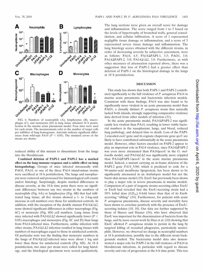
Combined deletion of PAPI-1 and PAPI-2 has a markedeffect on the lung immune response and a subtle effect on lunghistopathology. Groups of mice infected intranasally withPAO1, PA14, or one of the three PA14 island-minus strainswere sacrificed at 18 h postinfection. The lungs and nasophar-ynx were removed and processed for immunological cell countsand/or histology. Surprisingly, despite marked differences indisease severity, at the 18-h time point there were no signifi-cant differences between any two strains in the numbers ofneutrophils (Fig. 6A) or lymphocytes (Fig. 6B) that had infil-trated lung tissue; all five strains showed a 1- to 2-log-unitincrease in cell numbers over those for uninfected controls. Inaddition, with the exception of the double mutant PA14�1�2,none showed significant differences in either macrophage (Fig.6C) or monocyte (Fig. 6D) cell numbers. Lung tissue frommice infected with PA14�1�2 showed significantly fewer (P �0.05) macrophages and monocytes (Fig. 6C and D) than lungsfrom PA14-infected mice. Furthermore, unlike infection withother strains, PA14�1�2 infection resulted in lung tissues withnumbers of macrophages equal to those in uninfected controls.Of particular note was the finding that monocyte numbers inthe lungs of PA14�1�2-infected mice were about 1 log unitlower than those for uninfected controls (Fig. 6D). At 18 hpostinfection, two mice per strain were culled for lung histol-ogy, and the histological specimens were scored qualitatively.
The lung sections were given an overall score for damageand inflammation. The score ranged from 1 to 5 based onthe levels of hypertrophy of bronchial walls, general consol-idation, and cellular infiltration. A score of 1 representednegligible tissue damage or inflammation, and a score of 5represented severe tissue damage and inflammation. Thelung histology scores obtained with the different strains, inorder of decreasing severity by subjective assessment, wereas follows: PA14, 4.5; PA14�PAPI-1, 3.5; PAO1, 3.0;PA14�PAPI-2, 3.0; PA14�1�2, 3.0. Furthermore, as withother measures of attenuation reported above, there was asuggestion that loss of PAPI-2 had a greater effect thandeletion of PAPI-1 on the histological damage in the lungsat 18 h postinfection.
DISCUSSION
This study has shown that both PAPI-1 and PAPI-2 contrib-uted significantly to the full virulence of P. aeruginosa PA14 inmurine acute pneumonia and bacteremia infection models.Consistent with these findings, PA14 was also found to besignificantly more virulent in an acute pneumonia model thanPAO1, a clonally distinct P. aeruginosa strain that naturallylacked both islands, strongly supporting comparative virulencedata derived from other models of infection (31).
In the acute pneumonia model, PA14�PAPI-2 was signifi-cantly less virulent than PA14, resulting in lower viable bacte-rial numbers in the nasopharynx, lungs, and blood, reducedlung pathology, and delayed time to death. Loss of the PAPI-2-encoded exoU gene and its cognate chaperone gene spcU arelikely to have contributed substantially in the acute pneumoniamodel. However, other factors encoded on PAPI-2 appear toplay an important role in PA14 virulence, since PA14�PAPI-2was even more attenuated than PA14�exoU in the G. mel-lonella model, and PA14�1�2 was significantly more crippledthan PA14�PAPI-1�exoU in the acute murine pneumoniamodel. Indeed, a mutant carrying an in-frame deletion of thePAPI-2 gene PA14_5160, which is predicted to code for a94-amino-acid membrane lipoprotein, has been shown to besignificantly attenuated in an Arabidopsis model but not theburnt-skin mouse model (19). ExoU has previously been shownto play a major role in severe pneumonia in murine models.Comparison of a pair of isogenic strains secreting either ExoUor ExoS had revealed that the ExoU-secreting strain had a50% lethal dose (LD50) 8-fold lower than that of its ExoS-secreting “sibling” (54). Furthermore, for patients with acuteP. aeruginosa pneumonia, disease severity and mortality havebeen shown to correlate positively with the presence of ExoU-secreting isolates (10, 18). Our data are further supported bythose of Shaver and Hauser (54), who have observed thatExoU was important for the dissemination of bacteria from thelungs, and by more-recent work by Diaz et al. (10) showing thatExoU allowed P. aeruginosa strains to persist in the lung bytargeted killing of recruited phagocytes, particularly neutro-phils. However, we observed no change in neutrophil numbersat 18 h postinfection, possibly due to the different strain back-grounds studied. The bacteremia model data also demon-strated a major role for PAPI-2 in the full virulence of PA14 inbloodstream infections, in particular with regard to diseaseseverity and rate of progression at the 6-h time point. This was
FIG. 6. Numbers of neutrophils (A), lymphocytes (B), macro-phages (C), and monocytes (D) in lung tissue obtained 18 h postin-fection in the murine acute pneumonia model. Four mice were usedfor each strain. The measurements refer to the number of target cellsper milliliter of lung homogenate. Asterisks indicate significant differ-ences from wild-type PA14 (P � 0.05). The standard errors of themeans are shown.
VOL. 78, 2010 PAPI-1 AND PAPI-2 IN P. AERUGINOSA PA14 VIRULENCE 1443
on April 6, 2020 by guest
http://iai.asm.org/
Dow
nloaded from
entirely consistent with the recent observation that the exoS-negative, exoU-positive genotype dominates among blood-stream infection-associated isolates in humans (59). Furtherinvestigations are required to identify the other PAPI-2 genesimplicated in virulence in this study and to dissect the mecha-nistics of the newly demonstrated PAPI-1–PAPI-2 synergisticinteraction.
The effects of PAPI-1 were more subtle; strain PA14�PAPI-1was as virulent as PA14 in the murine pneumonia model, asjudged by the 100% mortality rate at 18 h. Nevertheless, thePAPI-1-minus strain probably exhibited minor attenuation,since it resulted in slightly lower disease severity scores onaverage and marginally less lung histological damage. In con-trast, in the bacteremia model, the PA14�PAPI-1 mutant wassubstantially impaired: all mice infected with this strain sur-vived up to the 24-h endpoint, while PA14-infected mice uni-versally succumbed at 6 h. As would be expected with thesefindings, the rate of progression of disease severity scores par-alleled the mortality figures.
The most dramatic result was the marked reduction in vir-ulence of the double mutant PA14�1�2 in the murine acutepneumonia model. This strain showed a highly significant re-duction in virulence compared to the wild-type strain PA14and even compared to each of the single-island deletion mu-tants, PA14�PAPI-1 and PA14�PAPI-2, clearly demonstrat-ing that both PAPI-1 and PAPI-2 played key roles in acutepneumonia. Despite the minimal differences observed in qual-itative lung histological scores, the broader set of data obtainedwas perfectly consistent with PA14 being the most virulentstrain and PA14�1�2 being at the other end of the spectrum inthe acute pneumonia model. Twenty-two genes borne onPAPI-1 and PAPI-2 have previously been implicated in viru-lence by using defined PA14 single-gene knockouts and theArabidopsis leaf infiltration, murine burnt-skin, G. mellonella,and Caenorhabditis elegans slow-killing infection models (8, 19,31, 41), but our findings are the first data to clearly demon-strate a role for PAPI-1 in acute pneumonia.
The markedly lower numbers of monocytes and macro-phages in the lungs of mice infected with the double mutantthan those with the other strains that caused more-severe dis-ease could be attributed to apoptosis of monocytes and/ormacrophages as part of a successful modulation of the immuneresponse to PA14�1�2 infection; such a defense has previouslybeen hypothesized to occur during P. aeruginosa lung infection(65). This fits with data derived from P. aeruginosa infections inhumans and a rabbit sepsis model (2, 16), which showed thatearly apoptosis of blood monocytes was correlated with posi-tive resolution of infection. This association was postulated tobe due to a reduced inflammatory response and a consequentdampening of the sepsis cascade. A similar phenomenon hasbeen described for pneumococcal pneumonia and the likeli-hood of its progression to bacteremia (35). It is possible thatthe host is able to mount a protective immune response merelyas a result of the marked attenuation of PA14�1�2 or that therecruitment of monocytes and macrophages is caused by adifferent antigenic profile. However, an alternative hypothesiswould be that loss of PAPI-1 and PAPI-2 resulted in a strainthat was specifically unable to modulate the immune responsein its favor. Previous studies have shown that P. aeruginosa, likeSalmonella (28), is capable of preventing Kupffer cell (3) and
epithelial cell (64) apoptosis by secretion of unknown anti-apoptotic factors. The ability of P. aeruginosa to prevent theapoptosis of monocytes and macrophages and to shift the im-mune response toward inflammation would constitute a signif-icant virulence factor.
The PA14 PAPI-1 island encodes more than 30 putativevirulence genes that are also present on several other se-quenced P. aeruginosa genomes (see Table S2 and Fig. S1 inthe supplemental material). The RscBC two-component sys-tem is an excellent candidate, since counterparts in E. coli,Salmonella, Erwinia, and Yersinia have been shown to playessential roles in the regulation of T3SSs and many othervirulence factors. A recently study published by Mikkelsen andcoworkers demonstrated that the PAPI-1-encoded RcsBC andPvrRS systems antagonistically regulated the expression of thePAPI-1-borne fimbrial cupD operon (40), affecting the abilityof the bacteria to attach and form biofilms. However, cupDregulation was not directly demonstrable under laboratorygrowth conditions, suggesting that specific, as yet undefinedstimuli were required to engage the two-component systems.Reduced levels of cell fimbriation may contribute to the lowernumbers of monocytes and macrophages present in the lungsof mice infected with the double mutant, since E. coli fimbriaehave previously been reported to be potent stimulators of theToll-like receptor 4 immune cell recruitment pathway (3).Other work, by Nicastro and Baldini (44), demonstrated that aP. aeruginosa rscC rscB double mutant exhibited an alteredlipopolysaccharide O-antigen profile, as has been reportedpreviously for Salmonella (25). The length and structure ofO-antigen chains are a key factor in serum resistance, type IIIsecretion, and humoral immunogenicity (4, 49), potentiallyexplaining the finding that PA14�PAPI-1 appeared to defyclearance from the blood, relative to that of PA14 and theother strains, despite being found to be significantly attenuatedin the murine bacteremia model.
Synergistic relationships between pathogenicity islands havebeen described for E. coli (PAI-I and PAI-II) (7) and Salmo-nella enterica (SPI-1 and SPI-4) (14, 15). However, to the bestof our knowledge, this phenomenon has not been describedpreviously for P. aeruginosa or any other bacterial species.Clearly, the mechanistic basis of the synergistic relationshipbetween PAPI-1 and PAPI-2 would be of key interest. Besidespotential contributions of PAPI-1 to bacterial fimbriationand/or biofilm formation, another plausible hypothesis wouldbe that loss of PAPI-1 leads to aberrant T3SS activity as aresult of an altered O-antigen profile, which has been impli-cated in T3SS function (4), and/or loss of the Rcs system, whichhas been shown to regulate the expression of T3SS in Yersiniaenterocolitica and S. enterica (33, 58). Despite the dominantrole of ExoU, it is likely that other T3SS effectors encodedelsewhere on the genome also play key roles in the fine tuningof the pathogenesis process; the deployment of multiple, spe-cifically targeted effectors is a common theme for several bac-terial pathogens.
Our findings with regard to PAPI-1 complement those ofBattle et al. (5), who demonstrated that novel non-PAPI-1sequences encoded on PAGI-5, a PAPI-1-like hybrid structure,played an important role in murine acute pneumonia. As far aswe are aware, this is the first report that clearly demonstratesa substantial role in virulence for PAPI-1 by using in vivo
1444 HARRISON ET AL. INFECT. IMMUN.
on April 6, 2020 by guest
http://iai.asm.org/
Dow
nloaded from

models. Further work is clearly required to identify the com-binations and temporal activation patterns of island-bornegenes brought into play during Pseudomonas infections and tofurther dissect their interactions with the genes of the coregenome. The widely recognized presence of PAPI-1-like is-lands in diverse environmental and clinical isolates (27, 61, 63),including those causing acute pneumonia, chronic cystic fibro-sis, and burn-associated infections, suggests that these islandsmay be subjected to considerable positive selection. This isparticularly so given the known spontaneous instability of theseelements.
We propose that the en bloc deletion strategy that we haveused for the first time to study the role of genomic islands in P.aeruginosa offers several major advantages. It allows for rapidtargeted virulence-related screening of large blocks of DNA intheir “native” genomic backgrounds, addresses the key evolu-tionary question of the impact of the en bloc acquisition ofgenomic islands, potentially reveals subtle but important con-tributions to virulence that may be masked by dominant traitsencoded by other targeted islands, and greatly facilitates thedetection of synergistic and/or combinatorial effects betweenany of the numerous genes collectively deleted from the mu-tant strains. The data obtained from these whole-island dele-tion experiments could then be investigated further in a better-targeted fashion by single-gene mutation, combination genemutation, transcriptomic, and/or other detailed genetic studiesto pinpoint the precise mechanisms involved. This study hasclearly demonstrated the key role of the horizontally acquiredPAPI-1 and PAPI-2 islands in murine acute pneumonia andbacteremia models, thus firmly cementing their designation asgenetic modules of virulence in a major pathogenic species.
ACKNOWLEDGMENTS
We thank James Lonnen, Barbara Rieck, and Sarah Smeaton forhelpful advice and discussions and Craig Winstanley and HerbertSchweizer for providing P. aeruginosa PA14 and key mutagenesis plas-mids, respectively. We are especially grateful to Eliana Drenkard forPA14-related strains and for prompt assistance with the generation ofthe mutants lacking exoU. Thanks also go to Marialuisa Crosatti forhelp with the Galleria assay and to Hannah Brewin for murine exper-iments.
This work was funded by an Action Medical Research grant to K.R.,A.K., and C.O. E.M.H. was funded by a Medical Research Council/University of Leicester Ph.D. Studentship. H.-Y.O. was funded by agrant from the National Natural Science Foundation of China(30700013/C010103) and the 863 Program, Ministry of Science andTechnology, China (2006AA02Z328). K.R. and Z.D. were supportedby a Royal Society–National Natural Science Foundation of ChinaInternational Joint Project grant (IJP2007/R3).
REFERENCES
1. Allewelt, M., F. T. Coleman, M. Grout, G. P. Priebe, and G. B. Pier. 2000.Acquisition of expression of the Pseudomonas aeruginosa ExoU cytotoxinleads to increased bacterial virulence in a murine model of acute pneumoniaand systemic spread. Infect. Immun. 68:3998–4004.
2. Antonopoulou, A., M. Raftogiannis, E. J. Giamarellos-Bourboulis, P. Kout-oukas, L. Sabracos, M. Mouktaroudi, T. Adamis, I. Tzepi, H. Giamarellou,and E. E. Douzinas. 2007. Early apoptosis of blood monocytes is a determi-nant of survival in experimental sepsis by multi-drug-resistant Pseudomonasaeruginosa. Clin. Exp. Immunol. 149:103–108.
3. Ashkar, A. A., K. L. Mossman, B. K. Coombes, C. L. Gyles, and R. Mack-enzie. 2008. FimH adhesin of type 1 fimbriae is a potent inducer of innateantimicrobial responses which requires TLR4 and type 1 interferon signal-ling. PLoS Pathog. 4:e1000233.
4. Augustin, D. K., Y. Song, M. S. Baek, Y. Sawa, G. Singh, B. Taylor, A.Rubio-Mills, J. L. Flanagan, J. P. Wiener-Kronish, and S. V. Lynch. 2007.Presence or absence of lipopolysaccharide O antigens affects type III secre-tion by Pseudomonas aeruginosa. J. Bacteriol. 189:2203–2209.
5. Battle, S. E., F. Meyer, J. Rello, V. L. Kung, and A. R. Hauser. 2008. Hybridpathogenicity island PAGI-5 contributes to the highly virulent phenotype ofa Pseudomonas aeruginosa isolate in mammals. J. Bacteriol. 190:7130–7140.
6. Battle, S. E., J. Rello, and A. R. Hauser. 2009. Genomic islands of Pseudo-monas aeruginosa. FEMS Microbiol. Lett. 290:70–78.
7. Brzuszkiewicz, E., H. Bruggemann, H. Liesegang, M. Emmerth, T.Olschlager, G. Nagy, K. Albermann, C. Wagner, C. Buchrieser, L. Emody, G.Gottschalk, J. Hacker, and U. Dobrindt. 2006. How to become a uropatho-gen: comparative genomic analysis of extraintestinal pathogenic Escherichiacoli strains. Proc. Natl. Acad. Sci. U. S. A. 103:12879–12884.
8. Choi, J. Y., C. D. Sifri, B. C. Goumnerov, L. G. Rahme, F. M. Ausubel, andS. B. Calderwood. 2002. Identification of virulence genes in a pathogenicstrain of Pseudomonas aeruginosa by representational difference analysis. J.Bacteriol. 184:952–961.
9. Choi, K. H., and H. P. Schweizer. 2005. An improved method for rapidgeneration of unmarked Pseudomonas aeruginosa deletion mutants. BMCMicrobiol. 5:30.
10. Diaz, M. H., C. M. Shaver, J. D. King, S. Musunuri, J. A. Kazzaz, and A. R.Hauser. 2008. Pseudomonas aeruginosa induces localized immunosuppres-sion during pneumonia. Infect. Immun. 76:4414–4421.
11. Drenkard, E., and F. M. Ausubel. 2002. Pseudomonas biofilm formation andantibiotic resistance are linked to phenotypic variation. Nature 416:740–743.
12. El Solh, A. A., M. E. Akinnusi, J. P. Wiener-Kronish, S. V. Lynch, L. A.Pineda, and K. Szarpa. 2008. Persistent infection with Pseudomonas aerugi-nosa in ventilator-associated pneumonia. Am. J. Respir. Crit. Care Med.178:513–519.
13. Evans, D. J., D. W. Frank, V. Finck-Barbancon, C. Wu, and S. M. Fleiszig.1998. Pseudomonas aeruginosa invasion and cytotoxicity are independentevents, both of which involve protein tyrosine kinase activity. Infect. Immun.66:1453–1459.
14. Gerlach, R. G., N. Claudio, M. Rohde, D. Jackel, C. Wagner, and M. Hensel.2008. Cooperation of Salmonella pathogenicity islands 1 and 4 is required tobreach epithelial barriers. Cell. Microbiol. 10:2364–2376.
15. Gerlach, R. G., D. Jackel, N. Geymeier, and M. Hensel. 2007. Salmonellapathogenicity island 4-mediated adhesion is coregulated with invasion genesin Salmonella enterica. Infect. Immun. 75:4697–4709.
16. Giamarellos-Bourboulis, E. J., C. Routsi, D. Plachouras, V. Markaki, M.Raftogiannis, D. Zervakis, V. Koussoulas, S. Orfanos, A. Kotanidou, A.Armaganidis, C. Roussos, and H. Giamarellou. 2006. Early apoptosis ofblood monocytes in the septic host: is it a mechanism of protection in theevent of septic shock? Crit. Care 10:R76.
17. Hanahan, D. 1983. Studies on transformation of Escherichia coli with plas-mids. J. Mol. Biol. 166:557–580.
18. Hauser, A. R., E. Cobb, M. Bodi, D. Mariscal, J. Valles, J. N. Engel, and J.Rello. 2002. Type III protein secretion is associated with poor clinical out-comes in patients with ventilator-associated pneumonia caused by Pseudo-monas aeruginosa. Crit. Care Med. 30:521–528.
19. He, J., R. L. Baldini, E. Deziel, M. Saucier, Q. Zhang, N. T. Liberati, D. Lee,J. Urbach, H. M. Goodman, and L. G. Rahme. 2004. The broad host rangepathogen Pseudomonas aeruginosa strain PA14 carries two pathogenicityislands harboring plant and animal virulence genes. Proc. Natl. Acad. Sci.U. S. A. 101:2530–2535.
20. Hendrickson, E. L., J. Plotnikova, S. Mahajan-Miklos, L. G. Rahme, andF. M. Ausubel. 2001. Differential roles of the Pseudomonas aeruginosa PA14rpoN gene in pathogenicity in plants, nematodes, insects, and mice. J. Bac-teriol. 183:7126–7134.
21. Hoang, T. T., R. R. Karkhoff-Schweizer, A. J. Kutchma, and H. P. Schweizer.1998. A broad-host-range Flp-FRT recombination system for site-specificexcision of chromosomally-located DNA sequences: application for isolationof unmarked Pseudomonas aeruginosa mutants. Gene 212:77–86.
22. Inglis, R. F., A. Gardner, P. Cornelis, and A. Buckling. 2009. Spite andvirulence in the bacterium Pseudomonas aeruginosa. Proc. Natl. Acad. Sci.U. S. A. 106:5703–5707.
23. Juhas, M., D. W. Crook, I. D. Dimopoulou, G. Lunter, R. M. Harding, D. J.Ferguson, and D. W. Hood. 2007. Novel type IV secretion system involved inpropagation of genomic islands. J. Bacteriol. 189:761–771.
24. Kadioglu, A., N. A. Gingles, K. Grattan, A. Kerr, T. J. Mitchell, and P. W.Andrew. 2000. Host cellular immune response to pneumococcal lung infec-tion in mice. Infect. Immun. 68:492–501.
25. Kintz, E., and J. B. Goldberg. 2008. Regulation of lipopolysaccharide Oantigen expression in Pseudomonas aeruginosa. Future Microbiol. 3:191–203.
26. Klockgether, J., O. Reva, K. Larbig, and B. Tummler. 2004. Sequence anal-ysis of the mobile genome island pKLC102 of Pseudomonas aeruginosa C. J.Bacteriol. 186:518–534.
27. Klockgether, J., D. Wurdemann, O. Reva, L. Wiehlmann, and B. Tummler.2007. Diversity of the abundant pKLC102/PAGI-2 family of genomic islandsin Pseudomonas aeruginosa. J. Bacteriol. 189:2443–2459.
28. Knodler, L. A., and B. B. Finlay. 2001. Salmonella and apoptosis: to live orlet die? Microbes Infect. 3:1321–1326.
29. Kukavica-Ibrulj, I., A. Bragonzi, M. Paroni, C. Winstanley, F. Sanschagrin,G. A. O’Toole, and R. C. Levesque. 2008. In vivo growth of Pseudomonas
VOL. 78, 2010 PAPI-1 AND PAPI-2 IN P. AERUGINOSA PA14 VIRULENCE 1445
on April 6, 2020 by guest
http://iai.asm.org/
Dow
nloaded from
aeruginosa strains PAO1 and PA14 and the hypervirulent strain LESB58 ina rat model of chronic lung infection. J. Bacteriol. 190:2804–2813.
30. Kulasekara, B. R., H. D. Kulasekara, M. C. Wolfgang, L. Stevens, D. W.Frank, and S. Lory. 2006. Acquisition and evolution of the exoU locus inPseudomonas aeruginosa. J. Bacteriol. 188:4037–4050.
31. Lee, D. G., J. M. Urbach, G. Wu, N. T. Liberati, R. L. Feinbaum, S. Miyata,L. T. Diggins, J. He, M. Saucier, E. Deziel, L. Friedman, L. Li, G. Grills, K.Montgomery, R. Kucherlapati, L. G. Rahme, and F. M. Ausubel. 2006.Genomic analysis reveals that Pseudomonas aeruginosa virulence is combi-natorial. Genome Biol. 7:R90.
32. Lee, V. T., R. S. Smith, B. Tummler, and S. Lory. 2005. Activities of Pseudo-monas aeruginosa effectors secreted by the type III secretion system in vitroand during infection. Infect. Immun. 73:1695–1705.
33. Lin, D., C. V. Rao, and J. M. Slauch. 2008. The Salmonella SPI1 type threesecretion system responds to periplasmic disulfide bond status via the flagel-lar apparatus and the RcsCDB system. J. Bacteriol. 190:87–97.
34. Luck, S. N., L. Badea, V. Bennett-Wood, R. Robins-Browne, and E. L.Hartland. 2006. Contribution of FliC to epithelial cell invasion by entero-hemorrhagic Escherichia coli O113:H21. Infect. Immun. 74:6999–7004.
35. Marriott, H. M., P. G. Hellewell, S. S. Cross, P. G. Ince, M. K. Whyte, andD. H. Dockrell. 2006. Decreased alveolar macrophage apoptosis is associatedwith increased pulmonary inflammation in a murine model of pneumococcalpneumonia. J. Immunol. 177:6480–6488.
36. Mathee, K., G. Narasimhan, C. Valdes, X. Qiu, J. M. Matewish, M. Koe-hrsen, A. Rokas, C. N. Yandava, R. Engels, E. Zeng, R. Olavarietta, M.Doud, R. S. Smith, P. Montgomery, J. R. White, P. A. Godfrey, C. Kodira, B.Birren, J. E. Galagan, and S. Lory. 2008. Dynamics of Pseudomonas aerugi-nosa genome evolution. Proc. Natl. Acad. Sci. U. S. A. 105:3100–3105.
37. McMorran, B., L. Town, E. Costelloe, J. Palmer, J. Engel, D. Hume, and B.Wainwright. 2003. Effector ExoU from the type III secretion system is animportant modulator of gene expression in lung epithelial cells in responseto Pseudomonas aeruginosa infection. Infect. Immun. 71:6035–6044.
38. Meissner, A., V. Wild, R. Simm, M. Rohde, C. Erck, F. Bredenbruch, M.Morr, U. Romling, and S. Haussler. 2007. Pseudomonas aeruginosa cupA-encoded fimbriae expression is regulated by a GGDEF and EAL domain-dependent modulation of the intracellular level of cyclic diguanylate. Envi-ron. Microbiol. 9:2475–2485.
39. Michel-Briand, Y., and C. Baysse. 2002. The pyocins of Pseudomonasaeruginosa. Biochimie 84:499–510.
40. Mikkelsen, H., G. Ball, C. Giraud, and A. Filloux. 2009. Expression ofPseudomonas aeruginosa CupD fimbrial genes is antagonistically controlledby RcsB and the EAL-containing PvrR response regulators. PLoS One4:e6018.
41. Miyata, S., M. Casey, D. W. Frank, F. M. Ausubel, and E. Drenkard. 2003.Use of the Galleria mellonella caterpillar as a model host to study the roleof the type III secretion system in Pseudomonas aeruginosa pathogenesis.Infect. Immun. 71:2404–2413.
42. Morton, D. B., and P. H. Griffiths. 1985. Guidelines on the recognition ofpain, distress and discomfort in experimental animals and an hypothesis forassessment. Vet. Rec. 116:431–436.
43. Murphy, T. F., A. L. Brauer, K. Eschberger, P. Lobbins, L. Grove, X. Cai,and S. Sethi. 2008. Pseudomonas aeruginosa in chronic obstructive pulmo-nary disease. Am. J. Respir. Crit. Care Med. 177:853–860.
44. Nicastro, G. G., and R. L. Baldini. 2007. Functional characterization ofPseudomonas aeruginosa virulence-related genes located in a pathogenicityisland, abstr. F-20. Abstr. XXXVI Annu. Meet. Braz. Soc. Biochem. Mol.Biol. (SBBq)–10th IUBMB Conf., Salvador da Bahia, Brazil, 2007. http://sbbq.iq.usp.br/arquivos/2007/cdlivro/resumos/R8586.pdf.
45. O’Toole, G. A., and R. Kolter. 1998. Initiation of biofilm formation inPseudomonas fluorescens WCS365 proceeds via multiple, convergent signal-ling pathways: a genetic analysis. Mol. Microbiol. 28:449–461.
46. Qiu, X., A. U. Gurkar, and S. Lory. 2006. Interstrain transfer of the largepathogenicity island (PAPI-1) of Pseudomonas aeruginosa. Proc. Natl. Acad.Sci. U. S. A. 103:19830–19835.
47. Rajakumar, K., C. Sasakawa, and B. Adler. 1997. Use of a novel approach,termed island probing, identifies the Shigella flexneri she pathogenicity is-
land which encodes a homolog of the immunoglobulin A protease-like familyof proteins. Infect. Immun. 65:4606–4614.
48. Rashid, M. H., and A. Kornberg. 2000. Inorganic polyphosphate is neededfor swimming, swarming, and twitching motilities of Pseudomonas aerugi-nosa. Proc. Natl. Acad. Sci. U. S. A. 97:4885–4890.
49. Rocchetta, H. L., L. L. Burrows, and J. S. Lam. 1999. Genetics of O-antigenbiosynthesis in Pseudomonas aeruginosa. Microbiol. Mol. Biol. Rev. 63:523–553.
50. Ryall, B., J. C. Davies, R. Wilson, A. Shoemark, and H. D. Williams. 2008.Pseudomonas aeruginosa, cyanide accumulation and lung function in CF andnon-CF bronchiectasis patients. Eur. Respir. J. 32:740–747.
51. Sadikot, R. T., T. S. Blackwell, J. W. Christman, and A. S. Prince. 2005.Pathogen-host interactions in Pseudomonas aeruginosa pneumonia. Am. J.Respir. Crit. Care Med. 171:1209–1223.
52. Schulert, G. S., H. Feltman, S. D. Rabin, C. G. Martin, S. E. Battle, J. Rello,and A. R. Hauser. 2003. Secretion of the toxin ExoU is a marker for highlyvirulent Pseudomonas aeruginosa isolates obtained from patients with hos-pital-acquired pneumonia. J. Infect. Dis. 188:1695–1706.
53. Schweizer, H. P. 1992. Allelic exchange in Pseudomonas aeruginosa usingnovel ColE1-type vectors and a family of cassettes containing a portable oriTand the counter-selectable Bacillus subtilis sacB marker. Mol. Microbiol.6:1195–1204.
54. Shaver, C. M., and A. R. Hauser. 2004. Relative contributions of Pseudo-monas aeruginosa ExoU, ExoS, and ExoT to virulence in the lung. Infect.Immun. 72:6969–6977.
55. Simon, R., U. Priefer, and A. Puhler. 1983. A broad host range mobilizationsystem for in vivo genetic engineering: transposon mutagenesis in Gramnegative bacteria. Nat. Biotechnol. 1:784–791.
56. Stover, C. K., X. Q. Pham, A. L. Erwin, S. D. Mizoguchi, P. Warrener, M. J.Hickey, F. S. Brinkman, W. O. Hufnagle, D. J. Kowalik, M. Lagrou, R. L.Garber, L. Goltry, E. Tolentino, S. Westbrock-Wadman, Y. Yuan, L. L.Brody, S. N. Coulter, K. R. Folger, A. Kas, K. Larbig, R. Lim, K. Smith, D.Spencer, G. K. Wong, Z. Wu, I. T. Paulsen, J. Reizer, M. H. Saier, R. E.Hancock, S. Lory, and M. V. Olson. 2000. Complete genome sequence ofPseudomonas aeruginosa PAO1, an opportunistic pathogen. Nature 406:959–964.
57. Vogel, H. J., and D. M. Bonner. 1956. Acetylornithinase of Escherichia coli:partial purification and some properties. J. Biol. Chem. 218:97–106.
58. Walker, K. A., and V. L. Miller. 2009. Synchronous gene expression of theYersinia enterocolitica Ysa type III secretion system and its effectors. J.Bacteriol. 191:1816–1826.
59. Wareham, D. W., and M. A. Curtis. 2007. A genotypic and phenotypiccomparison of type III secretion profiles of Pseudomonas aeruginosa cysticfibrosis and bacteremia isolates. Int. J. Med. Microbiol. 297:227–234.
60. West, S. E., H. P. Schweizer, C. Dall, A. K. Sample, and L. J. Runyen-Janecky. 1994. Construction of improved Escherichia-Pseudomonas shuttlevectors derived from pUC18/19 and sequence of the region required for theirreplication in Pseudomonas aeruginosa. Gene 148:81–86.
61. Wiehlmann, L., G. Wagner, N. Cramer, B. Siebert, P. Gudowius, G. Morales,T. Kohler, C. van Delden, C. Weinel, P. Slickers, and B. Tummler. 2007.Population structure of Pseudomonas aeruginosa. Proc. Natl. Acad. Sci.U. S. A. 104:8101–8106.
62. Wolfgang, M. C., B. R. Kulasekara, X. Liang, D. Boyd, K. Wu, Q. Yang, C. G.Miyada, and S. Lory. 2003. Conservation of genome content and virulencedeterminants among clinical and environmental isolates of Pseudomonasaeruginosa. Proc. Natl. Acad. Sci. U. S. A. 100:8484–8489.
63. Wurdemann, D., and B. Tummler. 2007. In silico comparison of pKLC102-like genomic islands of Pseudomonas aeruginosa. FEMS Microbiol. Lett.275:244–249.
64. Zhang, J., H. Li, J. Wang, Z. Dong, S. Mian, and F. S. Yu. 2004. Role ofEGFR transactivation in preventing apoptosis in Pseudomonas aeruginosa-infected human corneal epithelial cells. Invest. Ophthalmol. Vis. Sci. 45:2569–2576.
65. Zhang, Y., X. Li, A. Carpinteiro, and E. Gulbins. 2008. Acid sphingomyeli-nase amplifies redox signaling in Pseudomonas aeruginosa-induced macro-phage apoptosis. J. Immunol. 181:4247–4254.
Editor: B. A. McCormick
1446 HARRISON ET AL. INFECT. IMMUN.
on April 6, 2020 by guest
http://iai.asm.org/
Dow
nloaded from
Related Documents