RESEARCH ARTICLE Pathogenic waterborne free-living amoebae: An update from selected Southeast Asian countries Mohamad Azlan Abdul Majid 1 , Tooba Mahboob 1 , Brandon G. J. Mong 1 , Narong Jaturas 1 , Reena Leeba Richard 1 , Tan Tian-Chye 1 , Anusorn Phimphila 2 , Panomphanh Mahaphonh 2 , Kyaw Nyein Aye 3 , Wai Lynn Aung 3 , Joon Chuah 4 , Alan D. Ziegler 5 , Atipat Yasiri 6 , Nongyao Sawangjaroen 7 , Yvonne A. L. Lim 1 , Veeranoot Nissapatorn 1 * 1 Department of Parasitology (Southeast Asia Water Team), Faculty of Medicine, University of Malaya, Kuala Lumpur, Malaysia, 2 Department of Medical Laboratory, Faculty of Medical Technology, University of Health Sciences, Vientiane, Laos PDR, 3 Ecological Laboratory, Advancing Life and Regenerating Motherland (ALARM), Yangon, Myanmar, 4 Institute of Water Policy, National University of Singapore, Singapore, Singapore, 5 Department of Geography, National University of Singapore, Singapore, Singapore, 6 Chulabhorn International College of Medicine, Thammasat University, Pathum Thani, Thailand, 7 Department of Microbiology, Faculty of Science, Prince of Songkhla University, Hat-Yai, Thailand * [email protected], [email protected] Abstract Data on the distribution of free-living amoebae is still lacking especially in Southeast Asian region. The aquatic environment revealed a high occurrence of free-living amoebae (FLA) due to its suitable condition and availability of food source, which subsequently causes infection to humans. A total of 94 water samples consisted of both treated and untreated from Laos (31), Myanmar (42), and Singapore (21) were investigated for the presence of pathogenic FLA. Each water sample was filtered and cultured onto non-nutrient agar seeded with live suspension of Escherichia coli and incubated at room temperature. Mor- phological identification was conducted for both trophozoites and cysts via microscopic stains (Giemsa and immunofluorescence). The presence of Naegleria-like structures was the most frequently encountered in both treated and untreated water samples, followed by Acanthamoeba-like and Vermamoeba-like features. To identify the pathogenic isolates, species-specific primer sets were applied for molecular identification of Acanthamoeba, Naegleria, and Vermamoeba. The pathogenic species of Acanthamoeba lenticulata and A. triangularis were detected from untreated water samples, while Vermamoeba vermiformis was found in both treated and untreated water samples. Our results suggested that poor water quality as well as inadequate maintenance and treatment might be the cause of this alarming problem since chlorine disinfection is ineffective in eradicating these amoebas in treated water samples. Regular monitoring and examination of water qualities are neces- sary in order to control the growth, hence, further preventing the widespread of FLA infec- tions among the public. PLOS ONE | DOI:10.1371/journal.pone.0169448 February 17, 2017 1 / 17 a1111111111 a1111111111 a1111111111 a1111111111 a1111111111 OPEN ACCESS Citation: Abdul Majid MA, Mahboob T, Mong BGJ, Jaturas N, Richard RL, Tian-Chye T, et al. (2017) Pathogenic waterborne free-living amoebae: An update from selected Southeast Asian countries. PLoS ONE 12(2): e0169448. doi:10.1371/journal. pone.0169448 Editor: Tzen-Yuh Chiang, National Cheng Kung University, TAIWAN Received: June 2, 2016 Accepted: December 16, 2016 Published: February 17, 2017 Copyright: © 2017 Abdul Majid et al. This is an open access article distributed under the terms of the Creative Commons Attribution License, which permits unrestricted use, distribution, and reproduction in any medium, provided the original author and source are credited. Data Availability Statement: All relevant data are within the paper. Funding: University of Malaya High Impact Research Grant (UM.C/625/1/HIR/MoHE/MED/23 and UM. 0000041/HIR.C3) and University of Malaya Research Grants (UMRG 544/14HTM and UMRG 362/15AFR), received by VN and TTC. Competing interests: The authors have declared that no competing interests exist.

Welcome message from author
This document is posted to help you gain knowledge. Please leave a comment to let me know what you think about it! Share it to your friends and learn new things together.
Transcript
RESEARCH ARTICLE
Pathogenic waterborne free-living amoebae:
An update from selected Southeast Asian
countries
Mohamad Azlan Abdul Majid1, Tooba Mahboob1, Brandon G. J. Mong1, Narong Jaturas1,
Reena Leeba Richard1, Tan Tian-Chye1, Anusorn Phimphila2, Panomphanh Mahaphonh2,
Kyaw Nyein Aye3, Wai Lynn Aung3, Joon Chuah4, Alan D. Ziegler5, Atipat Yasiri6,
Nongyao Sawangjaroen7, Yvonne A. L. Lim1, Veeranoot Nissapatorn1*
1 Department of Parasitology (Southeast Asia Water Team), Faculty of Medicine, University of Malaya, Kuala
Lumpur, Malaysia, 2 Department of Medical Laboratory, Faculty of Medical Technology, University of Health
Sciences, Vientiane, Laos PDR, 3 Ecological Laboratory, Advancing Life and Regenerating Motherland
(ALARM), Yangon, Myanmar, 4 Institute of Water Policy, National University of Singapore, Singapore,
Singapore, 5 Department of Geography, National University of Singapore, Singapore, Singapore,
6 Chulabhorn International College of Medicine, Thammasat University, Pathum Thani, Thailand,
7 Department of Microbiology, Faculty of Science, Prince of Songkhla University, Hat-Yai, Thailand
* [email protected], [email protected]
Abstract
Data on the distribution of free-living amoebae is still lacking especially in Southeast Asian
region. The aquatic environment revealed a high occurrence of free-living amoebae (FLA)
due to its suitable condition and availability of food source, which subsequently causes
infection to humans. A total of 94 water samples consisted of both treated and untreated
from Laos (31), Myanmar (42), and Singapore (21) were investigated for the presence of
pathogenic FLA. Each water sample was filtered and cultured onto non-nutrient agar
seeded with live suspension of Escherichia coli and incubated at room temperature. Mor-
phological identification was conducted for both trophozoites and cysts via microscopic
stains (Giemsa and immunofluorescence). The presence of Naegleria-like structures was
the most frequently encountered in both treated and untreated water samples, followed by
Acanthamoeba-like and Vermamoeba-like features. To identify the pathogenic isolates,
species-specific primer sets were applied for molecular identification of Acanthamoeba,
Naegleria, and Vermamoeba. The pathogenic species of Acanthamoeba lenticulata and A.
triangularis were detected from untreated water samples, while Vermamoeba vermiformis
was found in both treated and untreated water samples. Our results suggested that poor
water quality as well as inadequate maintenance and treatment might be the cause of this
alarming problem since chlorine disinfection is ineffective in eradicating these amoebas in
treated water samples. Regular monitoring and examination of water qualities are neces-
sary in order to control the growth, hence, further preventing the widespread of FLA infec-
tions among the public.
PLOS ONE | DOI:10.1371/journal.pone.0169448 February 17, 2017 1 / 17
a1111111111
a1111111111
a1111111111
a1111111111
a1111111111
OPENACCESS
Citation: Abdul Majid MA, Mahboob T, Mong BGJ,
Jaturas N, Richard RL, Tian-Chye T, et al. (2017)
Pathogenic waterborne free-living amoebae: An
update from selected Southeast Asian countries.
PLoS ONE 12(2): e0169448. doi:10.1371/journal.
pone.0169448
Editor: Tzen-Yuh Chiang, National Cheng Kung
University, TAIWAN
Received: June 2, 2016
Accepted: December 16, 2016
Published: February 17, 2017
Copyright: © 2017 Abdul Majid et al. This is an
open access article distributed under the terms of
the Creative Commons Attribution License, which
permits unrestricted use, distribution, and
reproduction in any medium, provided the original
author and source are credited.
Data Availability Statement: All relevant data are
within the paper.
Funding: University of Malaya High Impact
Research Grant (UM.C/625/1/HIR/MoHE/MED/23
and UM. 0000041/HIR.C3) and University of
Malaya Research Grants (UMRG 544/14HTM and
UMRG 362/15AFR), received by VN and TTC.
Competing interests: The authors have declared
that no competing interests exist.
Introduction
Free-living amoebae (FLA) belonging to the genera Acanthamoeba, Balamuthia, Naegleria,
Sappinia, and Vermamoeba (= Hartmannella) are potentially pathogenic to humans [1,2].
They are also known as amphizoic amoeba due to the ability to exist within a host or in the
environment as ‘free-living’. They are able to survive and proliferate in the environment inde-
pendently and can be found in various natural and man-made aquatic environments, both
fresh and marine, including lakes, ponds, swimming pools, and even treated water supplies
[3–5]. The amoeba cysts are highly resistant to harsh conditions due to the process of encyst-
ment [6,7].
Many species under the genus of Acanthamoeba are reported to be the causative agent of
keratitis in healthy individuals, often among contact lens wearers. In opportunistic infections,
Acanthamoeba species can cause pneumonitis, fatal granulomatous encephalitis and skin
infections [8]. To date, 20 different genotypes (T1-T20) of Acanthamoeba have been described
[9]. Meanwhile, Naegleria is the only FLA that has the advantage of exhibiting a flagellate stage
to ease its movement by swimming in water. Moreover, more than 40 species of Naegleriahave been identified, only N. fowleri is found to be the causative agent of primary amoebic
meningoencephalitis (PAM), a lethal brain infection [10]. Approximately, 440 of PAM cases
have been reported worldwide until the year 2008 [11], with exposure of healthy individuals to
warm (water temperatures of 25 to 44˚C), untreated or poorly disinfected water systems. In
addition, Vermamoeba (= Hartmannella) vermiformis seems to be the potential causative agent
of human keratitis [12] and it can serves as a host to pathogenic bacteria, Legionella pneumo-phila [13,14].
In Southeast Asia, the occurrence of FLA were reported in Malaysia [15,16], Thailand [17],
Vietnam [18] and the Philippines [19]. Although the reported cases are extremely rare, the
diagnosis of FLA must never be overlooked as it is able to cause serious and fatal diseases. Ker-
atitis infection caused by Acanthamoeba species was reported among contact lens wearers in
Malaysia [20] and Singapore [21]. Meanwhile, N. fowleri, was reported to be the etiological
agent for 12 cases of PAM in Thailand [22,23]. In addition, the first case of Balamuthia man-drillaris causing meningoencephalitis was reported from a Thai male patient after falling into a
swamp [24].
The knowledge of pathogenic FLA emergence has gained much interest throughout the
world due to its possible health implications. However, inadequate studies of FLA attributed
to the lack of prevalence information across the Southeast Asian region. Therefore, we
undertook an investigation to detect the occurrence and distribution of the FLA, together
with the identification based on culture, staining, and molecular assay of each isolate. This
information should be useful for early detection of potential infestation of pathogenic FLA
in various water sources and further evaluating the risks of human contact with FLA. In fact,
humans can be exposed to FLA via accidental splash/squirt of contaminated water to the
face or through an open wound, hence, necessitating this study in bridging the gaps of
awareness on FLA.
Materials and methods
Ethics statement/Permission approval
This study was carried out in three selected Southeast Asian countries namely; Lao PDR,
Myanmar and Singapore during 2013 to 2015. Invitation letters were approved from the rele-
vant institutions namely University of Health Sciences, Vientiane, Lao PDR, Advancing Life
and Regenerating Motherland (ALARM), Yangon, Myanmar, and National University of
Pathogenic waterborne free-living amoeba from Southeast Asia
PLOS ONE | DOI:10.1371/journal.pone.0169448 February 17, 2017 2 / 17
Singapore, Singapore. The collaborators from each country had been consulted at the time of
water samples collection.
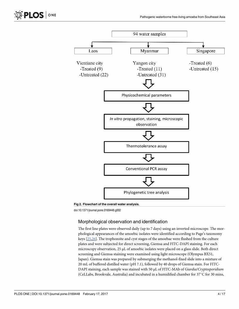
Sample collection
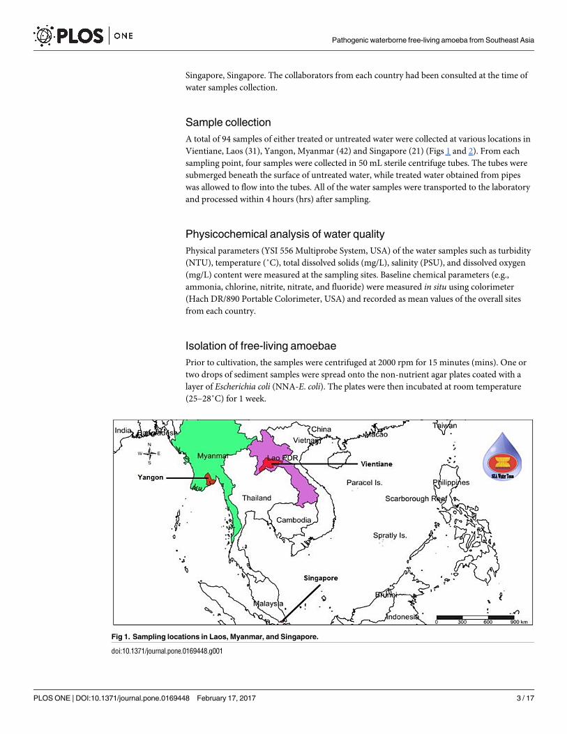
A total of 94 samples of either treated or untreated water were collected at various locations in
Vientiane, Laos (31), Yangon, Myanmar (42) and Singapore (21) (Figs 1 and 2). From each
sampling point, four samples were collected in 50 mL sterile centrifuge tubes. The tubes were
submerged beneath the surface of untreated water, while treated water obtained from pipes
was allowed to flow into the tubes. All of the water samples were transported to the laboratory
and processed within 4 hours (hrs) after sampling.
Physicochemical analysis of water quality
Physical parameters (YSI 556 Multiprobe System, USA) of the water samples such as turbidity
(NTU), temperature (˚C), total dissolved solids (mg/L), salinity (PSU), and dissolved oxygen
(mg/L) content were measured at the sampling sites. Baseline chemical parameters (e.g.,
ammonia, chlorine, nitrite, nitrate, and fluoride) were measured in situ using colorimeter
(Hach DR/890 Portable Colorimeter, USA) and recorded as mean values of the overall sites
from each country.
Isolation of free-living amoebae
Prior to cultivation, the samples were centrifuged at 2000 rpm for 15 minutes (mins). One or
two drops of sediment samples were spread onto the non-nutrient agar plates coated with a
layer of Escherichia coli (NNA-E. coli). The plates were then incubated at room temperature
(25–28˚C) for 1 week.
Fig 1. Sampling locations in Laos, Myanmar, and Singapore.
doi:10.1371/journal.pone.0169448.g001
Pathogenic waterborne free-living amoeba from Southeast Asia
PLOS ONE | DOI:10.1371/journal.pone.0169448 February 17, 2017 3 / 17
Morphological observation and identification
The first line plates were observed daily (up to 7 days) using an inverted microscope. The mor-
phological appearances of the amoebic isolates were identified according to Page’s taxonomy
keys [25,26]. The trophozoite and cyst stages of the amoebae were flushed from the culture
plates and were subjected for direct screening, Giemsa and FITC-DAPI staining. For each
microscopy observation, 25 μL of amoebic isolates were placed on a glass slide. Both direct
screening and Giemsa staining were examined using light microscope (Olympus BX51,
Japan). Giemsa stain was prepared by submerging the methanol-fixed slide into a mixture of
20 mL of buffered distilled water (pH 7.1), followed by 40 drops of Giemsa stain. For FITC-
DAPI staining, each sample was stained with 50 μL of FITC-MAb of Giardia/Cryptosporidium(CeLLabs, Brookvale, Australia) and incubated in a humidified chamber for 37˚C for 30 mins,
Fig 2. Flowchart of the overall water analysis.
doi:10.1371/journal.pone.0169448.g002
Pathogenic waterborne free-living amoeba from Southeast Asia
PLOS ONE | DOI:10.1371/journal.pone.0169448 February 17, 2017 4 / 17
followed by washing step using PBS solution (pH 7.2). The sample was further stained with
50 μL of DAPI solution (Louis, Missouri, USA) for 2 mins before the addition of 50 μL of dis-
tilled water for 1 min. Finally, a 20 μL aliquot of mounting medium was placed onto the slide,
covered with coverslip, and left air-dried prior to examination under epifluorescence micro-
scope (Olympus BX51, Tokyo, Japan).
Thermotolerance assay
Thermotolerance assay was carried out for the selected FLA isolates (V. vermiformis and
Acanthamoeba spp.) that had been confirmed through microscopy examination. Sub-culturing
was performed as described above to obtain axenic culture of the amoeba. The samples were
introduced to certain temperatures (34˚C, 37˚C, 40˚C and 45˚C) for a period of 24 hrs, respec-
tively [27]. The isolates were then observed microscopically to confirm its viability.
DNA extraction
The trophozoites stage of each amoebic isolate were harvested from the plates with 5 mL of
cold Page’s Amoeba Saline (PAS) and transferred into 1.5 mL tubes. DNA was further
extracted using a commercial QIAamp DNA Blood Mini Kit (Qiagen, Hilden, France) follow-
ing the manufacturer’s procedures and stored at -20˚C until further analysis.
PCR amplification
For Acanthamoeba species, amplification was carried out by targeting the 18S region, with a
PCR mix containing 10X DNA polymerase (Thermo Scientific, Lithuania, USA), 25 mM of
magnesium chloride (MgCl2) (Thermo Scientific, Lithuania, USA), 10 mM of deoxynucleotide
triphosphate (dNTP) mix (Thermo Scientific, Lithuania, USA) and 1 U/μL of Taq DNA poly-
merase (Thermo Scientific, Lithuania, USA) with 200 nmoles of each primer: JDP1 (5`-GGCCCAGATCGTTACCGTGAA–3`) and JDP2 (5`- TCTCACAAGCTGCTAGGGAGTCA–3`)
[28], with 5 ng of DNA templates. The reaction was performed at 94˚C for 5 mins, followed by
40 cycles at 94˚C for 1 min, 60˚C for 1 min, 72˚C for 1 min, and an extension at 72˚C for 5
mins [28].
N. fowleri was detected by amplification of the ITS region (ITS1, 5.8S, and ITS2) using spe-
cies-specific primers: the forward primer NFITSFW (5’-TGAAAACCTTTTTTCCATTTACA-3’) and the reverse primer NFITSRV (5’-AATAAAAGATTGACCATTTGAAA-3’) [29].
Amplifications were performed in a PCR mix containing 10X DNA polymerase buffer
(Thermo Scientific, Lithuania, USA), 25 mM of magnesium chloride (MgCl2) (Thermo Scien-
tific, Lithuania, USA), 10 mM of deoxynucleotide triphosphate (dNTP) mix (Thermo Scien-
tific, Lithuania), 200 nmoles of each primers, and 1 U/μL of Taq DNA polymerase (Thermo
Scientific, Lithuania, USA), with 5 ng of DNA templates. The PCR temperature profiles con-
sisted of 94˚C for 6 mins, followed by 30 cycles at 94˚C for 1 min, 55˚C for 1.5 mins, 72˚C for
2 mins and the elongation step at 72˚C for 10 mins [29].
The PCR reaction of H. vermiformis was performed in a 20 μL of Pre-mix (Thermo Scien-
tific, Lithuania, USA) with 200 nmoles primers: the forward primer NA1 (5’-GCTCCA ATAGCG TAT ATT AA-3’) and the reverse primer NA2 (5’-AGAAAG AGC TAT CAA TCT GT-3’) [30] with 5 ng of DNA templates. The PCR cycling condition included the denaturation at
94˚C for 1 min, followed by 35 repetition cycles at 94˚C for 35 seconds, 56˚C for 45 seconds,
72˚C for 1 min, and the final elongation at 72˚C for 5 mins [30].
Amplifications were conducted in a thermal cycler (BioRad, Hercules, USA) with the
amount of 50–100 ng DNA templates in a final volume of 25 μL. PCR amplicon was run in a
1.5% agarose gel in Tris-Acetate-EDTA (TAE) buffer (Thermo Scientific, Lithuania, USA) at
Pathogenic waterborne free-living amoeba from Southeast Asia
PLOS ONE | DOI:10.1371/journal.pone.0169448 February 17, 2017 5 / 17
100 V for 45 mins. A 100-bp DNA ladder was used to compare the expected amplicon sizes in
the gel. Post-staining method was carried out using ethidium bromide (Amresco, Ohio, USA)
and visualized under UV-transilluminator.
Nucleotide sequencing and phylogenetic analysis
Positive samples of PCR were purified using QIAamp PCR purification kit (Qiagen, Hilden,
France) and sent for sequencing with the respective forward and reverse primers. A homology
search was conducted for each sequence results with the FLA species deposited in the GenBank
using Basic Local Alignment Search Tool (BLAST) software provided by the National Centre
for Biotechnology Information (NCBI). The similarity between the species were compared by
performing maximum-likelihood into phylogenetic tree using MEGA version 6 software, fol-
lowed by Kimura 2-parameter algorithm with bootstrap analysis of 1000 replicates. In addi-
tion, V. vermiformis from ice-cube sample in our study was compared to a similar isolate (i.e.
SS1 and SS2 strains) discovered from snow sample in Spain [31].
Results
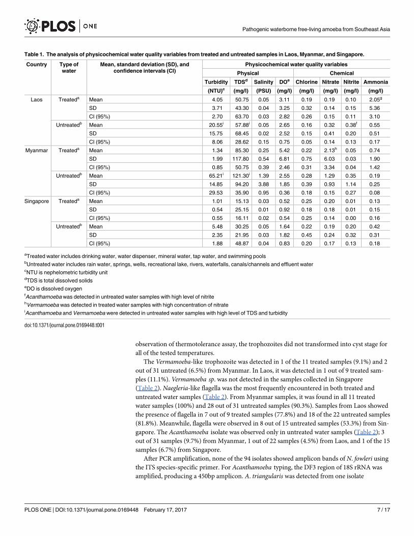
Table 1 summarizes the physicochemical analysis of water quality parameters, presented as cal-
culated means ± standard deviation (SD), with 95% confidence intervals (CIs). Overall results
showed untreated water samples from Myanmar had the highest readings of turbidity (65.21
±14.85 NTU; CI: 29.53), TDS (121.30±94.20 mg/L; CI: 35.90), chlorine (0.28±0.39 mg/L; CI:
0.18). The presence of Acanthamoeba and Vermamoeba were detected in water samples with
high TDS and turbidity. In Myanmar, Vermamoeba was the only pathogenic FLA found in
treated water samples with high nitrate concentration (2.13±6.03 mg/L; CI: 3.34). In Laos,
Acanthamoeba was detected in untreated water samples with high level of nitrite (0.38±0.20
mg/L; CI: 0.13) and Vermamoeba was also detected in treated water samples with high level of
ammonia (2.05±5.36 mg/L; CI: 3.10).
Initially, positive amoebic-like cells exist as mixed isolates with other organisms. The tro-
phozoite and cyst stages were normally seen after 2–3 days and at least 5–6 days of cultivation,
respectively. Both stages were photographed in Fig 3. All isolates were grown at room tempera-
ture. The morphology of all isolates can be clearly differentiated based on its motile stage of
trophozoite/flagella. The Acanthamoeba-like cell exhibited irregular shape with distinct projec-
tion of pseudopodia/acanthopodia in multidirectional movement. The Vermamoeba-like tro-
phozoite showed a predominant cylindrical form with monopodia and moving in one
direction. Meanwhile, for Naegleria-like cell, the flagellate stage was observed on the watery
surface of the medium agar (not shown).
Two staining methods (i.e. Giemsa and immunofluorescence) were used for the morpho-
logical identification of pathogenic amoebae and were compared with the non-stained slides
as a control. In control slides, the nucleus and cytoplasm of the trophozoite were clearly distin-
guished, whereas two distinct layers of the cyst were observed for both Acanthamoeba and Ver-mamoeba isolates (Fig 3A–3D). For Giemsa, the trophozoite and cyst of the parasites were
stained purple and showed a good contrast with the background. The eruptive pseudopodia of
Acanthamoeba and the cylindrical form of Vermamoeba trophozoites were seen but unclear
colour contrast between nucleus and cytoplasm was encountered (Fig 3E and 3F). Neverthe-
less, both outer and inner layers of the cysts were successfully observed as dark purple in colour
(Fig 3G and 3H). The USEPA method 1623 was implemented to stain the amoebic cysts, simi-
larly to Cryptosporidium and Giardia (oo)cysts. The inner layer was stained green, whereas the
outer layer was appeared unstained against the dark background (Fig 3I and 3J). From the
Pathogenic waterborne free-living amoeba from Southeast Asia
PLOS ONE | DOI:10.1371/journal.pone.0169448 February 17, 2017 6 / 17
observation of thermotolerance assay, the trophozoites did not transformed into cyst stage for
all of the tested temperatures.
The Vermamoeba-like trophozoite was detected in 1 of the 11 treated samples (9.1%) and 2
out of 31 untreated (6.5%) from Myanmar. In Laos, it was detected in 1 out of 9 treated sam-
ples (11.1%). Vermamoeba sp. was not detected in the samples collected in Singapore
(Table 2). Naegleria-like flagella was the most frequently encountered in both treated and
untreated water samples (Table 2). From Myanmar samples, it was found in all 11 treated
water samples (100%) and 28 out of 31 untreated samples (90.3%). Samples from Laos showed
the presence of flagella in 7 out of 9 treated samples (77.8%) and 18 of the 22 untreated samples
(81.8%). Meanwhile, flagella were observed in 8 out of 15 untreated samples (53.3%) from Sin-
gapore. The Acanthamoeba isolate was observed only in untreated water samples (Table 2); 3
out of 31 samples (9.7%) from Myanmar, 1 out of 22 samples (4.5%) from Laos, and 1 of the 15
samples (6.7%) from Singapore.
After PCR amplification, none of the 94 isolates showed amplicon bands of N. fowleri using
the ITS species-specific primer. For Acanthamoeba typing, the DF3 region of 18S rRNA was
amplified, producing a 450bp amplicon. A. triangularis was detected from one isolate
Table 1. The analysis of physicochemical water quality variables from treated and untreated samples in Laos, Myanmar, and Singapore.
Country Type of
water
Mean, standard deviation (SD), and
confidence intervals (CI)
Physicochemical water quality variables
Physical Chemical
Turbidity TDSd Salinity DOe Chlorine Nitrate Nitrite Ammonia
(NTU)c (mg/l) (PSU) (mg/l) (mg/l) (mg/l) (mg/l) (mg/l)
Laos Treateda Mean 4.05 50.75 0.05 3.11 0.19 0.19 0.10 2.05g
SD 3.71 43.30 0.04 3.25 0.32 0.14 0.15 5.36
CI (95%) 2.70 63.70 0.03 2.82 0.26 0.15 0.11 3.10
Untreatedb Mean 20.55i 57.88i 0.05 2.65 0.16 0.32 0.38f 0.55
SD 15.75 68.45 0.02 2.52 0.15 0.41 0.20 0.51
CI (95%) 8.06 28.62 0.15 0.75 0.05 0.14 0.13 0.17
Myanmar Treateda Mean 1.34 85.30 0.25 5.42 0.22 2.13h 0.05 0.74
SD 1.99 117.80 0.54 6.81 0.75 6.03 0.03 1.90
CI (95%) 0.85 50.75 0.39 2.46 0.31 3.34 0.04 1.42
Untreatedb Mean 65.21i 121.30i 1.39 2.55 0.28 1.29 0.35 0.19
SD 14.85 94.20 3.88 1.85 0.39 0.93 1.14 0.25
CI (95%) 29.53 35.90 0.95 0.36 0.18 0.15 0.27 0.08
Singapore Treateda Mean 1.01 15.13 0.03 0.52 0.25 0.20 0.01 0.13
SD 0.54 25.15 0.01 0.92 0.18 0.18 0.01 0.15
CI (95%) 0.55 16.11 0.02 0.54 0.25 0.14 0.00 0.16
Untreatedb Mean 5.48 30.25 0.05 1.64 0.22 0.19 0.20 0.42
SD 2.35 21.95 0.03 1.82 0.45 0.24 0.32 0.31
CI (95%) 1.88 48.87 0.04 0.83 0.20 0.17 0.13 0.18
aTreated water includes drinking water, water dispenser, mineral water, tap water, and swimming poolsbUntreated water includes rain water, springs, wells, recreational lake, rivers, waterfalls, canals/channels and effluent watercNTU is nephelometric turbidity unitdTDS is total dissolved solidseDO is dissolved oxygenfAcanthamoeba was detected in untreated water samples with high level of nitritehVermamoeba was detected in treated water samples with high concentration of nitrateiAcanthamoeba and Vermamoeba were detected in untreated water samples with high level of TDS and turbidity
doi:10.1371/journal.pone.0169448.t001
Pathogenic waterborne free-living amoeba from Southeast Asia
PLOS ONE | DOI:10.1371/journal.pone.0169448 February 17, 2017 7 / 17
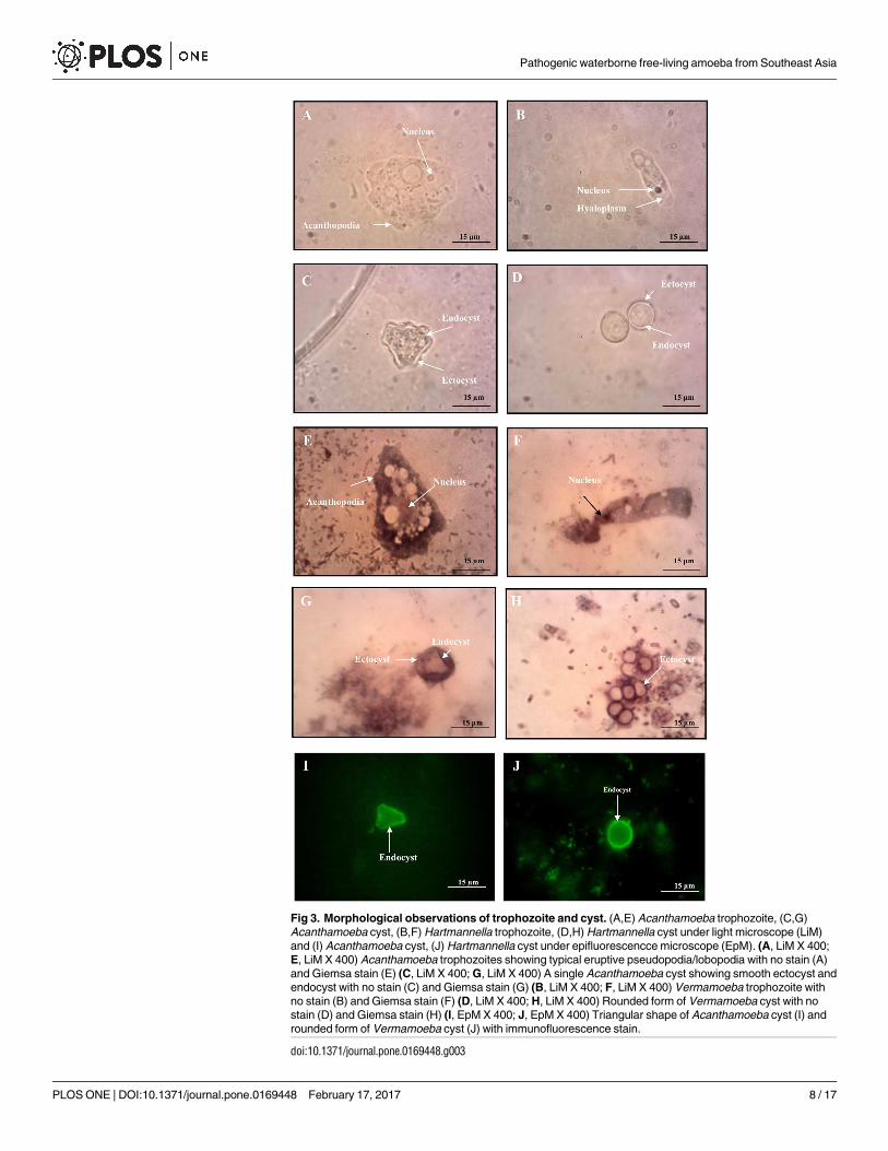
Fig 3. Morphological observations of trophozoite and cyst. (A,E) Acanthamoeba trophozoite, (C,G)
Acanthamoeba cyst, (B,F) Hartmannella trophozoite, (D,H) Hartmannella cyst under light microscope (LiM)
and (I) Acanthamoeba cyst, (J) Hartmannella cyst under epifluorescencce microscope (EpM). (A, LiM X 400;
E, LiM X 400) Acanthamoeba trophozoites showing typical eruptive pseudopodia/lobopodia with no stain (A)
and Giemsa stain (E) (C, LiM X 400; G, LiM X 400) A single Acanthamoeba cyst showing smooth ectocyst and
endocyst with no stain (C) and Giemsa stain (G) (B, LiM X 400; F, LiM X 400) Vermamoeba trophozoite with
no stain (B) and Giemsa stain (F) (D, LiM X 400; H, LiM X 400) Rounded form of Vermamoeba cyst with no
stain (D) and Giemsa stain (H) (I, EpM X 400; J, EpM X 400) Triangular shape of Acanthamoeba cyst (I) and
rounded form of Vermamoeba cyst (J) with immunofluorescence stain.
doi:10.1371/journal.pone.0169448.g003
Pathogenic waterborne free-living amoeba from Southeast Asia
PLOS ONE | DOI:10.1371/journal.pone.0169448 February 17, 2017 8 / 17
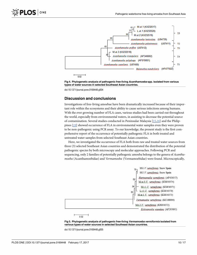
(KX232518) from Myanmar and formed a clade with both A. triangularis (AF346662) and A.
polyphaga (AF019061) in T4 group. Another two isolates (KX232517, KX232519) from Myan-
mar and one isolate (KX232520) from Laos were identified as A. lenticulata. They were
grouped as T5 and formed a clade with A. lenticulata (U94739). Balamuthia mandrillaris(AF477022) that doesn’t contain DF3 region showed distantly related with the Acanthamoebagroups (Table 3, Fig 4). A 800 bp PCR product was obtained for all of the Vermamoeba isolates
(KX856370, KX856371, KX856372, KX856373, KX856374) and sequence analysis revealed that
the amoebae had a high homology of 99% to V. vermiformis (KC188996). All other V. vermifor-mis strains, including 5 environmental sequences from present study, clearly showed a clade
with each other under the group of Vermamoebidae. At the family level, E. exundans formed a
distinct group of Echinamoebidae against the Vermamoebidae, within the order of Echina-
moebida (Table 3, Fig 5). V. vermiformis pairwise distance between ice cube isolate and snow
strain from Spain showed 0.5% intraspecific variation. BLAST results revealed the similarity at
99% and 98% of ice cube isolate-SS1 and ice cube isolate-SS2 strains, respectively (data not
shown).
Table 2. The occurrence of pathogenic FLA via microscopic examination and PCR in Laos, Myanmar, and Singapore.
Country of origin Type of water No. of samples Free-living amoeba (FLA)
Vermamoeba sp. Naegleria sp. Acanthamoeba sp.
M (n) PCR (n) M (n) PCR (n) M (n) PCR (n)
Laos Treateda 9 1 V. vermiformis (1) 7 NDc NDc NDc
Untreatedb 22 NDc NDc 18 NDc 1 A. lenticulata (1)
Myanmar Treateda 11 2 V. vermiformis (2) 11 NDc NDc NDc
Untreatedb 31 2 V. vermiformis (2) 28 NDc 3 A. lencticulata (2); A. triangularis (1)
Singapore Treateda 6 NDc NDc NDc NDc NDc NDc
Untreatedb 15 NDc NDc 8 NDc 1 NDc
Total 94
aTreated water includes drinking water, water dispenser, mineral water, tap water, and swimming poolsbUntreated water includes rain water, springs, wells, recreational lake, rivers, waterfalls, canals/channels and effluent watercND = not detected; M = Microscopy; n = Number of samples; PCR = Polymerase chain reaction; V = Vermamoeba; A = Acanthamoeba
doi:10.1371/journal.pone.0169448.t002
Table 3. Pathogenic FLA isolated from selected Southeast Asian countries.
Pathogenic FLA Code Accession number Country of origin Source of water
A. lenticulata M.ut.1 KX232517 Myanmar Well water
A. lenticulata M.ut.2 KX232519 Myanmar Recreational lake
A. lenticulata L.ut.1 KX232520 Laos Mekong up 2
A. triangularis M.ut.3 KX232518 Myanmar Fish pond 2
V. vermiformis M.t.1 KX856371 Myanmar Ice cube
V. vermiformis M.t.2 KX856372 Myanmar Swimming pool 2
V. vermiformis M.ut.1 KX856373 Myanmar Well water
V. vermiformis M.ut.3 KX856374 Myanmar Fish pond 2
V. vermiformis L.t.1 KX856370 Laos Swimming pool adult
V = Vermamoeba; A = Acanthamoeba; FLA = Free-living amoeba
doi:10.1371/journal.pone.0169448.t003
Pathogenic waterborne free-living amoeba from Southeast Asia
PLOS ONE | DOI:10.1371/journal.pone.0169448 February 17, 2017 9 / 17
Discussion and conclusions
Investigations of free-living amoebae have been dramatically increased because of their impor-
tant role within the ecosystems and their ability to cause serious infections among humans.
With the ever growing number of FLA cases, various studies had been carried out throughout
the world, especially from environmental waters, in assisting to decrease the potential source
of contamination. Several studies conducted in Peninsular Malaysia [15,16] and the Philip-
pines [19] showed occurrence of FLA in environmental water samples even they were proven
to be non-pathogenic using PCR assay. To our knowledge, the present study is the first com-
prehensive report of the occurrence of potentially pathogenic FLA in both treated and
untreated water samples from selected Southeast Asian countries.
Here, we investigated the occurrence of FLA both from raw and treated water sources from
three (3) selected Southeast Asian countries and demonstrated the distribution of the potential
pathogenic species by both microscopy and molecular approaches. Following PCR and
sequencing, only 2 families of potentially pathogenic amoeba belongs to the genera of Acantha-moeba (Acanthamoebidae) and Vermamoeba (Vermamoebidae) were found. Microscopically,
Fig 4. Phylogenetic analysis of pathogenic free-living Acanthamoeba spp. isolated from various
types of water sources in selected Southeast Asian countries.
doi:10.1371/journal.pone.0169448.g004
Fig 5. Phylogenetic analysis of pathogenic free-living Vermamoeba vermiformis isolated from
various types of water sources in selected Southeast Asian countries.
doi:10.1371/journal.pone.0169448.g005
Pathogenic waterborne free-living amoeba from Southeast Asia
PLOS ONE | DOI:10.1371/journal.pone.0169448 February 17, 2017 10 / 17
Myanmar reported the highest occurrence of FLA (92%, 39/42) followed by Laos (87%, 27/31),
and Singapore (43%, 9/21). The rate of FLA in Laos and Myanmar were surprisingly found
higher in comparison to a previous study conducted in this region such as Thailand (45.2%,
43/95) [17], though these countries share much similarity in geographical distribution. More-
over, this figure is remarkably higher than other countries namely; Japan (49.5%, 47/95) [17],
USA (43.4%, 143/330) and Iran (35%, 42/120), but lower than Bulgaria (93.9%, 31/33) [32–34].
The presence of both Acanthamoeba and Vermamoeba were revealed with high values for
both turbidity and TDS. High turbidity showed that the water contained other soluble matters
that can provide nutrition and encourage growth rate for the amoeba. The high reading of tur-
bidity demonstrated that these sediments supported the microbial growth and in return,
becoming the source of food for the amoeba [35]. This finding also supported by a study con-
ducted in the Philippines’s water sources with the occurrence of Acanthamoeba and Naegleriathat were rich in TDS [19]. This scenario will lead to reduction of water quality which reflects
the nutrient availability that could help the parasites to revive in a suitable environment. The
result also showed the ability of A. triangularis to uphold high concentration of nitrite
(> 0.5 ppm) and ammonia (> 0.5 ppm) [36]. The high content of both nitrite and ammonia
assist in the growth of Gram-negative bacteria that may become the food source for A. triangu-laris. In addition, nitrite was also found to be a good indicator for the presence of other para-
sites, namely, Cryptosporidium and Giardia [19, 37].
Another study was conducted in France, showing a wide biodiversity of FLA in water treat-
ment plants that used river water as its main source [38]. A similar study was also carried out
in Sarawak ofEast Malaysia that revealed the presence of FLA in various processing sites in
treatment plants [39]. Relatively, similar treatment processes that include chlorination and fil-
tration are practiced in Myanmar and Laos. Given this, the treated water in both aforemen-
tioned countries was contaminated with V. vermiformis. This may be due to the ability of V.
vermiformis cyst to resist the harsh condition of physical and chemical treatments that are used
in the treatment plants.
Cultivation of FLA obtained from water samples was carried out to impede excessive
growth of unwanted contaminants such as bacteria and fungi. Hence, this technique was cho-
sen as it is able to grow large quantity of FLA within a short period of time. FLA is able to grow
at room temperature (i.e. 25˚C), as higher temperature may trigger the overgrowth of bacteria
instead of amoeba. The high ratio of bacteria to amoeba (i.e. 10 > 1) that contained in the
uncultured water can suppress the growth of Acanthamoeba [40]. The cultivation method was
performed on the NNA (non-nutrient agar) with non-mucoid bacteria (i.e. Escherichia coli) as
the food source. In addition, bacteria with mucoid were not preferable as it may delay the
phagocytosis by amoeba that may leads to bacterial overgrowth. Sub-cultivation was also per-
formed to assist in removing debris that may contain living/dead bacterial cells, trapped inor-
ganic particles, and organic fibers [41]. Furthermore, the contaminants may obstruct the
detection of FLA, especially those obtained from environmental water samples. Staining of the
stages of all pathogenic FLA revealed a better observation of the features (i.e. shape) and cellu-
lar organelles. The smears of the samples were firstly fixed using methanol to prevent the tro-
phozoites and cysts to become distorted or shrunk. Giemsa, which is commonly used for
blood parasites, has produced good contrast against the background for both trophozoites and
cysts in the present study. Nevertheless, the space between the endocyst and ectocyst were also
stained blue and can cause confusion in determining or confirming the distinct features of
both layers. Moreover, trophozoite possessed a better contrast due to its larger size in compari-
son to cysts. The findings from this study showed that Giemsa stain can be used, especially for
routine screening to identify the developmental stages of FLA in general, and trophozoites
detection in particular, for clinical epidemiology and public health purposes.
Pathogenic waterborne free-living amoeba from Southeast Asia
PLOS ONE | DOI:10.1371/journal.pone.0169448 February 17, 2017 11 / 17
In addition, fluorescence method is used in FLA identification based on its concept of con-
jugation of protein-antigen and proved to be useful because of the failure of Giemsa stain
intake by the cysts. Only endocyst was stained with bright apple-green colour and able to dif-
ferentiate the number of arms and polygonal shapes for detailed morphological classification
of pathogenic Acanthamoeba. Hence, it is revealed that the endocyst of FLA consisted of cellu-
lose, a similar material that covers the (oo)cysts of Cryptosporidium and Giardia [42] that
enables simultaneous detection in detecting waterborne parasites containing FLA, Cryptospo-ridium, and Giardia.
Overall, observation of the trophozoite stage of FLA by microscopy has permitted the dif-
ferentiation at genus level. The trophozoites of Naegleria were smaller in size as compared to
those of Acanthamoeba and Vermamoeba and were observed to move faster in a unidirectional
manner. Vermamoeba trophozoites with a transient cylindrical form possessed similar pattern
of movement as Naegleria. Acanthamoeba showed multiple shapes of trophozoites and moving
by the projection of lobopodia/acanthopodia in multidirectional. It is known that the classifi-
cation of FLA based on morphological criteria is insufficient, while their identification is not
problematic using different PCR assays, including conventional PCR.
The diagnosis of infection and identification of pathogenic FLA remains unsatisfactory,
time-consuming, expensive, laborious, and prone to ethical issues when mouse pathogenicity
test was taken into consideration. Due to the advancements in molecular detection, conven-
tional PCR has been developed and seem to be reliable method for routine screening of FLA in
environmental samples. One-step based PCR, paired with specific primer by targeting the
region of ITS and 18S rDNA [27] is sufficient enough to differentiate between and within spe-
cies of various organisms. Furthermore, the primer sets used in the present study showed high
specificity and capable to amplify a product from potentially pathogenic FLA genotypes, but
not with other closely related genera of amoeba. For example, the JDP1-JDP2 primers had
been proven to produce an amplicon even from a single trophozoite of Acanthamoeba [27].
The internal transcribed spacers (ITS) region has been chosen to study the differences
within the genus of Naegleria [43]. The ITS analysis has been used in studying heterogeneity in
several organisms such as Cryptosporidium parvum, trichomonadid protozoa, and Valkamphia[42,44]. The species-specific primer was used to detect the presence of pathogenic N. fowleri[45]. Nevertheless, no positive amplification of N. fowleri was obtained against the Naegleria-
like isolates. This might be due to the fact that N. fowleri is a thermophilic organism that prolif-
erates at an ambient temperature (as high as 45˚C) [46].
On the other hand, four of the environmental Acanthamoeba-like isolates were successfully
amplified using the species-specific primer of JDP1 and JDP2, producing approximately 460
bp amplicons of potentially pathogenic A. triangularis and A. lenticulata. The sequence of 18S
rDNA from Acanthamoeba isolates led into 13 different lineages containing either single or
complexes of species. Based on the classification, both A. triangularis and A. lenticulata had
been classified as type T4 and T5, respectively, and often been associated with human systemic
infection and keratitis. Many previous studies similarly reported that T4 is the most prevalent
genotype among both clinical specimens and environment samples. The presence of both iso-
lates in our samples probably reflects their better adaptation to various growth conditions rela-
tive to isolates from other genotypes.
Morphology of Vermamoeba vermiformis isolates from the present study was similar to pre-
viously reported species under the microscope [47]. The typical trophozoites of Vermamoebadisplayed worm-like cell body, stable anterior hyaline cap, and possess a tendency branch
when they changed a direction. The 18S rRNA gene sequences of all reported V. vermiformisisolates in present study showed extremely high similarity with more than 96% identical to
AF426157 and KC188996. Further, phylogenetic analysis of 18S rRNA gene sequences in
Pathogenic waterborne free-living amoeba from Southeast Asia
PLOS ONE | DOI:10.1371/journal.pone.0169448 February 17, 2017 12 / 17
Amoebozoa revealed that Hartmannellidae and Vermamoebidae are paraphyletic and mono-
phyletic group, respectively.
Findings conducted throughout the world had reported that the Vermamoeba species is
considered pathogenic that is also tolerant towards high temperature [48]. The thermotoler-
ance behaviour is one of the factors that contributed to the virulence characteristic that able to
cause diseases and leads to death. Interestingly, our study revealed positive isolate from cold
condition of ice cube that are similar to the result carried out in Spain [31]. Thus, it was pur-
ported that V. vermiformis has the ability to survive in various conditions, besides exhibiting
thermotolerant properties.
The presence of these parasites is rather alarming due to its ability in causing parasitic infec-
tions. Site observation in Myanmar and Laos revealed that fishing activities are prominent in
the rivers and ponds. Consumption of these contaminated fishes (raw or under-cooked) with
potentially pathogenic FLA could cause a fatal disease to humans. A previous study revealed
that freshwater fishes contained several Hartmannella vermiformis strains found from its
organs [49], thus, there is a possibility that fish can be a host for this parasite. However, FLA
transmissions via food are still questionable and more studies in the future need to be carried
out to further proven the presence of amoeba within host tissues.
For the past decade, keratitis cases had increased dramatically parallel with the popularity
of contact lens usage, hence, resulting in more reports of clinically confirmed cases in South-
east Asia. Acanthamoeba species was revealed to be the main cause of keratitis infection in raw
water in Thailand [50,51] and Vietnam [52], in regards with water-related activities (i.e. swim-
ming and diving) and accidental water-splashing. In the present study, A. triangularis was
found in fish pond sample obtained in Myanmar. A previous study has confirmed the role of
A. triangularis in causing human keratitis [53]. Usually, Acanthamoeba spp. can be isolated
from environmental water that is rich with sediments and other particulate matters [54]. In
addition, our study confirmed the presence of A. lenticulata in water sources such as Mekong
River, well water, as well as recreational lake in Myanmar and Laos. This amoeba has also been
reported causing granulomatous amoebic encephalitis (GAE) in immunocompromised
patients [55].
The occurrence of FLA in treated water cannot be neglected due to the increasing reports
of keratitis in Malaysia [56], Indonesia [57], and the Philippines [58]. Keratitis had always
been associated with the infection caused by Acanthamoeba, but Hartmannella (= Verma-moeba) too, is able to cause similar symptoms of watery eyes, redness and blurred vision
[59,60]. In the present study, V. vermiformis was isolated mostly from treated water of swim-
ming pools from Myanmar and Laos. This situation revealed that V. vermiformis is able to
adapt in chlorinated water and showed the ability to survive in the ecology. Although lim-
ited epidemiological data available, chlorinated swimming pool can possibly be a new niche
for FLA to survive. The alarming phenomenon is considered a major concern, especially to
contact lens wearers as they are susceptible to keratitis infection if lens are not appropriately
handled especially before and after entering the pool. In addition, the pipe that connects
water into the swimming pools may contain biofilm that support the colonization of FLA
[61].
From this finding, the isolation of FLA revealed a better understanding on the distribution
and prevalence of both pathogenic Acanthamoeba and Vermamoeba in Myanmar and Laos.
The high occurrence of amoebae in Myanmar must not be neglected by the local authorities to
ensure good water quality is supplied to the consumers. Proper precaution measures in ensur-
ing water quality must be carried out to assist in regular monitoring on any possibilities of the
emerging amoebae. This is because of the wide occurrences of FLA is not only limited to one
species at any particular water source or area. Meanwhile, water that is commonly used by
Pathogenic waterborne free-living amoeba from Southeast Asia
PLOS ONE | DOI:10.1371/journal.pone.0169448 February 17, 2017 13 / 17
public, such as swimming pool, must undergo efficient and appropriate treatment processes.
The usage of alternative disinfectants such as Baquacil, chlorinated cyanurate and chlorine
dioxide can be added into the water to control the growth of FLA [62,63]. Raw water that is
used for recreational purposes (i.e. hotspring, coastal) must also be monitored closely as the
untreated water may contain high number of FLA, thus, can transmit infections to the public
[64,65]. In addition, the infection caused by FLA is still misdiagnosed or under-reported, par-
ticularly in this region. Thus, future investigations of the occurrence as well as distribution of
FLA in treated and untreated water with a larger sample size need to be taken in serious con-
sideration. The findings from the investigations will be reported to relevant authorities to pre-
vent further widespread of water contamination with potentially pathogenic species of these
free-living parasites.
Acknowledgments
The authors would like to thank Ms. Thulasi Kumar and Ms. Subashini Onichandran from
Malaysia, technical staffs from Lao PDR; Dr. Aye Aye Win and Ms. Lin Myat Myat Aung from
Myanmar, as well as Mr. Meng-Hwee Lim from Singapore, who helped us with the water sam-
pling and laboratory works. We would also like to thank Dr. Kruawan Chotelersak from Srina-
kharinwirot University, Thailand, and Dr Romano Ngui from Malaysia for phylogenetic
analysis, Dr Hany Sady from Yemen for thermotolerance assay and Ms. Alicia JC Choo from
Malaysia for graphic improvement and data retrieved from external sources. Last but not least,
we would also like to convey our deepest gratitude to Prof. Dr. Jacob Lorenzo-Morales from
University of La Laguna, Spain for providing us the sequences of Vermamoeba vermiformisstrains obtained from snow sample. This work was presented in part at the 1st International
Conference on Pollutant Toxic Ions and Molecules (PTIM2015) on 2nd– 4th November, 2015
in Caparica, Portugal.
Author Contributions
Conceptualization: VN TTC MAAM.
Data curation: MAAM TM BGJM NJ RLR.
Formal analysis: MAAM RLR.
Funding acquisition: VN TTC.
Investigation: VN TTC AP PM KNA WLA JC ADZ AY NS YALL.
Methodology: MAAM TM BGJM NJ RLR.
Project administration: VN TTC.
Resources: VN TTC.
Software: MAAM.
Supervision: VN TTC.
Validation: VN TTC MAAM BGJM AY.
Writing – original draft: MAAM.
Writing – review & editing: VN TTC JC ADZ AY NS YALL MAAM.
Pathogenic waterborne free-living amoeba from Southeast Asia
PLOS ONE | DOI:10.1371/journal.pone.0169448 February 17, 2017 14 / 17
References1. Visvesvara GS, Moura H, Schuster FL. Pathogenic and opportunistic free living amoebae: Acantha-
moeba spp., Balamuthia mandrillaris, Naegleria fowleri, and Sappinia diploidea. FEMS Immun Med
Microbiol. 2007; 50: 1–26.
2. Qvarnstrom Y, da Silva AJ, Schuster FL, Gelman BB, Visvesvara GS. Molecular confirmation of Sappi-
nia pedata as a causative agent of amoebic encephalitis. J Infect Dis. 2009; 199: 1139–1142. doi: 10.
1086/597473 PMID: 19302010
3. Schuster FL, Visvesvara GS. Free-living amoebae as opportunistic and non-opportunistic pathogens of
humans and animals. Int J Parasitol. 2004; 34: 1001–1027. doi: 10.1016/j.ijpara.2004.06.004 PMID:
15313128
4. Khan NA. Acanthamoeba: biology and increasing importance in human health. FEMS Microbiol Rev.
2006; 30: 564–595. doi: 10.1111/j.1574-6976.2006.00023.x PMID: 16774587
5. Teixera LH, Rocha S, Pinto RMF, Caseiro MM, da Costa SOP. Prevalence of potentially pathogenic
pathogenic free-living amoebae from Acanthamoeba and Naegleria genera in non-hospital, public,
interval environments from the city of Santos, Brazil. Braz J of Infect Dis. 2009; 13: 395–397.
6. Greub G, Raoult D. Biocides currently used for bronchoscope decontamination are poorly effective
against free-living amoebae. Infect Cont Hosp Ep. 2003; 24: 784–786.
7. Marciano-Cabral F, Cabral G. Acanthamoeba spp. as agents of disease in humans. Clin Microbiol Rev.
2003; 16: 273–307. doi: 10.1128/CMR.16.2.273-307.2003 PMID: 12692099
8. Marciano-Cabral F, Puffenbarger R, Cabral GA. The increasing importance of Acanthamoeba infec-
tions. J Eukaryot Microbiol. 2000; 47: 29–36. PMID: 10651293
9. Corsaro D, Walochnik J, Kohsler M, Rott MB. Acanthamoeba misidentification and multiple labels: rede-
fining genotypes T16, T19, and T20 and proposal for Acanthamoeba micheli sp. nov. (genotype T19).
Parasitol Res. 2015; 114: 2481–2490. doi: 10.1007/s00436-015-4445-8 PMID: 25869957
10. De Jonckheere JF. A century of research on the amoeboflagellate genus Naegleria. Acta Protozool.
2002; 47: 309–342.
11. Kaushal V, Chhina DK, Ram S, Singh G, Kaushal RK, Kumar R. Primary Amoebic Meningoencephalitis
due to Naegleria fowleri. J Assoc Phy Ind. 2008; 56: 459–62.
12. Aitken D, Hay J, Kinnear FB, Kirkness CM, Lee WR, Seal DV. Amoebic keratitis in a wearer of dispos-
able contact lenses due to a mixed Vahlkampfia and Hartmannella infection. Ophthalmology. 1996;
103: 485–494. PMID: 8600427
13. Brieland JK, Fantone JC, Remick DG, LeGendre M, McClain M, Engleberg NC. The role of Legionella
pneumophila infected Hartmannella vermiformis as an infectious particle in a murine model of Legion-
naires’ disease. Infect Immun. 1997; 65: 5330–5333. PMID: 9393834
14. Greub G, Raoult D. Microorganisms resistant to free-living amoeba. Clin Microbiol. 2004; 17: 413–433.
15. Init I, Lau YL, Arine FA, Foead AI, Neilson RS, Nissapatorn V. Detection of free-living amoebae,
Acanthamoeba and Naegleria, in swimming pools, Malaysia. Trop Biomed. 2010; 27: 566–577. PMID:
21399599
16. Ithoi I, Ahmad AF, Nissapatorn V, Lau YL, Mahmud R, Joon WM. Detection of Naegleria species in envi-
ronmental samples from Peninsular Malaysia. Plos One. 2011; 6: 324–327.
17. Nacapunchai D, Kino H, Ruangsitticha C, Sriwichai P, Ishih A, Terada M. A brief survey of free-living
amebae in Thailand and Hamamatsu District Japan. Southeast Asian J Trop Med Public Health. 2001;
32: 179–182. PMID: 12041586
18. Herriman R. Vietnam reports second ever ‘brain-eating amoeba’ death. 2012. http://www.examiner.
com/article/vietnam-reports-second-ever-brain-eating-amoeba-death.
19. Onichandran S, Kumar T, Salibay CC, Dungca JZ, Tabo HAL, Tabo N, et al. Waterborne parasites: a
current status from the Philippines. Parasit Vectors. 2014; 7: 244. doi: 10.1186/1756-3305-7-244 PMID:
24885105
20. Ghani MKA, Annuar FH, Ahmad N, Abdullah NR, Hay J, Seal D. A case of waterborne contact lens
associated Acanthamoeba keratitis from Malaysia: Successful treatment with Chlorhexidine and Propa-
midine. Int Med J. 2000; 7: 63–65.
21. Por YM, Mehta JS, Chua JLL, Koh TH, Khor WB, Fong AC, et al. Acanthamoeba keratitis associated
with contact lens wear in Singapore. Am J Ophthalmol. 2009; 148: 1879–1891.
22. Bunjongpuk S. Primary amoebic meningoencephalitis (PAM). Thai J Paed. 2000; 39: 38–49.
23. Sithinamsuwan P, Sangruchi T, Chiewvit P, Poungvarin N. Free-living amoeba infections of the central
nervous system in Thailand. Report of two patients. Int Med J Thai. 2000; 17: 350–360.
Pathogenic waterborne free-living amoeba from Southeast Asia
PLOS ONE | DOI:10.1371/journal.pone.0169448 February 17, 2017 15 / 17
24. Intalapaporn P, Suankratay C, Shuangshoti S, Phantumchinda K, Keelawat S, Wilde H. Balamuthia
mandrillaris meningoencaphalitis: the first case in Southeast Asia. Am J Trop Med Hyg. 2004; 70: 666–
669. PMID: 15211011
25. Page FC. Taxonomic criteria for limax amoebae, with descriptions of 3 new species of Hartmannella
and 3 of Vahlkampfia. J Protozool. 1967; 14: 499–521. PMID: 6050658
26. Page FC. A new key to freshwater and soil gymnamoebae. Freshwater Biol Assoc, Ambleside. 1988
UK
27. Qvarnstrom Y, Nerad TA, Visvesvara GS. Characterization of a new pathogenic Acanthamoeba spe-
cies, A. byersi n. sp., isolated from a human with fatal amoebic encephalitis. J Eukaryot Microbiol. 2013;
60(6): 626–633. doi: 10.1111/jeu.12069 PMID: 23879685
28. Schroeder JM, Booton GC, Hay J, Niszl IA, Seal DV, Markus MB, et al. Use of subgenic 18S ribosomal
DNA PCR and sequencing for genus and genotype identification of Acanthamoeba from humans with
keratitis and from sewage sludge. J Clin Microbiol. 2001; 39: 1903–1911. doi: 10.1128/JCM.39.5.1903-
1911.2001 PMID: 11326011
29. Pelandakis M, Serre S, Pernin P. Analysis of the 5.8S rRNA gene and internal transcribed spacers in
Naegleria spp. and in N. fowleri. J Eukaryot Microbiol. 2000; 47: 116–121. PMID: 10750838
30. Lasjardi Z, Niyyati M, Haghighi A, Shahabi S, Biderouni FT, Taghipour N, et al. Potentially pathogenic
free-living amoebae isolated from hospital wards with immunodeficient patients in Tehran, Iran. Parasi-
tol Res. 2011; 109: 575–580. doi: 10.1007/s00436-011-2288-5 PMID: 21365453
31. Reyes-Batlle M, Niyyati M, Martin-Navarro CM, Lopez-Arencibia A, Valladares B, Martinez-Carretero E,
et al. Unusual Vermamoeba vermiformis strain isolated from snow in Mount Teide, Tenerife, Canary
Islands, Spain. Novelty in Biomedicine. 2015; 4: 189–192.
32. Ettinger MR, Webb SR, Harris SA, McIninch SP, Garman G, Brown BL. Distribution of free-living amoe-
bae in James River,Virginia, USA. Parasitol Res. 2003; 89: 6–15. doi: 10.1007/s00436-002-0707-3
PMID: 12474037
33. Sh Ghadar-ghadr, Solhjoo K, Norouz-nejad MJ, Rohi R, Zia-Jahromi S. Isolation and identification of
free living amoeba (Naegleria and Acanthamoeba) in Shiraz water resources by morphological criteria.
J Jahrom Uni Med Sci. 2012; 10: 26–33.
34. Tsvetkova N, Schild M, Panaiotov S, Kurdova-Mintcheva R, Gottstein B, Walochnik J, et al. The identifi-
cation of free-living environmental isolates of amoebae from Bulgaria. Parasitol Res. 2004; 92: 405–
413. doi: 10.1007/s00436-003-1052-x PMID: 14760525
35. Valster RM, Wullings BA, van den Berg R, van der Kooji D. Relationships between free-living protozoa,
cultivable Legionella spp., and water quality characteristics in three drinking water supplies in the Carri-
bean. Appl Environ Microbiol. 2011; 77: 7321–7328. doi: 10.1128/AEM.05575-11 PMID: 21873489
36. Patil PN, Sawant DV, Deshmukh RN. Physico-chemical parameters for testing of water—A review. Int J
Environ Sci. 2012; 3: 1194–1207.
37. Natividad FF, Buerano CC, Lago CB, de Guzman BB, Seraspe EB, Samentar LP, et al. Prevalence
rates of Giardia and Cryptosporidium among diarrheic patients in the Philippines. Southeast Asian J
Trop Med. 2008; 39: 991–999.
38. Thomas V, Loret JF, Jousset M, Greub G. Biodiversity of amoebae and amoebae-resisting bacteria in a
drinking water treatment plant. Environ Microbiol. 2008; 10: 2728–2745. doi: 10.1111/j.1462-2920.
2008.01693.x PMID: 18637950
39. Richard RL, Ithoi I, Abd Majid MA, Wan Sulaiman WY, Tan TC, Nissapatorn V, Lim YAL. Monitoring of
waterborne parasites in two drinking water treatment plants: a study in Sarawak, Malaysia. Int J Environ
Res Public Health. 2016; 13(7): 641.
40. Schuster FL. Cultivation of pathogenic and opportunistic free-living amoebas. Clin Microbiol Rev. 2002;
15: 342–354. doi: 10.1128/CMR.15.3.342-354.2002 PMID: 12097243
41. Eikelboom DH. Characteristics of activated sludge flocs. In: Process control of activated sludge plants
by microscopic investigation. United Kingdom: IWA Publishing; 2000. pp.29–41.
42. Brian JH. Microscopy of 4 Pathogenic Enteric Protozoan Parasites: A Review. Lab Med. 2008; 39: 231–
238.
43. Pelandakis M, Pernin P. Use of multiplex PCR and PCR restriction enzyme analysis for detection and
exploration of the variability in the free-living amoeba Naegleria in the environment. Appl Environ Micro-
biol. 2002; 68: 2061–2065. doi: 10.1128/AEM.68.4.2061-2065.2002 PMID: 11916734
44. Gartstecki T, Brown S, De Jonckheere JF. Description of Vahlkamphia signyensis sp. (Heterolobosea),
based on morphological, ultrastructural and molecular characteristics. Eur J Protistol. 2005; 41: 119–
127.
45. De Jonckheere JF. Molecular definition and the ubiquity of species in the genus Naegleria. Protistol.
2004; 155: 89–103.
Pathogenic waterborne free-living amoeba from Southeast Asia
PLOS ONE | DOI:10.1371/journal.pone.0169448 February 17, 2017 16 / 17
46. Izumiyama S, Yagita K, Furushima-Shimogawara R, Asakura T, Karasudani T, Endo T. Occurrence
and distribution of Naegleria species in thermal waters in Japan. J Eukaryot Microbiol. 2003; 50: 514–
515. PMID: 14736147
47. Smirnov AV, Brown S. Guide to the methods of study and identification of soil gymnamoebae. Protistol.
2004; 3: 148–190.
48. Solgi R, Niyyati M, Haghighi A, Nazemalhosseini Mojarad E. Occurrence of thermotolerant Hartman-
nella vermiformis and Naegleria spp. in hot springs of Ardebil Province, Northwest Iran. Iranian J Parasi-
tol. 2012; 7(2): 47–52.
49. Dyakoya I, Pindova Z, Fiala I, Dvorakova H, Machackova B. Fish-isolated of Hartmannella vermiformis
Page, 1967: morphology, phylogeny and molecular diagnosis of the species in tissue lesions. Folia
Parasitologica. 2005; 52: 295–303. PMID: 16405292
50. Jongwutiwes S, Pariyakanok L, Charoenkorn M, Yagita K, Endo T. Heterogeneity in cyst morphology
within isolates of Acanthamoeba from keratitis patients in Thailand. Trop Med Int Heal. 2000; 5: 335–
340.
51. Siripanth C. Amphizoic amoebae: pathogenic free-living protozoa; review of the literature and review of
cases in Thailand. J Med Assoc Thai. 2005; 88: 701–707. PMID: 16149694
52. Phu NH, Mai NTH, Nghia HDT, Chau TTH, Loc PP, Thai IH, et al. Fatal consequences of freshwater
pearl diving. The Lancet. 2013; 381: 176.
53. Xuan YH, Chung BS, Hong YC, Kong HH, Hahn TW, Chung DI. Keratitis by Acanthamoeba triangularis:
Report of cases and characterization of isolates. Korean J Parasitol. 2008; 46: 157–164. doi: 10.3347/
kjp.2008.46.3.157 PMID: 18830055
54. Kyle DE, Noblet GP. Vertical distribution of potentially pathogenic free-living amoebae in freshwater
lakes. J Protozool. 1985; 33: 422–434.
55. Berete S, Combes A, de Jonkheere JF, Datry A, Varnous S, Martinez V, et al. Fatal disseminated
Acanthamoeba lenticulata infection in a heart transplant patient. Emerg Infect Dis. 2007; 13: 736–738.
PMID: 17553253
56. Mohamed Kamel AG, Faridah H, Yusof S, Norazah A, Nakisah MA. A case of trauma related Acantha-
moeba keratitis. Trop Biomed. 2004; 21: 135–8. PMID: 16493405
57. Putri DE, Edwar L, Susiyanti M. Acanthamoeba keratitis in a non-contact lens wearer: A challenge in
diagnosis and management. J Case Rep. 2014; 4: 419–423.
58. Buerano CC, Trinidad AD, Fajardo LSN, Cua IY, Baclig MO, Natividad FF. Isolation of Acanthamoeba
genotype T4 from a non-contact lens wearer from the Philippines. Trop Med Health. 2014; 4: 145–147.
59. Abedkhojasteh H, Niyyati M, Rahimi F, Hei-Dari M, Farnia S, Rezaeian M. First report of Hartmannella
keratitis in a cosmetic soft contact lens wearer in Iran. Iran J Parasitol. 2013; 8: 481–485. PMID:
24454444
60. Lorenzo-Morales J, Martinez-Carretero E, Batista N, Alvarez-Marin J, Bahaya Y, Walochnik J, et al.
Early diagnosis of amoebic keratitis due to a mixed infection with Acanthamoeba and Hartmannella.
Parasitol Res. 2007; 102: 167–169. doi: 10.1007/s00436-007-0754-x PMID: 17899193
61. Kilvington S, Gray T, Dart J, Morlet N, Beeching JR, Frazer DG, et al. Acanthamoeba keratitis: The role
of domestic tap water contamination in the United Kingdom. Invest Ophth Vis Sci. 2004; 45: 165–169.
62. Roslee NF, Abd Ghani MK, Nordin A, Suboh Y, Ab Rahim N. Pemencilan Acanthamoeba spp. daripada
persekitaran akuatik. Jurnal Sains Kesihatan. 2010; 8: 15–19
63. Marciano-Cabral F. Biology of Naegleria spp. Microbiol Rev. 1988; 52: 114–133. PMID: 3280964
64. Szenasi Z, Endo T, Yagita K, Nagy E. Isolation, identification and increasing importance of free living
amoebae causing human disease. J Med Microbiol. 1998; 47: 5–16. doi: 10.1099/00222615-47-1-5
PMID: 9449945
65. Ma P, Visvesvara GS, Martinez AJ, Theodore FH, Daggett PM, Sawyer TK. Naegleria and Acantha-
moeba infections. Rev Infect Dis. 1990; 12: 490–512. PMID: 2193354
Pathogenic waterborne free-living amoeba from Southeast Asia
PLOS ONE | DOI:10.1371/journal.pone.0169448 February 17, 2017 17 / 17

Reproduced with permission of the copyright owner. Further reproduction prohibited withoutpermission.
Related Documents