Pathogenic microorganisms associated with gulls and terns (Laridae) Author: Hubálek, Zdeněk Source: Journal of Vertebrate Biology, 70(3) Published By: Institute of Vertebrate Biology, Czech Academy of Sciences URL: https://doi.org/10.25225/jvb.21009 BioOne Complete (complete.BioOne.org) is a full-text database of 200 subscribed and open-access titles in the biological, ecological, and environmental sciences published by nonprofit societies, associations, museums, institutions, and presses. Your use of this PDF, the BioOne Complete website, and all posted and associated content indicates your acceptance of BioOne’s Terms of Use, available at www.bioone.org/terms-of-use. Usage of BioOne Complete content is strictly limited to personal, educational, and non - commercial use. Commercial inquiries or rights and permissions requests should be directed to the individual publisher as copyright holder. BioOne sees sustainable scholarly publishing as an inherently collaborative enterprise connecting authors, nonprofit publishers, academic institutions, research libraries, and research funders in the common goal of maximizing access to critical research. Downloaded From: https://bioone.org/journals/Journal-of-Vertebrate-Biology on 12 Feb 2022 Terms of Use: https://bioone.org/terms-of-use

Welcome message from author
This document is posted to help you gain knowledge. Please leave a comment to let me know what you think about it! Share it to your friends and learn new things together.
Transcript

Pathogenic microorganisms associated with gulls andterns (Laridae)
Author: Hubálek, Zdeněk
Source: Journal of Vertebrate Biology, 70(3)
Published By: Institute of Vertebrate Biology, Czech Academy ofSciences
URL: https://doi.org/10.25225/jvb.21009
BioOne Complete (complete.BioOne.org) is a full-text database of 200 subscribed and open-access titlesin the biological, ecological, and environmental sciences published by nonprofit societies, associations,museums, institutions, and presses.
Your use of this PDF, the BioOne Complete website, and all posted and associated content indicates youracceptance of BioOne’s Terms of Use, available at www.bioone.org/terms-of-use.
Usage of BioOne Complete content is strictly limited to personal, educational, and non - commercial use.Commercial inquiries or rights and permissions requests should be directed to the individual publisher ascopyright holder.
BioOne sees sustainable scholarly publishing as an inherently collaborative enterprise connecting authors, nonprofitpublishers, academic institutions, research libraries, and research funders in the common goal of maximizing access tocritical research.
Downloaded From: https://bioone.org/journals/Journal-of-Vertebrate-Biology on 12 Feb 2022Terms of Use: https://bioone.org/terms-of-use
Introduction
The avian family Laridae of the order Charadriiformes comprises 23 genera and 103 species, arranged in three subfamilies: Larinae 56 spp., Sterninae 44 spp. and Rynchopinae three spp. (Roskov et al. 2019). Although larids represent one of the most frequent, widely-distributed and well-studied groups of birds that also often interact with human population, relatively little attention has been paid to the pathogenic microbial organisms associated with them, except for some notorious pathogens such as salmonellae, campylobacters or avian influenza viruses. Some larid species can be long-distance carriers of various viral, bacterial, microfungal, protozoan and metazoan diseases transmissible
to man, other vertebrates, invertebrates and even plants (Threlfall 1967, McDiarmid 1969, Cooper 1990, Hubálek 1994, Nuttall 1997, Benskin et al. 2009). One of the few recent papers about this issue compiled 3,619 wild avian mortality events in the USA between 1971 and 2005: Laridae were involved in 233 events comprising infectious causes, and additional 450 events were regarded as environmental which included avian botulism (Newman et al. 2007). It is accepted that certain infections (e.g. avian botulism, salmonellosis, avian cholera, avian influenza, West Nile encephalitis) can adversely impact populations of some wild bird species. However, microbial diseases of larids have not yet been addressed comprehensively, and the aim of this paper is to present such a survey.
This is an open access article under the terms of the Creative Commnons Attribution Licence (CC BY 4.0), which permits use, distribution and reproduction in any medium provided the original work is properly cited.
Pathogenic microorganisms associated with gulls and terns (Laridae)
Zdeněk HUBÁLEK
Institute of Vertebrate Biology of the Czech Academy of Sciences, Brno, Czech Republic; e-mail: [email protected]
Received 8 February 2021; Accepted 26 March 2021; Published online 4 August 2021
Abstract. The monograph reviews viruses, bacteria, microfungi and protozoa pathogenic to homeotherm vertebrates (including humans) associated with birds of the family Laridae (larids, for short). The survey also presents a review of larid microbial diseases worldwide: a total of 569 determined microbial morbidity and mortality events in larids have been reported. The dominating disease is avian botulism (in fact, microbial toxicosis) representing 38% of all recorded microbial disease events. Additional relatively frequent and important diseases in larids are salmonellosis (10% of all recorded microbial events), aspergillosis (9%), avian cholera (9%), Newcastle disease (5%) and ornithosis (5%), while other microbial diseases have occurred in < 5% of the reported events: West Nile virus disease, haemosporidiosis, avian influenza, avian tuberculosis, toxoplasmosis, coccidiosis, avian pox, tick-borne virus diseases, circovirus infection, avian papilloma, erysipelas, candidosis, staphylococcosis, sarcosporidiosis, cryptosporidiosis, necrotic clostridial enteritis, colibacillosis, babesiosis, calicivirus and avian bornavirus infections. However, many observations indicate that some microbial diseases of larids have remained unidentified and additional investigations about infectious morbidity and mortality in them is warranted. Key words: avian diseases, birds, epidemiology, viruses, bacteria, fungi, protozoa
J. Vertebr. Biol. 2021, 70(3): 21009 DOI: 10.25225/jvb.21009
Journal of Vertebrate Biology Open Access
MONOGRAPH
Downloaded From: https://bioone.org/journals/Journal-of-Vertebrate-Biology on 12 Feb 2022Terms of Use: https://bioone.org/terms-of-use
Microbial pathogens in laridsJ. Vertebr. Biol. 2021, 70(3): 21009 2
In this survey, only microbial pathogens of homeotherm vertebrates have been described. We did not include microorganisms pathogenic to poikilothermous vertebrates, invertebrates or plants although some of these pathogens have been found to be transmitted by gulls and terns. As just one important example we could mention Taura syndrome adversely affecting shrimps. The etiologic agent is Aparavirus of the family Dicistroviridae (order Picornavirales). Lightner et al. (1997) described this serious viral disease emerging in shrimp farming in many countries: Taura syndrome (TS) and related infectious hypodermal and haematopoietic necrosis in the Western Hemisphere (plus Hawaii), while similar diseases (white spot syndrome and yellow head) in Asia. The international transfer of live shrimp for aquaculture purposes or frozen commodity shrimp is an obvious mechanism by which the agents have spread. However, shrimp-eating seagulls are an additional factor in the spread of shrimp viruses within and between regions. The main factor is improper disposal of solid waste from shrimp processing plants in landfills accessible to gulls. Garza et al. (1997) demonstrated infectious TS virus in fresh faeces of Leucophaeus atricilla gulls observed feeding on infected shrimp Penaeus vannamei during an epizootic of TS at a south Texas aquaculture farm in 1995. The gulls thus might transport the virus among nearby shrimp farms. Vanpatten et al. (2004) stated that seagulls may be capable of carrying infectious viral particles from affected ponds to unaffected ponds and farms. TS virus remained infectious for up to one day following experimental passage through seagulls.
Epidemiologically important aspects of larid biology
Motto: “For most gulls, it is not flying that matters, but eating …Don’t you forget that the reason you fly is to eat” (Bach 2007).
The ecology of gulls and terns make them significant hosts of pathogens from the epidemiological point of view. For instance, many synanthropic species of gulls are well-suited for transmission of various zoonotic agents, in that they often visit beaches, farm-yards, chicken-yards, gardens, orchards, arable fields, fishponds, harbours, food-processing plants, refuse dumps, rubbish-heaps and surface waters as reservoirs in human settlements. They also regularly and often abundantly forage at trawlers, fish markets, on urban and countryside
landfill sites (Ahlstrom et al. 2020) and in sewage processing plants throughout the year. The gulls might contract and spill over microbial pathogens during their aggregations in large numbers on rubbish dumps or similar feeding sites during the winter.
Food resources and feeding habits are very important, and omnivorous gulls can generally be more relevant epidemiologically as the hosts and carriers of diverse pathogens than granivorous species. In addition, myophagous and scavenging gull species usually play a more important epizootiological role than the other larids. Gulls consume small terrestrial vertebrates including rodents (especially voles), kitchen waste (remnants of meat, fish, baked goods, rice etc.), whereas plants (grains, cherries) are less represented. Diet composition generally varies markedly according to season. For instance, in the food of Chroicocephalus ridibundus in Central Europe, fish forms a substantial proportion in early spring and autumn while being a less important component from May to August. During the breeding season, the fish components is supplemented by invertebrates. The ratio of small mammals, largely voles, in its food is quite high (more than 10% by weight) in June and July, and from September to November (Kondělka 1969). Several authors also found an avian component in the diet of C. ridibundus, e.g. chicken and gull eggs, rests nestlings of small birds like pipits, warblers and house sparrows (Boháč 1968, Kondělka 1969). In nesting colonies, occasional cannibalism on young gulls and eggs has been described. Kondělka (1969) often observed black-headed gulls feeding on minced meat added as food supplement to chickens or on remnants of sea fish transported from a city fish processing plant to arable fields as manure. The frequency with which parents feed young gulls in their nests is high: on average, 19 times daily for the first 10 days post hatching, then 13 times daily from 11-20 days and 5-6 times daily after three weeks. In general, the period between May and July is epidemiologically the most relevant season in mild climatic zones for the dissemination of pathogens by gulls because this covers the period with both the highest frequency of food collection (due to feeding of nestlings) and the greatest diversity in the diet. A very important epidemiological factor is also the age structure of the black-headed gull population with a high proportion of non-immune, susceptible young birds and, in addition, an environmental temperature favourable for replication of pathogens.
Downloaded From: https://bioone.org/journals/Journal-of-Vertebrate-Biology on 12 Feb 2022Terms of Use: https://bioone.org/terms-of-use
Microbial pathogens in laridsJ. Vertebr. Biol. 2021, 70(3): 21009 3
In general, gull and tern species with high population densities, large breeding colonies, roosting in high numbers on water reservoirs, congregating for food resources during migration stops and overwintering, are more important epidemiologically than the larid species with low population densities, and living alone or in small groups. Gregarious gulls have more frequent inter-individual and inter-species contacts and also attract more ectoparasites. This enables effective horizontal transmission of disease agents. Moreover, they can easily contaminate water reservoirs with their excretion. Gulls also contribute to faecal contamination of beach sand, and in turn of bathing water. The importance of these sources must not be overlooked when considering the impact of poor bathing water quality on human health.
The mobility and capacity for migration of gulls and terns is another important factor in the spread of various pathogens. For instance, breeding C. ridibundus usually forage up to 10 km from their nesting colonies, sometimes up to 20 km, and occasionally even 21-30 km (Creutz 1963, Kondělka 1969). Outside the breeding season the radius of daily flights might be substantially longer, up to several tens of kilometres (Glutz von Blotzheim & Bauer 1980). The possibility of long-distance transport and dissemination of pathogenic microorganisms by migrating gulls and terns appears to be of great importance. In general, migratory birds including larids are thought to be a mechanism for the wide geographic dispersal of certain important arboviruses, myxoviruses, bacteria and other pathogens, and for their evolution as well. Certain argasid and ixodid nidicolous tick species parasitize sea larids frequently. Immature ticks, potential vectors of infectious agents, can then be transported on their gull and tern hosts from one place to another, even inter-continentally (Hoogstraal 1967, Hubálek 2004). The efficiency of this dispersal depends, however, on a number of biotic and abiotic factors affecting the survival of the agent in a new, sometimes adverse environment.
Annotated list of pathogenic microorganisms associated with larids
The survey is arranged systematically according to the pathogenic agents, and includes viruses, bacteria, microfungi, and protozoa (but not parasitic
metazoa). Recent nomenclature of larids is used (Roskov et al. 2019), scientific names of larid spp. are applied preferentially in this text; their generic names are abbreviated. The common English names of all Laridae mentioned in this review are presented in Table 1. In general, references in particular paragraphs are arranged chronologically.
Viruses
Togaviridae
Alphavirus Sindbis (SINV; synonyms or variants: Ockelbo, Pogosta, Karelian fever) A number of virologists have found antibodies to SINV in Eurasian larids. Berezin et al. (1971) detected antibodies to SINV in the Volga Delta in 1964-1968, the overall seropositivity rate in larids was 5/32 (number positive/number examined) in Larus argentatus (probably Larus cachinnans), 1/12 in Ichthyaetus ichthyaetus and 1/20 in Hydroprogne caspia. Yastrebov et al. (1973) and Yastrebov & Yurlov (1978) detected HI antibodies to SINV in L. cachinnans on Lake Chany in the Baraba Lowland (Western Siberia). Lipin et al. (1974) detected SINV antibodies in Larus canus 2/51, Hydrocoloeus minutus 1/4, Chlidonias leucopterus 1/25 and Sterna hirundo 1/60 in the Selenga Delta (Buryatsk ASSR), 1971-1972. Kislenko & Chunikhin (1986) detected HI antibodies to SINV in C. ridibundus (1/26) and S. hirundo (1/32) in the southern Primorye territory in 1975-1985, and Yakimenko et al. (1991) in three adult and two juvenile L. argentatus complex gulls in Western Siberia.
Flaviviridae
Flavivirus of Japanese encephalitis (JEV)Hammon et al. (1958) carried out a serosurvey (VNT) for mosquito-borne JEV of wild birds shot in Japan: Sternula albifrons 7/16, Larus crassirostris 6/27 and C. ridibundus 1/10 were positive. Roslaya et al. (1974) found HI antibodies to JEV in S. hirundo (1/18) in the Lower Amur River, 1970-1972.
Shestakov et al. (1975) inoculated JEV into L. crassirostris and observed viremia 3-9 DPI, but no clinical symptoms. Nemeth et al. (2012) infected two JEV genotypes (I and III) in Larus delawarensis gulls: the peak viremia occurred after inoculation with the genotype I JEV strain. Oral JEV shedding was minimal and cloacal shedding was rarely detected. The majority of birds seroconverted 14 DPI.
Downloaded From: https://bioone.org/journals/Journal-of-Vertebrate-Biology on 12 Feb 2022Terms of Use: https://bioone.org/terms-of-use
Microbial pathogens in laridsJ. Vertebr. Biol. 2021, 70(3): 21009 4
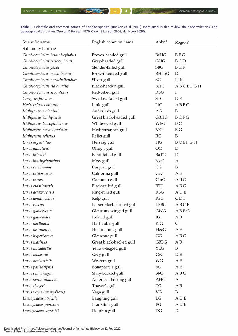
Table 1. Scientific and common names of Laridae species (Roskov et al. 2019) mentioned in this review, their abbreviations, and geographic distribution (Gruson & Forster 1976, Olsen & Larson 2003, del Hoyo 2020).
Scientific name English common name Abbr.† Region*
Subfamily LarinaeChroicocephalus brunnicephalus Brown-headed gull BrHG B F GChroicocephalus cirrocephalus Grey-headed gull GHG B C DChroicocephalus genei Slender-billed gull SBG B C FChroicocephalus maculipennis Brown-hooded gull BHooG DChroicocephalus novaehollandiae Silver gull SG I J KChroicocephalus ridibundus Black-headed gull BHG A B C E F G HChroicocephalus scopulinus Red-billed gull RBG ICreagrus furcatus Swallow-tailed gull STG D EHydrocoloeus minutus Little gull LiG A B F GIchthyaetus audouinii Audouin’s gull AG BIchthyaetus ichthyaetus Great black-headed gull GBHG B C F GIchthyaetus leucophthalmus White-eyed gull WEG B CIchthyaetus melanocephalus Mediterranean gull MG B GIchthyaetus relictus Relict gull RG BLarus argentatus Herring gull HG B C E F G HLarus atlanticus Olrog’s gull OG DLarus belcheri Band-tailed gull BaTG DLarus brachyrhynchus Mew gull MeG ALarus cachinnans Caspian gull CG BLarus californicus California gull CaG A ELarus canus Common gull CmG A B GLarus crassirostris Black-tailed gull BTG A B GLarus delawarensis Ring-billed gull RBG A D ELarus dominicanus Kelp gull KeG C D ILarus fuscus Lesser black-backed gull LBBG A B C FLarus glaucescens Glaucous-winged gull GWG A B E GLarus glaucoides Iceland gull IG A BLarus hartlaubii Hartlaub’s gull KiG CLarus heermanni Heermann’s gull HeeG A ELarus hyperboreus Glaucous gull GG A B GLarus marinus Great black-backed gull GBBG A BLarus michahellis Yellow-legged gull YLG BLarus modestus Gray gull GrG D ELarus occidentalis Western gull WG A ELarus philadelphia Bonaparte’s gull BG A ELarus schistisagus Slaty-backed gull SbG A B GLarus smithsonianus American herring gull AHG ALarus thayeri Thayer’s gull TG A BLarus vegae (mongolicus) Vega gull VG BLeucophaeus atricilla Laughing gull LG A D ELeucophaeus pipixcan Franklin’s gull FG A D ELeucophaeus scoresbii Dolphin gull DG D
Downloaded From: https://bioone.org/journals/Journal-of-Vertebrate-Biology on 12 Feb 2022Terms of Use: https://bioone.org/terms-of-use
Microbial pathogens in laridsJ. Vertebr. Biol. 2021, 70(3): 21009 5
Flavivirus West Nile (WNV)A plausible hypothesis supposes dispersal of mosquito-borne WNV by migratory birds, including gulls (Rappole & Hubálek 2003).
Grešíková et al. (1962) found HI antibodies against flaviviruses (most probably WNV) in larids in east Bulgaria at Burgas: S. hirundo 3/13, Chlidonias niger 7/50, H. minutus 1/2. Drăgănescu et al. (1978) detected HI antibodies to flaviviruses (most probably WNV) in H. minutus in the Danube Delta, Romania. Berezin et al. (1967) observed
3.9% seropositive (HIT) larids (S. hirundo, C. niger) in the Volga Delta (Astrakhan region). Berezin et al. (1971) then tested sentinel (caged) larids in the Volga Delta between 1964 and 1968 for seroconversion against WNV that occurred in 1/32 L. argentatus complex (probably L. cachinnans) (HI titre 1:160) and in 2/20 H. caspia (titres 1:40, 1:80). More recently, Alkhovsky et al. (2003) examined organs of wild birds in the Volga Delta for WNV RNA using RT-PCR. In the middle delta, the infection rate among gulls and terns was 8-13%. All positive samples belonged to the WNV lineage
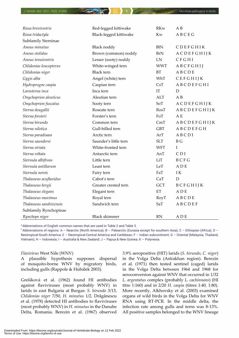
Rissa brevirostris Red-legged kittiwake RKw A BRissa tridactyla Black-legged kittiwake Kw A B C E GSubfamily SterninaeAnous minutus Black noddy BlN C D E F G H I K Anous stolidus Brown (common) noddy BrN A C D E F G H I J KAnous tenuirostris Lesser (sooty) noddy LN C F G H I Chlidonias leucopterus White-winged tern WWT A B C F G H I JChlidonias niger Black tern BT A B C D EGygis alba Angel (white) tern WhT C E F G H I J KHydroprogne caspia Caspian tern CsT A B C D E F G H ILarosterna inca Inca tern IT DOnychoprion aleuticus Aleutian tern ALT A BOnychoprion fuscatus Sooty tern SoT A C D E F G H I J KSterna dougallii Roseate tern RosT A B C D E F G H I J KSterna forsteri Forster’s tern FoT A ESterna hirundo Common tern CmT A B C D E F G H I J KSterna nilotica Gull-billed tern GBT A B C D E F G HSterna paradisaea Arctic tern ArT A B C D ISterna saundersi Saunder’s little tern SLT B GSterna striata White-fronted tern WFT ISterna vittata Antarctic tern AnT C D ISternula albifrons Little tern LiT B C F GSternula antillarum Least tern LeT A D ESternula nereis Fairy tern FaT I KThalasseus acuflavidus Cabot’s tern CaT DThalasseus bergii Greater crested tern GCT B C F G H I J KThalasseus elegans Elegant tern ET A D EThalasseus maximus Royal tern RoyT A B C D EThalasseus sandvicensis Sandwich tern SaT A B C D E FSubfamily RynchopinaeRynchops niger Black skimmer RN A D E
† Abbreviations of English common names that are used in Table 2 and Table 3. * Abbreviations of regions: A – Nearctic (North America); B – Palearctic (Eurasia except for southern Asia); C – Ethiopian (Africa); D – Neotropical-South America; E – Neotropical-Central America and Caribbean; F – Indian subcontinent; G – Oriental (Malaysia, Thailand, Vietnam); H – Indonesia; I – Australia & New Zealand; J – Papua & New Guinea; K – Polynesia.
Downloaded From: https://bioone.org/journals/Journal-of-Vertebrate-Biology on 12 Feb 2022Terms of Use: https://bioone.org/terms-of-use
Microbial pathogens in laridsJ. Vertebr. Biol. 2021, 70(3): 21009 6
1. The results indicated relatively high circulation of WNV among the birds including larids in the region. Sidenko et al. (1973) carried out isolation attempts in the Ukrainian Prichernomorye (Black Sea) and recovered flaviviruses other than tick-borne encephalitis (TBE) virus, most probably WNV, one each from S. hirundo and Thalasseus sandvicensis. WNV was also isolated from one C. ridibundus just arriving in eastern Slovakia from the winter quarter (Ernek et al. 1977). Figuerola et al. (2007) detected WNV neutralizing antibodies in L. cachinnans 1/17 from southern Spain, June 2003. Petrović et al. (2013) investigated wild birds in the Voyvodina Province of northern Serbia in 2012. The tissues from 81 birds were tested and WNV RNA was detected by PCR in nine birds, among including one Larus michahellis.
Yastrebov et al. (1973), during a serosurvey (HIT) in Baraba Lowland at Lake Chany (West Siberia), detected WNV antibodies in C. ridibundus (more commonly in young than in adult birds which indicates local circulation) and H. minutus gulls. Gromashevsky et al. (1973) isolated WNV from Ornithodoros capensis (coniceps) ticks collected in a L. argentatus (probably L. cachinnans) colony on Glinyanyi Island in Baku Archipelago of the Caspian Sea, Azerbaijan, 1970-1971. Mosquitoes are absent on that island. Also, Andreev et al. (1974) reported one WNV isolate from Ornithodoros coniceps coniceps ticks collected from 40 nests of S. hirundo on islands of the eastern Caspian Sea, May 1973. Lvov et al. (1975) summarized that out of eight WNV isolates from argasid and ixodid ticks collected in larid colonies in the USSR, 1969-1974, five isolates were obtained from larid colonies on Glinyanyi Island. Lipin et al. (1974) carried out a serosurvey (HIT) of larids in the Selenga Delta (Buryatsk ASSR), 1971-1972: the WNV seropositivity rate was in L. canus 3/19, C. ridibundus 1/7, H. minutus 1/1 and C. leucopterus 1/2. Gordeeva (1980) performed a virological survey in Tadjikistan, 1976-1979, and isolated one strain of WNV from S. albifrons sampled in September 1977 in Dombrachi, Dzhirgitalsky rayon. Kislenko & Chunikhin (1986) demonstrated WNV infections in larids at the southern Primorye territory: HI antibodies were found in C. ridibundus (1/26) and S. hirundo (1/32). Yakimenko et al. (1991) did a WNV serosurvey (HIT) in Western Siberia (northern forest-steppe): one juvenile L. argentatus (probably L. cachinnans) was positive. Lan et al. (2013) found specific WNV antibodies (PRNT) in one of 12 Sterna saundersi terns examined in
Shanghai, China. Malkinson et al. (2002) recovered one WNV isolate from nonmigratory Ichthyaetus leucophthalmus gull belonging to a breeding colony kept in the zoological garden at the University Tel Aviv, Israel 1999. Schvartz et al. (2020) detected and isolated WNV (lineage 1, clade East European) in one L. michahellis gull that was found dead in Tel Aviv, July 2018; the autopsy showed intracranial haemorrhages, mild congestion in the leptomeninges and very pale kidneys.
Steele et al. (2000) studied pathology of fatal WNV infection in birds during the famous 1999 WNV outbreak in New York City which also involved two L. atricilla gulls, with lesions in the brain, heart, kidney, but less expressive than in other bird species such as crows. Bernard et al. (2001) found that several gulls tested positive in the New York State in 2000: in Larus smithsonianus 3/9, in Larus delawarensis 21/66, in Larus marinus 2/7, in L. atricilla 1/1 and in Rynchops niger 1/1. In summer 2003, a number of WNV epornitics in free-living birds occurred across the USA (New York, North and South Dakota, Minnesota) and included also hundreds of L. delawarensis. Tens of larids also died from WNV disease in multiple US states in the summers 2004 (L. delawarensis, Larus californicus, Leucophaeus pipixcan), 2005 (S. hirundo), 2006, 2007 (L. delawarensis), 2009 (L. delawarensis), 2010, 2011 (a few L. marinus) and 2012 (L. californicus) (Quarterly Wildlife Mortality Reports – QWMR). Newman et al. (2007) also reported WNV deaths of L. delawarensis, L. californicus and L. pipixcan. Barbachano-Guerrero et al. (2019) analysed cloacal and tracheal samples from 200 wild birds collected in Mexico, 2008-2009, using PCR: the overall prevalence for WNV was 8% (16/200), including L. delawarensis.
Experimental infection of larids with WNVTwo papers described experimental inoculation with WNV in gulls. Leonova et al. (1975) infected L. crassirostris: most birds died, but some survived; the virus was recovered from the blood 2 and 4 DPI, and from the brain and internal organs 10-12, 18 and 28 DPI. Antibodies were detected after 7 DPI. Komar et al. (2003) infected two L. delawarensis gulls by mosquito bites: the birds revealed lethargy, ruffled feathers, inability to hold the head upright, ataxia, and died 5 and 13 DPI; Viral shedding was observed in the two gulls: cloacal swabs (3-7 DPI; max. 2.4 log PFU/swab); oral swabs (2-7 DPI; max. 5.1 log PFU/swab). In a contact gull, direct transmission in cage was observed – the individual
Downloaded From: https://bioone.org/journals/Journal-of-Vertebrate-Biology on 12 Feb 2022Terms of Use: https://bioone.org/terms-of-use
Microbial pathogens in laridsJ. Vertebr. Biol. 2021, 70(3): 21009 7
had high viremia (mean 7.4 log PFU/ml blood) for 4-7 days. The virus was reisolated from all organs (including skin and eye) of dead gulls. This gull species is therefore a moderately competent host for WNV.
Flavivirus Usutu (USUV) Hubálek et al. (2008) found specific antibodies to USUV (PRNT) in one Polish C. ridibundus.
Flavivirus of tick-borne encephalitis (TBEV)Lipin et al. (1974) detected HI antibodies to TBEV in larids in the Selenga Delta (Buryatsk ASSR), 1971-1972: L. canus 4/57 (1/40 in CFT), S. hirundo 8/75 (2/52 in CFT). Yastrebov et al. (1973) and Yastrebov & Yurlov (1978) found HI antibodies to TBEV in C. ridibundus on Lake Chany in Barabinskaya Lowland (Western Siberia) in 1970-1974. However, serological cross-reactions with flaviviruses other than TBE virus cannot be excluded in HIT and CFT.
Flavivirus of Omsk haemorrhagic fever (OHFV) Vorobieva et al. (1973, 1975) isolated OHFV twice from larids in Barabinskaya Lowland (Lake Chany) in 1971-1972: S. hirundo nestling (strain K-101, from the viscera) and adult C. ridibundus (strain CH-135, from the brain). In the same area, Vorobieva et al. (1978) described a spontaneous subclinical OHF infection in larids. HI antibodies to OHFV were detected in L. argentatus (probably L. cachinnans) 5/42, C. ridibundus 24/164, H. minutus 11/103, C. niger 5/43 and S. hirundo 19/141. Karavaev et al. (1975) tested 926 birds (29 spp.) by HIT in this study in 1971-1972, and demonstrated a high seropositivity rate in C. ridibundus (19.4%); antibodies to OHFV were detected even in 2-3 day old nestlings. Khodkov & Karavaev (1978) and Karavaev et al. (1975) detected a high proportion of OHFV antibodies in C. ridibundus (from a colony of 300-1,000 pairs) examined in 1972-1974. They observed ill gulls, in which the acute phase of disease continued to chronic, and OHFV persisted for more than 30 days. They concluded that C. ridibundus participates in the natural circulation of OHFV.
Experimental infection of C. ridibundus with OHFV revealed viremia (lasting up to 14 DPI), excretion of the virus in faeces, and a long-term virus carrier state: OHFV was reisolated from the brain, kidney and liver (Vorobieva & Karavaev 1975).
Flavivirus Tyuleniy (TYUV) The virus, distantly related to TBE virus, is carried by the seabird ectoparasite Ixodes uriae living in
avian nests in the Holarctic (Lvov et al. 1971b, Lvov & Ilyichev 1979).
Bekleshova et al. (1971) found a 3.1% seroprevalence rate (HIT) against TYUV in Rissa tridactyla in the Far East of Russia. Lvov et al. (1972) examined seroprevalence on the Commodore Islands: it was 11% among R. tridactyla (in local people 6%, and in fur seals 22%). Roslaya et al. (1974) carried out a serosurvey (HIT) for TYUV in birds of the Lower Amur, 1970-1972: 2/18 S. hirundo were positive (the birds were TBE virus seronegative).
Votyakov et al. (1974) isolated two strains of TYUV from I. uriae ticks collected in colonial R. tridactyla on the shore of the Barents Sea, northern Europe. V.A. Smirnov (pers. comm.) carried out a serosurvey for TYUV in gulls L. argentatus (11.9% positive), L. marinus (5.7%) and R. tridactyla (5.3%). Voinov et al. (1979) examined seabird “bazars” on Murmansk shore of the Barents Sea, 1972-1978: 4.8% of 197 R. tridactyla had antibodies; 5,485 I. uriae ticks yielded 11 isolates of TYUV. In addition, three entomologists revealed TYUV fever (confirmed serologically) with lymphadenopathy, arthralgia, laryngitis, and skin petechiae. Saikku et al. (1980) searched for viruses in I. uriae collected from seabird (including R. tridactyla) colonies at Rost Island, Lofoten (Norway), July 1974, and isolated one TYUV strain from 1,229 ticks. Chastel et al. (1985a) detected antibodies against TYUV in L. argentatus, Larus fuscus, L. marinus, C. ridibundus and R. tridactyla in Brittany (western France).
In North America, Clifford (1971) also isolated TYUV from I. uriae collected in seagull nests on Three Arch Rocks National Refuge, Oregon (USA). Yunker (1975) reported serosurveys (HIT) in gull chicks in North America, 1973-1974: 6/14 were positive to TYUV. Main et al. (1976b) isolated TYUV in a L. smithsonianus breeding habitat of the Witless Bay seabird sanctuary, Newfoundland (Canada), 1971-1972; however, no HI antibodies to TYUV were found in 28 chicks of L. smithsonianus and two L. marinus chicks.
TYUV is potentially pathogenic to gulls. This was demonstrated by Berezina et al. (1974) when they inoculated i.c. R. tridactyla (one adult, two juveniles), L. marinus (one juvenile) and L. argentatus (one juvenile) with ca. 107 SMicLD50 of TYUV. The infection resulted in pareses (3 DPI), ataxia (5 DPI) and death (6-8 DPI) of half of the inoculated birds (L. marinus remained asymptomatic). The virus
Downloaded From: https://bioone.org/journals/Journal-of-Vertebrate-Biology on 12 Feb 2022Terms of Use: https://bioone.org/terms-of-use
Microbial pathogens in laridsJ. Vertebr. Biol. 2021, 70(3): 21009 8
was recovered from the brain, liver, spleen, kidneys and ovary 5-8 DPI but only in birds with clinical symptoms. Previously, V.A. Smirnov (quoted in Berezina et al. 1974) inoculated s.c. the same bird species: the symptoms were similar, and started to appear three days later. Nonetheless, no cases of disease in free-living larids caused by TYUV have been described.
Flavivirus Saumarez Reef This virus, closely related to TYUV, was originally isolated from O. capensis ticks inhabiting the nests of terns Onychoprion fuscatus, Thalasseus bergii and Anous minutus in Queensland (Australia), but also from Ixodes eudyptidis ticks found on two dead Chroicocephalus novaehollandiae gulls in northern Tasmania (St. George et al. 1977, Humphrey-Smith & Cybinski 1987); 4/14 gulls were seropositive (HIT and VNT) to this virus.
Flavivirus Meaban Seven strains of this new arbovirus were isolated from Ornithodoros maritimus ticks collected in nests of L. argentatus on the islands of south Brittany (France) during 1981-1982 (Chastel et al. 1985b). The virus is antigenically closely related to, but different from, Saumarez Reef virus. Chastel et al. (1985b) also detected antibodies to this virus in L. argentatus and L. fuscus in Brittany. More recently, Arnal et al. (2014) sampled L. michahellis eggs from 19 breeding colonies in Spain, France, Algeria and Tunisia for three years. In the colonies where flavivirus ELISA-positive eggs were found, chick serum samples and vectors (O. maritimus) were collected and analysed using serology and PCR. In north-eastern Spain, on the Medes Islands and in the nearby village of L‘Escala, as many as 56% of eggs had antibodies to the flavivirus envelope protein, being negative for VN antibodies against three other flaviviruses: WNV, USUV and TBE virus. Ornithodoros ticks from Medes were then screened for flaviviral RNA and positive samples sequenced: the RNA (NS5 gene) was 95% similar to that of Meaban virus. All ELISA-positive samples subsequently tested positive (VNT) for Meaban virus. Dupraz et al. (2017) sampled O. maritimus ticks repeatedly in a colony of L. michahellis during the gull breeding season, and recovered one Meaban virus isolate.
Jaeger et al. (2016) detected flavivirus antibodies by ELISA in 12 of 146 O. fuscatus terns on Juan de Nova Island in western Indian Ocean. Some of the positive sera reacted specifically in VNT with Meaban virus.
Bunyaviridae
Bunyavirus Lednice (Turlock group)A virus related to African M‘Poko virus was isolated from Culex modestus mosquitoes in south Moravia, Czech Republic. Experimental inoculation of young (2-4 days old) C. ridibundus with Lednice virus caused an asymptomatic infection of the birds; the viremia lasted 5-6 days with relatively low viremic titres (max. 3.5 log LD50/ml blood). VN antibodies appeared after 21 DPI (Málková et al. 1979).
Bunyavirus Upolu The virus was isolated from O. capensis ticks collected from nests of O. fuscatus terns in Australia (Yunker et al. 1979).
Phleboviruses Zaliv Terpeniya and St. Abb’s Head (Uukuniemi group) The viruses were isolated from ticks inhabiting seabird nests. Klisenko et al. (1973) recovered two strains (N14, N38) of the Uukuniemi group viruses from I. uriae ticks collected on breeding grounds of seabirds (including R. tridactyla gulls) in the Far East (Tyuleniy Island). Voinov et al. (1979) examined by SM i.c. inoculation 5,485 I. uriae ticks and visceral organs from 197 R. tridactyla on seabird ”bazars” in the Murmansk shore of the Barents Sea, 1972-1978, and recovered one isolate of Zaliv Terpeniya virus. Saikku et al. (1980) detected 30 Uukuniemi group virus isolates in I. uriae (1,229 ticks in 204 pools) from seabird (including R. tridactyla) colonies at Rost Island, Lofoten, Norway, July 1974. Nuttall et al. (1981) recovered 15 isolates related to the Uukuniemi group of viruses from I. uriae ticks collected on R. tridactyla in St. Abb’s Head, 1974-1979; one virus strain (St. Abb’s Head, GM710) was isolated from the brain of a juvenile R. tridactyla. Spence et al. (1985) examined I. uriae ticks collected from R. tridactyla nests on Isle of May, Scotland. The ticks yielded one isolate of St. Abb’s Head virus. Nuttall et al. (1984) investigated mixed infections with tick-borne viruses in a seabird colony in Eire and recovered one Uukuniemi-like isolate (GS80-10) from a female I. uriae engorging on a R. tridactyla chick. Sera of three R. tridactyla chicks tested positive in VNT against the virus isolate. Eley & Nuttall (1984) isolated an uukuvirus “S23” from the organs of a moribund R. tridactyla and from I. uriae ticks feeding on this bird in north-eastern England. Chastel (1988) detected antibodies to Zaliv Terpeniya virus in 7% L. marinus.
Downloaded From: https://bioone.org/journals/Journal-of-Vertebrate-Biology on 12 Feb 2022Terms of Use: https://bioone.org/terms-of-use
Microbial pathogens in laridsJ. Vertebr. Biol. 2021, 70(3): 21009 9
Nairovirus Caspiy (CASV) The virus was isolated from O. capensis and O. coniceps argasid ticks, and also from an ill juvenile L. cachinnans in the Caspian Sea. In the USSR during 1969-1974, three isolates of CASV were recovered from Glinyanyi Island, and two isolates from Kara Bogaz Gol Bay (Lvov et al. 1975). Lvov et al. (2014) carried out full-genome sequencing of the CASV (GenBank KF801658) and attributed it to the Nairovirus genus as a separate species. CASV is related to the Hughes antigenic group of viruses.
Nairoviruses Hughes, Farallon, Soldado and Puffin Island (Hughes group)Hughes group viruses occur in argasid ticks (O. capensis group) living in seabird nests. Seabird migrations obviously account for the remarkably extensive geographic distribution of these arboviruses (Converse et al. 1975, Hoogstraal et al. 1976); geographic and biotic factors also influence the antigenic diversity of these viruses and favour the development of insular variants (Yunker 1975, Chastel et al. 1979, 1983b, 1988, Nuttall et al. 1984, 1986).
Hughes et al. (1964) isolated a new virus from Ornithodoros denmarki (O. capensis group) ticks collected on nesting site of O. fuscatus terns on Bush Key, Florida in 1962. Radovsky et al. (1967) recovered a variant of Hughes virus (called Farallon virus) from a pool of 20 O. denmarki ticks collected from nests of Larus occidentalis on the Farallon Islands off San Francisco. Aitken et al. (1968) recovered a Hughes group virus from Trinidadian ticks and Anous stolidus terns on Soldado Rock (a small limestone island), 1962-1965: seven strains from O. capensis (plus O. denmarki as well) and eight strains from the blood of 8-12 day old O. fuscatus, 1964. These isolates were later described as Soldado virus (TRVL-52214) by Jonkers et al. (1973).
Soldado virus was also recovered (Converse et al. 1975) from O. capensis infesting colonial O. fuscatus in the Seychelles, Indian Ocean, in summer 1973 when ca. 5,000 pairs of sooty terns abandoned a part of their breeding grounds on Bird Island; eggs and newly hatched chicks were left unattended by birds (due to very abundant ticks) and the area was not reoccupied in 1974. The virus was isolated in 1973 and 1974 from the ticks collected on Bird Island from the ground and from both sick and asymptomatic chicks of O. fuscatus. When ticks from the ground and from the tern chicks were
fed on domestic chicks, they transmitted Soldado virus and caused the death of their hosts.
Converse et al. (1976) then isolated Soldado virus (three strains) from nymphal and adult O. maritimus ticks collected in and near the nests of L. argentatus on Puffin Island, northern Wales. All isolates killed mice and guinea pigs, as well as 1-2 day old domestic chicks when i.c. inoculated. Johnson et al. (1979) isolated Soldado virus from 34/173 O. maritimus tested individually and from 9/27 tick pools (226 individuals) collected on Puffin Island, North Wales. Antibodies neutralizing Soldado virus were detected in sera of two L. argentatus. Chastel et al. (1988) isolated Soldado virus from O. maritimus infesting the nests of L. argentatus in Brittany, western France and from O. maritimus ticks collected in a L. cachinnans nest in southern France (Ile de Porquerolles). Subsequently, Chastel et al. (1990) also recovered Soldado virus from larval and adult O. maritimus parasitizing a juvenile R. tridactyla that died in France; the virus was also isolated from the brain of the dead chick. Chastel et al. (1981, 1983a) reported that Soldado virus can infect humans working in colonies of seagulls: the infection is associated with a self-limited febrile illness.
Two strains were isolated from O. maritimus, the parasite of L. cachinnans, in Morocco 1979; one entomologist bitten by O. maritimus developed fever, prolonged rhinopharyngitis and pruritus, and antibodies neutralizing Soldado virus persisted for two years. A further 14 Soldado virus isolates were recovered from O. capensis ticks taken from nests of Chroicocephalus cirrocephalus in Senegal and South Mauretania, January to February 1977 (Main et al. 1980). Puffin Island virus was isolated from O. maritimus collected in the nests of L. argentatus (Nuttall 1984). Chastel et al. (1985a) detected antibodies against Hughes and Puffin Island viruses in L. argentatus gulls in Brittany (western France).
Danielová et al. (1982) isolated 13 strains of Hughes group from 750 O. denmarki ticks collected from the nests of O. fuscatus and A. stolidus on a small island Cayo Mono Grande (Cuba) in the autumn of 1979 (the birds were then absent) – the isolates differed from Soldado virus.
Nairoviruses Sakhalin and Avalon (Sakhalin group)Sakhalin group viruses are transmitted among seabirds by I. uriae ticks in the subpolar regions.
Downloaded From: https://bioone.org/journals/Journal-of-Vertebrate-Biology on 12 Feb 2022Terms of Use: https://bioone.org/terms-of-use

Microbial pathogens in laridsJ. Vertebr. Biol. 2021, 70(3): 21009 10
Timofeeva et al. (1974) detected CF antibodies to Sakhalin virus in 2/15 R. tridactyla gulls on Iona Island, the Sea of Okhotsk. Lvov et al. (1974b) studied the ecology of Sakhalin virus in the north of the Far East and isolated the virus from I. uriae mainly from R. tridactyla nests. But antibodies were detected in only 0.6% R. tridactyla.
Main et al. (1976a) found Avalon virus in I. uriae ticks from L. smithsonianus gull habitat and in the blood of L. smithsonianus chicks on Great Island (Newfoundland, Canada), 1972. Of the L. smithsonianus chicks, 9% were seropositive (VNT). Quillien et al. (1986) isolated nine strains of Avalon virus from I. uriae collected in R. tridactyla nests but no CF antibody was detected in the gull sera in Cape Sizun seabird reserve, Brittany (France), 1979-1985. Subsequently however, Chastel (1988) isolated Avalon virus from the blood of a sick L. argentatus in France and antibodies to Avalon virus were detected in R. tridactyla.
Nyaviridae
Nyavirus MidwayThe virus belongs to a new genus and new family of the order Mononegavirales (Mihindukulasuriya et al. 2009). It was originally isolated from O. capensis and O. denmarki ticks collected on breeding grounds of O. fuscatus terns and L. crassirostris gulls in Midway, Kure and Manana Islands in the Central Pacific (Hawaiian Archipelago) and from northern Honshu (Japan) in 1966 (Takahashi et al. 1982). Midway virus shows a close relationship to Nyamanini virus, which was isolated from ardeid birds and Argas ticks in Africa, the Indian subcontinent and south-eastern Asia. However, cross-tests (CFT, VNT, IF) show that these two viruses are distinct. Antibodies to the virus were prevalent among nestlings of L. crassirostris on Aomatsushima Island (Japan). Midway virus is lethal for SM inoculated by i.c. but not i.p. route and it fails to kill four week old mice by either route.
Reoviridae
Orbiviruses Great Island (GIV), Bauline, Okhotskiy, Cape Wrath and Yaquina Head (Kemerovo group, Great Island antigenic complex)The viruses of Great Island complex are isolated from I. uriae ticks inhabiting mainly seabird nests (Main et al. 1973, Yunker 1975). They exhibit a great genomic variability (Oprandy et al. 1988) and may represent a single viral gene pool because
of relative homogeneity when compared by RNA-RNA hybridization and gene reassortment to other Orbivirus serogroups (Brown et al. 1989). Viruses of this antigenic complex are dispersed by seabirds transoceanically (they occur in both subantarctic and subarctic regions).
Main et al. (1973, 1976c) recovered two GIV and four Bauline virus isolates from I. uriae removed from L. smithsonianus chicks on Great Island, Newfoundland (eastern Canada) in 1971-1972, but did not find HI antibodies to Bauline or Cape Wrath viruses in chicks of L. smithsonianus, L. marinus and R. tridactyla. Chastel (1988) mentioned antibodies to Yaquina Head virus detected in L. occidentalis chicks.
Votyakov et al. (1974) investigated arboviruses in colonial seabirds on the Murmansk shore of Barents Sea (Russia) and recorded two strains of Okhotskiy virus from 156 birds (R. tridactyla, Uria lomvia). Also, Voinov et al. (1979) examined (by i.c. inoculation of bird visceral organs) seabird “bazars” in the same area, 1972-1978, and among others 197 R. tridactyla yielded two strains of Okhotskiy virus. Saikku et al. (1980) examined 1,229 I. uriae ticks collected from seabird (including R. tridactyla) colonies at Rost Island, Lofoten (Norway) in 1974, and recovered 13 Kemerovo group isolates. Sera of two of three R. tridactyla chicks in a seabird colony in Ireland contained antibodies (VNT) against the Kemerovo-like isolate GS806 (Nuttall et al. 1984). Three Cape Wrath virus isolates were detected in I. uriae collected from R. tridactyla and Uria algae nests on Isle of May, Scotland (Spence et al. 1985).
Orbivirus Baku, Essaouira, Kala Iris and Mono Lake (Kemerovo group, Chenuda antigenic complex)Lvov et al. (1971a) isolated and described a new “Baku” virus from 1% of O. coniceps ticks collected in a breeding colony of L. argentatus (probably L. cachinnans) on Glinyanyi Island (Baku archipelago, Caspian Sea, Azerbaijan), 1970. Gromashevsky et al. (1973) examined ornithophilic ticks on the same island: the ticks O. capensis (O. coniceps) in a L. argentatus (L. cachinnans) colony, 1970-1971 yielded 13 strains of Baku virus; 9% of 78 gull nestlings had CF antibody to Baku virus. Andreev et al. (1973b, 1974) obtained two isolates of Baku virus from O. coniceps ticks collected from 22 nests of Chroicocephalus genei (one isolate) and 40 nests of S. hirundo (one isolate) in a mixed colony on islands in the eastern Caspian Sea, 1973. Andreev et al. (1973a) isolated one Baku virus strain in western
Downloaded From: https://bioone.org/journals/Journal-of-Vertebrate-Biology on 12 Feb 2022Terms of Use: https://bioone.org/terms-of-use
Microbial pathogens in laridsJ. Vertebr. Biol. 2021, 70(3): 21009 11
Turkmenistan from 100 O. coniceps ticks collected in 1972 from the nests of L. argentatus (probably L. cachinnans), Kara Bogaz Gol Bay.
Jacobs et al. (1986), Chastel (1988) and Chastel et al. (1993) reported that O. maritimus ticks infesting L. cachinnans gulls on Essaouira Island (Morocco) in 1979 yielded a new Essaouira virus that is antigenically related to Baku virus. A similar Kala Iris virus originated from Kala Iris islet, Morocco, 1981. Terns Sterna paradisaea might disperse the two viruses. The pathogenicity of these viruses for man and animals, including seabirds, remains unknown.
Mono Lake virus was isolated from Argas monolakensis ticks inhabiting nesting colonies of L. californicus on islands of Mono Lake (California) in 1981-1982 (Jacobs et al. 1986, Schwan et al. 1988). The virus was recovered from 2-8% of the ticks on various islands. In 1981, > 90% L. californicus nestlings died on this lake. In July 1984, a total of 800 argasid ticks collected from L. californicus nests on the rocks were examined and yielded 28 isolates of Mono Lake virus. Antibodies to this virus were detected in 17/46 juvenile (37%) and 2/2 adult L. californicus. Experimentally infected one day old L. californicus chickens revealed viremia 3-7 DPI and subsequently VN antibodies.
Other reoviruses A reovirus was isolated from faeces of L. crassirostris nestlings in many areas of Japan. One isolate from the gulls living on Kabu Island, Hachinohe city, had a 62% antigenic relatedness to TS-17 strain, a prototype of avian reovirus in Japan, and showed no significant virulence to one day old chickens and low mortalities to chicken embryos, although it formed remarkable lesions on the chorioallantoic membrane (Takehara et al. 1989).
Petermann et al. (1989) reported a reovirus from one dead C. ridibundus gull (34 examined) in the German Bight, 1982-1985. Stenzel et al. (2008) found antibodies to avian reovirus in six of eight injured L. argentatus delivered in 2005-2007 to a Polish wildlife rehabilitation centre.
Aride virus This virus is a still an unclassified arbovirus, recovered from female Amblyomma loculosum ticks taken from the foot of two freshly dead S. dougallii arideensis terns in the Seychelles, during a die-off
of thousands of sooty terns; it is unclear whether Aride virus could have been the etiologic agent (Converse et al. 1976, Hoogstraal et al. 1976).
Orthomyxoviridae
Orthomyxovirus influenza A Avian influenza A viruses (AIVs) are the causative agents of the presently most important poultry disease (Kaleta et al. 2005). According to their HA (haemagglutinin) and N (neuraminidase) antigens, there exist many lineages (subtypes) of AIVs, and they also differ in pathogenicity – several are highly pathogenic (HPAI), while pathogenicity is low in others (LPAI). The natural host and reservoir of AIVs is wild waterfowl, shorebirds and gulls. AIVs have frequently been isolated worldwide from wild birds of nearly 100 species and 12 orders – most commonly anseriforms, but also often larids (Bahl et al. 1977, Sinnecker et al. 1977, 1983, Lvov & Ilyichev 1979, Hinshaw et al. 1980, 1985, Tsubokura et al. 1981, Ottis & Bachmann 1983, Otsuki et al. 1987a, b, c, Stallknecht & Shane 1988, Graves 1992, Webster et al. 1992, Kaleta et al. 2005, Pearce et al. 2010). Importantly, the predominant HA subtypes (H13, H16, H9, H11) of AIVs isolated from larids (and shorebirds) differ from those occurring in wild anseriforms, and are largely non-pathogenic or weakly pathogenic for vertebrate hosts (Hinshaw et al. 1982, 1983, Kawaoka et al. 1988, Chambers et al. 1989, Webster et al. 1992, Alexander 2000, Arnal et al. 2015, Verhagen et al. 2015, 2020, Benkaroun et al. 2016, Lindh et al. 2017). These viruses from time to time spill over into seals, whales, pigs, domestic poultry and humans (Hinshaw et al. 1986, Okazaki et al. 1989, Mandler et al. 1990). In general, gulls and terns may disseminate worldwide AIVs, occasionally including some HPAIVs (Hinshaw & Webster 1982, Hinshaw et al. 1985).
Interestingly, some AIV isolations from larid eggs and embryo indicate the possibility of transovarial (vertical) transmission of AIVs in the birds. For instance, C. ridibundus eggs in Slovakia yielded an AIV A/Larus/36/77 (Hav7Nav1) (Grešíková et al. 1979). An AIV isolate was obtained from 3/55 embryos of the same gull species in the Khabarovsk region, Russia in 1979 (Roslaya et al. 1984). Yamnikova et al. (1989a) also described the isolation of several AIV strains from larid embryos (I. ichthyaetus, L. argentatus (probably L. cachinnans), H. caspia) on Zhemchuzhny Island in north-west Caspian Sea in 1979-1985.
Downloaded From: https://bioone.org/journals/Journal-of-Vertebrate-Biology on 12 Feb 2022Terms of Use: https://bioone.org/terms-of-use
Microbial pathogens in laridsJ. Vertebr. Biol. 2021, 70(3): 21009 12
AIVs have been reported in larids from all continents except Antarctica. Lang et al. (2016) prepared a survey of AIV detection rates in pelagic terns and noddies from multiple continents (positive virus cultivation / PCR / serology / AIV subtypes detected; nt = not tested): A. minutus: 0/182 / nt / 43/88 / H3N6; A. stolidus: 0/335 / 0/160 / 39/121 / H6,7,11; A. tenuirostris: 1/254 / 16/314 / 119/197 / H1,2,3,5,6,8,9,12,14,16; O. aleuticus: 3/302 / 1/302 / nt; S. paradisaea: 15/841 / 4/358 / nt; H. caspia: nt / 11/484 / nt; T. sandvicensis: 1/63 / 0/84 / nt / H?N1; O. fuscatus: 1/294 / 0/902 / 56/882 / H15N9; C. leucopterus: nt / 1/122 / nt / H6N2.
In Africa, a very unusual outbreak of AI with mortality in S. hirundo (> 1,300 dead terns were counted) was caused by a HPAI virus strain (A/tern/South Africa/61; H5N3 subtype) along the coast of Cape Province (South Africa) in April 1961. Many of the affected terns were unable to fly, had severe diarrhoea, and developed encephalitis. The disease spread rapidly along 1,000 miles of the coast from Port Elizabeth to Lambert Bay over a period of ca. three weeks. Surprisingly, there were no overt signs of disease in other local bird species such as T. bergii (Rowan 1962, Becker 1966). Another mass die-off of C. leucopterus terns on Lake Victoria in Uganda, January 2017, was caused by HPAI virus H5N8 clade 2.3.4.4 (Abolnik et al. 2019). Lebarbenchon et al. (2015) detected AIV antibodies by ELISA in 25 of 234 O. fuscatus terns on Juan de Nova Island in the western Indian Ocean.
In Europe, Zakstelskaya et al. (1972, 1973) tested gulls serologically (HIT) for AIVs in the Arkhangelsk region (northern European Russia) during spring and autumn 1969: prevalence rate of antibodies was 17/78 in L. argentatus and 2/16 in L. fuscus. Timofeeva et al. (1973) tested 35 S. paradisaea terns by HIT with 12 influenza A antigens in the Pechora Delta (Arkhangelsk region) in spring 1972 and found positivity against antigens of several AIV subtypes: A2/Hongkong/1/68 (20.0%), A/Sterna/South Africa/61 (2.9%), A/duck/England/56 (5.7%), A/turkey/Ontario/68 (5.7%) and A/turkey/USA/65 (2.9%). Zakstelskaya et al. (1975) isolated two AIV strains from organs of 20 S. paradisaea terns in the North of European USSR: A/Sterna/Pechora/105/72 (HAv6) and A/Sterna/Pechora/112/72 (Hav7NL2). They also recovered AIV in 2/23 L. argentatus (probably L. cachinnans) gulls in the European USSR. Andreev et al. (1974) isolated AIV (Hav5Nav2, identical to A/Sterna/South Africa/61) in the Black Sea, 1978: eight strains
from juvenile C. genei and one strain from juvenile S. hirundo. No epizootics were observed among the birds there in 1976, 1978 or 1979. Lvov et al. (1978) and Podchernyaeva et al. (1979) isolated AIVs from cloacal washings during an epizootic among young larids (the four week old chicks had clinical influenza with pathologic pulmonary changes) in the Volga Delta, Astrakhan region in July 1976: nesting larids (32 ill and healthy birds) yielded five strains of AIVs (three S. albifrons, one S. hirundo, one C. genei) identical to A/Sterna hirundo/South Africa/61 (Hav5Nav2) virus; C. genei additionally yielded two strains of a new Hav4Nav2 subtype that could be a reassortant between Hav5Nav2 and Hav4Nav1 (from European domestic ducks). Myasnikova & Pysina (1980) identified several strains AIV from wild birds in Belarus in 1979, e.g. Hsw1Nav2 subtype from C. ridibundus: A/Larus ridibundus/Belarus/212/76, A/Larus ridibundus/Belarus/446/77. Romváry et al. (1976) tested Hungarian birds serologically (HIT) against the Victoria(3)75 variant of H3N2 subtype of AIVs: 29-40% of 105 wild migrating birds (among others C. ridibundus) were seropositive. Romváry et al. (1978) also isolated AIVs from five spp. of wild birds in Hungary, including C. ridibundus. One year after the Victoria/75 epidemic in Hungary in 1976, they detected antibodies in 7.8% and 13.8% of gull and tern eggs: 129 C. ridibundus eight H3, 24 H7; five H. minutus two H5; 134 S. hirundo two H5, 10 H7. Furthermore, H7 virus was isolated from one gull and one tern, and Vic/75 from another gull (Romváry et al. 1979). Romváry et al. (1980) carried out a serosurvey (HIT) in larid eggs also in 1978, with the following H subtypes seropositivity. 535 S. hirundo eggs: A/Vic/75 7.1%, A/Texas/77 19.4%, A/H1N1/77 13.5%, H7 7.7%. 156 C. ridibundus eggs: A/Vic/75 17.9%, A/Texas/77 32.7%, A/H1N1/77 3.8%, H1 7.7%, H3 2.6%, H6 4.5%, H7 3.8%. Janout et al. (1979) examined cloacal swabs from 311 C. ridibundus at Záhlinice near Přerov (Moravia, Czech Republic) collected during March 1977 to June 1978, and recovered four (1.3%) LPAI isolates of the subtype Hav2Nav4. Andreev et al. (1980) recovered AIVs from a gull breeding colony on Zhemchuzhny Island in north-west Caspian Sea (Astrakhan region), 1976-1979: 15 isolates (all of H4N1 subtype) AIV strains, 1979-1981: I. ichthyaetus 26/562, L. argentatus (probably L. cachinnans) 7/110, and H. caspia 5/284. In 1979, the isolates were H1N2 Hsw1N2 close to A/Larus ridibundus/Belarus/212/76, in 1980 H3N6/Hav7Nav1. Aristova et al. (1982) surveyed the area in 1979-1981: cloacal and tracheal swabs were positive for AI viruses in
Downloaded From: https://bioone.org/journals/Journal-of-Vertebrate-Biology on 12 Feb 2022Terms of Use: https://bioone.org/terms-of-use
Microbial pathogens in laridsJ. Vertebr. Biol. 2021, 70(3): 21009 13
I. ichthyaetus 26/562, L. cachinnans 7/110, H. caspia 5/284; all viruses in 1979 were H1N2/Hsw1N2 close to A/Larus ridibundus/Belarus/212/76; in 1980: H3N6/Hav7Nav1, close to A/duck/HK/78. Later, Yamnikova et al. (1989a, b) studied circulation of AIVs of H13 subtype among lariforms on the same island from 1979-1985: total 95 isolates yielded lariform nestlings (6.5% of 1,060 I. ichthyaetus, 2.7% of 522 L. argentatus (probably L. cachinnans) and 1.8% of 497 H. caspia). Antigenic formulae of the strains isolated from larids were: 1979 H13N2; 1980 H13N6; 1981 H5N2; 1982 H13N2; 1983 H13N2, H13N6; 1984 H13N6; 1985 H13N6, H13N3. In 2003 Yashkulov et al. (2008) isolated AIV subtype H13N1 here from organs of larids (1/11 I. ichthyaetus, 2/17 L. argentatus (probably L. cachinnans), 1/8 H. caspia). However, no influenza A epizootics among gulls and terns were observed in this location. Votyakov et al. (1981) isolated two AIVs from mixed pools of viscera from C. ridibundus gulls collected in the Minsk area of Belarus, 1976-1977. The viruses revealed antigenic relationships with porcine Hsw1, one strain (1977) had neuraminidase N2, the other (1976) Nav2. Sinnecker et al. (1983) found 7/316 C. ridibundus gulls and 1/351 T. sandvicensis terns positive for AIVs by isolation in east Germany. Later, Süss et al. (1994) detected AIVs in C. ridibundus and L. canus gulls in eastern Germany – prevalent subtypes were H7N3 and H11N6. The isolation rate from gulls was 1.1%, much lower than that from wild ducks. Röhm et al. (1996) found that four HPAI isolates H7N7 from a single outbreak in domestic chickens in Leipzig, Germany, shared an immediate common ancestral HA with A/tern/Potsdam/342-6/79 (H7N7, from S. hirundo) and A/swan/Potsdam/63-6/81 (H7N7). Fouchier et al. (2003, 2005) carried out an AIV surveillance in wild birds in Sweden, 1999-2000: 10/886 (1.1%) gulls were RT-PCR positive; e.g. C. ridibundus 6/10 in Ottenby. H16 subtype was detected in four gulls (A/black-headed gull/Sweden/2/1999). The H16 (and H13) gull viruses were distinguishable from other AIVs based on their PB2, NP and NS genes. Germundsson et al. (2010) examined 1,213 gulls shot in Norway during ordinary hunting from August to December in 2006 and 2007. Molecular screening of cloacal and tracheal swabs, using a pan-influenza A RT-PCR, found overall 6.1% gulls positive for AIVs: L. canus 6.5% of 384, L. argentatus 5.8% of 691, C. ridibundus 16.7% of 30 and L. marinus 4.1% of 98. H13 and H16 subtypes were found most frequently, but occasionally also subtypes H6 (L. canus, L.
argentatus), H1 and H5 (L. argentatus), and H4 (C. ridibundus, L. marinus). Krauss et al. (2007) and Pereda et al. (2008) analysed European gull AIV isolates and described three subtypes among them: H16N3 (A/black-headed gull/Sweden/2-5/1999); H13N8 (A/black-headed gull/Netherlands/1/2000, A/black-headed gull/Sweden/1/1999, A/gull/Astrakhan/227/1984); H7N2 (A/gull/Italy/692-2/1993). In addition, three other, extra-European larid subtypes were: H13N2 (A/laughing gull/Delaware/2635/1987); H7N3 (A/laughing gull/Delaware/42/2006); H5N3 (A/tern/South Africa/1961). De Marco et al. (2005) reported that during two Italian epidemics of HPAI due to H5N2 and H7N1 in poultry (1997/1998 and 1999/2000), the seroprevalence rate against AIVs was 11% in 150 gulls (while 45% in wild ducks) trapped in 1998-2000. However, none of the gulls was found seropositive for H5 or H7. Munster et al. (2006) examined > 27,000 wild birds for AIVs in the Netherlands and Sweden since 1997: C. ridibundus yielded new HA subtype H16. Munster et al. (2007) summarized the AIV prevalence tested by RT-PCR (cloacal swabs) in European migratory birds. In 2,602 gulls of four spp. sampled in the Netherlands, Iceland, Estonia, Latvia and Finland, they detected AIVs in 0.85%, namely: C. ridibundus 14/1583, L. argentatus 5/753, L. canus 2/226 and L. marinus 2/41. AIV subtypes isolated from gulls were H6N8 (10%), H13N6 (10%), H13N8 (40%) and H16N3 (40%). AIVs were not detected in other larid spp. tested: 1,402 L. fuscus, four L. cachinnans, nine L. crassirostris, 58 R. tridactyla, 18 S. hirundo and four S. paradisaea. Gronesova et al. (2008) examined wild waterfowl in Slovakia by PCR, summer 2007: five of 11 tested C. ridibundus gulls yielded RNA of AIV (subtypes H1N2, H2N2, H7N6, H9N5, H13N6 and H16N6). Muzinic et al. (2010) and Savić et al. (2010) described the introduction and spread of HPAI H5N1 subtype virus in Croatia. All isolated strains belonged to clade 2.2 (Quinghai-like viruses). One isolate of the H5N1 lineage was recovered from an apparently healthy C. ridibundus in 2006. Lebarbenchon et al. (2007, 2009) detected one LPAI H9N2 virus in Ichthyaetus melanocephalus gull in Camargue (South France). This subtype is rare in gulls in Europe and the finding supports different origins of the HA and N segments for this virus. H9 is likely to result from natural exchanges of viruses between North American and European gull species. Jurinović et al. (2014) tested cloacal swabs of 142 C. ridibundus gulls, captured on the Zagreb city (Croatia) rubbish dump in the spring 2009. They isolated one AIV that was of the H16
Downloaded From: https://bioone.org/journals/Journal-of-Vertebrate-Biology on 12 Feb 2022Terms of Use: https://bioone.org/terms-of-use
Microbial pathogens in laridsJ. Vertebr. Biol. 2021, 70(3): 21009 14
subtype. Further, the sera were tested by blocking ELISA for AIVs, resulting in 28.2% positive samples, which were retested by HIT using H5 and H7 subtype antigens. Only one serum sample was positive for H5 but none for H7 antibodies. Kohls et al. (2011) examined cloacal and tracheal swabs of 74 gulls serving as avian prey of hunting raptors in Lower Saxony (Germany) for AIVs (PCR): 4.1% of the birds were positive (two L. argentatus H?N6; one L. canus H13N6). Blood samples of the falconry raptors tested negative for AIVs, and serum samples from all 43 falconers reacted positively to AIVs in ELISA but remained negative using VNT and HIT against subtypes H5, H6, H7, H9 and H13. Tønnessen et al. (2011a) examined apparently healthy R. tridactyla gulls in a colony breeding on a cliff in south-west Barents region of Norway in 2008 and 2009. AIVs were detected from the oropharynx and cloaca in small amounts, with prevalence of 15% and 5%, in 2008 and 2009, respectively. In 2009, AIV antibodies were detected in sera from 57 of 80 adult birds; in contrast, none of 18 three week old kittiwake chicks tested seropositive. HIT assays demonstrated that the adult had antibodies specific to H13 and H16 subtypes, with antibodies to H16 being more common. Tønnessen et al. (2011a, 2013a, b) analysed three AIV L. canus isolates from Norway (H6N8, H13N2, H16N3) and compared them with ten available AIV genomes from gulls in Eurasia (H13 and H16) to search for evidence of intracontinental and intercontinental reassortment of gene segments encoding the internal viral proteins. The N gene from the Norwegian H13N2 gull isolate was of Eurasian avian origin. The relatively high virus prevalence detected in gulls (7.8%) and the evidence of intracontinental reassortment in AIVs indicate that gulls are a possible mixing vessel for AIVs. No evidence of inter-continental reassortment was found. Van Borm et al. (2012) isolated nine AIVs from gulls and shorebirds in Belgium, 2008-2010, including H3N8, H5N2, H6N1, H11N9, H13N6, H13N8 and H16N3 subtypes. Notably, an H6N1 and an H5N2 isolate from L. argentatus had mainly Eurasian avian genes but shared a matrix segment of American avian origin (first documentation in European gulls of transhemispheric reassortment). Hulsager et al. (2012) detected LPAIVs in C. ridibundus and L. argentatus in Denmark. Tønnessen et al. (2013b) isolated and characterized three strains of LPAIV (H6N8, H13N2, H16N3) from L. canus gulls in Norway during 2005-2010. Höfle et al. (2012) examined 111 C. ridibundus gulls in the
Netherlands and found that 24 were infected with LPAIVs (ten birds with H16N3, one with H13N8); the gulls expressed virus antigen in the epithelial cells of the intestine and cloacal bursa, but without histopathologic lesions. Verhagen et al. (2012) investigated AIV prevalence in gulls sampled in cities in the Netherlands from 2006-2009 and compared it with AIV surveillance data from low urbanized areas in the Netherlands. The prevalence of AIVs in gulls sampled in cities vs. low urbanized areas were in C. ridibundus 16/3,789 (0.4%) vs. 270/3,653 (7.4%), in L. canus 2/609 (0.3%) vs. 0/65 (0.0%), in L. fuscus 1/479 (0.2%) vs. 0/72 (0.0%), and in L. argentatus 1/314 (0.3%) vs. 8/325 (2.5%). AIV seropositivity in C. ridibundus was 34/98 (34.7%) vs. 38/78 (48.7%), in L. canus 68/81 (84.0%) vs. 6/6 (100%), and in L. argentatus 9/17 (52.9%) vs. 2/3 (67%). Within cities the virus was detected in ca. 0.5% of birds, while seroprevalence exceeded 50%. This suggests that gulls may play a role in the introduction of AIVs into cities. Verhagen et al. (2014) examined > 7,500 C. ridibundus in the Netherlands from 2006-2010 and found that even LPAI subtypes H13 and H16 might cause disease in fledgling gulls. Arriero et al. (2015) examined L. fuscus gulls serologically from Finland (23), the Netherlands (28) and Spain (55) and detected AIV antibodies in all populations (H16, H13 and H5). Barbara et al. (2017) tested fresh faecal samples of C. ridibundus visiting two landfills in south-central Spain for AIV during the winter season 2014-2015: 5/381 yielded LPAIV subtypes H16N3, H11N9 and H11N3. Lindh et al. (2017) screened gulls for AIVs in Finland from 2005-2010 and detected subtypes H13 (in 11 birds), H16 (two birds) and H3 (one bird). All but one of the H13 genes clustered together with northern European and north-eastern Asian viruses, whereas one virus clustered with North American AIVs. Interestingly, a high prevalence (10/14) of these LPAIVs was detected in dead or diseased gulls. Swieton et al. (2017) tested wild birds for AIV (swab and faecal samples) in Poland during 2008-2015. The RNA of AIVs was detected in three C. ridibundus out of 580 examined (H6N?, H11N9 and H13N? subtypes). Guan et al. (2019) isolated four H10N7 subtype AIVs from gulls in Iceland, 2015. Four gene segments of the viruses were genetically associated with H10 AIVs that caused influenza outbreaks and deaths among European seals in 2014. Lee et al. (2020) screened gulls for AIV antibodies in the Svalbard archipelago, 2015-2018. Overall seropositivity rate in R. tridactyla was 9/53 and in Larus hyperboreus 5/15. Susloparov et al. (2019) and Adlhoch et al. (2019) isolated a
Downloaded From: https://bioone.org/journals/Journal-of-Vertebrate-Biology on 12 Feb 2022Terms of Use: https://bioone.org/terms-of-use
Microbial pathogens in laridsJ. Vertebr. Biol. 2021, 70(3): 21009 15
HPAIV subtype H5N6 (A/common gull/Saratov/1676/2018) from L. canus in the Volga region in October 2018 during HPAI outbreaks in poultry in Russia. The genome of this isolate clustered with HPAIV’s clade 2.3.4.4c and is closely related to the virus that transmitted AIVs in China. Globig et al. (2018) surveyed the occurrence of HPAI H5N8 clade 2.3.4.4b imported in Germany, 2016-2017. During the outbreak, the virus strain was found in > 1,150 dead wild birds of 53 species, including the gulls C. ridibundus, L. argentatus, L. marinus, L. canus, H. minutus and L. fuscus. Poen et al. (2018) also described an outbreak of HPAIV H5N8 (clade 2.3.4.4) fatal infection of birds in the Netherlands, including gulls. They detected the virus in the following gull species found dead in the winter 2016-2017: C. ridibundus three of four examined, L. argentatus two, L. marinus five of eight, L. fuscus one, L. canus one. In addition, they detected other LPAIVs in gulls without symptoms in C. ridibundus 61/712 and L. argentatus 1/44. Caliendo et al. (2020) reported other H5N8 detections in one each L. marinus, L. fuscus and C. ridibundus found dead in the Netherlands. Two L. fuscus and several L. argentatus gulls dead due to H5N8 virus were also reported from England in 2020.
In Asia, Slepuškin et al. (1972) carried out a serosurvey (HIT) of seabirds for influenza viruses in the Far East of the USSR: they found antibodies in 17.7% of 62 L. crassirostris gulls tested. Sazonov et al. (1973) performed a similar serological (HIT) survey in the north of the Russian Far East: 33% of 296 R. tridactyla gulls were seropositive against different influenza antigens. Chernetsov et al. (1974) also detected HI antibodies to various influenza viruses (A2/Hongkong/68, A/chicken/Scotland/59, and additional 18 influenza antigens) annually in 3.5% to 15.4% of 326 L. crassirostris gulls nesting on the islands of Zaliv Petra Velikogo Bay during the years 1970-1973. Roslaya et al. (1974) detected HI antibodies to AIVs A/England/42/72 (H3N2) in one of 19 S. hirundo terns in the Lower Amur area, 1971-1973. They also isolated AIVs from two S. hirundo and four C. leucopterus terns on Lake Evoron. Subsequently, they isolated AIVs from 15.4% of gulls (Larus spp.) and terns in Khabarovsk region (Roslaya et al. 1976). Roslaya et al. (1984) reported that tracheal washings of 18 C. ridibundus nestlings at Khabarovsk in 1978 yielded two AIV isolates. In 1979, 3/55 C. ridibundus embryos had AIVs (antigenically identical to those isolated in 1978). Yastrebov et al. (1973) detected HI antibodies to AIVs in C. ridibundus on Lake
Chany in Baraba Lowland, Western Siberia. Lvov et al. (1974a) reported the isolation of AIV A/tern/Turkmenistan/18/73 (Hav7Nav2) from S. hirundo in the eastern Caspian Sea (Turkmenistan). Terns in Krasnovodsk reserve, June 1973 possessed HI antibodies against AIV chicken/Kamchatka/12/71 [H3N2]: 8/50 T. sandvicensis and 3/40 S. hirundo. Zakstelskaya et al. (1974) characterized several AIVs from terns: A/tern/Turkmenistan/18/73 (Hav7Nav2) and A/tern/SA/61 (Hav5Nav2) from young S. hirundo collected on Caspian Sea, June 1973. Lvov et al. (1980) recovered two AIV strains from S. hirundo and C. ridibundus near Tedzhen Reservoir (Turkmenistan) in October 1977: A/Sterna hirundo/Turkmenia/45/77 (Hav6Neq2) and A/Larus ridibundus/Turkmenia/13/77, respectively (both with a previously unknown combination of surface antigens Hswl(H0)Nav2). Chernetsov et al. (1980) isolated an AIV from a C. niger tern in Kazakhstan, summer 1977. Beysembayeva et al. (1981) isolated an AIV H1N1 from C. ridibundus also in Kazakhstan. Karamendin et al. (2011) recovered a LPAIV A/herring gull/Atyrau/2186/07 (H11N2) from L. argentatus (probably L. cachinnans) in Kazakhstan. Phylogenetic analysis revealed a rare case of Eurasian-American reassortment in the HA gene of this virus. Kydyrmanov et al. (2017) organized an 8-year monitoring study of wild birds for AIVs at important bird resting places in Kazakhstan. About 3,200 birds (155 spp.) were sampled and 95 AIV isolates were identified of eight subtypes with a high prevalence of H13 and H3 viruses. The vast majority of the H13 viruses were isolated from larids living mainly in wetlands north of the Caspian Sea. The virus A/common gull/Altai/804/2011 (H16N3) was isolated from L. canus in the Altai region. Lewis et al. (2013) and Venkatesh et al. (2018) established a surveillance network for AIV in wild birds, using a number of sampling sites geographically spread in Georgia below the slopes of the Caucasus since August 2009. The AIV prevalence (PCR) in gulls in L. armenicus was 6/624, L. michahellis 9/1,328, L. cachinnans 1/1, C. ridibundus 36/526. LPAIVs subtypes H11N1, H9N1, H9N3, H13N6 and H13N8 were isolated from cloacal and fresh faeces samples of gulls and varied annually; H9 and H13 subtypes were found exclusively in gulls. The peak prevalence of AIVs in large gulls was observed during the autumn migration (5.3-9.8%), but in C. ridibundus in the spring (4.2-13.0%). Serological and virologic monitoring of a breeding colony of Larus armenicus showed that adult gulls were seropositive on arrival at the breeding colony, but juveniles remained serologically and
Downloaded From: https://bioone.org/journals/Journal-of-Vertebrate-Biology on 12 Feb 2022Terms of Use: https://bioone.org/terms-of-use
Microbial pathogens in laridsJ. Vertebr. Biol. 2021, 70(3): 21009 16
virologically negative for AIV throughout their time on the breeding grounds. Gulls obviously mediate the long-distance dispersal of AIVs during seasonal migration. Spackman et al. (2009) examined wild birds for AIVs in Mongolia between 2005 and 2007. H16N3 and H13N6 viruses were isolated from two C. ridibundus gulls and two gulls Larus vegae were positive for H16N6 LPAIVs. Marchenko et al. (2010) detected AIVs in L. argentatus (probably L. vegae) in Mongolia. Ishtiaq et al. (2012) tested 26 spp. of wild waterbirds sampled between July 2006 and September 2009 in Mongolia for AIV antibodies (blocking ELISA) which were detected among others in 3.9% of 255 L. vegae mongolicus gulls. Mehrabanpour et al. (2012) examined fresh droppings and cloacal swabs collected from migratory and resident birds in the Boushehr wetlands (Iran) from October 2009 to June 2010: two LPAIVs (H9 subtype) were detected in resident gulls C. genei. Tsubokura et al. (1981) isolated AIV from 13/145 L. crassirostris (11× Hav1Neq1, 1× Hav6Nav3) in San-in District, western Japan in winter 1979-1980. Also, Otsuki et al. (1982) isolated AIV Hav1Neq1 from L. crassirostris gulls in the San-in District, 1980. When they experimentally inoculated five week old domestic chicks, no symptoms were observed, but the virus replicated. On the other hand, the virus was highly pathogenic for chicken embryos. Otsuki et al. (1987a, b, c) continued in the isolation attempts in the same area in the winters 1981/1982 to 1983/1984, and recovered two isolates (H13N3 and H13N6) from 240 L. crassirostris. They also tested pathogenicity for chickens of AIVs isolated from free-living waterfowl in Japan, including L. crassirostris: most isolates were pathogenic when given i.c., but virulence was lower than for fowl plague virus (Otsuki et al. 1988).
During the famous outbreak of human pathogenic HPAI H5N1 which started in domestic poultry in Hong Kong in late 2002, the etiologic strain was isolated among others from one C. ridibundus found in a good body condition but dehydrated, with its spleen enlarged and congested, and small necrotic foci on the pancreas, caecum and upper oesophagus (Ellis et al. 2004). The same AIV serotype was later recovered from I. ichthyaetus and Chroicocephalus brunnicephalus during a big outbreak in western China in 2005 (Liu et al. 2006), from a dead I. ichthyaetus on Lake Qinghai in northern China in 2007 (Li et al. 2010), and from 11 dead C. brunnicephalus gulls during another big epornitic on Lake Qinghai in summer 2009
(Sabirovic & Roberts 2009). The Chinese National Bird Flu Reference Lab found AIV subtype H5N1 in dead wild birds in Co Nyi, Nagqu Prefecture: 170 dead wild birds appeared there in May 2010, including 141 C. brunnicephalus gulls (ProMED 2010). Hu et al. (2011) and Li et al. (2011) also reported on this epornitic caused by HPAI subtype H5N1 and classified the virus strain as belonging to H5N1 clade 2.2. Subsequently, this virus clade was found in Mongolia, Asian Russia, Europe and Africa, along avian migratory flyways. For instance, it caused a die-off of larids at Lake Chany in the Russian Novosibirsk region in 2006 (Gulyaeva et al. 2016). Sharshov et al. (2010a, b, c) studied the H5N1 influenza virus isolated from L. canus on Lake Chany in 2006: it belonged to a group of Qinghai-related variants of H5N1 virus. During another Asian outbreak of HPAI in wild birds on Lake Uvs-Nuur in Western Siberia in June 2009, a dead C. ridibundus was found to be infected with H5N1 virus A/black-headed gull/Tyva/115/2009 belonging to clade 2.3.2 (Sharshov et al. 2010b). Experimental i.v. infection of chickens demonstrated high pathogenicity of the isolated virus. Domestic poultry are not present in the area, and there were no reports of HPAI in Russia since early 2008. The Qinghai-like H5N1 viruses were introduced to the same region from central China by wild birds. An important role of larids in the unprecedented spreading of H5N1 influenza virus started in 2005. Chen et al. (2018) tested the prevalence of AIV antibodies in eggs from C. leucopterus terns collected from Zhalong and Xianghai wetlands in north-eastern China from April to September, 2016. HIT detected the presence of H1, H3, H5, and H7 subtype-specific antibodies. One C. ridibundus gull was found dead in Hong Kong on 2 March 2012 and diagnosed as infected with H5N1 AI virus (Anonymous 2012a), and another on 25 January 2013 (Anonymous 2013). Yu et al. (2018, 2019a, b) reported the infection of L. crassirostris gulls and chickens in eastern China with LPAI H13N2 (WH42) and H13N8 viruses. They found that these H13 viruses were transmitted from migratory birds to domestic poultry. Genetic analysis of the HA and N segments of the isolates showed that the virus WH42 has been a reassortant whose genes were transferred from AIVs circulating in Asia, Europe and North America. Additionally, WH42 possessed several molecular markers associated with mammalian virulence and transmissibility. Li et al. (2020) performed AIV surveillance in major wild bird gatherings across western China from
Downloaded From: https://bioone.org/journals/Journal-of-Vertebrate-Biology on 12 Feb 2022Terms of Use: https://bioone.org/terms-of-use
Microbial pathogens in laridsJ. Vertebr. Biol. 2021, 70(3): 21009 17
2017 to 2019 and isolated two H16N3 subtype AIVs from C. ridibundus: A/great black-headed gull/Ningxia/1/2018 and A/great black-headed gull/Ningxia/2/2018. Phylogenetic analysis showed these viruses belong to the Eurasian lineage, and both viruses presented the characteristics of inter-species reassortment. Receptor-binding assays indicated that the two H16N3 viruses are able to bind to both human and avian-type receptors. Tsunekuni et al. (2019) isolated an H5N6 HPAIV strain (A/Muscovy duck/Long An/AI470/2018; AI470) from an outbreak at a duck farm in Long An Province in South Vietnam in July 2018. Basic genomic analysis revealed that the eight segments of AI470 were most closely (99.6%-99.9%) related to A/common gull/Saratov/1676/2018 (H5N6) virus, which was isolated in October 2018 in European Russia. H5N6 HPAIVs have been responsible for outbreaks in poultry and wild birds around Asia since 2013, and even sporadic human diseases due to the virus are being reported in China.
In Australia, Downie et al. (1973) carried out a serosurvey among A. minutus noddies for AIV antigen A/shearwater/East Australia/1/72 (Hav6Nav5) on the Great Barrier Reef in 1972 and revealed antibodies in 31 noddies. Downie et al. (1977) isolated AIVs from Australian pelagic birds including larids. Mackenzie et al. (1984) isolated AIVs from cloacal swabs of wild birds (67 spp.) in Western Australia: among others O. fuscatus 1/294 and A. stolidus 1/254 were positive. Kishida et al. (2008) examined faecal samples from terns collected on Heron Island, Australia, in December 2004: six H2N5 influenza isolates were detected. Pereda et al. (2008) compared several AIV isolates genomically, including the strain A/tern/Australia/G70C/1975 (H11N9).
In North America, Hinshaw et al. (1982, 1983) stated that the new H13 subtype AIV isolates from L. delawarensis, L. pipixcan, L. marinus/fuscus (“blackback gull”) and L. smithsonianus in USA do not replicate in ducks or chickens but replicate in ferrets and represent a genomically distinct group. Subsequently, Hinshaw et al. (1986) found that H13N2 and H13N9 AIVs from the lungs and hilar node of the pilot whale are closely related to H13 influenza viruses from gulls by serology, nucleotide sequence and biology. Chambers et al. (1989) also concluded that H13 subtype is common in shorebirds and gulls, usually absent in wild ducks, but has been also isolated from whales. Sequencing of A/gull/Maryland/77, A/gull/Astrakhan/84, and
A/pilot whale/Maine/84 showed that the whale H13 agglutinin is similar to that of gulls, supporting the hypothesis that AIVs from avian sources can enter marine mammals. Stallknecht & Shane (1988) reviewed the host range of AIV in larids: L. delawarensis 64/3024 (number positive/tested), L. pipixcan 1/30. Kawaoka et al. (1988) detected AIV isolates in faecal samples from gulls in New Jersey, Delaware, Maryland and Virginia, 1985-1986: L. smithsonianus 9/466 (H1N3 3×, H2N2 1×, H2N7 1×, H13N2 3×, H13N7 1×); L. atricilla 4/118 (H2N7 1×, H2N9 1×, H4N6 1×, H13N2 1×). At least a portion of the gene pool of AIVs in gulls (and shorebirds) is different from that in ducks. Graves (1992) isolated AIVs from the faeces of birds of the Atlantic Flyway during a study in 1977-1979. The positivity was 70/3,403 for L. delawarensis in Baltimore (Maryland); six HA and seven N subtypes in 15 combinations were detected. The H13N6 virus was the only subtype found each year (40% of all isolates). The isolation rate from gulls was 0.26% in the cold months and 3.0% in the warm months. Both H5 and N2 subtypes, which were responsible for lethal chicken outbreaks in 1983 in Pennsylvania, were isolated from gulls in 1978 with subtypes not found in chickens. Schäfer et al. (1993) ascertained that North American AIV strain A/herring gull/DE/677/88 (H2N8), isolated from a L. smithsonianus in Delaware (USA), belongs to the European H2 lineage. Interregional transmission of gull H2 viruses is thus supported. Widjaja et al. (2004) did phylogenetic analysis of 70 matrix (M) genes of influenza viruses isolated from gulls (L. atricilla: H2N7, H2N9, H7N2; and L. smithsonianus: H1N3, H2N7, H5N8, H13N2, H13N7) in the US Delaware Bay region and New Jersey over more than 18 years. M genes of these gull viruses shared ancestors with the M genes of North American poultry viruses. Some North American avian viruses contained M genes closely related to those of Eurasian viruses. Therefore, there may be interregional mixing of the two clades. Krauss et al. (2007) carried out AIV surveillance from 2001 to 2006 in wild ducks in Alberta, and in shorebirds and gulls at Delaware Bay (New Jersey). A total of 114 from 1,970 faecal samples of shorebirds and gulls were positive (isolation rate 5.8%). The H16 subtype of AIV was detected in American shorebirds and gulls but not in ducks; they also found an unusual cluster of H7N3 AIVs in shorebirds and gulls that was able to kill chicken embryos. The authors also presented a review of American AIV gull isolates: 1) H16N3: A/herring gull/Delaware/712/1988 (L. smithsonianus); A/
Downloaded From: https://bioone.org/journals/Journal-of-Vertebrate-Biology on 12 Feb 2022Terms of Use: https://bioone.org/terms-of-use
Microbial pathogens in laridsJ. Vertebr. Biol. 2021, 70(3): 21009 18
black-legged kittiwake/Alaska/295/1975 (R. tridactyla); 2) H13N6: A/gull/Maryland/704/1977; 3) H13N2: A/laughing gull/Delaware/2635/1987 (L. atricilla); 4) H7N2; H7N3: A/laughing gull/Delaware/42/2006. Pereda et al. (2008) compared American gull isolates A/laughing gull/DE/2838/1987 (H13N2); A/herring gull/Delaware/660/1988 (H13N6); A/gull/Minnesota/945/1980 (H13N6); A/ring-billed gull/Maryland/704/1977 (H13N6); A/herring gull/NJ/782/1986 (H13N2); A/herring gull/DE/475/1986 (H13N2); A/gull/Maryland/19/1977 (H2N9); A/laughing gull/NJ/75/1985 (H2N9); A/laughing gull/NJ/72/1985 (H4N9); A/herring gull/Delaware/471/1986 (H2N7); A/laughing gull/Delaware/42/2006 (H7N3); A/laughing gull/DE/94/2000 (H12N4) – i.e. corresponding gull spp. were L. atricilla, L. smithsonianus, L. delawarensis, R. tridactyla and Larus sp. Hanson et al. (2008) sampled charadriiform birds at multiple sites in the eastern half of continental USA, as well as in Argentina, Chile and Bermuda, from 1999-2005, and tested for AIV. Of > 9,400 birds sampled, AIV was isolated from 290 birds (mainly Arenaria interpres). Only eight cases of AIV were detected from birds at four locations outside Delaware Bay, USA; six of these were gulls. Winker et al. (2008) examined 770 fresh faecal samples from Larus glaucescens in the Copper River Delta during springs 2006 and 2007: one AIV H16N? (0.13% gulls) was detected. Ramey et al. (2010) isolated AIV from L. pipixcan sampled at St. Lawrence Island, Alaska. Velarde et al. (2010) studied AIV H13 circulating in L. delawarensis gulls in breeding colonies on Lakes Erie and Ontario (Canada) in 2000 and 2004. Antibodies to this virus were detected in 92% adults in 2000 and 82% in 2004. Antibody prevalence in three week old gull chicks was 5-30% (overall 15%) in 2000 and 21-76% (overall 48%) in 2004. In 2000, the prevalence of AIV isolation from cloaca was 32% (three weeks old), 13% (five weeks old) but 0% in adults; AIV was also cultured from kidney and lungs at high proportions from three-week old birds in one colony. Isolates were characterized predominantly as subtype H13N6; only three of the 28 AIV detections were of the subtype H16. The presence of AIV antibodies in chicks was associated with inflammation in the heart, kidney, pancreas, and liver. Nevertheless, AIV infected chicks did not appear to suffer from clinical disease. Maxted et al. (2012) found AIV prevalence in L. atricilla as 1.9% (332 examined) at Delaware Bay, 2005-2008; antibody prevalence was 57.8% (83 tested). Hall et al. (2013) detected that a 2008 AIV isolate from a
Newfoundland L. marinus contained a mix of North American waterfowl, North American gull and Eurasian lineage genes. They analysed six 2009 AIV isolates (two L. delawarensis, one R. tridactyla) from Canada and found that the same phylogenetic lineage had persisted over a larger geographic area, with an expanded host range that included wild ducks as well as gulls. All of the 2009 virus isolates were H13N6 and contained an internal protein coding set of genes of the same Eurasian lineage genes except PB1 that was from a North American lineage, and these genes continued to evolve by genetic drift. The surface glycoprotein genes have switched several times creating H13N6, H13N2, and H16N3 subtypes. Thus, gulls may serve as genetic mixing vessels for different lineages of AIV, similar to the role of swine with regards to human influenza. Huang et al. (2014) studied the prevalence of AIV in gulls in Newfoundland, Canada from 2008 to 2011 and found it in L. smithsonianus (13/1,083) and L. marinus (6/200). The remaining positive sample 1/21 was from a juvenile L. delawarensis captured in the fall of 2010. There was a distinct peak of infection in the fall. Overall, AIV seroprevalence was high in Newfoundland gulls: L. marinus 2/10, L. smithsonianus 33/63, L. delawarensis 5/7 and Larus glaucoides 4/8. Larus smithsonianus eggs showed the presence of maternal AIV antibodies (4/11). Nucleotide sequences of 16 gull AIVs (H1, H9, H13, H16) were determined and indicated that intercontinental and waterfowl lineage reassortment was prevalent. Of particular note were a whole Eurasian AIV and another with an intercontinental reassortant waterfowl lineage virus. Dusek et al. (2014) tested cloacal swab samples from 1,078 waterfowl, gulls, and shorebirds collected in southwest and west Iceland in the spring and autumn of 2010-2011. Genomes of 29 AIVs were detected and fully sequenced: some viruses were entirely (all eight genomic segments) of American lineage (Am), other viruses were entirely of Eurasian lineage (Eu), and some viruses were of mixed American-Eurasian lineage (reassortants). The North Atlantic is therefore a corridor for the movement of AIVs between Europe and North America. Gull isolates were from: L. fuscus H11N2 (7Eu 1Am), C. ridibundus H16N3 (8Eu) and H10N5 (5Am 3Eu), L. marinus H5N2 (8Eu), H4N8 (8Am), H2N5 (8Am), L. hyperboreus H16N3 (8Eu), H2N5 (8Am), L. smithsonianus H2N5 (8Am), L. glaucoides H2N5 (8Am). Lindsay et al. (2014) recovered two reassortant H16 AIVs from faeces of 1,750 L. californicus gulls in California.
Downloaded From: https://bioone.org/journals/Journal-of-Vertebrate-Biology on 12 Feb 2022Terms of Use: https://bioone.org/terms-of-use
Microbial pathogens in laridsJ. Vertebr. Biol. 2021, 70(3): 21009 19
Seven of the eight genomic segments were most closely related to H16 and H13 isolates from eastern North America and Iceland. Guinn et al. (2016) serologically tested L. atricilla gulls collected at Delaware Bay during May, 2010 and 2014. Antibody prevalence determined by competitive blocking ELISA ranged from 25-72%. Antibodies to H13 and H16 were detected by HIT in 12% and 24% of tested gulls, respectively. Micro-VNTs for antibodies to H1-H12, H14, and H15 varied among years but the highest prevalence of VN antibodies was detected against H1 (24%), H5 (25%), H6 (35%), H9 (33%), and H11 (42%) of AIVs. Reeves et al. (2018) sampled waterfowl and L. glaucescens gulls for AIVs at Izembek National Wildlife Refuge in western Alaska, USA, during late summer and autumn 2011-2015. In all years, they recovered AIVs from the gulls (total 1,256 gulls examined, 79 AIVs RNA detected, 20 isolates). The most frequent AIV subtype was H13N2. Viruses from L. glaucescens were grouped in phylogenetic clades that included AIV sequences originating from wild birds in Asia. Velarde et al. (2010) studied AIVs in a L. delawarensis breeding colony on Lake Erie in 2000, and two colonies on Lake Ontario in 2000 and 2004. Antibodies to H13 AIV were detected in 92% of adults in 2000 and 82% in 2004. Antibody prevalence in three week old chicks was 5-30% (overall 15%) in 2000 and 21-76% (overall 48%) in 2004. In five week old chicks, antibody prevalence was 23-75% (overall 53%) in 2000 and 53% and 79% (overall 66%) in 2004. Geometric mean antibody titres at three and five weeks did not differ in 2000, but increased significantly at one colony in 2004. In 2000, overall prevalence of AIV isolation from cloaca in chickens was 32% (three weeks old), 13% (five weeks old), and 0% (adults), but AIV was also isolated from the kidney and lungs. All isolates tested were of the subtype H13N6. In archived cloacal swabs tested retrospectively by PCR, three AIV detections were subtype H16 while 25 were subtype H13. The presence of AIV antibodies in chicks was associated with inflammation in the heart, kidney, pancreas, and liver. AIV infected the chicks within the first three weeks of life but did not appear to cause clinical disease. Wille et al. (2011a) sequenced six gull AIVs isolated in Alaska and analysed them along with 142 other available gull virus sequences. The analyses revealed a high frequency of geographic reassortment in gull viruses isolated in America but not in gulls from Eurasia. However, Wille et al. (2011b) isolated an H13N2 virus, A/great black-backed gull/Newfoundland/296/2008 from L. marinus in
Newfoundland (Canada) and determined its complete genome sequence. The virus contained two genes in the American gull clade (PB1, HA), two genes in the American avian clade (PA, NA), and four genes in the Eurasian gull clade (PB2, NP, M, NS). Larus marinus could thus be an important avian species for movement of AIVs across the Atlantic Ocean and within North America. Froberg et al. (2019) sampled 1,346 L. delawarensis gulls during spring and fall migrations at three sites across Minnesota in 2017. Young birds represented the cohort with the highest AIV prevalence (58%). AIV was more often detected in oropharyngeal than in cloacal swabs.
In South America, Ghersi et al. (2009) screened wild birds for AIV at central coast of Peru, using fresh faecal samples of nine day old chicken embryos: one L. pipixcan (collected in Paraiso, November 2007) out of 92 examined was positive (subtype H13N2) while other gull spp. (L. modestus 101, L. dominicanus 10, L. belcheri 20) were negative.
Pereda et al. (2008) carried out surveillance for AIV in wild waterfowl in Argentina, 2006-2007, using RT-PCR: they detected AIVs of the H13N9 subtype (A/kelp gull/Argentina/LDC4/2006) from one L. dominicanus gull (162 examined) captured on the South Atlantic coastline: of the eight viral genes, the six internal genes were related to the AIV isolates from Chile and Bolivia.
Mathieu et al. (2015) reported three LPAIV isolates of the subtypes H13N2 and H13N9 (L. pipixcan), and H5N9 (L. dominicanus) from gulls in Chile, 2007-2009. The nucleotide sequences of these viruses best matched AIVs detected in wild birds from Delaware Bay (USA) a year or two prior, indicating a dissemination route for AIVs along the American coasts. Nelson et al. (2016) identified 38 AIVs from gulls in Peru: the most frequent subtype (23 isolates) was H13N2; several isolates were H13N? and H?N6; nine gulls yielded H1N1 virus; eight R. niger skimmers yielded H13N2 virus. The majority of Peruvian AIVs were closely related to AIVs found in North America. However, unusual reassortants, including one H13 virus containing a segment related to extremely divergent Argentinian viruses, suggest that AIV diversity remains greater in South America.
Experimental inoculation of larids with AIVs H5N3 strain A/tern/South Africa/61 was highly pathogenic for S. hirundo and L. crassirostris
Downloaded From: https://bioone.org/journals/Journal-of-Vertebrate-Biology on 12 Feb 2022Terms of Use: https://bioone.org/terms-of-use
Microbial pathogens in laridsJ. Vertebr. Biol. 2021, 70(3): 21009 20
when inoculated experimentally (Becker & Uys 1963, 1967, Becker 1967, Uys & Becker 1967). Sorochenko et al. (1974) inoculated 4-6 week old L. crassirostris gulls caught on Furugelma Island (Zaliv Petra Velikogo Bay) with three HPAI strains: A/Hongkong/68 (H3N2), A/chicken/Scotland/59 (H5N1)‚ A/Sterna hirundo/South Africa/61 (H5N3); 15 gulls were tested per virus. The inoculation resulted in clinical symptoms in most birds, especially severe for the virus H5N3 (all 15 birds died 4 DPI), and two additional direct contact gulls also acquired severe influenza with neurological symptoms; with the two other viruses, the disease exacerbated after applying cold stress (–10 °C/60 m) or giving i.m. hydrocortisone. Antibodies were formed with the two other viruses. Pysina et al. (1975) experimentally infected young L. crassirostris with two HPAI strains. The gulls were more susceptible to A/chick/Scotland/59 than to A/human/Hongkong/68. Roslaya et al. (1975) infected S. hirundo with AI virus A/Anas falcata/Khabarovsk/1365/72 (H3N2). The terns became ill after 3 DPI. Cold stress (4 °C/2 h) increased their susceptibility: neuropathological symptoms appeared in 4-5 DPI; the virus was reisolated from the cloaca, oral cavity (2-10 DPI) and visceral organs (up to 24 DPI). Bahl et al. (1977) exposed L. pipixcan gulls to influenza virus A/turkey/Minn/BF/72 (Hav6 Neq2). No clinical symptoms were observed but tracheal shedding of the virus persisted for 24 DPI compared to only 6 DPI in simultaneously inoculated mallard ducks; HI antibodies formed abundantly in gulls, but slightly in ducks. Wood et al. (1985) found HPAIV H5N2 A/chicken/Pennsylvania/83, causing 80% lethality in domestic chickens, produced little or no clinical symptoms in L. delawarensis; it was only recovered from the upper respiratory tract of the gulls for 1-2 DPI. Perkins & Swayne (2002) inoculated L. atricilla gulls i.n. with H5N1 (A/chicken/Hong Kong/220/97; eight gulls) and H5N3 (A/tern/South Africa/61; six gulls) HPAIVs, but neither morbidity nor mortality was observed within 14 DPI. Gross lesions in the H5N3-inoculated gulls included a decreased lucency of the air sacs (2/6), splenomegaly (2/6), pancreatic moulting (1/6) and hepatitis at 14 DPI; histopathologic lesions were present as a mild to moderate heterophilic to lymphoplasmacytic airsacculitis (6/6), mild interstitial pneumonia (3/6), and moderate necrotizing pancreatitis and hepatitis (2/6) at 14 DPI. In contrast, the H5N1-inoculated gulls revealed inflammatory lesions confined to the air sacs (3/8) and lungs (3/8), but viral antigen was not demonstrable in any tissues from these
gulls. Both viruses were recovered at low titres (< 101.7 embryo LD50) from oropharyngeal and cloacal swabs up to 7 DPI, and from the lungs of one of the two gulls at 7 DPI. Antibodies were detected at 14 DPI only in the two H5N3-inoculated gulls and not in the two H5N1-inoculated gulls. Brown et al. (2006, 2008) tested susceptibility of L. atricilla to H5N1 viruses. In the first study, 12 week old gulls were inoculated i.n. (106 embryo ID50) with two Asian HPAIV strains (A/whooper swan/Mongolia/244/05 or A/duck meat/Anyang/AVL-1/01): two of three birds inoculated with each virus died (all three were clinically ill). Symptoms were severe: cloudy eyes, ruffled feathers, weakness, incoordination and torticollis. Gross lesions included petechial haemorrhages in the ventriculus, heart, cerebrum and pancreas. Histopathology revealed necrotizing heterophilic pancreatitis and cerebral neuronal necrosis, less frequently necrotizing adrenalitis. Virus shedding was detected in 6-10 DPI (oral swab), 3-7 DPI (cloacal swab); maximum virus titre (in log EID50/ml) was 4.2 and 5.0 (oral), and 2.4 and 2.0 (faecal). In the second study, inoculations of L. smithsonianus with the same two HPAIV strains were done intranasally (106 embryo ID50, six gulls) or by ingestion of virus-infected chicken meat (two gulls). Morbidity and mortality occurred in the gulls infected with A/whooper swan/Mongolia/244/05 strain by both routes of exposure, whereas the gulls infected with A/duck meat/Anyang/AVL-1/01 exhibited severe symptoms but no mortality. The concentration and oropharyngeal and cloacal duration of viral shedding (up to 5 DPI) were similar between gulls infected with either strain by the i.n. route. The gull fed meat contaminated with A/whooper swan/Mongolia/244/05 strain shed the virus for three days without clinical symptoms of disease, which indicates a potential role of scavenging species in the epidemiology of H5N1 virus. Costa et al. (2011) inoculated L. atricilla gulls with three wild mallard-origin LPAIVs representing multiple subtypes. In the gulls, AIV was detected more often in oropharyngeal swabs than cloacal swabs. Sharshov et al. (2010a, b) and Zaykovskaya et al. (2012) inoculated HPAIV H5N1 strain A/common gull/Chany/P/2006, isolated from L. canus in Novosibirsk region, into L. canus gulls. The virus caused no clinical symptoms but replicated effectively in the lungs, spleen, upper respiratory tract and intestinal mucosal cells with mucinous discharges from the cloaca for two weeks. Gulyaeva et al. (2016) inoculated L. canus with a high dose of the same virus strain. Moderate symptoms were observed (conjunctivitis,
Downloaded From: https://bioone.org/journals/Journal-of-Vertebrate-Biology on 12 Feb 2022Terms of Use: https://bioone.org/terms-of-use
Microbial pathogens in laridsJ. Vertebr. Biol. 2021, 70(3): 21009 21
diarrhoea, respiratory distress and neuritis) and 50% of the gulls died; common histopathologic findings included pancreatic necrosis, mild encephalitis, mild myocarditis, liver parenchymal haemorrhages, lymphocytic hepatitis and interstitial pneumonia. High viral loads were shed via the oropharynx at 25 DPI while in the cloaca the virus was detected only sporadically in low titres. The virus was also transmitted experimentally to contact gulls. Brown et al. (2012) found that L. delawarensis gulls were susceptible to H13 LPAIV experimental inoculation and excreted the virus via the oropharynx and cloaca for several days. Curran et al. (2013, 2014) tested pathogenicity of LPAIV H6N2 (avian origin) for C. novaehollandiae: 19 of 22 inoculated gulls showed evidence of infection, with respiratory symptoms prevailing and mostly low titre viral excretion up to 4 DPI. HIT and ELISA antibody responses were detected. Ramis et al. (2014) inoculated 16 C. ridibundus gulls intratracheally and intraoesophageally with HPAIV H5N1 strain A/turkey/Turkey/1/2005. All tested birds developed systemic disease (including encephalitis) and high mortality. The virus was detected mainly in the respiratory tract on the initial days after inoculation, and in the pancreas and central nervous system from 4 DPI onwards; the birds shed the virus from the pharynx and cloaca until 6-7 DPI. Chroicocephalus ridibundus can thus serve as a sentinel species for the presence of the HPAIV H5N1 in the environment. Bahnson et al. (2020) inoculated L. atricilla with several LPAIV strains: one of five gulls challenged with H6N1 subtype shed the virus, while birds inoculated with other strains (H10N7, H11N9, H12N4, H13N6) were resistant.
Experimental inoculation of other birds and mammals with larid AIVs H5N3 strain A/tern/South Africa/61 was highly pathogenic for domestic chickens when inoculated experimentally (Becker & Uys 1963, 1967, Uys & Becker 1967). Kobayashi et al. (1996) infected chickens with HPAIVs A/tern/South Africa/61 (H5N3), A/chicken/Victoria/1/85 (H7N7) and A/turkey/England/50-92/91 (H5N1): no significant differences in the virulence and histopathologic lesions were observed among the three strains. Sharshov et al. (2010a, b) found i.v. pathogenicity index of HPAIV A/duck/Omsk/1822/2006, A/chicken/Reshoty/ 02/2006 and A/duck/Tuva/01/2006 ranging from 2.7 to 3.0, while the virus A/common gull/Chany/P/2006 had a markedly lower the i.v. pathogenicity index (1.7).
The latter virus had a unique pattern of six amino acid substitutions in the regions of viral proteins crucial for the virulence of H5N1 viruses. These substitutions may affect the pathogenicity of this strain. Tønnessen et al. (2011b) tested the AIV subtype H16N3 isolated from L. smithsonianus: 19 domestic six week old chickens were inoculated i.n. with 106 50% egg infectious dose. In one bird, bilateral serous nasal discharge was observed at 2 DPI and viral RNA was detected in oropharyngeal swabs at 2 and 4 DPI. Viral RNA was also detected from the oropharynx of an additional bird at 5 DPI. Antibodies were formed in these two birds at 14 and 21 DPI. No viral RNA was detected from cloacal swabs, and no contact transmission between chickens was observed. Tissue samples from the nasal cavity, trachea and lung were negative for viral RNA and no gross or histopathologic lesions were observed in the virus-inoculated birds. These results confirm that gull-derived AIV subtype H16N3 causes only limited infection in chickens. Lindskog et al. (2013) examined the attachment of an H16N3 influenza virus to human, mallard, and gull tissues. The virus most readily attached to the human respiratory tract and eye tissues. These results underscore the need to further investigate the role of gulls in influenza virus ecology. Daoust et al. (2013) performed intratracheal and intraoesophageal inoculation of ducks with LPAIV H13N6, isolated from a L. delawarensis. The virus did not cause detectable clinical symptoms but replicated in the lungs and air sacs of the ducks until 3 DPI and produced locally extensive interstitial, exudative and proliferative pneumonia; it did not infect the intestinal mucosa. Guan et al. (2019), using H10N7 subtype AIV from gulls in Iceland, experimentally infected ferrets and observed the transmission between them through direct contact and aerosol routes.
Orthomyxovirus Johnston Atoll (Quaranfil group) This virus occurs in argasid ticks (O. capensis, Argas arboreus, Argas hermanni) in seabird nests (Yunker 1975). Experimental i.m. inoculation of four day old L. dominicanus chicks with this virus resulted in viremia and antibody production (Austin 1978). The agent was isolated also as “Abal” virus from O. capensis collected in the nests of O. fuscatus tern on Upolu Cay, Great Barrier Reef, Australia (Doherty et al. 1969).
Orthomyxovirus Wellfleet Bay (WFBV)Ballard et al. (2017) described the virus recently as a new agent, isolated it from the tissues of
Downloaded From: https://bioone.org/journals/Journal-of-Vertebrate-Biology on 12 Feb 2022Terms of Use: https://bioone.org/terms-of-use
Microbial pathogens in laridsJ. Vertebr. Biol. 2021, 70(3): 21009 22
common eiders (Somateria mollissima) collected during recurrent mortality events on Cape Cod, Massachusetts; this is the only location where disease or mortality associated with this virus has been detected in wild birds. Antibodies to WFBV were found (micro-VNT) in L. smithsonianus (3/77) and L. delawarensis (2/252) gulls.
Paramyxoviridae
Orthoavulavirus 1 (Avian Paramyxovirus 1, Newcastle disease virus – NDV)This virus is also known as avian Paramyxovirus 1 (PMV-1) or, historically, as Newcastle disease virus (NDV). It causes Newcastle disease (ND), a highly contagious avian illness that can affect many species of bird, causing significant economic losses to the poultry industry worldwide. NDV is less common in free-living birds, causing serious nervous system symptoms: twisting of the head, torticollis, poor coordination of the muscles, pareses or paralyses (often unilateral) of the wings and legs. NDV lentogenic (low virulent), mesogenic or velogenic (highly virulent) strains were isolated from many species of free-living birds including gulls. NDV occasionally affects humans (conjunctivitis).
Sazonov et al. (1975) and Lvov et al. (1977) isolated NDV from L. glaucescens gulls on the Commodore Islands in 1974. Mackenzie et al. (1984) and Alexander et al. (1986) reported the isolation of lentogenic NDV from cloacal swabs of wild birds in Western Australia in 1979-1980, among others from one A. stolidus, three A. tenuirostris and one O. fuscatus.
Tůmová et al. (1984) detected PMV-1 in three C. ridibundus gulls out of 98 sampled in western Slovakia, autumn-winter 1978-1982. Telbis et al. (1989) isolated a lentogenic NDV strain (black-headed gull/Germany/SSP-233/83) from C. ridibundus, 1983. Hlinak et al. (2006) isolated four PMV-1 strains from tracheal and cloacal swabs of 29 C. ridibundus at the Havel River, Germany. Munir et al. (2010) analysed the complete genome sequence of PMV-1 isolated from a C. ridibundus in Sweden: this isolate contained a typical, avirulent fusion protein cleavage site. Arriero et al. (2015) examined L. fuscus gulls from Finland (23), the Netherlands (28) and Spain (55) and detected antibodies to APMV-1 (NDV) and APMV-6 in all populations.
Extensive cormorant mortality due to NDV occurred on lakes in Central Canadian provinces (Alberta, Saskatchewan, Manitoba) in late summer 1990. A number of pelicans and gulls were also affected with clinical symptoms similar to those in cormorants (including high mortality), and a velogenic NDV was isolated among others from L. delawarensis. The number of dead gulls in Saskatchewan reached ca. 1,780 L. delawarensis and 100 L. californicus. About half the gull eggs collected from affected colonies in the spring next year 1991 contained NDV antibodies, but antibodies were not detected in the gull fledglings in June-July 2001 (QWMR 1991, Wobeser et al. 1993, Kuiken 1999). In summer 1992, an even more widespread NDV epornitic among cormorants occurred in Canada and the USA (QWMR 1992, 1993) that also involved larids (L. delawarensis, L. californicus, L. smithsonianus, L. pipixcan, S. hirundo, S. forsteri, C. niger, H. caspia) in several areas (Upper Great Lakes, North Dakota, South Dakota, Wisconsin, Michigan, Minnesota, etc.). Velogenic NDV strain was isolated from one H. caspia tern with symptoms of nervous system disease in Ontario; another pathogenic NDV strain was recovered from L. delawarensis but without histologic lesions; however, of 149 gulls and terns from Ontario, only one L. delawarensis had HI antibodies to NDV (Glaser et al. 1999). In summer 2003, several L. delawarensis died from NDV at Lake Champlain (New York), and the disease was also recorded (QWMR 2006, 2007, 2008) among gulls on Pilot Island in Wisconsin, 2006, and 2007 in Wisconsin and Minnesota (Lac Qui Parle: tens of L. delawarensis; Cass County, Minnesota: L. delawarensis, L. smithsonianus, H. caspia); however, the 2007 epornitic was mixed with botulism C, avian cholera and ornithosis). Tens of L. delawarensis died from NDV in several counties of Minnesota again in summer 2008. Diel et al. (2012) characterized 13 NDV isolates obtained from cormorants and gulls in 2010, when 400 L. delawarensis died on Marsh Lake near Appleton. All isolates were closely related to the viruses that caused the NDV outbreaks in Minnesota in 2008. NDV-positive gulls were then captured on the eastern shore of Maryland, which showed a geographic expansion of the virus since its emergence in North America. Another big epornitic of NDV appeared among cormorants and also involved L. delawarensis gulls in three Minnesota counties in summer 2012 (QWMR 2012). Pedersen et al. (2016) isolated PMV-1 from L. delawarensis (2/125), and detected
Downloaded From: https://bioone.org/journals/Journal-of-Vertebrate-Biology on 12 Feb 2022Terms of Use: https://bioone.org/terms-of-use
Microbial pathogens in laridsJ. Vertebr. Biol. 2021, 70(3): 21009 23
PMV-1 antibodies in L. californicus (1/15), L. atricilla (2/138) and L. delawarensis (5/118) in the USA. Anonymous (2012b) collected sera from 1,024 wild birds including 42 species in Korea in 2011, and the seroprevalence of NDV was evaluated by HIT: one L. crassirostris of 34 examined was seropositive. Experimental inoculations of larids with NDV have not been reported but these birds are obviously less susceptible to NDV than for instance cormorants (Kuiken et al. 1998).
Orthoavulaviruses other than PMV-1Different avian paramyxoviruses have been isolated from free-living aquatic and other birds infrequently including gulls. However, these PMVs are probably not pathogenic for larids. Gonsovski et al. (1975) isolated two paramyxovirus strains from L. crassirostris gulls feeding on food wastes from a fur farm in the Primorskiy region (Russian Far East) in autumn 1972. Petermann et al. (1989) examined dead gulls (41 R. tridactyla, 26 L. argentatus and 34 C. ridibundus) in the German Bight, 1982-1985: a paramyxovirus was isolated from one C. ridibundus. Coffee et al. (2010) received two isolates of PMV-2 (Yucaipa) from L. atricilla gulls (Jamaica Bay, New York; Reed’s Beech, New Jersey between 2000 and 2005) by inoculation of embryonating chicken eggs. Karamendin et al. (2017, 2019) isolated previously unknown paramyxovirus strains from wild birds in 2013-2014 in Kazakhstan and subsequently identified them as novel avian avulavirus-20 species. It was shown that isolates Avian avulavirus 20/black-headed gull/Balkhash/5844/2013 and Avian avulavirus 20/great black-headed gull/Atyrau/5541/2013, originating from C. ridibundus, were 86% and 95%, respectively, closely related to the previously described reference strain APMV/gull/Kazakhstan/5976/2014. The authors showed the existence of two independent lineages – the Caspian and the Balkhash. The study confirmed the role of gulls as the main reservoir of Avian avulavirus 20.
Avian Metapneumovirus (AMPV)Bennett et al. (2004) found that the nasal turbinates and choanal cleft tissue of five dead L. delawarensis gulls sampled on a turkey farm in Minnesota (USA), 2002, were PCR positive for AMPV; the farm had a history of AMPV infection. Sequence analysis of the gull strain showed high amino acid sequence similarity with prototype virulent turkey AMPV isolate (95.2-97.9% in N, P, M, F and M2 genes). Canuti et al. (2019b) determined the
complete genome of a novel gull metapneumovirus (GuMPVB29) identified in oropharyngeal/cloacal swabs from American L. smithsonianus (1/24) and L. marinus (4/26) gulls. Phylogenetic analyses indicated that this virus could represent a novel AMPV subgroup, intermediate between AMPV-C and the subgroup of the other AMPVs. Van Boheemen et al. (2012) found AMPV-C in L. canus in the Netherlands. However, the role of gulls in spreading AMPV to domestic birds remains unclear.
Picornaviridae
Aphthovirus foot-and-mouth disease (FMD) According to Keymer (1958) and Borg (1980), gulls may act as mechanical carriers of FMD virus and can contribute to its spread. Kaleta (2002) reported that L. canus gulls were successfully infected with FMD virus and developed vesicular lesions on the skin and mucosal membranes of the mouth. During FMD epizootics the plumage of gulls can be contaminated with FMD virus and spread over distances during migration periods in spring and autumn.
Circoviridae
CircovirusCircovirus infections in birds are associated with immunodeficiency of the hosts, and may be more common than previously recognized. Infections with each of the four known avian circoviruses are associated with potentially fatal disease in which virus-induced damage to lymphoid tissue and consequent immunosuppression results in adverse effects (Todd 2000, Woods & Latimer 2000).
The first circovirus disease in gulls was described in a juvenile L. dominicanus with chronic airsacculitis (due to aspergillosis) from New Zealand; histologically, the bird had moderately severe inflammation in the bursa of Fabricius (bursa Fabricii) associated with large, basophilic, intracytoplasmic circovirus inclusions (Twentyman et al. 1999).
Increased mortality in multiple species of wild birds (gulls, corvids, pigeons) has been reported in the Netherlands since May 2001: clinical symptoms involved ataxia, apathy and diarrhoea. In one C. ridibundus, circovirus inclusions in the bursa of Fabricius (bursa Fabricii) were detected by electron microscopy (Kuiken et al. 2002). This was the first
Downloaded From: https://bioone.org/journals/Journal-of-Vertebrate-Biology on 12 Feb 2022Terms of Use: https://bioone.org/terms-of-use
Microbial pathogens in laridsJ. Vertebr. Biol. 2021, 70(3): 21009 24
detection of circoviruses in wild birds in Europe. Later on (summer 2001), similar epornitics appeared in Swedish coastal waters and inland lakes, mostly in L. argentatus (Mörner 2001). Smyth et al. (2006) identified circovirus infection in three species of gulls (L. argentatus etc.) and the virus was isolated from bursa of Fabricius of a L. argentatus that was known to be circovirus-infected. Nucleotide sequence determination showed that this virus represents a novel member of the genus Circovirus. The virus has been tentatively named gull circovirus (GuCV), sharing high amino acid similarity with pigeon circovirus (Todd et al. 2007). Lindh et al. (2017) screened gulls in Finland from 2005-2010 and detected circovirus-like inclusion bodies in ill gulls. The nucleic acid of circovirus was detected in 54% (7/13) of the tested AIV-positive gulls, while only in 25% (14/56) of AIV-negative gulls. Circovirus infection as a cause of death of ca. 200 gulls (L. delawarensis, L. californicus) was reported in USA (North Dakota) in July 2012 (QWMR 2012).
Coronaviridae
Gammacoronavirus Muradrasoli et al. (2010) examined 1,002 cloacal and faecal samples collected from 26 wild bird species in the Beringia area for the presence of coronaviruses (CoVs) and detected diverse CoVs by RT-PCR including in gulls: C. ridibundus 5/61, L. glaucescens 2/148, L. vegae 2/36 and L. hyperboreus 1/11. Sequence analysis showed that the detected viruses are related to the infectious bronchitis virus (IBV) that causes respiratory viral disease in chickens. This finding indicates that wild birds are able to carry these coronaviruses without symptoms. Avian CoVs can thus be efficiently disseminated over long distances and could possibly be a reservoir for future emerging pathogenic CoVs. This is important considering the recent emergence of novel coronaviruses such as SARS and SARS-2 coronaviruses. Wille et al. (2016b) screened for coronavirus samples from 22 avian spp. of Anseriformes and Charadriiformes collected in Sweden, 2006/2007. A single C. ridibundus represented the only positive sample from the Charadriiformes. Canuti et al. (2019b) detected a novel gull gammacoronavirus (GuCoV B29) in L. marinus (3/26) and American L. smithsonianus (2/24) gulls. Phylogenetic analysis suggested that this virus is close to other recently identified novel avian coronaviral species. Rahman et al. (2020) reviewed the available evidence on the global spread of coronaviruses by birds. The
main genus of these viruses found in wild birds is Gammacoronavirus. They wrote: “It is imperative to enable thorough research and continuous monitoring to fill the study gap in terms of understanding (wild birds’) role as zoonotic vectors and the frequent appearance of novel CoVs”.
Adenoviridae
Adenovirus of the egg-drop syndrome (EDS) and other Aviadenovirus spp.Antibodies against avian adenovirus EDS-76 were detected in L. argentatus prior to 1975, when the disease was first recognized in domestic chickens (Bartha et al. 1982). Wilcox et al. (1983) carried out a serosurvey of wild birds in Australia for EDS-76 virus and detected the antibodies in numerous Australian wild avian species, including larids O. fuscatus, C. novaehollandiae and A. stolidus. Stenzel et al. (2008) reported that antibodies to EDS-76 virus were detected in one out of eight L. argentatus gulls delivered to a rehabilitation centre in Poland, 2005-2007. An adenovirus was detected in bursa Fabricii of one L. smithsonianus in Newfoundland, Canada (Leighton 1984) and in several dead L. smithsonianus and L. fuscus in the Netherlands, 2001 (Bodewes et al. 2013). The agent, tentatively named Gull adenovirus, belongs to the genus Aviadenovirus. Vaz et al. (2020) detected four unrelated adenovirus sequences in 30% samples (droppings or tissues) of 16 gulls C. novaehollandiae from the Greater Sydney area in Australia: one Atadenovirus lineage 4, and Aviadenovirus lineages 2 and 3; the Aviadenovirus lineage 3 of pigeon origin suggests environmental virus exposure.
Birnaviridae
Avibirnavirus (Infectious bursal disease virus, IBDV) The virus causes an immunosuppressive disease in poultry with a lethality rate up to 30%. Wilcox et al. (1983) detected antibodies against IBDV in Australian O. fuscatus, C. novaehollandiae and A. stolidus. Hollmen et al. (2000) found IBDV antibodies in 45% of 42 four week old L. argentatus in Finland, 1998, but without evidence that IBDV exposure impaired the immune function of the chicks.
Bornaviridae
Avian Bornavirus Avian bornaviruses may cause lethal neurologic disease in birds. Guo et al. (2015) reported detection
Downloaded From: https://bioone.org/journals/Journal-of-Vertebrate-Biology on 12 Feb 2022Terms of Use: https://bioone.org/terms-of-use
Microbial pathogens in laridsJ. Vertebr. Biol. 2021, 70(3): 21009 25
of bornavirus RNA in the brain in nine out of 439 gulls (L. smithsonianus, L. delawarensis and L. atricilla) in the north-eastern USA. The PCR products were closely related to the aquatic bird bornavirus 1 (ABBV-1). Histopathologic examination of the brain from one affected L. smithsonianus showed lymphocytic encephalitis similar to that observed in avibornavirus-infected non larid birds.
Caliciviridae
CalicivirusA blistering disease in Gygis alba tern nestlings was observed on Tern Island, Hawaii, in 1992, and was characterized by vesicular lesions on the webbing between toes; the vesicular fluid contained calicivirus particles (as visualized by electron microscopy), and calicivirus RNA was detected (Poet et al. 1996). However, cultivation attempts were unsuccessful. This is the first report of calicivirus infection in a wild avian species.
Norovirus Human noroviruses (HuNoVs) are one of the leading causes of diarrhoeal diseases and are transmitted by contact but also through contaminated food, water and fomites. Summa et al. (2018) investigated 115 avian faecal samples collected in springs 2009-2013 from Finnish dump sites using PCR. HuNoVs were detected in 27% faecal samples of wild birds that were identified as gulls and crows using DNA barcoding. Most of the positive bird samples contained genogroup II, and six positive bird samples contained genogroup I HuNoV. The results show that gulls are capable of spreading HuNoVs in the environment.
Lagovirus (Rabbit haemorrhagic disease virus, RHDV)Parkes et al. (2004) tested for antibodies to RHD using competition ELISA: one L. dominicanus gull out of 30 examined was positive – these gulls were known to scavenge on rabbit carcasses, this being a possible means of exposure to RHDV.
Herpesviridae
AlphaherpesvirusSebastiano et al. (2020) described a novel herpesvirus from O. fuscatus terns in French Guyana, 2009. They recorded annual episodes of clinical symptoms among local sooty terns from 2005. The authors identified and characterized a herpesvirus by molecular screening from five
birds. The nucleotide sequence belonged to the Alphaherpesvirinae subfamily, but was distinct from the frigatebird herpesvirus. It was tentatively named O. fuscatus alphaherpesvirus 1 (OfusAHV1).
Poxviridae
AvipoxvirusAvian pox appears as wart-like lesions affecting largely the eyelids and feet; it is generally the most common virus skin disease in birds (Bolte et al. 1999). Pox lesions have often been colonized secondarily with other microorganisms, including pathogens.
Poxvirus lesions were described in L. argentatus in Great Britain (Miles & Stoker 1948) and in L. canus in Denmark (Christiansen 1949) many years ago. A case of mixed pox and aspergillosis was recorded (based on gross and microscopic lesions in legs and lungs) in Thalasseus maximus that died in Florida – the first known report of avian pox in a tern (Jacobson et al. 1980). Subsequently, avipoxvirus was isolated from asymptomatic terns O. fuscatus, A. stolidus and A. tenuirostris in western Australia (Annuar et al. 1983).
Papillomaviridae
PapillomavirusThe papillomavirus (PV) representatives infect vertebrates, and there are currently more than 130 recognized PV species in more than 50 genera.
Canuti et al. (2019a) identified novel avian papillomavirus (APV) types in oropharyngeal and cloacal swabs collected from gulls; these viruses could represent five distinct species and two genera. Three types (GuPV-1, -2, and -3) were identified in two gull species with an estimated prevalence of 17% and 3% in American L. smithsonianus and L. marinus, respectively, and seven types (KiPV-1 through -7) were found in R. tridactyla gulls (81% prevalence). Probably many more APVs remain to be discovered.
Bacteria
Chlamydiaceae
Chlamydophila psittaciThe agent causes ornithosis in birds of ca. 10 orders and 230 species, especially in juvenile individuals while the adults are more resistant (Davis et al. 1971, Brand 1989, Kaleta & Taday 2003). Clinical
Downloaded From: https://bioone.org/journals/Journal-of-Vertebrate-Biology on 12 Feb 2022Terms of Use: https://bioone.org/terms-of-use
Microbial pathogens in laridsJ. Vertebr. Biol. 2021, 70(3): 21009 26
symptoms in birds are extremely variable, ranging from inapparent infection to fatal septicaemia. A typical gross lesion is hepatosplenomegaly. Surprisingly, some avian species may remain serologically negative despite active chlamydiosis; others shed the infective agent intermittently for months (Davis et al. 1971). The most important routes of transmission of the agent in gulls are ingestion (e.g. parent-to-young during feeding) and aerosol inhalation (Borg 1980), but transovarial transmission was reported in C. ridibundus (Illner 1962). Colonial ground-nesting of gulls offers potential for enhanced transmission. Chlamydophila psittaci strains isolated from apparently healthy gulls can seriously affect domestic fowl and humans (Page 1976). Ornithosis epornitics can cause population decrease in wild birds.
Pollard et al. (1947) first reported ornithosis in several larid species: L. atricilla, L. delawarensis, S. hirundo, S. albifrons, O. fuscatus, Sterna nilotica, T. maximus. The disease was then described in young L. argentatus and L. fuscus on Skomer Island, UK (Miles & Shrivastav 1951). Chlamydophila psittaci was isolated from the brain of an ill C. ridibundus in eastern Bohemia, Czech Republic, during the appearance of local human cases of ornithosis 1950-1956 (Strauss 1957, Strauss et al. 1957a, b, Šerý & Strauss 1957, 1960) and later also in Germany (Illner 1962, Ewald 1979). Terskikh (1964) diagnosed ornithosis in H. minutus and L. fuscus on the Caspian Sea, and later in H. minutus, L. cachinnans and C. niger in southern Russia. Malikova et al. (1973) described a die-off in a I. melanocephalus population on the Babin Island (southern Ukraine) in summer 1972: two strains of C. psittaci were isolated and antibodies detected in 39% of the birds; a second die-off was observed among young C. genei gulls on the Orlov Island (Ukraine), summer 1972: C. psittaci was isolated and 25% gulls seroreacted; another strain was recovered from a S. hirundo tern in the same locality. Lipin et al. (1974) performed a complex serosurvey of larids for ornithosis in the Selenga Delta (Buryatsk ASSR), 1971-1972: L. canus 3/40, C. leucopterus 1/23 and S. hirundo 3/52 were positive. Ryll et al. (1994) detected C. psittaci in a L. argentatus in Germany. Aaziz et al. (2015) examined seabirds admitted to a French Wildlife Rescue Center (WRC) in 2011-2014 using PCR. The prevalence of C. psittaci in L. argentatus was 14%. Navarro et al. (2019) generated pathogen risk maps of Chlamydia based on the spatial movements of four pathogen-infected L. michahellis gulls equipped with GPS recorders. The infected-gulls
could potentially increase the risk of infection to humans.
In the USA, Winsor & Grimes (1988) reported the isolation of C. psittaci from L. marinus. Also, epizootics of ornithosis with high mortality were described in gulls in North America (Brand 1989). For instance, > 400 L. californicus and L. delawarensis gulls, primarily fledglings, died on Lake Sakakawea (North Dakota) in summer 1986; necropsy findings included splenomegaly, hepatomegaly and pericarditis; liver and spleen samples of the gulls were positive for C. psittaci by IFA and cultivation (Franson & Pearson 1995). Further epornitics of ornithosis that affected larids were reported (QWMR) in summer seasons of the years 2002 (Roessler Lake Waterfowl Production Area, North Dakota: ca. 3,000 L. delawarensis), 2003 (Kulm Wetland Management District, North Dakota: hundreds of L. delawarensis), 2007 (Cass County Minnesota: several hundreds of L. delawarensis, L. smithsonianus and H. caspia) and 2008 (Lake Nettic National Wildlife Refuge, North Dakota: 50 L. delawarensis).
Kaleta & Taday (2003) in their comprehensive review listed the following larid species in which ornithosis has been detected in different countries: L. atricilla, L. marinus, L. delawarensis, L. canus, L. argentatus, L. smithsonianus, L. fuscus, L. hyperboreus, L. glaucescens, C. ridibundus, L. californicus, Larosterna inca, H. minutus, R. tridactyla, Rissa brevirostris, R. niger, T. maximus, S. hirundo, O. fuscatus, S. albifrons, S. nilotica, C. niger. No experimental inoculations of larids with C. psittaci have been reported.
Mycoplasmataceae
Mycoplasma synoviaeIt was isolated from tracheal swabs of a dead R. tridactyla chick in western France at Cape Sizun (Finistere), July 1994. The microbe was close to M. pullorum (but distinct), within the hominis phylogenetic group. Experimental inoculation of chicken embryos showed no pathogenicity (Kempf et al. 2000). Petermann et al. (1989) detected six strains of mycoplasmas (Mycoplasma sp.) in dead gulls (L. argentatus, C. ridibundus, R. tridactyla) in the German Bight, 1982-1985.
Rickettsiaceae
Rickettsia sibiricaDuring a serological survey in the Selenga Delta (Buryatsk ASSR), 1971-1972, Lipin et al. (1974)
Downloaded From: https://bioone.org/journals/Journal-of-Vertebrate-Biology on 12 Feb 2022Terms of Use: https://bioone.org/terms-of-use
Microbial pathogens in laridsJ. Vertebr. Biol. 2021, 70(3): 21009 27
detected antibodies to R. sibirica in 13 of 40 L. canus, 3/14 C. ridibundus, 6/23 C. leucopterus and 9/52 S. hirundo. This finding is difficult to interpret, because main vectors of R. sibirica are hard ticks of the genus Dermacentor that usually do not infest larids.
Francisellaceae
Francisella tularensisThe pathogen was isolated from several wild birds in Russia, e.g. from a carcass of Ichthyaetus relictus in Chita, Siberia in 1978 (Khamaganov et al. 1984) and also in North America, including C. ridibundus, L. pipixcan and S. hirundo (Burroughs et al. 1945). However, it is uncertain that the bacterium caused disease and death of the larids.
Coxiellaceae
Coxiella burnetiiThe agent of Q-fever, has been isolated from > 40 spp. of wild birds. The birds do not develop any symptoms of infection; moreover, they could maintain viable coxiellae in the kidneys for several weeks while being seronegative.
Lipin et al. (1974), during a complex serosurvey of gulls and terns in the Selenga Delta (Buryatsk ASSR), 1971-1972, found seropositivity in L. canus 5/40, C. ridibundus 2/14, C. leucopterus 1/23 and S. hirundo 1/52. However, the involvement of larids in the Q-fever epizootiology is questionable.
Pseudomonadaceae
Pseudomonas aeruginosaLévesque et al. (2000) examined droppings of L. delawarensis collected at three colonies in Montreal and Quebec in 1996: they found an average concentration of 1.2 × 10⁶ CFU/g P. aeruginosa (an opportunistic human pathogen). Steele et al. (2005) identified several antibiotic-resistant strains of P. aeruginosa from cloacal swabs of 49 gulls (Larus spp.) treated in wildlife rehabilitation centres in California and Washington between 2001 and 2003.
Enterobacteriaceae
Plesiomonas shigelloides This potentially pathogenic bacterium has occasionally been isolated from gulls. For instance, Rollin et al. (1983) detected it in 4/107 L. cachinnans
examined in Camargue, South France. Kinzelman et al. (2008) identified P. shigelloides in gull faeces on southwestern Lake Michigan bathing beaches, where L. delawarensis and L. smithsonianus are predominant species of shorebirds. The study examined human enteric pathogens in gull populations at Racine, Wisconsin. In 2004, 22/313 gull faecal samples contained P. shigelloides; in 2005, 2/300 samples were positive, and in 2006, 5/111.
Salmonella entericaNumerous serovars of S. enterica have been isolated from gulls, most often Typhimurium. The relatively very frequent carriage of salmonellae by gulls is not usually associated with a manifestation of disease. Nevertheless, clinical salmonellosis in larids is occasionally reported (as lethargy, fluffed-up plumage, difficult swallowing, enteritis and neurological symptoms such as incoordination) as a significant contributor to gull morbidity and mortality (Davis et al. 1971, Hall & Saito 2008). From an epidemiological point of view, gulls may serve as effective carriers of Salmonella and as a source of the infection for other vertebrates including humans. Most gulls are omnivorous and gregarious, feeding on fish, small mammals, animal carrion, vegetation, fruit and waste. They are opportunistic scavengers who feed at sites where raw sewage is released, and may become infected accidentally from anthropogenic sources such as landfills and untreated sewage. Gulls and humans often share the same habitat and increasing numbers of gulls occupy urban areas so that there are opportunities for faecal contamination. Gulls can present a potential health hazard when congregating near food processing plants or contaminating products on fish markets (Berg & Anderson 1972, Girdwood et al. 1985). Threats to public health arise when gulls feed at such sites and then visit reservoirs of potable water. Some human cases have been reported after drinking water contaminated by gulls (Koplan et al. 1978, Benton 1983, Benton et al. 1993) or handling an injured gull (Macdonald & Brown 1974). But in general, gulls are more likely to be dispersal agents of salmonellae than a primary source of infection for humans. Interestingly, no experimental inoculations of larids with salmonellae have been reported.
In Europe, Van Dorssen (1935) diagnosed S. enterica serovar Typhimurium in an ill L. canus gull. Lerche (1938) described an epidemic of human
Downloaded From: https://bioone.org/journals/Journal-of-Vertebrate-Biology on 12 Feb 2022Terms of Use: https://bioone.org/terms-of-use
Microbial pathogens in laridsJ. Vertebr. Biol. 2021, 70(3): 21009 28
salmonellosis during the 1st World War – the source were gull eggs collected on a seashore. Microbiologists in the Netherlands isolated serovar Typhimurium from the eggs. Kumerloeve & Steiniger (1952) recovered Salmonella Paratyphi B disease in L. argentatus. Schmidt (1954) observed that Salmonella Typhimurium caused mass mortality in C. ridibundus and L. canus on the German islands of the Baltic Sea in 1953. Grinfeld et al. (1955) reported an epidemic caused by Salmonella Typhimurium in southern Ukraine during summer of 1953. The source was smoked Atlantic horse mackerel (Trachurus trachurus), and the reservoir a colony of gulls. Isolation experiments demonstrated one of six gulls (species not given) was positive for Salmonella Typhimurium (blood from the heart), indicating carriership. Strauss et al. (1957a, b) examined 15 young (four weeks old) C. ridibundus gulls in Eastern Bohemia (Czech Republic) in 1956 and cultivated Salmonella Typhimurium from their spleen + liver. Šerý & Strauss (1960) examined 177 C. ridibundus in the same area (summer 1956 and 1957) and cultivated Salmonella Typhimurium from 72 (young 8-10 weeks old 67/136; less frequently from adult birds: 5/41) and Salmonella London from four (young 3/136, adult 1/41). Both salmonella serovars were also detected in gull eggs (Salmonella Typhimurium 3/100, Salmonella London 2/100) and in water samples collected near the gull breeding colony in July 1957. Local human cases of salmonellosis occurred in 1957, in one case Salmonella Typhimurium was diagnosed. Jennings & Soulsby (1958) detected salmonellosis in a C. ridibundus in Great Britain. Ellemann (1959) isolated salmonellae from gulls (Larus spp.) shot in Copenhagen, Denmark. Nielsen (1960) detected Salmonella Typhimurium in two dead L. argentatus in Denmark. Petzelt & Steiniger (1961) described salmonellae excreted by gulls on Hannover sewage treatment works in Germany. Steiniger (1962) found salmonellae in waterfowl and seabirds (including gulls), also in Germany. Müller (1965) reported that in Hamburg 78% of gull (C. ridibundus etc.) faecal samples tested positive for S. enterica and the range of serotypes exactly matched the serotypes found in the Hamburg rivers. Threlfall (1967) found salmonellosis in a juvenile L. argentatus due to Salmonella Paratyphi B in North Wales. Luttman (1967) found salmonellae in the German Weser-Leine area, 1964-1967. Larus fuscus gulls can spread S. enterica serovars during migration. Snoeyenbos et al. (1967) observed various serovars of S. enterica in L. argentatus and L.
marinus but not Typhimurium. Wilson & Macdonald (1967) detected salmonella infection in wild birds, among others seagulls. Steiniger (1967) detected salmonellae in S. paradisaea terns in Iceland and Greenland. Macdonald & Cornelius (1969) detected salmonellosis in several gulls. Macdonald (1976) reported on further cases of salmonellosis in British gulls. Müller (1970) is convinced that gulls are the most frequent Salmonella carriers among wild birds, especially in the vicinity of sewage outlets (78%); at Hamburg 23% gulls were positive, the dominating serovars being Typhimurium, Paratyphi B, Manchester and Infantis. The presence of gulls on markets risks the transmission of salmonella to humans. Pannwitz & Pulst (1972) examined 43 gulls at a Brandenburg slaughterhouse (Germany) between April and June 1965: 18.6% birds were positive (serovars Derby 7, Anatum 1). Nass (1972) isolated Salmonella Typhimurium from excreta of C. ridibundus on the premises of the Halle City (Germany) municipal sewage treatment plant. Wuthe (1972) cultivated salmonellae from 401 gull faecal samples (mostly from L. argentatus and L. canus) on the German Baltic coast in an area where untreated urban sewage discharged into the sea: 15.2% samples were positive for S. enterica; the serovars identified were Branderup 9, paratyphi B 7, paratyphi B variant odense 7, Typhimurium 4, Typhimurium variant Copenhagen 3, Montevideo 7, Thompson 6, Heidelberg 4, Panama 3, Infantis 3, Agona 2, Derby 2, Newington 2, one each Arizona, Brandenburg, Bredeney, Duisburg and Manhattan. On the other hand, 130 faecal samples from gulls collected in an urban seaside area 20 km away were negative for salmonellae. Wuthe (1973) also cultivated salmonellae from a breeding colony of C. ridibundus in Germany: he collected 196 faecal samples: 12.3% contained S. enterica involving 12 serovars (particularly Typhimurium, Thompson, Infantis and Enteritidis). He also stored 12 C. ridibundus eggs for three weeks at room temperature, and three of them contained viable Salmonella Branderup at the end of that period. Hellmann et al. (1973) cultivated salmonellae from gull faecal samples collected at Steinhuder Meer near Hannover (9.5% of 95 samples positive, six serovars) and from the sewage reservoir of a sugar processing plant (13.9% of 187 samples positive, 12 serovars). Dominant serovars were Typhimurium, Agona and Montevideo. Hellmann (1977) concluded that gulls acquire latent infection via contact with surface waters contaminated with salmonellae. At the same time, infection of gulls
Downloaded From: https://bioone.org/journals/Journal-of-Vertebrate-Biology on 12 Feb 2022Terms of Use: https://bioone.org/terms-of-use
Microbial pathogens in laridsJ. Vertebr. Biol. 2021, 70(3): 21009 29
with salmonellae can be an indicator of water quality. Macdonald & Brown (1974) examined wild birds in Britain for Salmonella infection and found serovar Typhimurium in several dead young L. argentatus, C. ridibundus and L. marinus. Between 1968 and 1973, 83 gulls (Larus spp.) submitted by members of the public yielded Salmonella Typhimurium. The high prevalence in gulls may be related to their proclivity for feeding at sewage outfalls and municipal rubbish tips. Pagon et al. (1974) investigated sewage in St. Gall (Switzerland), 1969-1970: the majority of salmonellae isolated originated from gulls and human excreta. The role of gulls was followed on the coast of lake Constance: 6.9% of 996 excrements were positive, dominant serovars being Brandenburg, Typhimurium, Manchester and Newport. Salmonella Manchester was probably introduced by gulls migrating from northern Germany. Salmonellae cannot be removed from sewage even with modern technology. Fennel et al. (1974) ascertained pollution of a water storage reservoir by Salmonella from roosting gulls. Nielsen & Clausen (1975) found that other than gulls, Danish wildlife does not seem to pick up Salmonella infection from polluted areas. Steiniger (1976) reviewed salmonellae dispersal via larids. Salmonella serovars (usually those which occur in humans of respective areas: e.g. Paratyphi B, Typhimurium) were detected in C. ridibundus, L. canus, L. argentatus and L. marinus gulls in Europe. Meanwhile, terns with long migratory routes (S. hirundo, T. sandvicensis, S. paradisaea etc.) have introduced serovars from the Southern Hemisphere (Bareilly, Blockley, Johannesburg) e.g. at big fish processing plants in south-west Africa, into Ireland, Belgium, Germany and Sweden. Larus fuscus gulls, following migratory routes similar to those of terns, carry similar serovars. Rissa tridactyla kittiwakes are free of salmonellae except for populations living close to humans: then the serovar composition is similar to that in humans. Edel et al. (1976, 1977) studied the presence of Salmonella in gulls (Larus spp.) and in effluents from sewage treatment plants in the Netherlands (Walcheren area). The most frequently isolated serovars were Typhimurium, Panama and Brandenburg. Macdonald (1976) and Macdonald & Bell (1980) reported on cases of salmonellosis in British gulls. Williams et al. (1976, 1977) described the transmission of Salmonella serovar Livingstone to cattle by L. argentatus in England. Plant (1978) described salmonellosis in gulls feeding at sewage treatment works. Ewald (1979) reported on the
infection prevalence of salmonellae in C. ridibundus gulls in a fishpond area, Bavaria (Germany). Janout et al. (1979) collected cloacal swabs from 311 C. ridibundus at Záhlinice near Přerov (Czech Republic) from March 1977 to June 1978 and cultivated S. enterica in 1.6% of the birds, with serovars Typhimurium, Mission, Bareilly and London. Johnston et al. (1979) observed a series of salmonellosis in a herd of dairy cows in Scotland, caused by different Salmonella serotypes (Munchen, Panama, etc.) over a seven-year period. The source of infection appeared to be a private water supply contaminated by gulls. Bo (1980) studied Salmonella prevalence in gulls on a refuse tip in Oslo. Parssinen (1980) detected Salmonella infection in L. argentatus gulls scavenging at the Turku City refuse tip, in southwest Finland. Rosef (1981) found salmonellae in 3.7% of Danish C. ridibundus gulls (54 examined), with serovars Typhimurium and Indiana. One gull was found to be a carrier of both Salmonella Typhimurium and Campylobacter jejuni. Fenlon (1981a, b) confirmed gulls as vectors of salmonellae: 12.9% of 1,242 faecal samples were positive, significantly higher (17-21%) near sewage outfalls. 27 serotypes were identified, including one new (Grampian). The concentration in faeces was 0.2-191 CFU/g (i.e. low), similar to sewage (10-801 CFU/l). This indicates that gulls may only carry infected material (from sewage) without becoming infected themselves. Fenlon (1983) compared Salmonella serovars found in the faeces of gulls (L. argentatus) feeding at a Scottish sewage works with serotypes present in the sewage. Resting gulls that had previously been feeding on the sewage were disturbed and 20 individual faecal samples collected: 11 were positive (a carriage rate of 55%), the most common serovar was Stanley, followed by Typhimurium (PT40 and PT110), Virchow, Binza, Newport, Ohio and Schwarzengrund; serovar Stanley was found in all types of samples and dominated in human salmonellosis in Scotland at that time. Three serovars were common to both sewage and gull faeces. Fricker et al. (1983a) examined faecal samples of 560 gulls in Scotland: 10.7% were positive for salmonellae, with 11 S. enterica serovars, the most prevalent being Virchow. Fricker (1984) also studied salmonellae excretion by C. ridibundus feeding at sewage treatment works in Glasgow, Scotland. The range of Salmonella serovars found in sewage sludge and in the faeces of gulls feeding on the sludge showed a close association. Serovar Takoradi (uncommon in Scotland) appeared in the sludge for two short periods during the 12 week study and on both
Downloaded From: https://bioone.org/journals/Journal-of-Vertebrate-Biology on 12 Feb 2022Terms of Use: https://bioone.org/terms-of-use

Microbial pathogens in laridsJ. Vertebr. Biol. 2021, 70(3): 21009 30
occasions, it was later found in the gull faeces. Other serovars recovered were: Typhimurium, Anatum, Agona, Virchow, Derby, Bredeney, Saintpaul, Panama, Infantis. Gulls become infected after feeding on contaminated sewage sludge but the infection is probably short-lived. Kapperud & Rosef (1983) cultivated salmonellae from cloacal swabs of apparently healthy adult gulls at Oslo city refuse dump; the prevalence rate was C. ridibundus 2/35, L. argentatus 0/19; and in young larids in rural areas (Hvaler Islands): L. argentatus 1/24, L. canus 0/37, L. fuscus 0/8, L. marinus 1/4, C. ridibundus 0/53, S. hirundo 0/36. Identified serovars were Typhimurium, Indiana and Djugu. Overall Salmonella prevalence rate in gulls in Oslo was 3.7% and on Hvaler Islands 1.5%. Butterfield et al. (1983) found that the proportion of salmonella carriers among town-nesting L. argentatus in North England increased significantly from 2.1% in 1975-76 to 8.4% in 1979. The overall isolation rate was 3.4% (96/2,786), with serovars Heidelberg comprising 24% isolates, Typhimurium 17%, Hadar 14%, Agona 10%, Livingstone 5%, Derby 4%, Virchow 3%, Infantis 2%, and remaining < 1%: Anatum, Panama, Indiana, Bredeney, Senftenberg, Montevideo, etc. (26 serotypes were identified, no Enteritidis). The range of serovars carried by L. argentatus was similar to that causing infection in local humans; it is likely that the gulls ingest these serotypes when feeding at untreated sewage outfalls on the coast. This is supported by the higher proportion of salmonella carriers among first year birds (9.7%) than among older birds (2.0%). It is known that higher proportions of immature L. argentatus feed on the coast. The gulls feed at a variety of sites and fly many miles between food sources and from feeding areas to the roost. Coulson et al. (1983) cite circumstantial evidence for the involvement of L. argentatus as the vector in outbreaks of S. enterica serovar Montevideo in sheep and cattle in Scotland, transferring the agent over considerable distances. Benton (1983) observed that the reservoir supplying Loch Katrine water to Glasgow is contaminated by Larus spp. roosting nocturnally there in winter. Salmonella of identical serovar were isolated from the gulls, untreated water and, on occasion, from treated water. Disappearance of the gulls resulted in improved water quality. Girdwood et al. (1985) studied the incidence and significance of Salmonella carriage by gulls in Scotland. They examined 5,888 individuals of three species (L. fuscus, C. ridibundus, L. argentatus) sampled by cloacal lavage: 7.8% gulls were infected, with marked geographical and
seasonal differences. The most common serovars were Virchow and Typhimurium, and these were also very common in both humans and cattle in Scotland during the same period. The duration of salmonella excretion in 17 laboratory-maintained gulls was 2-4 days and the concentration was < 170 CFU/g faeces. The authors conclude that gulls are thus probably not important factors in the epidemiology of human salmonellosis as the source of infection. Monaghan et al. (1985) examined the faeces of 2,985 L. argentatus gulls captured at Scottish refuse tips between February 1982 and 1984: overall, 9.5% carried salmonellae. The most common serovars were Virchow and Typhimurium. The proportion of L. argentatus carrying salmonellae was positively correlated with the incidence of salmonellosis in the human population in the same area at the same time, and presumably reflects the level of environmental contamination. There was no effect of age class of gulls, but outside the breeding season, the rate of female carriage was more than double that of males reflecting different feeding ecology (Monaghan 1986). Literák & Kraml (1985) studied the occurrence of salmonellae in larids on Heřmanický fishpond in north Moravia, Czech Republic: C. ridibundus gulls yielded Salmonella Typhimurium (1/23). Treml & Folk (1988) cultivated Salmonella Typhimurium and other serovars from cloacal swabs of 97 C. ridibundus collected in the Czech Republic, 1976-1984. Petermann et al. (1989) examined 101 dead gulls (41 R. tridactyla, 26 L. argentatus and 34 C. ridibundus) in the German Bight, 1982-1985. Salmonella was found in the organs of 5% of the birds, most frequently Salmonella Typhimurium variant Copenhagen which was detected in four R. tridactyla and two L. argentatus. Selbitz et al. (1991) isolated salmonellae from the viscera of 42/852 C. ridibundus in Cottbus, east Germany, 1986-1987: Salmonella Typhimurium was the most frequent serovar (32 isolates, including 17 variant Copenhagen), other serovars were Thompson 5, Agona 2, Enteritidis 1, Derby 1, Saintpaul 1. The authors assumed that Salmonella carriage by gulls probably reflects the contamination of the environment. Ring & Woerlen (1991) examined bacteriologically 50 C. ridibundus shot on a slaughterhouse site: Salmonella Typhimurium was cultivated from eight gulls (18%). Glünder et al. (1991) examined 207 gulls in northern Germany (20 C. ridibundus, 185 L. argentatus, two L. canus): 23 (11%) birds were infected with S. enterica with serovars Salmonella Typhimurium (C. ridibundus
Downloaded From: https://bioone.org/journals/Journal-of-Vertebrate-Biology on 12 Feb 2022Terms of Use: https://bioone.org/terms-of-use
Microbial pathogens in laridsJ. Vertebr. Biol. 2021, 70(3): 21009 31
2/20, L. argentatus 19/185), Salmonella Enteritidis (L. argentatus 1/185) and Salmonella Infantis (L. argentatus 1/185). Salmonellae dominated in young gulls under six months and were mainly isolated in the period from September to February from gulls < 1 year old and from birds from the coast (26%). Böhm (1993) demonstrated the long-term survival of Salmonella serovars (e.g. Typhimurium and Enteritidis) in soil, disposal water, or gull faeces. Köhler (1993) tested the occurrence of salmonellae on rubbish tips around Berlin over three winters: 15.1% of samples were positive (77/511). Most salmonellae were detected in soil samples contaminated with avian faeces. The serovars detected were Typhimurium 20, Anatum 12, Saintpaul 5, Enteritidis 2, Tennessee 2, Senftenberg 2, Cottbus 1, Montevideo 1. The prevalence rate reached maximum during autumn and winter, when large flocks of birds (gulls, rooks, crows) lived on the tips. Diseased children in Berlin (West) were the source for contamination of refuse of households and wild birds (crows, gulls) with the Enteritidis lysotype PT17.
Literák et al. (1992a, 1994) examined for Salmonella cloacal swabs and eggs of C. ridibundus from a breeding colony on the Middle Reservoir at Mušov in south Moravia (Czech Republic), sampled in May and June 1991: while all 79 gull eggs were negative (i.e. transovarial transmission is insignificant in the gull), 83/267 (31.1%) pulli yielded S. enterica with a number of serovars: Typhimurium 49 (PT141 and P104), Enteritidis 8 (PT8, PT6), Agona 5, Montevideo 4, Berta 2, Hadar 1, Infantis 1 and Abony 1. In adults, the prevalence was lower, 2.2%: two Enteritidis (PT8), one Typhimurium (PT141). The water flowing into the reservoir from the Rivers Jihlava and Svratka in June 1991 was also contaminated with salmonellae (Salmonella Typhimurium, Salmonella Montevideo). Juvenile gulls can be used as a bioindicators of salmonellae in water. Literák et al. (1992b, 1995) screened C. ridibundus for salmonellae in several areas of the Czech Republic between 1986 and 1991: 19.3% of 740 young gulls and 4.2% of 189 adult gulls were infected. Juvenile birds according to localities: Nymburk 2/96 (serovar Typhimurium), Mušov 83/267 (Typhimurium 19, Enteritidis 8, Derby 13, Agona 5, Montevideo 4, Berta 2, Hadar 1, Infantis 1), Chropyně 56/377 (Typhimurium 38, Agona 11, Enteritidis 2, Panama 2, Hadar 1, Schwarzengrund 1). Salmonella prevalence in adult gulls near Brno-Modřice was 5/17 (Typhimurium, Agona, Enteritidis, Isangi, Thompson) and Mušov 3/134
(Typhimurium, Enteritidis). Salmonellae were also isolated around the gulls’ (C. ridibundus) breeding colonies at Hodonín: 11/63 water samples (Typhimurium 8, Derby 2, Anatum 1); 16/64 soil samples (Typhimurium 15, Bareilly 1, Derby 1). The authors therefore concluded that the presence of salmonellae in gulls is due to contamination of surface waters. Čížek et al. (1994) tested C. ridibundus gulls from various Czech habitats in 1984-1991: 4.2% of 189 adults while 19.2% of 740 non-flying juveniles yielded salmonellae; Typhimurium was the most common serovar, and other serovars included Enteritidis, Agona, Isangi, Thompson, Hadar, Schwarzengrund, Panama, Derby, Berta, Montevideo, Infantis. PT141 was identified in 3% of typed strains. They also found one L. canus gull infected with Salmonella at a Czech municipal waste site. This study again showed a relationship between the contamination of the environment with salmonellae and their incidence in gulls. A. Přívorová & V. Plesník (unpublished data) described an extensive Czech epidemic called “Makrela” (mackerel). The first human case was reported on August 11, 1993, a rapid increase of followed: ca. 2,000 cases were reported (427 hospitalized) in Moravia, additional cases in Bohemia. Salmonella Typhimurium was detected in the smoked fish (packed mackerel) from a fish processing plant in Hodonín. Additional information was published and communicated Čížek et al. (1995) who studied the outbreak: C. ridibundus gulls in an insular breeding colony on Písečný fishpond in Hodonín were found the source of the fish contamination. The agent of the outbreak was Salmonella Typhimurium PT141, an identical strain (including meso-inositol and rhamnose negative phenotype, plasmid profile, restriction endonuclease analysis) detected in gulls, soil and water in/around the colony. The fishpond water contaminated by gulls was used for the initial thawing and washing of fish (imported frozen from abroad) prior to processing (smoking) in the plant. Hubálek et al. (1995) found that 25.2% of 151 examined (cloacal swabs) C. ridibundus nestlings from three breeding colonies (fishpond Písečný at Hodonín 26.9% positivity, water reservoir at Mušov 29.5%, and fishpond Starý at Pohořelice 19.4%) in south Moravia yielded salmonellae by cultivation in 1992, disproportionally more often than other wild bird species tested. The serovar Typhimurium was the most frequent (31 isolates), followed by Enteritidis (five isolates), Panama and Anatum (one isolate each). The isolates of serovar Typhimurium belonged to PT141 (11 isolates),
Downloaded From: https://bioone.org/journals/Journal-of-Vertebrate-Biology on 12 Feb 2022Terms of Use: https://bioone.org/terms-of-use
Microbial pathogens in laridsJ. Vertebr. Biol. 2021, 70(3): 21009 32
PT104 (three isolates), and PT41 (one); of Enteritidis isolates, three were PT8, and one each PT4 and PT6e. Three C. ridibundus chicks that died from severe enteritis at Hodonín fishpond in June 1992 were necropsied, and Salmonella Typhimurium was isolated abundantly from each bird. Sixl et al. (1997) cultivated cloacal swabs of 41 young C. ridibundus from the breeding colony at Hodonín, in 1996: Salmonella (only Salmonella Typhimurium) was present in 51% of the birds; the proportions of the strains resistant to sulfomethoxazol-trimethoprim, tetracyclin and streptomycin were 58%, 16% and 8%, respectively. Literák et al. (1996) studied survival of salmonellae in the colony of C. ridibundus gulls (in the soil) at Hodonín fishponds between two nesting periods (1993 and 1994): they isolated 14 strains Typhimurium, one Derby and one Bareilly from a total of 64 soil samples. Salmonellae were demonstrated on all sampling dates, and survived in the soil of the colony between the two nesting periods. Mikulášková et al. (1994) showed links (pathogen transmission) between the occurrence of salmonellae at breeding sites of C. ridibundus gulls and in local chicken farms. Sixl et al. (1994) confirmed the role of C. ridibundus gulls in the dispersal of salmonellae in the Czech agrocenoses.
Bosch & Muniesa (1996) observed L. cachinnans gulls from the Medes Islands (NE Spain) breeding colony as possible transmitting agents of microbial (Salmonella) contamination. Hatch (1996) maintains that Larus spp. numbers have increased enormously in recent decades due to the growing availability of food resulting from human activities. Salmonellae carried by gulls chiefly originate from anthropogenic sources such as landfills and untreated sewage. The greatest threats to public health arise when gulls feed at such sites and then visit reservoirs of potable water. Gulls are more likely dispersal agents than a primary source of infection for humans. Palmgren et al. (1997) cultivated faecal samples from 50 gulls just entering Sweden from their winter grounds: 41 C. ridibundus yielded two isolates of Salmonella Typhimurium while nine L. canus no isolate. The two isolates (DT22, NTS, the birds were four and seven years old, asymptomatic) had multiple antibiotic resistance. Ferns & Mudge (2000) studied gulls feeding at sewage outfalls in Wales and southern England, 1972-1999. In winter, C. ridibundus was most abundant, followed by L. argentatus, L. canus and L. fuscus. Gulls selectively consumed waste foodstuffs from sewage. Some individuals carried
bacterial pathogens, and could thus have contaminated nearby bodies of freshwater or grassland, by washing and roosting there. Salmonella occurred relatively often in C. ridibundus droppings (49/780 = 6.3%), with serotypes Bredeney 19, Heidelberg 13, Typhimurium 10, Virchow 8, Hadar 2, Derby 1. In the breeding season, 12.5% gull droppings were infected, whereas in winter significantly less, 4.1%. The authors concluded that “Of all wild birds, gulls seem to be the most important in Salmonella epidemiology”. Refsum et al. (2002a) summarized avian post-mortem records in Norway from 1969 to 2000: salmonellae were isolated among others from 15 gulls – serovar Typhimurium was recovered from all cases. Among C. ridibundus, the serotypes were: two O:4,5,12; three O:4,12 (Copenhagen variant); in L. argentatus: one O:4,12; in L. canus: one O:4,12. Refsum et al. (2002b) carried out PFGE on 142 S. enterica serovar Typhimurium isolates in Norway. A total of 13 main clusters were discernible at the 90% similarity level. Isolates from gulls dominated within five subclusters; gulls, based on PFGE analysis, represent only a minor source of human serovar Typhimurium infections. Duarte et al. (2002) tested 285 samples of gull faeces for Salmonella in Portugal: 13% samples were positive, and the most common serovars were Typhimurium (37.8%) and Derby (18.9%). Simultaneous presence of two serovars was detected in six samples. Phage types identified in Typhimurium were PT12 and U302. Wahlström et al. (2003) shot 111 gulls (90 L. argentatus, 15 L. canus + L. marinus, six C. ridibundus) in Sweden and examined them for Salmonella: only four L. argentatus proved positive, with serovars Oranienburg, Livingstone, Agona, and Typhimurium DT195. Vlahović et al. (2004) examined eight C. ridibundus and one L. marinus in Croatia, and isolated Salmonella from fresh faecal samples of one C. ridibundus (Salmonella Typhimurium). Pennycott et al. (2006) identified salmonellosis as responsible for the death of six gulls (L. argentatus, L. fuscus) in Great Britain 1995-2003. Stenzel et al. (2008) detected S. enterica in seven of eight ill L. argentatus, delivered to a Polish wildlife rehabilitation centre (serovar Typhimurium in seven and Enteritidis in four gulls). Nesse et al. (2005) studied 98 isolates of Salmonella Agona (27), Montevideo (42) and Senftenberg (29) from gulls and other sources including domestic animals in Norway using PFGE and computerized numerical analysis. One of the Salmonella Agona profiles was detected in two infected poultry farms. One of the Salmonella
Downloaded From: https://bioone.org/journals/Journal-of-Vertebrate-Biology on 12 Feb 2022Terms of Use: https://bioone.org/terms-of-use
Microbial pathogens in laridsJ. Vertebr. Biol. 2021, 70(3): 21009 33
Montevideo profiles was observed in a human isolate. The presence of isolates with identical PFGE profiles indicates potential epidemiological links. Literák et al. (2006) summarized the long-term surveillance of C. ridibundus for salmonellae carried out by cultivation of cloacal swabs in the Czech Republic, 1991-2005: 207/1,095 positive (18.9% prevalence). These comprised 16 S. enterica serovars: Typhimurium 119, Enteritidis 27, Derby 23, Agona 16, Montevideo 4, Abony 2, Bareily 2, Berta 2, Hadar 2, Indiana 2, Isangi 2, Panama 2, Infantis 1, Schwarzengrund 1, Kentucky 1, Saintpaul 1. They also tested antibiotic resistance of the isolates, which was 2.1% in the period up to 1994 but increased to 12.5% in 2005; one strain Typhimurium PT104 was resistant to five antimicrobial compounds + nalidixic acid. Čížek et al. (2007) examined 1,095 young C. ridibundus gulls from six breeding colonies in the Czech Republic (Chropyně, Nymburk, Nové Mlýny, Bartošovice, Hodonín, Ostrava) from 1984-2005. A total of 207 (18.9%) gulls were positive for S. enterica, with 16 serovars: Typhimurium 119, Enteritidis 27, Derby 22, Agona 16, Montevideo 4, Panama 2, Bareilly 2, Berta 2, Isangi 2, Hadar 2, Indiana 2, Schwarzengrund 1, Abony 1, Infantis 1, Kentucky 1 and Saintpaul 1. From 1984-1994 Salmonella Typhimurium dominated over Salmonella Enteritidis, but in 2005 Salmonella Enteritidis was as common as Salmonella Typhimurium. A total of 10 of 207 isolates were resistant to antibiotics (ampicillin 4, streptomycin 3, tetracycline 2, sulphonamides 2, chloramphenicol 1, nalidixic acid 1) and one of the isolates was multidrug resistant (Typhimurium PT104: five antimicrobial compounds + nalidixic acid). Resistance depends to a large extent on contamination of the environment where the gulls feed; gulls are potential disseminators of resistant strains. Gulls may present a risk of contamination of surface waters, including drinking water. The authors concluded that C. ridibundus is for salmonellosis: 1) a reservoir; 2) a bioindicator; 3) a possible source of human infection. Palmgren et al. (2006) tested the prevalence of Salmonella in C. ridibundus in south Sweden, 1998-2000: 1,047 faecal swabs from C. ridibundus yielded Salmonella in 28 birds (2.7%), the dominant serotype was Typhimurium (83%). Salmonella infection in gulls was of short duration, and infection was predominantly carriage without disease manifestation. They found genetic relatedness between Typhimurium DT195 isolates from gulls, domestic animals and humans: the gulls might thus play a role in the spread of
Typhimurium in Sweden. Pennycott et al. (2006) isolated different serovars of S. enterica from wild birds in Great Britain between 1995 and 2003. Salmonellosis was also responsible for the death of gulls. Salmonella Typhimurium PT41 and PT195 were the most common strains isolated from gulls. Stenzel et al. (2008) reported eight injured L. argentatus, delivered to a rehabilitation centre in Poland, 2005-2007: serovars isolated were Typhimurirum and Enteritidis. Gionechetti et al. (2008) obtained 17 S. enterica isolates from cloacal swabs of 92 L. michahellis chicks from a nature reserve in northeast Italy; 41% were resistant to ampicillin and tetracycline, 20% to streptomycin, 12% to nalidixic acid, 6% to chloramphenicol. Dolejská et al. (2009) isolated S. enterica from 51 of 216 young C. ridibundus cloacal swabs from a breeding colony on Heřmanický fishpond at Ostrava in north Moravia (Czech Republic), 2006; Enteritidis (PT4, PT8) was the most frequent serovar, others were Hadar and Derby. Similar results were found in samples of the pond surface water. The authors detected resistance to some antibodies in 29% of Salmonella isolates from the gulls: nalidixic acid, streptomycin, tetracycline and ampicillin. Decors & Mastain (2010) identified Salmonella Typhimurium in several cases of C. ridibundus mortality in 2006 in the Somme and in 2008 in Indre et Loire. Ramos et al. (2010) documented S. enterica carriage in faecal samples from L. michahellis chicks sampled throughout three western Mediterranean breeding colonies, Medes Islands (75 samples), the Ebro Delta (36 samples), Columbretes Islands (71 samples). The serovars identified were Typhimurium 9, Bredeney 5, Hadar 4, Derby 3, Corvallis 2, Newport 2, and one each Azteca, Bardo, Brandenburg, Enteritidis, Ituri, Lexington, Paratyphi B, Rissen, Sofia and Virchow. Total prevalence of salmonellae at the three sites were 21.3%, 11.1% and 15.5%, respectively. Antilles et al. (2015) studied a multidrug-resistant S. enterica serovar Agona strain isolated from Ichthyaetus audouinii in Spain. The isolate showed high levels of resistance to different antimicrobials, including third generation cephalosporins and fluoroquinolones, which is of public health concern as these are used to treat severe salmonellosis in humans. Whole genome sequencing revealed the strain to be ST13 with eight resistance genes belonging to seven antimicrobial classes. Migratory I. audouinii gulls have the potential to disseminate multidrug-resistant Salmonella and resistance genes in the environment and even over great geographic
Downloaded From: https://bioone.org/journals/Journal-of-Vertebrate-Biology on 12 Feb 2022Terms of Use: https://bioone.org/terms-of-use
Microbial pathogens in laridsJ. Vertebr. Biol. 2021, 70(3): 21009 34
distances, contributing to the global distribution of resistance genes. Migura-Garcia et al. (2017) assessed the role of L. michahellis as a reservoir of antimicrobial resistance in Salmonella at three gull colonies on the north-eastern Iberian coast. Of the 39 Salmonella isolates tested, 17 were multi-resistant (to three antimicrobial families), with eight being the serovar Typhimurium. Other clinically relevant Salmonella serovars showing multi-resistance included Hadar, Bredeney and Virchow. Navarro et al. (2019) generated risk maps for salmonellae based on the spatial movements of five pathogen-infected L. michahellis gulls equipped with GPS trackers. They identified critical habitats for the potential transmission of these bacteria in southern Europe. The use of human-made habitats by infected gulls increases the potential risk of direct and indirect bidirectional transmission of pathogens between humans and gulls. Antilles et al. (2020) sampled fledglings from nine colonies of L. michahellis and I. audouinii in Spain and Tunisia. The frequency of S. enterica was 20.8% (371/1785). A high diversity of serovars was isolated of which the most frequent was Typhimurium. A marked proportion of isolates showed resistance to at least one antimicrobial agent (51.5%), while 19.2% of the isolates were multidrug-resistant. These results show the importance of gulls as reservoirs of by maintaining and spreading these bacteria, including multidrug resistant strains.
In Asia, Karagüzel et al. (1993) examined 616 samples of seagull (L. canus, C. ridibundus, L. argentatus, I. melanocephalus) faeces for enteric human pathogens in three areas in Turkey, 1990-1991: 1.3% samples contained Salmonella typhi. All positive samples were near sewage outfalls and refuse tips in the Trabzon area. Kobayashi et al. (2007) obtained Salmonella isolates from cloacal swabs and footpads of gulls in the immediate environment of Tokyo Bay. C. ridibundus: Typhimurium 2/6, Bareilly 1/6; L. argentatus: 1/3. The footpad might be carried from farm to farm and to other places when the birds migrate; all these isolates were multiple antibiotic-resistant. Liao et al. (2019) found that Salmonella was frequently isolated from migratory C. ridibundus (prevalence 21%) in southwest China. The isolates were not identical to local human-isolated strains suggesting that there was little cross-infection between humans and gulls, despite close proximity.
In North America, Wilson & Baade (1959) traced salmonellosis to seagulls in Ketchikan, Alaska: this
was the first isolation of Salmonella from gull faeces in America.
Steiniger (1967) detected salmonellae in S. paradisaea terns in Greenland and Iceland. Radwan & Lampky (1972) isolated Salmonella Typhimurium from one L. smithsonianus in Michigan that was sick and died. Berg & Anderson (1972) examined 521 faecal samples (Larus spp. flocks) on the Oregon coast: the prevalence of Salmonella was 2.1%, most frequent serovars being Typhimurium, Enteritidis and Reading. The gulls have been observed to travel 25 miles daily to feed at dumps on the mainland. Hall et al. (1977) isolated salmonellae from dead gulls (a total of about 500 L. californicus, L. delawarensis) at a south-western Idaho irrigation reservoir, April 1975. Brand et al. (1988) examined avian morbidity and mortality from salmonellosis at Jamaica Bay Wildlife Refuge, New York, 1981-1982: L. smithsonianus 4/86 examined, L. marinus 1/8, and several L. delawarensis; the isolated serovars of S. enterica were Typhimurium 12, Heidelberg 6, Blockley 3, and one each Manhattan, Thompson, Agona, Infantis, Stanley and Arizona. Quessy & Messier (1992) examined cloacal swabs from 264 apparently healthy L. delawarensis at four sites near Montreal: Salmonella prevalence was 8.7%. Klauber (1996) regarded gulls as vectors of Salmonella Enteritidis in the Lower Mohawk Valley region of New York. Mikaelian et al. (1997) investigated Salmonella infection in wild birds from Quebec, 1992-1997 submitted to the Wildlife Health Center and reported salmonellosis in one R. tridactyla, and one die-off involving 38 L. delawarensis – all with acute salmonellosis (serovar Typhimurium). Hudson et al. (2000) obtained a Salmonella isolate serovar Typhimurium from L. atricilla in Florida, 1998 (the bird had histologic, but not gross lesions in the lower gastrointestinal tract); the isolate had all three virulence-associated genes (invasion gene invA; gene spvB; plasmid-encoded fimbrial gene pef), but no antibiotic resistance genes. They wrote “Nondomestic birds can obviously serve as a reservoir for transmission of salmonellae to pet owners and bird watchers”. Lévesque et al. (1993) tested salmonellae in L. delawarensis droppings at the beach. They obtained almost 200 Salmonella isolates of seven serovars: Brandenburg 12, Agona 11, Hadar 6, Stanley 4, Anatum 3, Typhimurium 2. Gulls can contribute to the bacterial degradation of recreational water (Salmonella transmission to humans through bathing water contaminated by gull faeces). Lévesque et al. (2000) collected L. delawarensis droppings at three colonies at Montreal
Downloaded From: https://bioone.org/journals/Journal-of-Vertebrate-Biology on 12 Feb 2022Terms of Use: https://bioone.org/terms-of-use
Microbial pathogens in laridsJ. Vertebr. Biol. 2021, 70(3): 21009 35
and Quebec in 1996: estimated concentration of salmonellae was 2.6 × 10⁶ CFU/g. There were few differences in bacterial content as a function of age, colony site or sampling date. Smith et al. (2002) cultivated salmonellae from California wildlife species in rehabilitation centres, 1999-2000: one L. occidentalis was positive. Kinzelman et al. (2008) studied the occurrence of enteric pathogens in gull (L. delawarensis, L. smithsonianus) populations at Racine Beach (Wisconsin), 2004: 5/313 samples contained S. enterica serovar Choleraesuis; but at Milwaukee Beach salmonellae were not found in 411 gull samples. Oates et al. (2012a) cultured faeces from 149 gulls (Larus spp.) in the Monterey Bay area from 2007-2010: 6.7% samples yielded S. enterica (serovars Typhimurium and Newport). Gruszynski et al. (2014) reported S. enterica serovar Newport pattern JJPX01.0061 as causing several multistate human outbreaks in the last ten years, primarily due to contamination of tomatoes grown in Virginia. Gull faecal samples were collected at four sites in eastern Virginia for three months (May to July) in 2012, resulting in 360 samples, among which Salmonella was isolated from 62; 22 serotypes and 26 PFGE DNA fingerprint patterns, including Salmonella Newport pattern 61, were identified. The results suggest that patterns of Salmonella Newport are endemic to sites on the eastern coast where the gulls were sampled. This provides data regarding the epidemiology of Salmonella Newport pattern 61 in Virginia and how tomatoes distributed interstate may become contaminated in the field. Liakopoulos et al. (2016) reported the presence of a known epidemic clone of S. enterica serovar Heidelberg (JF6X01.0326/XbaI. 1966) among L. dominicanus gulls. Bodenstein et al. (2015) reported salmonellosis in one gull (L. hyperboreus or L. glaucescens) in Alaska, November 2013, and serovar Typhimurium was isolated from its visceral organs.
In Central and South America, Koplan et al. (1978) isolated a rare serovar Arechavaleta of S. enterica in a group of persons with gastrointestinal illness in a rural camp in Trinidad. This serovar was then recovered from a water tap which was supplied by a storage tank for rainwater collected from the roof. The surface of the roof was covered with gull faeces.
Ferreira-Garcia & Schonhofen (1982) detected S. enterica serovar Enteritidis in C. maculipennis gulls in Cuba. Frere et al. (2000) cultivated 100 cloacal swabs from L. dominicanus at Puerto Deseado (fisheries
tip) on the Patagonian coast in 1995-1996. Salmonella enterica serovar Typhimurium was isolated frequently. Albarnaz et al. (2007) documented contamination of gulls (L. dominicanus) and oysters with the same Salmonella serovar Typhimurium by PCR-RFLP. Shellfish can bioaccumulate pathogens present in water in their tissue and have been associated with several outbreaks of food-borne diseases worldwide. Vigo et al. (2011) isolated Salmonella Enteritidis from faecal samples of 5.4% L. dominicanus gulls, collected in the summer 2003 in Hope Bay (Argentina). All the isolates showed identical genomic profiles by PFGE and by Random Amplified Polymorphic DNA but these Antarctic strains differed from S. enterica isolates from other sources in Argentina. Lopez-Martin et al. (2011) examined 123 L. dominicanus and 60 L. pipixcan gulls captured off the coast at the seaport of Talcahuano (Chile). Salmonella enterica cultures were positive in 25% of L. dominicanus and 7% of L. pipixcan. Four serovars were identified: Enteritidis and Senfteberg in both species and Anatum and Infantis only in L. dominicanus. Faeces of gulls captured during the winter had the highest yield of positive cultures (36%). Seagulls are thus an important vector of salmonellae in Chile. Rodriguez et al. (2012) confirmed by PCR that the two gull species in Talcahuano shed salmonellae frequently. Resident L. dominicanus had Salmonella spp. prevalence of 51% in faecal samples (26% for serovar), but the prevalence in samples from migratory L. pipixcan were even higher, 75% (and 30% for Salmonella Enteritidis). Risks to public health thus may exist. La Sala et al. (2013) stated that the Bahia Blanca Estuary in Argentina is subject to chronic microbial pollution with untreated raw sewage. In this estuary, there are breeding colonies of Larus atlantics and L. dominicanus, where parents feed chicks and themselves at or near sewage outfalls and refuse tips. Faecal samples of both species were positive for S. enterica serovars Typhimurium, Gallinarum and others. Salmonella enterica was frequent in L. atlanticus chicks. Retamal et al. (2015) analysed Salmonella Enteritidis transmission among seabirds, poultry and humans in Chile. Genotyping was performed using PCR, PFGE and MLST. Larus dominicanus yielded 28 isolates: some isolates showed identical genotypic patterns, and through MLST it was determined that all belonged to ST11. The isolates from seabirds were resistant to gentamicin, tetracycline and ampicillin. Overall, the very close genetic and phenotypic traits shown by isolates from humans, poultry and seabirds suggest the
Downloaded From: https://bioone.org/journals/Journal-of-Vertebrate-Biology on 12 Feb 2022Terms of Use: https://bioone.org/terms-of-use
Microbial pathogens in laridsJ. Vertebr. Biol. 2021, 70(3): 21009 36
inter-species transmission of Salmonella Enteritidis, probably through anthropogenic environmental contamination. Toro et al. (2016) also compared whole genomes of 30 Salmonella Enteritidis strains isolated from gulls, domestic chicken eggs, and humans in Chile. Core genome MLST showed that only 246/4,065 shared loci differed among these Chilean strains, separating them into two clusters (I and II), with cluster II being further divided into five subclusters. Subcluster 2 contained strains from all surveyed sources that differed at one to 18 loci (out of 4,065) with one to 18 SNPs, suggesting interspecies transmission of Salmonella Enteritidis in Chile. Moreover, clusters were formed by strains distant geographically, which could imply that gulls might be spreading the pathogen throughout the country. MLST analysis showed that Salmonella Enteritidis strains from Chile and the United States belonged to different lineages. The study confirms the importance of gulls in the spread of Salmonella Enteritidis in Chile; a very close genetic relationship between some human and gull Enteritidis strains was revealed, with as few as only two of 4,065 genes being different. Tardone et al. (2020) tested faecal samples from raptors and aquatic birds from different regions of central and southern areas of Chile for Salmonella. All samples were obtained in 2015 and 2017, covering all four seasons. Salmonella enterica was isolated from 12 of the 519 samples (2%) analysed. One isolate from L. dominicanus showed resistance to gentamicin, and another one a multidrug resistance (ampicillin, ceftriaxone, ciprofloxacin, chloramphenicol, streptomycin, gentamicin, kanamycin, trimethoprim-sulfamethoxazole and tetracycline).
In Africa, More et al. (2017) examined chicks of 129 L. dominicanus gulls and 100 T. bergii terns at five breeding colonies on the Western Cape coast (South Africa) during summers 2013-2014. Salmonella enterica occurrence was 27.5% overall, with a higher prevalence in gulls (43%) than terns (7%). Among 16 serovars found, Anatum, Enteritidis and Hadar were the most frequent. The same or highly similar PFGE genotype was found in some S. enterica isolates from seabirds and humans presenting with salmonellosis in Cape Town hospitals. Salmonella isolates exhibited antimicrobial resistance to several agents, including critically important ones (quinolones, tetracyclines and beta-lactams) and multidrug resistance in the serovars from L. dominicanus. The results indicate seabirds as reservoirs of resistant Salmonella strains and their
role in the maintenance and transmission of these bacteria in the environment, with implications for public health.
In Australia and New Zealand, Salisbury (1958) traced a small “house epidemic” of salmonellosis in New Zealand to cistern water collected from roofs contaminated with Salmonella-bearing gull droppings. Clark (2001) and Clark et al. (2004) studied S. enterica serovar Brandenburg which emerged on the South Island of New Zealand first among sheep in 1996, causing high morbidity and mortality in affected flocks, and subsequently in horses, goats, deer, pigs and humans. Larus dominicanus gulls carried the disease between sheep farms during the breeding season. Dolejská et al. (2016) examined 504 cloacal samples from C. novaehollandiae gull chicks at three nesting colonies in New South Wales, Australia (White Bay, Five Islands and Montague Island): 13% of the birds were infected with S. enterica. The highest prevalence of salmonellae was found in gulls from Five Islands (56/200; 28%), while the other two locations showed significantly lower prevalence of 0.6% and 6%. Salmonellae belonged to 17 serovars, with Typhimurium being dominant (33/66; 50%). Salmonella Typhimurium DT2 and DT12 were the most common phage types, found in 16 and eight gulls, respectively. Antibiotic resistance was revealed only in 3/66 (4.5%) salmonella isolates. The isolates showed significant similarities with clinical isolates from Australia, suggesting their human origin. Cummins et al. (2020) studied one S. enterica serovar Agona isolate with multidrug resistance plasmids from a C. novaehollandiae in Australia.
Escherichia coli – pathogenic strainsEnteritis and septicaemia (colibacillosis) can be caused by certain E. coli strains called enteropathogenic (EPEC), enterohaemorrhagic (EHEC), enterotoxigenic (ETEC), enteroinvasive (EIEC) or enteroaggregative (EAEC); the toxins most commonly produced are verotoxin (VTEC) and thermostable “shiga-like toxin” Stx1 or Stx2 (STEC); the most common antigenic types of enteropathogenic E. coli are O157, O111 and O26. EPEC strains have often been isolated from healthy or ill wild birds, including gulls.
In Europe, Petermann et al. (1989) described that an EPEC strain caused granulomatous colitis in two L. argentatus in the German Bight (Germany) 1982-1985, and Vauk-Henzelt et al. (1991) observed
Downloaded From: https://bioone.org/journals/Journal-of-Vertebrate-Biology on 12 Feb 2022Terms of Use: https://bioone.org/terms-of-use
Microbial pathogens in laridsJ. Vertebr. Biol. 2021, 70(3): 21009 37
disease caused by EPEC in one L. argentatus and one L. marinus on Helgoland Island (German North Sea). Wallace et al. (1997) carried out a survey of gull faeces (L. argentatus, C. ridibundus, L. canus) in England for VTEC O157, using different cultivation procedures. Kobayashi et al. (2002) studied the prevalence of intimin- and Stx-producing E. coli from gulls and other birds in Finland. Faecal samples from 54 C. ridibundus and 32 L. argentatus were analysed for stx and eae genes. No stx (VTEC) positive samples were detected, but eae containing E. coli were highly prevalent among gulls (40%: 15 strains of nine O-serogroups; no O157). All eae isolates from birds differed from human pathogenic strains by the lack of EHEC-hlyA and bfp/EAF. Gulls thus need not be regarded as important carriers of zoonotic stx or eae E. coli in Finland. Ejidokun et al. (2006) suspect that gulls may act as intermediaries for human VTEC O157 infection because this serotype has been occasionally recovered from gulls. Camarda et al. (2007) isolated 39 E. coli strains from cloacal swabs and unhatched I. audouinii eggs at the Salento coast (Italy), serotyped and characterized them for the presence of irp2, fyuA, tsh, papC, fimC, iucD and eae genes described for avian pathogenic E. coli (EPEC). Eight serotypes (O1, O6, O8, O15, O75, O139, O146, O147) were distinguished. The genes fimC and irp2, described for EPEC strains, were the most predominant circulating in the gull population, accounting for 94.9% and 97.4% respectively. A significant co-existence of virulence genes was demonstrated to belong to E. coli of egg origin. In particular, fimC/tsh/iucD pathotype, recognized as most often responsible for illness in poultry, was detected in 8.7% of E. coli of gull egg origin. Bertelloni et al. (2019) detected EPEC in eight healthy L. michahellis gulls in central Italy; five with stx1 and three with eaeA genes.
In Asia, Makino et al. (2000) isolated a STEC from one of 50 Larus sp. faeces on a harbour bank in Hokkaido, July 1998, and two other STEC strains (serotypes O136 and O153) confirmed by Vero cell cytotoxicity assay to be producing active Stx2 and Stx1. They harboured large plasmids, but did not carry the haemolysin or eaeA genes of STEC O157:H7. The isolates differed in their plasmid profiles, antibiotic resistance patterns, PFGE and the stx genes sequences. The Stx toxins of gull-origin STEC were closely associated with those of human-origin. In addition, Stx2 Phi-K7 phage purified from O136 STEC resembled Stx2 Phi-II from human-origin O157:H7, and was able to
convert non-toxigenic E. coli to STEC. Gulls may be thus one of the STEC carriers. Liao et al. (2019) found that the most frequently isolated pathogenic enterobacteria from C. ridibundus in southwest China were EPEC (32%). None of the potentially pathogenic isolates was identical to human-isolated counterparts suggesting that there was little cross-infection between humans and gulls.
In Africa, Barguigua et al. (2019) found that 13 of 28 isolates of E. coli recovered from L. michahellis in Morocco carried STEC stx1, stx2 and eae genes. Of the 28 isolates, 93% were resistant to more than three antibiotics and 71% were multi-drug resistant. One isolate was resistant to ertapenem and contained the bla(OXA-48) gene. Plasmid-mediated quinolone resistance determinants were detected in nine isolates (aac(6’)-Ib-cr, qnrS1, qnrB1). Gull faeces may thus create a potential public health risk.
In Australia, Nešporová et al. (2020) compared globally dominant extraintestinal pathogenic E. coli ST457 isolated from C. novaehollandiae (42 samples) and other sources in New South Wales (Australia) and elsewhere. A phylogenetic analysis of whole-genome sequences showed that E. coli ST457 strains are phylogenetic group F, carry fimH145, and form five main clades based on predominant flagella H-antigen carriage. Australian human and gull isolates were closely related, suggesting transmission between humans and wild birds or vice versa. The authors also identified a high prevalence of nearly identical I1/ST23 plasmids carrying bla CMY-2 among Australian gull and clinical human strains. ST457 E. coli lineage can cause human extraintestinal disease, including urinary tract infection, and displays a remarkable ability to capture mobile elements that carry and transmit genes encoding resistance to critically important antibiotics. In South America, D‘Amico et al. (2018) showed in L. dominicanus infection prevalence of 30.5% with EPEC in Argentina.
Klebsiella pneumoniae, Klebsiella oxytocaBoth species are opportunistic pathogens causing septicemia, pneumonia, and urinary tract infections in humans and animals. Steele et al. (2005) found antibiotic resistance in several isolates of K. pneumoniae isolated from cloacal swabs taken from 49 gulls (Larus spp.) treated in rehabilitation centres in California and Washington (USA) in 2001-2003. Gionechetti et al. (2008) obtained six isolates K. oxytoca from cloacae of 92 L. cachinnans (probably
Downloaded From: https://bioone.org/journals/Journal-of-Vertebrate-Biology on 12 Feb 2022Terms of Use: https://bioone.org/terms-of-use
Microbial pathogens in laridsJ. Vertebr. Biol. 2021, 70(3): 21009 38
L. michahellis) chicks collected in northeast Italy. They showed a high resistance rate to ampicillin and streptomycin. La Sala et al. (2013) found faecal samples of L. atlanticus and L. dominicanus gulls positive for K. pneumoniae in the Bahia Blanca Estuary in Argentina; the gulls foraged at sewage outfalls and refuse tips. Bonnedahl et al. (2014) detected ESBL in K. pneumoniae in gulls from Alaska, USA. Chang et al. (2018) ascertained that K. oxytoca isolates obtained from C. ridibundus in China severely damaged hepatocytes, renal tubular epithelial cells, lymphocytes, and alveolar bronchioles in experimentally infected mice. Detection of this pathogen in gulls indicates that it may be transmitted via water pollution, allowing human infection. Dolejská et al. (2016) collected cloacal samples from 504 gull chicks at three nesting colonies in New South Wales, Australia. Nine K. pneumoniae isolates revealed resistance to sulfamethoxazole/trimethoprim and gentamicin. The isolates showed significant similarities with human clinical isolates from Australia, suggesting their human origin. Ahlstrom et al. (2019) tested ESBL producing bacteria from L. michahellis gulls sampled in Spain, 2009, and reported the detection of mcr-1 of one K. pneumoniae isolate (located on plasmid IncHI2) that also exhibited colistin resistance. Aires-de-Sousa et al. (2020) examined 88 faecal samples of gulls (species not given) on the Lisbon coastline, Portugal, for multidrug-resistant enteric bacteria. They detected 15 K. pneumoniae isolates (STs 17, 321, 4,845, 13, 1,490) that produced ESBL or carbapenemase and were resistant to amoxicillin, ertapenem, gentamicin, ticarcillin etc. The authors conclude that “Seagulls constitute an important source for spreading multidrug-resistant bacteria in the environment and their gut microbiota is a formidable microenvironment for transfer of resistance genes within bacterial species”.
Shigella sonnei, S. boydii, S. dysenteriaeKaragüzel et al. (1993) examined seagull faeces (616 samples from L. canus, C. ridibundus, L. argentatus and I. melanocephalus) for enteric human pathogens in three Turkish areas, 1990-1991: four (0.6%) were found to contain S. sonnei. All positive samples were collected near sewage outfalls and refuse tips only in the Trabzon area. Lévesque et al. (2000) examined droppings of L. delawarensis at three colonies (Montreal, Quebec) in 1996: S. boydii appeared in one sample. La Sala et al. (2013) examined breeding colonies of L. atlanticus and L. dominicanus in the Bahia Blanca Estuary
(Argentina) where the gulls foraged at sewage outfalls and refuse tips; the authors recovered S. dysenteriae from faecal samples of both gull species.
Yersinia intermedia Petermann et al. (1989) examined dead gulls (41 R. tridactyla, 26 L. argentatus and 34 C. ridibundus) in the German Bight, 1982-1985, and recorded one isolate of Y. intermedia serovar O:17. Kaneuchi et al. (1989) tested faecal samples of gulls (200 L. crassirostris and 33 C. ridibundus) for Yersinia spp. in Tokyo, 1987-1988: Y. intermedia was isolated from eight and 13 birds, respectively.
Yersinia enterocoliticaThe avian hosts of Y. enterocolitica are usually asymptomatic, but sometimes the clinical symptoms include anorexia, diarrhoea and weight loss. Yersinia enterocolitica is less frequent in birds than in mammals (contrary to Yersinia pseudotuberculosis), and there is no evidence that birds are significant reservoirs.
Kapperud & Olsvik (1982) isolated an enterotoxigenic Y. enterocolitica from a L. argentatus gull in Norway. Shayegani et al. (1986) isolated one Y. enterocolitica (serogroup NG) from 15 L. delawarensis in New York State. Kaneuchi et al. (1989) examined migratory birds including gulls for Yersinia spp. in Tokyo, 1987-1988 (faecal samples of 200 L. crassirostris and 33 C. ridibundus), and Y. enterocolitica was isolated from 24 and five birds, respectively. Frere et al. (2000) tested 100 cloacal swabs of L. dominicanus at Puerto Deseado on the Patagonian coast in 1995-1996, and isolated one Yersinia. Liao et al. (2019) found that Y. enterocolitica formed 4% of the enterobacterial isolates from migratory C. ridibundus in southwest China.
Yersinia pseudotuberculosisThis pathogen can cause mortality in wild birds (Mair 1968). Avian pseudotuberculosis sometimes occurs in epizootics (especially during severe winter conditions); its manifestations are varied and include ruffled feathers, anorexia, diarrhoea, incoordination and sudden death. Some wild avian species are known to be refractory to natural infection.
Isolations of Y. pseudotuberculosis were reported from apparently healthy gulls (C. ridibundus, L. crassirostris) in the Far East (Lvov & Ilyichev 1979) and elsewhere. Free-living birds carrying and shedding the agent (via faeces) may represent
Downloaded From: https://bioone.org/journals/Journal-of-Vertebrate-Biology on 12 Feb 2022Terms of Use: https://bioone.org/terms-of-use
Microbial pathogens in laridsJ. Vertebr. Biol. 2021, 70(3): 21009 39
an occasional source of infection for other homeotherms including humans. Kaneuchi et al. (1989) examined faecal samples of migratory gulls (200 L. crassirostris and 33 C. ridibundus) for Yersinia spp. in Tokyo: Y. pseudotuberculosis was isolated in two and one bird, respectively, and was not found in birds other than gulls.
Pasteurellaceae
Pasteurella multocidaPasteurella multocida serovar A is the agent of avian cholera (fowl cholera) in domestic and wild waterfowl, an important disease with high degree of contagiousness and mortality (Davis et al. 1971). Also, some gull species can be affected by avian cholera. Birds are lethargic, exhibit mucous discharge from the bill, some fly erratically, have convulsions, and die quickly (especially when captured). At necropsy, they have marked lesions on the heart and liver.
In North America, avian cholera is quite common. Rosen & Bischoff (1949, 1950) reported an outbreak of fowl cholera in birds in the San Francisco Bay area and surrounding counties in 1948-1949. The source of P. multocida for wild birds were the carcasses of infected chickens discarded at a dump site near San Francisco Bay: gulls were observed scavenging on the chicken remains and returned to ponds in the surrounding area, thereby serving as a source of infection to wild waterfowl. Locke et al. (1970) described an outbreak of fowl cholera in waterfowl on the Chesapeake Bay. Heddleston et al. (1972a) tested many P. multocida strains, among others very virulent P-1312 (serotype 1) from a gull (species unknown) in California 1962 (isolated by F. Steck). Heddleston et al. (1972b) cultured three strains from gulls: P-1312 (see above) and two strains of a new serotype 7 from L. smithsonianus in New York 1971 (isolated by L. Leibovitz). Gull strains were biochemically similar to the domestic waterfowl strains indicating epidemiological significance. Wobeser et al. (1982) studied avian cholera in waterfowl in western Canada, 1978-1981: the disease occurred mainly in wild geese, but sporadically affected gulls: two L. delawarensis and one L. californicus; the gulls were observed scavenging on the dead geese. Brogden & Rhoades (1983) isolated P. multocida from gulls – one L. canus in California 1964 (serotype 1) and another in New York 1971 (serotype 7); one L. delawarenis in California 1982 (serotype 1); one L. smithsonianus in Maine 1981 (serotypes 3, 12) and another in
New York 1971 (serotype 7). Hirsh et al. (1990) studied 295 isolates of P. multocida from 23 spp. of dead birds including a few from gulls (two Larus sp., four L. delawarensis) in California, collected in 1986-1988. QWMR (1991, 1992) reported several L. californicus dead from avian cholera on Market Lake (Idaho) in March 1991 and in spring 1992, several L. smithsonianus, L. delawarensis and L. californicus at Modesto Ponds (California), Salton Sea (California) and Benton Lake (Montana). One L. smithsonianus died from avian cholera on Sauvie Island (Oregon) in February 1993 (QWMR 1993). In winter 1993/1994, several Larus spp. died in Oak Hammock Marsh (Manitoba) and Chesapeake Bay (Maryland, Virginia, North Carolina), while several dead L. californicus were found in Farmington Bay (Utah) in winter 1994/1995. In June and July 1995, a few L. hyperboreus were affected by avian cholera on Banks Island, Northwest Territories and tens L. californicus on the Great Salt Lake (Utah) in the winters 1995/1996, 1998/1999, 2002/2003 and 2007/2008; several L. delawarensis and L. smithsonianus succumbed in each of the winters 1998/1999 to 2003/2004 at Salton Sea, California. Tens L. californicus and Larus sp. were affected by avian cholera on Upper Klamath Lake (Oregon) in November 1996 and April to May 2006. A large epornitic occurred in Cass County (Minnesota) in August to September 2007 that affected several hundred larids (L. delawarensis, L. smithsonianus, H. caspia) and was caused by a combination of avian cholera, botulism type C, chlamydiosis and Newcastle disease. On the Great Salt Lake (Utah) avian cholera killed hundreds of L. californicus in 2007/2008. Several L. delawarensis succumbed to avian cholera in Kern (California) in January 2009. Kinzelman et al. (2008) found that 0.9% of gull 313 faecal samples (L. delawarensis and L. smithsonianus) contained Pasteurella haemolytica or Pasteurella pneumotropica in 2004 at southwestern Lake Michigan bathing beaches. QWMR reported 13 dead gulls (L. californicus, L. delawarensis) on Klamath and Tule Lakes in April 2011, at least tens L. delawarensis on Salton Sea (California) from January to March 2013, some L. hyperboreus on Saint Lawrence Island (Alaska) in winter 2013. Great losses of L. californicus were reported on Great Salt Lake (Utah) in winter 2014. Bodenstein et al. (2015) reported an avian cholera outbreak among wild birds in Alaska during November 2013; it also affected four gulls (L. hyperboreus or L. glaucescens or hybrids; species could not be exactly determined), and P. multocida serotype one was isolated from their liver, lungs or spleen. Wille et
Downloaded From: https://bioone.org/journals/Journal-of-Vertebrate-Biology on 12 Feb 2022Terms of Use: https://bioone.org/terms-of-use
Microbial pathogens in laridsJ. Vertebr. Biol. 2021, 70(3): 21009 40
al. (2016a) recorded an avian cholera outbreak 300-400 km off the coast of Newfoundland, Canada where scavenging gulls (R. tridactyla and others) were the primary species affected; common gross necropsy findings were acute fibrinous and necrotizing lesions affecting the spleen, air sacs and pericardium, and hepatomegaly and splenomegaly; P. multocida serotype 1 was recovered from the carcasses examined.
In European gulls, avian cholera occurs only sporadically. Macdonald et al. (1981) and Spencer et al. (1981) described the disease in a dying C. ridibundus (P. multocida type 13 was isolated) in Lancashire. Petermann et al. (1989) examined dead gulls (R. tridactyla, L. argentatus, C. ridibundus) in the German Bight, 1982-1985: P. multocida was recorded twice. Christensen et al. (1998) isolated P. multocida during two outbreaks of avian cholera in wild birds in Denmark in 1996, affecting primarily breeding eider ducks, but also gulls. The outbreak clone was closely related to isolates of P. multocida from poultry in Denmark (chickens, pheasants, turkeys, ducks). These results indicated a possible exchange of P. multocida between populations of wild birds and backyard poultry. Pedersen et al. (2003) examined a very similar outbreak among wild birds including several gulls in a few Danish localities in 2001.
In Asia, P. multocida was also isolated sporadically from gulls in the Far East (Lvov & Ilyichev 1979). Wang et al. (2009) reported a big outbreak of avian cholera among wild waterfowl in Ordos wetland, Inner Mongolia (northern China) that occurred in late summer 2007. Pasteurella multocida serotype A1 was isolated from tissue samples of dead birds, including 15 I. relicts’ gulls. This is the first report of fowl cholera in wild waterfowl in China.
In Africa, Kaschula & Truter (1951) described avian cholera in L. dominicanus on Dassen Island, South Africa. Another African epornitic occurred on islands off western South Africa in 1991, that killed thousands of cormorants, but also smaller numbers of L. dominicanus gulls and other waterbirds (Crawford et al. 1992). Leotta et al. (2006) described two outbreaks of avian cholera in Hope Bay, Antarctica, during austral summers 1999-2000 and 2000-2001: five dead L. dominicanus were found (in addition, 36 skuas and 45 Adelie penguins); in the 2000-2001 breeding season, lethality in polar skuas was 47%, 24% in brown skuas, 1.4% in kelp gulls and 0.01% in penguins. In kelp gulls the
presentation of avian cholera was chronic, whereas skuas suffered from subacute or acute disease. A possible explanation that nonbreeding kelp gulls carried P. multocida to Hope Bay, and avian cholera was transmitted through the water to skuas and penguins.
Moraxellaceae
Moraxella septicaemiaeThis anatid pathogen was isolated from internal organs of two R. tridactyla gulls in the German Bight, 1982-1985 (Petermann et al. 1989).
Flavobacteriaceae
Riemerella anatipestifer (syn. Moraxella anatipestifer)It is an important pathogen in the duck industry worldwide. Riemella anatipestifer was isolated from C. ridibundus and R. tridactyla in Germany. Hinz et al. (1998) characterized R. anatipestifer (RA) and RA-like isolates from birds of 12 spp. (one L. argentatus, one C. ridibundus: RA 62-83 = RA-like). On the basis of the classical phenotypic characteristics studied and numerical analysis of the whole-cell fatty acids patterns, the RA type strain ATCC 11,845 and 123 field isolates were assigned to the indole negative (IN) variant and ten to the indole positive (IP) variant of RA. The IN strains were isolated not only from poultry and free-living wild ducks, but also from one L. argentatus, while the IP strains were isolated only from domestic ducks. A strain from C. ridibundus, which was distinguished from RA mainly by the production of yellow pigment, showed fatty acids methyl ester profiles different from those of RA.
Vibrionaceae
Vibrio choleraeLee et al. (1982) isolated V. cholerae non-O1 from gulls L. argentatus, L. marinus and C. ridibundus in England. Ogg et al. (1989) cultivated V. cholerae from cloacal swabs and fresh faeces of aquatic birds in Colorado and Utah, 1986-1987: isolation rate was in the gulls L. californicus 1/93, L. pipixcan 8/41 and L. delawarensis 17/112. Both O1 (the causative agent of epidemic cholera) and non-O1 (causing cholera-like illness and extraintestinal human and animal infections) serovars of V. cholerae strains were isolated; serotype O1 biovar “ElTor” subtype Ogawa was isolated from one L. delawarensis (cloacal swab and fresh faeces) and showed reactions typical of cholera toxin. The rest
Downloaded From: https://bioone.org/journals/Journal-of-Vertebrate-Biology on 12 Feb 2022Terms of Use: https://bioone.org/terms-of-use
Microbial pathogens in laridsJ. Vertebr. Biol. 2021, 70(3): 21009 41
were non-O1 V. cholerae serovars. This important study suggested that aquatic birds including gulls can serve as carriers and disseminators of V. cholerae. For instance, migratory birds may have transported the organism to Colorado from a focus where O1 serotype persists, such as estuaries along the Gulf Coast and Chesapeake Bay. Avian droppings contaminating a water supply or inland surface waters with the epidemic strain of V. cholerae could thus cause cholera outbreaks far from areas where cholera is endemic. Buck (1990) isolated a non-O1 V. cholerae from fresh gull faeces in Connecticut. Also, Halpern et al. (2008) considered the hypothesis that V. cholerae can be transported by migratory waterfowl, including gulls, feeding on contaminated copepod, larval chironomids and fish. Non-pathogenic serotypes of V. cholerae were often (23.5%) isolated from seagulls by Oates et al. (2012a) in California. Aberkane et al. (2015) described a non-O1/non-O139 V. cholerae strain producing VIM-1 and VIM-4 carbapenemases. It was isolated from a L. michahellis gull in southern France. The blaVIM genes were part of a class 1 integron structure located in an IncA/C plasmid. Laviad-Shitrit et al. (2018) obtained a non-O1/non-O139 V. cholerae strain from C. ridibundus in Israel. Laviad-Shitrit et al. (2019) support the idea that gulls (and other waterbirds) might disperse different serovars of V. cholerae among stagnant brackish waters. Cardoso et al. (2018) isolated non-O1/non O139 V. cholerae from Chroicocephalus cirrocephalius gull and Thalasseus acuflavidus tern in Brazil. Strauch et al. (2020) recently examined a mass dying of S. hirundo tern chicks (ca. 1,500 birds died) in a breeding colony in estuary of the River Elbe (Germany) in 2019. They obtained isolates of V. cholerae non-O1, non-O139 from visceral organs of two dead chicks tested. The genomes of the isolates possessed an SXT/R391-like integrative conjugative element (ICE) related to a 103-kb ICEVchBan8 element of a human pathogenic V. cholerae O37 strain. This ICE encodes potential virulence factors which might contributed to the deaths of the young terns. Aberkane et al. (2015) described a non-O1/non-O139 V. cholerae isolate producing VIM-1 and VIM-4 carbapenemases. It was isolated from L. michahellis gull in southern France.
Vibrio parahaemolyticus, V. alginolyticusZekič (1981) isolated Vibrio spp. from the sea, shellfish and L. argentatus gulls in the off-shore areas of Pula (Croatia). Buck (1990) examined fresh gull faeces in Connecticut (45 samples: L.
smithsonianus, L. marinus) and Florida (42 samples: L. smithsonianus, L. atricilla, L. delawarensis): human pathogenic, halophilic vibrios V. alginolyticus and V. parahaemolyticus were isolated from 51% of the samples in Connecticut and 62% samples in Florida. The prevalence rate in gulls was much higher than in, e.g. Branta canadensis in Connecticut. Oates et al. (2012a) tested faeces from 149 gulls (Larus spp.) to determine prevalence, potential virulence, and diversity of selected opportunistic enteric bacterial pathogens in the Monterey Bay region (California) from 2007-2010. Vibrio spp. in the gulls were V. parahaemolyticus 1.3% and V. alginolyticus 11.4%. Contreras-Rodriguez et al. (2019) investigated by several methods freshly deposited faecal droppings from Larus hermanni and Thalasseus elegans from Isla Rasa, Gulf of California: the isolates were related to V. alginolyticus and V. parahaemolyticus.
Aeromonadaceae
Aeromonas hydrophila, Ae. caviaeThese potential pathogens have been occasionally isolated from gulls (Glünder 1988, Glünder & Siegmann 1989). Rollin et al. (1983) isolated one strain of Ae. hydrophila from 107 L. cachinnans in Camargue, south France. Petermann et al. (1989) examined dead gulls (41 R. tridactyla, 26 L. argentatus and 34 C. ridibundus) in the German Bight, 1982-1985. The second most common bacterium isolated from organs and intestinal tract was Ae. hydrophila. Lévesque et al. (2000) collected L. delawarensis droppings in three breeding colonies (Montreal, Quebec) in 1996 and found Aeromonas spp. in a concentration of 4 × 10⁵
CFU/g. Körkoca & Boynukara (2003) and Körkoca et al. (2013) examined protein profiles (SDS-PAGE, PFGE) of Ae. hydrophila and Ae. caviae strains isolated from gull faeces. Five motile aeromonads were recovered (one Ae. hydrophila, four Ae. caviae strains). Körkoca et al. (2014) recovered 11 isolates of Ae. caviae from gulls and investigated the presence of virulence genes in the isolates. These genes were manifested abundantly, especially when compared with clinical human isolates. Their studies demonstrated the potential importance of gulls in human Aeromonas infections. Kinzelman et al. (2008) recovered two isolates of Ae. hydrophila from 313 faecal samples of gulls (L. delawarensis and L. smithsonianus) at Racine, Wisconsin, in 2004. Laviad-Shitrit et al. (2018) isolated Aeromonas spp. from five C. ridibundus. The migration of waterfowl including gulls is a potential mechanism for global distribution of Aeromonas.
Downloaded From: https://bioone.org/journals/Journal-of-Vertebrate-Biology on 12 Feb 2022Terms of Use: https://bioone.org/terms-of-use
Microbial pathogens in laridsJ. Vertebr. Biol. 2021, 70(3): 21009 42
Alcaligenaceae
Bordetella avium Bordetellosis is a highly contagious infection of the respiratory tract in young poultry, resulting in significant losses in poultry farming worldwide. Stenzel et al. (2017) studied the prevalence of B. avium in wild birds in Poland. Tracheal swab samples were collected from 650 birds representing 27 species and examined by TaqMan real-time PCR. The DNA of B. avium was found in 3.2% of C. ridibundus gulls.
Campylobacteraceae
Campylobacter jejuni, C. coli, C. lariCampylobacter jejuni is the most frequently isolated campylobacter from a wide variety of birds including gulls, followed by C. coli and C. lari. Campylobacters cause either asymptomatic infection (associated with carriership) in adult birds or anorexia, diarrhoea, and occasionally mortality in fledglings. Campylobacteriosis is a very common human gastrointestinal bacterial disease (but C. lari is less pathogenic for humans).
In Europe, Skirrow & Benjamin (1980) studied a group of nalidixic acid-resistant thermophilic (NARTC) campylobacters from 19% of 59 gulls L. argentatus, L. fuscus and C. ridibundus. Benjamin et al. (1983) described this NARTC group later as C. lari n. sp. Tauxe et al. (1985) diagnosed, for the first time, six cases of human illness caused by C. lari, the species found primarily in seagulls. Rosef (1981) cultivated from 54 Danish C. ridibundus 27 Campylobacter spp. (16 C. jejuni, seven C. coli, four C. lari). One seagull was found to be a carrier of both C. jejuni and Salmonella Typhimurium. Kapperud & Rosef (1983) recorded very high Campylobacter isolation rates in cloacal swabs of omnivorous and scavenging apparently healthy gulls at a refuse dump in Oslo: L. argentatus adult 12/19; C. ridibundus adult 15/35. In rural areas (Hvaler Islands): L. argentatus juvenile 1/24; L. canus juvenile 7/37; L. fuscus juvenile 0/8; L. marinus juvenile 0/4; C. ridibundus juvenile 7/53; S. hirundo 2/36. Total gull isolates: 24 C. jejuni, 12 C. coli, nine C. lari, S. hirundo tern: one C. jejuni, one C. coli. Fricker et al. (1983b) compared two enrichment broths and four solid media to determine their efficiencies in recovering campylobacters from 389 fresh L. argentatus faecal samples from a refuse tip on the outskirts of Glasgow: using the optimum cultivation procedure (a combination of enrichment in Preston broth
followed by plating onto Preston agar), 71% samples contained campylobacters; only 31% of campylobacter isolates were obtained by direct plating on agar. The frequency of isolated Campylobacter spp. was C. lari > C. jejuni > C. coli. Rosef et al. (1985) identified C. jejuni, C. coli and C. lari from seagulls in Denmark. Sacks et al. (1986) found seagulls to be involved in the indirect spread of campylobacters to domestic animals or man via feedstuffs or water. Whelan et al. (1988) assessed the significance of wild birds in the epidemiology of campylobacterosis in humans. In particular, gulls (L. argentatus, L. fuscus) appear to be associated with C. lari. Glünder & Petermann (1989) detected Campylobacter spp. in L. argentatus (24.6%) and R. tridactyla (29.4%). Altogether 16 campylobacter isolates were recovered from 65 L. argentatus, namely five C. lari, two C. jejuni biovar 1, four C. jejuni biovar 2, five C. coli. Campylobacters were further isolated from 15 of 51 R. tridactyla: two C. jejuni biovar 1 and 13 C. lari isolates. The gulls might be a source of infection for humans and domestic animals. Ring & Woerlen (1991) shot 50 C. ridibundus near a German slaughterhouse: Campylobacter spp. was found in the intestines of 21 birds. Moreover, S. enterica Typhimurium was detected in nine gulls; in four gulls campylobacters were co-cultured with Salmonella Typhimurium. There are risks to hygiene from direct and indirect contamination of the environment via faeces particles. Glünder et al. (1991, 1992) examined 207 gulls in northern Germany (20 C. ridibundus, 185 L. argentatus, two L. canus): 128 (62%) birds were infected with Campylobacter spp.: C. jejuni (C. ridibundus 13/20, L. argentatus 55/185, L. canus 2/2); C. coli (C. ridibundus 2/20, L. argentatus 42/185) and C. lari (L. argentatus 14/185). Campylobacter jejuni was predominant in gulls < two years old (89%) and C. coli in older birds (75%). The campylobacter infection rate with depended on the birds’ habitat: the rate for C. jejuni/C. coli was 78%/4% in gulls from garbage dumps, 58%/21% in those from the coast and 47%/47% in those from islands. They observed 27 young (three week old) L. argentatus gulls that excreted C. jejuni biotype III when they were caught from their colony. Four weeks later all but one was negative, indicating that the carrier state lasts until about the 7th week of life, with self-elimination. Only one of these birds became reinfected one year later when the gulls were brought into contact with ten freshly caught young gulls, four of which were infected with the same biotype of campylobacter, indicating protection against re-infection with the same biotype. Hatch (1996) stated that the risk of
Downloaded From: https://bioone.org/journals/Journal-of-Vertebrate-Biology on 12 Feb 2022Terms of Use: https://bioone.org/terms-of-use
Microbial pathogens in laridsJ. Vertebr. Biol. 2021, 70(3): 21009 43
faecal contamination is greatest through water supplies. Enteric pathogens such as Campylobacter carried by gulls often originate from anthropogenic habitats such as landfills and untreated sewage. The greatest threats to public health arise when gulls feed at such sites and then visit reservoirs of potable water. Gulls are more likely to be dispersal agents than a primary source of infection for humans. Sixl et al. (1997) cultivated cloacal swabs of 41 young C. ridibundus in a breeding colony near the town of Hodonín, South Moravia (Czech Republic) in 1996 and obtained Campylobacter from 27 birds (66%), mainly C. jejuni in 26 birds (63%). The 26 isolates were tested for antibiotic and drug sensitivity: all were resistant to at least three agents (penicillin, tetracyclin and sulfomethoxazol-trimethoprim) while all were sensitive to augmentan, cefotaxim, ciprofloxacin, erythromycin, nitrofurantoin and cephazidine. A total of 4% of isolates were resistant to ampicillin and nalidixic acid. Teunis et al. (1997) reports that shellfish are often contaminated by Campylobacter spp., presumably originating from faeces of gulls feeding in the waters. The dominant species in Dutch shellfish is C. lari. The usual steaming process of mussels was found to inactivate campylobacters completely so that substantial risks for humans are restricted to raw/undercooked shellfish. Stanley & Jones (1998) examined 2,157 strains of C. jejuni for resistance to metronidazole. High rates of metronidazole resistance (82-100%) were observed among strains of avian origin, including gulls (20/20). Avian isolates had a higher average MIC value (15 mg/l) than cattle, lambs and clinical isolates (3 mg/l). Kaneko et al. (1999) isolated three strains of urease-positive thermophilic campylobacters (UPTC) from gull faeces (Larus spp.) in North Ireland in 1996. This was the first isolation of UPTC from birds. Moore et al. (2002) investigated the prevalence of thermophilic campylobacters in fresh Larus spp. faecal samples from three coastal locations in County Down, North Ireland. Overall, 28/205 specimens were positive for Campylobacter spp. and of these, 21 (75%) belonged to the urease-positive thermophilic Campylobacter (UPTC) taxon, followed by five C. lari and two C. jejuni strains. It is significant that seagulls are the sole warm-blooded animal host of the latter bacterial taxon in North Ireland. It is proposed that physiological adaptation to starvation by gulls may lead to increased concentrations of urea, yielding increased levels of urea for metabolism by UPTC organisms. Environmental contamination of surface waters
with campylobacters might be mediated by wild birds (such as gulls) and thus represent a risk to public health. Matsuda et al. (2004) found that UPTC Campylobacter isolates A1, A2 and A3 from seagulls in north Ireland were phenotypically and genomically indistinguishable. Broman et al. (2002) isolated C. jejuni from cloacal swabs of C. ridibundus in southern Sweden: 117/419 samples (27.9%) were positive in 1999, and 133/367 (36.2%) in 2000; 92% and 96% of these isolates were C. jejuni; other spp. were C. lari (eight isolates) and C. coli (seven isolates). The highest carriage of C. jejuni was in autumn, and infection prevalence was higher in young gulls than in adults. All the infected gulls appeared healthy. Engvall et al. (2002) analysed thermophilic campylobacters isolated from gulls in Sweden: C. jejuni 12 isolates, C. lari 5. Waldenström et al. (2002) studied the prevalence of C. jejuni, C. lari and C. coli in gulls from different habitats in Sweden. Wahlström et al. (2003) examined 111 gulls (90 L. argentatus, 15 L. canus + L. marinus, six C. ridibundus; 59 gulls were sampled at refuse dumps) in Sweden for thermophilic campylobacters: these were isolated from 22% gulls (more frequently than from B. canadensis geese and mammals) that were found infected with C. jejuni, C. lari and Campylobacter sp. Debruyne et al. (2010) described Campylobacter volucris sp. nov. from C. ridibundus in Sweden during a study of the prevalence of C. jejuni. The three isolates were initially identified as C. lari. Further characterization by both AFLP and SDS-PAGE analyses revealed that they formed a distinct group in the genus Campylobacter. This unique position was confirmed by phenotypic characterization, 16S rRNA and hsp60 gene sequence analysis and DNA-DNA hybridization. Chicks inoculated with 70BB C. volucri-like or 63A C. jejuni-like isolates had consistently lower rates of infection than those of both C. lari-like isolates. Ramos et al. (2010) investigated Campylobacter occurrence in faecal samples from L. michahellis chicks along the northeaster Iberian coast (three colonies). Importantly, the prevalence was directly related to the degree of refuse consumption. Overall campylobacter carriage rates in Medes Islands (75 samples), the Ebro Delta (36 samples) and Columbretes Islands (71 samples) were 18.7%, 5.6% and 4.2%, respectively. The Campylobacter species identified was C. jejuni (nine isolates), while C. coli and C. lari were not recorded. In the same area, Migura-Garcia et al. (2017) assessed the role of L. michahellis gulls as reservoirs of antimicrobial resistance. Three C. jejuni isolates recovered from gulls at three colonies with varying degrees of
Downloaded From: https://bioone.org/journals/Journal-of-Vertebrate-Biology on 12 Feb 2022Terms of Use: https://bioone.org/terms-of-use
Microbial pathogens in laridsJ. Vertebr. Biol. 2021, 70(3): 21009 44
dependence on refuse dumps as food sources revealed antimicrobial resistances: all three showed resistance to nalidixic acid, two were resistant to ciprofloxacin, one to enrofloxacin, and one to tetracycline. The results highlight the importance of gulls with opportunistic feeding habits in the dissemination of campylobacters to multiple antimicrobial agents of public health concern. Iglesias-Torrens et al. (2018) studied a collection of 150 C. jejuni isolates from three different sources (broilers, wild birds, and human patients), many strains from L. michahellis and I. audouinii gulls recovered in Spain (Ebro, Medes Islands) were also included. Despite the high genetic diversity, two distinct clusters were found: one formed mostly by broiler and human isolates and the other mostly by wild bird isolates (ST1275 complex was present only in avian strains). Remarkably, many wild bird strains were negative for putative virulence genes cdtA, cdtB, or cdtC, whereas all broiler and human strains were positive. Nonetheless, some gull strains belong to clonal complexes also detected among broiler or human strains suggesting reverse zoonotic transmission as a consequence of the scavenging feeding habitats of the birds. These results contribute to the understanding of the role of diverse Campylobacter hosts in the transmission of campylobacterosis to humans. Navarro et al. (2019) generated pathogen risk maps for Campylobacter (C. jejuni, C. coli) based on the spatial movements of five pathogen-infected L. michahellis gulls equipped with GPS trackers. The authors identified critical habitats for the potential transmission of these bacteria in southern Europe. The use of human-made habitats by infected gulls could potentially increase the risk of direct and indirect bidirectional transmission of pathogens between humans and wildlife. Antilles et al. (2020) sampled fledglings from nine L. michahellis and I. audouinii colonies in Spain and Tunisia. Overall, the occurrence of Campylobacter spp. was 5.2% (93/1,785), the dominant species was C. jejuni (94.6%). A high proportion of the isolates showed resistance to at least one antimicrobial agent (20.2%). Jurinović et al. (2020) tested cloacal swabs from 643 gulls captured on a rubbish tip in Zagreb (Croatia) for the presence of Campylobacter spp. and found 168 (26.1%) positive samples. Using MLST to genotype 62 random C. jejuni isolates from gulls, 24 isolates from broiler caeca, 27 isolates from broiler neck skins and 23 human isolates, they identified 44 different STs, of which 19 were unknown (14 of these originated from gulls). Although humans and broilers share the majority of STs and isolates from
gulls are separated from these, ST145 was present in all three hosts. Susceptibility to six antimicrobials was tested on 22 C. jejuni strains isolated from broiler caeca, 20 strains from neck skins of broilers, 50 from gull cloacal swabs and 31 from human faeces. Results showed gull isolates had lower resistance to nalidixic acid and ciprofloxacin, while resistance to tetracycline was as high as in human and chicken isolates.
In Asia, Kaneuchi et al. (1987) isolated thermophilic campylobacters from faecal samples of C. ridibundus (29/70) and L. crassirostris (22/100) along the coast of Tokyo Bay and at the estuary of the River Sagami. Of the strains identified, 50.6% were C. jejuni, 23.0% C. coli and 26.4% C. lari.
In North America, Kakoyiannis et al. (1984, 1988) tested 10/99 C. coli isolates from the faeces of Larus spp. (mainly L. dominicanus). They found that while 49.7% of tested human C. jejuni isolates were indistinguishable from those from poultry which constitutes a major source of infection in humans, none of the 102 isolates of Campylobacter spp. from wild birds (mainly gulls) gave BRENDA patterns similar to human isolates. Quessy & Messier (1992) cultured cloacal swabs from 264 apparently healthy L. delawarensis collected at four sites near Montreal – Campylobacter was present in 15.9% of them. Gulls probably play only a minor role in the epizootiology of these bacteria, but could present a potential health hazard when roosting in large numbers, especially near food processing plants (Girdwood et al. 1985).
Morris et al. (1998) recorded an instance of human disease, consisting of non-bloody diarrhoea and abdominal cramping, in a healthy host, caused by a recently described species of C. lari, associated with sewage contamination of drinking water by gulls in Canada. Previously only one case of C. lari associated with bacteraemia was recorded, in an elderly male with multiple myeloma and documented bacteraemia who died despite treatment with broad-spectrum antibiotics. Lévesque et al. (2000) collected droppings of L. delawarensis at three breeding colonies (Montreal and Quebec) in 1996 and examined them for Campylobacter spp.; the yield was 1.4 × 10⁵ CFU/g. Little difference in bacterial content was found as a function of age, colony site or sampling date. Kinzelman et al. (2008) examined the occurrence of human enteric pathogens in gull populations (L. delawarensis, L. smithsonianus) at two beaches
Downloaded From: https://bioone.org/journals/Journal-of-Vertebrate-Biology on 12 Feb 2022Terms of Use: https://bioone.org/terms-of-use
Microbial pathogens in laridsJ. Vertebr. Biol. 2021, 70(3): 21009 45
in southwestern Lake Michigan. The gulls are predominant species of local shorebirds, and contribute to the faecal indicator burden in beach sands, which may result in water quality failures. Positive cases by cultivation of Campylobacter in 2004 were: Racine 45/313, Milwaukee 2/100; in 2005: 43/200; and in 2006: 11/111. C. jejuni or C. coli were only detected (by PCR) in 1.0% of 226 Milwaukee gulls, and 1.3% of 190 Racine gulls in 2004 and 2005. In 2005-2006, of 79 Campylobacter isolates 63 were C. coli, eight C. lari, two C. jejuni, two C. sputorum, two C. upsaliensis and two C. foetus. Ogden et al. (2009) analysed 443 isolates of C. jejuni and C. coli obtained from 2,031 faecal samples excreted by diverse animals (including gulls). A number of clonal complexes (CCs) and sequence types (STs) were characteristic of particular hosts (e.g. CC-179, ST637 and ST1341 found only in pigeons and gulls). Keller et al. (2011) evaluated the occurrence of three Campylobacter spp. from wild bird faecal samples collected at Tri-State Bird Rescue and Research in Newark, Delaware, in 2008. Using multiplex PCR, they detected C. jejuni in 5/15 L. atricilla. Campylobacter jejuni prevalence varied widely between different avian families with Corvidae and Laridae having the highest prevalence. Lu et al. (2011) examined excreta of 159 L. californicus collected from Hobie Beach, Southern California, using culture and PCR assays. Campylobacter prevalence and abundance were relatively high, but C. jejuni and C. lari were detected in fewer than 2% of the isolates. Moreover, molecular and sequencing data indicated that most L. californicus campylobacters were novel (< 97% 16S rRNA gene sequence identity to known Campylobacter spp.) and not closely related to species commonly associated with human illness. Oates et al. (2012a) tested faeces from 149 gulls (Larus spp.) in the Monterey Bay region (California) from 2007-2010. Campylobacter was present in 8.7% of the birds. Keller & Shriver (2014) sampled wild birds in eastern North America. The prevalence rate of Campylobacter spp. in L. delawarensis was 2/9, L. smithsonianus 11/66, L. marinus 0/7, L. atricilla 1/45. Campylobacter jejuni was the most prevalent species, while C. coli and C. lari prevalence was low. Multilocus sequence typing PCR specific to C. jejuni was used to characterize sequence types in gulls: ST637 (one L. delawarensis), ST1223 (one L. smithsonianus), ST1268 (three L. smithsonianus), ST1275 (one L. delawarensis), ST5883 (one L. delawarensis) and ST5890 (one L. smithsonianus) were determined. Lye et al. (2019) tested the pathogenicity of four isolates obtained from L.
californicus faecal samples collected from Hobie Beach, Southern California (Lu et al. 2011): 58BB (C. lari), 63A (C. jejuni), 64BB (C. lari) and 70BB (C. volucri). Chicks inoculated with 70BB or 63A isolates had consistently lower rates of infection than those of both C. lari isolates. The results suggest that the chicken model can be used to assess infectivity of Campylobacter isolates. This method involved exposure of one day old chicks through ingestion of water, the natural route of infection.
In South America, Fernández et al. (1986) examined 15 L. dominicanus gulls in southern Chile and detected three C. jejuni, three C. coli and two C. lari.
In Africa, More et al. (2017) studied chicks of 129 L. dominicanus gulls and 100 T. bergii terns at five breeding colonies on the Western Cape coast (South Africa) during summer 2013/2014. Campylobacter spp. occurred in 14.0%, with C. jejuni the most frequently isolated species. Campylobacter isolates exhibited antimicrobial resistance to several agents, including critically important antimicrobials (quinolones, tetracyclines and beta-lactams). These results highlight the importance of larids as reservoirs of campylobacter resistant strains and their role in the maintenance and transmission of these bacteria, with implications for public health.
In New Zealand, Moriarty et al. (2011) collected fresh droppings from 80 gulls around the country (Auckland, Hamilton, Farewell Spit and Christchurch). Overall prevalence of Campylobacter spp. was 59%, mean count 7.66 × 10² cells per g wet weight. Campylobacters were C. jejuni, but nine isolates were C. lari. Estimated mean daily microbial output per gull was 3.83 × 10⁴ campylobacter cells. These outputs suggest that gulls may shed campylobacters associated with human illness in New Zealand.
Helicobacteraceae
Helicobacter spp. Seymour et al. (1994), using genus-specific PCR, identified as Helicobacter sp. nine isolates from gull, tern, house sparrow and pig faeces on Cape Cod (Massachusetts). Antibiotic sensitivity and urease tests distinguished three phenotypes. The strains rapidly lost capturability under simulated natural conditions. Tang (1995) isolated a Helicobacter sp. (ATCC 51480) from a tern (Sterna sp.) in USA. Kinzelman et al. (2008) examined gull populations (L. delawarensis, L. smithsonianus) on Lake Michigan
Downloaded From: https://bioone.org/journals/Journal-of-Vertebrate-Biology on 12 Feb 2022Terms of Use: https://bioone.org/terms-of-use
Microbial pathogens in laridsJ. Vertebr. Biol. 2021, 70(3): 21009 46
bathing beaches at Racine (Wisconsin) in 2004-2006: 724 faecal samples were cultivated on Campy blood agar under microaerophilic conditions at 42 °C. An additional 226 gull faecal samples, collected in 2004 from a beach in Milwaukee (Wisconsin) were evaluated with standard microbiological methods and PCR. In the 16S rRNA sequencing of presumptive Campylobacter isolates, 80% were found to be Helicobacter. Oxley & McKay (2005) tested the occurrence of Helicobacter spp. in faeces from several C. novaehollandiae gulls on Kangaroo Island, Australia. As detected by PCR, one possibly novel helicobacter type was identified from the gulls.
Spirochaetaceae
Borrelia burgdorferi sensu latoThe causative agent of tick-borne Lyme borreliosis (LB), was isolated from I. uriae ticks collected in nests of colonial seabirds including gulls in the Baltic Sea (Olsén et al. 1993). Gasparini et al. (2001) demonstrated that B. burgdorferi s.l. is transmitted in a breeding colony of R. tridactyla by I. uriae. Antibodies were found in their eggs which shows the transfer of maternal immunity to offspring. There was a correlation between seropositive clutches and corresponding I. uriae-infested nests. Gasparini et al. (2002) confirmed a positive relationship between antibody to B. burgdorferi s.l. concentrations in maternal serum of R. tridactyla and that in their eggs and chick serum. Staszewski et al. (2007) captured breeding R. tridactyla during four consecutive breeding seasons, and quantified I. uriae infestation. Using ELISA and immunoblots, they established that year-to-year changes of anti-borrelia antibody levels were related to exposure to ticks in the previous year. Staszewski et al. (2008) then tested (ELISA) antibody to B. burgdorferi s.l. in adult birds sampled at eight locations across the North Atlantic and found that guillemots had higher seroprevalence (77.1%) than puffins (22.6%) and kittiwakes (18.6%). Duneau et al. (2008): collected I. uriae ticks (69 adults, 38 nymphs) from 73 R. tridactyla in Norway (Hornoya) and Iceland (Skrudur, Grimsey, Breidafjordur) and examined them by PCR for borrelia. In Norway, 2/29 (6.9%) ticks from 21 birds were positive (Borrelia garinii), and in Iceland 24/78 (30.8%) ticks were positive (18 B. garinii, 11 B. lusitaniae – including five ticks co-infected). Bin Muzaffar et al. (2012) recorded B. garinii in L. smithsonianus and other seabirds from several islands in Newfoundland (Canada). Prevalence of infections varied between years. Ticks from American herring gulls had the
highest prevalence (37.5%) in 2005. Borrelia garinii was closely related to European strains of the spirochete, and its likely source is from areas of endemicity – the northeast Atlantic seabird colonies where seabirds and ticks come into close proximity. Atlantic puffins seem to be a suitable reservoir.
Borrelia turricataeLafri et al. (2017) detected B. turricatae by PCR in 5/48 (10.4%) Carios capensis argasid ticks collected from ten L. michahellis gull nests in Algeria between May 2013 and October 2015. Relapsing fever borreliosis is, however, only rarely diagnosed in that country though argasid ticks may bite humans. In larids this disease is unknown.
Erysipelotrichaceae
Erysipelothrix rhusiopathiaeThe agent of erysipelas can cause epornitics and even mass mortality in waterfowl (McDiarmid 1969, Davis et al. 1971). Clinical symptoms and gross lesions in birds are nonspecific. Some cases involve gulls, often with a link to either sheep disease or ichthyophagy. The bacterium is able to survive in the environment for a long period.
Christiansen (1949) isolated E. rhusiopathiae from a dead L. argentatus in Denmark. Macdonald (1968) detected erysipelas in one R. tridactyla that died (probably through consumption of contaminated fish) in Durham (England), April 1964. Surkov et al. (1972) described natural focality of erysipelas on Sakhalin Island, where gulls also were involved. Timofeeva et al. (1974, 1975) isolated E. rhusiopathiae from 2/19 R. tridactyla gulls on Iona Island (the Sea of Okhotsk); the gulls contributed to local circulation of erysipelas. In the Far East, erysipeloid cutaneous lesions have been observed in people visiting the infected avian colonies (Surkov et al. 1972, Timofeeva et al. 1975, Lvov & Ilyichev 1979). In North America, several L. californicus gulls died from erysipelas on the Great Salt Lake (Utah) in December 2001 (QWMR 2002).
Listeriaceae
Listeria monocytogenes, other Listeria spp. Fenlon (1985) cultivated faecal samples of gulls feeding at Scottish sewage works and detected a higher rate of carriage of L. monocytogenes (15.2%), the agent of listeriosis, than in gulls feeding elsewhere (4.5%). No seasonal difference
Downloaded From: https://bioone.org/journals/Journal-of-Vertebrate-Biology on 12 Feb 2022Terms of Use: https://bioone.org/terms-of-use
Microbial pathogens in laridsJ. Vertebr. Biol. 2021, 70(3): 21009 47
in carriage was detected. Other Listeria spp. isolated from gull faeces were Listeria innocua (4.5%) and Listeria seeligeri (3.4%). Gulls have been suggested as possible sources of Listeria (from sewage) in silage. Girdwood et al. (1985) suggest that gulls probably play only a minor role in the epizootiology of listeriosis, but could present a potential health hazard when large numbers roost near food processing plants. Quessy & Messier (1992) examined cloacal swabs from 264 apparently healthy L. delawarensis at four sites near Montreal and found the prevalence of L. monocytogenes was 9.4%. Moreover, Listeria ivanovii 0.4%, Listeria grayi 0.4% and Listeria welshimeri 14% were also detected. Duarte et al. (2002) cultivated 285 samples of gull faeces in Portugal, and Listeria spp. were present in 9.8%: 17 L. monocytogenes (6.0%), L. innocua (5.3%), L. seeligeri (0.7%), L. welshimeri (0.7%). Seven samples were co-infected with two or more Listeria spp. Listeria and Salmonella were simultaneously isolated from 12 samples (4.2%). Cao et al. (2018) obtained nine L. monocytogenes isolates from C. ridibundus in Kunming (China) belonging to serotypes 1/2b (2/9), 1/2c (3/9), and 4a (4/9), four different pulsotypes, and four sequence types: ST3 (1), ST5 (1), ST35 (3) and ST201 (4). The result of antimicrobial susceptibility revealed that all isolates were sensitive to most antibiotics and had pathogenic potential. There is a potential infection risk for people who come to frequent contact with black-headed gulls in Kunming. Gan et al. (2019) investigated the prevalence and molecular characteristics of L. monocytogenes from C. ridibundus in Dianchi Lake, Kunming (China). The prevalence rates in the gull faeces in 2016, 2017 and 2018 were 1.0%, 1.0% and 0.6% respectively. The predominant serotype of the 28 isolates was 4b, while the dominant sequence types were ST145 and ST201. Based on their prevalence and genomic relationships, ST5 and ST87 were likely to be sourced locally while ST145 and ST201 were likely to be non-local. Although the prevalence of L. monocytogenes was low, its carriage by the migratory C. ridibundus poses potential public health risks in regions where the migratory birds transit and reside.
Bacillaceae
Bacillus anthracisRaptors and gulls scavenging on contaminated carcasses (e.g. on reindeer in Siberia) may shed the anthrax agent for some time and thus transport it over distances (Kolonin 1972).
Staphylococcaceae
Staphylococcus aureus, S. intermedius The agents of staphylococcosis have occasionally been isolated from gulls. For instance, Macdonald (1965) reported staphylococcal arthritis in British gulls. Threlfall (1967) reported that pox lesions on the feet of one adult L. argentatus examined in North Wales contained pus with a heavy load of coagulase-positive staphylococci. Also, Blackmore & Keymer (1969) isolated a coagulase-positive staphylococcus from pox lesions on the feet of two L. argentatus and one L. canus. Cragg & Clayton (1971) cultivated 166 fresh droppings originating largely from L. argentatus and C. ridibundus on Jersey, England, 1967-1968, and isolated S. aureus regularly. Staphylococcus aureus was also isolated from the excreta of L. glaucescens seagulls by Wood & Trust (1972). Many coagulase-positive staphylococci isolated from C. ridibundus gulls were identified as S. intermedius in addition to S. aureus (Hájek et al. 1988, 1991). Hájek & Balusek (1988) and Hájek et al. (1988) found coagulase-positive staphylococci in the throats of 47/229 (21%) C. ridibundus, the majority of the strains being classified as S. aureus and about a quarter as S. intermedius; 50% of the S. aureus were biotyped: most as biotypes D and B, only a few as biotype A. 86 strains of S. aureus and 25 strains of S. intermedius from rooks and gulls were typed with human, bovine, chicken and canine phages: no gull S. aureus strains showed characteristic phage patterns. Staphylococcus intermedius strains isolated from both species of bird could only be typed with canine phages and this correlated with their classification into biotypes. Petermann et al. (1989) cultivated four isolates of a DNase positive Staphylococcus sp. from dead gulls (41 R. tridactyla, 26 L. argentatus and 34 C. ridibundus) in the German Bight, 1982-1985. Lévesque et al. (2000) examined L. delawarensis droppings collected in three gull colonies (Montreal and Quebec) in 1996. Staphylococcus aureus density was 5.3 × 10⁶ CFU/g with little effect of age, colony or sampling date. Contreras-Rodriguez et al. (2019) examined freshly deposited faecal droppings from L. heermanni and T. elegans on Isla Rasa, Gulf of California. The analysis of 16S rRNA and MALDI identified isolates related to S. aureus, S. saprophyticus and S. sciuri.
Enterococcaceae
Enterococcus faecalis, E. faeciumThese enterococci are opportunistic pathogens in humans. Wood & Trust (1972) examined the
Downloaded From: https://bioone.org/journals/Journal-of-Vertebrate-Biology on 12 Feb 2022Terms of Use: https://bioone.org/terms-of-use
Microbial pathogens in laridsJ. Vertebr. Biol. 2021, 70(3): 21009 48
intestinal microflora of 48 adult L. glaucescens gulls and found 104 isolates of E. faecalis (and 89 isolates of other enterococci); the counts of enterococci were generally very high, 2 × 10⁵ CFU/g intestinal content. Sellin et al. (2000) isolated vancomycin-resistant E. faecalis (VRE) from a faecal sample of a north-migrating C. ridibundus (1/318) in southern Sweden (Malmö) in March 1998. Fogarty et al. (2003) collected gull faeces at representative Great Lakes beaches in USA. Enterococci recovered were: E. faecalis, E. faecium, E. avium, E. gallinarum, E. hirae, E. durans and E. casseliflavus. Gull enterococci displayed wide variation in antibiotic resistance patterns, and high-level resistance to some antibiotics. Gull faeces could be a major source of enterococci (104-108 CFU/g) in Great Lakes recreational waters. Enterococci in gull faeces are highly variable with respect to their genotypic and phenotypic characteristics and may exhibit temporal or geographic trends. Harwood et al. (2004) isolated three strains of enterococci from gull faeces collected in Florida (two E. faecium, one E. durans). Gulhan et al. (2012) examined the faeces of 80 C. ridibundus and 59 L. michahellis in the Van Lake Basin (Turkey). Bacteria isolated and identified included E. faecium (6.4%) and E. faecalis (2%). When the virulence factors were evaluated, 78% E. faecium strains were positive for gelatinase; 17% for cytolysin; 9% for aggregation substance (AS). 57% E. faecalis isolates were positive for gelatinase; 57% for cytolysin; and 14% for AS. Ryu et al. (2013) developed a PCR assay and evaluated it against eight of the most common enterococcal species. Gull species examined were L. californicus in California, L. atricilla and L. smithsonianus in Delaware, and L. argentatus in France. Overall, 82/220 samples were positive for E. faecalis. Akgul et al. (2016) examined 500 faecal samples of gulls in contact with humans in Van Lake basin. Enterococci were isolated from 23.8% samples: 65.5% of the isolates were E. faecalis, 17.6% E. faecium, etc. The Vancomycin resistance gene (van) was found in 6.4% of the isolates: five of E. faecalis and three of E. faecium isolates were carrying vanA. For the first time in Turkey, this research revealed the presence of VRE. Oravcová et al. (2017) studied C. novaehollandiae gulls in Australia as potential carriers and reservoirs of acquired vancomycin resistance. They found two multi-resistant isolates belonging to E. faecium (ST341, vanB genotype) and Enterococcus dispar (vanA genotype). This is the first report of VRE in Australian wildlife.
Streptococcaceae
Streptococcus spp.Blackmore & Keymer (1969) isolated streptococci from vesicular lesions on the feet of two L. argentatus with puffinosis and one L. canus in England. Bosch & Muniesa (1996) recovered Streptococcus sp. in L. cachinnans from the Medes Islands (NE Spain) colony. Petermann et al. (1989) examined dead gulls (41 R. tridactyla, 26 L. argentatus, 34 C. ridibundus) in the German Bight, 1982-1985: Streptococcus sp. was recorded six times. Schiavo & Normanno (2000) isolated S. morbillorum from the faeces of L. argentatus.
Clostridiaceae
Clostridium perfringensThis causative agent of human and animal wound infections, gas gangrene, foodborne illness, infectious diarrhoea, acute gastric dilatation, enterotoxaemia and necrotic enteritis was found in several hundred brown pelicans, some gulls and other birds that died of severe gastroenteritis and haemorrhagic peritonitis at a sewage lift station in Florida (Ankerberg 1984). In addition to C. perfringens, other clostridia (C. limosum, C. ramosum, but no C. botulinum) were also found at high concentrations in the birds, water and sediments.
Wood & Trust (1972) studied intestinal microflora of L. glaucescens. Out of a total of 842 isolates from adult gulls, 269 were C. perfringens and 112 another Clostridium sp. 112. The concentration of clostridia was very high (4 × 10⁶ CFU/g). The authors estimated that a gull produces on average 3 × 10¹⁰ viable clostridia daily. Gould & Fletcher (1978) examined droppings from four species of captive gulls for C. perfringens. QWMR (2006) reported that about 70 gulls (L. delawarensis, L. atricilla, L. smithsonianus, Larus sp.) died in Richmond County (Virginia) in 2006; C. perfringens was strongly suspected as the etiologic agent. Petermann et al. (1989) isolated C. perfringens from four R. tridactyla, two L. argentatus and one C. ridibundus with necrotic enteritis in the German Bight in 1982-1985.
Clostridium botulinumAvian botulism is a major disease of wild waterfowl, causing mass mortality (Wobeser 1997). It is caused by toxins of anaerobic sporulating bacterium C. botulinum, largely of serotype C, and
Downloaded From: https://bioone.org/journals/Journal-of-Vertebrate-Biology on 12 Feb 2022Terms of Use: https://bioone.org/terms-of-use
Microbial pathogens in laridsJ. Vertebr. Biol. 2021, 70(3): 21009 49
less commonly type E which has been observed in fish-eating birds including larids in North America (predominantly in the Great Lakes) sporadically since 1963, with a recent pulse starting in the late 1990s (Kaufman & Fay 1964, Kaufman et al. 1966, Davis et al. 1971, Smith 1977, 1982, 1987, Brand et al. 1983, 1988, Eklund & Dowell 1987, Sagmeister & Willinger 1989, Gophen et al. 1991, Campbell et al. 2005, Neimanis et al. 2007, Shutt et al. 2014). Clostridium botulinum type A toxin is quite exceptional in avian botulism: it was detected in the gut contents of a British gull (Ortiz & Smith 1994a), and 12 L. occidentalis gulls were reported to be killed by this type of toxin in Huntington Beach, California in October 2019 (Anonymous 2019).
Typical symptoms of botulism in birds are torticollis, inability to held the head upright, “limberneck”; the neurologic symptoms ranging from mild incoordination and weakness to severe flaccid paralysis of wings and legs. Clinical symptoms can appear very rapidly – e.g. within three hours of ingestion of a fish infected with C. botulinum type E (Kaufman & Crecelius 1967). At autopsy, the affected birds are in normal nutritional condition, but dehydrated and with empty stomachs. There are no characteristic gross lesions in the visceral organs of the dead birds.
Avian botulism occurs worldwide, mainly during warm seasons, when the environmental water temperature is ≥ 20 °C (Grüll et al. 1987, Grüll & Rauer 2000, Soos & Wobeser 2006). The losses can be enormous among gulls, and cause concern to conservationists especially in bird areas of regional and international importance (Gophen et al. 1991, Shuford et al. 2002, Newman et al. 2007). Newman et al. (2007) summarized 1,002 instances of Laridae mortality and found that environmental (mostly avian botulism) events were responsible for 45%. Botulism events in larids are presented chronologically in Table 2. It seems that avian botulism outbreaks in larids have tended to occur more frequently in recent decades or emerge in new areas – e.g. in southern Sweden (Blekinge Archipelago) since 2000, where no wide-scale botulism outbreak had been documented previously (Neimanis et al. 2007) and in the United Arab Emirates (Wilson 2013).
Our survey shows the proportion of botulism events out of all microbial disease events in the Laridae family as 38% (Table 3); a quarter of these events were caused by botulinum toxin type E.
Avian botulism is thus by far the most frequent microbial disease (in fact, microbial toxicosis) in larids. Declines of breeding numbers in gull populations, attributable to large die-off due to botulism, have been observed in the UK. (Smith 1982, 1987, Sutcliffe 1986, Worrall 1987: L. argentatus, L. fuscus, L. marinus), Austria (Grüll & Rauer 2000: C. ridibundus), Czech Republic (Chytil & Macháček 2000: C. ridibundus) and elsewhere (Shutt et al. 2014: L. marinus).
Smith (1977) noted the following larid species involved in outbreaks of botulism: C. ridibundus, L. delawarensis, L. californicus, L. atricilla, L. philadelphia, L. pipixcan, C. niger, Sternula antillarum. Eklund & Dowell (1987) summarized that avian botulism affected additional larid species: L. canus, C. cirrocephalius, L. dominicanus, L. marinus, L. fuscus, L. glaucoides, Larus hartlaubii, H. minutus, C. novaehollandiae, R. tridactyla, C. leucopterus, H. caspia, T. bergii, S. hirundo, Sternula nereis, S. paradisaea and T. sandvicensis.
The three most common mechanisms of botulinum intoxication in larids are: i) feeding on dead birds or fish containing botulotoxin in wetlands; ii) feeding on garbage dumps (refuse tips, disposal sites) with waste products containing botulotoxin (e.g. chicken carcasses, fish remains, etc.: Gophen et al. 1991, Ortiz & Smith 1994b, Lloyd 1996, Gourreau et al. 1998); iii) feeding on maggots: fly larvae living in intoxicated avian cadavers might contain very high concentrations of type C toxin (Sutcliffe 1986, Brand et al. 1988, Gophen et al. 1991, Hubálek & Halouzka 1991, Lloyd 1996, Wobeser 1997, Gourreau et al. 1998). Soos & Wobeser (2006) found that hatch-year L. pipixcan carcasses were the primary source of toxin-laden maggots prior to outbreaks of botulism in local waterfowl, and thus a major initiating factor for avian botulism outbreaks in Saskatchewan (Canada) at least in the years 1999-2001. The proportion of gull carcasses with developing maggots and the proportion of maggot samples containing toxin increased as the season progressed, and carcasses were 23 times more likely to develop toxin-laden maggots at mean daily water temperature ≥ 20 °C than at temperatures < 20 °C.
Avian botulism type E is now a major cause of death for both resident and migratory ichthyophagous birds in North America, especially in the area of the Great Lakes. Shutt et al. (2014) monitored six islands in eastern Lake Ontario, Canada, for dead/
Downloaded From: https://bioone.org/journals/Journal-of-Vertebrate-Biology on 12 Feb 2022Terms of Use: https://bioone.org/terms-of-use
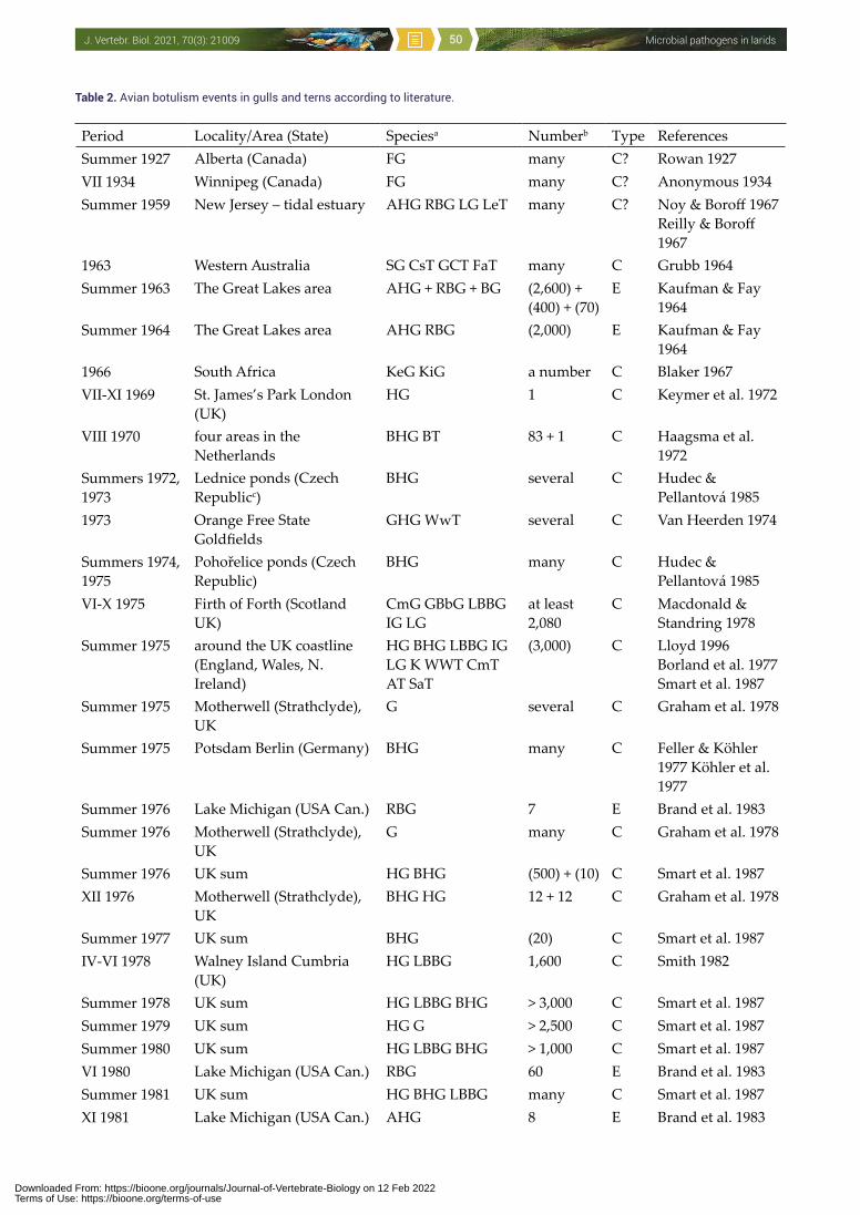
Microbial pathogens in laridsJ. Vertebr. Biol. 2021, 70(3): 21009 50
Table 2. Avian botulism events in gulls and terns according to literature.
Period Locality/Area (State) Speciesa Numberb Type ReferencesSummer 1927 Alberta (Canada) FG many C? Rowan 1927VII 1934 Winnipeg (Canada) FG many C? Anonymous 1934Summer 1959 New Jersey – tidal estuary AHG RBG LG LeT many C? Noy & Boroff 1967
Reilly & Boroff 1967
1963 Western Australia SG CsT GCT FaT many C Grubb 1964Summer 1963 The Great Lakes area AHG + RBG + BG (2,600) +
(400) + (70)E Kaufman & Fay
1964Summer 1964 The Great Lakes area AHG RBG (2,000) E Kaufman & Fay
19641966 South Africa KeG KiG a number C Blaker 1967VII-XI 1969 St. James’s Park London
(UK)HG 1 C Keymer et al. 1972
VIII 1970 four areas in the Netherlands
BHG BT 83 + 1 C Haagsma et al. 1972
Summers 1972, 1973
Lednice ponds (Czech Republicc)
BHG several C Hudec & Pellantová 1985
1973 Orange Free State Goldfields
GHG WwT several C Van Heerden 1974
Summers 1974, 1975
Pohořelice ponds (Czech Republic)
BHG many C Hudec & Pellantová 1985
VI-X 1975 Firth of Forth (Scotland UK)
CmG GBbG LBBG IG LG
at least 2,080
C Macdonald & Standring 1978
Summer 1975 around the UK coastline (England, Wales, N. Ireland)
HG BHG LBBG IG LG K WWT CmT AT SaT
(3,000) C Lloyd 1996 Borland et al. 1977 Smart et al. 1987
Summer 1975 Motherwell (Strathclyde), UK
G several C Graham et al. 1978
Summer 1975 Potsdam Berlin (Germany) BHG many C Feller & Köhler 1977 Köhler et al. 1977
Summer 1976 Lake Michigan (USA Can.) RBG 7 E Brand et al. 1983Summer 1976 Motherwell (Strathclyde),
UKG many C Graham et al. 1978
Summer 1976 UK sum HG BHG (500) + (10) C Smart et al. 1987XII 1976 Motherwell (Strathclyde),
UKBHG HG 12 + 12 C Graham et al. 1978
Summer 1977 UK sum BHG (20) C Smart et al. 1987IV-VI 1978 Walney Island Cumbria
(UK)HG LBBG 1,600 C Smith 1982
Summer 1978 UK sum HG LBBG BHG > 3,000 C Smart et al. 1987Summer 1979 UK sum HG G > 2,500 C Smart et al. 1987Summer 1980 UK sum HG LBBG BHG > 1,000 C Smart et al. 1987VI 1980 Lake Michigan (USA Can.) RBG 60 E Brand et al. 1983Summer 1981 UK sum HG BHG LBBG many C Smart et al. 1987XI 1981 Lake Michigan (USA Can.) AHG 8 E Brand et al. 1983
Downloaded From: https://bioone.org/journals/Journal-of-Vertebrate-Biology on 12 Feb 2022Terms of Use: https://bioone.org/terms-of-use
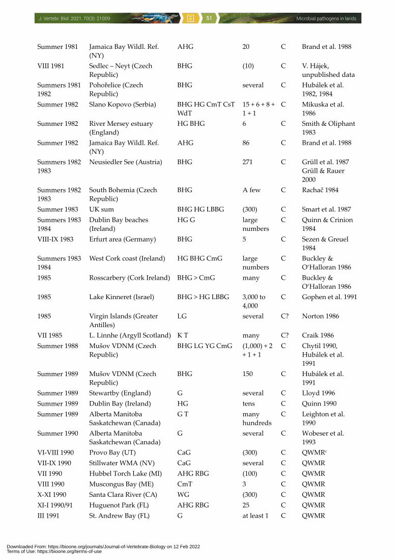
Microbial pathogens in laridsJ. Vertebr. Biol. 2021, 70(3): 21009 51
Summer 1981 Jamaica Bay Wildl. Ref. (NY)
AHG 20 C Brand et al. 1988
VIII 1981 Sedlec – Neyt (Czech Republic)
BHG (10) C V. Hájek, unpublished data
Summers 1981 1982
Pohořelice (Czech Republic)
BHG several C Hubálek et al. 1982, 1984
Summer 1982 Slano Kopovo (Serbia) BHG HG CmT CsT WdT
15 + 6 + 8 + 1 + 1
C Mikuska et al. 1986
Summer 1982 River Mersey estuary (England)
HG BHG 6 C Smith & Oliphant 1983
Summer 1982 Jamaica Bay Wildl. Ref. (NY)
AHG 86 C Brand et al. 1988
Summers 1982 1983
Neusiedler See (Austria) BHG 271 C Grüll et al. 1987 Grüll & Rauer 2000
Summers 1982 1983
South Bohemia (Czech Republic)
BHG A few C Rachač 1984
Summer 1983 UK sum BHG HG LBBG (300) C Smart et al. 1987Summers 1983 1984
Dublin Bay beaches (Ireland)
HG G large numbers
C Quinn & Crinion 1984
VIII-IX 1983 Erfurt area (Germany) BHG 5 C Sezen & Greuel 1984
Summers 1983 1984
West Cork coast (Ireland) HG BHG CmG large numbers
C Buckley & O‘Halloran 1986
1985 Rosscarbery (Cork Ireland) BHG > CmG many C Buckley & O‘Halloran 1986
1985 Lake Kinneret (Israel) BHG > HG LBBG 3,000 to 4,000
C Gophen et al. 1991
1985 Virgin Islands (Greater Antilles)
LG several C? Norton 1986
VII 1985 L. Linnhe (Argyll Scotland) K T many C? Craik 1986Summer 1988 Mušov VDNM (Czech
Republic)BHG LG YG CmG (1,000) + 2
+ 1 + 1 C Chytil 1990,
Hubálek et al. 1991
Summer 1989 Mušov VDNM (Czech Republic)
BHG 150 C Hubálek et al. 1991
Summer 1989 Stewartby (England) G several C Lloyd 1996Summer 1989 Dublin Bay (Ireland) HG tens C Quinn 1990Summer 1989 Alberta Manitoba
Saskatchewan (Canada)G T many
hundredsC Leighton et al.
1990Summer 1990 Alberta Manitoba
Saskatchewan (Canada)G several C Wobeser et al.
1993VI-VIII 1990 Provo Bay (UT) CaG (300) C QWMRc
VII-IX 1990 Stillwater WMA (NV) CaG several C QWMRVII 1990 Hubbel Torch Lake (MI) AHG RBG (100) C QWMRVIII 1990 Muscongus Bay (ME) CmT 3 C QWMRX-XI 1990 Santa Clara River (CA) WG (300) C QWMRXI-I 1990/91 Huguenot Park (FL) AHG RBG 25 C QWMRIII 1991 St. Andrew Bay (FL) G at least 1 C QWMR
Downloaded From: https://bioone.org/journals/Journal-of-Vertebrate-Biology on 12 Feb 2022Terms of Use: https://bioone.org/terms-of-use
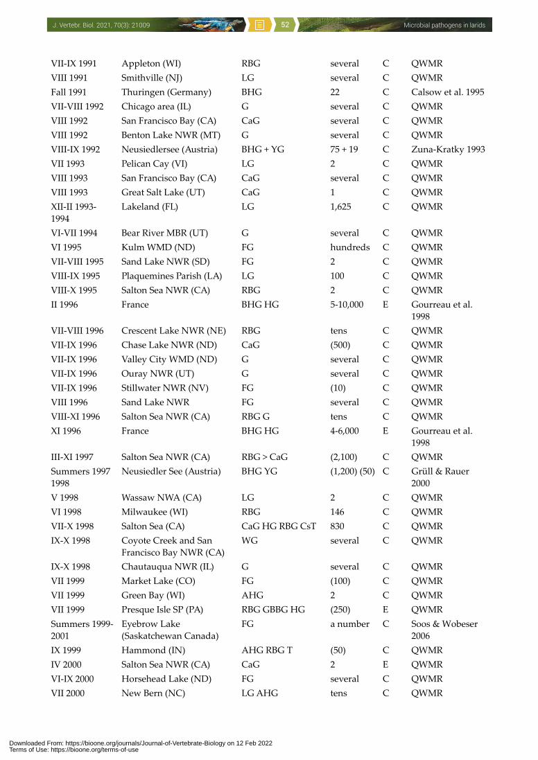
Microbial pathogens in laridsJ. Vertebr. Biol. 2021, 70(3): 21009 52
VII-IX 1991 Appleton (WI) RBG several C QWMRVIII 1991 Smithville (NJ) LG several C QWMRFall 1991 Thuringen (Germany) BHG 22 C Calsow et al. 1995VII-VIII 1992 Chicago area (IL) G several C QWMRVIII 1992 San Francisco Bay (CA) CaG several C QWMRVIII 1992 Benton Lake NWR (MT) G several C QWMRVIII-IX 1992 Neusiedlersee (Austria) BHG + YG 75 + 19 C Zuna-Kratky 1993VII 1993 Pelican Cay (VI) LG 2 C QWMRVIII 1993 San Francisco Bay (CA) CaG several C QWMRVIII 1993 Great Salt Lake (UT) CaG 1 C QWMRXII-II 1993-1994
Lakeland (FL) LG 1,625 C QWMR
VI-VII 1994 Bear River MBR (UT) G several C QWMRVI 1995 Kulm WMD (ND) FG hundreds C QWMRVII-VIII 1995 Sand Lake NWR (SD) FG 2 C QWMRVIII-IX 1995 Plaquemines Parish (LA) LG 100 C QWMRVIII-X 1995 Salton Sea NWR (CA) RBG 2 C QWMRII 1996 France BHG HG 5-10,000 E Gourreau et al.
1998VII-VIII 1996 Crescent Lake NWR (NE) RBG tens C QWMRVII-IX 1996 Chase Lake NWR (ND) CaG (500) C QWMRVII-IX 1996 Valley City WMD (ND) G several C QWMRVII-IX 1996 Ouray NWR (UT) G several C QWMRVII-IX 1996 Stillwater NWR (NV) FG (10) C QWMRVIII 1996 Sand Lake NWR FG several C QWMRVIII-XI 1996 Salton Sea NWR (CA) RBG G tens C QWMRXI 1996 France BHG HG 4-6,000 E Gourreau et al.
1998III-XI 1997 Salton Sea NWR (CA) RBG > CaG (2,100) C QWMRSummers 1997 1998
Neusiedler See (Austria) BHG YG (1,200) (50) C Grüll & Rauer 2000
V 1998 Wassaw NWA (CA) LG 2 C QWMRVI 1998 Milwaukee (WI) RBG 146 C QWMRVII-X 1998 Salton Sea (CA) CaG HG RBG CsT 830 C QWMRIX-X 1998 Coyote Creek and San
Francisco Bay NWR (CA)WG several C QWMR
IX-X 1998 Chautauqua NWR (IL) G several C QWMRVII 1999 Market Lake (CO) FG (100) C QWMRVII 1999 Green Bay (WI) AHG 2 C QWMRVII 1999 Presque Isle SP (PA) RBG GBBG HG (250) E QWMRSummers 1999-2001
Eyebrow Lake (Saskatchewan Canada)
FG a number C Soos & Wobeser 2006
IX 1999 Hammond (IN) AHG RBG T (50) C QWMRIV 2000 Salton Sea NWR (CA) CaG 2 E QWMRVI-IX 2000 Horsehead Lake (ND) FG several C QWMRVII 2000 New Bern (NC) LG AHG tens C QWMR
Downloaded From: https://bioone.org/journals/Journal-of-Vertebrate-Biology on 12 Feb 2022Terms of Use: https://bioone.org/terms-of-use
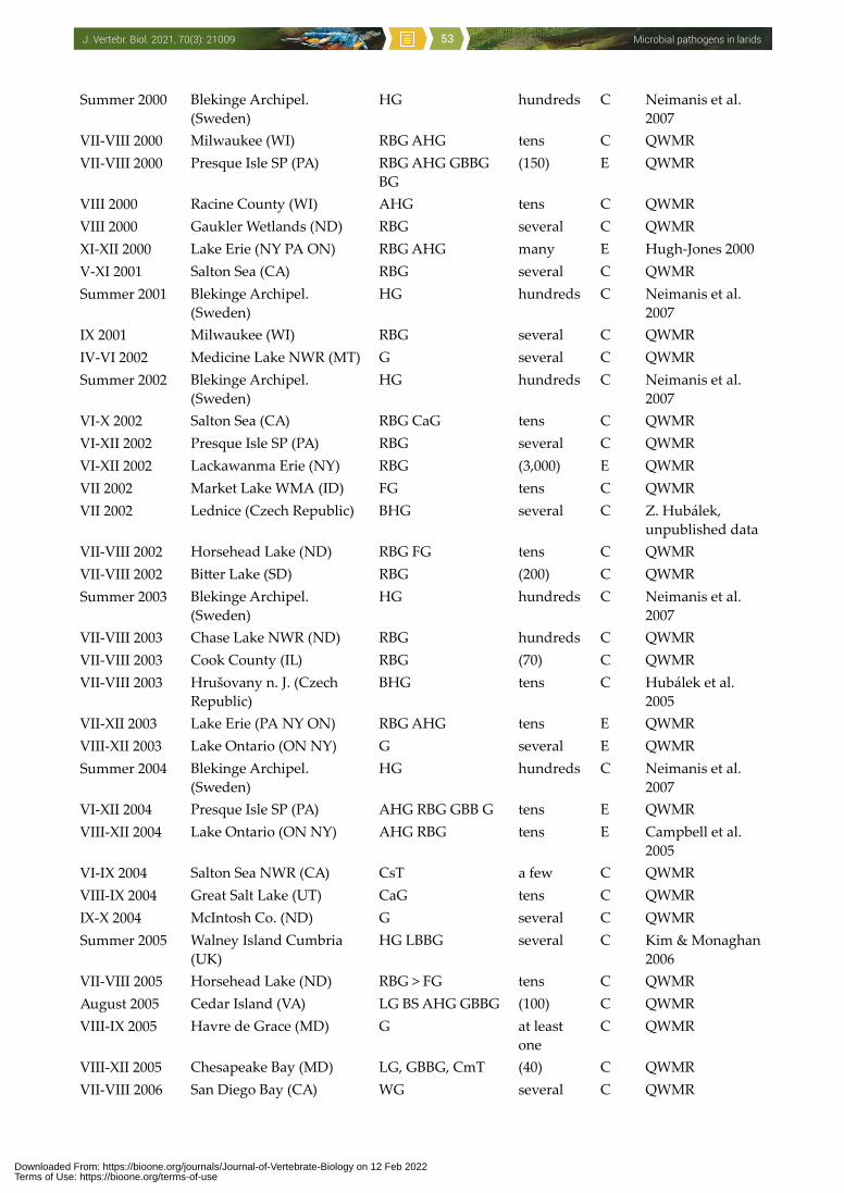
Microbial pathogens in laridsJ. Vertebr. Biol. 2021, 70(3): 21009 53
Summer 2000 Blekinge Archipel. (Sweden)
HG hundreds C Neimanis et al. 2007
VII-VIII 2000 Milwaukee (WI) RBG AHG tens C QWMRVII-VIII 2000 Presque Isle SP (PA) RBG AHG GBBG
BG(150) E QWMR
VIII 2000 Racine County (WI) AHG tens C QWMRVIII 2000 Gaukler Wetlands (ND) RBG several C QWMRXI-XII 2000 Lake Erie (NY PA ON) RBG AHG many E Hugh-Jones 2000V-XI 2001 Salton Sea (CA) RBG several C QWMRSummer 2001 Blekinge Archipel.
(Sweden)HG hundreds C Neimanis et al.
2007IX 2001 Milwaukee (WI) RBG several C QWMRIV-VI 2002 Medicine Lake NWR (MT) G several C QWMRSummer 2002 Blekinge Archipel.
(Sweden)HG hundreds C Neimanis et al.
2007VI-X 2002 Salton Sea (CA) RBG CaG tens C QWMRVI-XII 2002 Presque Isle SP (PA) RBG several C QWMRVI-XII 2002 Lackawanma Erie (NY) RBG (3,000) E QWMRVII 2002 Market Lake WMA (ID) FG tens C QWMRVII 2002 Lednice (Czech Republic) BHG several C Z. Hubálek,
unpublished dataVII-VIII 2002 Horsehead Lake (ND) RBG FG tens C QWMRVII-VIII 2002 Bitter Lake (SD) RBG (200) C QWMRSummer 2003 Blekinge Archipel.
(Sweden)HG hundreds C Neimanis et al.
2007VII-VIII 2003 Chase Lake NWR (ND) RBG hundreds C QWMRVII-VIII 2003 Cook County (IL) RBG (70) C QWMRVII-VIII 2003 Hrušovany n. J. (Czech
Republic)BHG tens C Hubálek et al.
2005VII-XII 2003 Lake Erie (PA NY ON) RBG AHG tens E QWMRVIII-XII 2003 Lake Ontario (ON NY) G several E QWMRSummer 2004 Blekinge Archipel.
(Sweden)HG hundreds C Neimanis et al.
2007VI-XII 2004 Presque Isle SP (PA) AHG RBG GBB G tens E QWMRVIII-XII 2004 Lake Ontario (ON NY) AHG RBG tens E Campbell et al.
2005VI-IX 2004 Salton Sea NWR (CA) CsT a few C QWMRVIII-IX 2004 Great Salt Lake (UT) CaG tens C QWMRIX-X 2004 McIntosh Co. (ND) G several C QWMRSummer 2005 Walney Island Cumbria
(UK)HG LBBG several C Kim & Monaghan
2006VII-VIII 2005 Horsehead Lake (ND) RBG > FG tens C QWMRAugust 2005 Cedar Island (VA) LG BS AHG GBBG (100) C QWMRVIII-IX 2005 Havre de Grace (MD) G at least
oneC QWMR
VIII-XII 2005 Chesapeake Bay (MD) LG, GBBG, CmT (40) C QWMRVII-VIII 2006 San Diego Bay (CA) WG several C QWMR
Downloaded From: https://bioone.org/journals/Journal-of-Vertebrate-Biology on 12 Feb 2022Terms of Use: https://bioone.org/terms-of-use
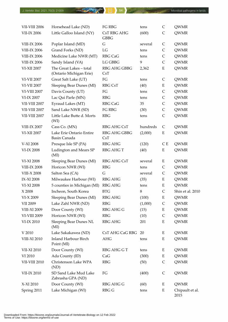
Microbial pathogens in laridsJ. Vertebr. Biol. 2021, 70(3): 21009 54
VII-VIII 2006 Horsehead Lake (ND) FG RBG tens C QWMRVII-IX 2006 Little Galloo Island (NY) CsT RBG AHG
GBBG(600) C QWMR
VIII-IX 2006 Poplar Island (MD) G several C QWMRVIII-IX 2006 Grand Forks (ND) LG tens C QWMRVIII-IX 2006 Medicine Lake NWR (MT) RBG CaG tens C QWMRVIII-IX 2006 Sandy Island (VA) LG GBBG 9 C QWMRVI-XII 2007 The Great Lakes – total
(Ontario Michigan Erie)RBG AHG GBBG CsT
2,362 E QWMR
VI-VII 2007 Great Salt Lake (UT) FG tens C QWMRVI-VII 2007 Sleeping Bear Dunes (MI) RBG CsT (40) E QWMRVI-VIII 2007 Davis County (UT) FG tens C QWMRVI-IX 2007 Lac Qui Parle (MN) RBG tens C QWMRVII-VIII 2007 Eyraud Lakes (MT) RBG CaG 35 C QWMRVII-VIII 2007 Sand Lake NWR (SD) FG RBG (30) C QWMRVII-VIII 2007 Little Lake Butte d. Morts
(WI)RBG tens C QWMR
VIII-IX 2007 Cass Co. (MN) RBG AHG CsT hundreds C QWMRVI-XII 2007 Lake Erie Ontario Entire
Basin CanadaRBG AHG GBBG CsT
(2,000) E QWMR
V-XI 2008 Presque Isle SP (PA) RBG AHG (120) C E QWMRVI-IX 2008 Ludington and Mears SP
(MI)RBG AHG T (40) E QWMR
VI-XI 2008 Sleeping Bear Dunes (MI) RBG AHG CsT several E QWMRVIII-IX 2008 Horicon NWR (WI) RBG tens C QWMRVIII-X 2008 Salton Sea (CA) G several C QWMRIX-XI 2008 Milwaukee Harbour (WI) RBG AHG (35) E QWMRVI-XI 2008 5 counties in Michigan (MI) RBG AHG tens E QWMRX 2008 Incheon, South Korea BTG 8 C Shin et al. 2010VI-X 2009 Sleeping Bear Dunes (MI) RBG AHG (100) E QWMRVII 2009 Lake Zahl NWR (ND) RBG (1,000) C QWMRVIII-XI 2009 Door County (WI) RBG AHG G (15) E QWMRVI-VIII 2009 Horicon NWR (WI) RBG (10) C QWMRVI-IX 2010 Sleeping Bear Dunes NL
(MI)RBG AHG 201 E QWMR
V 2010 Lake Sakakawea (ND) CsT AHG CaG RBG 20 E QWMRVIII-XI 2010 Inland Harbour Birch
Point (MI)AHG tens E QWMR
VII-XI 2010 Door County (WI) RBG AHG G T tens E QWMRVI 2010 Ada County (ID) CaG (300) E QWMRVII-VIII 2010 Christenson Lake WPA
(ND)RBG (50) C QWMR
VII-IX 2010 SD Sand Lake Mud Lake Zabrasha GPA (ND)
FG (400) C QWMR
X-XI 2010 Door County (WI) RBG AHG G (60) E QWMRSpring 2011 Lake Michigan (WI) RBG G tens E Chipault et al.
2015
Downloaded From: https://bioone.org/journals/Journal-of-Vertebrate-Biology on 12 Feb 2022Terms of Use: https://bioone.org/terms-of-use
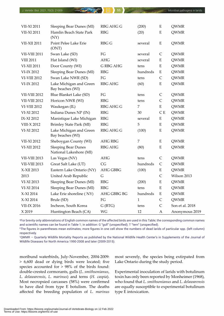
Microbial pathogens in laridsJ. Vertebr. Biol. 2021, 70(3): 21009 55
moribund waterbirds, July-November, 2004-2009: > 6,600 dead or dying birds were located; five species accounted for > 98% of the birds found: double-crested cormorants, gulls (L. smithsonianus, L. delawarensis, L. marinus) and terns (H. caspia). Most necropsied carcasses (58%) were confirmed to have died from type E botulism. The deaths affected the breeding population of L. marinus
most severely, the species being extirpated from Lake Ontario during the study period.
Experimental inoculation of larids with botulinum toxin has only been reported by Monheimer (1968), who found that L. smithsonianus and L. delawarensis are equally susceptible to experimental botulinum type E intoxication.
VII-XI 2011 Sleeping Bear Dunes (MI) RBG AHG G (200) E QWMRVII-XI 2011 Hamlin Beach State Park
(NY)RBG (20) E QWMR
VII-XII 2011 Point Pelee Lake Erie (ONT)
RBG G several E QWMR
VII-VIII 2011 Swan Lake (SD) FG several C QWMRVIII 2011 Hat Island (WI) AHG several E QWMRVI-XII 2011 Door County (WI) G RBG AHG tens E QWMRVI-IX 2012 Sleeping Bear Dunes (MI) RBG hundreds E QWMRVI-VIII 2012 Swan Lake NWR (SD) FG tens C QWMRVI-IX 2012 Lake Michigan and Green
Bay beaches (WI)RBG AHG (60) E QWMR
VII-VIII 2012 Blue Blanket Lake (SD) FG tens C QWMRVII-VIII 2012 Horicon NWR (WI) RBG tens C QWMRVI-VIII 2012 Waukegan (IL) RBG AHG G 7 E QWMRVI-XI 2012 Indiana Dunes NP (IN) RBG 30 C E QWMRIX-XI 2012 Manistique Lake Michigan RBG several E QWMRVIII-X 2012 Brimley State Park (MI) RBG 5 E QWMRVI-XI 2012 Lake Michigan and Green
Bay beaches (WI)RBG AHG G (100) E QWMR
VII-XI 2012 Sheboygan County (WI) AHG RBG 7 E QWMRVI-XII 2012 Sleeping Bear Dunes
National Lakeshore (MI)RBG AHG (80) E QWMR
VII-VIII 2013 Las Vegas (NV) AHG tens C QWMRVII-VIII 2013 Great Salt Lake (UT) CG hundreds C QWMRX-XII 2013 Eastern Lake Ontario (NY) AHG GBBG (100) E QWMR2013 United Arab Republic G C Wilson 2013VI-XI 2013 Sleeping Bear Dunes (MI) RBG (200) E QWMRVI-XI 2014 Sleeping Bear Dunes (MI) RBG tens E QWMRX-XI 2014 Lake Erie shoreline ( NY) AHG GBBG BG hundreds E QWMRX-XI 2014 Brule (SD) FG 1 C QWMRVII-IX 2016 Incheon, South Korea G (BTG) tens C Son et al. 2018X 2019 Huntington Beach (CA) WG 12 A Anonymous 2019
aFor brevity only abbreviations of English common names of the affected birds are used in this Table; the corresponding common names and scientific names can be found in Table 1; in addition: G “gull“ (unspecified); T “tern“ (unspecified).bThe figures in parentheses mean estimates; more figures in one cell show the numbers of dead larids of particular spp. (left column) respectively.cQWMR – Quarterly Wildlife Mortality Reports as published by the National Wildlife Health Center’s in Supplements of the Journal of Wildlife Diseases for North America 1990-2008 and later (2009-2015).
Downloaded From: https://bioone.org/journals/Journal-of-Vertebrate-Biology on 12 Feb 2022Terms of Use: https://bioone.org/terms-of-use
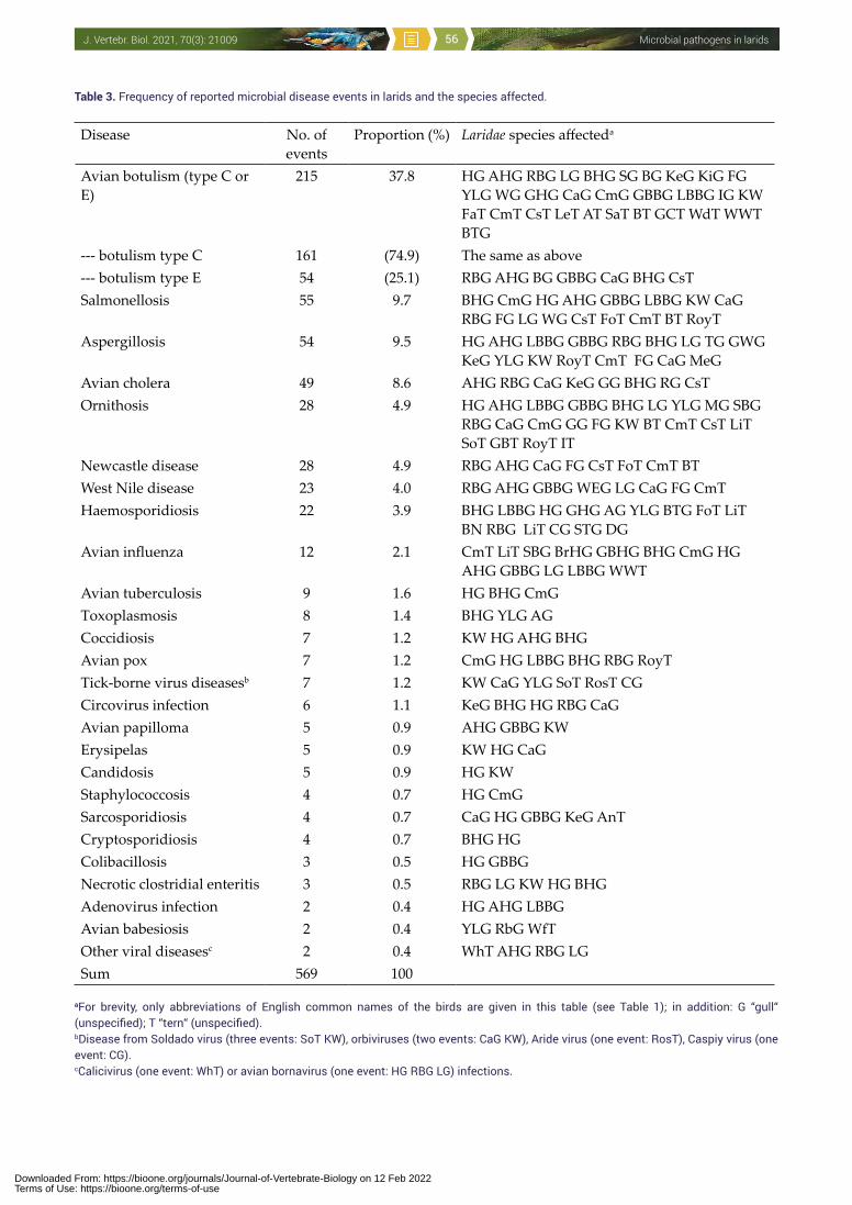
Microbial pathogens in laridsJ. Vertebr. Biol. 2021, 70(3): 21009 56
Table 3. Frequency of reported microbial disease events in larids and the species affected.
Disease No. of events
Proportion (%) Laridae species affecteda
Avian botulism (type C or E)
215 37.8 HG AHG RBG LG BHG SG BG KeG KiG FG YLG WG GHG CaG CmG GBBG LBBG IG KW FaT CmT CsT LeT AT SaT BT GCT WdT WWT BTG
--- botulism type C 161 (74.9) The same as above--- botulism type E 54 (25.1) RBG AHG BG GBBG CaG BHG CsTSalmonellosis 55 9.7 BHG CmG HG AHG GBBG LBBG KW CaG
RBG FG LG WG CsT FoT CmT BT RoyTAspergillosis 54 9.5 HG AHG LBBG GBBG RBG BHG LG TG GWG
KeG YLG KW RoyT CmT FG CaG MeGAvian cholera 49 8.6 AHG RBG CaG KeG GG BHG RG CsTOrnithosis 28 4.9 HG AHG LBBG GBBG BHG LG YLG MG SBG
RBG CaG CmG GG FG KW BT CmT CsT LiT SoT GBT RoyT IT
Newcastle disease 28 4.9 RBG AHG CaG FG CsT FoT CmT BTWest Nile disease 23 4.0 RBG AHG GBBG WEG LG CaG FG CmTHaemosporidiosis 22 3.9 BHG LBBG HG GHG AG YLG BTG FoT LiT
BN RBG LiT CG STG DG Avian influenza 12 2.1 CmT LiT SBG BrHG GBHG BHG CmG HG
AHG GBBG LG LBBG WWT Avian tuberculosis 9 1.6 HG BHG CmGToxoplasmosis 8 1.4 BHG YLG AG Coccidiosis 7 1.2 KW HG AHG BHGAvian pox 7 1.2 CmG HG LBBG BHG RBG RoyTTick-borne virus diseasesb 7 1.2 KW CaG YLG SoT RosT CG Circovirus infection 6 1.1 KeG BHG HG RBG CaGAvian papilloma 5 0.9 AHG GBBG KWErysipelas 5 0.9 KW HG CaGCandidosis 5 0.9 HG KWStaphylococcosis 4 0.7 HG CmGSarcosporidiosis 4 0.7 CaG HG GBBG KeG AnT Cryptosporidiosis 4 0.7 BHG HGColibacillosis 3 0.5 HG GBBGNecrotic clostridial enteritis 3 0.5 RBG LG KW HG BHGAdenovirus infection 2 0.4 HG AHG LBBGAvian babesiosis 2 0.4 YLG RbG WfTOther viral diseasesc 2 0.4 WhT AHG RBG LGSum 569 100
aFor brevity, only abbreviations of English common names of the birds are given in this table (see Table 1); in addition: G “gull“ (unspecified); T “tern“ (unspecified).bDisease from Soldado virus (three events: SoT KW), orbiviruses (two events: CaG KW), Aride virus (one event: RosT), Caspiy virus (one event: CG).cCalicivirus (one event: WhT) or avian bornavirus (one event: HG RBG LG) infections.
Downloaded From: https://bioone.org/journals/Journal-of-Vertebrate-Biology on 12 Feb 2022Terms of Use: https://bioone.org/terms-of-use
Microbial pathogens in laridsJ. Vertebr. Biol. 2021, 70(3): 21009 57
Flavobacteriaceae
Chryseobacterium indologenesMalka et al. (2020) described an adult male L. delawarensis gull presented to a wildlife rescue centre on Long Island, New York, for bilateral lameness. Veterinary evaluation diagnosed septic arthritis and osteomyelitis caused by C. indologenes and treated with orbifloxacin until recovery. This sapronotic bacterial species is infrequently diagnosed as an opportunistic human pathogen causing various infections, including meningitis and pneumonia.
Mycobacteriaceae
Mycobacterium aviumMycobacterium avium ssp. avium causes avian tuberculosis in many bird species. There are no specific clinical symptoms of acute tuberculosis in affected larids, but the birds with chronic infection are emaciated, weak and lethargic, with wasted muscles. Gross lesions at autopsy involve yellow, whitish to grey nodules embedded in the infected tissues (liver, spleen, lungs, intestine, skin). However, the prevalence of tuberculosis in gulls is low: e.g. only 1% of 1,048 examined C. ridibundus and L. canus were found to have died from avian tuberculosis in the Netherlands (Smit et al. 1987).
Galli Vallerio (quoted by Plum 1942) was the first who diagnosed tuberculosis in two gulls shot in a meadow grazed by a herd of tuberculin positive cattle. Plum (1942) found macroscopic lesions similar to tuberculosis in 48 of 816 gulls (L. argentatus, L. canus, C. ridibundus) shot at Copenhagen, but Mycobacterium was recovered only from four of them, and from an additional 28 of 768 other apparently healthy gulls. Tuberculosis in a few gulls (C. ridibundus, L. argentatus) has been repeatedly found in Great Britain since 1954 (Jennings & Soulsby 1957, Poulding 1957, Jennings 1959, Threlfall 1967, McDiarmid 1969). Blackmore & Keymer (1969) found one immature L. argentatus suffering from a generalized tuberculosis: a large ulcerated swelling affecting the carpal joint of one wing, and military tubercular lesions were in the liver. Avian tuberculosis was also detected in several L. argentatus living on Helgoland Island, Germany (Vauk et al. 1980, Vauk-Hentzelt 1986). Hejlíček & Treml (1993) did not find any tuberculous lesions in 35 C. ridibundus caught at four localities of Czech Republic. Gronesova et al. (2008) detected M. avium in six of 11 examined C. ridibundus gulls in Slovakia, summer 2007.
Experimental infections of C. ridibundus with M. avium were carried out by Hejlíček & Treml (1994, 1995). After i.m. inoculation of M. avium into several gulls, tuberculous lesions were found at the spot of puncture 35 DPI, and in the liver and spleen 70 DPI. In the simultaneously infected domestic fowl the lesions appeared at the spot of puncture, spleen and liver 35 DPI. Mycobacteria were isolated from several organs and tissues of both avian species 35 DPI. Tuberculous lesions were also found in the liver, spleen and intestine 118 days after feeding gulls with liver from tuberculous domestic chickens, and mycobacteria were demonstrated in the liver, spleen, intestine and brain of the gulls at the same time. The susceptibility of C. ridibundus to M. avium was the same as that of domestic chicken and house sparrow, while other birds (guinea fowl, turkey, partridge and pheasant) were less sensitive to infection from infected food and other sources.
Microfungi
Ajellomycetaceae
Histoplasma capsulatum (teleomorph: Ajellomyces capsulatus)Waldman et al. (1983) described an outbreak of acute pulmonary histoplasmosis caused by this dimorphic fungus that attacked 138 employees of a Michigan limestone quarry. The source of exposure was a vessel repair building containing a pulley which had been stored in a L. delawarensis nesting area. Histoplasma capsulatum was recovered from the working area, the gull nesting area and the sputum of several patients. The authors do not exclude, however, the presence of bats, bat guano being the principal source of the fungus. Hamrick et al. (1986) isolated H. capsulatum from the soil at a nesting site of C. ridibundus in Rogers City, Michigan (USA).
Eurotiaceae
Aspergillus fumigatus Aspergillosis rates among the most important mycoses of waterbirds (including gulls), with a high mortality rate and a worldwide distribution (Urbain & Guillot 1938, McDiarmid 1955, Ainsworth & Austwick 1959, McDiarmid 1969, Brand et al. 1988, Wobeser 1997). The most common clinical form of avian aspergillosis is the invasion of the respiratory tract (pneumonia, airsacculitis) by this thermophilic fungus. The affected birds are emaciated and show difficulties in breathing. Post-mortem autopsy reveals numerous thoracic yellow
Downloaded From: https://bioone.org/journals/Journal-of-Vertebrate-Biology on 12 Feb 2022Terms of Use: https://bioone.org/terms-of-use
Microbial pathogens in laridsJ. Vertebr. Biol. 2021, 70(3): 21009 58
nodules and green caverns up to 15 mm in diameter in pulmonary tissue and air sacs, and microscopy shows hyphae and granulomatous reactions in the lung lesions. Sometimes the infection affects other visceral organs, including the gastrointestinal tract, and certain cases of aspergillosis in birds including larids have been suggestive of generalized infection or mycotoxicosis, with the liver and kidney lesions (Davis & McClung 1940, McDiarmid 1955, 1969, Wobeser 1997). The vast majority of fatal cases of avian aspergillosis cases are due to A. fumigatus, other species (e.g. Aspergillus flavus) are rare agents in its aetiology. The source of infection for birds is contaminated food and vegetation. Birds under stress are more vulnerable to the disease – for example suffering starvation, in contact with oil on the sea, overheated, exhausted by migration, captive, or suffering other primary disease or toxicosis. The most developed clinical forms of aspergillosis have been observed in birds following traumas, long detention in captivity, starvation, concurrent presence of other diseases, stress-induced immunosuppression and extensive antibiotic treatments.
In Europe, Robin (1853) first described aspergillosis (A. fumigatus) in a gull L. “griseus” (probably L. canus) in France: the bird died after being kept in captivity. Much later, Poulding (1952) recorded five cases in immature L. argentatus gulls, McDiarmid (1955) three cases in L. argentatus in England between 1943 and 1953, McCartan (1958a, b) several cases in R. tridactyla during the breeding season, Jennings & Soulsby (1958) in breeding C. ridibundus, Jennings (1959, 1961) in L. argentatus and Threlfall (1967) in three adult and one juvenile L. argentatus in North Wales from 1962-1964. Baker (1977) reported aspergillosis in one C. ridibundus in Cheshire (England), Vauk-Henzelt (1986) in two juvenile L. argentatus and one L. marinus in the German Bight, Germany, Petermann et al. (1989) in two R. tridactyla found moribund or dead in the German Bight. One dead juvenile C. ridibundus with pulmonary aspergillosis (A. fumigatus was isolated from the lungs and air sacs), hepatitis and gastroenteritis due to salmonellosis (Salmonella Typhimurium) on a fishpond at Hodonín (south Moravia, Czech Republic) was examined in June 1992 (Z. Hubálek, unpublished data). Juhasz-Judit & Andrikovics (2006) described how 29 captured C. ridibundus nestlings (1-12 days old, healthy) showed dyspnoeic symptoms after three-four days in captivity and several days later 80% of them died, displaying tiny greenish-yellow loading in the
lungs and on the air sacs from which A. fumigatus was cultured. In the same area of Hungary, die-off due to aspergillosis occurred among free-living C. ridibundus. Nardoni et al. (2006) recorded eight cases of disseminated aspergillosis in L. cachinnans (probably L. michahellis) at a bird rehabilitation centre in Livorno (Tuscany), and Cacciuttolo et al. (2009) found five additional fatal cases of systemic aspergillosis (with lesions in the lungs, air sacs, liver and spleen: A. fumigatus cultivated) in the same gull species and locality. Suvorov et al. (2014) reported a rare observation of exotic L. hyperboreus gull in Prague in 2012. The gull died and the main cause of death was aspergillosis caused by A. fumigatus, as shown by dissection.
In North America, many cases and several outbreaks of avian aspergillosis were reported in gulls. Davis & McClung (1940) observed fatal aspergillosis in at least 60 adult L. smithsonianus at Boston Harbour in 1939 (three gulls were examined in detail, and A. fumigatus was isolated). Cowan (1943) found aspergillosis in a Larus thayeri in Canada, Herman & Bolander (1943) in L. glaucescens, Beaudette (1945) in a gull, Herman & Rosen (1947) in Larus spp. Friend & Trainer (1969) observed fatal aspergillosis developing in 32 of 140 juvenile L. smithsonianus within one month of capture. Brand et al. (1983) observed a mixed infection in a L. delawarensis that died of botulism at Lake Michigan (USA) in 1976, but also had airsacculitis due to A. fumigatus. Brand et al. (1988) observed morbidity and mortality from aspergillosis in L. smithsonianus (9/86 examined), L. atricilla (one of two), L. marinus (three of eight) and several L. delawarensis at Jamaica Bay Wildlife Refuge, New York, in 1981-1982. According to QWMR reports and Kidd (1991), events of gull aspergillosis occurred on Dyer Island (Rhode Island: ca. 100 dead L. marinus and L. smithsonianus) in summer 1990, on Lake Champlain (Vermont: ca. 3,500 L. smithsonianus and L. delawarensis, combined with salmonellosis), at Beaver Dam (Wisconsin: 15 L. delawarensis), in Deer Flat National Wildlife Refuge (Idaho, several gulls), at Plattsburg (New York: 17 L. delawarensis) in summer 1991, and 21 L. atricilla and L. smithsonianus on Sanibel Island (Florida) in winter 1992/1993. Two L. smithsonianus died of aspergillosis in Providence (Rhode Island) in autumn 1994, several L. smithsonianus on Gulf Shores State Park (Alabama) in winter 2001/2002, hundreds of L. delawarensis in Kulm (North Dakota) in summer 2003 (but combined with WNV encephalitis, salmonellosis, chlamydiosis:
Downloaded From: https://bioone.org/journals/Journal-of-Vertebrate-Biology on 12 Feb 2022Terms of Use: https://bioone.org/terms-of-use
Microbial pathogens in laridsJ. Vertebr. Biol. 2021, 70(3): 21009 59
QWMR 2003). In winter 2005/2006, 20 gulls (L. smithsonianus, L. atricilla, L. delawarensis) died from aspergillosis on Daytona Beach Lake (Florida), tens of L. delawarensis and L. smithsonianus on Presque Isle (Pennsylvania) in summer 2008, 30 L. delawarensis in Audubon National Wildlife Refuge (North Dakota), ca. 100 L. pipixcan in J. Clark Salyer National Wildlife Refuge (North Dakota) in May to June 2009, a few L. delawarensis and L. smithsonianus in Androscoggin Country (Maine) in September 2009, hundreds L. delawarensis on Pelican Lake (Minnesota) in summer 2009, 15 L. delawarensis in Carrington (North Dakota) in August 2009, four L. marinus and L. smithsonianus on Poplar Island Restoration Site (Maryland) in autumn 2009, 30 L. californicus on Eyraud Lakes (Montana), 65 larids (L. atricilla, T. maximus, S. hirundo) on Longboat Key and Lido Key (Florida), 16 gulls (L. smithsonianus, Larus sp.) on Poplar Island (Maryland) and several L. delawarensis on Lake Johanna (Minnesota) in July to September 2010, 25 L. smithsonianus on the Alfred Plourde (Maine) in autumn 2011, a few L. smithsonianus in Mackinac Country (Michigan) in autumn 2012 and several gulls (L. smithsonianus, Larus brachyrhynchus) on Staats Lake (Oregon) in March to April 2014.
In Central and South America, Rosiles-Martinez et al. (2000) described an aspergillosis outbreak in L. smithsonianus (ca. 500 birds) at Playa Linda Zihuatanejo in the state of Guerrero, Mexico, and Melo et al. (2020) a case of fatal pulmonary aspergillosis (A. fumigatus as the etiological agent) in one C. maculipennis at Rio Grande, Brazil in 2018.
In New Zealand, Williams (1955) reported aspergillosis in one L. dominicanus. Aspergillosis in a L. dominicanus with chronic airsacculitis (and a circovirus infection in addition) was also reported in the Manawatu region of New Zealand by Twentyman et al. (1999).
In addition to symptomatic aspergillosis, A. fumigatus was occasionally isolated from seemingly healthy larids. For instance, from the throats of 13% of 102 L. argentatus examined in the UK (Beer 1963).
Aspergillus flavus Aspergillus flavus may cause either avian aspergillosis (sporadically) or, more often, aflatoxicosis, a disease caused by aflatoxins. For instance, pulmonary aspergillosis caused by A. flavus and poxvirus infection on legs were
diagnosed in a male Royal tern (T. maximus) that died in Florida (Jacobson et al. 1980). The significance of aflatoxicosis for free-living birds including larids is unknown.
Yeasts and yeast-like fungi
Candida albicansThe main agent of candidosis in homeotherm vertebrates that frequently occurs in fresh gull droppings; the birds may be long-term hosts and carriers, but usually do not show signs of disease. Van Uden & Branco (1963) studied the distribution and population densities of yeasts in seabirds off Southern California, and found C. albicans in L. occidentalis gulls. Kawakita & van Uden (1965) examined 69 larids in Portugal and detected C. albicans in 9% of 26 L. fuscus (27 CFU/g wet weight), and one C. ridibundus (2 CFU/g). Cragg & Clayton (1971) examined 166 fresh droppings and 122 dry droppings originating largely from L. argentatus and C. ridibundus on Jersey, England, 1967-1968: C. albicans was detected in 21.7% of the fresh droppings but only 1.6% of dry droppings. Saez (1971) isolated C. albicans from L. argentatus in France. Buck (1983) recovered C. albicans in 78% of 133 fresh faecal samples of gulls (L. smithsonianus, L. marinus, L. atricilla, L. delawarensis) in Connecticut and 38% of 106 samples on the west coast of Florida. The prevalence rate was much higher in gulls than in B. canadensis geese in Connecticut. The frequency of C. albicans in gull droppings (overall, 60.3%) was much higher than that reported for England, Portugal and California. Buck (1990) carried out a similar study: fresh gull faeces were examined in Connecticut (L. smithsonianus, L. marinus) and Florida (L. smithsonianus, L. atricilla, L. delawarensis); C. albicans was detected in 58% and 41%, respectively.
Experimental inoculation of a gull (Larus sp.) with C. albicans and fed fish containing antimycotic ketoconazole excreted the yeast heavily for the next 13 days, and sporadically up to 40 DPI despite the ketoconazole treatment. The bird displayed no clinical symptoms of candidosis and was released after 57 DPI with faeces testing negative for C. albicans (Buck 1986). Al-Yasiri et al. (2016, 2017) analysed the gut yeast communities in five L. michahellis breeding colonies along the French Mediterranean coast. Candida grew in 113 of 177 samples. The most frequent species were C. krusei (53.5%), C. glabrata (40.9%) and C. albicans (20.5%). Notably, the proportion of human pathogenic yeast species, including C. albicans
Downloaded From: https://bioone.org/journals/Journal-of-Vertebrate-Biology on 12 Feb 2022Terms of Use: https://bioone.org/terms-of-use
Microbial pathogens in laridsJ. Vertebr. Biol. 2021, 70(3): 21009 60
and C. glabrata, in the gull mycobiota increased with gull colony synanthropy. The gut yeast communities of these gulls included antifungal-resistant human pathogens, and these common synanthropic seabirds thus represent a reservoir and disseminator of drug-resistant human pathogenic yeast into the environment. Dynowska et al. (2018) examined swabs from the beaks and cloacae of adult and young C. ridibundus in Poland for fungi: C. albicans was cultured most frequently.
However, the yeast might function as a pathogen in immunosuppressed gulls. Threlfall (1967) examined 657 L. argentatus in North Wales 1962-1964 and observed thrush lesions in the oral cavity and oesophagus due to C. albicans in many apparently healthy birds; he wrote “…. the lesions did not seem to affect the birds except in the case of a heavily infected bird which weighed only 550 g instead of the normal 800 g”. Davis et al. (1971) mentioned candidosis in an ill L. argentatus, and Petermann et al. (1989) diagnosed candidosis in a few dead or moribund gulls (two R. tridactyla, one L. argentatus) in the German Bight, Germany, 1982-1985. Yang & Jae-Hoon (2019) examined a young Larus schistisagus gull found dead near Seongsan Harbour (Korea). The bird had focal ulceration and a protuberant nodule between the proventriculus and gizzard. Numerous white-yellowish nodules were scattered on the surface of the liver and kidney, and in the spleen. Also, haemorrhages were present in the gizzard and multifocal granulomatous inflammations in the liver, kidney, spleen, and lungs. Yeasts and pseudohyphae in granulomatous lesions were confirmed by periodic acid-Schiff stains, and C. albicans was isolated from lesions of the liver, kidney, spleen and gizzard. This was a systemic candidosis.
Candida dubliniensisThis species is closely related to C. albicans. Although C. dubliniensis is less pathogenic than C. albicans, it is found quite often as superficial infection in immunocompromised humans. Nunn et al. (2007) obtained 11 isolates of C. dubliniensis from the surface of I. uriae ticks that lived in cracks filled with seabird (including gull) excrement at a seabird breeding colony on Great Saltee Island off the south-eastern coast of Ireland. McManus et al. (2009) isolated this yeast species from 3/134 fresh faecal samples of L. argentatus directly from the campus of Trinity College in Dublin (Ireland), 2008. Of a total of 14 avian excrement-associated C. dubliniensis isolates tested, one originating from
L. argentatus was indistinguishable from human isolates. The authors mean that “it may be a human isolate that colonized a gull scavenging on the Trinity College campus”.
Candida kruseiIt was cultivated from the intestinal content of seagulls and terns (van Uden & Branco 1963); Kawakita & van Uden (1965) isolated it from 4% of 69 fresh larid droppings in Portugal: positive cases were L. fuscus (1/26), L. argentatus (1/4) and S. hirundo (1/6). Cragg & Clayton (1971) cultivated C. krusei from two faecal samples from L. argentatus and C. ridibundus on Jersey, England, 1967-1968. Al-Yasiri et al. (2017) analysed the gut yeast communities in five L. michahellis breeding colonies along the French Mediterranean coast. The yeast grew in 113 of 177 cultures and the most frequent Candida species was C. krusei (53.5%). The gut yeast communities of these yellow-legged gulls included antifungal-resistant human pathogens.
Candida tropicalisIt was isolated from the gut content of L. occidentalis in southern California (van Uden & Branco 1963). Kawakita & van Uden (1965) examined 69 larids in Portugal and isolated C. tropicalis in 9%: L. fuscus 2/26, C. genei 1/4, S. hirundo 1/6, S. albifrons 1/15.
Candida glabrata (syn. Torulopsis grabrata)The opportunistic pathogenic yeast C. glabrata was isolated from gull droppings (van Uden & Branco 1963). Kawakita & van Uden (1965) examined faeces of 69 larids in Portugal and isolated C. glabrata from 12%. Cragg & Clayton (1971) found that 1/166 of fresh droppings originating largely from L. argentatus and C. ridibundus on Jersey, England, 1967-1968, contained C. glabrata. Al-Yasiri et al. (2016, 2017) found that C. glabrata is a component of the mycobiota of L. michahellis gulls. They collected faecal samples from gull breeding colonies located in five areas along the French Mediterranean coast. Yeasts grew in 113 of 177 cultures, and C. glabrata in 40.9%. Fluconazole-resistant isolates occurred in both gull and human populations.
Protozoa
Hexamitidae
Giardia lamblia (syn. Giardia intestinalis, Giardia duodenalis)Graczyk et al. (2008) estimated that a flock of seabirds (including gulls) can deposit millions of
Downloaded From: https://bioone.org/journals/Journal-of-Vertebrate-Biology on 12 Feb 2022Terms of Use: https://bioone.org/terms-of-use
Microbial pathogens in laridsJ. Vertebr. Biol. 2021, 70(3): 21009 61
G. lamblia cysts into the water during an average visit to a reservoir and can thus serve as carriers. Ingesting as few as ten cysts can lead to infection in humans. Lasek-Nesselquist et al. (2008) provided molecular characterization of G. lamblia in marine vertebrates. Both human-infecting assemblages A and B of this protozoan were identified in the fresh faecal material of six nesting adult L. smithsonianus at Kent Island, New Brunswick (Canada). Seagulls can serve as mechanical vectors (cysts may passively transfer through the digestive tract without infecting the birds). Lasek-Nesselquis et al. (2010) also determined the prevalence of G. lamblia in one gull sample (L. smithsonianus) from Cape Cod, Massachusetts (USA), and showed the presence of assemblage H haplotype divergent from the other seven assemblages of G. lamblia. The discovery of a previously uncharacterized lineage of G. lamblia suggests that this parasite has more genetic diversity and perhaps a larger host range than previously believed. Oates et al. (2012b) examined faecal samples collected from 145 gulls (Larus spp.) on the Central California coast and found G. lamblia prevalence of 2.1%
Eimeriidae
Toxoplasma gondiiNo clinical form of toxoplasmosis in larids has been described but the parasite has repeatedly been detected in them. Literák et al. (1992c) detected schizonts of T. gondii in 16.4% of 61 C. ridibundus examined in the Czech Republic. Cabezon et al. (2016) found antibodies in 525 seagull chicks (L. michahellis, I. audouinii) from six breeding colonies in Spain (overall seroprevalence was 21.0%), using the modified agglutination test, and demonstrated that seagulls can be intermediate hosts of this pathogen. A similar result was found later for L. michahellis in the Western Mediterranean by Gamble et al. (2019): samples gathered in 15 colonies from France, Spain and Tunisia were analysed for antibodies against T. gondii using ELISA. The prevalence of specific antibodies in eggs was high overall while varying significantly among colonies. These results revealed that T. gondii circulates in the local L. michahellis population, highlighting its potential role in the maintenance of this parasite. Miao et al. (2014) assayed sera (modified agglutination test) for T. gondii antibodies from 659 C. ridibundus sampled in in Dianchi Lake (China), 2012-2013. Specific antibodies were detected in 19.9% gulls, some titres were high (1:40 or higher in nine gulls). These results indicate that T. gondii infection is common in C. ridibundus.
Isospora, EimeriaIt can cause fatal coccidiosis mainly in young birds. There are two clinical forms of the disease: intestinal and renal; gross lesions are presented in the upper small intestine and in the kidneys, respectively.
Soulsby & Jennings (1957) described an intestinal coccidium from R. tridactyla as a new species, Eimeria rissae. Threlfall (1967) examined 657 L. argentatus in North Wales from 1962-1964, and four adult gulls had swollen small intestines and distended caeca containing numerous coccidial oocysts (chronic infection). Petermann et al. (1989) found intestinal coccidia in cadavers of one R. tridactyla, three L. argentatus and one C. ridibundus in the German Bight (Germany), 1982-1985.
Renal coccidia (Eimeria renicola) in gulls were first reported in C. ridibundus in Germany by Creutz & Gottschalk (1969). Gajadhar & Leighton (1988) later detected renal coccidia Eimeria wobeseri sp. n. and Eimeria goelandi sp. nov. in the kidneys of L. smithsonianus in Newfoundland (Canada) in 1985: the oocysts of one or both species were recovered from as many as 90% of 100 gulls examined.
Sarcocystidae
Sarcocystis spp.Sarcosporidiosis (pale small cysts in striated muscles) has been observed in adult avian intermediate hosts (the final hosts are carnivores), occasionally including gulls. The disease is usually asymptomatic, although a severe sarcosporidiosis could negatively affect host fitness; no mortality has as yet been observed in wild birds.
Drouin & Mahrt (1979) detected cysts of Sarcocystis sp. in L. californicus gulls in western Canada. Sarcocystis sp. was also reported in L. dominicanus and Sterna vittata from the Antarctic Peninsula (Ippen & Henne 1989). Prakas et al. (2011) observed cysts of Sarcocystis wobeseri complex in neck and leg muscles in four of 11 L. argentatus from Lithuania. Prakas et al. (2014) later described Sarcocystis lari sp. nov. from one L. marinus, on the basis of cyst morphology and molecular data. This new species is closely related to Sarcocystis spp. whose definitive hosts are predatory birds. Prakas et al. (2020) evaluated Sarcocystis prevalence in the leg muscles of 35 L. argentatus gulls from Lithuania between 2013 and 2019: sarcocysts were detected
Downloaded From: https://bioone.org/journals/Journal-of-Vertebrate-Biology on 12 Feb 2022Terms of Use: https://bioone.org/terms-of-use
Microbial pathogens in laridsJ. Vertebr. Biol. 2021, 70(3): 21009 62
in nine herring gulls (25.7%). On the basis of nucleotide sequences, four species were identified: Sarcocystis columbae, Sarcocystis halieti, S. lari and S. wobeseri. Furthermore, it was demonstrated that a single infected herring gull could host two Sarcocystis spp. that are indistinguishable by light microscopy.
Cryptosporidiidae
Cryptosporidium baileyi Cryptosporidium baileyi is a coccidian parasite causing gastrointestinal and respiratory tract disorders (cryptosporidiosis) or, more often, subclinical and asymptomatic infections in birds. Its oocysts were found in the faeces of L. argentatus and C. ridibundus gulls in Scotland and England (Smith et al. 1993) and very frequently also in C. ridibundus in the Czech Republic where 28-100% of gull chicks examined were positive, and respiratory cryptosporidiosis was diagnosed in young gulls (Pavlásek 1993). During 1991-1992, the latter author examined 264 C. ridibundus nestlings (4-30 days old) from six localities in three Czech regions: cryptosporidia were prevalent at all localities. Cryptosporidium baileyi was demonstrated in 22/24 dead gull chicks submitted to autopsy. Oocysts occurred abundantly in the cloacal washes. Asexual and sexual endogenous developmental stages, including oocysts, were localized in the caudal part of the colon, cloaca, and very frequently in the bursa of Fabricius. In two dead and six ill chicks, a respiratory form of infection was also demonstrated. Hatch (1996) stated that gulls often occupy urban areas so that there are chances for faecal contamination. Pathogens such as Cryptosporidium carried by gulls are chiefly enteric microorganisms originating from anthropogenic sources such as landfills and untreated sewage. Gulls are more likely to be dispersal agents than a primary source of infection for humans. Moore et al. (2002) surveyed the prevalence of Cryptosporidium in fresh faecal samples collected from 205 gulls (Larus spp.) in three coastal locations of County Down, Northern Ireland, none of which was positive. Also, Oates et al. (2012b) failed to find Cryptosporidium in faecal samples collected from 145 gulls (Larus spp.) on the Central California coast. Gomez-Couso et al. (2006) noted that in Galicia (NW Spain, the main mussel (Mytilus galloprovincialis) producing region in Europe), Cryptosporidium spp. were very frequently detected by IFA and molecular methods in water samples, raw sewage samples,
effluent samples from wastewater treatment plants, and mussel samples: Cryptosporidium parvum in all samples of contaminated mussels, Cryptosporidium muris in three samples of effluent from wastewater treatment plants, and C. baileyi in a sample of raw sewage. Cryptosporidium could be a public health risk from consumption of raw or undercooked contaminated molluscs and the use of contaminated waters for recreational purposes. L. michahellis are probably associated with cryptosporidia dissemination by feeding on mussels.
An experimental transmission of C. baileyi oocysts from gulls to five four day old domestic chickens was successful (Pavlásek 1993): the prepatent period lasted four days, and two chicks died after 3 and 6 DPI. The excretion of oocysts (patent period) lasted 14 days, and cloacal, bursal as well as respiratory forms of C. baileyi were found at the same time.
Haemoproteidae
Haemosporidiosis is an insect-borne infection caused by three related haematozoan genera: Plasmodium, Haemoproteus and Leucocytozoon. Information on haematozoans in larids is relatively scarce (Martinez-Abrain et al. 2002).
Plasmodium Avian malaria (caused by Plasmodium) in larids is uncommon and clinical symptoms seem to be negligible. Vectors are mosquitoes. Greiner et al. (1975) detected Plasmodium sp. only in one S. forsteri tern, while another 778 larids of 11 species examined in North America were negative; this is a very low infection rate when compared with other avian families. Williams & Bennett (1978) examined 186 L. atricilla gulls and 18 other larids in New Jersey and Maryland, but Plasmodium was not detected. Bennett et al. (1993) reported avian plasmodia in Laridae of the Wallacean zone – two species were recorded: Plasmodium cathemerium and Plasmodium relictum – both of which are cosmopolitan. Murata (2002) examined sick or injured birds caught in the suburbs of Kobe city (Japan) between 1988 and 2001. Overall, 11% of birds were infected with plasmodia and other haematozoa; a relatively high prevalence rate was found in L. crassirostris (four of seven birds examined were infected). Cloutier et al. (2011) examined blood samples of Chroicocephalus scopulinus gulls at Kaikoura Peninsula (New Zealand): Plasmodium prevalence
Downloaded From: https://bioone.org/journals/Journal-of-Vertebrate-Biology on 12 Feb 2022Terms of Use: https://bioone.org/terms-of-use
Microbial pathogens in laridsJ. Vertebr. Biol. 2021, 70(3): 21009 63
assessed with PCR was 8% (while only 0.7% by microscopy). Genomic analysis of parasite cytochrome b sequences detected in the gulls indicated that one lineage (RBG1) clustered with P. cathemerium, whereas the other (RBG2), detected in a single individual, clustered with Plasmodium elongatum. Chronic infections with lineage RBG1 were maintained in the gull population breeding at Kaikoura over several years (at least from 1992), and the infection was associated with decreased body condition in breeding individuals of both sexes. Seimon et al. (2016) screened blood samples from L. vegae mongolicus in Mongolia, using nested PCR. They identified Plasmodium by mitochondrial DNA sequencing of the cytochrome b gene. One lineage shared 100% nucleotide identity with P. relictum.
Haemoproteus A current total of 128 species of Haemoproteus (vectors are biting midges – ceratopogonids) include avian parasites that are relatively benign and not known to cause serious harm to their hosts. Bennett et al. (1994) remarks that the only valid Haemoproteus species found in gulls is H. lari.
Yakunin (1972) described a new species, Haemoproteus larae (the name was later changed to H. lari), from C. ridibundus in south-eastern Kazakhstan. Zeiniev (1975) published a description of Haemoproteus sternae sp. nov. from S. albifrons in north-eastern Azerbaijan, but Peirce & Bennett (1979) did not support H. sternae as a valid new species (“nomen nudum”). Greiner et al. (1975) and Fiorello et al. (2009) found no haemoparasites in North-American larids, although they examined 782 and 98, respectively. Similarly, Williams & Bennett (1978) examined birds for haematozoa in New Jersey and Maryland, including 186 L. atricilla and 18 other larids – Haemoproteus was not found. Peirce (1981) found a low prevalence of Haemoproteus in C. ridibundus (in one of 22 examined). He also detected H. lari in L. fuscus in Buckinghamshire, England. Bennett et al. (1992) summarized Haemoproteus records in sub-Saharan Africa: found in three of 56 examined larids: L. argentatus (one H. lari), C. cirrocephalius (one Haemoproteus sp.) and A. stolidus (one Haemoproteus sp.). Surprisingly, Ruiz et al. (1995) observed H. lari frequently in I. audouinii and L. cachinnans in the Ebro Delta (eastern Spain) and on the Chafarinas Islands (5 km off the Moroccan Mediterranean coast). Both gull species had higher prevalence and intensity of parasitism on the Chafarinas
Islands (92% I. audouinii infected) than at the Ebro Delta (29% I. audouinii infected), which was attributed to higher densities of Culicoides vectors in the former area. These prevalence rates for Haemoproteus are the highest ever recorded for Laridae. Males were more intensely parasitized than females. Bosch et al. (1997) studied a colony of L. cachinnans (probably L. michahelis) on the Medes Islands (northeast Spain, 1 km off the coast) in 1994-1995: high prevalence of H. lari (81-92%) did not differ by year, sex, clutch size or nesting habitat, while intensity of parasitemia (average 4-7 organisms/1,000 erythrocytes) varied between years and among nesting microhabitats. More heavily infected females tended to lay smaller clutches and to be in a leaner body condition; in males, intensity of parasites was unrelated to body condition. Furthermore, a tendency to protozoan relapse was observed in stressed birds. Liberti & Morgante (1996) detected H. lari in L. argentatus in Italy. Arriero et al. (2015) detected Haemoproteus in L. fuscus gulls from Finland (23), the Netherlands (28) and Spain (55) in nearly all populations. Murata (2002) examined injured or sick birds in the suburbs of Kobe City (Japan) from 1988 to 2001. In total, 10.6% birds were infected with haematozoa. Relatively high positive rates were, among others in L. crassirostris (4/7). Quillfeldt et al. (2010) found one of 20 Leucophaeus scoresbii gulls on the Falkland Islands was infected with a Haemoproteus lineage close to a species isolated from passeriform birds (like Haemoproteus lanii, Haemoproteus fringillae etc.). Quillfeldt et al. (2011) then reviewed seabirds for haemoparasites and found that Haemoproteus parasites were common in gulls, while Hepatozoon and Plasmodium rarely occurred in larids. The prevalence of Haemoproteus decreased towards polar environments. Intensity of infection was generally low – this may explain the absence of major effects on the body condition of infected birds. Levin et al. (2012) recently described a novel species, H. jenniae, from a Galapagos Creagrus furcatus gull. This protozoan is closely related to H. iwa, a parasite of frigatebirds.
LeucocytozoonLeucocytozoon is not known to cause apparent disease in larids. Vectors are blackflies – simuliids. Greiner et al. (1975) found no Leucocytozoon in 782 North American Laridae of 12 spp. examined. Williams & Bennett (1978) also failed to detect Leucocytozoon in 186 L. atricilla and 18 other Laridae examined in New Jersey and Maryland. Zagalska-Neubauer & Bensch (2016) used PCR to amplify
Downloaded From: https://bioone.org/journals/Journal-of-Vertebrate-Biology on 12 Feb 2022Terms of Use: https://bioone.org/terms-of-use
Microbial pathogens in laridsJ. Vertebr. Biol. 2021, 70(3): 21009 64
a DNA fragment from the cytochrome b gene of the parasites to search for infections of the genera Plasmodium, Haemoproteus and Leucocytozoon in individuals of two sympatrically breeding gull species – L. argentatus and L. cachinnans and their hybrids. Out of 56 analysed individuals, 95% were infected with Leucocytozoon (lineage LARCAC02), whereas three individuals carried double and triple infections with at least one Leucocytozoon and one Plasmodium lineage. No Haemoproteus was detected. Molecular analysis showed that the two identified Leucocytozoon lineages belonged to different clusters. Seimon et al. (2016) examined L. vegae mongolicus gulls from Mongolia, using nested PCR, and detected one Leucocytozoon lineage identical to CIAE02 found in a Marsh Harrier (Circus aeruginosus) in Germany.
Babesiidae
Babesia bennetti This was described as a new species parasitizing L. michahellis gulls living on Benidorm Island, in the Mediterranean, in July 1996 (Merino 1998). This is the first and only report of B. bennetti in Laridae (Peirce 2000). However, the pathogenic effects of B. bennetti on gulls remain to be discovered.
Paparini et al. (2014) screened blood smears and DNA from blood samples of nine C. scopulinus gulls and two Sterna striata terns in New Zealand by microscopy and PCR. The birds were infected with unknown variants of a Babesia poelea-like parasite (recorded as genotypes I and II) and also harboured a piroplasm that was genetically similar to Babesia kiwiensis. Gulls were positive for all three parasites, while the terns were positive for the B. kiwiensis-like and the B. poelea-like genotype I parasites. The B. kiwiensis-like parasite was also found in local ornithophilic ticks C. capensis and I. eudyptidis.
Microbial diseases of gulls and terns
Larids can suffer from a number of microbial infectious diseases or microbial toxicoses. In this review, 569 microbial morbidity and mortality events in larids are presented (Table 3). The most important microbial disease (in terms of both incidence and abundance of victims) in larids is avian botulism (in fact a bacterial intoxication) representing 38% of all published events; 75% of these were caused by C. botulinum type C toxin, and 25% by C. botulinum type E. Newman et al. (2007)
summarized 1,002 “determined mortality events” (DME): infectious diseases accounted for 233 events and environmental (mostly avian botulism) 450 events. Additional frequent and important microbial diseases in our survey are salmonellosis (9.7% of all microbial DME), aspergillosis (9.5%), avian cholera (8.6%), ornithosis (4.9%) and Newcastle disease (4.9%), whereas other microbial diseases comprise less than 5% of the events: West Nile disease, haemosporidiosis, avian influenza, avian tuberculosis, toxoplasmosis, coccidiosis, avian pox, tick-borne virus diseases, circovirus infection, avian papilloma, erysipelas, candidosis, staphylococcosis, sarcosporidiosis, cryptosporidiosis, colibacillosis, necrotic clostridial enteritis, babesiosis, adenovirus, calicivirus and avian bornavirus infections.
Mixed (combined) microbial diseases have also been observed in gulls and terns, e.g. pox with aspergillosis, pox with staphylococcosis, cutaneous mycosis with staphylococcosis, various viruses with bacteria or viruses with coccidia, botulism combined with some microbial infectious diseases, etc. In addition, microbial diseases can also interact with helminthiasis, non-infectious pathogenetic causes (injury, oil, lead or pesticide poisoning) and environmental factors (overheating etc.) that may aggravate the course of an infectious disease.
According to the extent and severity, microbial diseases (including microbial intoxications) in larids can be conveniently classified in three groups: I. Sudden outbreaks of mass mortality (epornitics): avian botulism (occurring mainly in the late summer season); avian cholera (mainly in winter and spring); II. Less extensive epornitics, sometimes affecting only young birds in nesting colonies: certain arbovirus infections (WNV, Hughes serogroup viruses etc.); avian influenza; Newcastle disease; ornithosis; salmonellosis; aspergillosis; coccidiosis; cryptosporidiosis; III. Sporadic mortality: several viral infections (Adenovirus, Parvovirus, Reovirus, Enterovirus, Coronavirus, Rotavirus, Poxvirus); mycoplasmas; listeriosis; enteritis by some pathogenic enterobacteria; pseudotuberculosis; avian tuberculosis; candidosis; toxoplasmosis.
Nevertheless, the morbidity data in our survey are based on cases and events formally reported and this might create a bias in that a number of events certainly remain unpublished and unreported. Moreover, many observations indicate that some microbial diseases of larids have not been
Downloaded From: https://bioone.org/journals/Journal-of-Vertebrate-Biology on 12 Feb 2022Terms of Use: https://bioone.org/terms-of-use
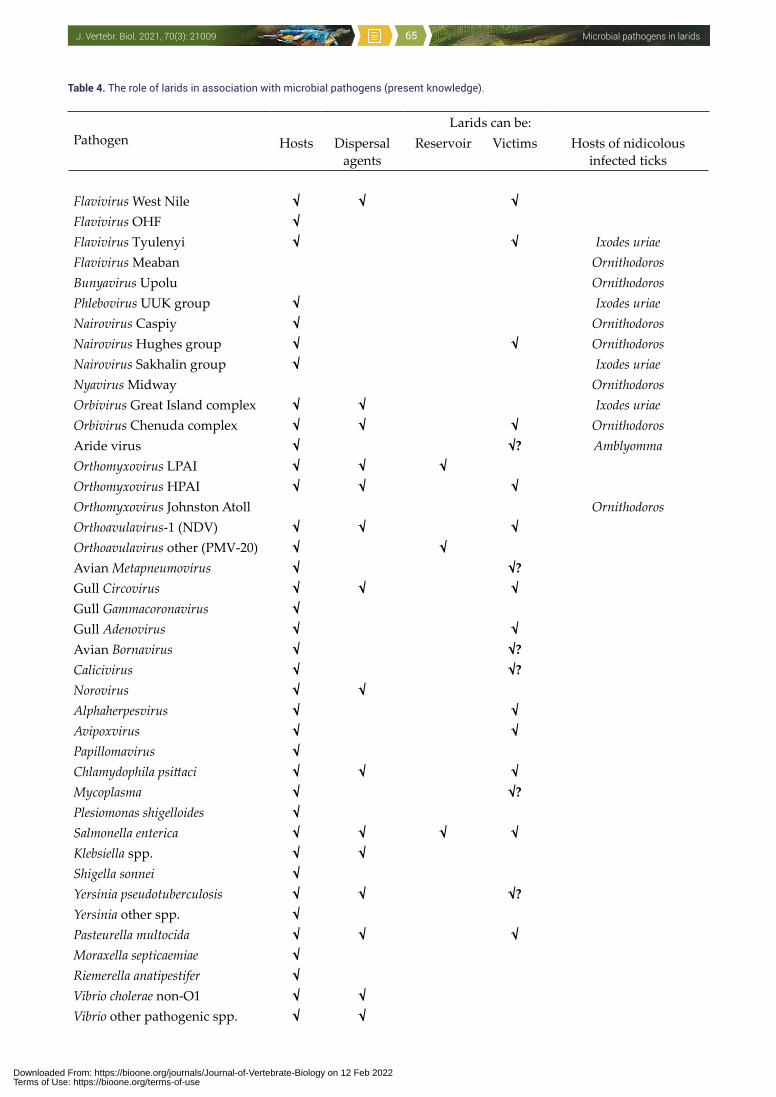
Microbial pathogens in laridsJ. Vertebr. Biol. 2021, 70(3): 21009 65
Table 4. The role of larids in association with microbial pathogens (present knowledge).
PathogenLarids can be:
Hosts Dispersal agents
Reservoir Victims Hosts of nidicolous infected ticks
Flavivirus West Nile √ √ √Flavivirus OHF √Flavivirus Tyulenyi √ √ Ixodes uriaeFlavivirus Meaban OrnithodorosBunyavirus Upolu OrnithodorosPhlebovirus UUK group √ Ixodes uriaeNairovirus Caspiy √ OrnithodorosNairovirus Hughes group √ √ OrnithodorosNairovirus Sakhalin group √ Ixodes uriaeNyavirus Midway OrnithodorosOrbivirus Great Island complex √ √ Ixodes uriaeOrbivirus Chenuda complex √ √ √ OrnithodorosAride virus √ √? AmblyommaOrthomyxovirus LPAI √ √ √Orthomyxovirus HPAI √ √ √Orthomyxovirus Johnston Atoll OrnithodorosOrthoavulavirus-1 (NDV) √ √ √Orthoavulavirus other (PMV-20) √ √Avian Metapneumovirus √ √?Gull Circovirus √ √ √Gull Gammacoronavirus √Gull Adenovirus √ √Avian Bornavirus √ √?Calicivirus √ √?Norovirus √ √Alphaherpesvirus √ √Avipoxvirus √ √Papillomavirus √Chlamydophila psittaci √ √ √Mycoplasma √ √?Plesiomonas shigelloides √Salmonella enterica √ √ √ √Klebsiella spp. √ √Shigella sonnei √Yersinia pseudotuberculosis √ √ √?Yersinia other spp. √Pasteurella multocida √ √ √Moraxella septicaemiae √Riemerella anatipestifer √Vibrio cholerae non-O1 √ √Vibrio other pathogenic spp. √ √
Downloaded From: https://bioone.org/journals/Journal-of-Vertebrate-Biology on 12 Feb 2022Terms of Use: https://bioone.org/terms-of-use
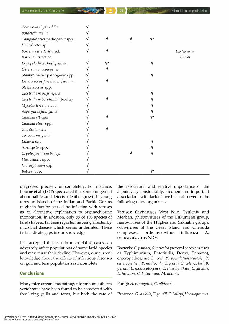
Microbial pathogens in laridsJ. Vertebr. Biol. 2021, 70(3): 21009 66
diagnosed precisely or completely. For instance, Bourne et al. (1977) speculated that some congenital abnormalities and defects of feather growth in young terns on islands of the Indian and Pacific Oceans might in fact be caused by infection with viruses as an alternative explanation to organochlorine intoxication. In addition, only 55 of 103 species of larids have so far been reported as being affected by microbial disease which seems underrated. These facts indicate gaps in our knowledge.
It is accepted that certain microbial diseases can adversely affect populations of some larid species and may cause their decline. However, our current knowledge about the effects of infectious diseases on gull and tern populations is incomplete.
Conclusions
Many microorganisms pathogenic for homeotherm vertebrates have been found to be associated with free-living gulls and terns, but both the rate of
the association and relative importance of the agents vary considerably. Frequent and important associations with larids have been observed in the following microorganisms:
Viruses: flaviviruses West Nile, Tyuleniy and Meaban, phleboviruses of the Uukuniemi group, nairoviruses of the Hughes and Sakhalin groups, orbiviruses of the Great Island and Chenuda complexes, orthomyxovirus influenza A, orthoavulavirus NDV.
Bacteria: C. psittaci, S. enterica (several serovars such as Typhimurium, Enteritidis, Derby, Panama), enteropathogenic E. coli, Y. pseudotuberculosis, Y. enterocolitica, P. multocida, C. jejuni, C. coli, C. lari, B. garinii, L. monocytogenes, E. rhusiopathiae, E. faecalis, E. faecium, C. botulinum, M. avium.
Fungi: A. fumigatus, C. albicans.
Protozoa: G. lamblia, T. gondii, C. baileyi, Haemoproteus.
Aeromonas hydrophila √Bordetella avium √Campylobacter pathogenic spp. √ √ √ √?Helicobacter sp. √Borrelia burgdorferi s.l. √ √ Ixodes uriaeBorrelia turricatae Carios Erysipelothrix rhusiopathiae √ √? √Listeria monocytogenes √ √Staphylococcus pathogenic spp. √ √Enterococcus faecalis, E. faecium √ √Streptococcus spp. √Clostridium perfringens √ √Clostridium botulinum (toxins) √ √ √Mycobacterium avium √ √Aspergillus fumigatus √ √Candida albicans √ √ √?Candida other spp. √Giardia lamblia √ √Toxoplasma gondii √Eimeria spp. √ √Sarcocystis spp. √ √Cryptosporidium baileyi √ √ √Plasmodium spp. √Leucocytozoon spp. √Babesia spp. √ √?
Downloaded From: https://bioone.org/journals/Journal-of-Vertebrate-Biology on 12 Feb 2022Terms of Use: https://bioone.org/terms-of-use
Microbial pathogens in laridsJ. Vertebr. Biol. 2021, 70(3): 21009 67
In general, larids may be involved in the circulation of pathogenic microorganisms in diverse ways as: 1) biological hosts (the pathogen multiplies in/on the avian body) with an acute (e.g. ornithosis), chronic (e.g. tuberculosis) or latent infection, and carriers shedding the agent for a prolonged period (e.g. salmonellosis, campylobacterosis); 2) mechanical hosts (passive, contaminative hosts: the pathogen does not multiply in/on the avian body); the carriage can be either external (on the surface of the bird’s body) or internal (when the agent passes through the digestive tract and is excreted viable); 3) hosts and disseminators of infected vectors (e.g. viruliferous ixodid ticks or I. uriae ticks with B. garinii) to new areas; 4) “lessors” providing a substrate (droppings, nest lining) suitable for the reproduction and/or survival of the pathogenic agent; 5) reservoirs of infection, when they ensure the long-term reproduction
or survival of the agent, especially in the inter-epizootic periods (e.g. LPAIVs, salmonellosis or campylobacterosis). Moreover, larids can spread the agents over short or long distances.
The final, popular question would be: are larids the hosts, dispersing carriers, reservoirs or victims of these microbial pathogens? The answer is not trivial and differs for particular infective agents. An attempt at a full evaluation is presented in Table 4, but there are still many gaps in our knowledge.
Gulls are gregarious birds, they often visit rubbish tips, farm yards, pastures, sewage sites etc. which facilitates the spread of diseases. Given that gulls are so widespread and often live in close proximity to humans, the study of their infectious diseases, in that some cases can also be communicable to domestic animals or humans, merits more attention.
Downloaded From: https://bioone.org/journals/Journal-of-Vertebrate-Biology on 12 Feb 2022Terms of Use: https://bioone.org/terms-of-use
Microbial pathogens in laridsJ. Vertebr. Biol. 2021, 70(3): 21009 68
Literature
Aaziz R., Gourlay P., Vorimore F. et al. 2015: Chlamydiaceae in North Atlantic seabirds admitted to a wildlife rescue center in western France. Appl. Environ. Microbiol. 81: 4581–4590.
Aberkane S., Compain F., Barraud O. et al. 2015: Non-O1/Non-O139 Vibrio cholerae avian isolate from France co-carrying the bla(VIM-1) and bla(VIM-4) genes. Antimicrob. Agents Chemother 59: 6594–6596.
Abolnik C., Pieterse R., Peyrot B.M. et al. 2019: The incursion and spread of highly pathogenic avian influenza H5N8 clade 2.3.4.4 within South Africa. Avian Dis. 63: 149–156.
Adlhoch C., Kuiken T., Monne I. et al. 2019: Avian influenza overview November 2018-February 2019. EFSA J. 18: e06096.
Ahlstrom C.A., Ramey A.M., Woksepp H. & Bonnedahl J. 2019: Early emergence of mcr-1-positive Enterobacteriaceae in gulls from Spain and Portugal. Environ. Microbiol. Rep. 11: 669–671.
Ahlstrom C.A., van Toor M.L., Woksepp H. et al. 2020: Evidence for continental-scale dispersal of antimicrobial resistant bacteria by landfill-foraging gulls. Sci. Total Environ. 764: 144551.
Ainsworth G.C. & Austwick P.K.C. 1959: Fungal diseases of animals. Commonwealth Agricultural Bureaux, Farnham Royal, UK.
Aires-de-Sousa M., Fournier C., Lopes E. et al. 2020: High colonization rate and heterogeneity of ESBL- and carbapenemase-producing Enterobacteriaceae isolated from gull feces in Lisbon, Portugal. Microorganisms 8: E1487.
Aitken T.H.G., Jonkers A.H., Tikasingh E.S. & Worth C.B. 1968: Hughes virus from Trinidadian ticks and terns. J. Med. Entomol. 5: 501–503.
Akgul O., Gulhan T. & Guducuoglu H. 2016: Phenotypic and genotypic analysis for antibiotic resistance of Enterococcus species with chicken and gull origin. Ankara Univ. Vet. Fak. 63: 235–244.
Albarnaz J.D., Toso J., Correa A.A. et al. 2007: Relationship between the contamination of gulls (Larus dominicanus) and oysters (Crassostrea gigas) with Salmonella serovar Typhimurium by PCR-RFLP. Int. J. Environ. Health Res. 17: 133–140.
Alexander D.J. 2000: A review of avian influenza in different bird species. Vet. Microbiol. 74: 3–13.
Alexander D.J., Mackenzie J.S. & Russell P.H. 1986: Two types of Newcastle disease viruses
isolated from feral birds in Western Australia detected by monoclonal antibodies. Aust. Vet. J. 63: 365–367.
Alkhovsky S.V., Lvov D.N., Samokhvalov E.I. et al. 2003: Examinations of birds in the Volga delta (Astrakhan region, 2001) for West Nile virus by using RT-PCR. Vopr. Virusol. 48: 14–17. (in Russian)
Al-Yasiri M.H., Normand A.C., L‘Ollivier C. et al. 2016: Opportunistic fungal pathogen Candida glabrata circulates between humans and yellow-legged gulls. Sci. Rep. 6: 36157.
Al-Yasiri M.H., Normand A.C., Piarroux R. et al. 2017: Gut yeast communities in Larus michahellis from various breeding colonies. Med. Mycol. 55: 436–444.
Andreev V.P., Aristova V.A., Braude N.A. et al. 1980: Isolation of influenza viruses from lariform birds on islands of North Caspian Sea and Black Sea and Newcastle disease virus from ducks in the Volga delta in 1978. Ekol. Virus. 1980: 81–85. (in Russian)
Andreev V.P., Gromashevski V.L., Veselovskaya O.V. et al. 1973a: Isolation of Baku virus in western Turkmenistan. Ekol. Virus. 1: 107–110. (in Russian)
Andreev V.P., Skvortsova T.M., Gromashevski V.L. et al. 1974: A mixed focus of arbovirus infections at the nesting grounds of Larus genei and Sterna hirundo on the islands of the eastern part of the Caspian Sea. Ekol. Virus. 2: 105–108. (in Russian)
Andreev V.P., Vasilev V.I. & Shcherbina A.A. 1973b: Conditions of nesting and possible epizootic role of four larid species on some islands of the eastern Caspian Sea. Ekol. Virus. 1: 110–115. (in Russian)
Ankerberg C.W. 1984: Pelican deaths in the vicinity of a sewage lift station: a bacteriological investigation. Microbios Lett. 26: 33−42.
Annuar B.O., Mackenzie J.S. & Lalor P.A. 1983: Isolation and characterization of avipoxvirus from wild birds in Western Australia. Arch. Virol. 76: 217–229.
Anonymous 1934: The Franklin gull tragedy. The Winnipeg Free Press.
Anonymous 2012a: Avian influenza: China (Hong Kong) H5N1, gull, peregrine falcon, confirmed. ProMED Digest 2012: number 134.
Anonymous 2012b: Seroprevalance of Newcastle disease virus in wild birds in Korea. Korean J. Poult. Sci. 39: 97–104.
Anonymous 2013: Avian influenza: China (Hong Kong) H5N1, gull. ProMED Digest 2013: issue 50.
Downloaded From: https://bioone.org/journals/Journal-of-Vertebrate-Biology on 12 Feb 2022Terms of Use: https://bioone.org/terms-of-use
Microbial pathogens in laridsJ. Vertebr. Biol. 2021, 70(3): 21009 69
Anonymous 2019: Botulism, avian – USA: California gulls, type A. ProMED Digest 89: issue 35. http://healthmap.org/promed/p/204
Antilles N., García-Bocanegra I., Alba-Casals A. et al. 2020: Occurrence and antimicrobial resistance of zoonotic enteropathogens in gulls from southern Europe. Sci. Total Environ. 763: e143018.
Antilles N., García-Migura L., Grimstrup J.K. et al. 2015: Audouin’s gull, a potential vehicle of an extended spectrum beta-lactamase producing Salmonella Agona. FEMS Microbiol. Lett. 362: 1–4.
Aristova V.A., Yamnikova S.S., Myasnikova I.A. & Gavrilov N.N. 1982: Isolation of influenza viruses from nestling lariform birds on the Zhemchuzhniy Island, northern Caspian Sea. Ekol. Virus. 1982: 91–94. (in Russian)
Arnal A., Gomez-Diaz E., Cerda-Cuellar M. et al. 2014: Circulation of a Meaban-like virus in yellow-legged gulls and seabird ticks in the Western Mediterranean Basin. PLOS ONE 9: e89601.
Arnal A., Vittecoq M., Pearce-Duvet J. et al. 2015: Laridae: a neglected reservoir that could play a major role in avian influenza virus epidemiological dynamics. Crit. Rev. Microbiol. 41: 508–519.
Arriero E., Müller I., Juvaste R. et al. 2015: Variation in immune parameters and disease prevalence among lesser black-backed gulls (Larus fuscus) with different migratory strategies. PLOS ONE: e118279.
Austin F.J. 1978: Johnston Atoll virus (Quaranfil group) from Ornithodoros capensis (Ixodoidea: Argasidae) infesting a gannet colony in New Zealand. Am. J. Trop. Med. Hyg. 27: 1045–1048.
Bach R.D. 2007: Jonathan Livingston seagull. Argo, Praha, Czech Republic.
Bahl A.K., Pomeroy B.S., Mangundimedjo S. & Easterday B.C. 1977: Isolation of type A influenza and Newcastle disease viruses from migratory waterfowl in the Mississippi Flyway. J. Am. Vet. Med. Assoc. 171: 949–951.
Bahnson C.S., Poulson R.L., Hollander L.P. et al. 2020: Susceptibility of laughing gulls (Leucophaeus atricilla) and mallards (Anas platyrhynchos) to ruddy turnstone (Arenaria interpres morinella) origin type A influenza viruses. J. Wildl. Dis. 56: 167–174.
Baker J.R. 1977: The results of post-mortem examination of 132 wild birds. Br. Vet. J. 133: 327−333.
Ballard J.R., Mickley R., Brown J.D. et al. 2017: Detection of Wellfleet Bay virus antibodies in
seabirds of the northeastern USA. J. Wildl. Dis. 53: 875–879.
Barbachano-Guerrero A., Vasquez-Aguilar A.A., Aguirre A.A. et al. 2019: West Nile virus prevalence in wild birds from Mexico. J. Wildl. Dis. 55: 425–431.
Barbara A., Torrontegi O., Camacho M.-C. et al. 2017: Avian influenza virus surveillance in south-central Spain using fecal samples of aquatic birds foraging at landfills. Front. Vet. Sci. 4: 178.
Barguigua A., Rguibi Idrissi H., Nayme K. & Timinouni M. 2019: Virulence and antibiotic resistance patterns in E. coli, Morocco. Ecohealth 16: 570–575.
Bartha A., Meszaros J. & Tanyi J. 1982: Antibodies against EDS-76 avian adenovirus in bird species before 1975. Avian Pathol. 11: 511–513.
Beaudette F.R. 1945: Aspergillosis and parasitism in a gull. Bird-Band. 16: 94−101.
Becker W.B. 1966: The isolation and classification of tern virus: influenza A/Tern/South Africa/1961. J. Hyg. (Lond.) 64: 309−320.
Becker W.B. 1967: Experimental infection of common terns with tern virus: influenza virus A/Tern/South Africa/1961. J. Hyg. (Lond.) 65: 61−65.
Becker W.B. & Uys C.J. 1963: The pathogenesis of tern virus infection in chickens: virus studies. S. Afr. Med. J. 37: 1095.
Becker W.B. & Uys C.J. 1967: Experimental infection of chickens with influenza A/Tern/South Africa/1961 and Chicken/Scotland/1959 viruses. I. Clinical picture and virology. J. Comp. Pathol. 77: 159−165.
Beer J.V. 1963: The incidence of Aspergillus fumigatus in the throats of wild geese and gulls. Sabouraudia 2: 238–247.
Bekleshova A.J., Smirnov V.A. & Terskikh I.I. 1971: Role of marine birds and their ectoparasites in the epizootic process. Vopr. Virusol. 16: 596–600. (in Russian)
Benjamin J., Leaper S., Owen R.J. & Skirrow M.B. 1983: Description of Campylobacter laridis, new species, comprising the nalidixic acid resistant thermophilic Campylobacter (NARTC) group. Curr. Microbiol. 8: 231–238.
Benkaroun J., Shoham D., Kroyer A.N.K. et al. 2016: Analysis of influenza A viruses from gulls: an evaluation of inter-regional movements and interactions with other avian and mammalian influenza A viruses. Cogent Biol. 2: 1.
Bennett G.F., Bishop M.A. & Peirce M.A. 1993: Checklist of the avian species of Plasmodium
Downloaded From: https://bioone.org/journals/Journal-of-Vertebrate-Biology on 12 Feb 2022Terms of Use: https://bioone.org/terms-of-use
Microbial pathogens in laridsJ. Vertebr. Biol. 2021, 70(3): 21009 70
Marchiafava and Celli, 1885 (Apicomplexa) and their distribution by avian family and Wallacean life zones. Syst. Parasitol. 26: 171−179.
Bennett G.F., Earle R.A., Du Toit H. & Huchzermeyer F.W. 1992: A host-parasite catalogue of the haematozoa of the sub-Saharan birds. Onderstep. J. Vet. Res. 59: 1−73.
Bennett G.F., Peirce M.A. & Earle R.A. 1994: An annotated checklist of the valid avian species of Haemoproteus, Leucocytozoon (Apicomplexa: Haemosporida) and Hepatozoon (Apicomplexa: Haemogregarinidae). Syst. Parasitol. 29: 61−73.
Bennett R.S., Nezworski J., Velayudhan B.T. et al. 2004: Evidence of avian pneumovirus spread beyond Minnesota among wild and domestic birds in central North America. Avian Dis. 48: 902–908.
Benskin C.M.W.H., Wilson K., Jones K. & Hartley I.R. 2009: Bacterial pathogens in wild birds: a review of the frequency and effects of infection. Biol. Rev. 84: 349-373.
Benton C. 1983: Seagull pollution of reservoirs. J. Inst. Water Engineers Scientists 37: 347–363.
Benton C., Khan F., Monaghan P. et al. 1993: The contamination of a major water supply by gulls (Larus spp.). A study of the problem and remedial action taken. Water Res. 17: 789–798.
Berezin V.V., Chumakov M.P. & Semenov B.F. 1967: About the role of birds in the ecology of West Nile virus in the Volga river delta. Vopr. Obšč. Virusol. 1: 206–207. (in Russian)
Berezin V.V., Chumakov M.P., Semenov B.F. et al. 1971: Investigation of ecology of mosquito-borne viruses by using sentinel animals in the Volga delta. Vopr. Virusol. 16: 739–745. (in Russian)
Berezina L.K., Smirnov V.A. & Zelensky V.A. 1974: Experimental infection of birds with Tyuleniy virus. Ekol. Virus. 2: 13–17. (in Russian)
Berg R.W. & Anderson A.W. 1972: Salmonellae and Edwardsiella tarda in gull feces: a source of contamination in fish processing plants. Appl. Microbiol. 24: 501–503.
Bernard K.A., Maffei J.G., Jones S.A. et al. 2001: West Nile virus infection in birds and mosquitoes, New York State, 2000. Emerg. Infect. Dis. 4: 679–685.
Bertelloni F., Lunardo E., Rocchigiani G. et al. 2019: Occurrence of Escherichia coli virulence genes in feces of wild birds from Central Italy. Asian Pac. J. Trop. Med. 12: 142–146.
Beysembayeva R.U., Sayatov M.C., Dubova E.A. et al. 1981: Isolation and identification of
influenza viruses from wild birds in southern Kazakhstan. Vest. AN Kaz. SSR 11: 63–66. (in Russian)
Bin Muzaffar S., Smith R.P., Jones I.L. et al. 2012: The Trans-Atlantic movement of the spirochete Borrelia garinii: the role of ticks and their seabird hosts. In: Paul E. (ed.), Emerging avian disease. University of California Press, Berkeley California: 23–30.
Blackmore D.K. & Keymer I.F. 1969: Cutaneous diseases of wild birds in Britain. Br. Birds 62: 316−331.
Blaker D. 1967: An outbreak of botulism among waterbirds. Ostrich 38: 144–147.
Bo G. 1980: Salmonella prevalence in gulls on the Gronma refuse tip in Oslo. Viltrapport 10: 147–154. (in Norwegian)
Bodenstein B., Beckmen K., Sheffield G. et al. 2015: Avian cholera causes marine bird mortality in the Bering Sea of Alaska. J. Wildl. Dis. 51: 934–937.
Bodewes R., de Bildt M.W.G., van Schapendonk C.M.E. et al. 2013: Identification and characterization of a novel adenovirus in the cloacal bursa of gulls. Virology 440: 84–88.
Boháč D. 1968: The diet and economic significance of black-headed gull (Larus ridibundus L.). PhD thesis, Charles University, Praha, Czech Republic. (in Czech)
Bolte A.L., Meurer J. & Kaleta E.F. 1999: Avian host spectrum of avipoxviruses. Avian Pathol. 28: 415–432.
Bonnedahl J., Hernandez J., Stedt J. et al. 2014: Extended-spectrum beta-lactamases in Escherichia coli and Klebsiella pneumoniae in gulls, Alaska, USA. Emerg. Infect. Dis. 20: 897–899.
Borg K. 1980: The spread of disease and other sanitary problems by gulls and crows. Viltrapport 10: 129–133.
Borland E.D., Moryson C.J. & Smith G.R. 1977: Avian botulism and the high prevalence of Clostridium botulinum in the Norfolk Broads. Vet. Rec. 100: 106–109.
Bosch M., Figuerola J., Cantos F.J. & Veralde R. 1997: Intracolonial differences in the infestation by Haemoproteus lari on yellow-legged gulls Larus cachinnans. Ornis Fenn. 74: 105−112.
Bosch M. & Muniesa M. 1996: Yellow-legged gulls (Larus cachinnans) from the Medes Islands colony as possible transmitting agents of microbial contamination. Doñana: Acta Vertebrata 23: 75–81.
Bourne W.R.P., Bogan J.A. & Bullock D. et al. 1977: Abnormal terns, sick sea and shore birds,
Downloaded From: https://bioone.org/journals/Journal-of-Vertebrate-Biology on 12 Feb 2022Terms of Use: https://bioone.org/terms-of-use
Microbial pathogens in laridsJ. Vertebr. Biol. 2021, 70(3): 21009 71
organochlorines and arboviruses in the Indian Ocean. Mar. Pollut. Bull. 8: 154–158.
Böhm R. 1993: Verhalten ausgewählter Salmonellen in der Umwelt. Dtsch. Tierärztl. Wochenschr. 100: 275–278.
Brand C.J. 1989: Chlamydial infections in free-living birds. J. Am. Vet. Med. Assoc. 195: 1531−1535.
Brand C.J., Duncan R.M., Garrow S.P. et al. 1983: Waterbird mortality from botulism type E in Lake Michigan (USA, Canada): an update. Wilson Bull. 95: 269–275.
Brand C.J., Windingstad R.M., Siegfried L.M. et al. 1988: Avian morbidity and mortality from botulism, aspergillosis, and salmonellosis at Jamaica Bay Widlife Refuge, New York, USA. Colon. Waterbird. 11: 284–292.
Brogden K.A. & Rhoades K.R. 1983: Prevalence of serologic types of Pasteurella multocida from 57 species of birds and mammals in the United States. J. Wildl. Dis. 19: 315–320.
Broman T., Palmgren H., Bergstrom S. et al. 2002: Campylobacter jejuni in black-headed gulls (Larus ridibundus): prevalence, genotypes, and influence on C. jejuni epidemiology. J. Clin. Microbiol. 40: 4594–4602.
Brown J., Poulson R., Carter D. et al. 2012: Susceptibility of avian species to North American H13 low pathogenic avian influenza viruses. Avian Dis. 56: 969–975.
Brown J.D., Stallknecht D.E., Beck J.R. et al. 2006: Susceptibility of North American ducks and gulls to H5N1 highly pathogenic avian influenza viruses. Emerg. Infect. Dis. 12: 1663−1670.
Brown J.D., Stallknecht D.E. & Swayne D.E. 2008: Experimental infections of herring gulls (Larus argentatus) with H5N1 highly pathogenic avian influenza viruses by intranasal inoculation of virus and ingestion of virus-infected chicken meat. Avian Pathol. 37: 393−397.
Brown S.E., Morrison H.G. & Knudson D.L. 1989: Genetic relatedness of the Kemerovo serogroup viruses. II. RNA-RNA blot hybridization and gene reassortment in vitro of the Great Island serocomplex. Acta Virol. 33: 206–220.
Buck J.D. 1983: Occurrence of Candida albicans in fresh gull feces in temperate and subtropical areas. Microb. Ecol. 9: 171−176.
Buck J.D. 1986: A note on the experimental uptake and clearance of Candida albicans in a young captive gull (Larus sp.). Mycopathologia 94: 59–61.
Buck J.D. 1990: Isolation of Candida albicans and halophilic Vibrio spp. from aquatic birds in Connecticut and Florida. Appl. Environ. Microbiol. 56: 826−828.
Buckley N.J. & O‘Halloran J. 1986: Mass mortality of gulls in west Cork attributed to botulism. Ir. Birds 3: 283–285.
Burroughs L.A., Holdenried R., Longanecker D.S. & Meyer K.F. 1945: A field study of latent tularemia in rodents with a list of all known naturally infected vertebrates. J. Infect. Dis. 76: 115–119.
Butterfield J., Coulson J.C., Kearsey S.V. et al. 1983: The herring gull Larus argentatus as a carrier of Salmonella. J. Hyg. (Lond.) 91: 429–436.
Cabezon O., Cerda-Cuellar M., Morera V. et al. 2016: Toxoplasma gondii infection in seagull chicks is related to the consumption of freshwater food resources. PLOS ONE 11: e0150249.
Cacciuttolo E., Rossi G., Nardoni S. et al. 2009: Anatomo-pathological aspects of avian aspergillosis. Vet. Res. Commun. 33: 521–527.
Caliendo V., Leijten L., Begeman L. et al. 2020: Enterotropism of highly pathogenic avian influenza virus H5N8 from the 2016/2017 epidemic in some wild bird species. Vet. Res. 51: 117.
Calsow P., Helbing H., Ludwig C. & Schlegelmilch R. 1995: Seuchenhaftes Sterben von Wasservögeln an einem Stausee - Fallbericht. Tierärztl. Umsch. 50: 477–480.
Camarda A., Circella E., Giovanardi D. et al. 2007: Avian pathogenic Escherichia coli in Audouin gulls (Larus audouinii). Could they affect the surviving of the bird colonies? Ital. J. Anim. Sci. 6: 317–320.
Campbell J.M., Gauriloff L.P., Domske H.M. & Obert E.C. 2005: Re-emergence of avian botulism type E in the North American Great Lakes. In: Majumdar S.K., Huffman J.E., Brenner F.J. & Panah A.I. (eds.), Wildlife diseases: landscape epidemiology, spatial distribution and utilization of remote sensing technology. Pennsylvania Academy of Science, Easton, USA: 50–61.
Canuti M., Munro H.J., Robertson G.J. et al. 2019a: New insight into avian papillomavirus ecology and evolution from characterization of novel wild bird papillomaviruses. Front. Microbiol. 10: 701.
Canuti M., Kroyer A.N.K., Ojkic D. et al. 2019b: Discovery and characterization of novel RNA viruses in aquatic North American wild birds. Viruses 11: 768.
Downloaded From: https://bioone.org/journals/Journal-of-Vertebrate-Biology on 12 Feb 2022Terms of Use: https://bioone.org/terms-of-use
Microbial pathogens in laridsJ. Vertebr. Biol. 2021, 70(3): 21009 72
Cao X., Wang Yi., Wang Y. & Ye C. 2018: Isolation and characterization of Listeria monocytogenes from the black-headed gull feces in Kunming, China. J. Infect. Public Health 11: 59–63.
Cardoso M.D., Lemos L.S., Roges E.M. et al. 2018: A comprehensive survey of Aeromonas sp. and Vibrio sp. in seabirds from southeastern Brazil: outcomes for public health. J. Appl. Microbiol. 124: 1283–1293.
Chambers T.M., Yamnikova S., Kawaoka Y. et al. 1989: Antigenic and molecular characterization of subtype H13 hemagglutinin of influenza virus. Virology 172: 180–188.
Chang H., Dai F.Y., Duan G. et al. 2018: Pathogenicity analysis of Klebsiella oxytoca isolated from Larus ridibundus migratory birds. Pak. Vet. J. 38: 449–451.
Chastel C.E. 1988: Tick-borne virus infections of marine birds. Adv. Dis. Vector Res. 5: 25−60.
Chastel C., Bailly-Choumara H. & Le Lay G. 1981: Pouvoir pathogene naturel pour l‘homme d‘un variant antigenique du virus Soldado, isole au Maroc. Bull. Soc. Pathol. Exot. 74: 499–505.
Chastel C., Guiguen C., Le Lay G. & Le Goff F. 1988: First isolation of Soldado virus in southern France. Acta Virol. 32: 191.
Chastel C., Guiguen C., Le Lay G. et al. 1985a: Enquéte sérologique arbovirus chez des oiseaux marins et non marins de Bretagne. Bull. Soc. Pathol. Exot. 78: 594–605.
Chastel C., Guiguen C., Le Lay G. et al. 1983a: Les arbovirus des colonies d‘oiseaux de mer de Bretagne peuvent-ils infecter l‘homme? Rev. Epidém. Méd. Soc. et Santé Publ. 31: 445–457.
Chastel C., Launay H., Rogues G. & Beaucournu J.C. 1979: Isolement en France du virus Soldado (arbovirus, groupe Hughes) a partir d‘Ornithodoros (A.) maritimus Vermeil et Marguet 1967. C. R. Acad. Sci. Paris, Ser. D 288: 559–561.
Chastel C., Le Goff F. & Le Lay G. 1983b: Antigenic variants of the Soldado virus (Nairovirus, Bunyaviridae) isolated in different parts of the world. Acta Virol. 27: 51–58.
Chastel C., Le Lay G., Le Goff F. & Monnat J.Y. 1990: Isolation of Soldado virus from a seabird (Rissa tridactyla L.) in France. Acta Virol. 34: 109.
Chastel C., Main A.J., Bailly Choumara H. et al. 1993: Essaouira and Kala Iris: two new orbiviruses of the Kemerovo serogroup, Chenuda complex, isolated from Ornithodoros (Alectorobius) maritimus ticks in Morocco. Acta Virol. 37: 484–492.
Chastel C., Main A.J., Guiguen C. et al. 1985b: The isolation of Meaban virus, a new flavivirus from the seabird tick Ornithodoros (Alectorobius) maritimus in France. Arch. Virol. 83: 129–140.
Chen X.L., Qi Y.P., Wang H.H. et al. 2018: Prevalence of multiple subtypes of avian influenza virus antibodies in egg yolks of mallards (Anas platyrhynchos) and white-winged terns (Chlidonias leucopterus) in the Northeastern Republic of China. J. Wildl. Dis. 54: 834–837.
Chernetsov Y.V., Obrosova-Serova N.P., Slepuškin A.N. et al. 1974: The presence of antihaemagglutinins to influenza viruses in gulls nesting on the islands of Zaliv Petra Velikogo. Ekol. Virus. 2: 170–174. (in Russian)
Chernetsov Y.V., Slepuškin A.N., Lvov D.K. et al. 1980: Isolation of influenza virus from a black tern (Chlidonias nigra) and serological survey of birds for influenza virus. Vopr. Virusol. 1980: 35–40. (in Russian)
Chipault J.G., White C.L.A., Blehert D.S. et al. 2015: Avian botulism type E in waterbirds of Lake Michigan, 2010-2013. J. Great Lakes Res. 41: 659–664.
Christensen J.P., Dietz H.H. & Bisgaard M. 1998: Phenotypic and genotypic characters of isolates of Pasteurella multocida obtained from back-yard poultry and from two outbreaks of avian cholera in avifauna in Denmark. Avian Pathol. 27: 373−381.
Christiansen M. 1949: Diseases in wild birds. Dan. Ornitol. Foren. Tidsskr. 43: 189−215.
Chytil J. 1990: Avian botulism in South Moravia, 1988. Birds in a cultural landscape. Proceedings II South Bohemian Ornithological Conference, České Budějovice, Czech Republic: 233−243. (in Czech)
Chytil J. & Macháček P. 2000: Development in breeding population size of gulls (Laridae) and terns (Sternidae) in the southernmost Moravia. Sylvia 36: 113–126. (in Czech)
Clark R.G. 2001: Black-backed gulls (Larus dominicanus) and their role in the spread of Salmonella brandenburg. Proceeding of the New Zealand Society of Animal Production 61: 71–73.
Clark R.G., Fenwick S.G., Nicol C.M. et al. 2004: Salmonella brandenburg – emergence of a new strain affecting stock and humans in the South Island of New Zealand. N. Z. Vet. J. 52: 26–36.
Clifford C.M. 1971: Isolation of a group B arbovirus from Ixodes uriae collected on Three Arch Rocks National Refuge, Oregon. Am. J. Trop. Med. Hyg. 20: 461–468.
Downloaded From: https://bioone.org/journals/Journal-of-Vertebrate-Biology on 12 Feb 2022Terms of Use: https://bioone.org/terms-of-use
Microbial pathogens in laridsJ. Vertebr. Biol. 2021, 70(3): 21009 73
Cloutier A., Mills J.A., Yarrall J.W. & Baker A.J. 2011: Plasmodium infections of red-billed gulls (Larus scopulinus) show associations with host condition but not reproductive performance. J. R. Soc. N. Z. 41: 261–277.
Coffee L.L., Hanson B.A., Luttrell M.P. et al. 2010: Avian paramyxoviruses in shorebirds and gulls. J. Wildl. Dis. 46: 481–487.
Contreras-Rodriguez A., Aguilera-Arreola M.G., Osorio A.R. et al. 2019: Detection of potential human pathogenic bacteria isolated from feces of two colonial seabirds nesting on Isla Rasa, Gulf of California: Heermann’s gull (Larus heermanni) and elegant tern (Thalasseus elegans). Trop. Conserv. Sci. 12: 1–8.
Converse J.D., Hoogstraal H., Moussa M.I. et al. 1975: Soldado virus (Hughes group) from Ornithodoros (Alectorobius) capensis (Ixodoidea: Argasidae) infesting sooty tern colonies in the Seychelles, Indian Ocean. Am. J. Trop. Med. Hyg. 24: 1010–1018.
Converse J.D., Hoogstraal H., Moussa M.I. & Evans D.E. 1976: Soldado virus from Ornithodoros (Alectorobius) maritimus (Ixodoidea: Argasidae) infesting herring gull nests on Puffin Island, northern Wales. Acta Virol. 20: 243–246.
Cooper J.E. 1990: Birds and zoonoses. Ibis 132: 181–191.
Costa T.P., Brown J.D., Howerth E.W. & Stallknecht D.E. 2011: Variation in viral shedding patterns between different wild bird species infected experimentally with low-pathogenicity avian influenza viruses that originated from wild birds. Avian Pathol. 40: 119–124.
Coulson J.C., Butterfield J. & Thomas C. 1983: The herring gull Larus argentatus as a likely transmitting agent of Salmonella montevideo to sheep and cattle. J. Hyg. (Lond.) 91: 437–443.
Cowan McT.J. 1943: Aspergillosis in a thayer gull. Murrelet 24: 29.
Cragg J. & Clayton Y.M. 1971: Bacterial and fungal flora of seagull droppings in Jersey. J. Clin. Pathol. 24: 317–319.
Craik J.C.A. 1986: Exceptional mortality of auks, terns and kittiwakes in L. Linnhe area of Argyll in July 1985. Argyll Bird Rep. 3: 95−99.
Crawford R.J.M., Allwright D.M. & Heyl C.W. 1992: High mortality of Cape cormorants (Phalacrocorax capensis) off western South Africa in 1991 caused by Pasteurella multocida. Colon. Waterbirds 15: 236−238.
Creutz G. 1963: Ernährungsweise und Aktionsradius der Lachmöwe (Larus ridibundus L.). Beitr. Vogelk. 9: 3–58.
Creutz G. & Gottschalk C. 1969: Endoparasitenbefall bei Lachmöwen in Abhängigkeit vom Alter. Angew. Parasitol. 10: 80−91.
Cummins M.L., Sanderson-Smith M., Newton P. et al. 2020: Whole-genome sequence analysis of an extensively drug-resistant Salmonella enterica serovar Agona isolate from an Australian silver gull (Chroicocephalus novaehollandiae) reveals the acquisition of multidrug resistance plasmids. mSphere 5: e00743-20.
Curran J.M., Robertson I.D., Ellis T.M. et al. 2013: Variation in the responses of wild species of duck, gull, and wader to inoculation with a wild-bird-origin H6N2 low pathogenicity avian influenza virus. Avian Dis. 57: 581–586.
Curran J.M., Robertson I.D., Ellis T.M. & Selleck P.W. 2014: Evaluation of avian influenza serologic and virologic diagnostic methods in wild Anseriformes and Charadriiformes. Avian Dis. 58: 53–59.
Čížek A., Dolejská M., Karpíšková R. et al. 2007: Wild black-headed gulls (Larus ridibundus) as an environmental reservoir of Salmonella strains resistant to antimicrobial drugs. Eur. J. Wildl. Res. 53: 55–60.
Čížek A., Literák I., Hejlíček K. et al. 1994: Salmonella contamination of the environment and its incidence in wild birds. Zentralbl. Vet. Med. B 41: 320–327.
Čížek A., Mikula I. & Kovaříková V. 1995: Characteristics of the plasmid profile of inositol- and rhamnose-negative strains of Salmonella typhimurium. Vet. Med. (Praha) 40: 29–32. (in Czech)
D‘Amico V.L., Fazio A., Palacios M.G. et al. 2018: Evaluation of physiological parameters of kelp gulls (Larus dominicanus) feeding on fishery discards in Patagonia, Argentina. Waterbirds 41: 310–315.
Danielová V., Marhoul Z., Dusbábek F. et al. 1982: Isolation of Hughes virus from ticks in Cuba. Acta Virol. 26: 186–189.
Daoust P.-Y., van de Bildt M., van Riel D. et al. 2013: Replication of 2 subtypes of low-pathogenicity avian influenza virus of duck and gull origins in experimentally infected mallard ducks. Vet. Pathol. 50: 548–559.
Davis J.W., Anderson R.C., Karstad L. & Trainer D.O. 1971: Infectious and parasitic diseases of wild birds. Iowa State University Press, Ames, USA.
Davis W.A. & McClung L.S. 1940: Aspergillosis in wild herring gulls. J. Bacteriol. 40: 321−323.
Debruyne L., Broman T., Bergstrom S. et al. 2010: Campylobacter volucris sp. nov., isolated from
Downloaded From: https://bioone.org/journals/Journal-of-Vertebrate-Biology on 12 Feb 2022Terms of Use: https://bioone.org/terms-of-use
Microbial pathogens in laridsJ. Vertebr. Biol. 2021, 70(3): 21009 74
black-headed gulls (Larus ridibundus). Int. J. Syst. Evol. Microbiol. 60: 1870–1875.
Decors A. & Mastain O. 2010: Wildlife epidemiological surveillance – results of analyses performed from 2006 to 2008 within the framework of the SAGIR network. National Hunting and Wildlife Agency, Paris, France.
De Marco M.A., Foni E., Campitelli L. et al. 2005: Influenza virus circulation in wild aquatic birds in Italy during H5N2 and H7N1 poultry epidemic periods (1998 to 2000). Avian Pathol. 34: 480–485.
del Hoyo J. 2020: All the birds of the world. Lynx, Barcelona, Spain.
Diel D.G., Miller P.J., Wolf P.C. et al. 2012: Characterization of Newcastle disease viruses isolated from cormorant and gull species in the United States in 2010. Avian Dis. 56: 128–133.
Doherty R.L., Whitehead R.H., Wetters E.J. & Johnson H.N. 1969: Isolation of viruses from Ornithodoros capensis Neumann from a tern colony on the Great Barrier Reef, North Queensland. Aust. J. Sci. 31: 363–364.
Dolejská M., Bierošová B., Kohoutová L. et al. 2009: Antibiotic-resistant Salmonella and Escherichia coli isolates with integrons and extended-spectrum beta-lactamases in surface water and sympatric black-headed gulls. J. Appl. Microbiol. 106: 1941–1950.
Dolejská M., Masaříková M., Dobiášová H. et al. 2016: High prevalence of Salmonella and IMP-4-producing Enterobacteriaceae in the silver gull on Five Islands, Australia. J. Antimicrob. Chemother. 71: 63–70.
Downie J.C., Webster R.G., Hinshaw V. & Laver W.G. 1977: Ecology of influenza – isolation of type-A influenza viruses from Australian pelagic birds. Aust. J. Exp. Biol. Med. Sci. 55: 635–644.
Downie J.C., Webster R.G., Schild G.C. et al. 1973: Characterization and ecology of a type A influenza virus isolated from a shearwater. Bull. World Wealth Org. 49: 559–566.
Drăgănescu N., Iftimovici R., Mutiu A. et al. 1978: Detection of antibodies to alphaviruses and flaviviruses in some migratory birds of the Danube Delta. Virologie 29: 113–116.
Drouin T.E. & Mahrt J.L. 1979: The prevalence of Sarcocystis in some bird species in western Canada, with notes on the life cycle. Can. J. Zool. 57: 1915−1921.
Duarte E.L., Guerra M.M. & Bernardo F.M. 2002: Salmonella and Listeria spp. carriage by gulls (larids). Rev. Port. Cienc. Vet. 97: 181–187.
Duneau D., Boulinier T., Gómez-Diaz E. et al. 2008: Prevalence and diversity of Lyme borreliosis bacteria in marine birds. Infect. Genet. Evol. 8: 352–359.
Dupraz M., Toty C., Devillers E. et al. 2017: Population structure of the soft tick Ornithodoros maritimus and its associated infectious agents within a colony of its seabird host Larus michahellis. Int. J. Parasitol. Parasites Wildl. 6: 122–130.
Dusek R.J., Hallgrimsson G.T., Ip H.S. et al. 2014: North Atlantic migratory bird flyways provide routes for intercontinental movement of avian influenza viruses. PLOS ONE 9: e92075.
Dynowska M., Indykiewicz P. & Ejdys E. 2018: Black-headed gulls (Chroicocephalus ridibundus) – a natural reservoir of potentially pathogenic microfungi? Biologia 73: 241–246.
Edel W., Van Schothorst M. & Kampelmacher E.H. 1976: Epidemiological studies on Salmonella in a certain area (“Walcheren project”). I. The presence of Salmonella in man, pigs, insects, seagulls and in foods and effluents. Zentralbl. Bakteriol. Orig. A 235: 476–484.
Edel W., Van Schothorst M., Van Leusden F.M. & Kampelmacher E.H. 1977: Epidemiological studies on Salmonella in a particular area (“Walcheren Project”). III. The incidence of Salmonella in man, insects, gulls as well as foods scrapings from butcher’s blocks, effluents of sewage treatment plants and drains from butcher’s shops. Tijdschr. Diergeneeskd. 102: 365–375.
Ejidokun O.O., Walsh A., Barnett J. et al. 2006: Human Vero cytotoxigenic Escherichia coli (VTEC) O157 infection linked to birds. Epidemiol. Infect. 134: 421–423.
Eklund M.W. & Dowell V.R. 1987: Avian botulism. An international perspective. Charles C. Thomas Publisher Ltd., Springfield, USA.
Eley S.M. & Nuttall P.A. 1984: Isolation of an English uukuvirus (family Bunyaviridae). J. Hyg. (Lond.) 93: 313–316.
Ellemann G. 1959: Isolation of Salmonella bacteria from gulls shot down over Copenhagen. Nord. Vet. Med. 11: 607–618.
Ellis T.M., Bousfield R.B., Bissett L.A. et al. 2004: Investigation of outbreaks of highly pathogenic H5N1 avian influenza in waterfowl and wild birds in Hong Kong in late 2002. Avian Pathol. 33: 492−505.
Engvall E.O., Braendstrom B., Gunnarsson A. et al. 2002: Validation of a polymerase chain reaction/restriction enzyme analysis method
Downloaded From: https://bioone.org/journals/Journal-of-Vertebrate-Biology on 12 Feb 2022Terms of Use: https://bioone.org/terms-of-use
Microbial pathogens in laridsJ. Vertebr. Biol. 2021, 70(3): 21009 75
for species identification of thermophilic campylobacters isolated from domestic and wild animals. J. App. Microbiol. 92: 47–54.
Ernek E., Kožuch O., Nosek J. et al. 1977: Arboviruses in birds captured in Slovakia. J. Hyg. Epidemiol. Microbiol. Immunol. 21: 353–359.
Ewald K. 1979: Beiträge zur Ernährung sowie zum Parasiten-, Salmonellen- und Chlamydienbefall von Lachmöwen (Larus ridibundus) in bayerischem Karpfenteichgebiet. J. Ornithol. 120: 98−101.
Feller M. & Köhler B. 1977: Massensterben von Wasservögeln durch Botulismus auf der Potsdamer Havel im Sommer 1975. Der Falke 24: 226–239.
Fenlon D.R. 1981a: Birds as vectors of enteric pathogenic bacteria. J. Appl. Bacteriol. 51: 13–14.
Fenlon D.R. 1981b: Seagulls (Larus spp.) as vectors of salmonellae: an investigation into the range of serotypes and numbers of salmonellae in gull feces. J. Hyg. (Lond) 86: 195–202.
Fenlon D.R. 1983: A comparison of Salmonella serotypes found in the feces of gulls feeding at a sewage works with serotypes present in the sewage. J. Hyg. (Lond) 91: 47–52.
Fenlon D.R. 1985: Wild birds and silage as reservoirs of Listeria in the agricultural environment. J. Appl. Bacteriol. 59: 537–543.
Fennel H., James D.B. & Morris J. 1974: Pollution of a water storage reservoir by roosting gulls. Wat. Treat. Exam. 23: 5–20.
Fernández H., Gesche W., Montefusco A. & Schlatter R. 1986: Wild birds as reservoir of thermophilic enteropathogenic Campylobacter species in southern Chile. Mem. Inst. Oswaldo Cruz 91: 699–700.
Ferns P.N. & Mudge G.P. 2000: Abundance, diet and Salmonella contamination of gulls feeding at sewage outfalls. Water Res. 34: 2653–2660.
Ferreira-Garcia P.G. & Schonhofen C.A. 1982: Salmonellosis of seabirds in the Paranagua bay. Arq. Biol. Tecnol. 25: 237–242. (in Portuguese)
Figuerola J., Jiménez-Clavero M.A., Rojo G. et al. 2007: Prevalence of West Nile virus neutralizing antibodies in colonial aquatic birds in southern Spain. Avian Pathol. 36: 209–212.
Fiorello C.V., Nisbet I.C.T., Hatch J.J. et al. 2009: Hematology and absence of hemoparasites in breeding common terns (Sterna hirundo) from Cape Cod, Massachusetts. J. Zoo Wildl. Med. 40: 409–413.
Fogarty L.R., Haack S.K., Wolcott M.J. & Whitman R.L. 2003: Abundance and characteristics of the recreational water quality indicator bacteria Escherichia coli and enterococci in gull faeces. J. Appl. Microbiol. 94: 865–878.
Fouchier R.A.M., Munster V., Wallensten A. et al. 2005: Characterization of a novel influenza a virus hemagglutinin subtype (H16) obtained from black-headed gulls. J. Virol. 79: 2814–2822.
Fouchier R.A.M., Olsen B., Bestebroer T.M. et al. 2003: Influenza A virus surveillance in wild birds in northern Europe in 1999 and 2000. Avian Dis. 47: 857–860.
Franson J.C. & Pearson J.E. 1995: Probable epizootic chlamydiosis in wild California (Larus californicus) and ring-billed (Larus delawarensis) gulls in North Dakota. J. Wildl. Dis. 31: 424−427.
Frere E., Gandini P. & Martinez-Peck R. 2000: Kelp gull (Larus dominicanus) as a carrier of pathogens in the Patagonian coast. Hornero 15: 93–97.
Fricker C.R. 1984: A note on Salmonella excretion in the black headed gull (Larus ridibundus) feeding at sewage treatment works. J. Appl. Bacteriol. 56: 499–502.
Fricker C.R., Girdwood R.W.A. & Munro D. 1983a: A comparison of enrichment media for the isolation of salmonellae from seagull cloacal swabs. J. Hyg. (Lond.) 91: 53–58.
Fricker C.R., Girdwood R.W.A. & Munro D. 1983b: A comparison of procedures for the isolation of campylobacters from seagull faeces. J. Hyg. (Lond.) 91: 445–450.
Friend M. & Trainer D.O. 1969: Aspergillosis in captive herring gulls. Bull. Wildl. Dis. Ass. 5: 271−275.
Froberg T., Cuthbert F., Jennelle C. et al. 2019: Avian influenza prevalence and viral shedding routes in Minnesota ring-billed gulls (Larus delawarensis). Avian Dis. 63: 120–125.
Gajadhar A.A. & Leighton F.A. 1988: Eimeria wobeseri sp.n. and Eimeria goelandi sp.n. (Protozoa: Apicomplexa) in the kidneys of herring gulls (Larus argentatus). J. Wildl. Dis. 24: 538−546.
Gamble A., Ramos R., Parra-Torres Y. et al. 2019: Exposure of yellow-legged gulls to Toxoplasma gondii along the Western Mediterranean coasts: tales from a sentinel. Int. J. Parasitol. Parasites Wildl. 8: 221–228.
Gan L., Cao X.L., Wang Y. et al. 2019: Carriage and potential long distance transmission of Listeria
Downloaded From: https://bioone.org/journals/Journal-of-Vertebrate-Biology on 12 Feb 2022Terms of Use: https://bioone.org/terms-of-use
Microbial pathogens in laridsJ. Vertebr. Biol. 2021, 70(3): 21009 76
monocytogenes by migratory black-headed gulls in Dianchi Lake, Kunming. Emerg. Microbes Infect. 8: 1195–1204.
Garza J.R., Hasson K.W., Poulos B.T. et al. 1997: Demonstration of infectious taura syndrome virus in the feces of seagulls collected during an epizootic in Texas. J. Aquat. Anim. Health 9: 156–159.
Gasparini J., McCoy K.D., Haussy C. et al. 2001: Induced maternal response to the Lyme disease spirochaete Borrelia burgdorferi sensu lato in a colonial seabird, the kittiwake Rissa tridactyla. Proc. Biol. Sci. 268: 647–650.
Gasparini J., McCoy K.D., Tveraa T. & Boulinier T. 2002: Related concentrations of specific immunoglobulins against the Lyme disease agent Borrelia burgdorferi sensu lato in eggs, young and adults of the kittiwake (Rissa tridactyla). Ecol. Lett. 5: 519–524.
Germundsson A., Madslien K.I., Hjortaas M.J. et al. 2010: Prevalence and subtypes of influenza A viruses in wild waterfowl in Norway 2006-2007. Acta Vet. Scand. 52: 28.
Ghersi B.M., Blazes D.L., Icochea E. et al. 2009: Avian influenza in wild birds, central coast of Peru. Emerg. Infect. Dis. 15: 935–938.
Gionechetti F., Zucca P., Gombac F. et al. 2008: Characterization of antimicrobial resistance and class 1 integrons in Enterobacteriaceae isolated from Mediterranean herring gulls (Larus cachinnans). Microb. Drug Resist. 14: 93–99.
Girdwood R.W.A., Fricker C.R., Munro D. et al. 1985: The incidence and significance of Salmonella carriage by gulls (Larus spp.) in Scotland. J. Hyg. (Lond.) 95: 229–241.
Glaser L.C., Barker I.K., Weseloh D.V.C. et al. 1999: The 1992 epizootic of Newcastle disease in double-crested comorants in North America. J. Wildl. Dis. 35: 319−330.
Globig A., Staubach C., Sauter-Louis C. et al. 2018: Highly pathogenic avian influenza H5N8 clade 2.3.4.4b in Germany in 2016/2017. Front. Vet. Sci. 4: 240.
Glünder G. 1988: Zum Vorkommen von Aeromonas hydrophila bei Vögeln. J. Vet. Med. B 35: 331–337.
Glünder G., Neumann U. & Braune S. 1992: Occurrence of Campylobacter spp. in young gulls, duration of Campylobacter infection and reinfection by contact. J. Vet. Med. B 39: 119–122.
Glünder G., Neumann U., Braune S. et al. 1991: Zum Vorkommen von Campylobacter spp. und Salmonella spp. bei Möwen in
Norddeutschland. Dtsch. Tierärztl. Wochenschr. 98: 152–155.
Glünder G. & Petermann S. 1989: Vorkommen und Charakterisierung von Campylobacter spp. bei Silbermöwen (Larus argentatus), Dreizehenmöwen (Rissa tridactyla) und Haussperlingen (Passer domesticus). Zentralbl. Vet. Med. B 36: 123–130.
Glünder G. & Siegmann O. 1989: Occurrence of Aeromonas hydrophila in wild birds. Avian Pathol. 18: 685–695.
Glutz von Blotzheim U. & Bauer K. 1980: Handbuch der Vögel Mitteleuropas, band 8. Ahademische Verlagsgesellschaft, Wiesbaden, Germany.
Gomez-Couso H., Mendez-Hermida F., Castro-Hermida J.A. & Ares-Mazas E. 2006: Cryptosporidium contamination in harvesting areas of bivalve molluscs. J. Food Prot. 69: 185–190.
Gonsovski F.K., Klimenko S.M., Chernetsov Y.V. et al. 1975: Study of two strains of viruses isolated from black-tailed gulls in the Primorsk region in autumn 1972. Vopr. Virusol. 1975: 67–70. (in Russian)
Gophen M., Cohen A., Grinberg K. et al. 1991: Implications of botulism outbreaks in gulls (Larus ridibundus) on the watershed management of Lake Kinneret (Israel). Environ. Toxicol. Water Qual. 6: 77−84.
Gordeeva Z.E. 1980: Ecological associations of arboviruses with wild birds in Tadjikistan. Ekol. Virus. 1980: 126–130. (in Russian)
Gould D.J. & Fletcher M.R. 1978: Gull droppings and their effects on water quality. Water Res. 12: 665–672.
Gourreau J.M., Debaere O., Raevel P. et al. 1998: Etude d‘un episode d’botulisme de type E chez des mouettes rieuses (Larus ridibundus) et des goelands argentes (Larus argentatus) en baie de Canche (Pas de Calais). Gibier Faune Sauvage 15: 357−363.
Graczyk T.K., Majewska A.C. & Kellogg J.C. 2008: The role of birds in dissemination of human waterborne enteropathogens. Trends Parasitol. 24: 55–59.
Graham J.M., Smith G.R., Borland E.D. & Macdonald J.W. 1978: Avian botulism in winter and spring and the stability of Clostridium botulinum type C toxin. Vet. Rec. 102: 40–41.
Graves I.L. 1992: Influenza viruses in birds of the Atlantic Flyway. Avian Dis. 36: 1–10.
Greiner E.C., Bennett G.F., White E.M. et al. 1975: Distribution of avian hematozoa in North America. Can. J. Zool. 53: 1762−1787.
Downloaded From: https://bioone.org/journals/Journal-of-Vertebrate-Biology on 12 Feb 2022Terms of Use: https://bioone.org/terms-of-use
Microbial pathogens in laridsJ. Vertebr. Biol. 2021, 70(3): 21009 77
Grešíková M., Donchev S., Rusakiev M. et al. 1962: Studies on haemagglutination-inhibiting antibodies against A and B group arboviruses in birds in Bulgaria. Bull. Inst. Microbiol. 14: 69–74. (in Russian)
Grešíková M., Sekeyová M., Tůmová B. & Štumpa A. 1979: Isolation of an influenza A virus strain from a bird embryo (Larus ridibundus) collected in Slovakia. Acta Virol. 23: 89–92.
Grinfeld A.A., Brutman E.I. & Dionisyeva E.N. 1955: Feeding infective toxications affected by natural focality of Breslau bacillus. In: Pavlovsky E.N., Petrishcheva P.A., Zasukhin D.N. & Olsufjev N.G. (eds.), Natural focality of human diseases and regional epidemiology. Medgiz, Leningrad, USSR: 194–199. (in Russian)
Gromashevsky V.L., Lvov D.K., Sidorova G.A. et al. 1973: A complex natural focus of arboviruses on Glinyanyi Island, Baku archipelago, Azerbaidzhan S.S.R. Acta Virol. 17: 155–158.
Gronesova P., Ficova M., Mizakova A. et al. 2008: Prevalence of avian influenza viruses, Borrelia garinii, Mycobacterium avium, and Mycobacterium avium subsp. paratuberculosis in waterfowl and terrestrial birds in Slovakia, 2006. Avian Pathol. 37: 537–543.
Grubb W.B. 1964: Avian botulism in western Australia. Aust. J. Exp. Biol. Med. Sci. 42: 1726.
Grüll A. & Rauer G. 2000: Auftreten von Wasservögel-Botulismus an den Salzlacken des Seewinkels, Burgenland. Egretta 43: 119−128.
Grüll A., Rauer G. & Sagmeister H. 1987: Ökologische Untersuchungen am Wasservogel-Botulismus im Seewinkel (Neusiedler See-Gebiet). Wissenschaft. Arb. Burgenland 77: 301–351.
Gruson E.S. & Forster A. 1976: Checklist of the birds of the world. Collins, London, UK.
Gruszynski K., Pao S., Kim C. et al. 2014: Evaluating gulls as potential vehicles of Salmonella enterica serotype Newport (JJPX01.0061) contamination of tomatoes grown on the eastern shore of Virginia. Appl. Environ. Microbiol. 80: 235–238.
Guan M., Hall J.S., Zhang X.J. et al. 2019: Aerosol transmission of gull-origin Iceland subtype H10N7 Influenza A virus in ferrets. J. Virol. 93: e00282-19.
Guinn K., Fojtik A., Davis-Fields N. et al. 2016: Antibodies to influenza A viruses in gulls at Delaware Bay, USA. Avian Dis. 60: 341–345.
Gulhan T., Boynukara B., Durmus A. et al. 2012: Enteric bacteria and some pathogenic properties of Enterococcus faecalis, Enterococcus
faecium and Escherichia coli strains isolated from wild ducks and gulls. Fresenius Environ. Bull. 21: 1961–1966.
Gulyaeva M.A., Sharshov K.A., Zaykovskaya A.V. et al. 2016: Experimental infection and pathology of clade 2.2 H5N1 virus in gulls. J. Vet. Sci. 17: 179–188.
Guo J., Tizard I., Baroch J. et al. 2015: Avian bornaviruses in North American gulls. J. Wildl. Dis. 51: 754–758.
Haagsma J., Over H.J., Smit T. & Hoekstra J. 1972: Botulism in waterfowl in the Netherlands in 1970. Neth. J. Vet. Sci. 5: 12–33.
Hájek V. & Balusek J. 1988: Biochemical properties and differentiation of coagulase-positive staphylococci from rooks and gulls. Res. Vet. Sci. 44: 242–246.
Hájek V., Balusek J., Horák V. & Koukalová D. 1991: Characterization of coagulase-positive staphylococci isolated from free-living birds. J. Hyg. Epidemiol. Microbiol. Immunol. 35: 407–418.
Hájek V., Horák V. & Balusek J. 1988: Phage typing coagulase-positive staphylococci from rooks and gulls. Res. Vet. Sci. 44: 247–250.
Hall A.J. & Saito E.K. 2008: Avian wildlife mortality events due to salmonellosis in the United States 1985−2004. J. Wildl. Dis. 44: 585−593.
Hall J.S., TeSlaa J.L., Nashold S.W. et al. 2013: Evolution of a reassortant North American gull influenza virus lineage: drift, shift and stability. Virol. J. 10: 179.
Hall R.F., Waldhalm D.G., Meinershagen W.A. & Dubose D.A. 1977: Isolation of Salmonella spp. from dead gulls (Larus californicus and Larus delawarensis) from an Idaho irrigation reservoir. Avian Dis. 21: 452–454.
Halpern M., Senderovich Y. & Izhaki I. 2008: Waterfowl – the missing link in epidemic and pandemic cholera dissemination? PLOS Pathog. 4: e1000173.
Hammon W.M., Sather G.E. & McClure H.E. 1958: Serologic survey of Japanese encephalitis virus infection in birds in Japan. Am. J. Hyg. 67: 118–133.
Hamrick J.L., Lichtwardt R.W. & Lan C. 1986: Levels of isozyme variation within and among Histoplasma capsulatum localities. Trans. Kans. Acad. Sci. 89: 49–56.
Hanson B.A., Luttrell M.P., Goekjian V.H. et al. 2008: Is the occurrence of avian influenza virus in Charadriiformes species and location dependent? J. Wildl. Dis. 44: 351–361.
Harwood V.J., Delahoya N.C., Ulrich R.M. et al. 2004: Molecular confirmation of Enterococcus
Downloaded From: https://bioone.org/journals/Journal-of-Vertebrate-Biology on 12 Feb 2022Terms of Use: https://bioone.org/terms-of-use
Microbial pathogens in laridsJ. Vertebr. Biol. 2021, 70(3): 21009 78
faecalis and E. faecium from clinical, faecal and environmental sources. Lett. Appl. Microbiol. 38: 476–482.
Hatch J.J. 1996: Threats to public health from gulls (Laridae). Int. J. Environ. Health Res. 6: 5–16.
Heddleston K.L., Gallagher J.E., Rebers P.A. 1972a: Fowl cholera: gel diffusion precipitin test for serotyping Pasteurella multocida from avian species. Avian Dis. 16: 925−936.
Heddleston K.L., Goodson T., Leibovitz L. & Angstrom C.I. 1972b: Serological and biochemical characteristics of Pasteurella multocida from free-flying birds and poultry. Avian Dis. 16: 729–734.
Hejlíček K. & Treml F. 1993: The occurrence of avian mycobacteriosis in wild birds during various epizootic conditions of tuberculosis in poultry. Vet. Med. (Praha) 38: 305–317. (in Czech)
Hejlíček K. & Treml F. 1994: Epizootology and pathogenesis of avian mycobacteriosis in the laughing gull (Larus ridibundus). Vet. Med. (Praha) 39: 271−278.
Hejlíček K. & Treml F. 1995: Comparison of pathogenesis and epizootology signification of avian mycobacteriosis in different sorts of domestic and free-living synanthropic fowl. Vet. Med. (Praha) 40: 187−194. (in Czech)
Hellmann E. 1977: Latente Salmonellen-Infektionen der Tiere und ihre Ursachen. Wien. Tierärztl. Monatsschr. 64: 173–180.
Hellmann G., Höpken W. & Mühlenberg W. 1973: Salmonellen bei Lachmöwen – ein neues Problem für die Landwirtschaft. Gesundh. Desinfekt. 65: 145–147.
Herman C.M. & Bolander G. 1943: Fungus disease in a glaucous-winged gull. Condor 45: 160−161.
Herman C.M. & Rosen M.N. 1947: Another outbreak of fungus disease in gulls. Condor 49: 212.
Hinshaw V.S., Air G.M., Gibbs A.J. et al. 1982: Antigenic and genetic characterization of a novel hemagglutinin subtype of influenza A viruses from gulls. J. Virol. 42: 865–872.
Hinshaw V.S., Air G.M., Schild G.C. & Newman R.W. 1983: Characterization of a novel hemagglutinin subtype (H13) of influenza A viruses from gulls. Bull. World Health Organ. 61: 677–679.
Hinshaw V.S., Bean W.J., Geraci J. et al. 1986: Characterization of two influenza A viruses from a pilot whale. J. Virol. 58: 655–656.
Hinshaw V.S. & Webster R.G. 1982: The natural history of influenza A viruses. In: Beare A.S.
(ed.), Basic and applied influenza research. CRC Press, Boca Raton, USA: 79–104.
Hinshaw V.S., Webster R.G. & Turner B. 1980: The perpetuation of orthomyxoviryses and paramyxoviruses in Canadian waterfowl. Can. J. Microbiol. 26: 622–629.
Hinshaw V.S., Wood J.M., Webster R.G. et al. 1985: Circulation of influenza viruses and paramyxoviruses in waterfowl: comparison of different migratory flyways in North America. Bull. World Health Organ. 63: 711–719.
Hinz K.H., Ryll M., Köhler B. & Glünder G. 1998: Phenotypic characteristics of Riemerella anatipestifer and similar microorganisms from various hosts. Avian Pathol. 27: 33–42.
Hirsh D.C., Jessup D.A., Snipes K.P. et al. 1990: Characteristics of Pasteurella multocida isolated from waterfowl and associated avian species in California. J. Wildl. Dis. 26: 204−209.
Hlinak A., Mühle R.U., Werner O. et al. 2006: A virological survey in migrating waders and other waterfowl in one of the most important resting sites of Germany. J. Vet. Med. B 53: 105–110.
Höfle U., van de Bildt M.W.G., Leijten L.M. et al. 2012: Tissue tropism and pathology of natural influenza virus infection in black-headed gulls (Chroicocephalus ridibundus). Avian Pathol. 41: 547–553.
Hollmen T., Franson J.C., Docherty D.E. et al. 2000: Infectious bursal disease virus antibodies in Eider ducks and herring gulls. Condor 102: 688–691.
Hoogstraal H. 1967: The potential of virus-infected and tick-infested migrating birds in the spread of disease. Annu. Rev. Entomol. 12: 87–91.
Hoogstraal H., Wassef H.Y., Converse J.D. et al. 1976: Amblyomma loculosum (Ixodoidea: Ixodidae): identity, marine bird and human hosts, virus infection, and distribution in the southern oceans. Ann. Entomol. Soc. Am. 69: 3–14.
Hu X., Liu D., Wang M.Y. et al. 2011: Clade 2.3.2 avian influenza virus (H5N1), Qinghai Lake Region, China, 2009-2010. Emerg. Infect. Dis. 17: 560–562.
Huang Y., Wille M., Benkaroun J. et al. 2014: Perpetuation and reassortment of gull influenza A viruses in Atlantic North America. Virology 456: 353–363.
Hubálek Z. 1994: Pathogenic microorganisms associated with free-living birds. Acta Sc. Nat. Brno 28: 1–74.
Hubálek Z. 2004: An annotated checklist of pathogenic microorganisms associated with migratory birds. J. Wildl. Dis. 40: 639–659.
Downloaded From: https://bioone.org/journals/Journal-of-Vertebrate-Biology on 12 Feb 2022Terms of Use: https://bioone.org/terms-of-use
Microbial pathogens in laridsJ. Vertebr. Biol. 2021, 70(3): 21009 79
Hubálek Z. & Halouzka J. 1991: Persistence of Clostridium botulinum type C toxin in blow fly (Calliphoridae) larvae as a possible cause of avian botulism in spring. J. Wildl. Dis. 27: 81–85.
Hubálek Z., Hudec K., Neubauer M. & Pellantová J. 1984: Botulism of aquatic birds on the Starý pond at Pohořelice, Břeclav district. Vet. Med. (Praha) 29: 747–752. (in Czech)
Hubálek Z., Hudec K. & Pellantová J. 1982: Botulism in wild waterfowl in southern Moravia (Czechoslovakia). Folia Parasitol. 29: 331–332.
Hubálek Z., Pellantová J., Hudec K. et al. 1991: Botulism of birds on the water reservoirs at Nové Mlýny (district Břeclav). Vet. Med. (Praha) 36: 57–63. (in Czech)
Hubálek Z., Sixl W., Mikulášková M. et al. 1995: Salmonellae in gulls and other free-living birds in the Czech Republic. Cent. Eur. J. Public Health 3: 21–24.
Hubálek Z., Škorpíková V. & Horal D. 2005: Avian botulism at a sugar beet processing plant in South Moravia (Czech Republic). Vet. Med. (Praha) 50: 443–445. (in Czech)
Hubálek Z., Wegner E., Halouzka J. et al. 2008: Serologic survey of potential vertebrate hosts for West Nile virus in Poland. Viral Immunol. 21: 247–253.
Hudec K. & Pellantová J. 1985: Massensterben der Wasservögel in der ČSSR. Beitr. Vogelkd. 31: 81–92.
Hudson C.R., Quist C., Lee M.D. et al. 2000: Genetic relatedness of Salmonella isolates from nondomestic birds in southeastern United States. J. Clin. Microbiol. 38: 1860−1865.
Hughes L.E., Clifford C.M., Thomas L.A. et al. 1964: Isolation and characterization of a virus from soft ticks (Ornithodoros capensis group) collected on Bush Key, Dry Tortugas, Florida. Am. J. Trop. Med. Hyg. 13: 118–122.
Hugh-Jones M. 2000: 8000 Lake Erie waterbirds dead of botulism. The New York Times, 6 December.
Hulsager C.K., Breum S.Ø., Trebbien A.R. et al. 2012: Surveillance for avian influenza viruses in wild birds in Denmark and Greenland, 2007-10. Avian Dis. 56: 992–998.
Humphery-Smith I. & Cybinski D.H. 1987: Health risks from tick-transmitted arboviruses on Australia’s great barrier reef. Med. J. Aust. 146: 606–607.
Iglesias-Torrens Y., Miro E., Guirado P. et al. 2018: Population structure, antimicrobial
resistance, and virulence-associated genes in Campylobacter jejuni isolated from three ecological niches: gastroenteritis patients, broilers, and wild birds. Front. Microbiol. 9: 1676.
Illner F. 1962: Ein Beitrag zur Enten-Ornithose und ihrer Epizootiologie. Monatsh. Veterinaermed. 17: 141−146.
Ippen R. & Henne D. 1989: Weitere Sarkocystis-befunde bei Vögeln und Säugetieren der Antarktis. Erkrank. Zootiere 31: 371−376.
Ishtiaq F., Gilbert M., Brown J. et al. 2012: Antibodies to influenza A virus in wild birds across Mongolia, 2006-2009. J. Wildl. Dis. 48: 768–775.
Jacobs S.C., Carey D., Chastel C. et al. 1986: Characterization of orbiviruses of the Kemerovo serogroup: isoltions and serological comparisons. Arch. Virol. 91: 107–116.
Jacobson E.R., Raphael B.L., Nguyen H.T. et al. 1980: Avian pox infection, aspergillosis and renal trematodiasis in a Royal tern (Thalasseus maximus). J. Wildl. Dis. 16: 627−631.
Jaeger A., Lecollinet S., Beck C. et al. 2016: Serological evidence for the circulation of flaviviruses in seabird populations of the western Indian Ocean. Epidemiol. Infect. 144: 652–660.
Janout V., Uvízl M., Chmela J. et al. 1979: A study on the role of birds in the spread of infections. J. Hyg. Epidemiol. Microbiol. Immunol. 23: 457–461.
Jennings A.R. 1959: Diseases in wild birds, 5th report. Bird Study 6: 19−22.
Jennings A.R. 1961: An analysis of 1000 deaths in wild birds. Bird Study 8: 25–31.
Jennings A.R. & Soulsby J.E. 1957: Diseases in wild birds, 4th report. Bird Study 4: 216−220.
Jennings A.R. & Soulsby J.E. 1958: Disease in a colony of black-headed gulls Larus ridibundus. Ibis 100: 305−312.
Johnson B.K., Chanas A.C., Shockley P. et al. 1979: Investigations of the ecology of Soldado virus on Puffin Island, North Wales. Acta Virol. 12: 428–432.
Johnston W.S., MacLachlan G.K. & Hopkins G.F. 1979: The possible involvement of seagulls (Larus spp.) in the transmission of Salmonella in dairy cattle. Vet. Rec. 105: 526–527.
Jonkers A.H., Casals J., Aitken T.H.G. & Spence L. 1973: Soldado virus, a new agent from Trinidadian Ornithodoros ticks. J. Med. Entomol. 10: 517–519.
Juhasz-Judit G.G. & Andrikovics S. 2006: Aspergillus fumigatus infection of black-headed gulls
Downloaded From: https://bioone.org/journals/Journal-of-Vertebrate-Biology on 12 Feb 2022Terms of Use: https://bioone.org/terms-of-use
Microbial pathogens in laridsJ. Vertebr. Biol. 2021, 70(3): 21009 80
(Larus ridibundus). Magy. Allatorv. Lapja 128: 109−110. (in Hungarian)
Jurinović L., Duvnjak S., Kompes G. et al. 2020: Occurrence of Campylobacter jejuni in gulls feeding on Zagreb rubbish tip, Croatia; their diversity and antimicrobial susceptibility in perspective with human and broiler isolates. Pathogens 9: 695.
Jurinović L., Savić V., Balenović M. et al. 2014: Virological and serological investigation of avian influenza in black-headed gulls captured on a rubbish dump in Zagreb, Croatia. Vet. Arh. 84: 521–528.
Kakoyiannis C.K., Winter P.J. & Marshall R.B. 1984: Identification of Campylobacter coli isolates from animals and humans by bacterial restriction endonuclease DNA analysis. Appl. Environ. Microbiol. 48: 545–549.
Kakoyiannis C.K., Winter P.J. & Marshall R.B. 1988: The relationship between intestinal Campylobacter species isolated from animals and humans as determined by BRENDA. Epidemiol. Infect. 100: 379–387.
Kaleta E.F. 2002: Foot-and-mouth disease: susceptibility of domestic poultry and free-living birds to infection and to disease – a review of the historical and current literature concerning the role of birds in spread of foot-and-mouth disease viruses. Dtsch. Tierärztl. Wochenschr. 109: 391–399.
Kaleta E.F., Hergarten G. & Yilmaz A. 2005: Avian influenza A viruses in birds – an ecological, ornithological and virological view. Dtsch. Tierärztl. Wochenschr. 112: 448–456.
Kaleta E.F. & Taday E.M.A. 2003: Avian host range of Chlamydophila spp. based on isolation, antigen detection and serology. Avian Pathol. 32: 435−462.
Kaneko A., Matsuda M., Miyajima M. et al. 1999: Urease-positive thermophilic strains of Campylobacter isolated from seagulls (Larus spp.). Lett. Appl. Microbiol. 29: 7–9.
Kaneuchi C., Imaizumi T., Sugiyama Y. et al. 1987: Thermophilic campylobacters in seagulls and DNA-DNA hybridization test of isolates. Jpn. J. Vet. Sci. 49: 787–794.
Kaneuchi C., Shibata M., Kawasaki T. et al. 1989: Occurrence of Yersinia spp. in migratory birds, ducks, seagulls, and swallows in Japan. Jpn. J. Vet. Sci. 51: 805–808.
Kapperud G. & Olsvik O. 1982: Isolation of enterotoxigenic Yersinia enterocolitica from birds in Norway. J. Wildl. Dis. 18: 247–248.
Kapperud G. & Rosef O. 1983: Avian wildlife reservoir of Campylobacter fetus ssp. jejuni, Yersinia spp. and Salmonella spp. in Norway. Appl. Environ. Microbiol. 45: 375–380.
Karagüzel A., Köksal I., Baki I. et al. 1993: Salmonella and Shigella carriage by gulls (Larus sp.) on the east Black Sea region of Turkey. Microbios 74: 77–80.
Karamendin K., Kydyrmanov A., Kasymbekov Y. et al. 2017: Novel avian paramyxovirus isolated from gulls in Caspian seashore in Kazakhstan. PLOS ONE 12: e0190339.
Karamendin K., Kydyrmanov A., Zhumatov K. et al. 2011: Phylogenetic analysis of avian influenza viruses of H11 subtype isolated in Kazakhstan. Virus Genes 43: 46–54.
Karamendin K.O., Sayatov M.K., Kydyrmanov A.I. et al. 2019: Molecular-genetic characterization of avian avulavirus 20 strains isolated from wild birds. Vopr. Virusol. 64: 185–192. (in Russian)
Karavaev V.S., Strizhak V.M. & Dokuchaeva Y.I. 1975: Serological investigation of birds in natural focus of Omsk haeomorrhagic disease. Biol.-Pochv. Inst. Dalnevost. Otd. Ross. Akad. Nauk. 28: 55–59. (in Russian)
Kaschula V.R. & Truter D.E. 1951: Fowl cholera in seagulls on Dassen Island. J. S. Afr. Vet. Med. Assoc. 22: 191–192.
Kaufman O.W. & Crecelius E.M. 1967: Experimentally induced immunity in gulls to type E botulism. Am. J. Vet. Res. 28: 1857−1862.
Kaufman O.W. & Fay L.D. 1964: Clostridium botulinum type E toxin in tissues of dead loons and gulls. Michigan State University, Agricultural Experimental Station, Quarterly Bulletin 47: 236−242.
Kaufman O.W., Monheimer R.H. & Solomon H.M. 1966: Field and laboratory studies on type E botulism in gulls. Bull. Wildl. Dis. Assoc. 2: 65.
Kawakita S. & van Uden N. 1965: Occurrence and population densities of yeast species in the digestive tracts of gulls and terns. J. Gen. Microbiol. 39: 125–129.
Kawaoka Y., Chambers T.M., Sladen W.L. & Webster R.G. 1988: Is the gene pool of influenza viruses in shorebirds and gulls different from that in wild ducks? Virology 163: 247–250.
Keller J.I. & Shriver W.G. 2014: Prevalence of three Campylobacter species, C. jejuni, C. coli, and C. lari, using multilocus sequence typing in wild birds of the mid-Atlantic region, USA. J. Wildl. Dis. 50: 31–41.
Downloaded From: https://bioone.org/journals/Journal-of-Vertebrate-Biology on 12 Feb 2022Terms of Use: https://bioone.org/terms-of-use
Microbial pathogens in laridsJ. Vertebr. Biol. 2021, 70(3): 21009 81
Keller J.I., Shriver W.G., Waldenstrom J. et al. 2011: Prevalence of Campylobacter in wild birds of the Mid-Atlantic region, USA. J. Wildl. Dis. 47: 750–754.
Kempf I., Chastel C., Ferris S. et al. 2000: Isolation and characterisation of a mycoplasma from a kittiwake (Rissa tridactyla). Vet. Rec. 146: 168.
Keymer I.F. 1958: A survey and review of the causes of mortality in British birds and the significance of wild birds as disseminators of disease. Vet. Rec. 70: 736–739.
Keymer I.F., Smith G.R., Roberts T.A. et al. 1972: Botulism as a factor in waterfowl mortality at St. James’s Park, London. Vet. Rec. 90: 111–114.
Khamaganov S.A., Dubovoi A.A., Zhivolyapina R.R. et al. 1984: Potential role of birds in dissemination of zoonotic disease agents in south-eastern Baikal region. Zh. Mikrobiol. Epidemiol. Immunobiol. 5: 115.
Khodkov G.I. & Karavaev V.S. 1978: Ecological peculiarities of the black-headed gull (Larus ridibundus L.) related to its role in the natural focus of Omsk haemorhagic fever in the South Barabina lowland. In: Cherepanov I.A. (ed.), Transcontinental connections of migratory birds and their role in the distribution of arboviruses. Nauka, Novosibirsk: 185–187. (in Russian)
Kidd G. 1991: Quarterly wildlife mortality report, July 1990-September 1990. J. Wildl. Dis. 27, Supplement.
Kim S.Y. & Monaghan P. 2006: Interspecific differences in foraging preferences, breeding performance and demography in herring (Larus argentatus) and lesser black-backed gulls (Larus fuscus) at a mixed colony. J. Zool. 270: 664–671.
Kinzelman J., McLellan S.L., Amick A. et al. 2008: Identification of human enteric pathogens in gull feces at southwestern Lake Michigan bathing beaches. Can. J. Microbiol. 54: 1006–1015.
Kishida N., Sakoda Y., Shiromoto M. et al. 2008: H2N5 influenza virus isolates from terns in Australia: genetic reassortants between those of the Eurasian and American lineages. Virus Genes 37: 16–21.
Kislenko G.S. & Chunikhin S.P. 1986: Arboviral infections in the south of the Primorye territory. Med. Parazitol. 3: 68–74. (in Russian)
Klauber B. 1996: Gulls as vectors of Salmonella enteritidis in the Lower Mohawk Valley region of New York. Kingbird 46: 117–122.
Klisenko G.A., Gaydamovich S.Ya., Obukhova V.R. et al. 1973: Identification of a virus of the
Uukiuniemi group, isolated in Far East. Vopr. Virusol. 1973: 594–596. (in Russian)
Kobayashi H., Kanazaki M., Shimizu Y. et al. 2007: Salmonella isolates from cloacal swabs and footpads of wild birds in the immediate environment of Tokyo Bay. J. Vet. Med. Sci. 69: 309–311.
Kobayashi H., Pohjanvirta T. & Pelkonen S. 2002: Prevalence and characteristics of intimin- and Shiga toxin-producing Escherichia coli from gulls, pigeons and broilers in Finland. J. Vet. Med. Sci. 64: 1071–1073.
Kobayashi Y., Horimoto T., Kawaoka Y. et al. 1996: Neuropathological studies of chickens infected with highly pathogenic avian influenza viruses. J. Comp. Pathol. 114: 131–147.
Köhler B. 1993: Beispiele für die Anreicherung von Salmonellen in der Umwelt. Dtsch. Tierärztl. Wochenschr. 100: 264–274.
Köhler B., Feller M., Friedrichs F. & Böttcher E. 1977: Ausbruch von Botulismus bei Wasservögeln. Monatsh. Veterinärmed. 32: 178–182.
Kohls A., Hafez H.M., Harder T. et al. 2011: Avian influenza virus risk assessment in falconry. Virol. J. 8: 187.
Kolonin G.V. 1972: Role of birds in the anthrax epizootiology. Ornitologija (Moskva) 10: 345. (in Russian)
Komar N., Langevin S., Hinten S. et al. M. 2003: Experimental infection of North American birds with the New York 1999 strain of West Nile virus. Emerg. Infect. Dis. 9: 311–322.
Kondělka D. 1969: The food of the black-headed gull (Larus ridibundus L.). PhD thesis, University of Ostrava, Ostrava, Czech Republic. (in Czech)
Koplan J.P., Deen R.D., Swanston W.H. & Tota B. 1978: Contaminated roof-collected rainwater as a possible source of an outbreak of salmonellosis. J. Hyg. (Lond.) 81: 303–310.
Körkoca H., Alan Y., Bozari S. et al. 2014: Detection of putative virulence genes in Aeromonas isolates from humans and animals. J. Infect. Develop. Countr. 8: 1398–1406.
Körkoca H., Berktaş M., Durmaz R. & Gürsoy N.C. 2013: The research of clonal relationship among Aeromonas strains isolated from human, animal and drinking water by PFGE. Kafkas Univ. Vet. Fak. Derg. 19: 271–276.
Körkoca H. & Boynukara B. 2003: The characterization of protein profiles of the Aeromonas hydrophila and A. caviae strains isolated from gull and rainbow trout feces by SDS-PAGE. Turk. J. Vet. Anim. Sci. 27: 1173–1177.
Downloaded From: https://bioone.org/journals/Journal-of-Vertebrate-Biology on 12 Feb 2022Terms of Use: https://bioone.org/terms-of-use
Microbial pathogens in laridsJ. Vertebr. Biol. 2021, 70(3): 21009 82
Krauss S., Obert C.A., Franks J. et al. 2007: Influenza in migratory birds and evidence of limited intercontinental virus exchange. PLOS Pathog. 3: 1684–1693.
Kuiken T. 1999: Review of Newcastle disease in cormorants. Waterbirds 22: 333−347.
Kuiken T., Fouchier R. & Osterhaus A.B. 2002: Circovirus in wild birds in the Netherlands. J. Wildl. Dis. 38 (Suppl. 1): 1.
Kuiken T., Leighton F.A., Wobeser G. et al. 1998: An epidemic of Newcastle disease in double-crested cormorants from Saskatchewan. J. Wildl. Dis. 34: 457−471.
Kumerloeve H. & Steiniger F. 1952: Über das Krankheitsbild des Paratyphus B (Schottmüller) bei der Silbermöwe (Salmonella paratyphi B). Dtsch. Tierärztl. Wochenschr. 59: 312.
Kydyrmanov A., Sayatov M., Karamendin K. et al. 2017: Monitoring of influenza A viruses in wild bird populations in Kazakhstan in 2002-2009. Arch. Virol. 162: 147–155.
La Sala L.F., Petracci P.F., Randazzo V. et al. 2013: Enteric bacteria in Olrog’s gull (Larus atlanticus) and kelp gull (Larus dominicanus) from the Bahia Blanca Estuary, Argentina. Hornero 28: 59–64.
Lafri I., El Hamzaoui B., Bitam I. et al. 2017: Detection of relapsing fever Borrelia spp., Bartonella spp. and Anaplasmataceae bacteria in argasid ticks in Algeria. PLOS Negl. Trop. Dis. 11: e0006064.
Lan D.L., Wang C.S., Deng B. et al. 2013: Serological investigations on West Nile virus in birds and horses in Shanghai, China. Epidemiol. Infect. 141: 596–600.
Lang A.S., Lebarbenchon C., Ramey A.M. et al. 2016: Assessing the role of seabirds in the ecology of influenza A viruses. Avian Dis. 60: 378–386.
Lasek-Nesselquist E., Bogomolni A.L., Gast R.J. et al. 2008: Molecular characterization of Giardia intestinalis haplotypes in marine animals: variation and zoonotic potential. Dis. Aquat. Organ. 81: 39–51.
Lasek-Nesselquist E., Welch D.M. & Sogin M.L. 2010: The identification of a new Giardia duodenalis assemblage in marine vertebrates and a preliminary analysis of G. duodenalis population biology in marine systems. Int. J. Parasitol. 40: 1063–1074.
Laviad-Shitrit S., Izhaki I., Arakawa E. & Halpern M. 2018: Wild waterfowl as potential vectors of Vibrio cholerae and Aeromonas species. Trop. Med. Int. Health 23: 758–764.
Laviad-Shitrit S., Izhaki I. & Halpern M. 2019: Accumulating evidence suggests that some waterbird species are potential vectors of Vibrio cholerae. PLOS Pathog. 15: e1007814.
Lebarbenchon C., Chang C.-M., Gauthier-Clerc M. et al. 2009: H9N2 avian influenza virus in a Mediterranean gull. J. Mol. Genet. Med. 3: 121–123.
Lebarbenchon C., Chang C.-M., van der Werf S. et al. 2007: Influenza A virus in birds during spring migration in the Camargue, France. J. Wildl. Dis. 43: 789–793.
Lebarbenchon C., Jaeger A., Feare C. et al. 2015: Influenza A virus on oceanic islands: host and viral diversity in seabirds in the western Indian Ocean. PLOS Pathog. 11: e1004925.
Lee J.V., Bashford D.J., Donovan T.J. et al. 1982: The incidence of Vibrio cholerae in water, animals and birds in Kent, England. J. Appl. Bacteriol. 52: 281–291.
Lee M.M., Jaspers V.L.B., Gabrielsen G.W. et al. 2020: Evidence of avian influenza virus in seabirds breeding on a Norwegian high-Arctic archipelago. BMC Vet. Res. 16: 48.
Leighton F.A. 1984: Adenovirus-like agent in the bursa of Fabricius of herring gulls (Larus argentatus pontoppidan) from Newfoundland, Canada. J. Wildl. Dis. 20: 226−230.
Leighton F.A., Wobeser G., Cliplef D. et al. 1990: Botulism in wild waterfowl: Alberta, Manitoba and Saskatchewan. Can. Vet. J. 31: 395.
Leonova G.N., Safronov A.V., Isachkova L.M. & Shestakov V.I. 1975: Experimental infection of Larus crassirostris with West Nile virus in the Primoriye region. Biol.-Pochv. Inst. Dalnevost. Otd. Ross. Akad. Nauk. 28: 30−32. (in Russian)
Leotta G.A., Chinen I., Vigo G.B. et al. 2006: Outbreaks of avian cholera in Hope Bay, Antarctica. J. Wildl. Dis. 42: 259−270.
Lerche M. 1938: Möweneier als Ursache von Lebensmittelschädigungen. Tierärztl. Rundschau 44: 713–714.
Lévesque B., Brousseau P., Bernier F. et al. 2000: Study of the bacterial content of ring-billed gull droppings in relation to recreational water quality. Water Res. 34: 1089–1096.
Lévesque B., Brousseau P., Simard P. et al. 1993: Impact of the ring-billed gull (Larus delawarensis) on the microbiological quality of recreational water. Appl. Environ. Microbiol. 59: 1228–1230.
Levin I.I., Valkiunas G., Iezhova T.A. et al. 2012: Novel Haemoproteus species (Haemosporida: Haemoproteidae) from the swallow-tailed gull
Downloaded From: https://bioone.org/journals/Journal-of-Vertebrate-Biology on 12 Feb 2022Terms of Use: https://bioone.org/terms-of-use
Microbial pathogens in laridsJ. Vertebr. Biol. 2021, 70(3): 21009 83
(Laridae), with remarks on the host range of hippoboscid-transmitted avian hemoproteids. J. Parasitol. 98: 847–854.
Lewis N.S., Javakhishvili Z., Russell C.A. et al. 2013: Avian influenza virus surveillance in wild birds in Georgia: 2009-2011. PLOS ONE 8: e58534.
Li Y., Li P., Lei F.M. et al. 2010: Persistent circulation of highly pathogenic influenza H5N1 virus in Lake Qinghai area of China. Avian Dis. 54: 821–829.
Li Y.J., Li M.H., Tian J.M. et al. 2020: Characteristics of the first H16N3 subtype influenza A viruses isolated in western China. Transbound. Emerg. Dis. 67: 1677–1687.
Li Y., Liu L., Zhang Y. et al. 2011: New avian influenza virus (H5N1) in wild birds, Qinghai, China. Emerg. Infect. Dis. 17: 265–267.
Liakopoulos A., Olsen B., Geurts Y. et al. 2016: Molecular characterization of extended-spectrum-cephalosporin-resistant Enterobacteriaceae from wild kelp gulls in South America. Antimicrob. Agents Chemother. 60: 6924–6927.
Liao F., Gu W.P., Li D. et al. 2019: Characteristics of microbial communities and intestinal pathogenic bacteria for migrated Larus ridibundus in southwest China. MicrobiologyOpen: e693.
Liberti L. & Morgante M. 1996: Haemoproteus lari nel gabbiano reale (Larus argentatus). Suppl. Ric. Biol. Selvag. 24: 179−187.
Lightner D.V., Redman R.M., Poulos B.T. et al. 1997: Risk of spread of penaeid shrimp viruses in the Americas by the international movement of live and frozen shrimp. Rev. Sci. Tech. 16: 146–160.
Lindh E., Ek-Kommonen C., Isomursu M. et al. 2017: Genetic characterization of H13 and H16 influenza A viruses in gulls (Larus spp.) with clinically severe disease and concurrent circovirus infection. J. Wildl. Dis. 53: 561–571.
Lindsay L.L., Plancarte M., Brenn-White M. & Boyce W.M. 2014: Complete genome sequences of the first reported California H16 influenza A viruses. Genome Announc. 2: e00329-14.
Lindskog C., Ellstrom P., Olsen B. et al. 2013: European H16N3 gull influenza virus attaches to the human respiratory tract and eye. PLOS ONE 8: e60757.
Lipin S.I., Gorin O.Z. & Litvinenko R.P. 1974: A complex serological survey of birds in the Selenga delta (Buryatsk ASSR) in 1971-1972. Ekol. Virus. 1: 60–66. (in Russian)
Literák I., Čížek A. & Honza M. 1992a: Using examinations of young black-headed gulls (Larus ridibundus) for the detection of salmonellae in the environment. Acta Vet. Brno 61: 141–146.
Literák I., Čížek A. & Honza M. 1994: Salmonellae in the black-headed gulls (Larus ridibundus) on the Middle reservoir of VD Nové Mlýny. In: Pellantová J. & Franek M. (eds.), Research in the area of Novomlýnské reservoirs, 1988-1993. Czech Institute for Nature Protection, Brno, Czech Republic: 149–152. (in Czech)
Literák I., Čížek A. & Smola J. 1992b: Salmonella contamination of the environment and its incidence in wild birds. International Studies on Sparrows 19: 15–22.
Literák I., Čížek A. & Smola J. 1995: Salmonellae in the black-headed gull (Larus ridibundus). Epidemiol. Mikrobiol. Imunol. 44: 111–120. (in Czech)
Literák I., Čížek A. & Smola J. 1996: Survival of salmonellas in a colony of common black-headed gulls Larus ridibundus between two nesting periods. Colon. Waterbirds 19: 268–269.
Literák I., Dolejská M. & Čížek A. 2006: Black-headed gull – a reservoir and bioindicator of salmonellae. „Lukešův den“. Czechoslovak Society of Microbiology, Brno, Czech Republic. (in Czech)
Literák I., Hejlíček K., Nezval J. & Folk Č. 1992c: Incidence of Toxoplasma gondii in populations of wild birds in the Czech Republic. Avian Pathol. 21: 659−665.
Literák I. & Kraml F. 1985: The occurrence of salmonellae in free-living birds (Passeriformes, Lariformes). Vet. Med. (Praha) 30: 353–358. (in Czech)
Liu J., Xiao H., Lei F. et al. 2006: Highly pathogenic H5N1 influenza virus infection in migratory birds. Science 309: 1206.
Lloyd B.J. 1996: Avian botulism associated with a waste disposal site. Geol. Soc. Eng. Geol. Spec. Publ. 11: 67−70.
Locke L.N., Stotts V. & Wolfhard G. 1970: An outbreak of fowl cholera in waterfowl on the Chesapeake Bay. J. Wildl. Dis. 6: 404–407.
Lopez-Martin J., Junod T., Riquelme F. et al. 2011: Detection of Salmonella and Mycobacterium species in seagulls captured in Talcahuano, Chile. Rev. Méd. Chil. 139: 1496–1502.
Lu J.R., Ryu H., Santo Domingo J.W. et al. 2011: Molecular detection of Campylobacter spp. in California gull (Larus californicus) excreta. Appl. Environ. Microbiol. 77: 5034–5039.
Downloaded From: https://bioone.org/journals/Journal-of-Vertebrate-Biology on 12 Feb 2022Terms of Use: https://bioone.org/terms-of-use
Microbial pathogens in laridsJ. Vertebr. Biol. 2021, 70(3): 21009 84
Luttman U. 1967: Untersuchungen zur Freilandbiologie der Salmonellen im Weser-Leine-Gebiet 1964-67. Gesundheitsw. Desinfekt. 59: 159–163.
Lvov D.K., Alkhovsky S.V., Shchelkanov M.Y. et al. 2014: Genetic characterization of the Caspiy virus (CASV) (Bunyaviridae, Nairovirus) isolated from the Laridae (Vigors, 1825) and Sternidae (Bonaparte, 1838) birds and the Argasidae (Koch, 1844) ticks Ornithodoros capensis Neumann, 1901, in Western and Eastern coasts of the Caspian Sea. Vopr. Virusol. 59: 24–29. (in Russian)
Lvov D.K., Andreev V.P., Braude N.A. et al. 1978: Isolation of influenza viruses with the antigenic formula Hav4Nav2 and Hav5Nav2 during an epizootic among Laridae in the Astrakhan region, summer 1976. Vopr. Virusol. 1978: 399−403. (in Russian)
Lvov D.K., Andreev V.P., Isachenko V.A. et al. 1974a: Isolation of influenza virus A/tern/Turkmenistan/18/73 (Hav7,Nav2) from Sterna hirundo in the eastern Caspian Sea (Turkmenia). Ekol. Virus. 2: 88–93. (in Russian)
Lvov D.K., Chervonski V.I., Gostinshchikova I.N. et al. 1972: Isolation of Tyuleniy virus from ticks Ixodes (Ceratixodes) putus Pick.-Gamb. 1878 collected on Commodore Islands. Arch. Ges. Virusforsch. 38: 139–142.
Lvov D.K., Gromashevski V.L., Sidorova G.A. et al. 1971a: Isolation of a new Baku virus of the Kemerovo group from argasid ticks Ornithodoros coniceps in Azerbaijan. Vopr. Virusol. 1971: 434–437. (in Russian)
Lvov D.K. & Ilyichev V.D. 1979: Bird migrations and transmission of infectious agents. Nauka, Moskva, USSR. (in Russian)
Lvov D.K., Sidorova G.A., Eminov A.E. et al. 1980: Properties of Hav6Neq2 and Hswl(H0)Hav2 influenza viruses isolated from waterfowl in southern Turkmenia. Vopr. Virusol. 1980: 415–419. (in Russian)
Lvov D.K., Syurin V.N., Nikiforov L.P. et al. 1977: Detection of natural foci of Newcastle disease in the USSR. Vopr. Virusol. 1977: 311–316. (in Russian)
Lvov D.K., Timofeeva A.A., Chervonsky V.I. et al. 1971b: Tuleniy virus: a new group Barbovirus isolated from Ixodes (Ceratixodes) putus Pick.-Camb. 1878 collected on Tuleniy Island, Sea of Okhotsk. Am. J. Trop. Med. Hyg. 20: 456–460.
Lvov D.K., Timofeeva A.A., Gromashevski V.L. et al. 1974b: The ecology of Sakhalin virus in the
north of the Far East of the U.S.S.R. J. Hyg. Epidemiol. Microbiol. Immunol. 18: 87–95.
Lvov D.K., Timofeeva A.A., Smirnov V.A. et al. 1975: Ecology of tick-borne viruses in colonies of birds in the USSR. Med. Biol. 53: 325–330.
Lye D., Struewing I., Gruber T.M. et al. 2019: A Gallus gallus model for determining infectivity of zoonotic Campylobacter. Front. Microbiol. 10: 2292.
Macdonald J.W. 1965: Mortality in wild birds. Bird Study 12: 181−195.
Macdonald J.W. 1968: Listeriosis and erysipelas in wild birds. Bird Study 15: 37−38.
Macdonald J.W. 1976: Salmonellosis in gulls. Vet. Rec. 99: 344.
Macdonald J.W. & Bell C. 1980: Salmonellosis in horses and wild birds. Vet. Rec. 107: 46–47.
Macdonald J.W. & Brown D.D. 1974: Salmonella infection in wild birds in Britain. Vet. Rec. 94: 321−322.
Macdonald J.W. & Cornelius L.W. 1969: Salmonellosis in wild birds. Br. Birds 62: 28–30.
Macdonald J.W., Owen D., Spencer K.G. & Curtis P.E. 1981: Pasteurellosis in wild birds. Vet. Rec. 109: 58.
Macdonald J.W. & Standring K.T. 1978: An outbreak of botulism in gulls on the Firth of Forth, Scotland. Biol. Conserv. 14: 149–155.
Mackenzie J.S., Edwards E.C., Holmes R.M. & Hinshaw V.S. 1984: Isolation of orthomyxoviruses and paramyxoviruses from wild birds in Western Australia, and the characterization of novel influenza A viruses. Aust. J. Exp. Biol. Med. Sci. 62: 89–99.
Main A.J., Downs W.G., Shope R.E. & Wallis R.C. 1973: Great Island and Bauline: two new Kemerovo group orbiviruses from Ixodes uriae in eastern Canada. J. Med. Entomol. 10: 229–235.
Main A.J., Downs W.G., Shope R.E. & Wallis R.C. 1976a: Avalon and Clo Mor: two new Sakhalin group viruses from the North Atlantic. J. Med. Entomol. 13: 309–315.
Main A.J., Downs W.G., Shope R.E. & Wallis R.C. 1976b: Avian arboviruses of the Witless Bay seabird sanctuary, Newfoundland, Canada. J. Wildl. Dis. 12: 182–194.
Main A.J., Kloter K.O., Camicas J.L. et al. 1980: Wad Medani and Soldado viruses from ticks (Ixodoidea) in West Africa. J. Med. Entomol. 17: 380–382.
Main A.J., Shope R.E. & Wallis R.C. 1976c: Cape Wrath: a new Kemerovo group orbivirus from
Downloaded From: https://bioone.org/journals/Journal-of-Vertebrate-Biology on 12 Feb 2022Terms of Use: https://bioone.org/terms-of-use
Microbial pathogens in laridsJ. Vertebr. Biol. 2021, 70(3): 21009 85
Ixodes uriae (Acari: Ixodidae) in Scotland. J. Med. Entomol. 13: 304–308.
Mair N.S. 1968: Psedudotuberculosis in free-living wild animals. Symposia of the Zoological Society of London 24: 107–117.
Makino S., Kobori H., Asakura H. et al. 2000: Detection and characterization of Shiga toxin-producing Escherichia coli from seagulls. Epidemiol. Infect. 125: 55–61.
Malikova M.V., Grekov V.S., Katuntsevskaya G.P. et al. 1973: The role of migratory birds in the ecology of ornithosis in southern Ukraine. Ekol. Virus. 1: 156−161. (in Russian)
Malka S., Goller M. & Dennison-Gibby S. 2020: Septic arthritis and osteomyelitis caused by Chryseobacterium indologenes in a mute swan (Cygnus olor) and a ring-billed gull (Larus delawarensis). J. Avian Med. Surg. 34: 289–294.
Malkinson M., Banet C., Weisman Y. et al. 2002: Introduction of West Nile virus in the Middle East by migrating white storks. Emerg. Infect. Dis. 8: 392–397.
Málková D., Danielová V. & Lím D. 1979: Experimental infection of blackheaded gulls (Larus ridibundus L.) and coots (Fulica atra L.) with Lednice (M‘Poko) virus. Folia Parasitol. (Praha) 26: 85–88.
Mandler J., Gorman O.T., Ludwig S. et al. 1990: Derivation of the nucleoproteins (NP) of influenza A viruses isolated from marine mammals. Virology 176: 255–261.
Marchenko V.I., Alekseev A.I., Susloparov I.M. et al. 2010: Isolation of influenza A virus from wild birds in western part of Mongolia. Zh. Mikrobiol. Epidemiol. Immunobiol. 6: 18–21. (in Russian)
Martinez-Abrain A., Merino S., Oro D. & Esparza B. 2002: Prevalence of blood parasites in two western-Mediterranean local populations of the yellow-legged gull Larus cachinnans michahellis. Ornis Fenn. 79: 34−40.
Mathieu C., Moreno V., Pedersen J. et al. 2015: Avian Influenza in wild birds from Chile, 2007-2009. Virus Res. 199: 42–45.
Matsuda M., Tai K., Moore J.E. et al. 2004: Nucleotide sequence analysis of the recA gene and discrimination of the three isolates of urease-positive thermophilic Campylobacter (UPTC) isolated from seagulls (Larus spp.) in Northern Ireland. J. Basic Microbiol. 44: 215–219.
Maxted A.M., Luttrell M.P., Goekjian V.H. et al. 2012: Avian influenza virus infection dynamics in shorebird hosts. J. Wildl. Dis. 48: 322–334.
McCartan L. 1958a: The wreck of kittiwakes in early 1957. Br. Birds 51: 253–266.
McCartan L. 1958b: Mortality of kittiwakes during the breeding season. Br. Birds 51: 267−268.
McDiarmid A. 1955: Aspergillosis in free-living wild birds. J. Comp. Pathol. 65: 246−249.
McDiarmid A. 1969: Diseases in free-living wild animals. Academic Press, New York-London.
McManus B.A., Sullivan D.J., Moran G.P. et al. 2009: Genetic differences between avian and human isolates of Candida dubliniensis. Emerg. Infect. Dis. 15: 1467–1470.
Mehrabanpour M.J., Rahimian A., Shirazinezhad A. et al. 2012: Avian influenza virus in migratory and resident birds during migratory season in Boushehr, Iran. Turk. J. Vet. Anim. Sci. 36: 446–450.
Melo A.M., da Silva-Filho R.P., Poester V.R. et al. 2020: Aspergillosis in free-ranging aquatic birds. Med. Mycol. Case Rep. 28: 36–38.
Merino S. 1998: Babesia bennetti n.sp. from the yellow-legged gull (Larus cachinnans, Aves, Laridae) on Benidorm island, Mediterranean Sea. J. Parasitol. 84: 422−424.
Miao Q., Han J.Q., Xiang X. et al. 2014: Prevalence of antibody to Toxoplasma gondii in black-headed gulls (Chroicocephalus ridibundus), Dianchi Lake, China. J. Wildl. Dis. 50: 717–719.
Migura-Garcia L., Ramos R. & Cerda-Cuellar M. 2017: Antimicrobial resistance of Salmonella serovars and Campylobacter spp. isolated from an opportunistic gull species, Yellow-legged Gull (Larus michahellis). J. Wildl. Dis. 53: 148–152.
Mihindukulasuriya K.A., Nguyen N.L., Wu G. et al. 2009: Nyamanini and Midway viruses define a novel taxon of RNA viruses in the order Mononegavirales. J. Virol. 83: 5109–5116.
Mikaelian I., Daignault D., Duval M.C. & Martineau D. 1997: Salmonella infection in wild birds from Quebec. Can. Vet. J. 38: 385.
Mikulášková M., Sixl-Voigt B., Hubálek Z. et al. 1994: Zusammenhänge beim Auftreten von Salmonellen in der Geflügelzucht und Brutplätzen von Möwen. Geogr. Med. (Szeged) (Suppl. 10): 121–129.
Mikuska J., Mikuska T., Pelle I. & Pelle Z. 1986: The dying of birds at Slano Kopovo near Novi Becej in the summer of 1982. Larus 36-37: 311–315. (in Serbo-Croatian with English summary)
Miles J.A.R. & Shrivastav J.B. 1951: Ornithosis in certain sea-birds. J. Anim. Ecol. 20: 195−200.
Miles J.A.R. & Stoker M.G.P. 1948: Puffinosis, a virus epizootic of the Manx Shearwater (Puffinus puffinus). Nature 161: 1016–1017.
Downloaded From: https://bioone.org/journals/Journal-of-Vertebrate-Biology on 12 Feb 2022Terms of Use: https://bioone.org/terms-of-use
Microbial pathogens in laridsJ. Vertebr. Biol. 2021, 70(3): 21009 86
Monaghan P. 1986: Gulls, man and environmental contamination. Appl. Anim. Behav. Sci. 15: 184–185.
Monaghan P., Shedden C.B., Ensor K. et al. 1985: Salmonella carriage by herring gulls (Larus argentatus) in the Clyde area of Scotland (U.K.) in relation to their feeding ecology. J. Appl. Ecol. 22: 669–680.
Monheimer R.H. 1968: The relationship of Lake Michigan waterbird mortalities to naturally occurring Clostridium botulinum type E toxin. Bull. Wildl. Dis. Ass. 4: 81−85.
Moore J.E., Gilpin D., Crothers E. et al. 2002: Occurrence of Campylobacter spp. and Cryptosporidium spp. in seagulls (Larus spp.). Vector Borne Zoonotic Dis. 2: 111–114.
More E., Ayats T., Ryan P.G. et al. 2017: Seabirds (Laridae) as a source of Campylobacter spp., Salmonella spp. and antimicrobial resistance in South Africa. Environ. Microbiol. 19: 4164–4176.
Moriarty E.M., Karki N., Mackenzie M. et al. 2011: Faecal indicators and pathogens in selected New Zealand waterfowl. N. Z. J. Mar. Freshw. Res. 45: 679–688.
Mörner T. 2001: Avian die-off − Sweden. ProMED mail, no. 193, International Society for Infectious Diseases, Brookline, USA.
Morris C.N., Scully B. & Garvey G.J. 1998: Campylobacter lari associated with permanent pacemaker infection and bacteremia. Clin. Infect. Dis. 27: 220–221.
Müller G. 1965: Salmonella in bird faeces. Nature 207: 1315.
Müller G. 1970: Möwen als Ausscheider und Verbreiter von Salmonellen. Naturwiss. Rundsch. 23: 104–107.
Munir M., Linde A.-M., Zohari S. et al. 2010: Complete genome analysis of an avian paramyxovirus type 1 strain isolated in 1994 from an asymptomatic black-headed gull (Larus ridibundus) in southern Sweden. Avian Dis. 54: 923–930.
Munster V.J., Baas C., Lexmond P. et al. 2007: Spatial, temporal, and species variation in prevalence of influenza A viruses in wild migratory birds. PLOS Pathog. 3: e61.
Munster V.J., Veen J., Olsen B. et al. 2006: Towards improved influenza A virus surveillance in migrating birds. Vaccine 24: 6729–6733.
Muradrasoli S., Balint A., Wahlgren J. et al. 2010: Prevalence and phylogeny of coronaviruses in wild birds from the Bering Strait area (Beringia). PLOS ONE 5: e13640.
Murata K. 2002: Prevalence of blood parasites in Japanese wild birds. J. Vet. Med. Sci. 64: 785−790.
Muzinic J., Feare C.J., Pavlak M. et al. 2010: The epidemiological environment of avian influenza H5N1 outbreaks in wild birds in Croatia. Pol. J. Environ. Stud. 19: 379–385.
Myasnikova I.A. & Pysina T.V. 1980: Study on the ecology of influenza viruses in the USSR, 1979. Ekol. Virus. 1980: 10–14. (in Russian)
Nardoni S., Ceccherelli R., Rossi G. & Mancianti F. 2006: Aspergillosis in Larus cachinnans michaellis: survey of eight cases. Mycopathologia 161: 317−321.
Nass W. 1972: Beitrag zum Problem aviärer Salmonellosen. Reihe 21: 7–12.
Navarro J., Gremillet D., Afan I. et al. 2019: Pathogen transmission risk by opportunistic gulls moving across human landscapes. Sci. Rep. 9: 10659.
Neimanis A., Gavier-Widen D., Leighton F. et al. 2007: An outbreak of type C botulism in herring gulls (Larus argentatus) in southeastern Sweden. J. Wildl. Dis. 43: 327−336.
Nelson M.I., Pollett S., Ghersi B. et al. 2016: The genetic diversity of influenza A viruses in wild birds in Peru. PLOS ONE 11: e0146059.
Nemeth N., Bosco-Lauth A., Oesterle P. et al. 2012: North American birds as potential amplifying hosts of Japanese encephalitis virus. Am. J. Trop. Med. Hyg. 87: 760–767.
Nešporová K., Wyrsch E.R., Valcek A. et al. 2020: Escherichia coli sequence type 457 is an emerging extended-spectrum-β-lactam-resistant lineage with reservoirs in wildlife and food-producing animals. Antimicrob. Agents Chemother. 65: e01118-20.
Nesse L.L., Refsum T., Heir E. et al. 2005: Molecular epidemiology of Salmonella spp. isolates from gulls, fish-meal factories, feed factories, animals and humans in Norway based on pulsed-field gel electrophoresis. Epidemiol. Infect. 133: 53–58.
Newman S.H., Chmura A., Converse K. et al. 2007: Aquatic bird disease and mortality as an indicator of changing ecosystem health. Mar. Ecol. Prog. Ser. 352: 299−309.
Nielsen B.B. 1960: Salmonella typhi murium carriers in seagulls and mallards as a possible source of infection to domestic animals. Nord. Vet. Med. 6: 417−424.
Nielsen B.B. & Clausen B. 1975: The incidence of Salmonella bacteria in Danish wildlife and in imported animals. Nord. Vet. Med. 27: 633–640.
Downloaded From: https://bioone.org/journals/Journal-of-Vertebrate-Biology on 12 Feb 2022Terms of Use: https://bioone.org/terms-of-use
Microbial pathogens in laridsJ. Vertebr. Biol. 2021, 70(3): 21009 87
Norton R.L. 1986: Case of botulism in laughing gulls at a landfill in the Virgin Islands, Greater Antilles. Fla. Field Nat. 14: 97–98.
Noy J.R. & Boroff D.A. 1967: Botulism in an estuary in New Jersey. Bull. Wildl. Dis. Ass. 3: 26–29.
Nunn M.A., Schaefer S.M., Petrou M.A. & Brown J.R. 2007: Environmental source of Candida dubliniensis. Emerg. Infect. Dis. 13: 747–750.
Nuttall P.A. 1984: Tick-borne viruses in seabird colonies. Seabird 7: 31–41.
Nuttall P.A. 1997: Viruses, bacteria and fungi of birds. In: Clayton D.H. & Moore J. (eds.), Host-parasite evolution. Oxford University Press, Oxford-New York-Tokyo: 271–302.
Nuttall P.A., Carey D., Moss S.R. et al. 1986: Hughes group viruses (Bunyaviridae) from the seabird tick Ixodes (Ceratixodes) uriae (Acari: Ixodidae). J. Med. Entomol. 23: 437–440.
Nuttall P.A., Carey D., Reid H.W. & Harrap K.A. 1981: Orbiviruses and bunyaviruses from a seabird colony in Scotland. J. Gen. Virol. 57: 127−137.
Nuttall P.A., Kelly T.C., Carey D. et al. 1984: Mixed infections with tick-borne viruses in a seabird colony in Eire. Arch. Virol. 79: 35–44.
Oates S.C., Miller M.A., Byrne B.A. et al. 2012a: Epidemiology and potential land-sea transfer of enteric bacteria from terrestrial to marine species in the Monterey Bay region of California. J. Wildl. Dis. 48: 654–668.
Oates S.C., Miller M.A., Hardin D. et al. 2012b: Prevalence, environmental loading, and molecular characterization of Cryptosporidium and Giardia isolates from domestic and wild animals along the Central California coast. Appl. Environ. Microbiol. 78: 8762–8772.
Ogden I.D., Dallas J.F., MacRae M. et al. 2009: Campylobacter excreted into the environment by animal sources: prevalence, concentration shed, and host association. Foodborne Pathog. Dis. 6: 1161–1170.
Ogg J.E., Ryder R.A. & Smith H.L. 1989: Isolation of Vibrio cholerae from aquatic birds in Colorado and Utah. Appl. Environ. Microbiol. 55: 95–99.
Okazaki K., Kawaoka Y. & Webster R.G. 1989: Evolutionary pathways of the PA genes of influenza A viruses. Virology 172: 601–608.
Olsén B., Jaenson T.G.T., Noppa L. et al. 1993: A Lyme borreliosis cycle in seabirds and Ixodes uriae ticks. Nature 362: 340–342.
Olsen K.M. & Larsson H. 2003: Gulls of Europe, Asia and North America. Christopher Helm, London, UK.
Oprandy J.J., Schwan T.G. & Main A.J. 1988: Tick-borne Kemerovo group orbiviruses in a Newfoundland seabird colony. Can. J. Microbiol. 34: 782–786.
Oravcová V., Švec P. & Literák I. 2017: Vancomycin-resistant enterococci with vanA and vanB genes in Australian gulls. Environ. Microbiol. Rep. 9: 316–318.
Ortiz N.E. & Smith G.R. 1994a: Clostridium botulinum type A in the gut contents of a British gull. Vet. Rec. 135: 68.
Ortiz N.E. & Smith G.R. 1994b: Landfill sites, botulism and gulls. Epidemiol. Infect. 112: 385–391.
Otsuki K., Kawaoka Y., Nakamura T. & Tsubokura M. 1982: Pathogenicity for chickens of avian influenza viruses isolated from whistling swans and a black-tailed gull in Japan. Avian Dis. 26: 314–320.
Otsuki K., Takemoto O., Fujimoto R. et al. 1987a: Isolation of influenza A viruses from migratory waterfowl in San-in District, western Japan, in winters 1980-1982. Zentralbl. Bakteriol. Microbiol. Hyg. A 265: 235–242.
Otsuki K., Takemoto O., Fujimoto R. et al. 1987b: Isolation of influenza A viruses from migratory waterfowls in San-in District, western Japan, in the winter of 1982-1983. Acta Virol. 31: 439–442.
Otsuki K., Takemoto O., Fujimoto R. et al. 1987c: Isolation of influenza A viruses from migratory waterfowl in San-in District, western Japan, in the winter of 1983-1984. Res. Vet. Sci. 43: 177–179.
Otsuki K., Yamazaki K., Kawaoka Y. & Tsubokura M. 1988: Intracerebral pathogenicity for chickens of avian influenza viruses isolated from free-living waterfowl in Japan. Vet. Microbiol. 18: 357–362.
Ottis K. & Bachmann P.A. 1983: Isolation and characterization of ortho- and paramyxoviruses from feral birds in Europe. Zentralbl. Veterinarmed. B 30: 22–35.
Oxley A.P.A. & McKay D.B. 2005: Comparison of Helicobacter spp. genetic sequences in wild and captive seals, and gulls. Dis. Aquat. Organ. 65: 99–105.
Page L.A. 1976: Observations on the involvement of wildlife in an epornitic of chlamydiosis in domestic turkeys. J. Am. Vet. Med. Assoc. 169: 932–935.
Pagon S., Sonnabend W. & Krech U. 1974: Epidemiologische Zusammenhänge zwischen
Downloaded From: https://bioone.org/journals/Journal-of-Vertebrate-Biology on 12 Feb 2022Terms of Use: https://bioone.org/terms-of-use
Microbial pathogens in laridsJ. Vertebr. Biol. 2021, 70(3): 21009 88
menschlichen und tierischen Salmonella-Ausscheidern und deren Umwelt im schweizerischen Bodenseeraum. Zentralbl. Bakteriol. Orig. B 158: 395–411.
Palmgren H., Aspan A., Broman T. et al. 2006: Salmonella in black-headed gulls (Larus ridibundus): prevalence, genotypes and influence on Salmonella epidemiology. Epidemiol. Infect. 134: 635–644.
Palmgren H., Sellin M., Bergstrom S. & Olsén B. 1997: Enteropathogenic bacteria in migrating birds arriving in Sweden. Scand. J. Infect. Dis. 29: 565–568.
Pannwitz E. & Pulst H. 1972: Vorkommen von Salmonellen bei Stadttauben und Möwen. Monatsh. Veterinäermed. 27: 573–575.
Paparini A., McInnes L.M., Di Placido D. et al. 2014: Piroplasms of New Zealand seabirds. Parasitol. Res. 113: 4407–4414.
Parkes J.P., Heyward R.P., Henning J. & Motha M.X.J. 2004: Antibody responses to rabbit haemorrhagic disease virus in predators, scavengers, and hares in New Zealand during epidemics in sympatric rabbit populations. N. Z. Vet. J. 52: 85–89.
Parssinen I. 1980: Salmonella infection in herring gulls Larus argentatus scavenging at the Turku City refuse tip, SW Finland. Viltrapport 10: 154–156.
Pavlásek I. 1993: The black-headed gull (Larus ridibundus L.), a new host for Cryptosporidium baileyi (Apicomplexa, Cryptosporidiidae). Vet. Med. (Praha) 38: 629−638. (in Czech)
Pearce J.M., Ramey A.M., Ip H.S. & Gill R.E. 2010: Limited evidence of trans-hemispheric movement of avian influenza viruses among contemporary North American shorebird isolates. Virus Res. 148: 44–50.
Pedersen K., Dietz H.H., Jorgensen J.C. et al. 2003: Pasteurella multocida from outbreaks of avian cholera in wild and captive birds in Denmark. J. Wildl. Dis. 39: 808−816.
Pedersen K., Marks D.R., Afonso C.L. et al. 2016: Identification of avian paramyxovirus serotype-1 in wild birds in the USA. J. Wildl. Dis. 52: 657–662.
Peirce M.A. 1981: Haematozoa of British birds. VI. Redescription of Haemoproteus larae Yakunin from Larus fuscus. J. Nat. Hist. 15: 459−462.
Peirce M.A. 2000: A taxonomic review of avian piroplasms of the genus Babesia Starcovici, 1893 (Apicomplexa: Piroplasmorida: Babesiidae). J. Nat. Hist. 34: 317−332.
Peirce M.A. & Bennett G.F. 1979: Avian haematozoa: some taxonomic and nomenclatural problems arising from the Russian literature. J. Protozool. 26: 357−359.
Pennycott T.W., Park A. & Mather H.A. 2006: Isolation of different serovars of Salmonella enterica from wild birds in Great Britain between 1995 and 2003. Vet. Rec. 158: 817−820.
Pereda A.J., Uhart M., Perez A.A. et al. 2008: Avian influenza virus isolated in wild waterfowl in Argentina: evidence of a potentially unique phylogenetic lineage in South America. Virology 378: 363–370.
Perkins L.E.L. & Swayne D.E. 2002: Susceptibility of laughing gulls (Larus atricilla) to H5N1 and H5N3 highly pathogenic avian influenza viruses. Avian Dis. 46: 877–885.
Petermann S., Glünder G., Heffels-Redmann U. & Hinz K.H. 1989: Untersuchungsbefunde an “krank” bzw. “tot” gefundenen Trottellummen (Uria aalge), Dreizehen- (Rissa tridactyla), Silber- (Larus argentatus) und Lachmöwen (Larus ridibundus) aus dem Bereich der Deutschen Bucht 1982-1985. Dtsch. Tierärztl. Wochenschr. 96: 271−277.
Petrović T., Blazquez A.B., Lupulovic D. et al. 2013: Monitoring West Nile virus (WNV) infection in wild birds in Serbia during 2012: first isolation and characterisation of WNV strains from Serbia. Euro Surveill. 18: 7–14.
Petzelt K. & Steiniger F. 1961: Die Vögel der Kläranlage von Hannover und die von ihnen ausgeschiedenen Salmonellen. Arch. Hyg. Bakteriol. 145: 605–619.
Plant C.W. 1978: Salmonellosis in wild birds feeding at sewage treatment works. J. Hyg. (Lond.) 81: 43–48.
Plum N. 1942: Studies on the occurrence of avian tuberculosis among wild birds, especially gulls and sparrows, and rats and hares. Skand. Vet. Tskr. 32: 464−487.
Podchernyaeva R.Y., Myasnikova I.A., Blinova V.K. et al. 1979: Properties of the influenza viruses isolated in 1976 from gulls in the Astrakhan region. Vopr. Virusol. 1979: 227–232. (in Russian)
Poen M.J., Bestebroer T.M., Vuong O. et al. 2018: Local amplification of highly pathogenic avian influenza H5N8 viruses in wild birds in the Netherlands, 2016 to 2017. Euro Surveill. 23: 17-00449.
Poet S.E., Skilling D.E., Megyesi J.L. et al. 1996: Detection of a non-cultivatable calicivirus
Downloaded From: https://bioone.org/journals/Journal-of-Vertebrate-Biology on 12 Feb 2022Terms of Use: https://bioone.org/terms-of-use
Microbial pathogens in laridsJ. Vertebr. Biol. 2021, 70(3): 21009 89
from the white tern (Gygis alba rothschildi). J. Wildl. Dis. 32: 461−467.
Pollard M., Caplowitz C. & Swanch C. 1947: Ornithosis in sea-shore birds. Proc. Soc. Exp. Biol. Med. 64: 200−202.
Poulding R.H. 1952: Five cases of aspergillosis in immature herring gulls. Ibis 94: 364−366.
Poulding R.H. 1957: Tuberculosis in gulls: a preliminary investigation. Bull. Br. Ornithol. Club 77: 144−149.
Prakas P., Butkauskas D. & Juozaityte-Ngugu E. 2020: Molecular identification of four Sarcocystis species in the herring gull, Larus argentatus, from Lithuania. Parasit. Vectors 13: 2.
Prakas P., Kutkiene L., Butkauskas D. et al. 2014: Description of Sarcocystis lari sp. n. (Apicomplexa: Sarcocystidae) from the great black-backed gull, Larus marinus (Charadriiformes: Laridae), on the basis of cyst morphology and molecular data. Folia Parasitol. (Praha) 61: 11–17.
Prakas P., Kutkiene L., Sruoga A. & Butkauskas D. 2011: Sarcocystis sp. from the herring gull (Larus argentatus) identity to Sarcocystis wobeseri based on cyst morphology and DNA results. Parasitol. Res. 109: 1603–1608.
ProMED – International Society for Infectious Diseases 2010: Mail report 2010. https://promedmail.org/
Pysina T.V., Litvinenko N.M., Sorochenko S.A. & Gorbunova A.S. 1975: Influenza infection in Larus crassirostris. Biol.-Pochv. Inst. Dalnevost. Otd. Ross. Akad. Nauk. 28: 49–52. (in Russian)
Quessy S. & Messier S. 1992: Prevalence of Salmonella spp., Campylobacter spp. and Listeria spp. in ring-billed gulls (Larus delawarensis). J. Wildl. Dis. 28: 526–531.
Quillfeldt P., Arriero E., Martinez J. et al. 2011: Prevalence of blood parasites in seabirds – a review. Front. Zool. 8: 26.
Quillfeldt P., Martinez J., Hennicke J. et al. 2010: Hemosporidian blood parasites in seabirds – a comparative genetic study of species from Antarctic to tropical habitats. Naturwissenschaften 97: 809–817.
Quillien M.C., Monnat J.Y., Le Lay G. et al. 1986: Avalon virus, Sakhalin group (Nairovirus, Bunyaviridae) from the seabird tick Ixodes (Ceratixodes) uriae White 1852 in France. Acta Virol. 30: 418–427.
Quinn P.J. 1990: Botulism in gulls around Dublin Bay: some predisposing factors. In: Chambers P.L. & Chambers C.M. (eds.), Estuarine ecotoxicology. Japaga, Ashford, UK: 73–79.
Quinn P.J. & Crinion R.A.P. 1984: A two year study of botulism in gulls in the vicinity of Dublin Bay. Ir. Vet. J. 38: 214–219.
QWMR – Quarterly Wildlife Mortality Reports 1990-2009: The National Wildlife Health Center’s for North America 1990-2009. J. Wildl. Dis. (Supplements): 27−46.
QWMR – Quarterly Wildlife Mortality Reports 2008-2014: The Wildlife Disease Association report for North America, 2008 to 2014. https://www.wildlifedisease.org/wda/PUBLICATIONS/ WDANewsletter/Archive.aspx
Rachač V. 1984: Botulism of domestic fowl, pheasants and free-living birds in South-Bohemian region. Veterinářství (Praha) 34: 365–368. (in Czech)
Radovsky F.J., Stiller D., Johnson H.N. & Clifford C.M. 1967: Descriptive notes on Ornithodoros ticks from gull nests on the Farallon Islands and isolation of a variant of Hughes virus. J. Parasitol. 53: 890–892.
Radwan A.I. & Lampky J.R. 1972: Enterobacteriaceae isolated from cowbirds (Molothrus ater) and other species of wild birds in Michigan. Avian Dis. 16: 343−350.
Rahman M.M., Talukder A., Chowdhury M.M.H. et al. 2020: Coronaviruses in wild birds – a potential and suitable vector for global distribution. Vet. Med. Sci. 7: 264–272.
Ramey A.M., Pearce J.M., Ely C.R. et al. 2010: Transmission and reassortment of avian influenza viruses at the Asian-North American interface. Virology 406: 352–359.
Ramis A., van Amerongen G., van de Bildt M. et al. 2014: Experimental infection of highly pathogenic avian influenza virus H5N1 in black-headed gulls (Chroicocephalus ridibundus). Vet. Res. 45: 84.
Ramos R., Cerda-Cuellar M., Ramirez F. et al. 2010: Influence of refuse sites on the prevalence of Campylobacter spp. and Salmonella serovars in seagulls. Appl. Environ. Microbiol. 76: 3052–3056.
Rappole J.H. & Hubálek Z. 2003: Migratory birds and West Nile virus. J. Appl. Microbiol. 94: 47–58.
Reeves A.B., Hall J.S., Poulson R.L. et al. 2018: Influenza A virus recovery, diversity, and intercontinental exchange: a multi-year assessment of wild bird sampling at Izembek National Wildlife Refuge, Alaska. PLOS ONE 13: e0195327.
Refsum T., Handeland K., Baggesen D.L. et al. 2002a: Salmonellae in avian wildlife in Norway
Downloaded From: https://bioone.org/journals/Journal-of-Vertebrate-Biology on 12 Feb 2022Terms of Use: https://bioone.org/terms-of-use
Microbial pathogens in laridsJ. Vertebr. Biol. 2021, 70(3): 21009 90
from 1969 to 2000. Appl. Environ. Microbiol. 68: 5595−5599.
Refsum T., Heir E., Kapperud G. et al. 2002b: Molecular epidemiology of Salmonella enterica serovar Typhimurium isolates determined by pulsed-field gel electrophoresis: comparison of isolates from avian wildlife, domestic animals, and the environment in Norway. Appl. Environ. Microbiol. 68: 5600–5606.
Reilly J.R. & Boroff D.A. 1967: Botulism in a tidal estuary in New Jersey. Bull. Wildl. Dis. Ass. 3: 26–29.
Retamal P., Fresno M., Dougnac C. et al. 2015: Genetic and phenotypic evidence of the Salmonella enterica serotype Enteritidis human-animal interface in Chile. Front. Microbiol. 6: 464.
Ring C. & Woerlen F. 1991: Hygiene risks due to pigeons and gulls on slaughterhouse. Fleischwirtschaft 71: 881–883.
Robin C. 1853: Histoire naturelle des vegetaux parasites qui croissent sur l‘homme et sur les animaux vivants. Baillière, Paris, France.
Rodriguez F., Moreno J., Ortega R. et al. 2012: Evidence for kelp gulls (Larus dominicanus) and Franklin’s gulls (Leucophaeus pipixcan) as carriers of Salmonella by real-time polymerase chain reaction. J. Wildl. Dis. 48: 1105–1108.
Röhm C., Süss J., Pohle V. & Webster R.G. 1996: Different hemagglutinin cleavage site variants of H7N7 in an influenza outbreak in chickens in Leipzig, Germany. Virology 218: 253–257.
Rollin P.E., Rollin D., Baylet R. & Walmsley J.G. 1983: Flore digestive bacterienne et fongique du Goeland Argente (Larus argentatus) en Camargue. Avian Pathol. 12: 147–150.
Romváry J., Mészáros J., Barb K. & Matskasi I. 1979: Postepidemic spreading by wild birds of the influenza A virus variant Victoria/75. Acta Microbiol. Acad. Sci. Hung. 26: 199–205.
Romváry J., Mészáros J., Barb K. & Matskási I. 1980: The role of wild birds in the spread of influenza viruses. Acta Microbiol. Acad. Sci. Hung. 27: 269–277.
Romváry J., Mészáros J., Tanyi J. et al. 1976: Spreading of virus infection among wild birds and monkeys during the influenza epidemic caused by the Victoria(3)75 variant of a (H3N2) virus. Acta Vet. Acad. Sci. Hung. 26: 369–376.
Romváry J., Mészáros J., Tanyi J. et al. 1978: Isolation of avian influenza viruses from wild birds in Hungary. Acta Microbiol. Acad. Sci. Hung. 25: 134.
Rosef O. 1981: The occurrence of Campylobacter fetus subsp. jejuni and Salmonella bacteria in some wild birds. Br. Med. J. 33: 539–543.
Rosef O., Kapperud G., Lauwers S. & Gondrosen B. 1985: Serotyping of Campylobacter jejuni, C. coli and C. laridis from domestic and wild animals. Appl. Environ. Microbiol. 49: 1507–1510.
Rosen N. & Bischoff A.I. 1949: The 1948-49 outbreak of fowl cholera in birds in the San Francisco Bay area and surrounding counties. Calif. Fish Game 35: 185–192.
Rosen N. & Bischoff A.I. 1950: The epidemiology of fowl cholera as it occurs in the wild. Trans. N. Am. Wildl. Conf. 15: 147–154.
Rosiles-Martinez R., Cerecero J. & Cervantes J. 2000: Brote de aspergilosis en gaviotas. Veterinaria (Mexico) 31: 259−260.
Roskov Y., Ower G., Orrell T. et al. 2019: Species 2000 & ITIS catalogue of life, 2019 annual checklist. Naturalis, Leiden, the Netherlands. www.catalogueoflife.org/annual-checklist/2019
Roslaya I.G., Lvov D.K. & Yamnikova S.S. 1984: Incidence of influenza virus infection in black-headed gulls. Vopr. Virusol. 29: 155–157. (in Russian)
Roslaya I.G., Roslyakov G.E., Lvov D.K. et al. 1974: Circulation of arbo-, myxoviruses in populations of waterfowl, shore birds of the lower Amur. Ekol. Virus. 2: 148–156. (in Russian)
Roslaya I.G., Roslyakov G.E., Morozov V.I. & Burlyaeva E.M. 1976: Isolation rate of influenza viruses form some migratory birds in Khabarovsk area. Natural Focality Infections and Invasions in the Far East 4: 40–41. (in Russian)
Roslaya I.G., Zakstelskaya L.J. & Roslyakov G.E. 1975: Experimental infection of Sterna hirundo terns with avian influenza virus. Sb. Tr. Inst. Polio. Virusn. Akad. Med. Nauk. SSSR 3: 142−146. (in Russian)
Rowan M.K. 1962: Mass mortality among European common terns in South Africa. Br. Birds 55: 103−114.
Rowan W. 1927: Banding Franklin gulls in Alberta. Wilson Bull. 39: 44–49.
Ruiz X., Oro D. & Gonzales-Solis J. 1995: Incidence of a Haemoproteus lari parasitaemia in a threatened gull: Larus audouinii. Ornis Fenn. 72: 159−164.
Ryll M., Pruter J., Neumann U. & Braune S. 1994: Untersuchungen zum Nachweis von Chlamydia psittaci in Silbermöwen (Larus argentatus) Norddeutschlands. Seevögel 15: 87−90.
Downloaded From: https://bioone.org/journals/Journal-of-Vertebrate-Biology on 12 Feb 2022Terms of Use: https://bioone.org/terms-of-use
Microbial pathogens in laridsJ. Vertebr. Biol. 2021, 70(3): 21009 91
Ryu H., Henson M., Elk M. et al. 2013: Development of quantitative PCR assays targeting the 16s rRNA genes of Enterococcus spp. and their application to the identification of Enterococcus species in environmental samples. Appl. Environ. Microbiol. 50: 196–204.
Sabirovic M. & Roberts H. 2009: HPAI H5N1 in wild birds in Russia. ProMED Mail, no. 315, International Society for Infectious Diseases, Brookline, USA.
Sacks J.J., Lieb S., Baldy L.M. et al. 1986: Epidemic campylobacteriosis associated with a community water supply. Am. J. Public Health 76: 424–429.
Saez H. 1971: Bilan mycologique de quelques oiseaux ayant souffert de ‘marée noire’. Mykosen 14: 31–40.
Sagmeister H. & Willinger H. 1989: Beitrag zur Epidemiologie des massenhaften Wasservogelsterbens durch Clostridium botulinum Typ C. Wien. Tierärztl. Monatsschr. 76: 205−209.
Saikku P., Main A.J., Ulmanen I. & Brummer-Korvenkontio M. 1980: Viruses in Ixodes uriae (Acari: Ixodidae) from seabird colonies at Rost Islands, Lofoten, Norway. J. Med. Entomol. 17: 360–366.
Salisbury R.M. 1958: Salmonella infections in animals and birds in New Zealand. N. Z. Vet. J. 6: 76–85.
Savić V., Labrović A., Zelenika T.A. et al. 2010: Multiple introduction of Asian H5N1 avian influenza virus in Croatia by wild birds during 2005-2006 and isolation of the virus from apparently healthy black-headed gulls (Larus ridibundus). Vector Borne Zoonotic Dis. 10: 915–920.
Sazonov A.A., Chumakov V.M., Konstantinova L.A. et al. 1975: Isolation of Newcastle disease virus from seabirds on Commodor Islands. Sb. Tr. Inst. Polio. Virusn. Akad. Med. Nauk. SSSR 3: 147–150. (in Russian)
Sazonov A.A., Lvov D.K., Zskstelskaja L.Y. et al. 1973: A serological examination of seabirds for antibodies to influenza viruses in the north of the Soviet Far East. Ekol. Virus. 1: 41–46. (in Russian)
Schäfer J.R., Kawaoka Y., Bean W.J. et al. 1993: Origin of the pandemic 1957 H2 influenza A virus and the persistence of its possible progenitors in the avian reservoir. Virology 194: 781–788.
Schiavo A. & Normanno G. 2000: Bacteriological investigations on aquatic avifauna in an
intensive fishfarm. Boll. Soc. Ital. Patol. Ittica 12: 43–46.
Schmidt U. 1954: Massensterben von Möwen infolge Bacterium enteritidis Breslau-Infektion. Zentralbl. Bakteriol. Orig. 160: 487–494.
Schvartz G., Farnoushi Y., Berkowitz A. et al. 2020: Molecular characterization of the re-emerging West Nile virus in avian species and equids in Israel, 2018, and pathological description of the disease. Parasit. Vectors 13: 528.
Schwan T.G., Oprandyand J.J. & Main A.J. 1988: Mono Lake virus infecting Argas ticks (Acari, Argasidae) associated with California gulls breeding on islands in Mono Lake, California. J. Med. Entomol. 25: 381−387.
Sebastiano M., Canestrelli D., Bisconti R. et al. 2020: Detection and phylogenetic characterization of a novel herpesvirus in sooty terns Onychoprion fuscatus. Front. Vet. Sci. 7: 567.
Seimon T.A., Gilbert M., Neabore S. et al. 2016: Avian hemosporidian parasite lineages in four species of free-ranging migratory waterbirds from Mongolia, 2008. J. Wildl. Dis. 52: 682–687.
Selbitz H.J., Lorenz F., Kuhn H. et al. 1991: Nachweis und Charakterisierung von Salmonella-Stämmen bei Lachmöwen (Larus ridibundus). Berl. Münch. Tierärztl. Wochenschr. 104: 411–414.
Sellin M., Palmgren H., Broman T. et al. 2000: Involving ornithologists in the surveillance of vancomycin-resistant enterococci. Emerg. Infect. Dis. 6: 87–88.
Šerý V. & Strauss J. 1957: The incidence of ornithosis and salmonellosis in the black-headed gull (Larus ridibundus L.). J. Hyg. Epidemiol. Microbiol. Immunol. 1: 100.
Šerý V. & Strauss J. 1960: Ornithosis and salmonellosis in free-living birds in a reserve and in domestic fowls and humans in the area. Vet. Med. (Praha) 5: 799–808. (in Czech)
Seymour C., Lewis R.G., Kim M. et al. 1994: Isolation of Helicobacter strains from wild bird and swine feces. Appl. Environ. Microbiol. 60: 1025–1028.
Sezen I.Y. & Greuel E. 1984: Botulismus als Todesursache in Wasservogelpopulationen des Erfurtkreises. Z. Jagdwiss. 30: 59–63.
Sharshov K.A., Kurskaya O.G., Zaykovskaya A.V. et al. 2010a: Characteristic of high pathogenic avian influenza virus subtype H5N1 isolated from common gull (Larus canus). Zh. Mikrobiol. Epidemiol. Immunobiol. 1: 29–32.
Sharshov K., Romanovskaya A., Uzhachenko R. et al. 2010b: Genetic and biological
Downloaded From: https://bioone.org/journals/Journal-of-Vertebrate-Biology on 12 Feb 2022Terms of Use: https://bioone.org/terms-of-use
Microbial pathogens in laridsJ. Vertebr. Biol. 2021, 70(3): 21009 92
characterization of avian influenza H5N1 viruses isolated from wild birds and poultry in western Siberia. Arch. Virol. 155: 1145–1150.
Sharshov K., Silko N., Sousloparov I. et al. 2010c: Avian influenza (H5N1) outbreak among wild birds, Russia, 2009. Emerg. Infect. Dis. 16: 349–351.
Shayegani M., Stone W.B., DeForge I. et al. 1986: Yersinia enterocolitica and related species isolated from wildlife in New York State. Appl. Environ. Microbiol. 52: 420–424.
Shestakov V.I., Leonova G.N. & Safronov A.V. 1975: Susceptibility of wild birds to Japanese encephalitis virus. Biol.-Pochv. Inst. Dalnevost. Otd. Ross. Akad. Nauk. 28: 17–20. (in Russian)
Shin N.R., Byun S.H., Chun J.H. et al. 2010: An outbreak of type C botulism in waterbirds: Incheon, Korea. J. Wildl. Dis. 46: 912–917.
Shuford W.D., Warnock N., Molina K.C. & Sturm K. 2002: The Salton Sea as critical habitat to migratory and resident waterbirds. Hydrobiologia 473: 255−274.
Shutt J.L., Andrews D.W., Weseloh D.V.C. et al. 2014: The importance of island surveys in documenting disease-related mortality and botulism E in Great Lakes colonial waterbirds. J. Great Lakes Res. 40: 58–63.
Sidenko V.P., Dumina A.L., Grekov V.S. et al. 1973: Circulation of West Nile virus among birds of Ukrainian Prichernomorye. Ekol. Virus. 1: 164–169. (in Russian)
Sinnecker H., Sinnecker R., Zilske E. & Apitzsch L. 1977: Serologische Untersuchungen zum Vorkommen von Influenzavirus A-Subtypen bei Silbermöwen und anderen Vogelarten. Z. Gesamte Hyg. 23: 669–672.
Sinnecker R., Sinnecker H., Zilske E. & Köhler D. 1983: Surveillance of pelagic birds for influenza A viruses. Acta Virol. 27: 75–79.
Sixl W., Hubálek Z. & Mikulášková M. 1994: Salmonellae in birds – the role of black-headed gull in the dispersal of pathogenic agents in agrocenoses. Veterinářství 44: 264–265. (in Czech)
Sixl W., Karpíšková R., Hubálek Z. et al. 1997: Campylobacter spp. and Salmonella spp. in black-headed gulls (Larus ridibundus). Centr. Eur. J. Public Health 5: 24–26.
Skirrow M.B. & Benjamin J. 1980: ‘1001’ campylobacters: cultural characteristics of intestinal campylobacters from man and animals. J. Hyg. (Lond.) 85: 427–442.
Slepuškin A.N., Pysina T.V., Gonsovsky F.K. et al. 1972: Haemagglutination-inhibiting activity
to type A influenza viruses in the sera of wild birds from the Far East of the USSR. Bull. World Health Organ. 47: 527–523.
Smart J.L., Roberts T.A. & Underwood L. 1987: Avian botulism in the British Isles. In: Eklund M.W. & Dowell V.R. (eds.), Avian botulism: an international perspective. Charles C. Thomas Publisher Ltd., Springfield, USA: 111–122.
Smit T., Eger A., Haagsma J. & Bakhuizen T. 1987: Avian tuberculosis in wild birds in the Netherlands. J. Wildl. Dis. 3: 485−487.
Smith G.R. 1982: Botulismus in waterfowl. Symposia of the Zoological Society of London 50: 97−119.
Smith G.R. 1987: Botulism in water birds and its relation to comparative medicine. In: Eklund M.W. & Dowell V.R. (eds.), Avian botulism: an international perspective. Charles C. Thomas Publisher Ltd., Springfield, USA: 73−88.
Smith G.R. & Oliphant J.C. 1983: Diagnosis for botulism in water birds. Vet. Rec. 112: 457–458.
Smith H.V., Brown J., Coulson J.C. et al. 1993: Occurrence of oocysts of Cryptosporidium sp. in Larus spp. gulls. Epidemiol. Infect. 110: 135−143.
Smith L.D. 1977: Botulism. The organism, its toxin, the disease. Charles C. Thomas Publisher Ltd., Springfield, USA.
Smith W.A., Mazet J.A.K. & Hirsh D.C. 2002: Salmonella in California wildlife species: prevalence in rehabilitation centers, characterization of isolates. J. Zoo Wildl. Med. 33: 228–235.
Smyth J.A., Todd D., Scott A. et al. 2006: Identification of circovirus infection in three species of gulls. Vet. Rec. 159: 212−214.
Snoeyenbos G.H., Morin E.W. & Wetherbee D.K. 1967: Naturally occurring Salmonella in ‘blackbirds’ and gulls. Avian Dis. 11: 642–646.
Son K., Kim Y.K., Woo C. et al. 2018: Minimizing an outbreak of avian botulism (Clostridium botulinum type C) in Incheon, South Korea. J. Vet. Med. Sci. 80: 553–556.
Soos C. & Wobeser G. 2006: Identification of primary substrate in the initiation of avian botulism outbreaks. J. Wildl. Manag. 70: 43−53.
Sorochenko S.A., Pysina T.V., Savelyeva E.K. & Gorbunova A.S. 1974: Experimental infection of wild birds with influenza A viruses. In: Votyakov V.I. & Lvov D.K. (eds.), Ecology of viruses associated with birds. Mater. Resp. Simp. Kamenyuki “Belovezh Puschuchu”, Minsk: 35−36. (in Russian)
Soulsby E.J. & Jennings A.R. 1957: A new species of coccidium (Eimeria rissae n. sp. Eimeriidae, Sporozoa) from the Kittiwake, Rissa tridactyla tridactyla (L). Nature 180: 1057−1058.
Downloaded From: https://bioone.org/journals/Journal-of-Vertebrate-Biology on 12 Feb 2022Terms of Use: https://bioone.org/terms-of-use
Microbial pathogens in laridsJ. Vertebr. Biol. 2021, 70(3): 21009 93
Spackman E., Swayne D.E., Gilbert M. et al. 2009: Characterization of low pathogenicity avian influenza viruses isolated from wild birds in Mongolia 2005 through 2007. Virol. J. 6: 190.
Spence R.P., Harrap K.A. & Nuttall P.A. 1985: The isolation of Kemerovo group orbiviruses and Uukuniemi group viruses of the family Bunyaviridae from Ixodes uriae ticks from the Isle of May, Scotland. Acta Virol. 29: 129–136.
Spencer K.G., Curtis P.E., Macdonald J.W. & Owen D. 1981: Pasteurellosis in wild birds. Vet. Rec. 109: 58.
Stallknecht D.E. & Shane S.M. 1988: Host range of avian influenza virus in free-living birds. Vet. Res. Commun. 12: 125–141.
Stanley K.N. & Jones K. 1998: High frequency of metronidazole resistance among strains of Campylobacter jejuni isolated from birds. Lett. Appl. Microbiol. 27: 247–250.
Staszewski V., McCoy K.D. & Boulinier T. 2008: Variable exposure and immunological response to Lyme disease Borrelia among North Atlantic seabird species. Proc. Biol. Sci. 275: 2101–2109.
Staszewski V., McCoy K.D., Tveraa T. & Boulinier T. 2007: Interannual dynamics of antibody levels in naturally infected long-lived colonial birds. Ecology 88: 3183–3191.
Steele C.M., Brown R.N. & Botzler R.G. 2005: Prevalences of zoonotic bacteria among seabirds in rehabilitation centers along the Pacific Coast of California and Washington, USA. J. Wildl. Dis. 41: 735–744.
Steele K.E., Linn M.J., Schoepp R.J. et al. 2000: Pathology of fatal West Nile virus infections in native and exotic birds during the 1999 outbreak in New York City, New York. Vet. Pathol. 37: 208−224.
Steiniger F. 1962: Salmonella sp. and Clostridium botulinum in waterfowl and sea-birds. Wildfowl Trust Annu. Rep. 13: 149–152.
Steiniger F. 1967: Über Nahrungserwerb und Bakterien-Trägertum bei Küstenseeschwalben (Sterna paradisaea) auf Island und Grönland. J. Ornithol. 108: 228.
Steiniger F. 1976: Salmonellenverschleppung durch Seevögel. Bundesgesundheitsblatt 19: 140–145.
Stenzel T., Pestka D., Tykalowski B. et al. 2017: Detection of Bordetella avium by TaqMan real-time PCR in tracheal swabs from wildlife birds. Pol. J. Vet. Sci. 20: 31–36.
Stenzel T., Tykalowski B., Mazur-Lech B. & Koncicki A. 2008: Infections in wildlife birds –
results of serological screening. Bull. Vet. Inst. Pulawy 52: 63–66.
St. George T.D., Standfast H.A., Doherty R.L. et al. 1977: The isolation of Saumarez Reef virus, a new flaviviurs, from bird ticks Ornithodoros capensis and Ixodes eudyptidis in Australia. Aust. J. Exp. Biol. Med. Sci. 55: 493–499.
Strauch E., Jäckel C., Hammerl J.A. et al. 2020: Draft genome sequences of Vibrio cholerae non-O1, non-O139 isolates from common tern chicks (Sterna hirundo) following a mass mortality event. Microbiol. Resour. Announc. 9: e01053-20.
Strauss J. 1957: Ornithosis in Czechoslovakia. Acta Virol. 1: 132−137.
Strauss J., Bednář B. & Šerý V. 1957a: The incidence of ornithosis and salmonellosis in the black-headed gull (Larus ridibundus). J. Hyg. Epidem. Microbiol. Immunol. 1: 230.
Strauss J., Bednář B. & Šerý V. 1957b: Occurrence of ornithosis and salmonellosis (S. typhi murium) in the black-headed gull, Larus ridibundus. Česk. Epidemiol. Mikrobiol. Imunol. (Praha) 6: 231–240. (in Czech)
Summa M., Henttonen H. & Maunula L. 2018: Human noroviruses in the faeces of wild birds and rodents-new potential transmission routes. Zoones Public Heallth 65: 512–518.
Surkov V.S., Timofeeva N.S. & Suchkova N.G. 1972: Natural focality of erysipeloid on Sakhalin. Zh. Mikrobiol. Epidemiol. Immunobiol 49: 3−5. (in Russian)
Susloparov I.M., Goncharova N., Kolosova N. et al. 2019: Genetic characterization of avian influenza A (H5N6) virus clade 2.3.4.4, Russia, 2018. Emerg. Infect. Dis. 25: 2338–2339.
Süss J., Schäfer J., Sinnecker H. & Webster R.G. 1994: Influenza virus subtypes in aquatic birds of eastern Germany. Arch. Virol. 135: 101–114.
Sutcliffe S.J. 1986: Changes in the gull populations of SW Wales. Bird Study 33: 91−97.
Suvorov P., Sládeček M., Sitko J. et al. 2014: Historical data on the glaucous gull (Larus hyperboreus) in the Czech Republic and a report on a recent record in 2012. Sylvia 50: 86–93. (in Czech)
Swieton E., Wyrostek K., Jozwiak M. et al. 2017: Surveillance for avian influenza virus in wild birds in Poland, 2008-15. J. Wildl. Dis. 53: 330–338.
Takahashi M., Yunker C.E., Clifford C.M. et al. 1982: Isolation and characterization of Midway virus: a new tick-borne virus related to Nyamanini. J. Med. Virol. 10: 181–193.
Downloaded From: https://bioone.org/journals/Journal-of-Vertebrate-Biology on 12 Feb 2022Terms of Use: https://bioone.org/terms-of-use
Microbial pathogens in laridsJ. Vertebr. Biol. 2021, 70(3): 21009 94
Takehara K., Kawai C., Seki A. et al. 1989: Identification and characterization of an avian reovirus isolated from black-tailed gull (Larus crassirostris). Kitasato Arch. Exp. Med. 62: 187–198.
Tang J. 1995: Helicobacter: spiral bacteria in the gastroduodenal system. ATCC Quarterly Newsletter 15: 10–11.
Tardone R., Rivera D., Duenas F. et al. 2020: Salmonella in raptors and aquatic wild birds in Chile. J. Wildl. Dis. 56: 707–712.
Tauxe R.V., Patton C.M., Edmonds P. et al. 1985: Illness associated with Campylobacter laridis, a newly recognized Campylobacter species. J. Clin. Microbiol. 21: 222–225.
Telbis C., Neumann U., Heffels-Redmann U. et al. 1989: Vergleichende Untersuchungen an Paramyxovirus-Isolaten aus Wildvögeln. Zentralbl. Veterinärmed B 36: 279–291.
Terskikh I.I. 1964: Epidemiologie der Ornithose in der USSR. Arch. Veterinaer. Med. 18: 19−29.
Teunis P., Havelaar A., Vliegenthart J. et al. 1997: Risk assessment of Campylobacter species in shellfish: identifying the unknown. Water Sci. Technol. 35: 29–34.
Threlfall W. 1967: Diseases and pathological conditions of the herring gull (Larus argentatus Pontopp.) excluding helminth infestations. Bull. Wildl. Dis. Ass. 3: 62−67.
Timofeeva A.A., Isachenko V.A., Zskstelskaja L.Y. & Lvov D.K. 1973: Ecological peculiarities of some birds of Zapolarye and serological examination for influenza. Ekol. Virus. 1: 144–152. (in Russian)
Timofeeva A.A., Pogrebenko A.G., Shcherbina R.D. et al. 1974: Distribution of bacterial and viral infections among birds and haematophagous arthropods on Iona Island, the Sea of Okhotsk. Ekol. Virus. 1: 54−60. (in Russian)
Timofeeva A.A., Ščerbina R.D., Evseeva T.I. et al. 1975: On erysipeloid on islands of the Okhotsk Sea. Zh. Mikrobiol. Epidemiol. Immunobiol. 52: 119−124. (in Russian)
Todd D. 2000: Circoviruses: immunosuppressive threats to avian species: a review. Avian Pathol. 29: 373−394.
Todd D., Scott A.N.J., Fringuelji E. et al. 2007: Molecular characterization of novel circoviruses from finch and gull. Avian Pathol. 36: 75−81.
Tønnessen R., Germundsson A., Jonassen C.M. et al. 2011a: Virological and serological surveillance for type A influenza in the black-legged kittiwake (Rissa tridactyla). Virol. J. 8: 21.
Tønnessen R., Hauge A.G., Hansen E.F. et al. 2013a: Host restrictions of avian influenza viruses: in silico analysis of H13 and H16 specific signatures in the internal proteins. PLOS ONE 8: e63270.
Tønnessen R., Kristoffersen A.B., Jonassen C.M. et al. 2013b: Molecular and epidemiological characterization of avian influenza viruses from gulls and dabbling ducks in Norway. Virol. J. 10: 112.
Tønnessen R., Valheim M., Rimstad E. et al. 2011b: Experimental inoculation of chickens with gull-derived low pathogenic avian influenza virus subtype H16N3 causes limited infection. Avian Dis. 55: 680–685.
Toro M., Retamal P., Ayers S. et al. 2016: Whole-genome sequencing analysis of Salmonella enterica serovar Enteritidis isolates in Chile provides insights into possible transmission between gulls, poultry, and humans. Appl. Environ. Microbiol. 82: 6223–6232.
Treml F. & Folk Č. 1988: Occurrence of some zoonoses in free-living birds in vicinity of large animal farms. Congress Zoonoses 29.8.-1.9.1988, Brno, Czech Republic.
Tsubokura M., Otsuki K., Kawaoka Y. & Yanagawa R. 1981: Isolation of influenza A viruses from migratory waterfowls in San-in District, western Japan in 1979-1980. Zentralbl. Bakteriol. Mikrobiol. Hyg. B 173: 494–500.
Tsunekuni R., Sudo K., Phuong T.N. et al. 2019: Isolation of highly pathogenic H5N6 avian influenza virus in southern Vietnam with genetic similarity to those infecting humans in China. Transbound. Emerg. Dis. 66: 2209–2217.
Tůmová B., Turek R., Kubínová I. et al. 1984: Incidence of paramyxoviruses in free-living birds in 1978-1982. Acta Virol. 28: 114–121.
Twentyman C.M., Alley M.R., Meers J. et al. 1999: Circovirus-like infection in a southern black-backed gull (Larus dominicanus). Avian Pathol. 28: 513–516.
Urbain A. & Guillot G. 1938: Les aspergilloses aviaires. Rev. Pathol. Comp. 38: 929–955.
Uys C.J. & Becker W.B. 1967: Experimental infection of chickens with influenza A Tern/South Africa/1961 and Chicken/Scotland/1959 viruses. II. Pathology. J. Comp. Pathol. 77: 167–173.
Van Boheemen S., Bestebroer T.M., Verhagen J.H. et al. 2012: A family-wide RT-PCR assay for detection of paramyxoviruses and application to a large-scale surveillance study. PLOS ONE 7: e34961.
Downloaded From: https://bioone.org/journals/Journal-of-Vertebrate-Biology on 12 Feb 2022Terms of Use: https://bioone.org/terms-of-use
Microbial pathogens in laridsJ. Vertebr. Biol. 2021, 70(3): 21009 95
Van Borm S., Rosseel T., Vangeluwe D. et al. 2012: Phylogeographic analysis of avian influenza viruses isolated from Charadriiformes in Belgium confirms intercontinental reassortment in gulls. Arch. Virol. 157: 1509–1522.
Van Dorssen C.A. 1935: Salmonella typhi-murium infectie bij een kleine zeemeeuw (Larus canus). Tijdschr. Diergeneeskd. 62: 1263.
Van Heerden J. 1974: Botulism in the Orange Free State Goldfields. Ostrich 45: 182–184.
Van Uden N. & Branco R.C. 1963: Distribution and population densities of yeast species in Pacific water, air, animals, and keep off Southern California. Limnol. Oceanogr. 8: 322–329.
Vanpatten K.A., Nunan L.M. & Lightner D.V. 2004: Seabirds as potential vectors of penaeid shrimp viruses and the development of a surrogate laboratory model utilizing domestic chickens. Aquaculture 241: 31–46.
Vauk G., Vauk-Hentzelt E. & Stede M. 1980: Nachweise von Geflügeltuberkulose (Tuberkulosis avium) bei freilebenden Silbermöwen (Larus argentatus). Angew. Ornithol. 5: 185−189.
Vauk-Hentzelt E. 1986: Krankheiten bei wildlebenden Möwen (Larus spec.) aus dem Bereich der Insel Helgoland. Verhandlungsber. 28. International Symposium Erkrankungen Zootiere, Rostock, Germany: 129−134.
Vauk-Hentzelt E., Gunkel W. & Klings K.W. 1991: Microbial diseases in special consideration of coli septicaemia Escherichia coli of gulls Laridae around the Isle of Helgoland (German Bight). In: Bobek B., Perzanowski K. & Regelin W.L. (eds.), Global trends in wildlife management. 18th IUGB Congress, Jagiellonian University, Krakow, Poland: 273–275.
Vaz F.F., Raso T.F., Agius J.E. et al. 2020: Opportunistic sampling of wild native and invasive birds reveals a rich diversity of adenoviruses in Australia. Virus Evol. 6: veaa024.
Velarde R., Calvin S.E., Ojkic D. et al. 2010: Avian influenza virus H13 circulating in ring-billed gulls (Larus delawarensis) in Southern Ontario, Canada. Avian Dis. 54: 411–419.
Venkatesh D., Poen M.J., Bestebroer T.M. et al. 2018: Avian influenza viruses in wild birds: virus evolution in a multihost ecosystem. J. Virol. 92: e00433–18.
Verhagen J.H., Hoefle U., van Amerongen G. et al. 2015: Long-term effect of serial infections with H13 and H16 low-pathogenic avian influenza
viruses in black-headed gulls. J. Virol. 89: 11507–11522.
Verhagen J.H., Majoor F., Lexmond P. et al. 2014: Epidemiology of influenza A virus among black-headed gulls, the Netherlands, 2006-2010. Emerg. Infect. Dis. 20: 138–141.
Verhagen J.H., Munster V.J., Majoor F. et al. 2012: Avian influenza A virus in wild birds in highly urbanized areas. PLOS ONE 7: e38256.
Verhagen J.H., Poen M., Stallknecht D.E. et al. 2020: Phylogeography and antigenic diversity of low-pathogenic avian influenza H13 and H16 viruses. J. Virol. 94: e00537-20.
Vigo G.B., Leotta G.A., Ines C.M. et al. 2011: Isolation and characterization of Salmonella enterica from Antarctic wildlife. Polar Biol. 34: 675–681.
Vlahović K., Matica B., Bata I. et al. 2004: Campylobacter, Salmonella and chlamydia in free-living birds of Croatia. Eur. J. Wildl. Res. 50: 127–132.
Voinov I.N., Rytik P.B., Samojlova T.I. et al. 1979: Seabird ‘bazars’ in the Zapolarye as natural foci of arbovirus infections. Tezisy Dokl. 2: 39–40. (in Russian)
Vorobieva N.N. & Karavaev V.S. 1975: The study of viraemia in birds infected experimentally with Omsk haemorrhagic fever virus. Biol.-Pochv. Inst. Dalnevost. Otd. Ross. Akad. Nauk. 28: 65–69. (in Russian)
Vorobieva N.N., Karavaev V.S. & Grigorev O.V. 1973: Isolation of the Omsk haemorrhagic fever virus from lariform birds. Prirodno-ochag. Infekcii Daln. Vostoka (Khabarovsk) 2: 66–69. (in Russian)
Vorobieva N.N., Yurlov K.T. & Karavaev V.S. 1975: Virological examination of some very frequent species of migratory birds in Baraba lowland. Biol.-Pochv. Inst. Dalnevost. Otd. Ross. Akad. Nauk. 28: 60–64. (in Russian)
Vorobieva N.N., Yurlov K.T., Kukharchuk L.P. et al. 1978: Involvement of birds and mosquitoes of the early spring species in the focus of Omsk haemorrhagic fever in the Barabina basin. In: Cherepanov I.A. (ed.), Transcontinental connections of migratory birds and their role in the distribution of Arboviruses. Nauka, Novosibirsk: 156–160. (in Russian)
Votyakov V.I., Lvov D.K., Shumkov V.V. et al. 1981: Isolation of influenza A viruses with a new antigenic formula from wild birds. Vopr. Virusol. 2: 141–145. (in Russian)
Votyakov V.I., Voinov I.N., Samoylova T.I. et al. 1974: Isolation of arboviruses from colonial
Downloaded From: https://bioone.org/journals/Journal-of-Vertebrate-Biology on 12 Feb 2022Terms of Use: https://bioone.org/terms-of-use
Microbial pathogens in laridsJ. Vertebr. Biol. 2021, 70(3): 21009 96
seabirds on the shore of Barents Sea. In: Votyakov V.I. & Lvov D.K. (eds.), Ecology of viruses associated with birds. Mater. Resp. Simp. Kamenyuki “Belovezh Puschuchu”, Minsk: 42−44. (in Russian)
Wahlström H., Tysén E., Engvall E.O. et al. 2003: Survey of Campylobacter species, VTEC O157 and Salmonella species in Swedish wildlife. Vet. Rec. 153: 74–80.
Waldenström J., Broman T., Carlsson I. et al. 2002: Prevalence of Campylobacter jejuni, C. lari, and C. coli in different ecological guilds and taxa of migrating birds. Appl. Environ. Microbiol. 68: 5911–5917.
Waldman R.J., England A.C. & Tauxe R. 1983: A winter outbreak of acute histoplasmosis in Northern Michigan. Am. J. Epidemiol. 117: 68–75.
Wallace J.S., Cheasty T. & Jones K. 1997: Isolation of Vero cytotoxin-producing Escherichia coli O157 from wild birds. J. Appl. Microbiol. 82: 399–404.
Wang C.M., Wu Y.Y., Xing X.J. et al. 2009: An outbreak of avian cholera in wild waterfowl in Ordos wetland, Inner Mongolia, China. J. Wildl. Dis. 45: 1194−1197.
Webster R.G., Bean W.J., Gorman O.T. et al. 1992: Evolution and ecology of influenza A viruses. Microbiol. Rev. 56: 152–179.
Whelan C.D., Monaghan P., Girdwood R.W.A. & Fricker C.R. 1988: The significance of wild birds (Larus spp.) in the epidemiology of Campylobacter infections in humans. Epidemiol. Infect. 101: 259–267.
Widjaja L., Krauss S.L., Webby R.J. et al. 2004: Matrix gene of influenza A viruses isolated from wild aquatic birds: ecology and emergence of influenza A viruses. J. Virol. 78: 8771–8779.
Wilcox G.E., Flower R.L.P., Baxendale W. & Mackenzie J.S. 1983: Serological survey of wild birds in Australia for prevalence of antibodies to egg drop syndrome 1976 (EDS-76) and infectious bursal disease viruses. Avian Pathol. 12: 135–139.
Wille M., McBurney S., Robertson G.J. et al. 2016a: A pelagic outbreak of avian cholera in North American gulls: scavenging as a primary mechanism for transmission? J. Wildl. Dis. 52: 793–802.
Wille M., Muradrasoli S., Nilsson A. & Jarhult J.D. 2016b: High prevalence and putative lineage maintenance of avian coronaviruses in Scandinavian waterfowl. PLOS ONE 11: e0150198.
Wille M., Robertson G.J., Whitney H. et al. 2011a: Extensive geographic mosaicism in avian influenza viruses from gulls in the northern hemisphere. PLOS ONE 6: e20664.
Wille M., Robertson G.J., Whitney H. et al. 2011b: Reassortment of American and Eurasian genes in an influenza A virus isolated from a great black-backed gull (Larus marinus), a species demonstrated to move between these regions. Arch. Virol. 156: 107–115.
Williams B.M., Richards D.W. & Lewis J. 1976: Salmonella infection in the herring gull (Larus argentatus). Vet. Rec. 98: 51.
Williams B.M., Richards D.W., Stephens D.P. & Griffiths D. 1977: The transmission of Salmonella livingstone to cattle by the herring gull, Larus argentatus. Vet. Rec. 100: 450–451.
Williams G.R. 1955: A case of aspergillosis in the black-backed gull. Notornis 6: 166–167.
Williams N.A. & Bennett G.F. 1978: Hematozoa of some birds of New Jersey and Maryland. Can. J. Zool. 56: 596–603.
Willson A.N. & Baade R. 1959: Salmonellosis traced to sea gulls in Ketchikan area. Alaska Med. 3: 18–19.
Wilson J.E. & Macdonald J.W. 1967: Salmonella infection in wild birds. Br. Vet. J. 123: 212–219.
Wilson K.D.P. 2013: Botulism affecting wintering gulls in UAE. Phoenix 29: 11.
Wilson A.N. & Baade R. 1959: Salmonellosis traced to sea gulls in Ketchikan. Alaska Med. 1: 18-19.
Winker K., Spackman E. & Swayne D.E. 2008: Rarity of influenza A virus in spring shorebirds, southern Alaska. Emerg. Infect. Dis. 14: 1314–1316.
Winsor D.K. & Grimes J.E. 1988: Relationship between infectivity and cytopathology for L-929 cells, membrane proteins, and antigenicity of avian isolates of Chlamydia psittaci. Avian Dis. 32: 421–431.
Wobeser G. 1997: Diseases of wild waterfowl, 2nd ed. Plenum Press, New York, USA.
Wobeser G., Leighton F.A., Norman R. et al. 1993: Newcastle disease in wild water birds in western Canada, 1990. Can. Vet. J. 34: 353–359.
Wobeser G., Leighton F.A., Osborne A.D. et al. 1982: Avian cholera in waterfowl in western Canada 1978-1981. Can. Field-Nat. 96: 317−322.
Wood A.J. & Trust T.J. 1972: Some qualitative and quantitative aspects of the intestinal microflora of the glaucous-winged gull (Larus glaucescens). Can. J. Microbiol. 18: 1577–1583.
Wood J.M., Webster R.G. & Nettles V.F. 1985: Host range of A/chicken/Pennsylvania/83 (H5N2) influenza virus. Avian Dis. 29: 198−207.
Downloaded From: https://bioone.org/journals/Journal-of-Vertebrate-Biology on 12 Feb 2022Terms of Use: https://bioone.org/terms-of-use
Microbial pathogens in laridsJ. Vertebr. Biol. 2021, 70(3): 21009 97
Woods L.W. & Latimer K.S. 2000: Circovirus infection of nonpsittacine birds. J. Avian Med. Surg. 14: 154−163.
Worrall D.H. 1987: The effect of botulism on a breeding colony of gulls. Erkrankungen der Zootiere Symposium. Akademie-Verlag, Berlin, Germany: 29: 25−31.
Wuthe H.H. 1972: Salmonellen in Ausscheidungen von Möwen an der Ostseeküste. Zbl. Bakt. Orig. A 221: 453–457.
Wuthe H.H. 1973: Salmonellen in einer Brutkolonie von Lachmöwen. Berl. Münch. Tierärztl. Wochenschr. 86: 255–256.
Yakimenko V.V., Bogdanov I.I., Tagiltsev A.A. et al. 1991: Characteristics of the relationships between nest-burrowing arthropods and causative agents of transmissible viral infections in colonies of birds. Parazitologia 25: 156–162. (in Russian)
Yakunin M.P. 1972: Blood parasites of wild birds in south-east Kazakhstan. Trudy Zool. Inst. Akad. Nauk SSSR 33: 69−79. (in Russian)
Yamnikova S.S., Kovtun T.O., Dmitriev G.A. et al. 1989a: Circulation of the influenza A virus of H13 serosubtype among seagulls in the Northern Caspian, 1979-1985. Vopr. Virusol. 34: 426–430. (in Russian)
Yamnikova S.S., Kovtun T.O., Dmitriev G.A. et al. 1989b: Antigenic variability of avian influenza viruses A/H13 isolated in the USSR. Vopr. Virusol. 34: 568–572. (in Russian)
Yang H.-S. & Jae-Hoon K. 2019: Systemic candidiasis of a wild slaty-backed gull (Larus schistisagus) in Jeju, Korea. J. Vet. Clin. 36: 172–175.
Yashkulov K.B., Shchelkanov M.Y., Lvov S.S. et al. 2008: Isolation of influenza virus A (Orthomyxoviridae, Influenza A virus), Dhori virus (Orthomyxoviridae, Thogotovirus), and Newcastle’s disease virus (Paramyxoviridae, Avulavirus) on the Malyi Zhemchuzhnyi Island in the north-western area of the Caspian Sea. Vopr. Virusol. 53: 34–38. (in Russian)
Yastrebov V.K., Busygin F.F., Bogdanov I.I. et al. 1973: Data for investigation of viruses ecologically associated with migratory birds in Baraba lowland. Vopr. Infekts. Patol. 20: 159–160. (in Russian)
Yastrebov V.K. & Yurlov K.T. 1978: Possibility of dissemination of arboviruses associated with migratory and wintering birds on the territory of western Siberia. In: Cherepanov I.A. (ed.), Transcontinental connections of migratory birds and their role in the distribution of
Arboviruses. Nauka, Novosibirsk: 167–172. (in Russian)
Yu Z.J., Cheng K.H. & Gao Y.W. 2018: Poultry infection with influenza viruses of wild bird origin, China, 2016. Emerg. Infect. Dis. 24: 1375–1377.
Yu Z.J., Gao Y.W., He H.B. et al. 2019a: Isolation and genetic characterization of H13N8 low pathogenic avian influenza virus from migratory birds in eastern China. Transbound. Emerg. Dis. 66: 588–591.
Yu Z.J., He H.B., Cheng K.H. et al. 2019b: Genetic characterization of an H13N2 low pathogenic avian influenza virus isolated from gulls in China. Transbound. Emerg. Dis. 66: 1063–1066.
Yunker C.E. 1975: Tick-borne viruses associated with seabirds in North America and related islands. Med. Biol. 53: 302–311.
Yunker C.E., Clifford C.M., Keirans J.E. et al. 1979: Aransas Bay virus, a new arbovirus of the Upolu serogroup from Ornithodoros capensis (Acari: Argasidae) in coastal Texas. J. Med. Entomol. 16: 453–460.
Zagalska-Neubauer M. & Bensch S. 2016: High prevalence of Leucocytozoon parasites in fresh water breeding gulls. J. Ornithol. 157: 525–532.
Zakstelskaya L.J., Isachenko V.A., Osidze N.G. et al. 1972: Some observations on the circulation of influenzaviruses in domestic and wild birds. Bull. World Health Organ. 47: 497–501.
Zakstelskaya L.J., Isachenko V.A. & Timofeeva S.S. 1973: Serologic evidence of circulation of some variants of influenza type A viruses among migratory birds in the northern and western areas of the USSR. Vopr. Virusol. 18: 760–764. (in Russian)
Zakstelskaya L.J., Timofeeva S.S., Yachno M.A. et al. 1975: Isolation of influenza A viruses from wild migratory waterfowl in the North of European USSR. Ivanovsky Institute of Virology AMS USSR 3: 58–63. (in Russian)
Zakstelskaya L.J., Yachno M.A., Isačenko V.A. et al. 1974: Isolation and characteristics of the influenza A/tern/Turkmenistan/18/73 virus. Ekol. Virus. 2: 93–98. (in Russian)
Zaykovskaya A.V., Sharshov K.A., Sherckov E.A. et al. 2012: Experimental infection caused by influenza A (H5N1) virus in common gull (Larus canus). Vopr. Virusol. 57: 43–46. (in Russian)
Zeiniev N.R. 1975: Parasitic protozoa in the blood of birds inhabiting North-Eastern Azerbaidzhan. Izvestiya Akademii Nauk Azerbaidzhanskoi SSR, Biologicheskie Nauk 4: 86–89. (in Russian)
Downloaded From: https://bioone.org/journals/Journal-of-Vertebrate-Biology on 12 Feb 2022Terms of Use: https://bioone.org/terms-of-use
Microbial pathogens in laridsJ. Vertebr. Biol. 2021, 70(3): 21009 98
Zekič R. 1981: Epidemiological significance of bacteria of Vibrio and of Salmonella genus in the sea and shell-fish in the off-shore areas of Pula. Veterinaria (Sarajevo) 30: 527–536.
Zuna-Kratky T. 1993: Beobachtungen Herbstzug 1992. Vogelkundliche Nachrichten aus Ostösterreich 4: 23–43.
Abbreviations used
AFLP – amplified fragment length polymorphismAI(V) – avian influenza (virus)BRENDA – (Braunschweig Enzyme Database): a comprehensive enzyme information systemCF(T) – complement fixation (test)CFU – colony-forming unitsDPI – days post-inoculation (vertebrate experiments) ELISA – enzyme-linked immunosorbent assay ESBL – extended-spectrum β-lactamasesHI(T) – haemagglutination inhibition (test)HPAI(V) – highly pathogenic avian influenza (virus) IF(A) – immunofluorescence (assay)HA – haemagglutinini.c. – intracerebral (inoculation)i.m. – intramuscular (inoculation) i.n. – intranasal (inoculation)i.p. – intraperitoneal (inoculation)i.v. – intravenous (inoculation)LPAI(V) – low pathogenic avian influenza (virus)MALDI – matrix-assisted laser desorption/ionizationMIC – minimum inhibitory concentrationMLST – multilocus sequence typingN – neuraminidasePCR – polymerase chain reactionPFU – plaque-forming unitsPRNT – plaque-reduction neutralization testPFGE – pulsed-field gel electrophoresisSARS – severe acute respiratory syndromes.c. – subcutaneous (inoculation)SDS-PAGE – sodium dodecyl sulphate-polyacrylamide gel electrophoresisSM – suckling (new-born) miceSMicLD50 – suckling mouse i.c. dose causing 50% lethality in the miceSNP – single nucleotide polymorphismspp. – multiple speciesST – sequence typeVN(T) – virus neutralization (test)VRE – vancomycin resistant enterococci
Downloaded From: https://bioone.org/journals/Journal-of-Vertebrate-Biology on 12 Feb 2022Terms of Use: https://bioone.org/terms-of-use
Related Documents