Vol. 60, No. 12 INFECTION AND IMMUNITY, Dec. 1992, p. 5017-5024 0019-9567/92/125017-08$02.00/0 Copyright © 1992, American Society for Microbiology Pathogenesis of Providencia alcalifaciens-Induced Diarrhea M. JOHN ALBERT,1* K. ALAM,1 M. ANSARUZZAMAN,1 M. M. ISLAM,' A. S. M. H. RAHMAN,1 K. HAIDER,' N. A. BHUIYAN,1 S. NAHAR,1 N. RYAN,2 J. MONTANARO,3 AND M. M. MATHAN4 International Centre for Diarrhoeal Disease Research, Bangladesh, GPO Box 128, Dhaka 1000, Bangladesh' Infectious Disease Hospital, Fairfield,2 and Royal Children's Hospital, Parkville,3 Victoria, Australia; and Christian Medical College and Hospital, Vellore, Tamil Nadu, India4 Received 2 March 1992/Accepted 14 September 1992 Providencia akalifaciens is a member of the family Enterobacteriaceae. There are reports that P. akalfaciens can cause diarrhea, but the mechanism(s) by which it causes diarrhea is not known. We studied P. akalfaciens isolated from a child and two adults with diarrhea for enteropathogenicity. The three isolates did not exhibit any characteristic adherence to cultured HEp-2 cell monolayers, and they did not produce enterotoxins, cytotoxins, or keratoconjunctivitis in the Sereny test. Two isolates invaded cultured HEp-2 cell monolayers, producing localized bacterial clusters and actin condensation. The pattern of actin condensation was different from that produced by enteropathogenic Escherichia coli but similar to that produced by ShigeUla jlexneri. Invasion and actin condensation were poor for the third isolate. Histology of adult rabbit small intestinal loops inoculated with all three isolates revealed bacterial attachment to, penetration of, and microulcer formation on the surface epithelium and hyperemia, edema, and polymorphonuclear cell infiltration of lamina propria. All the isolates produced diarrhea in rabbits with removable intestinal ties, and some of these rabbits developed hindlimb paralysis. Intestinal histology of the rabbits with removable intestinal ties which developed diarrhea showed changes similar to that in adult rabbits on which ileal loop assays had been performed. Transmission electron microscopy of intestinal tissues also confirmed tissue penetration by the isolates. Nerve tissue histology of two rabbits that developed hindlimb paralysis showed focal mononuclear cell infiltration around peripheral nerve sheaths. It is concluded that some strains of P. akahfaciens are enteropathogenic and that they cause diarrhea by invading the intestinal mucosal epithelium. However, the relevance to human disease of the hindlimb paralysis observed in this animal model is not clear. Providencia alcalifaciens is a member of the family En- terobacteriaceae and a component of the normal flora of the feces (7). Some studies in the past have implicated the Providence group of organisms in the causation of diarrhea (16, 18), and a recent study suggested that P. alcalifaciens can cause traveler's diarrhea (10). However, the mechanism by which P. alcalifaciens causes diarrhea is not known. In our clinical laboratories, we have sometimes isolated P. alcalifaciens either as the predominant organism or as a pure culture from the diarrheal stools of some patients in the absence of recognized enteric pathogens. This prompted us to study the enteropathogenicity of some P. alcalifaciens isolates. MATERIALS AND METHODS Bacteria. Three isolates of P. alcalifaciens were studied. Strain 2939/90 was isolated in Dhaka, Bangladesh, from a child with diarrhea who was dead on arrival. A rectal swab of the dead child grew a pure culture of P. alcalifaciens. Strains F90-2004 and R90-1475 were isolated from adult diarrheal patients in Melbourne, Australia. These strains grew as the predominant organisms on culture of the feces. Recognized enteric bacterial pathogens and parasites were absent in rectal swabs or feces of these patients. The specimens were not tested for viruses. Identification of P. alcalifaciens. The identification of non- lactose-fermenting colonies on MacConkey agar was accom- plished biochemically as outlined by Ewing (7) and by using * Corresponding author. an API-20E biochemical strip for the family Enterobac- teriaceae (API System, Montalieu, Vericieu, France). Enterotoxin and cytotoxin production. Bacteria were grown in Trypticase soy broth (TSB) supplemented with 0.6% yeast extract (TSBY; GIBCO Laboratories, Grand Island, N.Y.) at 37°C for 20 h with shaking. Cell-free culture filtrates and polymyxin B extracts of bacterial pellets (9) were tested in mouse adrenal tumor Y1 cells for heat-labile enterotoxin (15), in suckling mice for heat-stable enterotoxin (6), and in HeLa cells for cytotoxins (8). The positive controls included were culture filtrates from an Escherichia coli strain positive for heat-labile and heat-stable enterotox- ins (for the Y1 cell assay and the suckling mouse assay) and Shigella dysenteriae 1 (for the HeLa cell assay). One milli- gram of protein from crude culture filtrate from the control E. coli positive for heat-labile toxin had approximately 5 x 10² 50% tissue culture dose units for Y1 cells, and 1 mg of protein from crude culture filtrate from the control S. dys- enteriae 1 positive for cytotoxin had approximately 2 x 10⁴ 50% tissue culture dose units for HeLa cells. Sereny assay. Shigella-like invasiveness was tested by the Sereny assay with guinea pigs (17). Shaker cultures (4 and 20 h) in TSBY incubated at 37°C were tested. A Shigella flemneri 2a strain, 611R, was included as a positive control. Cell culture adherence and penetration. Adherence to HEp-2 cells was tested by the method of Cravioto et al. (5) with some modifications. Stationary and shaker cultures (4 and 20 h) in Luria broth incubated at 37°C were used. Bacteria were incubated with HEp-2 cell monolayers at 37°C for 3 h in the presence and absence of 0.5% D-mannose. After the monolayers were washed to remove nonadherent bacteria, they were fixed in 70% methanol and stained with Giemsa stain. A localized adherence-positive serotype 5017 on August 22, 2019 by guest http://iai.asm.org/ Downloaded from

Welcome message from author
This document is posted to help you gain knowledge. Please leave a comment to let me know what you think about it! Share it to your friends and learn new things together.
Transcript

Vol. 60, No. 12INFECTION AND IMMUNITY, Dec. 1992, p. 5017-50240019-9567/92/125017-08$02.00/0Copyright © 1992, American Society for Microbiology
Pathogenesis of Providencia alcalifaciens-Induced DiarrheaM. JOHN ALBERT,1* K. ALAM,1 M. ANSARUZZAMAN,1 M. M. ISLAM,' A. S. M. H. RAHMAN,1K. HAIDER,' N. A. BHUIYAN,1 S. NAHAR,1 N. RYAN,2 J. MONTANARO,3 AND M. M. MATHAN4
International Centre for Diarrhoeal Disease Research, Bangladesh, GPO Box 128, Dhaka 1000, Bangladesh'Infectious Disease Hospital, Fairfield,2 and Royal Children's Hospital, Parkville,3 Victoria, Australia;
and Christian Medical College and Hospital, Vellore, Tamil Nadu, India4
Received 2 March 1992/Accepted 14 September 1992
Providencia akalifaciens is a member of the family Enterobacteriaceae. There are reports that P. akalfacienscan cause diarrhea, but the mechanism(s) by which it causes diarrhea is not known. We studied P. akalfaciensisolated from a child and two adults with diarrhea for enteropathogenicity. The three isolates did not exhibitany characteristic adherence to cultured HEp-2 cell monolayers, and they did not produce enterotoxins,cytotoxins, or keratoconjunctivitis in the Sereny test. Two isolates invaded cultured HEp-2 cell monolayers,producing localized bacterial clusters and actin condensation. The pattern of actin condensation was differentfrom that produced by enteropathogenic Escherichia coli but similar to that produced by ShigeUla jlexneri.Invasion and actin condensation were poor for the third isolate. Histology of adult rabbit small intestinal loopsinoculated with all three isolates revealed bacterial attachment to, penetration of, and microulcer formation onthe surface epithelium and hyperemia, edema, and polymorphonuclear cell infiltration of lamina propria. Allthe isolates produced diarrhea in rabbits with removable intestinal ties, and some of these rabbits developedhindlimb paralysis. Intestinal histology of the rabbits with removable intestinal ties which developed diarrheashowed changes similar to that in adult rabbits on which ileal loop assays had been performed. Transmissionelectron microscopy of intestinal tissues also confirmed tissue penetration by the isolates. Nerve tissue histologyof two rabbits that developed hindlimb paralysis showed focal mononuclear cell infiltration around peripheralnerve sheaths. It is concluded that some strains of P. akahfaciens are enteropathogenic and that they causediarrhea by invading the intestinal mucosal epithelium. However, the relevance to human disease of thehindlimb paralysis observed in this animal model is not clear.
Providencia alcalifaciens is a member of the family En-terobacteriaceae and a component of the normal flora of thefeces (7). Some studies in the past have implicated theProvidence group of organisms in the causation of diarrhea(16, 18), and a recent study suggested that P. alcalifacienscan cause traveler's diarrhea (10). However, the mechanismby which P. alcalifaciens causes diarrhea is not known.
In our clinical laboratories, we have sometimes isolated P.alcalifaciens either as the predominant organism or as a pureculture from the diarrheal stools of some patients in theabsence of recognized enteric pathogens. This prompted usto study the enteropathogenicity of some P. alcalifaciensisolates.
MATERIALS AND METHODS
Bacteria. Three isolates of P. alcalifaciens were studied.Strain 2939/90 was isolated in Dhaka, Bangladesh, from achild with diarrhea who was dead on arrival. A rectal swabof the dead child grew a pure culture of P. alcalifaciens.Strains F90-2004 and R90-1475 were isolated from adultdiarrheal patients in Melbourne, Australia. These strainsgrew as the predominant organisms on culture of the feces.Recognized enteric bacterial pathogens and parasites wereabsent in rectal swabs or feces of these patients. Thespecimens were not tested for viruses.
Identification of P. alcalifaciens. The identification of non-lactose-fermenting colonies on MacConkey agar was accom-plished biochemically as outlined by Ewing (7) and by using
* Corresponding author.
an API-20E biochemical strip for the family Enterobac-teriaceae (API System, Montalieu, Vericieu, France).
Enterotoxin and cytotoxin production. Bacteria weregrown in Trypticase soy broth (TSB) supplemented with0.6% yeast extract (TSBY; GIBCO Laboratories, GrandIsland, N.Y.) at 37°C for 20 h with shaking. Cell-free culturefiltrates and polymyxin B extracts of bacterial pellets (9)were tested in mouse adrenal tumor Y1 cells for heat-labileenterotoxin (15), in suckling mice for heat-stable enterotoxin(6), and in HeLa cells for cytotoxins (8). The positivecontrols included were culture filtrates from an Escherichiacoli strain positive for heat-labile and heat-stable enterotox-ins (for the Y1 cell assay and the suckling mouse assay) andShigella dysenteriae 1 (for the HeLa cell assay). One milli-gram of protein from crude culture filtrate from the controlE. coli positive for heat-labile toxin had approximately 5 x10² 50% tissue culture dose units for Y1 cells, and 1 mg ofprotein from crude culture filtrate from the control S. dys-enteriae 1 positive for cytotoxin had approximately 2 x 10450% tissue culture dose units for HeLa cells.
Sereny assay. Shigella-like invasiveness was tested by theSereny assay with guinea pigs (17). Shaker cultures (4 and 20h) in TSBY incubated at 37°C were tested. A Shigellaflemneri 2a strain, 611R, was included as a positive control.
Cell culture adherence and penetration. Adherence toHEp-2 cells was tested by the method of Cravioto et al. (5)with some modifications. Stationary and shaker cultures (4and 20 h) in Luria broth incubated at 37°C were used.Bacteria were incubated with HEp-2 cell monolayers at 37°Cfor 3 h in the presence and absence of 0.5% D-mannose.After the monolayers were washed to remove nonadherentbacteria, they were fixed in 70% methanol and stained withGiemsa stain. A localized adherence-positive serotype
5017
on August 22, 2019 by guest
http://iai.asm.org/
Dow
nloaded from

5018 ALBERT ET AL.
0127:H6 enteropathogenic E. coli (EPEC) strain was in-cluded as a positive control in the adherence assays.The HEp-2 cell invasion assay described by Small and
Falkow (19), with some modifications, was performed.HEp-2 cells were seeded on glass coverslips (Bellco GlassInc., Vineland, N.J.) placed in the bottom of 1-dram (3.7-ml)glass shell vials (American Scientific Products, McGawPark, Ill.) and grown in minimum essential medium withEarle's salts and glutamine (MEM) containing 10% fetalbovine serum and antibiotics (GIBCO). Before infection, theoriginal growth medium was replaced with MEM lackingantibiotics (the monolayers contained approximately 2.0 x10⁵ HEp-2 cells). After the antibiotic-free medium wasremoved, the vial was inoculated with bacterial suspension.For each strain, a 4- and 20-h-old stationary Luria brothcultures grown at 37°C were tested. Each 4- or 20-h-oldculture was inoculated into three vials. For this, 10 ,ul ofappropriately diluted bacterial culture mixed with 1.0 ml ofMEM (approximately 1.0 x 10⁶ CFU) was inoculated intoeach vial. This resulted in a ratio of approximately 10bacteria per HEp-2 cell. The bacterial inoculum was centri-fuged onto the HEp-2 cell monolayer at 800 x g for 10 minand then incubated at 37°C in 5% CO2 for 2 h. After themonolayer was washed to remove nonadherent bacteria, itwas incubated in MEM containing gentamicin (100 ,ug/ml) at37°C for 1 h to kill extracellular bacteria. Two of the threemonolayers per bacterial inoculum were lysed with 1.0%Triton X-100 for 5 min to release intracellular bacteria (thistreatment did not affect the survival of bacteria). Differentdilutions of the lysed monolayer were plated on MacConkeyagar (GIBCO) to determine the number of CFU per milliliter.The other infected monolayer was fixed in methanol, stainedwith Giemsa stain, and observed under a light microscopefor integrity of the monolayer and evidence of bacterialinvasion. Experiments were run in duplicate and repeated atleast three times per isolate. Bacterial isolates were alsoexposed to gentamicin-containing MEM (without HEp-2cells) to ensure that the strains did not survive the antibiotictreatment. The positive control used was the invasive S.flexneri 2a strain 611R, and the negative control was strainEC101, a local derivative of E. coli K-12 (2), hereafterreferred to as E. coli K-12 EC101.
Fluorescent actin staining test for attachment-effacementlesions. The proxy test for attachment-effacement lesions inthe intestine was performed with HEp-2 cell monolayers in3- and 6-h assays as despribed previously (11). The localizedadherence-positive EPEC serotype 0127:H6 strain and theinvasive S. fleneri 2a 611R strain were included as controls.
Rabbit ileal loop test. One milliliter of a 20-h TSBY shakerculture containing 109 bacteria grown at 37°C or bacterium-free filtrate was injected into an approximately 10-cm-long,small intestinal loop of an adult New Zealand White rabbitthat had previously fasted for 24 h. After 20 h, the rabbit wassacrificed and the loop was examined for fluid accumulationand other gross pathological changes (12). A portion of themucosa of the loop was fixed in buffered formal saline, andparaffin sections were stained with hematoxylin and eosin.Another portion was fixed in 3% glutaraldehyde and embed-ded in araldite, and 1-,um-thick sections were stained withtoluidine blue for light microscopy. Each isolate was testedin two rabbits, and nonpathogenic E. coli K-12 EC101 wasincluded as a negative control.RITARD assay. The ability of bacteria to cause diarrhea
was tested in 30 rabbits with removable intestinal ties(removable intestinal tie adult rabbit diarrhea [RITARD]model; rabbits used for this assay are referred to herein as
RITARD rabbits) (21). Abdominal surgery was performedafter rabbits had fasted for 24 h, and a permanent tie wasintroduced in the cecum close to the ileocecal junction. Atemporary tie was introduced in the terminal part of thesmall intestine above the mesoappendix.
Aliquots (10 ml) of overnight shaker cultures of bacteriagrown in TSBY at 37°C (each aliquot contained 1010 bacte-ria) or bacterium-free culture filtrates were injected into theanterior part of the jejunum. The temporary tie was removed2 h later. Control experiments were performed on fourrabbits with E. coli K-12 EC101 whole culture. Unless statedotherwise, the animals were observed for 7 days for diar-rhea, other symptoms, and death. Rectal swabs were takendaily and plated onto MacConkey agar to monitor sheddingof the challenge bacteria. The animals were sacrificed atvarious periods after challenge when they developed diar-rhea or hindlimb paralysis. All animals, including controlanimals, that did not develop any symptom (see resultsbelow) were sacrificed at the end of day 7. At autopsy, theintestines were examined for fluid accumulation and othergross pathological changes. Intestinal sections were takenfrom the midjejunum, upper and lower ileum, cecum, prox-imal and distal colon, appendix, rectum, and mesentericlymph nodes. Sections were fixed in buffered formal salinefor histology (1). Mucosal scrapings of the midjejunum andupper and lower ileum were cultured quantitatively. Serial10-fold dilutions of homogenized tissue were prepared insterile physiological saline, plated on MacConkey agar, andincubated at 37°C for 24 h.
Electron microscopy. Ileal loop tissues from adult rabbitsand intestinal tissues from RITARD rabbits were processedfor electron microscopy. The tissues were rapidly fixed in3% glutaraldehyde and processed and embedded in araldite.After 1-p.m-thick survey sections were examined by lightmicroscopy, ultrathin sections of selected areas were cut onan LKB ultratome IV with a diamond knife, stained withuranyl acetate, and examined under a Philips EM201Ctransmission electron microscope.
Blood culture. Blood was aseptically drawn by syringefrom the ear veins of rabbits at 24 h postchallenge. A portionof the blood specimen (0.1 ml) was directly plated onto bloodagar and MacConkey agar and incubated for 24 h at 37°C; 1ml was inoculated into TSB for 24 h of incubation at 37°Cand then subcultured onto blood agar and MacConkey agar.Rectal swab culture, quantitative bacteriology of intestine,histology, and blood culture were not done for rabbitsinoculated with culture filtrates.
RESULTS
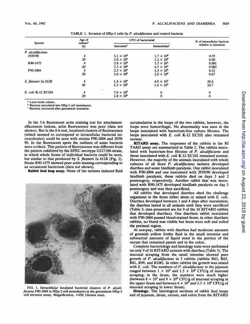
The organisms were negative for enterotoxins, cytotoxins,adherence to HEp-2 cell monolayers (even when grown andassayed under different conditions), and keratoconjunctivitisin the Sereny test, although the controls behaved as ex-pected in these tests. In the gentamicin-HEp-2 cell assay,however, both 4- and 20-h-old cultures of P. alcalifacienswere clearly invasive (Table 1). Strain R90-1475 was lessinvasive than the other two strains were. These differenceswere evident even in the Giemsa-stained monolayer slides atthe end of invasion assay. Strains F90-2004 and 2939/90produced intracellular bacterial clusters (Fig. 1), which wereabsent with strain R90-1475. The positive control, S. flexneri2a 611R, was more invasive than P. alcalifaciens strains, andthe negative control, E. coli K-12 EC101, was not invasive.The integrities of the monolayers were not significantlyaltered at the end of the invasion assays.
INFECT. IMMUN.
on August 22, 2019 by guest
http://iai.asm.org/
Dow
nloaded from
P. ALCALIFACIENS AND DIARRHEA 5019
TABLE 1. Invasion of HEp-2 cells by P. alcalifaciens and control bacteria
Age of CFU of bacteria/mi % of intracellular bacteriaSpecies culturea
(h) Inoculumb Intracellularc relative to inoculum
P. alcalifaciens2939/90 4 3.1 x 10⁶ 1.7 x 10⁴ 0.55
20 3.0 x 10⁶ 1.2 x 10⁴ 0.40R90-1475 4 2.8 x 10⁶ 3.3 x 101 0.001
20 3.3 x 10⁶ 6.5 x 101 0.002F90-2004 4 2.5 x 10⁶ 1.3 x 10 0.52
20 3.0 x 10⁶ 2.0 x 10 0.67
S. flexneri 2a 611R 4 1.4 x 10⁶ 4.0 x 10 28.620 1.5 x 10⁶ 1.6 x 10⁵ 10.7
E. coli K-12 EC101 4 7.0 x 10⁶ 0 020 2.8 x 10⁶ 0 0
a Luria broth culture.b Bacteria inoculated into HEp-2 cell monolayers.c Bacteria recovered after gentamicin treatment.
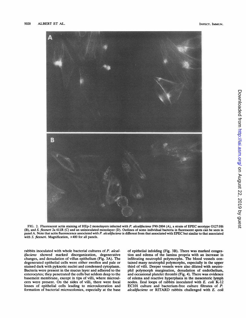
In the 3-h fluorescent actin staining test for attachment-effacement lesions, actin fluorescence was poor (data notshown). But in the 6-h test, localized clusters of fluorescence(which seemed to correspond to intracellular bacterial mi-crocolonies) could be seen with strains F90-2004 and 2939/90. In the fluorescent spots the outlines of some bacteriawere evident. This pattern of fluorescence was different fromthe pattern exhibited by the EPEC serotype 0127:H6 strain,in which whole forms of individual bacteria could be seen,but similar to that produced by S. flexneri 2a 611R (Fig. 2).Strain R90-1475 showed poor actin staining corresponding toan occasional bacterium (data not shown).
Rabbit ileal loop assay. None of the isolates induced fluid
FIG. 1. Intracellular localized bacterial clusters of P. alcali-faciens F90-2004 in HEp-2 cell monolayers in the gentamicin-HEp-2cell invasion assay. Magnification, x430; Giemsa stain.
accumulation in the loops of the two rabbits; however, theloops were hemorrhagic. No abnormality was seen in theloops inoculated with bacterium-free culture filtrates. Theloops inoculated with E. coli K-12 EC101 also remainednormal.RITARD assay. The responses of the rabbits in the RI
TARD assay are summarized in Table 2. The rabbits inocu-lated with bacterium-free filtrates of P. alcalifaciens andthose inoculated with E. coli K-12 EC101 remained normal.However, the majority of the animals inoculated with wholecultures of all three P. alcalifaciens isolates developeddiarrhea and some hindlimb paralysis. One rabbit inoculatedwith F90-2004 and one inoculated with 2939/90 developedhindlimb paralysis; these rabbits died on days 3 and 2postsurgery, respectively. Another rabbit that was inocu-lated with R90-1475 developed hindlimb paralysis on day 3postsurgery and was then sacrificed.
All rabbits that developed diarrhea shed the challengeorganisms in the feces either alone or mixed with E. coli.Diarrhea developed between 1 and 4 days after inoculation;the diarrhea lasted in all animals until they were sacrificed(Table 3; data presented are for 9 of the 16 RITARD rabbitsthat developed diarrhea). One diarrheic rabbit inoculatedwith F90-2004 passed blood-stained feces; in other diarrheicrabbits, no blood was visible but feces were soft and soiledthe perianal region.At autopsy, rabbits with diarrhea had moderate amounts
of greenish yellow frothy fluid in the small intestine andsubstantial amounts of liquid stool in the portion of thececum that remained patent and in the colon.Complete bacteriology and histology tests were performed
on only 9 of 16 RITARD animals with diarrhea (Table 3). Themucosal scraping from the small intestine showed puregrowth of P. alcalifaciens in 5 rabbits (rabbits R63, R65,R81, R99, and R100). In other rabbits the growth was mixedwith E. coli. The numbers of P. alcalifaciens in the jejunumranged between 1 x 10³ and 1.5 x 10⁸ CFU/g of mucosalscraping; in the ileum, the numbers were much higher(between 8 x 10³ and 9 x 10⁸ CFU/g of mucosal scraping inthe upper ileum and between 6 x 10⁴ and 3.5 x 10⁸ CFU/g ofmucosal scraping in lower ileum).
Histology. The histological sections of rabbit ileal loopsand of jejunum, ileum, cecum, and colon from the RITARD
VOL. 60, 1992
on August 22, 2019 by guest
http://iai.asm.org/
Dow
nloaded from
5020 ALBERT ET AL.
FIG. 2. Fluorescent actin staining of HEp-2 monolayers infected with P. alcalifaciens F90-2004 (A), a strain of EPEC serotype 0127:H6(B), and S. flexneri 2a 611R (C) and an uninoculated monolayer (D). Outlines of some individual bacteria in fluorescent spots can be seen inpanel A. Note that actin fluorescence associated with P. alcalifaciens is different from that associated with EPEC but similar to that associatedwith S. flexneri. Magnification, x400 for all panels.
rabbits inoculated with whole bacterial cultures of P. alcal-ifaciens showed marked disorganization, degenerativechanges, and denudation of villus epithelium (Fig. 3A). Thedegenerated epithelial cells were either swollen and pale orstained dark with pyknotic nuclei and condensed cytoplasm.Bacteria were present in the mucus layer and adhered to theenterocytes; they penetrated the cells but seldom deep to thebasement membrane, except in tips of villi, where microul-cers were present. On the sides of villi, there were focallosses of epithelial cells leading to microulceration andformation of bacterial microcolonies, especially at the base
of epithelial infolding (Fig. 3B). There was marked conges-tion and edema of the lamina propria with an increase ininfiltrating neutrophil polymorphs. The blood vessels con-tained many neutrophil polymorphs, especially in the upperthird of villi. Deeper vessels were also dilated with neutro-phil polymorph margination, denudation of endothelium,and occasional platelet thrombi (Fig. 4). There was evidenceof edema and reactive hyperplasia in the mesenteric lymphnodes. Ileal loops of rabbits inoculated with E. coli K-12EC101 culture and bacterium-free culture filtrates of P.alcalifaciens or RITARD rabbits challenged with E. coli
INFECT. IMMUN.
on August 22, 2019 by guest
http://iai.asm.org/
Dow
nloaded from

P. ALCALIFACIENS AND DIARRHEA 5021
K-12 EC101 showed no histological abnormalities. Thehistology of RITARD rabbits challenged with bacterium-freeculture filtrates of P. alcalifaciens was not determined.The histology of nerve tissue in two rabbits that developed
hindlimb paralysis and one rabbit that developed diarrheabut no paralysis was determined. Focal mononuclear cellinfiltration around the peripheral nerve sheath was found inthe two rabbits with paralysis but not in the rabbit withdiarrhea only.
Electron microscopy. The ileal loops of rabbits and intes-tines of RITARD rabbits that developed diarrhea showed
evidence of penetration of intestinal epithelial cells by bac-teria (Fig. 5); this penetration was absent in animals inocu-lated with nonpathogenic E. coli K-12 EC101.
Blood culture. None of the blood cultures from any of therabbits grew P. alcalifaciens.
DISCUSSION
P. alcalifaciens was suspected as a cause of diarrhea in thethree patients because it was either the only organismcultured or present in abundance in the stools in the absence
VOL. 60, 1992
on August 22, 2019 by guest
http://iai.asm.org/
Dow
nloaded from
5022 ALBERT ET AL.
TABLE 2. Responses of RITARD rabbits inoculated with whole bacterial cultures (BC) of P. alcalifaciens and E. coli K-12 EC101 andbacterium-free culture filtrates (CF) of P. alcalifaciens
No. of rabbits with the following response:Challenge bacterium Inoculum studied Diarrhea Hindlimb No change
Diarrhea ~~paralysisNocagP. alcalifaciens
F90-2004 BC 11 10 1 0CF 3 0 0 3
2939/90 BC 5 4 1 0CF 3 0 0 3
R90-1475 BC 5 2 1 2CF 3 0 0 3
E. coli K-12 EC101 BC 4 0 0 4
of recognized enteric pathogens. None of the isolates pro- gate the diarrheagenic properties of a variety of organismsduced any toxin, but all of them invaded the HEp-2 cell (1, 13, 21). All three P. alcalifaciens isolates producedmonolayer. Two isolates (F90-2004 and 2939/90) were more diarrhea in this model, supporting the suspicion of diarrhe-invasive than the third isolate (R90-1475) in this assay agenic potential of these organisms. The isolates colonizedsystem. The former two isolates also showed intracellular the gut, resulting in the excretion of bacteria in feces,bacterial clusters on light microscopic examination of Giemsa- sometimes with total displacement of normal flora. Again, asstained cell monolayers and produced striking actin condensa- in the in vitro virulence tests, differences were noted in thetion in the in vitro fluorescent actin staining test. The actin disease-producing abilities of the three isolates. Isolatescondensation seemed to correspond to the bacterial microcol- F90-2004 and 2939/90 produced diarrhea in 91% (10 of 11onies within the cells. This pattem of actin condensation is rabbits) and 80% (4 of 5 rabbits) of the animals, respectively,similar to that produced by another invasive organism, S. and also colonized the intestines well. However, isolate,plexneri (4), but different from that due to EPEC (11). The R90-1475 produced diarrhea in only 40% (two of five rabbits)third isolate of P. alcalifaciens, R90-1475, showed weaker of the animals and was not recovered from the small intes-properties of penetration and actin condensation. The nega- tine of one rabbit (R108) at the time of sacrifice (Tables 2 andtive control of E. coli K-12 EClOl did not show any invasion 3). These data suggest that there are differences in theof HEp-2 cells. This is contrary to observations in other disease-producing abilities of different strains of P. alcali-studies (14, 20), in which negative controls showed some faciens. None of the strains produced bacteremia, whichdegree of invasion. The property of cell invasion might be suggests that these bacteria do not invade beyond thestrain dependent; the strain in the present study was a local intestinal mucosa.derivative of E. coli K-12 (2). None of the Providencia Histology showed mucosal lesions suggestive of invasiveisolates showed any characteristic adherence to HEp-2 cells, diarrhea. The lesions included bacterial attachment to, pen-even though bacteria were grown and assayed under differ- etration of, and microulcer formation on the surface epithe-ent conditions. This situation is perhaps analogous to that of lium and edema, hyperemia, and polymorphonuclear cellS. flexnen (the prototype invasive organism), with which it is infiltration of lamina propria. Transmission electron micros-difficult to demonstrate adherence to cell monolayers (4). copy of the intestinal mucosa of affected animals alsoThe RITARD model has previously been used to investi- showed that the organisms are invasive. The finding of
TABLE 3. Fecal shedding, diarrhea, and colonization of the gut in RITARD rabbits inoculated with P. alcalifaciens
Diarrheab O No. of organisms (species)/g of mucosal Histological evidence of bacterial adhesion (A) and/orRabbit no.a (day after Organism(s)c scraping of: invasion (I) in:
inoculation) shed in stoolMidjejunum Upper ileum Lower ileum Jejunum Ileum Cecum Colon Rectum Appendix
R63 + (2) PE (3) 4 x 10⁵ (P) 5 x 10⁶ (P) 2.5 x 10⁷ (p) _d Al AR64 + (1) P (5) 2 x 10³ (P) 8 x 10³ (P) 6 x 10⁴ (P) A A A A
4 x10² (E) 4.5 x 10² (E)R65 + (2) P (5) 1.6 x 10⁵ (P) 4 x 10⁷ (P) 1.4 x 10⁸ (P) I AR79 + (2) PE (5) 3.5 x 10⁵ (P) 1 x 10⁶ (P) 4.2 x 10⁶ (P) A A A
1 X 10⁴ (E) 2 x 10⁵ (E) 1.5 x 10⁵ (E)R81 + (2) PE (3) 5 x 10⁷ (P) 2.6 x 10⁸ (P) 2.5 x 10⁸ (P) A Al Al AR99 + (2) PE(3) 1.5 x 10⁸(P) 1 x 10⁸ (P) 3.5 x 10⁸(P) AI I A Al AIR100 + (2) PE(5) 4 x 10⁶(P) 1.5 x 10⁷ (P) 2 x 10⁸(P) - AI AI AIR101 + (3) PE (5) 2 x 10⁷ (P) 9 X 10⁸ (p) 1.5 x 10⁷ (P) Al AI AI -
2 x 10⁴ (E)R108 + (4) PE (4) 1 x 10³ (E) 1.4 x 10⁵ (E) 1.8 x 10⁵ (E) - - A - - -
a Rabbits R63, R64, R65, R79, and R81 were challenged with strain F90-2004; rabbits R99, R100, and R101 were challenged with strain 2939/90; and rabbit R108was challenged with strain R90-1475.
b +, presence of diarrhea.c P,p. alcalifaciens; E, E. coli. Shedding of the indicated organism(s) occurred until the day of sacrifice (in parentheses).d -,no evidence of adhesion or invasion.
INFECT. IMMUN.
on August 22, 2019 by guest
http://iai.asm.org/
Dow
nloaded from

P. ALCALIFACIENS AND DIARRHEA 5023
-_; ~~W'`* -__i;*;-X-aJFIG. 3. Luminal aspect of the jejunal mucosa of a RITARD animal inoculated with P. akalifaciens F90-2004 showing bacterial invasion
(arrow) with luminal cell degeneration and desquamation forming surface ulceration (arrowhead). Marked congestion of microvasculature isseen in the lamina propria (A). The adjacent mucosa shows many bacteria adherent to a degenerating enterocyte (arrowhead). The laminapropria shows many neutrophil polymorphs (arrows) (B). Magnifications, x750 for panel A and x580 for panel B; toluidine blue stain.
invasive diarrhea in the animal model is supported by theinvasive picture in the stool of diarrheal patients excretingorganisms of the Providencia group (16). Unfortunately,there was no information on what types of diarrheas werepresent in the three patients from whom the present studyisolates originated.The organisms also produced hindlimb paralysis in some
rabbits, and the clinical relevance of this observation is notclear. It is known that S. dysenteriae 1 toxin induces
hindlimb paralysis when injected into mice (3). It is not clearwhether a similar toxin is elaborated by P. alcalifaciens,although no evidence of cytotoxicity was found in the invitro tissue culture systems inoculated with bacterium-freefiltrate and bacterial extract or in the rabbit ileal loopchallenged with bacterium-free filtrate. Further studies areneeded to elucidate the mechanism of paralysis.
Histology of rabbit ileal loops injected with bacterium-freeculture filtrates showed no abnormality. Moreover, RI-
FIG. 4. Same section as that shown in Fig. 3. (A) The capillary near the crypt base shows many neutrophil polymorphs (arrows). (B) Notethe markedly congested venule with endothelial swelling (E), degeneration, and endothelial cell loss. The lumen shows platelet thrombi (P).Magnification, x 800 for both panels; toluidine blue stain.
VOL. 60, 1992
on August 22, 2019 by guest
http://iai.asm.org/
Dow
nloaded from

5024 ALBERT ET AL.
FIG. 5. Transmission electron microscopy of an ileal sectionfrom the same RITARD rabbit as that shown in Fig. 3. Note the twointracellular bacteria within vacuoles (arrowheads). The microvilliof the enterocyte are vesiculated (arrows). Magnification, x 17,500.
TARD rabbits inoculated with culture filtrates also remainednormal. These findings suggest that soluble bacterial prod-ucts may not be involved in the pathogenesis.
Past studies (16, 18) have looked at the roles of Providencegroup organisms as a whole in the causation of diarrhea, andthere are no reliable data on the frequencies of isolation of P.alcalifaciens from patients with diarrhea and from controlswithout diarrhea. Clearly, case control studies are needed toassess the importance of this organism in diarrheal illness.
ACKNOWLEDGMENTS
This research was supported by the International Centre forDiarrhoeal Disease Research, Bangladesh, which is supported bycountries and agencies that share its concern for the health problemsof developing countries. Current donors include the aid agencies ofthe governments of Australia, Bangladesh, Belgium, Canada, Den-mark, France, Japan, the Netherlands, Norway, Saudi Arabia,Sweden, Switzerland, the United Kingdom, and the United States;international organizations including the United Nations Develop-ment Programme, the United Nations Children's Fund, and theWorld Health Organization; and private foundations, including theFord Foundation and the Sasakawa Foundation.We thank Manzurul Haque of the International Centre for Diar-
rhoeal Disease Research, Bangladesh, for his secretarial assistance.
REFERENCES1. Albert, M. J., K. Alam, M. Islam, J. Montanaro, A. S. M. H.
Rahman, K. Haider, M. A. Hossain, A. K. M. G. Kibriya, and S.Tzipori. 1991. Hafnia alvei, a probable cause of diarrhea inhumans. Infect. Immun. 59:1507-1513.
2. Ashraf, M. H., Z. U. Ahmed, and D. A. Sack 1991. Unusualassociation of a plasmid with nalidixic acid resistance in an
epidemic strain of Shigella dysenteriae type 1 from Asia. Can. J.Microbiol. 37:59-63.
3. Cavanagh, J. B., J. C. Howard, and J. L. Whitby. 1956. Theneurotoxin of Shigella shigae. A comparative study of theeffects produced in various laboratory animals. Br. J. Exp.Pathol. 37:272-278.
4. Clerc, P., and P. J. Sansonetti. 1987. Entry of Shigella flexneriinto HeLa cells: evidence for directed phagocytosis involvingactin polymerization and myosin accumulation. Infect. Immun.55:2681-2688.
5. Cravioto, A., R. J. Gross, S. M. Scotland, and B. Rowe. 1979. Anadhesive factor found in strains of Escherichia coli belonging tothe traditional infantile enteropathogenic serotypes. Curr. Mi-
-crobiol. 3:95-99.6. Dean, A. G., Y. C. Ching, R. G. Williams, and L. B. Harden.
1972. Test for Escherichia coli enterotoxin using infant mice:application in a study of diarrhea in children in Honolulu. J.Infect. Dis. 125:407-411.
7. Ewing, W. H. 1986. Edwards and Ewing's identification ofEnterobactericeae, 4th ed, p. 443-459. Elsevier Science Pub-lishing Co., Inc., New York.
8. Gentry, M. K., and J. M. Dalrymple. 1980. Quantitative micro-titer cytotoxicity assay for Shigella toxin. J. Clin. Microbiol.12:361-366.
9. Griffin, D. F., and P. Gemski, Jr. 1983. Release of Shiga toxinfrom Shigella dysenteriae 1 by polymyxin B. Infect. Immun.40:425-428.
10. Haynes, J., and P. M. Hawkey. 1989. Providencia alcalifaciensand travellers' diarrhoea. Br. Med. J. 299:94-95.
11. Knutton, S., A. D. Phillips, H. R. Smith, R. J. Gross, R. Shaw,P. Watson, and E. Price. 1991. Screening for enteropathogenicEscherichia coli in infants with diarrhea by the fluorescent actinstaining test. Infect. Immun. 59:365-371.
12. Moon, H. W., S. C. Whipp, R. A. Argenzio, M. M. Levine, andR. A. Gianella. 1983. Attaching and effacing activities of rabbitand human enteropathogenic Escherichia coli in pig and rabbitintestines. Infect. Immun. 41:1340-1351.
13. Pazzaglia, G., R. B. Sack, A. L. Bourgeois, J. Froehlich, and J.Eckstein. 1990. Diarrhea and intestinal invasiveness ofAeromo-nas strains in the removable intestinal tie rabbit model. Infect.Immun. 58:1924-1931.
14. Robins-Browne, R., and V. Bennett-Wood. 1992. Quantitativeassessment of the ability of Eschenchia coli to invade culturedanimal cells. Microb. Pathog. 12:159-164.
15. Sack, D. A., and R. B. SacL 1975. Test for enterotoxigenicEscherichia coli using Y1 adrenal cells in miniculture. Infect.Immun. 11:334-336.
16. Sen, R. 1962. Isolation of strains ofProvidence group from caseswith diarrhoea in Ibadan, Nigeria, West Africa. Indian J. Med.Res. 50:622-626.
17. Sereny, B. 1957. Experimental keratoconjunctivitis shigellosa.Acta Microbiol. Acad. Sci. Hung. 4:367-376.
18. Singer, J., and J. Bar-Chay. 1954. Biochemical investigation ofProvidence strains and their relationship to the Proteus group.J. Hyg. Camb. 52:1.
19. Small, P. L. C., and S. Falkow. 1988. Identification of regions ona 230-kilobase plasmid from enteroinvasive Escherichia coli thatare required for entry into HEp-2 cells. Infect. Immun. 56:225-229.
20. Small, P. L. C., R. R. Isberg, and S. Falkow. 1987. Comparisonof the ability of enteroinvasive Escherichia coli, Salmonellatyphimunium, Yersiniapseudotuberculosis, and Yersinia entero-colitica to enter and replicate within HEp-2 cells. Infect. Im-mun. 55:1674-1679.
21. Spira, W. M., R. B. Sack, and J. L. Froehlich. 1981. Simpleadult rabbit model for Vibrio cholerae and enterotoxigenicEschenichia coli diarrhea. Infect. Immun. 32:739-749.
INFECT. IMMUN.
on August 22, 2019 by guest
http://iai.asm.org/
Dow
nloaded from
Related Documents