Pathogenesis of aortic dilatation in mucopolysaccharidosis VII mice may involve complement activation Guilherme Baldo a , Susan Wu a , Ruth A. Howe a , Meera Ramamoothy a , Russell H. Knutsen b , Jiali Fang a , Robert P. Mecham b , Yuli Liu a , Xiaobo Wu a , John P. Atkinson a , and Katherine P. Ponder a,c,* a Department of Internal Medicine, Washington University School of Medicine, St. Louis, MO, USA b Department of Cell Biology, Washington University School of Medicine, St. Louis, MO, USA c Biochemistry and Molecular Biophysics, Washington University School of Medicine, St. Louis, MO, USA Abstract Mucopolysaccharidosis VII (MPS VII) is due to mutations within the gene encoding the lysosomal enzyme β-glucuronidase, and results in the accumulation of glycosaminoglycans. MPS VII causes aortic dilatation and elastin fragmentation, which is associated with upregulation of the elastases cathepsin S (CtsS) and matrix metalloproteinase 12 (MMP12). To test the role of these enzymes, MPS VII mice were crossed with mice deficient in CtsS or MMP12, and the effect upon aortic dilatation was determined. CtsS deficiency did not protect against aortic dilatation in MPS VII mice, but also failed to prevent an upregulation of cathepsin enzyme activity. Further analysis with substrates and inhibitors specific for particular cathepsins suggests that this enzyme activity was due to CtsB, which could contribute to elastin fragmentation. Similarly, MMP12 deficiency and deficiency of both MMP12 and CtsS could not prevent aortic dilatation in MPS VII mice. Microarray and reverse-transcriptase real-time PCR were performed to look for upregulation of other elastases. This demonstrated that mRNA for complement component D was elevated in MPS VII mice, while immunostaining demonstrated high levels of complement component C3 on surfaces within the aortic media. Finally, we demonstrate that neonatal intravenous injection of a retroviral vector encoding β-glucuronidase reduced aortic dilatation. We conclude that neither CtsS nor MMP12 are necessary for elastin fragmentation in MPS VII mouse aorta, and propose that CtsB and/or complement component D may be involved. Complement may be activated by the GAGs that accumulate, and may play a role in signal transduction pathways that upregulate elastases. Keywords Mucopolysaccharidosis VII; Cathepsin S; Matrix metalloproteinase 12; Complement system; Aortic dilatation; Gene therapy © 2011 Elsevier Inc. All rights reserved. * Corresponding author. Department of Internal Medicine, Washington University School of Medicine, 660 South Euclid Avenue, St. Louis, MO 63110, USA. Fax: +1 314 362 8813. [email protected] (K.P. Ponder). Supplementary materials related to this article can be found online at doi:10.1016/j.ymgme.2011.08.018. NIH Public Access Author Manuscript Mol Genet Metab. Author manuscript; available in PMC 2012 December 1. Published in final edited form as: Mol Genet Metab. 2011 December ; 104(4): 608–619. doi:10.1016/j.ymgme.2011.08.018. NIH-PA Author Manuscript NIH-PA Author Manuscript NIH-PA Author Manuscript

Welcome message from author
This document is posted to help you gain knowledge. Please leave a comment to let me know what you think about it! Share it to your friends and learn new things together.
Transcript

Pathogenesis of aortic dilatation in mucopolysaccharidosis VIImice may involve complement activation
Guilherme Baldoa, Susan Wua, Ruth A. Howea, Meera Ramamoothya, Russell H. Knutsenb,Jiali Fanga, Robert P. Mechamb, Yuli Liua, Xiaobo Wua, John P. Atkinsona, and Katherine P.Pondera,c,*
aDepartment of Internal Medicine, Washington University School of Medicine, St. Louis, MO, USAbDepartment of Cell Biology, Washington University School of Medicine, St. Louis, MO, USAcBiochemistry and Molecular Biophysics, Washington University School of Medicine, St. Louis,MO, USA
AbstractMucopolysaccharidosis VII (MPS VII) is due to mutations within the gene encoding the lysosomalenzyme β-glucuronidase, and results in the accumulation of glycosaminoglycans. MPS VII causesaortic dilatation and elastin fragmentation, which is associated with upregulation of the elastasescathepsin S (CtsS) and matrix metalloproteinase 12 (MMP12). To test the role of these enzymes,MPS VII mice were crossed with mice deficient in CtsS or MMP12, and the effect upon aorticdilatation was determined. CtsS deficiency did not protect against aortic dilatation in MPS VIImice, but also failed to prevent an upregulation of cathepsin enzyme activity. Further analysis withsubstrates and inhibitors specific for particular cathepsins suggests that this enzyme activity wasdue to CtsB, which could contribute to elastin fragmentation. Similarly, MMP12 deficiency anddeficiency of both MMP12 and CtsS could not prevent aortic dilatation in MPS VII mice.Microarray and reverse-transcriptase real-time PCR were performed to look for upregulation ofother elastases. This demonstrated that mRNA for complement component D was elevated in MPSVII mice, while immunostaining demonstrated high levels of complement component C3 onsurfaces within the aortic media. Finally, we demonstrate that neonatal intravenous injection of aretroviral vector encoding β-glucuronidase reduced aortic dilatation. We conclude that neitherCtsS nor MMP12 are necessary for elastin fragmentation in MPS VII mouse aorta, and proposethat CtsB and/or complement component D may be involved. Complement may be activated bythe GAGs that accumulate, and may play a role in signal transduction pathways that upregulateelastases.
KeywordsMucopolysaccharidosis VII; Cathepsin S; Matrix metalloproteinase 12; Complement system;Aortic dilatation; Gene therapy
© 2011 Elsevier Inc. All rights reserved.*Corresponding author. Department of Internal Medicine, Washington University School of Medicine, 660 South Euclid Avenue, St.Louis, MO 63110, USA. Fax: +1 314 362 8813. [email protected] (K.P. Ponder).Supplementary materials related to this article can be found online at doi:10.1016/j.ymgme.2011.08.018.
NIH Public AccessAuthor ManuscriptMol Genet Metab. Author manuscript; available in PMC 2012 December 1.
Published in final edited form as:Mol Genet Metab. 2011 December ; 104(4): 608–619. doi:10.1016/j.ymgme.2011.08.018.
NIH
-PA Author Manuscript
NIH
-PA Author Manuscript
NIH
-PA Author Manuscript

1. IntroductionMucopolysaccharidoses (MPS) are a group of 11 diseases caused by mutations in genes thatencode lysosomal enzymes that degrade glycosaminoglycans (GAGs) [1]. MPS is associatedwith accumulation of GAGs throughout the body and multisystemic disease. The focus ofthis project was to better understand the pathogenesis of aortic disease in MPS using themurine model of MPS VII, which is an autosomal recessive disease due to β-glucuronidase(GUSB) deficiency (Online Mendelian Inheritance in Man #253220). An elegant model forthe pathogenesis of MPS involves the binding of GAGs to Toll-like Receptor 4 (TLR4),which upregulates cytokines such as tumor necrosis factor α (TNFα), Ccl4 (MIP-1β), andinterleukin 6 (IL-6), which in turn upregulate destructive proteases [2–4]. Adult humanswith attenuated MPS I have aortas that are 122% of normal diameter [5] and reducedelasticity [6], while one patient with MPS VII required an aortic graft [7]. Mice with MPS I[8–10] and MPS VII [11], cats with MPS I and MPS VI [12], and dogs with MPS I [13–15]and MPS VII [14,16,17] also have aortic dilatation.
Elastin represents 30% of the dry weight of the aorta [18]. Tropoelastin monomers aresecreted and then crosslinked into elastic fibers in a process that involves elastin bindingprotein, extracellular matrix microfibrils, and crosslinking enzymes [19]. Elastin wasfragmented in the ascending aorta of humans, mice, and dogs with MPS I, and in humansand dogs with MPS VII [10,13,20–22]. Hinek et al. demonstrated that exogenousadministration of dermatan sulfate, a GAG that accumulates in many types of MPS, reducedelastin binding protein levels and inhibited elastin assembly in vitro, and proposed thatreduced assembly caused elastin defects in MPS I [23]. Alternatively, we reported in MPS Imice and dogs and in MPS VII dogs [10,14] that elastin fragmentation was temporallyassociated with increases in RNA and enzyme activity for two elastases, cathepsin S (CtsS)and matrix metalloproteinase 12 (MMP12), which contribute to aortic aneurisms in mousemodels [24,25], and proposed that degradation was the major factor leading to elastinfragmentation. Although collagen is another important extracellular matrix protein of theaorta, collagen fibrils appeared to be relatively intact with histochemical stains in MPS I andMPS VII dogs [14].
Hematopoietic stem cell transplantation can reduce clinical manifestations of MPS, ashematopoietic cells migrate into tissues and secrete mannose 6-phosphate (M6P)-modifiedenzyme that can be taken up via the M6P receptor by nearby cells [26]. This has reduced,but not prevented, accumulation of GAGs, elastin fragmentation, and/or dilatation of theaorta in MPS VII mice [27] and dogs [16], MPS VI rats [28], and MPS I dogs [15] and cats[29]. Enzyme replacement therapy (ERT) involves intravenous (IV) injection of M6P-modified enzyme that can diffuse to organs and be taken up via the M6P receptor [30,31],although ERT is not available for MPS VII. ERT had little effect on lysosomal storageaccumulation in aortic smooth muscle cells in MPS VI cats [32]. Gene therapy is also beingtested in animal models [33]. One approach involves neonatal IV injection of a retroviralvector (RV) expressing the appropriate enzyme, which results in transduction of liver cellsand secretion of enzyme into blood [33–35]. This reduced aortic dilatation, but MPS I micerequired very high expression for a full therapeutic effect [8–10], and MPS VII dogsdeveloped aortic dilatation after 5 years [14].
The data that CtsS and MMP12 are upregulated in MPS aortas led us to hypothesize thatdeletion of CtsS and/or MMP12 might reduce elastin fragmentation. To test this hypothesis,CtsS−/− and MMP12−/− mice were crossed with MPS VII (GUSB−/−) mice and the effectupon the aorta diameters was determined. In addition, microarray analysis was performed todetermine if other genes that could contribute to aortic dilatation were upregulated. Theresults demonstrate that CtsS and MMP12 are not essential for aortic dilatation, but a related
Baldo et al. Page 2
Mol Genet Metab. Author manuscript; available in PMC 2012 December 1.
NIH
-PA Author Manuscript
NIH
-PA Author Manuscript
NIH
-PA Author Manuscript
cathepsin, CtsB, may contribute. These studies also demonstrate that the complement systemmay directly result in elastin fragmentation, or may indirectly contribute by induction ofsignal transduction pathways that result in upregulation of elastases.
2. Materials and methodsReagents were from Sigma-Aldrich Chemical (St. Louis, MO) unless otherwise stated.
2.1. AnimalsNational Institutes of Health (NIH) guidelines for the care and use of animals in researchwere followed. MPS VII [27], CtsS-deficient [36], and MMP12 deficient [37] mice were allin a C57BL/6 background. Some MPS VII mice were injected IV with 1 × 10¹⁰ transducingunits/kg of the RV designated hAAT-cGUSB-WPRE [38] at 2 to 3 days after birth to enablethem to survive and breed. Genotyping for MPS VII mice used a SNP assay on tail DNAwith a forward primer (5′ CCATAGTCATGATACCAAGAAAAGTAGCT-3′), a reverseprimer (5′-TGACTATTCTGACCTCAGTGTGTGA-3′), a wild-type minor-groove bindingprobe labeled with Vic (5′-TTGTCTTAGGCCCCGTACGT-3′; the underlined C representsthe position deleted in the MPS VII mouse), a mutant minor groove binding probe labeledwith FAM (5′-TTGTCTTAGGCCC-GTACGT-3′) and a One-Step Plus PCR machine(Applied Biosystems; Foster City, CA). PCR for the wild-type CtsS allele used the primers5′-CTTGAAGGGCAGCTGAAGCTG-3′ (forwards) and 5′-GTAGGAAGCGTCTGCCTCTAT-3′ (reverse), and PCR for the mutant CtsS allele used theprimers 5′-CTCTGTGTAGCCTGGAATTC-3′ (forwards) and 5′-CTAAAGCGCATGCTCCAGACTGCC-3′ (reverse) [36] with analysis of the CT usingSYBR green real-time PCR of DNA. MMP12 mice genotyping PCR used a forward primercommon to the wild-type and mutant MMP12 alleles (5′-CCCTCGATGTGGAGTGCCCG-3′), a reverse primer specific for the PGK-neo cassette(5′-AAGAACGGAGCCGGTTGGCG-3′), and a reverse primer specific for the MMP12wild-type allele (5′-ACTTGCCCTGAGCACCCCCT-3′), with gel electrophoresis used toidentify wild-type (337 bp) or mutant (460 bp) alleles.
2.2. Measurement of aortic diameter and histopathologyMice were anesthetized with 120 mg/kg ketamine/40 mg/kg xylazine in phosphate bufferedsaline (PBS) at pH 7.4. For some animals, aortic compliance was assessed, in which theouter diameter of isolated aortas was determined at different internal pressures [39]. Forhistopathology, ascending aortas obtained from 1 to 2 mm from the aortic valve were fixedwith buffered formalin, embedded in paraffin, and 6 µm sections were stained with VerhoeffVan Gieson (VVG) stain. For biochemical analyses, animals were perfused with 20 ml ofPBS, and the aorta from just above the aortic valve to just before the first branch wascleaned of surrounding fat. To test the effect of gene therapy, the width of dissectedascending aortas was measured after gently pressing it against a surface.
2.3. RNA analysisFrozen ascending aortas were homogenized in Trizol for 30 s with a hand-held homogenizer(Kimble-Kontes; Vineland, NJ), and RNA was isolated using a Qiagen column. Reversetranscription (RT) was performed on 1 µg of DNase I-treated RNA with an oligo (dT) 20primer using a Superscript III kit (Invitrogen Corp., Carlsbad, CA) in 20 µl, followed byreal-time PCR on 0.4 µl of each cDNA sample using SYBR green reagents from AppliedBiosystems [10]. Primer sequences are in our previous publication [10] or in SupplementaryTable 1. The percentage of a test RNA to that of β-actin was calculated by subtracting thecycle to reach the threshold (CT) for a gene from the CT for a separate assay using β-actinprimers to determine the ΔCT, and the formula: percent β-actin = (100) × 2ΔCT. The percent
Baldo et al. Page 3
Mol Genet Metab. Author manuscript; available in PMC 2012 December 1.
NIH
-PA Author Manuscript
NIH
-PA Author Manuscript
NIH
-PA Author Manuscript

β-actin for MPS animals was divided by the percent β-actin in normal animals to determinethe ratio of the gene in MPS to normal mice.
For microarray, RNA was reverse transcribed with primers with a T7 RNA polymerasebinding site, amplified with T7 RNA polymerase with fluorescently-labeleddeoxynucleotides, and hybridized to an Illumina bead microarray (Mouse8, version 2).Expression analysis was performed with ParTek software (St. Louis, MO). Pathway analysiswas performed with GeneGo interactions software(https://portal.genego.com/cgi/data_manager.cgi; St. Joseph, MI).
2.4. Enzyme and GAG assaysFor the GUSB, α-L-iduronidase (IDUA), and cathepsin assays, frozen aortas werehomogenized with the hand-held homogenizer in 100 mM sodium acetate pH 5.5 containing2.5 mM ethylenediaminetetraacetic acid, 0.1% Triton X-100, and 2.5 mM dithiothreitol, andcentrifuged at 10,000 g for 5 min at 4 °C. The protein concentration was determined with theBradford assay (BioRad Laboratories, Hercules CA). For the MMP12 and GAG assays,extracts were homogenized in the neutral buffer provided with the MMP12 kit with 0.1%Triton-X.
GUSB and IDUA assays were performed with the extracts prepared at pH 5.5 using thefluorogenic substrates 4-methylumbelliferyl-β-L-glucuronide (Sigma-Aldrich, St. Louis,MO) for GUSB and 4-methylumbelliferyl-α-L-iduronide (Toronto Research Chemicals,North York, Canada) for IDUA and a Fluoroskan Ascent microplate fluorometer (ThermoElectron, Milford, MA) as previously described [9]. One unit of enzyme converts 1 nmol ofsubstrate to product per hour at 37 °C. GAG content was determined in the samples obtainedat neutral pH using the commercial kit Blyscan (Biocolor, Carrickfergus, UK) using 30 µgof protein from each sample as described [10].
For the general cathepsin assay, 1 µg or less of the supernatant was incubated with 100 µMbenzyloxycarbonyl-L-phenylalanyl-L-arginine-7-amido-4-methylcoumarin (Z-Phe-Arg-AMC) from Anaspec (San Jose, CA) at pH 7.5 in 100 mM sodium acetate with 2.5 mMethylenediaminetetraacetic acid, 0.01% Triton X-100, and 2.5 mM dithiothreitol in amicrotiter plate at 37 °C for 1 h [10]. The amount of product was determined by excitation at355 nm and emission at 460 nm using kinetic readings and comparison with 7-amino-4-methylcoumarin (AMC) standards from Anaspec. One unit (U) of enzyme released 1 nmolof product per hour at 37 °C. The CtsB assay used the same extracts, the substrate Z-Arg-Arg-AMC (Bachem, Torrance, CA) at pH 7.5, and the same wavelengths as for the generalcathepsin assay. CtsK activity was measured at pH 7.5 with 10 µM of the substrate 2-aminobenzoic acid-HPGGPQ-N-(2,4-dinitrophenyl)-ethylenediamine (Abz-HPGGPQ-EDDnp) from Anaspec, which is cleaved by CtsK but not other cathepsins, and 2-aminobenzoic acid was the standard. The CtsD assay was performed at pH 4 with 10 µM ofthe substrate 7-methoxycoumarin-4-acetyl (Mca)-Gly-Lys-Pro-Ile-Leu-Phe-Phe-Arg-Leu-Lys-2,4 nitrophenyl (Dnp)-D-Arg-NH2, which can also be cleaved by CtsE, with Mca-Pro-Leu-OH (Enzo Life Sciences) as the standard. CtsK and CtsD assays were read at 320 nmfor excitation and 420 nm for emission. Inhibitors were from Calbiochem (San Diego, CA)and included the CtsS inhibitor Z-FL-COCHO (#219393), the CtsK inhibitor I [1,3-Bis (N-carbobenzoyloxy-L-leucyl) amino acetone; #219377] and the CtsB inhibitor Ac-Leu-Val-Lysinal (#219385). Samples were incubated with the inhibitor for 10 min prior to startingthe assay. Additional assays were performed with human recombinant purified CtsB [R&Dsystems, Minneapolis, MN; specific activity 150 nmol of substrate cleaved per hour (U)/µgprotein], CtsK (Enzo Life Sciences, Farmington, NY; 90 U/µg protein), CtsL (R&Dsystems; 900 U/µg protein), CtsS (R&D systems; 18 U/µg protein) and with CtsH purifiedfrom human liver (Enzo Life Sciences; 61 U/µg protein). An MMP12 assay kit
Baldo et al. Page 4
Mol Genet Metab. Author manuscript; available in PMC 2012 December 1.
NIH
-PA Author Manuscript
NIH
-PA Author Manuscript
NIH
-PA Author Manuscript
(SensolyteTM 490 MMP12) was obtained from Anaspec for which the substrate can also becleaved by MMP1, 2, 3, 8, and 13 and was performed as described previously with ~5 µg ofextract in 100 µl reactions [10].
2.5. ImmunostainingImmunostaining for STAT3 that was phosphorylated at tyrosine 705 was performed asdescribed previously [10]. For C3 immunostaining, frozen sections of aorta in optimalcutting temperature compound were fixed with formalin for 10 min at room temperature,and washed 3 times with TBS (Tris-buffered saline; 50 mM Tris pH 7.6, 150 mM NaCl,0.1% Triton X-100). Endogenous peroxidase was inhibited with 0.6% H2O2 in water for 30min. Samples were washed 3 times with TBS, and then preincubated with blocking buffer(TBS with 10% horse serum) for 30 min at room temperature. A goat-anti-mouse antibodyspecific for C3 (MP Biomedicals #55474, Solon, OH) was incubated overnight at 4 °C at a1:100 dilution in blocking buffer, and then washed 3 times with TBS. A horse-radishperoxidase-conjugated horse anti-goat IgG (Vector Laboratories, Burlingame CA) wasincubated at a 1:100 dilution for 1 h at RT in blocking buffer, and samples were washed 3times with TBS. Samples were developed with 0.7 mg/ml 3,3′-diaminobenzidine with 0.7mg/ml urea for 5 min.
2.6. StatisticsThe Student's t test compared values between 2 groups, and ANOVA with Tukey post-hocanalysis compared values between 3 or more groups using Sigma Stat software (SystatSoftware, Inc., Point Richmond, CA).
3. Results3.1. Aortic dilatation in MPS VII mice
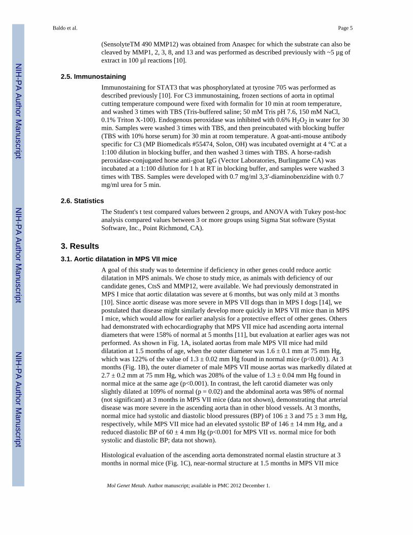
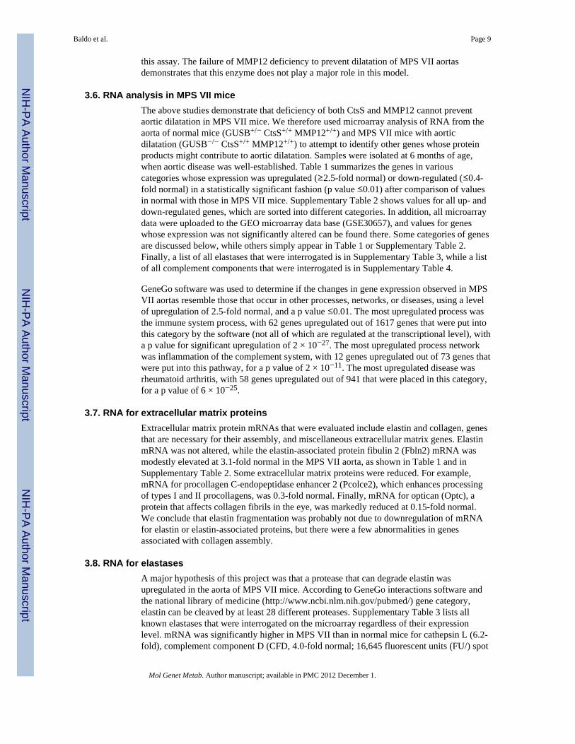
A goal of this study was to determine if deficiency in other genes could reduce aorticdilatation in MPS animals. We chose to study mice, as animals with deficiency of ourcandidate genes, CtsS and MMP12, were available. We had previously demonstrated inMPS I mice that aortic dilatation was severe at 6 months, but was only mild at 3 months[10]. Since aortic disease was more severe in MPS VII dogs than in MPS I dogs [14], wepostulated that disease might similarly develop more quickly in MPS VII mice than in MPSI mice, which would allow for earlier analysis for a protective effect of other genes. Othershad demonstrated with echocardiography that MPS VII mice had ascending aorta internaldiameters that were 158% of normal at 5 months [11], but evaluation at earlier ages was notperformed. As shown in Fig. 1A, isolated aortas from male MPS VII mice had milddilatation at 1.5 months of age, when the outer diameter was 1.6 ± 0.1 mm at 75 mm Hg,which was 122% of the value of 1.3 ± 0.02 mm Hg found in normal mice (p<0.001). At 3months (Fig. 1B), the outer diameter of male MPS VII mouse aortas was markedly dilated at2.7 ± 0.2 mm at 75 mm Hg, which was 208% of the value of 1.3 ± 0.04 mm Hg found innormal mice at the same age (p<0.001). In contrast, the left carotid diameter was onlyslightly dilated at 109% of normal (p = 0.02) and the abdominal aorta was 98% of normal(not significant) at 3 months in MPS VII mice (data not shown), demonstrating that arterialdisease was more severe in the ascending aorta than in other blood vessels. At 3 months,normal mice had systolic and diastolic blood pressures (BP) of 106 ± 3 and 75 ± 3 mm Hg,respectively, while MPS VII mice had an elevated systolic BP of 146 ± 14 mm Hg, and areduced diastolic BP of 60 ± 4 mm Hg (p<0.001 for MPS VII vs. normal mice for bothsystolic and diastolic BP; data not shown).
Histological evaluation of the ascending aorta demonstrated normal elastin structure at 3months in normal mice (Fig. 1C), near-normal structure at 1.5 months in MPS VII mice
Baldo et al. Page 5
Mol Genet Metab. Author manuscript; available in PMC 2012 December 1.
NIH
-PA Author Manuscript
NIH
-PA Author Manuscript
NIH
-PA Author Manuscript
(Fig. 1D), and marked elastin fragmentation at 3 months in MPS VII mice (Fig. 1E). Inaddition, there was increased phosphorylation at tyrosine 705 of STAT3 at 3 months in thenuclei of MPS VII mice (Fig. 1G) as compared with normal mice (Fig. 1F). Furthermore,analysis of RNA levels at 3 months (Fig. 1H) demonstrated that elastin levels were near-normal at 2.1 ± 1.9-fold normal (not significant vs. normal), collagen Iα1 levels were near-normal at 1.9 ± 1.5-fold normal (not significant), MMP12 levels were elevated at 6.8 ± 4.9-fold normal (p = 0.02), and CtsS mRNA was elevated at 4.8 ± 2.3-fold normal (p = 0.004).mRNA for osteopontin (SPP1), a protein that can activate MMPs by non-proteolyticmechanisms and plays a role in signal transduction, was markedly elevated at 28 ± 25.6-foldnormal (p = 0.03). In addition, mRNA for MMP3, an inhibitor of MMPs (TIMP1), cathepsinB (CtsB), cathepsin D (CtsD), cathepsin L (CtsL), and the MMP-activating enzymeurokinase plasminogen activator (uPA) were also significantly elevated, although themagnitude was not as high. These data demonstrate that aortic dilatation is due to elastinfragmentation in the MPS VII mouse model, which in turn is associated with upregulation ofCtsS and MMP12 mRNA. In addition, there was upregulation of other genes andphosphorylation of the transcription factor STAT3 that mimic closely what occurs in theMPS I mouse model [10], suggesting that the pathogenesis is similar. Since aortic dilatationwas severe by 3 months in MPS VII mice, analysis at 3 months or later should beinformative.
3.2. Effect of CtsS or MMP12 deficiency on aortic dilatation in MPS VII miceMPS VII (GUSB−/−) mice on a C57BL/6 background were crossed with CtsS−/− orMMP12−/− mice on C57BL/6 backgrounds, and the F1 offspring were then crossed togenerate mice that were deficient in one or both genes. During breeding, some GUSB−/−
mice were injected with 1 × 10¹⁰ transducing units/kg of the RV designated hAAT-cGUSB-WPRE, which contains the canine GUSB cDNA, and was previously demonstrated to resultin high expression of GUSB in liver cells, high serum GUSB activity, and the ability tosurvive and breed long-term [38]. Aortas from RV-treated MPS VII mice were not includedin analyses for mice of a particular genotype, as treatment reduced aortic dilatation. Micefrom the GUSB−/− CtsS−/− colony were then crossed with mice from the GUSB−/−
MMP12−/− colony to generate mice that were deficient in GUSB, CtsS, and MMP12.
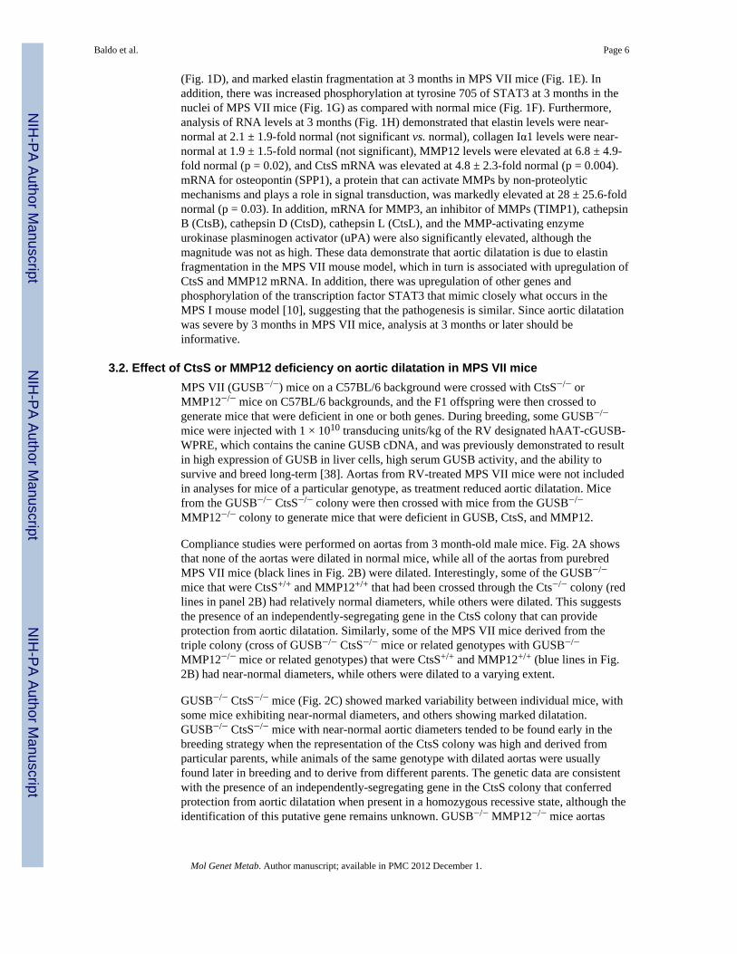
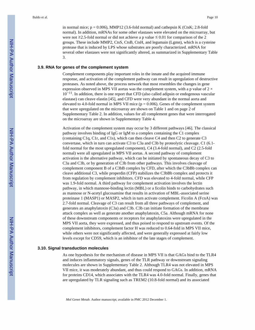
Compliance studies were performed on aortas from 3 month-old male mice. Fig. 2A showsthat none of the aortas were dilated in normal mice, while all of the aortas from purebredMPS VII mice (black lines in Fig. 2B) were dilated. Interestingly, some of the GUSB−/−
mice that were CtsS+/+ and MMP12+/+ that had been crossed through the Cts−/− colony (redlines in panel 2B) had relatively normal diameters, while others were dilated. This suggeststhe presence of an independently-segregating gene in the CtsS colony that can provideprotection from aortic dilatation. Similarly, some of the MPS VII mice derived from thetriple colony (cross of GUSB−/− CtsS−/− mice or related genotypes with GUSB−/−
MMP12−/− mice or related genotypes) that were CtsS+/+ and MMP12+/+ (blue lines in Fig.2B) had near-normal diameters, while others were dilated to a varying extent.
GUSB−/− CtsS−/− mice (Fig. 2C) showed marked variability between individual mice, withsome mice exhibiting near-normal diameters, and others showing marked dilatation.GUSB−/− CtsS−/− mice with near-normal aortic diameters tended to be found early in thebreeding strategy when the representation of the CtsS colony was high and derived fromparticular parents, while animals of the same genotype with dilated aortas were usuallyfound later in breeding and to derive from different parents. The genetic data are consistentwith the presence of an independently-segregating gene in the CtsS colony that conferredprotection from aortic dilatation when present in a homozygous recessive state, although theidentification of this putative gene remains unknown. GUSB−/− MMP12−/− mice aortas
Baldo et al. Page 6
Mol Genet Metab. Author manuscript; available in PMC 2012 December 1.
NIH
-PA Author Manuscript
NIH
-PA Author Manuscript
NIH
-PA Author Manuscript
were consistently dilated, although one animal was less severe than the others (Fig. 2D).GUSB−/− CtsS−/− MMP12−/− mice (Fig. 2E) all had dilated aortas.
Average aortic diameters ± SD at 75 mm Hg for each of these groups are shown in Fig. 2F.Aortic diameters from GUSB−/− CtsS−/− mice, GUSB−/− MMP12−/− mice, or GUSB−/−
CtsS−/− MMP12−/− mice were not statistically different from GUSB−/− mice withoutadditional deficiencies, and all MPS VII groups were statistically different from normalmice. We conclude that deficiency of CtsS, MMP12, or both cannot prevent aortic dilatationin MPS VII mice.
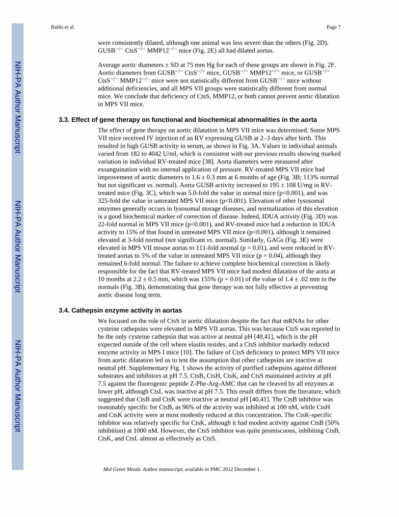
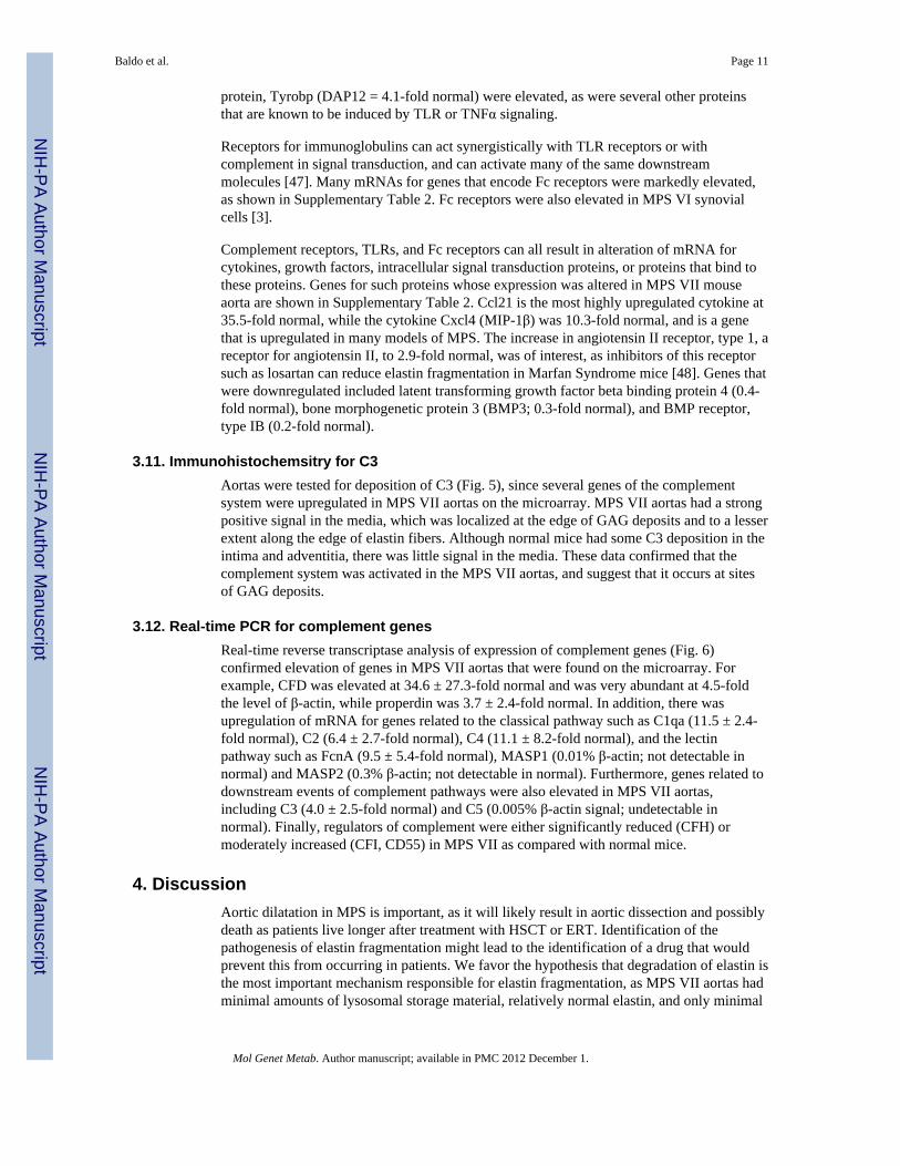
3.3. Effect of gene therapy on functional and biochemical abnormalities in the aortaThe effect of gene therapy on aortic dilatation in MPS VII mice was determined. Some MPSVII mice received IV injection of an RV expressing GUSB at 2–3 days after birth. Thisresulted in high GUSB activity in serum, as shown in Fig. 3A. Values in individual animalsvaried from 182 to 4042 U/ml, which is consistent with our previous results showing markedvariation in individual RV-treated mice [38]. Aorta diameters were measured afterexsanguination with no internal application of pressure. RV-treated MPS VII mice hadimprovement of aortic diameters to 1.6 ± 0.3 mm at 6 months of age (Fig. 3B; 113% normalbut not significant vs. normal). Aorta GUSB activity increased to 195 ± 108 U/mg in RV-treated mice (Fig. 3C), which was 5.0-fold the value in normal mice (p<0.001), and was325-fold the value in untreated MPS VII mice (p<0.001). Elevation of other lysosomalenzymes generally occurs in lysosomal storage diseases, and normalization of this elevationis a good biochemical marker of correction of disease. Indeed, IDUA activity (Fig. 3D) was22-fold normal in MPS VII mice (p<0.001), and RV-treated mice had a reduction in IDUAactivity to 15% of that found in untreated MPS VII mice (p<0.001), although it remainedelevated at 3-fold normal (not significant vs. normal). Similarly, GAGs (Fig. 3E) wereelevated in MPS VII mouse aortas to 111-fold normal (p = 0.01), and were reduced in RV-treated aortas to 5% of the value in untreated MPS VII mice (p = 0.04), although theyremained 6-fold normal. The failure to achieve complete biochemical correction is likelyresponsible for the fact that RV-treated MPS VII mice had modest dilatation of the aorta at10 months at 2.2 ± 0.5 mm, which was 155% (p = 0.01) of the value of 1.4 ± .02 mm in thenormals (Fig. 3B), demonstrating that gene therapy was not fully effective at preventingaortic disease long term.
3.4. Cathepsin enzyme activity in aortasWe focused on the role of CtsS in aortic dilatation despite the fact that mRNAs for othercysteine cathepsins were elevated in MPS VII aortas. This was because CtsS was reported tobe the only cysteine cathepsin that was active at neutral pH [40,41], which is the pHexpected outside of the cell where elastin resides, and a CtsS inhibitor markedly reducedenzyme activity in MPS I mice [10]. The failure of CtsS deficiency to protect MPS VII micefrom aortic dilatation led us to test the assumption that other cathepsins are inactive atneutral pH. Supplementary Fig. 1 shows the activity of purified cathepsins against differentsubstrates and inhibitors at pH 7.5. CtsB, CtsH, CtsK, and CtsS maintained activity at pH7.5 against the fluorogenic peptide Z-Phe-Arg-AMC that can be cleaved by all enzymes atlower pH, although CtsL was inactive at pH 7.5. This result differs from the literature, whichsuggested that CtsB and CtsK were inactive at neutral pH [40,41]. The CtsB inhibitor wasreasonably specific for CtsB, as 96% of the activity was inhibited at 100 nM, while CtsHand CtsK activity were at most modestly reduced at this concentration. The CtsK-specificinhibitor was relatively specific for CtsK, although it had modest activity against CtsB (50%inhibition) at 1000 nM. However, the CtsS inhibitor was quite promiscuous, inhibiting CtsB,CtsK, and CtsL almost as effectively as CtsS.
Baldo et al. Page 7
Mol Genet Metab. Author manuscript; available in PMC 2012 December 1.
NIH
-PA Author Manuscript
NIH
-PA Author Manuscript
NIH
-PA Author Manuscript

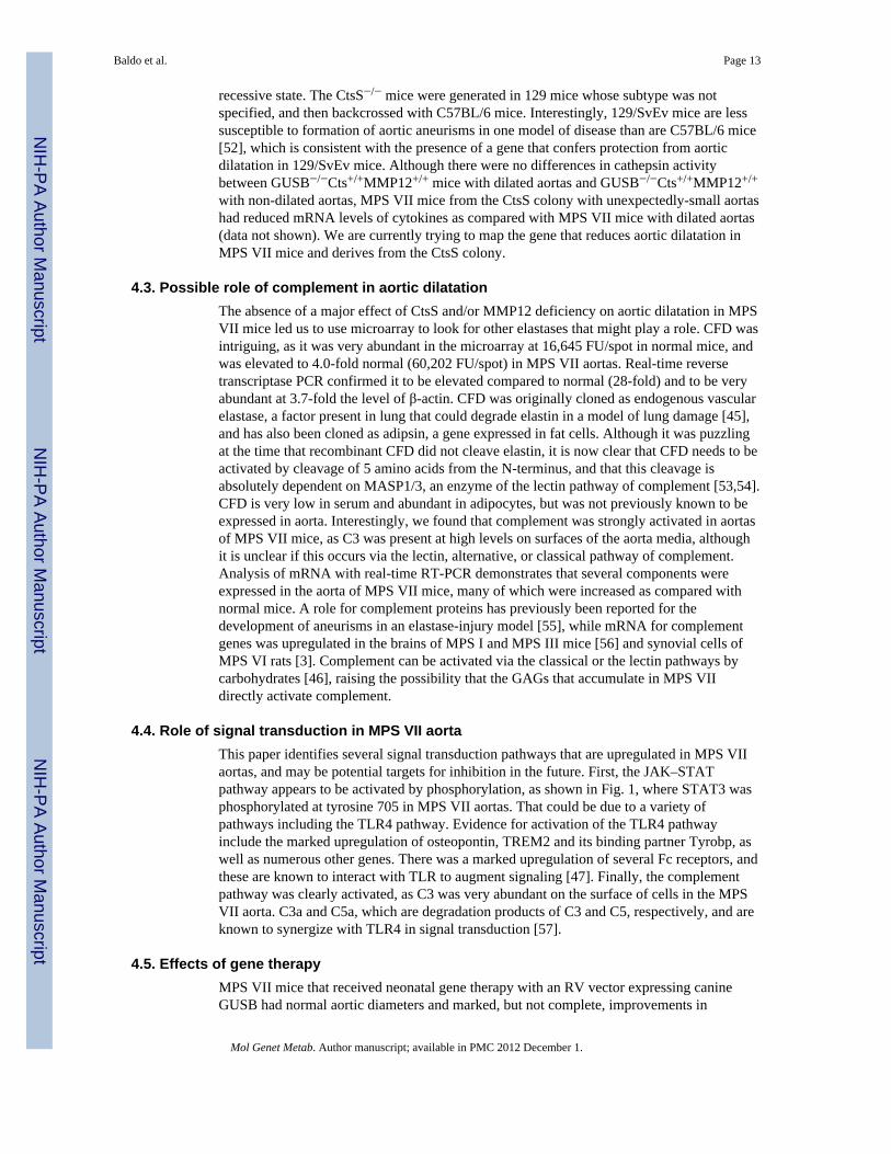
The ability of extracts from aortas of MPS VII mice with or without deficiency of CtsS tocleave cathepsin substrates is shown in Fig. 4. Extracts from GUSB−/− CtsS+/+ aortas hadhigh activity against Z-Phe-Arg-AMC at pH 7.5 (4838 ± 777 U/mg), which was 7-fold thevalue in normal mice of 678 ± 159 U/mg (p = 0.01), as shown in Fig. 4A. Surprisingly,extracts from GUSB−/− CtsS−/− mice also had very high activity against Z-Phe-Arg-AMC atpH 7.5 at 5377 ± 2354 U/mg, which was 111% of the value in purebred MPS VII mice.
To further investigate the identity of the cathepsin responsible for the activity in extractsfrom GUSB−/− CtsS−/− mice, specific inhibitors of cathepsins and other substrates wereused. Fig. 4B demonstrates that the CtsB inhibitor reduced overall activity of bothGUSB−/−Cts+/+ and GUSB−/−CtsS−/− extracts vs. Z-Phe-Arg-AMC by 95%, suggesting thatCtsB was the major cathepsin responsible for activity, while the CtsK inhibitor had a modesteffect, which could be due to some activity against CtsB (see Supplementary Fig. 1). Z-Arg-Arg-AMC is a substrate that is specific for CtsB, as shown in Supplementary Fig. 1. Asshown in Fig. 4C, aortic extracts from GUSB−/− Cts+/+ and GUSB−/− CtsS−/− had 2049 ±847 and 1580 ± 898 U/mg vs. Z-Arg-Arg-AMC, which were 8-fold and 6-fold, respectively,the value of 251 ± 54 in normal aortas. The lower activity of these extracts against Z-Arg-Arg-AMC as compared with Z-Phe-Arg-AMC is consistent with the fact that purified CtsBhas only 61% as much activity against Z-Arg-Arg-AMC as against Z-Phe-Arg-AMC.
The data shown above suggest that CtsB is the major enzyme responsible for cathepsinactivity in MPS VII mice. It is possible, however, that some other cathepsin is active atneutral pH, and that this is difficult to detect due to the high CtsB activity. CtsK is a veryimportant enzyme, as it maintains activity at pH 7.5 (Supplementary Fig. 1), it has elastin-degrading activity [42], and is activated by chondroitin sulfate [43], which is one of theGAGs that accumulates in MPS VII. A CtsK substrate was obtained that was not cleaved byCtsB or CtsS to an appreciable extent, although it was cleaved by CtsL 1% as efficiently asCtsK (data not shown). Activity with this CtsK substrate was elevated in GUSB−/−Cts+/+ orin GUSB−/− CtsS−/− mice to 16.5 ± 3.3 U/mg and 16.9 ± 5.6 U/mg, respectively, which was2.2-fold the value of 7.4 ± 4 U/mg in normal mice (Fig. 4D). Although activity wassignificantly elevated in the MPS VII mice (p = 0.019 vs. normal), the CtsK activity was<0.1% of the CtsB activity on a U/mg basis.
CtsD is an aspartyl protease that can activate other cathepsins such as CtsB [44], although ithas no activity against substrates that are cleaved by cysteine cathepsins. CtsD activity at pH4.0 (Fig. 4E) was 9.5- and 11.2-fold normal in GUSB−/−CtsS+/+ and GUSB−/− CtsS−/−
mice, respectively, suggesting a mechanism for activation of CtsB.
We conclude that CtsS does not play an important role in aortic dilatation in MPS VII, andthat the major cysteine cathepsin that is upregulated is CtsB, which is generally believed tohave low, albeit detectable, activity against elastin [50]. Although there was some increasein CtsK activity, which is a known elastase, this activity was very low. It was difficult toassess CtsH activity, as no specific assay was identified.
3.5. MMP12 activityMMP12 was a good candidate for an enzyme that could degrade elastin in MPS VII aortas,as it is a known elastase, its mRNA was 7-fold normal at 3 months in MPS VII mice, itsenzyme activity was elevated in MPS I mouse aortas [10] and in MPS I and MPS VII dogaortas [14], and its deficiency reduced aortic dilatation in one model [24]. Ascending aortaextracts from MPS VII mice were tested for MMP12 activity using a fluorogenic substratethat can be cleaved by MMP12. This demonstrated that MMP12 activity was not elevated inMPS VII mice at 6 months of age (Fig. 4F). This substrate can be cleaved by other MMPs,as demonstrated by the fact that extracts from MMP12-deficient mice still had activity in
Baldo et al. Page 8
Mol Genet Metab. Author manuscript; available in PMC 2012 December 1.
NIH
-PA Author Manuscript
NIH
-PA Author Manuscript
NIH
-PA Author Manuscript
this assay. The failure of MMP12 deficiency to prevent dilatation of MPS VII aortasdemonstrates that this enzyme does not play a major role in this model.
3.6. RNA analysis in MPS VII miceThe above studies demonstrate that deficiency of both CtsS and MMP12 cannot preventaortic dilatation in MPS VII mice. We therefore used microarray analysis of RNA from theaorta of normal mice (GUSB+/− CtsS+/+ MMP12+/+) and MPS VII mice with aorticdilatation (GUSB−/− CtsS+/+ MMP12+/+) to attempt to identify other genes whose proteinproducts might contribute to aortic dilatation. Samples were isolated at 6 months of age,when aortic disease was well-established. Table 1 summarizes the genes in variouscategories whose expression was upregulated (≥2.5-fold normal) or down-regulated (≤0.4-fold normal) in a statistically significant fashion (p value ≤0.01) after comparison of valuesin normal with those in MPS VII mice. Supplementary Table 2 shows values for all up- anddown-regulated genes, which are sorted into different categories. In addition, all microarraydata were uploaded to the GEO microarray data base (GSE30657), and values for geneswhose expression was not significantly altered can be found there. Some categories of genesare discussed below, while others simply appear in Table 1 or Supplementary Table 2.Finally, a list of all elastases that were interrogated is in Supplementary Table 3, while a listof all complement components that were interrogated is in Supplementary Table 4.
GeneGo software was used to determine if the changes in gene expression observed in MPSVII aortas resemble those that occur in other processes, networks, or diseases, using a levelof upregulation of 2.5-fold normal, and a p value ≤0.01. The most upregulated process wasthe immune system process, with 62 genes upregulated out of 1617 genes that were put intothis category by the software (not all of which are regulated at the transcriptional level), witha p value for significant upregulation of 2 × 10⁻²⁷. The most upregulated process networkwas inflammation of the complement system, with 12 genes upregulated out of 73 genes thatwere put into this pathway, for a p value of 2 × 10⁻¹¹. The most upregulated disease wasrheumatoid arthritis, with 58 genes upregulated out of 941 that were placed in this category,for a p value of 6 × 10⁻²⁵.
3.7. RNA for extracellular matrix proteinsExtracellular matrix protein mRNAs that were evaluated include elastin and collagen, genesthat are necessary for their assembly, and miscellaneous extracellular matrix genes. ElastinmRNA was not altered, while the elastin-associated protein fibulin 2 (Fbln2) mRNA wasmodestly elevated at 3.1-fold normal in the MPS VII aorta, as shown in Table 1 and inSupplementary Table 2. Some extracellular matrix proteins were reduced. For example,mRNA for procollagen C-endopeptidase enhancer 2 (Pcolce2), which enhances processingof types I and II procollagens, was 0.3-fold normal. Finally, mRNA for optican (Optc), aprotein that affects collagen fibrils in the eye, was markedly reduced at 0.15-fold normal.We conclude that elastin fragmentation was probably not due to downregulation of mRNAfor elastin or elastin-associated proteins, but there were a few abnormalities in genesassociated with collagen assembly.
3.8. RNA for elastasesA major hypothesis of this project was that a protease that can degrade elastin wasupregulated in the aorta of MPS VII mice. According to GeneGo interactions software andthe national library of medicine (http://www.ncbi.nlm.nih.gov/pubmed/) gene category,elastin can be cleaved by at least 28 different proteases. Supplementary Table 3 lists allknown elastases that were interrogated on the microarray regardless of their expressionlevel. mRNA was significantly higher in MPS VII than in normal mice for cathepsin L (6.2-fold), complement component D (CFD, 4.0-fold normal; 16,645 fluorescent units (FU/) spot
Baldo et al. Page 9
Mol Genet Metab. Author manuscript; available in PMC 2012 December 1.
NIH
-PA Author Manuscript
NIH
-PA Author Manuscript
NIH
-PA Author Manuscript
in normal mice; p = 0.006), MMP12 (3.6-fold normal) and cathepsin K (CtsK; 2.8-foldnormal). In addition, mRNAs for some other elastases were elevated on the microarray, butwere not ≥2.5-fold normal or did not achieve a p value ≤ 0.01 for comparison of the 2groups. These include MMP2, CtsS, CtsB, CtsH, and legumain (Lgmn), which is a cysteineprotease that is induced by LPS whose substrates are poorly characterized. mRNA forseveral other elastases were not significantly altered, as summarized in Supplementary Table3.
3.9. RNA for genes of the complement systemComplement components play important roles in the innate and the acquired immuneresponse, and activation of the complement pathway can result in upregulation of destructiveproteases. As noted above, the process network that most resembles the changes in geneexpression observed in MPS VII aortas was the complement system, with a p value of 2 ×10⁻¹¹. In addition, there is one report that CFD (also called adipsin or endogenous vascularelastase) can cleave elastin [45], and CFD were very abundant in the normal aorta andelevated to 4.0-fold normal in MPS VII mice (p = 0.006). Genes of the complement systemthat were upregulated on the microarray are shown on Table 1 and on page 2 ofSupplementary Table 2. In addition, values for all complement genes that were interrogatedon the microarray are shown in Supplementary Table 4.
Activation of the complement system may occur by 3 different pathways [46]. The classicalpathway involves binding of IgG or IgM to a complex containing the C1 complex(containing C1q, C1r, and C1s), which can then cleave C4 and then C2 to generate C3convertase, which in turn can activate C3 to C3a and C3b by proteolytic cleavage. C1 (6.1-fold normal for the most upregulated component), C4 (3.4-fold normal), and C2 (2.5-foldnormal) were all upregulated in MPS VII aortas. A second pathway of complementactivation is the alternative pathway, which can be initiated by spontaneous decay of C3 toC3a and C3b, or by generation of C3b from other pathways. This involves cleavage ofcomplement component B of a C3bB complex by CFD, after which the C3bBb complex cancleave additional C3, while properdin (CFP) stabilizes the C3bBb complex and protects itfrom regulation by complement inhibitors. CFD was elevated to 4-fold normal, while CFPwas 1.9-fold normal. A third pathway for complement activation involves the lectinpathway, in which mannose-binding lectin (MBL) or a ficolin binds to carbohydrates suchas mannose or N-acetyl glucosamine that results in activation of MBL-associated serineproteinase 1 (MASP1) or MASP2, which in turn activate complement. Ficolin A (FcnA) was2.7-fold normal. Cleavage of C3 can result from all three pathways of complement, andgenerates an anaphylatoxin (C3a) and C3b. C3b can initiate formation of the membraneattack complex as well as generate another anaphylatoxin, C5a. Although mRNA for noneof these downstream components or receptors for anaphylatoxins were upregulated in theMPS VII aorta, they were expressed, and thus poised to respond to upstream events. Of thecomplement inhibitors, complement factor H was reduced to 0.64-fold in MPS VII mice,while others were not significantly affected, and were generally expressed at fairly lowlevels except for CD59, which is an inhibitor of the late stages of complement.
3.10. Signal transduction moleculesAs one hypothesis for the mechanism of disease in MPS VII is that GAGs bind to the TLR4and induces inflammatory signals, genes of the TLR pathway or downstream signalingmolecules are shown in Supplementary Table 2. Although TLR4 was not elevated in MPSVII mice, it was moderately abundant, and thus could respond to GAGs. In addition, mRNAfor proteins CD14, which associates with the TLR4 was 4.0-fold normal. Finally, genes thatare upregulated by TLR signaling such as TREM2 (10.8-fold normal) and its associated
Baldo et al. Page 10
Mol Genet Metab. Author manuscript; available in PMC 2012 December 1.
NIH
-PA Author Manuscript
NIH
-PA Author Manuscript
NIH
-PA Author Manuscript
protein, Tyrobp (DAP12 = 4.1-fold normal) were elevated, as were several other proteinsthat are known to be induced by TLR or TNFα signaling.
Receptors for immunoglobulins can act synergistically with TLR receptors or withcomplement in signal transduction, and can activate many of the same downstreammolecules [47]. Many mRNAs for genes that encode Fc receptors were markedly elevated,as shown in Supplementary Table 2. Fc receptors were also elevated in MPS VI synovialcells [3].
Complement receptors, TLRs, and Fc receptors can all result in alteration of mRNA forcytokines, growth factors, intracellular signal transduction proteins, or proteins that bind tothese proteins. Genes for such proteins whose expression was altered in MPS VII mouseaorta are shown in Supplementary Table 2. Ccl21 is the most highly upregulated cytokine at35.5-fold normal, while the cytokine Cxcl4 (MIP-1β) was 10.3-fold normal, and is a genethat is upregulated in many models of MPS. The increase in angiotensin II receptor, type 1, areceptor for angiotensin II, to 2.9-fold normal, was of interest, as inhibitors of this receptorsuch as losartan can reduce elastin fragmentation in Marfan Syndrome mice [48]. Genes thatwere downregulated included latent transforming growth factor beta binding protein 4 (0.4-fold normal), bone morphogenetic protein 3 (BMP3; 0.3-fold normal), and BMP receptor,type IB (0.2-fold normal).
3.11. Immunohistochemsitry for C3Aortas were tested for deposition of C3 (Fig. 5), since several genes of the complementsystem were upregulated in MPS VII aortas on the microarray. MPS VII aortas had a strongpositive signal in the media, which was localized at the edge of GAG deposits and to a lesserextent along the edge of elastin fibers. Although normal mice had some C3 deposition in theintima and adventitia, there was little signal in the media. These data confirmed that thecomplement system was activated in the MPS VII aortas, and suggest that it occurs at sitesof GAG deposits.
3.12. Real-time PCR for complement genesReal-time reverse transcriptase analysis of expression of complement genes (Fig. 6)confirmed elevation of genes in MPS VII aortas that were found on the microarray. Forexample, CFD was elevated at 34.6 ± 27.3-fold normal and was very abundant at 4.5-foldthe level of β-actin, while properdin was 3.7 ± 2.4-fold normal. In addition, there wasupregulation of mRNA for genes related to the classical pathway such as C1qa (11.5 ± 2.4-fold normal), C2 (6.4 ± 2.7-fold normal), C4 (11.1 ± 8.2-fold normal), and the lectinpathway such as FcnA (9.5 ± 5.4-fold normal), MASP1 (0.01% β-actin; not detectable innormal) and MASP2 (0.3% β-actin; not detectable in normal). Furthermore, genes related todownstream events of complement pathways were also elevated in MPS VII aortas,including C3 (4.0 ± 2.5-fold normal) and C5 (0.005% β-actin signal; undetectable innormal). Finally, regulators of complement were either significantly reduced (CFH) ormoderately increased (CFI, CD55) in MPS VII as compared with normal mice.
4. DiscussionAortic dilatation in MPS is important, as it will likely result in aortic dissection and possiblydeath as patients live longer after treatment with HSCT or ERT. Identification of thepathogenesis of elastin fragmentation might lead to the identification of a drug that wouldprevent this from occurring in patients. We favor the hypothesis that degradation of elastin isthe most important mechanism responsible for elastin fragmentation, as MPS VII aortas hadminimal amounts of lysosomal storage material, relatively normal elastin, and only minimal
Baldo et al. Page 11
Mol Genet Metab. Author manuscript; available in PMC 2012 December 1.
NIH
-PA Author Manuscript
NIH
-PA Author Manuscript
NIH
-PA Author Manuscript

dilatation at 6 weeks of age, when elastin formation is believed to be largely completed [19].Elastin fragmentation then developed in conjunction with progressive accumulation oflysosomal storage material, suggesting that degradation was involved. It remains possiblethat elastin assembly contributes to abnormal elastin structure, as proposed by Hinek et al.for MPS I [23]. The present work focused on the role of elastin, as collagen fibers were notovertly abnormal in MPS VII aortas (data not shown) or in MPS I or MPS VII aortas [14].
4.1. CtsS and MMP12 deficiency do not prevent elastin fragmentation in MPS VII aortaA hypothesis of this project was that CtsS and/or MMP12 played pivotal roles in the elastinfragmentation that is likely responsible for the dilatation that occurs in MPS VII aortas. Thishypothesis was clearly wrong, as deficiency of CtsS, MMP12, or both could not preventaortic dilatation in MPS VII mice. In the case of CtsS deficiency, biochemical analysesdemonstrated that the elevation in cysteine proteinase activity that we had previouslyattributed to CtsS in MPS I mice [10] and in MPS I and MPS VII dogs [14] was still presentin GUSB−/− CtsS−/− mice, suggesting that another cathepsin was responsible. There are 11lysosomal cysteine cathepsins, all of which are primarily destined for the lysosome but canalso be secreted [49]. Further biochemical analyses demonstrated that this cathepsin activityin MPS VII aortas was primarily due to CtsB activity based upon efficient cleavage of aCtsB-specific substrate (Z-Arg-Arg-AMC) and inhibition with a CtsB-specific inhibitor at100 nM. However, CtsB is believed to have relatively low activity against elastin [50],although it remains possible that the very high CtsB activity observed could have sufficientelastin-degrading activity to result in damage over time. One interesting feature was the factthat CtsB activity was markedly elevated at 8-fold normal, while CtsB mRNA was only 1.5-fold normal. It is possible that an activator of CtsB was upregulated, and indeed, the enzymeactivity for the aspartyl protease CtsD that can activate CtsB by cleavage [44] was elevatedto 10-fold normal. It is also possible that CtsK activity contributed to elastin fragmentation,as CtsK RNA and enzyme activity were 2.8- and 2.2-fold normal, respectively, and CtsK isknown to be a potent elastase, although the activity appeared to be very low. Although CtsLmRNA was elevated at 6.2-fold normal, it is unlikely to be important, as the levels of RNAwere very low, and CtsL is inactive at neutral pH. Although it is possible that CtsHcontributes to elastin degradation, as its mRNA was 1.5-fold normal, we were unable to testits activity due to the absence of a specific assay. Finally, legumain is a poorly characterizedcysteine protease whose mRNA was elevated to 2.9-fold normal.
MMP12 was clearly not necessary for aortic dilatation, as deficiency of MMP12 did notprevent aortic dilatation in MPS VII mice. This was likely due to the fact that MMP12activity was only 1.5-fold normal in GUSB−/− mice at 6 months, and was not statisticallydifferent from values in normal mice, or from values in GUSB−/− MMP12−/− mice despitethe fact that MMP12 mRNA was 6.8 ± 4.9-fold normal at 3 months, and was 3.6-foldnormal at 6 months. This discrepancy may reflect the fact that MMP12 needs to activated byproteolytic cleavage. The matrix metalloproteinase (MMP) family has at least 20 members,of which MMP-2, -7, -8, -9, -10, -12, and -14 have elastase activity [51], and of whichMMP2 and MMP8 can cleave the peptide that was used in our MMP12 assay. Thus,although MMP2 mRNA was modestly elevated at 2-fold normal in the microarray, it isunlikely that MMP2 contributes to elastin fragmentation, as upregulation of MMP2 enzymeactivity should have been detected in this enzyme assay.
4.2. Possible role of another gene involved in aortic dilatation in MPS VIIAn interesting feature of this study was the fact that some GUSB−/− CtsS+/+ MMP12+/+
mice that derived from breeding through the CtsS colony did not have dilated aortas. Wehypothesize that this was due to an independently-segregating gene that initiated from theCtsS−/− colony that conferred protection from aortic dilatation when present in an autosomal
Baldo et al. Page 12
Mol Genet Metab. Author manuscript; available in PMC 2012 December 1.
NIH
-PA Author Manuscript
NIH
-PA Author Manuscript
NIH
-PA Author Manuscript
recessive state. The CtsS−/− mice were generated in 129 mice whose subtype was notspecified, and then backcrossed with C57BL/6 mice. Interestingly, 129/SvEv mice are lesssusceptible to formation of aortic aneurisms in one model of disease than are C57BL/6 mice[52], which is consistent with the presence of a gene that confers protection from aorticdilatation in 129/SvEv mice. Although there were no differences in cathepsin activitybetween GUSB−/−Cts+/+MMP12+/+ mice with dilated aortas and GUSB−/−Cts+/+MMP12+/+
with non-dilated aortas, MPS VII mice from the CtsS colony with unexpectedly-small aortashad reduced mRNA levels of cytokines as compared with MPS VII mice with dilated aortas(data not shown). We are currently trying to map the gene that reduces aortic dilatation inMPS VII mice and derives from the CtsS colony.
4.3. Possible role of complement in aortic dilatationThe absence of a major effect of CtsS and/or MMP12 deficiency on aortic dilatation in MPSVII mice led us to use microarray to look for other elastases that might play a role. CFD wasintriguing, as it was very abundant in the microarray at 16,645 FU/spot in normal mice, andwas elevated to 4.0-fold normal (60,202 FU/spot) in MPS VII aortas. Real-time reversetranscriptase PCR confirmed it to be elevated compared to normal (28-fold) and to be veryabundant at 3.7-fold the level of β-actin. CFD was originally cloned as endogenous vascularelastase, a factor present in lung that could degrade elastin in a model of lung damage [45],and has also been cloned as adipsin, a gene expressed in fat cells. Although it was puzzlingat the time that recombinant CFD did not cleave elastin, it is now clear that CFD needs to beactivated by cleavage of 5 amino acids from the N-terminus, and that this cleavage isabsolutely dependent on MASP1/3, an enzyme of the lectin pathway of complement [53,54].CFD is very low in serum and abundant in adipocytes, but was not previously known to beexpressed in aorta. Interestingly, we found that complement was strongly activated in aortasof MPS VII mice, as C3 was present at high levels on surfaces of the aorta media, althoughit is unclear if this occurs via the lectin, alternative, or classical pathway of complement.Analysis of mRNA with real-time RT-PCR demonstrates that several components wereexpressed in the aorta of MPS VII mice, many of which were increased as compared withnormal mice. A role for complement proteins has previously been reported for thedevelopment of aneurisms in an elastase-injury model [55], while mRNA for complementgenes was upregulated in the brains of MPS I and MPS III mice [56] and synovial cells ofMPS VI rats [3]. Complement can be activated via the classical or the lectin pathways bycarbohydrates [46], raising the possibility that the GAGs that accumulate in MPS VIIdirectly activate complement.
4.4. Role of signal transduction in MPS VII aortaThis paper identifies several signal transduction pathways that are upregulated in MPS VIIaortas, and may be potential targets for inhibition in the future. First, the JAK–STATpathway appears to be activated by phosphorylation, as shown in Fig. 1, where STAT3 wasphosphorylated at tyrosine 705 in MPS VII aortas. That could be due to a variety ofpathways including the TLR4 pathway. Evidence for activation of the TLR4 pathwayinclude the marked upregulation of osteopontin, TREM2 and its binding partner Tyrobp, aswell as numerous other genes. There was a marked upregulation of several Fc receptors, andthese are known to interact with TLR to augment signaling [47]. Finally, the complementpathway was clearly activated, as C3 was very abundant on the surface of cells in the MPSVII aorta. C3a and C5a, which are degradation products of C3 and C5, respectively, and areknown to synergize with TLR4 in signal transduction [57].
4.5. Effects of gene therapyMPS VII mice that received neonatal gene therapy with an RV vector expressing canineGUSB had normal aortic diameters and marked, but not complete, improvements in
Baldo et al. Page 13
Mol Genet Metab. Author manuscript; available in PMC 2012 December 1.
NIH
-PA Author Manuscript
NIH
-PA Author Manuscript
NIH
-PA Author Manuscript
biochemical abnormalities at 6 months. However, some aortic dilatation was observed at 10months, suggesting that gene therapy was not fully corrective, which likely reflects poordiffusion of GUSB in the interior of the relatively avascular aorta. We observed a similaroutcome in the dog MPS VII model [14], which developed aortic dilatation at 5 years afterneonatal gene therapy. These results highlight the importance of searching for themechanisms responsible for the pathogenesis of aortic disease, as some ancillary therapymay be needed to prevent this manifestation.
4.6. Implications and further directionsThese data demonstrate that CtsS and MMP12 are not essential for elastin fragmentation,and hence would not be good targets for drug inhibition in attempts to prevent aorticdilatation. A candidate for another elastase is CFD, as this is a known elastase, is veryabundant, and there is evidence of complement activation in the MPS VII aortas. CtsB andCtsK are also candidates, although CtsB has low elastase activity, while CtsK levels wererelatively low. It is also possible that there are other enzymes with elastase activity that areupregulated in the aorta. These studies also illustrate the activation of complement as well asother signal transduction pathways that are almost certainly critical for the upregulation and/or activation of destructive proteases, and may be targets for drug inhibition.
Supplementary MaterialRefer to Web version on PubMed Central for supplementary material.
Abbreviations
MPS mucopolysaccharidosis
Cts cathepsin
MMP matrix metalloproteinase
GUSB β-glucuronidase
GAGs glycosaminoglycans
TLR4 toll-like receptor 4
TNFα tumor necrosis factor alpha
IL-6 interleukin 6
M6P mannose 6-phosphate
ERT enzyme replacement therapy
RV retroviral vector
PBS phosphate buffered saline
VVG Verhoeff Van Gieson
AMC 7-amino-4-methylcoumarin
IDUA α-L-iduronidase
TBS tris-buffered saline
BP blood pressure
STAT3 signal transducer and activator of transcription 3
Baldo et al. Page 14
Mol Genet Metab. Author manuscript; available in PMC 2012 December 1.
NIH
-PA Author Manuscript
NIH
-PA Author Manuscript
NIH
-PA Author Manuscript
AcknowledgmentsThis work was supported by the MPS Society and the National Institutes of Health (HD061879 awarded to KPP,and R01 AI041592 and U19 AI070489 awarded to JPA). Histology was supported by P30 DK52574 from the NIH.GB received support from the Conselho Nacional de Desenvolvimento Cientifico-CNPg Brazil, grant number200584/2010-3. We thank Robert Thompson and John Curci (Washington University St. Louis) for the generousgift of CtsS and MMP12 deficient mice, Paul W. Bigg for helpful comments on the manuscript, and Doug Tollefsenfor help with enzyme assays.
References1. Neufeld, EF.; Muenzer, J. The mucopolysaccharidoses. In: Scriver, CR.; Beaudet, AL.; Sly, WS.;
Valle, D., editors. Metabolic and Molecular Basis of Inherited Disease. New York: McGraw Hill;2001. p. 3421-3452.
2. Ausseil J, Desmaris N, Bigou S, Attali R, Corbineau S, Vitry S, Parent M, Cheillan D, Fuller M,Maire I, Vanier MT, Heard JM. Early neurodegeneration progresses independently of microglialactivation by heparan sulfate in the brain of mucopolysaccharidosis IIIB mice. PLoS One. 2008;3:1–11.
3. Simonaro CM, D'Angelo M, He X, Eliyahu E, Shtraizent N, Haskins ME, Schuchman EH.Mechanism of glycosaminoglycan-mediated bone and joint disease: implications for themucopolysaccharidoses and other connective tissue diseases. Am. J. Pathol. 2008; 172:112–122.[PubMed: 18079441]
4. Simonaro CM, Ge Y, Eliyahu E, He X, Jepsen KJ, Schuchman EH. Involvement of the Toll-likereceptor 4 pathway and use of TNF-alpha antagonists for treatment of the mucopolysaccharidoses.Proc. Natl. Acad. Sci. U. S. A. 2010; 107:222–227. [PubMed: 20018674]
5. Soliman OI, Timmermans RG, Nemes A, Vletter WB, Wilson JH, ten Cate FJ, Geleijnse ML.Cardiac abnormalities in adults with the attenuated form of mucopolysaccharidosis type I. J. Inherit.Metab. Dis. 2007; 30:750–757. [PubMed: 17574537]
6. Nemes A, Timmermans RG, Wilson JH, Soliman OI, Krenning BJ, ten Cate FJ, Geleijnse ML. Themild form of mucopolysaccharidosis type I (Scheie syndrome) is associated with increasedascending aortic stiffness. Hear. Vessel. 2008; 23:108–111.
7. Beaudet AL, Ferrante NM, Ferry GD, Nichols BL, Mullins CE. Variation in the phenotypicexpression of beta-glucuronidase deficiency. J. Pediatr. 1975; 86:388–394. [PubMed: 803560]
8. Liu Y, Xu L, Hennig AK, Kovacs A, Fu A, Chung S, Lee D, Wang B, Herati RS, Mosinger OJ, CaiS-R, Ponder KP. Liver-directed neonatal gene therapy prevents cardiac, bone, ear, and eye diseasein mucopolysaccharidosis I mice. Mol. Ther. 2005; 11:35–47. [PubMed: 15585404]
9. Metcalf JA, Ma X, Linders B, Wu S, Schambach A, Ohlemiller KK, Kovacs A, Bigg M, He L,Tollefsen DM, Ponder KP. A self-inactivating gamma retroviral vector reduces manifestations ofmucopolysaccharidosis I in mice. Mol. Ther. 2010; 18:334–342. [PubMed: 19844196]
10. Ma X, Tittiger M, Knutsen RH, Kovacs A, Schaller L, Mecham RP, Ponder KP. Upregulation ofelastase proteins results in aortic dilatation in mucopolysaccharidosis I mice. Mol. Genet. Metab.2008; 94:298–304. [PubMed: 18479957]
11. Woloszynek JC, Kovacs A, Ohlemiller KK, Roberts M, Sands MS. Metabolic adaptations tointerrupted glycosaminoglycan recycling. J. Biol. Chem. 2009; 23:29684–29691. [PubMed:19700765]
12. Sleeper MM, Kusiak CM, Shofer FS, O'Donnell P, Bryan C, Ponder KP, Haskins ME. Clinicalcharacterization of cardiovascular abnormalities associated with feline mucopolysaccharidosis Iand VI. J. Inherit. Metab. Dis. 2008; 31:424–431. [PubMed: 18509743]
13. Traas AM, Wang P, Ma X, Tittiger M, Schaller L, O'donnell P, Sleeper MM, Vite C, Herati R,Aguirre GD, Haskins M, Ponder KP. Correction of clinical manifestations of caninemucopolysaccharidosis I with neonatal retroviral vector gene therapy. Mol. Ther. 2007; 15:1423–1431. [PubMed: 17519893]
14. Metcalf JA, Linders B, Wu S, Bigg P, O'Donnell P, Sleeper MM, Whyte MP, Haskins M, PonderKP. Upregulation of elastase activity in aorta in mucopolysaccharidosis I and VII dogs may be dueto increased cytokine expression. Mol. Genet. Metab. 2010; 99:396–407. [PubMed: 20044292]
Baldo et al. Page 15
Mol Genet Metab. Author manuscript; available in PMC 2012 December 1.
NIH
-PA Author Manuscript
NIH
-PA Author Manuscript
NIH
-PA Author Manuscript

15. Gompf RE, Shull RM, Breider MA, Scott JA, Constantopoulos GC. Cardiovascular changes afterbone marrow transplantation in dogs with mucopolysaccharidosis I. Am. J. Vet. Res. 1990;51:2054–2060. [PubMed: 2150744]
16. Sammarco C, Weil M, Just C, Weimelt S, Hasson C, O'Malley T, Evans SM, Wang P, Casal ML,Wolfe J, Haskins M. Effects of bone marrow transplantation on the cardiovascular abnormalitiesin canine mucopolysaccharidosis VII. Bone Marrow Transplant. 2000; 25:1289–1297. [PubMed:10871735]
17. Sleeper MM, Fornasari B, Ellinwood NM, Weil MA, Melniczek J, O'Malley TM, Sammarco CD,Xu L, Ponder KP, Haskins ME. Gene therapy ameliorates cardiovascular disease in dogs withmucopolysaccharidosis VII. Circulation. 2004; 110:815–820. [PubMed: 15289379]
18. Cattell MA, Anderson JC, Hasleton PS. Age-related changes in amounts and concentrations ofcollagen and elastin in normotensive human thoracic aorta. Clin. Chim. Acta. 1996; 245:73–84.[PubMed: 8646817]
19. Mithieux SM, Weiss AS. Elastin. Adv. Protein Chem. 2005; 70:437–461. [PubMed: 15837523]20. Jordan MD, Zheng Y, Ryazantsev S, Rozengurt N, Roos KP, Neufeld EF. Cardiac manifestations
in the mouse model of mucopolysaccharidosis I. Mol. Genet. Metab. 2005; 86:233–243. [PubMed:15979918]
21. Renteria VG, Ferrans VJ, Roberts WC. The heart in the hurler syndrome: gross, histologic andultrastructural observations in five necropsy cases. Am. J. Cardiol. 1976; 38:487–501. [PubMed:823811]
22. Braunlin E, Mackey-Bojack S, Panoskaltsis-Mortari A, Berry JM, McElmurry RT, Riddle M, SunLY, Clarke LA, Tolar J, Blazar BR. Cardiac functional and histopathologic findings in humansand mice with mucopolysaccharidosis type I. Pediatr. Res. 2006; 59:27–32. [PubMed: 16326988]
23. Hinek A, Wilson SE. Impaired elastogenesis in Hurler disease: dermatan sulfate accumulationlinked to deficiency in elastin-binding protein and elastic fiber assembly. Am. J. Pathol. 2000;156:925–938. [PubMed: 10702409]
24. Longo GM, Buda SJ, Fiotta N, Xiong W, Griener T, Shapiro S, Baxter BT. MMP-12 has a role inabdominal aortic aneurysms in mice. Surgery. 2005; 137:457–462. [PubMed: 15800495]
25. Sukhova Y-O, Zhang J-H, Pan GK, Wada Y, Yamamoto T, Naito M, Kodama T, Tsimikas S,Witztum JL, Lu ML, Sakara Y, Chin MT, Libby P, Shi GP. Deficiency of cathepsin S reducesatherosclerosis in LDL receptor-deficient mice. J. Clin. Invest. 2003; 111:897–906. [PubMed:12639996]
26. Braunlin EA, Stauffer NR, Peters CH, Bass JL, Berry JM, Hopwood JJ, Krivit W. Usefulness ofbone marrow transplantation in the Hurler syndrome. Am. J. Cardiol. 2003; 92:882–886.[PubMed: 14516901]
27. Birkenmeier EH, Barker JE, Vogler CA, Kyle JW, Sly WS, Gwynn B, Levy B, Pegors C.Increased life span and correction of metabolic defects in murine mucopolysaccharidosis type VIIafter syngeneic bone marrow transplantation. Blood. 1991; 78:3081–3092. [PubMed: 1954394]
28. Simonaro CM, Haskins ME, Kunieda T, Evans SM, Visser JW, Schuchman EH. Bone marrowtransplantation in newborn rats with mucopolysaccharidosis type VI: biochemical, pathological,and clinical findings. Transplantation. 1997; 63:1386–1393. [PubMed: 9175798]
29. Ellinwood NM, Colle MA, Weil MA, Casal ML, Vite CH, Wiemelt S, Hasson CW, O'Malley TM,He X, Prociuk U, Verot L, Melniczek JR, Lannon A, Aguirre GD, Knox VW, Evans SM, VanierMT, Schuchman EH, Walkley SU, Haskins ME. Bone marrow transplantation for felinemucopolysaccharidosis I. Mol. Genet. Metab. 2007; 91:239–250. [PubMed: 17482862]
30. Sifuentes M, Doroshow R, Hoft R, Mason G, Walot I, Diament M, Okazaki S, Huff K, Cox GF,Swiedler SJ, Kakkis ED. A follow-up study of MPS I patients treated with laronidase enzymereplacement therapy for 6 years. Mol. Genet. Metab. 2007; 90:171–180. [PubMed: 17011223]
31. Braunlin EA, Berry JM, Whitley CB. Cardiac findings after enzyme replacement therapy formucopolysaccharidosis type I. Am. J. Cardiol. 2006; 98:416–418. [PubMed: 16860035]
32. Auclair D, Hopwood JJ, Brooks DA, Lemontt JF, Crawley AC. Replacement therapy inmucopolysaccharidosis type VI: advantages of early onset of therapy. Mol. Genet. Metab. 2003;78:163–174. [PubMed: 12649061]
Baldo et al. Page 16
Mol Genet Metab. Author manuscript; available in PMC 2012 December 1.
NIH
-PA Author Manuscript
NIH
-PA Author Manuscript
NIH
-PA Author Manuscript
33. Ponder KP, Haskins ME. Gene therapy for mucopolysaccharidosis. Expert. Opin. Biol. Ther. 2007;7:1333–1345. [PubMed: 17727324]
34. Ponder KP, Melniczek JR, Xu L, Weil MA, O'Malley TM, O'Donnell PA, Knox VW, Aguirre GD,Mazrier H, Ellinwood NM, Sleeper M, Maguire AM, Volk SW, Mango RL, Zweigle J, Wolfe JH,Haskins ME. Therapeutic neonatal hepatic gene therapy in mucopolysaccharidosis VII dogs. Proc.Natl. Acad. Sci. U. S. A. 2002; 99:13102–13107. [PubMed: 12232044]
35. Xu L, Haskins ME, Melniczek JR, Gao C, Weil MA, O'Malley TM, O'Donnell PA, Mazrier H,Ellinwood NM, Zweigle J, Wolfe JH, Ponder KP. Transduction of hepatocytes after neonataldelivery of a Moloney murine leukemia virus based retroviral vector results in long-termexpression of beta-glucuronidase in mucopolysaccharidosis VII dogs. Mol. Ther. 2002; 5:141–153. [PubMed: 11829521]
36. Shi GP, Sukhova GK, Kuzuya M, Ye Q, Du J, Zhang Y, Pan ML, Lu JH, Cheng XW, Iguchi A,Perrey S, Lee AM, Chapman HA, Libby P. Deficiency of the cysteine protease cathepsin S impairsmicrovessel growth. Circ. Res. 2003; 92:493–500. [PubMed: 12600886]
37. Shipley JP, Wesselschmidt RL, Kobayashi DK, Ley TJ, Shapiro Sd. Metaloelastase is required formacrophage-mediated proteolysis and matrix invasion in mice. Proc. Natl. Acad. Sci. U. S. A.1996; 93:3942–3946. [PubMed: 8632994]
38. Xu L, Mango RL, Sands MS, Haskins ME, Ellinwood NM, Ponder KP. Evaluation of pathologicalmanifestations of disease in mucopolysaccharidosis VII mice after neonatal hepatic gene therapy.Mol. Ther. 2002; 6:745–758. [PubMed: 12498771]
39. Wagenseil JE, Nerurkar NL, Knutsen RH, Okamoto RJ, Li DY, Mecham RP. Effects of elastinhaploinsufficiency on the mechanical behavior of mouse arteries. Am. J. Physiol. Heart Circ.Physiol. 2005; 289:H1209–H1217. [PubMed: 15863465]
40. Jordans S, Jenko-Kokalj S, Kühl NM, Tedelind S, Sendt W, Brömme D, Turk D, Brix K.Monitoring compartment-specific substrate cleavage by cathepsins B, K, L, and S at physiologicalpH and redox conditions. BMC Biochem. 2009; 10:23. [PubMed: 19772638]
41. Vasiljeva O, Dolinar M, Pungercar JR, Turk V, Turk B. Recombinant human procathepsin S iscapable of autocatalytic processing at neutral pH in the presence of glycosaminoglycans. FEBSLett. 2005; 579:1285–1290. [PubMed: 15710427]
42. Novinec M, Grass RN, Stark WJ, Turk V, Baici A, Lenarcic B. Interaction between humancathepsins K, L, and S and elastins: mechanism of elastinolysis and inhibition by macromolecularinhibitors. J. Biol. Chem. 2007; 282:7893–7902. [PubMed: 17227755]
43. Li Z, Yasuda Y, Li W, Bogyo M, Katz N, Gordon RE, Fields GB, Brömme D. Regulation ofcollagenase activities of human cathepsins by glycosaminoglycans. J. Biol. Chem. 2004;279:5470–5479. [PubMed: 14645229]
44. van der Stappen JW, Williams AC, Maciewicz RA, Paraskeva C. Activation of cathepsin B,secreted by a colorectal cancer cell line requires low pH and is mediated by cathepsin D. Int. J.Cancer. 1996; 67:547–554. [PubMed: 8759615]
45. Zhu L, Wigle D, Hinek A, Kobayashi J, Ye C, Zuker M, Dodo H, Keeley FW, Rabinovitch M. Theendogenous vascular elastase that governs development and progression of monocrotaline-inducedpulmonary hypertension in rats is a novel enzyme related to the serine proteinase adipsin. J. Clin.Invest. 1994; 94:1163–1171. [PubMed: 8083356]
46. Speth, C.; Prodinger, WM.; Wurzner, R.; Stoiber, H.; Dierich, MP. Complement. In: Paul, WilliamE., editor. Fundamental Immunology (0-7817-6519-6, 978-0-7817-6519-0). Wolters Kluwer/Lippincott Williams & Wilkins; 2008. p. 1048-1107.
47. Ivashkiv LB. Cross-regulation of signaling by ITAM-associated receptors. Nat. Immunol. 2009;10:340–347. [PubMed: 19295630]
48. Habashi JP, Doyle JJ, Holm TM, Aziz H, Schoenhoff F, Bedja D, Chen Y, Modiri AN, Judge DP,Dietz HC. Angiotensin II type 2 receptor signaling attenuates aortic aneurysm in mice throughERK antagonism. Science. 2011; 332:361–365. [PubMed: 21493863]
49. Bromme, D.; Wilson, S. Role of cysteine cathepsins in extracellular proteolysis. In: Parks, WC.;Mecham, RP., editors. Extracellular Matrix Degradation (Biology of Extracellular Matrix). NewYork: Springer; 2011. p. 23-51.
Baldo et al. Page 17
Mol Genet Metab. Author manuscript; available in PMC 2012 December 1.
NIH
-PA Author Manuscript
NIH
-PA Author Manuscript
NIH
-PA Author Manuscript
50. Mason RW, Johnson DA, Barrett AJ, Chapman HA. Elastinolytic activity of human cathepsin L.Biochem. J. 1986; 233:925–927. [PubMed: 3518704]
51. Fingleton B. MMPs as therapeutic targets—still a viable option? Semin. Cell Dev. Biol. 2008;19:61–68. [PubMed: 17693104]
52. Thompson RW, Curci JA, Ennis D, Mao MB, Pagano TL, Pham CT. Pathophysiology ofabdominal aortic aneurysms: insights from the elastase-induced model in mice with differentgenetic backgrounds. Ann. N. Y. Acad. Sci. 2006; 1085:59–73. [PubMed: 17182923]
53. Banda NK, Takahashi M, Levitt B, Glogowska M, Nicholas J, Takahashi K, Stahl GL, Fujita T,Arend WP, Holers VM. Essential role of complement mannose-binding lectin-associated serineproteases-1/3 in the murine collagen antibody-induced model of inflammatory arthritis. J.Immunol. 2010; 185:5598–5606. [PubMed: 20870940]
54. Takahashi M, Ishida Y, Iwaki D, Kanno K, Suzuki T, Endo Y, Homma Y, Fujita T. Essential roleof mannose-binding lectin-associated serine protease-1 in activation of the complement factor D. J.Exp. Med. 2010; 207:29–37. [PubMed: 20038603]
55. Pagano MB, Zhou HF, Ennis TL, Wu X, Lambris JD, Atkinson JP, Thompson RW, Hourcade DE,Pham CT. Complement-dependent neutrophil recruitment is critical for the development ofelastase-induced abdominal aortic aneurysm. Circulation. 2009; 119:1805–1813. [PubMed:19307471]
56. Ohmi K, Greenberg DS, Rajavel KS, Ryazantsev S, Li HH, Neufeld EF. Activated microglia incortex of mouse models of mucopolysaccharidoses I and IIIB. Proc. Natl. Acad. Sci. U. S. A.2003; 100:1902–1907. [PubMed: 12576554]
57. Hajishengallis G, Lambris JD. Crosstalk pathways between Toll-like receptors and the complementsystem. Trends Immunol. 2010; 31:154–163. [PubMed: 20153254]
Baldo et al. Page 18
Mol Genet Metab. Author manuscript; available in PMC 2012 December 1.
NIH
-PA Author Manuscript
NIH
-PA Author Manuscript
NIH
-PA Author Manuscript

Fig. 1.Evaluation of MPS VII aortas. A and B. Compliance curves. Aortas were isolated fromnormal (GUSB+/−, +/+ for other genes) or MPS VII (GUSB−/−, +/+ for other genes) malesat 1.5 or 3 months of age, and the average outer diameter of the aorta at the indicated mmHg ± SD was determined. The Student's t-test was used to compare values in the 2 groups ateach pressure, and ** indicates a p valueb0.01. C–E. Elastin stain. Sections of fixedascending aortas were stained with VVG, which stains elastin a dark color, at the indicatedage. The arrows indicate the edges of fragmented elastin fibers in the MPS VII mouse, sizebars indicate 20 µm. For all panels, the intima is located on the right side, and the adventitiais on the left side. F and G. Immunostain for tyrosine-phosphorylated STAT3. Frozensections were stained for the presence of STAT3 that was phosphorylated at tyrosine 705.The arrow identifies the dark nucleus of a positive cell in the MPS VII mouse. Bars indicate10 µm. H. mRNA levels at 3 months. RNA was isolated from aortas at 3 months of age, andreal-time PCR was used to compare levels of genes in normal (N = 6) and MPS VII (N = 5)mice after normalization to β-actin levels. Abbreviations are TIMP for tissue inhibitor ofmetalloprotease, ELA2 for neutrophil elastase, and uPA for urokinase plasminogenactivator.
Baldo et al. Page 19
Mol Genet Metab. Author manuscript; available in PMC 2012 December 1.
NIH
-PA Author Manuscript
NIH
-PA Author Manuscript
NIH
-PA Author Manuscript
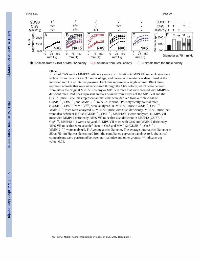
Fig. 2.Effect of CtsS and/or MMP12 deficiency on aortic dilatation in MPS VII mice. Aortas wereisolated from male mice at 3 months of age, and the outer diameter was determined at theindicated mm Hg of internal pressure. Each line represents a single animal. Black linesrepresent animals that were never crossed through the CtsS colony, which were derivedfrom either the original MPS VII colony or MPS VII mice that were crossed with MMP12-deficient mice. Red lines represent animals derived from a cross of the MPS VII and theCtsS−/− mice. Blue lines represent animals that were derived from a triple cross ofGUSB−/−, CtsS−/−, and MMP12−/− mice. A. Normal. Phenotypically normal mice(GUSB+/− CtsS+/+ MMP12+/+) were analyzed. B. MPS VII mice. GUSB−/− CtsS+/+
MMP12+/+ mice were analyzed C. MPS VII mice with CtsS deficiency. MPS VII mice thatwere also deficient in CtsS (GUSB−/−, CtsS−/−, MMP12+/+) were analyzed. D. MPS VIImice with MMP12 deficiency. MPS VII mice that also deficient in MMP12 (GUSB−/−,CtsS+/+, MMP12−/−) were analyzed. E. MPS VII mice with CtsS and MMP12 deficiency.MPS VII mice that were also deficient in CtsS and MMP12 (GUSB−/−, CtsS−/−,MMP12−/−) were analyzed. F. Average aortic diameter. The average outer aortic diameter ±SD at 75 mm Hg was determined from the compliance curves in panels A to E. Statisticalcomparisons were performed between normal mice and other groups; ** indicates a pvalue<0.01.
Baldo et al. Page 20
Mol Genet Metab. Author manuscript; available in PMC 2012 December 1.
NIH
-PA Author Manuscript
NIH
-PA Author Manuscript
NIH
-PA Author Manuscript

Fig. 3.Effects of gene therapy on aortic abnormalities. Some MPS VII mice were injected IV with1 × 1010 transducing units/kg of the RV designated hAAT-cGUSB-WPRE at 2–3 days afterbirth, while other mice were untreated. A. Serum GUSB activity. GUSB activity wasdetermined at 2 to 6 months of age for normal, untreated MPS VII, and RV-treated MPS VIImice for the indicated number of animals. B. Aortic width. Aortas were dissected and thewidth was measured at 6 months of age for normal and MPS VII mice, and at 6 months or10 months as indicated for RV-treated MPS VII mice. C and D. Aorta GUSB and IDUAactivity. Extracts prepared at pH 5.5 were tested for GUSB and IDUA activity. E. GAGlevels. Extracts prepared at pH 7.5 were tested for GAG levels. Values were compared usingANOVA with Tukey post-hoc analysis, and * represents p ≤ 0.05 and ** represents p ≤ 0.01as indicated above the bars for comparison with values in untreated MPS VII mice.
Baldo et al. Page 21
Mol Genet Metab. Author manuscript; available in PMC 2012 December 1.
NIH
-PA Author Manuscript
NIH
-PA Author Manuscript
NIH
-PA Author Manuscript
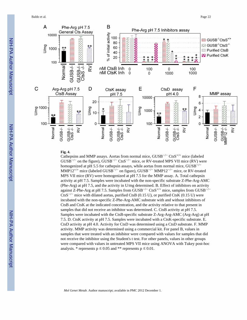
Fig. 4.Cathepsins and MMP assays. Aortas from normal mice, GUSB−/− CtsS+/+ mice (labeledGUSB−/− on the figure), GUSB−/− CtsS−/− mice, or RV-treated MPS VII mice (RV) werehomogenized at pH 5.5 for cathepsin assays, while aortas from normal mice, GUSB−/−
MMP12+/+ mice (labeled GUSB−/− on figure), GUSB−/− MMP12−/− mice, or RV-treatedMPS VII mice (RV) were homogenized at pH 7.5 for the MMP assay. A. Total cathepsinactivity at pH 7.5. Samples were incubated with the non-specific substrate Z-Phe-Arg-AMC(Phe-Arg) at pH 7.5, and the activity in U/mg determined. B. Effect of inhibitors on activityagainst Z-Phe-Arg at pH 7.5. Samples from GUSB−/− CtsS+/+ mice, samples from GUSB−/−
CtsS−/− mice with dilated aortas, purified CtsB (0.15 U), or purified CtsK (0.15 U) wereincubated with the non-specific Z-Phe-Arg-AMC substrate with and without inhibitors ofCtsB and CtsK at the indicated concentration, and the activity relative to that present insamples that did not receive an inhibitor was determined. C. CtsB activity at pH 7.5.Samples were incubated with the CtsB-specific substrate Z-Arg-Arg-AMC (Arg-Arg) at pH7.5. D. CtsK activity at pH 7.5. Samples were incubated with a CtsK-specific substrate. E.CtsD activity at pH 4.0. Activity for CtsD was determined using a CtsD substrate. F. MMPactivity. MMP activity was determined using a commercial kit. For panel B, values insamples that were treated with an inhibitor were compared with values for samples that didnot receive the inhibitor using the Student's t test. For other panels, values in other groupswere compared with values in untreated MPS VII mice using ANOVA with Tukey post-hocanalysis. * represents p ≤ 0.05 and ** represents p ≤ 0.01.
Baldo et al. Page 22
Mol Genet Metab. Author manuscript; available in PMC 2012 December 1.
NIH
-PA Author Manuscript
NIH
-PA Author Manuscript
NIH
-PA Author Manuscript
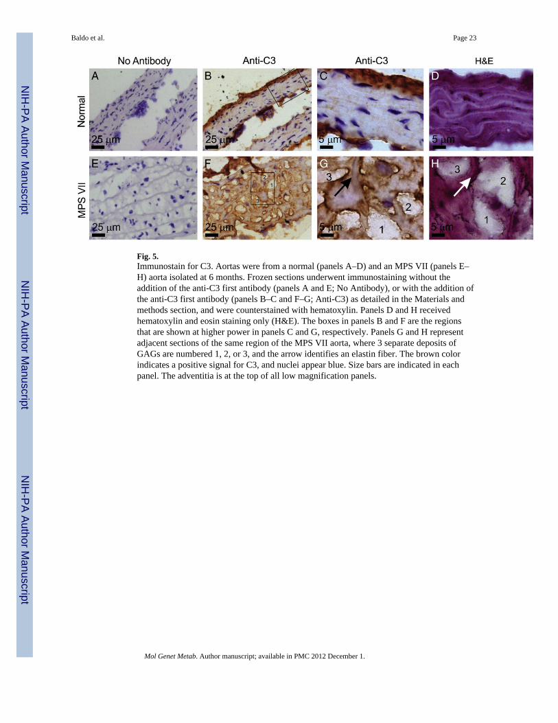
Fig. 5.Immunostain for C3. Aortas were from a normal (panels A–D) and an MPS VII (panels E–H) aorta isolated at 6 months. Frozen sections underwent immunostaining without theaddition of the anti-C3 first antibody (panels A and E; No Antibody), or with the addition ofthe anti-C3 first antibody (panels B–C and F–G; Anti-C3) as detailed in the Materials andmethods section, and were counterstained with hematoxylin. Panels D and H receivedhematoxylin and eosin staining only (H&E). The boxes in panels B and F are the regionsthat are shown at higher power in panels C and G, respectively. Panels G and H representadjacent sections of the same region of the MPS VII aorta, where 3 separate deposits ofGAGs are numbered 1, 2, or 3, and the arrow identifies an elastin fiber. The brown colorindicates a positive signal for C3, and nuclei appear blue. Size bars are indicated in eachpanel. The adventitia is at the top of all low magnification panels.
Baldo et al. Page 23
Mol Genet Metab. Author manuscript; available in PMC 2012 December 1.
NIH
-PA Author Manuscript
NIH
-PA Author Manuscript
NIH
-PA Author Manuscript

Fig. 6.mRNA for complement genes. RNA was extracted from normal (N = 4) and MPS VII (N =6) mice at 6 months of age and used for reverse transcriptase real-time PCR to assesscomplement gene mRNA levels. Results are shown as the percentage of β-actin. Genes withexpression lower than 0.002% of β-actin were considered not present and are shown as0.002% in the figure. *p<0.05 or **p<0.01 for comparison of values in MPS VII with thosein normal mice using Student's t test. Abbreviations include Fcna for ficolin A, Fcnb forficolin B, MBL for mannose-binding lectin, MASP for mannan-binding lectin associatedserine protease, CD55 for decay accelerating factor, and CD59 for protectin.
Baldo et al. Page 24
Mol Genet Metab. Author manuscript; available in PMC 2012 December 1.
NIH
-PA Author Manuscript
NIH
-PA Author Manuscript
NIH
-PA Author Manuscript
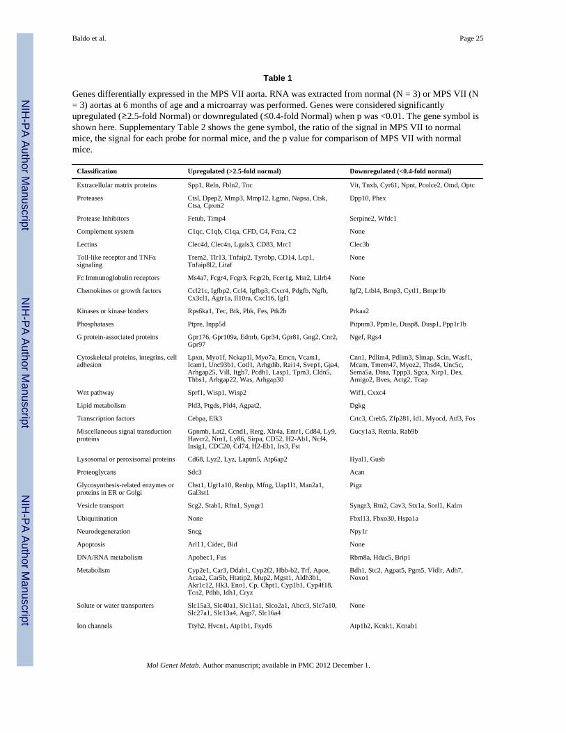
NIH
-PA Author Manuscript
NIH
-PA Author Manuscript
NIH
-PA Author Manuscript
Baldo et al. Page 25
Table 1
Genes differentially expressed in the MPS VII aorta. RNA was extracted from normal (N = 3) or MPS VII (N= 3) aortas at 6 months of age and a microarray was performed. Genes were considered significantlyupregulated (≥2.5-fold Normal) or downregulated (≤0.4-fold Normal) when p was <0.01. The gene symbol isshown here. Supplementary Table 2 shows the gene symbol, the ratio of the signal in MPS VII to normalmice, the signal for each probe for normal mice, and the p value for comparison of MPS VII with normalmice.
Classification Upregulated (>2.5-fold normal) Downregulated (<0.4-fold normal)
Extracellular matrix proteins Spp1, Reln, Fbln2, Tnc Vit, Tnxb, Cyr61, Npnt, Pcolce2, Omd, Optc
Proteases Ctsl, Dpep2, Mmp3, Mmp12, Lgmn, Napsa, Ctsk,Ctsa, Cpxm2
Dpp10, Phex
Protease Inhibitors Fetub, Timp4 Serpine2, Wfdc1
Complement system C1qc, C1qb, C1qa, CFD, C4, Fcna, C2 None
Lectins Clec4d, Clec4n, Lgals3, CD83, Mrc1 Clec3b
Toll-like receptor and TNFαsignaling
Trem2, Tlr13, Tnfaip2, Tyrobp, CD14, Lcp1,Tnfaip8l2, Litaf
None
Fc Immunoglobulin receptors Ms4a7, Fcgr4, Fcgr3, Fcgr2b, Fcer1g, Msr2, Lilrb4 None
Chemokines or growth factors Ccl21c, Igfbp2, Ccl4, Igfbp3, Cxcr4, Pdgfb, Ngfb,Cx3cl1, Agtr1a, Il10ra, Cxcl16, Igf1
Igf2, Ltbl4, Bmp3, Cytl1, Bmpr1b
Kinases or kinase binders Rps6ka1, Tec, Btk, Pbk, Fes, Ptk2b Prkaa2
Phosphatases Ptpre, Inpp5d Pitpnm3, Ppm1e, Dusp8, Dusp1, Ppp1r1b
G protein-associated proteins Gpr176, Gpr109a, Ednrb, Gpr34, Gpr81, Gng2, Cnr2,Gpr97
Ngef, Rgs4
Cytoskeletal proteins, integrins, celladhesion
Lpxn, Myo1f, Nckap1l, Myo7a, Emcn, Vcam1,Icam1, Unc93b1, Cotl1, Arhgdib, Rai14, Svep1, Gja4,Arhgap25, Vill, Itgb7, Pcdh1, Lasp1, Tpm3, Cldn5,Thbs1, Arhgap22, Was, Arhgap30
Cnn1, Pdlim4, Pdlim3, Slmap, Scin, Wasf1,Mcam, Tmem47, Myoz2, Thsd4, Unc5c,Sema5a, Dtna, Tppp3, Sgca, Xirp1, Des,Amigo2, Bves, Actg2, Tcap
Wnt pathway Sprf1, Wisp1, Wisp2 Wif1, Cxxc4
Lipid metabolism Pld3, Ptgds, Pld4, Agpat2, Dgkg
Transcription factors Cebpa, Elk3 Crtc3, Creb5, Zfp281, Id1, Myocd, Atf3, Fos
Miscellaneous signal transductionproteins
Gpnmb, Lat2, Ccnd1, Rerg, Xlr4a, Emr1, Cd84, Ly9,Havcr2, Nrn1, Ly86, Sirpa, CD52, H2-Ab1, Ncf4,Insig1, CDC20, Cd74, H2-Eb1, Irs3, Fst
Gucy1a3, Retnla, Rab9b
Lysosomal or peroxisomal proteins Cd68, Lyz2, Lyz, Laptm5, Atp6ap2 Hyal1, Gusb
Proteoglycans Sdc3 Acan
Glycosynthesis-related enzymes orproteins in ER or Golgi
Chst1, Ugt1a10, Renbp, Mfng, Uap1l1, Man2a1,Gal3st1
Pigz
Vesicle transport Scg2, Stab1, Rftn1, Syngr1 Syngr3, Rtn2, Cav3, Stx1a, Sorl1, Kalrn
Ubiquitination None Fbxl13, Fbxo30, Hspa1a
Neurodegeneration Sncg Npy1r
Apoptosis Arl11, Cidec, Bid None
DNA/RNA metabolism Apobec1, Fus Rbm8a, Hdac5, Brip1
Metabolism Cyp2e1, Car3, Ddah1, Cyp2f2, Hbb-b2, Trf, Apoe,Acaa2, Car5b, Htatip2, Mup2, Mgst1, Aldh3b1,Akr1c12, Hk3, Eno1, Cp, Chpt1, Cyp1b1, Cyp4f18,Tcn2, Pdhb, Idh1, Cryz
Bdh1, Stc2, Agpat5, Pgm5, Vldlr, Adh7,Noxo1
Solute or water transporters Slc15a3, Slc40a1, Slc11a1, Slco2a1, Abcc3, Slc7a10,Slc27a1, Slc13a4, Aqp7, Slc16a4
None
Ion channels Ttyh2, Hvcn1, Atp1b1, Fxyd6 Atp1b2, Kcnk1, Kcnab1
Mol Genet Metab. Author manuscript; available in PMC 2012 December 1.

NIH
-PA Author Manuscript
NIH
-PA Author Manuscript
NIH
-PA Author Manuscript
Baldo et al. Page 26
Classification Upregulated (>2.5-fold normal) Downregulated (<0.4-fold normal)
Miscellaneous function None Hspa2, Olfml2b, Hspa1l
Unclear function MsA46d, Evi2a, Tmem176b, Art4, Klhl6, Mlana,Lrrc33,
Tspan11, Stbd1, Crispld2, Fam171b, Vstm2b,Fam107a, Scube3
Mol Genet Metab. Author manuscript; available in PMC 2012 December 1.
Related Documents


