ARTICLE Pathogen-derived extracellular vesicles mediate virulence in the fatal human pathogen Cryptococcus gattii Ewa Bielska 1 , Marta Arch Sisquella 2 , Maha Aldeieg 3 , Charlotte Birch 1 , Eloise J. O’Donoghue 1 & Robin C. May 1 The Pacific Northwest outbreak of cryptococcosis, caused by a near-clonal lineage of the fungal pathogen Cryptococcus gattii, represents the most significant cluster of life-threatening fungal infections in otherwise healthy human hosts currently known. The outbreak lineage has a remarkable ability to grow rapidly within human white blood cells, using a unique ‘division of labour’ mechanism within the pathogen population, where some cells adopt a dormant behaviour to support the growth of neighbouring cells. Here we demonstrate that pathogenic ‘division of labour’ can be triggered over large cellular distances and is mediated through the release of extracellular vesicles by the fungus. Isolated vesicles released by virulent strains are taken up by infected host macrophages and trafficked to the phagosome, where they trigger the rapid intracellular growth of non-outbreak fungal cells that would otherwise be eliminated by the host. Thus, long distance pathogen-to-pathogen communication via extracellular vesicles represents a novel mechanism to control complex virulence phenotypes in Cryptococcus gattii and, potentially, other infectious species. DOI: 10.1038/s41467-018-03991-6 OPEN 1 Institute of Microbiology and Infection, School of Biosciences, University of Birmingham, Edgbaston, Birmingham B15 2TT, UK. 2 Institut d’ Investigació en Ciències de la Salut Germans Trias i Pujol (IGTP), Crta de Can Ruti s/n, Badalona 08916 Catalonia, Spain. 3 School of Biological Sciences, University of Reading, Knight Building, Whiteknights Campus, Reading RG6 6AJ, UK. Correspondence and requests for materials should be addressed to E.B. (email: [email protected]) or to R.C.M. (email: [email protected]) NATURE COMMUNICATIONS | (2018)9:1556 | DOI: 10.1038/s41467-018-03991-6 | www.nature.com/naturecommunications 1 1234567890():,;

Welcome message from author
This document is posted to help you gain knowledge. Please leave a comment to let me know what you think about it! Share it to your friends and learn new things together.
Transcript
ARTICLE
Pathogen-derived extracellular vesicles mediatevirulence in the fatal human pathogen CryptococcusgattiiEwa Bielska 1, Marta Arch Sisquella2, Maha Aldeieg3, Charlotte Birch1, Eloise J. O’Donoghue1 &
Robin C. May 1
The Pacific Northwest outbreak of cryptococcosis, caused by a near-clonal lineage of the
fungal pathogen Cryptococcus gattii, represents the most significant cluster of life-threatening
fungal infections in otherwise healthy human hosts currently known. The outbreak lineage
has a remarkable ability to grow rapidly within human white blood cells, using a unique
‘division of labour’ mechanism within the pathogen population, where some cells adopt a
dormant behaviour to support the growth of neighbouring cells. Here we demonstrate that
pathogenic ‘division of labour’ can be triggered over large cellular distances and is mediated
through the release of extracellular vesicles by the fungus. Isolated vesicles released by
virulent strains are taken up by infected host macrophages and trafficked to the phagosome,
where they trigger the rapid intracellular growth of non-outbreak fungal cells that
would otherwise be eliminated by the host. Thus, long distance pathogen-to-pathogen
communication via extracellular vesicles represents a novel mechanism to control complex
virulence phenotypes in Cryptococcus gattii and, potentially, other infectious species.
DOI: 10.1038/s41467-018-03991-6 OPEN
1 Institute of Microbiology and Infection, School of Biosciences, University of Birmingham, Edgbaston, Birmingham B15 2TT, UK. 2 Institut d’ Investigació enCiències de la Salut Germans Trias i Pujol (IGTP), Crta de Can Ruti s/n, Badalona 08916 Catalonia, Spain. 3 School of Biological Sciences, University ofReading, Knight Building, Whiteknights Campus, Reading RG6 6AJ, UK. Correspondence and requests for materials should be addressed toE.B. (email: [email protected]) or to R.C.M. (email: [email protected])
NATURE COMMUNICATIONS | (2018) 9:1556 | DOI: 10.1038/s41467-018-03991-6 | www.nature.com/naturecommunications 1
1234
5678
90():,;
Cryptococcosis is a major human and animal life-threatening fungal disease1–3. Globally, most humaninfections are caused by Cryptococcus neoformans, with
the related species Cryptococcus gattii causing less than 1% of allhuman cryptococcal disease. However, in the late 1990s a near-clonal lineage of C. gattii became established in British Columbia,Canada, and subsequently caused a major cluster of human andanimal disease that has come to be known as the PacificNorthwest Outbreak4. A defining feature of cells within theoutbreak lineage is their ability to proliferate very rapidly withinhost phagocytes5. We previously demonstrated that this rapidproliferation is driven by a ‘division of labour’ mechanism, inwhich individual fungal cells coordinate their behaviour tomaximise proliferation of the population as a whole6. However,the mechanism by which this coordination occurs at a cellularlevel has remained enigmatic.
Here we demonstrate that the key regulator of this ‘division oflabour’ process is the release and exchange of extracellular vesicles(EVs) by outbreak strains of C. gattii. These EVs are efficientlytaken up by infected host macrophages and trafficked to thefungal phagosome, where they induce rapid proliferation ofthe recipient pathogen cell and thereby drive pathogenesis in thishighly virulent lineage.
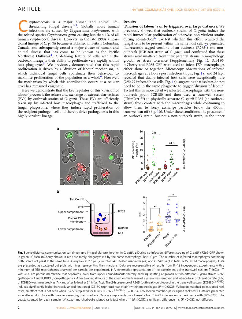
Results‘Division of labour’ can be triggered over large distances. Wepreviously showed that outbreak strains of C. gattii induce therapid intracellular proliferation of otherwise non-virulent strainsduring co-infection6. To test whether this effect required thefungal cells to be present within the same host cell, we generatedfluorescently tagged versions of an outbreak (R2657) and non-outbreak (ICB180) strain of C. gattii and confirmed that thesestrains were unaltered from their parental strains in morphology,growth or stress tolerance (Supplementary Fig. 1). ICB180-mCherry and R265-GFP were used to infect J774 macrophageseither alone or together. Microscopy observations of infectedmacrophages at 2 hours post infection (h.p.i.; Fig. 1a) and 24 h.p.irevealed that dually infected host cells were exceptionally rare(2/5479 infected host cells; Fig. 1a), suggesting that isolates do notneed to be in the same phagocyte to trigger ‘division of labour’.To test this in more detail we infected macrophages with the non-outbreak strain ICB180 and then used a transwell system(ThinCertTM) to physically separate C. gattii R265 (an outbreakstrain) from contact with the macrophages while continuing toallow them to freely exchange particles below the 400 nmtranswell cut off (Fig. 1b). Under these conditions, the presence ofan outbreak strain, but not a non-outbreak strain, in the upper
30ICB180R265
2 h
24 h
n =
12/
12/8
/12/
12/8
exp
ICB18
0-m
Ch
R265-
GFP
co-in
fecti
on
co-in
fecti
on
ICB18
0-m
Ch
R265-
GFP
25
20
15
MO
con
tain
ing
yeas
ts (
%)
10
5
0
IPR
6
10n
= 2
2/21
/22/
12 e
xp
ns
**5
4
3
2
1
0
ICB18
0
ICB18
0(+
R265)
R265
R265(+
ICB18
0)
ThinCertTM
Well
R265
ICB180
MO
Upper compartment
Porous PETmembrane
Lower compartment
a
b
Fig. 1 Long-distance communication can drive rapid intracellular proliferation in C. gattii. a During co-infection, different strains of C. gattii (R265-GFP shownin green; ICB180-mCherry shown in red) are rarely phagocytosed by the same macrophage. Bar: 10 μm. The number of infected macrophages containingboth isolates of yeast at the same time is very low at 2 h.p.i. (2 in total 5479 tested macrophages) and at 24 h.p.i (1 in total 3235 tested macrophages). Dataare presented as scattered dot plots with lines representing their medians. Data are representative of results from 8−12 independent experiments with aminimum of 150 macrophages analysed per sample per experiment. b A schematic representation of the experiment using transwell system ThinCertTM
with 400 nm porous membrane that separates lower from upper compartments thereby allowing splitting of growth of two different C. gattii strains R265(pathogenic) and ICB180 (non-pathogenic). After two initial hours of the infection the transwell system was removed and intracellular proliferation rate (IPR)of ICB180 was measured (as T0) and after following 24 h (as T24). The 2-h presence of R265 (outbreak) cryptococci in the transwell system (ICB180(+R265))induces significantly higher intracellular proliferation of ICB180 (non-outbreak strain) within macrophages (P= 0.0038, Wilcoxon matched-pairs signed ranktest), an effect that is not seen when R265 is replaced for ICB180 (R265(+ICB180); P= 0.9263, Wilcoxon matched-pairs signed rank test). Data are presentedas scattered dot plots with lines representing their medians. Data are representative of results from 12–22 independent experiments with 879–5238 totalyeasts counted for each sample. Wilcoxon matched-pairs signed rank test where ** (P≤ 0.01), significant difference; ns (P > 0.05), not different
ARTICLE NATURE COMMUNICATIONS | DOI: 10.1038/s41467-018-03991-6
2 NATURE COMMUNICATIONS | (2018) 9:1556 | DOI: 10.1038/s41467-018-03991-6 | www.nature.com/naturecommunications
compartment of the transwell was sufficient to raise the intra-cellular proliferation rate (IPR) of ICB180 within macrophages by2.5 fold (Fig. 1b). Thus released molecules from outbreak C. gattiiare sufficient to recapitulate the ‘division of labour’ phenotypeover large distances.
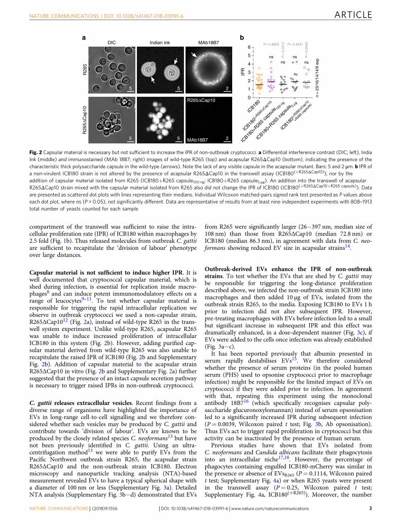
Capsular material is not sufficient to induce higher IPR. It iswell documented that cryptococcal capsular material, which isshed during infection, is essential for replication inside macro-phages8 and can induce potent immunomodulatory effects on arange of leucocytes9–11. To test whether capsular material isresponsible for triggering the rapid intracellular replication weobserve in outbreak cryptococci we used a non-capsular strain,R265ΔCap1012 (Fig. 2a), instead of wild-type R265 in the trans-well system experiment. Unlike wild-type R265, acapsular R265was unable to induce increased proliferation of intracellularICB180 in this system (Fig. 2b). However, adding purified cap-sular material derived from wild-type R265 was also unable torecapitulate the raised IPR of ICB180 (Fig. 2b and SupplementaryFig. 2b). Addition of capsular material to the acapsular strainR265ΔCap10 in vitro (Fig. 2b and Supplementary Fig. 2a) furthersuggested that the presence of an intact capsule secretion pathwayis necessary to trigger raised IPRs in non-outbreak cryptococci.
C. gattii releases extracellular vesicles. Recent findings from adiverse range of organisms have highlighted the importance ofEVs in long-range cell-to-cell signalling and we therefore con-sidered whether such vesicles may be produced by C. gattii andcontribute towards ‘division of labour’. EVs are known to beproduced by the closely related species C. neoformans13 but havenot been previously identified in C. gattii. Using an ultra-centrifugation method13 we were able to purify EVs from thePacific Northwest outbreak strain R265, the acapsular strainR265ΔCap10 and the non-outbreak strain ICB180. Electronmicroscopy and nanoparticle tracking analysis (NTA)-basedmeasurement revealed EVs to have a typical spherical shape witha diameter of 100 nm or less (Supplementary Fig. 3a). DetailedNTA analysis (Supplementary Fig. 3b−d) demonstrated that EVs
from R265 were significantly larger (26−397 nm, median size of108 nm) than those from R265ΔCap10 (median 72.8 nm) orICB180 (median 86.3 nm), in agreement with data from C. neo-formans showing reduced EV size in acapsular strains14.
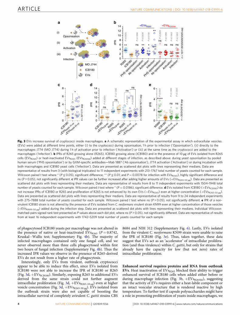
Outbreak-derived EVs enhance the IPR of non-outbreakstrains. To test whether the EVs that are shed by C. gattii maybe responsible for triggering the long-distance proliferationdescribed above, we infected the non-outbreak strain ICB180 intomacrophages and then added 10 μg of EVs, isolated from theoutbreak strain R265, to the media. Exposing ICB180 to EVs 1 hprior to infection did not alter subsequent IPR. However,pre-treating macrophages with EVs before infection led to a smallbut significant increase in subsequent IPR and this effect wasdramatically enhanced, in a dose-dependent manner (Fig. 3c), ifEVs were added to the cells once infection was already established(Fig. 3a−c).
It has been reported previously that albumin presented inserum rapidly destabilises EVs15. We therefore consideredwhether the presence of serum proteins (in the pooled humanserum (PHS) used to opsonise cryptococci prior to macrophageinfection) might be responsible for the limited impact of EVs oncryptococci if they were added prior to infection. In agreementwith that, repeating this experiment using the monoclonalantibody 18B716 (which specifically recognises capsular poly-saccharide glucuronoxylomannan) instead of serum opsonisationled to a significantly increased IPR during subsequent infection(P= 0.0039, Wilcoxon paired t test; Fig. 3b, Ab opsonisation).Thus EVs act to trigger rapid proliferation in cryptococci but thisactivity can be inactivated by the presence of human serum.
Previous studies have shown that EVs isolated fromC. neoformans and Candida albicans facilitate their phagocytosisinto an intracellular niche17,18. However, the percentage ofphagocytes containing engulfed ICB180-mCherry was similar inthe presence or absence of EVsR265 (P= 0.1114, Wilcoxon pairedt test; Supplementary Fig. 4a) or when R265 yeasts were presentin the transwell assay (P= 0.25, Wilcoxon paired t test;Supplementary Fig. 4a, ICB180(+R265)). Moreover, the number
DIC
R26
55
5
5
5 2
2
R265ΔCap10
MAb18B7
R26
5ΔC
ap10
Indian ink MAb18B76
nsns
nsns
n =
23/
16/1
4/14
/9 e
xp
P= 0.8603 P= 0.4631
P=
0.2
412
P=
0.2
500
5
4
3IPR
2
1
0
ICB18
0
ICB18
0+R26
5 ca
psule
100
ng
ICB18
0+R26
5 ca
psule
5 μg
ICB18
0(+
R265ΔCap
10
+R26
5 ca
psule
)
ICB18
0(+
R265ΔCap
10)
a b
Fig. 2 Capsular material is necessary but not sufficient to increase the IPR of non-outbreak cryptococci. a Differential interference contrast (DIC; left), IndiaInk (middle) and immunostained (MAb 18B7; right) images of wild-type R265 (top) and acapsular R265ΔCap10 (bottom), indicating the presence of thecharacteristic thick polysaccharide capsule in the wild-type (arrows). Note the lack of any visible capsule in the acapsular mutant. Bars: 5 and 2 μm. b IPR ofa non-virulent ICB180 strain is not altered by the presence of acapsular R265ΔCap10 in the transwell assay (ICB180(+R265ΔCap10)), nor by theaddition of capsular material isolated from R265 (ICB180+R265 capsule100 ng; ICB180+R265 capsule5 μg). An addition into the transwell of acapsularR265ΔCap10 strain mixed with the capsular material isolated from R265 also did not change the IPR of ICB180 (ICB180(+R265ΔCap10+R265 capsule)). Dataare presented as scattered dot plots with lines representing their medians. Individual Wilcoxon matched-pairs signed rank test presented as P values aboveeach dot plot, where ns (P > 0.05), not significantly different. Data are representative of results from at least nine independent experiments with 808–1913total number of yeasts counted for each sample
NATURE COMMUNICATIONS | DOI: 10.1038/s41467-018-03991-6 ARTICLE
NATURE COMMUNICATIONS | (2018) 9:1556 | DOI: 10.1038/s41467-018-03991-6 | www.nature.com/naturecommunications 3
of phagocytosed ICB180 yeasts per macrophage was not altered inthe presence of native or heat-inactivated EVsR265 (P= 0.8742,Kruskal−Wallis test; Supplementary Fig. 4b). The majority ofinfected macrophages contained only one fungal cell, and wenever observed more than three cells phagocytosed within firsttwo hours of fungal infection (Supplementary Fig. 4b). Thus theincreased IPR values we observe in the presence of R265-derivedEVs do not result from a higher rate of phagocytosis.
Interestingly, only EVs from virulent, outbreak cryptococciappear to be able to induce this effect, since EVs isolated fromICB180 were not able to increase the IPR of ICB180 or R265(Fig. 3d, +EVsICB180). Similarly, exposing R265 to additional EVsderived from the same strain could not further augmentintracellular proliferation (Fig. 3d, +EVsR265-10 μg) even at highervesicle concentration (Fig. 3d, +EVsR265-50 μg). EVs isolated fromthe outbreak strain were also not capable of boosting theintracellular survival of completely avirulent C. gattii strains CBS
8684 and NIH 312 (Supplementary Fig. 6). Lastly, EVs isolatedfrom the virulent C. neoformans KN99 strain were unable to raisethe IPR of ICB180 (Fig. 3e). Thus, taken together, these datasuggest that EVs act as an ‘accelerator’ of intracellular prolifera-tion (and thus virulence) within C. gattii, but only for strains thatalready have the capacity for low (but not zero) rates ofintracellular proliferation.
Enhanced survival requires proteins and RNA from outbreakEVs. Heat inactivation of EVsR265 blocked their ability to triggerenhanced survival of ICB180 cells when added either before orduring macrophage infection (Fig. 3b, +EVsR265hk), suggestingthat the activity of EVs requires either a heat-labile component oran intact vesicular structure that is rendered inactive by hightemperature. To further test if capsule polysaccharides might havea role in promoting proliferation of yeasts inside macrophages, we
Opsonisation
Activation
IPR
IPR
IPR
IPR
8 14
12
10
8
ns
ns ****
*ns
ns ns
ns
P=
0.82
03 P=
0.12
89
P=
0.40
37
P>
0.99
99 P<
0.00
01
P=
0.14
90
P=
0.33
03
6
4
2
0
6
4
2
0
6
4
2
0n =
9/9
/9/9
/24/
16/1
1 ex
p
n =
20/
24/2
4/16
exp
n =
8/1
1/11
exp
ICB18
0
ICB18
0
R265
KN99
ICB18
0
+EVs KN99
-10
μg
+EVs KN99
-50
μg
R265
+EVs R26
5-10
μg
+EVs IC
B180
+EVs IC
B180
+EVs R26
5-10
μg
+EVs R26
5-50
μg
+EVs R26
5-50
μg
14 +EVsR265 +EVsR265hk
12
10
8 P=
0.00
20
P=
0.10
94
P=
0.00
39
P=
0.01
56
P=
0.06
25
P=
0.12
89
P=
0.19
53
P=
0.19
53
6 ** nsns
ns ns ns
n =
10/
11/8
/3/9
/9/8
/3/9
/9 e
xp
**
**
*4
2
0
R265
ICB18
0
PHS ops
onisa
tion
PHS ops
onisa
tion
Ab op
sonis
ation
Ab op
sonis
ation
Activa
tion
Activa
tion
Infe
ction
Infe
ction
Infection
MO J744
ICB180
EVs
(i)
(ii)
(iii)
a b
c d e
Fig. 3 EVs increase survival of cryptococci inside macrophages. a A schematic representation of the experimental assay in which extracellular vesicles(EVs) were added at different time points, either (i) to the cryptococci during opsonisation, 1 h prior to infection (‘Opsonisation’), (ii) directly to themacrophages J774 (MO J774) during 1 h of activation prior to infection (‘Activation’) or (iii) at the same time as the cryptococci are added to themacrophages (‘Infection’). b IPRs of R265 growing alone (R265), ICB180 growing alone (ICB180) and in the presence of 10 μg of EVs isolated from R265cells (EVsR265) or heat-inactivated EVsR265 (EVsR265hk) added at different stages of infection, as described above: during yeast opsonisation by pooledhuman serum (‘PHS opsonisation’) or by GXM-specific antibodies—Mab 18B7 (‘Ab opsonisation’), J774 activation (‘Activation’) or during incubation withboth macrophages and ICB180 yeast cells (‘Infection’). Data are presented as scattered dot plots with lines representing their medians. Data arerepresentative of results from 3 (with biological triplicates) to 11 independent experiments with 213–1767 total number of yeasts counted for each sample.Wilcoxon paired t test where * (P≤ 0.05), significant difference; ** (P≤ 0.01; and P= 0.0078 for infection with EVsR265), highly significant difference andns (P > 0.05), not significantly different. c IPR values can be further increased after adding higher amounts of EVs (+EVsR265-50 μg). Data are presented asscattered dot plots with lines representing their medians. Data are representative of results from 8 to 11 independent experiments with 1504–1948 totalnumber of yeasts counted for each sample. Wilcoxon paired t test where * (P= 0.0186), significant difference. d EVs isolated from ICB180 (+EVsICB180) donot increase IPRs of ICB180 or R265 and proliferation of R265 is not enhanced by its own EVs (+EVsR265) even at higher concentration (+EVsR265-50 μg).Data are presented as scattered dot plots with lines representing their medians. Data are representative of results from 9 to 24 independent experimentswith 275–7888 total number of yeasts counted for each sample. Wilcoxon paired t test where ns (P > 0.05), not significantly different. e IPR of a non-virulent ICB180 strain is not altered by the presence of EVs isolated from C. neoformans virulent strain KN99 even at higher concentration of those vesicles(+EVsKN99-50 μg) added during the infection step. Data are presented as scattered dot plots with lines representing their medians. Individual Wilcoxonmatched-pairs signed rank test presented as P values above each dot plot, where ns (P > 0.05), not significantly different. Data are representative of resultsfrom at least 16 independent experiments with 1742–5209 total number of yeasts counted for each sample
ARTICLE NATURE COMMUNICATIONS | DOI: 10.1038/s41467-018-03991-6
4 NATURE COMMUNICATIONS | (2018) 9:1556 | DOI: 10.1038/s41467-018-03991-6 | www.nature.com/naturecommunications
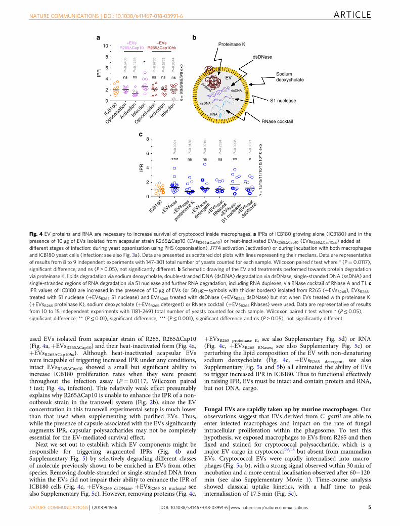
used EVs isolated from acapsular strain of R265, R265ΔCap10(Fig. 4a, +EVsR265ΔCap10) and their heat-inactivated form (Fig. 4a,+EVsR265ΔCap10hk). Although heat-inactivated acapsular EVswere incapable of triggering increased IPR under any conditions,intact EVsR265ΔCap10 showed a small but significant ability toincrease ICB180 proliferation rates when they were presentthroughout the infection assay (P= 0.0117, Wilcoxon pairedt test; Fig. 4a, infection). This relatively weak effect presumablyexplains why R265ΔCap10 is unable to enhance the IPR of a non-outbreak strain in the transwell system (Fig. 2b), since the EVconcentration in this transwell experimental setup is much lowerthan that used when supplementing with purified EVs. Thus,while the presence of capsule associated with the EVs significantlyaugments IPR, capsular polysaccharides may not be completelyessential for the EV-mediated survival effect.
Next we set out to establish which EV components might beresponsible for triggering augmented IPRs (Fig. 4b andSupplementary Fig. 5) by selectively degrading different classesof molecule previously shown to be enriched in EVs from otherspecies. Removing double-stranded or single-stranded DNA fromwithin the EVs did not impair their ability to enhance the IPR ofICB180 cells (Fig. 4c, +EVsR265 dsDNase, +EVsR265 S1 nuclease; seealso Supplementary Fig. 5c). However, removing proteins (Fig. 4c,
+EVsR265 proteinase K; see also Supplementary Fig. 5d) or RNA(Fig. 4c, +EVsR265 RNases; see also Supplementary Fig. 5c) orperturbing the lipid composition of the EV with non-denaturingsodium deoxycholate (Fig. 4c, +EVsR265 detergent; see alsoSupplementary Fig. 5a and 5b) all eliminated the ability of EVsto trigger increased IPR in ICB180. Thus to functional effectivelyin raising IPR, EVs must be intact and contain protein and RNA,but not DNA, cargo.
Fungal EVs are rapidly taken up by murine macrophages. Ourobservations suggest that EVs derived from C. gattii are able toenter infected macrophages and impact on the rate of fungalintracellular proliferation within the phagosome. To test thishypothesis, we exposed macrophages to EVs from R265 and thenfixed and stained for cryptococcal polysaccharide, which is amajor EV cargo in cryptococci19,15 but absent from mammalianEVs. Cryptococcal EVs were rapidly internalised into macro-phages (Fig. 5a, b), with a strong signal observed within 30 min ofincubation and a more central localisation observed after 60−120min (see also Supplementary Movie 1). Time-course analysisshowed classical uptake kinetics, with a half time to peakinternalisation of 17.5 min (Fig. 5c).
10+EVs
R265ΔCap10+EVs
R265ΔCap10hk
8
6
IPR
4 ns
P=
0.64
06
P=
0.12
89
P=
0.97
66
P=
0.57
03
P=
0.98
44
ns ns ns ns
n =
9/9
/9/9
/8/9
/9 e
xp
n =
15/
15/1
1/10
/10/
10/1
0 ex
p
*
2
0
8
P=
0.00
01
P=
0.91
50
P=
0.92
19
P=
0.23
24
P=
0.00
98
P=
0.03
71
6ns ns ns*** ** *
IPR 4
2
0
ICB18
0
+EVs R26
5
+EVs R26
5
prot
einas
e K
+EVs R26
5
dete
rgen
t
+EVs R26
5
RNases
+EVs R26
5
S1 nu
cleas
e
+EVs R26
5
dsDNas
e
ICB18
0
Opson
isatio
n
Opson
isatio
n
Activa
tion
Activa
tion
Infe
ction
Infe
ction
Proteinase K
EV
lipids
dsDNA
ssDNA
RNA
Proteins
dsDNase
Sodiumdeoxycholate
S1 nuclease
RNase cocktail
a b
c
Fig. 4 EV proteins and RNA are necessary to increase survival of cryptococci inside macrophages. a IPRs of ICB180 growing alone (ICB180) and in thepresence of 10 μg of EVs isolated from acapsular strain R265ΔCap10 (EVsR265ΔCap10) or heat-inactivated EVsR265ΔCap10 (EVsR265ΔCap10hk) added atdifferent stages of infection: during yeast opsonisation using PHS (opsonisation), J774 activation (activation) or during incubation with both macrophagesand ICB180 yeast cells (infection; see also Fig. 3a). Data are presented as scattered dot plots with lines representing their medians. Data are representativeof results from 8 to 9 independent experiments with 147–301 total number of yeasts counted for each sample. Wilcoxon paired t test where * (P= 0.0117),significant difference; and ns (P > 0.05), not significantly different. b Schematic drawing of the EV and treatments performed towards protein degradationvia proteinase K, lipids degradation via sodium deoxycholate, double-stranded DNA (dsDNA) degradation via dsDNase, single-stranded DNA (ssDNA) andsingle-stranded regions of RNA degradation via S1 nuclease and further RNA degradation, including RNA duplexes, via RNase cocktail of RNase A and T1. cIPR values of ICB180 are increased in the presence of 10 μg of EVs (or 50 μg—symbols with thicker borders) isolated from R265 (+EVsR265), EVsR265treated with S1 nuclease (+EVsR265 S1 nuclease) and EVsR265 treated with dsDNase (+EVsR265 dsDNase) but not when EVs treated with proteinase K(+EVsR265 proteinase K), sodium deoxycholate (+EVsR265 detergent) or RNase cocktail (+EVsR265 RNases) were used. Data are representative of resultsfrom 10 to 15 independent experiments with 1181–2691 total number of yeasts counted for each sample. Wilcoxon paired t test where * (P≤ 0.05),significant difference; ** (P≤ 0.01), significant difference, *** (P≤ 0.001), significant difference and ns (P > 0.05), not significantly different
NATURE COMMUNICATIONS | DOI: 10.1038/s41467-018-03991-6 ARTICLE
NATURE COMMUNICATIONS | (2018) 9:1556 | DOI: 10.1038/s41467-018-03991-6 | www.nature.com/naturecommunications 5
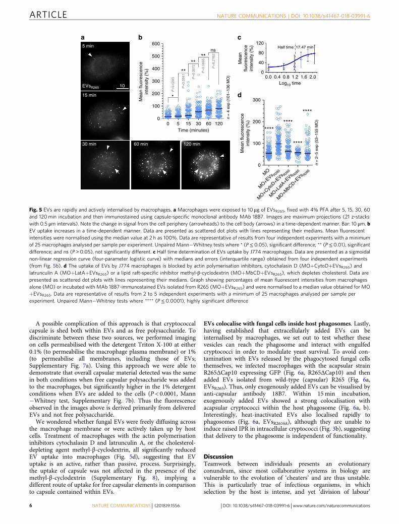
A possible complication of this approach is that cryptococcalcapsule is shed both within EVs and as free polysaccharide. Todiscriminate between these two sources, we performed imagingon cells permeabilised with the detergent Triton X-100 at either0.1% (to permeabilise the macrophage plasma membrane) or 1%(to permeabilise all membranes, including those of EVs;Supplementary Fig. 7a). Using this approach we were able todemonstrate that overall capsular material detected was the samein both conditions when free capsular polysaccharide was addedto the macrophages, but significantly higher in the 1% detergentconditions when EVs are added to the cells (P < 0.0001, Mann−Whitney test, Supplementary Fig. 7b). Thus the fluorescenceobserved in the images above is derived primarily from deliveredEVs and not free polysaccharide.
We wondered whether fungal EVs were freely diffusing acrossthe macrophage membrane or were actively taken up by hostcells. Treatment of macrophages with the actin polymerisationinhibitors cytochalasin D and latrunculin A, or the cholesterol-depleting agent methyl-β-cyclodextrin, all significantly reducedEV uptake into macrophages (Fig. 5d), suggesting that EVuptake is an active, rather than passive, process. Surprisingly,the uptake of capsule was not affected in the presence of themethyl-β-cyclodextrin (Supplementary Fig. 8), implying adifferent route of uptake for free capsular elements in comparisonto capsule contained within EVs.
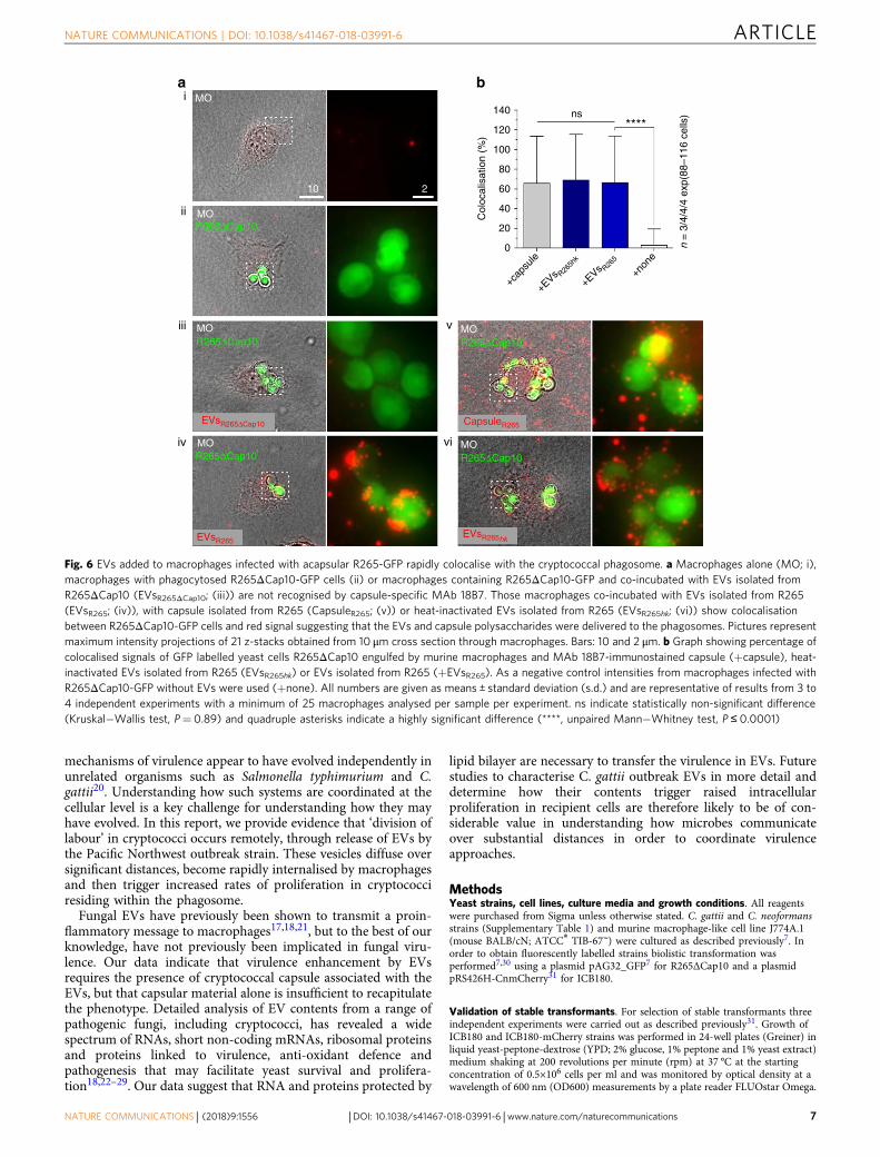
EVs colocalise with fungal cells inside host phagosomes. Lastly,having established that extracellularly added EVs can beinternalised by macrophages, we set out to test whether thesevesicles can reach the phagosome and interact with engulfedcryptococci in order to modulate yeast survival. To avoid con-tamination with EVs released by the phagocytosed fungal cellsthemselves, we infected macrophages with the acapsular strainR265ΔCap10 expressing GFP (Fig. 6a, R265ΔCap10) and thenadded EVs isolated from wild-type (capsular) R265 (Fig. 6a,EVsR265). Thus, only exogenously added EVs can be visualised byanti-capsular antibody 18B7. Within 15 min incubation,exogenously added EVs showed a strong colocalisation withacapsular cryptococci within the host phagosome (Fig. 6a, b).Interestingly, heat-inactivated EVs also localised rapidly tophagosomes (Fig. 6a, EVsR265hk), although they are unable toinduce raised IPR in intracellular cryptococci (Fig. 3b), suggestingthat delivery to the phagosome is independent of functionality.
DiscussionTeamwork between individuals presents an evolutionaryconundrum, since most collaborative systems in biology arevulnerable to the evolution of ‘cheaters’ and are thus unstable.This is particularly true of infectious organisms, in whichselection by the host is intense, and yet ‘division of labour’
5 min 600 120Half time: 17.47 min
80
40
300
200
100
0
MO
MO+E
Vs R265
MO+C
ytoD+E
Vs R265
MO+L
atA+E
Vs R265
MO+M
bCD+E
Vs R265
00.0 0.4 0.8
Log10 time1.2 1.6
****
********
****
2.0
Mea
nflu
ores
cenc
ein
tens
ity (
%)
Mea
n flu
ores
cenc
ein
tens
ity (
%)
500
400 **
*
****
ns
Mea
n flu
ores
cenc
ein
tens
ity (
%)
300
P=
0.02
83
P=
0.00
41
P=
0.00
72
P=
0.00
55
P=
0.27
62
200
100
00 5 15 30 60 120
n =
4 e
xp (
101–
136
MO
)
n =
2–5
exp
(53
–155
MO
)
Time (minutes)
10EVsR265
15 min
30 min 60 min 120 min
a b c
d
Fig. 5 EVs are rapidly and actively internalised by macrophages. a Macrophages were exposed to 10 μg of EVsR265, fixed with 4% PFA after 5, 15, 30, 60and 120min incubation and then immunostained using capsule-specific monoclonal antibody MAb 18B7. Images are maximum projections (21 z-stackswith 0.5 μm intervals). Note the change in signal from the cell periphery (arrowheads) to the cell body (arrows) in a time-dependent manner. Bar: 10 μm. bEV uptake increases in a time-dependent manner. Data are presented as scattered dot plots with lines representing their medians. Mean fluorescentintensities were normalised using the median value at 2 h as 100%. Data are representative of results from four independent experiments with a minimumof 25 macrophages analysed per sample per experiment. Unpaired Mann−Whitney tests where * (P≤ 0.05), significant difference; ** (P≤ 0.01), significantdifference; and ns (P > 0.05), not significantly different. c Half time determination of EVs uptake by J774 macrophages. Data are presented as a sigmoidalnon-linear regression curve (four-parameter logistic curve) with medians and errors (interquartile range) obtained from four independent experiments(from Fig. 5b). d The uptake of EVs by J774 macrophages is blocked by actin polymerisation inhibitors, cytochalasin D (MO+CytoD+EVsR265) andlatrunculin A (MO+LatA+EVsR265) or a lipid raft-specific inhibitor methyl-β-cyclodextrin (MO+MbCD+EVsR265), which depletes cholesterol. Data arepresented as scattered dot plots with lines representing their medians. Graph showing percentages of mean fluorescent intensities from macrophagesalone (MO) or incubated with MAb 18B7-immunostained EVs isolated from R265 (MO+EVsR265) and were normalised to a median value obtained for MO+EVsR265. Data are representative of results from 2 to 5 independent experiments with a minimum of 25 macrophages analysed per sample perexperiment. Unpaired Mann−Whitney tests where **** (P≤ 0.0001), highly significant difference
ARTICLE NATURE COMMUNICATIONS | DOI: 10.1038/s41467-018-03991-6
6 NATURE COMMUNICATIONS | (2018) 9:1556 | DOI: 10.1038/s41467-018-03991-6 | www.nature.com/naturecommunications
mechanisms of virulence appear to have evolved independently inunrelated organisms such as Salmonella typhimurium and C.gattii20. Understanding how such systems are coordinated at thecellular level is a key challenge for understanding how they mayhave evolved. In this report, we provide evidence that ‘division oflabour’ in cryptococci occurs remotely, through release of EVs bythe Pacific Northwest outbreak strain. These vesicles diffuse oversignificant distances, become rapidly internalised by macrophagesand then trigger increased rates of proliferation in cryptococciresiding within the phagosome.
Fungal EVs have previously been shown to transmit a proin-flammatory message to macrophages17,18,21, but to the best of ourknowledge, have not previously been implicated in fungal viru-lence. Our data indicate that virulence enhancement by EVsrequires the presence of cryptococcal capsule associated with theEVs, but that capsular material alone is insufficient to recapitulatethe phenotype. Detailed analysis of EV contents from a range ofpathogenic fungi, including cryptococci, has revealed a widespectrum of RNAs, short non-coding mRNAs, ribosomal proteinsand proteins linked to virulence, anti-oxidant defence andpathogenesis that may facilitate yeast survival and prolifera-tion18,22–29. Our data suggest that RNA and proteins protected by
lipid bilayer are necessary to transfer the virulence in EVs. Futurestudies to characterise C. gattii outbreak EVs in more detail anddetermine how their contents trigger raised intracellularproliferation in recipient cells are therefore likely to be of con-siderable value in understanding how microbes communicateover substantial distances in order to coordinate virulenceapproaches.
MethodsYeast strains, cell lines, culture media and growth conditions. All reagentswere purchased from Sigma unless otherwise stated. C. gattii and C. neoformansstrains (Supplementary Table 1) and murine macrophage-like cell line J774A.1(mouse BALB/cN; ATCC® TIB-67™) were cultured as described previously7. Inorder to obtain fluorescently labelled strains biolistic transformation wasperformed7,30 using a plasmid pAG32_GFP7 for R265ΔCap10 and a plasmidpRS426H-CnmCherry31 for ICB180.
Validation of stable transformants. For selection of stable transformants threeindependent experiments were carried out as described previously31. Growth ofICB180 and ICB180-mCherry strains was performed in 24-well plates (Greiner) inliquid yeast-peptone-dextrose (YPD; 2% glucose, 1% peptone and 1% yeast extract)medium shaking at 200 revolutions per minute (rpm) at 37 °C at the startingconcentration of 0.5×10⁶ cells per ml and was monitored by optical density at awavelength of 600 nm (OD600) measurements by a plate reader FLUOstar Omega.
MO
MO
MO
MO
EVsR265ΔCap10
EVsR265hk
CapsuleR265
EVsR265
R265ΔCap10MOR265ΔCap10
MOR265ΔCap10R265ΔCap10
R265ΔCap10
140i
ii
iii
iv
v
vi
ns****120
Col
ocal
isat
ion
(%)
100
80
60
40
20
0
+cap
sule
+EVs R26
5hk
+EVs R26
5
+non
e
10 2
n =
3/4
/4/4
exp
(88–
116
cells
)
a b
Fig. 6 EVs added to macrophages infected with acapsular R265-GFP rapidly colocalise with the cryptococcal phagosome. a Macrophages alone (MO; i),macrophages with phagocytosed R265ΔCap10-GFP cells (ii) or macrophages containing R265ΔCap10-GFP and co-incubated with EVs isolated fromR265ΔCap10 (EVsR265ΔCap10; (iii)) are not recognised by capsule-specific MAb 18B7. Those macrophages co-incubated with EVs isolated from R265(EVsR265; (iv)), with capsule isolated from R265 (CapsuleR265; (v)) or heat-inactivated EVs isolated from R265 (EVsR265hk; (vi)) show colocalisationbetween R265ΔCap10-GFP cells and red signal suggesting that the EVs and capsule polysaccharides were delivered to the phagosomes. Pictures representmaximum intensity projections of 21 z-stacks obtained from 10 μm cross section through macrophages. Bars: 10 and 2 μm. b Graph showing percentage ofcolocalised signals of GFP labelled yeast cells R265ΔCap10 engulfed by murine macrophages and MAb 18B7-immunostained capsule (+capsule), heat-inactivated EVs isolated from R265 (EVsR265hk) or EVs isolated from R265 (+EVsR265). As a negative control intensities from macrophages infected withR265ΔCap10-GFP without EVs were used (+none). All numbers are given as means ± standard deviation (s.d.) and are representative of results from 3 to4 independent experiments with a minimum of 25 macrophages analysed per sample per experiment. ns indicate statistically non-significant difference(Kruskal−Wallis test, P= 0.89) and quadruple asterisks indicate a highly significant difference (****, unpaired Mann−Whitney test, P ≤ 0.0001)
NATURE COMMUNICATIONS | DOI: 10.1038/s41467-018-03991-6 ARTICLE
NATURE COMMUNICATIONS | (2018) 9:1556 | DOI: 10.1038/s41467-018-03991-6 | www.nature.com/naturecommunications 7
Linear regressions were calculated using GraphPad Prism Software Inc. Cell size ofICB180 and ICB180-mCherry refers to the yeast diameter without its capsule andwas measured from 24-h YPD cultures using Fiji software32 after acquiring pictureson Nikon TE2000-U live microscope equipped with Digital Sight camera (DS-Qi1MC) and NIS elements AR software and ×100 oil immersion lens.
Isolation of cryptococcal capsule. Capsular purification was adapted fromCherniak et al.33 and slightly modified as follows: R265 cells grew in 500 ml YPDliquid culture at room temperature with continuous slow shaking (40 rpm). After10 days growth, the culture was autoclaved (127 °C, 35 min) and cells wereremoved via centrifugation (10,000 × g for 10 min), followed by filtering of thesupernatant through 0.45 μm membranes (GE Healthcare Life Sciences #7184-004)to ensure only cell-free supernatant remained. Precipitation of the capsular poly-saccharides from the supernatant was performed by adding three volumes of coldethanol followed by centrifugation to pellet the capsule (12,000 × g for 15 min). Thepellet was washed twice in cold ethanol and vacuum dried over night. Driedcapsule was kept at 4 °C and dissolved in PBS to the stock concentration of 1 mgml−1 which was kept at −20 °C.
Isolation and characterisation of EVs. Cryptococcal EVs were isolated by dif-ferential centrifugation according to Rodrigues et al.13. Briefly, C. gattii cells weregrown in 30 ml YPD cultures for 48 h slowly shaking (50–100 rpm) at 25 °C, then15 ml was transferred to 500 ml YPD cultures for 72 h vigorously shaking (180rpm) at 25 °C. Supernatant was separated from cells in initial centrifugation at4000 × g for 10–15 min and transferred to 250 ml vessels for the following two-stepcentrifugation at 5000 × g for 15 min at 4 °C and subsequent 12,000 × g for 20 minat 4 °C using rotor JLA 16.250 and Avanti JXN-26 high-speed centrifuge system(Beckman Coulter). Obtained supernatants were transferred through Whatman0.8 μmmembranes (GE Healthcare Life Sciences #7188-004) using a vacuum pumpand later through 0.45 μm membranes (GE Healthcare Life Sciences #7184-004) orgently through Acrodisc® Syringe Filters with Supor® Membrane (Pall Life #4654).The resulting flow through was concentrated using Amicon-Ultra columns cut off100 kDa (Millipore #UCF910008). Concentrated solutions were ultracentrifuged at100,000 × g for 1 h at 4 °C using Optima ultracentrifuge XPN-80 and rotor 70 Ti(Beckman Coulter). Pelleted vesicles were resuspended in 200 μl of filtered PBS orfiltered sterile water (for TEM) and checked by plating onto YPD agar for thepresence of live cells. EV preparations were tested for protein concentration usingMicro BCA Protein Assay Kit (Thermo Fisher Scientific #23235) and were stored at−20 °C until further use. Particle diameter for each isolation was measured induplicates using the Particle Size Analyser Insight NanoSight (1450/118) and prismNTA4000 LM10 Optical Flat (Malvern) with camera shutter 1495 and gain of 400.Size distribution histograms were created using GraphPad Prism Software Inc.Transmission electron microscope was performed as described previously34. Wherenecessary, EVs were heat inactivated at 60 °C for 1 h.
Infection assays, transwells, IPR, phagocytic index. Macrophages (0.5–1.0×10⁵
cells per ml) were seeded into a 24-well plate (Greiner) in a low-glucose (1000 mg l−1) Dulbecco’s modified Eagle’s medium (DMEM) supplemented with 2 mM L-glutamine, 100 Uml−1 penicillin, 100 Uml−1 streptomycin and 10% foetal bovineserum (FBS) at 37 °C and 5% CO2 and after 18–24 h they were activated for 1 hwith 150 ng ml−1 phorbol 12-myristate 13-acetate (PMA; #P8139) in serum-freeDMEM (SF-DMEM). During that time 107 yeasts per ml were opsonised with 5%PHS or 10 μg ml−1 MAb 18B7 for 1 h at room temperature. Phagocytes wereinfected with opsonised cryptococci (0.5–1.0×10⁶ cells per ml; MOI 10:1) for 2 h at37 °C and 5% CO2. For co-infections with transwell filter system (Thincert, GreinerBio-One #662641) top compartment contained 0.5−1.0×10⁶ opsonised yeastsresuspended in 350 μl of SF-DMEM to the total volume of 400 μl. Transwells wereremoved after 2 h of infections. After 2 h (T0) and following 24 h of infection (T24)IPR measurements were performed as described previously6. Recovery of thecapsule by the acapsular strain R265ΔCap10 was performed at room temperaturefor 1 h, where 0.5×10⁶ cells were mixed with 12.5 μg of the capsule isolated fromR265.
Phagocytic index was scored microscopically within 2 h of infection as anumber of macrophages containing one or more cryptococci in the absence or inthe presence of 10 μg of EVs. Number of yeast cells phagocytosed by macrophageswas calculated as the number of individual ICB180 cells inside the infectedphagocytes within 2 h of infection, where budded but non-detached cells werecounted as single cells. For co-infection studies, R265_GFP67 and ICB180-mCherrystrains were used at a ratio of 1:1 and the concentration of 0.5×10⁶ cells per ml andthe growth was performed in 48-well plates (Greiner) seeded with 0.5×10⁵ cells perml macrophages as described above. After 2- or 24-h of infection brightfield andfluorescent images were taken using Nikon TE2000-U live microscope with DigitalSight camera (DS-Qi 1MC) and NIS elements AR software.
EV treatments. To remove proteins from the EVs, 50 μl of EVsR265 (at proteinconcentration of around 6mgml−1) was gently mixed with proteinase K fromTritirachium album (at final 100 μg ml−1 concentration; #P4850) and incubatedfor 1.5 h at 37 °C. Proteinase K was then inactivated by addition of 1 mMphenylmethanesulfonyl fluoride (#93482) and transferring the tube to room
temperature for 30 min. The reduction of protein levels after proteinase K treat-ment was confirmed by running 10 μg of untreated and proteinase K-treatedEVsR265 on a 4–20% Mini-PROTEAN® TGX Stain-Free™ Protein Gel (Bio-Rad#4568095) using PageRuler™ Plus Prestained Protein Ladder, 10−250 kDa (ThermoFisher Scientific #26619).
To remove lipids, 50 μl of EVsR265 (at protein concentration of around6 mgml−1) was incubated in the presence of 0.25% sodium deoxycholate(resuspended in PBS) for 24 h at 4 °C. The reduction of lipids was confirmed bystaining detergent-treated EVsR265 with Vybrant DiI35 (see below).
To remove double-stranded DNA, 0.4 μl dsDNase (Thermo Fisher Scientific#EN0771) was added to 20 μl (at protein concentration of around 6mgml−1)EVsR265 for 15 min at 37 °C.
To degrade single-stranded DNA and RNA deprived of double-strandedregions, 0.4 μl of S1 nuclease (Thermo Fisher Scientific #EN0321) was added to 20μl EVsR265 for 30 min at room temperature.
To degrade RNA, 50 μl of EVsR265 (at protein concentration of around 6mgml−1) was incubated in the presence of 2.5 μl RNase cocktail of RNase A and T1(RNase Cocktail™ Enzyme Mix, Thermo Fisher Scientific #AM2286) for 15 min at37 °C. Degradation of nucleic acids was confirmed by isolation of nucleic acidsfrom treated and untreated EVsR265 using Wizard Genomic DNA Purification Kit(Promega) without RNase A added and visualising them on 1% agarose gel.
All treated EVs were used immediately in IPR studies or were stored at −20 °Cuntil further use.
Vybrant DiI staining and immunostaining. Cryptococci or infected macrophagesthat were grown on 13mm-diameter coverslips (VWr #631-0150) in 24-well platesunderwent fixation using pre-warmed 4% paraformaldehyde for 10min at roomtemperature, washed in triplicate in warmed PBS and then treated with 50mMNH4Cl (Fluka) to quench fixation followed by three washes with PBS. After that cellswere treated with 0.1 or 1% Triton X-100 (in PBS) for 5 min at room temperature.Wells were washed in PBS in triplicate and cells were blocked using goat serum(#G9023) for 30–60min at room temperature. Wells were washed in PBS in triplicateand incubated with primary antibodies MAb 18B7 at the concentration 1:500 (20 μgml−1) for 30–60min at room temperature. Wells were washed in PBS in triplicateand incubated in dark with goat anti-mouse IgG conjugated with CF594 at theconcentration 1:500 (#SAB4600402) for 30–60min at room temperature. Wells werewashed in PBS in triplicate and the coverslips were transferred onto poly-L-lysineadhesive microscope slides (Grale HDS, Trajan Scientific) and mounted usingProLong Gold Antifade Mountant (Thermo Fisher Scientific #P36934).
EVsR265 and detergent-treated EVsR265 were stained with 20 μM (finalconcentration) of a fluorescent lipophilic dye 1,1′-Dioctadecyl-3,3,3′,3′-Tetramethylindocarbocyanine Perchlorate (Vybrant DiI cell-labeling solution,Molecular Probes) for 105 min at room temperature followed by 15 min at 37 °C.Unbound dye was removed from the samples by washing twice in 14 ml PBS andfiltering the resuspended samples through Amicon columns with a cut off of 100kDa. Labelled EVs were then incubated for 30 min with macrophages, washed threetimes with warmed PBS and fixed with 4% paraformaldehyde as described aboveand observed microscopically.
Inhibition of endocytosis. To inhibit endocytosis, cytochalasin (#C2618-200ul)was used at the final concentration of 5 μM (in DMSO) and latrunculin A (Cal-biochem #428026-50UG) at 2 μM. These inhibitors were added to macrophages 1 hprior to addition of EVs. Methyl-β-cyclodextrin (#C4555-1G) at the final con-centration of 5 mM (in PBS) was added to macrophages 30 min prior to adding theEVs. After initial inhibition, macrophages were washed with warmed PBS andincubated with 10 μg of EVs or 1–10 μg of capsule for 15 min in SF-DMEM.
Microscopy. Infected/immunostained macrophages were observed using ZeissAxio Observer equipped with a ORCA-Flash 4.0 camera and ×63 oil immersionlens. Twenty-one z-stacks with 0.5 μm intervals were acquired at 150 ms exposuretime for red fluorescent signals (100% lamp intensity) and 100 ms exposure timefor GFP signals (20% lamp intensity). Maximum projections were performed usingZeiss software or Fiji32. The mean or maximum pixel intensities of red fluorescentsignals derived from immunostained EVs were corrected for the adjacent back-ground outside the macrophage, and additionally in co-localisation studies betweenEVs and phagocytosed yeasts, for the adjacent background in the macrophage.
Statistical analysis. All statistical analyses were performed using GraphPad Prismsoftware (GraphPad Software Inc.).
Data availability. The data that support the findings of this study are available inthis article and its Supplementary Information files, or from the correspondingauthors upon request.
Received: 22 August 2017 Accepted: 27 March 2018
ARTICLE NATURE COMMUNICATIONS | DOI: 10.1038/s41467-018-03991-6
8 NATURE COMMUNICATIONS | (2018) 9:1556 | DOI: 10.1038/s41467-018-03991-6 | www.nature.com/naturecommunications
References1. Kim, J. Y. Human fungal pathogens: why should we learn? J. Microbiol. 54,
145–148 (2016).2. Park, B. J. et al. Estimation of the current global burden of cryptococcal
meningitis among persons living with HIV/AIDS. AIDS 23, 525–530 (2009).3. Perfect, J. R. & Bicanic, T. Cryptococcosis diagnosis and treatment: what do
we know now. Fungal Genet. Biol. 78, 49–54 (2015).4. MacDougall, L. et al. Spread of Cryptococcus gattii in British Columbia,
Canada, and detection in the Pacific Northwest, USA. Emerg. Infect. Dis. 13,42–50 (2007).
5. Ma, H. et al. The fatal fungal outbreak on Vancouver Island is characterized byenhanced intracellular parasitism driven by mitochondrial regulation. Proc.Natl. Acad. Sci. USA 106, 12980–12985 (2009).
6. Voelz, K. et al. ‘Division of labour’ in response to host oxidative burst drives afatal Cryptococcus gattii outbreak. Nat. Commun. 5, 5194 (2014).
7. Voelz, K., Johnston, S. A., Rutherford, J. C. & May, R. C. Automated analysisof cryptococcal macrophage parasitism using GFP-tagged cryptococci. PLoSONE 5, e15968 (2010).
8. Feldmesser, M., Kress, Y., Novikoff, P. & Casadevall, A. Cryptococcusneoformans is a facultative intracellular pathogen in murine pulmonaryinfection. Infect. Immun. 68, 4225–4237 (2000).
9. Vecchiarelli, A. et al. Cryptococcus neoformans galactoxylomannan is a potentnegative immunomodulator, inspiring new approaches in anti-inflammatoryimmunotherapy. Immunotherapy 3, 997–1005 (2011).
10. Rocha, J. D. et al. Capsular polysaccharides from Cryptococcus neoformansmodulate production of neutrophil extracellular traps (NETs) by humanneutrophils. Sci. Rep. 5, 8008 (2015).
11. Vecchiarelli, A. et al. Elucidating the immunological function of theCryptococcus neoformans capsule. Future Microbiol. 8, 1107–1116 (2013).
12. Hu, G. & Kronstad, J. W. Gene disruption in Cryptococcus neoformans andCryptococcus gattii by in vitro transposition. Curr. Genet. 49, 341–350 (2006).
13. Rodrigues, M. L. et al. Vesicular polysaccharide export in Cryptococcusneoformans is a eukaryotic solution to the problem of fungal trans-cell walltransport. Eukaryot. Cell 6, 48–59 (2007).
14. Tefsen, B. et al. Deletion of the CAP10 gene of Cryptococcus neoformansresults in a pleiotropic phenotype with changes in expression of virulencefactors. Res. Microbiol. 165, 399–410 (2014).
15. Wolf, J. M., Rivera, J. & Casadevall, A. Serum albumin disrupts Cryptococcusneoformans and Bacillus anthracis extracellular vesicles. Cell. Microbiol. 14,762–773 (2012).
16. Casadevall, A. et al. Characterization of a murine monoclonal antibody toCryptococcus neoformans polysaccharide that is a candidate for humantherapeutic studies. Antimicrob. Agents Chemother. 42, 1437–1446 (1998).
17. Oliveira, D. L. et al. Extracellular vesicles from Cryptococcus neoformansmodulate macrophage functions. Infect. Immun. 78, 1601–1609 (2010).
18. Vargas, G. et al. Compositional and immunobiological analyses ofextracellular vesicles released by Candida albicans. Cell. Microbiol. 17,389–407 (2015).
19. Yoneda, A. & Doering, T. L. A eukaryotic capsular polysaccharide issynthesized intracellularly and secreted via exocytosis. Mol. Biol. Cell 17,5131–5140 (2006).
20. West, S. A. & Cooper, G. A. Division of labour in microorganisms: anevolutionary perspective. Nat. Rev. Micro 14, 716–723 (2016).
21. da Silva, T. A., Roque-Barreira, M. C., Casadevall, A. & Almeida, F.Extracellular vesicles from Paracoccidioides brasiliensis induced M1polarization in vitro. Sci. Rep. 6, 35867 (2016).
22. Silva, B. M. et al. Characterization of Alternaria infectoria extracellularvesicles. Med. Mycol. 52, 202–210 (2014).
23. Rodrigues, M. L. et al. Extracellular vesicles produced by Cryptococcusneoformans contain protein components associated with virulence. Eukaryot.Cell 7, 58–67 (2008).
24. Rodrigues, M. L., Nakayasu, E. S., Almeida, I. C. & Nimrichter, L. The impactof proteomics on the understanding of functions and biogenesis of fungalextracellular vesicles. J. Proteom. 97, 177–186 (2014).
25. Vallejo, M. C. et al. Vesicle and vesicle-free extracellular proteome ofParacoccidioides brasiliensis: comparative analysis with other pathogenicfungi. J. Proteome Res. 11, 1676–1685 (2012).
26. Albuquerque, P. C. et al. Vesicular transport in Histoplasma capsulatum: aneffective mechanism for trans-cell wall transfer of proteins and lipids inascomycetes. Cell. Microbiol. 10, 1695–1710 (2008).
27. Gil-Bona, A. et al. Proteomics unravels extracellular vesicles as carriers ofclassical cytoplasmic proteins in Candida albicans. J. Proteome Res. 14,142–153 (2015).
28. Peres da Silva, R. et al. Extracellular vesicle-mediated export of fungal RNA.Sci. Rep. 5, 7763 (2015).
29. Nimrichter, L. et al. Extracellular vesicle-associated transitory cell wallcomponents and their impact on the interaction of fungi with host cells. Front.Microbiol. 7, 1034 (2016).
30. Toffaletti, D. L., Rude, T. H., Johnston, S. A., Durack, D. T. & Perfect, J. R.Gene transfer in Cryptococcus neoformans by use of biolistic delivery of DNA.J. Bacteriol. 175, 1405–1411 (1993).
31. Gibson, R. H. et al. Mycophenolate mofetil increases susceptibility toopportunistic fungal infection independent of lymphocytes. bioRxiv, https://doi.org/10.1101/131540 (2017).
32. Schindelin, J. et al. Fiji: an open-source platform for biological-image analysis.Nat. Methods 9, 676–682 (2012).
33. Cherniak, R., Morris, L. C., Anderson, B. C. & Meyer, S. A. Facilitatedisolation, purification, and analysis of glucuronoxylomannan of Cryptococcusneoformans. Infect. Immun. 59, 59–64 (1991).
34. O’Donoghue, E. J. et al. Lipopolysaccharide structure impacts the entrykinetics of bacterial outer membrane vesicles into host cells. PLoS Pathog. 13,e1006760 (2017).
35. Nicola, A. M., Frases, S. & Casadevall, A. Lipophilic dye staining ofCryptococcus neoformans extracellular vesicles and capsule. Eukaryot. Cell 8,1373–1380 (2009).
AcknowledgementsWe are grateful to Peter Williamson (Laboratory of Clinical Infectious Diseases, NIAID,NIH, Bethesda, Maryland, USA) for providing the pKUTAP plasmid and to ArturoCasadevall (Department of Microbiology and Immunology, Albert Einstein College ofMedicine of Yeshiva University, New York, USA) for mouse IgG 18B7. We would like tothank HAPI lab members for fruitful discussions and Mrs Jude Williams for overalladministration of the MITOFUN project. The research leading to these results hasreceived funding from the European Research Council under the European Union’sSeventh Framework Programme (FP/2007-2013)/ERC Grant Agreement No. 614562 andR.C.M is additionally supported by a Wolfson Royal Society Research Merit Award.
Author contributionsE.B. conceived, designed and directed the project, performed most experiments, analysedthe data and assembled all figures. M.A.S. performed EV isolations, IPR measurementswith EVs and analysed the data. M.A. performed co-infections between R265-GFP andICB180-mCh and analysed the data. C.B. tested survival of ICB180-mCh under stressconditions. E.J.O’D. conducted inhibition of endocytic uptake. E.B. and R.C.M. wrote themanuscript. Feedback was provided from M.A.S., M.A. and E.J.O’D.
Additional informationSupplementary Information accompanies this paper at https://doi.org/10.1038/s41467-018-03991-6.
Competing interests: The authors declare no competing interests.
Reprints and permission information is available online at http://npg.nature.com/reprintsandpermissions/
Publisher's note: Springer Nature remains neutral with regard to jurisdictional claims inpublished maps and institutional affiliations.
Open Access This article is licensed under a Creative CommonsAttribution 4.0 International License, which permits use, sharing,
adaptation, distribution and reproduction in any medium or format, as long as you giveappropriate credit to the original author(s) and the source, provide a link to the CreativeCommons license, and indicate if changes were made. The images or other third partymaterial in this article are included in the article’s Creative Commons license, unlessindicated otherwise in a credit line to the material. If material is not included in thearticle’s Creative Commons license and your intended use is not permitted by statutoryregulation or exceeds the permitted use, you will need to obtain permission directly fromthe copyright holder. To view a copy of this license, visit http://creativecommons.org/licenses/by/4.0/.
© The Author(s) 2018
NATURE COMMUNICATIONS | DOI: 10.1038/s41467-018-03991-6 ARTICLE
NATURE COMMUNICATIONS | (2018) 9:1556 | DOI: 10.1038/s41467-018-03991-6 | www.nature.com/naturecommunications 9
Related Documents