Patellar Maltracking Correlates With Vastus Medialis Activation Delay in Patellofemoral Pain Patients Saikat Pal, * y PhD, Christine E. Draper, z PhD, Michael Fredericson, § MD, Garry E. Gold, z MD, Scott L. Delp, y§ PhD, Gary S. Beaupre, || PhD, and Thor F. Besier, § PhD Investigation performed at Stanford University, Stanford, California Background: Delayed onset of vastus medialis (VM) activity compared with vastus lateralis activity is a reported cause for patellofemoral pain. The delayed onset of VM activity in patellofemoral pain patients likely causes an imbalance in muscle forces and lateral maltracking of the patella; however, evidence relating VM activation delay to patellar maltracking is sparse. The aim of this study was to investigate the relationship between VM activation delay and patellar maltracking measures in pain-free controls and patellofemoral pain patients. Hypothesis: Patellar tilt and bisect offset, measures of patellar tracking, correlate with VM activation delay in patellofemoral pain patients classified as maltrackers. Study Design: Case control study; Level of evidence, 3. Methods: Vasti muscle activations were recorded in pain-free (n = 15) and patellofemoral pain (n = 40) participants during walking and jogging. All participants were scanned in an open-configuration magnetic resonance scanner in an upright weightbearing position to acquire the position of the patella with respect to the femur. Patellar tilt and bisect offset were measured, and patel- lofemoral pain participants were classified into normal tracking and maltracking groups. Results: Correlations between VM activation delay and patellar maltracking measures were statistically significant in only the patellofemoral pain participants classified as maltrackers with both abnormal tilt and abnormal bisect offset (R 2 = .89, P \ .001, with patellar tilt during walking; R 2 = .75, P = .012, with bisect offset during jogging). There were no differences between the means of activation delays in pain-free and all patellofemoral pain participants during walking (P = .516) or jogging (P = .731). Conclusion: There was a relationship between VM activation delay and patellar maltracking in the subgroup of patellofemoral pain participants classified as maltrackers with both abnormal tilt and abnormal bisect offset. Clinical Relevance: A clinical intervention such as VM retraining may be effective in only a subset of patellofemoral pain parti- cipants—namely, those with excessive tilt and excessive bisect offset measures. The results highlight the importance of appro- priate classification of patellofemoral pain patients before selection of a clinical intervention. Keywords: patellofemoral pain; vastus medialis activation delay; surface electromyography; patellar maltracking; lateral patellar maltracking; anterior knee pain Patellofemoral pain syndrome is a common ailment, accou- nting for 18% to 33% of reported knee disorders in sports medicine clinics. 21,22,32 There are likely several causes of patellofemoral pain, 5,25,47,60 and the origins of this condition remain unclear. One possible mechanism of patellofemoral pain is elevated stress at the cartilage-bone interface. 31 This hypothesis is based on the premise that localized stresses that are transmitted through the cartilage have the potential to excite nociceptors in the subchondral bone. Studies have demonstrated rich sensory innervation of mineralized bone 49 and the presence of substance-P pain receptor fibers in the subchondral plate of the human patellae. 69 Sanchis- Alfonso et al 61 reported a greater proportion of innervated soft tissues in patients with knee pain, but a direct relation- ship between tissue stress and pain is difficult to establish. In cases where soft tissue abnormality is not present, clini- cians and researchers have accepted elevated cartilage-bone stress as a potential cause for pain in the patellofemoral joint. One potential mechanism of elevated cartilage-bone stress is excessive lateral tracking of the patella within *Address correspondence to Saikat Pal, PhD, Department of Bioengi- neering, Stanford University, James H. Clark Center, Room S-331, 318 Campus Drive, Stanford, CA 94305-5450 (e-mail: [email protected]). y Department of Bioengineering, Stanford University, Stanford, California. z Department of Radiology, Stanford University, Stanford, California. § Department of Orthopaedic Surgery, Stanford University, Stanford, Cal- ifornia. || Bone and Joint Rehabilitation Research and Development Center, VA Palo Alto Health Care System, Palo Alto, California. One or more authors has declared the following potential conflict of interest or source of funding: Financial support was provided by National Institutes of Health (EB005790-05) and the Office of Research and Devel- opment (Rehabilitation Research and Development Service grant A2592R), Department of Veterans Affairs. The American Journal of Sports Medicine, Vol. 39, No. 3 DOI: 10.1177/0363546510384233 Ó 2011 The Author(s) 590

Welcome message from author
This document is posted to help you gain knowledge. Please leave a comment to let me know what you think about it! Share it to your friends and learn new things together.
Transcript
Patellar Maltracking Correlates With VastusMedialis Activation Delay in PatellofemoralPain Patients
Saikat Pal,*y PhD, Christine E. Draper,z PhD, Michael Fredericson,§ MD, Garry E. Gold,z MD,Scott L. Delp,y§ PhD, Gary S. Beaupre,|| PhD, and Thor F. Besier,§ PhDInvestigation performed at Stanford University, Stanford, California
Background: Delayed onset of vastus medialis (VM) activity compared with vastus lateralis activity is a reported cause forpatellofemoral pain. The delayed onset of VM activity in patellofemoral pain patients likely causes an imbalance in muscle forcesand lateral maltracking of the patella; however, evidence relating VM activation delay to patellar maltracking is sparse. The aim ofthis study was to investigate the relationship between VM activation delay and patellar maltracking measures in pain-free controlsand patellofemoral pain patients.
Hypothesis: Patellar tilt and bisect offset, measures of patellar tracking, correlate with VM activation delay in patellofemoral painpatients classified as maltrackers.
Study Design: Case control study; Level of evidence, 3.
Methods: Vasti muscle activations were recorded in pain-free (n = 15) and patellofemoral pain (n = 40) participants during walkingand jogging. All participants were scanned in an open-configuration magnetic resonance scanner in an upright weightbearingposition to acquire the position of the patella with respect to the femur. Patellar tilt and bisect offset were measured, and patel-lofemoral pain participants were classified into normal tracking and maltracking groups.
Results: Correlations between VM activation delay and patellar maltracking measures were statistically significant in only thepatellofemoral pain participants classified as maltrackers with both abnormal tilt and abnormal bisect offset (R2 = .89, P \.001, with patellar tilt during walking; R2 = .75, P = .012, with bisect offset during jogging). There were no differences betweenthe means of activation delays in pain-free and all patellofemoral pain participants during walking (P = .516) or jogging (P = .731).
Conclusion: There was a relationship between VM activation delay and patellar maltracking in the subgroup of patellofemoralpain participants classified as maltrackers with both abnormal tilt and abnormal bisect offset.
Clinical Relevance: A clinical intervention such as VM retraining may be effective in only a subset of patellofemoral pain parti-cipants—namely, those with excessive tilt and excessive bisect offset measures. The results highlight the importance of appro-priate classification of patellofemoral pain patients before selection of a clinical intervention.
Keywords: patellofemoral pain; vastus medialis activation delay; surface electromyography; patellar maltracking; lateral patellarmaltracking; anterior knee pain
Patellofemoral pain syndrome is a common ailment, accou-nting for 18% to 33% of reported knee disorders in sports
medicine clinics.21,22,32 There are likely several causes ofpatellofemoral pain,5,25,47,60 and the origins of this conditionremain unclear. One possible mechanism of patellofemoralpain is elevated stress at the cartilage-bone interface.31 Thishypothesis is based on the premise that localized stressesthat are transmitted through the cartilage have the potentialto excite nociceptors in the subchondral bone. Studies havedemonstrated rich sensory innervation of mineralizedbone49 and the presence of substance-P pain receptor fibersin the subchondral plate of the human patellae.69 Sanchis-Alfonso et al61 reported a greater proportion of innervatedsoft tissues in patients with knee pain, but a direct relation-ship between tissue stress and pain is difficult to establish.In cases where soft tissue abnormality is not present, clini-cians and researchers have accepted elevated cartilage-bonestress as a potential cause for pain in the patellofemoral joint.
One potential mechanism of elevated cartilage-bonestress is excessive lateral tracking of the patella within
*Address correspondence to Saikat Pal, PhD, Department of Bioengi-neering, Stanford University, James H. Clark Center, Room S-331, 318Campus Drive, Stanford, CA 94305-5450 (e-mail: [email protected]).
yDepartment of Bioengineering, Stanford University, Stanford, California.zDepartment of Radiology, Stanford University, Stanford, California.§Department of Orthopaedic Surgery, Stanford University, Stanford, Cal-
ifornia.||Bone and Joint Rehabilitation Research and Development Center, VA
Palo Alto Health Care System, Palo Alto, California.
One or more authors has declared the following potential conflict ofinterest or source of funding: Financial support was provided by NationalInstitutes of Health (EB005790-05) and the Office of Research and Devel-opment (Rehabilitation Research and Development Service grantA2592R), Department of Veterans Affairs.
The American Journal of Sports Medicine, Vol. 39, No. 3DOI: 10.1177/0363546510384233� 2011 The Author(s)
590

the trochlear groove.29,32 Several factors may contribute toexcessive lateral tracking of the patella. A large quadricepsangle—defined as the angle between the lines connectingthe tibial tubercle to the middle of the patella to the anteriorsuperior iliac spine of the pelvis—can direct the line ofaction of the extensor mechanism laterally, resulting in lat-eral displacement of the patella, reduced patellofemoral con-tact area, and elevated joint stress.26,36,44 Measures of jointcongruity, such as sulcus angle and depth index, are relatedto joint dysplasia19 and can affect patellar alignment.27,40
Passive soft tissue structures provide stability to the patel-lofemoral joint; furthermore, tightness in the lateral retinac-ulum or insufficient tension in the medial patellofemoralligament can result in lateral displacement of thepatella.35,51,54 Altered muscle functioning and kinematicsof the hip7,9,15,65 and foot24,46,50 joints are also reported tocause abnormal alignment of the patella within the trochleargroove. Another potential cause of patellar maltracking isimbalance of the quadriceps muscle forces; this may bea function of differences in muscle volume, insertion loca-tions, fiber angles,41 or altered activity of the muscles.43
Altered activity in the form of delayed onset of vastusmedialis (VM) activity in comparison with vastus lateralis(VL) activity is believed to cause patellar maltracking insome patellofemoral pain patients. A delayed onset of theVM activity may result in a medial-lateral force imbalanceat the patella during the initial phase of knee extensoractivity.43 Several studies have reported delays in VMactivity in patellofemoral pain patients, compared withpain-free controls,14,16,62,68 whereas other studies havereported no differences in VL and VM activation timingbetween the 2 groups.4,43,55,59,64 In a recent review article,Chester et al12 reported substantial and unexplained het-erogeneity among studies, making it difficult to extractclinical or therapeutic relevance from existing literature.
We have developed an approach for classifying patello-femoral pain patients based on measurements of patellartracking obtained using weightbearing magnetic reso-nance (MR) imaging.23 The purpose of this study was todetermine if classifying patellofemoral pain patients intosubgroups would lend insight into the large variation inVM activation delay. We evaluated potential relationshipsbetween VM activation delay and patellar tracking meas-ures in the different subgroups. We hypothesized thatmeasures of patellar tracking, patellar tilt and bisect off-set, correlate with VM activation delay in patellofemoralpain patients classified as lateral maltrackers.
METHODS
Participant Recruitment
Fifty-five participants were recruited for this study: 15active pain-free controls (28.2 6 3.9 years, 1.72 6 0.09 m,65.2 6 8.8 kg) and 40 with patellofemoral pain (28.9 6
4.6 years, 1.71 6 0.09 m, 66.5 6 12.3 kg). The control groupconsisted of 7 men and 8 women, whereas the patello-femoral pain group included 21 men and 19 women. Partic-ipants with consistent patellofemoral pain for longer than
3 months (range, 3 months to 11 years) were recruitedfrom the university’s Orthopaedic Clinics and Sports Med-icine Center and were diagnosed by a sports medicine phy-sician. Inclusion criteria included reproducible anteriorknee pain during at least 2 of the following activities: stairascent/descent, kneeling, squatting, prolonged sitting, andisometric quadriceps contraction.8 Exclusion criteriaincluded knee ligament instability, patellar tendinitis,joint line tenderness or knee effusion, previous kneetrauma or surgery, patellar dislocation, or signs of osteoar-thritis. The severity of pain ranged from 42 to 97 on theKujala questionnaire45 (with 100 indicating no pain or dis-ability). The pain-free controls were recruited from withinthe local community and screened to ensure that no onehad previous traumatic injury or knee pathologic abnor-mality. All participants were notified about the nature ofthe study and provided prior consent consistent with thepolicies of the Institutional Review Board.
Gait and Electromyography Measurements
Each participant was analyzed while walking and jogging atself-selected speeds in a motion analysis laboratory. Average6 standard deviation (SD) speeds were 1.50 6 0.16 m/s dur-ing walking and 2.65 6 0.27 m/s during jogging. A minimumof 3 valid trials, with foot placement entirely on a force plateduring an activity, was a criterion for a participant to beincluded in the study; all 55 participants met this criterionfor walking, whereas jogging trials for 3 participants wereexcluded on the basis of this criterion.
Electromyography (EMG) signals were recorded witha 16-channel system (Motion Lab Systems, Baton Rouge,Louisiana). Surface electrodes were placed on the VM andVL muscles.57 For the patellofemoral pain participants,EMG data were recorded from the symptomatic or morepainful knee. For the controls, the selected knee for EMGdata was chosen at random. Participants performed 5 trialsof isometric muscle contractions to elicit maximum activa-tion of the quadriceps muscles; they sat on a chair withthe knee at approximately 80� of knee flexion and wereinstructed to extend the knee against the resistance of thetester. Verbal encouragement was given to the participantto try to improve the effort with each trial. The peak EMGvalue from all 5 trials was assigned as a muscle’s maximumactivation. Resting EMG signals were recorded with theparticipant seated and relaxed. Raw EMG signals werehigh-pass filtered with a zero-lag fourth-order recursiveButterworth filter (30 Hz) to remove motion artifact andthen full-wave rectified and filtered with a Butterworthlow-pass filter (6 Hz). Muscle activity was normalized tothe maximum contraction values for each muscle.
Retro-reflective markers were placed on lower limblandmarks,42 and 3-dimensional marker trajectories weremeasured at 60 Hz using a 6-camera motion capture sys-tem (Motion Analysis Corporation, Santa Rosa, California).Ground-reaction forces were simultaneously measuredwith EMG signals at 2400 Hz from a force plate (BertecCorporation, Columbus, Ohio). Marker trajectories werelow-pass filtered with a zero-lag fourth-order Butterworth
Vol. 39, No. 3, 2011 Vastus Medialis Activation Delay in Patellofemoral Pain Patients 591

filter with a cutoff frequency of 15 Hz. Spatiotemporal gaitparameters—including walking and running speed, stridelength, and cadence—were calculated from marker trajec-tories. Standard Newton-Euler inverse dynamics calcula-tions were performed17,18 to calculate lower limb jointkinematics and kinetics.
Anticipatory quadriceps EMG activations during legswing phase before heel strike were evaluated (Figure 1).The EMG signals were synchronized with knee flexionangle and the vertical ground-reaction force. Toe-off, the ini-tiation of swing phase, marked the beginning of data collec-tion. Trial time was shifted to represent heel strike as timezero, and quadriceps activation onset times were measuredrelative to heel strike. To detect activation onset, a thresholdfunction based on a muscle’s resting and maximum activa-tion values was chosen. The function assigned a muscle’sonset threshold to be the greater of 3 standard deviationsof its resting value14,16 or 2% of the larger peak activationbetween the VM and VL muscles. We added the 2% criterionbecause 3 standard deviations alone produced multiple spu-rious EMG onset times before heel strike in some partici-pants. For some participants with weak VM activation,neither 3 standard deviations from rest threshold nor 2%of VM activation identified the burst of activity before heelstrike. We found that the larger peak activation betweenVM/VL muscles was a reliable method that identified theclear burst of activity of each muscle before heel strike inall participants. We measured VM activation delay relativeto VL activation in all participants.
MR Imaging
Weightbearing scans of participants’ knees were acquiredin an open-configuration MR scanner (0.5-T SP/i MR, GEHealthcare Medical Systems, Milwaukee, Wisconsin). Theparticipants were asked to maintain an upright posturewithout locking their knees (about 5� of knee flexion, quad-riceps engaged) with the support of a custom-built low-friction backrest.3 The backrest was designed to requirea participant to support about 90% of his or her ownbody weight. A 3-dimensional fast spoiled gradient-echosequence was used to obtain 2-mm contiguous sagittalplane images of the patellofemoral joint. The scan timewas approximately 2 minutes per participant using the fol-lowing parameters: repetition time, 33 milliseconds; echotime, 9 milliseconds; flip angle, 45�; matrix, 256 3 160interpolated to 256 3 256; field of view, 20 3 20 cm. Allparticipants were able to maintain a static upright posturewithin the scanner for the duration of the scan.
Classification of Patellofemoral Pain Participants
Weightbearing MR images were analyzed to obtain the rel-ative position of the patella with respect to the femur. Tomaintain consistency in methodology with a previousstudy,23 2-dimensional measurements were acquired froman oblique-axial plane of the 3-dimensional MR volume.The oblique-axial plane intersected the center of the patella
and the most posterior points of the femoral condyles. Therelative position of the patella with respect to the femurwas calculated with anatomical landmarks (Figure 2). Thelandmarks were the deepest point of the trochlea, themost lateral and most medial points on the patella, andthe most posterior points on the femoral condyles.23
Medial-lateral translation of the patella relative to the
−400 −200 0 200 400 600 800
Filt
ered
EM
G
Time (ms)
VL
VM
Raw
EM
G
TO
VL
VM
Swing Stance
HSTO
Figure 1. Raw and filtered electromyography (EMG) activa-tions of the vastus lateralis (VL) and vastus medialis (VM)muscles during a representative walk trial. Toe-off (TO; atleft) marks the initiation of swing phase, which was the begin-ning of data collection. Trial time was adjusted to representheel strike (HS) as time zero. The arrows indicate measuredEMG onset times determined when activations crossedonset threshold, the greater of 3 standard deviations of rest-ing EMG14,16 and 2% of the larger peak activation betweenthe VM and VL muscles.
Figure 2. Axial-plane patellofemoral joint kinematics illus-trating (A) bisect offset (BO) index, a measure of the percent-age of the patella lateral to the midline of the femur, and (B)patellar tilt (�), the angle formed by lines joining the posteriorfemoral condyles and the maximum width of the patella. Ana-tomical landmarks used to compute each measurement areindicated by the black dots. M, medial; L, lateral.
592 Pal et al The American Journal of Sports Medicine

femur was described with the bisect offset index, the per-centage of the patella lateral to the midline of the femur.10,66
A greater bisect offset percentage indicated that the patellawas more lateral relative to the femur. Oblique-axial planepatellar rotation was measured with the patellar tilt angle,the angle between the patella and the posterior femoral con-dyles.30 A more positive patellar tilt angle indicated greaterexternal rotation relative to the femur. The average vari-ance between measurements was 2� and 4% for patellartilt and bisect offset, respectively.
The patellofemoral pain participants were classifiedinto normal tracking and maltracking groups. Gender-based histograms of measured patellar tilt and bisect offsetvalues were created, including both pain and control par-ticipants (Figure 3). A non-Gaussian 2-parameter Weibulldistribution was the best fit to the measured patellar tiltand bisect offset data, with coefficients of determinations(R2) greater than .90 in all 4 cases (male tilt, male bisectoffset, female tilt, and female bisect offset). The Weibulldistribution has been applied to model biological phenom-ena6,33,37 and described in Haldar and Mahadevan.34 Mal-tracking thresholds were defined as the 75% confidenceintervals acquired from the Weibull distributions; a partic-ipant was classified as a maltracker if his or her patellartilt or bisect offset values were in the highest quartiles ofthe measured patellar tilt and bisect offset values.
Relationships between VM activation delay and mal-tracking measures during walking and jogging were evalu-ated for 5 groups: pain-free controls, all patellofemoralpain participants together, patellofemoral pain partici-pants classified as normal trackers, patellofemoral painparticipants classified as maltrackers with either abnor-mal tilt or abnormal bisect offset, and patellofemoral painparticipants classified as maltrackers with abnormal tiltand abnormal bisect offset. Linear regression modelswere used to test the significance of a relationship. Aver-age VM activation delays were compared between thepain-free controls and all patellofemoral pain participants.Significant differences between the groups were assessedwith 2-tailed, unpaired t tests.
RESULTS
For all participants, average patellar tilt was 4� greater inwomen than in men (P = .022). Within controls, averagepatellar tilt was 6� greater in women than in men (P =.014), whereas average bisect offset was 11% greater inwomen than in men (P = .005). Maltracking thresholdsfor abnormal patellar tracking were 11.0� (tilt) and68.1% (bisect offset) for men and 15.3� (tilt) and 72.3%(bisect offset) for women. Patellar tilt and bisect offsetvaried substantially among participants with patellofe-moral pain, with several such participants havingtracking measurements well below the maltrackingthresholds (Figure 4). Of the 40 patellofemoral pain par-ticipants, 15 were classified as maltrackers with eitherabnormal tilt or abnormal bisect offset or both. Amongmaltracking patellofemoral pain participants, 7 were mal-trackers (4 men, 3 women) with either abnormal tilt orabnormal bisect offset, and 8 were maltrackers (4 men,4 women) with both abnormal tilt and abnormal bisect off-set (Figure 4).
Maltracking patellofemoral pain participants with bothabnormal tilt and abnormal bisect offset displayed signifi-cant relationships between patellar maltracking and VMactivation delay (R2 = .89, P\ .001, between VM activationdelay and patellar tilt during walking; R2 = .75, P = .012,between VM activation delay and bisect offset during jog-ging) (Figure 5; Tables 1 and 2). There was large variationin measured VM activation delay and patellar trackingmeasures (Figures 5 and 6). There was no correlationbetween VM activation delay and patellar tracking meas-ures in the control participants or in the patellofemoralpain group as a whole (Figures 5A and 6A; Tables 1 and 2).
Broad classification of the controls and patellofemoralpain participants resulted in average 6 SD VM activationdelays of 18 6 57 milliseconds and 9 6 39 milliseconds forthe control and patellofemoral pain groups, respectively,during walking. Average 6 SD VM activation delayswere 28 6 49 milliseconds and 21 6 67 milliseconds forthe controls and patellofemoral pain groups, respectively,
0
5
10
15
5 10 15 20 25 30 35
Fre
qu
ency
Tilt (deg.)
Men
Women
A
0
5
10
15
Fre
qu
ency
50 60 70 80 90 100Bisect Offset (%)
Men
Women
B
Figure 3. Distributions of (A) patellar tilt and (B) bisect offset values for men (controls and patellofemoral pain, n = 28) and women(controls and patellofemoral pain, n = 27) measured during weightbearing at full extension.
Vol. 39, No. 3, 2011 Vastus Medialis Activation Delay in Patellofemoral Pain Patients 593
during jogging. There were no differences between themeans of VM activation delays between the control andpatellofemoral pain groups during walking (P = .516) orjogging (P = .731).
DISCUSSION
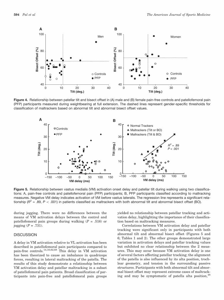
A delay in VM activation relative to VL activation has beendescribed in patellofemoral pain participants compared topain-free controls.14,16,62,68 This delay in VM activationhas been theorized to cause an imbalance in quadricepsforces, resulting in lateral maltracking of the patella. Theresults of this study demonstrate a relationship betweenVM activation delay and patellar maltracking in a subsetof patellofemoral pain patients. Broad classification of par-ticipants into pain-free and patellofemoral pain groups
yielded no relationship between patellar tracking and acti-vation delay, highlighting the importance of their classifica-tion based on maltracking measures.
Correlations between VM activation delay and patellartracking were significant only in participants with bothabnormal tilt and abnormal bisect offset (Figures 5 and6; Tables 1 and 2). The other groups demonstrated largevariation in activation delays and patellar tracking valuesbut exhibited no clear relationship between the 2 meas-ures. This may occur because VM activation delay is oneof several factors affecting patellar tracking; the alignmentof the patella is also influenced by its alta position, troch-lear geometry, and tension in the surrounding passivestructures. Participants with both abnormal tilt and abnor-mal bisect offset may represent extreme cases of maltrack-ing and may be symptomatic of patella alta position,48
40
60
80
100
0 10 20 30 40
Bis
ect
Off
set
(%)
Tilt (deg.)
Men
ControlsPFP
A
40
60
80
100
Bis
ect
Off
set
(%)
0 10 20 30 40Tilt (deg.)
Women
Controls
PFP
B
Figure 4. Relationship between patellar tilt and bisect offset in (A) male and (B) female pain-free controls and patellofemoral pain(PFP) participants measured during weightbearing at full extension. The dashed lines represent gender-specific thresholds forclassification of maltrackers based on abnormal tilt and abnormal bisect offset values.
0
10
20
30
40
–150 –100 –50 0 50 100 150
Tilt
(d
eg.)
VM delay (ms)
Controls
PFP
A
–150 –100 –50 0 50 100 150VM delay (ms)
R ² = .89P < .001
0
10
20
30
40
Tilt
(d
eg.)
Normal TrackersMaltrackers (Tilt or BO)Maltrackers (Tilt & BO)
B
Figure 5. Relationship between vastus medialis (VM) activation onset delay and patellar tilt during walking using two classifica-tions: A, pain-free controls and patellofemoral pain (PFP) participants; B, PFP participants classified according to maltrackingmeasures. Negative VM delay indicates activation of VM before vastus lateralis. The regression line represents a significant rela-tionship (R2 = .89, P \ .001) in patients classified as maltrackers with both abnormal tilt and abnormal bisect offset (BO).
594 Pal et al The American Journal of Sports Medicine

trochlear dysplasia,19 and abnormal tensioning in the lat-eral retinacula13,39 and/or the medial patellofemoral liga-ment.1,11,56 We are in the process of evaluating theeffects of these anatomic conditions on patellar kinematicsin our participant population.
Comparison of VM activation delay with patellar track-ing measures provides additional insight into the contro-versial question of altered VM activity in patellofemoralpain participants. Previous studies consistently reportedsubstantial variability in VM activation timings in controland patellofemoral pain groups and quantified the differ-ences between the groups by testing the means of VMdelays for statistical significance.{ Using this method,some studies reported differences,14,16,62,68 whereasothers4,43,59,64 reported no difference between the pain-freeand patellofemoral pain groups. In our study, we found nodifference between the means of VM activation delays inpain-free controls and all patellofemoral pain patientsgrouped together. Wong70 hypothesized that the discrepan-cies among studies are due to a lack of standardized meth-ods in recognizing vasti activation times. An analysis ofthe different onset thresholds reported in the literature onour data set suggested minimal sensitivity of VM activationdelay to onset threshold, so long as the algorithm was ableto consistently detect the anticipatory activations beforeheel strike. We theorize that the primary reason for the dis-crepancies among studies may be the selection of patellofe-moral pain participants. A study with a large number ofmaltracking patellofemoral pain participants with high tiltand bisect offset values would likely report significant delay
in VM activation, compared with pain-free participants. Inour study, only 20% of patellofemoral pain participants (8of 40) were classified as lateral maltrackers with both abnor-mal tilt and abnormal bisect offset, which may explain thelack of significant differences between the means of VM acti-vation delays in the pain-free controls and all patellofemoralpain participants. Furthermore, evidence relating VM acti-vation delay to lateral tracking of the patella is sparse.Powers58 evaluated relationships between vasti activationratio and patellar maltracking measures acquired fromsupine MR imaging. Ingersoll and Knight38 reported changesin patella locations after EMG biofeedback intervention onpain-free participants; Owings and Grabiner55 evaluatedthe effects of patellar medial-lateral mobility by measuringvasti muscle activation onsets at flexed and extended kneepositions; and Neptune et al52 predicted reduced patellofe-moral joint loads by advancing the onset of VM in a computa-tional simulation. The present study utilizes a novelcombination of weightbearing MR imaging and gait analysisto directly compare VM activation delay with patellar track-ing measures in patellofemoral pain participants.
One challenge in understanding the mechanism of patel-lar maltracking is appropriate classification of participants.Fredericson and Yoon28 noted that studies have not consis-tently demonstrated biomechanical differences betweenpain-free and patellofemoral pain participants, likelybecause of the difficulty in defining where the range of nor-mal alignment ends and malalignment begins. In thisstudy, gender-specific thresholds based on population meas-urements (28 men, 27 women) were introduced, and thefemale participants demonstrated greater patellar tilt (P =.022). The patellofemoral pain participants with tilt andbisect offset values in the highest quartiles were classified
TABLE 1Relationship Between Vastus Medialis Activation Onset Delay and Patellar Maltracking Measures During Walkinga
Tilt Bisect Offset
Group n R2 P R2 P
Controls 15 \.01 .860 .01 .744Patellofemoral pain 40 .02 .330 .02 .358
Normal trackers 25 .02 .497 .08 .171Maltrackers: tilt or bisect offset 7 .12 .446 .37 .144Maltrackers: tilt and bisect offset 8 .89 \.001 .43 .077
aExpressed as coefficients of determination (R2) and regression significance values.
TABLE 2Relationship Between Vastus Medialis Activation Delay and Patellar Maltracking Measures During Jogginga
Tilt Bisect Offset
Group n R2 P R2 P
Controls 15 \.01 .878 \.01 .999Patellofemoral pain 37 .07 .117 .05 .202
Normal trackers 24 .08 .181 .06 .268Maltrackers: tilt or bisect offset 6 .06 .629 .54 .094Maltrackers: tilt and bisect offset 7 .42 .113 .75 .012
aExpressed as coefficients of determination (R2) and regression significance values.
{References 4, 14, 16, 43, 59, 62, 64, 68.
Vol. 39, No. 3, 2011 Vastus Medialis Activation Delay in Patellofemoral Pain Patients 595

as maltrackers. This approach resulted in almost 40% ofpatellofemoral pain participants being classified as mal-trackers with either abnormal tilt or abnormal bisect offsetor both; the percentage of patellofemoral pain participantsclassified as maltrackers may vary among studies20,63 owingto differences in methodology and participant population.The definition of the 75% confidence interval as a maltrack-ing threshold is subjective; however, small changes to ourmaltracking threshold have minimal influence on the signif-icant relationships presented in this study. This techniqueprovides a robust method to represent the existing variabil-ity in patellar tilt and bisect offset measurements. Anotherpotential cause for debate is the use of all participants (pain-free and patellofemoral pain) in determination of maltrack-ing thresholds. Maltracking thresholds should arguably bebased on pain-free participants, as previously reported.23
This is ideal in research settings, with access to both pain-free and patellofemoral pain participants. In clinical set-tings, however, only symptomatic participants are evalu-ated. In the absence of a large research data set of pain-free participants, this method needs to be applicable toa clinician’s data set to classify his or her patients into mal-tracking subgroups. In this study, we included pain-free andpatellofemoral pain participants in our classificationbecause of accessibility to both participant groups. Further-more, the current results are based on 55 participants (15pain-free, 40 patellofemoral pain), with 8 patellofemoralpain participants (4 men and 4 women) classified as mal-trackers with both abnormal tilt and abnormal bisect offset.Analysis of additional participants from other centers mayhelp test the generality of our findings. Also, there remainsa need for obtaining patellar tracking data on a large popu-lation of healthy controls to provide a baseline for quantify-ing maltracking thresholds.
A potential limitation of this study is that we measuredactivity of the entire VM muscle, as opposed to activity ofthe isolated VMO fibers previously reported.# It was diffi-cult to clearly distinguish between VMO and VML activa-tions using surface electrodes; accordingly, we used oneelectrode to measure the activation of the entire VM muscle.It is unclear what influence characterizing delay of the iso-lated VMO fibers would have on the results of this study.Another limitation is that patellar alignment and activationtiming were measured during separate activities. It is diffi-cult to acquire quadriceps activation onset data duringa backrest-assisted weightbearing squat because the quad-riceps muscles are active as soon as a participant positionshimself or herself. Also, reproducing a walking or joggingactivity under MR surveillance is not feasible.
Patellofemoral pain syndrome has been described asa motor control problem.32,52,67 Delayed activation of theVM relative to the VL muscle is theorized to cause a tempo-rary imbalance in muscle forces, resulting in excessive lat-eral tracking of the patella. Several intervention studiesusing biofeedback and VM retraining have reported reduc-tions in VM delays in patellofemoral pain participants2,53;however, there is little evidence relating reduced VM acti-vation delay to improved patellar tracking. This studydemonstrated a significant relationship between VM acti-vation delay and patellar maltracking in one subset ofpatellofemoral pain participants, suggesting that clinicalinterventions to improve VM activation may improvepatellar tracking only in participants with high tilt andbisect offset values. This finding underscores the impor-tance of appropriate classification of patellofemoral painparticipants before selection of a clinical intervention. Anintervention study incorporating VM retraining and patel-lar tracking measurements would provide the much-needed evidence for relating reduced VM activation delayto improved patellar tracking in patellofemoral painparticipants.
40
60
80
100
–150 –100 –50 0 50 100 150
Bis
ect
Off
set
(%)
VM delay (ms)
Controls
PFP
A
–150 –100 –50 0 50 100 150
VM delay (ms)
R ² = .43P = .077
40
60
80
100
Bis
ect
Off
set
(%)
Normal Trackers
Maltrackers (Tilt or BO)
Maltrackers (Tilt & BO)
B
Figure 6. Relationship between vastus medialis (VM) activation onset delay and patella bisect offset (BO) during walking usingtwo classifications: A, pain-free controls and patellofemoral pain (PFP) participants; B, PFP participants classified according tomaltracking measures. Negative VM delay indicates activation of VM before vastus lateralis.
#References 4, 14, 16, 43, 55, 59, 62, 64, 68.
596 Pal et al The American Journal of Sports Medicine

REFERENCES
1. Amis AA, Firer P, Mountney J, Senavongse W, Thomas NP. Anatomy
and biomechanics of the medial patellofemoral ligament. Knee.
2003;10(3):215-220.
2. Bennell K, Duncan M, Cowan S, McConnell J, Hodges P, Crossley K.
Effects of vastus medialis oblique retraining versus general quadri-
ceps strengthening on vasti onset. Med Sci Sports Exerc. 2010;42(5):
856-864.
3. Besier TF, Gold GE, Beaupre GS, Delp SL. A modeling framework to
estimate patellofemoral joint cartilage stress in vivo. Med Sci Sports
Exerc. 2005;37(11):1924-1930.
4. Bevilaqua-Grossi D, Felicio LR, Leocadio LP. Analysis of the reflex
response time of the patellar stabilizer muscles in individuals with
patellofemoral pain syndrome. Rev Bras Fisioter. 2008;12(1):26-30.
5. Biedert RM, Sanchis-Alfonso V. Sources of anterior knee pain. Clin
Sports Med. 2002;21(3):335-347.
6. Bigley RF, Gibeling JC, Stover SM, Hazelwood SJ, Fyhrie DP, Martin
RB. Volume effects on fatigue life of equine cortical bone. J Biomech.
2007;40(16):3548-3554.
7. Bolgla LA, Malone TR, Umberger BR, Uhl TL. Hip strength and hip
and knee kinematics during stair descent in females with and without
patellofemoral pain syndrome. J Orthop Sports Phys Ther. 2008;
38(1):12-18.
8. Brechter JH, Powers CM. Patellofemoral joint stress during stair
ascent and descent in persons with and without patellofemoral
pain. Gait Posture. 2002;16(2):115-123.
9. Brindle TJ, Mattacola C, McCrory J. Electromyographic changes in the
gluteus medius during stair ascent and descent in subjects with anterior
knee pain. Knee Surg Sports Traumatol Arthrosc. 2003;11(4):244-251.
10. Brossmann J, Muhle C, Schroder C, et al. Patellar tracking patterns
during active and passive knee extension: evaluation with motion-
triggered cine MR imaging. Radiology. 1993;187(1):205-212.
11. Camanho GL, Viegas Ade C, Bitar AC, Demange MK, Hernandez AJ.
Conservative versus surgical treatment for repair of the medial patel-
lofemoral ligament in acute dislocations of the patella. Arthroscopy.
2009;25(6):620-625.
12. Chester R, Smith TO, Sweeting D, Dixon J, Wood S, Song F. The rel-
ative timing of VMO and VL in the aetiology of anterior knee pain:
a systematic review and meta-analysis. BMC Musculoskelet Disord.
2008;9:64.
13. Clifton R, Ng CY, Nutton RW. What is the role of lateral retinacular
release? J Bone Joint Surg Br. 2010;92(1):1-6.
14. Cowan SM, Bennell KL, Hodges PW, Crossley KM, McConnell J.
Delayed onset of electromyographic activity of vastus medialis obli-
quus relative to vastus lateralis in subjects with patellofemoral pain
syndrome. Arch Phys Med Rehabil. 2001;82(2):183-189.
15. Cowan SM, Crossley KM, Bennell KL. Altered hip and trunk muscle
function in individuals with patellofemoral pain. Br J Sports Med.
2009;43(8):584-588.
16. Cowan SM, Hodges PW, Bennell KL, Crossley KM. Altered vastii
recruitment when people with patellofemoral pain syndrome com-
plete a postural task. Arch Phys Med Rehabil. 2002;83(7):989-995.
17. Crowninshield RD, Brand RA. A physiologically based criterion of mus-
cle force prediction in locomotion. J Biomech. 1981;14(11):793-801.
18. Crowninshield RD, Brand RA. The prediction of forces in joint struc-
tures; distribution of intersegmental resultants. Exerc Sport Sci Rev.
1981;9:159-181.
19. Davies AP, Costa ML, Shepstone L, Glasgow MM, Donnell ST. The
sulcus angle and malalignment of the extensor mechanism of the
knee. J Bone Joint Surg Br. 2000;82(8):1162-1166.
20. DeHaven KE, Dolan WA, Mayer PJ. Chondromalacia patellae in ath-
letes: clinical presentation and conservative management. Am J
Sports Med. 1979;7(1):5-11.
21. DeHaven KE, Lintner DM. Athletic injuries: comparison by age, sport,
and gender. Am J Sports Med. 1986;14(3):218-224.
22. Devereaux MD, Lachmann SM. Patello-femoral arthralgia in athletes
attending a sports injury clinic. Br J Sports Med. 1984;18(1):18-21.
23. Draper CE, Besier TF, Santos JM, et al. Using real-time MRI to quan-
tify altered joint kinematics in subjects with patellofemoral pain and
to evaluate the effects of a patellar brace or sleeve on joint motion.
J Orthop Res. 2009;27(5):571-577.
24. Duffey MJ, Martin DF, Cannon DW, Craven T, Messier SP. Etiologic
factors associated with anterior knee pain in distance runners. Med
Sci Sports Exerc. 2000;32(11):1825-1832.
25. Dye SF. The pathophysiology of patellofemoral pain: a tissue homeo-
stasis perspective. Clin Orthop Relat Res. 2005;436:100-110.
26. Elias JJ, Cech JA, Weinstein DM, Cosgarea AJ. Reducing the lateral
force acting on the patella does not consistently decrease patellofe-
moral pressures. Am J Sports Med. 2004;32(5):1202-1208.
27. Farahmand F, Senavongse W, Amis AA. Quantitative study of the
quadriceps muscles and trochlear groove geometry related to insta-
bility of the patellofemoral joint. J Orthop Res. 1998;16(1):136-143.
28. Fredericson M, Yoon K. Physical examination and patellofemoral
pain syndrome. Am J Phys Med Rehabil. 2006;85(3):234-243.
29. Fulkerson JP. The etiology of patellofemoral pain in young, active
patients: a prospective study. Clin Orthop Relat Res. 1983;179:
129-133.
30. Fulkerson JP, Schutzer SF, Ramsby GR, Bernstein RA. Computer-
ized tomography of the patellofemoral joint before and after lateral
release or realignment. Arthroscopy. 1987;3(1):19-24.
31. Fulkerson JP, Shea KP. Mechanical basis for patellofemoral pain and
cartilage breakdown. In: Ewing JW, ed. Articular Cartilage and Knee
Joint Function: Basic Science and Arthroscopy. New York, NY:
Raven Press; 1990:93-101.
32. Grabiner MD, Koh TJ, Draganich LF. Neuromechanics of the patello-
femoral joint. Med Sci Sports Exerc. 1994;26(1):10-21.
33. Guo Z, De Vita R. Probabilistic constitutive law for damage in liga-
ments. Med Eng Phys. 2009;31(9):1104-1109.
34. Haldar A, Mahadevan S. Probability, Reliability and Statistical Methods
in Engineering Design. New York, NY: John Wiley & Sons Inc; 2000.
35. Hautamaa PV, Fithian DC, Kaufman KR, Daniel DM, Pohlmeyer AM.
Medial soft tissue restraints in lateral patellar instability and repair.
Clin Orthop Relat Res. 1998;349:174-182.
36. Huberti HH, Hayes WC. Patellofemoral contact pressures: the influ-
ence of q-angle and tendofemoral contact. J Bone Joint Surg Am.
1984;66(5):715-724.
37. Hurschler C, Provenzano PP, Vanderby R Jr. Application of a proba-
bilistic microstructural model to determine reference length and toe-
to-linear region transition in fibrous connective tissue. J Biomech
Eng. 2003;125(3):415-422.
38. Ingersoll CD, Knight KL. Patellar location changes following EMG
biofeedback or progressive resistive exercises. Med Sci Sports
Exerc. 1991;23(10):1122-1127.
39. Ishibashi Y, Okamura Y, Otsuka H, Tsuda E, Toh S. Lateral patellar
retinaculum tension in patellar instability. Clin Orthop Relat Res.
2002;397:362-369.
40. Jafaril A, Farahmand F, Meghdari A. The effects of trochlear groove
geometry on patellofemoral joint stability: a computer model study.
Proc Inst Mech Eng H. 2008;222(1):75-88.
41. Jan MH, Lin DH, Lin JJ, Lin CH, Cheng CK, Lin YF. Differences in
sonographic characteristics of the vastus medialis obliquus between
patients with patellofemoral pain syndrome and healthy adults. Am
J Sports Med. 2009;37(9):1743-1749.
42. Kadaba MP, Ramakrishnan HK, Wooten ME. Measurement of lower
extremity kinematics during level walking. J Orthop Res. 1990;8:
383-392.
43. Karst GM, Willett GM. Onset timing of electromyographic activity in
the vastus medialis oblique and vastus lateralis muscles in subjects
with and without patellofemoral pain syndrome. Phys Ther. 1995;
75(9):813-823.
44. Kramer PG. Patella malalignment syndrome: rationale to reduce exces-
sive lateral pressure. J Orthop Sports Phys Ther. 1986;8(6):301-309.
45. Kujala UM, Jaakkola LH, Koskinen SK, Taimela S, Hurme M,
Nelimarkka O. Scoring of patellofemoral disorders. Arthroscopy.
1993;9(2):159-163.
Vol. 39, No. 3, 2011 Vastus Medialis Activation Delay in Patellofemoral Pain Patients 597
46. Levinger P, Gilleard W. Tibia and rearfoot motion and ground reaction
forces in subjects with patellofemoral pain syndrome during walking.
Gait Posture. 2007;25(1):2-8.
47. LLopis E, Padron M. Anterior knee pain. Eur J Radiol. 2007;62(1):
27-43.
48. Luyckx T, Didden K, Vandenneucker H, Labey L, Innocenti B,
Bellemans J. Is there a biomechanical explanation for anterior knee
pain in patients with patella alta? Influence of patellar height on patel-
lofemoral contact force, contact area and contact pressure. J Bone
Joint Surg Br. 2009;91(3):344-350.
49. Mach DB, Rogers SD, Sabino MC, et al. Origins of skeletal pain: sen-
sory and sympathetic innervation of the mouse femur. Neuroscience.
2002;113(1):155-166.
50. McClay I, Manal K. A comparison of three-dimensional lower extre-
mity kinematics during running between excessive pronators and
normals. Clin Biomech (Bristol, Avon). 1998;13(3):195-203.
51. Merican AM, Amis AA. Iliotibial band tension affects patellofemoral
and tibiofemoral kinematics. J Biomech. 2009;42(10):1539-1546.
52. Neptune RR, Wright IC, van den Bogert AJ. The influence of orthotic
devices and vastus medialis strength and timing on patellofemoral
loads during running. Clin Biomech (Bristol, Avon). 2000;15(8):611-618.
53. Ng GY, Zhang AQ, Li CK. Biofeedback exercise improved the EMG
activity ratio of the medial and lateral vasti muscles in subjects with patel-
lofemoral pain syndrome. J Electromyogr Kinesiol. 2008;18(1):128-133.
54. Nomura E, Horiuchi Y, Kihara M. Medial patellofemoral ligament
restraint in lateral patellar translation and reconstruction. Knee.
2000;7(2):121-127.
55. Owings TM, Grabiner MD. Motor control of the vastus medialis obli-
que and vastus lateralis muscles is disrupted during eccentric con-
tractions in subjects with patellofemoral pain. Am J Sports Med.
2002;30(4):483-487.
56. Panagopoulos A, van Niekerk L, Triantafillopoulos IK. MPFL recon-
struction for recurrent patella dislocation: a new surgical technique
and review of the literature. Int J Sports Med. 2008;29(5):359-365.
57. Perotto A, Delagi EF, Iazzetti J, Morrison D. Anatomical Guide for the
Electromyographer. 4th ed. Springfield, IL: Charles C Thomas; 2005.
58. Powers CM. Patellar kinematics, part I: the influence of vastus mus-
cle activity in subjects with and without patellofemoral pain. Phys
Ther. Oct 2000;80(10):956-964.
59. Powers CM, Landel R, Perry J. Timing and intensity of vastus muscle
activity during functional activities in subjects with and without patello-
femoral pain. Phys Ther. 1996;76(9):946-955.
60. Sanchis-Alfonso V, Rosello-Sastre E. Anterior knee pain in the young
patient: what causes the pain? ‘‘Neural model.’’ Acta Orthop Scand.
2003;74(6):697-703.
61. Sanchis-Alfonso V, Rosello-Sastre E, Monteagudo-Castro C,
Esquerdo J. Quantitative analysis of nerve changes in the lateral ret-
inaculum in patients with isolated symptomatic patellofemoral
malalignment: a preliminary study. Am J Sports Med. 1998;26(5):
703-709.
62. Santos EP, Bessa SNF, Lins CAA, Marinho AMF, Silva KMP, Brasi-
leiro JS. Electromyographic activity of vastus medialis obliquus and
vastus lateralis muscles during functional activities in subjects
with patellofemoral pain syndrome. Rev Bras Fisioter. 2008;12(4):
304-310.
63. Sheehan FT, Derasari A, Fine KM, Brindle TJ, Alter KE. Q-angle and
J-sign: indicative of maltracking subgroups in patellofemoral pain.
Clin Orthop Relat Res. 2010;468(1):266-275.
64. Sheehy P, Burdett RG, Irrgang JJ, VanSwearingen J. An electromyo-
graphic study of vastus medialis oblique and vastus lateralis activity
while ascending and descending steps. J Orthop Sports Phys Ther.
1998;27(6):423-429.
65. Souza RB, Powers CM. Differences in hip kinematics, muscle
strength, and muscle activation between subjects with and
without patellofemoral pain. J Orthop Sports Phys Ther. 2009;39(1):
12-19.
66. Stanford W, Phelan J, Kathol MH, et al. Patellofemoral joint motion:
evaluation by ultrafast computed tomography. Skeletal Radiol.
1988;17(7):487-492.
67. Van Tiggelen D, Cowan S, Coorevits P, Duvigneaud N, Witvrouw E.
Delayed vastus medialis obliquus to vastus lateralis onset timing
contributes to the development of patellofemoral pain in previously
healthy men: a prospective study. Am J Sports Med. 2009;37(6):
1099-1105.
68. Voight ML, Wieder DL. Comparative reflex response times of vastus
medialis obliquus and vastus lateralis in normal subjects and sub-
jects with extensor mechanism dysfunction: an electromyographic
study. Am J Sports Med. 1991;19(2):131-137.
69. Wojtys EM, Beaman DN, Glover RA, Janda D. Innervation of the
human knee joint by substance-P fibers. Arthroscopy. 1990;6(4):
254-263.
70. Wong YM. Recording the vastii muscle onset timing as a diagnostic
parameter for patellofemoral pain syndrome: fact or fad? Phys Ther
Sport. 2009;10(2):71-74.
For reprints and permission queries, please visit SAGE’s Web site at http://www.sagepub.com/journalsPermissions.nav
598 Pal et al The American Journal of Sports Medicine
Related Documents