Particle size and shape of calcium hydroxide Takashi Komabayashi, D.D.S., M. Dent. Sc., Ph.D. * , Rena N D’souza, D.D.S., M.S., Ph.D. ** , Paul C Dechow, Ph.D. ** , Kamran E. Safavi, D.M.D., M.Ed *** , and Larz S.W. Spångberg, D.D.S., Ph.D. *** * Department of Endodontics, Texas A&M Health Science Center, Baylor College of Dentistry ** Department of Biomedical Sciences, Texas A&M Health Science Center, Baylor College of Dentistry *** Division of Endodontology, Department of Oral Health and Diagnostic Sciences, School of Dental Medicine, University of Connecticut Abstract The aim of this study was to examine the particle length, width, perimeter, and aspect ratio of calcium hydroxide powder using a flow particle image analyzer (FPIA). Five sample groups each with 10mg calcium hydroxide were mixed with 15mL of alcohol and sonicated. Digital images of the particle samples were taken using the FPIA and analyzed with a one-way ANOVA. The overall averages ±S.D. among the five groups for particle length (μm), width (μm), perimeter (μm), and aspect ratio were 2.255±1.994, 1.620±1.464, 6.699±5.598, and 0.737±0.149, respectively. No statistical significance was observed among the groups for all parameters. When the total of 46,818 particles from all five groups were classified into the five length categories of 0.5μm increments, there were significant differences in width, perimeter, and aspect ratio (all p-values<0.0001). In conclusion, calcium hydroxide particles have a size and shape that may allow direct penetration into open dentin tubules. Keywords Calcium hydroxide; Image Analysis; Particle size/shape; Dentin tubule; Aspect ratio Introduction Calcium hydroxide aqueous slurry is widely used as an interim (interappointment) antimicrobial dressing in root canal treatment (1–5). Less than 0.2 % of the calcium hydroxide slurry dissociates at body temperature into calcium ions (Ca 2+ ) and hydroxide ions (OH − ), leaving most of the particles undissolved (6). Studies indicate that the dentin tubules of the root canal walls harbor microorganisms (7,8). The penetration of microorganisms into infected dentin tubules is reported to extend generally from 50 to 100 μm (9). However, it has been shown that the application of calcium hydroxide into instrumented and irrigated root canals eliminates microorganisms effectively (1). Corresponding author: Takashi Komabayashi D.D.S., M. Dent. Sc., Ph.D. Assistant Professor, Department of Endodontics, Texas A&M Health Science Center, Baylor College of Dentistry, 3302 Gaston Avenue, Dallas, TX 75246 USA, TEL: 214-828-8365 FAX: 214-874-4507, E-mail address: [email protected] [email protected]. Publisher's Disclaimer: This is a PDF file of an unedited manuscript that has been accepted for publication. As a service to our customers we are providing this early version of the manuscript. The manuscript will undergo copyediting, typesetting, and review of the resulting proof before it is published in its final citable form. Please note that during the production process errors may be discovered which could affect the content, and all legal disclaimers that apply to the journal pertain. NIH Public Access Author Manuscript J Endod. Author manuscript; available in PMC 2010 February 1. Published in final edited form as: J Endod. 2009 February ; 35(2): 284–287. doi:10.1016/j.joen.2008.11.017. NIH-PA Author Manuscript NIH-PA Author Manuscript NIH-PA Author Manuscript

Welcome message from author
This document is posted to help you gain knowledge. Please leave a comment to let me know what you think about it! Share it to your friends and learn new things together.
Transcript
Particle size and shape of calcium hydroxide
Takashi Komabayashi, D.D.S., M. Dent. Sc., Ph.D.*, Rena N D’souza, D.D.S., M.S., Ph.D.**,Paul C Dechow, Ph.D.**, Kamran E. Safavi, D.M.D., M.Ed***, and Larz S.W. Spångberg, D.D.S.,Ph.D.**** Department of Endodontics, Texas A&M Health Science Center, Baylor College of Dentistry
** Department of Biomedical Sciences, Texas A&M Health Science Center, Baylor College of Dentistry
*** Division of Endodontology, Department of Oral Health and Diagnostic Sciences, School of DentalMedicine, University of Connecticut
AbstractThe aim of this study was to examine the particle length, width, perimeter, and aspect ratio of calciumhydroxide powder using a flow particle image analyzer (FPIA). Five sample groups each with 10mgcalcium hydroxide were mixed with 15mL of alcohol and sonicated. Digital images of the particlesamples were taken using the FPIA and analyzed with a one-way ANOVA. The overall averages±S.D. among the five groups for particle length (μm), width (μm), perimeter (μm), and aspect ratiowere 2.255±1.994, 1.620±1.464, 6.699±5.598, and 0.737±0.149, respectively. No statisticalsignificance was observed among the groups for all parameters. When the total of 46,818 particlesfrom all five groups were classified into the five length categories of 0.5μm increments, there weresignificant differences in width, perimeter, and aspect ratio (all p-values<0.0001). In conclusion,calcium hydroxide particles have a size and shape that may allow direct penetration into open dentintubules.
KeywordsCalcium hydroxide; Image Analysis; Particle size/shape; Dentin tubule; Aspect ratio
IntroductionCalcium hydroxide aqueous slurry is widely used as an interim (interappointment)antimicrobial dressing in root canal treatment (1–5). Less than 0.2 % of the calcium hydroxideslurry dissociates at body temperature into calcium ions (Ca2+) and hydroxide ions (OH−),leaving most of the particles undissolved (6).
Studies indicate that the dentin tubules of the root canal walls harbor microorganisms (7,8).The penetration of microorganisms into infected dentin tubules is reported to extend generallyfrom 50 to 100 μm (9). However, it has been shown that the application of calcium hydroxideinto instrumented and irrigated root canals eliminates microorganisms effectively (1).
Corresponding author: Takashi Komabayashi D.D.S., M. Dent. Sc., Ph.D. Assistant Professor, Department of Endodontics, Texas A&MHealth Science Center, Baylor College of Dentistry, 3302 Gaston Avenue, Dallas, TX 75246 USA, TEL: 214-828-8365 FAX:214-874-4507, E-mail address: [email protected] [email protected]'s Disclaimer: This is a PDF file of an unedited manuscript that has been accepted for publication. As a service to our customerswe are providing this early version of the manuscript. The manuscript will undergo copyediting, typesetting, and review of the resultingproof before it is published in its final citable form. Please note that during the production process errors may be discovered which couldaffect the content, and all legal disclaimers that apply to the journal pertain.
NIH Public AccessAuthor ManuscriptJ Endod. Author manuscript; available in PMC 2010 February 1.
Published in final edited form as:J Endod. 2009 February ; 35(2): 284–287. doi:10.1016/j.joen.2008.11.017.
NIH
-PA Author Manuscript
NIH
-PA Author Manuscript
NIH
-PA Author Manuscript

The antimicrobial action of calcium hydroxide depends on the concentration of hydroxide ionsin the solution (1,10). This selective permeability of the hydroxide ions in the dentin tubulesis known because of the buffering capacity of hydroxyapatite (11). Depending on theconcentration of hydroxide ions, the antimicrobial effect of calcium hydroxide in the dentintubules may or may not be effective.
A newly proposed theory about using calcium hydroxide in endodontic treatment suggests thatif the calcium hydroxide particles are inserted into the open dentin tubules, the particles mayact as a direct source of dissociated calcium hydroxide by continuing to dissolve into theaqueous form of calcium hydroxide. This theory may result in the maintenance of a high localpH, which will enhance the antimicrobial effectiveness. While most calcium hydroxideaqueous slurry is comprised of undissolved particles, information about the morphology ofthese particles is scarce (12–15). The reports do not sufficiently answer the clinical endodonticquestions with respect to particle penetration into the dentin tubules. Further, evaluatingparticle morphology based on representative SEM images, which has been the conventionaltechnique used in the past, is challenging. Therefore, in the present study, a novel techniqueof image analysis to characterize the particles will be described.
Image analysis technology is used in the ceramics, pharmaceutical, and toner cartridgeindustries for optimizing product and process performance (16–18). This technique providesstatistically valid information and images showing the size and shape of each particle in largenumbers of samples. A flow particle image analyzer (FPIA-3000; Sysmex, Kobe, Japan)analyzes the size and shape of particles (1.5 μm to 160 μm) in emulsions and suspensions andproduces quantitative shape information expressed as the morphological parameters ofparticles (19). The low-power field mode allows the analysis of coarse particles ranging in sizefrom 6 to 160 μm. The high-power field (HPF) mode allows analysis of fine particles between1.5 and 40 μm with a minimum detectable level of 0.5 μm for selected analysis parameters.Because several studies reported that calcium hydroxide particles vary from 0.5 μm to 20 μm(12–15), the HPF mode was used for this study.
The aim of this study was to quantify the particle length, width, perimeter, and aspect ratio ofcalcium hydroxide powder using the HPF mode of the flow particle image analyzer.
Materials and MethodsCalcium hydroxide powder (Product Number C7887, Lot Number 31H3445; Sigma-Aldrich,St. Louis, MO) was examined with a flow particle image analyzer (FPIA-3000). Polystyrenelatex particles (2 μm in diameter; Polymer Microspheres 5200A; Duke Scientific Corporation,Fremont, CA) were used as test objects to adjust the focus before the calcium hydroxidesamples were tested. An alcohol suspension of calcium hydroxide provided a fluid particlesuspension that prevented clogging the FPIA machine flow chamber. Ten milligrams ofcalcium hydroxide were mixed with 15 mL of alcohol and then sonicated for 1 minute to createa homogeneous fluid. Five milliliters of the dispersion was added to the FPIA. The finalanalyzed volume was set at 0.35 μL.
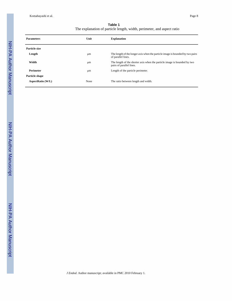
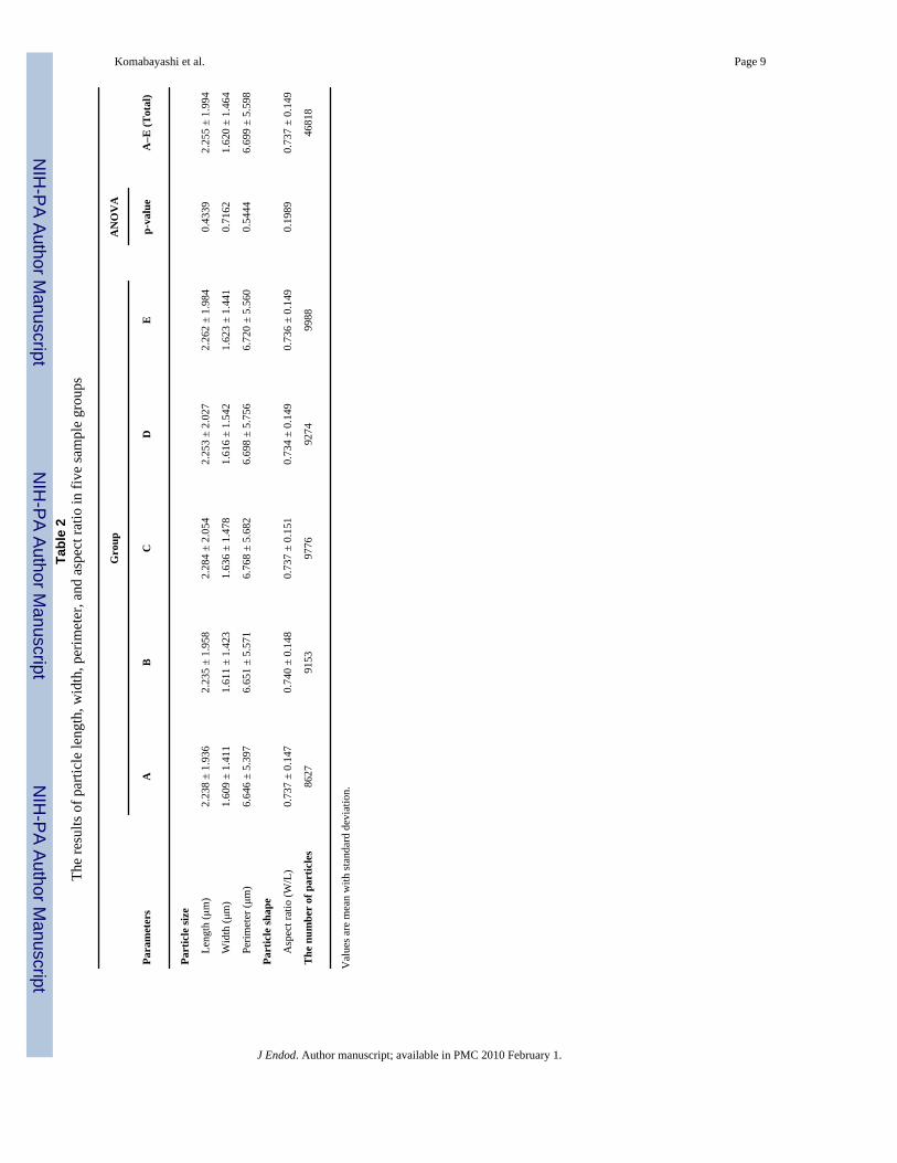
Five sample groups were randomly prepared by using material from one calcium hydroxidepackage (samples A, B, C, D, and E). The reproducibility of the calcium hydroxide preparationsmimicked the clinical setting in which calcium hydroxide from the same package is preparedfor each patient. The samples were tested once at HPF mode. The particle size and shape wereanalyzed using the particle size parameters of length, width, and perimeter; the shape parameterwas considered the aspect ratio. The analysis of the parameters, units, calculation methods andcomments are summarized in Table 1. Digital images of the particle samples wereautomatically collected by the FPIA machine, along with the above-described parameters.
Komabayashi et al. Page 2
J Endod. Author manuscript; available in PMC 2010 February 1.
NIH
-PA Author Manuscript
NIH
-PA Author Manuscript
NIH
-PA Author Manuscript

Two statistical analyses were performed. In part 1, the mean, standard deviation, and thenumber of particles were calculated for each sample group. A one-way analysis of variance(ANOVA) was conducted to identify any significant differences in length (μm), width (μm),perimeter (μm), and aspect ratio among the five sample groups. A p value less than 0.05 wasconsidered statistically significant.
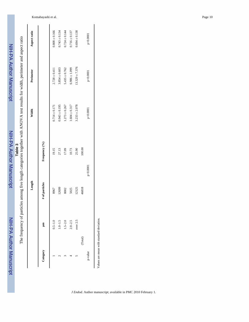
In part 2 of the statistical analysis, the length was classified into five categories: Category 1(0.5–1.0 μm), Category 2 (1.0–1.5 μm), Category 3 (1.5–2.0 μm), Category 4 (2.0–2.5 μm),and Category 5 (more than 2.5 μm). The chi-square test was used to test for significantdifferences between the number of particles and the five length categories. An ANOVA wasused to investigate whether there were any significant differences in width, perimeter, andaspect ratio among the five categories.
ResultsThe results of particle length, width, perimeter, and aspect ratio in five groups are summarizedin Table 2. A total of 46,818 particles from all five sample groups were analyzed. No statisticalsignificance was observed among the groups for all the parameters.
Table 3 shows the results of the analyses of particle number, width, perimeter and aspect ratio.The chi-square test showed that there was a significant association between the number ofparticles and the five categories of length (p<0.0001). The highest number was found forcategory 2 (1.0–1.5 μm). The cumulative percentage of particles between 0.5 and 2.0 μm was63%, while 74% of the particles were between 0.5 and 2.5 μm. The ANOVA showed that therewere significant differences in width, perimeter and aspect ratio among the five categories (allp-values < 0.0001). The aspect ratio in category 1 (0.5–1.0 μm) was the highest among all thecategories, and category 5 (over 2.5 μm) was the lowest of all. As the particle length decreased,the particles changed to a rounder shape. As the particle lengthened, the particle shape becamemore rectangular.
DiscussionThe permeability of dentin is governed largely by dentin tubule anatomy, density, diameter,and length as well as features of the solute such as size and charge (20). Dentin tubule densityand size in root dentin have been studied by various investigators (21–27). Mjör et al. reportedthat at the apical portion of permanent human teeth, the dentin tubules were irregular indirection and density (22). In general, the dentin tubules are considered to have a diameter of2 to 5 μm.
Dentin is a substrate, whereas calcium hydroxide is a material. The size of the dentin tubulescorrelates with the size of the calcium hydroxide particles. This study found that the cumulativepercentage of particles between 0.5 and 2.0 μm was 63%. Therefore, in theory, the geometryof these small particles makes it possible for calcium hydroxide to enter the open dentin tubules.The penetrating particles may act as a direct source of dissociated calcium hydroxide, resultingin a high local pH with a slight chance of being reduced by dentin buffering. An even higherpH would result in a stronger and more effective antimicrobial action.
The solubility of calcium hydroxide in water is very low. Aqueous slurry contains manyundissolved particles together with the calcium and hydroxide ions. Since calcium hydroxideis not soluble in ethanol, using ethanol for the suspension of calcium hydroxide particles isalso beneficial to prevent clogging of the flow chamber of the FPIA machine.
A newly proposed theory in endodontic treatment suggests that if calcium hydroxide particlespenetrate into open dentin tubules, the particles may act as a direct source of dissociated calcium
Komabayashi et al. Page 3
J Endod. Author manuscript; available in PMC 2010 February 1.
NIH
-PA Author Manuscript
NIH
-PA Author Manuscript
NIH
-PA Author Manuscript
hydroxide as they continue to dissolve into the aqueous form of calcium hydroxide. Based onthe particle size data collected in this study, it is speculated that undissolved fine particles mayplay an important role in the antimicrobial action inside dentin tubules. These particles maysteadily dissolve and ionize the dissolute in and around the dentin tubules and function as acontinuing source of hydroxide ions to maintain a high pH locally for a prolonged time periodin the dentin.
An advantage of FPIA analysis is the capacity to determine not only the particle size but alsothe particle shape (28,29). The data in the present study show that the particle shape is notround but irregular. The aspect ratio may relate to the rate of dissolution and ionization in andaround the dentin tubules due to the increased surface area of the particle. It may also determine,wholly or partially, the flow of the particle into a dentin tubule. In addition, the direction andorientation of the particle may control the depth of penetration. For example, the length mayprevent deep penetration of a thin elongated particle. The enhanced FPIA capabilities allowedadvanced statistical analysis, which was classified according to the five different particlelengths (Table 3). The results show that the aspect ratio was the highest in category 1 and thelowest in category 5. As the particle length decreased, the particle shape became rounder. Asthe particle lengthened, the particle shape changed to a more rectangular shape. Thus, in theory,short particles are more desirable for deep penetration into the dentin.
The choice of one-visit versus two-visit root canal therapy for necrotic teeth with apicalperiodontitis is a current topic of debate (30). While two-visit root canal therapy uses calciumhydroxide as an interim (interappointment) antimicrobial dressing, one-visit therapy does not.To resolve this dilemma, one line of thought is that the potential new development of effectivecalcium hydroxide slurry through enhancing its biologic quality may improve the treatmentmodality. Aspect ratio may be one important factor determining the suitability of the calciumhydroxide preparation. The influence of particle size and shape might increase the surface area,hence increasing the amount of potential reactivity for clinical and biological quality control.
A research-grade type of commercial calcium hydroxide powder was used in this study. Itshould be noted that the geometry of the calcium hydroxide may be different, depending onthe manufacturer, processing, country of origin, price, and mixing solution. For example, thecalcium hydroxide powders used in recent clinical/materials studies were different in each case(30–33). Calcium hydroxide powder mixed with 2% chlorhexidine gel as an intracanalmedication has also been studied (30,31); however, the dissociation into calcium ions andhydroxide ions might have been influenced by the chlorhexidine gel. Therefore, the results ofvarious studies on calcium hydroxide powder may not be entirely comparable since the calciumhydroxide power products available on the world market cannot be considered identical.
In future research, it will be interesting to examine the relationship between the particle sizeand shape distribution, as well as the reaction speed and rate, as these factors relate toantimicrobial characteristics. The results of the present research are valuable for clinicalendodontics, but the question remains as to the disposition of the calcium hydroxide particles.It is possible that they are ingested by bacteria, but some residual unreacted particles mayremain. Taking into consideration the anatomical stability of dentin tubules, the penetrationmechanism of residual calcium hydroxide particles is a clinical interest. Although the presentstudy focused on calcium hydroxide as an intracanal medication, calcium hydroxide, togetherwith a mineral trioxide aggregate, has been clinically studied for use in pulpotomies in primarymolars (32), as a pulp capping agent in permanent premolars (33), and a regenerative treatmentof an immature traumatized tooth with apical periodontitis (34). Therefore, further research onthe calcium hydroxide particle size and shape will be applicable to other emerging areas ofendodontic research, such as the dentin-material interface, biomineralization, stem cell/scaffold research, and nanotechnology.
Komabayashi et al. Page 4
J Endod. Author manuscript; available in PMC 2010 February 1.
NIH
-PA Author Manuscript
NIH
-PA Author Manuscript
NIH
-PA Author Manuscript

Conclusion1. The size and shape of calcium hydroxide particles may allow direct penetration into
open dentin tubules.
2. No statistically significant differences were observed among the five groups (A–E)in the analysis of all parameters, including particle length, width, perimeter, and aspectratio.
3. The number of particles in each of the five categories indicated that category 2 (1.0–1.5 μm) had the highest number of particles. The cumulative percentage of the finecalcium hydroxide particles between 0.5 and 2.0 μm was 63%; this length is less thanthe reported diameter of the dentin tubules in root dentin.
4. The aspect ratio was the highest in category 1 (0.5–1.0 μm), whereas it was the lowestin category 5 (over 2.5 μm). As the particle size increased, the particle shape becamemore rectangular.
AcknowledgementsThe authors thank Dr. Kevin Dahl, Mr. Andrew Jones, Mr. Terry Stauffer, Dr. Yoshiyuki Inoue, and Mr. Koji Sanada,(Malvern Instrument Inc [USA/UK]/Hosokawa Micron Corporation [Japan]) for analyzing the calcium hydroxidesamples and providing technical information.
The authors also thank Dr. Yohji Imai (Professor Emeritus, Tokyo Medical & Dental University, Japan) for his usefulcomments.
In addition, the authors thank Dr. Chul Ahn, Dr. M. E. Blair Holbein (University of Texas Southwestern MedicalCenter), and Ms. Jeanne Santa Cruz (Texas A&M Health Science Center Baylor College of Dentistry) for the statisticalconsultation and critical editing.
This publication was supported by Grant Number NIH KL2RR024983 (TK) and UL1 RR024982, entitled, “Northand Central Texas Clinical and Translational Science Initiative” (Milton Packer, M.D., PI) from the National Centerfor Research Resources (NCRR), a component of the National Institutes of Health (NIH), and NIH Roadmap forMedical Research, and its contents are solely the responsibility of the authors and do not necessarily represent theofficial views of the NCRR or NIH. Information on NCRR is available at http://www.ncrr.nih.gov/. Information onRe-engineering the Clinical Research Enterprise can be obtained fromhttp://nihroadmap.nih.gov/clinicalresearch/overview-translational.asp.”
References1. Byström A, Claesson R, Sundqvist G. The antibacterial effect of camphorated paramonochlorophenol,
camphorated phenol and calcium hydroxide in the treatment of infected root canals. Endod DentTraumatol 1985;1:170–5. [PubMed: 3865763]
2. Safavi KE, Dowden WE, Introcaso JH, Langeland K. A comparison of antimicrobial effects of calciumhydroxide and iodine-potassium iodide. J Endod 1985;11:454–6. [PubMed: 3865992]
3. Sjögren U, Figdor D, Spångberg L, Sundqvist G. The antimicrobial effect of calcium hydroxide as ashort-term intracanal dressing. Int Endod J 1991;24:119–25. [PubMed: 1778624]
4. Stuart KG, Miller CH, Brown CE Jr, Newton CW. The comparative antimicrobial effect of calciumhydroxide. Oral Surg Oral Med Oral Pathol 1991;72:101–4. [PubMed: 1891227]
5. Evans M, Davies JK, Sundqvist G, Figdor D. Mechanisms involved in the resistance of Enterococcusfaecalis to calcium hydroxide. Int Endod J 2002;35:221–8. [PubMed: 11985673]
6. Lange’s handbook of chemistry. New York: McGraw-Hill; 1979.7. Akpata ES, Blechman H. Bacterial invasion of pulpal dentin wall in vitro. J Dent Res 1982;61:435–8.
[PubMed: 6120188]8. Haapasalo M, Ørstavik D. In vitro infection and disinfection of dentinal tubules. J Dent Res
1987;66:1375–9. [PubMed: 3114347]
Komabayashi et al. Page 5
J Endod. Author manuscript; available in PMC 2010 February 1.
NIH
-PA Author Manuscript
NIH
-PA Author Manuscript
NIH
-PA Author Manuscript
9. Safavi KE, Spångberg LS, Langeland K. Root canal dentinal tubule disinfection. J Endod 1990;16:207–10. [PubMed: 2074411]
10. Safavi K, Nakayama TA. Influence of mixing vehicle on dissociation of calcium hydroxide in solution.J Endod 2000;26:649–51. [PubMed: 11469293]
11. Wang JD, Hume WR. Diffusion of hydrogen ion and hydroxyl ion from various sources throughdentine. Int Endod J 1988;21:17–26. [PubMed: 3273292]
12. Diamond S. Calcium hydroxide in cement paste and concrete- a microstructural appraisal. MaterialsScience of Concrete 2001;(Special):37–58.
13. Park SY, Choi WS. Effects of operating factors on the particle size distribution and particle shape ofsynthesized precipitated CaCO3: effect of reaction temperature, blowing rate of CO2 gas and initialslurry concentration of Ca(OH)2 on reaction completion time. Adv Powder Technol 2004;15:1–12.
14. Barker AP, Brett NH, Sharp JH. An x-ray investigation of strain in calcium hydroxide and sodiumchloride powders. Science of Ceramics 1981;11:375–83.
15. Renedo MJ, Fernandez J, Garea A, Irabien JA. Influence of particle size and structural properties ofsorbents prepared from fly-ash and Ca(OH)2 on the SO2 removal ability. Chem Eng Comm2000;182:69–80.
16. Sieracki C, Sieracki M, Yentsch C. An imaging-in-flow system for automated analysis of marinemicroplankton. Mar Ecol 1998;168:285–96.
17. Abriak NE, Gandola F. Image Analysis Techniques Applied to the Study of Granular Materials inSilo. Powder Handling Proc 1999;11:49–51.
18. Svoboda V, Doering H, Garche J. The influence of fast charging on the performance of VRLAbatteries. J Power Sourc 2005;144:244–54.
19. Sysmex FPIA-3000/FPIA-3000S operator’s manual. Kobe, Japan: Sysmex Corporation; 2006.20. Pashley, D. Dentin permeability: Theory and practice. In: Spangberg, L., editor. Experimental
endodontics. Boca Raton, FL: CRC Press; 1990.21. Mjör IA, Nordahl I. The density and branching of dentinal tubules in human teeth. Arch Oral Biol
1996;41:401–12. [PubMed: 8809302]22. Mjör IA, Smith MR, Ferrari M, Mannocci F. The structure of dentine in the apical region of human
teeth. Int Endod J 2001;34:346–53. [PubMed: 11482717]23. Carrigan PJ, Morse DR, Furst ML, Sinai IH. A scanning electron microscopic evaluation of human
dentinal tubules according to age and location. J Endod 1984;10:359–63. [PubMed: 6590745]24. Fogel HM, Marshall FJ, Pashley DH. Effects of distance from the pulp and thickness on the hydraulic
conductance of human radicular dentin. J Dent Res 1988;67:1381–5. [PubMed: 3183154]25. Harran Ponce E, Canalda Sahli C, Vilar Fernandez JA. Study of dentinal tubule architecture of
permanent upper premolars: evaluation by SEM. Aust Endod J 2001;27:66–72. [PubMed: 12360689]26. Ferrari M, Mannocci F, Vichi A, Cagidiaco MC, Mjor IA. Bonding to root canal: structural
characteristics of the substrate. Am J Dent 2000;13:255–60. [PubMed: 11764112]27. Pashley DH. Dentin: a dynamic substrate—a review. Scanning Microsc 1989;3:161–74. [PubMed:
2662395]28. Komabayashi T, Spångberg LS. Comparative analysis of the particle size and shape of commercially
available mineral trioxide aggregates and Portland cement: A study with a flow particle imageanalyzer. J Endod 2008;34:94–8. [PubMed: 18155503]
29. Komabayashi T, Spångberg LS. Particle size and shape analysis of MTA finer fractions using Portlandcement. J Endod 2008;34:709–11. [PubMed: 18498895]
30. Penesis VA, Fitzgerald PI, Fayad MI, Wenckus CS, BeGole EA, Johnson BR. Outcome of one-visitand two-visit endodontic treatment of necrotic teeth with apical periodontitis: a randomizedcontrolled trial with one-year evaluation. J Endod 2008;34:251–7. [PubMed: 18291270]
31. Kontakiotis EG, Tsatsoulis IN, Papanakou SI, Tzanetakis GN. Effect of 2% chlorhexidine gel mixedwith calcium hydroxide as an intracanal medication on sealing ability of permanent root canal filling:a 6-month follow-up. J Endod 2008;34:866–70. [PubMed: 18570998]
32. Sonmez D, Sari S, Cetinbas T. A Comparison of four pulpotomy techniques in primary molars: along-term follow-up. J Endod 2008;34:950–5. [PubMed: 18634926]
Komabayashi et al. Page 6
J Endod. Author manuscript; available in PMC 2010 February 1.
NIH
-PA Author Manuscript
NIH
-PA Author Manuscript
NIH
-PA Author Manuscript

33. Accorinte Mde L, Holland R, Reis A, Bortoluzzi MC, Murata SS, Dezan E Jr, et al. Evaluation ofmineral trioxide aggregate and calcium hydroxide cement as pulp-capping agents in human teeth. JEndod 2008;34:1–6. [PubMed: 18155482]
34. Cotti E, Mereu M, Lusso D. Regenerative treatment of an immature, traumatized tooth with apicalperiodontitis: report of a case. J Endod 2008;34:611–6. [PubMed: 18436046]
Komabayashi et al. Page 7
J Endod. Author manuscript; available in PMC 2010 February 1.
NIH
-PA Author Manuscript
NIH
-PA Author Manuscript
NIH
-PA Author Manuscript
NIH
-PA Author Manuscript
NIH
-PA Author Manuscript
NIH
-PA Author Manuscript
Komabayashi et al. Page 8
Table 1The explanation of particle length, width, perimeter, and aspect ratio
Parameters Unit Explanation
Particle size
Length μm The length of the longer axis when the particle image is bounded by two pairsof parallel lines.
Width μm The length of the shorter axis when the particle image is bounded by twopairs of parallel lines.
Perimeter μm Length of the particle perimeter.
Particle shape
AspectRatio (W/L) None The ratio between length and width.
J Endod. Author manuscript; available in PMC 2010 February 1.
NIH
-PA Author Manuscript
NIH
-PA Author Manuscript
NIH
-PA Author Manuscript
Komabayashi et al. Page 9Ta
ble
2Th
e re
sults
of p
artic
le le
ngth
, wid
th, p
erim
eter
, and
asp
ect r
atio
in fi
ve sa
mpl
e gr
oups
Gro
upA
NO
VA
Para
met
ers
AB
CD
Ep-
valu
eA
–E (T
otal
)
Part
icle
size
Le
ngth
(μm
)2.
238
± 1.
936
2.23
5 ±
1.95
82.
284
± 2.
054
2.25
3 ±
2.02
72.
262
± 1.
984
0.43
392.
255
± 1.
994
W
idth
(μm
)1.
609
± 1.
411
1.61
1 ±
1.42
31.
636
± 1.
478
1.61
6 ±
1.54
21.
623
± 1.
441
0.71
621.
620
± 1.
464
Pe
rimet
er (μ
m)
6.64
6 ±
5.39
76.
651
± 5.
571
6.76
8 ±
5.68
26.
698
± 5.
756
6.72
0 ±
5.56
00.
5444
6.69
9 ±
5.59
8
Part
icle
shap
e
A
spec
t rat
io (W
/L)
0.73
7 ±
0.14
70.
740
± 0.
148
0.73
7 ±
0.15
10.
734
± 0.
149
0.73
6 ±
0.14
90.
1989
0.73
7 ±
0.14
9
The
num
ber
of p
artic
les
8627
9153
9776
9274
9988
4681
8
Val
ues a
re m
ean
with
stan
dard
dev
iatio
n.
J Endod. Author manuscript; available in PMC 2010 February 1.
NIH
-PA Author Manuscript
NIH
-PA Author Manuscript
NIH
-PA Author Manuscript
Komabayashi et al. Page 10Ta
ble
3Th
e fr
eque
ncy
of p
artic
les a
mon
g fiv
e le
ngth
cat
egor
ies t
oget
her w
ith A
NO
VA
test
resu
lts fo
r wid
th, p
erim
eter
and
asp
ect r
atio
Len
gth
Wid
thPe
rim
eter
Asp
ect r
atio
Cat
egor
yμm
# of
par
ticle
sFr
eque
ncy
(%)
10.
5–1.
089
6719
.15
0.71
4 ±
0.17
12.
728
± 0.
411
0.80
8 ±
0.16
6
21.
0–1.
512
699
27.1
30.
943
± 0.
195
3.85
4 ±
0.60
30.
743
± 0.
134
31.
5–2.
080
0217
.09
1.27
3 ±
0.26
75.
435
± 0.
762
0.72
4 ±
0.14
4
42.
0–2.
550
2510
.73
1.60
4 ±
0.31
76.
986
± 1.
099
0.71
6 ±
0.13
7
5ov
er 2
.512
125
25.9
03.
233
± 2.
078
13.3
29 ±
7.3
760.
694
± 0.
138
(Tot
al)
4681
810
0.00
p-va
lue
p<0.
0001
p<0.
0001
p<0.
0001
p<0.
0001
Val
ues a
re m
ean
with
stan
dard
dev
iatio
n.
J Endod. Author manuscript; available in PMC 2010 February 1.
Related Documents


