Oral diagnosis and treatment planning: part 3. Periodontal disease and assessment of risk E. F. Corbet 1 VERIFIABLE CPD PAPER radiographic assessment (chapter 5 of BDJ clinical guide). The periodontal diagnosis aims to capture in a few words an impres- sion of what affects a patient’s periodontal tissues, so that through the use of these few words the impression of the patient’s periodontal condition can be communi- cated to the patient, the dentist, and other dental practitioners. CURRENT CLASSIFICATION OF PERIODONTAL DISEASES AND CONDITIONS The periodontal diagnosis arrived at should be compatible with whatever sys- tem for classification of periodontal dis- eases is currently in operation. However, a periodontal diagnosis generally goes beyond what is detailed in any classifica- tion criteria. The diagnosis will, in addition to labelling a patient with a periodontal disease state or condition, also attempt to convey the severity of the disease or con- dition and whether it is either localised, affecting only up to one-third of teeth in a reasonably complete dentition (and, perhaps if localised, localised to which particular teeth or mouth areas), or gen- eralised, affecting more than one-third of the remaining teeth. In terms of severity, there are no hard and fast rules for assigning a descriptor. If considering periodontal attachment loss, the current classification system suggests that ‘mild’ might indicate attachment loss PERIODONTAL DIAGNOSIS A periodontal diagnosis is basically a ‘label(s)’ applied to a patient’s periodontal condition(s) given on the basis of the find- ings from the case history and the clini- cal examination (as described in chapter 4 of the BDJ clinical guide associated with this BDJ series), perhaps supplemented by information gathered from the dental Periodontal diseases are diagnosed on the basis of clinical signs, with radiographs assisting in treatment planning decisions and in the delivery of periodontal care. In the first instance, use of a screening system, to screen out patients with minimal or no disease, and to screen in those with disease requiring full diagnosis and treatment planning for periodontitis, is a very helpful approach. The full diagnostic approach relies on periodontal probing and the response to probing. Periodon- tal probing depth measurements and periodontal attachment level registrations are recordings of clinical findings, which depend upon the state of health of the periodontal tissues to a large extent. up to 2 mm, ‘moderate’ might indicate attachment loss of 3-5 mm, and ‘severe’ might indicate attachment loss in excess of 5 mm. Periodontal attachment loss is measured from the cemento-enamel junc- tion to the base of the gingival sulcus or periodontal pocket using a graduated or banded periodontal probe. However, the terms ‘mild’, ‘moderate’ and ‘severe’ (or their equivalents) are also applied to the inflammatory changes in the periodontal tissues and to gingival enlargements/over- growths, yet there is no guidance on what severities of redness and gingival swell- ing or enlargements/overgrowths these descriptors actually describe from histo- logic appearances. Plaque-induced inflammatory periodon- tal diseases have been traditionally divided into gingivitis and periodontitis. Gingivitis refers to inflammatory reactions largely confined to marginal gingival tissues 1 Professor in Periodontology, Periodontology, Faculty of Dentistry, The University of Hong Kong, Hong Kong Correspondence to: Professor Esmonde Francis Corbet Email: [email protected] Accepted 7 June 2012 DOI: 10.1038/sj.bdj.2012.666 © British Dental Journal 2012; 213: 111-121 • The periodontal diagnosis aims to capture in a few words an impression of what affects a patient’s periodontal tissues. • Highlights that aspects of case history have particular relevance to establishing a periodontal diagnosis. • Informs that the initial periodontal examination is a visual examination of the marginal periodontal tissues and the teeth in relation to these. IN BRIEF PRACTICE Part 1. Introduction to oral diagnosis and treatment planning Part 2. Dental caries and assessment of risk Part 3. Periodontal disease and assessment of risk Part 4. Non-carious tooth surface loss and assessment of risk Part 5. Preventive and treatment planning for dental caries Part 6. Preventive and treatment planning for periodontal disease Part 7. Treatment planning for missing teeth Part 8. Reviews and maintenance of restorations *This series represents chapters 1, 7, 8, 9, 14, 15, 16 and 19 from the BDJ book A Clinical Guide to Oral Diagnosis and Treatment Planning, edited by Roger Smales and Kevin Yip. All other chapters are published in the complete clinical guide available from the BDJ Books online shop. ORAL DIAGNOSIS AND TREATMENT PLANNING* Fig. 1 Marginal gingivitis associated with dental plaque deposits in a patient with chronological enamel hypoplasia BRITISH DENTAL JOURNAL VOLUME 213 NO. 3 AUG 11 2012 111 © 2012 Macmillan Publishers Limited. All rights reserved.

Welcome message from author
This document is posted to help you gain knowledge. Please leave a comment to let me know what you think about it! Share it to your friends and learn new things together.
Transcript
Oral diagnosis and treatment planning: part 3. Periodontal disease and assessment of riskE. F. Corbet1
VERIFIABLE CPD PAPER
radiographic assessment (chapter 5 of BDJ clinical guide). The periodontal diagnosis aims to capture in a few words an impres-sion of what affects a patient’s periodontal tissues, so that through the use of these few words the impression of the patient’s periodontal condition can be communi-cated to the patient, the dentist, and other dental practitioners.
CURRENT CLASSIFICATION OF PERIODONTAL DISEASES AND CONDITIONS
The periodontal diagnosis arrived at should be compatible with whatever sys-tem for classification of periodontal dis-eases is currently in operation. However, a periodontal diagnosis generally goes beyond what is detailed in any classifica-tion criteria. The diagnosis will, in addition to labelling a patient with a periodontal disease state or condition, also attempt to convey the severity of the disease or con-dition and whether it is either localised, affecting only up to one-third of teeth in a reasonably complete dentition (and, perhaps if localised, localised to which particular teeth or mouth areas), or gen-eralised, affecting more than one-third of the remaining teeth.
In terms of severity, there are no hard and fast rules for assigning a descriptor. If considering periodontal attachment loss, the current classification system suggests that ‘mild’ might indicate attachment loss
PERIODONTAL DIAGNOSIS
A periodontal diagnosis is basically a ‘label(s)’ applied to a patient’s periodontal condition(s) given on the basis of the find-ings from the case history and the clini-cal examination (as described in chapter 4 of the BDJ clinical guide associated with this BDJ series), perhaps supplemented by information gathered from the dental
Periodontal diseases are diagnosed on the basis of clinical signs, with radiographs assisting in treatment planning decisions and in the delivery of periodontal care. In the first instance, use of a screening system, to screen out patients with minimal or no disease, and to screen in those with disease requiring full diagnosis and treatment planning for periodontitis, is a very helpful approach. The full diagnostic approach relies on periodontal probing and the response to probing. Periodon-tal probing depth measurements and periodontal attachment level registrations are recordings of clinical findings, which depend upon the state of health of the periodontal tissues to a large extent.
up to 2 mm, ‘moderate’ might indicate attachment loss of 3-5 mm, and ‘severe’ might indicate attachment loss in excess of 5 mm. Periodontal attachment loss is measured from the cemento-enamel junc-tion to the base of the gingival sulcus or periodontal pocket using a graduated or banded periodontal probe. However, the terms ‘mild’, ‘moderate’ and ‘severe’ (or their equivalents) are also applied to the inflammatory changes in the periodontal tissues and to gingival enlargements/over-growths, yet there is no guidance on what severities of redness and gingival swell-ing or enlargements/overgrowths these descriptors actually describe from histo-logic appearances.
Plaque-induced inflammatory periodon-tal diseases have been traditionally divided into gingivitis and periodontitis. Gingivitis refers to inflammatory reactions largely confined to marginal gingival tissues
1Professor in Periodontology, Periodontology, Faculty of Dentistry, The University of Hong Kong, Hong Kong Correspondence to: Professor Esmonde Francis Corbet Email: [email protected]
Accepted 7 June 2012 DOI: 10.1038/sj.bdj.2012.666 ©British Dental Journal 2012; 213: 111-121
• The periodontal diagnosis aims to capture in a few words an impression of what affects a patient’s periodontal tissues.
• Highlights that aspects of case history have particular relevance to establishing a periodontal diagnosis.
• Informs that the initial periodontal examination is a visual examination of the marginal periodontal tissues and the teeth in relation to these.
I N B R I E F
PRA
CTICE
Part 1. Introduction to oral diagnosis and treatment planning
Part 2. Dental caries and assessment of risk
Part 3. Periodontal disease and assessment of risk
Part 4. Non-carious tooth surface loss and assessment of risk
Part 5. Preventive and treatment planning for dental caries
Part 6. Preventive and treatment planning for periodontal disease
Part 7. Treatment planning for missing teeth
Part 8. Reviews and maintenance of restorations
*This series represents chapters 1, 7, 8, 9, 14, 15, 16 and 19 from the BDJ book A Clinical Guide to Oral Diagnosis and Treatment Planning, edited by Roger Smales and Kevin Yip. All other chapters are published in the complete clinical guide available from the BDJ Books online shop.
ORAL DIAGNOSIS AND TREATMENT PLANNING*
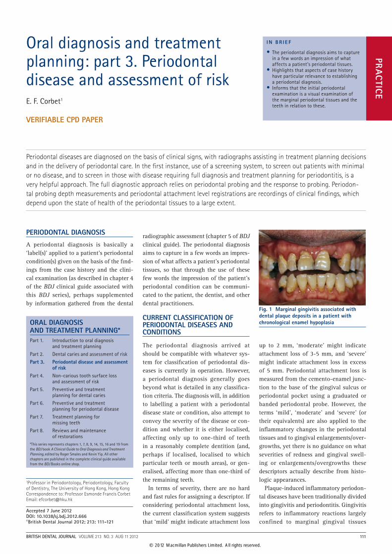
Fig. 1 Marginal gingivitis associated with dental plaque deposits in a patient with chronological enamel hypoplasia
BRITISH DENTAL JOURNAL VOLUME 213 NO. 3 AUG 11 2012 111
© 2012 Macmillan Publishers Limited. All rights reserved.
PRACTICE
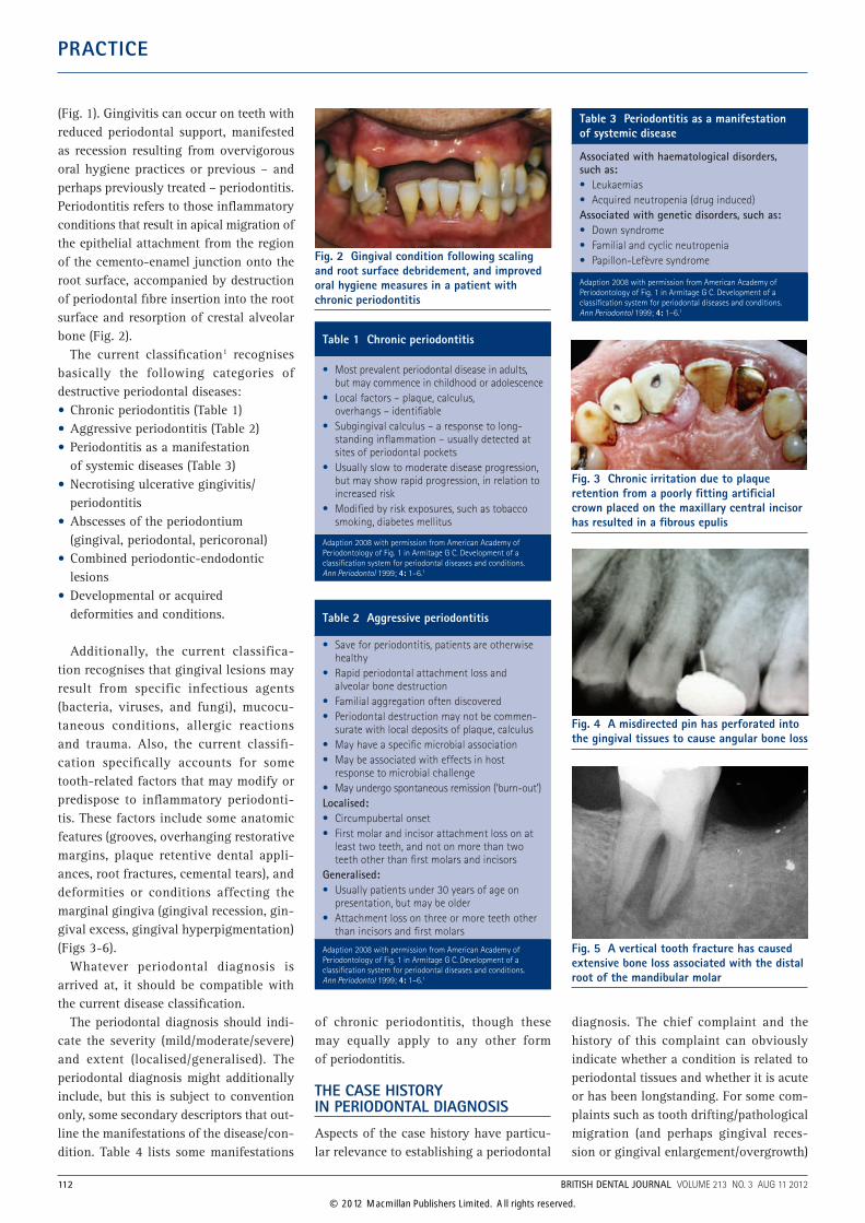
(Fig. 1). Gingivitis can occur on teeth with reduced periodontal support, manifested as recession resulting from overvigorous oral hygiene practices or previous – and perhaps previously treated – periodontitis. Periodontitis refers to those inflammatory conditions that result in apical migration of the epithelial attachment from the region of the cemento-enamel junction onto the root surface, accompanied by destruction of periodontal fibre insertion into the root surface and resorption of crestal alveolar bone (Fig. 2).
The current classification1 recognises basically the following categories of destructive periodontal diseases:•Chronic periodontitis (Table 1)•Aggressive periodontitis (Table 2)•Periodontitis as a manifestation
of systemic diseases (Table 3)•Necrotising ulcerative gingivitis/
periodontitis•Abscesses of the periodontium
(gingival, periodontal, pericoronal)•Combined periodontic-endodontic
lesions•Developmental or acquired
deformities and conditions.
Additionally, the current classifica-tion recognises that gingival lesions may result from specific infectious agents (bacteria, viruses, and fungi), mucocu-taneous conditions, allergic reactions and trauma. Also, the current classifi-cation specifically accounts for some tooth-related factors that may modify or predispose to inflammatory periodonti-tis. These factors include some anatomic features (grooves, overhanging restorative margins, plaque retentive dental appli-ances, root fractures, cemental tears), and deformities or conditions affecting the marginal gingiva (gingival recession, gin-gival excess, gingival hyperpigmentation) (Figs 3-6).
Whatever periodontal diagnosis is arrived at, it should be compatible with the current disease classification.
The periodontal diagnosis should indi-cate the severity (mild/moderate/severe) and extent (localised/generalised). The periodontal diagnosis might additionally include, but this is subject to convention only, some secondary descriptors that out-line the manifestations of the disease/con-dition. Table 4 lists some manifestations
of chronic periodontitis, though these may equally apply to any other form of periodontitis.
THE CASE HISTORY IN PERIODONTAL DIAGNOSIS
Aspects of the case history have particu-lar relevance to establishing a periodontal
diagnosis. The chief complaint and the history of this complaint can obviously indicate whether a condition is related to periodontal tissues and whether it is acute or has been longstanding. For some com-plaints such as tooth drifting/pathological migration (and perhaps gingival reces-sion or gingival enlargement/overgrowth)
Fig. 2 Gingival condition following scaling and root surface debridement, and improved oral hygiene measures in a patient with chronic periodontitis
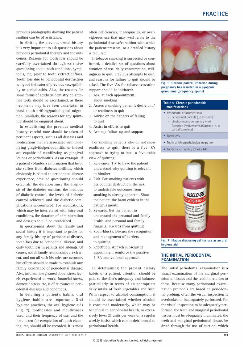
Fig. 3 Chronic irritation due to plaque retention from a poorly fitting artificial crown placed on the maxillary central incisor has resulted in a fibrous epulis
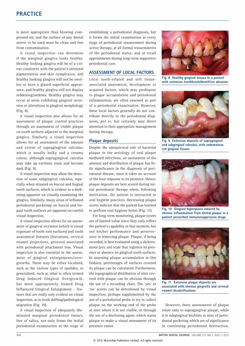
Fig. 4 A misdirected pin has perforated into the gingival tissues to cause angular bone loss
Fig. 5 A vertical tooth fracture has caused extensive bone loss associated with the distal root of the mandibular molar
Table 1 Chronic periodontitis
• Most prevalent periodontal disease in adults, but may commence in childhood or adolescence
• Local factors – plaque, calculus, overhangs – identifiable
• Subgingival calculus – a response to long-standing inflammation – usually detected at sites of periodontal pockets
• Usually slow to moderate disease progression, but may show rapid progression, in relation to increased risk
• Modified by risk exposures, such as tobacco smoking, diabetes mellitus
Adaption 2008 with permission from American Academy of Periodontology of Fig. 1 in Armitage G C. Development of a classification system for periodontal diseases and conditions. Ann Periodontol 1999; 4: 1–6.1
Table 2 Aggressive periodontitis
• Save for periodontitis, patients are otherwise healthy
• Rapid periodontal attachment loss and alveolar bone destruction
• Familial aggregation often discovered• Periodontal destruction may not be commen-
surate with local deposits of plaque, calculus• May have a specific microbial association• May be associated with effects in host
response to microbial challenge• May undergo spontaneous remission (‘burn-out’)Localised:• Circumpubertal onset• First molar and incisor attachment loss on at
least two teeth, and not on more than two teeth other than first molars and incisors
Generalised:• Usually patients under 30 years of age on
presentation, but may be older• Attachment loss on three or more teeth other
than incisors and first molars
Adaption 2008 with permission from American Academy of Periodontology of Fig. 1 in Armitage G C. Development of a classification system for periodontal diseases and conditions. Ann Periodontol 1999; 4: 1–6.1
Table 3 Periodontitis as a manifestation of systemic disease
Associated with haematological disorders, such as:• Leukaemias• Acquired neutropenia (drug induced)Associated with genetic disorders, such as:• Down syndrome• Familial and cyclic neutropenia• Papillon-Lefèvre syndrome
Adaption 2008 with permission from American Academy of Periodontology of Fig. 1 in Armitage G C. Development of a classification system for periodontal diseases and conditions. Ann Periodontol 1999; 4: 1–6.1
112 BRITISH DENTAL JOURNAL VOLUME 213 NO. 3 AUG 11 2012
© 2012 Macmillan Publishers Limited. All rights reserved.
PRACTICE
previous photographs showing the patient smiling can be of assistance.
In eliciting the previous dental history it is very important to ask questions about previous periodontal therapy and the out-comes. Reasons for tooth loss should be carefully ascertained through extensive questioning about tooth conditions, symp-toms, etc, prior to tooth extraction/loss. Tooth loss due to periodontal destruction is a good indicator of previous susceptibil-ity to periodontitis. Also, the reasons for some forms of aesthetic dentistry on ante-rior teeth should be ascertained, as these treatments may have been undertaken to mask tooth drifting/pathological migra-tion. Similarly, the reasons for any splint-ing should be enquired about.
In establishing the previous medical history, careful note should be taken of pertinent aspects, such as all diseases and medications that are associated with mod-ifying gingivitis/periodontitis, or indeed are capable of manifesting as gingival lesions or periodontitis. As an example, if a patient volunteers information that he or she suffers from diabetes mellitus, which obviously is related to periodontal disease experience, detailed questioning should establish: the duration since the diagno-sis of the diabetes mellitus, the methods of diabetic control, the levels of diabetic control achieved, and the diabetic com-plications encountered. For medications, which may be interrelated with intra-oral conditions, the duration of administration and dosages should be established.
In questioning about the family and social history it is important to probe for any family history of periodontal disease, tooth loss due to periodontal disease, and early tooth loss in parents and siblings. Of course, not all family relationships are clear-cut, and not all such histories are accurate, but efforts should be made to establish any family experience of periodontal disease. Also, information gleaned about stress lev-els experienced at work, financial stress, domestic stress, etc, is of relevance to peri-odontal diseases and conditions.
In detailing a patient’s habits, oral hygiene habits are important. Oral hygiene practices, the oral hygiene aids (Fig. 7), toothpastes and mouthrinses used, and their frequency of use, and the time taken for completion of tooth clean-ing, etc, should all be recorded. It is most
often deficiencies, inadequacies, or over-vigorous use that may well relate to the periodontal disease/condition with which the patient presents, so a detailed history is required.
If tobacco smoking is suspected or con-firmed, a detailed set of questions about duration of use, daily consumption, will-ingness to quit, previous attempts to quit, and reasons for failure to quit should be asked. The five ‘A’s for tobacco cessation support should be initiated:1. Ask, at each appointment,
about smoking2. Assess a smoking patient’s desire and/
or readiness to quit3. Advise on the dangers of failing
to quit4. Assist in efforts to quit5. Arrange follow-up and support.
For smoking patients who do not show readiness to quit, there is a five ‘R’s approach to trying to instil a favourable view of quitting:1. Relevance. Try to have the patient
understand why quitting is relevant to him/her
2. Risk. For smoking patients with periodontal destruction, the risk to undesirable outcomes from smoking is already apparent. Show the patient the harm evident in the patient’s mouth
3. Rewards. Get the patient to understand the personal and family health, and personal and family financial rewards from quitting
4. Road-blocks. Discuss the recognition and management of barriers to quitting
5. Repetition. At each subsequent appointment reinforce the positive 5 ‘R’s motivational approach.
In determining the present dietary habits of a patient, attention should be paid to the diet’s adequacy and balance, particularly in terms of an appropriate daily intake of fresh vegetables and fruit. With respect to alcohol consumption, it should be ascertained whether alcohol is consumed moderately, which may be beneficial to periodontal health, or exces-sively (over 21 units per week on a regular weekly basis), which can be detrimental to periodontal health.
THE INITIAL PERIODONTAL EXAMINATION
The initial periodontal examination is a visual examination of the marginal peri-odontal tissues and the teeth in relation to these. Because many periodontal exami-nation protocols are based on periodon-tal probing, often the visual inspection is overlooked or inadequately performed. For the visual inspection to be adequately per-formed, the teeth and marginal periodontal tissues must be adequately illuminated, the teeth and marginal periodontal tissues be dried through the use of suction, which
Fig. 6 Chronic palatal irritation during pregnancy has resulted in a pyogenic granuloma (pregnancy epulis)
Fig. 7 Plaque disclosing gel for use as an oral hygiene aid
Table 4 Chronic periodontitis – manifestations
• Periodontal attachment loss- periodontal pockets (up to x mm)- gingival recession (up to y mm)- furcation involvements (Classes 1-4, or
partial/complete)
• Tooth loss
• Tooth drifting/pathological migration
• Tooth hypermobility (Grades I-III)
BRITISH DENTAL JOURNAL VOLUME 213 NO. 3 AUG 11 2012 113
© 2012 Macmillan Publishers Limited. All rights reserved.
PRACTICE
is more appropriate than blowing com-pressed air, and the surface of any dental mirror to be used must be clean and free from contamination.
A visual inspection can determine if the marginal gingiva looks healthy. Healthy looking gingiva will be of a col-our consistent with the patient’s intraoral pigmentation and skin complexion, and healthy looking gingiva will not be swol-len or have a glazed superficial appear-ance, and healthy gingiva will not display reddening/oedema. Healthy gingiva may occur at areas exhibiting gingival reces-sion or alterations in gingival morphology (Fig. 8).
A visual inspection also allows for an assessment of plaque control practices through an assessment of visible plaque on tooth surfaces adjacent to the marginal gingiva. Similarly, a visual inspection allows for an assessment of the amount and extent of supragingival calculus, which is usually bulky and a creamy colour, although supragingival calculus may take up extrinsic stain and become dark (Fig. 9).
A visual inspection may allow the detec-tion of some subgingival calculus, espe-cially when situated on buccal and lingual tooth surfaces, which is evident as a dark-ening apparent on visually examining the gingiva. Similarly, many areas of inflamed periodontal pocketing on buccal and lin-gual tooth surfaces are apparent on careful visual inspection.
A visual inspection allows for an assess-ment of gingival recession (which is visual exposure of tooth root surfaces) and tooth anatomical features (furcations, cervical enamel projections, grooves) associated with periodontal attachment loss. Visual inspection is also essential in the assess-ment of gingival enlargements/over-growths. These may be either localised, such as the various types of epulides, or generalised, such as what is often termed Drug Induced Gingival Overgrowth, but more appropriately termed Drug Influenced Gingival Enlargement – fea-tures that are really only evident on visual inspection, as is tooth drifting/pathological migration (Fig. 10).
A visual inspection of adequately illu-minated marginal periodontal tissues, free of saliva, not only forms the initial periodontal examination at the stage of
establishing a periodontal diagnosis, but it forms the initial examination at every stage of periodontal reassessment during active therapy, at all formal reassessments of the periodontal status, and at recall appointments during long-term supportive periodontal care.
ASSESSMENT OF LOCAL FACTORSLocal tooth-related and soft tissue-associated anatomical, development or acquired factors, which may predispose to plaque accumulation and periodontal inflammation, are often assessed as part of a periodontal examination. However, these local factors generally do not con-tribute directly to the periodontal diag-nosis, per se, but certainly may direct attention to their appropriate management during therapy.
Plaque depositsDespite the unequivocal role of bacterial plaque in the aetiology of oral plaque mediated infections, an assessment of the amount and distribution of plaque has lit-tle significance in the diagnosis of peri-odontal disease, since it takes no account of the host response to its presence. Hence, plaque deposits are best scored during ini-tial periodontal therapy when, following motivation, the patient is instructed in oral hygiene practices. Decreasing plaque scores indicate that the patient has learned to perform oral hygiene better (Fig. 11).
For long-term monitoring, plaque scores are of limited value since they only reflect the patient’s capability at that moment, but not his/her performance and persever-ance in removing plaque. Plaque, if being recorded, is best evaluated using a dichoto-mous (yes, no) scale that registers its pres-ence or absence on gingival tooth surfaces. In assessing plaque accumulation in this fashion, percentages of surfaces covered by plaque can be calculated. Furthermore, the topographical distribution of sites cov-ered with plaque can be obvious through the use of a recording chart. The ‘yes’ or ‘no’ scores can be determined by visual inspection, perhaps supplemented by the use of a periodontal probe to try to collect plaque on the working end of the probe at sites where it is not visible, or through the use of a disclosing agent, which stains plaque to make a visual assessment of its presence easier.
However, these assessments of plaque relate only to supragingival plaque, while it is subgingival biofilms at sites of perio-dontal pocketing which are of significance in continuing periodontal destruction.
Fig. 8 Healthy gingival tissues in a patient with extensive toothbrush/dentifrice abrasion
Fig. 9 Extensive deposits of supragingival and subgingival calculus, with oedematous red gingival tissues
Fig. 10 Gingival hyperplasia induced by chronic inflammation from dental plaque in a patient prescribed immunosuppressive drugs
Fig. 11 Extensive plaque deposits are associated with obvious gingivitis and cervical enamel decalcifications
114 BRITISH DENTAL JOURNAL VOLUME 213 NO. 3 AUG 11 2012
© 2012 Macmillan Publishers Limited. All rights reserved.
PRACTICE
Assessments of subgingival plaque pres-ence on a ‘yes’ or ‘no’ basis can be deter-mined by using a periodontal probe or a periodontal curette to bring plaque to the surface. While no recording systems for subgingival plaque presence have been described, in reality the determination of subgingival contamination (perhaps when supragingival plaque is not evident because of sporadic, but good plaque con-trol), is a very important assessment.
CalculusCalculus has to be considered as min-eralised plaque deposits. It is the prime plaque retaining factor and, because of this undisputed role, its removal is cru-cial in periodontal therapy. Supragingival calculus is evident on visual inspection and its presence may be recorded tooth surface-by-tooth surface. Subgingival cal-culus can be detected through the use of periodontal probes on tooth root surfaces. Ball-ended probes are advocated as being of particular value in this assessment. Subgingival calculus may be evident on radiographs, although radiographic exami-nation ought never to be performed solely for the assessment of subgingival calculus. The presence of subgingival calculus may be recorded tooth root surface-by-tooth root surface.
OverhangsOverhanging and other plaque retentive margins of restorations may be detected by visual inspection, through the use of a periodontal probe (again, ball-ended probes have been advocated as being especially favourable in this regard), and through assessing whether or not dental floss catches and shreds/tears on attempted interproximal removal in a coronal direc-tion. Interproximal overhangs may be visible on radiographs if the restorations are of radiopaque materials, but only sel-dom are radiographs prescribed solely for the assessment of marginal adaptation of restorations.
Local anatomical factorsLocal anatomical factors that may predis-pose to periodontal inflammation or may alter the course or presentation of peri-odontal destruction are assessed, and these may be both tooth-related and associated with soft tissues.
Tooth-related local factors
Among tooth-related factors often recorded are exposed furcations, cervi-cal enamel projections, root grooves, root concavities, cervical root resorptions, and evident longitudinal root fractures.
Soft tissue-associated local factorsSoft tissue features that may be recorded include (in addition to gingival recession), inconsistent heights of gingival margins (as in altered passive eruption), aber-rant fraenal insertions, gingival enlarge-ments/overgrowths, and oral hygiene induced lesions such as floss-induced gingival clefting.
PERIODONTAL PROBING ASSESSMENTS AND METHODS
Gingival bleeding
Bleeding following gentle running of a periodontal probe around the gingival margin area at the dento-gingival junc-tion denotes inadequate marginal plaque control by a patient. When performed, this testing for gingival bleeding is often combined with the recording of visible plaque for a more comprehensive evalu-ation of a patient’s sustained efforts at personal plaque control. However, more usually the assessment of bleeding is per-formed through noting bleeding following periodontal probing of the gingival sulcus or pocket.
Bleeding on probing (BOP)Absence of bleeding on probing is a reli-able parameter to indicate periodontal stability if the probing is performed using only gentle probing forces with a probe of suitable dimensions. Presence of bleed-ing upon probing indicates the presence of gingival inflammation. Since the absence of bleeding on probing with a gentle force has indicated periodontal stability with a negative predictive value of 98-99%, this clinical parameter is most reliable to moni-tor patients over time in daily practice. Non-bleeding sites may be considered as periodontally stable. On the other hand, bleeding sites seem to have an increased risk for progression of periodontitis, especially when the same site bleeds at repeated evaluations over time. Therefore, it is advisable to register the sites which bleed on probing in a dichotomous way
using a gentle probing force. This allows the calculation of a mean BOP score for the patient, and also yields the topographical location of the bleeding sites.
Probing depth and loss of attachment
Clinical probing is the most commonly used parameter both to document loss of attachment and to establish a diagnosis of periodontitis. There are some sources of error inherent to this method, which contribute to the variability in the meas-urements. Among these, the following factors are discussed: dimensions of the periodontal probe, placement of the probe and obtaining a reference point, crudeness of the measurement scale, probing force, and gingival/periodontal tissue conditions.
Dimensions of the periodontal probeIt is obvious that different sizes of peri-odontal probes will yield different penetra-tion depths into the periodontal tissues, even if all other variables are controlled. Hence, the use of probing instruments with standardised dimensions is a prerequisite for repeated probing depth measurements. Usually, periodontal probes with a point diameter of 0.3-0.5 mm, and ball-ended probes with the ball-end having a diameter of 0.5 mm, have been used successfully.
Placement of the probe and obtaining a reference point
Manual probing involves variable meas-urements generated by the different angu-lations and site localisations of the probe, and by the difficulty in obtaining a fixed landmark as a reference point.
Crudeness of the measurement scaleProbing pocket depth measurements are generally assessed to the nearest millimetre or in ranges of 2.0-3.5 mm. Registering probing pocket depths by bands/ranges is meant to reflect that objective metric measurements of probing are not exact. However, when using banded probes a small difference such as 0.5 mm may change the assessment of probing pocket depth from one category of lesser severity of probing pocket depth to a more ‘severe’ category of probing pocket depth. But, on a metric scale such a small difference may not be registered or may only change the probing pocket depth recorded by 1.0 mm.
BRITISH DENTAL JOURNAL VOLUME 213 NO. 3 AUG 11 2012 115
© 2012 Macmillan Publishers Limited. All rights reserved.
PRACTICE
Probing force
Probing force is a significant vari-able that should be controlled to obtain reproducible measurements of probing depth. If a probing force of 0.25-0.30 N is applied, the apical termination of the probing tip lies usually within the junc-tional epithelium (epithelial attachment) in healthy gingival tissues. Probing forces of this magnitude have gener-ally been accepted for use in electronic pressure sensitive probes. If treatment effects are to be monitored, the probing force will influence the clinical attach-ment levels. At least 0.25 N of probing force has to be applied to yield treatment-related differences in clinical attachment level measurements.
Gingival/periodontal tissue conditionsAttachment level and probing pocket depth measurements are significantly influenced by the density of the connective tissue col-lar around the teeth. In healthy tissues, or those with slight gingivitis, the probe tip generally reaches the most apical cell of the junction epithelium. However, in a periodontal pocket the probe tip routinely exceeds the level of the connective tissue attachment (gingival fibre attachment), by penetrating into the inflamed tissue, to be hindered in its apical penetration either by intact connective tissue fibres or eventually by the alveolar bone crest. The significant influence of the inflammatory infiltrate on probing pocket depth meas-urements has been clearly documented. If tight adaptation of the gingival tissues is obtained as the result of therapy, which may have produced a long junctional epi-thelium as the expected and beneficial out-come of therapy, the most apical cell of the junctional epithelium may not always be reached.
Clinical probingIn spite of it being recognised that peri-odontal probing does not really measure any particular aspect of interest (true pocket depth, true attachment loss, true attachment levels), this diagnostic proce-dure has not only been the most commonly used, but is also the most reliable method for the evaluation of the periodontal tis-sues in both health and disease. It has to be realised that increased probing depth and clinical attachment loss are conditions that
reflect the history of periodontitis rather than its current state of activity.
To obtain a more realistic assessment of disease progression, or healing follow-ing therapy, multiple evaluations should be performed. Obviously, the first evalu-ation before therapy will yield results confounded by a greater impact on measurements from inflammation than probing evaluations following therapy. The reference point (the cemento-enamel junction) may be obstructed by calculus or by dental restorations, and the con-dition of the gingival tissues may allow an easy penetration of the periodontal probe into the tissues, even though the probe position and gentle force applied are standardised.
Traumatic haemorrhage, in addition to readily-evoked inflammatory haem-orrhage, resulting from the probing may make the registration of metric measure-ments imprecise, as blood may obscure both the gingival margin and the cemento-enamel junction at sites of initial reces-sion. These biological variables (tissue conditions, bleeding and calculus) may be minimised following initial periodon-tal therapy. Hence, a repeat periodontal evaluation using probing after the initial therapy will improve the periodontal prob-ing assessment. Therefore, the first peri-odontal evaluation after healing, following initial non-surgical periodontal therapy, should be taken as the baseline for long-term clinical monitoring.
Periodontal probing is routinely per-formed at six sites around each tooth/root (mesio-buccal, buccal, disto-buccal, disto-lingual, lingual and mesio-lingual), and in line with the direction of the long axis of the tooth, for recording purposes. However, for true diagnosis and for the appropriate treatment decisions to be made, periodontal probing may have to be performed at many more sites on a tooth and also the probe may have to be angled away from the long axis direction to explore periodontal pockets.
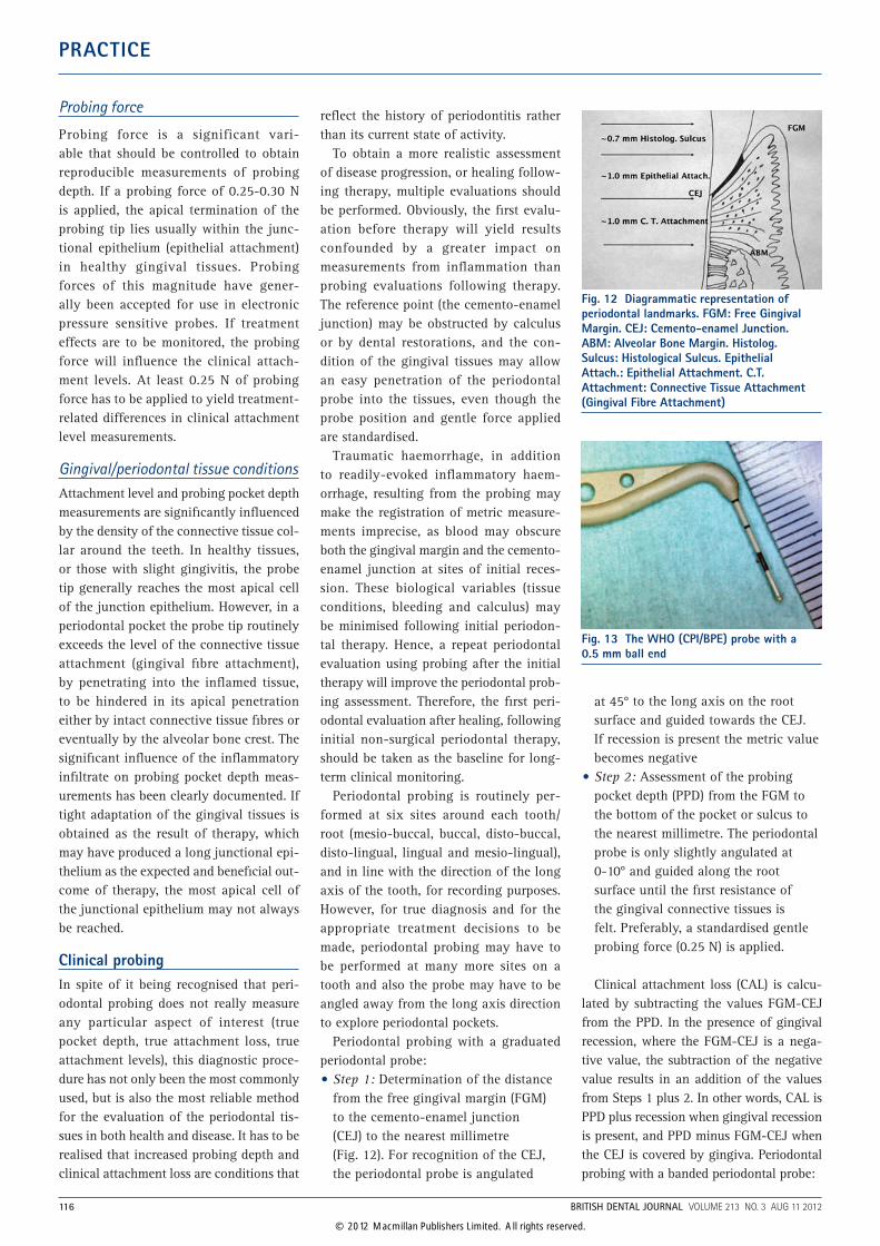
Periodontal probing with a graduated periodontal probe:•Step 1: Determination of the distance
from the free gingival margin (FGM) to the cemento-enamel junction (CEJ) to the nearest millimetre (Fig. 12). For recognition of the CEJ, the periodontal probe is angulated
at 45° to the long axis on the root surface and guided towards the CEJ. If recession is present the metric value becomes negative
•Step 2: Assessment of the probing pocket depth (PPD) from the FGM to the bottom of the pocket or sulcus to the nearest millimetre. The periodontal probe is only slightly angulated at 0-10° and guided along the root surface until the first resistance of the gingival connective tissues is felt. Preferably, a standardised gentle probing force (0.25 N) is applied.
Clinical attachment loss (CAL) is calcu-lated by subtracting the values FGM-CEJ from the PPD. In the presence of gingival recession, where the FGM-CEJ is a nega-tive value, the subtraction of the negative value results in an addition of the values from Steps 1 plus 2. In other words, CAL is PPD plus recession when gingival recession is present, and PPD minus FGM-CEJ when the CEJ is covered by gingiva. Periodontal probing with a banded periodontal probe:
Fig. 12 Diagrammatic representation of periodontal landmarks. FGM: Free Gingival Margin. CEJ: Cemento-enamel Junction. ABM: Alveolar Bone Margin. Histolog. Sulcus: Histological Sulcus. Epithelial Attach.: Epithelial Attachment. C.T. Attachment: Connective Tissue Attachment (Gingival Fibre Attachment)
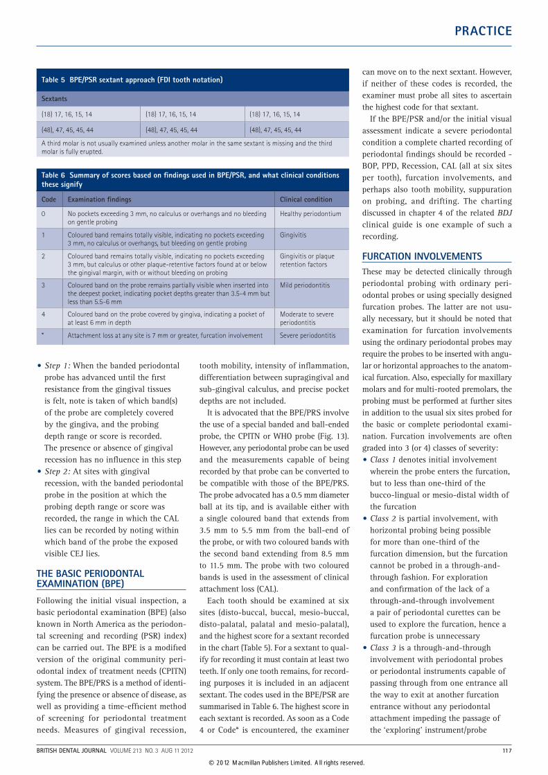
Fig. 13 The WHO (CPI/BPE) probe with a 0.5 mm ball end
116 BRITISH DENTAL JOURNAL VOLUME 213 NO. 3 AUG 11 2012
© 2012 Macmillan Publishers Limited. All rights reserved.
PRACTICE
•Step 1: When the banded periodontal probe has advanced until the first resistance from the gingival tissues is felt, note is taken of which band(s) of the probe are completely covered by the gingiva, and the probing depth range or score is recorded. The presence or absence of gingival recession has no influence in this step
•Step 2: At sites with gingival recession, with the banded periodontal probe in the position at which the probing depth range or score was recorded, the range in which the CAL lies can be recorded by noting within which band of the probe the exposed visible CEJ lies.
THE BASIC PERIODONTAL EXAMINATION (BPE)
Following the initial visual inspection, a basic periodontal examination (BPE) (also known in North America as the periodon-tal screening and recording (PSR) index) can be carried out. The BPE is a modified version of the original community peri-odontal index of treatment needs (CPITN) system. The BPE/PRS is a method of identi-fying the presence or absence of disease, as well as providing a time-efficient method of screening for periodontal treatment needs. Measures of gingival recession,
tooth mobility, intensity of inflammation, differentiation between supragingival and sub-gingival calculus, and precise pocket depths are not included.
It is advocated that the BPE/PRS involve the use of a special banded and ball-ended probe, the CPITN or WHO probe (Fig. 13). However, any periodontal probe can be used and the measurements capable of being recorded by that probe can be converted to be compatible with those of the BPE/PRS. The probe advocated has a 0.5 mm diameter ball at its tip, and is available either with a single coloured band that extends from 3.5 mm to 5.5 mm from the ball-end of the probe, or with two coloured bands with the second band extending from 8.5 mm to 11.5 mm. The probe with two coloured bands is used in the assessment of clinical attachment loss (CAL).
Each tooth should be examined at six sites (disto-buccal, buccal, mesio-buccal, disto-palatal, palatal and mesio-palatal), and the highest score for a sextant recorded in the chart (Table 5). For a sextant to qual-ify for recording it must contain at least two teeth. If only one tooth remains, for record-ing purposes it is included in an adjacent sextant. The codes used in the BPE/PSR are summarised in Table 6. The highest score in each sextant is recorded. As soon as a Code 4 or Code* is encountered, the examiner
can move on to the next sextant. However, if neither of these codes is recorded, the examiner must probe all sites to ascertain the highest code for that sextant.
If the BPE/PSR and/or the initial visual assessment indicate a severe periodontal condition a complete charted recording of periodontal findings should be recorded - BOP, PPD, Recession, CAL (all at six sites per tooth), furcation involvements, and perhaps also tooth mobility, suppuration on probing, and drifting. The charting discussed in chapter 4 of the related BDJ clinical guide is one example of such a recording.
FURCATION INVOLVEMENTSThese may be detected clinically through periodontal probing with ordinary peri-odontal probes or using specially designed furcation probes. The latter are not usu-ally necessary, but it should be noted that examination for furcation involvements using the ordinary periodontal probes may require the probes to be inserted with angu-lar or horizontal approaches to the anatom-ical furcation. Also, especially for maxillary molars and for multi-rooted premolars, the probing must be performed at further sites in addition to the usual six sites probed for the basic or complete periodontal exami-nation. Furcation involvements are often graded into 3 (or 4) classes of severity:•Class 1 denotes initial involvement
wherein the probe enters the furcation, but to less than one-third of the bucco-lingual or mesio-distal width of the furcation
•Class 2 is partial involvement, with horizontal probing being possible for more than one-third of the furcation dimension, but the furcation cannot be probed in a through-and-through fashion. For exploration and confirmation of the lack of a through-and-through involvement a pair of periodontal curettes can be used to explore the furcation, hence a furcation probe is unnecessary
•Class 3 is a through-and-through involvement with periodontal probes or periodontal instruments capable of passing through from one entrance all the way to exit at another furcation entrance without any periodontal attachment impeding the passage of the ‘exploring’ instrument/probe
Table 5 BPE/PSR sextant approach (FDI tooth notation)
Sextants
(18) 17, 16, 15, 14 (18) 17, 16, 15, 14 (18) 17, 16, 15, 14
(48), 47, 45, 45, 44 (48), 47, 45, 45, 44 (48), 47, 45, 45, 44
A third molar is not usually examined unless another molar in the same sextant is missing and the third molar is fully erupted.
Table 6 Summary of scores based on findings used in BPE/PSR, and what clinical conditions these signify
Code Examination findings Clinical condition
0 No pockets exceeding 3 mm, no calculus or overhangs and no bleeding on gentle probing
Healthy periodontium
1 Coloured band remains totally visible, indicating no pockets exceeding 3 mm, no calculus or overhangs, but bleeding on gentle probing
Gingivitis
2 Coloured band remains totally visible, indicating no pockets exceeding 3 mm, but calculus or other plaque-retentive factors found at or below the gingival margin, with or without bleeding on probing
Gingivitis or plaque retention factors
3 Coloured band on the probe remains partially visible when inserted into the deepest pocket, indicating pocket depths greater than 3.5-4 mm but less than 5.5-6 mm
Mild periodontitis
4 Coloured band on the probe covered by gingiva, indicating a pocket of at least 6 mm in depth
Moderate to severe periodontitis
* Attachment loss at any site is 7 mm or greater, furcation involvement Severe periodontitis
BRITISH DENTAL JOURNAL VOLUME 213 NO. 3 AUG 11 2012 117
© 2012 Macmillan Publishers Limited. All rights reserved.
PRACTICE
• (Class 4 is a class/grade assigned by some authorities to a furcation completely visible in a through-and-through fashion as a result of recession).In clinical practice, such as in the chart-
ing discussed in chapter 4 (of the associ-ated BDJ clinical guide), many clinicians collapse classes/grades 1 and 2 together, indicating partial furcation involvement, and classes/grades 3 and 4 together, indi-cating complete furcation involvement.
MOBILITYIncreased tooth mobility (tooth hypermo-bility) may be the result of one or a com-bination of three phenomena:•An increased width of the periodontal
ligament space, resulting from a traumatic event or trauma from occlusion
•A decreased height of the supporting periodontal structures
• Inflammation anywhere within the periodontal attachment apparatus.
As periodontitis-affected teeth will always yield a different ratio of supporting struc-tures to tooth-root length than healthy teeth with unaltered levels of attachment, increased tooth mobility may almost inevi-tably be expected, and may not relate to the current disease status. Therefore, tooth mobility is one of the least reliable assess-ments in diagnosing periodontal disease. If recorded, mobility is usually assessed on a 3-point scale as follows:•Grade I is assigned when assessed
mobility using instrument handles to displace a tooth is greater than physiological mobility, but less than 1.0 mm in a bucco-lingual direction and no vertical displacement of the tooth in its socket is possible
•Grade II is assigned when horizontal mobility is greater than 1.0 mm in a bucco-lingual direction, but no vertical displacement of the tooth in its socket is possible
•Grade III is assigned when vertical displacement of the tooth in its socket is possible.
SUPPURATIONPus will develop in a proportion of peri-odontal lesions, and it may drain through the orifice of a pocket. This criterion for
suppuration may be recognised while clinically probing the periodontal lesion, and the suppuration may be recorded.
TOOTH DRIFTING/PATHOLOGICAL MIGRATION
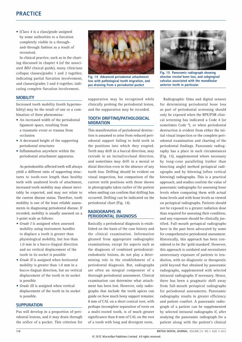
This manifestation of periodontal destruc-tion is assumed to arise from reduced peri-odontal support failing to hold teeth in the positions into which they erupted. Teeth may drift in a buccal direction, may extrude in an incisal/occlusal direction, and sometimes may drift in a mesial or distal direction even in the absence of any tooth loss. Drifting should be evident on visual inspection, but comparison of the current teeth positions with those shown in photographs taken earlier of the patient when smiling can confirm that drifting has occurred. Drifting can be indicated on the periodontal chart (Fig. 14).
RADIOGRAPHS IN PERIODONTAL DIAGNOSIS
Basically a periodontal diagnosis is estab-lished on the basis of the case history and the clinical examination. Information gleaned from appropriate radiographic examinations, except for aspects such as the diagnosis of combined periodontal-endodontic lesions, do not play a deter-mining role in the establishment of a periodontal diagnosis. But, radiographs are often an integral component of a thorough periodontal assessment. Clinical examination can determine what attach-ment has been lost. However, only radio-graphs that include the tooth apices can guide on how much bony support remains: 8 mm of CAL on a short conical root, with perhaps incomplete separation of roots on a multi-rooted tooth, is of much greater significance than 8 mm of CAL on the root of a tooth with long and divergent roots.
Radiographic films and digital sensors for determining periodontal bone loss as part of periodontal screening should only be exposed when the BPE/PSR clini-cal screening has indicated a Code 4 (or sometimes Code *), or when periodontal destruction is evident from either the ini-tial visual inspection or the complete peri-odontal examination and charting of the periodontal findings. Panoramic radiog-raphy has a place in such circumstances (Fig. 15), supplemented where necessary by long-cone paralleling (rather than bisecting angle) method periapical radi-ographs and by bitewing (often vertical bitewing) radiographs. This is a practical approach, and studies confirm the value of panoramic radiographs for assessing bone levels when comparing them with actual bone levels and with bone levels as viewed on periapical radiographs. Patients should not be exposed to a greater radiation dose than required for assessing their condition, and any exposure should be clinically jus-tified. Full-mouth periapical radiographs have in the past been advocated by some for comprehensive periodontal assessment. Historically, this approach has been con-sidered to be the ‘gold standard’. However, this approach is outdated and necessitates unnecessary exposure of patients to irra-diation, with no diagnostic or therapeutic yield beyond that obtained by panoramic radiographs, supplemented with selected intraoral radiographs if necessary. Hence, there has been a pragmatic shift away from full-mouth periapical radiographs for periodontal assessments. Panoramic radiography results in greater efficiency and patient comfort. A panoramic radio-graph of a patient can be supplemented by selected intraoral radiographs if, after studying the panoramic radiograph for a patient along with the patient’s clinical
Fig. 14 Advanced periodontal attachment loss with pathological tooth migration, and pus draining from a periodontal pocket
Fig. 15 Panoramic radiograph showing alveolar crestal bone loss, and subgingival calculus associated with the mandibular anterior teeth in particular
118 BRITISH DENTAL JOURNAL VOLUME 213 NO. 3 AUG 11 2012
© 2012 Macmillan Publishers Limited. All rights reserved.
PRACTICE
periodontal findings, doubt remains about a diagnosis (in the case of perhaps a com-bined periodontalendodontic lesion), or an appropriate treatment decision.
Alveolar bone lossRadiographs can allow for an assessment of interdental crestal alveolar bone lev-els and for an appreciation of patterns of bone loss – horizontal and/or angular (a preferred term to vertical) bone loss. However, these assessments do not form part of the diagnostic process. Bone loss should be correlated with CAL findings, and it should be noted that bone destruc-tion can be underestimated in patients with early periodontal destruction, because buccal and lingual cortical bone plates are still largely intact. Conversely, bone loss may be overestimated in patients with severe periodontal disease, perhaps espe-cially on panoramic radiographs where the tooth roots and alveolar crest may be positioned largely within the focal trough of the X-ray beam (Fig. 15).
Radiographic features impinging upon treatment planning
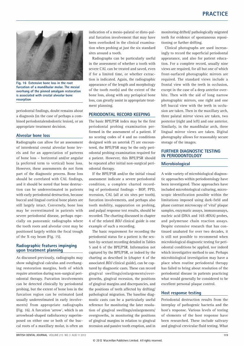
As discussed previously, radiographs may show subgingival calculus and overhang-ing restoration margins, both of which require attention during non-surgical peri-odontal therapy. Furcation involvements can be detected clinically by periodontal probing, but the extent of bone loss in the furcation region can be estimated (and usually underestimated in early involve-ments) from appropriate radiographs (Fig. 16). A furcation ‘arrow’, which is an arrowhead-shaped radiolucency superim-posed on either one or both of the buc-cal roots of a maxillary molar, is often an
indication of a mesio-palatal or disto-pal-atal furcation involvement that may have been overlooked in the clinical examina-tion when probing at just the six standard sites around a tooth.
Radiographs can be particularly useful in the assessment of whether a tooth with severe CAL can be treated and saved, even if for a limited time, or whether extrac-tion is indicated. Again, the radiographic appearance of the length and morphology of the tooth root(s) and the extent of the bone loss, along with any periapical bone loss, can greatly assist in appropriate treat-ment planning.
PERIODONTAL RECORD KEEPINGThe basic BPE/PSR index may be the first periodontal probing examination per-formed in the assessment of a patient. If no scoring codes of 4 and no conditions designed with an asterisk (*) are encoun-tered, the BPE/PSR may be the only peri-odontal probing examination required for a patient. However, this BPE/PSR should be repeated after initial non-surgical peri-odontal therapy.
If the BPE/PSR and/or the initial visual assessment indicate a severe periodontal condition, a complete charted record-ing of periodontal findings – BOP, PPD, recession, CAL (all at six sites per tooth), furcation involvements, and perhaps also tooth mobility, suppuration on probing, and pulpal response test results, should be recorded. The charting discussed in chapter 4 of the related BDJ clinical guide is one example of such a recording.
The basic requirement for recording the periodontal status for a patient is the sex-tant-by-sextant recording detailed in Tables 5 and 6 of the BPE/PSR. Information not captured by the BPE/PSR, or indeed by the charting as described in (chapter 4 of the associated BDJ clinical guide), can be cap-tured by diagnostic casts. These can record gingival swellings/enlargements/over-growths, gingival recession, the positions of gingival margins and discrepancies, and the positions of teeth affected by drifting/pathological migration. The baseline diag-nostic casts can be a particularly useful reference for monitoring the later resolu-tion of gingival swellings/enlargements/overgrowths, in monitoring the positions of gingival margins in relation to gingival recession and passive tooth eruption, and in
monitoring drifted/ pathologically migrated teeth for evidence of spontaneous reposi-tioning or further drifting.
Clinical photographs are used increas-ingly to record the superficial periodontal appearance, and also for patient educa-tion. For a complete record, usually nine views are required, for all but one of which front-surfaced photographic mirrors are required. The standard views include a frontal view with the teeth in occlusion, except in the case of a deep anterior over-bite. Then with the aid of long narrow photographic mirrors, one right and one left buccal view with the teeth in occlu-sion are taken. Then in the maxillary arch, three palatal mirror views are taken, two posterior (right and left) and one anterior. Similarly, in the mandibular arch, three lingual mirror views are taken. Digital photography allows for reasonably secure storage of the images.
FURTHER DIAGNOSTIC TESTING IN PERIODONTOLOGY
Microbiological
A wide variety of microbiological diagnos-tic approaches within periodontology have been investigated. These approaches have included microbiological culturing, micro-scopic identification possible within the limitations imposed using dark-field and phase contrast microscopy of ‘vital’ plaque samples, enzymatic assays, immunoassays, nucleic acid (DNA and 16S rRNA) probes, and polymerase chain reaction assays. Despite extensive research that has con-tinued unabated for over two decades, it is still not possible to recommend when microbiological diagnostic testing for peri-odontal conditions be applied, nor indeed which investigative method to use. Perhaps microbiological investigation may have a place when routine periodontal therapy has failed to bring about resolution of the periodontal disease in patients practicing what would generally be considered to be excellent personal plaque control.
Host response testingPeriodontal destruction results from the interplay of pathogenic bacteria and the host’s response. Various levels of testing of elements of the host response have been researched. These include salivary and gingival crevicular fluid testing. What
Fig. 16 Extensive bone loss in the root furcation of a mandibular molar. The mesial overhang of the pinned amalgam restoration is associated with crestal alveolar bone resorption
BRITISH DENTAL JOURNAL VOLUME 213 NO. 3 AUG 11 2012 119
© 2012 Macmillan Publishers Limited. All rights reserved.
PRACTICE
has been investigated in these fluids has included antibody responses, inflammatory cell products (enzymes and their inhibitors, inflammatory mediators and host response modifiers), and tissue breakdown products. Despite over three decades of research there is little compelling evidence to show that such testing is of significant clinical benefit for patients or their dentists in the diagnosis of periodontal diseases, beyond what is learned from the history of previ-ous disease experience gleaned from the clinical examination.
DIAGNOSES OF ACUTE PERIODONTAL LESIONS
Acute periodontal abscesses
Most, but not all, patients presenting with an acute periodontal abscess complain of pain. In the case history it is the history of previous periodontal therapy that is significant, as periodontal abscesses are known to occur in patients undergoing periodontal treatment. An explanation for this based on incomplete removal of calculus has been proposed, although the possibility of allowing microbial penetra-tion of the soft tissue pocket wall during subgingival instrumentation, shown to occur in periodontal abscesses, also can-not be ruled out. Recent previous antibiotic therapy, often for non-oral reasons, may be a trigger for periodontal abscess for-mation. Patients also may recall traumatic intraoral events, because forceful impac-tion of foreign bodies or materials into a pocket can predispose toward periodontal abscess formation at previous periodon-tally involved sites.
The clinical findings associated with periodontal abscesses are swelling, oedema and redness of the affected periodontal sites. Bleeding on probing the affected site will invariably occur, and suppuration on pressure or on probing occurs in the majority of instances. That suppuration does not always occur in response to peri-odontal probing may be due to the tortuous nature of some pockets. However, a ready release of pus on periodontal probing with alleviation of the symptoms of pain, pres-sure and discomfort can be helpful towards establishing the diagnosis of a periodontal abscess. A tooth with an associated peri-odontal abscess often displays hypermobil-ity, which can be helpful in determining
the tooth a periodontal abscess arises from, as periodontal abscesses may migrate from the involved tooth and present at a site some distance from the origin. Tenderness of the tooth to palpation may be present. Lymphadenopathy occurs only in a minor-ity of patients, and fever and malaise are rare. Periodontal pocket probing depths, while increased, are variable. Although the majority of periodontal abscesses have been reported to occur interdentally, they may occur on any tooth surface.
Periodontal abscesses must be differenti-ated from periapical abscesses.
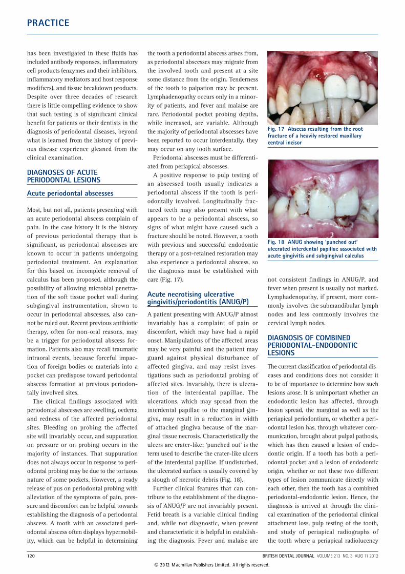
A positive response to pulp testing of an abscessed tooth usually indicates a periodontal abscess if the tooth is peri-odontally involved. Longitudinally frac-tured teeth may also present with what appears to be a periodontal abscess, so signs of what might have caused such a fracture should be noted. However, a tooth with previous and successful endodontic therapy or a post-retained restoration may also experience a periodontal abscess, so the diagnosis must be established with care (Fig. 17).
Acute necrotising ulcerative gingivitis/periodontitis (ANUG/P)
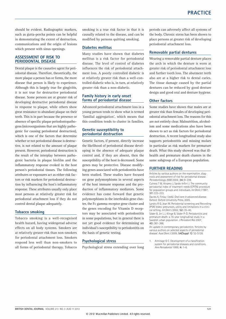
A patient presenting with ANUG/P almost invariably has a complaint of pain or discomfort, which may have had a rapid onset. Manipulations of the affected areas may be very painful and the patient may guard against physical disturbance of affected gingiva, and may resist inves-tigations such as periodontal probing of affected sites. Invariably, there is ulcera-tion of the interdental papillae. The ulcerations, which may spread from the interdental papillae to the marginal gin-giva, may result in a reduction in width of attached gingiva because of the mar-ginal tissue necrosis. Characteristically the ulcers are crater-like; ‘punched out’ is the term used to describe the crater-like ulcers of the interdental papillae. If undisturbed, the ulcerated surface is usually covered by a slough of necrotic debris (Fig. 18).
Further clinical features that can con-tribute to the establishment of the diagno-sis of ANUG/P are not invariably present. Fetid breath is a variable clinical finding and, while not diagnostic, when present and characteristic it is helpful in establish-ing the diagnosis. Fever and malaise are
not consistent findings in ANUG/P, and fever when present is usually not marked. Lymphadenopathy, if present, more com-monly involves the submandibular lymph nodes and less commonly involves the cervical lymph nodes.
DIAGNOSIS OF COMBINED PERIODONTAL-ENDODONTIC LESIONS
The current classification of periodontal dis-eases and conditions does not consider it to be of importance to determine how such lesions arose. It is unimportant whether an endodontic lesion has affected, through lesion spread, the marginal as well as the periapical periodontium, or whether a peri-odontal lesion has, through whatever com-munication, brought about pulpal pathosis, which has then caused a lesion of endo-dontic origin. If a tooth has both a peri-odontal pocket and a lesion of endodontic origin, whether or not these two different types of lesion communicate directly with each other, then the tooth has a combined periodontal-endodontic lesion. Hence, the diagnosis is arrived at through the clini-cal examination of the periodontal clinical attachment loss, pulp testing of the tooth, and study of periapical radiographs of the tooth where a periapical radiolucency
Fig. 17 Abscess resulting from the root fracture of a heavily restored maxillary central incisor
Fig. 18 ANUG showing ‘punched out’ ulcerated interdental papillae associated with acute gingivitis and subgingival calculus
120 BRITISH DENTAL JOURNAL VOLUME 213 NO. 3 AUG 11 2012
© 2012 Macmillan Publishers Limited. All rights reserved.
PRACTICE
should be evident. Radiographic markers, such as gutta-percha points can be helpful in demonstrating the extent of destruction, communications and the origin of lesions which present with sinus openings.
ASSESSMENT OF RISK TO PERIODONTAL DISEASE
Dental plaque is the causative agent for peri-odontal disease. Therefore, theoretically, the more plaque a person has or forms, the more disease that person is likely to experience. Although this is largely true for gingivitis, it is not true for destructive periodontal disease. Some persons are at greater risk of developing destructive periodontal disease in response to plaque, while others show great resistance to abundant plaque on their teeth. This is in part because the presence or absence of specific plaque periodontopatho-gens (microorganisms that are highly patho-genic for causing periodontal destruction), which is one of the factors that determine whether or not periodontal disease is destruc-tive, is not related to the amount of plaque present. However, periodontal destruction is the result of the interplay between patho-genic bacteria in plaque biofilm and the inflammatory response evoked in the host person’s periodontal tissues. The following attributes or exposures act as either risk fac-tors or risk markers for periodontal destruc-tion by influencing the host’s inflammatory response. These attributes usually only place most persons at relatively greater risk for periodontal attachment loss if they do not control dental plaque adequately.
Tobacco smokingTobacco smoking is a well-recognised health hazard, having widespread adverse effects on all body systems. Smokers are at relatively greater risk than non-smokers for periodontal attachment loss. Smokers respond less well than non-smokers to all forms of periodontal therapy. Tobacco
smoking is a true risk factor in that it is causally related to the disease, and can be modified by persons quitting smoking.
Diabetes mellitusMany studies have shown that diabetes mellitus is a risk factor for periodontal disease. The level of control of diabetes influences the risk of periodontal attach-ment loss. A poorly controlled diabetic is at relatively greater risk than a well-con-trolled diabetic who is, in turn, at relatively greater risk than a non-diabetic.
Family history in early onset forms of periodontal disease
Advanced periodontal attachment loss in a young person tends to show what is termed ‘familial aggregation’, which means that this condition tends to cluster in families.
Genetic susceptibility to periodontal destruction
Genetic factors, if present, directly increase the likelihood of periodontal disease devel-oping in the absence of adequate plaque control and, if they are absent, then the susceptibility of the host is decreased. Some genes may be protective. Disease modify-ing genes associated with periodontitis have been studied. These studies have focused on gene polymorphisms in several aspects of the host immune response and the pro-duction of inflammatory mediators. Some evidence has come forward that genetic polymorphisms in the interleukin gene clus-ter, the Fc gamma receptor gene cluster and the genes encoding for Vitamin D recep-tors may be associated with periodontitis in some populations, but in general there is not yet good evidence for determining an individual’s susceptibility to periodontitis on the basis of genetic testing.
Psychological stressPsychological stress extending over long
periods can adversely affect all systems of the body. Chronic stress has been shown to place persons at greater risk of developing periodontal attachment loss.
Removable partial denturesWearing a removable partial denture places the arch in which the denture is worn at greater risk of periodontal attachment loss and further tooth loss. The abutment teeth also are at a higher risk to dental caries. The tissue damage caused by the partial dentures can be reduced by good denture design and good oral and denture hygiene.
Other factorsSome studies have shown that males are at greater risk than females of developing peri-odontal attachment loss. The reasons for this are not entirely clear. Malnutrition, alcohol-ism and some medications also have been shown to act as risk factors for periodontal destruction. A recent longitudinal study also supports periodontitis and missing molars in particular as risk markers for premature death. What this study showed was that ill-health and premature death clusters in the same subgroup of a European population.
FURTHER READINGArticles by various authors on the examination, diag-nosis and assessment of risk for periodontal disease.Periodontology 2000 2004; 34: 9–239.Cutress T W, Ainamo J, Sardo-Infirri J. The community periodontal index of treatment needs (CPITN) procedure for population groups and individuals. Int Dent J 1987; 37: 222–233.Davies A, Finlay I (eds). Oral care in advanced disease. Oxford: Oxford University Press, 2005.Landry R G, Jean M. Periodontal screening and Recording (PSR) Index: precursors, utility and limitations in a clini-cal setting. Int Dent J 2002; 52: 35–40.Söder B, Jin L J, Klinge B, Söder P-Ö. Periodontitis and premature death: a 16-year longitudinal study in a Swedish urban population. J Periodont Res 2007; 42: 361–366.An update in contemporary periodontics. ‘Articles by various authors on selected aspects of periodontal disease’. Aust Dent J 2009; 54(Suppl 1): S2-S128.
1. Armitage G C. Development of a classification system for periodontal diseases and conditions. Ann Periodontol 1999; 4: 1–6.
BRITISH DENTAL JOURNAL VOLUME 213 NO. 3 AUG 11 2012 121
© 2012 Macmillan Publishers Limited. All rights reserved.
Related Documents