1 Chapter 28, Part 1 Cardiology 2 Part 1: Cardiovascular Anatomy & Physiology, ECG Monitoring, and Dysrhythmia Analysis 3 Cardiovascular Anatomy • Coronary Circulation 4 Cardiac Physiology • The cardiac cycle consists of ____________________ and Systole • Diastole: Relaxation phase • Systole: ____________________ phase • ____________________ fraction: during each contraction, the ventricles eject about 2/3 of the blood it contains

Welcome message from author
This document is posted to help you gain knowledge. Please leave a comment to let me know what you think about it! Share it to your friends and learn new things together.
Transcript
1
1
Chapter 28, Part 1Cardiology
2
Part 1: Cardiovascular Anatomy & Physiology, ECG Monitoring,
and Dysrhythmia Analysis
3
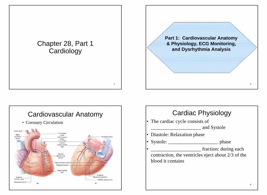
Cardiovascular Anatomy• Coronary Circulation
4
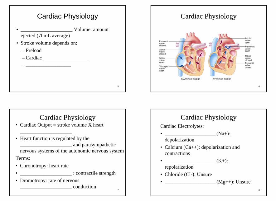
Cardiac Physiology• The cardiac cycle consists of
____________________ and Systole• Diastole: Relaxation phase• Systole: ____________________ phase• ____________________ fraction: during each
contraction, the ventricles eject about 2/3 of the blood it contains
2
5
Cardiac Physiology
• ____________________ Volume: amount ejected (70mL average)
• Stroke volume depends on:– Preload– Cardiac ____________________ – ____________________
6
Cardiac Physiology
7
Cardiac Physiology• Cardiac Output = stroke volume X heart
____________________ • Heart function is regulated by the
____________________ and parasympathetic nervous systems of the autonomic nervous system
Terms:• Chronotropy: heart rate• ____________________ : contractile strength• Dromotropy: rate of nervous
____________________ conduction8
Cardiac PhysiologyCardiac Electrolytes:• ____________________(Na+):
depolarization• Calcium (Ca++): depolarization and
contractions• ____________________(K+):
repolarization• Chloride (Cl-): Unsure• ____________________(Mg++): Unsure

3
9
Cardiac Depolarization• Resting Potential (____________________ ) : The
normal electrical state of cardiac cells. Negatively charged
• Action Potential: The stimulation of myocardial cells, as evidenced by a change in the membrane electrical charge, that spreads across the myocardium
• Cardiac ____________________ : a reversal of charges at a cell membrane so that the inside of the cell becomes positive in relation to the outside. Positively charged
10
Cardiac Depolarization
11
Cardiac Physiology• Properties of the Cardiac Conductive System• ____________________ : Cells are capable of
responding to electrical stimulus• ____________________ : Cells can transmit
electrical impulses from cell to cell• ____________________ : Each cell can
depolarize without any outside impulse• ____________________ : Cells have the ability
to expand12
Cardiac PhysiologyCardiac Conductive System Components:• ____________________ Node• Internodal Atrial Pathways• Atrioventricular Node• Atrioventricular ____________________ • Bundle of ____________________ • Left and Right Bundle Branches• ____________________ Fibers
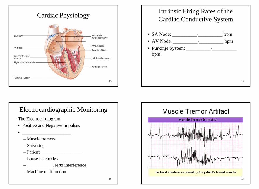
4
13
Cardiac Physiology
14
Intrinsic Firing Rates of the Cardiac Conductive System
• SA Node: __________-__________ bpm• AV Node: __________-__________ bpm• Purkinje System: __________-__________
bpm
15
Electrocardiographic MonitoringThe Electrocardiogram• Positive and Negative Impulses• ____________________
– Muscle tremors– Shivering– Patient ____________________ – Loose electrodes– ___________ Hertz interference– Machine malfunction
16
Muscle Tremor Artifact
5
17
60 Cycle Interference
18
The Electrocardiogram
ECG Leads• ____________________
(Limb)– Einthoven’s Triangle– Leads I, II, III
• ______________________-(Unipolar)– aVR, aVL, aVF
• ____________________– V1 – V6
19
Bipolar Lead Placement Sites
Lead Positive NegativeI Left Arm Right ArmII Left Leg Right ArmIII Left Leg Left Arm
20
The Electrocardiogram(Page 1141)
• Lead Systems and Heart Surfaces

6
21
The Electrocardiogram
Routine Monitoring• Information from a single lead shows:
– Rate & ____________________ .– ____________________ to conduct an impulse.
22
The ElectrocardiogramA single lead cannot:• Identify/locate an ____________________ .• Identify ____________________ deviation
or chamber enlargement.• Identify right-to-left differences in
conduction.• The quality or presence of
____________________ action.
23
The ElectrocardiogramECG Paper• Speed:
___________ mm/sec is normal
• Amplitude and Deflection: __________ large boxes = 1 millivolt
• Each small square = __________ seconds
• Each larger square = __________ seconds 24
The ElectrocardiogramECG Components:• __________ Wave• __________
Complex• __________ Wave• __________ Wave
(rare)• Isoelectric line: line
with __________ electrical activity (flat)

7
25
The Electrocardiogram
26
The Electrocardiogram
27
The Electrocardiogram
28
The Electrocardiogram

8
29
The Electrocardiogram
30
The Electrocardiogram
31
The Electrocardiogram
32
Normal Time Intervals
• P–R Interval (PRI) or P–Q Interval (PQI)– __________–__________
Seconds• QRS Interval
– __________ Seconds• S–T Segment• Q–T Interval
– __________ Seconds
9
33
Refractory Periods• ____________________ : Heart CAN beat
again but without adequate pumping action• ____________________ : Heart CANNOT
pump again
34
S-T Segment Changes• Elevation or depression of the S-T segment
above or below the ____________________ line• Associated with Myocardial Infarctions
– ____________________
– Injury– ____________________
35
Interpretation of Rhythm Strips• Always be ____________________ and
analytical.• Memorize the rules for each dysrhythmia.• Analyze a given rhythm strip according to a
specific ____________________ .• Compare your analysis to the rules for each
dysrhythmia.• Identify the dysrhythmia by its similarity to
established rules.36
Five-Step Procedure
1. Analyze the ____________________ (QRS).• Over 100 = tachycardia• Less than 60 = bradycardia2. Analyze the ____________________ .• Regular or irregular?• If irregular is it regularly irregular or irregularly
irregular? (____________________ )

10
37
Five-Step Procedure
3. Analyze the __________-waves.• Present?• ____________________ or inverted?4. Analyze the __________ interval.• 0.12 to 0.20 is normal5. Analyze the ____________________ complex.• Broad or narrow?
38
Analyzing the Rate
• 6 seconds method– Count the number of complexes in a 6 second
interval (___________ large squares) and multiply by 10
• Heart Rate Calculator ____________________
– Commercially available rulers
39
Analyzing the RateR-R Interval• Only if heart rate is ____________________
• Measure duration between R waves in seconds and divide into __________– Example: 60 ÷ 0.65 seconds = 92 bpm
• Count the number of large squares within the R-R interval and divide into __________– Example: 300 ÷ 3.5 boxes = 86 bpm
• Count the number of small squares within the R-R interval and divide into _________
40
Analyzing the RateTriplicate Method• Used only with ____________________ rhythms• Locate an R wave that falls on a dark line bordering a
large box. Then assign numbers corresponding with to the heart rate to the next __________ dark lines to the right.
• The order is 300, 150, 100, 75, 60, and 50.• The number that corresponds to the dark line closest to
the ____________________ of the next R wave is a rough estimate of the heart rate

11
41
What is the Rate?
________________ Beats Per Minute________ small boxes between R waves1500 divided by ________ = ________
42
What is the Rate?
• __________ Beats Per Minute• __________ small boxes between R waves• __________ divided by __________ = ________
43
Analyzing the Rhythm
• ____________________?• ____________________ Irregular?• ____________________ Irregular?• ____________________ Irregular?
44
Is This Rhythm Regular?
• _______________________________
12
45
Is This Rhythm Regular?
• ____________________________
46
Analyzing the P Wave
• Reflects ____________________ depolarization• Are P waves present?• Are the P wave ____________________ ?• Is there ____________________ P wave per QRS
complex?• Are the P waves upright or ____________________ ?• Do all P waves look alike?
47
Analyze the P Waves
• Present, ____________________ , 1 per QRS, upright, all look ____________________
48
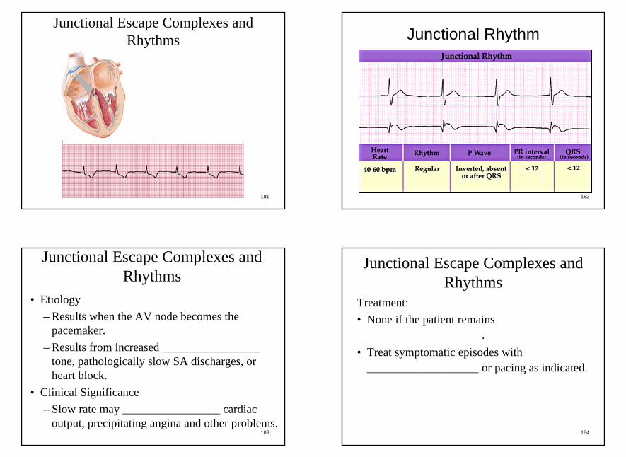
Analyze the P Waves
• _______________________________

13
49
Analyze the P Waves
• Present, regular, ____________________ than 1 P wave for some QRS complexes, ____________________ , all look alike
50
Analyze the P Waves
• _________________________ but not clear
51
Analyzing the P-R Interval
• Time needed for atrial depolarization and conduction of the impulse to the AV node
• Normal is __________ to __________ seconds (3-5 small boxes)
• Measured from beginning of __________ wave to beginning of __________ wave
• Any deviation is abnormal
52
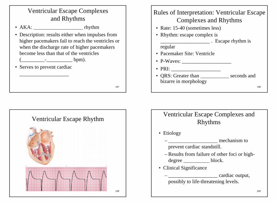
Analyze the P-R Interval
• _____________________ seconds

14
53
Analyze the P-R Interval
• Varies: ___________ to _______________ seconds
54
Analyzing the QRS Complexes
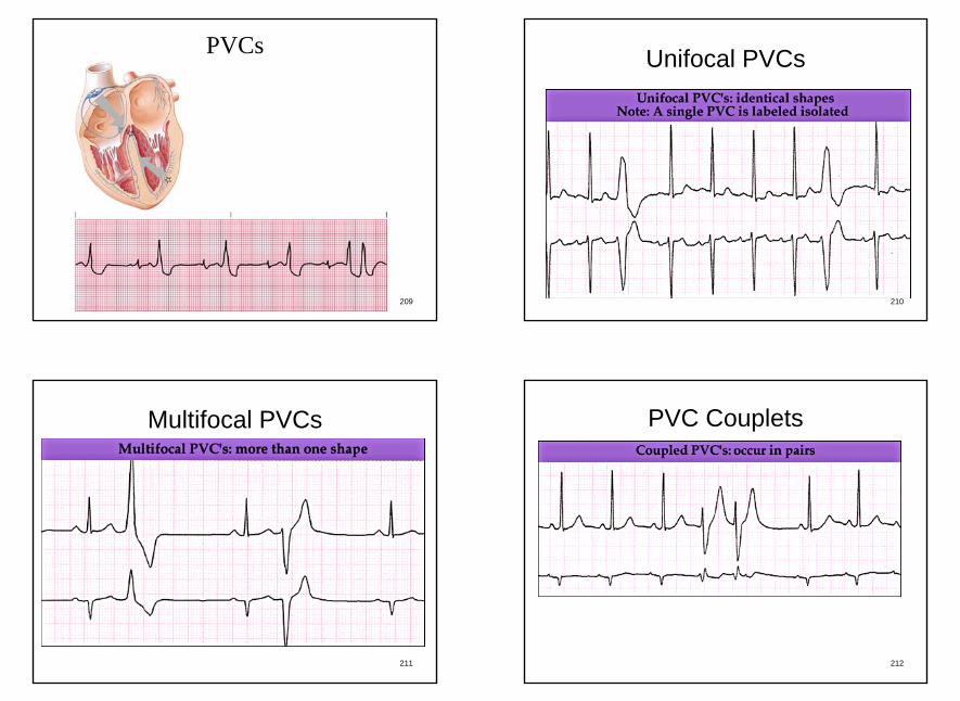
• Do all the QRS complexes look alike?• What is the QRS _________________________ • Normal duration is __________ to __________
seconds (narrow complexes)• Anything longer than __________ seconds is
abnormal (broad complexes)
55
What is the QRS Duration?
• ______________________ seconds
56
What is the QRS Duration?
• Narrow Complexes: ____________ seconds• Wide Complexes: ______________ seconds
15
57
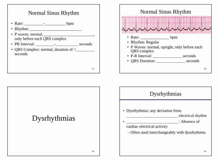
Normal Sinus Rhythm
• Rate: _________–__________ bpm• Rhythm: _________________________ • P waves: normal, ________________________,
only before each QRS complex• PR Interval: _________-___________ seconds• QRS Complex: normal, duration of <_________
seconds
58
Normal Sinus Rhythm
• Rate: ______________ bpm• Rhythm: Regular• P Waves: normal, upright, only before each
QRS complex• P-R Interval: ______________ seconds• QRS Duration: ______________ seconds
59
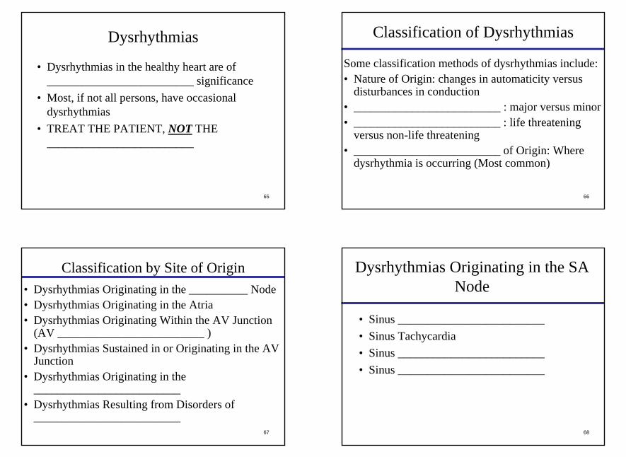
Dysrhythmias
60
Dysrhythmias
• Dysrhythmia: any deviation from _________________________ electrical rhythm
• _________________________ : Absence of cardiac electrical activity– Often used interchangeably with dysrhythmia

16
61
DysrhythmiasMechanism of Impulse Formation• _________________________ Foci
– Caused by increased automaticity– When heart cells other than the pacemaker cells
automatically _________________________
– Produces _________________________ (abnormal) Beats– Premature Ventricular contractions (__________) or
premature atrial contractions (___________)
62
Dysrhythmias_________________________• Caused when disease or ischemia alters 2
branches of a pathway, slowing conduction in 1 branch and causing a unidirectional block in the other
• May be isolated beats or tachydysrhythmias– Atrial fibrillation (___________________)– Paroxysmal supraventricular tachycardia
(_____________)
63
Causes of Dysrhythmias
• Myocardial Ischemia, Necrosis, or _________________________
• Autonomic Nervous System Imbalance• Distention of the Chambers of the Heart• Blood _________________________ Abnormalities• _________________________ Imbalances• Trauma to the Myocardium
64
Causes of Dysrhythmias
• Drug Effects and Drug Toxicity• _________________________• Hypothermia• _________________________ Damage• Idiopathic Events• _________________________ Occurrences
17
65
Dysrhythmias
• Dysrhythmias in the healthy heart are of _________________________ significance
• Most, if not all persons, have occasional dysrhythmias
• TREAT THE PATIENT, NOT THE _________________________
66
Classification of Dysrhythmias
Some classification methods of dysrhythmias include:• Nature of Origin: changes in automaticity versus
disturbances in conduction• _________________________ : major versus minor• _________________________ : life threatening
versus non-life threatening• _________________________ of Origin: Where
dysrhythmia is occurring (Most common)
67
Classification by Site of Origin• Dysrhythmias Originating in the __________ Node• Dysrhythmias Originating in the Atria• Dysrhythmias Originating Within the AV Junction
(AV _________________________ )• Dysrhythmias Sustained in or Originating in the AV
Junction• Dysrhythmias Originating in the
_________________________ • Dysrhythmias Resulting from Disorders of
_________________________ 68
• Sinus _________________________ • Sinus Tachycardia• Sinus _________________________ • Sinus _________________________
Dysrhythmias Originating in the SA Node

18
69
Rules of Interpretation:Sinus Bradycardia
• Description: results from slowing of the SA node• Rate: Less than ___________• Rhythm: _________________________ • Pacemaker site: SA Node• P Waves: _________________________ and
normal• PRI: _________________________ • QRS: Normal
70
Sinus Bradycardia
71
Sinus Bradycardia
72
• Etiology– Increased _________________________ (vagal)
tone, intrinsic disease of the SA node, drug effects.– May be a normal finding in healthy, well-
conditioned persons.• Clinical Significance
– May result in decreased cardiac output, hypotension, _________________________ , or CNS symptoms.
– In healthy, well-conditioned person, may have no significance.
Sinus Bradycardia

19
73
Sinus Bradycardia
Treatment:• Generally unnecessary unless
_________________________ or ventricular irritability is present.
• If treatment is necessary, normally treated with _________________________, fluids, or external _________________________
74
Atropine Sulfate (1 of 4)
• Extracted from the deadly nightshade jimsonweed• _________________________(parasympathetic)
agent• _________________________ of the acetylcholine
receptors– _________________________ is the main
neurotransmitter used by the PNS• Atropine lowers the "rest and digest" activity of all
muscles and glands regulated by the parasympathetic nervous system
75
Atropine Sulfate (2 of 4)
• Increases firing of ___________ node, conduction through AV node, opposes vagusnerve, blocks acetylcholine receptor sites, decreases bronchiole secretions.
• Indications: inadequate _____________________ bradyarrythmias, asystole (adults), organophosphate poisonings, premedication prior to ___________ in pediatrics
76
Atropine Sulfate (3 of 4)
• Contraindications include allergic, _________________________ , and 2nd or 3rd
degree heart _________________________ • Side Effects: Dilated pupils, headache, nausea,
vomiting, _________________________ vision
20
77
Atropine Sulfate (4 of 4)
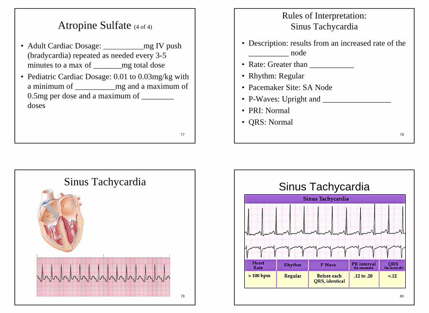
• Adult Cardiac Dosage: __________mg IV push (bradycardia) repeated as needed every 3-5 minutes to a max of _______mg total dose
• Pediatric Cardiac Dosage: 0.01 to 0.03mg/kg with a minimum of __________mg and a maximum of 0.5mg per dose and a maximum of ________ doses
78
Rules of Interpretation: Sinus Tachycardia
• Description: results from an increased rate of the __________ node
• Rate: Greater than ___________• Rhythm: Regular• Pacemaker Site: SA Node• P-Waves: Upright and _________________• PRI: Normal• QRS: Normal
79
Sinus Tachycardia
80
Sinus Tachycardia
21
81
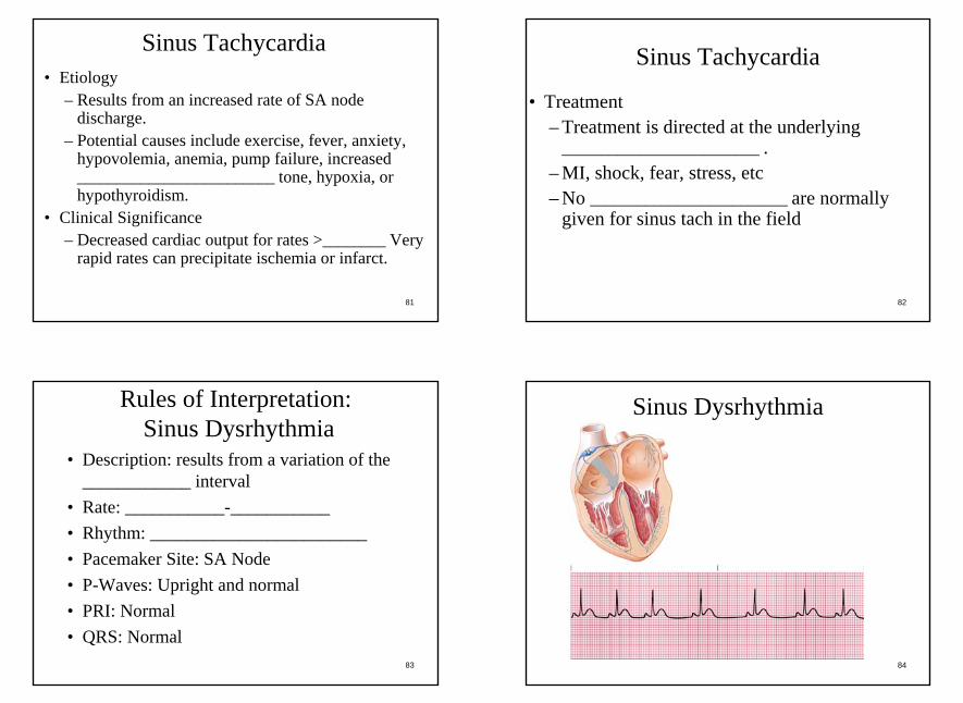
• Etiology– Results from an increased rate of SA node
discharge.– Potential causes include exercise, fever, anxiety,
hypovolemia, anemia, pump failure, increased _________________________ tone, hypoxia, or hypothyroidism.
• Clinical Significance– Decreased cardiac output for rates >________ Very
rapid rates can precipitate ischemia or infarct.
Sinus Tachycardia
82
Sinus Tachycardia
• Treatment– Treatment is directed at the underlying
_________________________ .– MI, shock, fear, stress, etc– No _________________________ are normally
given for sinus tach in the field
83
Rules of Interpretation:Sinus Dysrhythmia
• Description: results from a variation of the ____________ interval
• Rate: ___________-___________• Rhythm: ________________________• Pacemaker Site: SA Node• P-Waves: Upright and normal• PRI: Normal• QRS: Normal
84
Sinus Dysrhythmia

22
85
Sinus Dysrhythmia
86
• Etiology– Often a normal finding, sometimes related to the
respiratory cycle.– May be caused by enhanced
________________________ tone.• Clinical Significance
– Normal. Occurs in almost everyone• Treatment
– Typically, ______________________ required.
Sinus Dysrhythmia
87
Sinus Arrest
• Description: occurs when the sinus node fails to discharge, resulting in short periods of cardiac ________________________. This standstill can persist until pacemaker cells lower in the conduction system discharge (________________________ beats) or until the sinus node resumes discharging
88
Rules of Interpretation:Sinus Arrest
• Rate: Normal to _______________________• Rhythm: Irregular• Pacemaker Site: __________ Node• P-Waves: ________________________ and
normal• PRI: Normal• QRS: Normal
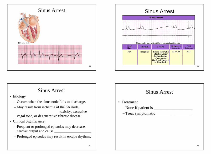
23
89
Sinus Arrest
90
Sinus Arrest
91
• Etiology– Occurs when the sinus node fails to discharge.– May result from ischemia of the SA node,
________________________ toxicity, excessive vagal tone, or degenerative fibrotic disease.
• Clinical Significance– Frequent or prolonged episodes may decrease
cardiac output and cause _____________________– Prolonged episodes may result in escape rhythms.
Sinus Arrest
92
Sinus Arrest
• Treatment– None if patient is ______________________– Treat symptomatic ____________________

24
93
• Wondering Atrial Pacemaker• Multifocal Atrial Tachycardia• Premature ________________________
Contractions• Paroxysmal Supraventricular Tachycardia• Atrial ________________________• Atrial ________________________
Dysrhythmias Originating in the Atria
94
Wandering Atrial Pacemaker
• Description: the ____________________ transfer of pacemaker sites from the sinus node to other latent pacemaker sites in the atria and AV junction. Often more than one ________________________ site will be present, causing variation in the R-R interval and P waves.
95
Rules of Interpretation:Wandering Atrial Pacemaker
• Rate: Usually normal (60-100)• Rhythm: Slightly _______________________• Pacemaker Site: varies among the SA node, atrial
tissue, and the AV junction• P-Waves: Variable or ___________________• PRI: Varies, depending on site of impulse• QRS: normal
96
Wandering Atrial Pacemaker

25
97
Wandering Atrial Pacemaker
98
• Etiology– Variant of sinus dysrhythmia, which is a natural
phenomenon in the very young or old.– May also be caused by ___________________ heart
disease or atrial dilation.• Clinical Significance
– None, but may be precursor to other atrial dysrhythmias.
• Treatment– Typically, ___________________ required.
Wandering Atrial Pacemaker
99
Multifocal Atrial Tachycardia• Description: usually seen in acutely ill patients.
Significant ___________________ disease is seen in about 60% of these patients.
• Certain medications used to treat lung diseases (Theophylline, ___________________) may worsen the condition.
• 3 different P waves are noticed, indicating various ectopic foci.
100
Rules of Interpretation:Multifocal Atrial Tachycardia
• Rate: Greater than __________• Rhythm: ___________________• Pacemaker Site: Ectopic sites in atria• P-Waves: Organized, non-sinus, with at
least __________ different forms• PRI: ___________________• QRS: Varies depending on AV node’s
refractory status when impulse begins

26
101
Multifocal Atrial Tachycardia
102
• Etiology– Often seen in acutely ill patients.– May result from pulmonary disease, metabolic
disorders, ischemic heart disease, or recent ___________________.
• Clinical Significance– Presence of multifocal atrial tachycardia often
indicates a serious underlying illness.• Treatment
– Treat the underlying ___________________.
Multifocal Atrial Tachycardia
103
Premature Atrial Contractions• Description: result from a single electrical
impulse originating in the atria ___________________ of the SA node, which causes a ___________________ depolarization of the heart before the next expected sinus beat.
• Interrupts the normal ___________________
104
Premature Atrial Contractions
• Creates a non-compensatory___________________ in the underlying rhythm. – Pause following an ectopic beat where the
SA node is ___________________ and the normal cadence is interrupted
27
105
Rules of Interpretation:Premature Atrial Contractions
• Rate: Depends on underlying rhythm• Rhythm: Usually regular except for _______• Pacemaker Site: Ectopic focus in the atrium• P-Waves: Occurs ___________________ than
expected• PRI: Varies depending on focus
– Near SA node = 0.12 or less– Near AV node = 0.12 or more
• QRS: Usually ___________________ 106
Premature Atrial Contractions
107
PACs
108
• Etiology– Single electrical impulse originating outside the SA
node.– May result from use of caffeine, tobacco, or
alcohol, sympathomimetic drugs, ischemic heart disease, hypoxia, or digitalis toxicity, or may be ___________________.
• Clinical Significance– ___________________. Presence of PACs may be a
precursor to other atrial dysrhythmias.
Premature Atrial Contractions
28
109
Premature Atrial Contractions
• Treatment– ___________________ if asymptomatic. Treat
symptomatic patients by administering high-flow oxygen and establishing _________ access.
110
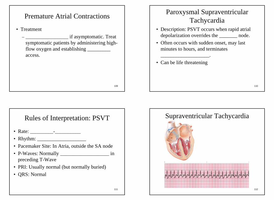
Paroxysmal Supraventricular Tachycardia
• Description: PSVT occurs when rapid atrial depolarization overrides the _______ node.
• Often occurs with sudden onset, may last minutes to hours, and terminates ___________________.
• Can be life threatening
111
Rules of Interpretation: PSVT
• Rate: _________-__________• Rhythm: ___________________• Pacemaker Site: In Atria, outside the SA node• P-Waves: Normally ___________________ in
preceding T-Wave• PRI: Usually normal (but normally buried)• QRS: Normal
112
Supraventricular Tachycardia
29
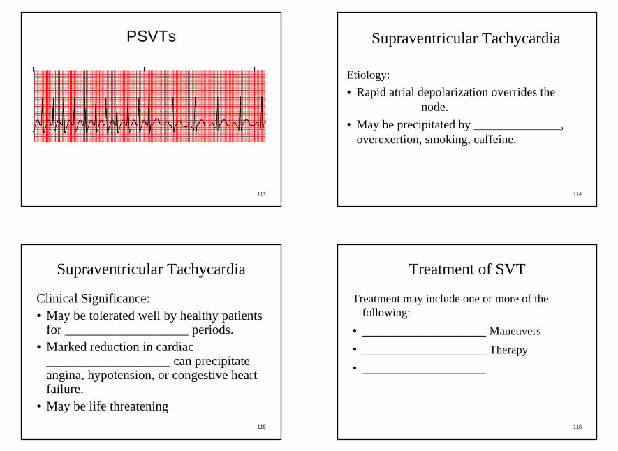
113
PSVTs
114
Etiology:• Rapid atrial depolarization overrides the
__________ node.• May be precipitated by _______________,
overexertion, smoking, caffeine.
Supraventricular Tachycardia
115
Supraventricular Tachycardia
Clinical Significance:• May be tolerated well by healthy patients
for ___________________ periods.• Marked reduction in cardiac
___________________ can precipitate angina, hypotension, or congestive heart failure.
• May be life threatening116
Treatment of SVT
Treatment may include one or more of the following:
• ___________________ Maneuvers• ___________________ Therapy• ___________________
30
117
• Vagal Maneuvers stimulate the ___________________ nerve which may slow the conduction through the SA Node
There are several types of vagal maneuvers:• Forced expiration against a closed glottis• “Bearing down” as if to move bowels
(___________________ Maneuver)• Immersion of face in ice water
(___________________ reflex)
Vagal Maneuvers
118
Vagal Maneuvers
• ___________________ artery massage– Contraindicated in patients with carotid
___________________ (sound of turbulent blood flow)
– Contraindicated in patients with known cerebrovascular disease carotid artery disease
119
Pharmacological Therapy for Supraventricular Tachycardia
120
Adenosine (Adenocard)
• Slows conduction through the ________ node• Contraindications:
-Allergic-2nd or 3rd degree ___________________ -Wolfe-Parkinson-White Syndrome
31
121
Adenosine (Adenocard)• Dosage:
– ______mg rapid IVP immediately followed by fluid bolus of 10-15cc
– If needed, repeat dosage (after 1-2 minutes) ______mg rapid IVP immediately followed by fluid bolus
• Adverse Reactions: dizziness, facial flushing, SOB• Causes a brief period of ___________________
122
Verapimil• AKA: ___________________ , Calan• ___________________ Channel Blocker• Contraindications:
– Allergies– ___________________ Shock– Patients receiving beta blockers
• Dosage: _________ to _________mg• Can be repeated once in 15-30 minutes at a
dose of 5 to 10mg
123
Verapimil
• Adverse Reactions:– Dizziness– Headache– ___________________– AV Blocks– ___________________
124
Electrical Therapy for SVTs• Used for hemodynamically unstable patients• Consider sedation prior if conscious and Systolic
BP above 90-100– ___________________(Diazepam) 5-10mg
IVP– ___________________(Versed) 2-5mg IVP– ___________________(Lorazepam) 1-4mg
SIVP

32
125
Electrical Therapy for SVTs
• Synchronized cardioversion starting at 100J or biphasic equivalent.– If unsuccessful, increase as directed by medical
control– Normally, ________J, ________ J, ________ J,
________ J (or biphasic equivalent)– Do not ___________________ if patient
converts
126
Treatment Summary for SVTs
Stable Patients• __________________
Maneuvers• Drug Therapy
– Adenosine– Verapimil
• Electrical Shock
Unstable Patients• __________________• Electrical Shock
– Synchronized cardioversion beginning at _______J
• Drug Therapy
127
Atrial Flutter
• Description: results from rapid atrial reentry circuit and an __________ node that cannot conduct all impulses through to the ventricles.
• The AV node may allow impulses in a 1:1 (rare), 2:1, 3:1 or 4:1 ratio or even greater resulting is a discrepancy between ___________________ and ___________________ rates.
128
Rules of Interpretation:Atrial Flutter
• Rate: Atrial rate of ________-__________. Ventricle rate varies
• Rhythm: Usually regular• Pacemaker Site: Atria; outside the SA node• P waves: Flutter (F-waves) are present.
“___________________ ” pattern• PR Interval: Usually normal• QRS Complex: ___________________

33
129
Atrial Flutter
130
Atrial Flutter
131
Etiology:• Results when the _______ node cannot conduct
all the impulses.• Impulses may be conducted in fixed or
___________________ ratios.• Usually associated with organic disease such as
congestive heart failure (rarely seen with ________).
Atrial Flutter
132
Atrial Flutter
Clinical Significance• Generally well tolerated.• Rapid ventricular rates may compromise
cardiac output and result in ___________________ .
• May occur in conjunction with atrial ___________________ .
34
133
Note: A-Flutter is NOT normally treated prehospital)
• Electrical Therapy– Consider if ventricular rate > __________ and
symptomatic.– Consider sedation with synchronized
cardioversion starting at 100J.
Treatment of Atrial Flutter
134
Treatment of Atrial Flutter
• Pharmacological Therapy– Diltiazem (Cardizem)– Verapamil, Digoxin, beta-blockers, and
Quinidine.– These drugs may not be commonly carried.
If rate is above __________bpm, consider sedation and ___________________
135
Atrial Fibrillation
• Description: results from multiple areas of ___________________ within the atria or from multiple ectopic foci bombarding the _________ node which cannot handle all of the incoming impulses.
• AV conduction is ___________________ and highly variable
136
Rules of Interpretation:Atrial Fibrillation (A-Fib)
• Rate: Atrial rate of __________-___________. Ventricular rate varies greatly
• Rhythm: ___________________ irregular• Pacemaker Site: numerous ectopic foci in atria
(Outside the SA node)• P-Waves: ___________________ discernable• PRI: none• QRS: Normal

35
137
Atrial Fibrillation (A-Fib)
138
Atrial Fibrillation (A-Fib)
139
• Etiology– Results from multiple ___________________ foci;
AV conduction is random and highly variable.– Often associated with underlying heart disease.
• Clinical Significance– Atria fail to contract effectively, reducing cardiac
___________________ .– Well tolerated with normal ventricular rates.– High or low ventricular rates can result in cardiac
___________________ .
Atrial Fibrillation (A-Fib)
140
Note: A-fib is not normally treated prehospital unless rate is above 150
• Electrical Therapy– Consider if ventricular rate > 150 and
___________________ .– Consider sedation and synchronized
cardioversion starting at __________J.
Treatment of A-Fib

36
141
Treatment of A-Fib
• Pharmacological Therapy– Diltiazem (___________________)– Verapamil, Digoxin, beta blockers, and
Quinidine.– ___________________ (heparin or warfarin).
142
AV Blocks• Locations:
– At the ______ Node
– At the Bundle of __________
– Below the Bundle of His
Dysrhythmias Originating Within the AV Junction (AV Blocks)
143
AV Blocks
• The electrical impulses are ___________________ or blocked as it passes through the AV node
• Can be caused by pathology of the AV junctional tissue or by a physiological block such as with ___________________ or A-Flutter
144
Classifications of AV Blocks• ___________________ -Degree AV Block• Type I Second-Degree AV Block
– Mobitz I– ___________________
• Type II Second-Degree AV Block– Mobitz II– ___________________
• ___________________ -Degree AV Block

37
145
First Degree AV Block
• Description: First degree AV block is a ___________________ in conduction at the level of the AV node rather than an actual block.
• First degree AV block is NOT a _______________itself, but a condition superimposed upon another rhythm.
• The ___________________ rhythm must also be identified
146
Rules of Interpretation:First Degree AV Block
• Rate: depends on underlying __________________
• Rhythm: Usually ___________________• Pacemaker Site: SA node or atria• P-Waves: normal• P-R interval: greater than ___________ seconds• QRS: Usually less than ___________ seconds
147
First Degree AV Block
Interpretation Keys• Every ________ is caused by a P-wave. But,
the PRI is consistently greater than 0.20 seconds and ___________________
• One _______ wave for each QRS
148
First Degree AV Block

38
149
First Degree AV Block
150
• Etiology– Delay in the conjunction of an impulse
through the AV node.– May occur in ___________________ hearts, but
often indicative of ischemia at the AV junction.
• Clinical Significance– Usually not significant, but new onset may
precede a more ___________________ block.
First Degree AV Block
151
First Degree AV Block
• Treatment– Generally, none required other than
___________________ .– Avoid drugs that may further
___________________ AV conduction.
152
Type I Second Degree AV Block• AKA: Mobitz I or Wenckebach• Description: an ___________________ block at
the level of the AV node• Produces a pattern which the ________ intervals
become progressively longer until an impulse is blocked.
• Cycle is repetitive and the P-P interval is ___________________
• Pattern may be constant or variable
39
153
Type I Second Degree AV BlockKeys to Interpretation:• PRI ___________________ until a QRS drops
out• Each _________ is caused by a P-Wave
154
Rules of Interpretation:Type I Second Degree AV Block
• Rate: ___________________ is normal, Ventricular is normal to slow
• Rhythm: Atrial is regular. Ventricular is irregular• Pacemaker Site: __________ node or atria• P-Waves: normal. Some P-waves are NOT
followed by ___________ complexes• QRS: Usually less than 0.12 seconds
155
Type I Second Degree AV Block
156
Type I Second Degree AV Block
40
157
• Etiology– Delay increases until an impulse is
___________________ .– Indicative of ischemia at the AV junction.
• Clinical Significance– Frequently dropped beats can result in
___________________ compromise.
Type I Second Degree AV Block
158
Type I Second Degree AV Block• Treatment
– Generally, none required other than observation.
– Avoid drugs that may further slow AV conduction.
– Treat symptomatic ___________________ .• ___________________ : 0.5mg repeated as
needed every __________-__________ minutes up to a max of 3mg total dose
• External Pacing if Atropine is unsuccessful
159
Type II Second Degree AV Block• AKA: Mobitz II, or infranodal• Description: an intermittent block
characterized by P wave that are not conducted to the ventricles, but ___________________ associated lengthening of the P-R interval before the dropped beats
160
Type II Second Degree AV BlockKeys to Interpretation:• More __________ waves than QRS but every
QRS is caused by a P wave• ____________ is constant for conducted beats

41
161
Rules of Interpretation:Type II Second Degree AV Block
• Rate: ___________________ is normal. Ventricular is slow
• Rhythm: regular or ___________________• Pacemaker Site: SA node or atria• P-Waves: normal, some P-waves not followed by
QRS• PRI: constant for conducted beats, may be greater
than ____________ seconds • QRS: Normal or greater than 0.12 seconds
162
Type II Second Degree AV Block
163
Type II Second Degree AV Block
164
• Etiology– Intermittent block of impulses.– Usually associated with ________ or septal
necrosis.• Clinical Significance
– May compromise cardiac output and is indicative of MI.
– Often develops into ___________________ AV blocks.
Type II Second Degree AV Block

42
165
Type II Second Degree AV BlockTreatment• Avoid drugs that may further slow AV
conduction.• Treat symptomatic bradycardia.
– Atropine should ___________________ be given
– May increase atrial ___________________ but worsen block
• Consider transcutaneous pacing.166
Third Degree AV Block
• AKA: Complete Heart Block• Description: the absence of conduction
between the atria and the ventricles resulting from complete electrical block at or below the ____________ node
• The ___________________ pacemaker, located below the atria, paces the heart
167
Third Degree AV Block
Keys to Interpretation:• More P wave than QRS• Each QRS is ___________________ caused
by a P-wave• Both the P-waves and QRS rhythm is
___________________ but unassociated
168
Rules of Interpretation:Third Degree AV Block
• Rate: Atrial is normal. Ventricular is _______-_______
• Rhythm: Both atrial and ventricular rate is regular.• Pacemaker Site: SA node and AV junction or
___________________• PRI: no relationship between P waves and R waves• QRS: greater than __________ if pacemaker is
ventricular; less than 0.12 if pacemaker is junctional

43
169
Third Degree AV Block
170
Third Degree AV Block
171
• Etiology– Absence of conduction between the atria
and the ventricles.– Results from ________, digitalis toxicity,
or degeneration of the conductive system.• Clinical Significance
– Severely compromised cardiac ___________________ .
Third Degree AV Block
172
Third Degree AV BlockTreatment• Pacemaker insertion is ___________________
treatment• Transcutaneous ___________________ for
acutely symptomatic patients.• Treat symptomatic ___________________ .
– ___________________ should NOT be given– May increase atrial rate but worsen block– Avoid drugs that may further slow AV
conduction.

44
173
Dysrhythmias:• Premature Junctional ___________________• Junctional ___________________ Complexes and
Rhythm• Accelerated Junctional Rhythm• Paroxysmal Junctional TachycardiaCharacteristics:• ___________________ P Waves in Lead II• PRI of < ____________ Seconds• Normal QRS Complex Duration
Dysrhythmias Sustained or Originating in the AV Junction
174
Premature Junctional Contractions• Description: PJCs result from a single
electrical impulse originating in the AV node that occurs before the next expected ___________________ beat.
• A PJC can result in a compensatory pause or a _________-compensatory pause.
• Compensatory pause: the pause following an ectopic beat where the ________ node is unaffected and the cadence of the heart is uninterrupted
175
Rules of Interpretation: PJC• Rate: depends on ___________________ rhythm• Rhythm: depends on underlying rhythm• Pacemaker Site: ectopic focus in the AV junction• P-Waves: flat or ___________________ . May
occur ___________________ QRS• PRI: Normal if P occurs before QRS• QRS: usually ___________________
176
PJC

45
177
PJC
178
• Etiology– Single electrical impulse originating in the
_________ node.– May occur with use of caffeine, tobacco, alcohol,
sympathomimetic drugs, ischemic heart disease, hypoxia, or digitalis toxicity, or may be idiopathic.
• Clinical Significance– Limited, frequent PJCs may precursor other
junctional dysrhythmias.• Treatment
– ___________________ usually required.
PJCs
179
Junctional Escape Complexes and Rhythms
• Description: results when the rate of the primary pacemaker (SA Node) is slower than that of the AV node.
• The AV node then becomes the ___________________ .
• AV node fires at it’s intrinsic rate: _______-__________
• Safety mechanism that prevents cardiac ___________________
180
Rules of Interpretation:Junctional Escape Complexes and Rhythms
• Rate: 40-60• Rhythm: irregular in single occurrence, regular
in junctional escape rhythm• Pacemaker Site: ___________ Junction• P-Waves: inverted, ___________________ ,
or after QRS• PRI: Normal if before QRS• QRS: Usually ___________________
46
181
Junctional Escape Complexes and Rhythms
182
Junctional Rhythm
183
• Etiology– Results when the AV node becomes the
pacemaker.– Results from increased ___________________
tone, pathologically slow SA discharges, or heart block.
• Clinical Significance– Slow rate may ___________________ cardiac
output, precipitating angina and other problems.
Junctional Escape Complexes and Rhythms
184
Junctional Escape Complexes and Rhythms
Treatment:• None if the patient remains
___________________ .• Treat symptomatic episodes with
___________________ or pacing as indicated.

47
185
Accelerated Junctional Rhythm• Description: results from increased automaticity
in the AV junction, causing the AV junction to discharge faster than its ___________________ rate.
• As the rate increases, the AV node overrides the __________ node.
• The rate is not, technically, a ___________________ but because it is faster than its intrinsic rate of the AV junction, it is considered accelerated.
186
Rules of Interpretation:Accelerated Junctional Rhythm
• Rate: ________-__________• Rhythm: ___________________• Pacemaker Site: AV Junction• P-Waves: Inverted, ___________________ , or
after QRS complexes• PRI: Normal if present and occurs before QRS• QRS: ___________________
187
Accelerated Junctional Rhythm
188
Accelerated Junctional Rhythm

48
189
• Etiology– Results from increased ___________________ in
the AV junction.– Often occurs due to ischemia of the AV
junction.• Clinical Significance
– Usually well tolerated, but ___________________ for other dysrhythmias.
• Treatment– ___________________ generally required in the
prehospital setting.
Accelerated Junctional Rhythm
190
Paroxysmal Junctional Tachycardia• Description: develops when rapid AV junctional
depolarization overrides the SA node.• Often occurs with sudden onset
(___________________ )• May last minutes or hours.• May be caused by increased automaticity of a
single AV nodal focus or by a ___________________ phenomenon at the AV node.
• Sometimes indistinguishable from _____________ due to rapid rate
191
Rules of Interpretation:Paroxysmal Junctional Tachycardia
• Rate: __________-__________• Rhythm: Regular• Pacemaker Site: AV Junction• P-Waves: Inverted, absent, or
___________________ QRS• PRI: Normal if occurs before QRS• QRS: ___________________
192
Paroxysmal Junctional Tachycardia

49
193
Paroxysmal Junctional Tachycardia
194
• Etiology– Rapid AV junction depolarization overrides the
__________ node.– Occurs with or without heart disease.– May be precipitated by stress, overexertion,
smoking, or ___________________ ingestion.• Clinical Significance
– May be well tolerated for brief periods.– Decreased cardiac output will result from
prolonged episodes, which may precipitate angina, hypotension, or congestive heart failure.
Paroxysmal Junctional Tachycardia
195
Treatment: Same as ___________________• ___________________ Maneuvers• Pharmacological Therapy
– ___________________– Verapamil
• Electrical Therapy– Use if rate is > 150 and patient is
hemodynamically unstable.– Synchronized cardioversion starting at
____________J.
Paroxysmal Junctional Tachycardia
196
• Ventricular Escape Complexes and Rhythms• Accelerated ___________________ Rhythm• Premature Ventricular Contractions• Ventricular ___________________• Related Dysrhythmia• Ventricular Fibrillation• ___________________• Artificial ___________________ Rhythm
Dysrhythmias Originating in the Ventricles
50
197
Ventricular Escape Complexes and Rhythms
• AKA: ___________________ rhythm• Description: results either when impulses from
higher pacemakers fail to reach the ventricles or when the discharge rate of higher pacemakers become less than that of the ventricles (_________-__________ bpm).
• Serves to prevent cardiac ___________________
198
Rules of Interpretation: Ventricular Escape Complexes and Rhythms
• Rate: 15-40 (sometimes less)• Rhythm: escape complex is
___________________ . Escape rhythm is regular
• Pacemaker Site: Ventricle• P-Waves: ___________________• PRI: ___________________• QRS: Greater than ___________ seconds and
bizarre in morphology
199
Ventricular Escape Rhythm
200
• Etiology– ___________________ mechanism to
prevent cardiac standstill.– Results from failure of other foci or high-
degree __________ block.• Clinical Significance
– ___________________ cardiac output, possibly to life-threatening levels.
Ventricular Escape Complexes and Rhythms

51
201
Ventricular Escape Complexes and Rhythms
Treatment:• For perfusing rhythms, administer
___________________ and/or TCP (Pacing).• For nonperfusing rhythms, follow pulseless
electrical activity (__________) protocols.
202
• Etiology– A subtype of ventricular escape rhythm that
frequently occurs with ____________.– Ventricular escape rhythm with a rate of
__________–__________.• Clinical Significance
– May cause ___________________ cardiac output if the rate slows.
Accelerated Idioventricular Rhythm
203
Accelerated Idioventricular Rhythm
204
Accelerated Idioventricular Rhythm
Treatment:• Does not usually require treatment unless the
patient becomes hemodynamically___________________ .
• Primary goal is to treat the underlying ___________.

52
205
Premature Ventricular Contractions• Description: A __________ is a single ectopic
impulse arising from an irritable focus in either ventricle that occurs earlier than the next expected beat.
• May result from increased automaticity in the ___________________ cell
• The altered sequence of ventricular depolarization results in a ___________________ and bizarre QRS and may cause the T-wave to occur in the opposite direction of the QRS.
206
Premature Ventricular Contractions
• Pause following a PVC is ___________________• Occasionally, an interpolated beat occurs when a
PVC falls between two sinus beats without interrupting the rhythm
• If more than 1 PVC occurs, each can be classified as Unifocal or ___________________
• Unifocal: from the same foci (looks alike)• Multifocal: from different sites (look different)
207
Groups of PVCs• ___________________ : Every other beat is a
PVC• Trigeminy: Every third beat is a PVC• Quadrageminy: Every fourth beat is a PVC• ___________________ : Two consecutive
PVCs• Triplet: _______ or more consecutive PVCs• Runs of V-Tach: group of _________ or more
consecutive PVCs
208
Rules of Interpretation:PVCs
• Rate: Depends on underlying rhythm• Rhythm: Normally ___________________ .
Interrupts underlying rhythm• Pacemaker Site: ___________________• P-Waves: ___________________• PRI: None• QRS: Greater than ___________ and bizarre
53
209
PVCs
210
Unifocal PVCs
211
Multifocal PVCs
212
PVC Couplets

54
213
PVC Triplets
214
Bigeminy
215
Etiology• Single ectopic impulse resulting from an
irritable focus in either ___________________• Causes may include myocardial ischemia,
increased ___________________ tone, hypoxia, idiopathic causes, acid–base disturbances, ___________________imbalances, or as a normal variation of the ECG.
PVCs
216
PVCs
• May occur in ___________________ – Bigeminy, trigeminy, or quadrigeminy.– ___________________ and triplets.– Runs of ___________________
55
217
Clinical Significance:• Malignant PVCs:
– More than __________/minute, R on T phenomenon, couplets or runs of ventricular tachycardia, ___________________ PVCs, or PVCs associated with chest pain.
• Ventricles do not adequately ___________________ , causing decreased cardiac output.
PVCs
218
• Non-malignant PVCs do not usually require treatment in patients without a cardiac history.
• Most people have ___________________ PVCs• Cardiac patient with nonmalignant PVCs
– Administer oxygen and establish _________ access
– Watch EKG closely
Treatment of Non-Malignant PVCs
219
Treatment of Malignant PVCs
• Treatment of PVCs is normally performed for __________ on __________ phenomenon and symptomatic patients
• Two drugs to treat PVCs:– ___________________– ___________________
• Do NOT mix anti-dysrhythmics
220
R on T phenomenon

56
221
Lidocaine for Malignant PVCs• ________ –__________mg/kg IV bolus.• If PVCs are not suppressed, repeat doses of
__________-__________ mg/kg to max dose of ____________ mg/kg.
• If PVCs are suppressed, administer lidocaine drip _________-_________ mg/min.
• ___________________ the dose in patients with decreased output or decreased hepatic function and patients > 70 years old.
• If patient is allergic to Lidocaine, consider Amiodarone
222
Lidocaine• Antidysrhythmic (Sodium channel blocker)• Contraindications: Allergic or allergic to
___________________• Indications: V-fib, PVCs, V-Tach• A bolus should be followed with a drip if
___________________• Lidocaine Drip: usually mixed
_________gm/250cc and is run at 2-4mg/min• Side Effects: Dizziness, drowsiness, N/V,
sensation of heat/cold, numbness
223
Amiodarone (Cordarone)• Amiodarone is an alternative to Lidocaine• Newer drug with proven success• Amiodarone is an antiarrhythmic (Calcium
Channel ___________________)• Long _________ life• Indications:• Wide Complex Tachycardia, V-Fib, V-Tach,
Supraventricular Tachycardia, Rapid A-Fib• Contraindications: Allergic, ___________________,
Bradycardia224
Amiodarone (Cordarone)
Effects:• Inhibits abnormal ___________________• Increases refractory period at all sites• Slows __________ and _________ node rate• Causes peripheral ___________________

57
225
Amiodarone (Cordarone)
Side Effects:• Can produce hypotension or
___________________• Worsens ___________________• Parestesias (numbness and tingling)• Tremor• ___________________
226
Amiodarone (Cordarone)
Initial Dosage (For non per):• __________mg IV push• Repeated (if needed) at _________mg in 3-
5 minutes• ½ dose (150mg) for perfusing rhythm over
10 minutes
227
Amiodarone (Cordarone)Maintenance Dosage:• IV Drip: __________-___________mg/min• Drip must be in glass or Viaflex bag
– Glass container is good for 24 hours– Viaflex bag is good for _________ hours
• Amiodarone drips normally not established in ___________________ setting
• Notify ER that Amiodarone was given228
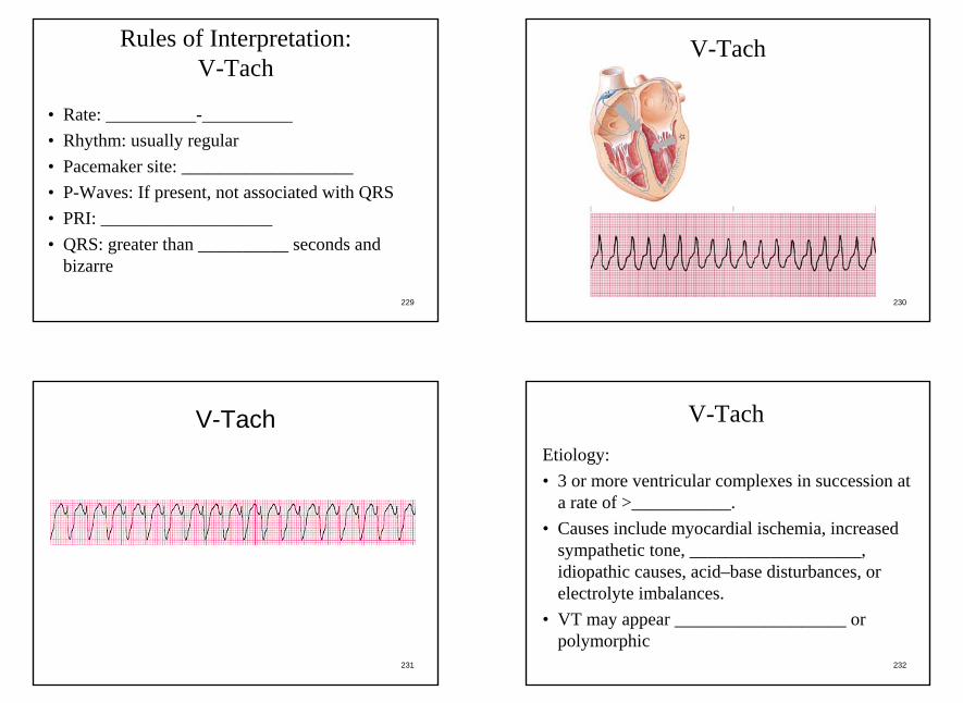
Ventricular Tachycardia V-Tach• Description: __________ or more ventricular
contractions in succession with a rate of ___________bpm or faster.
• Overrides the __________ node.• May be present with or without a _____________• Monomorphic V-Tach: all complexes appear the
___________________ (Most common).• Polymorphic V-Tach: complexes have different
sizes and shapes. (Torsade de Pointes)
58
229
Rules of Interpretation:V-Tach
• Rate: __________-__________• Rhythm: usually regular• Pacemaker site: ___________________• P-Waves: If present, not associated with QRS• PRI: ___________________• QRS: greater than __________ seconds and
bizarre
230
V-Tach
231
V-Tach
232
Etiology:• 3 or more ventricular complexes in succession at
a rate of >___________.• Causes include myocardial ischemia, increased
sympathetic tone, ___________________, idiopathic causes, acid–base disturbances, or electrolyte imbalances.
• VT may appear ___________________ or polymorphic
V-Tach

59
233
V-Tach
Clinical Significance:• Decreased ___________________ output,
possibly to life-threatening levels.• May deteriorate into ventricular
___________________.
234
Perfusing patient:• Administer ___________________ and
establish IV access.• Consider immediate synchronized
___________________ starting at 100J for hemodynamically ___________________ patients. (normally 100J, 200J, 300J, 360J or biphasic equivalent)– Sedate if necessary
Treatment of V-Tach
235
Treatment of V-TachPerfusing Patient (Cont’d)• ___________________ 150–300 mg IV over
10 minutes. Repeated once at 150mg• ___________________ 1.0–1.5 mg/kg IV.• Administer repeat doses of Lidocaine 0.5–
1mg/kg to the max dose of 3.0 mg/kg, or until VT is suppressed. (Lidocaine ___________________ if conversion)
236
V-Tach Treatment Summary Conscious Patient
• BLS, IV• Drugs
– Amiodarone– Lidocaine
• Synchronized Cardioversion– Sedation?– 100J, 200J, 300J,
360J
Unconscious Patient With a Pulse
• BLS, IV• Synchronized
Cardioversion– 100J, 200J, 300J,
360J• Drugs
– Amiodarone– Lidocaine

60
237
Treatment of V-Tach
Non-Perfusing Patients (No Pulse):• Treat as Ventricular ___________________
(V-Fib)
238
• Typically occurs in nonsustained bursts.– Prolonged ______-______ interval during
“breaks.”– QRS rates from 166–300.– RR interval highly ___________________ .
• Treatment– Do not treat as standard VT
(___________________ not indicated)– Magnesium sulfate 1–2 g diluted in 100 ml D5W
over 1–2 minutes is drug of choice.– Amiodarone 150–300 mg
Torsade de Pointes
239
Torsade de Pointes
240
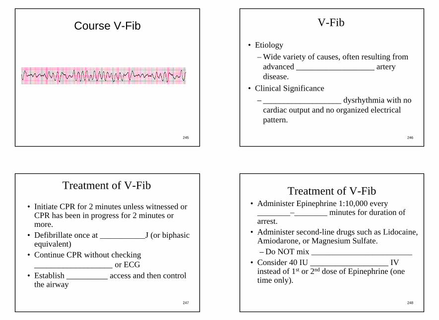
Ventricular Fibrillation (V-Fib)
• Description: a ___________________ventricular rhythm usually resulting from the presence of many reentry circuits within the ventricles.
• No ventricular polarization or depolarization• May be ___________________ or course• CANNOT produce a ___________________

61
241
Rules of Interpretation:V-Fib
• Rate: no organized rhythm• Rhythm: no organized rhythm• Pacemaker Site: numerous ectopic foci
throughout the ___________________• P-Waves: Usually absent• PRI: ___________________• QRS: ___________________
242
V-Fib
243
V-Fib
244
Fine V-Fib
62
245
Course V-Fib
246
• Etiology– Wide variety of causes, often resulting from
advanced ___________________ artery disease.
• Clinical Significance– ___________________ dysrhythmia with no
cardiac output and no organized electrical pattern.
V-Fib
247
• Initiate CPR for 2 minutes unless witnessed or CPR has been in progress for 2 minutes or more.
• Defibrillate once at ___________J (or biphasic equivalent)
• Continue CPR without checking ___________________ or ECG
• Establish __________ access and then control the airway
Treatment of V-Fib
248
Treatment of V-Fib• Administer Epinephrine 1:10,000 every
________–________ minutes for duration of arrest.
• Administer second-line drugs such as Lidocaine, Amiodarone, or Magnesium Sulfate.– Do NOT mix ____________________________
• Consider 40 IU ___________________ IV instead of 1st or 2nd dose of Epinephrine (one time only).
63
249
Epinephrine 1:10,000• ____________________________ agonist
(Adrenalin) • Stimulates both alpha and beta adrenergic
____________________________ • Alpha Receptors:
– Peripheral ____________________________ (arteries and veins)
– Bronchoconstriction
250
Epinephrine 1:10,000• Beta Receptors:
– Increase of cardiac contractility and ____________________________ thus increasing cardiac output and heart rate
– Relaxing of ____________________________ muscles in bronchi
– Dilation of ____________________________ arteries
251
Epinephrine 1:10,000• Epinephrine is mostly beta with some alpha• Increases cardiac
____________________________ and automaticity
• Dilates ____________________________passageways
• Decreases resistance to electrical shock• Dilates coronary arteries• ____________________________ peripheral
blood vessels252
Epinephrine 1:10,000• Indications: ALL cardiac arrests, allergic
reactions, severe asthma attacks• Contraindications: None in cardiac arrest• Adult Dosage: ______mg every ______-______
minutes for duration of arrest• Side effects: none in cardiac arrest• Normally supplied as ______mg in ______cc of
solvent in ____________________________syringes
64
253
Vasopressin• AKA: Pitressin, ADH (Anti-Diuretic Hormone)• Synthetic Pituitary Hormone• Indication: V-Fib, pulseless V-Tach• Actions: ____________________________
vasoconstrictor• Decreases ____________________________ to
electrical shock• Has no cardiac stimulatory properties• Useful when heart is over __________________
254
Vasopressin
• Contraindications: None in V-Fib or Pulseless V-Tach
• Dosage: _________ units IV push• Currently, no __________________________
dosage• Used instead of ________ or _______ round
of Epi
255
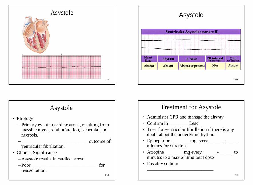
Asystole
• Description: Cardiac ____________________________
• The ____________________________ of all cardiac electrical activity
• CANNOT produce a pulse• “Flat Line”:
– However, very rarely ____________________________
256
Rules of Interpretation:Asystole
• Rate: No electrical activity• Rhythm: No electrical activity• Pacemaker Site: No electrical activity• P-Waves: ____________________________ • PRI: ____________________________ • QRS: ____________________________
65
257
Asystole
258
Asystole
259
• Etiology– Primary event in cardiac arrest, resulting from
massive myocardial infarction, ischemia, and necrosis.
– ____________________________ outcome of ventricular fibrillation.
• Clinical Significance– Asystole results in cardiac arrest.– Poor ____________________________ for
resuscitation.
Asystole
260
• Administer CPR and manage the airway.• Confirm in ________ Lead• Treat for ventricular fibrillation if there is any
doubt about the underlying rhythm.• Epinephrine ________mg every ______-______
minutes for duration• Atropine ________mg every ______-______ to
minutes to a max of 3mg total dose• Possibly sodium
____________________________ .
Treatment for Asystole

66
261
Artificial Pacemaker Rhythm
• Description: results from stimulation of the heart by an artificial pacemaker
• ____________________________ Rate Pacemakers: fire continuously at a preset rate; regardless of heart’s own electrical activity
• ____________________________ Pacemakers: Monitors heart’s electrical activity and only fires if heart rate drops below a preset rate
262
Artificial Pacemaker Rhythm• ____________________________ Pacemakers
stimulate only the right ventricle resulting in a rhythm that resembles an idioventricular rhythm
• ____________________________ chambered pacemakers stimulate the atria first and then the ventricles
• Usually inserted in patients with severe symptomatic ____________________________
263
Rules of Interpretation:Artificial Pacemaker Rhythm
• Rate: varies with rate of pacemaker• Rhythm: regular if pacing regularly• Pacemaker Site: Depends on electrode placement• P-Waves: ____________________________
produced by ventricular pacemaker. Sinus P-waves may be seen but are unassociated with QRS. Pacemaker ____________________________may be visible
• QRS: greater than ________ seconds, bizarre264
Artificial Pacemaker Rhythm

67
265
Atrial Pacemaker Rhythm
266
Ventricular Pacemaker Rhythm
267
Dual ChamberPacemaker Rhythm
268
• Etiology– Single vs. dual chamber pacemakers.– Fixed-rate vs. demand pacemakers.
• Clinical Significance– Used in patients with a chronic high-–grade
heart block, sick sinus syndrome, or severe symptomatic bradycardia.
– Pacemaker ____________________________ may NOT be seen. Obtain history in any patient who presents with broad QRS rhythms
Artificial Pacemaker Rhythm

68
269
• Problems with Pacemakers– ____________________________ failure– “____________________________ ” pacers– Displaced ____________________________
• Use a ____________________________ to turn unit off if needed (contact medical control first)
Artificial Pacemaker Rhythm
270
Artificial Pacemaker RhythmManagement Considerations:• Identify patients with pacemakers.• Treat the ____________________________ .• Use an ____________________________
pacemaker if malfunctioning• Try to avoid placing d-fib pads (or paddles) on
pacemaker site• The only way to confirm that a pacemaker is
working correctly is to assure pulse corresponds with ____________________________
271
Pulseless Electrical Activity (PEA)• Formerly called electrical mechanical dissociation
(____________)• CharacteristicsElectrical impulses are present, but with no
accompanying mechanical contractions of the ____________________________ .Treat the patient, not the
____________________________ .ECG could show ANY rhythm that is normally
a perfusing rhythm272
Causes of PEA
6 “H’s”• Hypovolemia• _____________________• Hydrogen Ion- Acidosis• Hyper/Hypokalemia• Hypothermia• _____________________
5 “T’s”• Tablets (OD)• _____________________• Tension Pneumothorax• Thrombosis (Cardiac)• ____________________,
pulmonary (PE)

69
273
Treatment of PEA
• Prompt recognition and early treatment.• ____________________________ 1 mg every
3–5 minutes.• Identify and treat underlying
____________________________ of PEA.
274
Treatment of Underlying Causes of PEACondition TreatmentHypovolemia ___________________Cardiac Tamponade PericardiosentesisTension Pneumothorax _______________________________________ Oxygen/Intubation____________________ Sodium BicarbonateHypoglycemia D50W
275
Categories of Conductive Disorders:• ____________________________ Blocks• Disturbances of Ventricular Conduction• ____________________________
Syndromes
Dysrhythmias Resulting from Disorders of Conduction
276
• ____________________________ Conduction: a single supraventricular beat conducted through the ventricles in a delayed manner
• ____________________________ Branch Block: disorder in which all supraventricular beats are conducted through the ventricles in a delayed manner
Disturbances of Ventricular Conduction

70
277
Bundle Branch Block
• Can involve either the left or right bundle branch• If both branches are involved, then a
____________________________ degree block exists
• Causes• Ischemia or necrosis of a bundle branch• PAC or _________ that reaches one of the
bundle branches in a refractory period• Causes wide _________ complexes with P-
waves present278
Left Bundle Branch Block
279
Pre-Excitation Syndromes• Excitation by an impulse that bypasses the AV
node• Most common is Wolf-Parkinson-White
(____________) Syndrome• Characterized by a short _________ and a long
___________ duration.• Upstroke of the QRS often has a slur called a
“____________________________ ” wave• Treatment is to treat underlying rhythm
280
Pre-Excitation Syndromes
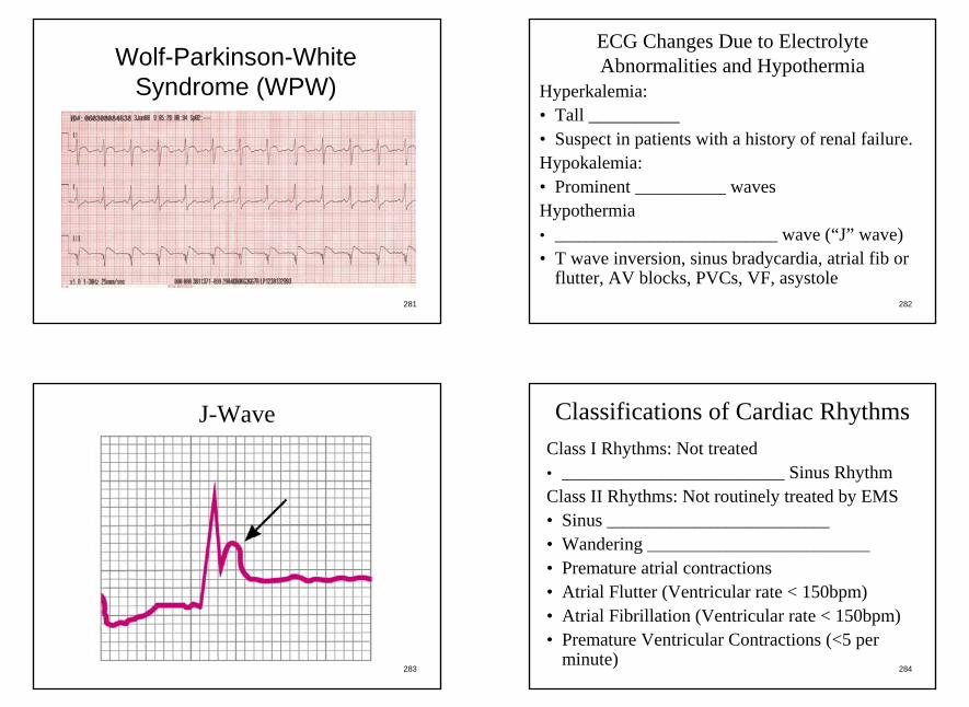
71
281
Wolf-Parkinson-White Syndrome (WPW)
282
ECG Changes Due to Electrolyte Abnormalities and Hypothermia
Hyperkalemia:• Tall __________• Suspect in patients with a history of renal failure.Hypokalemia:• Prominent __________ wavesHypothermia• ____________________________ wave (“J” wave)• T wave inversion, sinus bradycardia, atrial fib or
flutter, AV blocks, PVCs, VF, asystole
283
J-Wave
284
Classifications of Cardiac RhythmsClass I Rhythms: Not treated• ____________________________ Sinus RhythmClass II Rhythms: Not routinely treated by EMS• Sinus ____________________________• Wandering ____________________________• Premature atrial contractions• Atrial Flutter (Ventricular rate < 150bpm)• Atrial Fibrillation (Ventricular rate < 150bpm)• Premature Ventricular Contractions (<5 per
minute)

72
285
Classifications of Cardiac RhythmsClass II Rhythms: Not routinely treated by EMS
(Continued):• Premature junctional complex• ____________________________ rhythm• Accelerated junctional rhythm• Junctional tachycardia (ventricular rate < 150)• _________ degree AV block• _________ degree AV block, type I
(Wenckebach) (ventricular rate < 150)
286
Classifications of Cardiac Rhythms
Class III: Treated by EMS to prevent rhythm becoming Class IV:
• ____________________________• Supraventricular Tachycardia (ventricular
rate > 150)• 2nd Degree AV block, Type ___________• __________ Degree AV block
287
Classifications of Cardiac RhythmsClass III (Continued):• Premature Ventricular Contractions, if:
– ____________________________ Patients– Runs of V-Tach– _________ on __________ Phenomenon– ____________________________ PVCs
288
Classifications of Cardiac RhythmsClass IV: Must be treated in pre-hospital
setting, or death will result:• Ventricular Fibrillation (VF or V-Fib)• Ventricular Tachycardia (VT or V-Tach)• Pulseless Electrical Activity (___________)• ____________________________
73
289
Treating Cardiac Dysrhythmias • Determine that there is a true need to treat
the dysrhythmia before treating it• Obtain a chief
____________________________BEFORE treating the dysrhythmia
• Most importantly……..
• TREAT THE PATIENT, NOTNOTTHE MONITOR
Related Documents


![Cardiovascular System Anatomy Practical [PHL 212].](https://static.cupdf.com/doc/110x72/5697c01d1a28abf838cd05f5/cardiovascular-system-anatomy-practical-phl-212.jpg)