618 M MONOGRAPHS Abstract The methyl-CpG binding proteins (MBPs) interpret the methylation of DNA and its components. The number of MBPs in the human body currently stands at 15, which are split into 3 branches, a reflection of the intricate mechanisms of gene regulation. Each branch utilizes a different mechanism for interacting with methylated DNA or its components. These interactions function to direct gene expression and maintain or alter DNA architecture. It is these functions that are commonly exploited in human disease. For this review, we will focus on each protein and any roles it may have in initiating, promoting, progressing, or inhibiting cancer. This will highlight common threads in the roles of these proteins, which will allow us to speculate on potentially productive directions for future research. Keywords: methyl binding domain (MBD), cancer, transcription, repression, methylation, methyl binding protein (MBP) Introduction The field of epigenetics and cancer is broad and extensive. The extent of these studies is a reflection of how much eukaryotic biology is reliant on appro- priate DNA methylation. A key aspect of the field involves the study of the pro- teins that directly interpret the methyla- tion of DNA and its components. The methyl binding protein (MBP) family plays a pivotal role in this interpretation. Since the identification of the first MBP in 1989, MeCP2, 1 their numbers have increased, and their roles in gene regula- tion have been extensively studied as incorrect interpretation of methylation, or vice versa, the interpretation of incor- rect methylation, plays a role in many genetic diseases. For this review, we will focus on the roles of the MBPs in cancer. Initially, we will provide a brief history of the MBPs and a summary of the indi- vidual proteins. We will then provide an update on the most recent cancer-related findings for the MBPs since previous reviews on this topic in 2008. 2,3 Finally, we will speculate on future directions for understanding the roles of MBPs in inhibiting, initiating, progressing, and altering cancer phenotypes. A Brief History of MBPs In mammalian DNA, the MBPs were orig- inally characterized by their interactions with methylated DNA, namely CpG dinu- cleotides. The CpG is a cytosine-guano- sine (CG) dinucleotide DNA sequence, in which the cytosine undergoes chemical modification to contain a methyl group. This methylation of cytosine was first detected in 1925 in the tubercle bacillus. 4 It was another 23 years before it was observed in eukaryotes when it was detected in calf thymus DNA in 1948. 5 The importance of methylation became appar- ent in the 1970s following key findings such as the fact that the doublet CpG sequence occurs at approximately one fifth of the expected frequency within the genome 6,7 but represents the main target of methylation. 8,9 These findings suggested that the regulation of genes had to be due to the interaction of genomic DNA with the cytoplasmic enzymes that it encoded. 10,11 By the end of the 1970s, it had been dem- onstrated that an important aspect of eukaryote gene regulation came from pro- tein-methylated DNA interactions 12,13 and that DNA methylation patterns are inheritable. 14,15 Today, the nature of DNA methyla- tion and many of its roles within eukary- otic cells have been elucidated. The enzymes that catalyze the methylation of a CpG dinucleotide have been identified and characterized into the DNA methyltransferase family. 16 These enzymes take a methyl group, donated from S-adenosyl-L-methionine, and cova- lently attach it to the fifth carbon position of cytosine to form a symmetrical CpG dinucleotide. In the mammalian genome, approximately 60% to 90% of CpGs are methylated. 17 The remaining unmethyl- ated CpGs are clustered into what are termed CpG islands, which comprise around 1% of the genome. 18,19 These CpG islands are located within the pro- moter, first exon, or 3′UTR regions of approximately 60% of the RNA poly- merase II–transcribed genes in the verte- brate genome, consistent with their role in gene expression. 16,20 Indeed, methylated CpGs (mCpGs) are now syn- onymous with a repressed chromatin environment and silencing of gene expression. They play an important role in expression of patterns of imprinted genes, X chromosome inactivation, sup- pression of transposable elements, devel- opmental biology, and many disease mechanisms. 17 One of the key aspects of the relation- ship between methylation of DNA and the silencing of gene expression was established in the late 1980s and early School of Biosciences, Cardiff University, Cardiff, UK Corresponding Author: Alan R. Clarke, School of Biosciences, Cardiff University, Museum Avenue, Cardiff, CF10 3AX, UK Email: [email protected] The Roles of the Methyl-CpG Binding Proteins in Cancer Lee Parry and Alan R. Clarke Genes & Cancer 2(6) 618–630 © The Author(s) 2011 Reprints and permission: sagepub.com/journalsPermissions.nav DOI: 10.1177/1947601911418499 http://ganc.sagepub.com

Welcome message from author
This document is posted to help you gain knowledge. Please leave a comment to let me know what you think about it! Share it to your friends and learn new things together.
Transcript
618 M Monographs
Abstract
The methyl-CpG binding proteins (MBPs) interpret the methylation of DNA and its components. The number of MBPs in the human body currently stands at 15, which are split into 3 branches, a reflection of the intricate mechanisms of gene regulation. Each branch utilizes a different mechanism for interacting with methylated DNA or its components. These interactions function to direct gene expression and maintain or alter DNA architecture. It is these functions that are commonly exploited in human disease. For this review, we will focus on each protein and any roles it may have in initiating, promoting, progressing, or inhibiting cancer. This will highlight common threads in the roles of these proteins, which will allow us to speculate on potentially productive directions for future research.
Keywords: methyl binding domain (MBD), cancer, transcription, repression, methylation, methyl binding protein (MBP)
IntroductionThe field of epigenetics and cancer is broad and extensive. The extent of these studies is a reflection of how much eukaryotic biology is reliant on appro-priate DNA methylation. A key aspect of the field involves the study of the pro-teins that directly interpret the methyla-tion of DNA and its components. The methyl binding protein (MBP) family plays a pivotal role in this interpretation. Since the identification of the first MBP in 1989, MeCP2,1 their numbers have increased, and their roles in gene regula-tion have been extensively studied as incorrect interpretation of methylation, or vice versa, the interpretation of incor-rect methylation, plays a role in many genetic diseases. For this review, we will focus on the roles of the MBPs in cancer. Initially, we will provide a brief history of the MBPs and a summary of the indi-vidual proteins. We will then provide an update on the most recent cancer-related findings for the MBPs since previous reviews on this topic in 2008.2,3 Finally, we will speculate on future directions for understanding the roles of MBPs in inhibiting, initiating, progressing, and altering cancer phenotypes.
A Brief History of MBPsIn mammalian DNA, the MBPs were orig-inally characterized by their interactions
with methylated DNA, namely CpG dinu-cleotides. The CpG is a cytosine-guano-sine (CG) dinucleotide DNA sequence, in which the cytosine undergoes chemical modification to contain a methyl group. This methylation of cytosine was first detected in 1925 in the tubercle bacillus.4 It was another 23 years before it was observed in eukaryotes when it was detected in calf thymus DNA in 1948.5 The importance of methylation became appar-ent in the 1970s following key findings such as the fact that the doublet CpG sequence occurs at approximately one fifth of the expected frequency within the genome6,7 but represents the main target of methylation.8,9 These findings suggested that the regulation of genes had to be due to the interaction of genomic DNA with the cytoplasmic enzymes that it encoded.10,11 By the end of the 1970s, it had been dem-onstrated that an important aspect of eukaryote gene regulation came from pro-tein-methylated DNA interactions12,13 and that DNA methylation patterns are inheritable.14,15
Today, the nature of DNA methyla-tion and many of its roles within eukary-otic cells have been elucidated. The enzymes that catalyze the methylation of a CpG dinucleotide have been identified and characterized into the DNA methyltransferase family.16 These enzymes take a methyl group, donated
from S-adenosyl-L-methionine, and cova- lently attach it to the fifth carbon position of cytosine to form a symmetrical CpG dinucleotide. In the mammalian genome, approximately 60% to 90% of CpGs are methylated.17 The remaining unmethyl-ated CpGs are clustered into what are termed CpG islands, which comprise around 1% of the genome.18,19 These CpG islands are located within the pro-moter, first exon, or 3′UTR regions of approximately 60% of the RNA poly-merase II–transcribed genes in the verte-brate genome, consistent with their role in gene expression.16,20 Indeed, methylated CpGs (mCpGs) are now syn-onymous with a repressed chromatin environment and silencing of gene expression. They play an important role in expression of patterns of imprinted genes, X chromosome inactivation, sup-pression of transposable elements, devel-opmental biology, and many disease mechanisms.17
One of the key aspects of the relation-ship between methylation of DNA and the silencing of gene expression was established in the late 1980s and early
School of Biosciences, Cardiff University, Cardiff, UK
Corresponding Author:Alan R. Clarke, School of Biosciences, Cardiff University, Museum Avenue, Cardiff, CF10 3AX, UK Email: [email protected]
The Roles of the Methyl-CpG Binding Proteins in Cancer
Lee Parry and Alan R. Clarke
Genes & Cancer2(6) 618 –630© The Author(s) 2011Reprints and permission: sagepub.com/journalsPermissions.navDOI: 10.1177/1947601911418499 http://ganc.sagepub.com
619Methyl-CpG binding proteins in cancer / Parry and Clarke MMonographs
1990s, with the identification of proteins that could bind to mCpGs and recruit protein complexes. These complexes contained histone-modifying enzymes, which leads to heterochromatin forma-tion and gene silencing.21 The initial identification of a methyl-CpG binding protein1,22 (MeCP1), which was capable of binding at least 12 symmetrically methylated CpGs, was later shown to be a complex with other MBPs. It was the subsequent identification of the
first single protein capable of binding a single methylated CpG, the 53-kDa pro-tein MeCP2,1 that paved the way for greater understanding of the role of methylation and gene regulation. In MeCP2, a core 70–amino acid methyl binding domain (MBD)23-25 was identi-fied along with a transcriptional repres-sor domain (TRD).24,26 This MBD was subsequently used to identify other pro-teins, which initially made up the MBP family. However, the last decade has
seen the addition of 2 further branches following the identification of proteins that utilize different structures to recog-nize and bind methylated DNA or its components. In 2001, the protein Kaiso defined a second family that uses 3 tan-dem zinc fingers for binding mCpGs.27 The third family of MBPs was identified after the UHRF1 and UHRF228,29 pro-teins were shown to bind mCpG using their SET and RING finger–associated domain (SRA).
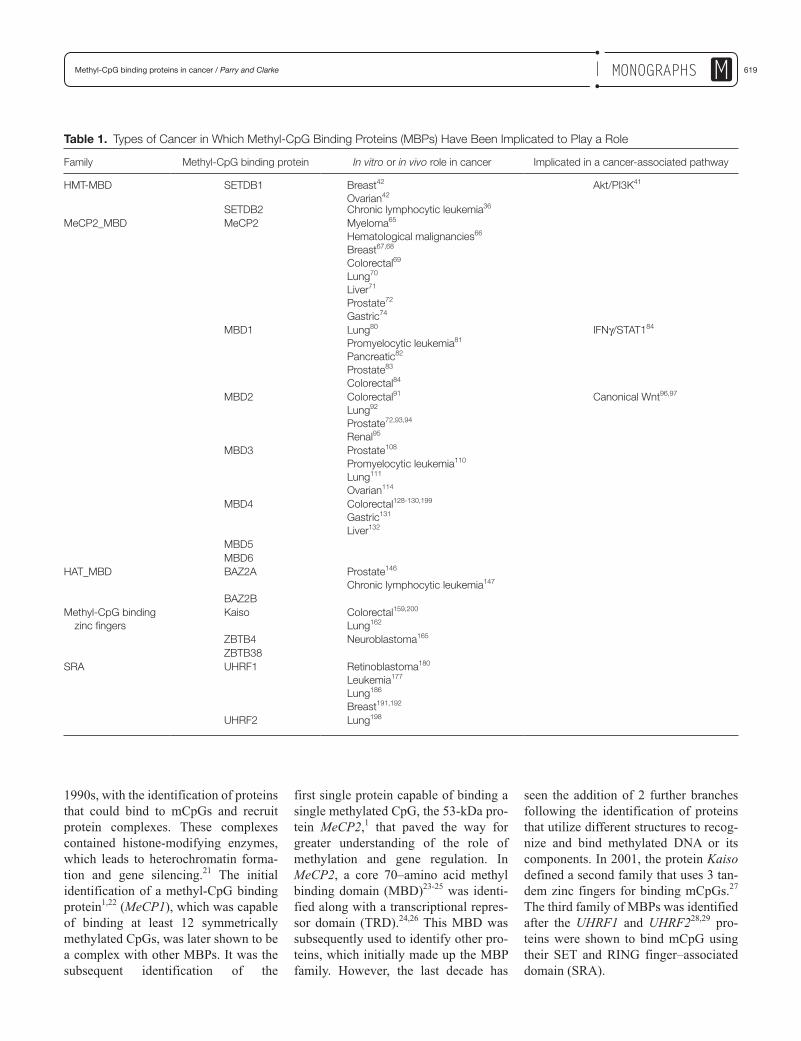
Table 1. Types of Cancer in Which Methyl-CpG Binding Proteins (MBPs) Have Been Implicated to Play a Role
Family Methyl-CpG binding protein In vitro or in vivo role in cancer Implicated in a cancer-associated pathway
HMT-MBD SETDB1 Breast42
Ovarian42Akt/PI3K41
SETDB2 Chronic lymphocytic leukemia36 MeCP2_MBD MeCP2 Myeloma65
Hematological malignancies66
Breast67,68
Colorectal69
Lung70
Liver71
Prostate72
Gastric74
MBD1 Lung80
Promyelocytic leukemia81
Pancreatic82
Prostate83
Colorectal84
IFNγ/STAT184
MBD2 Colorectal91
Lung92
Prostate72,93,94
Renal95
Canonical Wnt96,97
MBD3 Prostate108
Promyelocytic leukemia110
Lung111
Ovarian114
MBD4 Colorectal128-130,199
Gastric131
Liver132
MBD5 MBD6 HAT_MBD BAZ2A Prostate146
Chronic lymphocytic leukemia147
BAZ2B Methyl-CpG binding
zinc fingersKaiso Colorectal159,200
Lung162
ZBTB4 Neuroblastoma165 ZBTB38 SRA UHRF1 Retinoblastoma180
Leukemia177
Lung186
Breast191,192
UHRF2 Lung198

620 Genes & Cancer / vol 2 no 6 (2011)M Monographs
The MBP Superfamily and CancerAs previously mentioned, the MBP fam-ily is divided into 3 branches: 1) MBD containing proteins, 2) methyl-CpG binding zinc fingers, and 3) the SRA domain containing proteins.30 This sec-tion will cover the functions of the indi-vidual members of each family that are present in humans and highlight any roles in cancer (Table 1). Much of the data available on the roles of these pro-teins in cancer are related to their func-tions as transcriptional repressors or chromatin remodelers (Fig. 1). There is little evidence in the literature that defi-ciency of any of these proteins results in elevated cancer predisposition, a notion supported by a lack of any entries in the Catalogue of Somatic Mutations in Can-cer (COSMIC) database31 implicating their inactivation.
MBD-Containing Proteins
These proteins comprise the largest branch of the MBP family. Currently, the NCBI Conserved Domain Database (CDD)32-34 lists 11 human proteins con-taining the MBD derived from MeCP2
(CDD: cl00110 and cd00122). Based on the presence of other domains, these are further divided into 3 groups within the MBD superfamily according to the CDD30: the histone methyltransferases (HMT_MBD; CDD reference cd01395), the MeCP2_MBD proteins (CDD refer-ence cd01396), and the histone acetyl-transferases (HAT_MBD; CDD reference cd01397). Although all the members of this group contain the MBD, they do not all directly interact with methylated CpGs.
HMT_MBD
There are 2 members of this family: SETDB1 (also known as ESET) on chromosome 1 and SETDB2 (also known as CLLD8) on chromosome 13. As well as containing a MBD, they carry a PreSET and bifurcated SET domain, which mediates protein-protein interac-tions. SETDB1 was identified due to its interaction with the KAP-1 corepres-sor.35 SETDB2 was identified due to its role as a potential gene involved in leu-kemogenesis.36 They are both protein lysine methyltransferase enzymes that repress transcription through the forma-tion of heterochromatin.35,37,38
As may be expected from their func-tion, their roles in cancer are defined by the genes that they repress and interact with. Currently, the only evidence impli-cating SETDB2 in neoplasia is that it is 1 of the 14 genes encompassed by a 1-Mb deletion on chromosome 13q that is associated with disease progression in chronic lymphocytic leukemia.39 There is increased evidence for a tumorigene-sis role for SETDB1 as it has been shown to interact and mediate the functions of proteins, which have been demonstrated to have roles in cancer, for example, MCAF1,40 KAP1,38 serine/threonine kinase AKT,41 and DNMT3A.42
The SETDB1 interactions with MCAF1 and DNMT3A link promoter CpG hypermethylation to histone meth-ylation, a crucial precursory event to het-erochromatin formation. Its interaction with MCAF1 also links it to another tran-scriptional repressor, MBD1 (another MBP).43 Through MBD1, it is directed to mCpGs, where the MBD1:SETDB1: MCAF1 complex converts the dimethyl H3-K9 to trimethyl H3-K9, resulting in heterochromatin formation and transcrip-tional repression.40,44-46 In cancer, this
Figure 1. Mechanisms by which methyl-CpG binding proteins (MBPs) are implicated in cancer.
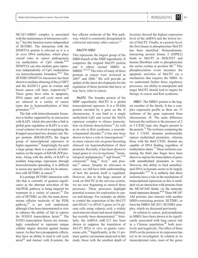
621Methyl-CpG binding proteins in cancer / Parry and Clarke MMonographs
MCAF1:MBD1 complex is associated with the maintenance of telomerase activ-ity,47 but this function seems independent of SETDB1. The interaction with the DNMT3A protein is relevant as it is a de novo DNA methylase, which plays crucial roles in cancer pathogenesis via methylation of CpG islands.48,49 DNMT3A can also mediate gene repres-sion independently of CpG methylation via heterochromatin formation.50,51 The SETDB1/DNMT3A interaction has been shown to mediate silencing of the p53BP2 and the RASSF1A gene in ovarian and breast cancer cell lines, respectively.42 These genes have roles in apoptosis, DNA repair, and cell cycle arrest and are silenced in a variety of cancer types due to hypermethylation of their promoters.52-56
The link with heterochromatin forma-tion is further supported by its interaction with KAP1, which also provides a link to global gene regulation as KAP1 is a uni-versal cofactor involved in regulating the Kruppel-associated box domain zinc fin-ger proteins (KRAB-ZFP), the largest group of transcriptional repressors in higher organisms.57 Surprisingly, for such a large group, there is a paucity of infor-mation on the targets of KRAB-ZFP pro-teins. Along with the ability of KAP1 to mediate long-range repression through heterochromatin spreading, it is difficult to assess any specific roles for its interac-tion with SETDB1 in cancer.38
It is perhaps SETDB1 interaction with Akt that is currently of greatest signifi-cance as the aberrant activation of the Akt/PI3K pathway is being targeted for treatment in a variety of cancers.58 The role of SETDB1 on Akt, the main down-stream effector molecule of the PI3K pathway,59 is not well understood, although it has been demonstrated in vitro to enhance the ability of Akt to repress the FOXO3 transcription factor.41 The FOXO transcription factors are increas-ingly considered to represent unique cellular targets directed against human cancer. As they have proapoptotic effects, they have an ability to lead to cell cycle arrest60 and interact with β-catenin, the
key effector molecule of the Wnt path-way, which is commonly deregulated in colorectal and many other cancers.61
MeCP2-MBD
This represents the largest group of the MBD branch of the MBP superfamily. It comprises the original MeCP2 protein and 6 others termed MBD1 to MBD6.19,62,63 The roles of many of these proteins in cancer were reviewed in 20072 and 2008.3 We will provide an update of the latest developments for the regulation of these proteins that have, or may have, roles in cancer.
MeCP2. The founder protein of the MBP superfamily MeCP2 is a global transcriptional repressor. It is a 50-kDa protein encoded by a gene on the X chromosome. It can bind to a single methylated CpG and recruit the Sin3A repressor complex to silence transcrip-tion via histone deacetylation.26 As well as its role in Rett syndrome, a neurode-velopmental disorder,64 it has also been shown to have a role in tumorigenesis2,3 as many cancers rely on genes becoming silenced via hypermethylation of their promoter. Recently, it has been shown to target genes in vivo in myeloma,65 hema-tological malignancies,66 and breast,67,68 colorectal,69 lung,70 liver,71 and pros-tate72 cancer. Despite its relevance to cancer, we still have little understanding of how the protein itself is regulated. However, due to the large amount of work on MeCP2 in the nervous system, we are now beginning to unravel these processes. These processes highlight potential avenues for exploration in can-cer and therapy. For example, an ability to control the expression of the MeCP2 and HDAC1 to HDAC3 genes in C6 gli-oma cells using valproic acid, a widely used anticonvulsant and mood stabilizer, has recently been demonstrated.73 Simi-larly, the miRNA miR-212 has been shown to suppress the translation of MeCP2 RNA in vitro in gastric carci-noma cells.74 Significantly, in the 11 pri-mary gastric carcinomas analyzed in this study, those with the smallest depth of
invasion showed the highest expression level of the miRNA and the lowest lev-els of MeCP2. Finally, at a protein level, the first kinase to phosphorylate MeCP2 has been identified. Homeodomain-interacting protein kinase 2 (HIPK2) binds to MeCP2 in HEK293T and human fibroblast cells to phosphorylate the serine residue at position 80.75 This phosphorylation event increases the apoptotic activities of MeCP2 via a mechanism that requires the MBD. As we understand further these regulatory processes, our ability to manipulate and target MeCP2 should lead to targets for therapy in cancer and Rett syndrome.
MBD1. The MBD1 protein is the larg-est member of the family. It has a com-plex expression profile as there are 13 isoforms of the gene expressed from chromosome 18. The main difference between the isoforms is the presence of 2 or 3 CXXC-type zinc fingers present in the protein.76 The isoforms containing the first 2 CXXC domains preferentially repress methylated promoters, whereas those with the third CXXC domain are capable of DNA binding regardless of methylation status.77 These isoforms con-taining the third zinc finger have been shown to repress the transcription of genes with unmethylated promoters in vitro. However, this ability to bind unmethyl-ated DNA in humans seems to be largely dispensable.78,79 It is unlikely that these isoforms have a role in the mechanism of transcriptional repression as this is medi-ated via an interaction with proteins from the MCAF/AM family via the transcrip-tional repression domain.40,44 These MCAF/AM proteins also interact with another MBD-containing protein, SETDB1, to form the MBD1:MCAF1: SETDB1 com-plex, which we discussed previously.
In relation to cancer, polymorphisms in MBD1 have been shown to be signifi-cantly associated with lung cancer risk in a Chinese population,80 both posi-tively and negatively. The effect of these SNPs on the protein or its expression has yet to be investigated. In relation to its transcriptional roles, most of the genes
622 Genes & Cancer / vol 2 no 6 (2011)M Monographs
silenced by MBD1 in cancer have been identified in vitro. These targets have been identified in acute promyelocytic leukemia81 and pancreatic,82 prostate,83 and colon84 cancer cell lines. MBD1 tar-gets may also have a role in drug resis-tance and the immune system interactions of cancers as the MBD1 gene is present on an amplified region of chromosome 18, which is associated with the development of vinblastine resistance in human cervical carcinoma KB cells.85 From an immune perspec-tive, MBD1 silences the IRF8 gene in colon carcinoma cells, which is a central mediator in the IFNγ/STAT1 signaling pathway.84 This silencing frequently occurs in human colorectal carcinoma in vivo, and IRF8 expression has been shown to induce apoptosis in tumor cells.86 Given that the IFNγ/STAT1 path-way has a critical role in governing the establishment of innate immune responses,87 disrupting it through such MBD1-mediated silencing clearly has implications for immune surveillance.
The capability of these MBD1 iso-forms to play a role in cancer is likely to be effected by factors such as redundancy and target specificity. As MBD1 binds with greater efficiency to genes in which the promoter has the sequence TCMGCA or TGCMGCA,78 and a comparison of cancer cell lines of different origins demonstrated that the promoters of many silenced genes were occupied by more than one MBD protein.79
MBD2. The gene for MBD2 is approx-imately 4 Mb downstream of MBD1 on the “q” arm of chromosome 18. They have limited similarity to each other, whereas MBD2 shows 71.1% overall amino acid identity to MBD3.62 MBD2 mediates the methylated DNA binding functions for 2 different transcriptional repressor complexes, MECP188 and Mi2/NuRD.89,90 Both these complexes use MBD2 to direct HDACs and chromatin remodelers to methylated promoters, where they effect transcriptional repres-sion. Once again, this protein has been
shown to silence genes in a variety of cancers: colorectal,91 lung,92 prostate,93,94 and renal.95 The relevance of the silenc-ing of these genes in cancer is apparent in colorectal cancer as mice deficient for MBD2 develop relatively normally, although they do show impaired maternal behavior and exhibit resistance to Wnt-driven cancer.96 Hyperactivity of the Wnt pathway is responsible for the majority of colorectal cancer cases. These Mbd2-null mice also show an increased ability to retain a degree of control over the Wnt pathway when it is constitutively acti-vated.97 As the deficiency of MBD2 in mice appears relatively benign, it makes an attractive therapeutic target. Work is underway to further identify the dere-pressed genes in these mice, which results in the manifestation of this phenotype. One identified in vivo target for MBD2 silencing is IL-4, which plays a role in the immune system and IFNγ regulation.98,99 The deregulation of IL-4 upregulates IFNγ due to a negative feedback loop,100 giving an increased Th1 immune response.101 As IFNγ has been shown to subdue Wnt targets102 and a Th1 response is a key element in tumor immune sur-veillance,103 this clearly provides an attractive potential mechanism of per-turbing tumorigenesis.
Despite its attractiveness as a thera-peutic target in colorectal cancer, no drugs that specifically target the MBD2 gene or protein have been identified to date. However, the drug B1 (N-[2-[dimethylamino]ethy]l-2-aminothia-zonaphthalimide) has been shown to alleviate MBD2 repression of the 14-3-3σ gene in promyelocytic leukemia cells and induce their apoptosis. Further char-acterization of this ability would be desir-able. An alternative way of targeting MBD2 is offered by targeting the PRMT1 or PRMT5 protein arginine methyltrans-ferases that have been shown to methyl-ate the MBD2 arginine residues. This methylation inhibits the ability of MBD2 to associate with its repressive complexes and silence genes.104
As attractive as MBD2 is as a thera-peutic target, caution must be exercised
in an application that will deregulate global gene transcription. By way of illustration, MBD2 is known to play a role in the silencing of the human telom-erase reverse transcriptase promoter (hTERT) in somatic cells. Given that upregulation of this gene is a key event for the ability of many cancer cells to overcome replicative senescence associ-ated with telomere shortening,105 there are clear potential negative ramifica-tions for inactivation of MBD2.
MBD3. The chromosome 19 gene, MBD3, has multiple spice variants, all of which are unable to bind methylated CpGs due to 2 alterations within the MBD.106 Despite this, it still plays a crucial role in the silencing of methylated genes. It is an important part of the Mi2/NuRD repres-sion complex, which can use MBD2 as its means of methylated DNA binding. The cancer-related genes repressed by this complex were discussed in the previous MBD2 section. In the absence of MBD2, the repression complex, hereafter termed MBD3/NuRD, uses other proteins for DNA binding. These non-MBD2–related roles are important in development, as Mbd3–/– mice die in utero, but it is its non-MBD2–related roles in cancer we will dis-cuss here.
Roles in human cancer that are spe-cific to MBD3 are not commonly observed.107 There are MBD2-indepen-dent reports of a role for the MBD3/NuRD complex shown in both prostate cancer cells108 and leukemias. In acute promyelocytic leukemia, it interacts with the fusion PML-RARa oncoprotein that is generated from the 15;17 translo-cation,109 and in acute myeloid leuke-mia, it interacts with the Evi1 gene to inhibit histone deacetylation and acti-vate genes.110 In human lung cancer cells, MBD3/NuRD regulates p21WAF1/Cip1 and ErbB2111 expression levels, and these can be altered by treatment with the HDAC inhibitor (HDACi) trichostatin A. Intriguingly, the HDACi released MBD3 from the p21WAF1/Cip1 promoter in cancer cells but recruited it in normal cells and vice versa with the ErbB2
623Methyl-CpG binding proteins in cancer / Parry and Clarke MMonographs
promoter. This may reflect the fact that MBD3 can bind unmethylated DNA and mediate the observed histone acetylation even in the presence of HDACi.106,112,113 The p21WAF1/Cip1 observation is corrobo-rated in HeLa cells, where depletion of MBD3 enhanced its transcription but repressed cyclin B1, Plk1, and sur-vivin.114 This resulted in an arrest at G2/M that led to cell death due to abnor-mal mitosis.
A different role has been shown in mouse lymphosarcoma cells in which Mbd3 interacts with Brg1 and Dnmt3a115 to silence the methylated MT-I gene. It remains to be identified which complex mediates the silencing as the association of Mbd3/NuRD repressor with Dnmt3a can link it to methylated DNA. Alterna-tively, the SWI/SNF complex, of which Brg1 is a central component, could also be used as it associates with the Mecp2/Sin3a/HDAC complex.116,117 Its roles in the SWI/SNF complex are not well understood but may make MBD3 rele-vant to many more cancers than previ-ously suspected.118
The successful development of drugs to target this protein will again be depen-dent on tumor-specific targeting as MBD3 has recently been shown to play a significant role in maintaining intesti-nal homeostasis through the interaction with nonphosphorylated c-Jun.119,120 The N-terminal phosphorylation of c-Jun inhibits this interaction and results in upregulation of its target genes in intes-tinal homeostasis and tumorigenesis. In the absence of MBD3, the intestines are characterized by hyperproliferation and an increased susceptibility to colitis-induced tumorigenesis. Nevertheless, therapeutically targeting MBD3 may prove to have benefits in cancer treat-ment. Potentially, we could develop drugs that are specific for the role in silencing methylated genes. The pro-teins of 2 other genes, MBD3L1 and MBD3L2, which show substantial homology to MBD2 and MBD3 but lack a MBD,121 may give us a clue to how to approach this as MBD3L2 can interact with the MBD2/MBD3/Mi2/NuRD complex in a way that displaces the
complex from methylated DNA and reactivate transcription.122
MBD4. MBD4 is primarily encoded from a single transcript on chromosome 362 and stands out from the other MBD proteins due to its interactions with the DNA repair machinery.123 The protein uses its N-terminal MBD to bind meth-ylated CpG and its C-terminal glycosyl-ase (which is homologous to base excision repair proteins) to mediate repair of the hypermutable CpG, which is susceptible to mismatch formation following either the hydrolytic deamina-tion of 5-methylcytosine to thymine or deamination of cytosine to uracil. Thus, MBD4 plays a key role in maintaining methylated DNA gene regulation and suppressing mutation at CpG sites.
These abilities, along with its interac-tion with the mismatch repair protein MLH1 (mutL homolog 1), potentially impact chemotherapy as MBD4 has been shown to be required for the maximal apoptotic effects of many of these che-motherapeutic drugs,124,125 a role that may in part be mediated through an interaction with Fas-associated death domain protein (FADD).126 This indi-cates that MBD4 function is crucial for normal mutation suppression and optimal response to chemotherapy; however, there is also evidence that its absence can be beneficial, as it has been shown in vitro that the radiosensitizing drug iododeoxyuridine is more effective in the absence of MBD4 due to the normal activity of MBD4 in repairing G:IU mis-matches located in CpG dinucleotides.
The significance of MBD4 in cancer is largely indirect as deficiency results in a mutator phenotype and subsequent alterations to bona fide tumor suppressor genes, evidenced by increased suscepti-bility conferred by MBD4 deficiency to intestinal tumorigenesis in the ApcMin/+ model.127 In this model, loss of function of MBD4 increases the number of “sec-ond hit” mutations at CpG sites in the wild-type Apc allele. This observation is echoed in man in whom an increased mutation rate at CpGs confers suscepti-bility to colorectal cancer,128,129 stomach
cancer,130,131 and progression of liver can-cer.132 Consistent with a role in mutation suppression, early downregulation of the gene has been observed in precancerous tissues, implicating it in tumor initiation rather than progression.133 Whether this loss is required for the onset of genomic instability due to repair defects or sup-pression of apoptosis is unclear.
Although it is best known for its repair activities, MBD4 has also been shown to repress p16INK4a and MLH1 genes in a methylation-dependent fash-ion134 in vitro, as it associates with the Sin3A repressor complex, which MeCP2 also interacts with. Its presence at the promoters of these genes is proposed to maintain the integrity of the CpG meth-ylation signal. However, this repression (along with repression by the MBD2/Mi-2/NuRD complex) is enhanced by an interaction with the RET finger protein (RFP; also known as TRIM27).135 This interaction with RET potentially links MBD4 to a much more extensive range of cancers.136
MBD5 and MBD6. MBD5 on chromo-some 2 and MBD6 on chromosome 6 were initially 2 uncharacterized proteins that were renamed in 2003 due to them containing a MBD domain.19,63 There is a limited amount of data available on the functions of these proteins. Neither of them is likely to bind methylated DNA, but they do associate with heterochroma-tin, where they may contribute to the for-mation or function of heterochromatin.137 As with other MBP proteins, MBD5 defi-ciency is linked to a developmental disorder, the 2q23.1 microdeletion syn-drome,138,139 but there have been no reports linking either of them to cancer.
HAT_MBD
BAZ2A and BAZ2B. There are 2 pro-teins in this final group: BAZ2A (also known as TIP5), encoded by the BAZ2A gene on chromosome 12, and BAZ2B, encoded by BAZ2B on chromosome 2. These proteins were identified by searching the EST and cDNA libraries for genes that encoded bromodo-mains.140 Bromodomain proteins have

624 Genes & Cancer / vol 2 no 6 (2011)M Monographs
been identified as integral components of chromatin remodeling complexes and frequently possess histone acetyltrans-ferase activity. They both contain a MBD, DDT, PHD, and bromodomain consecutively arranged in the same order.30 The MBD domain in these pro-teins differs at specific residues from other superfamily members, and it is unable to bind to a methylated oligonu-cleotide, which would be recognized by MBD2.141
The main identified role of these pro-teins is in the epigenetic silencing of ribosomal DNA (rRNA) within the nucleolus.141-144 The BAZ2A protein is a major component of the nucleolar remodeling complex (NoRC) that represses ribosomal gene transcription via histone deacetylation. This NoRC complex associates with the histone deacetylase HDAC1 in vivo and in vitro via the bromodomain of BAZ2A. It appears that NoRC silences the rDNA locus by targeting the SIN3 corepressor complex to the rDNA promoter, which alters the local chromatin structure and leads to methylation of critical CpG residues.145
There are very few reports of these proteins having a role in cancer. It has been shown that overexpression of miR-NAs, which can regulate expression of BAZ2A, correlates with progression to metastasis in prostate cancer146 and may also have a role in chronic lymphocytic leukemia.147 However, it currently remains unclear if these proteins will have a significant role in cancer.
Methyl-CpG Binding Zinc Fingers
This branch of MBPs is based on a 3–zinc finger motif found in the C-ter-minus of the Kaiso protein, which is capable of binding a pair of methylated CpG dinucleotides.27 The protein also binds unmethylated DNA and has a greater affinity for CTGCNA than mCGmCG.148 The Kaiso protein is the archetypal member of this branch, and the zinc finger motif has been used to identify 2 other members, ZBTB4 and ZBTB38,149 that are also capable of binding methylated DNA.
Kaiso
Kaiso is encoded by the ZBTB33 gene on the X chromosome. It belongs to the zinc finger and broad-complex, tram-track, and bric-a-brac/poxvirus and zinc finger (BTB/POZ) protein family. Kaiso was first identified as a binding partner of p120ctn in a yeast 2-hybrid screen.27 This p120ctn protein associates with Kaiso at the cell surface (as part of the E-cadherin-catenin cell adhesion com-plex) and in the nucleus.27,148,150 At the cell surface, it regulates the expression and stability of cadherins and thereby influences their roles in adhesion and signaling.151,152 In the nucleus, it has been shown to function as a transcrip-tional repressor by directing the nuclear corepressor complex N-CoR153 to meth-ylated and unmethylated gene promot-ers. N-CoR is a complex that contains histone deacetylase 3,154,155 which oper-ates to generate a transcriptionally inac-tive chromatin structure.
Given its multiple functions, it is per-haps not surprising that altered Kaiso activity is a feature of some cancers, par-ticularly given that Kaiso functions as a regulator of the target genes of the canonical and noncanonical Wnt path-ways.150,156-158 In Wnt-driven colon can-cer cell lines, Kaiso mediates the silencing of the tumor suppressor genes CDKN1A, HIC1,159 and Rb.160 Its relevance in vivo has been demonstrated by crossing the viable Kaiso–/y mutant mouse to a mouse model of Wnt-driven intestinal cancer, which leads to a suppression of tumori-genesis that is thought to be due to dere-pression of methylated or unmethylated Wnt target genes. However, an equally plausible hypothesis is that tumor sup-pression arises as a consequence of per-turbed p120ctn as it has been recently demonstrated that the complex that marks β-catenin (the key mediator of Wnt sig-naling) for destruction also has the ability to degrade p120ctn.161
Much of the current work focuses on the identification of the relevant Kaiso-silenced genes in cancer. Given Kaiso’s ability to repress transcription at both methylated and unmethylated promoters, it is currently difficult to assess how
important its mCpG binding role is in can-cer. For example, in lung cancers, Kaiso mediates silencing of both the methylated MTA2 gene and the unmethylated CCND1 and MMP7 genes.162 Given that all these genes are implicated in tumorigenesis, the precise significance of mCpG binding remains to be elucidated.
Beyond its role in transcriptional repression, Kaiso has also been local-ized to mitotic spindles and centrosomes at different stages of the cell cycle.163 Altering the ability of Kaiso to locate to these regions resulted in defective cen-trosomes, whereas overexpressing Kaiso resulted in mitotic arrest, and knocking it down led to elevated proliferation. Given that Kaiso is often mislocalized in cancer, clearly these observations are of high potential significance to cancer; combined with the observation that the Kaiso-deficient mouse is healthy and viable, it makes a potentially attractive therapeutic target. However, given its diversity of roles, great care will need to be taken in the use of such a therapeutic.
ZBTB4 and ZBTB38
The genes for ZBTB4 and ZBTB38 are located on chromosomes 17 and 3, respectively. Unlike Kaiso, these proteins can bind a single methylated CpG in vitro and in vivo.149 As yet, there are very little published data on these proteins in terms of biological roles, although ZBTB38 has been shown to play a role in human height determination.164
With respect to cancer, there is a sin-gle report of ZBTB4 associating with the Sin3/HDAC complex and silencing the CDKN1A gene in neuroblastoma cells.165 As would be expected, silencing of this gene resulted in cell cycle arrest due to p53 activation, suppression of apoptosis through regulation of p21CIP1, and promotion of long-term cell sur-vival. This was reflected in the primary human neuroblastoma tumors, which showed decreasing ZBTB4 levels as the tumors progressed.
A p53-independent apoptotic role has also been confirmed for ZBTB38 in the mouse through its interaction with CtBP.166 A role in apoptosis for ZBTB38
625Methyl-CpG binding proteins in cancer / Parry and Clarke MMonographs
is also suggested as it is a substrate for caspase-3, a feature of mitochondrial-driven apotosis.167 However, the signifi-cance of either of these genes in cancer therefore remains largely undetermined.
SRA Domain–Containing Proteins
There are 2 members of this family, UHRF1 and UHRF2, which contain a SET and RING finger–associated (SRA) domain as well as a ring finger motif, PHD finger, and ubiquitin-like domain. The ability of the SRA domain to bind methylated CpG was first recognized in human breast cancer cells28 as decreased expression and increased promoter methylation of EGR2 correlated with increased expression of UHRF1, which associated with the methylated EGR2 promoter. UHRF2 was subsequently identified as a MBP due to its homology with the SRA domain of UHRF1168; however, there is little similarity between this domain and the MeCP2_MBD in terms of recognizing and bind-ing mCpGs as the SRA domain prefers hemimethylated DNA, and its mode of recognition involves flipping methylcy-tosine out of the DNA helix.169-171 By contrast, the MeCP2_MBD prefers sym-metrically methylated DNA.172 It folds into an α/β sandwich, which presents a large, positively charged surface173 that recognizes hydration of the major groove of methylated DNA rather than cytosine methylation.172
UHRF1
Prior to the discovery that UHRF1 (also known as Np95 and ICBP90) on chromo-some 19 could bind methylated DNA, it had been identified as a protein of many functions. These include regulation of topoisomerase IIα gene expression174 and regulation of cell proliferation,175 the cell cycle,176 apoptosis,177 and tumor biology.178,179 Initially, its roles in these functions were thought to be related to its role as a transcriptional repressor as UHRF1 interacted with HDAC1 and recruited it to methylated tumor sup-pressor genes.180 However, its role in human biology and cancer has taken on a wider significance due to the observation
that UHRF1 associates with DNMT1 to maintain the epigenetic inheritance of DNA methylation.181,182 UHRF1 has an affinity for hemimethylated CpG DNA and recruits DNMT1 to ensure that the sequence becomes completely methyl-ated following replication. This role may also apply to the maintenance of histone marks.183 Additionally, it also has been shown to play a role in DNA damage response as it localizes to DNA damage and double-strand breaks.184,185
In human cancer, all of these func-tions have been shown to be relevant to different types of cancer. The mainte-nance of methylation plays a significant role in cancers by maintaining promot-ers in a hypermethylated state. In lung cancer, UHRF1 controls the cell cycle through maintenance of promoter meth-ylation at CDKN2A and RASSF1.186 It has also been shown to silence the CMV promoter in ES cells prior to methyla-tion due to its interaction with Dnmt3a and Dnmta3b.187 On the other hand, its roles in cancer may prove beneficial as, for example, depletion of UHRF1 has been shown to activate the DNA damage response leading to cell cycle arrest at G2/M and subsequent caspase 8–induced apoptosis.188 In this respect, it is interest-ing that polyphenols in grapes have been shown to inhibit growth and induce apoptosis through downregulation of UHRF1.189,190 These apoptotic effects may also account for the radiosensitiz-ing effect of UHRF1 depletion in human breast cancer cells.191 Furthermore, its function as a transcriptional repressor has the potential to overcome drug resis-tance as overexpression of UHRF1 inhibits the upregulation of MDR1, which normally engenders drug insensi-tivity by actively removing cytotoxic drugs from breast cancer cells.192
The many roles for UHRF1 in cancer have marked it for therapeutic target-ing.193,194 However, as for all the MBPs in this review, their wide-ranging func-tions in multiple tissues again urge caution when targeting them. In this respect, the role played by UHRF1 in maintaining DNA methylation patterns required for progenitor maintenance and
self-renewal of somatic tissue is of par-ticular importance.195
UHRF2
UHRF2 (also known as NIRF or Np97) was identified due to its role in the cell cycle.168 Its expression is highest in pro-liferating cells, where it ubiquitinates PCNP,168,196 and its overexpression results in G1 arrest, presumably through an interaction with Cdk2.197 Any other roles have yet to be described for this pro-tein, but it may play a role in lung cancer cells as UHRF2 expression has been shown to be repressed by let7-a miRNA, which subsequently led to elevation of p21waf1 levels in vitro and in vivo and in growth suppression.198 Any wider conse-quences of UHRF2 activity in cancer remain to be established.
SummaryThis review has highlighted the many and intricate roles that MBPs play in human biology. Primarily, this involve-ment in gene regulation has led to them being utilized by normal cells to exert transcriptional control but has also led to them being exploited by cancer cells to escape such control. The dramatic effects of suppressing or enhancing their roles in cancer make them eminently suitable targets for therapeutic interven-tion. However, their many basic regula-tory roles in a variety of tissues in the human body also present us with limita-tions and hazards of any such targeting. For this reason, we now need a much better understanding of the specific changes that these proteins induce in cancer cells to permit their propagation. The hope is that we can identify a subset of targets that are highly specific to can-cer cells and so begin to develop a range of novel targeted therapies.
Declaration of Conflicting InterestsThe author(s) declared no potential conflicts of inter-est with respect to the research, authorship, and/or publication of this article.
FundingThe author(s) received no financial support for the research, authorship, and/or publication of this article.
626 Genes & Cancer / vol 2 no 6 (2011)M Monographs
References
1. Meehan RR, Lewis JD, McKay S, Kleiner EL, Bird AP. Identification of a mammalian protein that binds specifically to DNA containing meth-ylated CpGs. Cell. 1989;58:499-507.
2. Sansom OJ, Maddison K, Clarke AR. Mecha-nisms of disease: methyl-binding domain pro-teins as potential therapeutic targets in cancer. Nat Clin Pract Oncol. 2007;4:305-15.
3. Lopez-Serra L, Esteller M. Proteins that bind methylated DNA and human cancer: reading the wrong words. Br J Cancer. 2008;98:1881-5.
4. Johnson TB, Coghill RD. Researches on pyrimi-dines: C111. The discovery of 5-methyl-cytosine in tuberculinic acid, the nucleic acid of the tuber-cle bacillus1. J Am Chem Soc. 1925;47:2838-44.
5. Hotchkiss RD. The quantitative separation of purines, pyrimidines, and nucleosides by paper chromatography. J Biol Chem. 1948;175:315-32.
6. Morrison JM, Keir HM, Subak-Sharpe H, Crawford LV. Nearest neighbour base sequence analysis of the deoxyribonucleic acids of a fur-ther three mammalian viruses: simian virus 40, human papilloma virus and adenovirus type 2. J Gen Virol. 1967;1:101-8.
7. Subak-Sharpe JH. Base doublet frequency pat-terns in the nucleic acid and evolution of viruses. Br Med Bull. 1967;23:161-8.
8. Grippo P, Iaccarino M, Parisi E, Scarano E. Methylation of DNA in developing sea urchin embryos. J Mol Biol. 1968;36:195-208.
9. Scarano E. The control of gene function in cell differentiation and in embryogenesis. Adv Cyto-pharmacol. 1971;1:13-24.
10. Holliday R, Pugh JE. DNA modification mecha-nisms and gene activity during development. Sci-ence. 1975;187:226-32.
11. Riggs AD. X inactivation, differentiation, and DNA methylation. Cytogenet Cell Genet. 1975;14:9-25.
12. McGhee JD, Ginder GD. Specific DNA meth-ylation sites in the vicinity of the chicken beta-globin genes. Nature. 1979;280:419-20.
13. Mandel JL, Chambon P. DNA methylation: organ specific variations in the methylation pattern within and around ovalbumin and other chicken genes. Nucleic Acids Res. 1979;7:2081-103.
14. Bird AP, Southern EM. Use of restriction enzymes to study eukaryotic DNA methylation: I. The methylation pattern in ribosomal DNA from Xen-opus laevis. J Mol Biol. 1978;118:27-47.
15. Bird AP. Use of restriction enzymes to study eukaryotic DNA methylation: II. The symmetry of methylated sites supports semi-conservative copying of the methylation pattern. J Mol Biol. 1978;118:49-60.
16. Svedruzić ZM, Reich NO. The mechanism of tar-get base attack in DNA cytosine carbon 5 meth-ylation. Biochemistry. 2004;43:11460-73.
17. Bird AP. CpG-rich islands and the function of DNA methylation. Nature. 1986;321:209-13.
18. Cooper DN, Taggart MH, Bird AP. Unmethylated domains in vertebrate DNA. Nucleic Acids Res. 1983;11:647-58.
19. Hendrich B, Tweedie S. The methyl-CpG binding domain and the evolving role of DNA methyla-tion in animals. Trends Genet. 2003;19:269-77.
20. Jones PA, Takai D. The role of DNA meth-ylation in mammalian epigenetics. Science. 2001;293:1068-70.
21. Fuks F. DNA methylation and histone modifi-cations: teaming up to silence genes. Curr Opin Genet Dev. 2005;15:490-5.
22. Meehan R, Antequera F, Lewis J, et al. A nuclear protein that binds preferentially to methyl-ated DNA in vitro may play a role in the inac-cessibility of methylated CpGs in mammalian nuclei. Philos Trans R Soc Lond B Biol Sci. 1990;326:199-205.
23. Lewis JD, Meehan RR, Henzel WJ, et al. Puri-fication, sequence, and cellular localization of a novel chromosomal protein that binds to methyl-ated DNA. Cell. 1992;69:905-14.
24. Nan X, Meehan RR, Bird A. Dissection of the methyl-CpG binding domain from the chro-mosomal protein MeCP2. Nucleic Acids Res. 1993;21:4886-92.
25. Nan X, Campoy FJ, Bird A. MeCP2 is a tran-scriptional repressor with abundant binding sites in genomic chromatin. Cell. 1997;88:471-81.
26. Nan X, Ng HH, Johnson CA, et al. Transcrip-tional repression by the methyl-CpG-binding protein MeCP2 involves a histone deacetylase complex. Nature. 1998;393:386-9.
27. Prokhortchouk A, Hendrich B, Jørgensen H, et al. The p120 catenin partner Kaiso is a DNA methylation-dependent transcriptional repressor. Genes Dev. 2001;15:1613-8.
28. Unoki M, Nishidate T, Nakamura Y. ICBP90, an E2F-1 target, recruits HDAC1 and binds to methyl-CpG through its SRA domain. Oncogene. 2004;23:7601-10.
29. Hashimoto H, Horton JR, Zhang X, Cheng X. UHRF1, a modular multi-domain protein, regu-lates replication-coupled crosstalk between DNA methylation and histone modifications. Epi-genetics. 2009;4:8-14.
30. Hung MS, Shen CK. Eukaryotic methyl-CpG-binding domain proteins and chromatin modifi-cation. Eukaryot Cell. 2003;2:841-6.
31. Forbes SA, Bhamra G, Bamford S, et al. The Cat-alogue of Somatic Mutations in Cancer (COS-MIC). Curr Protoc Hum Genet. 2008;Chapter 10:Unit 10.11.
32. Marchler-Bauer A, Anderson JB, Chitsaz F, et al. CDD: specific functional annotation with the Conserved Domain Database. Nucleic Acids Res. 2009;37:D205-10.
33. Marchler-Bauer A, Lu S, Anderson JB, et al. CDD: a Conserved Domain Database for the functional annotation of proteins. Nucleic Acids Res. 2011;39:D225-9.
34. Database NCD. Conserved Domain Database (CDD). Available from: http://www.ncbi.nlm.nih.gov/Structure/cdd/cdd.shtml
35. Schultz DC, Ayyanathan K, Negorev D, Maul GG, Rauscher FJ. SETDB1: a novel KAP-1-associated histone H3, lysine 9-specific meth-yltransferase that contributes to HP1-mediated silencing of euchromatic genes by KRAB zinc-finger proteins. Genes Dev. 2002;16:919-32.
36. Mabuchi H, Fujii H, Calin G, et al. Cloning and characterization of CLLD6, CLLD7, and CLLD8, novel candidate genes for leukemogen-esis at chromosome 13q14, a region commonly
deleted in B-cell chronic lymphocytic leukemia. Cancer Res. 2001;61:2870-7.
37. Falandry C, Fourel G, Galy V, et al. CLLD8/KMT1F is a lysine methyltransferase that is important for chromosome segregation. J Biol Chem. 2010;285:20234-41.
38. Groner AC, Meylan S, Ciuffi A, et al. KRAB-zinc finger proteins and KAP1 can mediate long-range transcriptional repression through heterochroma-tin spreading. PLoS Genet. 2010;6:e1000869.
39. Parker H, Rose-Zerilli MJ, Parker A, et al. 13q deletion anatomy and disease progression in patients with chronic lymphocytic leukemia. Leukemia. 2011;25:489-97.
40. Ichimura T, Watanabe S, Sakamoto Y, Aoto T, Fujita N, Nakao M. Transcriptional repres-sion and heterochromatin formation by MBD1 and MCAF/AM family proteins. J Biol Chem. 2005;280:13928-35.
41. Gao H, Yu Z, Bi D, et al. Akt/PKB interacts with the histone H3 methyltransferase SETDB1 and coordinates to silence gene expression. Mol Cell Biochem. 2007;305:35-44.
42. Li H, Rauch T, Chen ZX, Szabó PE, Riggs AD, Pfeifer GP. The histone methyltransfer-ase SETDB1 and the DNA methyltransferase DNMT3A interact directly and localize to pro-moters silenced in cancer cells. J Biol Chem. 2006;281:19489-500.
43. De Graeve F, Bahr A, Chatton B, Kedinger C. A murine ATFa-associated factor with transcriptional repressing activity. Oncogene. 2000;19:1807-19.
44. Fujita N, Watanabe S, Ichimura T, et al. MCAF mediates MBD1-dependent transcriptional repression. Mol Cell Biol. 2003;23:2834-43.
45. Fujita N, Watanabe S, Ichimura T, et al. Methyl-CpG binding domain 1 (MBD1) interacts with the Suv39h1-HP1 heterochromatic complex for DNA methylation-based transcriptional repres-sion. J Biol Chem. 2003;278:24132-8.
46. Wang H, An W, Cao R, et al. mAM facilitates conversion by ESET of dimethyl to trimethyl lysine 9 of histone H3 to cause transcriptional repression. Mol Cell. 2003;12:475-87.
47. Liu L, Ishihara K, Ichimura T, et al. MCAF1/AM is involved in Sp1-mediated maintenance of can-cer-associated telomerase activity. J Biol Chem. 2009;284:5165-74.
48. Rhee I, Bachman KE, Park BH, et al. DNMT1 and DNMT3b cooperate to silence genes in human cancer cells. Nature. 2002;416:552-6.
49. Shafiei F, Rahnama F, Pawella L, Mitchell MD, Gluckman PD, Lobie PE. DNMT3A and DNMT3B mediate autocrine hGH repression of plakoglobin gene transcription and consequent phenotypic conversion of mammary carcinoma cells. Oncogene. 2008;27:2602-12.
50. Bachman KE, Rountree MR, Baylin SB. Dnmt3a and Dnmt3b are transcriptional repressors that exhibit unique localization properties to hetero-chromatin. J Biol Chem. 2001;276:32282-7.
51. Fuks F, Hurd PJ, Deplus R, Kouzarides T. The DNA methyltransferases associate with HP1 and the SUV39H1 histone methyltransferase. Nucleic Acids Res. 2003;31:2305-12.
52. Liu ZJ, Lu X, Zhang Y, et al. Downregulated mRNA expression of ASPP and the hypermeth-ylation of the 5′-untranslated region in cancer

627Methyl-CpG binding proteins in cancer / Parry and Clarke MMonographs
cell lines retaining wild-type p53. FEBS Lett. 2005;579:1587-90.
53. Zhao J, Wu G, Bu F, et al. Epigenetic silence of ankyrin-repeat-containing, SH3-domain-contain-ing, and proline-rich-region-containing protein 1 (ASPP1) and ASPP2 genes promotes tumor growth in hepatitis B virus-positive hepatocellular carcinoma. Hepatology. 2010;51:142-53.
54. Patra SK, Szyf M. DNA methylation-mediated nucleosome dynamics and oncogenic Ras sig-naling: insights from FAS, FAS ligand and RASSF1A. FEBS J. 2008;275:5217-35.
55. Liu R, Gao L, Lu GX, Tang LS, Zhu XH, Wang J. [Methylation status of RASSF1A and DAPK promoter in retinoblastoma]. Zhonghua Yan Ke Za Zhi. 2009;45:631-5.
56. Laytragoon-Lewin N, Chen F, Castro J, et al. DNA content and methylation of p16, DAPK and RASSF1A gene in tumour and distant, normal mucosal tissue of head and neck squa-mous cell carcinoma patients. Anticancer Res. 2010;30:4643-8.
57. Urrutia R. KRAB-containing zinc-finger repres-sor proteins. Genome Biol. 2003;4:231.
58. Wu P, Hu YZ. PI3K/Akt/mTOR pathway inhibi-tors in cancer: a perspective on clinical progress. Curr Med Chem. 2010;17:4326-41.
59. Lawlor MA, Alessi DR. PKB/Akt: a key media-tor of cell proliferation, survival and insulin responses? J Cell Sci. 2001;114:2903-10.
60. Maiese K, Chong ZZ, Shang YC, Hou J. Clever cancer strategies with FoxO transcription factors. Cell Cycle. 2008;7:3829-39.
61. Hoogeboom D, Essers MA, Polderman PE, Voets E, Smits LM, Burgering BM. Interaction of FOXO with beta-catenin inhibits beta-catenin/T cell factor activity. J Biol Chem. 2008;283:9224-30.
62. Hendrich B, Bird A. Identification and character-ization of a family of mammalian methyl-CpG binding proteins. Mol Cell Biol. 1998;18:6538-47.
63. Roloff TC, Ropers HH, Nuber UA. Comparative study of methyl-CpG-binding domain proteins. BMC Genomics. 2003;4:1.
64. Amir RE, Van den Veyver IB, Wan M, Tran CQ, Francke U, Zoghbi HY. Rett syndrome is caused by mutations in X-linked MECP2, encod-ing methyl-CpG-binding protein 2. Nat Genet. 1999;23:185-8.
65. Wang Z, Zhang J, Zhang Y, Srivenugopal KS, Lim SH. SPAN-XB core promoter sequence is regulated in myeloma cells by specific CpG dinu-cleotides associated with the MeCP2 protein. Int J Cancer. 2006;119:2878-84.
66. Meklat F, Li Z, Wang Z, et al. Cancer-testis anti-gens in haematological malignancies. Br J Hae-matol. 2007;136:769-76.
67. Pampalakis G, Prosnikli E, Agalioti T, Vlahou A, Zoumpourlis V, Sotiropoulou G. A tumor-protective role for human kallikrein-related pep-tidase 6 in breast cancer mediated by inhibition of epithelial-to-mesenchymal transition. Cancer Res. 2009;69:3779-87.
68. Shin JE, Park SH, Jang YK. Epigenetic up-reg-ulation of leukemia inhibitory factor (LIF) gene during the progression to breast cancer. Mol Cells. 2011;31:181-9.
69. Pancione M, Sabatino L, Fucci A, et al. Epigene-tic silencing of peroxisome proliferator-activated receptor gamma is a biomarker for colorectal
cancer progression and adverse patients’ out-come. PLoS One. 2010;5:e14229.
70. Lin RK, Hsu HS, Chang JW, Chen CY, Chen JT, Wang YC. Alteration of DNA methyltransferases contributes to 5′CpG methylation and poor progno-sis in lung cancer. Lung Cancer. 2007;55:205-13.
71. Sohn BH, Park IY, Lee JJ, et al. Functional switch-ing of TGF-beta1 signaling in liver cancer via epi-genetic modulation of a single CpG site in TTP promoter. Gastroenterology. 2010;138:1898-908.
72. Pulukuri SM, Patibandla S, Patel J, Estes N, Rao JS. Epigenetic inactivation of the tissue inhibitor of metalloproteinase-2 (TIMP-2) gene in human prostate tumors. Oncogene. 2007;26:5229-37.
73. Kim B, Rincon Castro LM, Jawed S, Niles LP. Clinically relevant concentrations of valproic acid modulate melatonin MT(1) receptor, HDAC and MeCP2 mRNA expression in C6 glioma cells. Eur J Pharmacol. 2008;589:45-8.
74. Wada R, Akiyama Y, Hashimoto Y, Fukama-chi H, Yuasa Y. miR-212 is downregulated and suppresses methyl-CpG-binding protein MeCP2 in human gastric cancer. Int J Cancer. 2010;127:1106-14.
75. Bracaglia G, Conca B, Bergo A, et al. Methyl-CpG-binding protein 2 is phosphorylated by homeodomain-interacting protein kinase 2 and contributes to apoptosis. EMBO Rep. 2009;10:1327-33.
76. Jørgensen HF, Ben-Porath I, Bird AP. Mbd1 is recruited to both methylated and nonmethylated CpGs via distinct DNA binding domains. Mol Cell Biol. 2004;24:3387-95.
77. Nakao M, Matsui S, Yamamoto S, Okumura K, Shirakawa M, Fujita N. Regulation of transcrip-tion and chromatin by methyl-CpG binding pro-tein MBD1. Brain Dev. 2001;23 Suppl 1:S174-6.
78. Clouaire T, de Las Heras JI, Merusi C, Stancheva I. Recruitment of MBD1 to target genes requires sequence-specific interaction of the MBD domain with methylated DNA. Nucleic Acids Res. 2010;38:4620-34.
79. Muller I, Wischnewski F, Pantel K, Schwarzen-bach H. Promoter- and cell-specific epigenetic regulation of CD44, Cyclin D2, GLIPR1 and PTEN by methyl-CpG binding proteins and his-tone modifications. BMC Cancer. 2010;10:297.
80. Liu H, Jin G, Wang H, et al. Methyl-CpG bind-ing domain 1 gene polymorphisms and lung cancer risk in a Chinese population. Biomarkers. 2008;13:607-17.
81. Villa R, Morey L, Raker VA, et al. The methyl-CpG binding protein MBD1 is required for PML-RARalpha function. Proc Natl Acad Sci U S A. 2006;103:1400-5.
82. Liu C, Chen Y, Yu X, et al. Proteomic analysis of differential proteins in pancreatic carcinomas: effects of MBD1 knock-down by stable RNA interference. BMC Cancer. 2008;8:121.
83. Yaqinuddin A, Abbas F, Naqvi SZ, Bashir MU, Qazi R, Qureshi SA. Silencing of MBD1 and MeCP2 in prostate-cancer-derived PC3 cells pro-duces differential gene expression profiles and cel-lular phenotypes. Biosci Rep. 2008;28:319-26.
84. McGough JM, Yang D, Huang S, et al. DNA meth-ylation represses IFN-gamma-induced and signal transducer and activator of transcription 1-medi-ated IFN regulatory factor 8 activation in colon carcinoma cells. Mol Cancer Res. 2008;6:1841-51.
85. Wang J, Tai LS, Tzang CH, Fong WF, Guan XY, Yang M. 1p31, 7q21 and 18q21 chromosomal aberrations and candidate genes in acquired vin-blastine resistance of human cervical carcinoma KB cells. Oncol Rep. 2008;19:1155-64.
86. Yang D, Thangaraju M, Browning DD, et al. IFN regulatory factor 8 mediates apoptosis in nonhemopoietic tumor cells via regulation of Fas expression. J Immunol. 2007;179:4775-82.
87. Wang H, Morse HC 3rd. IRF8 regulates myeloid and B lymphoid lineage diversification. Immu-nol Res. 2009;43:109-17.
88. Feng Q, Zhang Y. The MeCP1 complex represses transcription through preferential binding, remodeling, and deacetylating methyl-ated nucleosomes. Genes Dev. 2001;15:827-32.
89. Feng Q, Cao R, Xia L, Erdjument-Bromage H, Tempst P, Zhang Y. Identification and func-tional characterization of the p66/p68 compo-nents of the MeCP1 complex. Mol Cell Biol. 2002;22:536-46.
90. Le Guezennec X, Vermeulen M, Brinkman AB, et al. MBD2/NuRD and MBD3/NuRD, two dis-tinct complexes with different biochemical and functional properties. Mol Cell Biol. 2006;26: 843-51.
91. Park HY, Jeon YK, Shin HJ, et al. Differential promoter methylation may be a key molecular mechanism in regulating BubR1 expression in cancer cells. Exp Mol Med. 2007;39:195-204.
92. Zhuravel E, Shestakova T, Glushko N, Soldat-kina M, Pogrebnoy P. Expression patterns of murine beta-defensin-2 mRNA in Lewis lung carcinoma cells in vitro and in vivo. Exp Oncol. 2008;30:206-11.
93. Pulukuri SM, Rao JS. CpG island promoter methylation and silencing of 14-3-3sigma gene expression in LNCaP and Tramp-C1 prostate cancer cell lines is associated with methyl-CpG-binding protein MBD2. Oncogene. 2006;25:4559-72.
94. Shukeir N, Pakneshan P, Chen G, Szyf M, Rab-bani SA. Alteration of the methylation status of tumor-promoting genes decreases prostate can-cer cell invasiveness and tumorigenesis in vitro and in vivo. Cancer Res. 2006;66:9202-10.
95. Majid S, Dar AA, Ahmad AE, et al. BTG3 tumor suppressor gene promoter demethyl-ation, histone modification and cell cycle arrest by genistein in renal cancer. Carcinogenesis. 2009;30:662-70.
96. Sansom OJ, Berger J, Bishop SM, Hendrich B, Bird A, Clarke AR. Deficiency of Mbd2 sup-presses intestinal tumorigenesis. Nat Genet. 2003;34:145-7.
97. Phesse T, Parry L, Reed K, et al. Deficiency of Mbd2 attenuates Wnt signaling. Mol Cell Biol. 2008;28:6094-103.
98. Hutchins AS, Artis D, Hendrich BD, Bird AP, Scott P, Reiner SL. Cutting edge: a critical role for gene silencing in preventing excessive type 1 immunity. J Immunol. 2005;175:5606-10.
99. Hutchins AS, Mullen AC, Lee HW, et al. Gene silencing quantitatively controls the function of a developmental trans-activator. Mol Cell. 2002;10:81-91.
100. Elser B, Lohoff M, Kock S, et al. IFN-gamma represses IL-4 expression via IRF-1 and IRF-2. Immunity. 2002;17:703-12.
628 Genes & Cancer / vol 2 no 6 (2011)M Monographs
101. Hutchins AS, Artis D, Hendrich BD, Bird AP, Scott P, Reiner SL. Cutting edge: a critical role for gene silencing in preventing excessive type 1 immunity. J Immunol. 2005;175:5606-10.
102. Nava P, Koch S, Laukoetter MG, et al. Interferon-gamma regulates intestinal epithelial homeosta-sis through converging beta-catenin signaling pathways. Immunity. 2010;32:392-402.
103. Dunn GP, Koebel CM, Schreiber RD. Interfer-ons, immunity and cancer immunoediting. Nat Rev Immunol. 2006;6:836-48.
104. Tan CP, Nakielny S. Control of the DNA methylation system component MBD2 by protein arginine methylation. Mol Cell Biol. 2006;26:7224-35.
105. Chatagnon A, Bougel S, Perriaud L, Lachuer J, Benhattar J, Dante R. Specific association between the methyl-CpG-binding domain pro-tein 2 and the hypermethylated region of the human telomerase reverse transcriptase promoter in cancer cells. Carcinogenesis. 2009;30:28-34.
106. Saito M, Ishikawa F. The mCpG-binding domain of human MBD3 does not bind to mCpG but interacts with NuRD/Mi2 components HDAC1 and MTA2. J Biol Chem. 2002;277:35434-9.
107. Zhu Y, Harrison DJ, Bader SA. Genetic and epigenetic analyses of MBD3 in colon and lung cancer. Br J Cancer. 2004;90:1972-5.
108. Das PM, Ramachandran K, Vanwert J, et al. Methylation mediated silencing of TMS1/ASC gene in prostate cancer. Mol Cancer. 2006;5:28.
109. Morey L, Brenner C, Fazi F, et al. MBD3, a component of the NuRD complex, facilitates chromatin alteration and deposition of epigen-etic marks. Mol Cell Biol. 2008;28:5912-23.
110. Spensberger D, Vermeulen M, Le Guezennec X, et al. Myeloid transforming protein Evi1 interacts with methyl-CpG binding domain protein 3 and inhibits in vitro histone deacety-lation by Mbd3/Mi-2/NuRD. Biochemistry. 2008;47:6418-26.
111. Noh EJ, Jang ER, Jeong G, Lee YM, Min CK, Lee JS. Methyl CpG-binding domain protein 3 mediates cancer-selective cytotoxicity by his-tone deacetylase inhibitors via differential tran-scriptional reprogramming in lung cancer cells. Cancer Res. 2005;65:11400-10.
112. Brackertz M, Boeke J, Zhang R, Renkawitz R. Two highly related p66 proteins comprise a new family of potent transcriptional repressors inter-acting with MBD2 and MBD3. J Biol Chem. 2002;277:40958-66.
113. Fraga MF, Ballestar E, Montoya G, Taysavang P, Wade PA, Esteller M. The affinity of different MBD proteins for a specific methylated locus depends on their intrinsic binding properties. Nucleic Acids Res. 2003;31:1765-74.
114. Noh EJ, Lim DS, Lee JS. A novel role for methyl CpG-binding domain protein 3, a component of the histone deacetylase complex, in regulation of cell cycle progression and cell death. Bio-chem Biophys Res Commun. 2009;378:332-7.
115. Datta J, Majumder S, Bai S, et al. Physical and functional interaction of DNA methyltransfer-ase 3A with Mbd3 and Brg1 in mouse lympho-sarcoma cells. Cancer Res. 2005;65:10891-900.
116. Harikrishnan KN, Chow MZ, Baker EK, et al. Brahma links the SWI/SNF chromatin-remodel-
ing complex with MeCP2-dependent transcrip-tional silencing. Nat Genet. 2005;37:254-64.
117. Pal S, Yun R, Datta A, et al. mSin3A/histone deacetylase 2- and PRMT5-containing Brg1 complex is involved in transcriptional repres-sion of the Myc target gene cad. Mol Cell Biol. 2003;23:7475-87.
118. Reisman D, Glaros S, Thompson EA. The SWI/SNF complex and cancer. Oncogene. 2009;28:1653-68.
119. Sancho R, Nateri AS, de Vinuesa AG, et al. JNK signalling modulates intestinal homeo-stasis and tumourigenesis in mice. EMBO J. 2009;28:1843-54.
120. Aguilera C, Nakagawa K, Sancho R, Chakraborty A, Hendrich B, Behrens A. c-Jun N-terminal phosphorylation antagonises recruitment of the Mbd3/NuRD repressor com-plex. Nature. 2011;469:231-5.
121. Jiang CL, Jin SG, Lee DH, et al. MBD3L1 and MBD3L2, two new proteins homologous to the methyl-CpG-binding proteins MBD2 and MBD3: characterization of MBD3L1 as a tes-tis-specific transcriptional repressor. Genomics. 2002;80:621-9.
122. Jin SG, Jiang CL, Rauch T, Li H, Pfeifer GP. MBD3L2 interacts with MBD3 and compo-nents of the NuRD complex and can oppose MBD2-MeCP1-mediated methylation silenc-ing. J Biol Chem. 2005;280:12700-9.
123. Hendrich B, Hardeland U, Ng HH, Jiricny J, Bird A. The thymine glycosylase MBD4 can bind to the product of deamination at methyl-ated CpG sites. Nature. 1999;401:301-4.
124. Cortellino S, Turner D, Masciullo V, et al. The base excision repair enzyme MED1 mediates DNA damage response to antitumor drugs and is associated with mismatch repair system integrity. Proc Natl Acad Sci U S A. 2003;100:15071-6.
125. Turner DP, Cortellino S, Schupp JE, et al. The DNA N-glycosylase MED1 exhibits preference for halogenated pyrimidines and is involved in the cytotoxicity of 5-iododeoxyuridine. Cancer Res. 2006;66:7686-93.
126. Screaton RA, Kiessling S, Sansom OJ, et al. Fas-associated death domain protein interacts with methyl-CpG binding domain protein 4: a potential link between genome surveillance and apoptosis. Proc Natl Acad Sci U S A. 2003;100:5211-6.
127. Millar CB, Guy J, Sansom OJ, et al. Enhanced CpG mutability and tumorigenesis in MBD4-deficient mice. Science. 2002;297:403-5.
128. Riccio A, Aaltonen LA, Godwin AK, et al. The DNA repair gene MBD4 (MED1) is mutated in human carcinomas with microsatellite instabil-ity. Nat Genet. 1999;23:266-8.
129. Bader S, Walker M, Hendrich B, et al. Somatic frameshift mutations in the MBD4 gene of spo-radic colon cancers with mismatch repair defi-ciency. Oncogene. 1999;18:8044-7.
130. Bader S, Walker M, Harrison D. Most micro-satellite unstable sporadic colorectal carci-nomas carry MBD4 mutations. Br J Cancer. 2000;83:1646-9.
131. Menoyo A, Alazzouzi H, Espin E, Armen-gol M, Yamamoto H, Schwartz S Jr. Somatic
mutations in the DNA damage-response genes ATR and CHK1 in sporadic stomach tumors with microsatellite instability. Cancer Res. 2001;61: 7727-30.
132. Saito Y, Kanai Y, Sakamoto M, Saito H, Ishii H, Hirohashi S. Expression of mRNA for DNA methyltransferases and methyl-CpG-binding proteins and DNA methylation status on CpG islands and pericentromeric satellite regions during human hepatocarcinogenesis. Hepatol-ogy. 2001;33:561-8.
133. Howard JH, Frolov A, Tzeng CW, et al. Epi-genetic downregulation of the DNA repair gene MED1/MBD4 in colorectal and ovarian cancer. Cancer Biol Ther. 2009;8:94-100.
134. Kondo E, Gu Z, Horii A, Fukushige S. The thymine DNA glycosylase MBD4 represses transcription and is associated with methylated p16(INK4a) and hMLH1 genes. Mol Cell Biol. 2005;25:4388-96.
135. Fukushige S, Kondo E, Gu Z, Suzuki H, Horii A. RET finger protein enhances MBD2- and MBD4-dependent transcriptional repression. Biochem Biophys Res Commun. 2006;351:85-92.
136. Tezel G, Nagasaka T, Iwahashi N, et al. Differ-ent nuclear/cytoplasmic distributions of RET finger protein in different cell types. Pathol Int. 1999;49:881-6.
137. Laget S, Joulie M, Le Masson F, et al. The human proteins MBD5 and MBD6 associ-ate with heterochromatin but they do not bind methylated DNA. PLoS One. 2010;5:e11982.
138. van Bon BW, Koolen DA, Brueton L, et al. The 2q23.1 microdeletion syndrome: clinical and behavioural phenotype. Eur J Hum Genet. 2010;18:163-70.
139. Chung BH, Stavropoulos J, Marshall CR, Weksberg R, Scherer SW, Yoon G. 2q23 de novo microdeletion involving the MBD5 gene in a patient with developmental delay, postnatal microcephaly and distinct facial features. Am J Med Genet A. 2011;155:424-9.
140. Jones MH, Hamana N, Nezu J, Shimane M. A novel family of bromodomain genes. Genom-ics. 2000;63:40-5.
141. Zhou Y, Santoro R, Grummt I. The chromatin remodeling complex NoRC targets HDAC1 to the ribosomal gene promoter and represses RNA polymerase I transcription. EMBO J. 2002;21:4632-40.
142. Strohner R, Nemeth A, Jansa P, et al. NoRC: a novel member of mammalian ISWI-containing chromatin remodeling machines. EMBO J. 2001;20:4892-900.
143. Strohner R, Nemeth A, Nightingale KP, Grummt I, Becker PB, Langst G. Recruitment of the nucleolar remodeling complex NoRC establishes ribosomal DNA silencing in chro-matin. Mol Cell Biol. 2004;24:1791-8.
144. Santoro R, Li J, Grummt I. The nucleolar remod-eling complex NoRC mediates heterochromatin formation and silencing of ribosomal gene tran-scription. Nat Genet. 2002;32:393-6.
145. Santoro R, Grummt I. Epigenetic mechanism of rRNA gene silencing: temporal order of NoRC-mediated histone modification, chroma-tin remodeling, and DNA methylation. Mol Cell Biol. 2005;25:2539-46.
629Methyl-CpG binding proteins in cancer / Parry and Clarke MMonographs
146. Leite KR, Sousa-Canavez JM, Reis ST, et al. Change in expression of miR-let7c, miR-100, and miR-218 from high grade localized prostate cancer to metastasis. Urol Oncol. 2011;29:265-9.
147. Hanlon K, Rudin CE, Harries LW. Investigat-ing the targets of MIR-15a and MIR-16-1 in patients with chronic lymphocytic leukemia (CLL). PLoS One. 2009;4:e7169.
148. Daniel JM, Spring CM, Crawford HC, Reyn-olds AB, Baig A. The p120(ctn)-binding partner Kaiso is a bi-modal DNA-binding protein that recognizes both a sequence-specific consensus and methylated CpG dinucleotides. Nucleic Acids Res. 2002;30:2911-9.
149. Filion GJ, Zhenilo S, Salozhin S, Yamada D, Prokhortchouk E, Defossez PA. A family of human zinc finger proteins that bind methylated DNA and repress transcription. Mol Cell Biol. 2006;26:169-81.
150. Kim SW, Park JI, Spring CM, et al. Non-canon-ical Wnt signals are modulated by the Kaiso transcriptional repressor and p120-catenin. Nat Cell Biol. 2004;6:1212-20.
151. Davis MA, Ireton RC, Reynolds AB. A core function for p120-catenin in cadherin turnover. J Cell Biol. 2003;163:525-34.
152. Ireton RC, Davis MA, van Hengel J, et al. A novel role for p120 catenin in E-cadherin func-tion. J Cell Biol. 2002;159:465-76.
153. Yoon HG, Chan DW, Reynolds AB, Qin J, Wong J. N-CoR mediates DNA methylation-dependent repression through a methyl CpG binding protein Kaiso. Mol Cell. 2003;12:723-34.
154. Li J, Wang J, Nawaz Z, Liu JM, Qin J, Wong J. Both corepressor proteins SMRT and N-CoR exist in large protein complexes containing HDAC3. EMBO J. 2000;19:4342-50.
155. Guenther MG, Barak O, Lazar MA. The SMRT and N-CoR corepressors are activating cofac-tors for histone deacetylase 3. Mol Cell Biol. 2001;21:6091-101.
156. Park JI, Kim SW, Lyons JP, et al. Kaiso/p120-catenin and TCF/beta-catenin complexes coor-dinately regulate canonical Wnt gene targets. Dev Cell. 2005;8:843-54.
157. Iioka H, Doerner SK, Tamai K. Kaiso is a bimodal modulator for Wnt/beta-catenin signal-ing. FEBS Lett. 2009;583:627-32.
158. Clevers H. Wnt/beta-catenin signaling in devel-opment and disease. Cell. 2006;127:469-80.
159. Lopes EC, Valls E, Figueroa ME, et al. Kaiso contributes to DNA methylation-dependent silencing of tumor suppressor genes in colon cancer cell lines. Cancer Res. 2008;68:7258-63.
160. De La Rosa-Velázquez IA, Rincón-Arano H, Benítez-Bribiesca L, Recillas-Targa F. Epigen-etic regulation of the human retinoblastoma tumor suppressor gene promoter by CTCF. Can-cer Res. 2007;67:2577-85.
161. Hong JY, Park JI, Cho K, et al. Shared molecular mechanisms regulate multiple catenin proteins: canonical Wnt signals and components modulate p120-catenin isoform-1 and additional p120 sub-family members. J Cell Sci. 2010;123:4351-65.
162. Dai SD, Wang Y, Zhang JY, et al. Upregulation of delta-catenin is associated with poor progno-sis and enhances transcriptional activity through Kaiso in non-small-cell lung cancer. Cancer Sci. 2011;102:95-103.
163. Soubry A, Staes K, Parthoens E, et al. The transcriptional repressor Kaiso localizes at the mitotic spindle and is a constituent of the peri-centriolar material. PLoS One. 2010;5:e9203.
164. Gudbjartsson DF, Walters GB, Thorleifsson G, et al. Many sequence variants affecting diver-sity of adult human height. Nat Genet. 2008; 40:609-15.
165. Weber A, Marquardt J, Elzi D, et al. Zbtb4 represses transcription of P21CIP1 and controls the cellular response to p53 activation. EMBO J. 2008;27:1563-74.
166. Sasai N, Matsuda E, Sarashina E, Ishida Y, Kawaichi M. Identification of a novel BTB-zinc finger transcriptional repressor, CIBZ, that interacts with CtBP corepressor. Genes Cells. 2005;10:871-85.
167. Oikawa Y, Matsuda E, Nishii T, Ishida Y, Kawaichi M. Down-regulation of CIBZ, a novel substrate of caspase-3, induces apoptosis. J Biol Chem. 2008;283:14242-7.
168. Mori T, Li Y, Hata H, Ono K, Kochi H. NIRF, a novel RING finger protein, is involved in cell-cycle regulation. Biochem Biophys Res Com-mun. 2002;296:530-6.
169. Arita K, Ariyoshi M, Tochio H, Nakamura Y, Shirakawa M. Recognition of hemi-methyl-ated DNA by the SRA protein UHRF1 by a base-flipping mechanism. Nature. 2008;455: 818-21.
170. Avvakumov GV, Walker JR, Xue S, et al. Struc-tural basis for recognition of hemi-methylated DNA by the SRA domain of human UHRF1. Nature. 2008;455:822-5.
171. Hashimoto H, Horton JR, Zhang X, Bostick M, Jacobsen SE, Cheng X. The SRA domain of UHRF1 flips 5-methylcytosine out of the DNA helix. Nature. 2008;455:826-9.
172. Ho KL, McNae IW, Schmiedeberg L, Klose RJ, Bird AP, Walkinshaw MD. MeCP2 binding to DNA depends upon hydration at methyl-CpG. Mol Cell. 2008;29:525-31.
173. Ohki I, Shimotake N, Fujita N, Nakao M, Shirakawa M. Solution structure of the methyl-CpG-binding domain of the methyla-tion-dependent transcriptional repressor MBD1. EMBO J. 1999;18:6653-61.
174. Hopfner R, Mousli M, Garnier JM, et al. Genomic structure and chromosomal map-ping of the gene coding for ICBP90, a protein involved in the regulation of the topoisomerase IIalpha gene expression. Gene. 2001;266:15-23.
175. Fujimori A, Matsuda Y, Takemoto Y, et al. Clon-ing and mapping of Np95 gene which encodes a novel nuclear protein associated with cell pro-liferation. Mamm Genome. 1998;9:1032-5.
176. Bonapace IM, Latella L, Papait R, et al. Np95 is regulated by E1A during mitotic reactivation of terminally differentiated cells and is essential for S phase entry. J Cell Biol. 2002;157:909-14.
177. Abbady AQ, Bronner C, Trotzier MA, et al. ICBP90 expression is downregulated in apop-tosis-induced Jurkat cells. Ann N Y Acad Sci. 2003;1010:300-3.
178. Sakai A, Kikuchi Y, Muroi M, et al. Overex-pression of NP95 mRNA by tumor promoters in the promotion phase of a two-stage BALB/3T3 cell transformation assay. Biol Pharm Bull. 2003;26:347-51.
179. Jenkins Y, Markovtsov V, Lang W, et al. Critical role of the ubiquitin ligase activity of UHRF1, a nuclear RING finger protein, in tumor cell growth. Mol Biol Cell. 2005;16:5621-9.
180. Jeanblanc M, Mousli M, Hopfner R, et al. The retinoblastoma gene and its product are targeted by ICBP90: a key mechanism in the G1/S transition during the cell cycle. Oncogene. 2005;24:7337-45.
181. Sharif J, Muto M, Takebayashi S, et al. The SRA protein Np95 mediates epigenetic inheri-tance by recruiting Dnmt1 to methylated DNA. Nature. 2007;450:908-12.
182. Bostick M, Kim JK, Esteve PO, Clark A, Prad-han S, Jacobsen SE. UHRF1 plays a role in maintaining DNA methylation in mammalian cells. Science. 2007;317:1760-4.
183. Rottach A, Frauer C, Pichler G, Bonapace IM, Spada F, Leonhardt H. The multi-domain protein Np95 connects DNA methylation and histone modification. Nucleic Acids Res. 2010;38:1796-804.
184. Mistry H, Gibson L, Yun JW, Sarras H, Tamblyn L, McPherson JP. Interplay between Np95 and Eme1 in the DNA damage response. Biochem Biophys Res Commun. 2008;375:321-5.
185. Mistry H, Tamblyn L, Butt H, et al. UHRF1 is a genome caretaker that facilitates the DNA dam-age response to gamma-irradiation. Genome Integr. 2010;1:7.
186. Daskalos A, Oleksiewicz U, Filia A, et al. UHRF1-mediated tumor suppressor gene inac-tivation in nonsmall cell lung cancer. Cancer. 2011;117:1027-37.
187. Meilinger D, Fellinger K, Bultmann S, et al. Np95 interacts with de novo DNA methyl-transferases, Dnmt3a and Dnmt3b, and medi-ates epigenetic silencing of the viral CMV promoter in embryonic stem cells. EMBO Rep. 2009;10:1259-64.
188. Tien AL, Senbanerjee S, Kulkarni A, et al. UHRF1 depletion causes a G2/M arrest, activa-tion of DNA damage response and apoptosis. Biochem J. 2011;435:175-85.
189. Walter A, Etienne-Selloum N, Brasse D, et al. Intake of grape-derived polyphenols reduces C26 tumor growth by inhibiting angiogenesis and inducing apoptosis. FASEB J. 2010;24:3360-9.
190. Sharif T, Auger C, Alhosin M, et al. Red wine polyphenols cause growth inhibition and apop-tosis in acute lymphoblastic leukaemia cells by inducing a redox-sensitive up-regulation of p73 and down-regulation of UHRF1. Eur J Cancer. 2010;46:983-94.
191. Li X, Meng Q, Rosen EM, Fan S. UHRF1 confers radioresistance to human breast cancer cells. Int J Radiat Biol. 2011;87:263-73.
192. Jin W, Liu Y, Xu SG, et al. UHRF1 inhibits MDR1 gene transcription and sensitizes breast cancer cells to anticancer drugs. Breast Cancer Res Treat. 2010;124:39-48.
193. Unoki M. Current and potential anticancer drugs targeting members of the UHRF1 com-plex including epigenetic modifiers. Recent Pat Anticancer Drug Discov. 2011;6:116-30.
194. Xu F, Mao C, Ding Y, et al. Molecular and enzymatic profiles of mammalian DNA meth-yltransferases: structures and targets for drugs. Curr Med Chem. 2010;17:4052-71.

630 Genes & Cancer / vol 2 no 6 (2011)M Monographs
195. Sen GL, Reuter JA, Webster DE, Zhu L, Khavari PA. DNMT1 maintains progenitor function in self-renewing somatic tissue. Nature. 2010;463:563-7.
196. Mori T, Li Y, Hata H, Kochi H. NIRF is a ubiquitin ligase that is capable of ubiquitinat-ing PCNP, a PEST-containing nuclear protein. FEBS Lett. 2004;557:209-14.
197. Li Y, Mori T, Hata H, Homma Y, Kochi H. NIRF induces G1 arrest and associates with Cdk2. Bio-chem Biophys Res Commun. 2004;319:464-8.
198. He X, Duan C, Chen J, et al. Let-7a elevates p21(WAF1) levels by targeting of NIRF and suppresses the growth of A549 lung cancer cells. FEBS Lett. 2009;583:3501-7.
199. Millar CB, Guy J, Sansom OJ, et al. Enhanced CpG mutability and tumorigenesis in MBD4-deficient mice. Science. 2002;297:403-5.
200. Prokhortchouk A, Sansom O, Selfridge J, et al. Kaiso-deficient mice show resistance to intestinal cancer. Mol Cell Biol. 2006;26: 199-208.
Related Documents


