357 Parenteral Glucose and Glucose Control in the Critically Ill: A Kinetic Appraisal Roman Hovorka, Ph.D. 1 and Jeremy Cordingley, M.B.B.S. 2 Author Affiliations: 1 Department of Paediatrics, University of Cambridge, Hills Road, Cambridge CB2 2QQ, United Kingdom; and 2 Department of Adult Intensive Care Unit, Royal Brompton Hospital, Sydney Street, London, SW3 6NP, United Kingdom Abbreviations: (ACCP) American College of Chest Physicians, (EGP) endogenous glucose production (mg/kg/min), (G) glucose (mmol/liter), (IMGU) insulin-mediated glucose uptake (mg/kg/min), (MCR) metabolic clearance rate (ml/kg/min), (NIMGU) noninsulin-mediated glucose uptake (mg/kg/min), (Rd) glucose uptake (mg/kg/min), (T½) equilibration half-time (min) Keywords: critical illness, glucose control, glucose metabolism, insulin titration, kinetic analysis, parenteral nutrition Corresponding Author: Dr. Roman Hovorka, Diabetes Modelling Group, Department of Paediatrics, University of Cambridge, Box 116, Addenbrooke’s Hospital, Hills Road, Cambridge CB2 2QQ, UK; email address [email protected] SYMPOSIUM Journal of Diabetes Science and Technology Volume 1, Issue 3, May 2007 © Diabetes Technology Society Abstract Background: We investigated the influence of parenteral glucose infusion on insulin-driven tight glucose control (4.4–6.1 mmol/liter) in the critically ill by appraising kinetic characteristics of the glucoregulatory system. Methods: Turnover characteristics of the glucoregulatory system associated with constant 0, 1.2, and 2.4 mg/kg/min parenteral glucose infusion were obtained by literature review and mass-balance calculations. Results: Without parenteral glucose infusion, the achievement of tight glucose control is hampered by long time delays with an anticipated glucose equilibration half-time ( T ½ ) of 185 min. The constant parenteral glucose infusions of 1.2 and 2.4 mg/kg/min reduce T ½ to 80 and 40 min, respectively. This follows on from the accelerated glucose turnover brought about by the insulin-modulated glucose uptake, which increases in response to increasing exogenous insulin required to achieve tight glucose control. However, large variations exist among glucose turnover characteristics in the critically ill. Conclusions: The constant parenteral glucose infusion greater or equal to 2.4 mg/kg/min is expected to simplify the achievement of tight glucose control by reducing system delays and may facilitate the development of more intuitive, efficacious, and safer insulin-titration guidelines. J Diabetes Sci Technol 2007;1(3):357-365

Welcome message from author
This document is posted to help you gain knowledge. Please leave a comment to let me know what you think about it! Share it to your friends and learn new things together.
Transcript

357
Parenteral Glucose and Glucose Control in the Critically Ill: A Kinetic Appraisal
Roman Hovorka, Ph.D.1 and Jeremy Cordingley, M.B.B.S.2
Author Affiliations: 1Department of Paediatrics, University of Cambridge, Hills Road, Cambridge CB2 2QQ, United Kingdom; and 2Department of Adult Intensive Care Unit, Royal Brompton Hospital, Sydney Street, London, SW3 6NP, United Kingdom
Abbreviations: (ACCP) American College of Chest Physicians, (EGP) endogenous glucose production (mg/kg/min), (G) glucose (mmol/liter), (IMGU) insulin-mediated glucose uptake (mg/kg/min), (MCR) metabolic clearance rate (ml/kg/min), (NIMGU) noninsulin-mediated glucose uptake (mg/kg/min), (Rd) glucose uptake (mg/kg/min), (T½) equilibration half-time (min)
Keywords: critical illness, glucose control, glucose metabolism, insulin titration, kinetic analysis, parenteral nutrition
Corresponding Author: Dr. Roman Hovorka, Diabetes Modelling Group, Department of Paediatrics, University of Cambridge, Box 116, Addenbrooke’s Hospital, Hills Road, Cambridge CB2 2QQ, UK; email address [email protected]
SYMPOSIUM Journal of Diabetes Science and Technology Volume 1, Issue 3, May 2007 © Diabetes Technology Society
Abstract
Background:We investigated the influence of parenteral glucose infusion on insulin-driven tight glucose control (4.4–6.1 mmol/liter) in the critically ill by appraising kinetic characteristics of the glucoregulatory system.
Methods:Turnover characteristics of the glucoregulatory system associated with constant 0, 1.2, and 2.4 mg/kg/min parenteral glucose infusion were obtained by literature review and mass-balance calculations.
Results:Without parenteral glucose infusion, the achievement of tight glucose control is hampered by long time delays with an anticipated glucose equilibration half-time (T½) of 185 min. The constant parenteral glucose infusions of 1.2 and 2.4 mg/kg/min reduce T½ to 80 and 40 min, respectively. This follows on from the accelerated glucose turnover brought about by the insulin-modulated glucose uptake, which increases in response to increasing exogenous insulin required to achieve tight glucose control. However, large variations exist among glucose turnover characteristics in the critically ill.
Conclusions:The constant parenteral glucose infusion greater or equal to 2.4 mg/kg/min is expected to simplify the achievement of tight glucose control by reducing system delays and may facilitate the development of more intuitive, efficacious, and safer insulin-titration guidelines.
J Diabetes Sci Technol 2007;1(3):357-365
358
Parenteral Glucose and Glucose Control in the Critically Ill: A Kinetic Appraisal Hovorka
www.journalofdst.orgJ Diabetes Sci Technol Vol 1, Issue 3, May 2007
Introduction
Since the landmark study by Van den Berghe and colleagues,1 tight glucose control is becoming a benchmark for the treatment of critical illness, particularly in postsurgical patients. Evidence is growing about the beneficial effect of tight glucose control on mortality and morbidity.2–4 New insulin-titration guidelines and algorithms have been developed to achieve tight glucose control while minimizing nurse workload.5–10
From a practical viewpoint, the achievement of safe and tight glucose control is intricate and potentially labor-intensive. A recent systematic review of insulin-titration algorithms in the critically ill concluded (i) that insulin should be delivered as an infusion rather than as a bolus and (ii) that two successive glucose measurements should be used to determine the insulin infusion rate.11 Beyond that, no further unifying guidelines have been put forward. The large intersubject variability of and the drug- and disease-induced temporal variations in insulin resistance complicate glucose control and increase the risk of hypoglycemia, which is arguably the limiting factor of tight glucose control in the critically ill.12 As a result, achievement of safe tight glucose control may require frequent glucose measurements.
In health, numerous glucoregulatory mechanisms operate, with insulin being the most potent agent. Glucose control is achieved through two primary mechanisms: insulin action and mass effect of glucose on its own disposal. Insulin suppresses endogenous glucose production (EGP) and stimulates glucose uptake (Rd), effectively lowering the plasma glucose concentration. The insulin action is not instantaneous. Insulin needs to reach target cells, to bind to the insulin receptor, and to facilitate the recruitment of glucose transporters.13 The half-time of the insulin action is at least 25 min,14 i.e., it takes at least 25 min to achieve the half-maximum effect of insulin on glucose lowering.
When disturbed from its equilibrium, plasma glucose returns to equilibrium, with all other metabolic variables being unchanged. This metabolic process is referred to as the mass effect of glucose on its own disposal or glucose effectiveness. Delays are also associated with glucose effectiveness, i.e., the return is not instantaneous and has a half-time of at least 30 min.
The term “slow process dynamics” is more appropriate to describe the slow glucose response to changes in insulin
infusion, but for the sake of intuitive understanding we use mostly the term “time delay” throughout the text.
Nutritional advice for the critically ill has extensively considered energy and metabolic aspects.15,16 Furthermore, investigations demonstrated that lower caloric input is associated with lower glycemia but without attempting to achieve tight glucose control.17–19 The latter observation reflects that traditional sliding scale insulin-titration protocols lack the ability to cope with the wide-ranging and time-varying insulin needs, which are exacerbated at high parenteral infusion rates. However, little is known about the association between parenteral glucose and tight glucose control.
In principle, parenteral glucose infusion could simplify glucose control by (i) reducing the delays associated with glucose kinetics, i.e., the equilibration time; (ii) reducing the proportion of the insulin appearance as a consequence of the endogenous component; and (iii) reducing the proportion of glucose fluxes due to endogenous origin, i.e., the EGP and Rd, versus glucose fluxes due to the exogenous origin, i.e., the parenteral glucose infusion. The first effect reduces the time delay of the glucoregulatory system, accelerating its time response, whereas the other two effects reduce unknown endogenous fluxes, reducing the overall system uncertainty.
Long time delays complicate control. In general, the longer the delays, the longer it takes to determine the insulin dose needed to achieve and maintain tight glucose control. The present study investigates system delays using published data to appraise the characteristics of the gluco-regulatory system under the condition of critical illness. Calculations have been carried out to investigate the effect of no glucose infusion, a medium glucose infusion of 1.2 mg/kg/min, and a medium glucose infusion of 2.4 mg/kg/min. The low infusion rate represents a realistic nonprotein caloric intake of 6.6 kcal/kg per day. The medium infusion rate is about half of the caloric nonprotein intake adopted by Van den Berghe et al.1
For the sake of brevity, “median” values are reported, although critical illness is associated with a large variability in insulin needs and glucose turnover rates. The analysis on “median” values provides important information for insulin treatment on a population basis but considerations need to be given to individual cases.

359
Parenteral Glucose and Glucose Control in the Critically Ill: A Kinetic Appraisal Hovorka
www.journalofdst.orgJ Diabetes Sci Technol Vol 1, Issue 3, May 2007
Materials and MethodsThe exogenous insulin infusion to achieve normoglycemia at the various levels of the parenteral infusion and the resulting plasma insulin concentration in critically ill were obtained by a literature review of nondiabetic subjects and subjects with type 2 diabetes. The endogenous insulin secretion was calculated assuming a 0.24-mU/kg/min basal insulin secretion suppressed to 30% by the exogenous insulin.8,20
The steady-state conditions were assumed to relate the EGP, noninsulin-mediated glucose uptake (NIMGU), insulin-mediated glucose uptake (IMGU), and total glucose uptake. The EGP, Rd, and NIMGU were obtained from published studies.20–22 In steady-state conditions, it follows that
IMGU = Rd - NIMGU
The metabolic clearance rate (MCR) of glucose was calculated as
MCR = Rd/G
where G is the glucose target for tight glucose control (5.5 mmol/liter). The generic formula for the calculation of the equilibration half-time (T½) is23
T½ = ln(2)/[F/G/VG]
where F represents the flux of glucose, which increases proportionally with an increasing glucose concentration, and VG is the glucose distribution volume at 150 ml/kg.24 The T½ represents the time it takes to halve the difference between an ambient glucose level and the equilibrium glucose level.
The flux F is difficult to quantify. It lies between the IMGU and the Rd given that a part or most of the NIMGU is constant because of the saturation of GLUT-3 glucose transporters, which facilitate glucose transport across insulin-insensitive tissue such as the brain.13 At a given plasma insulin concentration, we assume that all IMGU is proportionally related to the ambient glucose level, as glucose transport in insulin-sensitive tissues, such as the muscle and adipocytes, is facilitated by GLUT-4, which are not saturated at normoglycemia.25
The calculation of the half-time depends on the inclusion of glucose fluxes engaging in the mass effect of glucose on its own disposal, so-called glucose effectiveness.26 The
“optimistic” half-time (T½-optimistic) assumes that both IMGU and NIMGU participate in glucose effectiveness, whereas the “pessimistic” half-time (T½-pessimistic) assumes that only
the IMGU participates in glucose effectiveness. As glucose effectiveness is difficult to measure experimentally and no estimate is currently available for the critically ill except at insulin concentrations of 180 mU/l,27 the geometric mean (T½-mean) of optimistic and pessimistic half-times is a reasonable estimate of the true glucose effectiveness as transfer rates have generally a log-normal distribution.
The American College of Chest Physicians (ACCP) recommendation for total caloric intake is 25 kcal/kg per day with 30 to 70% given as glucose.28 This leads to a glucose infusion rate of 2.8 to 6.6 mg/kg/min. Krishnan and colleagues17 showed that about the second tertile (33 to 67%) of the ACCP guidelines is associated with the lowest mortality risk of death than a higher caloric intake. Assuming a 30 to 70% contribution from glucose, this implies that the optimum glucose infusion should be between 0.9 (33% of 2.8 mg/kg/min) and 4.4 (67% of 6.6 mg/kg/min). Thus our investigated range of 1.2 to 2.4 mg/kg/min falls in the bottom half of the optimum range as suggested by Krishnan et al.17
Results The glucose and insulin turnover characteristics associated with 0, 1.2, and 2.4 mg/kg/min parenteral glucose infusion are shown in Table 1. While a considerable intersubject variability is associated with turnover characteristics in the critically ill, Table 1 is useful in providing typical (median) values, which are, in general, well separated by the three parenteral glucose infusion rates.
The first two lines of Table 1 show the required exogenous insulin infusion and the associated endogenous secretion to achieve the target glucose range defined as 4.4–6.1mmol/liter.1 Underlying insulin resistance is assumed, i.e., glucose >6.1 mmol/liter without exogenous glucose and insulin. The third line shows a split between exogenous and endogenous insulin demonstrating the dominance of exogenous insulin at the three glucose infusion rates due to the suppressive effect of exogenous insulin on endogenous insulin secretion.29 The resulting plasma insulin concentration is shown facilitating the selection of the EGP and the Rd from tracer studies involving glucose clamps and other relevant experimental protocols.20–22 Without parenteral glucose infusion, the EGP is suppressed by the exogenous insulin to values observed in healthy subjects during fasting conditions.20,21 At the low parenteral glucose infusion rate, the EGP suppression attains its maximum.20,21

360
Parenteral Glucose and Glucose Control in the Critically Ill: A Kinetic Appraisal Hovorka
www.journalofdst.orgJ Diabetes Sci Technol Vol 1, Issue 3, May 2007
In agreement with data observed in healthy subjects,25 the NIMGU dominates in the critically ill in the absence of parenteral glucose infusion.22 With increasing plasma insulin concentration, insulin resistance is overcome and glucose uptake by the muscle tissues increases. The IMGU exceeds the NIMGU at the medium parenteral glucose infusion. The next three rows in Table 1 show the split between EGP and parenteral glucose infusion, the split between endogenous and exogenous glucose appearance rates, and the split between IMGU and NIMGU, respectively.
The MCR of glucose is shown in the next row of Table 1. It increases twofold at medium parenteral glucose infusion compared to that observed without parenteral glucose infusion. The last three rows show half-times associated with the glucose system. Figure 1 shows equilibration curves for the geometric means T½-mean demonstrating the time it takes to reach the target level starting 2 mmol/liter (18 mg/dl) below and 2 mmol/liter (18 mg/dl) above the target glucose level, respectively. The plot assumes that
Figure 1. Glucose equilibration curves. Anticipated glucose concentration profiles associated with 0, 1.2, and 2.4 mg/kg/min parenteral glucose infusion demonstrating the achievement of the target glucose level (T; dotted line) with an initial offset by 2 mmol/liter below (the bottom three curves) and 2 mmol/liter above the target. See text for details on kinetic parameters.
Table 1. Effect of Parenteral Infusion on Glucose and Insulin Indices during Tight Glucose Controla
Parenteral glucose infusion Reference/formulaeNone 1.2mg/kg/min 2.4mg/kg/min
Exogenous insulin infusion (mU/kg/min) 0.35 0.70 1.20 8,20
Endogenous insulin secretion (mU/kg/min) 0.08b 0.07 0.08 29
Endogenous:exogenous insulin (% of total insulin appearance)
10:80 10:90 5:95
Plasma insulin (mU/liter) 30 55 85 20,21
EGPc (μmol/min/kg) 10 6 6 20,21
Rd (μmol/min/kg) 10 13 19 20,21
NIMGUd (μmol/min/kg) 9 9 9 22
IMGUe (μmol/min/kg) 1 4 10 Rd — NIMGU
EGP:parenteral infusion (% of total glucose appearance)
100:0 50:50 30:70
Endo:Exo fluxes (% of glucose appearance and disappearance fluxes)
100:0 75:25 65:35 (EGP+Rd): (parenteral infusion)
IMGU:NIMGU (% of Rd) 10:90 30:70 55:45
MCRf (ml/kg/min) 1.8 2.4 3.5 Rd/5.5
T½-optimistic (min) 60 45 30 ln(2)/[MCR/VGg]
T½-pessimistic (min) 570 140 60 ln(2)/[IDGD/5.5/VGg]
T½ -mean (min) 185 80 40 exp[ln(T½-optimistic)/2 + ln(T½-pessimistic)/2]
aExpected metabolic and turnover quantities associated with parenteral glucose infusion of 0, 1.2, and 2.4 mg/kg/min under euglycemic conditions in insulin-treated critically ill subjects. Values such as ratios were rounded for the sake of readability.
b0.24 mU/kg/min basal secretion suppressed to 30% by exogenous insulin.
cEndogenous glucose production.dNoninsulin-mediated glucose uptake. eInsulin-mediated glucose uptake. fGlucose metabolic clearance rate assuming glucose concentration of 5.5mmol/liter.
gVolume of distribution at 150 ml/kg.
361
Parenteral Glucose and Glucose Control in the Critically Ill: A Kinetic Appraisal Hovorka
www.journalofdst.orgJ Diabetes Sci Technol Vol 1, Issue 3, May 2007
plasma insulin concentration and parenteral infusion remain unchanged throughout.
The plots demonstrate a considerable difference between the equilibration times among the three parenteral glucose infusion rates and exemplify difficulties associated with achieving and maintaining the target glucose level in the absence of parenteral glucose infusion as a consequence of the slow response of the glucoregulatory system.
DiscussionThe kinetic analysis suggests that the administration of parenteral glucose infusion may simplify the achievement of tight glucose control in the critically ill. The recommended parenteral infusion rate is at or above 2.4 mg/kg/min. This simplification is obtained by accelerating glucose turnover and specifically by reducing the equilibration time. This follows on from insulin-modulated glucose uptake, which is stimulated by the infusion of exogenous insulin. Confirmatory clinical studies are warranted.
An increased parenteral infusion on its own will not facilitate “better” glucose control. A necessary condition is that parenteral infusion is not switched on/off or modified too often as this will require concomitant modifications to the insulin infusion rate, potentially making glucose control more difficult unless such a change is part of the glucose control protocol.10 Second, suitable titration guidelines/algorithms need to be adopted utilizing the information about the parenteral infusion in the process. Finally, the benefit of a parenteral infusion is diminished by fast variations in insulin resistance brought about by a rapid administration of insulin resistance-inducing drugs such as corticosteroids, fever, infection, and therapeutic interventions.
The determination of the appropriate insulin infusion rate to maintain normoglycemia is an essential step in achieving tight glucose control in the critically ill. Insulin-titration guidelines are consulted, and experience and intuition are adopted to set the insulin infusion rate according to the ambient and preceding glucose levels as advocated by Meijering et al.11 Results of the present kinetic analysis indicate that without a concomitant parenteral glucose infusion, this process is confounded by a slow system response. It takes 3 h to halve the difference between the ambient and the target glucose levels with the correct insulin infusion rate (see Figure 1). This slow responsiveness is counterintuitive and may lead to too aggressive changes in insulin infusion, resulting in a greater risk of glucose swings and hypoglycemia. A specific example is that associated with the recovery from
hypoglycemia, which is acted upon by insulin withdrawal, followed by a slow glucose recovery, an accelerated raise to hyperglycemia, and a slow leveling of glucose in response to an increasing insulin infusion rate.
Insulin-titration guidelines are unable to capture fully the slow system response. The guidelines need to be simple but this is in direct conflict with the kinetic properties of the system requiring kinetic calculations of the insulin effect over 3 to 6 h. A formal kinetic appreciation is then justified such as the use of a model-based control, which can account for the slow system response.8
A similar appraisal of system delays applies to the condition associated with changing insulin needs because of a change in nutrition or a change in insulin sensitivity. The establishment of a new “correct” insulin infusion rate is again confounded by a slow response in the absence of parenteral glucose infusion.
Under the condition of the medium parenteral glucose infusion of 2.4 mg/kg/min, the glucose responds quickly to a change in the insulin infusion rate. This includes recovery from hypoglycemia and also normalization from hyperglycemia. The insulin-titration guidelines and intuitive thinking may even omit the preceding glucose level from consideration and utilize solely the ambient glucose level. As a result of the fast equilibration time, the glucose concentration within 1 h becomes nearly independent of its preceding value and provides a direct indication of the appropriateness of a given insulin infusion. This explains, at least in part, the unparalleled success of the Leuven staff to achieve tight glucose control11 using the uncommon practice of administering parenteral infusion commensurable with the medium parenteral glucose infusion rate used in the present analysis. However, it is stressed that a training provision for nurses administering insulin infusion remains important. The rate is well below the higher limit of 4 mg/kg/min suggested by Ahrens et al.19
to avoid hyperglycemia.
The beneficial effect of parenteral glucose on glucose control is less pronounced at the low parenteral infusion rate of 1.2 mg/kg/min. The half-time is more than halved from 185 to 80 min compared to the situation with zero parenteral glucose infusion and thus the delays are still reduced substantially. When developing titration guidelines, the preceding glucose value should be included as it will still influence the ambient glucose value within 1 to 2 h.
A sample simulation study was executed to exemplify the effect of parenteral glucose infusion on glucose control. The model of glucoregulation used in the simulation study is
362
Parenteral Glucose and Glucose Control in the Critically Ill: A Kinetic Appraisal Hovorka
www.journalofdst.orgJ Diabetes Sci Technol Vol 1, Issue 3, May 2007
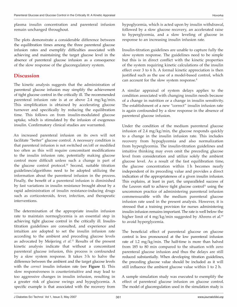
described in the Appendix together with the insulin-titration algorithm adopted from Kanji et al.30 The simulation study lasted over 24 h and assumed up to a 100% trapezoidal increase in insulin resistance starting at 6 h, culminating between 8 and 12 h, and decreasing to its original value at 14 h. Plasma glucose and insulin infusion rates associated with the simulation study are shown in Figure 2.
With zero parenteral glucose infusion, glucose decreases faster from the starting value of 9 mmol/liter as the control algorithm delivers the same dose at the three levels of the parenteral infusion. At 4 h, a borderline hypoglycemia occurs with the zero parenteral glucose infusion. Insulin resistance induces glucose oscillations at all levels of the parenteral infusion, but the amplitude with the zero parenteral infusion is greatest and the
oscillations are damped only slowly, whereas damping is greater and the achievement of the target level is faster with the low and medium parenteral infusions, with the medium parenteral infusion performing better at the end of the study. The insulin infusion rates displayed in Figure 2 (bottom) demonstrate that the most and the least oscillatory insulin patterns are associated with the zero and the median parenteral glucose infusion, respectively, demonstrating that a slow equilibrium time exacerbates amplitude of glucose oscillations.
The beneficial effect of the parenteral glucose infusion on glucose control needs to be considered in light of other evidence and suggestions related to the nutritional provision in the critically ill.15,16 Human errors may lead to severe hypoglycemia if a high parenteral glucose infusion is accidentally interrupted but exogenous insulin is not reduced. Furthermore, the exogenous insulin infusion associated with a medium parenteral glucose infusion may regularly exceed 10 U/h, an arbitrary threshold set by some intensive care units.5 In subjects with high insulin resistance, normally associated with severity of critical illness,31,32 the insulin titration needs to be sufficiently aggressive to achieve tight glucose control, avoiding hyperglycemia as observed in studies with traditional sliding-scale algorithms.17–19
The other two factors potentially leading to simplified glucose control during the parenteral glucose infusion have a negligible effect. The endogenous insulin secretion is expected to be already insignificant at the zero parenteral glucose infusion (see Table 1). No further benefit is obtained by further reducing the contribution of this unknown but small component to the overall insulin appearance. The contribution of unknown (i.e., of endogenous origin) glucose fluxes reduces from 100% with zero parenteral glucose infusion to 65% with medium parenteral glucose infusion (see Table 1). The contribution of unknown glucose fluxes is still high, and drug- or disease-induced changes in insulin sensitivity will have a similar impact on glucose excursions with or without parenteral glucose infusion.
The half-times reported in Table 1 are estimates based on the involvement of insulin-mediated and noninsulin-mediated glucose uptake in the ability of glucose to promote its own disposal. The value of 185 min during zero parenteral glucose infusion compares well with that of 145 min obtained as a geometric mean of six subjects with type 1 diabetes during basal insulin infusion.33 As critically ill subjects are more insulin resistant than subjects with type 1 diabetes, it is likely that the equilibration time is also reduced. On the other side of the spectrum,
Figure 2. Simulation study exemplifying the effect of parenteral glucose infusion on glucose control. The model and the insulin-titration algorithm are described in the Appendix. (Top) Glucose concentration associated with 0, 1.2, and 2.4 mg/kg/min parenteral glucose infusion; the horizontal dashed line indicates the target glucose range of 4.4 to 6.1 mmol/liter, and the trapezoid above the time axis indicates the shape of insulin resistance, which increased up to 100%. (Bottom) Insulin infusion rates as calculated by the insulin-titration algorithm.

363
Parenteral Glucose and Glucose Control in the Critically Ill: A Kinetic Appraisal Hovorka
www.journalofdst.orgJ Diabetes Sci Technol Vol 1, Issue 3, May 2007
a T½ of 25 min can be deduced from results reported in glucose clamp studies in subjects with sepsis involving a glucose infusion of 3.8 mg/kg/min and an associated plasma insulin concentration of 180 mU/liter,27 confirming the fast equilibration time at a medium parenteral glucose infusion.
A large intersubject variability in insulin resistance exists with insulin needs reaching up to 7 mU/kg/min in the critically ill with severe inflammation. Results of the present study are valid across the spectrum of insulin resistance as long as the relative contribution of the IMGU and the NIMGU to the total glucose uptake is unchanged. At present, there is no evidence that insulin resistance in the critically ill increases the contribution of the IMGU under the conditions of tight glucose control.
In addition to insulin-titration algorithms, glucose control can be comodulated by nutrition, specifically by parenteral glucose.10,34 Ahmed and colleagues35 indicated that modulation of nutrition is a first-choice method for treating hyperglycemia in neonates. The present work relates primarily to insulin-titration algorithms without comodulation of nutrition.
The considerable intersubject variability in insulin needs, glucose turnover rates, and thus the equilibration half-time and equilibration curves is not depicted in the present work as the focus is on kinetic appraisal of the median kinetic behavior. However, in practice, large variations among subjects exist and may confound the results expected from the present analysis.
Results of the present assessment apply to enteral infusion as the beneficial effect on glucose control is brought about by elevated plasma insulin concentration increasing glucose turnover and specifically the IMGU. The caveat of enteral nutrition is that the bioavailability of enteral nutrients may vary over time, causing an uncertainty about the amount of exogenous glucose reaching systemic circulation. However, under the condition of stable bio-availability, enteral nutrition will also have a beneficial effect on the achievement and maintenance of glucose control.
ConclusionsA parenteral glucose infusion at or above 2.4 mg/kg/min is suggested to exert a beneficial effect on the achievement of tight glucose control by increasing responsiveness of the glucoregulatory system to exogenous insulin, facilitating the development of more intuitive and simpler insulin-titration guidelines. Fewer benefits are anticipated with a lower glucose parenteral infusion rate of 1.2 mg/kg/min.
Appendix
Glucoregulatory Model with Time-Variant Insulin SensitivityFor the simulation study, we used a model of glucoregulation based on the minimal model of glucose kinetics36 and described by a set of differential equations
where G(t) represents the plasma glucose concentration (mmol/liter), X(t) remote (active) insulin (/min), Gb basal glucose (mmol/liter), I(t) plasma insulin concentration (mU/liter), and Ib(t) time-varying plasma insulin concentration at which glucose attains its basal value Gb (mU/liter). p1 is glucose effectiveness (/min) at basal insulin Ib,0 (mU/liter), SI(t) is time-varying insulin sensitivity (/min per mU/liter), SI,0 is basal insulin sensitivity (/min per mU/liter), uG is parenteral glucose infusion (mmol/kg/min), V is the distribution volume of glucose (liter/kg), and p2 is the fractional deactivation rate of the remote insulin (/min).
Plasma insulin I(t) is calculated from the insulin infusion rate uI(t) (U/h) as
where W is subject’s weight (kg) and MCRI is the metabolic clearance rate of insulin (liter/kg/min). We did not use a differential equation to describe the kinetics of plasma insulin as the half-time of insulin disappearance clearance from plasma is short (about 5–7 min37); consequently, plasma insulin achieves its steady state value quickly, corresponding to a given constant intravenous insulin infusion. The time-varying insulin needs are implemented by modifying insulin sensitivity and, reciprocally, basal insulin concentration
where m(t) is a trapezoidal-shape function (unitless)
364
Parenteral Glucose and Glucose Control in the Critically Ill: A Kinetic Appraisal Hovorka
www.journalofdst.orgJ Diabetes Sci Technol Vol 1, Issue 3, May 2007
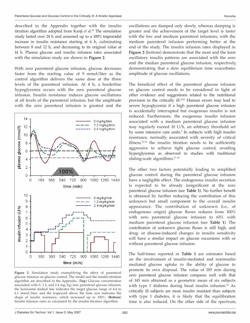
The glucose effectiveness p1 is calculated from the equilibration half-time T½-mean, reported in Table 1, as p1 = ln(2)/T½-mean, giving 0.37, 0.87, and 1.73 × 10⁻²/min for parenteral glucose infusions of 0, 1.2, and 2.4 mg/kg/min, respectively. Values of other model quantities are shown in Table 2.
Table 2. Model Quantities Associated with Testing the Effect of Parenteral Glucose Infusion on Glucose Control in a Sample Simulation Study (See Appendix for Details on the Model)
Model quantity Value
p2 (/min) 0.03
G0 (mmol/liter) 9.0
Gb (mmol/liter) 5.3
Ib,0 (mU/liter) 30
Si,0 (10-4 /min per mU/liter) 3.14
V (liter/kg) 0.16
W (kg) 70
MCRI (liter/kg/min) 0.0143
Table 3.Insulin-Titration Algorithm, Adopted from Kanji et al,30 and Used in the Simulation Studya
Plasma glucose
(mmol/liter)Insulin infusion adjustment
<4Stop insulin infusion. Check plasma glucose every 30 min. When glucose >6.1 mmol/liter, restart insulin at 50% of the previous rate
4–4.4If current rate >5 U/h, decrease rate by 2 U/hIf current rate ≤5 U/h, decrease rate by 0.5 U/h
4.5–6 Target range (no change)
6.1–8
If plasma glucose is lower than the last test, no changeIf plasma is the same or higher than the previous test, increase rate by 0.5 U/h
8.1–10
If plasma glucose is lower than the last test, no changeIf plasma is the same or higher than the previous test, increase rate by 1 U/h
>10.1
If plasma glucose is lower than the last test, no changeIf plasma is the same or higher than the previous test, increase rate by 1.5 U/h
aHourly glucose samples are taken unless specified otherwise.
Insulin-Titration AlgorithmThe insulin-titration algorithm was adopted from a study by Kanji and colleagues30 and is summarized in Table 3.
Acknowledgments:
The EU-funded Clincip Project (IST-2002-506965) provided funding to execute the work and facilitated collaboration between the authors. Tomas Hovorka helped with the simulation study.
References:
1. Van den Berghe G, Wouters P, Weekers F, Verwaest C, Bruyninckx F, Schetz M, Vlasselaers D, Ferdinande P, Lauwers P, Bouillon R. Intensive insulin therapy in the critically ill patients. N Engl J Med. 2001 Nov 8;345(19):1359-67.
2. Van den Berghe G, Wilmer A, Hermans G, Meersseman W, Wouters PJ, Milants I, Van Wijngaerden E, Bobbaers H, Bouillon R. Intensive insulin therapy in the medical ICU. N Engl J Med. 2006 Feb 2;354(5):449-61.
3. Krinsley JS. Effect of an intensive glucose management protocol on the mortality of critically ill adult patients. Mayo Clin Proc. 2004 Aug;79(8):992-1000.
4. Furnary AP, Gao G, Grunkemeier GL, Wu Y, Zerr KJ, Bookin SO, Floten HS, Starr A. Continuous insulin infusion reduces mortality in patients with diabetes undergoing coronary artery bypass grafting. J Thorac Cardiovasc Surg. 2003 May;125(5):1007-21.
5. Vogelzang M, Zijlstra F, Nijsten MW. Design and implementation of GRIP: a computerized glucose control system at a surgical intensive care unit. BMC Med Inform Decis Mak. 2005 Dec 19;5:38.
6. Davidson PC, Steed RD, Bode BW. Glucommander: a computer-directed intravenous insulin system shown to be safe, simple, and effective in 120,618 h of operation. Diabetes Care. 2005 Oct;28(10):2418-23.
7. Goldberg PA, Siegel MD, Sherwin RS, Halickman JI, Lee M, Bailey VA, Lee SL, Dziura JD, Inzucchi SE. Implementation of a safe and effective insulin infusion protocol in a medical intensive care unit. Diabetes Care. 2004 Feb;27(2):461-7.
8. Plank J, Blaha J, Cordingley J, Wilinska ME, Chassin LJ, Morgan C, Squire S, Haluzik M, Kremen J, Svacina S, Toller W, Plasnik A, Ellmerer M, Hovorka R, Pieber TR. Multicentric, randomized, controlled trial to evaluate blood glucose control by the model predictive control algorithm versus routine glucose management protocols in intensive care unit patients. Diabetes Care. 2006 Feb;29(2):271-6.
9. Preston S, Laver SR, Lloyd W, Padkin A. Introducing intensive insulin therapy: the nursing perspective. Nurs Crit Care. 2006 Mar-Apr;11(2):75-9.
10. Wong XW, Singh-Levett I, Hollingsworth LJ, Shaw GM, Hann CE, Lotz T, Lin J, Singh-Levett I, Hollingsworth LJ, Wong OS, Andreassen S. A novel, model-based insulin and nutrition delivery controller for glycemic regulation in critically ill patients. Diabetes Technol Ther. 2006 Apr;8(2):174-90.
11. Meijering S, Corstjens AM, Tulleken JE, Meertens JH, Zijlstra JG, Ligtenberg JJ. Towards a feasible algorithm for tight glycaemic control in critically ill patients: a systematic review of the literature. Crit Care. 2006 Feb;10(1):R19.
12. Cryer PE. Hypoglycaemia: the limiting factor in the glycaemic management of the critically ill? Diabetologia. 2006 Aug;49(8):1722-5.
365
Parenteral Glucose and Glucose Control in the Critically Ill: A Kinetic Appraisal Hovorka
www.journalofdst.orgJ Diabetes Sci Technol Vol 1, Issue 3, May 2007
13. Zierler K. Whole body glucose metabolism. Am J Physiol. 1999 Mar;276(3 Pt 1):E409-26.
14. Hovorka R, Shojaee-Moradie F, Carroll PV, Chassin LJ, Gowrie IJ, Jackson NC, Tudo RS, Umpleby AM, Jones RH. Partitioning glucose distribution/transport, disposal, and endogenous production during IVGTT. Am J Physiol. 2002 May;282(5):E992-1007.
15. Heyland DK, MacDonald S, Keefe L, Drover JW. Total parenteral nutrition in the critically ill patient: a meta-analysis. JAMA. 1998 Dec 16;280(23):2013-9.
16. Klein S, Kinney J, Jeejeebhoy K, Alpers D, Hellerstein M, Murray M, Twomey P. Nutrition support in clinical practice: review of published data and recommendations for future research directions. National Institutes of Health, American Society for Parenteral and Enteral Nutrition, and American Society for Clinical Nutrition. JPEN J Parenter Enteral Nutr. 1997 May-Jun;21(3):133-56.
17. Krishnan JA, Parce PB, Martinez A, Diette GB, Brower RG. Caloric intake in medical ICU patients: consistency of care with guidelines and relationship to clinical outcomes. Chest. 2003 Jul;124(1):297-305.
18. Weissman C. Nutrition in the intensive care unit. Crit Care. 1999;3(5):R67-75.
19. Ahrens CL, Barletta JF, Kanji S, Tyburski JG, Wilson RF, Janisse JJ, Devlin JW. Effect of low-calorie parenteral nutrition on the incidence and severity of hyperglycemia in surgical patients: a randomized, controlled trial. Crit Care Med. 2005 Nov;33(11):2507-12.
20. Sauerwein HP, Pesola GR, Godfried MH, Levinson MR, Jeevanandam M, Brennan MF. Insulin sensitivity in septic cancer-bearing patients. JPEN J Parenter Enteral Nutr. 1991 Nov-Dec;15(6):653-8.
21. Chambrier C, Laville M, Rhzioual Berrada K, Odeon M, Bouletreau P, Beylot M. Insulin sensitivity of glucose and fat metabolism in severe sepsis. Clin Sci (Lond). 2000 Oct;99(4):321-8.
22. Nelson KM, Long CL, Bailey R, Smith RJ, Laws HL, Blakemore WS. Regulation of glucose kinetics in trauma patients by insulin and glucagon. Metabolism. 1992 Jan;41(1):68-75.
23. Tozer TN, Rowland M. Introduction to pharmacokinetics and pharmacodynamics. Philadelphia: Lippincott Williams and Wilkins;2006.
24. Hovorka R, Shojaee-Moradie F, Carroll PV, Chassin LJ, Gowrie IJ, Jackson NC, Tudor RS, Umpleby AM, Jones RH. Partitioning glucose distribution/transport, disposal, and endogenous production during IVGTT. Am J Physiol. 2002 May;282(5):E992-1007.
25. Baron AD, Brechtel G, Wallace P, Edelman SV. Rates and tissue sites of non-insulin- and insulin-mediated glucose uptake in humans. Am J Physiol. 1988 Dec;255(6 Pt 1):E769-74.
26. Ader M, Ni TC, Bergman RN. Glucose effectiveness assessed under dynamic and steady state conditions. Comparability of uptake versus production components. J Clin Invest. 1997 Mar 15;99(6):1187-99.
27. Rusavy Z, Macdonald IA, Sramek V, Lacigova S, Tesinsky P, Novak I. Glycemia influences on glucose metabolism in sepsis during hyperinsulinemic clamp. JPEN J Parenter Enteral Nutr. 2005 May-Jun;29(3):171-5.
28. Cerra FB, Benitez MR, Blackburn GL, Irwin RS, Jeejeebhoy K, Katz DP, Pingleton SK, Pomposelli J, Rombeau JL, Shronts E, Wolfe RR, Zaloga GP. Applied nutrition in ICU patients. A consensus statement of the American College of Chest Physicians. Chest. 1997 Mar;111(3):769-78.
29. Garvey WT, Revers RR, Kolterman OG, Rubenstein AH, Olefsky JM. Modulation of insulin secretion by insulin and glucose in type II diabetes mellitus. J Clin Endocrinol Metab. 1985 Mar;60(3):559-68.
30. Kanji S, Singh A, Tierney M, Meggison H, McIntyre L, Hebert PC. Standardization of intravenous insulin therapy improves the efficiency and safety of blood glucose control in critically ill adults. Intensive Care Med. 2004 May;30(5):804-10.
31. Van den Berghe G, Wouters PJ, Bouillon R, Weekers F, Verwaest C, Schetz M, Vlasselaers D, Ferdinande P, Lauwers P. Outcome benefit of intensive insulin therapy in the critically ill: Insulin dose versus glycemic control. Crit Care Med. 2003 Feb;31(2):359-66.
32. Christiansen C, Toft P, Jorgensen HS, Andersen SK, Tonnesen E. Hyperglycaemia and mortality in critically ill patients. A prospective study. Intensive Care Med. 2004 Aug;30(8):1685-8.
33. Regittnig W, Trajanoski Z, Leis HJ, Ellmerer M, Wutte A, Sendlhofer G, Schaupp L, Brunner GA, Wach P, Pieber TR. Plasma and interstitial glucose dynamics after intravenous glucose injection - evaluation of the single-compartment glucose distribution assumption in the minimal models. Diabetes. 1999 May;48(5):1070-81.
34. Lonergan T, Compte AL, Willacy M, Chase JG, Shaw GM, Hann CE, Lotz T, Lin J, Wong XW. A pilot study of the SPRINT protocol for tight glycemic control in critically ill patients. Diabetes Technol Ther. 2006 Aug;8(4):449-62.
35. Ahmed M, Irwin S, Tuthill DP. Education and evidence are needed to improve neonatal parenteral nutrition practice. JPEN J Parenter Enteral Nutr. 2004 May-Jun;28(3):176-9.
36. Bergman RN, Ider YZ, Bowden CR, Cobelli C. Quantitative estimation of insulin sensitivity. Am J Physiol. 1979 Jun;236(6):E667-77.
37. Hovorka R, Powrie JK, Smith GD, Sonksen PH, Carson ER, Jones RH. Five-compartment model of insulin kinetics and its use to investigate action of chloroquine in NIDDM. Am J Physiol. 1993 Jul;265(1 Pt 1):E162-75.
Related Documents