Parasite fauna of Octopus vulgaris (Cephalopoda: Octopodidae) and Platichthys flesus (Actinopterygii: Pleuronectidae): morphology, systematics, life history strategies and ecology Francisca Isabel Merino Nunes Cabral Cavaleiro PhD thesis presented to the Faculty of Sciences of University of Porto, Biology 2013 Parasite fauna of Octopus vulgaris (Cephalopoda: Octopodidae) and Platichthys flesus (Actinopterygii: Pleuronectidae): morphology, systematics, life history strategies and ecology Francisca Cavaleiro PhD FCUP 2013 3. rd CYCLE D

Welcome message from author
This document is posted to help you gain knowledge. Please leave a comment to let me know what you think about it! Share it to your friends and learn new things together.
Transcript
Parasite fauna of
Octopus vulgaris
(Cephalopoda:
Octopodidae) and
Platichthys flesus
(Actinopterygii:
Pleuronectidae):
morphology,
systematics, life history
strategies and ecology
Francisca Isabel Merino Nunes Cabral
CavaleiroPhD thesis presented to the
Faculty of Sciences of University of Porto,
Biology
2013
Pa
ras
ite fa
un
a o
f Oc
top
us
vu
lgaris
(Ce
ph
alo
po
da
: Oc
top
od
ida
e) a
nd
Pla
tich
thys
flesu
s(A
ctin
op
tery
gii: P
leu
ron
ec
tidae
): mo
rph
olo
gy,
sys
tem
atic
s, life
his
tory
stra
teg
ies
an
d e
co
log
y
Fra
ncis
ca C
av
ale
iroP
hD
FCUP
2013
3.rd
CYCLE
D
Parasite fauna of Octopus vulgaris (Cephalopoda: Octopodidae) and Platichthys flesus (Actinopterygii: Pleuronectidae): morphology, systematics, life history strategies and ecology Francisca Isabel Merino Nunes Cabral Cavaleiro PhD in Biology Department of Biology 2013 Supervisor Maria João Faria Leite Dias dos Santos, Auxiliary Professor, Faculty of Sciences of University of Porto Co-supervisor Ju-Shey Ho, Emeritus Professor, California State University, Long Beach, California

2 FCUP
Acknowledgements
This thesis is dedicated to the memory of my grandmother,
Maria Luísa Pinto Merino Nunes

4 FCUP
Acknowledgements
FCUP
Acknowledgements
i
Acknowledgements
The present thesis represents the end of a long journey and one more step in my
career. There are a few people to whom I would like to give special acknowledgement.
I would like to begin by expressing my deepest gratitude to my supervisor,
Professor Maria João Santos (University of Porto, Faculty of Sciences, Portugal), and
co-supervisor, Professor Ju-Shey Ho (California State University at Long Beach,
California, United States of America), for their advice and guidance throughout the
research process, and for introducing me to the wonderful world of the parasitic
copepods.
I also thank the other co-authors of my published papers, Professor David
Gibson (Natural History Museum of London, Department of Zoology, United Kingdom),
Professor Fernanda Russell-Pinto (University of Porto, Abel Salazar Institute for the
Biomedical Sciences, Portugal), Professor José García-Estévez (University of Vigo,
Faculty of Biology, Spain), Professor Nuno Formigo (University of Porto, Faculty of
Sciences, Portugal), Professor Pedro Rodrigues (University of Porto, Abel Salazar
Institute for the Biomedical Sciences, Portugal), Professor Raúl Iglesias (University of
Vigo, Faculty of Biology, Spain) and Doctor Susana Pina (University of Porto, Abel
Salazar Institute for the Biomedical Sciences, Portugal), and the co-authors of the
papers which are still in review for publication, especially Doctor Elsa Froufe (University
of Porto, Interdisciplinary Centre of Marine and Environmental Research, Portugal).
Numerous parasitologists throughout the world, who kindly sent me reprints of
their papers, have undoubtedly contributed to the completion of this thesis and deserve
special thanks. My sincere gratitude to Professor Darren Shaw (University of
Edinburgh, Easter Bush Veterinary Centre), Professor Eric Hochberg (Santa Barbara
Museum of Natural History, Department of Invertebrate Zoology, California, United
States of America), Professor Geoff Boxshall (Natural History Museum of London,
Department of Zoology, United Kingdom), Professor Katarzyna Niewiadomska (Polish
Academy of Sciences at Warsaw, Poland), Professor Klaus Rohde (University of New
England, School of Environmental and Rural Science, Australia), Professor Marcelo
Oliva (University of Antofagasta, Institute of Oceanological Investigations, Chile),
Professor Robert Poulin (University of Otago, Department of Zoology, New Zeland),
Professor Robin Overstreet (University of Southern Mississipi, Department of Coastal
ii FCUP
Acknowledgements
Sciences, Marine Parasitology and Pathobiology, United States of America) and
Professor Santiago Pascual (Institute of Marine Investigations in Vigo, Spain).
Several biologists who welcomed me into their laboratories and aquariums and
made my training periods abroad a pleasant experience, namely by giving me the
opportunity to get to know their cultures, also deserve special thanks. I am grateful to
Doctor Julianne Kalman Passarelli (Cabrillo Marine Aquarium, California, United States
of America) and Professors Iker Uriarte and Ana Farías (Austral University of Chile,
Institute of Aquaculture, Chile).
A special word of gratitude is due to my friends, Magda Cerieira and Vítor Silva,
my colleagues, Ricardo Castro and Luís Rangel, the group of people at the Laboratory
of Animal Pathology, especially Professor Aurélia Saraiva, Professor Cristina Cruz and
Professor José Américo de Sousa, and Professor Maria Teresa Borges.
Finally, I would like to thank my parents and sisters for their unconditional
support.
This work was financed by Fundação para a Ciência e a Tecnologia, Ministério
da Educação e Ciência, Portugal, and Fundo Social Europeu, through a PhD grant
attributed to Francisca Cavaleiro (SFRH/BD/65258/2009).
FCUP
Abstract
iii
Abstract
This thesis compiles a series of papers on different aspects of the parasite fauna of an
invertebrate i.e. the common octopus Octopus vulgaris (Cephalopoda: Octopodidae)
(presently understood as a complex of species) and a vertebrate i.e. the European
flounder Platichthys flesus (Linnaeus, 1758) (Actinopterygii: Pleuronectidae) present in
Portuguese coastal waters.
Chapter 1 briefly addresses parasite diversity in morphology, systematics and
life history strategies and makes a general introduction to the basic concepts and
definitions in Parasite Ecology. Special emphasis is given to the proximate and ultimate
causes of niche restriction in parasites, and an attempt is made to systematize the
evidence on niche restriction in parasitic copepods, since the majority of the papers in
this thesis respect this particular group of parasites. A few examples retrieved from
studies in the literature are given. Finally, a brief introduction is made to the two hosts
studied.
In chapter 2, the metazoan parasite fauna of O. vulgaris is characterized, for the
first time, for Portuguese coastal waters. From the recorded parasitic taxa, Octopicola
superba Humes, 1957 (Copepoda: Octopicolidae) was the only component parasite in
the total sample of O. vulgaris. Furthermore, it was found to exhibit a marked
seasonality and the recorded trend was similar to those previously reported for
parasitic copepods of P. flesus from Portuguese waters. Also according to the evidence
found, it seems likely that macroenvironmental conditions determine (at least partly) the
seasonal occurrence of this and other parasitic copepods present on marine species of
the Portuguese coast. The number of octopicolid copepods was significantly higher for
female than for male octopuses. This, along with the fact that a significant correlation
between octopus’ size and parasite intensity was detected only for the female
octopuses suggests a differential influence of host sex in autoinfection. The metazoan
parasitic taxa so far reported for O. vulgaris in the studies of the literature is reviewed.
In chapter 3, the genus Octopicola Humes, 1957, which is exclusively found on
species of octopuses, is reviewed based on the information available in the literature
and morphological observations of octopicolids isolated from O. vulgaris. Comparative
morphological analysis led to the conclusion that Octopicola superba superba Humes,
1957, endemic to European waters, and O. s. antillensis Stock, Humes & Gooding,
1963, endemic to West Indian waters, exhibit sufficient differences to be raised to

iv FCUP
Abstract
species rank. A new identification key for all the species of the genus, i.e. O. superba
Humes, 1957, O. antillensis Stock, Humes & Gooding, 1963, O. stocki Humes, 1963
and O. regalis Humes, 1974, is provided.
In chapter 4, a new species of caligid copepod, Caligus musaicus Cavaleiro,
Santos & Ho, 2010, isolated from P. flesus, is described. The new species is unique in
that it possesses the following four character states: short abdomen; box of sternal
furca carrying two parallel pointed tines; bearing a long element IV at the tip of leg 1
exopod; and a slender leg 4 exopod bearing a long outer seta at the tip of this ramus.
The chosen specific name, musaicus, alludes to the fact that the specimens remind
one of a genetic mosaic, i.e. its resemblance with several congeners.
In chapter 5, a new diplostomid metacercarial genotype isolated from the eye
lenses of P. flesus is described. Aspects such as larval morphology, ultrastructure and
morphometrics are also considered. Two distinct morphotypes, referred to as ‘round’
and ‘long’, were identified. However, these had 100% genetic homology concerning the
18S+ITS1+5.8S region of the rDNA. This was found to represent an unknown
genotype, now referenced in GenBank as GQ370809. Furthermore, the molecular
phylogenetic analyses, in conjunction with the principal components and cluster
analyses of morphometric data indicate that the studied species of Diplostomum
corresponds with neither D. spathaceum (Rudolphi, 1819) nor D. mergi Dubois, 1932,
two species previously reported to infect P. flesus. The isolated marine specimens can
represent a new species of Diplostomum, but it is more likely that they belong to a
known species which has not yet been characterized in molecular terms.
In chapter 6, the trade-off between egg number and egg size is addressed for
the intraspecific level of analysis, based on data recorded for adult ovigerous females
of O. superba. The evidence found suggests that the parasite is essentially a K-
strategist, and conforms to the general assumption that ectoparasites do not follow
both an r- and K-strategy simultaneously. Furthermore, the environmental conditions
seem to force them into one of the alternatives, presumably by leading to adaptive
phenotypic plasticity in body dimensions and size-mediated changes in egg production.
A trade-off between egg number and egg size became apparent only at high levels of
fecundity, suggesting a state of physiological exhaustion.
In chapter 7, site selection is characterized in detail for Acanthochondria
cornuta (Müller, 1776) (Copepoda: Chondracanthidae), a common parasite of P. flesus.
A preference for the ocular side of the host’s body was observed and it is speculated
that this can be related with the fish’s behaviour, as this fish lives partially buried in the
FCUP
Abstract
v
ocean floor. The evidence found also suggests that, as the parasite develops from one
stage into another, it migrates towards different sites within the branchial chamber. This
argues against the idea that the microhabitat of some parasitic copepods is determined
by where infective stages settle first, i.e. that some parasitic copepods select a
permanent site for living, becoming immovably fixed to it for life. The occurrence of
bigamy, i.e. of bigamous females, is reported for the first time for A. cornuta.
In chapter 8, the occurrence of interference competition is addressed for O.
superba and the coccidian Aggregata sp. (Apicomplexa: Aggregatidae), two parasites
that occur at the gills of wild O. vulgaris. Both numerical and functional responses are
analysed and both the fundamental and realized spatial niches are measured.
According to the results found, the gills constitute the main and accessory site of
infection of O. superba and Aggregata sp., respectively, and were simultaneously
infected with the two parasites in 11 (9.2%) of the examined octopuses. While the
presence of O. superba on gill lamellae appears to be negatively affected by the
presence of Aggregata sp., the latter does not seem to be affected by the former.
Finally, chapter 9 presents some concluding remarks on the parasites studied.
A comparative analysis of the parasite fauna recorded for the studied hosts is
performed. Future lines of investigation are delineated.
Keywords: Metazoan parasites of Octopus vulgaris; review of Octopicola (Copepoda:
Octopicolidae); Caligus musaicus sp. nov. (Copepoda: Caligidae); metacercariae of
Diplostomum sp. from Platichthys flesus; trade-off between egg number and egg size;
site selection of Acanthochondria cornuta (Copepoda: Chondracanthidae); interference
competition between Octopicola superba (Copepoda: Octopicolidae) and Aggregata
sp. (Apicomplexa: Aggregatidae); parasitological survey

vi FCUP
Resumo
FCUP
Resumo
vii
Resumo
A presente tese compila uma série de artigos relacionados com diferentes aspetos da
parasitofauna de um invertebrado i.e. o polvo comum Octopus vulgaris (Cephalopoda:
Octopodidae) (atualmente entendido como um complexo de espécies) e de um
vertebrado i.e. a solha Europeia Platichthys flesus (Linnaeus, 1758) (Actinopterygii:
Pleuronectidae) presentes em águas costeiras Portuguesas.
O capítulo 1 considera, de forma abreviada, a diversidade morfológica dos
parasitas e sua sistemática e estratégias de vida, e faz uma introdução geral aos
conceitos e definições básicas em Ecologia Parasitária. É dado especial ênfase às
causas próximas e últimas da restrição de nichos em parasitas, e é feito um esforço no
sentido de sistematizar a evidência relativa à restrição de nichos em copépodes
parasitas, dado que a maioria dos artigos apresentados nesta tese respeita este grupo
particular de parasitas. São mencionados alguns exemplos encontrados nos estudos
da literatura. Finalmente, é feita uma breve introdução aos dois hospedeiros
estudados.
No capítulo 2, carateriza-se, pela primeira vez, a fauna de parasitas
metazoários de O. vulgaris de águas costeiras Portuguesas. Dos taxa parasitas
registados, Octopicola superba Humes, 1957 (Copepoda: Octopicolidae) foi o único
parasita componente na amostra total de O. vulgaris. Adicionalmente, este parasita
exibiu uma sazonalidade marcada e a tendência registada foi semelhante às
anteriormente reportadas para copépodes parasitas de P. flesus de águas
Portuguesas. De acordo ainda com a evidência encontrada, parece provável que as
condições macroambientais determinem (pelo menos parcialmente) a ocorrência
sazonal deste e de outros copépodes parasitas presentes em espécies marinhas da
costa Portuguesa. O número de copépodes octopicolídios foi significativamente mais
elevado em polvos do sexo feminino do que em polvos do sexo masculino. Isto, aliado
ao fato de uma correlação significativa entre o tamanho do polvo e a intensidade
parasitária ter sido detetada apenas para os polvos do sexo feminino sugere uma
influência diferencial do sexo do hospedeiro na auto-infeção. É feita uma revisão dos
taxa de parasitas metazoários reportados até à data para O. vulgaris nos estudos da
literatura.
No capítulo 3, o género Octopicola Humes, 1957, que é exclusivamente
encontrado em espécies de polvos, é revisto com base na informação disponível na

viii FCUP
Resumo
literatura e em observações morfológicas de octopicolídios isolados de O. vulgaris. A
análise morfológica comparativa levou à conclusão de que Octopicola superba
superba Humes, 1957, endémica de águas Europeias, e O. s. antillensis Stock, Humes
& Gooding, 1963, endémica de águas das Índias Ocidentais, exibem diferenças
suficientes para serem elevadas à categoria de espécie. É disponibilizada uma nova
chave de identificação para todas as espécies do género, i.e. O. superba Humes,
1957, O. antillensis Stock, Humes & Gooding, 1963, O. stocki Humes, 1963 e O.
regalis Humes, 1974.
No capítulo 4, é descrita uma nova espécie de copépode caligídio, Caligus
musaicus Cavaleiro, Santos & Ho, 2010, isolada de P. flesus. Esta nova espécie
distingue-se das demais por possuir as seguintes quatro caraterísticas: abdómen
curto; caixa da furca esternal com duas hastes pontiagudas e paralelas; armada com
um elemento IV longo na extremidade do exopodito da pata 1; e exopodito da pata 4
delgado, armado com uma cerda exterior longa na sua extremidade. O restritivo
específico escolhido, musaicus, alude ao fato de que os espécimes fazem lembrar um
mosaico genético, i.e. à semelhança da espécie relativamente a vários dos seus
congéneres.
No capítulo 5, é descrito um novo genótipo de metacercárias de diplostomídio
isolado da lente dos olhos de P. flesus. São considerados ainda aspetos como a
morfologia, ultraestrutura e morfometria larvar. Foram identificados dois morfotipos
distintos, referidos como ‘redondo’ e ‘longo’. Contudo, demonstrou-se que estes
apresentavam 100% de homologia genética no que concerne a região 18S+ITS1+5.8S
do rDNA. Descobriu-se, ainda, que esta última representava um genótipo
desconhecido, agora referenciado no GenBank como GQ370809. Além disso, as
análises filogenéticas moleculares, em conjugação com as análises de componentes
principais e de clusters de dados morfométricos indicam que a espécie de
Diplostomum estudada não corresponde nem a D. spathaceum (Rudolphi, 1819) nem
a D. mergi Dubois, 1932, duas espécies que foram anteriormente reportadas para P.
flesus. Os espécimes marinhos isolados podem representar uma nova espécie de
Diplostomum, sendo contudo mais provável que eles pertençam a uma espécie
conhecida que não foi ainda caraterizada em termos moleculares.
No capítulo 6, é considerado o trade-off entre o número e o tamanho dos ovos
ao nível intraespecífico de análise, tendo por base dados registados para fêmeas
adultas ovígeras de O. superba. A evidência encontrada sugere que o parasita é,
essencialmente, um estrategista K, e está de acordo com a suposição geral de que os
FCUP
Resumo
ix
ectoparasitas não seguem, simultaneamente, as estratégias r e K. Além disso, ela
sugere ainda que as condições ambientais influenciam a estratégia escolhida, na
medida em que são presumivelmente responsáveis por plasticidade fenotípica
adaptativa ao nível das dimensões do corpo e por mudanças na produção ovígera
mediadas pelas mudanças no tamanho corporal. Um trade-off entre o número e o
tamanho dos ovos foi observado apenas a elevados níveis de fecundidade, o que
sugere um estado de exaustão fisiológica.
No capítulo 7, carateriza-se, em detalhe, a seleção de sítio para
Acanthochondria cornuta (Müller, 1776) (Copepoda: Chondracanthidae), um parasita
vulgar de P. flesus. Foi observada uma preferência pelo lado ocular do corpo do
hospedeiro, especulando-se que esta poderá estar relacionada com o comportamento
do peixe, já que este vive parcialmente enterrado no fundo oceânico. A evidência
encontrada sugere ainda que, à medida que o parasita se desenvolve de estádio em
estádio, ele migra para diferentes sítios da cavidade branquial. Esta observação está
em desacordo com a ideia de que o microhabitat de alguns copépodes parasitas
corresponde ao local onde os estádios infeciosos se estabeleceram, i.e. de que alguns
copépodes parasitas selecionam um sítio permanente para viver, fixando-se a ele para
toda a vida. A ocorrência de bigamia, i.e. de fêmeas bígamas, é reportada pela
primeira vez para A. cornuta.
No capítulo 8, é considerada a ocorrência de competição por interferência entre
O. superba e o coccídio Aggregata sp. (Apicomplexa: Aggregatidae), dois parasitas
que ocorrem nas brânquias de O. vulgaris de meio natural. São consideradas para
análise as respostas numéricas e funcionais, e são medidos os nichos fundamental
espacial e realizado espacial. De acordo com os resultados obtidos, as brânquias
constituem, respetivamente, o sítio principal e acessório de infeção de O. superba e
Aggregata sp., tendo sido encontradas simultaneamente infetadas pelos dois parasitas
em 11 (9.2%) dos polvos examinados. Enquanto a presença de O. superba nas
lamelas branquiais parece ser negativamente afetada pela presença de Aggregata sp.,
a última não parece ser afetada pela primeira.
Finalmente, o capítulo 9 apresenta algumas observações finais acerca dos
parasitas estudados. É feita uma análise comparativa da fauna parasitária registada
para os hospedeiros estudados. Linhas de investigação futura são delineadas.
x FCUP
Resumo
Palavras-chave: Parasitas metazoários de Octopus vulgaris; revisão de Octopicola
(Copepoda: Octopicolidae); Caligus musaicus sp. nov. (Copepoda: Caligidae);
metacercárias de Diplostomum sp. de Platichthys flesus; trade-off entre o número e o
tamanho dos ovos; seleção de sítio por Acanthochondria cornuta (Copepoda:
Chondracanthidae); competição por interferência entre Octopicola superba (Copepoda:
Octopicolidae) e Aggregata sp. (Apicomplexa: Aggregatidae); exame parasitológico
FCUP
Scientific Papers
xi
Scientific Papers
This thesis includes seven scientific papers, published, in publication or in review for
publication in international journals (ISI) and concerning part of the results obtained
during the experimental work.
1) Cavaleiro, F. I., & Santos, M. J. (In Review for Publication). Helminth and copepod
parasites of the common octopus, Octopus vulgaris (Cephalopoda:
Octopodidae), in northwest Portuguese waters, Atlantic Ocean. Journal of
Parasitology.
2) Cavaleiro, F. I., Ho, J.-S., Iglesias, R., García-Estévez, J. M., & Santos, M. J.
(2013). Revisiting the octopicolid copepods (Octopicolidae: Octopicola Humes,
1957): comparative morphology and an updated key to species. Systematic
Parasitology, 86, 77–86.
3) Cavaleiro, F. I., Santos, M. J., & Ho, J.-S. (2010). Caligus musaicus n. sp.
(Copepoda, Caligidae) parasitic on the European flounder, Platichthys flesus
(Linnaeus) off Portugal. Crustaceana, 83, 457–464.
4) Cavaleiro, F. I., Pina, S., Russell-Pinto, F., Rodrigues, P., Formigo, N. E., Gibson,
D. I., & Santos, M. J. (2012). Morphology, ultrastructure, genetics, and
morphometrics of Diplostomum sp. (Digenea: Diplostomidae) metacercariae
infecting the European flounder, Platichthys flesus (L.) (Teleostei:
Pleuronectidae), off the northwest coast of Portugal. Parasitology Research, 110,
81–93.
5) Cavaleiro, F. I., & Santos, M. J. (In Press). Egg number-egg size: an important
trade-off in parasite life history strategies. International Journal for Parasitology.
xii FCUP
Scientific Papers
6) Cavaleiro, F. I., & Santos, M. J. (2011). Site selection of Acanthochondria cornuta
(Copepoda: Chondracanthidae) in Platichthys flesus (Teleostei: Pleuronectidae).
Parasitology, 138, 1061–1067.
7) Cavaleiro, F. I., & Santos, M. J. (In Press). Numerical and functional responses to
the presence of a competitor – the case of Aggregata sp. (Apicomplexa:
Aggregatidae) and Octopicola superba (Copepoda: Octopicolidae). Parasitology.
FCUP
Table of Contents xiii
Table of Contents
Acknowledgements ............................................................................................ i
Abstract ................................................................................................................... iii
Resumo ................................................................................................................... vii
Scientific Papers ................................................................................................ xi
Table of Contents ............................................................................................. xiii
Index of Tables ................................................................................................... xix
Index of Figures ............................................................................................... xxiii
Abbreviations ................................................................................................... xxxi
Chapter 1
General Introduction
1.1. Parasites: Diversity in Morphology, Systematics and Life History Strategies ...... 3
1.2. Parasite Ecology: A General Overview ............................................................... 3
1.2.1. Scope, Relevance and Key Study Issues ..................................................... 3
1.2.2. Basic Concepts and Definitions .................................................................... 5
1.2.3. The Case of the Parasitic Copepods ............................................................ 7
1.3. The Studied Hosts ............................................................................................ 16
xiv FCUP
Table of Contents
1.3.1. The Common Octopus, Octopus vulgaris (Cephalopoda: Octopodidae) .... 16
1.3.2. The European Flounder, Platichthys flesus (Actinopterygii: Pleuronectidae)............................................................................................................................... 17
1.4. Study Aims ......................................................................................................... 19
Chapter 2
Helminth and copepod parasites of the common octopus, Octopus vulgaris
(Cephalopoda: Octopodidae), in northwest Portuguese waters, Atlantic Ocean
2.1. Abstract .............................................................................................................. 23
2.2. Introduction ........................................................................................................ 25
2.3. Materials and Methods ....................................................................................... 25
2.4. Results ............................................................................................................... 26
2.5. Discussion .......................................................................................................... 30
2.6. Acknowledgements ............................................................................................ 33
Chapter 3
Revisiting the octopicolid copepods (Octopicolidae: Octopicola Humes, 1957):
comparative morphology and an updated key to species
3.1. Abstract .............................................................................................................. 37
3.2. Introduction ........................................................................................................ 39
3.3. Materials and Methods ....................................................................................... 40
3.4. Discussion .......................................................................................................... 51
3.5. Acknowledgements ............................................................................................ 54
FCUP
Table of Contents
xv
Chapter 4
Caligus musaicus n. sp. (Copepoda, Caligidae) parasitic on the European
flounder, Platichthys flesus (Linnaeus) off Portugal
4.1. Abstract .............................................................................................................. 59
4.2. Introduction ........................................................................................................ 61
4.3. Materials and Methods ....................................................................................... 61
4.4. Results ............................................................................................................... 62
4.5. Discussion .......................................................................................................... 68
4.6. Acknowledgements ............................................................................................ 68
Chapter 5
Morphology, ultrastructure, genetics, and morphometrics of Diplostomum sp.
(Digenea: Diplostomidae) metacercariae infecting the European flounder,
Platichthys flesus (L.) (Teleostei: Pleuronectidae), off the northwest coast of
Portugal
5.1. Abstract .............................................................................................................. 71
5.2. Introduction ........................................................................................................ 73
5.3. Materials and Methods ....................................................................................... 74
5.4. Results ............................................................................................................... 80
5.5. Discussion .......................................................................................................... 89
5.6. Acknowledgements ............................................................................................ 91
Chapter 6
Egg number-egg size: an important trade-off in parasite life history strategies
6.1. Abstract .............................................................................................................. 95
6.2. Introduction ........................................................................................................ 97
6.3. Materials and Methods ..................................................................................... 100
xvi FCUP
Table of Contents
6.4. Results ............................................................................................................. 104
6.5. Discussion ........................................................................................................ 113
6.6. Acknowledgements .......................................................................................... 116
Chapter 7
Site selection of Acanthochondria cornuta (Copepoda: Chondracanthidae) in
Platichthys flesus (Teleostei: Pleuronectidae)
7.1. Abstract ............................................................................................................ 121
7.2. Introduction ...................................................................................................... 123
7.3. Materials and Methods ..................................................................................... 124
7.4. Results ............................................................................................................. 126
7.5. Discussion ........................................................................................................ 132
7.6. Acknowledgements .......................................................................................... 134
Chapter 8
Numerical and functional responses to the presence of a competitor – the case
of Aggregata sp. (Apicomplexa: Aggregatidae) and Octopicola superba
(Copepoda: Octopicolidae)
8.1. Abstract ............................................................................................................ 137
8.2. Introduction ...................................................................................................... 139
8.3. Materials and Methods ..................................................................................... 140
8.4. Results ............................................................................................................. 144
8.5. Discussion ........................................................................................................ 155
8.6. Acknowledgements .......................................................................................... 158
8.7. Financial Support ............................................................................................. 158

FCUP
Table of Contents
xvii
Chapter 9
Concluding Remarks
9.1. Final Notes ....................................................................................................... 161
9.2. Future Research .............................................................................................. 163
References ........................................................................................................... 165

xviii FCUP
Table of Contents
FCUP
Index of Tables
xix
Index of Tables
Chapter 2
Helminth and copepod parasites of the common octopus, Octopus vulgaris
(Cephalopoda: Octopodidae), in northwest Portuguese waters, Atlantic Ocean
Table 2.1 – Metazoan parasitic taxa isolated from the seasonal and total samples of
Octopus vulgaris (Cephalopoda: Octopodidae), observed life-cycle stages, sites,
infection parameters (number of infected octopuses/prevalence [95% confidence
interval] %, mean intensity ± SD [range]), and values for the bootstrap estimator of taxa
richness (abbreviations: A, Adult; BS, Body Skin; CO, COpepodite; CMG, Covering
Mesentery of Gonad; EY, EYes; F, Funnel; G, Gills; I, Intestine; L, Larva; MM, internal
surface of the Mantle Musculature; OE, OEsophagus; and S, Stomach). .................... 27
Table 2.1 (continuation) – Metazoan parasitic taxa isolated from the seasonal and total
samples of Octopus vulgaris (Cephalopoda: Octopodidae), observed life-cycle stages,
sites, infection parameters (number of infected octopuses/prevalence [95% confidence
interval] %, mean intensity ± SD [range]), and values for the bootstrap estimator of taxa
richness (abbreviations: A, Adult; BS, Body Skin; CO, COpepodite; CMG, Covering
Mesentery of Gonad; EY, EYes; F, Funnel; G, Gills; I, Intestine; L, Larva; MM, internal
surface of the Mantle Musculature; OE, OEsophagus; and S, Stomach). .................... 28
Table 2.2 – Metazoan parasites recorded for the Octopus vulgaris (Cephalopoda:
Octopodidae) complex in the literature and respective localities and sites
(abbreviations: BS, Body Skin; CR, CRop; CT, Connective Tissue around the digestive
gland; DT, Digestive Tract; EG, EGgs; G, Gills; I, Intestine; MC, Mantle Cavity; and S,
Stomach). ..................................................................................................................... 32
Chapter 3
Revisiting the octopicolid copepods (Octopicolidae: Octopicola Humes, 1957):
comparative morphology and an updated key to species
Table 3.1 – Host and distribution data for the known taxa of octopicolid copepods. .... 45
xx FCUP
Index of Tables
Table 3.2 – Summary of metrical data for the known species and subspecies of
Octopicola. .................................................................................................................... 46
Chapter 5
Morphology, ultrastructure, genetics, and morphometrics of Diplostomum sp.
(Digenea: Diplostomidae) metacercariae infecting the European flounder,
Platichthys flesus (L.) (Teleostei: Pleuronectidae), off the northwest coast of
Portugal
Table 5.1 – Digenean species used in this study, their hosts, ITS1 length, geographical
origin, and GenBank accession numbers for the corresponding sequences. ............... 77
Table 5.1 (continuation) – Digenean species used in this study, their hosts, ITS1
length, geographical origin, and GenBank accession numbers for the corresponding
sequences. ................................................................................................................... 78
Table 5.2 – Metric dimensions of characters and indices (mean, range, coefficient of
variation, and limits of the 95% confidence interval for the population mean) for
Diplostomum sp. metacercariae isolated from the lens of the eye of the European
flounder, Platichthys flesus, caught off the northwest coast of Portugal (abbreviations:
BL, Length of the Body; BW, Width of the Body; OL, Length of the Oral sucker; OW,
Width of the Oral sucker; PHL, Length of the PHarynx; PHW, Width of the PHarynx;
VL, Length of the Ventral sucker; VW, Width of the Ventral sucker; HL, Length of the
Holdfast organ; HW, Width of the Holdfast organ; VD, Distance between the anterior
extremity of the body and the center of the Ventral sucker; LL, Length of the Lappet;
WaBI, Width of the body at the level of the Bifurcation of the Intestine; and WaO, Width
of the body at the mid-length of the Oral sucker). ......................................................... 86
Chapter 6
Egg number-egg size: an important trade-off in parasite life history strategies
Table 6.1 – Body dimensions and measures of reproductive effort (mean ± SD (RI)
[Range Interval], CV [Coefficient of Variation] (%), limits of the 95% CI [Confidence
Interval] for the population mean and results of the Kolmogorov-Smirnov’s test)
FCUP
Index of Tables
xxi
recorded for the total samples (N = 120) of mature, ovigerous females of Octopicola
superba and Octopus vulgaris. ................................................................................... 105
Table 6.2 – Multiple sample comparisons of body dimensions and measures of
reproductive effort for mature, ovigerous females of Octopicola superba (results of the
Kruskal-Wallis’ test). ................................................................................................... 110
Table 6.3 – Pairwise sample comparisons of body dimensions and measures of
reproductive effort for mature, ovigerous females of Octopicola superba (results of the
Mann-Whitney’s U test). ............................................................................................. 110
Table 6.4 – Results of the general linear model (GLM multivariate analysis) with
fecundity and mean egg diameter of Octopicola superba as dependent variables,
season as fixed factor and host total length, parasite total length and number of
conspecifics present at the site of infection as covariates. ......................................... 111
Table 6.5 – Results for the correlation between fecundity and mean egg diameter
evaluated for the different seasonal samples of Octopicola superba using a non-
parametric partial rank correlation test. ...................................................................... 113
Chapter 7
Site selection of Acanthochondria cornuta (Copepoda: Chondracanthidae) in
Platichthys flesus (Teleostei: Pleuronectidae)
Table 7.1 – Infection levels – prevalence (%) and intensity (mean ± SD) – of
Acanthochondria cornuta recorded for the different sites of attachment in the ocular
and blind branchial chambers of the European flounder, Platichthys flesus (L.)
(abbreviations: IWC, Internal Wall of the Chamber; HIF, Holobranch I Filaments; HIIF,
Holobranch II Filaments; HIIIF, Holobranch III Filaments; HIVF, Holobranch IV
Filaments; and PF, Pseudobranch Filaments). ........................................................... 128
Table 7.2 – Results for the Wilcoxon’s matched-pairs signed ranks test, which
compared between parasite abundance on the inner and outer hemibranchs of
holobranchs (I-IV) (abbreviations: HIF, Holobranch I Filaments; HIIF, Holobranch II
Filaments; HIIIF, Holobranch III Filaments; and HIVF, Holobranch IV Filaments). ..... 129
xxii FCUP
Index of Tables
Chapter 8
Numerical and functional responses to the presence of a competitor – the case
of Aggregata sp. (Apicomplexa: Aggregatidae) and Octopicola superba
(Copepoda: Octopicolidae)
Table 8.1 – The Realized Spatial Niche (RSN) of Aggregata sp. (as determined for the
seasonal subsamples of Octopus vulgaris infected with Aggregata sp. and Octopicola
superba): infection levels – number of octopuses/percentage of octopuses; and oocyst
counts (mean ± SD [range]) – recorded for the different sites and Levins’ (B) and
standardized (BA) measures (mean ± SD) of niche breadth. ...................................... 148
Table 8.2 – The Fundamental (FSN) (as determined for the seasonal subsample of
Octopus vulgaris infected only with Octopicola superba) and Realized (RSN) (as
determined for the seasonal subsamples of O. vulgaris infected with Aggregata sp. and
O. superba) Spatial Niches of O. superba: infection levels – number of
octopuses/percentage of octopuses; and specimen counts (mean ± SD [range]) –
recorded for the different sites and Levins’ (B) and standardized (BA) measures (mean
± SD) of niche breadth. ............................................................................................... 149
Table 8.3 – Infection levels of Aggregata sp. and Octopicola superba – number of
octopuses/percentage of octopuses; oocyst/specimen counts (mean ± SD [range]) –
recorded for the Proximal (PR), Middle (MR) and Distal (DR) lamellar Regions of the
Left (LG) and Right (RG) Gills (the seasonal subsamples considered for analysis
consisted of those octopuses whose gills were infected with both parasites). ........... 152
Table 8.4 – Infection levels of Aggregata sp. and Octopicola superba – number of
octopuses/percentage of octopuses; oocyst/specimen counts (mean ± SD [range]) –
recorded for the Proximal (PR), Middle (MR) and Distal (DR) lamellar Regions of the
Left (LG) and Right (RG) Gills (the seasonal subsamples considered for analysis
consisted of those octopuses whose gills were infected with only one of the two
parasites). ................................................................................................................... 154
FCUP
Index of Figures
xxiii
Index of Figures
Chapter 1
General Introduction
Fig. 1.1 – A few examples of the morphological variability found in some families of
parasitic copepods. A, Octopicolidae; B, Chondracanthidae (arrow, male); C,
Pennellidae; D, Lernaeopodidae; and E, Hatschekiidae. Scale-bars: A, 500 μm; B, 1.0
mm; C, 1.0 mm; D, 1.0 mm; and E, 15.0 mm. ................................................................ 7
Fig. 1.2 – A, Ovigerous females of Lepeophtheirus pectoralis (Müller, 1777)
(Copepoda: Caligidae) attached to the pectoral fin of the European flounder,
Platichthys flesus (Linnaeus, 1758) (Actinopterygii: Pleuronectidae); and B, An
ovigerous female of Phrixocephalus cincinnatus Wilson, 1908 (Copepoda: Pennellidae)
attached to the eye of the Pacific sanddab, Citharichthys sordidus (Girard, 1854)
(Actinopterygii: Paralichthyidae). Scale-bars: A, 5.0 mm; and B, 15.0 mm. ................. 10
Fig. 1.3 – A case of hyperparasitism involving a parasitic copepod, with numerous eggs
of Udonella sp. (Trematoda: Monogenea) seen on the cephalothorax of an ovigerous
female of Caligus sp. (Copepoda: Caligidae). Scale-bar: 1.0 mm. ............................... 13
Fig. 1.4 – The common octopus, Octopus vulgaris (Cephalopoda: Octopodidae). Scale-
bar: A, 10.0 cm. ............................................................................................................ 17
Fig. 1.5 – The European flounder, Platichthys flesus (Linnaeus, 1758) (Actinopterygii:
Pleuronectidae). Scale-bar: 5.0 cm. ............................................................................. 18
Chapter 2
Helminth and copepod parasites of the common octopus, Octopus vulgaris
(Cephalopoda: Octopodidae), in northwest Portuguese waters, Atlantic Ocean
Fig. 2.1 – Temporal trends in seawater temperature (at 83 m depth) and total number
of hours of sunlight recorded for the sampled area (off Matosinhos, northwest
Portuguese coast) (upper trend line, temperature levels; and lower trend line, total
number of hours of sunlight). ........................................................................................ 29
xxiv FCUP
Index of Figures
Chapter 3
Revisiting the octopicolid copepods (Octopicolidae: Octopicola Humes, 1957):
comparative morphology and an updated key to species
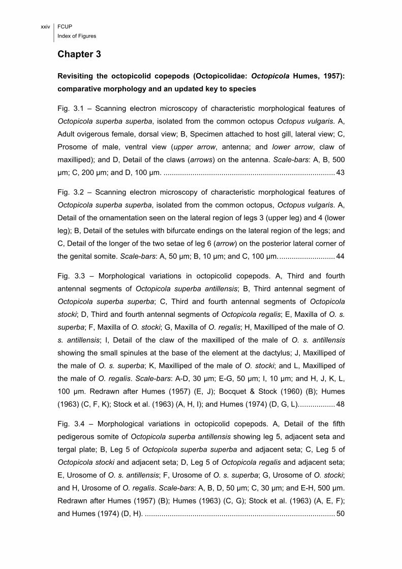
Fig. 3.1 – Scanning electron microscopy of characteristic morphological features of
Octopicola superba superba, isolated from the common octopus Octopus vulgaris. A,
Adult ovigerous female, dorsal view; B, Specimen attached to host gill, lateral view; C,
Prosome of male, ventral view (upper arrow, antenna; and lower arrow, claw of
maxilliped); and D, Detail of the claws (arrows) on the antenna. Scale-bars: A, B, 500
μm; C, 200 μm; and D, 100 μm. ................................................................................... 43
Fig. 3.2 – Scanning electron microscopy of characteristic morphological features of
Octopicola superba superba, isolated from the common octopus, Octopus vulgaris. A,
Detail of the ornamentation seen on the lateral region of legs 3 (upper leg) and 4 (lower
leg); B, Detail of the setules with bifurcate endings on the lateral region of the legs; and
C, Detail of the longer of the two setae of leg 6 (arrow) on the posterior lateral corner of
the genital somite. Scale-bars: A, 50 μm; B, 10 μm; and C, 100 μm. ........................... 44
Fig. 3.3 – Morphological variations in octopicolid copepods. A, Third and fourth
antennal segments of Octopicola superba antillensis; B, Third antennal segment of
Octopicola superba superba; C, Third and fourth antennal segments of Octopicola
stocki; D, Third and fourth antennal segments of Octopicola regalis; E, Maxilla of O. s.
superba; F, Maxilla of O. stocki; G, Maxilla of O. regalis; H, Maxilliped of the male of O.
s. antillensis; I, Detail of the claw of the maxilliped of the male of O. s. antillensis
showing the small spinules at the base of the element at the dactylus; J, Maxilliped of
the male of O. s. superba; K, Maxilliped of the male of O. stocki; and L, Maxilliped of
the male of O. regalis. Scale-bars: A-D, 30 μm; E-G, 50 μm; I, 10 μm; and H, J, K, L,
100 μm. Redrawn after Humes (1957) (E, J); Bocquet & Stock (1960) (B); Humes
(1963) (C, F, K); Stock et al. (1963) (A, H, I); and Humes (1974) (D, G, L). ................. 48
Fig. 3.4 – Morphological variations in octopicolid copepods. A, Detail of the fifth
pedigerous somite of Octopicola superba antillensis showing leg 5, adjacent seta and
tergal plate; B, Leg 5 of Octopicola superba superba and adjacent seta; C, Leg 5 of
Octopicola stocki and adjacent seta; D, Leg 5 of Octopicola regalis and adjacent seta;
E, Urosome of O. s. antillensis; F, Urosome of O. s. superba; G, Urosome of O. stocki;
and H, Urosome of O. regalis. Scale-bars: A, B, D, 50 μm; C, 30 μm; and E-H, 500 μm.
Redrawn after Humes (1957) (B); Humes (1963) (C, G); Stock et al. (1963) (A, E, F);
and Humes (1974) (D, H). ............................................................................................ 50

FCUP
Index of Figures
xxv
Chapter 4
Caligus musaicus n. sp. (Copepoda, Caligidae) parasitic on the European
flounder, Platichthys flesus (Linnaeus) off Portugal
Fig. 4.1 – Caligus musaicus n. sp., female. A, Habitus, dorsal; B, Abdomen and caudal
rami; C, Antennule; D, Antenna, postantennal process and maxillule; E, Mandible; F,
Maxilla; G, Maxilliped; and H, Sternal furca. Scale-bars: A, 0.5 mm; B, D, 100 μm; C,
50 μm; and E-H, 50 μm. ............................................................................................... 64
Fig. 4.2 – Caligus musaicus n. sp., female. A, Leg 1; B, Leg 2; C, Leg 3; and D, Leg 4.
Scale-bars: A, D, 50 μm; and B, C, 100 μm. ................................................................ 65
Fig. 4.3 – Caligus musaicus n. sp., male. A, Habitus, dorsal; B, Abdomen and caudal
rami; C, Antenna, postantennal process and maxillule; and D, Maxilliped. Scale-bars:
A, 0.5 mm; B, 100 μm; and C, D, 50 μm. ...................................................................... 67
Chapter 5
Morphology, ultrastructure, genetics, and morphometrics of Diplostomum sp.
(Digenea: Diplostomidae) metacercariae infecting the European flounder,
Platichthys flesus (L.) (Teleostei: Pleuronectidae), off the northwest coast of
Portugal
Fig. 5.1 – Measurements taken from the metacercariae of Diplostomum sp. isolated
from the lens of the eye of the European flounder, Platichthys flesus, caught off the
northwest coast of Portugal (abbreviations: BL, Length of the Body; BW, Width of the
Body; OL, Length of the Oral sucker; OW, Width of the Oral sucker; PHL, Length of the
PHarynx; PHW, Width of the PHarynx; VL, Length of the Ventral sucker; VW, Width of
the Ventral sucker; HL, Length of the Holdfast organ; HW, Width of the Holdfast organ;
VD, Distance between the anterior extremity of the body and the center of the Ventral
sucker; LL, Length of the Lappets; WaBI, Width of the body at the level of the
Bifurcation of the Intestine; and WaO, Width of the body at the mid-length of the Oral
sucker). ......................................................................................................................... 80
Fig. 5.2 – The Diplostomum sp. metacercariae, isolated from the lens of the eye of the
European flounder, Platichthys flesus, caught off the northwest coast of Portugal. Two
xxvi FCUP
Index of Figures
morphotypes A ‘round’, B ‘long’, and C a detail of the posterior region of the body and
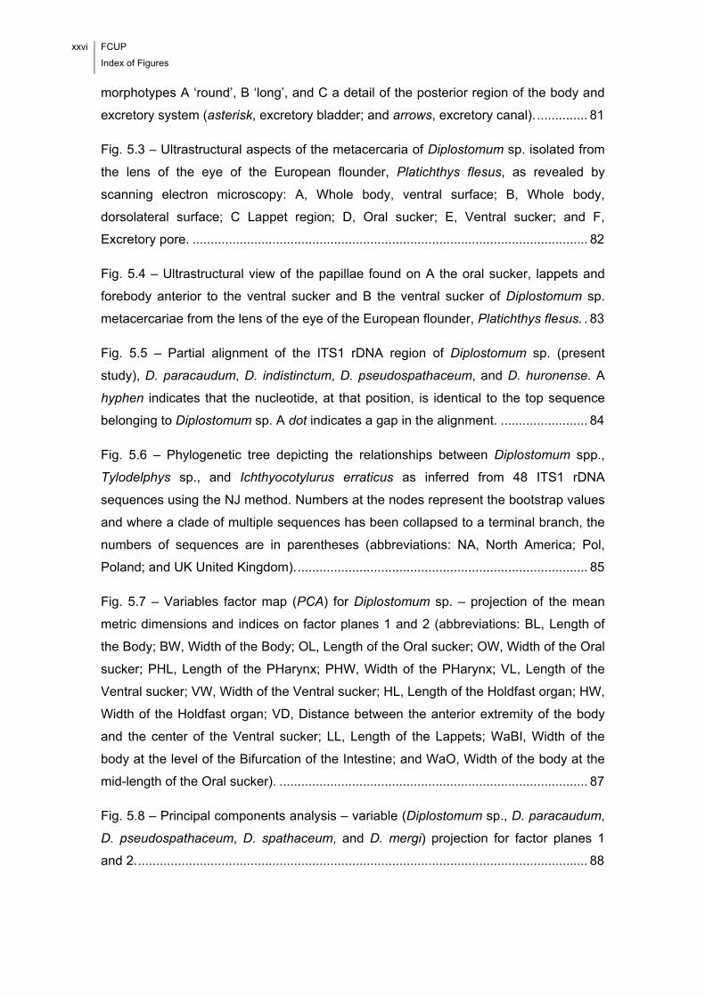
excretory system (asterisk, excretory bladder; and arrows, excretory canal). .............. 81
Fig. 5.3 – Ultrastructural aspects of the metacercaria of Diplostomum sp. isolated from
the lens of the eye of the European flounder, Platichthys flesus, as revealed by
scanning electron microscopy: A, Whole body, ventral surface; B, Whole body,
dorsolateral surface; C Lappet region; D, Oral sucker; E, Ventral sucker; and F,
Excretory pore. ............................................................................................................. 82
Fig. 5.4 – Ultrastructural view of the papillae found on A the oral sucker, lappets and
forebody anterior to the ventral sucker and B the ventral sucker of Diplostomum sp.
metacercariae from the lens of the eye of the European flounder, Platichthys flesus. . 83
Fig. 5.5 – Partial alignment of the ITS1 rDNA region of Diplostomum sp. (present
study), D. paracaudum, D. indistinctum, D. pseudospathaceum, and D. huronense. A
hyphen indicates that the nucleotide, at that position, is identical to the top sequence
belonging to Diplostomum sp. A dot indicates a gap in the alignment. ........................ 84
Fig. 5.6 – Phylogenetic tree depicting the relationships between Diplostomum spp.,
Tylodelphys sp., and Ichthyocotylurus erraticus as inferred from 48 ITS1 rDNA
sequences using the NJ method. Numbers at the nodes represent the bootstrap values
and where a clade of multiple sequences has been collapsed to a terminal branch, the
numbers of sequences are in parentheses (abbreviations: NA, North America; Pol,
Poland; and UK United Kingdom). ................................................................................ 85
Fig. 5.7 – Variables factor map (PCA) for Diplostomum sp. – projection of the mean
metric dimensions and indices on factor planes 1 and 2 (abbreviations: BL, Length of
the Body; BW, Width of the Body; OL, Length of the Oral sucker; OW, Width of the Oral
sucker; PHL, Length of the PHarynx; PHW, Width of the PHarynx; VL, Length of the
Ventral sucker; VW, Width of the Ventral sucker; HL, Length of the Holdfast organ; HW,
Width of the Holdfast organ; VD, Distance between the anterior extremity of the body
and the center of the Ventral sucker; LL, Length of the Lappets; WaBI, Width of the
body at the level of the Bifurcation of the Intestine; and WaO, Width of the body at the
mid-length of the Oral sucker). ..................................................................................... 87
Fig. 5.8 – Principal components analysis – variable (Diplostomum sp., D. paracaudum,
D. pseudospathaceum, D. spathaceum, and D. mergi) projection for factor planes 1
and 2. ............................................................................................................................ 88

FCUP
Index of Figures
xxvii
Fig. 5.9 – Cluster analysis (morphometrics) for Diplostomum sp. and genetically closely
related species of Diplostomum – D. paracaudum, D. pseudospathaceum, D.
spathaceum and D. mergi. ............................................................................................ 88
Chapter 6
Egg number-egg size: an important trade-off in parasite life history strategies
Fig. 6.1 – Morphometric measurements taken from the mature, ovigerous females of
Octopicola superba (modified from Humes, 1957). .................................................... 102
Fig. 6.2 – Graphical depiction of the projections of the body dimensions and measures
of reproductive effort on the principal multiple factorial analysis plane. Percentage
values are for the variability explained by each factor. ............................................... 106
Fig. 6.3 – Distributions of fecundity and mean egg diameter for the total sample of
female Octopicola superba. ........................................................................................ 107
Fig. 6.4 – The distribution of parasite total length, genital somite length, mean egg sac
length, fecundity, mean egg diameter and total reproductive effort values for each of
the seasonal samples of mature, ovigerous females of Octopicola superba/sites of
infection (abbreviations: BS, Body Skin; CMG, Covering Mesentery of Gonad; EY,
EYes; F, Funnel; G, Gills; and MM, Mantle Musculature). .......................................... 108
Fig. 6.5 – Discriminant function analysis of the four seasonal samples of mature,
ovigerous female Octopicola superba – projection of the cases on discriminant
functions 1 and 2. ....................................................................................................... 109
Fig. 6.6 – Variability in fecundity and mean egg diameter recorded for each of the
seasonal samples of mature, ovigerous female Octopicola superba (specimens
arranged by ascending fecundity). .............................................................................. 112
Chapter 7
Site selection of Acanthochondria cornuta (Copepoda: Chondracanthidae) in
Platichthys flesus (Teleostei: Pleuronectidae)
xxviii FCUP
Index of Figures
Fig. 7.1 – Geographical location of the four sampling areas in the north-central
Portuguese coast, eastern North Atlantic. .................................................................. 124
Fig. 7.2 – Relationship between the mean intensity of Acanthochondria cornuta and the
number of infection sites on the host’s body (the numbers of fish are given in
parentheses). .............................................................................................................. 127
Fig. 7.3 – Spatial distribution of Acanthochondria cornuta among the inner and outer
hemibranchs of the ocular and blind holobranchs (I-IV) of the European flounder,
Platichthys flesus (L.) (abbreviations: HIF, Holobranch I Filaments; HIIF, Holobranch II
Filaments; HIIIF, Holobranch III Filaments; and HIVF, Holobranch IV Filaments). ..... 129
Fig. 7.4 – Site distribution of the different stages of development and sexual maturity of
Acanthochondria cornuta inside the branchial chamber of the European flounder,
Platichthys flesus (L.). ................................................................................................. 130
Fig. 7.5 – Site distribution of Acanthochondria cornuta (copepodites (I-V), pre-adult
females, non-gravid adult females and gravid adult females) on the holobranchs (I-IV)
of the European flounder, Platichthys flesus (L.) (abbreviations: HIF, Holobranch I
Filaments; HIIF, Holobranch II Filaments; HIIIF, Holobranch III Filaments; and HIVF,
Holobranch IV Filaments). .......................................................................................... 131
Fig. 7.6 – Site distribution of Acanthochondria cornuta recorded for four size classes of
European flounder, Platichthys flesus (L.). ................................................................. 132
Chapter 8
Numerical and functional responses to the presence of a competitor – the case
of Aggregata sp. (Apicomplexa: Aggregatidae) and Octopicola superba
(Copepoda: Octopicolidae)
Fig. 8.1 – The different sites considered for analysis in each gill (abbreviations: BG,
Branchial Gland; GLA, Gill LAmellae; GLI, Gill LIgament; PR, Proximal Region; MR,
Middle Region; and DR, Distal Region; in black are the stalks joining the primary
lamellae to the branchial gland, while the white * marks the band of connective tissue
joining the dorsal and ventral lamellae) (modified from Budelmann et al., 1997). ...... 141
FCUP
Index of Figures
xxix
Fig. 8.2 – Mean (+ 2 SE) number of oocysts of Aggregata sp. and specimens of
Octopicola superba recorded for the gills of non-concomitantly (NO. vulgaris = 15) and
concomitantly (NO. vulgaris = 105) infected hosts. ........................................................... 146
Fig. 8.3 – Number of oocysts of Aggregata sp. and specimens of Octopicola superba
recorded for the gills of the examined octopuses (NO. vulgaris = 120). ............................ 146
Fig. 8.4 – Counts of oocysts of Aggregata sp. (in grey) and specimens of Octopicola
superba (in black) for the gills of each of the examined octopuses (ordered by
ascending total length in each group – immature females, mature females, immature
males and mature males): A, winter sample; B, spring sample; C, summer sample; and
D, autumn sample. ...................................................................................................... 147
Fig. 8.5 – Distribution of parasites (number of oocysts/specimens) across the different
lamellar regions according to season of sampling and host sex and stage of sexual
maturity: A, Aggregata sp.; and B, Octopicola superba (abbreviations: PR, Proximal
Region; MR, Middle Region; and DR, Distal Region). ................................................ 150

xxx FCUP
Index of Figures
FCUP
Abbreviations
xxxi
Abbreviations
A Adult
B Levins’ measure of niche breadth
BA Standardized Levins’ measure of niche breadth
BG Branchial Gland
BL Length of the Body
BLAST Basic Local Alignment Search Tool
BS Body Skin
BW Width of the Body
CI Confidence Interval
CMG Covering Mesentery of Gonad
CO COpepodite
CR CRop
CT Connective Tissue around the digestive gland
CV Coefficient of Variation
DF Degrees of Freedom
DFA Discriminant Function Analysis
DR Distal Region
DT Digestive Tract
EG EGgs
EY EYes
F Funnel
FSN Fundamental Spatial Niche
G Gills
GLA Gill LAmellae
GLI Gill LIgament
GLM General Linear Model
HIF Holobranch I Filaments
HIIF Holobranch II Filaments

xxxii FCUP
Abbreviations
HIIIF Holobranch III Filaments
HIVF Holobranch IV Filaments
HL Length of the Holdfast organ
HW Width of the Holdfast organ
I Intestine
IWC Internal Wall of the Chamber
L Larva
LG Left Gill
LL Length of the Lappets
MC Mantle Cavity
MFA Multiple Factorial Analysis
MM internal surface of the Mantle Musculature
MR Middle Region
NA North America
NJ Neighbour-Joining
OE OEsophagus
OL Length of the Oral sucker
OW Width of the Oral sucker
P Renkonen’s index
PCA Principal Component Analysis
PCR Polymerase Chain Reaction
PF Pseudobranch Filaments
PHL Length of the PHarynx
PHW Width of the PHarynx
Pol Poland
PR Proximal Region
RG Right Gill
RI Range Interval
RSN Realized Spatial Niche
S Stomach
FCUP
Abbreviations
xxxiii
Sb Bootstrap estimator of taxa richness
SD Standard Deviation
SE Standard Error
SEM Scanning Electron Microscopy
UK United Kingdom
VD Distance between the anterior extremity of the body and the center of the Ventral sucker
VL Length of the Ventral sucker
VW Width of the Ventral sucker
WaBI Width of the body at the level of the Bifurcation of the Intestine
WaO Width of the body at the mid-length of the Oral sucker

xxxiv FCUP
Abbreviations

Chapter 1 General Introduction
“The reality is that parasites are among the most
diverse of all organisms. It could even be argued that
the main purpose for preserving free-living organisms
is to protect their parasites.”
Windsor, 1995

2 FCUP
Chapter 1. General Introduction
FCUP
Chapter 1. General Introduction
3
1.1. Parasites: Diversity in Morphology, Systematics and Life History Strategies
Parasites present very different body shapes, some of which are truly bizarre! The
diversity in morphology is, indeed, astounding, and reflects the wide spectrum of
environments that parasites colonized during the course of evolution. Actually,
parasites are present in all ecosystems on Earth, including most extreme
environments, such as polar regions and abyssal depths. Furthermore, they infect all
living organisms, from the simplest to the most complex, including all animal phyla. The
fact that they have different life history strategies indicates that they are found on or in
every different site of the body of their hosts. Ideally, different types of data (i.e.,
morphological, ultrastructural, genetic and morphometric) should be assembled, to
characterize them fully.
1.2. Parasite Ecology: A General Overview
1.2.1. Scope, Relevance and Key Study Issues
All living organisms interact with their biotic and abiotic environment. However, their
interaction varies according to species and many different factors involved. Parasites
represent no exception to these general principles. However, there is a structural
difference between their environment and the environment of free-living organisms, so
that their ecology must be addressed from a different perspective. More specifically,
the environment of parasites is unique in including two components, namely the
macroenvironment, represented by the environment of the host, and the
microenvironment, represented by the host (sensu Rohde, 1984). It is essentially for
this reason that Parasite Ecology represents a distinct field of study. Basic concepts
and definitions for this subject have been treated by Rohde (1993, 1994), Bush et al.
(1997) and Poulin (2007a).
Parasite Ecology is, therefore, concerned with the interactions that parasites
maintain with the biotic and abiotic components of their macro- and microenvironments.
Studies are usually complex and challenging, mainly because the whole network of
interactions is intricate, with factors of different nature and at different levels affecting
4 FCUP
Chapter 1. General Introduction
parasites in very different ways. Nonetheless, their number has increased exponentially
in the past few years.
Several reasons justify the increasing interest in Parasite Ecology. One of them
is intimately linked with the idea of parasitism as a successful lifestyle on Earth (see
Windsor, 1998). Though we usually do not think of parasites as major components of
biodiversity (Dobson et al., 2008), the fact is that they are cosmopolitan, as evident
from the critical analysis of the literature. The variety of morphology, host associations
and life strategies is staggering, reflecting the success of parasitism as a lifestyle.
Furthermore, while representing the majority of species on Earth (Windsor, 1998),
parasites are of great biological relevance and the study of their ecology will
undoubtedly help us better understand life. Another important reason which justifies the
current interest in Parasite Ecology respects the fact that a sound body of knowledge
on the way in which parasites interact with their environment is necessary to define
effective control and management methods in aquaculture systems, where they can
cause pathological changes and a decrease in host fitness (Scholz, 1999) and lead,
therefore, to significant economic losses. This aspect is particularly important
nowadays since aquaculture production is increasing worldwide, representing an
important source of food with high protein content.
The need for a more mechanistic understanding of some aspects of Parasite
Ecology is eminent. Nonetheless, an excellent source of information has become
available in the published literature. Some of the numerous key study issues are: the
general laws in parasite and community ecology (Guégan et al., 2005; Poulin, 2007b);
the evolution of parasite and host life history traits (e.g. Poulin, 1995a; Débarre et al.,
2012); the parasite-host coevolution (e.g. May & Anderson, 1990); the nestedness in
assemblages of parasites (e.g. Rohde et al., 1998); the patterns in parasite community
structure and the processes operating at different spatial and temporal scales (e.g.
Poulin, 1997a; Vidal-Martínez & Poulin, 2003); the competition between parasites (e.g.
Dobson, 1985); the adaptations of parasites to within-host competition (e.g. Mideo,
2009); the niche restriction in parasites (Rohde, 1994); the diversity and evolution of
manipulative strategies in host-parasite interactions (e.g. Lefèvre et al., 2009); the
transmission of parasites and the host finding, recognition and invasion (e.g. Rea &
Irwin, 1994; Haas, 2003); the biogeographic patterns and processes (e.g. Poulin et al.,
2011); the occurrence of parasites in food webs (e.g. Sukhdeo, 2012); and the
usefulness of parasites as bioindicators of ecosystem health i.e. environmental
pollution (e.g. Vidal-Martínez et al., 2009) and climate change (e.g. Pickles et al.,
2013).
FCUP
Chapter 1. General Introduction
5
Basic concepts and definitions in Parasite Ecology will be addressed in the
following section. Since this thesis is focused on host-parasite systems found in the
marine environment, the examples given will be confined exclusively to this
environment.
1.2.2. Basic Concepts and Definitions
- The structural architecture of the parasite’s environment
As stated before, the environment of a parasite presents a very unique structural
architecture, including two distinct but interrelated components at different spatial
scales, namely the macro- and microenvironments.
The macroenvironment is represented by a particular set of biological (i.e.
species) and physicochemical (e.g. temperature, photoperiod, salinity and pH) factors.
These can affect parasites directly and/or indirectly, i.e. through the host (Rohde,
1984), and in very different ways. Actually, the overall effect of macroenvironment on
parasites is frequently difficult to characterize, owing to the large number of factors
involved and the difficulty to measure some of them with accuracy. Furthermore, the
study design is crucial when attempting to ascertain exactly how the macroenvironment
is affecting parasites, and must take into account all key variables. The effect of
parasites on their macroenvironment is negligible owing to their small size and the
barrier represented by the host (in the case of endoparasites) (Rohde, 1984). However,
the network of interactions is made more complex by the microenvironment, i.e. the
host individual, which, in itself, also represents a huge source of variability (with
different factors, genetically determined or not, involved). Furthermore, parasites can
affect their microenvironment both mechanically and chemically, and in many different
ways, depending on the species involved.
- The ecological niche: concept, types and causes of restriction
From the above considerations it is possible to conclude that parasites are
simultaneously affected by a combination of macro- and microenvironmental factors. In
the late 50’s of the past century, Hutchinson (1957) established the concept of
‘ecological niche’ to refer the ‘multidimensional hypervolume’ determined by a set of
6 FCUP
Chapter 1. General Introduction
biotic and abiotic factors within which a species can exist. This has become a key
concept in Parasite Ecology.
Depending on its origin, two types of ecological niche can be distinguished,
namely the fundamental niche and the realized niche (see Severtsov, 2013). The
fundamental niche is formed as a result of evolution and consists of all environmental
conditions (biotic and abiotic) in which a species can live and reproduce. As for the
realized niche, it consists of the subset of environmental conditions (again, biotic and
abiotic) that a species actually exploits as a result of the interactions that it maintains
with other species. Measures that can be used to characterize niche width include the
Levin’s measure of niche width (B), the Shannon-Wiener measure (H’) and the Smith’s
measure (FT) (Krebs, 1989).
There is no universal parasite, i.e. a parasite capable of infecting all tissues of
all free-living species of all geographical regions of the world. In other words, niche
restriction is universal among parasites. Its causes have been discussed in different
works (see e.g. Rohde, 1993, 1994; Rohde & Rohde, 2005). As a rule, two general
types are recognised, namely the proximate and ultimate causes. Proximate causes of
niche restriction respect the causal factors that determine the species’ niche, whereas
ultimate causes respect all those factors that are somehow related with the biological
function of the niche (Rohde & Rohde, 2005), i.e. the selection pressures leading to
niche restriction. The latter are particularly difficult to address, since they cannot be
demonstrated based on evidence for short ecological time-scales.
According to Rohde (1979), the number of morphological and biological aspects
that can be understood as niche dimensions is almost infinite. Nonetheless, many such
aspects overlap, so that it is reasonable to assume that the ‘niche volume’ of a parasite
species can be characterized to a high degree of accuracy by considering a few
dimensions only. These dimensions are regarded as the proximate causes of niche
restriction. They are: host species; geographical range and macrohabitat;
microhabitat(s) on or in the host; host sex and age; season of the year; food; and
hyperparasites (Rohde, 1994). It has been argued that some dimensions are difficult to
characterize, e.g. the exact type of food particles ingested by parasites, and that, for
this reason, parasitologists decided to focus their attention on the spatial dimension of
the niche (Poulin, 2007a). As for the ultimate causes of niche restriction, they include
aspects such as the saturation of niches with parasite species and individuals, the
avoidance of interspecific competition, the avoidance of predators, the avoidance of
hyperparasites, the facilitation of mating, the reinforcement of reproductive barriers and
FCUP
Chapter 1. General Introduction
7
the adaptations to environmental complexity. It must be emphasised here that niches
are dynamic, in the sense that they can be affected by a number of factors at the
parasite and host levels (Rohde, 1994).
1.2.3. The Case of the Parasitic Copepods
Copepods are cosmopolitan inhabitants of the aquatic environment, being usually
extremely abundant in terms of absolute numbers of individuals (Kearn, 2004). About
half of the known species developed symbiotic relationships with organisms from other
phyla (Huys & Boxshall, 1991; see also Boxshall & Halsey, 2004). Actually, the hosts of
parasitic copepods include species from virtually all animal phyla, i.e. from sponges to
vertebrates. The morphological diversity is staggering, the species in some groups
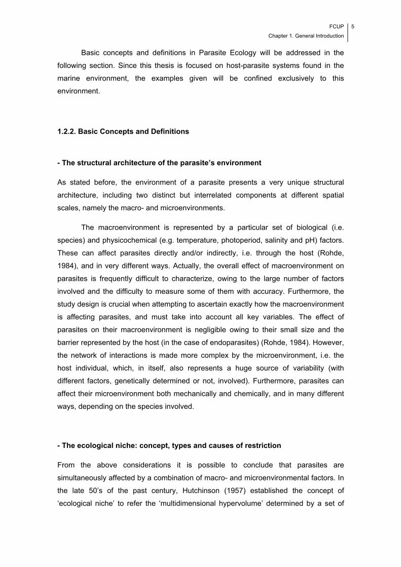
being more profoundly modified than those in others (Fig. 1.1).
Fig. 1.1 – A few examples of the morphological variability found in some families of parasitic copepods. A,
Octopicolidae; B, Chondracanthidae (arrow, male); C, Pennellidae; D, Lernaeopodidae; and E, Hatschekiidae. Scale-
bars: A, 500 μm; B, 1.0 mm; C, 1.0 mm; D, 1.0 mm; and E, 15.0 mm.
8 FCUP
Chapter 1. General Introduction
In families such as Chondracanthidae, the males are parasites of females and
incomparably smaller than them (Fig. 1.1 B), being often referred to as dwarfs in the
literature.
Despite the remarkable morphological variability, all parasitic copepods present
a body divided into two tagmata, i.e. an anterior prosome and a posterior urosome, with
an articulation between the fourth and fifth pedigerous somites (podoplean plan)
(Boxshall, 2005). Three types of parasites are recognised, namely the ectoparasites,
the mesoparasites and the endoparasites. The overwhelming majority of species falls
within the first type and infects external regions of the host’s body (as opposed to
endoparasites, which are found inside the host’s body). It is a common assumption that
ectoparasitic copepods may retain, or not, the freedom of their movements over the
surface of their hosts (Kabata, 1981). As for the mesoparasites, they live partly
embedded in the host. More specifically, in this type of parasites, the anterior end
forms an anchor process which allows them to penetrate deeply into the host’s tissues,
while a large part of their bodies protrudes from the host and remains exposed to the
external environment (Kabata, 1979, 1981; Boxshall & Halsey, 2004).
Parasitic copepods were reported to infect a wide spectrum of microhabitats
(e.g. body skin, fins, nostrils, buccal and branchial cavities, eyes, mucous canals of the
mandibular and preopercular areas, cephalic canal system adjacent to the nasal cavity
and urinary bladder) on or in their hosts (as ecto-, meso- and endoparasites) and
appear, therefore, particularly suited for addressing different aspects of Parasite
Ecology. It must be emphasised that a correct identification to species is crucial, as two
morphologically similar species can exhibit significant biological differences (e.g.
Kabata, 1973) and, therefore, ecological differences. Proximate and ultimate causes of
niche restriction in parasitic copepods are discussed in the literature. A few examples
are given below, since the majority of the papers in this thesis are dealing with these
parasites.
Proximate causes of niche restriction
Host species
Host specificity (sensu Rohde, 1984) varies according to parasite species and parasitic
copepods represent no exception to this general principle. Physicochemical and
FCUP
Chapter 1. General Introduction
9
morphological factors seem to be particularly important in determining niche restriction
in these parasites. More specifically, physicochemical factors seem to be capable of
attracting larvae and ensuring the settlement on the right host. For instance, two strains
of Lepeophtheirus pectoralis (Müller, 1777) (Copepoda: Caligidae) occurring on two
closely related species of flatfish i.e. the European flounder Platichthys flesus
(Linnaeus, 1758) (Actinopterygii: Pleuronectidae) and the plaice Pleuronectes platessa
Linnaeus, 1758 (Actinopterygii: Pleuronectidae) (named flesi and platessae) were
shown to respond to water currents, while chemical factors produced by the host are
likely involved in the settlement of copepodites on the right host (see Boxshall, 1976).
As for morphological factors, particular features of the attachment apparatus (among
other) can be highly adapted to particular sites on or in given host species.
Furthermore, the difference in activity between marine invertebrates and vertebrates
may have led to the development of a more robust type of antenna, capable to secure
fastening of parasites to their hosts, during switching events from invertebrate to
vertebrate hosts (see e.g. Ho, 1984).
Geographical range and macrohabitat
The geographical range of a species relates to the latitudinal gradient in its occurrence
and abundance. Distribution of marine parasites appears to be mainly determined by
temperature (Rohde, 1994), and parasitic copepods should not represent an exception
since this macroenvironmental factor has been recognised to influence development
and growth of parasitic copepods in general (Kabata, 1981). However, the data
currently available for parasitic copepods are still not enough to allow the establishment
of a conclusion on this subject. As for the macrohabitat, i.e. the fraction of the host
habitat in which the parasite is found (sensu Rohde & Rohde, 2005), the salinity
appears to be a particularly relevant macroenvironmental factor affecting that of
parasitic copepods. For instance, L. pectoralis and Acanthochondria cornuta (Müller,
1776) (Copepoda: Chondracanthidae) cannot develop within low salinity ranges, i.e. 7-
20‰ salinity (Möller, 1978), while their host, i.e. P. flesus, migrates regularly between
different salinity environments i.e. estuarine/brackish water environments and coastal
sea areas (Nikolsky, 1961; Berg, 1962). Actually, none of those parasite species was
found by Chibani & Rokicki (2004) in P. flesus caught in the Baltic Sea, the world’s
largest brackish water sea area (Leppäkoski et al., 2002).
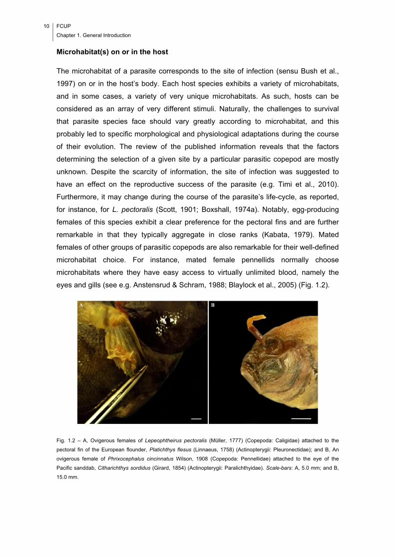
10 FCUP
Chapter 1. General Introduction
Microhabitat(s) on or in the host
The microhabitat of a parasite corresponds to the site of infection (sensu Bush et al.,
1997) on or in the host’s body. Each host species exhibits a variety of microhabitats,
and in some cases, a variety of very unique microhabitats. As such, hosts can be
considered as an array of very different stimuli. Naturally, the challenges to survival
that parasite species face should vary greatly according to microhabitat, and this
probably led to specific morphological and physiological adaptations during the course
of their evolution. The review of the published information reveals that the factors
determining the selection of a given site by a particular parasitic copepod are mostly
unknown. Despite the scarcity of information, the site of infection was suggested to
have an effect on the reproductive success of the parasite (e.g. Timi et al., 2010).
Furthermore, it may change during the course of the parasite’s life-cycle, as reported,
for instance, for L. pectoralis (Scott, 1901; Boxshall, 1974a). Notably, egg-producing
females of this species exhibit a clear preference for the pectoral fins and are further
remarkable in that they typically aggregate in close ranks (Kabata, 1979). Mated
females of other groups of parasitic copepods are also remarkable for their well-defined
microhabitat choice. For instance, mated female pennellids normally choose
microhabitats where they have easy access to virtually unlimited blood, namely the
eyes and gills (see e.g. Anstensrud & Schram, 1988; Blaylock et al., 2005) (Fig. 1.2).
Fig. 1.2 – A, Ovigerous females of Lepeophtheirus pectoralis (Müller, 1777) (Copepoda: Caligidae) attached to the
pectoral fin of the European flounder, Platichthys flesus (Linnaeus, 1758) (Actinopterygii: Pleuronectidae); and B, An
ovigerous female of Phrixocephalus cincinnatus Wilson, 1908 (Copepoda: Pennellidae) attached to the eye of the
Pacific sanddab, Citharichthys sordidus (Girard, 1854) (Actinopterygii: Paralichthyidae). Scale-bars: A, 5.0 mm; and B,
15.0 mm.
FCUP
Chapter 1. General Introduction
11
Host sex and age
It also may happen that parasites give preference to hosts of a given sex (Rohde,
1994), although the critical analysis of the literature suggests that differences of
infection levels between the two sexes of a host species are not common when the
infecting species is a parasitic copepod. Such differences may be due to different
factors, including gender-related differences in the composition of the skin resulting in
differential attraction of ectoparasites and differences in behaviour between female and
male hosts. As a result of the ontogenetic changes, hosts can be thought of as a
fluctuating microenvironment. Accordingly, the age of the host may represent an
important proximate cause of niche restriction as well. Concerning the parasitic
copepods, they may prefer to infect hosts of a certain age (Kabata, 1981), and such a
preference has already been evaluated for particular species, using laboratory
experiments. For instance, Anstensrud & Schram (1988) found that copepodites of
Lernaeenicus sprattae (Sowerby, 1806) (Copepoda: Pennellidae) do not exhibit a
preference for particular size groups of sprat, Sprattus sprattus (Linnaeus, 1758)
(Actinopterygii: Clupeidae). An influence of host ontogeny on parasite’s site selection
was, however, suggested for fish naturally infected with parasitic copepods. For
example, a displacement of Lernanthropus cynoscicola Timi & Etchegoin, 1996
(Copepoda: Lernanthropidae) over the gill arches of Cynoscion guatucupa (Cuvier,
1830) (Actinopterygii: Sciaenidae) as well as differential preferences for certain
sections of the gills were observed for parasites of both sexes and suggested to be
associated with the host’s increasing size (understood as an indication of age) (Timi,
2003).
Season of the year
As stated before, temperature has been recognised to influence development and
growth of parasitic copepods in general (Kabata, 1981). This parameter usually varies
from season to season (and, more markedly, at high latitudes), being likely that the
warmer seasons are more favourable for the occurrence of parasitic copepods. The
season of the year should represent, therefore, an important dimension of the niche of
parasitic copepods. It may further affect their occurrence by leading to changes in
reproductive behaviour. For instance, Ritchie et al. (1993) reported seasonal
differences in the reproductive output of winter and summer generations of females of
Lepeophtheirus salmonis (Krøyer, 1837) (Copepoda: Caligidae). More specifically, the
winter females were found to produce significantly longer egg sacs and a greater
12 FCUP
Chapter 1. General Introduction
number of smaller eggs compared with the summer females. Ritchie et al. (1993)
attributed the differences in reproductive output to macroenvironmental factors such as
temperature and photoperiod.
Food
Parasitic copepods can feed on host’s mucus, tissues and blood (Johnson et al., 2004).
More specifically, while some parasites appear to be less specific with respect to their
diet – e.g. caligids can feed on those three items (Kabata, 1974; Brandal et al., 1976) –
, others are definitely more restrictive – e.g. adult female pennellids usually feed on
blood and lymph of their host fish (Lester & Hayward, 2006). Accordingly, in many
cases, the type of food available at a particular site on or in the host’s body should
represent an important proximate cause of niche restriction.
Hyperparasites
The term ‘hyperparasite’ is used to refer any parasite of a parasite (Rohde, 2005). One
of the known cases of hyperparasitism is the occurrence of udonellids on copepods
parasitic on fish (Fig. 1.3). For many years, nothing was known about ‘host’ finding by
udonellid hyperparasites. However, a recent study suggested that copepod mating
represents the main route for dispersal of these hyperparasites in the ‘host’ population
of parasitic copepods, while the contact between copepods of the same sex appears to
be less important (Marin et al., 2007). It is therefore likely, that a specific chemical
factor is one of the causal factors which determine the hyperparasite’s niche.
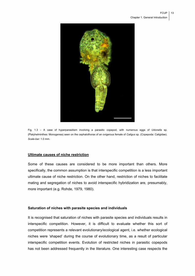
FCUP
Chapter 1. General Introduction
13
Fig. 1.3 – A case of hyperparasitism involving a parasitic copepod, with numerous eggs of Udonella sp.
(Platyhelminthes: Monogenea) seen on the cephalothorax of an ovigerous female of Caligus sp. (Copepoda: Caligidae).
Scale-bar: 1.0 mm.
Ultimate causes of niche restriction
Some of these causes are considered to be more important than others. More
specifically, the common assumption is that interspecific competition is a less important
ultimate cause of niche restriction. On the other hand, restriction of niches to facilitate
mating and segregation of niches to avoid interspecific hybridization are, presumably,
more important (e.g. Rohde, 1979, 1980).
Saturation of niches with parasite species and individuals
It is recognised that saturation of niches with parasite species and individuals results in
interspecific competition. However, it is difficult to evaluate whether this sort of
competition represents a relevant evolutionary/ecological agent, i.e. whether ecological
niches were ‘shaped’ during the course of evolutionary time, as a result of particular
interspecific competition events. Evolution of restricted niches in parasitic copepods
has not been addressed frequently in the literature. One interesting case respects the
14 FCUP
Chapter 1. General Introduction
asymmetrical competition between Lepeophtheirus thompsoni Baird, 1850 (a specialist
species) and L. europaensis Zeddam, Berrebi, Renaud, Raibaut & Gabrion, 1988 (a
generalist species) (Copepoda: Caligidae), demonstrated experimentally by Dawson et
al. (2000). These two parasites are naturally found on their sympatric hosts, i.e. the
former on turbot Scophthalmus maximus (Linnaeus, 1758) and the latter on brill S.
rhombus (Linnaeus, 1758) (Actinopterygii: Scophthalmidae). The experiments showed
that the two parasites are able to meet, mate and hybridize on S. maximus. However,
in natural conditions they are prevented from doing so, due to a strong host preference
(when they are given a choice). Therefore, it may be that interspecific competition led
to parasite species segregation between the two hosts over evolutionary time, i.e. that
it represents indeed, a relevant evolutionary agent in this particular case.
Avoidance of interspecific competition
Niche restriction occurs in extant communities as a result of interspecific competition,
the fundamental niche becoming a narrower realized niche (Rohde, 1994). The
literature provides some evidence for the parasitic copepods. For instance, Morales-
Serna & Gómez (2012) suggested that coexistence between Acantholochus zairae
Morales-Serna & Gómez, 2010 (Copepoda: Bomolochidae) and
Pseudochondracanthus diceraus Wilson, 1908 (Copepoda: Chondracanthidae) on the
gills of Sphoeroides annulatus (Jenyns, 1842) (Actinopterygii: Tetraodontidae) is
facilitated since intraspecific aggregation is stronger than interspecific aggregation.
Avoidance of predators
It is assumed that a given species of parasite become adapted to live in certain (i.e.
protected) microhabitats to avoid being predated by animals present in the
macroenvironment (Rohde, 1994). Many parasitic copepods are a potential target of
cleaner fish. For instance, caligid copepods (see e.g. Treasurer, 2002) are found on
different sites of the host’s body, e.g. body skin, beneath the pectoral and pelvic fins
and inside branchial chambers, and colonization of less exposed microhabitats by
these copepods can indeed reflect an evolutionary change to avoid predators.
Nonetheless, based on the information in the literature, it is not possible to make any
consideration as to whether avoidance of predators has determined niche restriction in
parasitic copepods over evolutionary time.
FCUP
Chapter 1. General Introduction
15
Facilitation of mating
Rohde (1976, 1977) established the ‘mating hypothesis’ of niche restriction, according
to which, selection favoured narrow microhabitats to enhance the chances of mating,
particularly in low-density infrapopulations of parasites. Timi (2003) tested this
hypothesis using infection data of L. cynoscicola infecting C. guatucupa. Unlike
parasites from other taxonomic groups, parasitic copepods are bisexual and,
accordingly, they must find a mate to reproduce. However, Timi (2003) found no
evidence supporting the mating hypothesis and concluded that the evolution of a
restricted niche in L. cynoscicola should be more related with other reproductive
benefits. More specifically, he found that individuals of the same sex were more
aggregated than females and males considered together and that the intensities of
females and males were negatively correlated. On the other hand, the finding that
intraspecific aggregation is stronger than interspecific aggregation (see e.g. Morales-
Serna & Gómez, 2012), can also be understood as an indication that niches of parasitic
copepods are restricted to facilitate mating. Furthermore, the aggregated distribution of
parasites among their hosts is considered a general feature of metazoan parasites
(Crofton, 1971; Poulin, 2007a,b), including parasitic copepods, and can be the result of
their need to reproduce (see e.g. Dippenaar et al., 2009).
Reinforcement of reproductive barriers
This aspect was commented by Rohde & Hobbs (1968). It concerns the possibility that
congeneric parasites overlap less than non-congeneric ones, in spite of the fact that all
of them depend on the same limited resource of space for attachment. The inevitable
conclusion to draw is that the difference found cannot be explained by interspecific
competition; instead, it most likely reflects a reinforcement of reproductive barriers. This
aspect was addressed for parasitic copepods in the published literature. Dippenaar et
al. (2009) searched for evidence of niche restriction in the spatial distribution of
Kroyeria dispar Wilson, 1935, K. papillipes Wilson, 1932 (Copepoda: Kroyeriidae) and
Eudactylina pusilla Cressey, 1967 (Copepoda: Eudactylinidae) on the gill filaments of
the tiger shark Galeocerdo cuvier (Péron & Lesueur, 1822) (Elasmobranchii:
Carcharhinidae). In spite of the fact that all those parasite species occupy the same
fundamental niche, no evidence of niche restriction was found. Accordingly, the spatial
distributions found do not suggest a reinforcement of reproductive barriers.
16 FCUP
Chapter 1. General Introduction
Adaptations to environmental complexity
Microhabitats are generally complex and parasites have become adapted to live in
them. More specifically, they should be able to ensure their survival, by making use of
a particular set of biological and physiological features. For instance, they must be able
to attach to a particular substratum, gain food, resist the immune reactions of the host,
react to the variations in the volume of water flushing over the gills and respond to the
chemical stimuli released by mating partners present in their microhabitat (Rohde,
1994). Optimal adaptation ensures the maximum possible chances of surviving
environmental changes and parasites will not occupy other microhabitats unless they
are obligated to do so. With respect to parasitic copepods, the work of Timi (2003) is
particularly relevant. In that work, it is suggested that adaptations to environmental
complexity, rather than increasing intraspecific contact, are more likely ultimate causes
of niche restriction.
1.3. The Studied Hosts
1.3.1. The Common Octopus, Octopus vulgaris (Cephalopoda: Octopodidae)
The common octopus, Octopus vulgaris (Cephalopoda: Octopodidae) (Fig. 1.4), is a
neritic, nektobenthic species, commonly found in moderately warm, shallow coastal
waters (< 200 m deep) (Hastie et al., 2009). Its geographic distribution is wide,
comprising the tropical, subtropical and temperate regions of the Atlantic, Indian and
Pacific Oceans, and also, the adjacent seas (Mangold, 1983). Actually, it has been
argued that it represents a complex of species rather than a single cosmopolitan
species (see e.g. Leite et al., 2008). Each species in the complex should be adapted to
the local environmental conditions (e.g. Guerra, 1982) and, accordingly, there can be
differences in the parasite fauna of different species.
The species in the O. vulgaris complex are marketed fresh, frozen, dried salted
and canned, representing an important food item and source of income to many people
throughout the world. In Portugal, O. vulgaris usually occurs in commercial landings in
fisheries off mainland Portugal, Madeira and the Azores Islands. The total world catch
has decreased in recent years (Food and Agriculture Organization, 2013), and the
cephalopod is presently considered a candidate species for marine aquaculture
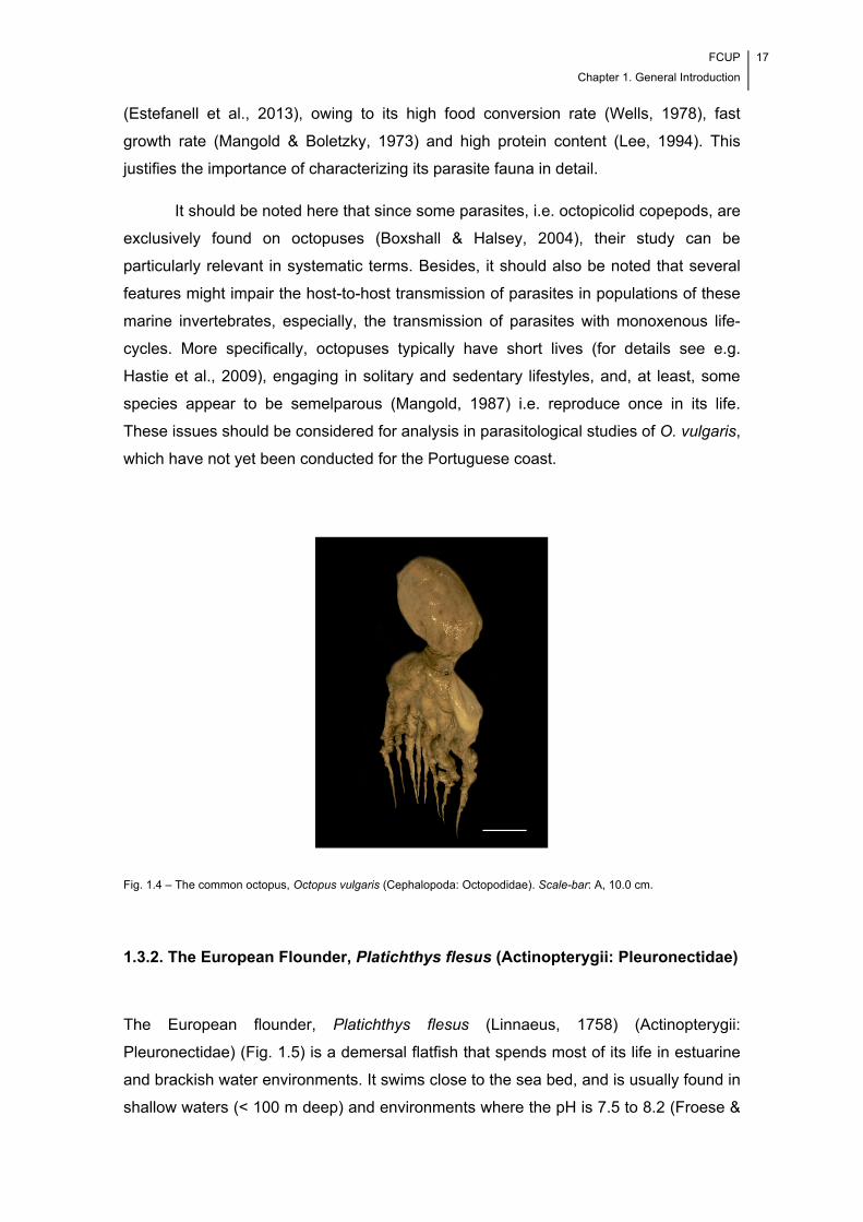
FCUP
Chapter 1. General Introduction
17
(Estefanell et al., 2013), owing to its high food conversion rate (Wells, 1978), fast
growth rate (Mangold & Boletzky, 1973) and high protein content (Lee, 1994). This
justifies the importance of characterizing its parasite fauna in detail.
It should be noted here that since some parasites, i.e. octopicolid copepods, are
exclusively found on octopuses (Boxshall & Halsey, 2004), their study can be
particularly relevant in systematic terms. Besides, it should also be noted that several
features might impair the host-to-host transmission of parasites in populations of these
marine invertebrates, especially, the transmission of parasites with monoxenous life-
cycles. More specifically, octopuses typically have short lives (for details see e.g.
Hastie et al., 2009), engaging in solitary and sedentary lifestyles, and, at least, some
species appear to be semelparous (Mangold, 1987) i.e. reproduce once in its life.
These issues should be considered for analysis in parasitological studies of O. vulgaris,
which have not yet been conducted for the Portuguese coast.
Fig. 1.4 – The common octopus, Octopus vulgaris (Cephalopoda: Octopodidae). Scale-bar: A, 10.0 cm.
1.3.2. The European Flounder, Platichthys flesus (Actinopterygii: Pleuronectidae)
The European flounder, Platichthys flesus (Linnaeus, 1758) (Actinopterygii:
Pleuronectidae) (Fig. 1.5) is a demersal flatfish that spends most of its life in estuarine
and brackish water environments. It swims close to the sea bed, and is usually found in
shallow waters (< 100 m deep) and environments where the pH is 7.5 to 8.2 (Froese &
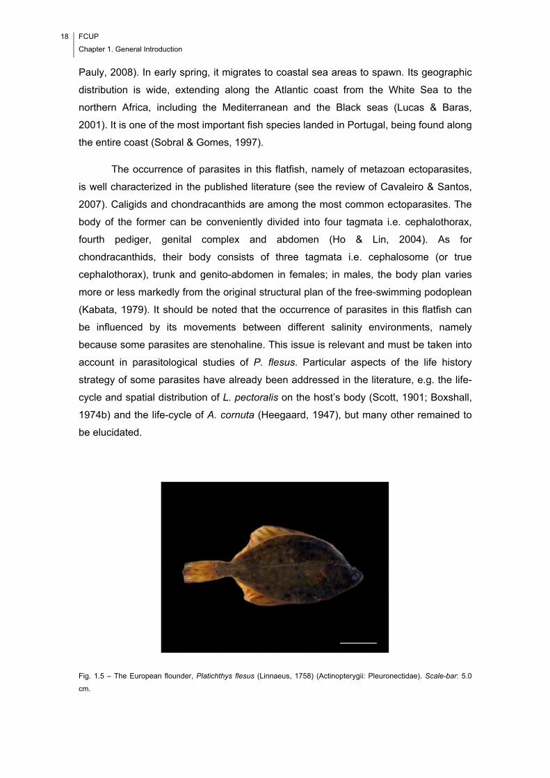
18 FCUP
Chapter 1. General Introduction
Pauly, 2008). In early spring, it migrates to coastal sea areas to spawn. Its geographic
distribution is wide, extending along the Atlantic coast from the White Sea to the
northern Africa, including the Mediterranean and the Black seas (Lucas & Baras,
2001). It is one of the most important fish species landed in Portugal, being found along
the entire coast (Sobral & Gomes, 1997).
The occurrence of parasites in this flatfish, namely of metazoan ectoparasites,
is well characterized in the published literature (see the review of Cavaleiro & Santos,
2007). Caligids and chondracanthids are among the most common ectoparasites. The
body of the former can be conveniently divided into four tagmata i.e. cephalothorax,
fourth pediger, genital complex and abdomen (Ho & Lin, 2004). As for
chondracanthids, their body consists of three tagmata i.e. cephalosome (or true
cephalothorax), trunk and genito-abdomen in females; in males, the body plan varies
more or less markedly from the original structural plan of the free-swimming podoplean
(Kabata, 1979). It should be noted that the occurrence of parasites in this flatfish can
be influenced by its movements between different salinity environments, namely
because some parasites are stenohaline. This issue is relevant and must be taken into
account in parasitological studies of P. flesus. Particular aspects of the life history
strategy of some parasites have already been addressed in the literature, e.g. the life-
cycle and spatial distribution of L. pectoralis on the host’s body (Scott, 1901; Boxshall,
1974b) and the life-cycle of A. cornuta (Heegaard, 1947), but many other remained to
be elucidated.
Fig. 1.5 – The European flounder, Platichthys flesus (Linnaeus, 1758) (Actinopterygii: Pleuronectidae). Scale-bar: 5.0
cm.
FCUP
Chapter 1. General Introduction
19
1.4. Study Aims
This thesis enlarges the body of knowledge about the parasite fauna of O. vulgaris and
P. flesus, by providing new information on the parasites occurring on or in these two
species of hosts. Specific aims were as follows:
1) To characterize, for the first time, the metazoan parasite infections occurring in O.
vulgaris from Portuguese coastal waters, including aspects such as seasonality trends
and ecology of established host-parasite relationships.
2) To review the current knowledge on a poorly studied group of parasitic copepods,
i.e. the octopicolid copepods. This is exclusively found on species of octopuses and
was isolated during the parasitological survey of O. vulgaris caught in Portuguese
coastal waters.
3) To describe a new and rare species of caligid copepod, Caligus musaicus sp. nov.
(Copepoda: Caligidae), isolated from P. flesus of Portuguese coastal waters.
4) To describe, in detail, the morphology, ultrastructure, genetics and morphometrics of
a diplostomid metacercaria isolated from the eye lenses of P. flesus.
5) To study the trade-off between egg number and egg size at the intraspecific level,
based on data recorded for adult ovigerous females of Octopicola superba Humes,
1957 (Copepoda: Octopicolidae).
6) To evaluate whether copepod parasites other than L. pectoralis, i.e. A. cornuta, also
exhibit a particular spatial distribution on the body of P. flesus.
20 FCUP
Chapter 1. General Introduction
7) To evaluate the evidence supporting the occurrence of interference competition
between an endoparasitic microparasite, i.e. Aggregata sp. (Apicomplexa:
Aggregatidae), and an ectoparasitic macroparasite, i.e. O. superba, that co-occur at the
gills of O. vulgaris.
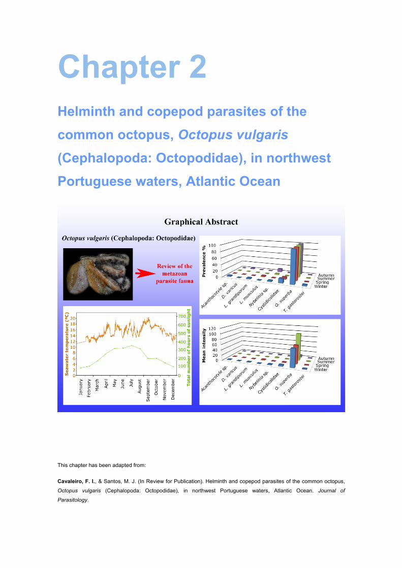
Chapter 2 Helminth and copepod parasites of the
common octopus, Octopus vulgaris
(Cephalopoda: Octopodidae), in northwest
Portuguese waters, Atlantic Ocean
This chapter has been adapted from:
Cavaleiro, F. I., & Santos, M. J. (In Review for Publication). Helminth and copepod parasites of the common octopus,
Octopus vulgaris (Cephalopoda: Octopodidae), in northwest Portuguese waters, Atlantic Ocean. Journal of
Parasitology.

22 FCUP
Chapter 2. Helminth and copepod parasites of Octopus vulgaris (Cephalopoda: Octopodidae)
FCUP
Chapter 2. Helminth and copepod parasites of Octopus vulgaris (Cephalopoda: Octopodidae)
23
2.1. Abstract
Octopus vulgaris (Cephalopoda: Octopodidae) from northwest Portuguese waters was
collected seasonally for one year and examined for metazoan parasites. Eight parasitic
taxa were found, including six taxa of helminths and two taxa of copepods. They are:
Acanthocotyle sp. (Monogenea: Acanthocotylidae); Derogenes varicus (Digenea:
Derogenidae); Lecithochirium grandiporum, and L. musculus (Digenea: Hemiuridae);
Nybelinia sp. (Cestoda: Tentaculariidae); Cystidicolidae (Nematoda: Spiruroidea);
Octopicola superba (Copepoda: Octopicolidae); and Thersitina gasterostei (Copepoda:
Ergasilidae). O. superba was the only component parasite in the total sample of O.
vulgaris. It exhibited a marked seasonality, with the lowest and highest mean intensity
levels recorded for autumn and summer, respectively. According to the evidence found
in this and other studies, the seawater temperature and the total number of hours of
sunlight influence the infection levels of parasitic copepods. Furthermore, significantly
higher numbers of octopicolid copepods were recorded for the female octopuses. This,
along with the fact that a significant correlation between octopus’ size and parasite
intensity was detected only for the female octopuses suggests a differential influence of
host sex in autoinfection. The infection levels recorded for Octopicola spp. infecting O.
vulgaris in contiguous estuarine waters off Galicia were lower, which suggests that
octopicolids are stenohaline.

24 FCUP
Chapter 2. Helminth and copepod parasites of Octopus vulgaris (Cephalopoda: Octopodidae)
FCUP
Chapter 2. Helminth and copepod parasites of Octopus vulgaris (Cephalopoda: Octopodidae)
25
2.2. Introduction
The common octopus, Octopus vulgaris (Cephalopoda: Octopodidae), is exploited by
commercial fisheries, commanding high prices in the market place (Food and
Agriculture Organization of the United Nations, 2007). Despite the fact that it is
presently considered a candidate for aquaculture (Vaz-Pires et al., 2004; Estefanell et
al., 2013), there is still little information about the parasite fauna of wild and reared
specimens. Phylogenetic analyses of mitochondrial genes indicate that it represents a
complex of species rather than a single cosmopolitan species (e.g. Söller et al., 2000;
Warnke et al., 2004; Guerra et al., 2010). This study aimed: (i) qualitative and
quantitative characterization of the metazoan parasite fauna of O. vulgaris from waters
off the coast of northern Portugal; (ii) comparison of the recorded parasite fauna with
that reported in the literature; and (iii) characterization of host-parasite relationships
(component parasites).
2.3. Materials and Methods
O. vulgaris was caught seasonally (N = 30 per season; on 2 March, 24 and 31 May, 7
September, and 22 November) for one year (2010) off Matosinhos (41º10’N, 8º42’W),
northeast Atlantic Ocean. The specimens were caught by a boat that fishes for O.
vulgaris exclusively, and the landed catch was collected and kept in a box for a few
hours, separated from the species fished by other boats. Octopuses were frozen,
defrosted days later and examined for metazoan parasites and gross pathology. The
total length and sex were recorded for all of them. External body surfaces were washed
with saline solution (3.5%) to isolate the ectoparasites and all organs were examined
for endoparasites. Mesozoans are not included in the survey because they could not
be detected in the frozen material. Parasites were fixed and preserved in 70% ethanol.
Later, they were identified to the lowest possible taxonomic level, according to
Monticelli (1899), Overstreet & Hochberg (1975), Kabata (1979), Gibson & Bray (1986),
Gestal et al. (1999), Palm (2004), Chabaud (2009) and Cavaleiro et al. (2013).
Digeneans and the single monogenean specimen were stained with iron acetocarmine
(Georgiev et al., 1986); nematodes (Hoffman, 1999) and copepods (Humes & Gooding,
1964) were cleared in lactophenol and lactic acid, respectively. Infection parameters
(number of infected octopuses/prevalence [95% confidence interval] % and mean
intensity ± SD [range]) were assessed for each parasitic taxon and considering the
seasonal and total samples of octopuses. Bootstrap estimator values of taxa richness
(Sb) (Poulin, 2007a) were also assessed for the seasonal and total samples. Seasonal,
26 FCUP
Chapter 2. Helminth and copepod parasites of Octopus vulgaris (Cephalopoda: Octopodidae)
sex and size differences in infection were evaluated for the component taxa (sensu
Bush et al., 1990; considering the total sample of octopuses) exclusively. Intensity data
were compared using the Kruskal-Wallis’ test (comparison between the four samples)
and the Mann-Whitney’s U test (pairwise sample comparisons). A significant correlation
between octopus’ size and parasite intensity was evaluated using the Spearman’s test
(females and males were considered separately for analysis). Significance was set at P
< 0.05 for all statistical tests (performed using SPSS, version 19.0) except the pairwise
sample comparisons (Bonferroni-adjusted level: 0.008(3)). Lastly, the effect of
temperature (data assessed from: Portuguese Hydrographic Institute, 2012) and
sunlight (data derived from: Portuguese Meteorology Institute, 2012) in infection levels
was evaluated. Terminology (locality, site, prevalence, intensity, and mean intensity)
follows Bush et al. (1997).
2.4. Results
O. vulgaris was infected with eight taxa of metazoan parasites: Acanthocotyle sp.
(Monogenea: Acanthocotylidae); Derogenes varicus (Müller, 1784) (Digenea:
Derogenidae); Lecithochirium grandiporum (Rudolphi, 1819) Lühe, 1901, and L.
musculus (Looss, 1907) (Digenea: Hemiuridae); Nybelinia sp. (Cestoda:
Tentaculariidae); Cystidicolidae (Nematoda: Spiruroidea); Octopicola superba Humes,
1957 (Copepoda: Octopicolidae); and Thersitina gasterostei (Pagenstecher, 1861)
(Copepoda: Ergasilidae) (Table 2.1). Gross pathology was not observed in any of the
examined octopuses.
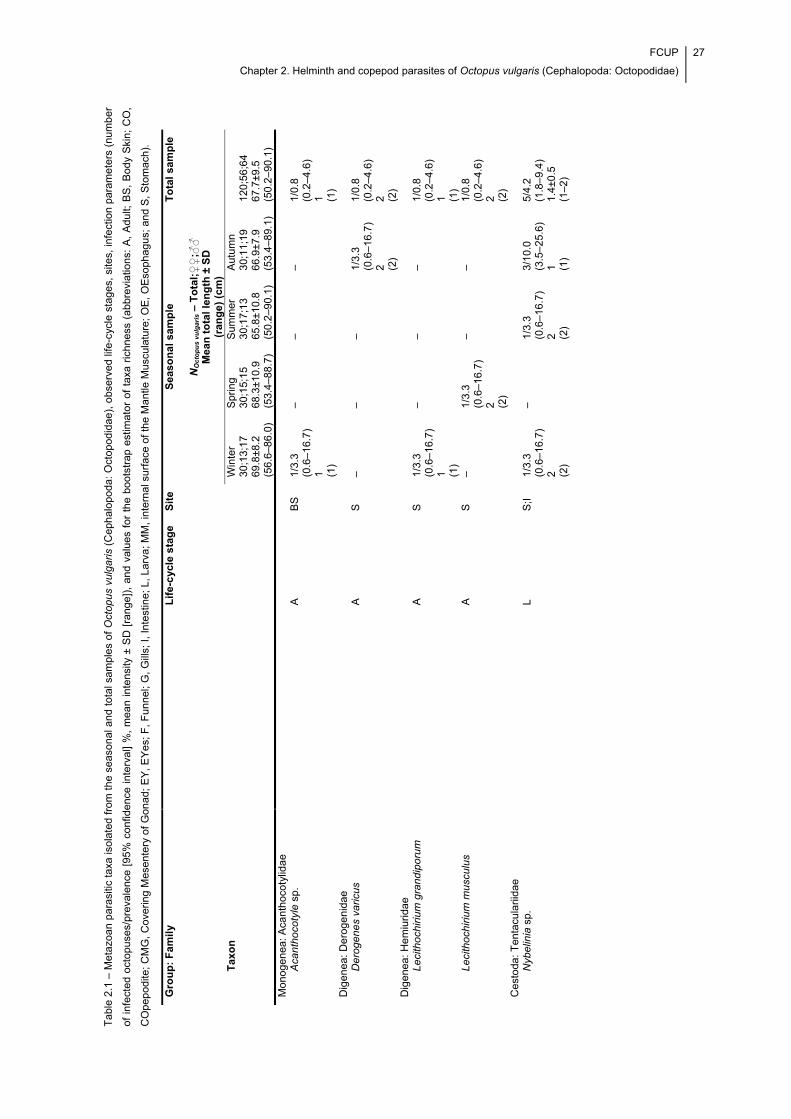
FCUP
Chapter 2. Helminth and copepod parasites of Octopus vulgaris (Cephalopoda: Octopodidae)
27
Tab
le 2
.1 –
Met
azo
an p
aras
itic
taxa
isol
ated
fro
m t
he s
easo
nal a
nd t
otal
sam
ples
of
Oct
opus
vul
garis
(C
eph
alop
oda:
Oct
opod
idae
), o
bser
ved
life-
cycl
e st
ages
, si
tes,
infe
ctio
n pa
ram
eter
s (n
umb
er
of in
fect
ed o
ctop
uses
/pre
vale
nce
[95%
con
fiden
ce in
terv
al]
%,
mea
n in
tens
ity ±
SD
[ra
nge]
), a
nd v
alue
s fo
r th
e bo
otst
rap
estim
ato
r of
tax
a ric
hnes
s (a
bbre
viat
ions
: A
, A
dult;
BS
, B
ody
Ski
n; C
O,
CO
pep
odite
; CM
G, C
over
ing
Mes
ente
ry o
f G
onad
; E
Y, E
Yes
; F,
Fu
nnel
; G,
Gill
s; I,
Inte
stin
e; L
, Lar
va; M
M, i
nter
nal s
urfa
ce o
f th
e M
antle
Mus
cula
ture
; OE
, OE
soph
agu
s; a
nd S
, Sto
mac
h).
Gro
up
: F
am
ily
Lif
e-c
ycle
sta
ge
S
ite
S
easo
nal
sam
ple
T
ota
l sa
mp
le
NO
cto
pu
s vu
lgar
is –
To
tal;♀♀
;♂♂
M
ean
to
tal
len
gth
± S
D
(ran
ge
) (c
m)
Tax
on
W
inte
r 30
;13;
17
69.8
±8.2
(5
6.6–
86.0
)
Spr
ing
30;1
5;15
68
.3±1
0.9
(5
3.4–
88.7
)
Sum
mer
30
;17;
13
65.8
±10.
8
(50.
2–90
.1)
Aut
umn
30;1
1;19
66
.9±7
.9
(53.
4–89
.1)
120;
56;6
4
67.7
±9.5
(5
0.2–
90.1
) M
onog
enea
: Aca
ntho
coty
lidae
Aca
ntho
coty
le s
p.
A
BS
1/
3.3
(0.6
–16.
7)
1 (1)
– –
– 1/
0.8
(0.2
–4.6
) 1 (1
) D
igen
ea: D
ero
geni
dae
D
erog
enes
var
icus
A
S
– –
– 1/
3.3
(0.6
–16.
7)
2 (2)
1/0.
8 (0
.2–4
.6)
2 (2)
Dig
enea
: Hem
iuri
dae
Le
cith
ochi
rium
gra
ndip
oru
m
A
S
1/
3.3
(0.6
–16.
7)
1 (1)
– –
– 1/
0.8
(0.2
–4.6
) 1 (1
) Le
cith
ochi
rium
mus
culu
s
A
S
– 1/
3.3
(0.6
–16.
7)
2 (2)
– –
1/0.
8 (0
.2–4
.6)
2 (2)
Ces
toda
: Ten
tacu
larii
dae
N
ybel
inia
sp.
L
S;I
1/3.
3 (0
.6–1
6.7
) 2 (2
)
– 1/
3.3
(0.6
–16.
7)
2 (2)
3/10
.0
(3.5
–25.
6)
1 (1)
5/4.
2 (1
.8–9
.4)
1.4±
0.5
(1–2
)
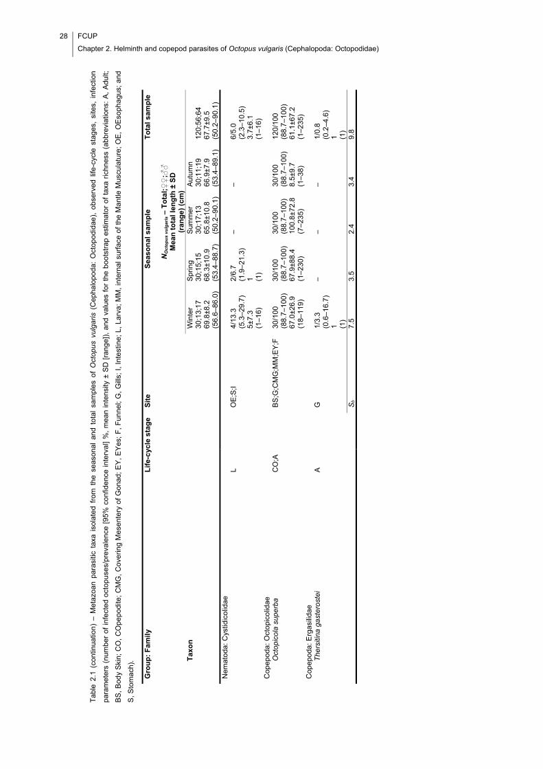
28 FCUP
Chapter 2. Helminth and copepod parasites of Octopus vulgaris (Cephalopoda: Octopodidae)
Tab
le 2
.1 (
cont
inua
tion)
– M
eta
zoan
par
asiti
c ta
xa i
sola
ted
from
the
sea
sona
l an
d to
tal
sam
ples
of
Oct
opus
vul
garis
(C
epha
lopo
da:
Oct
opod
idae
), o
bser
ved
life-
cycl
e st
ages
, si
tes,
inf
ectio
n
para
met
ers
(nu
mbe
r of
infe
cted
oct
opus
es/p
reva
lenc
e [9
5% c
onfid
ence
inte
rval
] %
, m
ean
inte
nsity
± S
D [
rang
e]),
and
val
ues
for
the
boot
stra
p es
timat
or o
f ta
xa r
ichn
ess
(abb
revi
atio
ns:
A,
Adu
lt;
BS
, B
ody
Ski
n; C
O,
CO
pepo
dite
; C
MG
, C
ove
ring
Mes
ente
ry o
f G
onad
; E
Y,
EY
es;
F,
Fu
nnel
; G
, G
ills;
I,
Inte
stin
e; L
, La
rva;
MM
, in
tern
al s
urfa
ce o
f th
e M
antle
Mus
cula
ture
; O
E,
OE
sop
ha
gu
s; a
nd
S, S
tom
ach)
.
Gro
up
: F
am
ily
Lif
e-c
ycle
sta
ge
S
ite
S
easo
nal
sam
ple
T
ota
l sa
mp
le
NO
cto
pu
s vu
lgar
is –
To
tal;♀♀
;♂♂
M
ean
to
tal
len
gth
± S
D
(ran
ge
) (c
m)
Tax
on
W
inte
r 30
;13;
17
69.8
±8.2
(5
6.6–
86.0
)
Spr
ing
30;1
5;15
68
.3±1
0.9
(5
3.4–
88.7
)
Sum
mer
30
;17;
13
65.8
±10.
8
(50.
2–90
.1)
Aut
umn
30;1
1;19
66
.9±7
.9
(53.
4–89
.1)
120;
56;6
4
67.7
±9.5
(5
0.2–
90.1
) N
emat
oda:
Cys
tidic
olid
ae
L O
E;S
;I
4/13
.3
(5.3
–29.
7)
5±7.
3 (1
–16)
2/6.
7 (1
.9–2
1.3
) 1 (1
)
– –
6/5.
0 (2
.3–1
0.5
) 3.
7±6.
1 (1
–16)
C
opep
oda:
Oct
opi
colid
ae
O
ctop
icol
a su
perb
a
C
O;A
B
S;G
;CM
G;M
M;E
Y;F
30
/100
(8
8.7–
100
) 67
.0±2
6.9
(1
8–11
9)
30/1
00
(88.
7–10
0)
67.9
±88.
4
(1–2
30)
30/1
00
(88.
7–10
0)
100.
8±72
.8
(7–2
35)
30/1
00
(88.
7–10
0)
8.5±
9.7
(1–3
8)
120/
100
(8
8.7–
100
) 61
.1±6
7.2
(1
–235
) C
opep
oda:
Erg
asi
lidae
The
rsiti
na g
aste
rost
ei
A
G
1/
3.3
(0.6
–16.
7)
1 (1)
– –
– 1/
0.8
(0.2
–4.6
) 1 (1
)
S
b 7.
5 3.
5 2.
4 3.
4 9.
8
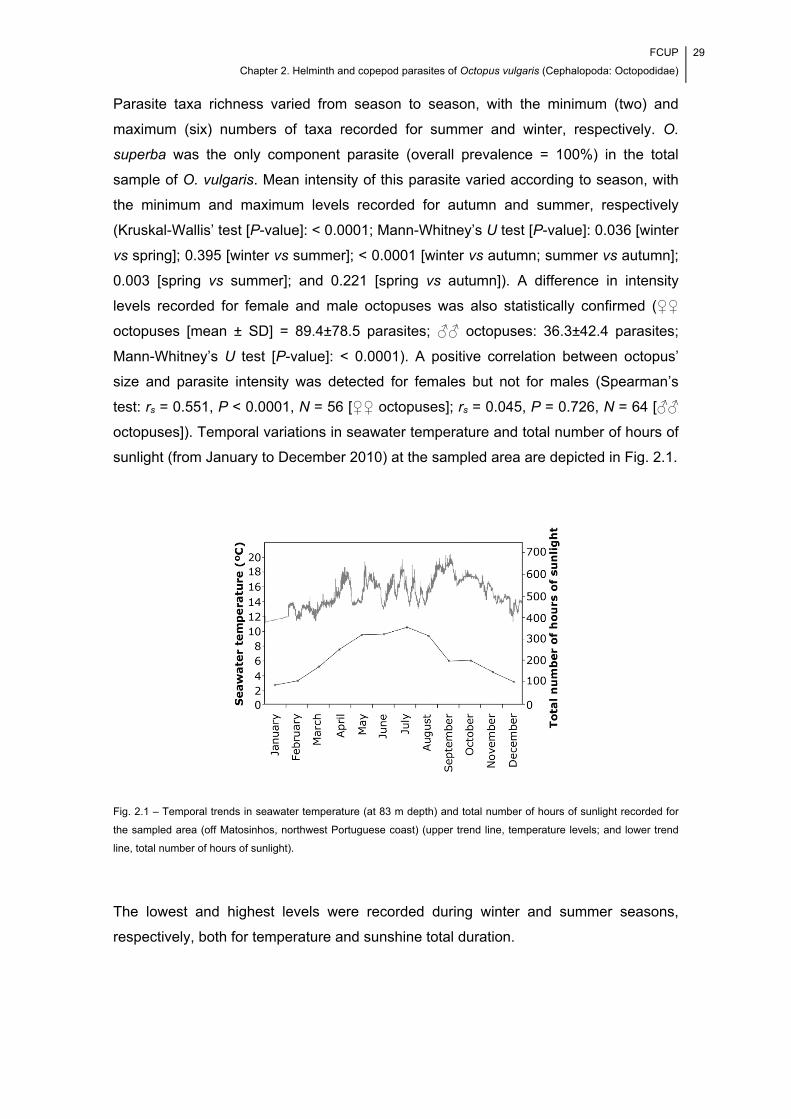
FCUP
Chapter 2. Helminth and copepod parasites of Octopus vulgaris (Cephalopoda: Octopodidae)
29
Parasite taxa richness varied from season to season, with the minimum (two) and
maximum (six) numbers of taxa recorded for summer and winter, respectively. O.
superba was the only component parasite (overall prevalence = 100%) in the total
sample of O. vulgaris. Mean intensity of this parasite varied according to season, with
the minimum and maximum levels recorded for autumn and summer, respectively
(Kruskal-Wallis’ test [P-value]: < 0.0001; Mann-Whitney’s U test [P-value]: 0.036 [winter
vs spring]; 0.395 [winter vs summer]; < 0.0001 [winter vs autumn; summer vs autumn];
0.003 [spring vs summer]; and 0.221 [spring vs autumn]). A difference in intensity
levels recorded for female and male octopuses was also statistically confirmed (♀♀
octopuses [mean ± SD] = 89.4±78.5 parasites; ♂♂ octopuses: 36.3±42.4 parasites;
Mann-Whitney’s U test [P-value]: < 0.0001). A positive correlation between octopus’
size and parasite intensity was detected for females but not for males (Spearman’s
test: rs = 0.551, P < 0.0001, N = 56 [♀♀ octopuses]; rs = 0.045, P = 0.726, N = 64 [♂♂
octopuses]). Temporal variations in seawater temperature and total number of hours of
sunlight (from January to December 2010) at the sampled area are depicted in Fig. 2.1.
Fig. 2.1 – Temporal trends in seawater temperature (at 83 m depth) and total number of hours of sunlight recorded for
the sampled area (off Matosinhos, northwest Portuguese coast) (upper trend line, temperature levels; and lower trend
line, total number of hours of sunlight).
The lowest and highest levels were recorded during winter and summer seasons,
respectively, both for temperature and sunshine total duration.
30 FCUP
Chapter 2. Helminth and copepod parasites of Octopus vulgaris (Cephalopoda: Octopodidae)
2.5. Discussion
From the metazoan parasites recorded in this study, Acanthocotyle sp., D. varicus, L.
grandiporum, L. musculus and T. gasterostei are new records for the O. vulgaris
complex (see the review presented in Table 2.2). The highest number of parasitic taxa
was recorded in winter, which suggests a reduced resistance to infections in this
season of the year. The recorded bootstrap values indicate that more parasitic taxa
should have been found in the winter and total samples. The unfound species likely
represent rare parasites of O. vulgaris. Acanthocotyle spp. are typically found on the
skin of elasmobranchs (Yamaguti, 1963); accordingly, Acanthocotyle sp. is probably an
accidental parasite of O. vulgaris (only one specimen found through the examination of
120 octopuses), i.e. its occurrence on O. vulgaris most likely reflects the absence of
suitable hosts. The larvae of Nybelinia were rare and exhibited a great morphological
similarity to Nybelinia lingualis Cuvier, 1817 (according to Palm, 2004). Nonetheless,
the assignment of larvae to this species must be confirmed by molecular analyses. The
infection levels of Cystidicolidae were also low, which indicates that this taxon is also
uncommon in O. vulgaris in the sampled area. Similar evidence was found by Pascual
et al. (1996) for Cystidicola sp. (prevalence = 11.4%) and Gestal et al. (1999) for
Cystidicolidae larvae (prevalence = 16%; mean intensity = 1.46 worms/host) infecting
O. vulgaris from the Ría de Vigo, a large estuary in northwestern Spain. The infection
levels of O. superba suggest that this is a common parasite of O. vulgaris in waters off
the coast of northern Portugal. Moreover, the seasonal trend of O. superba is similar to
the trends found for other parasitic copepods present at the sampled area, i.e.
Lepeophtheirus pectoralis (Müller, 1777) (Copepoda: Caligidae) and Acanthochondria
cornuta (Müller, 1776) (Copepoda: Chondracanthidae) (see Cavaleiro & Santos, 2009).
The temporal trends of the considered environmental variables were consistent enough
between the two years studied to underpin the hypothesis that high water temperature
and large photoperiod have a positive effect on the infection with parasitic copepods. T.
gasterostei is a common parasite, usually found on species of sticklebacks and others
(Kabata, 1979). Furthermore, the copepod is cosmopolitan at higher latitudes of the
northern hemisphere, which helps to justify its accidental occurrence on O. vulgaris
(only one specimen found through the examination of 120 octopuses). Octopicola spp.
occurred less frequently on O. vulgaris from estuarine waters of Ría de Vigo (34.3%)
and Ribadeo (38.5%) (Pascual et al., 1996) compared with O. superba on O. vulgaris
from waters off Matosinhos (100%). This suggests that octopicolids are stenohaline,
which conforms to what has been said for the parasitic copepods (Kabata, 1979;
Knudsen & Sundnes, 1998). The spatial distribution of O. superba on the body of O.
FCUP
Chapter 2. Helminth and copepod parasites of Octopus vulgaris (Cephalopoda: Octopodidae)
31
vulgaris was addressed in a previous study (Cavaleiro & Santos, In Press). The results
of the present study shed further light on that host-parasite system, since they suggest
an influence of host sex on parasite life strategy. More specifically, not only the
intensity was significantly higher in female octopuses, as a significant positive
correlation between octopus’ size and parasite intensity was recorded only for the
subsample of females. This evidence suggests that significant autoinfection takes
place in female octopuses and that these have a key role in host-to-host transmission
of O. superba. The infection with O. superba did not cause gross pathology;
accordingly, it should not cause economic losses to fisheries. It can however become
problematic in intensive rearing systems, and prophylactic measures can be crucial to
preventing economic losses.
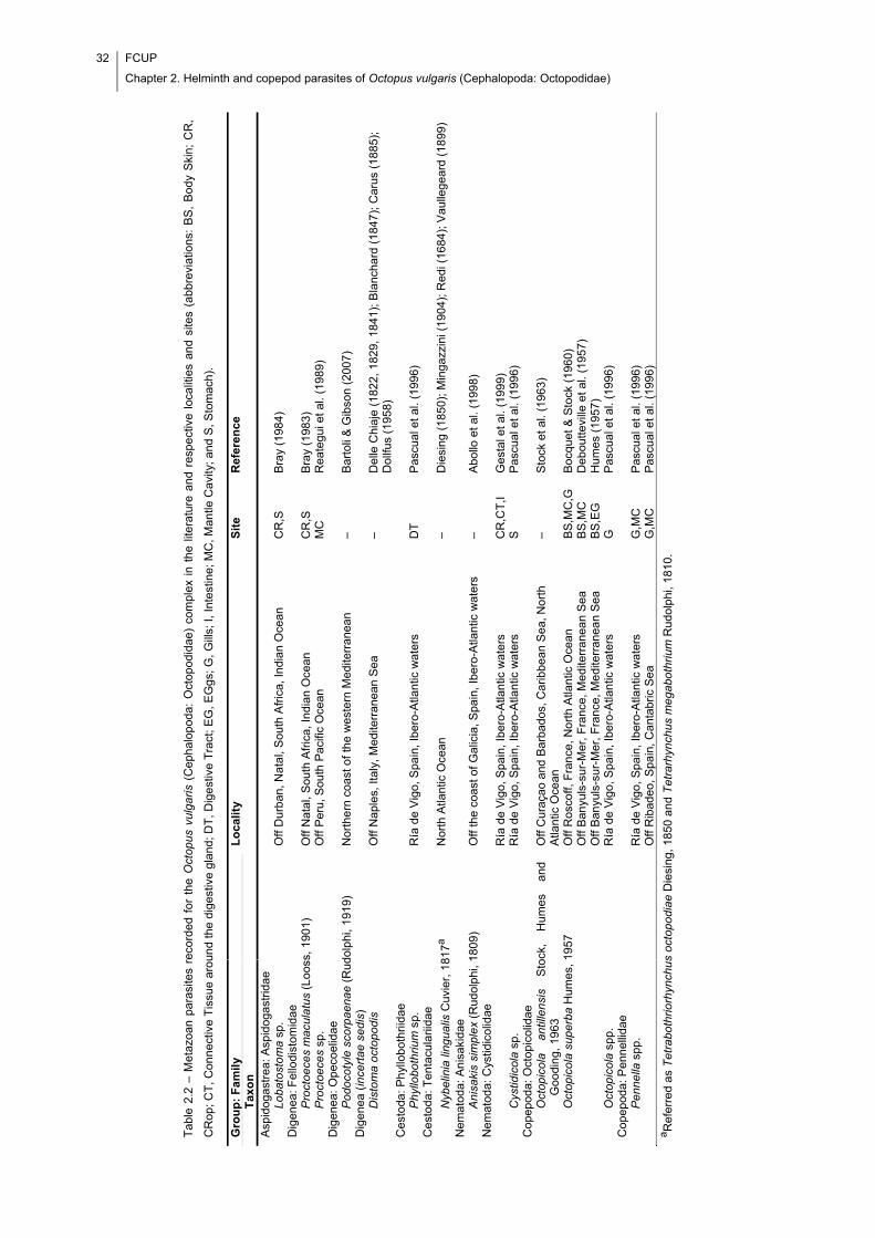
32 FCUP
Chapter 2. Helminth and copepod parasites of Octopus vulgaris (Cephalopoda: Octopodidae)
Tab
le 2
.2 –
Met
azoa
n pa
rasi
tes
reco
rded
fo
r th
e O
ctop
us v
ulga
ris (
Cep
halo
poda
: O
ctop
odid
ae)
com
plex
in
the
liter
atur
e an
d re
spe
ctiv
e l
ocal
ities
and
site
s (a
bbre
viat
ions
: B
S,
Bod
y S
kin;
CR
,
CR
op; C
T, C
onn
ectiv
e T
issu
e ar
ound
the
dige
stiv
e g
land
; DT
, Dig
est
ive
Tra
ct; E
G,
EG
gs; G
, G
ills;
I, In
test
ine;
MC
, Man
tle C
avity
; and
S, S
tom
ach)
.
Gro
up
: F
am
ily
Lo
cali
ty
Sit
e
Ref
eren
ce
Tax
on
Asp
idog
astr
ea: A
spid
ogas
trid
ae
Lo
bato
sto
ma
sp.
Off
Du
rban
, Nat
al,
Sou
th A
fric
a, In
dian
Oce
an
CR
,S
Bra
y (1
984
) D
igen
ea: F
ello
dist
omid
ae
P
roct
oece
s m
acul
atus
(Lo
oss,
190
1)
Off
Nat
al, S
outh
Afr
ica,
Indi
an O
cean
C
R,S
B
ray
(198
3)
Pro
ctoe
ces
sp.
Off
Per
u, S
outh
Pac
ific
Oce
an
MC
R
eate
gui e
t al.
(198
9)
Dig
enea
: Ope
coel
idae
Pod
ocot
yle
scor
paen
ae (
Rud
olph
i, 19
19)
Nor
the
rn c
oast
of
the
wes
tern
Med
iterr
anea
n
– B
arto
li &
Gib
son
(200
7)
Dig
enea
(in
cert
ae
se
dis
)
Dis
tom
a oc
topo
dis
O
ff N
aple
s, It
aly,
Med
iterr
ane
an S
ea
– D
elle
Chi
aje
(182
2, 1
829,
184
1); B
lanc
hard
(18
47);
Car
us (
1885
);
Dol
lfus
(195
8)
Ces
toda
: Ph
yllo
bot
hriid
ae
P
hyllo
both
rium
sp.
R
ía d
e V
igo,
Spa
in, I
bero
-Atla
ntic
wat
ers
DT
P
ascu
al e
t al.
(199
6)
Ces
toda
: Ten
tacu
larii
dae
Nyb
elin
ia li
ngua
lis C
uvie
r, 1
817a
N
orth
Atla
ntic
Oce
an
– D
iesi
ng (
1850
); M
inga
zzin
i (19
04);
Red
i (16
84);
Vau
llege
ard
(18
99)
Nem
atod
a: A
nisa
kida
e
An
isa
kis
sim
ple
x (R
udol
phi,
1809
) O
ff th
e co
ast o
f G
alic
ia, S
pain
, Ibe
ro-A
tlant
ic w
ate
rs
– A
bollo
et a
l. (1
998)
N
emat
oda:
Cys
tidic
olid
ae
Ría
de
Vig
o, S
pain
, Ibe
ro-A
tlant
ic w
ater
s C
R,C
T,I
G
esta
l et a
l. (1
999)
C
ystid
icol
a sp
. R
ía d
e V
igo,
Spa
in, I
bero
-Atla
ntic
wat
ers
S
Pas
cual
et a
l. (1
996)
C
opep
oda:
Oct
opi
colid
ae
O
ctop
icol
a an
tille
nsis
S
tock
, H
umes
an
d G
oodi
ng, 1
963
O
ff C
ura
çao
and
Bar
bado
s, C
arib
bean
Sea
, No
rth
Atla
ntic
Oce
an
– S
tock
et a
l. (1
963)
Oct
opic
ola
supe
rba
Hum
es, 1
957
O
ff R
osco
ff, F
ran
ce, N
orth
Atla
ntic
Oce
an
BS
,MC
,G
Boc
quet
& S
tock
(19
60)
O
ff B
anyu
ls-s
ur-M
er, F
ranc
e, M
edi
terr
anea
n S
ea
BS
,MC
D
ebou
ttevi
lle e
t al.
(195
7)
O
ff B
anyu
ls-s
ur-M
er, F
ranc
e, M
edi
terr
anea
n S
ea
BS
,EG
H
umes
(19
57)
Oct
opic
ola
spp.
R
ía d
e V
igo,
Spa
in, I
bero
-Atla
ntic
wat
ers
G
Pas
cual
et a
l. (1
996)
C
opep
oda:
Pen
nel
lidae
Pen
nella
spp
. R
ía d
e V
igo,
Spa
in, I
bero
-Atla
ntic
wat
ers
G,M
C
Pas
cual
et a
l. (1
996)
Off
Rib
adeo
, Spa
in, C
anta
bric
Sea
G
,MC
P
ascu
al e
t al.
(199
6)
a Ref
erre
d as
Tet
rabo
thrio
rhyn
chu
s oc
topo
diae
Die
sing
, 185
0 an
d T
etra
rhyn
chus
me
gabo
thriu
m R
udo
lphi
, 181
0.
FCUP
Chapter 2. Helminth and copepod parasites of Octopus vulgaris (Cephalopoda: Octopodidae)
33
2.6. Acknowledgements
Completion of this manuscript was aided by a grant from the Portuguese Foundation
for Science and Technology and the European Social Fund: F. I. Cavaleiro – PhD grant
number SFRH/BD/65258/2009. This work was partially funded by the Project
AQUAIMPROV (reference NORTE-07-0124-FEDER-000038), co-financed by the North
Portugal Regional Operational Programme (ON.2 – O Novo Norte), under the National
Strategic Reference Framework (NSRF), through the European Regional Development
Fund (ERDF); and the European Regional Development Fund (ERDF) through the
COMPETE – Operational Competitiveness Programme and national funds through
FCT – Foundation for Science and Technology, under the projects PEst-
C/MAR/LA0015/2013, DIRDAMyx FCOMP-01-0124-FEDER-020726 (FCT –
PTDC/MAR/116838/2010). Finally, gratitude is due to an anonymous reviewer, for his
comments on a previous version of the manuscript, and Professor Vítor Silva, for his
assistance with field collection of octopuses.

34 FCUP
Chapter 2. Helminth and copepod parasites of Octopus vulgaris (Cephalopoda: Octopodidae)
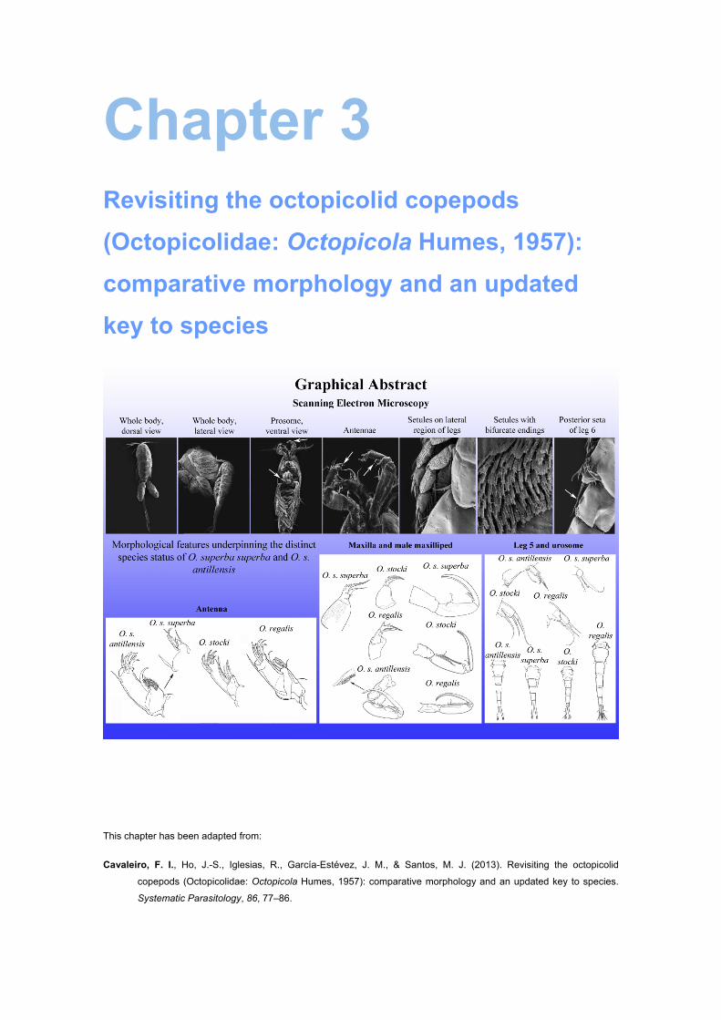
Chapter 3 Revisiting the octopicolid copepods
(Octopicolidae: Octopicola Humes, 1957):
comparative morphology and an updated
key to species
This chapter has been adapted from:
Cavaleiro, F. I., Ho, J.-S., Iglesias, R., García-Estévez, J. M., & Santos, M. J. (2013). Revisiting the octopicolid
copepods (Octopicolidae: Octopicola Humes, 1957): comparative morphology and an updated key to species.
Systematic Parasitology, 86, 77–86.

36 FCUP
Chapter 3. A review of the octopicolid copepods (Octopicolidae: Octopicola)
FCUP
Chapter 3. A review of the octopicolid copepods (Octopicolidae: Octopicola)
37
3.1. Abstract
A review of the present state of knowledge on the octopicolid copepods (Octopicolidae:
Octopicola Humes, 1957) is presented. Characteristic morphological features are
illustrated with scanning electron micrographs of Octopicola superba superba Humes,
1957. Comparative morphology analysis led to the conclusion that there is sufficient
evidence to justify raising the two subspecies of O. superba to full species rank. A new
identification key for the four species of Octopicola Humes, 1957, i.e. O. superba
Humes, 1957, O. antillensis Stock, Humes & Gooding, 1963, O. stocki Humes, 1963
and O. regalis Humes, 1974, is proposed after evaluation of the morphological
characters which vary more markedly between them. Among other characters, these
species differ in the ornamentation of the third antennal segment, maxilla and male
maxilliped. They are further distinguished by a combination of several character states
concerning the fifth pedigerous somite.

38 FCUP
Chapter 3. A review of the octopicolid copepods (Octopicolidae: Octopicola)
FCUP
Chapter 3. A review of the octopicolid copepods (Octopicolidae: Octopicola)
39
3.2. Introduction
The octopicolids (Octopicolidae: Octopicola Humes, 1957) are tiny, mobile,
poecilostomatoid copepods. As suggested by their name, they live in exclusive
association with octopuses (Cephalopoda: Octopodidae) (see Humes, 1974; Hochberg,
1983; Boxshall & Halsey, 2004). Their bodies are cyclopiform, meaning that the
general body shape closely resembles that of Cyclops spp. Although octopicolids seem
to prefer the mantle cavity of their hosts (Hochberg, 1983), different sites on the body
surface may be found infected (Cavaleiro & Santos, In Press; Humes & Stock, 1973);
they can be also found amongst the eggs (Humes, 1957, 1974; Humes & Stock, 1973;
Hochberg, 1983). While in the mantle cavity, octopicolids either move about freely over
the gills or attach to the arterial stems beneath the branchial leaflets (Hochberg, 1983).
At least one species of octopicolid copepod has been observed to exhibit a circadian
rhythm in site occupation, inhabiting the mantle cavity of the host during daytime and
moving out on the surface of the body after dark (Deboutteville et al., 1957).
The genus Octopicola Humes, 1957 was erected by Humes (1957), but it was
only 39 years later, in 1996, that a new family, named Octopicolidae Humes &
Boxshall, 1996 has been established to accommodate it (Humes & Boxshall, 1996).
These authors argued that the octopicolids are the only copepods in the
lichomolgoidean complex of families (following Humes & Boxshall, 1996) to retain the
primitive six-segmented condition of the female urosome and that they should therefore
be included in a separate family. Currently, three species of octopicolid copepods are
recognised: Octopicola superba Humes, 1957, O. stocki Humes, 1963, and O. regalis
Humes, 1974; the former comprised of two subspecies, O. s. superba, endemic to
European waters and corresponding to the species described by Humes (1957); and
O. s. antillensis Stock, Humes & Gooding, 1963, endemic to West Indian waters
(Humes, 1957, 1963, 1974; Stock et al., 1963; Humes & Stock, 1973).
The key to the species and subspecies of the genus Octopicola of Humes &
Stock (1973) is based on the morphological variability exhibited by the females and
males of O. s. superba, O. s. antillensis and O. stocki but does not include O. regalis,
described one year after its publication. Therefore, it needs a revision to include all
species described to date. Furthermore, a close examination of the morphology of O. s.
superba and O. s. antillensis suggested that these subspecies exhibit sufficient
differences to be raised to full species rank.
40 FCUP
Chapter 3. A review of the octopicolid copepods (Octopicolidae: Octopicola)
The present study aimed: (i) illustration of the characteristic morphological
features of the octopicolid copepods using scanning electron microscopy examination
of O. s. superba, (ii) discussion of the morphological evidence which justifies raising the
two subspecies of O. superba to full species rank; and (iii) elaboration of a key for
identification of the species of Octopicola.
3.3. Materials and Methods
Collection and identification of the octopicolid copepods
Unlike other parasitic copepods, whose presence on host tissues is readily detected by
naked eye, octopicolids will most certainly go unnoticed by the casual observer.
Moreover, their relatively small size, associated with their sometimes transparent
appearance, render it unlikely that an observer would be able to recognize them with
ease, without using appropriate instrumentation. Indeed, the detection of these
parasites can be problematic even under a stereomicroscope. Not infrequently,
infected host tissues are covered with a dark black ink expelled by the octopus.
Additionally, the large size of many species of octopuses renders them difficult to
handle, making it almost impossible to observe the parasites in situ, under a
stereomicroscope. Due to all these constraints, a particular method is to be followed
while examining octopuses for octopicolid copepods. In this study, O. s. superba was
used to illustrate characteristic morphological features of the octopicolid copepods. The
parasite was isolated from the body of naturally infected specimens of the common
octopus, Octopus vulgaris Cuvier, after washing the body, internal surface of the
mantle musculature and external surface of the organs with saline solution (3.5%); O.
s. superba was isolated from the sediment under a stereomicroscope. The specimens
were cleaned of mucus and other debris in saline solution (3.5%) and fixed in 70%
ethanol. Later, they were cleared in a drop of 90% lactic acid (Humes & Gooding,
1964) and identified to the subspecies level (Humes & Stock, 1973) under a compound
microscope (Carl Zeiss Axiophot Photomicroscope) at magnifications of up to 1000×.
Scanning electron microscopy of O. s. superba
A few specimens of O. s. superba were selected for study by scanning electron
microscopy (SEM). Their preparation for SEM examination included fixation in 2.5%
glutaraldehyde in 0.1 M sodium cacodylate buffer pH 7.2 for about 2–3 h and in 1%
FCUP
Chapter 3. A review of the octopicolid copepods (Octopicolidae: Octopicola)
41
osmium tetroxide for 30 min. Afterwards, the specimens were dehydrated through a
graded ethanol series, i.e., 50, 70, 80, 90% and absolute (2×), remaining immersed for
about 20–30 min in each of these ethanol solutions. The copepods were then
transferred to 25, 50, 75% (in ethanol) and pure isoamyl-acetate (2×15 min in each
solution), critical point-dried in CO2, mounted on stubs, and coated with a 15 nm layer
of gold using an automated sputter coater (Emitech K550X). They were observed
under a scanning electron microscope (Philips XL30) at an accelerating voltage of 5–
10 kV.
Family Octopicolidae Humes & Boxshall, 1996
Genus Octopicola Humes, 1957
Diagnosis
Body elongate and slender in both sexes; prosome formed by cephalosome and 4 free,
subequal pedigers; urosome 6-segmented in both sexes, with genital and first
abdominal somites separate; first pediger not fused with cephalosome. Caudal ramus
long, narrow, with 6 setae (4 terminal, 1 subterminal, 1 on external margin) and several
minute setules. Antennule 7-segmented (armature formula 4, 13, 6, 3, 4 + 1
aesthetasc, 2 + 1 aesthetasc, 7 + 1 aesthetasc), long, shorter than prosome, with
sclerotisation between second and third segments (especially ventrally) suggesting an
intercalary piece (incomplete segment). Rostrum triangular, slightly pointed, bearing
setules of variable length. Antenna uniramous, 4-segmented, with coxa and basis
fused to form coxobasis, armed with recurved spines on third and fourth segments.
Labrum with 2 elongate posteroventral lobes, delimited medially by deep incision of
posterior margin. Mandible strongly sclerotised with pointed tooth and wide, pointed
lobe, bearing row of spinules along inner margin. Paragnath a small unornamented
lobe at region of labrum. Maxillule a small lobe, bearing 3 setae, one much smaller.
Maxilla 2-segmented; first segment largest and tooth-like; second segment slender,
culminating in tapered process with graduated teeth along one side. Maxilliped in
females 3-segmented: first segment elongate, unornamented, second segment
elongate, slightly sinuous, bearing 2 small, naked setae and distal patch of small
spinules, third segment small, bearing terminally 3 claw-like processes; in males 4-
segmented (assuming that proximal part of claw represents fourth segment): first
segment unornamented, second segment armed with 2 unequal inner setae and
numerous spinules arranged in rows, third segment very small and unornamented, last
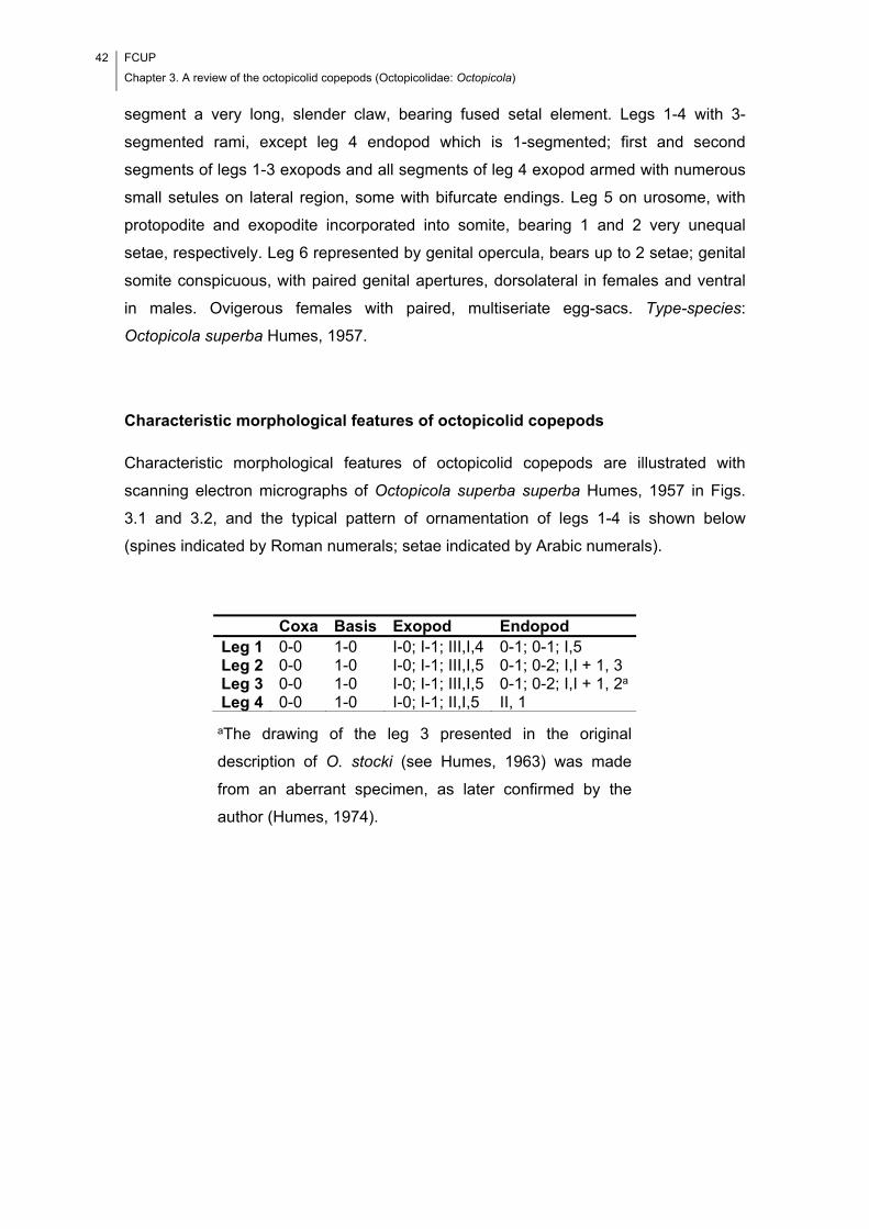
42 FCUP
Chapter 3. A review of the octopicolid copepods (Octopicolidae: Octopicola)
segment a very long, slender claw, bearing fused setal element. Legs 1-4 with 3-
segmented rami, except leg 4 endopod which is 1-segmented; first and second
segments of legs 1-3 exopods and all segments of leg 4 exopod armed with numerous
small setules on lateral region, some with bifurcate endings. Leg 5 on urosome, with
protopodite and exopodite incorporated into somite, bearing 1 and 2 very unequal
setae, respectively. Leg 6 represented by genital opercula, bears up to 2 setae; genital
somite conspicuous, with paired genital apertures, dorsolateral in females and ventral
in males. Ovigerous females with paired, multiseriate egg-sacs. Type-species:
Octopicola superba Humes, 1957.
Characteristic morphological features of octopicolid copepods
Characteristic morphological features of octopicolid copepods are illustrated with
scanning electron micrographs of Octopicola superba superba Humes, 1957 in Figs.
3.1 and 3.2, and the typical pattern of ornamentation of legs 1-4 is shown below
(spines indicated by Roman numerals; setae indicated by Arabic numerals).
aThe drawing of the leg 3 presented in the original
description of O. stocki (see Humes, 1963) was made
from an aberrant specimen, as later confirmed by the
author (Humes, 1974).
Coxa Basis Exopod Endopod Leg 1 0-0 1-0 I-0; I-1; III,I,4 0-1; 0-1; I,5 Leg 2 0-0 1-0 I-0; I-1; III,I,5 0-1; 0-2; I,I + 1, 3 Leg 3 0-0 1-0 I-0; I-1; III,I,5 0-1; 0-2; I,I + 1, 2a Leg 4 0-0 1-0 I-0; I-1; II,I,5 II, 1
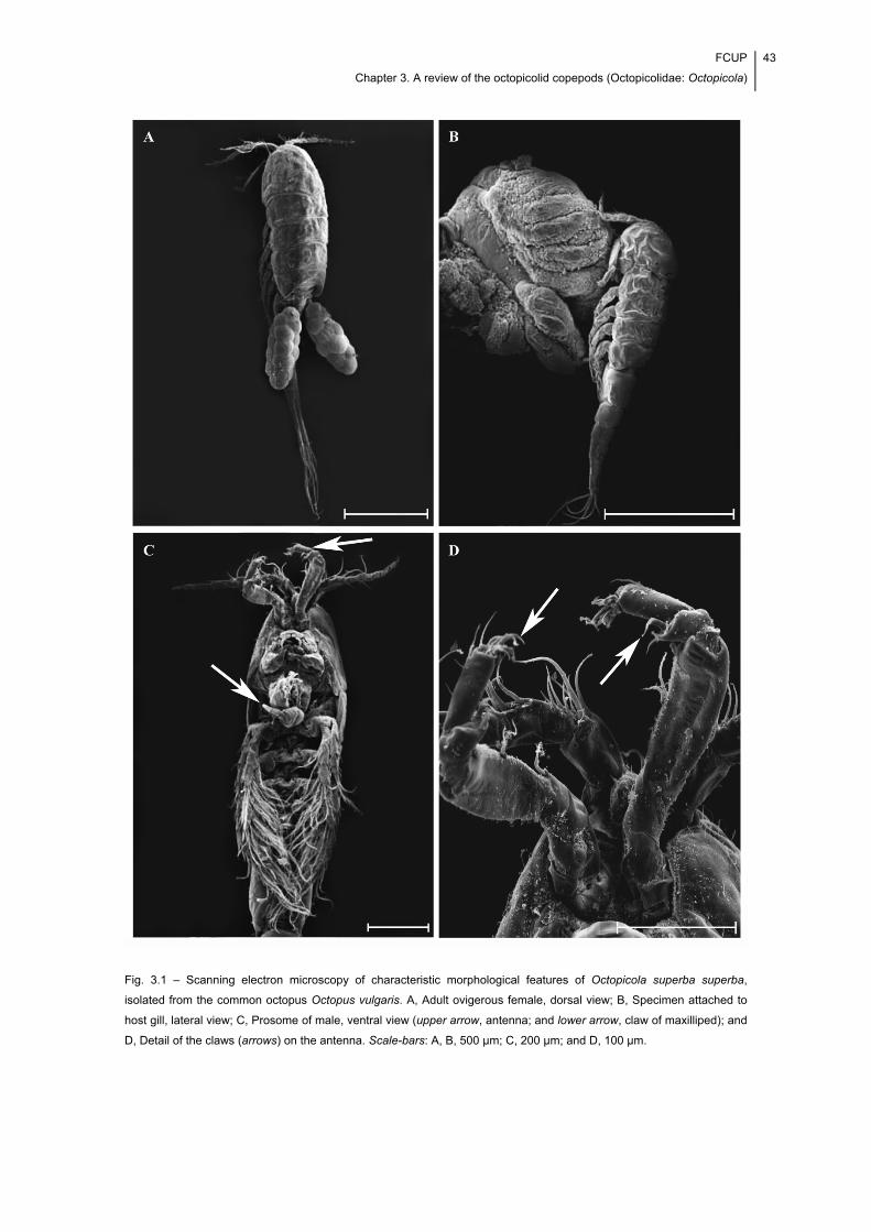
FCUP
Chapter 3. A review of the octopicolid copepods (Octopicolidae: Octopicola)
43
Fig. 3.1 – Scanning electron microscopy of characteristic morphological features of Octopicola superba superba,
isolated from the common octopus Octopus vulgaris. A, Adult ovigerous female, dorsal view; B, Specimen attached to
host gill, lateral view; C, Prosome of male, ventral view (upper arrow, antenna; and lower arrow, claw of maxilliped); and
D, Detail of the claws (arrows) on the antenna. Scale-bars: A, B, 500 μm; C, 200 μm; and D, 100 μm.
44 FCUP
Chapter 3. A review of the octopicolid copepods (Octopicolidae: Octopicola)
Fig. 3.2 – Scanning electron microscopy of characteristic morphological features of Octopicola superba superba,
isolated from the common octopus, Octopus vulgaris. A, Detail of the ornamentation seen on the lateral region of legs 3
(upper leg) and 4 (lower leg); B, Detail of the setules with bifurcate endings on the lateral region of the legs; and C,
Detail of the longer of the two setae of leg 6 (arrow) on the posterior lateral corner of the genital somite. Scale-bars: A,
50 μm; B, 10 μm; and C, 100 μm.
FCUP
Chapter 3. A review of the octopicolid copepods (Octopicolidae: Octopicola)
45
Species and subspecies distinction
Tables 3.1 and 3.2 summarise the relevant information on species biology, ecology and
morphometry.
Table 3.1 – Host and distribution data for the known taxa of octopicolid copepods.
aAnd not on Octopus (Tritaxeopus) cornutus Owen, 1881 as reported in the species description (see Boxshall & Halsey, 2004).
Species O. s. superba
Humes, 1957
O. s. antillensis
Stock, Humes & Gooding, 1963
O. stocki
Humes, 1963
O. regalis
Humes, 1974
Host Octopus vulgaris Cuvier
Octopus briareus Robson; O. vulgaris Cuvier
Octopus cyaneus
Graya
Octopus cyaneus Gray
Colour (live, under light microscope)
White Transparent to opaque
Opaque
Geographical distribution (source)
Off Mediterranean coast of France, Atlantic Ocean (Humes, 1957); off Channel coast of France, Atlantic Ocean (Bocquet & Stock, 1960); off Portuguese coast, Atlantic Ocean (present study)
Off Florida, Atlantic Ocean (Humes & Stock, 1973; O. briareus); off West Indies, Atlantic Ocean (Stock et al., 1963; O. vulgaris); off Florida, Atlantic Ocean (Humes & Stock, 1973; O. vulgaris)
Off Madagascar Island, Indian Ocean (Humes, 1963; unidentified species of octopus); off Madagascar Island, Indian Ocean (Humes, 1963)
Off New Caledonia Islands and Eniwetok Atoll, Marshall Islands, Pacific Ocean (Humes, 1974)
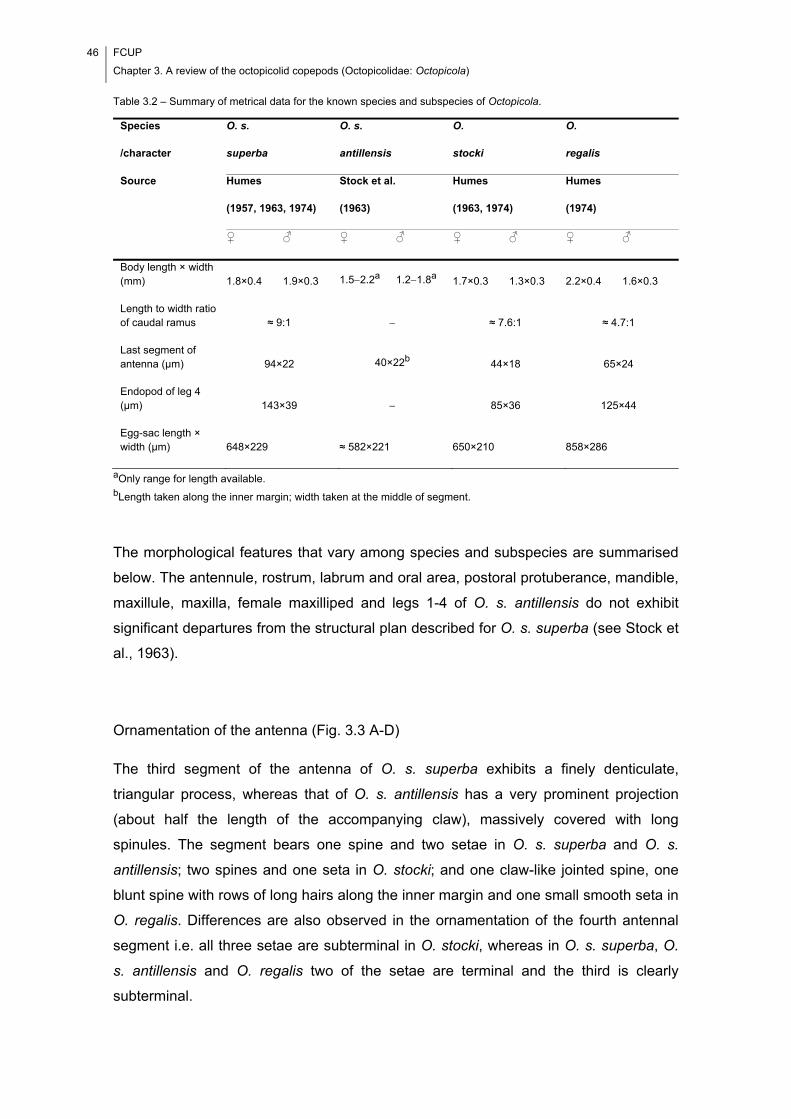
46 FCUP
Chapter 3. A review of the octopicolid copepods (Octopicolidae: Octopicola)
Table 3.2 – Summary of metrical data for the known species and subspecies of Octopicola.
aOnly range for length available. bLength taken along the inner margin; width taken at the middle of segment.
The morphological features that vary among species and subspecies are summarised
below. The antennule, rostrum, labrum and oral area, postoral protuberance, mandible,
maxillule, maxilla, female maxilliped and legs 1-4 of O. s. antillensis do not exhibit
significant departures from the structural plan described for O. s. superba (see Stock et
al., 1963).
Ornamentation of the antenna (Fig. 3.3 A-D)
The third segment of the antenna of O. s. superba exhibits a finely denticulate,
triangular process, whereas that of O. s. antillensis has a very prominent projection
(about half the length of the accompanying claw), massively covered with long
spinules. The segment bears one spine and two setae in O. s. superba and O. s.
antillensis; two spines and one seta in O. stocki; and one claw-like jointed spine, one
blunt spine with rows of long hairs along the inner margin and one small smooth seta in
O. regalis. Differences are also observed in the ornamentation of the fourth antennal
segment i.e. all three setae are subterminal in O. stocki, whereas in O. s. superba, O.
s. antillensis and O. regalis two of the setae are terminal and the third is clearly
subterminal.
Species
/character
O. s.
superba
O. s.
antillensis
O.
stocki
O.
regalis
Source Humes
(1957, 1963, 1974)
Stock et al.
(1963)
Humes
(1963, 1974)
Humes
(1974)
♀ ♂ ♀ ♂ ♀ ♂ ♀ ♂
Body length × width (mm) 1.8×0.4 1.9×0.3 1.52.2a 1.21.8a 1.7×0.3 1.3×0.3 2.2×0.4 1.6×0.3
Length to width ratio of caudal ramus ≈ 9:1 ≈ 7.6:1 ≈ 4.7:1
Last segment of antenna (μm) 94×22 40×22b 44×18 65×24
Endopod of leg 4 (μm) 143×39 85×36 125×44
Egg-sac length × width (μm) 648×229 ≈ 582×221 650×210 858×286
FCUP
Chapter 3. A review of the octopicolid copepods (Octopicolidae: Octopicola)
47
Ornamentation of the maxilla (Fig. 3.3 E-G)
The first segment of the maxilla of O. s. superba has numerous minute setules
distributed over almost its entire surface and one distal, small, smooth seta, whereas
that of O. stocki and O. regalis is unornamented. The second segment of the maxilla
possesses a spine-like seta with spinules along one side in O. s. superba and O.
regalis but not in O. stocki. The first of the graduated teeth at the tapered process of
the second segment is tooth-like in O. s. superba and O. regalis but not in O. stocki.
Ornamentation of the male maxilliped (Fig. 3.3 H-L)
The second segment of the maxilliped in the males of O. s. superba, O. s. antillensis
and O. stocki bears two rows of spinules along the inner surface whereas three rows of
spinules are present on the corresponding region of the maxilliped in the male of O.
regalis. Groups of spinules connecting the rows of spinules are present in O. s.
antillensis but were not reported for O. s. superba, O. stocki and O. regalis (see
Humes, 1957, 1963, 1974). A conspicuous hyaline process is seen at the base of the
claw of the maxilliped of the male of O. stocki, whereas a very small prominence is
seen at the corresponding region of the maxilliped in the male of O. s. superba. The
hyaline membrane near the tip of the claw (convex surface) is bluntly pointed and
smooth in O. s. superba and prolonged into a small element in O. s. antillensis, O.
stocki and O. regalis. This element is armed with a group of small spinules on its base
in O. s. antillensis.
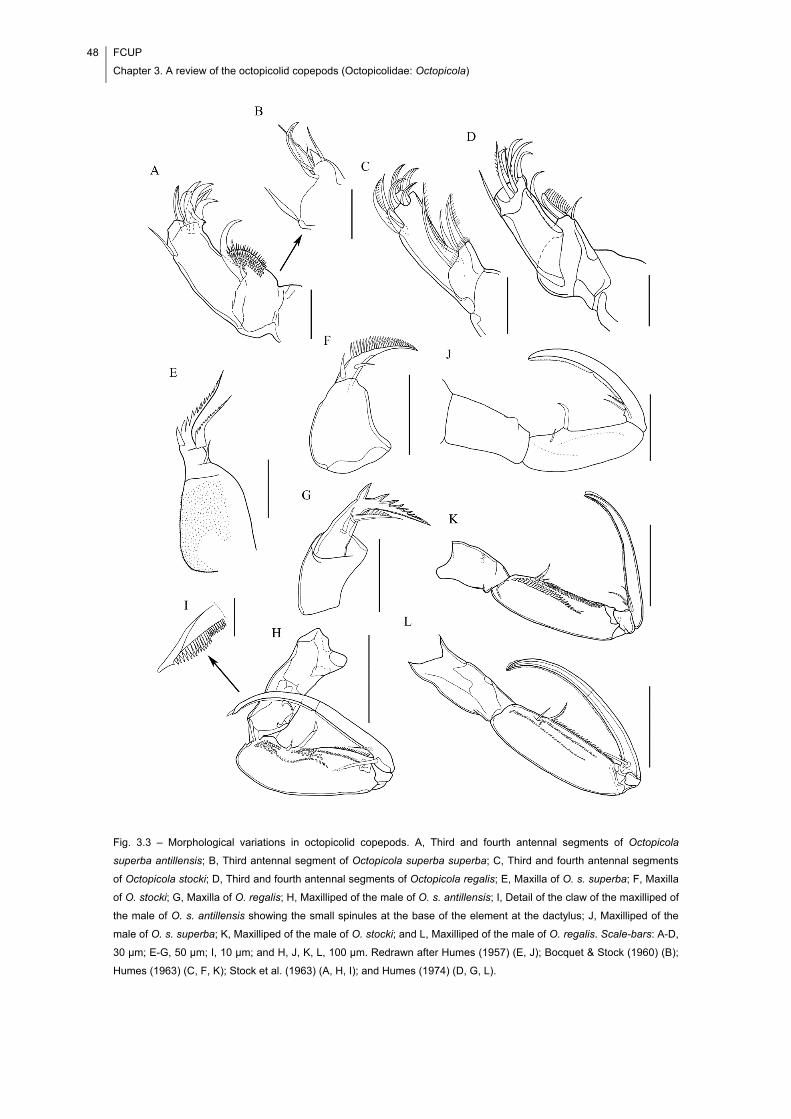
48 FCUP
Chapter 3. A review of the octopicolid copepods (Octopicolidae: Octopicola)
Fig. 3.3 – Morphological variations in octopicolid copepods. A, Third and fourth antennal segments of Octopicola
superba antillensis; B, Third antennal segment of Octopicola superba superba; C, Third and fourth antennal segments
of Octopicola stocki; D, Third and fourth antennal segments of Octopicola regalis; E, Maxilla of O. s. superba; F, Maxilla
of O. stocki; G, Maxilla of O. regalis; H, Maxilliped of the male of O. s. antillensis; I, Detail of the claw of the maxilliped of
the male of O. s. antillensis showing the small spinules at the base of the element at the dactylus; J, Maxilliped of the
male of O. s. superba; K, Maxilliped of the male of O. stocki; and L, Maxilliped of the male of O. regalis. Scale-bars: A-D,
30 μm; E-G, 50 μm; I, 10 μm; and H, J, K, L, 100 μm. Redrawn after Humes (1957) (E, J); Bocquet & Stock (1960) (B);
Humes (1963) (C, F, K); Stock et al. (1963) (A, H, I); and Humes (1974) (D, G, L).
FCUP
Chapter 3. A review of the octopicolid copepods (Octopicolidae: Octopicola)
49
Leg 5 and surrounding area of fifth pedigerous somite (Fig. 3.4 A-D)
The shape of the free segment of leg 5 varies from subquadrate in O. s. superba to
subrectangular in O. stocki (length to width ratio 1.8:1) and O. regalis (length to width
ratio 2.4:1). The larger of the two setae on this segment exhibits a swollen base in O.
regalis. The seta dorsal to the free segment is inserted into a lobe in O. stocki and O.
regalis, whereas in O. s. superba and O. s. antillensis it arises directly from the body
wall. Tergal plates were reported for the fifth pedigerous somite of O. s. antillensis
exclusively (Stock et al., 1963).
Relative length of setae of leg 6 (Fig. 3.4 E-H)
The two setae of leg 6 are short in O. s. antillensis, O. stocki and O. regalis, the most
posterior seta (the one on the posterolateral area of the genital somite) extends only
slightly beyond the posterior margin of its own somite. In O. s. superba the most
posterior seta of leg 6 is long, reaching to the posterior margin of the next urosomal
somite or beyond.
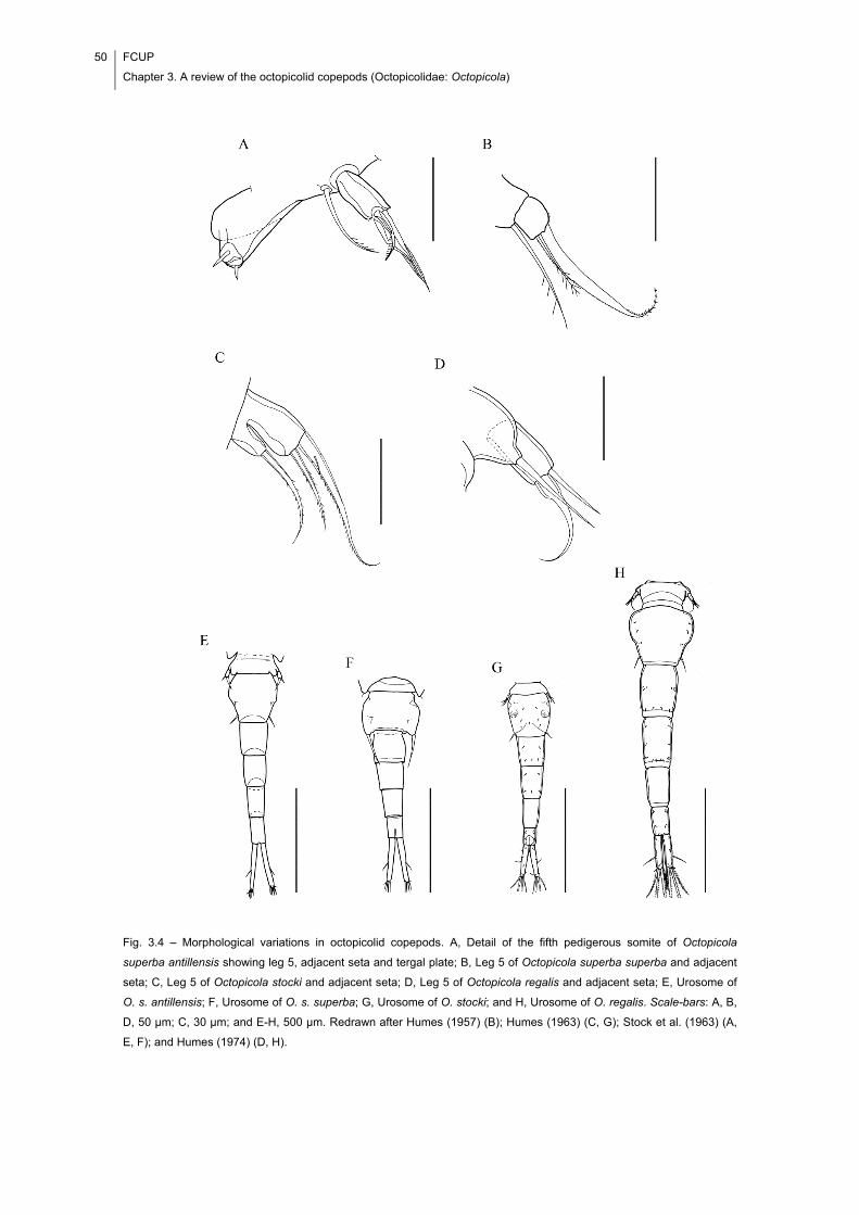
50 FCUP
Chapter 3. A review of the octopicolid copepods (Octopicolidae: Octopicola)
Fig. 3.4 – Morphological variations in octopicolid copepods. A, Detail of the fifth pedigerous somite of Octopicola
superba antillensis showing leg 5, adjacent seta and tergal plate; B, Leg 5 of Octopicola superba superba and adjacent
seta; C, Leg 5 of Octopicola stocki and adjacent seta; D, Leg 5 of Octopicola regalis and adjacent seta; E, Urosome of
O. s. antillensis; F, Urosome of O. s. superba; G, Urosome of O. stocki; and H, Urosome of O. regalis. Scale-bars: A, B,
D, 50 μm; C, 30 μm; and E-H, 500 μm. Redrawn after Humes (1957) (B); Humes (1963) (C, G); Stock et al. (1963) (A,
E, F); and Humes (1974) (D, H).
FCUP
Chapter 3. A review of the octopicolid copepods (Octopicolidae: Octopicola)
51
3.4. Discussion
The studies on the octopicolid copepods are scarce and date back to the past century.
This has motivated the present work, in which scanning electron microscopy was used
for the first time to illustrate relevant aspects of their morphology.
While comparing between the descriptions in the literature (Humes, 1957;
Bocquet & Stock, 1960; Humes, 1963; Stock et al., 1963; Humes, 1974), we found
convincing morphological evidence to justify raising the two subspecies of O. superba
to full species status. At the time of description of O. s. antillensis, only one other
octopicolid copepod, i.e. O. superba, had been described. The latter had also been
isolated from O. vulgaris and the specimens isolated from the West Indian octopuses
exhibited only small differences from the description of O. superba; this has led Stock
et al. (1963) to consider that their specimens represent a new subspecies and not a
new species. A critical analysis of the descriptions in the literature indicates that O.
stocki and O. regalis also exhibit small differences from O. s. superba and O. s.
antillensis, concerning the ornamentation of certain segments of given appendages;
nonetheless, they are regarded as different species.
The best features underpinning the distinct species status of O. s. superba and
O. s. antillensis are the characteristics of the antenna. This appendage exhibits the
greatest morphological variability in octopicolid copepods, which conforms with what
has been said before, that the antenna of the copepods in the lichomolgoidean
complex of families (following Humes & Boxshall, 1996) is particularly vulnerable to
morphological adaptations to the parasitic mode of life, i.e. changes that ensure an
effective attachment to the host (see e.g. Ho, 1984). Furthermore, all species can be
distinguished from one another by the specific features of the third antennal segment,
as illustrated in this work (see Fig. 3.3 A-D). In recognising the existence of four
different species of octopicolid copepods (i.e., O. superba, O. antillensis, O. stocki and
O. regalis) it must be said that two other appendages, i.e. the maxilla and the male
maxilliped, are also useful for identifying octopicolid copepods to the species level.
However, the differences between species in relation to the morphology of
these appendages are not as conspicuous as those associated with the morphology of
the antenna. Furthermore, the species differ from one another in the presence of
setules on the first segment of the maxilla (ornamented with numerous small setules in
O. superba and O. antillensis vs unornamented in O. stocki and O. regalis) and the
ornamentation of the second segment, i.e. the number and the type of setae: one
52 FCUP
Chapter 3. A review of the octopicolid copepods (Octopicolidae: Octopicola)
spine-like seta in O. superba, one smooth seta plus one spine-like seta in O. antillensis
and O. regalis, and two smooth setae in O. stocki. In the specific case of the male
maxilliped, it was found that certain features are exclusively observed in particular
species: O. antillensis is unique in bearing groups of spinules connecting the two rows
of spinules on the second segment and small spinules at the base of the element at the
dactylus; O. regalis is unique in bearing three rows of spinules on the second segment;
and O. stocki is unique in bearing a conspicuous hyaline process near the base of the
seta on the basal concave margin of the claw. However, the males of both O. superba
and O. stocki exhibit two rows of spinules on the second segment of the maxilliped, the
difference being in the degree of development of the spinules. Moreover, the latter
appear to be larger in O. stocki (compare Fig. 3.3 J, K). Octopicola superba possesses
a very small prominence at the base of the claw, which perhaps represents a hyaline
process, as in O. stocki. The latter species is also unique in that the outermost seta on
the endopod segment of leg 4 exhibits sexual dimorphism. This seta is spiniform and
armed with short lateral spinules in females, and distinctly spiniform, sinuous, and
armed with prominent lateral spinules in males.
As for the remaining species, O. antillensis is unique in bearing tergal plates on
the fifth pedigerous somite, O. superba in having a subquadrate leg 5 and a very long
seta representing leg 6 on the posterolateral area of the genital somite, and O. regalis
in possessing larger body dimensions. Further species differences are seen in the
combination of the following character states concerning the fifth pedigerous somite: (i)
free segment of leg 5 subquadrate/subrectangular; (ii) larger of the two setae on the
free segment of leg 5 with swollen base; (iii) seta dorsal and adjacent to the free
segment of leg 5 arising directly from the body wall/arising from a distinct lobe; and (iv)
presence of tergal plates.
It is worth noting that despite the large number of species of octopuses known
to date, little attention has so far been devoted to their parasites. Therefore, it is highly
likely that more octopicolid copepods remain to be discovered.
Key to the species of Octopicola
A critical analysis of the literature (Humes, 1957; Bocquet & Stock, 1960; Humes,
1963, 1974; Stock et al., 1963) resulted in identification of the morphological features
that vary among species and subspecies. The existence of sufficient morphological
FCUP
Chapter 3. A review of the octopicolid copepods (Octopicolidae: Octopicola)
53
evidence to justify raising the two subspecies of O. superba to full species rank was
evaluated; this resulted in the elaboration of the identification key provided below.
Females
1 Seta dorsal and adjacent to free segment of leg 5 arises directly from body wall
…………………………………………………………………………………...………….. 2
– Seta dorsal and adjacent to free segment of leg 5 arises from distinct lobe
…………………………………………………………...………………………..………… 3
2 Third antennal segment with finely denticulated triangular process in addition to a
spine and two setae; fifth pedigerous somite without tergal plates; most posterior
seta on leg 6 long, reaches to posterior margin of next urosomal somite or
beyond………………………………………………...……………..…………. O. superba
– Third antennal segment with very prominent projection, half the length of
accompanying claw, densely covered with long spinules in addition to a spine and
two setae; fifth pedigerous somite with tergal plates; most posterior seta on leg 6
short, extends only slightly beyond the posterior margin of its own somite
…………………………………………………………………………………. O. antillensis
3 Third antennal segment bears two spines and one seta; second segment of maxilla
armed with two smooth setae; first of graduated teeth on tapered process of second
segment of maxilla not tooth-like.…………...….....……………………….…… O. stocki
– Third antennal segment bears a claw-like jointed spine, a blunt spine with rows of
long hairs along inner margin and a small naked seta; second segment of maxilla
armed with one smooth seta plus one spine-like seta with spinules along one side;
first of graduated teeth on tapered process of second segment of maxilla tooth-
like………………………………………………………………………………... O. regalis
54 FCUP
Chapter 3. A review of the octopicolid copepods (Octopicolidae: Octopicola)
Males
1 Seta dorsal and adjacent to free segment of leg 5 arises directly from body
wall…………………………………………………………………………………………. 2
– Seta dorsal and adjacent to free segment of leg 5 arises from distinct lobe
………………………………………………………………………………..……….……. 3
2 Third antennal segment with finely denticulated triangular process in addition to a
spine and two setae; inner surface of second segment of maxilliped without groups
of spinules connecting rows of spinules; hyaline membrane near tip of claw (convex
surface) bluntly pointed and smooth…………………………………………. O. superba
– Third antennal segment with very prominent projection, half as long as accompanying
claw, densely covered with long spinules in addition to a spine and two setae; inner
surface of second segment of maxilliped with groups of spinules connecting rows of
spinules; hyaline membrane near tip of claw (convex surface) prolonged into a small
element with small spinules at its base………………………..………….. O. antillensis
3 Third antennal segment bears two spines and one seta; inner surface of second
segment of maxilliped bears two rows of spinules; conspicuous hyaline process
present at base of claw of maxilliped; outermost seta on endopod of leg 4 distinctly
spiniform, sinuous and armed with prominent lateral spinules…..………..… O. stocki
– Third antennal segment bears a claw-like jointed spine, a blunt spine with rows of
long hairs along inner margin, and a small naked seta; inner surface of second
segment of maxilliped bears three rows of spinules; hyaline process at base of claw
of maxilliped absent; outermost seta on endopod of leg 4 not spiniform, not sinuous
and unarmed……………………………………………………………….…..… O. regalis
3.5. Acknowledgements
Completion of this manuscript was aided by grants from the Portuguese Foundation for
Science and Technology and European Social Fund (to FIC) (SFRH/BD/65258/2009)
and the Paramitas Foundation (to JSH). This work was partially funded by the Project
AQUAIMPROV (reference NORTE-07-0124-FEDER-000038), co-financed by the North
Portugal Regional Operational Programme (ON.2 – O Novo Norte), under the National
Strategic Reference Framework (NSRF), through the European Regional Development
Fund (ERDF); and the European Regional Development Fund (ERDF) through the

FCUP
Chapter 3. A review of the octopicolid copepods (Octopicolidae: Octopicola)
55
COMPETE – Operational Competitiveness Programme and national funds through
FCT – Foundation for Science and Technology, under the projects PEst-
C/MAR/LA0015/2013, DIRDAMyx FCOMP-01-0124-FEDER-020726 (FCT –
PTDC/MAR/116838/2010). We are also grateful to J. Méndez and I. Pazos (CACTI,
University of Vigo) for their technical assistance in electron microscopy study.

56 FCUP
Chapter 3. A review of the octopicolid copepods (Octopicolidae: Octopicola)
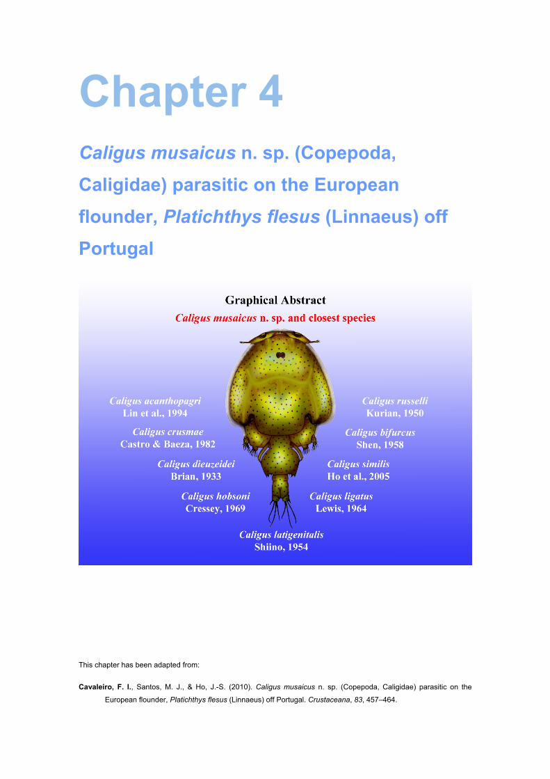
Chapter 4 Caligus musaicus n. sp. (Copepoda,
Caligidae) parasitic on the European
flounder, Platichthys flesus (Linnaeus) off
Portugal
This chapter has been adapted from:
Cavaleiro, F. I., Santos, M. J., & Ho, J.-S. (2010). Caligus musaicus n. sp. (Copepoda, Caligidae) parasitic on the
European flounder, Platichthys flesus (Linnaeus) off Portugal. Crustaceana, 83, 457–464.

58 FCUP
Chapter 4. Description of Caligus musaicus n. sp. (Copepoda, Caligidae)

FCUP
Chapter 4. Description of Caligus musaicus n. sp. (Copepoda, Caligidae)
59
4.1. Abstract
A new species of caligid copepod, Caligus musaicus n. sp., is described from the
European flounder, Platichthys flesus (Linnaeus, 1758), caught off the northern coast
of Portugal. The new species is distinguished from its congeners by the combination of
the following character states: (1) equipped with a short abdomen (about 1/3 the length
of the thoracic zone of the cephalothoracic shield); (2) armed with a pair of parallel
pointed tines on the box of the sternal furca; (3) bearing a long element IV (about 3
times as long as the next longest element) at the tip of leg 1 exopod; and (4) with a
slender leg 4 exopod bearing a long outer seta (about 3 times as long as the next
longest seta) at the tip of this ramus.

60 FCUP
Chapter 4. Description of Caligus musaicus n. sp. (Copepoda, Caligidae)
FCUP
Chapter 4. Description of Caligus musaicus n. sp. (Copepoda, Caligidae)
61
4.2. Introduction
The European flounder, Platichthys flesus (Linnaeus, 1758), is a demersal and
catadromous fish with high commercial value. It is widely distributed in coastal and
brackish waters of western Europe, extending from the White Sea to the Mediterranean
and the Black Sea (Froese & Pauly, 2008). So far as we are aware, five species of
parasitic copepods have been reported from this species of flounder. They are:
Acanthochondria cornuta (Müller, 1776) reported by Ho (1970); Caligus elongatus von
Nordmann, 1832 reported by Boxshall (1974a); Lepeophtheirus europaensis Zeddam,
Berrebi, Renaud, Raibaut & Gabrion, 1988 reported by Zeddam et al. (1988); L.
pectoralis (Müller, 1777) reported by Boxshall (1974a); and Lernaeocera branchialis
(Linnaeus, 1767) reported by Polyanski (1955). While the first four species of parasites
were found as adults on the flounder, the last one utilizes the flounder as an
intermediate host; in other words, only the chalimus stages were seen.
While one of us (F.I.C.) was studying the Crustacea infections on the European
flounder occurring off the northern coast of Portugal (Cavaleiro, 2007; Cavaleiro &
Santos, 2007), a species of Caligus was occasionally encountered. It is a rare species
of sea louse, with only 11 specimens being found through the examination of 210 host
fish collected between September 2005 and May 2006. Close studies of this parasite
revealed that it represents a new species. Inasmuch as both sexes are represented in
this rare collection, a full description of the species is given in the following.
4.3. Materials and Methods
Flounders collected at Matosinhos fish harbour (in northern Portugal) were brought
back to the laboratory on the campus of the Universidade do Porto for examination.
The copepod parasites were removed from the fish host and were preserved in 70%
ethanol. Later, the preserved parasites were cleared in 90% lactic acid for about 1 hour
before making dissection in a drop of lactic acid. The dissected body parts and
appendages were examined using a Zeiss Axiophot Photomicroscope at
magnifications of up to 1000×. All drawings were made with the aid of a camera lucida.
Measurements given are the mean followed by the range in parentheses. The
description of the female is given in full but that of the male is confined only to those
parts showing sexual dimorphism.
62 FCUP
Chapter 4. Description of Caligus musaicus n. sp. (Copepoda, Caligidae)
4.4. Results
CALIGIDAE Burmeister, 1835
Caligus Müller, 1785
Caligus musaicus n. sp. (Figs. 4.1-4.3)
Material examined. – Eleven specimens (4 ♀♀; 7 ♂♂) parasitic on the body skin and
the pectoral and ventral fins of the European flounder, Platichthys flesus (Linnaeus,
1758) (Teleostei: Pleuronectidae), landed at Matosinhos fishing port, Portugal (41º10’N
8º42’W), as follows: 1 ♀ from body skin (blind side) of 1 flounder collected on 2
September 2005; 1 ♂ from body skin (ocular side) of 1 flounder collected 2 September
2005; 1 ♂ from body skin (blind side) of 1 flounder collected 2 September 2005; 1 ♂
from pectoral fin (ocular side) of 1 flounder collected 2 September 2005; 1 ♂ from
ventral fin (ocular side) of 1 flounder collected 2 September 2005; 1 ♂ from body skin
(ocular side) of 1 flounder collected 23 May 2006; 2 ♂♂ from body skin (blind side) of 2
flounders collected 23 May 2006; and 3 ♀♀ from body skin (ocular side) of 3 flounders
collected on 23 May 2006.
All isolated parasite specimens were adults, the females being non-ovigerous.
One holotype (USNM 1136866) and an allotype (USNM 1136867) are deposited in the
Smithsonian Institution, Washington, D.C., and two paratypes have been deposited in
the Natural History Museum, London, (Catalogue numbers: NHM 2010.248 and NHM
2010.249). The remaining specimens have been retained in the personal collections of
the authors.
Female. – Body (Fig. 4.1 A) 4.41 (3.75–5.07) mm long, excluding setae on caudal rami.
Cephalothoracic shield roughly triangular in shape, 2.53 (2.08–3.00) × 2.24 (1.94–2.41)
mm, excluding lateral hyaline membrane; frontal plates well developed and carrying
moderately large lunules (width slightly less than 1/3 that of the plates); free margin of
thoracic zone projecting slightly beyond tips of lateral zones; sinuses deep. Fourth
pediger wider than long, 0.26 (0.20–0.32) × 0.64 (0.52–0.85) mm, not separated from
genital complex. Genital complex subcircular, 1.07 (0.75–1.22) × 1.24 (0.85–1.43) mm,
about equally long or slightly longer than thoracic zone of cephalothoracic shield.
Abdomen (Fig. 4.1 B) short, 1-segmented, measuring 0.47 (0.44–0.50) × 0.43 (0.40–
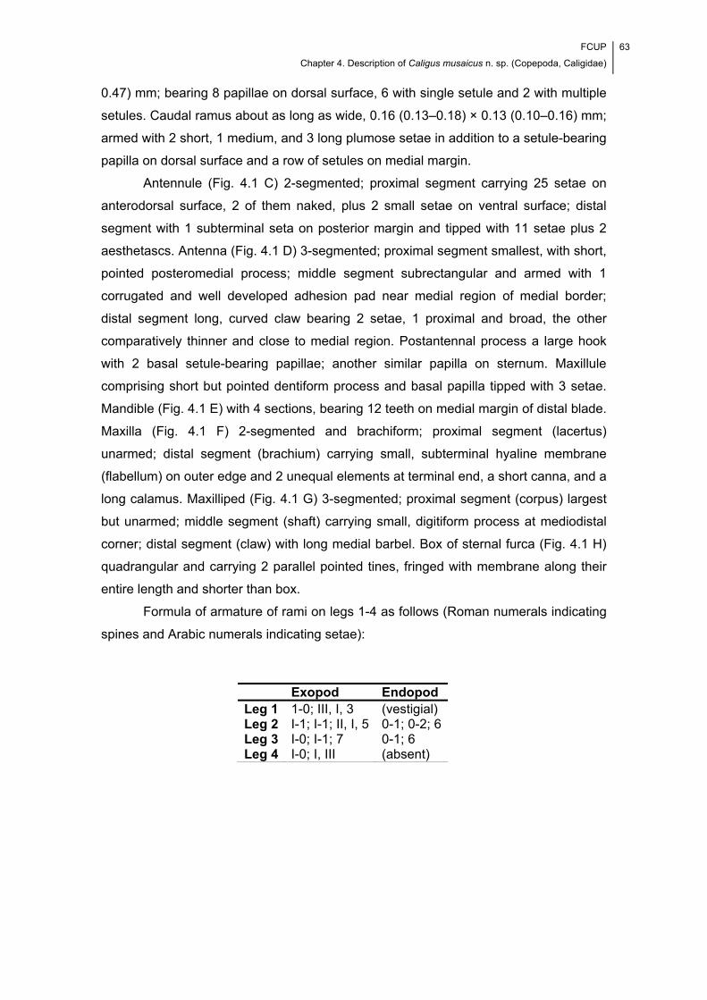
FCUP
Chapter 4. Description of Caligus musaicus n. sp. (Copepoda, Caligidae)
63
0.47) mm; bearing 8 papillae on dorsal surface, 6 with single setule and 2 with multiple
setules. Caudal ramus about as long as wide, 0.16 (0.13–0.18) × 0.13 (0.10–0.16) mm;
armed with 2 short, 1 medium, and 3 long plumose setae in addition to a setule-bearing
papilla on dorsal surface and a row of setules on medial margin.
Antennule (Fig. 4.1 C) 2-segmented; proximal segment carrying 25 setae on
anterodorsal surface, 2 of them naked, plus 2 small setae on ventral surface; distal
segment with 1 subterminal seta on posterior margin and tipped with 11 setae plus 2
aesthetascs. Antenna (Fig. 4.1 D) 3-segmented; proximal segment smallest, with short,
pointed posteromedial process; middle segment subrectangular and armed with 1
corrugated and well developed adhesion pad near medial region of medial border;
distal segment long, curved claw bearing 2 setae, 1 proximal and broad, the other
comparatively thinner and close to medial region. Postantennal process a large hook
with 2 basal setule-bearing papillae; another similar papilla on sternum. Maxillule
comprising short but pointed dentiform process and basal papilla tipped with 3 setae.
Mandible (Fig. 4.1 E) with 4 sections, bearing 12 teeth on medial margin of distal blade.
Maxilla (Fig. 4.1 F) 2-segmented and brachiform; proximal segment (lacertus)
unarmed; distal segment (brachium) carrying small, subterminal hyaline membrane
(flabellum) on outer edge and 2 unequal elements at terminal end, a short canna, and a
long calamus. Maxilliped (Fig. 4.1 G) 3-segmented; proximal segment (corpus) largest
but unarmed; middle segment (shaft) carrying small, digitiform process at mediodistal
corner; distal segment (claw) with long medial barbel. Box of sternal furca (Fig. 4.1 H)
quadrangular and carrying 2 parallel pointed tines, fringed with membrane along their
entire length and shorter than box.
Formula of armature of rami on legs 1-4 as follows (Roman numerals indicating
spines and Arabic numerals indicating setae):
Exopod Endopod Leg 1 1-0; III, I, 3 (vestigial) Leg 2 I-1; I-1; II, I, 5 0-1; 0-2; 6Leg 3 I-0; I-1; 7 0-1; 6 Leg 4 I-0; I, III (absent)
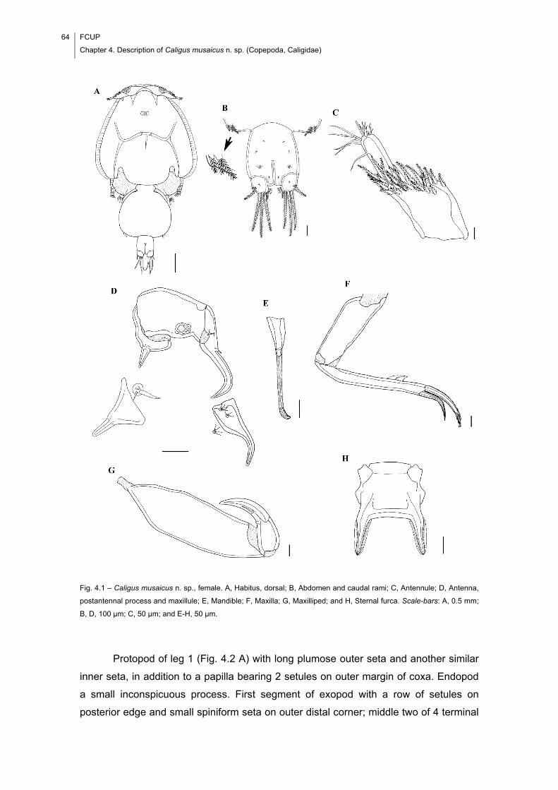
64 FCUP
Chapter 4. Description of Caligus musaicus n. sp. (Copepoda, Caligidae)
Fig. 4.1 – Caligus musaicus n. sp., female. A, Habitus, dorsal; B, Abdomen and caudal rami; C, Antennule; D, Antenna,
postantennal process and maxillule; E, Mandible; F, Maxilla; G, Maxilliped; and H, Sternal furca. Scale-bars: A, 0.5 mm;
B, D, 100 μm; C, 50 μm; and E-H, 50 μm.
Protopod of leg 1 (Fig. 4.2 A) with long plumose outer seta and another similar
inner seta, in addition to a papilla bearing 2 setules on outer margin of coxa. Endopod
a small inconspicuous process. First segment of exopod with a row of setules on
posterior edge and small spiniform seta on outer distal corner; middle two of 4 terminal

FCUP
Chapter 4. Description of Caligus musaicus n. sp. (Copepoda, Caligidae)
65
elements on last segment of exopod with accessory process; element 4 about 3 times
as long as element 2 and bearing setules only on outer margin. Leg 2 (Fig. 4.2 B) coxa
small, with large plumose inner seta on posterior edge and long setule-bearing papilla
on ventral surface. Basis carrying long seta on outer edge in addition to long setule-
bearing papilla on ventral surface, close to base of posterior marginal membrane.
Anterodistal surface of basis and first segment of exopod with large marginal
membrane. Outer margin of 3 endopodal segments with a tuft or row of small setules.
Leg 3 (Fig. 4.2 C) protopod (apron) with small outer and large inner plumose setae, in
addition to an outer and a posterior marginal membrane; ventral surface of protopod
with small setule-bearing papilla at both ends of that membrane; velum well developed
and fringed with marginal setules. Leg 4 (Fig. 4.2 D) protopod large, with plumose seta
at outer distal corner; exopod 2-segmented, due to fusion of distal two segments;
pecten at base of each seta on exopod; outer terminal seta about 3 times as long as
middle one. Leg 5 (Fig. 4.1 B) represented by 2 small papillae on posterolateral corner
of genital complex, one tipped with a single and the other with 2 small, plumose setae.
Fig. 4.2 – Caligus musaicus n. sp., female. A, Leg 1; B, Leg 2; C, Leg 3; and D, Leg 4. Scale-bars: A, D, 50 μm; and B,
C, 100 μm.
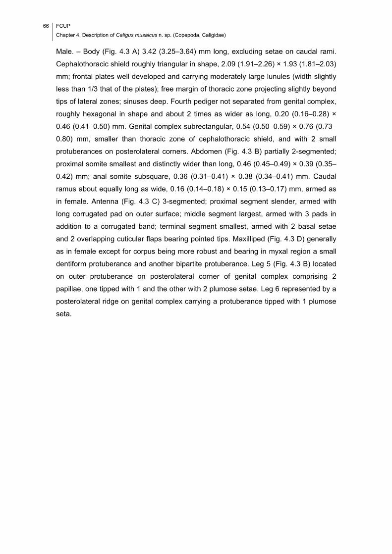
66 FCUP
Chapter 4. Description of Caligus musaicus n. sp. (Copepoda, Caligidae)
Male. – Body (Fig. 4.3 A) 3.42 (3.25–3.64) mm long, excluding setae on caudal rami.
Cephalothoracic shield roughly triangular in shape, 2.09 (1.91–2.26) × 1.93 (1.81–2.03)
mm; frontal plates well developed and carrying moderately large lunules (width slightly
less than 1/3 that of the plates); free margin of thoracic zone projecting slightly beyond
tips of lateral zones; sinuses deep. Fourth pediger not separated from genital complex,
roughly hexagonal in shape and about 2 times as wider as long, 0.20 (0.16–0.28) ×
0.46 (0.41–0.50) mm. Genital complex subrectangular, 0.54 (0.50–0.59) × 0.76 (0.73–
0.80) mm, smaller than thoracic zone of cephalothoracic shield, and with 2 small
protuberances on posterolateral corners. Abdomen (Fig. 4.3 B) partially 2-segmented;
proximal somite smallest and distinctly wider than long, 0.46 (0.45–0.49) × 0.39 (0.35–
0.42) mm; anal somite subsquare, 0.36 (0.31–0.41) × 0.38 (0.34–0.41) mm. Caudal
ramus about equally long as wide, 0.16 (0.14–0.18) × 0.15 (0.13–0.17) mm, armed as
in female. Antenna (Fig. 4.3 C) 3-segmented; proximal segment slender, armed with
long corrugated pad on outer surface; middle segment largest, armed with 3 pads in
addition to a corrugated band; terminal segment smallest, armed with 2 basal setae
and 2 overlapping cuticular flaps bearing pointed tips. Maxilliped (Fig. 4.3 D) generally
as in female except for corpus being more robust and bearing in myxal region a small
dentiform protuberance and another bipartite protuberance. Leg 5 (Fig. 4.3 B) located
on outer protuberance on posterolateral corner of genital complex comprising 2
papillae, one tipped with 1 and the other with 2 plumose setae. Leg 6 represented by a
posterolateral ridge on genital complex carrying a protuberance tipped with 1 plumose
seta.
FCUP
Chapter 4. Description of Caligus musaicus n. sp. (Copepoda, Caligidae)
67
Fig. 4.3 – Caligus musaicus n. sp., male. A, Habitus, dorsal; B, Abdomen and caudal rami; C, Antenna, postantennal
process and maxillule; and D, Maxilliped. Scale-bars: A, 0.5 mm; B, 100 μm; and C, D, 50 μm.
Etymology. – The species name musaicus is the Latin word for mosaic. It alludes to the
species’ resemblance with several of its congeners, in such a way that it reminds of a
genetic mosaic, i.e., an organism whose body consists of a mixture of cells of two or
more different genotypes.

68 FCUP
Chapter 4. Description of Caligus musaicus n. sp. (Copepoda, Caligidae)
4.5. Discussion
Caligus Müller, 1785 is the largest genus of parasitic copepods, containing over 250
species (Ho & Lin, 2004). Since the male remains unknown for many of them,
comparison of our specimens obtained from the flounder with its congeners is
accordingly restricted to the female.
As far as we can find, there are 9 species of Caligus showing closeness to C.
musaicus n. sp. in sharing the following 3 character states with the new species: (1) a
short abdomen (about 1/3 the length of the thoracic zone of the cephalothoracic
shield), (2) bearing a long seta IV (about 3 times as long as the next longest element)
at the tip of leg 1 exopod, and (3) with a slender, 2-segmented leg 4 exopod bearing a
long outer seta (about 3 times as long as the next longest seta) at the tip of this ramus.
Those 9 species of Caligus are: C. acanthopagri Lin et al., 1994; C. crusmae Castro &
Baeza, 1982; C. dieuzeidei Brian, 1933; C. hobsoni Cressey, 1969; C. latigenitalis
Shiino, 1954; C. ligatus Lewis, 1964; C. similis Ho et al., 2005; C. bifurcus Shen, 1958;
and C. russelli Kurian, 1950. Nevertheless, the new species can be distinguished from
the first 7 species mentioned above in the possession of a pair of parallel pointed tines
on the sternal furca (see Fig. 4.1 H). Of the remaining two species, C. bifurcus can be
distinguished from the new species by the structure of the sternal furca (being
narrower), and C. russelli, in the structure of the postantennal process and the corpus
of the maxilliped. Besides, seta IV (the longest element) at the tip of the exopod of leg
1 in the new species is unusual in bearing setules only on one side (outer margin) of
the element.
4.6. Acknowledgements
Completion of this manuscript was aided by a grant from the Portuguese Science and
Technology Foundation (SFRH/BM/23063/2005) to Francisca Cavaleiro and from the
Paramitas Foundation to Ju-Shey Ho. We also thank Professor Vítor Silva from the
Rector’s Office of the Universidade do Porto and Professor Maria Peixoto for their help.
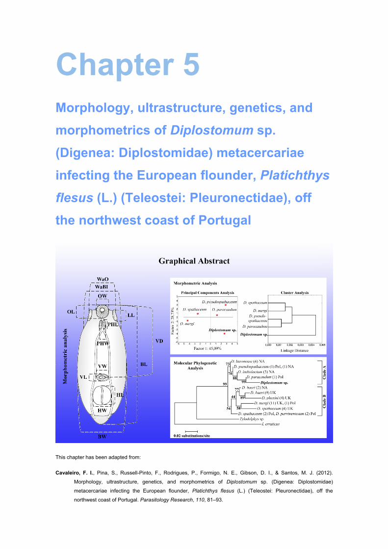
Chapter 5 Morphology, ultrastructure, genetics, and
morphometrics of Diplostomum sp.
(Digenea: Diplostomidae) metacercariae
infecting the European flounder, Platichthys
flesus (L.) (Teleostei: Pleuronectidae), off
the northwest coast of Portugal
This chapter has been adapted from:
Cavaleiro, F. I., Pina, S., Russell-Pinto, F., Rodrigues, P., Formigo, N. E., Gibson, D. I., & Santos, M. J. (2012).
Morphology, ultrastructure, genetics, and morphometrics of Diplostomum sp. (Digenea: Diplostomidae)
metacercariae infecting the European flounder, Platichthys flesus (L.) (Teleostei: Pleuronectidae), off the
northwest coast of Portugal. Parasitology Research, 110, 81–93.

70 FCUP
Chapter 5. Characterization of Diplostomum sp. metacercariae from Platichthys flesus
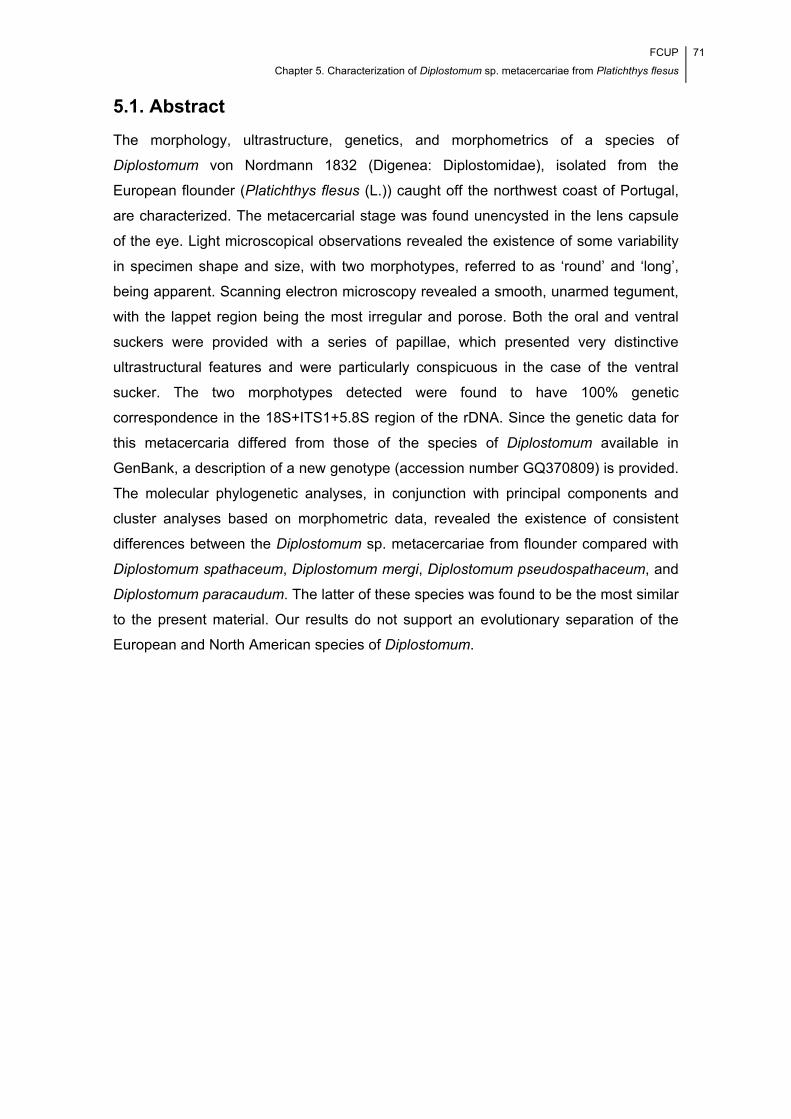
FCUP
Chapter 5. Characterization of Diplostomum sp. metacercariae from Platichthys flesus
71
5.1. Abstract
The morphology, ultrastructure, genetics, and morphometrics of a species of
Diplostomum von Nordmann 1832 (Digenea: Diplostomidae), isolated from the
European flounder (Platichthys flesus (L.)) caught off the northwest coast of Portugal,
are characterized. The metacercarial stage was found unencysted in the lens capsule
of the eye. Light microscopical observations revealed the existence of some variability
in specimen shape and size, with two morphotypes, referred to as ‘round’ and ‘long’,
being apparent. Scanning electron microscopy revealed a smooth, unarmed tegument,
with the lappet region being the most irregular and porose. Both the oral and ventral
suckers were provided with a series of papillae, which presented very distinctive
ultrastructural features and were particularly conspicuous in the case of the ventral
sucker. The two morphotypes detected were found to have 100% genetic
correspondence in the 18S+ITS1+5.8S region of the rDNA. Since the genetic data for
this metacercaria differed from those of the species of Diplostomum available in
GenBank, a description of a new genotype (accession number GQ370809) is provided.
The molecular phylogenetic analyses, in conjunction with principal components and
cluster analyses based on morphometric data, revealed the existence of consistent
differences between the Diplostomum sp. metacercariae from flounder compared with
Diplostomum spathaceum, Diplostomum mergi, Diplostomum pseudospathaceum, and
Diplostomum paracaudum. The latter of these species was found to be the most similar
to the present material. Our results do not support an evolutionary separation of the
European and North American species of Diplostomum.

72 FCUP
Chapter 5. Characterization of Diplostomum sp. metacercariae from Platichthys flesus
FCUP
Chapter 5. Characterization of Diplostomum sp. metacercariae from Platichthys flesus
73
5.2. Introduction
The lens of the eyes in freshwater fishes has frequently been highlighted in the
literature as the site of infection of metacercariae of Diplostomum spathaceum
(Rudolphi, 1819) (Digenea: Diplostomidae) (see e.g., Kennedy & Burrough, 1978;
Conneely & McCarthy, 1984; Dwyer & Smith, 1989; Inchausty et al., 1997; Moravec,
2003). Indeed, for many years, it was a common procedure to assign all metacercarial
specimens isolated from the lens to that particular species, whereas those isolated
from the vitreous body and retina were generally assumed as representatives of
Diplostomum gasterostei Williams, 1966 or simply Diplostomum sp. (Valtonen &
Gibson, 1997). Over the years, despite a huge amount of work on metacercariae of
species of Diplostomum von Nordmann 1832 and even a book (Shigin, 1986) and a
key (Shigin, 1976), identification has remained problematical. This dilemma was
commented on by Chappell (1995), Niewiadomska & Niewiadomska-Bugaj (1995), and
Gibson (1996). Despite the fact that numerous techniques have been used, e.g.,
chaetotaxy and multivariate analysis, morphometric studies have invariably led to
misidentifications or at least questionable identifications. Attempts at growing
metacercariae in birds, invariably in unnatural species (likely definitive hosts are often
protected), and even eggs have been disappointing. Nevertheless, some studies have
attempted to discriminate between different species, e.g., D. spathaceum from
Diplostomum baeri Dubois, 1937 (see Höglund & Thulin, 1992), D. spathaceum from
Diplostomum pseudobaeri Razmaskin & Andrejak, 1978 (see Field & Irwin, 1995),
Diplostomum paracaudum (Iles, 1959) from Diplostomum pseudospathaceum
Niewiadomska, 1984 (see Niewiadomska & Niewiadomska-Bugaj, 1995), and D.
spathaceum from Diplostomum mergi Dubois, 1932 (see Niewiadomska &
Niewiadomska-Bugaj, 1998), by comparing their morphometrics. In the past, great
efforts have been made to complete the life-cycles of Diplostomum species in order to
achieve an accurate identification (Field et al., 1994; Field & Irwin, 1995; McKeown &
Irwin, 1995). Presently, it is expected that a reliable identification of metacercariae to
the species level may only be assumed if different kinds of data, e.g., morphological,
ultrastructural, genetic, and morphometric, are linked in one and the same study,
especially when experimental infection data are used to help confirm the species
identity.
During the course of a recent investigation, metacercarial forms of Diplostomum
were isolated with some regularity from the eye lens of the European flounder,
Platichthys flesus (Linnaeus, 1758) (Teleostei: Pleuronectidae), caught off the
northwest coast of Portugal. The marine situation for larval Diplostomum is unusual,
74 FCUP
Chapter 5. Characterization of Diplostomum sp. metacercariae from Platichthys flesus
except in regions of low salinity, such as the Baltic Sea. However, flounders are
euryhaline, spending part of the year in estuaries and even moving deep into
freshwater (Lucas & Baras, 2001). The present study is intended to provide a full
characterization of these metacercariae from flounders, in an attempt to determine their
identity. Aspects of the morphology, ultrastructure, genetics, and morphometrics are
characterized, and the resulting data are compared with those available in the literature
and in the GenBank in an attempt to identify the specimens to the specific level.
5.3. Materials and Methods
Collection and identification of the metacercariae
Flounder specimens that were captured by beam trawling in northwest Portuguese
offshore waters were brought to the laboratory at Porto University campus for
parasitological examination. After dissection and removal from the fish, the eyes were
opened to reveal the lens, vitreous body, and subretinal regions, which were examined
for metacercariae. The worms recovered were washed in 0.9% saline solution and
roughly identified using the descriptions of and keys to the metacercarial diplostomoids
in Hughes (1929) and Gibson et al. (2002), and then following the identification key to
the metacercariae in fishes available in Gibson (1996). The further processing of
specimens depended on the analysis to be performed, i.e., morphology, ultrastructure,
genetics, or morphometrics and is described below.
Morphological analysis
In order to characterize the general body morphology, isolated metacercariae were first
examined alive under a stereomicroscope. Next, they were mounted in a drop of 0.9%
saline solution and observed using light microscopy (Carl Zeiss Axiophot
Photomicroscope) at magnifications of up to 1000×. Images of the entire worms and
the relevant structural details were recorded at different magnifications.
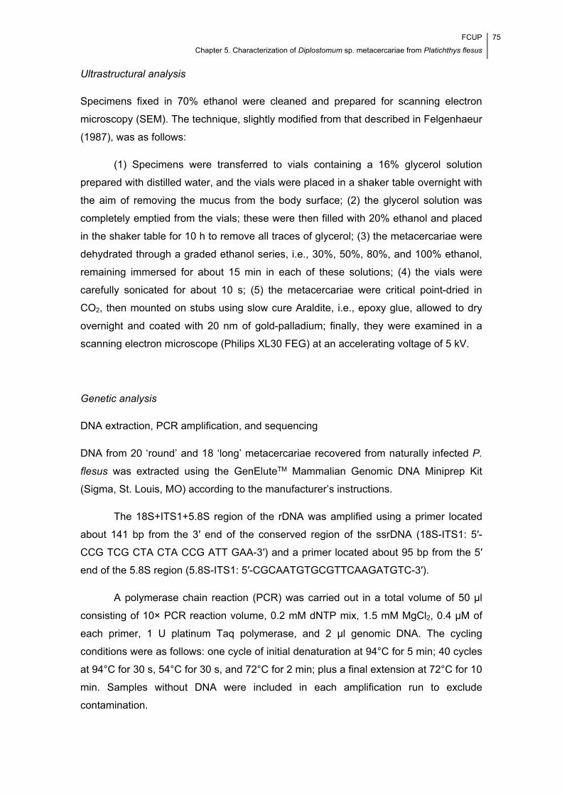
FCUP
Chapter 5. Characterization of Diplostomum sp. metacercariae from Platichthys flesus
75
Ultrastructural analysis
Specimens fixed in 70% ethanol were cleaned and prepared for scanning electron
microscopy (SEM). The technique, slightly modified from that described in Felgenhaeur
(1987), was as follows:
(1) Specimens were transferred to vials containing a 16% glycerol solution
prepared with distilled water, and the vials were placed in a shaker table overnight with
the aim of removing the mucus from the body surface; (2) the glycerol solution was
completely emptied from the vials; these were then filled with 20% ethanol and placed
in the shaker table for 10 h to remove all traces of glycerol; (3) the metacercariae were
dehydrated through a graded ethanol series, i.e., 30%, 50%, 80%, and 100% ethanol,
remaining immersed for about 15 min in each of these solutions; (4) the vials were
carefully sonicated for about 10 s; (5) the metacercariae were critical point-dried in
CO2, then mounted on stubs using slow cure Araldite, i.e., epoxy glue, allowed to dry
overnight and coated with 20 nm of gold-palladium; finally, they were examined in a
scanning electron microscope (Philips XL30 FEG) at an accelerating voltage of 5 kV.
Genetic analysis
DNA extraction, PCR amplification, and sequencing
DNA from 20 ‘round’ and 18 ‘long’ metacercariae recovered from naturally infected P.
flesus was extracted using the GenEluteTM Mammalian Genomic DNA Miniprep Kit
(Sigma, St. Louis, MO) according to the manufacturer’s instructions.
The 18S+ITS1+5.8S region of the rDNA was amplified using a primer located
about 141 bp from the 3′ end of the conserved region of the ssrDNA (18S-ITS1: 5′-
CCG TCG CTA CTA CCG ATT GAA-3′) and a primer located about 95 bp from the 5′
end of the 5.8S region (5.8S-ITS1: 5′-CGCAATGTGCGTTCAAGATGTC-3′).
A polymerase chain reaction (PCR) was carried out in a total volume of 50 μl
consisting of 10× PCR reaction volume, 0.2 mM dNTP mix, 1.5 mM MgCl2, 0.4 μM of
each primer, 1 U platinum Taq polymerase, and 2 μl genomic DNA. The cycling
conditions were as follows: one cycle of initial denaturation at 94°C for 5 min; 40 cycles
at 94°C for 30 s, 54°C for 30 s, and 72°C for 2 min; plus a final extension at 72°C for 10
min. Samples without DNA were included in each amplification run to exclude
contamination.
76 FCUP
Chapter 5. Characterization of Diplostomum sp. metacercariae from Platichthys flesus
Amplified PCR products were analysed by electrophoresis in a 1.0% agarose
gel stained with ethidium bromide, purified with the QIAquick Gel Extraction Kit
(Qiagen, Valencia, CA) and sequenced. The obtained sequence that included the
partial 18S, ITS1 (complete), and partial 5.8S was submitted to GenBank under
accession number GQ370809.
Molecular phylogenetic analysis
Nucleotide sequence data were compared for similarity by searching the GenBank-
NCBI database using the Basic Local Alignment Search Tool (BLAST,
www.ncbi.nlm.nih.gov/blast), and multiple sequence alignments were performed using
Multalin (available at http://bioinfo.genotoul.fr/multalin/multalin.html) and the ClustalW
version 2 software (Larkin et al., 2007).
Partial and complete ITS1 sequences of identified species of Diplostomum as
well as of two out-group species have been retrieved from GenBank for molecular and
phylogenetic studies. Tylodelphys sp. (Diplostomidae), which has been indicated as a
genus closely ancestral to Diplostomum by Galazzo et al. (2002), together with
Ichthyocotylurus erraticus (Rudolphi, 1809) (Strigeidae) were selected as out-groups.
Taxonomic names, developmental stage, hosts, ITS1 length, collecting sites, and
GenBank accession numbers are provided in Table 5.1.
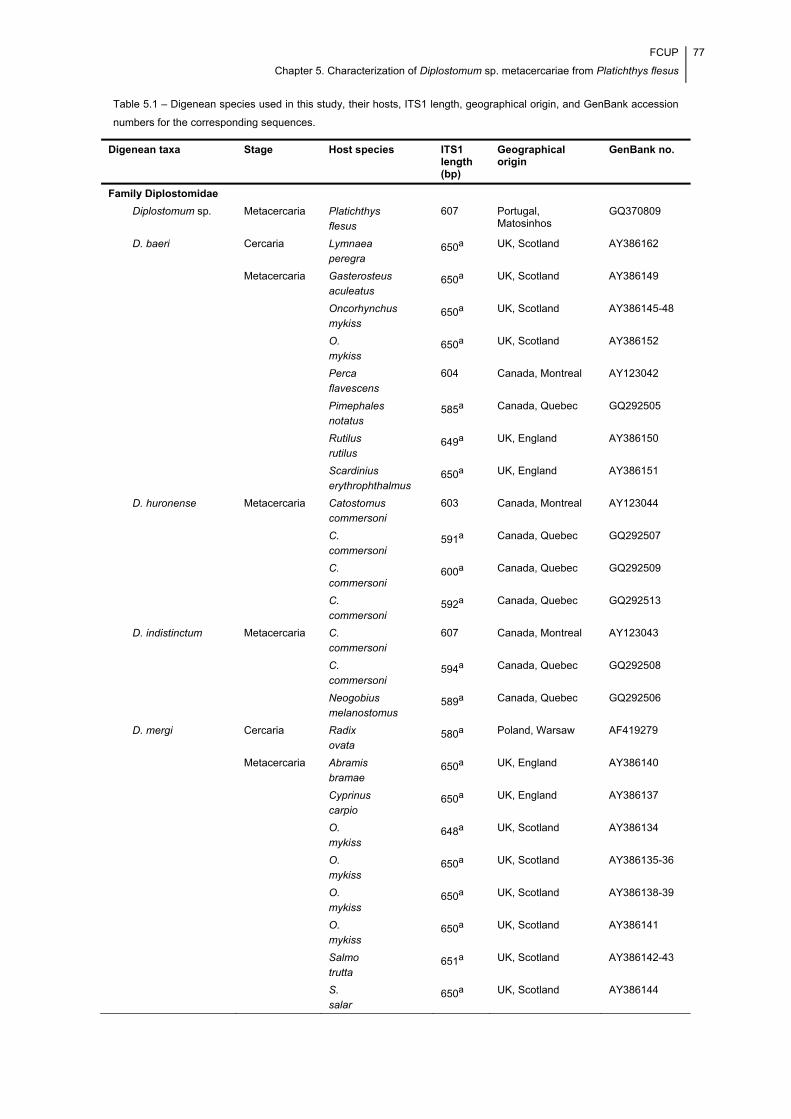
FCUP
Chapter 5. Characterization of Diplostomum sp. metacercariae from Platichthys flesus
77
Table 5.1 – Digenean species used in this study, their hosts, ITS1 length, geographical origin, and GenBank accession
numbers for the corresponding sequences.
Digenean taxa Stage Host species ITS1 length (bp)
Geographical origin
GenBank no.
Family Diplostomidae
Diplostomum sp. Metacercaria Platichthys
flesus
607 Portugal, Matosinhos
GQ370809
D. baeri Cercaria Lymnaea
peregra 650a UK, Scotland AY386162
Metacercaria Gasterosteus
aculeatus 650a UK, Scotland AY386149
Oncorhynchus
mykiss 650a UK, Scotland AY386145-48
O.
mykiss 650a UK, Scotland AY386152
Perca
flavescens
604 Canada, Montreal AY123042
Pimephales
notatus 585a Canada, Quebec GQ292505
Rutilus
rutilus 649a UK, England AY386150
Scardinius
erythrophthalmus 650a UK, England AY386151
D. huronense Metacercaria Catostomus
commersoni
603 Canada, Montreal AY123044
C.
commersoni 591a Canada, Quebec GQ292507
C.
commersoni 600a Canada, Quebec GQ292509
C.
commersoni 592a Canada, Quebec GQ292513
D. indistinctum Metacercaria C.
commersoni
607 Canada, Montreal AY123043
C.
commersoni 594a Canada, Quebec GQ292508
Neogobius
melanostomus 589a Canada, Quebec GQ292506
D. mergi Cercaria Radix
ovata 580a Poland, Warsaw AF419279
Metacercaria Abramis
bramae 650a UK, England AY386140
Cyprinus
carpio 650a UK, England AY386137
O.
mykiss 648a UK, Scotland AY386134
O.
mykiss 650a UK, Scotland AY386135-36
O.
mykiss 650a UK, Scotland AY386138-39
O.
mykiss 650a UK, Scotland AY386141
Salmo
trutta 651a UK, Scotland AY386142-43
S.
salar 650a UK, Scotland AY386144
78 FCUP
Chapter 5. Characterization of Diplostomum sp. metacercariae from Platichthys flesus
Table 5.1 (continuation) – Digenean species used in this study, their hosts, ITS1 length, geographical origin, and
GenBank accession numbers for the corresponding sequences.
aITS1 rDNA partial sequence.
Phylogenetic and molecular evolutionary analyses were conducted on the
aligned partial nucleotide sequences of ITS1 using MEGA software version 4 (Tamura
et al., 2007). The neighbour-joining (NJ) method (Saitou & Nei, 1987) was performed
using the program’s default settings. The reliability of internal branches in the NJ trees
was assessed using bootstrap analysis with 10,000 replicates. The resulting networks
were rooted with the out-group taxa.
Morphometric analysis
The morphometric data were assessed using a Carl Zeiss Axiophot Photomicroscope
equipped with an Axiocam ICc3 camera and connected to a computer with version
4.6.3 of the Axiovision digital image processing software (Carl Zeiss Microimaging Inc.,
Thornwood, NY, USA). A series of 14 metric dimensions were assessed from the
Digenean taxa Stage Host species ITS1 length (bp)
Geographical origin
GenBank no.
D. pseudospathaceum Cercaria Lymnaea
stagnalis 578a Poland, Warsaw AF419273
Metacercaria Micropterus
salmoides 595a Canada, Quebec GQ292511
D. spathaceum Cercaria R.
ovata 579a Poland, Warsaw AF419275-76
Metacercaria G.
aculeatus 650a UK, Scotland AY386153
O.
mykiss 650a UK, Scotland AY386155-56
S.
salar 650a UK, Scotland AY386154
D. parviventosum Cercaria R.
ovata 586a Poland, Warsaw AF419277-78
D. phoxini Metacercaria Phoxinuns
phoxinus 648a UK, Scotland AY386157-60
D. paracaudum Cercaria R.
ovata 579a Poland, Warsaw AF419272
Tylodelphys sp. Metacercaria R.
rutilus 652a UK, Scotland AY386164
Family Strigeidae
Ichthyocotylurus erraticus
Metacercaria Coregonus
lavaretus
781 Finland AJ301887
C.
albula
781 Finland AJ301887

FCUP
Chapter 5. Characterization of Diplostomum sp. metacercariae from Platichthys flesus
79
metacercariae (N = 30), i.e., the length (BL) and width (BW) of the body; the length
(OL) and width (OW) of the oral sucker; the length (PHL) and width (PHW) of the
pharynx; the length (VL) and width (VW) of the ventral sucker; the length (HL) and
width (HW) of the holdfast organ; the distance between the anterior extremity of the
body and the center of the ventral sucker (VD); the length of the lappets (LL); and the
width of the body at the level of the bifurcation of the intestine (WaBI) and at the mid-
length of the oral sucker (WaO) (see Fig. 5.1), plus eight indices, i.e., BW/BL (in
percent); BL×BW/HL×HW; BL×BW/VL×VW; OL×OW/VL×VW; HL×HW/VL×VW;
OL×OW/PHL×PHW; VD/BL (in percent); and WaO/WaBI, including the corresponding
means, ranges, coefficients of variation, and limits of the 95% confidence interval for
the population means (Niewiadomska & Niewiadomska-Bugaj, 1995, 1998). Those
metric dimensions and indices contributing most to the variability found among the
isolated specimens were evaluated by running a multiple factorial analysis on version
8.0 of the statistical program package STATISTICA for Windows (StatSoft Inc., Tulsa,
USA). The morphometric segregation of the species of Diplostomum isolated in this
study from D. paracaudum, D. pseudospathaceum, D. spathaceum, and D. mergi was
evaluated by running principal component and cluster (similarity measure, 1−Pearson’s
correlation coefficient) analyses using the same software. Morphometric comparisons
were limited to those made possible by the data available in the literature. Moreover,
the data used in such comparisons were retrieved from Niewiadomska &
Niewiadomska-Bugaj (1995) for D. paracaudum and D. pseudospathaceum and from
Niewiadomska & Niewiadomska-Bugaj (1998) for D. spathaceum and D. mergi.
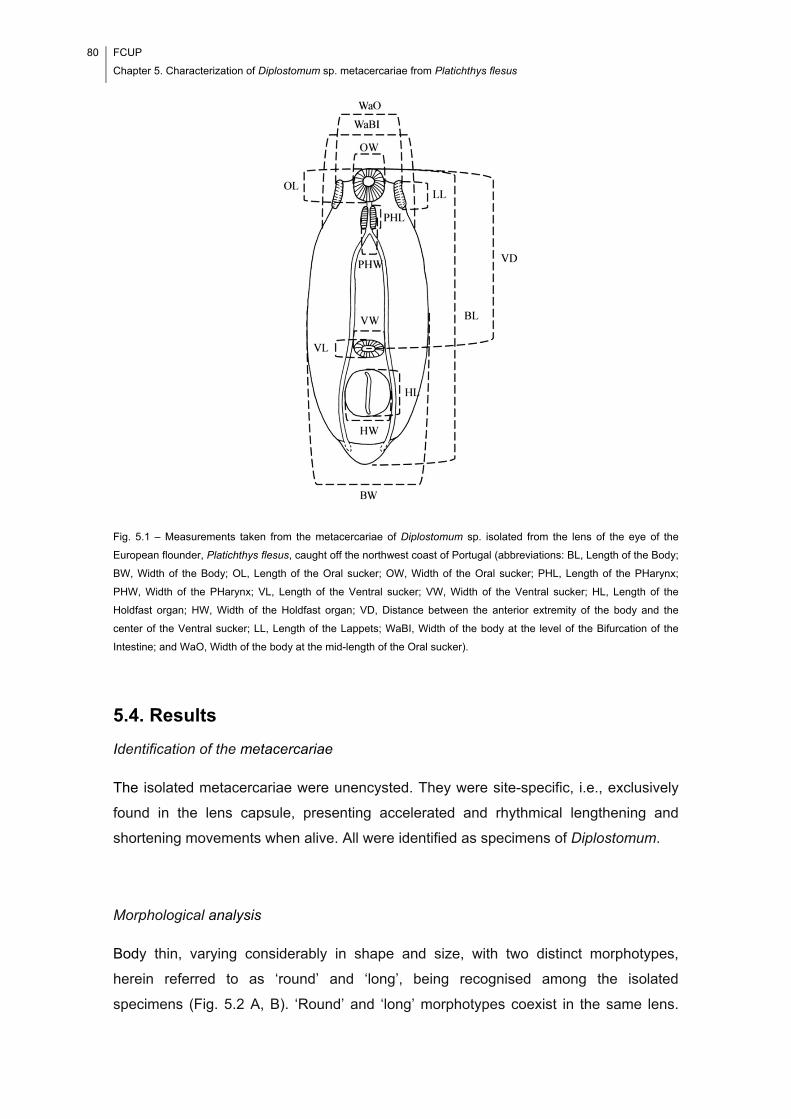
80 FCUP
Chapter 5. Characterization of Diplostomum sp. metacercariae from Platichthys flesus
Fig. 5.1 – Measurements taken from the metacercariae of Diplostomum sp. isolated from the lens of the eye of the
European flounder, Platichthys flesus, caught off the northwest coast of Portugal (abbreviations: BL, Length of the Body;
BW, Width of the Body; OL, Length of the Oral sucker; OW, Width of the Oral sucker; PHL, Length of the PHarynx;
PHW, Width of the PHarynx; VL, Length of the Ventral sucker; VW, Width of the Ventral sucker; HL, Length of the
Holdfast organ; HW, Width of the Holdfast organ; VD, Distance between the anterior extremity of the body and the
center of the Ventral sucker; LL, Length of the Lappets; WaBI, Width of the body at the level of the Bifurcation of the
Intestine; and WaO, Width of the body at the mid-length of the Oral sucker).
5.4. Results
Identification of the metacercariae
The isolated metacercariae were unencysted. They were site-specific, i.e., exclusively
found in the lens capsule, presenting accelerated and rhythmical lengthening and
shortening movements when alive. All were identified as specimens of Diplostomum.
Morphological analysis
Body thin, varying considerably in shape and size, with two distinct morphotypes,
herein referred to as ‘round’ and ‘long’, being recognised among the isolated
specimens (Fig. 5.2 A, B). ‘Round’ and ‘long’ morphotypes coexist in the same lens.
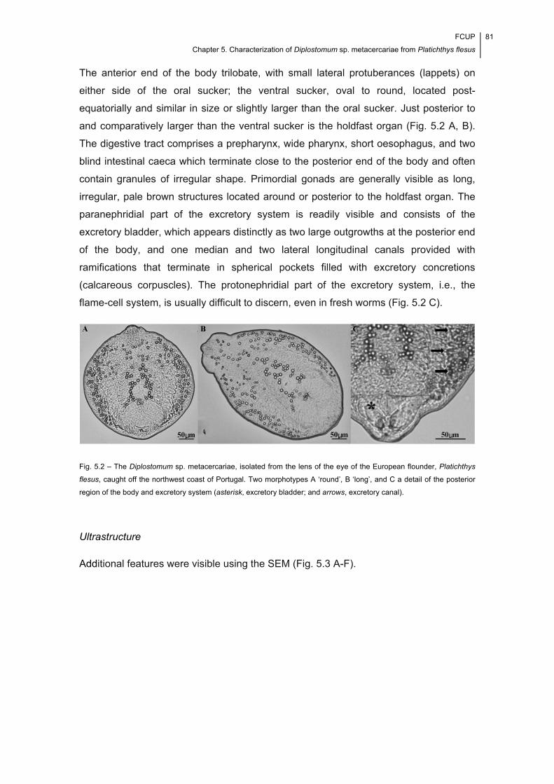
FCUP
Chapter 5. Characterization of Diplostomum sp. metacercariae from Platichthys flesus
81
The anterior end of the body trilobate, with small lateral protuberances (lappets) on
either side of the oral sucker; the ventral sucker, oval to round, located post-
equatorially and similar in size or slightly larger than the oral sucker. Just posterior to
and comparatively larger than the ventral sucker is the holdfast organ (Fig. 5.2 A, B).
The digestive tract comprises a prepharynx, wide pharynx, short oesophagus, and two
blind intestinal caeca which terminate close to the posterior end of the body and often
contain granules of irregular shape. Primordial gonads are generally visible as long,
irregular, pale brown structures located around or posterior to the holdfast organ. The
paranephridial part of the excretory system is readily visible and consists of the
excretory bladder, which appears distinctly as two large outgrowths at the posterior end
of the body, and one median and two lateral longitudinal canals provided with
ramifications that terminate in spherical pockets filled with excretory concretions
(calcareous corpuscles). The protonephridial part of the excretory system, i.e., the
flame-cell system, is usually difficult to discern, even in fresh worms (Fig. 5.2 C).
Fig. 5.2 – The Diplostomum sp. metacercariae, isolated from the lens of the eye of the European flounder, Platichthys
flesus, caught off the northwest coast of Portugal. Two morphotypes A ‘round’, B ‘long’, and C a detail of the posterior
region of the body and excretory system (asterisk, excretory bladder; and arrows, excretory canal).
Ultrastructure
Additional features were visible using the SEM (Fig. 5.3 A-F).
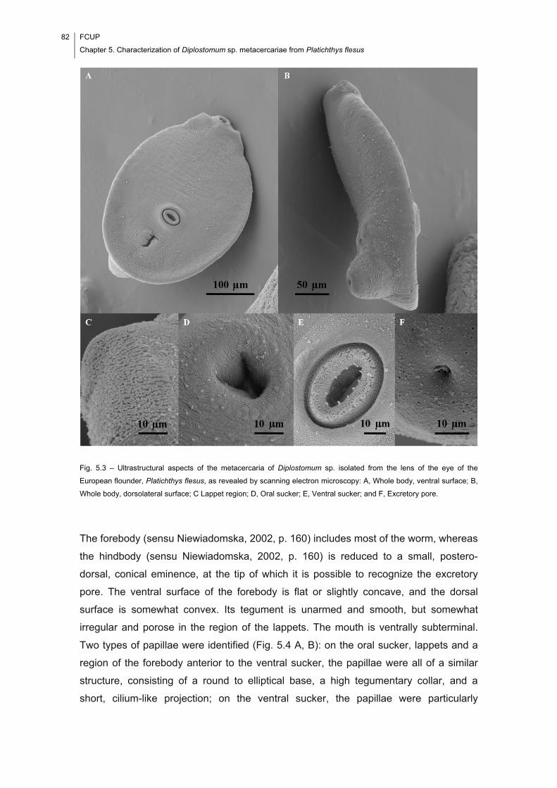
82 FCUP
Chapter 5. Characterization of Diplostomum sp. metacercariae from Platichthys flesus
Fig. 5.3 – Ultrastructural aspects of the metacercaria of Diplostomum sp. isolated from the lens of the eye of the
European flounder, Platichthys flesus, as revealed by scanning electron microscopy: A, Whole body, ventral surface; B,
Whole body, dorsolateral surface; C Lappet region; D, Oral sucker; E, Ventral sucker; and F, Excretory pore.
The forebody (sensu Niewiadomska, 2002, p. 160) includes most of the worm, whereas
the hindbody (sensu Niewiadomska, 2002, p. 160) is reduced to a small, postero-
dorsal, conical eminence, at the tip of which it is possible to recognize the excretory
pore. The ventral surface of the forebody is flat or slightly concave, and the dorsal
surface is somewhat convex. Its tegument is unarmed and smooth, but somewhat
irregular and porose in the region of the lappets. The mouth is ventrally subterminal.
Two types of papillae were identified (Fig. 5.4 A, B): on the oral sucker, lappets and a
region of the forebody anterior to the ventral sucker, the papillae were all of a similar
structure, consisting of a round to elliptical base, a high tegumentary collar, and a
short, cilium-like projection; on the ventral sucker, the papillae were particularly
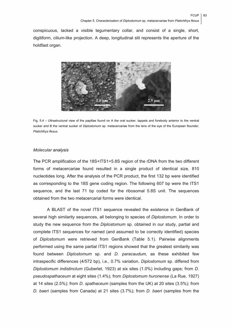
FCUP
Chapter 5. Characterization of Diplostomum sp. metacercariae from Platichthys flesus
83
conspicuous, lacked a visible tegumentary collar, and consist of a single, short,
digitiform, cilium-like projection. A deep, longitudinal slit represents the aperture of the
holdfast organ.
Fig. 5.4 – Ultrastructural view of the papillae found on A the oral sucker, lappets and forebody anterior to the ventral
sucker and B the ventral sucker of Diplostomum sp. metacercariae from the lens of the eye of the European flounder,
Platichthys flesus.
Molecular analysis
The PCR amplification of the 18S+ITS1+5.8S region of the rDNA from the two different
forms of metacercariae found resulted in a single product of identical size, 810
nucleotides long. After the analysis of the PCR product, the first 132 bp were identified
as corresponding to the 18S gene coding region. The following 607 bp were the ITS1
sequence, and the last 71 bp coded for the ribosomal 5.8S unit. The sequences
obtained from the two metacercarial forms were identical.
A BLAST of the novel ITS1 sequence revealed the existence in GenBank of
several high similarity sequences, all belonging to species of Diplostomum. In order to
study the new sequence from the Diplostomum sp. obtained in our study, partial and
complete ITS1 sequences for named (and assumed to be correctly identified) species
of Diplostomum were retrieved from GenBank (Table 5.1). Pairwise alignments
performed using the same partial ITS1 regions showed that the greatest similarity was
found between Diplostomum sp. and D. paracaudum, as these exhibited few
intraspecific differences (4/572 bp), i.e., 0.7% variation. Diplostomum sp. differed from
Diplostomum indistinctum (Guberlet, 1923) at six sites (1.0%) including gaps; from D.
pseudospathaceum at eight sites (1.4%); from Diplostomum huronense (La Rue, 1927)
at 14 sites (2.5%); from D. spathaceum (samples from the UK) at 20 sites (3.5%); from
D. baeri (samples from Canada) at 21 sites (3.7%); from D. baeri (samples from the
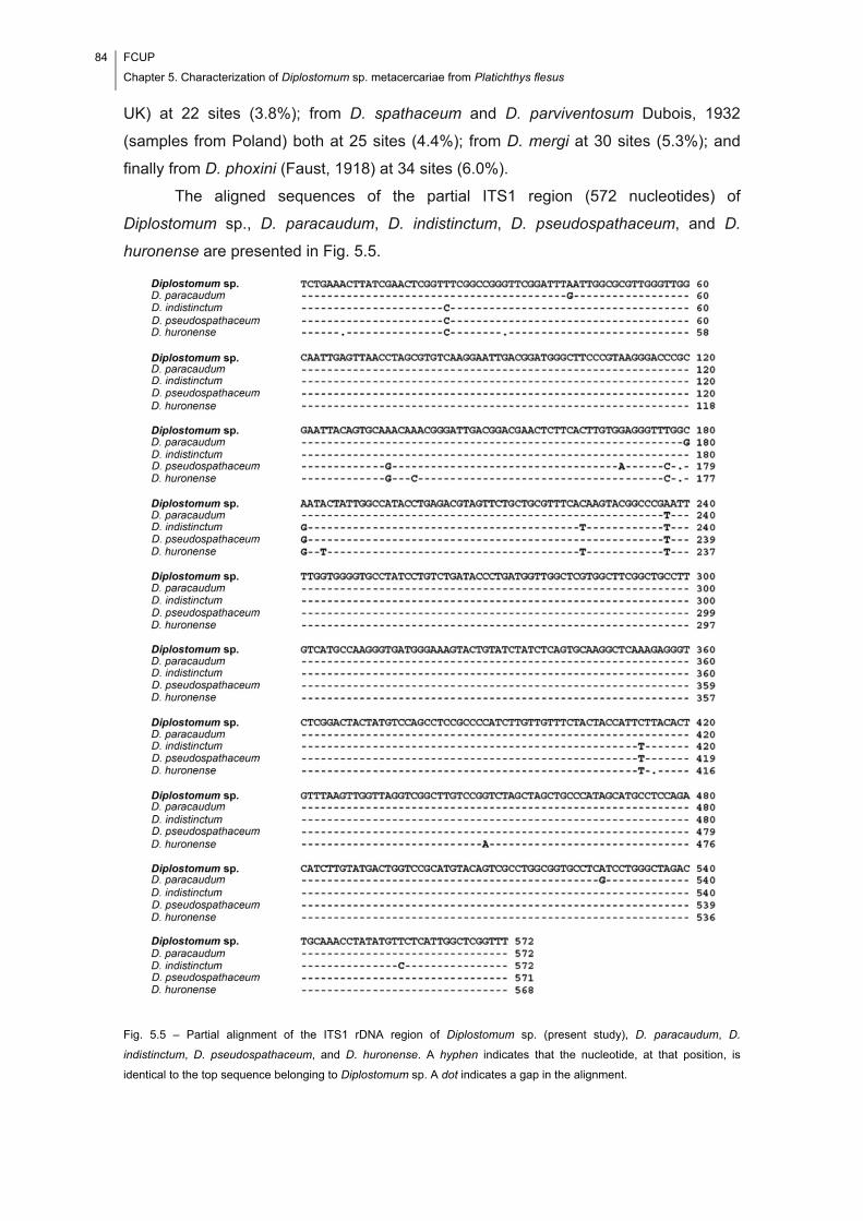
84 FCUP
Chapter 5. Characterization of Diplostomum sp. metacercariae from Platichthys flesus
UK) at 22 sites (3.8%); from D. spathaceum and D. parviventosum Dubois, 1932
(samples from Poland) both at 25 sites (4.4%); from D. mergi at 30 sites (5.3%); and
finally from D. phoxini (Faust, 1918) at 34 sites (6.0%).
The aligned sequences of the partial ITS1 region (572 nucleotides) of
Diplostomum sp., D. paracaudum, D. indistinctum, D. pseudospathaceum, and D.
huronense are presented in Fig. 5.5.
Fig. 5.5 – Partial alignment of the ITS1 rDNA region of Diplostomum sp. (present study), D. paracaudum, D.
indistinctum, D. pseudospathaceum, and D. huronense. A hyphen indicates that the nucleotide, at that position, is
identical to the top sequence belonging to Diplostomum sp. A dot indicates a gap in the alignment.
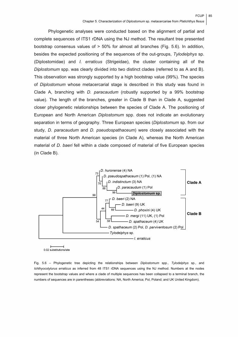
FCUP
Chapter 5. Characterization of Diplostomum sp. metacercariae from Platichthys flesus
85
Phylogenetic analyses were conducted based on the alignment of partial and
complete sequences of ITS1 rDNA using the NJ method. The resultant tree presented
bootstrap consensus values of > 50% for almost all branches (Fig. 5.6). In addition,
besides the expected positioning of the sequences of the out-groups, Tylodelphys sp.
(Diplostomidae) and I. erraticus (Strigeidae), the cluster containing all of the
Diplostomum spp. was clearly divided into two distinct clades (referred to as A and B).
This observation was strongly supported by a high bootstrap value (99%). The species
of Diplostomum whose metacercarial stage is described in this study was found in
Clade A, branching with D. paracaudum (robustly supported by a 99% bootstrap
value). The length of the branches, greater in Clade B than in Clade A, suggested
closer phylogenetic relationships between the species of Clade A. The positioning of
European and North American Diplostomum spp. does not indicate an evolutionary
separation in terms of geography. Three European species (Diplostomum sp. from our
study, D. paracaudum and D. pseudospathaceum) were closely associated with the
material of three North American species (in Clade A), whereas the North American
material of D. baeri fell within a clade composed of material of five European species
(in Clade B).
Fig. 5.6 – Phylogenetic tree depicting the relationships between Diplostomum spp., Tylodelphys sp., and
Ichthyocotylurus erraticus as inferred from 48 ITS1 rDNA sequences using the NJ method. Numbers at the nodes
represent the bootstrap values and where a clade of multiple sequences has been collapsed to a terminal branch, the
numbers of sequences are in parentheses (abbreviations: NA, North America; Pol, Poland; and UK United Kingdom).
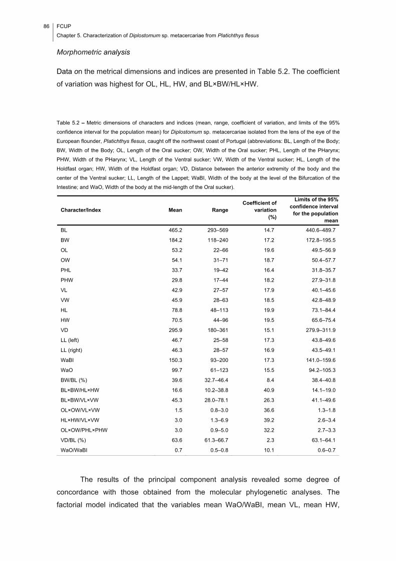
86 FCUP
Chapter 5. Characterization of Diplostomum sp. metacercariae from Platichthys flesus
Morphometric analysis
Data on the metrical dimensions and indices are presented in Table 5.2. The coefficient
of variation was highest for OL, HL, HW, and BL×BW/HL×HW.
Table 5.2 – Metric dimensions of characters and indices (mean, range, coefficient of variation, and limits of the 95%
confidence interval for the population mean) for Diplostomum sp. metacercariae isolated from the lens of the eye of the
European flounder, Platichthys flesus, caught off the northwest coast of Portugal (abbreviations: BL, Length of the Body;
BW, Width of the Body; OL, Length of the Oral sucker; OW, Width of the Oral sucker; PHL, Length of the PHarynx;
PHW, Width of the PHarynx; VL, Length of the Ventral sucker; VW, Width of the Ventral sucker; HL, Length of the
Holdfast organ; HW, Width of the Holdfast organ; VD, Distance between the anterior extremity of the body and the
center of the Ventral sucker; LL, Length of the Lappet; WaBI, Width of the body at the level of the Bifurcation of the
Intestine; and WaO, Width of the body at the mid-length of the Oral sucker).
The results of the principal component analysis revealed some degree of
concordance with those obtained from the molecular phylogenetic analyses. The
factorial model indicated that the variables mean WaO/WaBI, mean VL, mean HW,
Character/Index Mean Range Coefficient of
variation(%)
Limits of the 95% confidence interval
for the population mean
BL 465.2 293–569 14.7 440.6–489.7
BW 184.2 118–240 17.2 172.8–195.5
OL 53.2 22–66 19.6 49.5–56.9
OW 54.1 31–71 18.7 50.4–57.7
PHL 33.7 19–42 16.4 31.8–35.7
PHW 29.8 17–44 18.2 27.9–31.8
VL 42.9 27–57 17.9 40.1–45.6
VW 45.9 28–63 18.5 42.8–48.9
HL 78.8 48–113 19.9 73.1–84.4
HW 70.5 44–96 19.5 65.6–75.4
VD 295.9 180–361 15.1 279.9–311.9
LL (left) 46.7 25–58 17.3 43.8–49.6
LL (right) 46.3 28–57 16.9 43.5–49.1
WaBI 150.3 93–200 17.3 141.0–159.6
WaO 99.7 61–123 15.5 94.2–105.3
BW/BL (%) 39.6 32.7–46.4 8.4 38.4–40.8
BL×BW/HL×HW 16.6 10.2–38.8 40.9 14.1–19.0
BL×BW/VL×VW 45.3 28.0–78.1 26.3 41.1–49.6
OL×OW/VL×VW 1.5 0.8–3.0 36.6 1.3–1.8
HL×HW/VL×VW 3.0 1.3–6.9 39.2 2.6–3.4
OL×OW/PHL×PHW 3.0 0.9–5.0 32.2 2.7–3.3
VD/BL (%) 63.6 61.3–66.7 2.3 63.1–64.1
WaO/WaBI 0.7 0.5–0.8 10.1 0.6–0.7

FCUP
Chapter 5. Characterization of Diplostomum sp. metacercariae from Platichthys flesus
87
mean OL×OW/PHL×PHW, and mean VW were those contributing most to the
formation of factor 1, whereas mean PHL, mean OL, mean VD/BL (percent), and mean
OL×OW/VL×VW were most influential in the formation of factor 2 (Fig. 5.7).
Fig. 5.7 – Variables factor map (PCA) for Diplostomum sp. – projection of the mean metric dimensions and indices on
factor planes 1 and 2 (abbreviations: BL, Length of the Body; BW, Width of the Body; OL, Length of the Oral sucker;
OW, Width of the Oral sucker; PHL, Length of the PHarynx; PHW, Width of the PHarynx; VL, Length of the Ventral
sucker; VW, Width of the Ventral sucker; HL, Length of the Holdfast organ; HW, Width of the Holdfast organ; VD,
Distance between the anterior extremity of the body and the center of the Ventral sucker; LL, Length of the Lappets;
WaBI, Width of the body at the level of the Bifurcation of the Intestine; and WaO, Width of the body at the mid-length of
the Oral sucker).
When considering the case projections in relation to factor 1, which explained 45.1% of
the variability found, two groups could be identified, with D. spathaceum and D. mergi
in opposition to the other three species. When considering the case projections in
relation to factor 2, which explained 28.7% of the variability found, a group including all
of the species, except for Diplostomum sp., was apparent (Fig. 5.8).
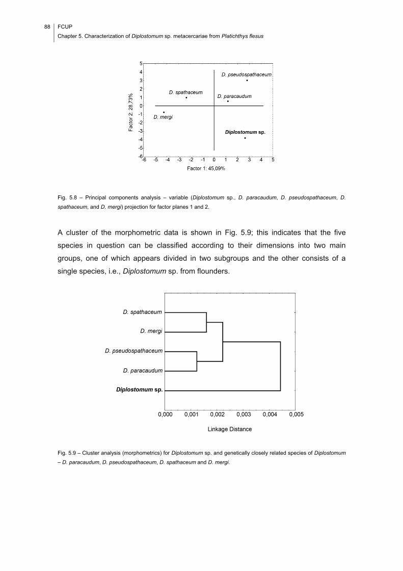
88 FCUP
Chapter 5. Characterization of Diplostomum sp. metacercariae from Platichthys flesus
Fig. 5.8 – Principal components analysis – variable (Diplostomum sp., D. paracaudum, D. pseudospathaceum, D.
spathaceum, and D. mergi) projection for factor planes 1 and 2.
A cluster of the morphometric data is shown in Fig. 5.9; this indicates that the five
species in question can be classified according to their dimensions into two main
groups, one of which appears divided in two subgroups and the other consists of a
single species, i.e., Diplostomum sp. from flounders.
Fig. 5.9 – Cluster analysis (morphometrics) for Diplostomum sp. and genetically closely related species of Diplostomum
– D. paracaudum, D. pseudospathaceum, D. spathaceum and D. mergi.
FCUP
Chapter 5. Characterization of Diplostomum sp. metacercariae from Platichthys flesus
89
5.5. Discussion
Literature records of Diplostomum spp. in the European flounder include references to
D. baeri (see Lüthen, 1988), D. mergi (see Chibani & Rokicki, 2004) and D.
spathaceum (e.g., Brucko-Stempkowski, 1970; Kennedy et al., 1992; Dorovskikh,
1997; Køie, 1999; Palm et al., 1999). Such records are mainly from the Baltic region
where the salinity is low. None of these corresponds to the metacercarial stage
described in this study – assuming that the species were correctly identified and
judging from both the phylogenetic analyses (i.e., all of these species are situated in
clade B, whereas Diplostomum sp. from flounders is in clade A; Fig. 5.6) and the
morphometric data. Furthermore, the species isolated does not correspond with any of
those whose genetic characterization is available in GenBank. Since more than forty
different species of Diplostomum exist (Niewiadomska, 1996), most of which have not
been characterized in molecular terms, it is not possible to determine the identity of the
material from flounders. Indeed, there also exists the possibility that it has been
previously detected as Diplostomum sp. in flounders from the Baltic Sea (Engelbrecht,
1958; Chibani & Rokicki, 2004) in cases where authors have been unwilling to
speculate on a specific identification.
Interestingly, the results of the molecular phylogenetic and morphometric
analyses exhibit some degree of concordance with regard to differences between
Diplostomum sp. and D. spathaceum, D. mergi, D. pseudospathaceum, and D.
paracaudum, and also suggest that it is most similar to D. paracaudum. Nevertheless,
the real value of the results derived from the morphometric analyses are a matter of
contention (compare, for example, the works of A. A. Shigin and K. Niewiadomska on
the same species). The implications of extensive morphometric variation in terms of the
reliability of specimen identification to the species level have recently been pointed out
by Chibwana & Nkwengulila (2009) in a work intended to discriminate between three
closely related diplostomid species, i.e., D. mashonense Beverley-Burton 1963 and
Tylodelphys spp. 1 and 2. Also, in another work, on opecoelid larvae, an extensive
morphological measurement overlap was reported in three genetically different worms
(Violante-González et al., 2009). According to the literature, different factors may act to
produce significant variations in body dimensions. These include, among others, the
host species, its size and age, the age of the metacercariae (e.g., Graczyk, 1991,
1992; Niewiadomska & Szymański, 1991, 1992), and the population size associated
with intensity-dependent growth (Saldanha et al., 2009). Even taking into account other
obvious factors, such as worm condition at fixation, the fixation technique, and other
procedural variations, one can assume that, in this study, the variability found in body

90 FCUP
Chapter 5. Characterization of Diplostomum sp. metacercariae from Platichthys flesus
shape represents different stages of metacercarial development, since the same lens
was found infected with both ‘round’ and ‘long’ morphotypes.
By assessing the rDNA sequence data (partial ITS1 sequences) from adult
forms, Galazzo et al. (2002) were able to demonstrate using phylogenetic analyses that
North American and European species of Diplostomum include divergent groups – the
American ones, i.e., D. indistinctum, D. huronense, and D. baeri, being basal to the
European, i.e., D. paracaudum, D. pseudospathaceum, D. spathaceum, D.
parviventosum, D. mergi, and D. baeri. These authors also showed that D. baeri from
Europe was not conspecific with ‘D. baeri’ from North America. The present study,
which was conducted on specimens from a flatfish, P. flesus, whose geographical
distribution is limited to European waters (Whitehead et al., 1986), lends further support
to the idea of differences in the geographical distribution of species within the genus,
since D. paracaudum, another European species, exhibited the greatest genetic
similarity to the present material. It should be noted, however, that, although in this
study the length of the ITS1 sequence (607 bp) equals that found by Galazzo et al.
(2002) for D. huronense, D. indistinctum, and North American D. baeri, and differs from
those found by Niewiadomska & Laskowski (2002) for D. parviventosum, D.
spathaceum, and D. paracaudum (all 580 bp), D. mergi (579 bp), D.
pseudospathaceum (578 bp), and European D. baeri (576 bp), it proved to be useful in
discriminating between Diplostomum sp. and the other nine species of Diplostomum
which have been characterized genetically. However, Niewiadomska & Laskowski
(2002) found no molecular differences between D. spathaceum and D. parviventosum,
although these species present apparent morphological differences at the cercarial,
metacercarial, and adult stages. According to Galazzo et al. (2002), the North
American and European species represent divergent groups within Diplostomum.
Although the distribution of the molluscan hosts also needs to be taken into account,
Locke et al. (2010) have suggested that the presence of these two groups do not
support an evolutionary history associated with a geographical divergence of the
species, given the mobility of the avian definitive hosts. This latter proposal is in
accordance with our findings, which indicate that there does not appear to be an
evolutionary separation of the European and North American species of Diplostomum.
The results of the present work also reinforce the idea that different kinds of data
should be considered for the accurate identification of diplostomid metacercariae at the
specific level.

FCUP
Chapter 5. Characterization of Diplostomum sp. metacercariae from Platichthys flesus
91
5.6. Acknowledgements
The authors would like to thank the Council of Rectors of the Portuguese Universities
and the British Council – Treaty of Winsor – UR58 (U13[05/06]), CIMAR – CIIMAR
Pluriannual Program, the Portuguese Foundation for Science and Technology and the
European Social Fund (grants SFRH/BM/23063/2005; SFRH/BD/65258/2009 [F.I.
Cavaleiro] SFRH/BD/31767/2006 [S. Pina]) for funding, Professor Vítor Silva from the
Rector’s Office of Porto University for his help with the images, and Dr. Alex Ball from
the Natural History Museum of London for his help with the scanning electron
microscopy.

92 FCUP
Chapter 5. Characterization of Diplostomum sp. metacercariae from Platichthys flesus
Chapter 6 Egg number-egg size: an important trade-off
in parasite life history strategies
This chapter has been adapted from:
Cavaleiro, F. I., & Santos, M. J. (In Press). Egg number-egg size: an important trade-off in parasite life history
strategies. International Journal for Parasitology.

94 FCUP
Chapter 6. The trade-off between egg number and egg size
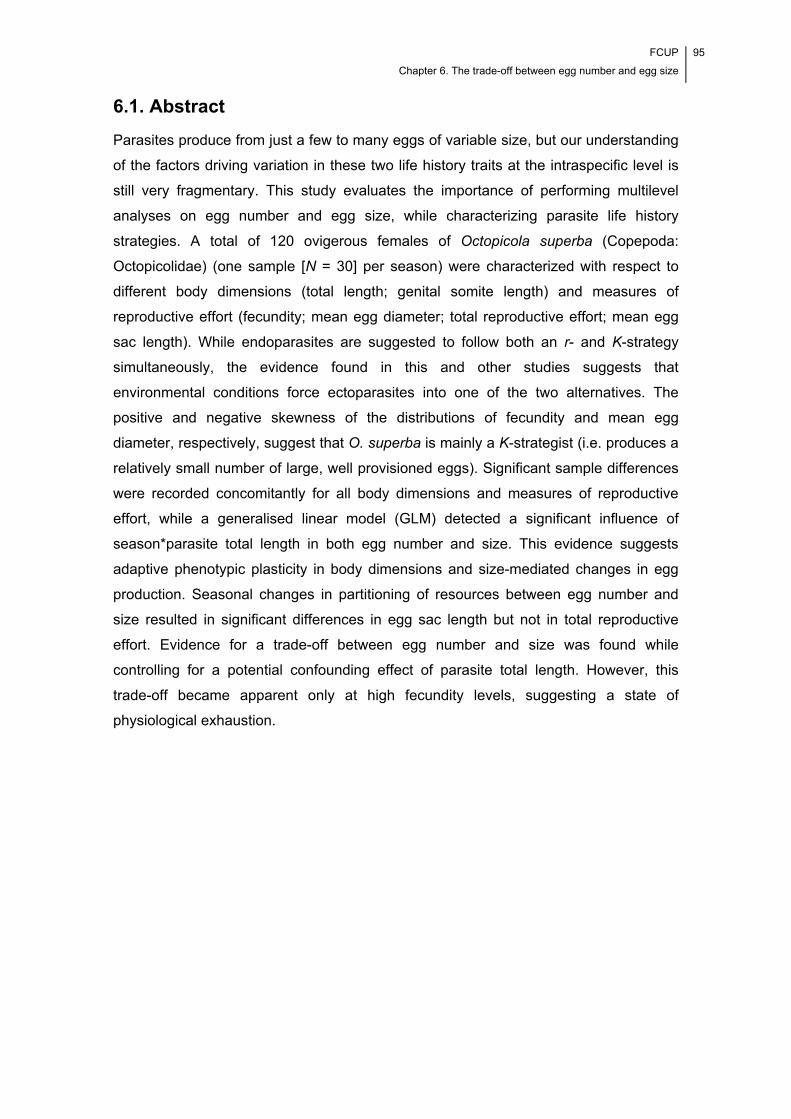
FCUP
Chapter 6. The trade-off between egg number and egg size
95
6.1. Abstract
Parasites produce from just a few to many eggs of variable size, but our understanding
of the factors driving variation in these two life history traits at the intraspecific level is
still very fragmentary. This study evaluates the importance of performing multilevel
analyses on egg number and egg size, while characterizing parasite life history
strategies. A total of 120 ovigerous females of Octopicola superba (Copepoda:
Octopicolidae) (one sample [N = 30] per season) were characterized with respect to
different body dimensions (total length; genital somite length) and measures of
reproductive effort (fecundity; mean egg diameter; total reproductive effort; mean egg
sac length). While endoparasites are suggested to follow both an r- and K-strategy
simultaneously, the evidence found in this and other studies suggests that
environmental conditions force ectoparasites into one of the two alternatives. The
positive and negative skewness of the distributions of fecundity and mean egg
diameter, respectively, suggest that O. superba is mainly a K-strategist (i.e. produces a
relatively small number of large, well provisioned eggs). Significant sample differences
were recorded concomitantly for all body dimensions and measures of reproductive
effort, while a generalised linear model (GLM) detected a significant influence of
season*parasite total length in both egg number and size. This evidence suggests
adaptive phenotypic plasticity in body dimensions and size-mediated changes in egg
production. Seasonal changes in partitioning of resources between egg number and
size resulted in significant differences in egg sac length but not in total reproductive
effort. Evidence for a trade-off between egg number and size was found while
controlling for a potential confounding effect of parasite total length. However, this
trade-off became apparent only at high fecundity levels, suggesting a state of
physiological exhaustion.

96 FCUP
Chapter 6. The trade-off between egg number and egg size
FCUP
Chapter 6. The trade-off between egg number and egg size
97
6.2. Introduction
Transition from a free-living existence to a parasitic mode of life impacted various life
history traits, including fecundity (egg number) and egg size (see Poulin, 1995b; Calow,
1983). However, our theoretical framework is still lacking some important elements if
we are to understand fully the mechanism of parasite egg production. These elements
will allow us to evaluate the existence of general laws (i.e. patterns and processes) in
parasite egg production. Furthermore, fitting the pieces of the puzzle together, namely
the evidence from multilevel analyses on egg number and egg size, is crucial to
elucidating parasite life history strategies.
Egg number and egg size are key concepts in parasite reproduction. For many
years, our understanding of the former of these traits was largely based on the
misconception that all parasites evolve toward extremely high egg output (Poulin,
1995a). There were different explanations for it: a high egg output represents the
expected outcome of natural selection – according to the ‘balanced mortality’
hypothesis (Smith, 1954), parasites must compensate for the massive losses of
infective stages that occur during the transmission phase of their life-cycles; a high egg
output is the direct outcome of the conditions provided by the host environment
(Jennings & Calow, 1975).
The strategy of egg production of a parasite is somewhere between two
extremes (the r-end and the K-end) in a continuum of possibilities. It is the outcome of
natural selection, representing the optimal compromise between egg number and egg
size. In perfect r-strategist organisms, there are no density effects or competition; all
available energy and matter are invested in reproduction, the smallest possible amount
into each individual offspring. On the other hand, in perfect K-strategist organisms, the
density effects are maximum and the competition is keen; the emphasis is on
preserving the adult and only the remaining energy and matter are used in
reproduction, i.e. in the production and maintenance of a small number of extremely fit
offspring (Jennings & Calow, 1975). The way in which parasites of a species partition
reproductive effort between egg number and egg size can however vary to some extent
(see e.g., Ritchie et al., 1993), i.e. reflect adaptive phenotypic plasticity. This process
enables individuals to accommodate changes in their environment, by making possible
the rapid movement to a new fitness optimum (Price et al., 2003); however, unlike
natural selection, it does not result in genetic adaptation, i.e. in changes in genotypic
frequencies in the population (Poulin, 1996, 2007a).
98 FCUP
Chapter 6. The trade-off between egg number and egg size
The strategies of parasite egg production (egg number and egg size) have been
addressed from broad perspectives in the literature (e.g. Gotto, 1962; Price, 1974;
Jennings & Calow, 1975; Calow, 1983; Poulin, 1995b, 1996, 1997b, 2007a). Also,
numerous studies have considered the egg production of parasites in different
taxonomic groups, i.e. Monogenea, Digenea, Cestoda, Nematoda and Copepoda, in
relation to different factors, i.e. adult size and longevity, maturation time or prepatency,
temperature, photoperiod, salinity, season/time, sampling site, origin of the host (wild
environment vs farm), host species, host size, number of parasites in the host,
interactions between parasites in the host and number of treatments with anti-parasitic
drugs (see e.g., Faust, 1949; Olsen, 1974; Anderson, 1982; Kennedy, 1983; Johnston
& Dykeman, 1987; McGladdery & Johnston, 1988; Mehlhorn, 1988; Cable & Tinsley,
1991; Johnson & Albright, 1991; Tocque & Tinsley, 1991; Ritchie et al., 1993; Tully &
Whelan, 1993; Roubal, 1994; Trouvé et al., 1998; Heuch et al., 2000; Rossin et al.,
2005; Bravo et al., 2009; Bravo, 2010; González et al., 2012; Ruiz Daniels et al., 2013).
Among these groups, the Copepoda is particularly suited to study parasite egg
production for different reasons: firstly, copepods frequently occur in high prevalence
and intensity levels year-round in their natural host populations; and secondly, unlike
parasites in other taxonomic groups, copepods produce egg sacs which can be easily
detached from the parasite and manipulated.
Causes of intraspecific variability in egg number and egg size can only be
understood properly if the possible effects of factors at different levels, i.e. the
macroenvironment, microenvironment, microhabitat (sensu Rohde, 1984) and parasite
levels, are considered for analysis (see e.g. Timi et al., 2010; Loot et al., 2011).
Moreover, unravelling how factors at these different levels interact with each other
appears to be crucial to understanding how the mechanism of egg production works at
the intraspecific level. For instance, evidence for developmental plasticity in size in
response to water temperature (e.g. Nordhagen et al., 2000) and that individual
parasites reach a size proportional to that of their hosts (e.g., Van Damme et al., 1993;
Poulin, 1995a; Loot et al., 2011) is documented in the literature, while it is a general
assumption that larger parasites tend to produce more eggs (Poulin, 2007a). Larger
hosts likely provide parasites with a more permanent habitat (Poulin, 2007a). In this
way, they may favour a delay in maturation and larger body sizes (Stearns, 1992; Roff,
1992; Poulin, 2007a), therefore interfering with parasite egg production. The effect of
host body size on parasite egg production appears, however, to be controversial.
Actually, Cole (1954) and Kennedy (1983) argued that a decrease in parasite
maturation time should result in an increase of the reproductive potential, while the
FCUP
Chapter 6. The trade-off between egg number and egg size
99
same effect is expected to be seen in parasites with delayed maturity, as these should
present larger body sizes. At the microhabitat level, the selective pressures affecting
parasites relate to the food resources (quality and quantity) and the site of infection
itself (i.e. its location within the host’s body – the stress imposed on parasites living on
body surfaces and in internal organs should vary greatly), as the host represents the
source of food and the home simultaneously (Crompton, 1991; Castro, 1991; Combes,
1991). The nutrients available to parasites should also vary greatly with the number of
conspecifics present at the site of infection, i.e. with the intensity of infection. According
to the ‘crowding effect’ (Read, 1951), the larger the parasite burden, the more intense
the competition for essential nutrients and, likely, the host immune response; in such a
scenario, both the body size and the fecundity of the parasite are expected to be
negatively affected. This type of effect has been documented for cestodes (Keymer et
al., 1983; Dobson, 1986; Shostak & Dick, 1987; Heins et al., 2002), nematodes (Krupp,
1961; Khamboonruang, 1971; Michel et al., 1971, 1978; Szalai & Dick, 1989) and
digeneans (Jones et al., 1989).
A phenotypic trade-off between egg number and egg size has already been
demonstrated using data from different taxa of parasitic copepods (see Poulin, 1995b,
2007a), but its occurrence at the intraspecific level is less consensual (see e.g., Rossin
et al., 2005; Timi et al., 2005). The trade-off appears to be influenced by a number of
factors, i.e. the host quality (Rossin et al., 2005), the female body size (Herreras et al.,
2007) and the site of infection (Loot et al., 2011), which should therefore be considered
for analysis.
This study aimed to investigate how the mechanism of egg production works in
parasites using a multilevel approach. Particular emphasis is given to the trade-off
between egg number and egg size and the factors having a significant influence on
these two life history traits. Octopicola superba (Copepoda: Octopicolidae), parasitic on
the common octopus, Octopus vulgaris (recently suggested to represent a complex of
species), was used as a model parasite. Data were assessed from mature, ovigerous
females. The specific questions addressed were the following: (i) which strategy of egg
production is followed by the parasite: is it mainly an r-strategist (i.e. produces a large
number of small, poorly provisioned eggs) or a K-strategist (i.e. produces a small
number of large, well provisioned eggs) species; (ii) was there an influence of season,
site of infection, host body size, number of conspecifics present at the site of infection
and parasite body size (or of interactions between these variables) in egg number
and/or egg size; and (iii) was there a phenotypic trade-off between egg number and
egg size, while controlling for a potential effect of confounding variables? While
100 FCUP
Chapter 6. The trade-off between egg number and egg size
considering each of these questions, the existence of general trends was evaluated on
the basis of the information available in the literature.
6.3. Materials and Methods
The parasite
Octopicola superba is a parasite of O. vulgaris, endemic to European waters (Humes,
1957; Deboutteville et al., 1957; Bocquet & Stock, 1960; Cavaleiro et al., 2013). It most
likely has a single host life-cycle. Indeed, according to our findings, both copepodites
and adults are commonly found on O. vulgaris. Information on this parasite is scarce
concerning its behaviour (Deboutteville et al., 1957) and infection levels (Bocquet &
Stock, 1960). Associated disease is not documented in the literature, which suggests
that O. superba might not be pathogenic for the natural population of octopus;
otherwise, the lack of records on associated disease is likely a consequence of the low
number of studies so far conducted on this parasitic infection. According to our
findings, ovigerous females of O. superba are present at high prevalence and intensity
and year-round on O. vulgaris off northwestern Portugal. The parasite can be easily
isolated from the sediment obtained from the washings of the octopus’ body surface
and mantle cavity. All of these aspects make it ideally suited to study the mechanism of
egg production in parasites. Besides, species associated with marine invertebrates
seem to be particularly suited to studies of the impact of the type, habitat and
behaviour of the host in the number and size of the eggs laid by the copepod (see
Gotto, 1962).
Host sampling and parasitological survey
Sampling of octopuses was conducted seasonally during 2010. Octopuses were
caught in marine waters off the northern Portuguese town of Matosinhos (41º10’N,
8º42’W) (pot catches), collected by a boat which regularly fishes these waters for O.
vulgaris exclusively, and individually placed in plastic bags to prevent loss of parasites.
Seasonal samples of octopuses (winter sample, collecting date: 2 March; spring
sample, 24 and 31 May; summer sample, 7 September; and autumn sample, 22
November) consisted of 30 specimens each. Shortly after being delivered to the fresh
fish market, at the harbour in Matosinhos, the octopuses were transported to the
laboratory, at the campus of University of Porto, Portugal; all of them were kept frozen
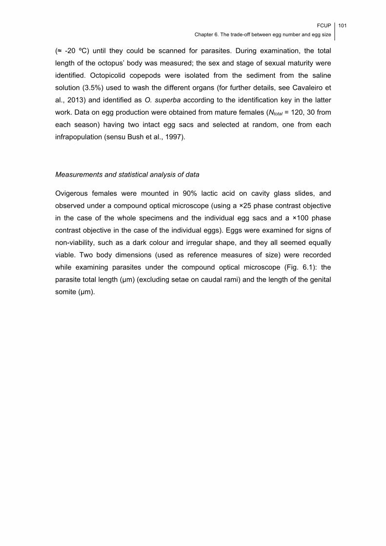
FCUP
Chapter 6. The trade-off between egg number and egg size
101
(≈ -20 ºC) until they could be scanned for parasites. During examination, the total
length of the octopus’ body was measured; the sex and stage of sexual maturity were
identified. Octopicolid copepods were isolated from the sediment from the saline
solution (3.5%) used to wash the different organs (for further details, see Cavaleiro et
al., 2013) and identified as O. superba according to the identification key in the latter
work. Data on egg production were obtained from mature females (Ntotal = 120, 30 from
each season) having two intact egg sacs and selected at random, one from each
infrapopulation (sensu Bush et al., 1997).
Measurements and statistical analysis of data
Ovigerous females were mounted in 90% lactic acid on cavity glass slides, and
observed under a compound optical microscope (using a ×25 phase contrast objective
in the case of the whole specimens and the individual egg sacs and a ×100 phase
contrast objective in the case of the individual eggs). Eggs were examined for signs of
non-viability, such as a dark colour and irregular shape, and they all seemed equally
viable. Two body dimensions (used as reference measures of size) were recorded
while examining parasites under the compound optical microscope (Fig. 6.1): the
parasite total length (µm) (excluding setae on caudal rami) and the length of the genital
somite (µm).

102 FCUP
Chapter 6. The trade-off between egg number and egg size
Fig. 6.1 – Morphometric measurements taken from the mature, ovigerous females of Octopicola superba (modified from
Humes, 1957).
Beyond these two measures, the following variables were determined as part of the
effort to characterize the mechanism of egg production: egg sac length (µm) (assessed
for the two sacs); fecundity; egg diameter (µm) (assessed for all eggs making up the
clutch); and total reproductive effort (a measure of the total resources invested in one
clutch). Fecundity was assessed as the total number of eggs in the two sacs; mean
egg sac length as the average length of the two egg sacs; mean egg diameter as the
average diameter of the eggs in the two sacs; and total reproductive effort by
multiplying fecundity by mean egg volume (Caley et al., 2001). Mean egg volume was
determined using the records for mean egg diameter and the formula for the volume of
a sphere (mean egg volume = ×π×[
]3), since the egg is nearly
spherical.
In order to evaluate the parasite life history strategy, the total sample of female
parasites was characterized considering the two sets of variables, i.e. each body
dimension (parasite total length and genital somite length) and measures of
FCUP
Chapter 6. The trade-off between egg number and egg size
103
reproductive effort (fecundity, mean egg diameter, total reproductive effort and mean
egg sac length) (mean, SD, range interval [RI, minimum-maximum], coefficient of
variation [CV], limits of the 95% confidence interval [CI] for the population mean and
distribution [one sample Kolmogorov-Smirnov’s test]). Furthermore, multiple factorial
analysis (MFA; extraction method: principal components) was used to analyse the
structure of the data. The skewness and kurtosis values for the distributions of
fecundity and mean egg diameter were determined, in order to evaluate which strategy
of egg production (r-strategy or K-strategy) is preferentially followed by the parasite.
Host total length was characterized with respect to the same statistical parameters
used in the case of the other studied variables and considering the total sample of
octopuses.
A potential influence of season on egg production was considered by
characterizing the seasonal samples of female parasites with respect to the two sets of
variables (i.e. by considering their distribution). Values for each body dimension
(parasite total length and genital somite length) and measures of reproductive effort
(fecundity, mean egg diameter, total reproductive effort and mean egg sac length) were
compared between the four seasons of the year (Kruskal-Wallis’ test); pairwise sample
comparisons were performed (Mann-Whitney’s U test) only when the Kruskal-Wallis’
test yielded a statistically significant difference. The same strategy was followed to
detect a potential influence of site of infection on egg production. Discriminant function
analysis (DFA; method: independent variables entered together) was conducted to
obtain a general picture of the differences between the females of different seasons.
The two sets of variables were considered in the analysis. A general linear analysis
model with type III sum of squares (GLM multivariate analysis) was then used to
evaluate the constraints of egg production in O. superba. In a preliminary analysis, only
the main effects were assessed. Fecundity and mean egg diameter were considered,
simultaneously, as the dependent variables; fixed factors included season exclusively;
and covariates included host total length, number of conspecifics present at the site of
infection and parasite total length. According to the results obtained in this analysis, the
following interaction terms were then considered for analysis (using the same
dependent variables, fixed factor and covariates): season*parasite total length; host
total length*parasite total length; and number of conspecifics present at the site of
infection*parasite total length.
An independent regulation of egg number and egg size was investigated using
two strategies. The first of these consisted of plotting on the same graph, data for
fecundity and mean egg diameter (the specimens were first arranged by ascending
104 FCUP
Chapter 6. The trade-off between egg number and egg size
fecundity). Each seasonal sample was considered separately for analysis. The second
strategy consisted of evaluating whether there was a phenotypic trade-off between egg
number and egg size. A non-parametric partial rank correlation test, conducted
separately for each season of sampling, evaluated the existence of a significant
negative correlation between the two life history traits while controlling for a potential
confounding effect of the variables shown by the GLM multivariate analysis to have a
significant influence on fecundity or/and egg size.
Measurements from female parasites were taken under a compound optical
microscope (Carl Zeiss Axiophot Photomicroscope) and using the digital image
processing software AXIOVISION of Carl Zeiss, version 4.6.3 (Carl Zeiss Microimaging
Inc., Thornwood, NY, USA). Data for body dimensions and measures of reproductive
effort for the different seasons of sampling/sites of infection were depicted as box-and-
whisker plots. In these, the minimum and maximum values encompass 95% of the
data; outliers and extremes appear outside this range interval. Statistical tests,
multivariate analyses and graphical representations of data were performed using
Statistical Package for Social Sciences (SPSS) for Windows, version 21.0 (SPSS Inc.,
Chicago, Illinois, USA), STATISTICA for Windows, version 10.0 (StatSoft Inc., Tulsa,
USA) and Microsoft Excel 2010 (Microsoft, Redmond, WA, USA). Non-parametric
partial rank correlation analyses were defined in the syntax editor window of SPSS,
according to the instructions available at the IBM website
(http://www01.ibm.com/support/docview.wss?uid=swg21474822). The significance
level considered was P < 0.05, except in the case of the pairwise sample comparisons.
In this case, the Bonferroni correction set significance at P < 0.008(3). In this way, it
was possible to account for the Type I error.
6.4. Results
The females of O. superba considered in the analyses were isolated from the body skin
(N = 66), gills (N = 26), mesentery covering the gonad (N = 13), mantle musculature (N
= 11), eyes (N = 2) and funnel (N = 2). Data recorded from the parasite and host
samples (total samples) are given in Table 6.1.
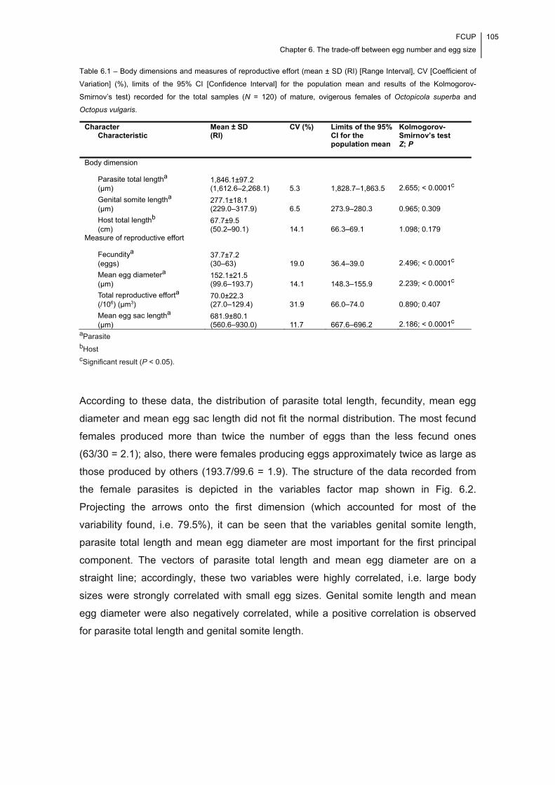
FCUP
Chapter 6. The trade-off between egg number and egg size
105
Table 6.1 – Body dimensions and measures of reproductive effort (mean ± SD (RI) [Range Interval], CV [Coefficient of
Variation] (%), limits of the 95% CI [Confidence Interval] for the population mean and results of the Kolmogorov-
Smirnov’s test) recorded for the total samples (N = 120) of mature, ovigerous females of Octopicola superba and
Octopus vulgaris.
aParasite bHost cSignificant result (P < 0.05).
According to these data, the distribution of parasite total length, fecundity, mean egg
diameter and mean egg sac length did not fit the normal distribution. The most fecund
females produced more than twice the number of eggs than the less fecund ones
(63/30 = 2.1); also, there were females producing eggs approximately twice as large as
those produced by others (193.7/99.6 = 1.9). The structure of the data recorded from
the female parasites is depicted in the variables factor map shown in Fig. 6.2.
Projecting the arrows onto the first dimension (which accounted for most of the
variability found, i.e. 79.5%), it can be seen that the variables genital somite length,
parasite total length and mean egg diameter are most important for the first principal
component. The vectors of parasite total length and mean egg diameter are on a
straight line; accordingly, these two variables were highly correlated, i.e. large body
sizes were strongly correlated with small egg sizes. Genital somite length and mean
egg diameter were also negatively correlated, while a positive correlation is observed
for parasite total length and genital somite length.
Character Characteristic
Mean ± SD (RI)
CV (%) Limits of the 95% CI for the population mean
Kolmogorov-Smirnov’s test Z; P
Body dimension
Parasite total lengtha (µm)
1,846.1±97.2 (1,612.6–2,268.1) 5.3 1,828.7–1,863.5 2.655; < 0.0001c
Genital somite lengtha (µm)
277.1±18.1 (229.0–317.9) 6.5 273.9–280.3 0.965; 0.309
Host total lengthb (cm)
67.7±9.5 (50.2–90.1) 14.1 66.3–69.1 1.098; 0.179
Measure of reproductive effort
Fecunditya (eggs)
37.7±7.2 (30–63) 19.0 36.4–39.0 2.496; < 0.0001c
Mean egg diametera (µm)
152.1±21.5 (99.6–193.7) 14.1 148.3–155.9 2.239; < 0.0001c
Total reproductive efforta (/106) (µm3)
70.0±22.3 (27.0–129.4) 31.9 66.0–74.0 0.890; 0.407
Mean egg sac lengtha (µm)
681.9±80.1 (560.6–930.0) 11.7 667.6–696.2 2.186; < 0.0001c
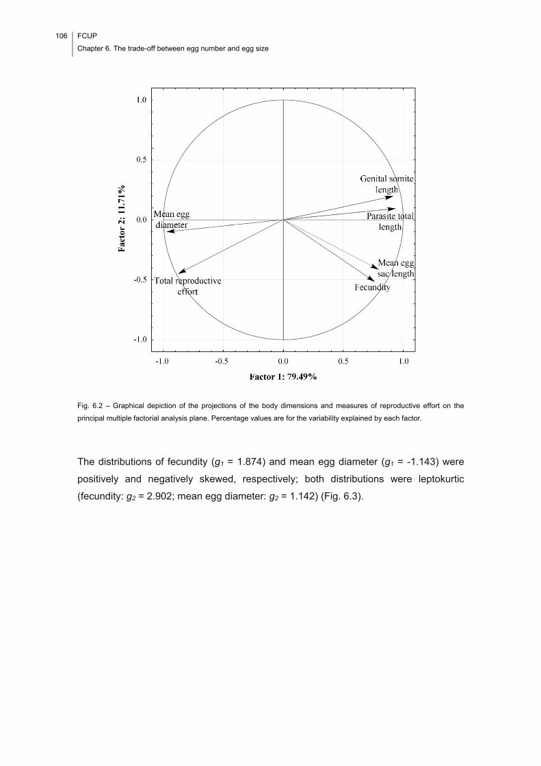
106 FCUP
Chapter 6. The trade-off between egg number and egg size
Fig. 6.2 – Graphical depiction of the projections of the body dimensions and measures of reproductive effort on the
principal multiple factorial analysis plane. Percentage values are for the variability explained by each factor.
The distributions of fecundity (g1 = 1.874) and mean egg diameter (g1 = -1.143) were
positively and negatively skewed, respectively; both distributions were leptokurtic
(fecundity: g2 = 2.902; mean egg diameter: g2 = 1.142) (Fig. 6.3).
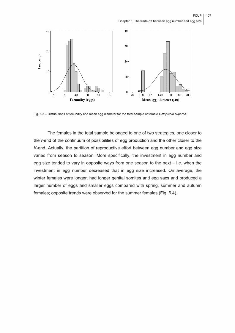
FCUP
Chapter 6. The trade-off between egg number and egg size
107
Fig. 6.3 – Distributions of fecundity and mean egg diameter for the total sample of female Octopicola superba.
The females in the total sample belonged to one of two strategies, one closer to
the r-end of the continuum of possibilities of egg production and the other closer to the
K-end. Actually, the partition of reproductive effort between egg number and egg size
varied from season to season. More specifically, the investment in egg number and
egg size tended to vary in opposite ways from one season to the next – i.e. when the
investment in egg number decreased that in egg size increased. On average, the
winter females were longer, had longer genital somites and egg sacs and produced a
larger number of eggs and smaller eggs compared with spring, summer and autumn
females; opposite trends were observed for the summer females (Fig. 6.4).
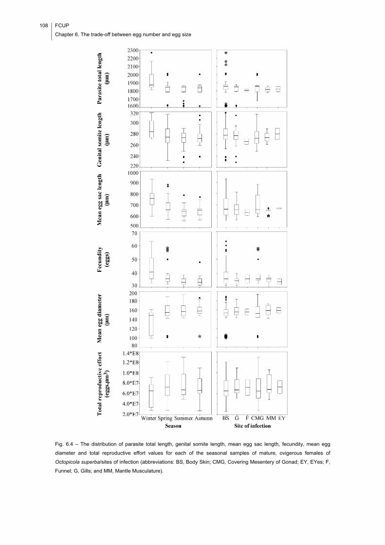
108 FCUP
Chapter 6. The trade-off between egg number and egg size
Fig. 6.4 – The distribution of parasite total length, genital somite length, mean egg sac length, fecundity, mean egg
diameter and total reproductive effort values for each of the seasonal samples of mature, ovigerous females of
Octopicola superba/sites of infection (abbreviations: BS, Body Skin; CMG, Covering Mesentery of Gonad; EY, EYes; F,
Funnel; G, Gills; and MM, Mantle Musculature).
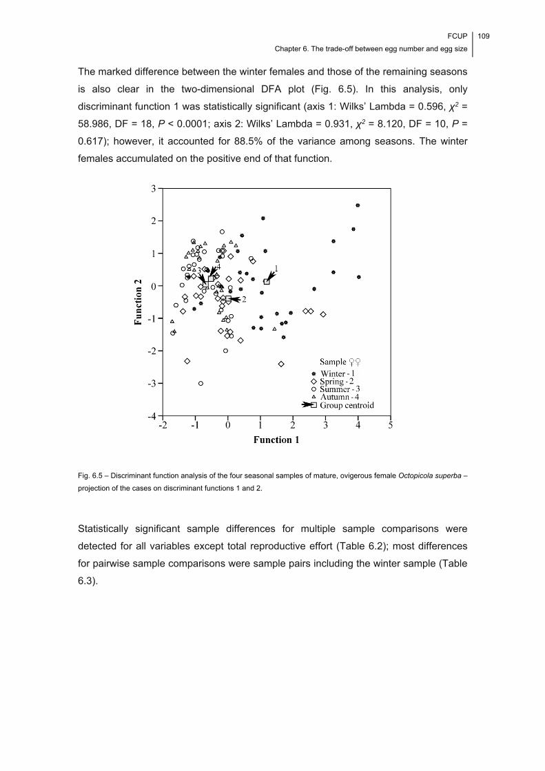
FCUP
Chapter 6. The trade-off between egg number and egg size
109
The marked difference between the winter females and those of the remaining seasons
is also clear in the two-dimensional DFA plot (Fig. 6.5). In this analysis, only
discriminant function 1 was statistically significant (axis 1: Wilks’ Lambda = 0.596, χ2 =
58.986, DF = 18, P < 0.0001; axis 2: Wilks’ Lambda = 0.931, χ2 = 8.120, DF = 10, P =
0.617); however, it accounted for 88.5% of the variance among seasons. The winter
females accumulated on the positive end of that function.
Fig. 6.5 – Discriminant function analysis of the four seasonal samples of mature, ovigerous female Octopicola superba –
projection of the cases on discriminant functions 1 and 2.
Statistically significant sample differences for multiple sample comparisons were
detected for all variables except total reproductive effort (Table 6.2); most differences
for pairwise sample comparisons were sample pairs including the winter sample (Table
6.3).
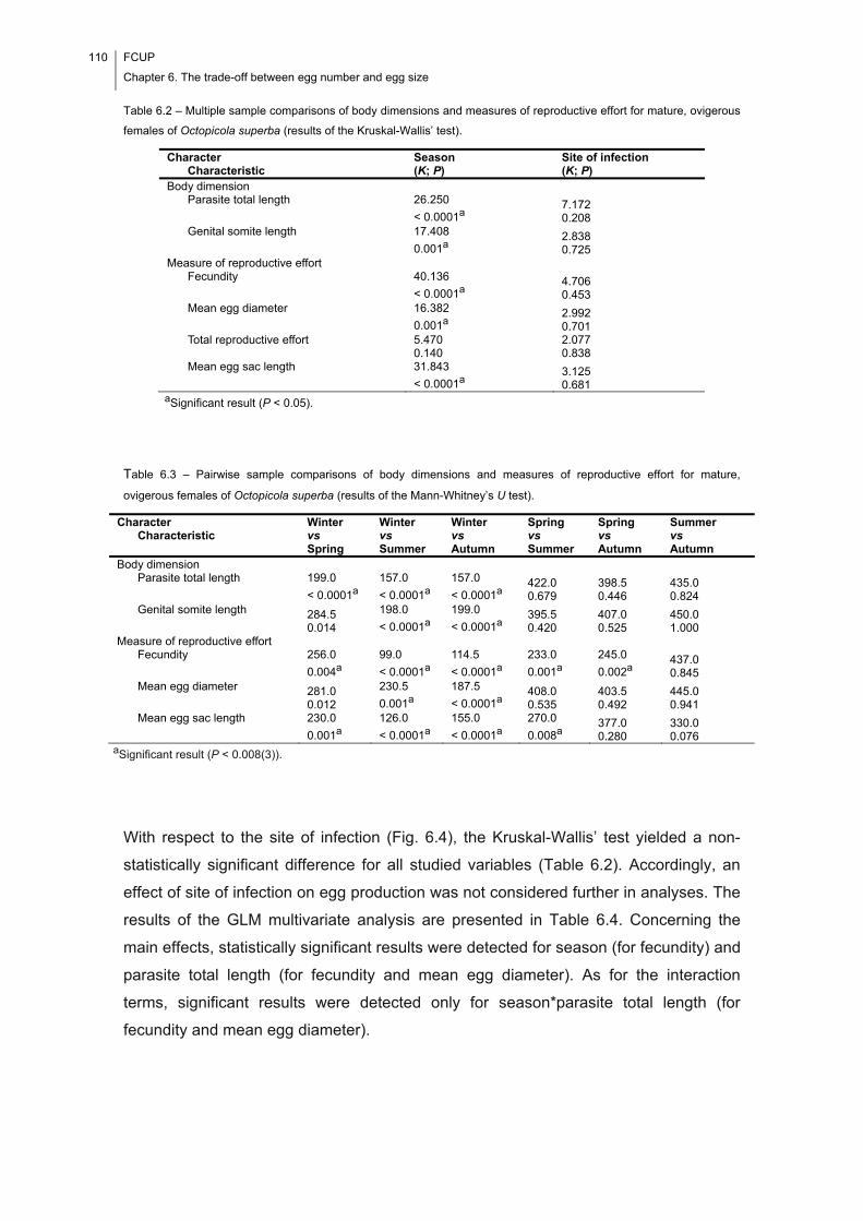
110 FCUP
Chapter 6. The trade-off between egg number and egg size
Table 6.2 – Multiple sample comparisons of body dimensions and measures of reproductive effort for mature, ovigerous
females of Octopicola superba (results of the Kruskal-Wallis’ test).
aSignificant result (P < 0.05).
Table 6.3 – Pairwise sample comparisons of body dimensions and measures of reproductive effort for mature,
ovigerous females of Octopicola superba (results of the Mann-Whitney’s U test).
aSignificant result (P < 0.008(3)).
With respect to the site of infection (Fig. 6.4), the Kruskal-Wallis’ test yielded a non-
statistically significant difference for all studied variables (Table 6.2). Accordingly, an
effect of site of infection on egg production was not considered further in analyses. The
results of the GLM multivariate analysis are presented in Table 6.4. Concerning the
main effects, statistically significant results were detected for season (for fecundity) and
parasite total length (for fecundity and mean egg diameter). As for the interaction
terms, significant results were detected only for season*parasite total length (for
fecundity and mean egg diameter).
Character Characteristic
Season (K; P)
Site of infection (K; P)
Body dimension Parasite total length 26.250
< 0.0001a 7.172 0.208
Genital somite length 17.408
0.001a 2.838 0.725
Measure of reproductive effort Fecundity 40.136
< 0.0001a 4.706 0.453
Mean egg diameter 16.382
0.001a 2.992 0.701
Total reproductive effort 5.470 0.140
2.077 0.838
Mean egg sac length 31.843
< 0.0001a 3.125 0.681
Character Characteristic
Winter vs Spring
Winter vs Summer
Winter vs Autumn
Spring vs Summer
Spring vs Autumn
Summer vs Autumn
Body dimension Parasite total length 199.0
< 0.0001a
157.0
< 0.0001a
157.0
< 0.0001a 422.0 0.679
398.5 0.446
435.0 0.824
Genital somite length 284.5 0.014
198.0
< 0.0001a
199.0
< 0.0001a 395.5 0.420
407.0 0.525
450.0 1.000
Measure of reproductive effort Fecundity 256.0
0.004a
99.0
< 0.0001a
114.5
< 0.0001a
233.0
0.001a
245.0
0.002a 437.0 0.845
Mean egg diameter 281.0 0.012
230.5
0.001a
187.5
< 0.0001a 408.0 0.535
403.5 0.492
445.0 0.941
Mean egg sac length 230.0
0.001a
126.0
< 0.0001a
155.0
< 0.0001a
270.0
0.008a 377.0 0.280
330.0 0.076
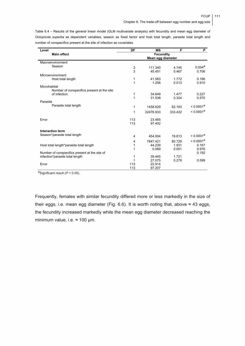
FCUP
Chapter 6. The trade-off between egg number and egg size
111
Table 6.4 – Results of the general linear model (GLM multivariate analysis) with fecundity and mean egg diameter of
Octopicola superba as dependent variables, season as fixed factor and host total length, parasite total length and
number of conspecifics present at the site of infection as covariates.
aSignificant result (P < 0.05).
Frequently, females with similar fecundity differed more or less markedly in the size of
their eggs, i.e. mean egg diameter (Fig. 6.6). It is worth noting that, above ≈ 43 eggs,
the fecundity increased markedly while the mean egg diameter decreased reaching the
minimum value, i.e. ≈ 100 μm.
Level DF MS F P Main effect Fecundity
Mean egg diameter Macroenvironment
Season 3 3
111.340 45.451
4.745 0.467
0.004a 0.706
Microenvironment Host total length 1 41.583 1.772 0.186
1 1.256 0.013 0.910 Microhabitat
Number of conspecifics present at the site of infection 1 34.649 1.477 0.227
1 31.538 0.324 0.570 Parasite
Parasite total length 1 1458.629 62.163 < 0.0001a 1 32476.933 333.432 < 0.0001a Error 113 23.465
113 97.402
Interaction term Season*parasite total length 4 454.004 19.813 < 0.0001a 4 7847.421 80.729 < 0.0001a Host total length*parasite total length 1 44.239 1.931 0.167 1 0.089 0.001 0.976 Number of conspecifics present at the site of infection*parasite total length 1 39.445 1.721
0.192
1 27.075 0.279 0.599 Error 113 22.914 113 97.207
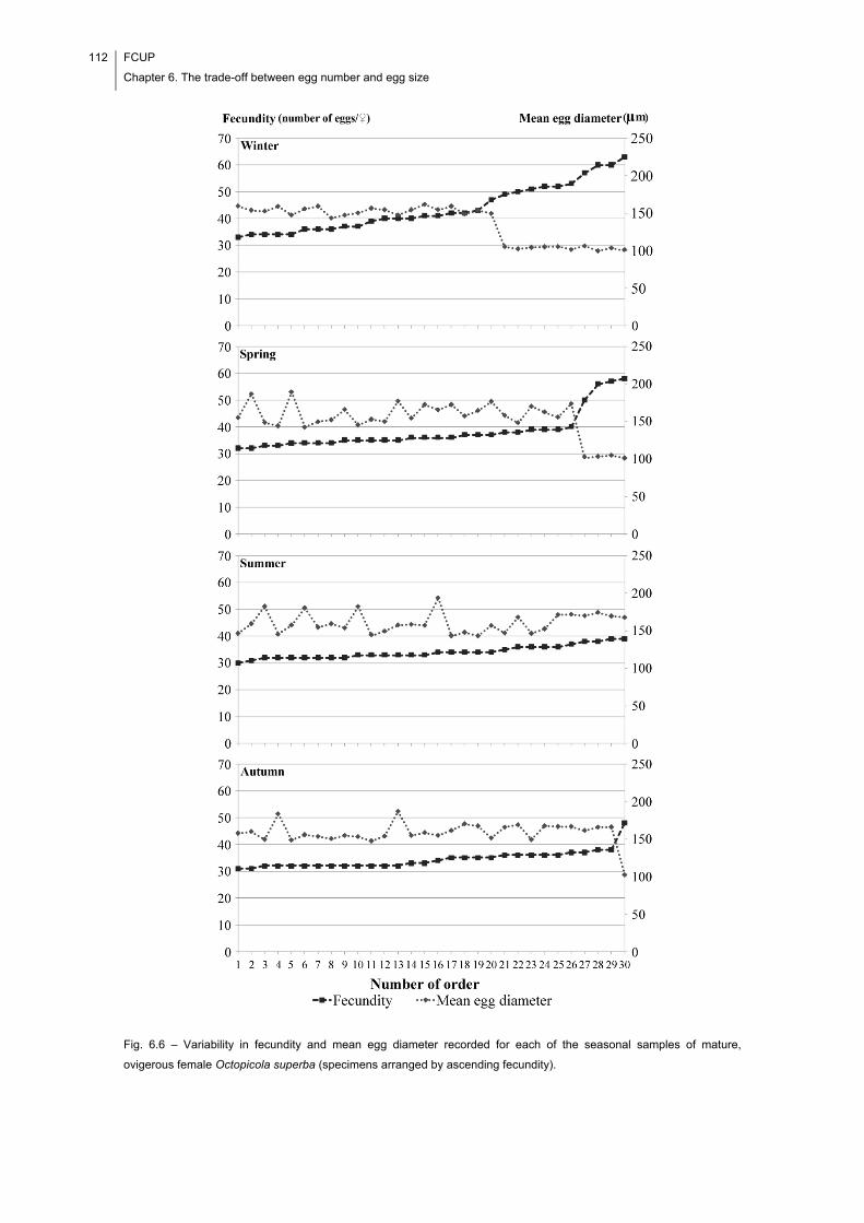
112 FCUP
Chapter 6. The trade-off between egg number and egg size
Fig. 6.6 – Variability in fecundity and mean egg diameter recorded for each of the seasonal samples of mature,
ovigerous female Octopicola superba (specimens arranged by ascending fecundity).
FCUP
Chapter 6. The trade-off between egg number and egg size
113
The results of the non-parametric partial rank correlation test are shown in Table 6.5.
According to this, significant negative relationships were recorded for the winter, spring
and autumn samples of females, while controlling for the potential confounding effect of
parasite total length.
Table 6.5 – Results for the correlation between fecundity and mean egg diameter evaluated for the different seasonal
samples of Octopicola superba using a non-parametric partial rank correlation test.
aSignificant result (P < 0.05).
6.5. Discussion
Egg number and egg size are two important reproductive traits which are crucial to
understanding parasite life history strategies.
In this study, the importance of the trade-off between egg number and egg size
was first addressed by considering the question of whether the parasite is more an r-
strategist or a K-strategist, i.e. how natural selection acted upon egg number and egg
size. According to Jennings & Calow (1975), endoparasites follow both an r- and K-
strategy at the same time, which is made possible by the stable, nutrient-rich
environment provided by the host. Actually, evolutionary theory predicts that all species
would follow an r- and K-strategy simultaneously, had it not been for
macroenvironmental conditions, which invariably force them into one of the
alternatives. In the case of the ectoparasites, they live on more unstable microhabitats
(i.e. on external body surfaces) than endoparasites, a situation which should, therefore,
have implications in terms of their reproductive strategy. Indeed, it has been argued
that in ectoparasitism, the premium on preserving the adult is greater than that on
producing a large number of eggs (Jennings & Calow, 1975), i.e. that ectoparasites are
mainly K-strategists. This positioning of ectoparasites more towards the K-end of the r-
K spectrum of life history strategies goes against the ‘balanced mortality’ hypothesis,
as this assumes massive egg production (see Smith, 1954; Price, 1974; Stunkard,
1975; Kennedy, 1976; Combes, 1995), and is supported by the results here reported
for O. superba. Indeed, the marked difference between the skewness of the
distributions of fecundity and mean egg diameter, the fact that both of these
distributions exhibited high peaks around the mean and the relative position of
Season Control variable Winter Spring Summer Autumn Parasite total length -0.620
< 0.0001a
-0.570
0.001a
0.400
0.031a
-0.457
0.013a

114 FCUP
Chapter 6. The trade-off between egg number and egg size
fecundity and mean egg diameter in the variables factor map can all be understood as
evidence that the females of O. superba do not follow both an r- and K-strategy at the
same time, contrary to that argued for endoparasites. Moreover, the positive and
negative skewness of the distributions of fecundity and mean egg diameter,
respectively, might be understood as an indication that females are particularly
committed to producing a relatively small number of large, well provisioned eggs. This
is because the skewness measures the level of symmetry of the distribution of each of
the two life history traits around the mean. As stated, in K-strategist organisms the
emphasis is on preserving the adult; the remaining energy and matter are used in the
production and maintenance of a small number of extremely fit offspring (Jennings &
Calow, 1975). Such preservation of the adult (i.e. of the parent) would be very
beneficial in the case of the host-parasite system studied here. More specifically, in this
particular system, host-to-host transmission of O. superba is impaired by a combination
of factors regarding the host’s and parasite’s ecology: octopuses are typically
sedentary animals with sparsely distributed populations, while O. superba is highly
host-specific and monoxenous (see Cavaleiro et al., 2013). Hence, the preservation of
the adult is beneficial, enhancing the lifetime reproductive success of individual females
by increasing the temporal spread of egg production. Besides, the production of large,
well provisioned eggs might also be beneficial once they result in infective stages that
are better equipped to seek out ‘new’ hosts or that have a better chance of reaching
areas where ‘new’ hosts may be found (Gotto, 1962). Earlier findings had already
suggested that sedentary hosts select for large egg sizes in parasitic copepods (see
Poulin, 1995b) and that ectoparasites, including monogeneans (Roubal, 1994) and
copepods (Ritchie et al., 1993), do not follow an r- and K-strategy at the same time. It is
worth noting that the coefficient of variation was lower for mean egg diameter than for
fecundity, which suggests that egg size is under a tighter regulation than egg number.
The second question addressed egg production by considering the possibility of
influences of different variables and variable interactions on the number of eggs
produced and their size. While there was no evidence for an influence of site of
infection on egg production, it was found that both egg number and egg size varied
according to season, in accordance to earlier evidence for copepods ectoparasitic on
fish (see Ritchie et al., 1993). The cause of the seasonal variation in reproductive
strategy can only be determined in an additional experimental study. Remarkably,
despite the variability observed in mean egg sac length, the total reproductive effort did
not vary significantly with the season of sampling. This means that, despite the
significant sample differences in fecundity and egg size, the total amount of resources
FCUP
Chapter 6. The trade-off between egg number and egg size
115
invested in one clutch did not vary to a significant extent between samples. Significant
sample differences were recorded concomitantly for all body dimensions and measures
of reproductive effort (Table 6.3), with the seasonal differences found being in
accordance with earlier evidence for parasitic copepods (e.g. Ritchie et al., 1993). This
evidence suggests adaptive phenotypic plasticity in body dimensions and size-
mediated changes in egg production. Actually, a significant influence of the interaction
term season*parasite total length on fecundity and mean egg diameter is supported by
the results of the GLM multivariate analysis. Moreover, larger specimens tended to
produce more eggs (see Fig. 6.4), which is in accordance with earlier findings for
monogeneans (Kearn, 1985), cestodes (Shostak & Dick, 1987), nematodes (Mössinger
& Wenk, 1986; Sinniah & Subramaniam, 1991; Rossin et al., 2005; Herreras et al.,
2007), copepods (Tedla & Fernando, 1970; Ritchie et al., 1993; Van Damme et al.,
1993; Timi et al., 2005), bopyrid isopods (Wenner & Windsor, 1979) and ticks
(Honzáková et al., 1975; Iwuala & Okpala, 1977). This association between body size
and egg number could have been related to the fact that larger females tended to have
larger genital somites (see Fig. 6.2). Actually, one can speculate that the genital somite
was not fully grown in smaller females and, therefore, that the space available for the
oocysts and yolk was smaller in these females. A significant influence of host body size
and number of conspecifics present at the site of infection (i.e., the ‘crowding effect’) on
egg number and size was not detected, which might be related to the fact that the host
is incomparably larger than the parasite, providing it with virtually infinite resources.
With respect to the existence of a phenotypic trade-off between egg number
and egg size, there were some interesting findings. To begin with, the seasonal
distributions of fecundity and mean egg diameter revealed that a negative association
between the two traits might only become apparent at high levels of fecundity, with the
mean egg diameter dropping to the minimum level. Unlike free-living organisms,
parasites do not experience shortages of food, having more than enough resources to
maintain a high rate of egg production (Poulin, 2007a). Nonetheless, it is likely that the
female physiology sets a limit to reproduction (and to clutch dimension – females
cannot carry infinitely large clutches). By this we mean that, beyond a certain fecundity
level (in the study case, ≈ 43 eggs), the females eventually become physiologically
exhausted due to the large energetic investment already made in egg production. As a
consequence, smaller amounts of yolk are produced and allocated to each individual
egg. The occurrence of the trade-off is reinforced by the results of the non-parametric
partial rank correlation test, as a significant negative correlation between the two life
history traits was detected in three of the cases. The fact that previous studies on
116 FCUP
Chapter 6. The trade-off between egg number and egg size
parasite egg production found no evidence for this negative association to occur at the
intraspecific level (e.g. Timi et al., 2005; Rossin et al., 2005), might indicate that the
fecundity levels recorded were just too low for it to be observed. The results for the
summer season (i.e. the significant positive correlation between egg number and egg
size) are not easy to explain but, also in this case, they might be related, at least to
some extent, with the fact that fecundity did not reach the threshold level (≈ 43 eggs)
above which the trade-off was observed. A close look at the data in Fig. 6.6 also
reveals that it is not possible to predict, with any confidence, the mean egg size that we
can expect to observe based on fecundity. This suggests an independent regulation of
egg number and egg size.
In conclusion, the findings of this research suggest that multilevel analyses of
egg number-egg size data sets are crucial to gain accurate knowledge on how the
mechanism of egg production works in parasites. The analysis of the data set recorded
for O. superba suggests that although the parasite tends to produce a relatively small
number of large, well provisioned eggs, the strategy followed is somewhat flexible
throughout the year, the changes in fecundity and egg size probably being determined
by an effect of season on the maturation time (i.e., body size) of the parasite. The
occurrence of a trade-off between egg number and egg size appears to be the
consequence of factors at the parasite level exclusively. Experimental infections in the
laboratory will be important to characterize it further, namely by evaluating the
existence of lifetime variation in egg production.
6.6. Acknowledgements
The authors would like to thank the Portuguese Foundation for Science and
Technology and the European Social Fund for the grant to Francisca I. Cavaleiro
(SFRH/BD/65258/2009). This work was partially funded by the Project AQUAIMPROV
(reference NORTE-07-0124-FEDER-000038), co-financed by the North Portugal
Regional Operational Programme (ON.2 – O Novo Norte), under the National Strategic
Reference Framework (NSRF), through the European Regional Development Fund
(ERDF); and the European Regional Development Fund (ERDF) through the
COMPETE – Operational Competitiveness Programme and national funds through
FCT – Foundation for Science and Technology, under the projects PEst-
C/MAR/LA0015/2013 and DIRDAMyx FCOMP-01-0124-FEDER-020726 (FCT –
PTDC/MAR/116838/2010). Gratitude is also due to three anonymous reviewers for
FCUP
Chapter 6. The trade-off between egg number and egg size
117
their valuable comments on a previous version of the manuscript and to Professor Vítor
Silva for his assistance during field collection of octopuses.

118 FCUP
Chapter 6. The trade-off between egg number and egg size
Chapter 7 Site selection of Acanthochondria cornuta
(Copepoda: Chondracanthidae) in
Platichthys flesus (Teleostei:
Pleuronectidae)
This chapter has been adapted from:
Cavaleiro, F. I., & Santos, M. J. (2011). Site selection of Acanthochondria cornuta (Copepoda: Chondracanthidae) in
Platichthys flesus (Teleostei: Pleuronectidae). Parasitology, 138, 1061–1067.

120 FCUP
Chapter 7. Site selection of Acanthochondria cornuta (Copepoda: Chondracanthidae)
FCUP
Chapter 7. Site selection of Acanthochondria cornuta (Copepoda: Chondracanthidae)
121
7.1. Abstract
Acanthochondria cornuta (Copepoda: Chondracanthidae) (N = 4841; prevalence:
80.0%; mean ± SD [range] intensity: 28.8±24.0 [1–110] parasites) infected the
branchial chambers of the European flounder, Platichthys flesus (L.), (N = 210)
according to an established spatial pattern. This was independent of host size. Higher
intensities resulted, most frequently, in higher numbers of infection sites, probably due
to increased intraspecific competition. Preferential infection of the ocular side was
supported by the recorded abundance data and reflected, probably, the fish’s bottom-
dwelling behaviour. As the parasite develops from one stage into another, it seems to
migrate towards different sites: the copepodites and pre-adult females occurred,
mainly, in the holobranchs; the adults preferred the internal wall (non-gravid/post-gravid
females; adult males) or the pseudobranchs (gravid females). The ventilating water
current along with the blood supply are suggested as two major factors in determining
parasite spatial distribution within the chamber. Parasite crowding in a restricted and
narrow space of the posterior region of the internal wall was recorded frequently and
resembled that previously reported for the plaice. Differences to other host-parasite
systems previously studied should relate with the anatomy of the respiratory apparatus.
Bigamous females are reported for the first time.

122 FCUP
Chapter 7. Site selection of Acanthochondria cornuta (Copepoda: Chondracanthidae)
FCUP
Chapter 7. Site selection of Acanthochondria cornuta (Copepoda: Chondracanthidae)
123
7.2. Introduction
Parasites are usually given the opportunity to choose between a variety of unique sites
on or in their hosts. Indeed, the current knowledge allows us to say that site selection is
universal among them, though varying between the species and groups (Rohde, 1979).
Each given site constitutes a unique microhabitat – due to its very own abiotic and
biotic features – determining, to some extent, the parasite’s choice.
Site selection is well documented in the literature for the parasitic copepods of
fish (see for example, Geets et al., 1997; Lo & Morand, 2001; Scott-Holland et al.,
2006; Timi et al., 2010). In particular, among the chondracanthid copepods, the
members of the genus Acanthochondria Oakley, 1927 were described to infect,
specifically, the branchial chambers of flatfish (see for example, Kabata, 1959, 1979,
1992). These latter constitute a unique and protected environment that offers the
parasites, a number of different possible sites for attachment. Also, they allow blood-
feeding species to feed abundantly, particularly when they infect the filaments of the
holobranchs and pseudobranchs. In accordance with Llewellyn (1956), variability in the
volumes of water that pass by the four holobranchs might lead to different infection
levels, once the larval specimens are given different opportunities to attach on each of
them. Such variability was demonstrated, for instance, for the brown trout (Salmo trutta
forma fario L.), with the volume of water passing over holobranchs II and III being
significantly greater than that passing over holobranchs I and IV (Paling, 1968).
Besides this, several intrinsic factors of the parasites may also determine how they
distribute among the different holobranchs (Ramasamy et al., 1985; Geets et al., 1997).
In recent years, several studies have documented high infection levels of a
species of Acanthochondria, i.e., A. cornuta (Müller, 1776), in European flounder,
Platichthys flesus (Linnaeus, 1758) (Teleostei: Pleuronectidae), of North Sea (Schmidt
et al., 2003) and Atlantic (Marques et al., 2006; Cavaleiro & Santos, 2007, 2009)
waters and considered issues like the spatial and seasonal occurrence of infection.
Notwithstanding, several aspects of the species ‘niche volume’ (see Rohde, 1994),
including, among others, parasite spatial distribution on the host and food, remain still
to be elucidated.
The main aim of this paper is to describe in both qualitative and quantitative
terms, the pattern of site selection of A. cornuta in the European flounder. The possible
driving forces of the observed pattern of spatial distribution are discussed.
124 FCUP
Chapter 7. Site selection of Acanthochondria cornuta (Copepoda: Chondracanthidae)
7.3. Materials and Methods
Host sampling
In total, 210 European flounder were collected: in September 2005, off four localities
(Viana do Castelo, Matosinhos, Aveiro and Figueira da Foz) of north-central Portugal
(41°40′N, 8°50′W to 40°8′N, 8°52′W), eastern North Atlantic (Fig. 7.1); and during one
year (once per season) in the geographical locality where flounders were most
infected, i.e., Matosinhos, between September 2005 and May 2006. Additional details
on sampling are provided in the papers by Cavaleiro & Santos (2007, 2009).
Fig. 7.1 – Geographical location of the four sampling areas in the north-central Portuguese coast, eastern North Atlantic.
Parasite survey
Ocular and blind branchial chambers were examined for Acanthochondria parasites. In
each of them, 17 possible infection sites were considered for analysis. They were: (i)
the operculum (internal surface); (ii) the branchiostegal pocket (delimited by the
operculum and the branchiostegal membrane); (iii) the urohyal; (iv) the internal wall of
the chamber; (v-xvi) the four holobranchs, in each of which, three sites were
considered for analysis – i.e., the bony part (raker) and the inner and outer
hemibranchs (filaments); and (xvii) the pseudobranch (filaments). The opercula and
holobranchs were first dissected out of the fish and only then examined for parasites.
Both the ocular and blind holobranchs were numbered from I to IV. The one nearest
the operculum was assigned as holobranch I, whereas the innermost was assigned as
holobranch IV. Besides the branchial chambers, examined sites on the fish’s body
FCUP
Chapter 7. Site selection of Acanthochondria cornuta (Copepoda: Chondracanthidae)
125
surface included also the skin, fins, eyes, nostrils and mouth cavity. Observations were
carried out under a stereo dissecting microscope at 30× magnification.
Chondracanthids did not detach from the tissues to which they were attached during
the host manipulation. Consequently, they could be localized (and counted) with
precision on the host’s body. Their identification to the species level, that is, as
representatives of A. cornuta, was made following the description of Kabata (1979,
1992). As for the life-cycle stages i.e. copepodite (I-V), pre-adult female or adult
female/male, they were identified in accordance with Heegaard (1947). Different stages
of sexual maturity were considered for the adult female: (i) non-gravid, i.e. female
without egg sacs; (ii) gravid, i.e. female with intact egg sacs; and (iii) post-gravid, i.e.
female with remnants of egg sacs.
Analysis of parasite site selection
Infection levels, that is, prevalence, intensity and abundance, were estimated in
accordance with Bush et al. (1997). Also, the mean, standard deviation (SD) and range
of variation were calculated (for intensity and abundance) and recorded whenever
necessary. Statistical analyses were conducted on SPSS for Windows, version 17.0
(SPSS Inc., Chicago, Illinois), with the significance level set at P < 0.05. The different
levels of analysis considered were as described below.
General site selection. To characterize the spatial distribution of A. cornuta on the
host’s body, the mean intensity was computed for each class of fish including
specimens with the same number of infection sites (sites on the ocular and blind sides
of the fish’s body were considered separately). The existence of a significant
relationship between the two variables, that is, intensity and number of infection sites,
was evaluated by the Spearman’s rank correlation test. Also, the existence of a
significant difference in infection occurrence among the ocular and blind sides was
investigated. Moreover, the infection prevalence in the two sides was compared using
the McNemar’s test for matched-pairs of dichotomous variables. The abundance levels
recorded for the two sides were compared using the Wilcoxon’s matched-pairs signed
ranks test. Non-parametric tests were preferred over parametric ones owing to the non-
normal distribution of both intensity and abundance data (Kolmogorov-Smirnov’s test:
1.598 ≤ Z ≤ 2.494; 0.000 < P ≤ 0.012).
126 FCUP
Chapter 7. Site selection of Acanthochondria cornuta (Copepoda: Chondracanthidae)
Site selection within the branchial chambers. Prevalence and intensity (mean ± SD)
levels were assessed for each infection site in the ocular and blind branchial chambers.
Next, the existence of particular spatial distributions inside them was investigated.
Moreover, significant differences in parasite abundance among the filaments of the
inner and outer hemibranchs of a given holobranch were evaluated by the Wilcoxon’s
matched-pairs signed ranks test, whereas differences in abundance among the four
holobranchs and among the internal wall of the chamber, the holobranch (I-IV)
filaments and the pseudobranch filaments were tested by the Friedman’s rank sum
test. All these analyses were conducted separately for the ocular and blind chambers.
The number of specimens in different stages of development and sexual maturity i.e.,
copepodites (I-V), pre-adult females, non-gravid adult females, gravid adult females,
post-gravid adult females and adult males, was quantified for each site of attachment in
the branchial chamber. In particular, the distribution of copepodites (I-V), pre-adult
females, non-gravid adult females and gravid adult females was assessed for the four
holobranchs. Finally, an analysis of the number of copepods at different infection sites
in the branchial chamber was performed for fish of different size. The classes
considered in this analysis were as follows: class 1: < 25.0 cm (N = 43); class 2: [25.0–
30.0[ cm (N = 107); class 3: [30.0–35.0[ cm (N = 39); and class 4: ≥ 35.0 cm (N = 21).
7.4. Results
The overall infection levels of a total of 4841 specimens of A. cornuta were: prevalence
= 80.0%; intensity (mean ± SD [range]) = 28.8±24.0 (1–110) parasites.
The branchial chamber constituted, as expected, the preferred site of
attachment. However on rare occasions, i.e. in three fish recording several infection
sites, a few parasites were also isolated from the skin (N = 2) and fins (N = 5).
Internally, no parasite was found attached to the bony parts of the holobranchs, urohyal
and operculum, and within the branchiostegal pocket. Also, no specimen was found
infecting the eyes, nostrils and mouth cavity. The recorded data support the existence
of a positive correlation between the intensity and the number of infection sites
(Spearman’s rank correlation test: rs = 0.795, N = 168, P < 0.0001) (Fig. 7.2).
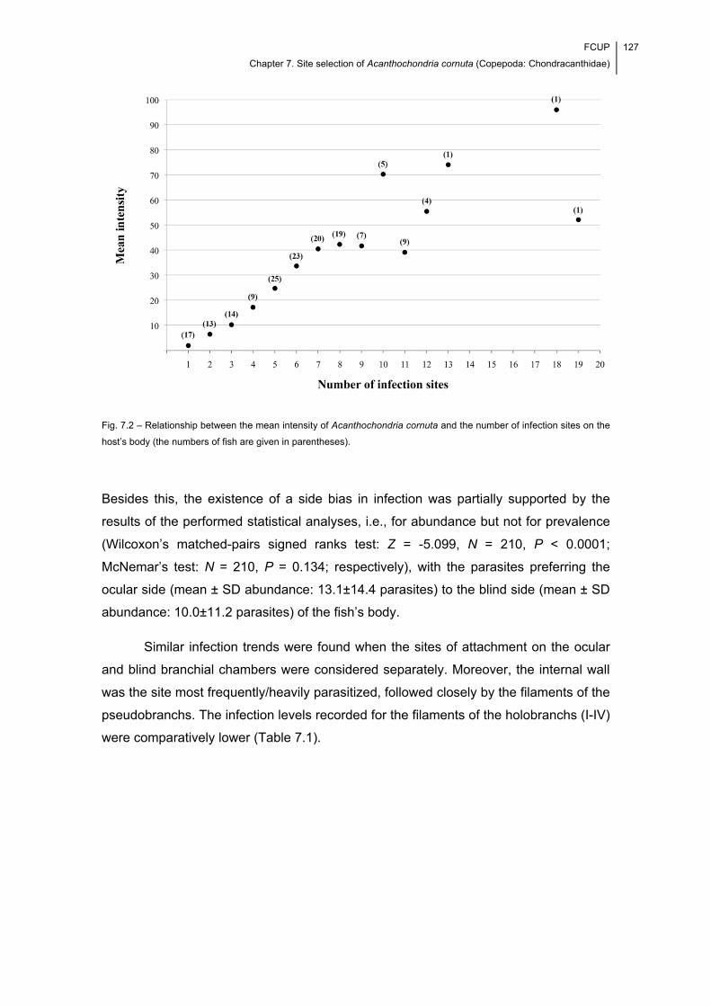
FCUP
Chapter 7. Site selection of Acanthochondria cornuta (Copepoda: Chondracanthidae)
127
Fig. 7.2 – Relationship between the mean intensity of Acanthochondria cornuta and the number of infection sites on the
host’s body (the numbers of fish are given in parentheses).
Besides this, the existence of a side bias in infection was partially supported by the
results of the performed statistical analyses, i.e., for abundance but not for prevalence
(Wilcoxon’s matched-pairs signed ranks test: Z = -5.099, N = 210, P < 0.0001;
McNemar’s test: N = 210, P = 0.134; respectively), with the parasites preferring the
ocular side (mean ± SD abundance: 13.1±14.4 parasites) to the blind side (mean ± SD
abundance: 10.0±11.2 parasites) of the fish’s body.
Similar infection trends were found when the sites of attachment on the ocular
and blind branchial chambers were considered separately. Moreover, the internal wall
was the site most frequently/heavily parasitized, followed closely by the filaments of the
pseudobranchs. The infection levels recorded for the filaments of the holobranchs (I-IV)
were comparatively lower (Table 7.1).
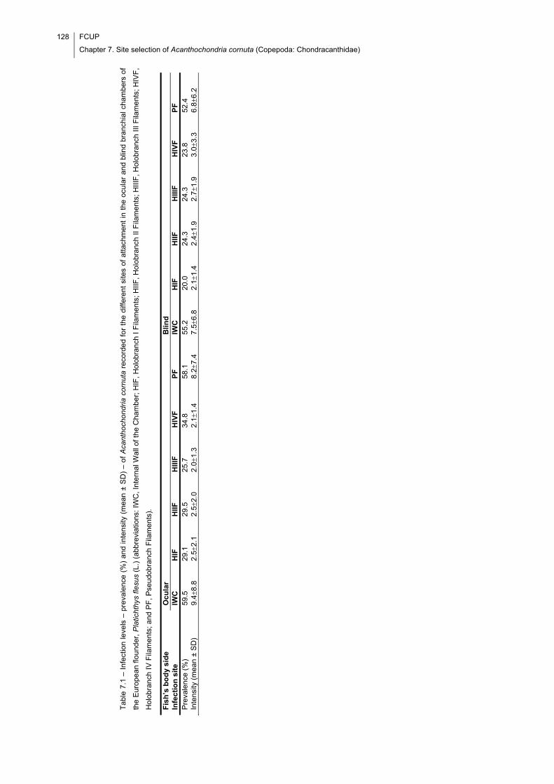
128 FCUP
Chapter 7. Site selection of Acanthochondria cornuta (Copepoda: Chondracanthidae)
Tab
le 7
.1 –
Inf
ectio
n le
vels
– p
reva
lenc
e (%
) an
d in
tens
ity (
mea
n ±
SD
) –
of A
cant
hoc
hond
ria c
ornu
ta r
ecor
ded
for
the
diffe
rent
site
s of
atta
chm
ent
in t
he o
cula
r an
d bl
ind
bran
chia
l cha
mbe
rs o
f
the
Eur
ope
an fl
oun
der,
Pla
ticht
hys
flesu
s (L
.) (
abb
revi
atio
ns: I
WC
, Int
erna
l Wal
l of t
he
Cha
mbe
r; H
IF,
Hol
obra
nch
I F
ilam
ents
; HIIF
, H
olob
ranc
h II
Fila
men
ts; H
IIIF
, H
olob
ranc
h III
Fila
men
ts; H
IVF
,
Hol
obra
nch
IV F
ilam
ents
; and
PF
, P
seud
obra
nch
Fila
men
ts).
Fis
h’s
bo
dy
sid
e O
cula
r B
lind
In
fec
tio
n s
ite
IW
C
HIF
H
IIF
HIII
F
HIV
F
PF
IW
C
HIF
H
IIF
HIII
F
HIV
F
PF
P
reva
lenc
e (%
) 59
.5
29.1
29
.5
25.7
34
.8
58.1
55
.2
20.0
24
.3
24.3
23
.8
52.4
In
tens
ity (
mea
n ±
SD
) 9.
48.
8
2.5
2.1
2.
52.
0
2.0
1.3
2.
11.
4
8.2
7.4
7.
56.
8
2.1
1.4
2.
41.
9
2.7
1.9
3.
03.
3
6.8
6.2
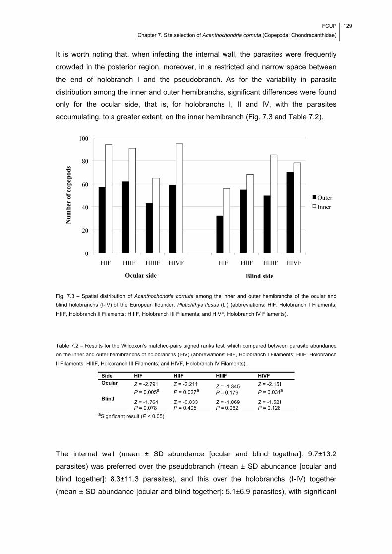
FCUP
Chapter 7. Site selection of Acanthochondria cornuta (Copepoda: Chondracanthidae)
129
It is worth noting that, when infecting the internal wall, the parasites were frequently
crowded in the posterior region, moreover, in a restricted and narrow space between
the end of holobranch I and the pseudobranch. As for the variability in parasite
distribution among the inner and outer hemibranchs, significant differences were found
only for the ocular side, that is, for holobranchs I, II and IV, with the parasites
accumulating, to a greater extent, on the inner hemibranch (Fig. 7.3 and Table 7.2).
Fig. 7.3 – Spatial distribution of Acanthochondria cornuta among the inner and outer hemibranchs of the ocular and
blind holobranchs (I-IV) of the European flounder, Platichthys flesus (L.) (abbreviations: HIF, Holobranch I Filaments;
HIIF, Holobranch II Filaments; HIIIF, Holobranch III Filaments; and HIVF, Holobranch IV Filaments).
Table 7.2 – Results for the Wilcoxon’s matched-pairs signed ranks test, which compared between parasite abundance
on the inner and outer hemibranchs of holobranchs (I-IV) (abbreviations: HIF, Holobranch I Filaments; HIIF, Holobranch
II Filaments; HIIIF, Holobranch III Filaments; and HIVF, Holobranch IV Filaments).
Side HIF HIIF HIIIF HIVF Ocular Z = -2.791
P = 0.005a
Z = -2.211
P = 0.027a Z = -1.345 P = 0.179
Z = -2.151
P = 0.031a Blind
Z = -1.764 P = 0.078
Z = -0.833 P = 0.405
Z = -1.869 P = 0.062
Z = -1.521 P = 0.128
aSignificant result (P < 0.05).
The internal wall (mean ± SD abundance [ocular and blind together]: 9.7±13.2
parasites) was preferred over the pseudobranch (mean ± SD abundance [ocular and
blind together]: 8.3±11.3 parasites), and this over the holobranchs (I-IV) together
(mean ± SD abundance [ocular and blind together]: 5.1±6.9 parasites), with significant
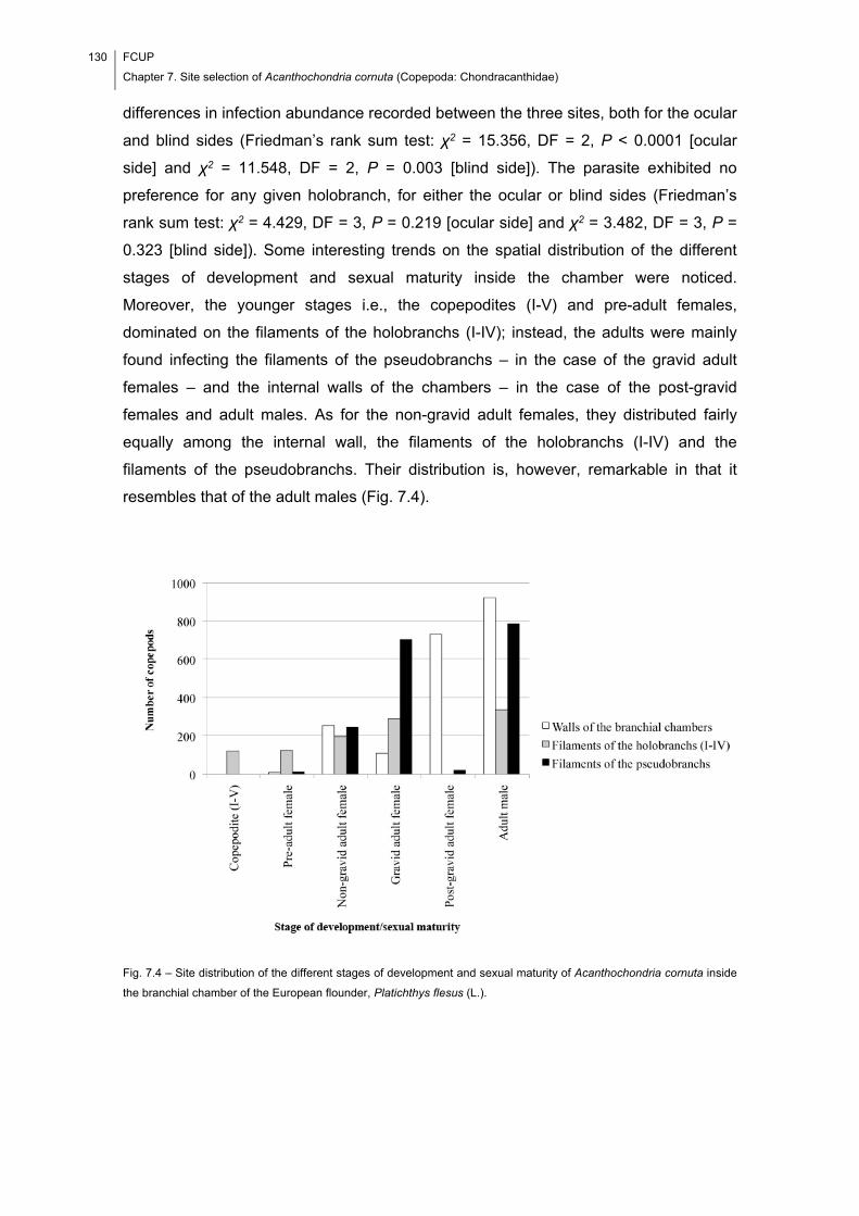
130 FCUP
Chapter 7. Site selection of Acanthochondria cornuta (Copepoda: Chondracanthidae)
differences in infection abundance recorded between the three sites, both for the ocular
and blind sides (Friedman’s rank sum test: χ2 = 15.356, DF = 2, P < 0.0001 [ocular
side] and χ2 = 11.548, DF = 2, P = 0.003 [blind side]). The parasite exhibited no
preference for any given holobranch, for either the ocular or blind sides (Friedman’s
rank sum test: χ2 = 4.429, DF = 3, P = 0.219 [ocular side] and χ2 = 3.482, DF = 3, P =
0.323 [blind side]). Some interesting trends on the spatial distribution of the different
stages of development and sexual maturity inside the chamber were noticed.
Moreover, the younger stages i.e., the copepodites (I-V) and pre-adult females,
dominated on the filaments of the holobranchs (I-IV); instead, the adults were mainly
found infecting the filaments of the pseudobranchs – in the case of the gravid adult
females – and the internal walls of the chambers – in the case of the post-gravid
females and adult males. As for the non-gravid adult females, they distributed fairly
equally among the internal wall, the filaments of the holobranchs (I-IV) and the
filaments of the pseudobranchs. Their distribution is, however, remarkable in that it
resembles that of the adult males (Fig. 7.4).
Fig. 7.4 – Site distribution of the different stages of development and sexual maturity of Acanthochondria cornuta inside
the branchial chamber of the European flounder, Platichthys flesus (L.).
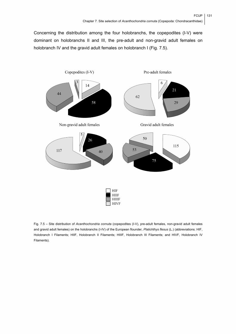
FCUP
Chapter 7. Site selection of Acanthochondria cornuta (Copepoda: Chondracanthidae)
131
Concerning the distribution among the four holobranchs, the copepodites (I-V) were
dominant on holobranchs II and III, the pre-adult and non-gravid adult females on
holobranch IV and the gravid adult females on holobranch I (Fig. 7.5).
Fig. 7.5 – Site distribution of Acanthochondria cornuta (copepodites (I-V), pre-adult females, non-gravid adult females
and gravid adult females) on the holobranchs (I-IV) of the European flounder, Platichthys flesus (L.) (abbreviations: HIF,
Holobranch I Filaments; HIIF, Holobranch II Filaments; HIIIF, Holobranch III Filaments; and HIVF, Holobranch IV
Filaments).
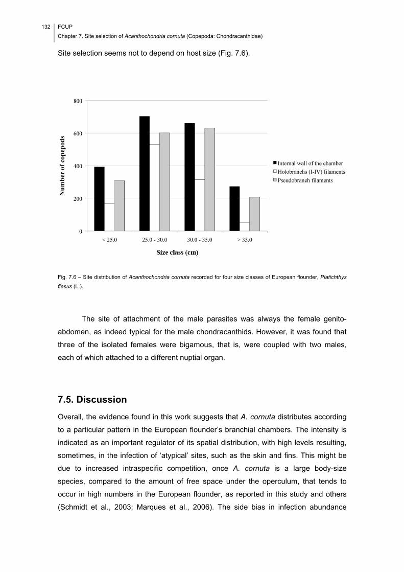
132 FCUP
Chapter 7. Site selection of Acanthochondria cornuta (Copepoda: Chondracanthidae)
Site selection seems not to depend on host size (Fig. 7.6).
Fig. 7.6 – Site distribution of Acanthochondria cornuta recorded for four size classes of European flounder, Platichthys
flesus (L.).
The site of attachment of the male parasites was always the female genito-
abdomen, as indeed typical for the male chondracanthids. However, it was found that
three of the isolated females were bigamous, that is, were coupled with two males,
each of which attached to a different nuptial organ.
7.5. Discussion
Overall, the evidence found in this work suggests that A. cornuta distributes according
to a particular pattern in the European flounder’s branchial chambers. The intensity is
indicated as an important regulator of its spatial distribution, with high levels resulting,
sometimes, in the infection of ‘atypical’ sites, such as the skin and fins. This might be
due to increased intraspecific competition, once A. cornuta is a large body-size
species, compared to the amount of free space under the operculum, that tends to
occur in high numbers in the European flounder, as reported in this study and others
(Schmidt et al., 2003; Marques et al., 2006). The side bias in infection abundance
FCUP
Chapter 7. Site selection of Acanthochondria cornuta (Copepoda: Chondracanthidae)
133
might reflect the fact that the fish spends most of its lifetime buried in the bottom
sediment. This means that the ocular side offers less resistance to the respiratory
water flow, being therefore, comparatively better ventilated. As a consequence, most of
the infective stages carried by the ventilating current will be directed to it. Despite the
previous efforts to characterize the ecology of Acanthochondria infections in flatfish
(see Kabata, 1959), no similar bias in parasite abundance was recorded. This latter
author reported, however, the occurrence of parasite crowding at the same region of
the internal wall of the plaice, Pleuronectes platessa Linnaeus, 1758. As for the
differences found in other host-parasite systems e.g., sites of attachment and overall
infection levels, they should relate, at the least in part, to differences in the anatomy of
the respiratory apparatus, that of the European flounder being of the ‘open’ type
(Kabata, 1959). As A. cornuta develops from one stage into another, it seems to
migrate to different sites within the chamber, with the copepodites (I-V) located mostly
in the central holobranchs, and the remaining stages in sites at some distance from the
chamber’s centre. Moreover, the intensity trend found for the copepodites (I-V), that is
II-III-I-IV, along with the preferential infection of the inner hemibranchs and the
crowding in the posterior region of the internal wall suggest that the ventilating water
current is a major driving force of spatial distribution. Another possible constraint of site
selection is the blood supply (probably a main food source) since (i) the parasite was
absent from the operculum and branchiostegal membrane, both of which are poorly
vascularized and (ii) the filaments of the holobranchs (I-IV) and pseudobranch together
accounted for most of the parasite records. Less easy to explain is the variability in the
main sites of attachment of the gravid and post-gravid females. On the one hand, the
pseudobranchs seem to constitute an adequate place for the egg sac development,
since they are not under the direct effect of the main direction of the ventilating current.
On the other hand, while in the internal wall, the ovigerous females can liberate their
eggs easily to the surrounding environment, therefore becoming post-gravid.
Competition (for space and nourishment) with other branchial parasites present in the
examined fish i.e., Nerocila orbignyi (Guérin-Méneville, 1832) (Isopoda: Cymothoidae)
and gnathiid pranizae (Isopoda: Gnathiidae) (see Cavaleiro & Santos, 2007, 2009),
should have been a less important factor in shaping A. cornuta spatial distribution. This
is because the infection levels recorded for those two taxa were very low.
In conclusion, the results found in this work seem valuable in providing new
insights on the behaviour of the chondracanthid copepods. Moreover, in the parasitic
copepods, site selection has been suggested as being highly dependent on the species
ability to move freely over the host’s body surface e.g., Lepeophtheirus pectoralis
134 FCUP
Chapter 7. Site selection of Acanthochondria cornuta (Copepoda: Chondracanthidae)
(Copepoda: Caligidae) on the European flounder (see Scott, 1901) and, to a lesser
extent, on both morphological and physiological factors (Lo & Morand, 2001). However,
in the case of A. cornuta (and all other gill copepods), the parasite was expected to
remain stationary, that is, attached to the initial infection site. This is because it lives in
a perpetual waterfall needing, therefore, to be securely fastened, a situation that
argues against mobility (Kabata, 1982). Such an expectation is not supported by our
data, which suggest instead, a displacement of the parasite along its successive stages
of development and sexual maturity. This behaviour might be advantageous since it will
reduce the competition among the different stages. The latter might become
particularly problematic in the summer season, when the number of adult females
increases approximately 3–7 times, as compared with other seasons of the year
(Cavaleiro & Santos, 2009). The condition of having more than one male attached is
not new to female chondracanthids. Indeed, it was previously reported for
Rhynchochondria longa Ho, 1967 by Ho (1967) and for Juanettia cornifera Wilson,
1921 by Ho (1970). To the best of our knowledge, however, no report on such a
condition exists for A. cornuta. In this species, the female is provided with paired
nuptial organs (Østergaard & Boxshall, 2004), which suggests that coupling with more
than one male may constitute a relevant aspect of its reproductive biology.
Notwithstanding, bigamous females were rare in this study.
7.6. Acknowledgements
The authors would like to thank the Portuguese Foundation for Science and
Technology and the European Social Fund for the grants awarded to Francisca I.
Cavaleiro (SFRH/BM/23063/2005; SFRH/BD/65258/2009), and also two anonymous
referees for their helpful comments and suggestions on a previous version of the
manuscript.
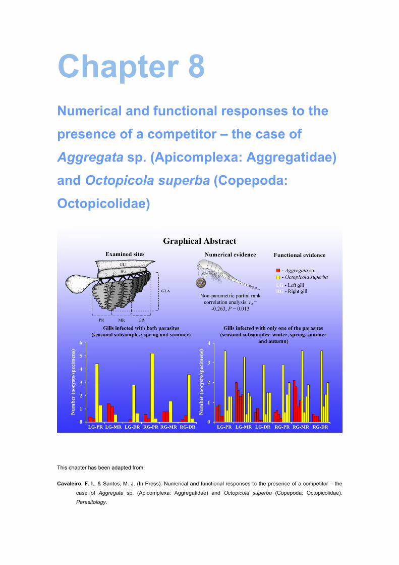
Chapter 8 Numerical and functional responses to the
presence of a competitor – the case of
Aggregata sp. (Apicomplexa: Aggregatidae)
and Octopicola superba (Copepoda:
Octopicolidae)
This chapter has been adapted from:
Cavaleiro, F. I., & Santos, M. J. (In Press). Numerical and functional responses to the presence of a competitor – the
case of Aggregata sp. (Apicomplexa: Aggregatidae) and Octopicola superba (Copepoda: Octopicolidae).
Parasitology.

136 FCUP
Chapter 8. Interference competition between Aggregata sp. and Octopicola superba
FCUP
Chapter 8. Interference competition between Aggregata sp. and Octopicola superba
137
8.1. Abstract
Evidence of interference competition between the eimeriorin coccidian Aggregata sp.
and the octopicolid copepod Octopicola superba at the level of the gills of naturally
infected Octopus vulgaris is evaluated. Numerical and functional responses are
considered for analysis, and the fundamental and realized spatial niches (FSNs and
RSNs) are measured as part of the study. While it was not possible to measure the
FSN of Aggregata sp., the analysis of the infection levels of O. superba recorded for
non-concomitantly and concomitantly infected hosts suggests that the gills and body
skin constitute, respectively, the main and accessory sites of infection of the parasite.
According to the evidence found, the gills function mainly as an accessory site of
infection of Aggregata sp., in specimens in which the caecum and intestine are
massively infected. Evidence for a negative interaction between Aggregata sp. and O.
superba has been found while controlling for a potential confounding effect of host size.
Furthermore, the presence of O. superba on gill lamellae appears to have been
negatively affected by the presence of Aggregata sp., while this latter remained mostly
undisturbed. The mean number of oocysts of Aggregata sp. in the gills was higher in
spring and summer, which were also the seasons presenting the broadest RSN for O.
superba.

138 FCUP
Chapter 8. Interference competition between Aggregata sp. and Octopicola superba

FCUP
Chapter 8. Interference competition between Aggregata sp. and Octopicola superba
139
8.2. Introduction
The common octopus, Octopus vulgaris Cuvier, 1797 (Cephalopoda: Octopodidae),
acts as host of parasites of different taxonomic groups. Among them, two, the
eimeriorin coccidian Aggregata octopiana (Schneider, 1875) Frenzel, 1885
(Apicomplexa: Aggregatidae) and the octopicolid copepod Octopicola superba Humes,
1957 (Copepoda: Octopicolidae), are highly host specific and were reported to occur in
high prevalence (Pascual et al., 1996) and abundance (Bocquet & Stock, 1960) in
samples of O. vulgaris from different geographical regions. Both of them were reported
to infect the gills (e.g. Hochberg, 1983; Gestal et al., 2002; Mladineo & Jozić, 2005;
Pascual et al., 2006; Mladineo & Bočina, 2007), but the occurrence of concomitantly
infected hosts – that is, the simultaneous infection of A. octopiana and O. superba in O.
vulgaris – and the possibility of interspecific interference competition at the level of the
gills have not yet been addressed in any study. The gill infection with eimeriorin
coccidians presumably impairs the octopicolid copepods’ ability to physically establish
on gill tissue resulting, therefore, in interspecific interference competition. Indeed, a
complete substitution of the epithelial and connective tissues by cysts and
developmental stages of A. octopiana, resulting in necrosis and desquamation, has
already been documented for O. vulgaris (Mladineo & Bočina, 2007).
Evidence of interspecific competition is best documented for helminth parasites
(see e.g. Poulin, 2007a; Randhawa, 2012). It can be numerical or functional and both
types are equally convincing (see Poulin, 2001, 2007a). When searching for numerical
evidence of interspecific competition in concomitantly infected hosts, one must test for
the existence of a negative relationship between the numbers of parasites of the two
species. Furthermore, a potential confounding effect of variables at the host and
environment levels on parasite populations and communities (see e.g. Thomas et al.,
2005) must be accounted for, if such a relationship is to be properly detected. In turn,
functional evidence of competition concerns a change in how a parasite uses a given
host resource, in response to the presence of another parasite. This type of evidence is
most frequently detected as a slight shift in the site of infection. Accordingly, it can be
derived by characterizing the ecological niches (sensu Hutchinson, 1957) of parasites,
or more specifically, by considering their spatial dimension. Both the fundamental
spatial niche (FSN) and the realized spatial niche (RSN) must be considered for
analysis (see Poulin, 2007a). The former refers to the potential distribution of a parasite
in the host’s body, that is, the range of sites in which a parasite species can develop,
while the latter concerns the actual niche occupied by a parasite, which is determined
by the interactions it establishes with other parasites. The FSN can only be measured if
140 FCUP
Chapter 8. Interference competition between Aggregata sp. and Octopicola superba
data from specimens harbouring single species infections are available (e.g. Holmes,
1961; Patrick, 1991). In summary, the interspecific competition can result in changes in
numbers of parasites and/or in changes in the spatial distribution of parasites in the
host’s body.
The gills of octopuses constitute an atypical site of infection of eimeriorin
coccidians, as these are usually transmitted trophically, that is, through predation of
crustaceans, the usual intermediate hosts (Hochberg, 1990). Nonetheless, they might
be found infected with them in cases of massive infection, as documented for O.
vulgaris and the genus Aggregata (e.g. Mladineo & Jozić, 2005; Pascual et al., 2006).
An association between the infection of the gills and the infection of the gastrointestinal
tract, the usual site of infection, has, however, not yet been tested.
This study follows on from a survey on the parasite fauna of wild-caught O.
vulgaris, during the course of which both eimeriorin coccidians (i.e. Aggregata sp.,
most likely A. octopiana; it was not possible to measure the sporozoite dimensions to
unequivocally ascertain the identity of the species) and octopicolid copepods (i.e. O.
superba, European subspecies [O. s. superba]) were observed at the gills. Its aims
were as follows: first, to characterize, in numerical terms, the occurrence of Aggregata
sp. and O. superba in the body and gills of the wild-caught specimens of O. vulgaris;
second, to characterize the FSNs and RSNs of Aggregata sp. and O. superba; and
third, to search for numerical and functional evidence of interference competition
between Aggregata sp. and O. superba at the level of the gills.
8.3. Materials and Methods
Octopus vulgaris sampling and parasitological examination
The samples of O. vulgaris examined for parasites consisted of 30 specimens each
and were collected seasonally during 2010 (winter sample: 2 March; spring sample: 24
and 31 May; summer sample: 7 September; and autumn sample: 22 November) off
Matosinhos (41°10′N, 8°42′W), northwest Portuguese coast, northeast Atlantic Ocean.
Each octopus was characterized with respect to different variables, which included the
total body length, sex and stage of sexual maturity (determined according to Dia &
Goutschine, 1990); the Kruskal-Wallis’ test evaluated whether octopuses in different
samples were of comparable size (i.e. total length). The body skin and connective
tissue of arms were washed with saline solution (35‰) to remove the ectoparasites
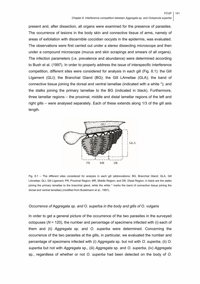
FCUP
Chapter 8. Interference competition between Aggregata sp. and Octopicola superba
141
present and, after dissection, all organs were examined for the presence of parasites.
The occurrence of lesions in the body skin and connective tissue of arms, namely of
areas of exfoliation with discernible coccidian oocysts in the epidermis, was evaluated.
The observations were first carried out under a stereo dissecting microscope and then
under a compound microscope (mucus and skin scrapings and smears of all organs).
The infection parameters (i.e. prevalence and abundance) were determined according
to Bush et al. (1997). In order to properly address the issue of interspecific interference
competition, different sites were considered for analysis in each gill (Fig. 8.1): the Gill
LIgament (GLI); the Branchial Gland (BG); the Gill LAmellae (GLA); the band of
connective tissue joining the dorsal and ventral lamellae (indicated with a white *); and
the stalks joining the primary lamellae to the BG (indicated in black). Furthermore,
three lamellar regions – the proximal, middle and distal lamellar regions of the left and
right gills – were analysed separately. Each of these extends along 1/3 of the gill axis
length.
Fig. 8.1 – The different sites considered for analysis in each gill (abbreviations: BG, Branchial Gland; GLA, Gill
LAmellae; GLI, Gill LIgament; PR, Proximal Region; MR, Middle Region; and DR, Distal Region; in black are the stalks
joining the primary lamellae to the branchial gland, while the white * marks the band of connective tissue joining the
dorsal and ventral lamellae) (modified from Budelmann et al., 1997).
Occurrence of Aggregata sp. and O. superba in the body and gills of O. vulgaris
In order to get a general picture of the occurrence of the two parasites in the surveyed
octopuses (N = 120), the number and percentage of specimens infected with (i) each of
them and (ii) Aggregata sp. and O. superba were determined. Concerning the
occurrence of the two parasites at the gills, in particular, we evaluated the number and
percentage of specimens infected with (i) Aggregata sp. but not with O. superba, (ii) O.
superba but not with Aggregata sp., (iii) Aggregata sp. and O. superba, (iv) Aggregata
sp., regardless of whether or not O. superba had been detected on the body of O.
142 FCUP
Chapter 8. Interference competition between Aggregata sp. and Octopicola superba
vulgaris; and (v) O. superba, regardless of whether or not Aggregata sp. had been
detected in the body of O. vulgaris. Beyond that, the number of oocysts of Aggregata
sp. and specimens of O. superba were assessed (mean and SE) for the gills of non-
concomitantly and concomitantly infected octopuses. Although other parasites were
found infecting the examined octopuses, only these two were found frequently (were
component taxa – prevalence for the total sample of octopuses > 10% [sensu Bush et
al., 1990]) and in high numbers. Hence, the occurrence of other parasites and the
possibility of interspecific competition between other pairs of parasites were
disregarded.
Characterization of the ecological niches of Aggregata sp. and O. superba
The characterization of the ecological niches of Aggregata sp. and O. superba focused
on the spatial dimension of the niche exclusively and considered both the FSN and the
RSN. The seasonal samples of octopuses were considered separately for analysis, so
that seasonal patterns of parasite occurrence and abundance could not interfere with
the results and it was possible to evaluate whether or not the observed niche
configuration was consistent between samples. The FSN of Aggregata sp. could not be
measured once O. superba was found infecting all the examined octopuses. The RSN
of Aggregata sp. and the fundamental and RSNs of O. superba were characterized by
quantifying the differences in parasite occurrence and abundance between the sites of
infection. In the case of the RSNs, only the octopuses infected with Aggregata sp. and
O. superba were considered for analysis. The infection parameters assessed for each
site of infection included the number and percentage of octopuses in which the site was
found infected with a particular parasite and parasite counts (mean ± SD [range]).
Concerning Aggregata sp., it is not possible to determine the true number of parasites
(that is, the exact number of sporozoites) present in a given site. A reliable estimate of
this infection parameter could however be obtained by counting the oocysts visible to
the naked eye, as those octopuses which were more heavily infected usually presented
both more oocysts (enclosing many sporocysts) and sporocysts (enclosing several
sporozoites). The oocyst counting was performed in tissue sections of about 1.0 cm2
(caecal wall, intestinal wall and proximal, middle and distal lamellar regions of gills) – a
measure henceforth referred to as ‘density of coverage of Aggregata sp.’; only the
oocysts visible on the surface were counted. This procedure could be adopted since,
as a rule, the oocysts were regularly distributed throughout the infected tissues. The
total numbers for the gastrointestinal tract and gills were obtained by summing the
FCUP
Chapter 8. Interference competition between Aggregata sp. and Octopicola superba
143
counts for the different sites of infection, that is, the densities of coverage for the
lamellar regions and the counts for the stalks and band of connective tissue in the case
of the gills, and the densities of coverage for the caecal and intestinal walls in the case
of the gastrointestinal tract. The Levins’ measure of niche breadth (B) was assessed
(following Geets et al., 1997; see also Šimková et al., 2000) for each infrapopulation
(sensu Bush et al., 1997) and standardized afterwards (BA). The mean and SD levels
of B and BA were determined for both types of niches (fundamental and RSNs). B and
BA were assessed as follows:
B = ∑
where pj is the proportion of specimens of a parasite found on infection site j.
BA =
where B is the Levins’ measure of niche breadth and N the number of infection sites.
The existence of a relationship between the infection of gills and gastrointestinal tract
was evaluated using the total numbers of oocysts recorded for the two sites
(Spearman’s rank order correlation test). The overlap between RSNs was measured
using the percentage overlap measure, also known as the Renkonen’s index (P)
(following Geets et al., 1997; see also Šimková et al., 2000):
P = 1 - (∑
)
where pia is the proportion of parasites of taxon i found on infection site a and pja the
proportion of parasites of taxon j found on infection site a.
Evaluation of numerical and functional evidence of interference competition
An influence of season and host sex and stage of sexual maturity in the distribution of
the two parasites across the different lamellar regions of the gills was evaluated
considering the total sample of octopuses. Moreover, the counts recorded for the
different seasons of sampling, sexes and stages of sexual maturity were plotted
together and the existence of substantial differences was evaluated. Afterwards,
numerical evidence of interspecific interference competition at the level of the gills was
evaluated by running a non-parametric partial rank correlation analysis in SPSS. This
144 FCUP
Chapter 8. Interference competition between Aggregata sp. and Octopicola superba
analysis tested the existence of a significant negative relationship between the counts
recorded for the two parasites, while controlling for a potential confounding effect of
host body size (i.e. total length) in the results. Since there is no direct way to conduct it
in SPSS, the analysis was specified in a syntax editor window, in accordance with the
instructions provided at the IBM website (http://www01.ibm.com/support/docview.
wss?uid=swg21474822). Only the octopuses infected with at least one of the two
parasites at the gills were considered for analysis. Functional evidence of competition
was evaluated by characterizing the occurrence of each parasite (number and
percentage of octopuses in which the site was found infected with a particular parasite
and density of coverage/parasite counts (mean ± SD [range])) in each of the three
lamellar regions. This characterization was performed separately for the seasonal
subsamples of octopuses infected with (i) both parasites at the gills and (ii) only one of
the two parasites at the gills and for the left and right gills. A change in the infection
levels of one parasite recorded for different lamellar regions, which could have been
determined by the presence of the other parasite, was evaluated.
Statistical analysis of data
Data were analysed using SPSS for Windows, version 19.0 (SPSS Inc., Chicago,
Illinois). The significance level was set at P < 0.05. Non-parametric tests were used
because the abundance data (sensu Bush et al., 1997) for O. superba did not fit the
normal distribution (one-sample Kolmogorov-Smirnov’s test: Z = 1.353, P = 0.051, N =
120 [Aggregata sp.]; and Z = 2.032, P = 0.001, N = 120 [O. superba]) (Zar, 1996).
8.4. Results
Characterization of the seasonal samples of O. vulgaris
The data recorded for the seasonal samples of O. vulgaris were as follows: winter
sample: 69.8±8.2 (56.6–86.0) cm, 13 ♀♀ and 17 ♂♂ and 16 immatures and 14
matures; spring sample: 68.3±10.9 (53.4–88.7) cm, 15 ♀♀ and 15 ♂♂ and 16
immatures and 14 matures; summer sample: 65.8±10.8 (50.2–90.1) cm, 17 ♀♀ and 13
♂♂ and 19 immatures and 11 matures; and autumn sample: 66.9±7.9 (53.4–89.1) cm,
11 ♀♀ and 19 ♂♂ and 15 immatures and 15 matures. The octopuses in different
samples were of comparable size (Kruskal-Wallis’ test [for total body length]: χ2 =
FCUP
Chapter 8. Interference competition between Aggregata sp. and Octopicola superba
145
3.755, DF = 3, P = 0.289). No area of exfoliation with discernible coccidian oocysts was
ever seen in body skin and connective tissue of arms.
Occurrence of Aggregata sp. and O. superba in the body and gills of O. vulgaris
Fifteen (12.5%) out of the 120 examined octopuses were infected only with O. superba,
while none was infected with Aggregata sp. exclusively; the two parasites co-occurred
in 105 (87.5%) octopuses. In 39 octopuses (32.5%), the gills were infected with
Aggregata sp. but not with O. superba; in 40 (33.3%), they were infected with O.
superba but not with Aggregata sp.; and in 11 (9.2%), they were infected with both
parasites. When disregarding whether the other parasite had also been detected in the
octopus’ body, it was found that Aggregata sp. and O. superba occurred at the gills of
50 (41.7%) and 51 (42.5%) octopuses, respectively. The number of specimens of O.
superba recorded for the gills was smaller, on average, for the subsample of
concomitantly infected octopuses (NO. vulgaris = 105), compared with that recorded for the
subsample of non-concomitantly infected octopuses (NO. vulgaris = 15). However, this
result was clearly not statistically significant. In this respect, no consideration is made
for Aggregata sp., as none of the octopuses was infected with it exclusively (Fig. 8.2).
Figures 8.3 and 8.4 show the oocyst and specimen counts for the gills of the examined
octopuses. A non-linear relationship between the counts for the two parasites is evident
(Fig. 8.3). Single and concomitant infections occurred in female and male octopuses,
as well as in immature and mature octopuses (Fig. 8.4).
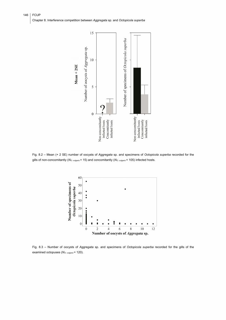
146 FCUP
Chapter 8. Interference competition between Aggregata sp. and Octopicola superba
Fig. 8.2 – Mean (+ 2 SE) number of oocysts of Aggregata sp. and specimens of Octopicola superba recorded for the
gills of non-concomitantly (NO. vulgaris = 15) and concomitantly (NO. vulgaris = 105) infected hosts.
Fig. 8.3 – Number of oocysts of Aggregata sp. and specimens of Octopicola superba recorded for the gills of the
examined octopuses (NO. vulgaris = 120).
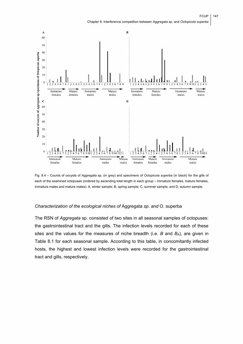
FCUP
Chapter 8. Interference competition between Aggregata sp. and Octopicola superba
147
Fig. 8.4 – Counts of oocysts of Aggregata sp. (in grey) and specimens of Octopicola superba (in black) for the gills of
each of the examined octopuses (ordered by ascending total length in each group – immature females, mature females,
immature males and mature males): A, winter sample; B, spring sample; C, summer sample; and D, autumn sample.
Characterization of the ecological niches of Aggregata sp. and O. superba
The RSN of Aggregata sp. consisted of two sites in all seasonal samples of octopuses:
the gastrointestinal tract and the gills. The infection levels recorded for each of these
sites and the values for the measures of niche breadth (i.e. B and BA), are given in
Table 8.1 for each seasonal sample. According to this table, in concomitantly infected
hosts, the highest and lowest infection levels were recorded for the gastrointestinal
tract and gills, respectively.
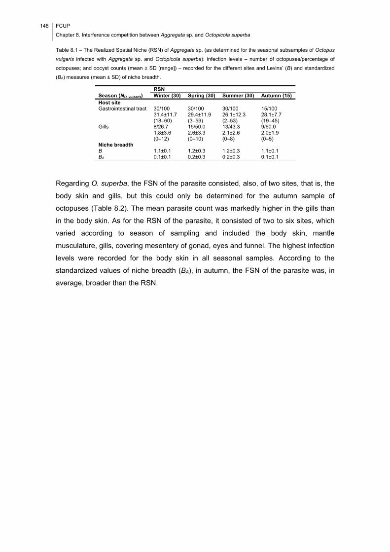
148 FCUP
Chapter 8. Interference competition between Aggregata sp. and Octopicola superba
Table 8.1 – The Realized Spatial Niche (RSN) of Aggregata sp. (as determined for the seasonal subsamples of Octopus
vulgaris infected with Aggregata sp. and Octopicola superba): infection levels – number of octopuses/percentage of
octopuses; and oocyst counts (mean ± SD [range]) – recorded for the different sites and Levins’ (B) and standardized
(BA) measures (mean ± SD) of niche breadth.
RSN Season (NO. vulgaris) Winter (30) Spring (30) Summer (30) Autumn (15) Host site Gastrointestinal tract 30/100
31.4±11.7 (18–60)
30/100 29.4±11.9 (3–59)
30/100 26.1±12.3 (2–53)
15/100 28.1±7.7 (19–45)
Gills 8/26.7 1.8±3.6 (0–12)
15/50.0 2.6±3.3 (0–10)
13/43.3 2.1±2.6 (0–8)
9/60.0 2.0±1.9 (0–5)
Niche breadth B 1.1±0.1 1.2±0.3 1.2±0.3 1.1±0.1 BA 0.1±0.1 0.2±0.3 0.2±0.3 0.1±0.1
Regarding O. superba, the FSN of the parasite consisted, also, of two sites, that is, the
body skin and gills, but this could only be determined for the autumn sample of
octopuses (Table 8.2). The mean parasite count was markedly higher in the gills than
in the body skin. As for the RSN of the parasite, it consisted of two to six sites, which
varied according to season of sampling and included the body skin, mantle
musculature, gills, covering mesentery of gonad, eyes and funnel. The highest infection
levels were recorded for the body skin in all seasonal samples. According to the
standardized values of niche breadth (BA), in autumn, the FSN of the parasite was, in
average, broader than the RSN.
FCUP
Chapter 8. Interference competition between Aggregata sp. and Octopicola superba
149
Table 8.2 – The Fundamental (FSN) (as determined for the seasonal subsample of Octopus vulgaris infected only with
Octopicola superba) and Realized (RSN) (as determined for the seasonal subsamples of O. vulgaris infected with
Aggregata sp. and O. superba) Spatial Niches of O. superba: infection levels – number of octopuses/percentage of
octopuses; and specimen counts (mean ± SD [range]) – recorded for the different sites and Levins’ (B) and
standardized (BA) measures (mean ± SD) of niche breadth.
A significant positive correlation was detected between the oocyst counts recorded for
the gills and gastrointestinal tract (Spearman’s rank order correlation test: rs = 0.370, P
< 0.0001, N = 105). The overlap between the RSNs of the two parasites (P) was 0.3.
Numerical and functional evidence of interference competition
An influence of season and host sex and stage of sexual maturity in the distribution of
the two parasites across the different lamellar regions of the gills could be excluded
after analysing the corresponding plots (Fig. 8.5 A and B).
FSN RSN Season (NO. vulgaris) Autumn (15) Winter (30) Spring (30) Summer (30) Autumn (15) Host site Body skin 15/100
1.0±0.0 (1)
30/100 62.5±22.7 (18–108)
30/100 58.6±76.5 (1–198)
30/100 83.2±59.7 (5–198)
15/100 7.4±7.7 (2–32)
Mantle musculature
- -
7/23.3 0.8±2.1 (0–8)
12/40.0 4.0±7.2 (0–32) -
Gills 11/73.3 8.5±11.7 (0–37)
6/20.0 4.6±12.7 (0–55)
16/53.3 4.3±9.6 (0–45)
16/53.3 3.6±5.6 (0–20)
2/13.3 0.1±0.4 (0–1)
Covering mesentery of gonad
- -
12/40.0 4.0±6.9 (0–30)
15/50.0 9.8±13.6 (0–48) -
Eyes
- -
4/13.3 0.1±0.3 (0–1)
3/10.0 0.1±0.3 (0–1) -
Funnel
- -
2/6.7 0.1±0.4 (0–2)
2/6.7 0.1±0.4 (0–2) -
Niche breadth B 1.3±0.3 1.1±0.3 1.3±0.4 1.5±0.5 1.1±0.2 BA 0.3±0.3 0.1±0.3 0.1±0.3 0.2±0.3 0.1±0.2
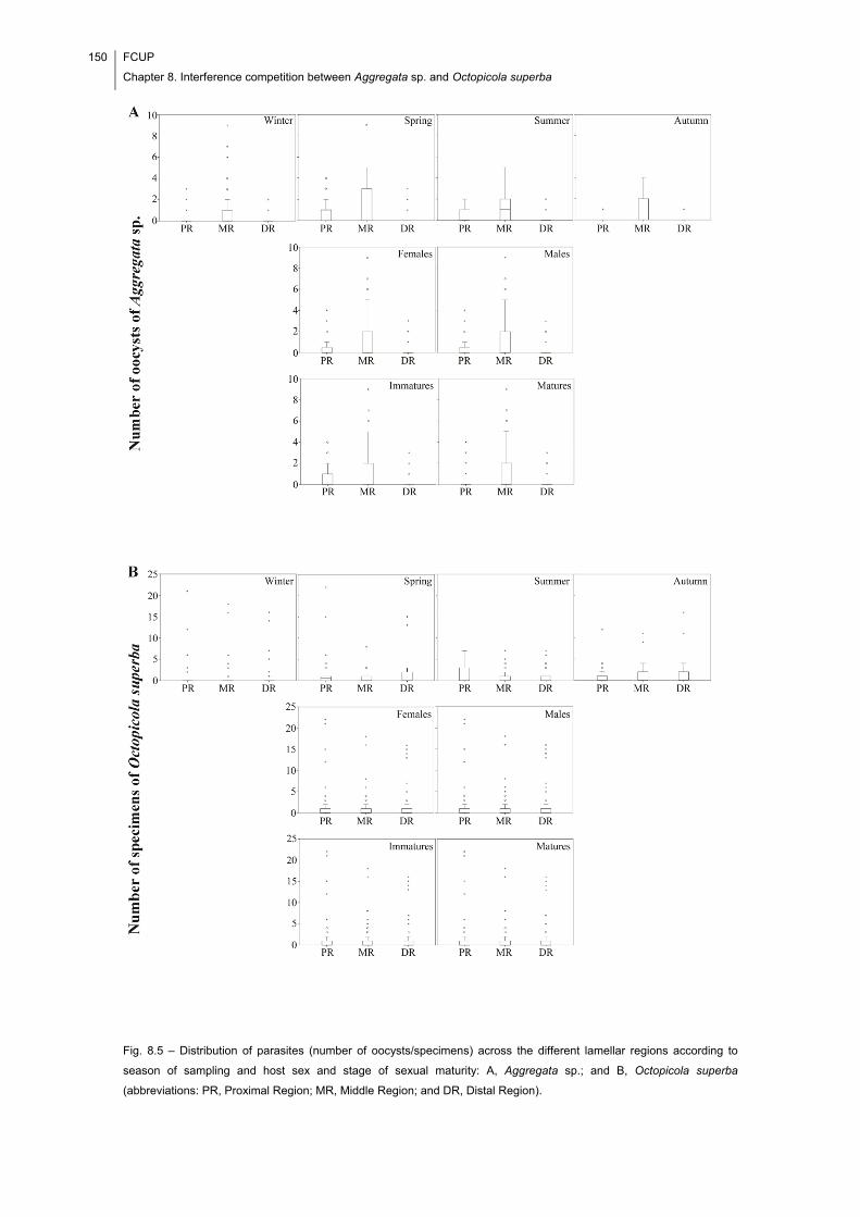
150 FCUP
Chapter 8. Interference competition between Aggregata sp. and Octopicola superba
Fig. 8.5 – Distribution of parasites (number of oocysts/specimens) across the different lamellar regions according to
season of sampling and host sex and stage of sexual maturity: A, Aggregata sp.; and B, Octopicola superba
(abbreviations: PR, Proximal Region; MR, Middle Region; and DR, Distal Region).
FCUP
Chapter 8. Interference competition between Aggregata sp. and Octopicola superba
151
Statistical support for a significant negative relationship between the two parasites has
been found (non-parametric partial rank correlation analysis: rs = -0.263, P = 0.013, N =
90). The sites of infection of Aggregata sp. in the gills included the stalks joining the
primary lamellae to the BG (1/0.8%, 0.0±0.1 [0–1] oocysts), the band of connective
tissue joining the dorsal and ventral lamellae (2/1.7%, 0.0±0.1 [0–1] oocysts) and the
lamellae (50/41.7%, 1.8±2.8 [0–12] oocysts); the gill ligament and the BG were never
found infected. Octopicola superba was found on the gill lamellae exclusively.
According to the infection levels in Table 8.3, which respects the seasonal subsamples
of octopuses whose gills were infected with the two parasites, Aggregata sp. was more
frequent and found in higher numbers in the middle lamellar regions of the left and right
gills, whereas O. superba was more frequent and found in higher numbers on the
proximal and distal lamellar regions of both gills. These trends were consistent
between spring and summer seasons.
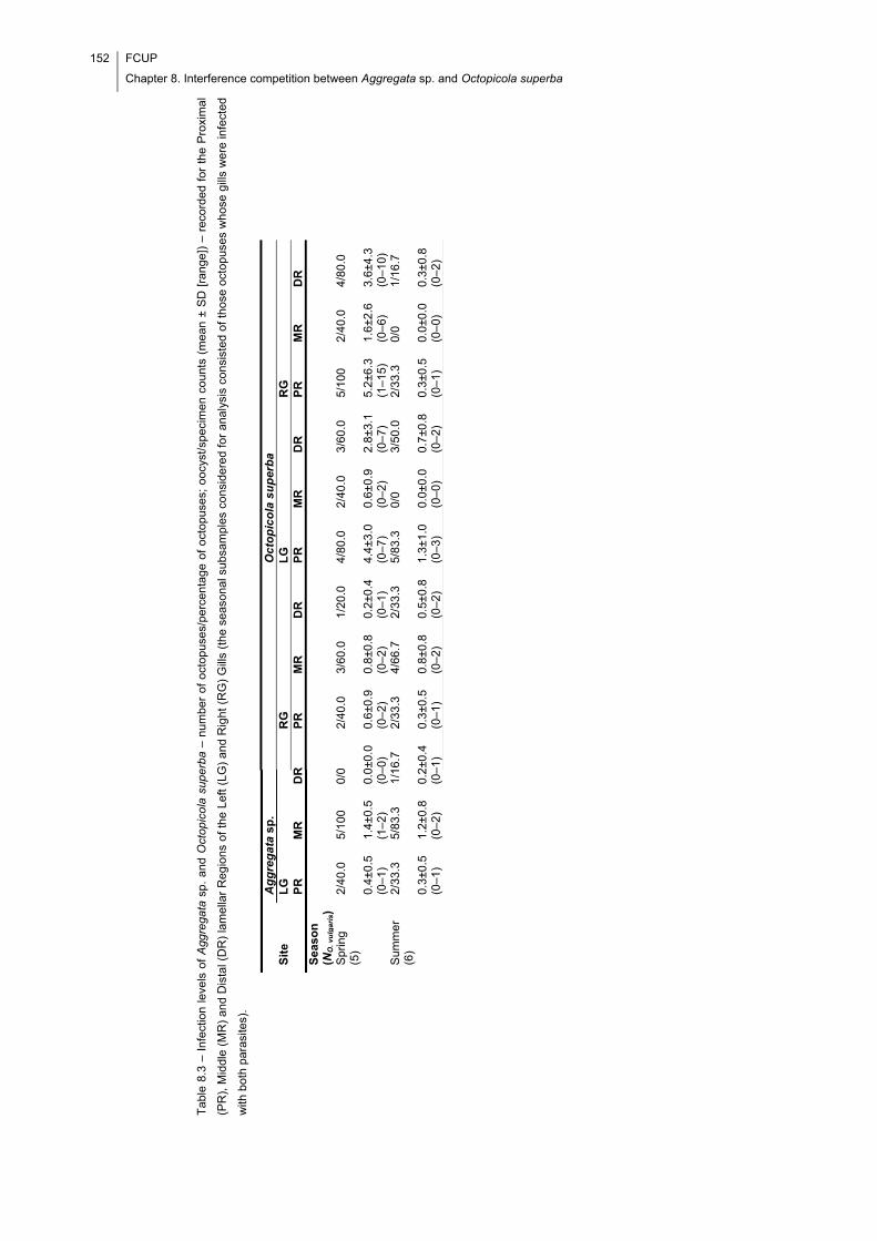
152 FCUP
Chapter 8. Interference competition between Aggregata sp. and Octopicola superba
Tab
le 8
.3 –
Inf
ectio
n le
vels
of
Agg
rega
ta s
p. a
nd O
ctop
icol
a su
perb
a –
num
ber
of o
ctop
uses
/per
cent
age
of o
ctop
uses
; oo
cyst
/spe
cim
en c
ount
s (m
ean
± S
D [
rang
e])
– re
cord
ed f
or t
he P
roxi
ma
l
(PR
), M
iddl
e (M
R)
and
Dis
tal (
DR
) la
mel
lar
Reg
ions
of
the
Left
(LG
) an
d R
ight
(R
G)
Gill
s (t
he s
easo
nal s
ubsa
mpl
es c
onsi
dere
d fo
r an
alys
is c
onsi
sted
of
thos
e oc
topu
ses
who
se g
ills
we
re in
fect
ed
with
bot
h pa
rasi
tes)
.
A
gg
reg
ata
sp.
Oct
op
ico
la s
up
erb
a
Sit
e
LG
R
G
LG
R
G
P
R
MR
D
R
PR
M
R
DR
P
R
MR
D
R
PR
M
R
DR
S
easo
n
(NO
. vu
lga
ris)
Spr
ing
(5)
2/40
.0
5/10
0
0/0
2/40
.0
3/60
.0
1/20
.0
4/80
.0
2/40
.0
3/60
.0
5/10
0
2/40
.0
4/80
.0
0.
4±0.
5 (0
–1)
1.4±
0.5
(1–2
) 0.
0±0.
0 (0
–0)
0.6±
0.9
(0–2
) 0.
8±0.
8 (0
–2)
0.2±
0.4
(0–1
) 4.
4±3.
0 (0
–7)
0.6±
0.9
(0–2
) 2.
8±3.
1 (0
–7)
5.2±
6.3
(1–1
5)
1.6±
2.6
(0–6
) 3.
6±4.
3 (0
–10)
S
umm
er
(6)
2/33
.3
5/83
.3
1/16
.7
2/33
.3
4/66
.7
2/33
.3
5/83
.3
0/0
3/50
.0
2/33
.3
0/0
1/16
.7
0.
3±0.
5 (0
–1)
1.2±
0.8
(0–2
) 0.
2±0.
4 (0
–1)
0.3±
0.5
(0–1
) 0.
8±0.
8 (0
–2)
0.5±
0.8
(0–2
) 1.
3±1.
0 (0
–3)
0.0±
0.0
(0–0
) 0.
7±0.
8 (0
–2)
0.3±
0.5
(0–1
) 0.
0±0.
0 (0
–0)
0.3±
0.8
(0–2
)
FCUP
Chapter 8. Interference competition between Aggregata sp. and Octopicola superba
153
No major difference in the spatial distribution of Aggregata sp. was found when
considering the subsamples of octopuses whose gills were infected with it exclusively.
However, when considering the subsamples of octopuses whose gills were infected
only with O. superba, no clear trend of spatial distribution could be identified (see Table
8.4).
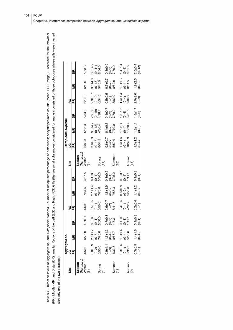
154 FCUP
Chapter 8. Interference competition between Aggregata sp. and Octopicola superba
T
able
8.4
– I
nfe
ctio
n le
vels
of
Agg
rega
ta s
p. a
nd O
ctop
icol
a su
perb
a –
num
ber
of o
ctop
uses
/per
cent
age
of o
ctop
uses
; oo
cyst
/spe
cim
en c
ount
s (m
ean
± S
D [
rang
e])
– re
cord
ed
for
the
Pro
xim
al
(PR
), M
iddl
e (M
R)
and
Dis
tal (
DR
) la
mel
lar
Re
gion
s of
the
Lef
t (L
G)
and
Rig
ht (
RG
) G
ills
(the
sea
sona
l sub
sam
ples
con
side
red
for
anal
ysis
con
sist
ed o
f th
ose
octo
pus
es w
hose
gill
s w
ere
infe
cte
d
with
onl
y on
e of
the
tw
o pa
rasi
tes)
.
A
gg
reg
ata
sp.
O
cto
pic
ola
su
per
ba
S
ite
L
G
RG
S
ite
L
G
RG
PR
M
R
DR
P
R
MR
D
R
P
R
MR
D
R
PR
M
R
DR
S
easo
n
(NO
. vu
lga
ris)
Sea
son
(N
O. v
ulg
ari
s)
Win
ter
(8)
4/50
.0
6/75
.0
4/50
.0
4/50
.0
7/87
.5
3/37
.5
Win
ter
(6)
5/83
.3
5/83
.3
5/83
.3
6/10
0
6/10
0
5/83
.3
0.
8±0.
9 (0
–2)
2.0±
1.7
(0–5
) 0.
5±0.
5 (0
–1)
0.5±
0.5
(0–1
) 2.
1±1.
4 (0
–4)
0.4±
0.5
(0–1
)
3.6±
5.3
(0–1
5)
3.3±
4.2
(0–1
2)
2.9±
3.5
(0–1
0)
2.9±
3.7
(0–1
0)
3.6±
4.6
(0–1
3)
3.6±
4.2
(0–1
1)
Spr
ing
(10)
5/
50.0
7/
70.0
5/
50.0
5/
50.0
7/
70.0
3/
30.0
S
prin
g (1
1)
6/54
.5
4/36
.4
4/36
.4
6/54
.5
5/45
.5
6/54
.5
0.
9±1.
1 (0
–3)
1.6±
1.3
(0–4
) 0.
7±0.
8 (0
–2)
0.6±
0.7
(0–2
) 1.
8±1.
9 (0
–6)
0.3±
0.5
(0–1
)
0.6±
0.7
(0–2
) 0.
4±0.
7 (0
–2)
0.4±
0.7
(0–2
) 0.
5±0.
5 (0
–1)
0.5±
0.7
(0–2
) 0.
8±0.
9 (0
–2)
Sum
mer
(1
2)
4/33
.3
8/66
.7
1/8.
3
5/41
.7
7/58
.3
3/25
.0
Sum
mer
(1
0)
5/50
.0
7/70
.0
7/70
.0
8/80
.0
8/80
.0
7/70
.0
0.
3±0.
5 (0
–1)
1.3±
1.4
(0–4
) 0.
1±0.
3 (0
–1)
0.4±
0.5
(0–1
) 0.
8±0.
8 (0
–2)
0.3±
0.5
(0–1
)
1.3±
1.6
(0–4
) 1.
5±1.
4 (0
–4)
1.5±
1.5
(0–4
) 1.
4±1.
4 (0
–4)
1.3±
1.3
(0–4
) 1.
4±1.
4 (0
–4)
Aut
umn
(9)
3/33
.3
5/55
.6
1/11
.1
2/22
.2
5/55
.6
1/11
.1
Aut
umn
(13)
10
/76.
9
10/7
6.9
8/
61.5
9/
69.2
8/
61.5
8/
61.5
0.
3±0.
5 (0
–1)
1.4±
1.6
(0–4
) 0.
1±0.
3 (0
–1)
0.2±
0.4
(0–1
) 1.
1±1.
2 (0
–3)
0.1±
0.3
(0–1
)
1.3±
1.2
(0–4
) 1.
3±1.
1 (0
–3)
1.5±
1.7
(0–5
) 2.
0±3.
0 (0
–9)
1.9±
2.5
(0–8
) 2.
0±3.
4 (0
–12)
FCUP
Chapter 8. Interference competition between Aggregata sp. and Octopicola superba
155
8.5. Discussion
The eimeriorin coccidians of the genus Aggregata can develop in different sites of the
body of O. vulgaris, including the body skin, connective tissue of arms, mantle
musculature, gills, covering mesentery of digestive gland, covering mesentery of gonad
and different sections of the gastrointestinal tract (oesophagus, crop, caecum and
intestine) (Gestal, 2000; Gestal et al., 2002; Mladineo & Jozić, 2005; Pascual et al.,
2006; Mladineo & Bočina, 2007). These cited studies focused on the eimeriorin
coccidians, and failed to mention the occurrence of other parasites which, being
present, could have influenced the spatial occurrence pattern of Aggregata. In this way,
the available literature cannot be used to characterize the actual FSN of the parasite.
The only consideration that can be made is that the RSN of the parasite consisted of
two of the infection sites mentioned in the literature. In the case of O. superba, the FSN
and RSN consisted of the same two sites in autumn; nonetheless, according to the
recorded BA values, the FSN was broader, on average, than the RSN. By definition, the
RSNs are subsets of the FSNs, which means that they comprise only some of the sites
in which a parasite species can develop. Moreover, in cases where interactions with
other parasite species are unimportant – that is, have no significant effect on any of the
parasites – they represent the optimal sites within the FSN, whereas in cases where
interactions are actually important, they represent the sites of the FSN which are
available to the parasite (Poulin, 2007a). According to these ideas, it is possible to
conclude that the FSN of O. superba is not characterized in full in this study.
Furthermore, it excludes some of the sites in which the parasite can develop (i.e.
mantle musculature, covering mesentery of gonad, eyes and funnel). A possible cause
for this situation may be the number of octopuses infected with O. superba but not with
Aggregata sp. Moreover, this was too low (i.e. NO. vulgaris = 15) to characterize it in full.
The infection levels recorded for the FSN of O. superba are interesting, inasmuch the
mean parasite count was higher for the gills than for the body skin. Furthermore, while
comparing the infection levels recorded for the RSN with those recorded for the FSN, it
was found that lower and higher levels were recorded, respectively, for the gills and
body skin. These findings suggest that the gills constitute the preferred site of infection
of O. superba. Also, they might be understood as preliminary functional evidence of
interspecific interference competition. A preference for the gills is not surprising, once
these provide parasitic copepods with suitable food, that is, epithelial cells, mucus and
blood. The body skin also provides them with epithelial cells and mucus constituting,
therefore, an adequate alternative site of infection. When the gills are infected with
eimeriorin coccidians, the octopicolid copepods’ ability to physically establish on them
156 FCUP
Chapter 8. Interference competition between Aggregata sp. and Octopicola superba
is probably impaired. As a consequence, they may have to move to other sites of the
host’s body, most likely the body skin, as suggested by the infection levels recorded for
the RSN of O. superba. The infection with Aggregata sp. can also affect the spatial
distribution of O. superba on the host’s body by leading to changes in the octopus’
behaviour, as those found by Mladineo and Jozić (2005) – specimens of O. vulgaris
became excited, left their shelters and swam and became inactive inside their shelters
a few days before dying. The reason for this is two-fold: on the one hand, in addition to
crawling, the octopuses move by jet propulsion, and changes in their locomotory
behaviour (and ultimately, in the respiratory water flow through the gills) can affect the
distribution of O. superba on the gills, as this probably moves while under the
dislodging action of the respiratory water current; on the other hand, a prolonged stay
inside a shelter can affect the spatial distribution of O. superba, as this was reported to
exhibit a circadian behavioural rhythm, inhabiting the mantle cavity of O. vulgaris during
daytime and moving out along its arms, mantle and head after dark (Deboutteville et
al., 1957). The significant positive correlation between the numbers of oocysts
recorded for the gills and the gastrointestinal tract can be understood as evidence that
the gills function mainly as an accessory site of infection in octopuses in which the
main sites of absorption along the gastrointestinal tract (that is, the caecum and
intestine) are massively infected. The Renkonen’s index (P) ranges from 0 (no overlap
between niches) to 1 (complete overlap), which means that the overlap between the
RSNs of the two parasites was low. Such a low level can be understood as preliminary
evidence for interactive site segregation (see Holmes, 1973; Poulin, 2007a), that is, of
adjustments in the infection site of O. superba in response to the presence of
Aggregata sp. in the gills. Moreover, although the gills seem to function mainly as an
accessory site of infection of Aggregata sp., they were found infected with the
coccidian in 41.7% of the examined octopuses, while they seem to constitute the
preferred site of infection of O. superba but were only infected with the copepod in
42.5% of the examined octopuses. The standardization of the Levins’ values of niche
breadth (B) resulted in low values, once the Levins’ standardized measure of niche
breadth (BA) ranges from 0 to 1. Such low values indicate that the spatial niches are
dominated by few sites or, more precisely, that the two parasites are specialists with
respect to the sites they infect.
Numerical evidence of a negative interaction between the two parasites at the
level of the gills was given by the non-parametric partial rank correlation analysis.
Furthermore, this analysis could demonstrate the existence of a significant negative
relationship between the counts recorded for the two parasites, while controlling for a
FCUP
Chapter 8. Interference competition between Aggregata sp. and Octopicola superba
157
potential confounding effect of host body size (i.e. total length) in the results. It is worth
noting, that the mean number of oocysts of Aggregata sp. in the gills was higher in
spring and summer and that these were also the seasons for which the RSN of O.
superba consisted of more sites, that is, was broader. These data suggest, therefore, a
negative effect of Aggregata sp. on O. superba. The characterization of the spatial
distribution of the two parasites at the level of the gills further suggested the existence
of such a negative effect. On the one hand, the spatial distribution patterns of the two
parasites were complementary in octopuses whose gills were infected with both of
them; on the other hand, the spatial distribution pattern of Aggregata sp. was
consistent between octopuses whose gills were infected with the two parasites and
with it exclusively (contrary to that found for O. superba). Despite the evidence
underpinning the existence of a negative interaction between Aggregata sp. and O.
superba, the non-linear relationship between the oocyst and specimen counts for the
gills suggests that both parasites occurred aggregated among hosts. This aggregated
distribution of parasites, where a few hosts harboured many parasites while most
harboured none or just a few, was first noted by Crofton (1971), being consistent with
one of the few general laws in parasite ecology (Shaw & Dobson, 1995; Poulin, 2007b).
A possible cause of the aggregation of Aggregata sp. could have been the differential
exposure and susceptibility of the octopuses to the parasite. Furthermore, Aggregata
sp. is a trophically transmitted parasite, and aggregation could have resulted from the
uneven distribution of the infective stages in the population of first intermediate hosts.
Besides, the octopuses were of different size and host body size has been recognized
as a reliable proxy for different factors closely related with susceptibility to infection
(see Poulin, 2013). In the case of O. superba, the aggregation might not only be related
with the different size of the octopuses; indeed, it might also be the result of the
combined effect of a series of factors usually associated with the octopodid
cephalopods (i.e. sedentarism and solitary behaviour) and the octopicolid copepods
(i.e. direct life-cycle and high host specificity).
In conclusion, this study’s findings suggest that the octopicolid copepods are
able to detect changes in the gills resulting from infection with eimeriorin coccidians,
and that their behaviour is mobile enough to allow them to adjust the site of infection.
158 FCUP
Chapter 8. Interference competition between Aggregata sp. and Octopicola superba
8.6. Acknowledgements
The authors thank two anonymous referees, for their valuable comments on a previous
version of the manuscript, Professor Vítor Silva, for his assistance during the field
collection of octopuses, and Professor António Múrias dos Santos, for his advice
concerning the statistical analysis of the data.
8.7. Financial Support
The authors are grateful to the Portuguese Foundation for Science and Technology
and the European Social Fund for the grant to F. I. Cavaleiro (PhD grant reference:
SFRH/BD/65258/2009). This research was partially supported by the European
Regional Development Fund (ERDF) through the COMPETE – Operational
Competitiveness Programme and national funds through FCT – Foundation for Science
and Technology, under the projects PEst-C/MAR/LA0015/2013, DIRDAMyx FCOMP-
01-0124-FEDER-020726 (FCT – PTDC/MAR/116838/2010) and AQUAIMPROV –
Sustainable Aquaculture and Animal Welfare (NORTE-07-0124-FEDER-000038).

Chapter 9
Concluding Remarks

160 FCUP
Chapter 9. Concluding Remarks

FCUP
Chapter 9. Concluding Remarks
161
9.1. Final Notes
The parasitological data recorded for the common octopus, Octopus vulgaris
(Cephalopoda: Octopodidae) and the European flounder, Platichthys flesus (Linnaeus,
1758) (Actinopterygii: Pleuronectidae) (see also the previous works of Cavaleiro, 2007;
Cavaleiro & Santos, 2007, 2009) are interesting from a comparative perspective for the
following reasons:
- First, the recorded infection levels (i.e. prevalence and intensity) suggest that
copepods are the most common ectoparasites occurring on those two species of hosts
in Portuguese coastal waters. The infection with parasitic copepods can become
particularly problematic in aquaculture systems since (i) animals are usually kept at
high densities in tanks and (ii) copepods usually have monoxenous life-cycles.
Furthermore, since O. vulgaris is presently considered a candidate species for marine
aquaculture (Estefanell et al., 2013) and since Portugal has all conditions necessary to
implement aquaculture systems for this cephalopod, it is important to evaluate whether
Octopicola superba Humes, 1957 (Copepoda: Octopicolidae) is pathogenic for the
natural population of octopuses (by analysing the occurrence of associated
histopathology) and also to characterize its life-cycle in detail, including the macro- and
microenvironmental factors involved.
- Second, the recorded seasonality trends were similar for three of the species of
parasitic copepods present on the two hosts, i.e. O. superba, Acanthochondria cornuta
(Müller, 1776) (Copepoda: Chondracanthidae) and Lepeophtheirus pectoralis (Müller,
1777) (Copepoda: Caligidae), and it may be that all parasitic copepods present at the
studied geographic area follow the same seasonality trend. Furthermore, all those
parasites seemed to be influenced by variations in macroenvironmental factors, i.e.
seawater temperature and photoperiod, which indicates that the season of the year is
an important proximate cause of niche restriction. This type of information is crucial to
define effective control and management methods in aquaculture systems, but should
be complemented with data from laboratory experiments.
- Third, the metazoan ectoparasite communities of O. vulgaris and P. flesus consisted
of a few species only, most of which were species of copepods. Furthermore, the high
162 FCUP
Chapter 9. Concluding Remarks
intensity levels recorded for the three species of copepods more commonly isolated
from those two hosts (i.e. O. superba, A. cornuta and L. pectoralis) suggest that they
are highly adapted to them. Actually, and as concerns O. superba, this parasite
presumably has a monoxenous life-cycle, and since octopuses are typically sedentary,
solitary and short-living, strong adaptation is, indeed, very likely. Moreover, the
intensity of O. superba was significantly greater in female than in male octopuses (i.e.,
the sex of the host appears to be an important proximate cause of niche restriction in
O. superba), and such difference in intensity levels can reflect part of the strategy that
ensures the long-term survival of the species.
- Fourth, it should be noted that, despite the large variety of microhabitats provided by
the host, the copepods ectoparasitic on O. vulgaris and P. flesus were found to exhibit
a clear preference for a few, well-defined sites on the body of their hosts. Presumably,
the type of food available at a given microhabitat is likely an important proximate cause
of niche restriction in these parasites. The case of A. cornuta is particularly remarkable.
This parasite was mainly found in the branchial chambers of P. flesus, which can be
related with the fact that, while on them, it has easy access to virtually unlimited blood.
However, the preference for this site can also reflect avoidance of interspecific
competition (with L. pectoralis, present on the body skin and fins of P. flesus),
avoidance of predators (the large dimension of the females makes them easily
noticeable by predators present in the macroenvironment) and facilitation of mating
(mate finding by males should be enhanced in a more confined microhabitat). O.
superba shows a preference for the gills but may have become less restrictive with
respect to the site of infection during the course of evolution, as a result of the
competition with Aggregata sp.
- And fifth, the parasite fauna recorded for O. vulgaris and P. flesus is remarkable,
inasmuch as it reflects the ecology of the host, i.e. the feeding ecology in the case of O.
vulgaris (Aggregata sp., the second most frequent parasite, is transmitted trophically,
i.e. by predation of the crustaceans intermediate hosts), and the migratory behaviour
between different salinity environments in the case of P. flesus (the marine situation for
larval Diplostomum is unusual, and the infection with Diplostomum sp. was probably
acquired while the flounder stayed at low salinity environments). Therefore, the
evidence found indicates that parasitological studies help us better understand animal
life.
FCUP
Chapter 9. Concluding Remarks
163
9.2. Future Research
The aims of future research are the following:
1) To characterize further the parasite fauna of O. vulgaris from Portuguese waters
(infections with coccidians and mesozoans).
2) To evaluate whether O. superba is pathogenic for the natural population of O.
vulgaris and, if so, which type of histopathological lesions are associated with the
infection.
3) To evaluate the exact effect of temperature and photoperiod on life-cycle
progression of O. superba (laboratory experiments).
4) To characterize, for the first time, the basic life-cycle pattern of octopicolid copepods
and to describe the evolution of the population age structure along the year, using the
collection of parasites isolated from O. vulgaris.
5) To establish a hypothesis on how O. superba ensures its host-to-host transmission.
6) To elucidate further the systematics of octopicolid copepods, by unraveling the
phylogenetic position of Octopicolidae in the lichomolgoidean complex of families
(following Humes & Boxshall, 1996) through molecular data analysis.
7) To characterize the larval parasites isolated from O. vulgaris and the latter in
molecular terms, and to evaluate the existence of differences in the parasite fauna of
different species in the O. vulgaris complex.

164 FCUP
References
FCUP
References
165
References
Abollo, E., Gestal, C., López, A., González, A. F., Guerra, A., & Pascual, S. (1998).
Squid as trophic bridges for parasite flow within marine ecosystems: the case of
Anisakis simplex (Nematoda: Anisakidae), or when the wrong way can be right.
South African Journal of Marine Science, 20, 223–232.
Anderson, R. M. (Ed.), (1982). The population dynamics of infectious diseases: theory
and applications. London and New York: Chapman & Hall.
Anstensrud, M., & Schram, T. A. (1988). Host and site selection by larval stages and
adults of the parasitic copepod Lernaeenicus sprattae (Sowerby) (Copepoda,
Pennellidae) in the Oslofjord. Hydrobiologia, 167/168, 587–595.
Bartoli, P., & Gibson, D. I. (2007). Synopsis of the life cycles of Digenea
(Platyhelminthes) from lagoons of the northern coast of the western
Mediterranean. Journal of Natural History, 41, 1553–1570.
Berg, L. S. (1962). Freshwater fishes of the U.S.S.R. and adjacent countries, Volume
1. Jerusalem: Israel Program for Scientific Translations.
Blanchard, E. (1847). Recherches sur l’organisation des vers. Annales des Sciences
Naturelles, Zoologie Series 3, 8, 271–341.
Blaylock, R. B., Overstreet, R. M., & Morton, A. B. (2005). The pathogenic copepod
Phrixocephalus cincinnatus (Copepoda: Pennellidae) in the eye of arrowtooth
flounder, Atherestes stomias, and rex sole, Glyptocephalus zachirus, from British
Columbia. Bulletin of the European Association of Fish Pathologists, 25, 116–
123.
Bocquet, C., & Stock, J. H. (1960). Copépodes parasites d’invertébrés des cotes de la
Manche. VII. Sur la présence d’Octopicola superbus Humes, lichomolgide
associé a Octopus, le long des cotes de Bretagne. Archives de Zoologie
Expérimentale et Générale Volume 99, Notes et Revue, 1, 1–7.
Boxshall, G. A. (1974a). Infections with parasitic copepods in North Sea marine fishes.
Journal of the Marine Biological Association of the United Kingdom, 54, 355–372.
Boxshall, G. A. (1974b). The developmental stages of Lepeophtheirus pectoralis
(Müller, 1776) (Copepoda: Caligidae). Journal of Natural History, 8, 681–700.
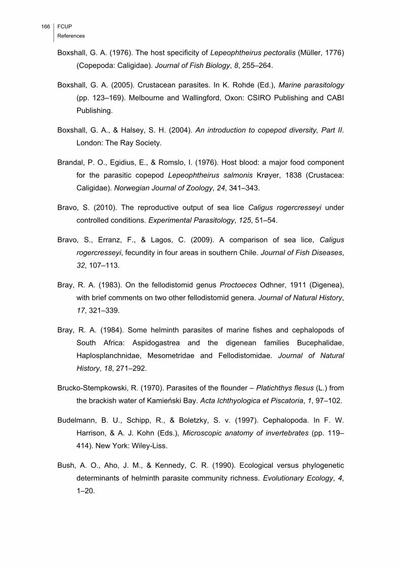
166 FCUP
References
Boxshall, G. A. (1976). The host specificity of Lepeophtheirus pectoralis (Müller, 1776)
(Copepoda: Caligidae). Journal of Fish Biology, 8, 255–264.
Boxshall, G. A. (2005). Crustacean parasites. In K. Rohde (Ed.), Marine parasitology
(pp. 123–169). Melbourne and Wallingford, Oxon: CSIRO Publishing and CABI
Publishing.
Boxshall, G. A., & Halsey, S. H. (2004). An introduction to copepod diversity, Part II.
London: The Ray Society.
Brandal, P. O., Egidius, E., & Romslo, I. (1976). Host blood: a major food component
for the parasitic copepod Lepeophtheirus salmonis Krøyer, 1838 (Crustacea:
Caligidae). Norwegian Journal of Zoology, 24, 341–343.
Bravo, S. (2010). The reproductive output of sea lice Caligus rogercresseyi under
controlled conditions. Experimental Parasitology, 125, 51–54.
Bravo, S., Erranz, F., & Lagos, C. (2009). A comparison of sea lice, Caligus
rogercresseyi, fecundity in four areas in southern Chile. Journal of Fish Diseases,
32, 107–113.
Bray, R. A. (1983). On the fellodistomid genus Proctoeces Odhner, 1911 (Digenea),
with brief comments on two other fellodistomid genera. Journal of Natural History,
17, 321–339.
Bray, R. A. (1984). Some helminth parasites of marine fishes and cephalopods of
South Africa: Aspidogastrea and the digenean families Bucephalidae,
Haplosplanchnidae, Mesometridae and Fellodistomidae. Journal of Natural
History, 18, 271–292.
Brucko-Stempkowski, R. (1970). Parasites of the flounder – Platichthys flesus (L.) from
the brackish water of Kamieński Bay. Acta Ichthyologica et Piscatoria, 1, 97–102.
Budelmann, B. U., Schipp, R., & Boletzky, S. v. (1997). Cephalopoda. In F. W.
Harrison, & A. J. Kohn (Eds.), Microscopic anatomy of invertebrates (pp. 119–
414). New York: Wiley-Liss.
Bush, A. O., Aho, J. M., & Kennedy, C. R. (1990). Ecological versus phylogenetic
determinants of helminth parasite community richness. Evolutionary Ecology, 4,
1–20.
FCUP
References
167
Bush, A. O., Lafferty, K. D., Lotz, J. M., & Shostak, A. W. (1997). Parasitology meets
ecology on its own terms: Margolis et al. revisited. Journal of Parasitology, 83,
575–583.
Cable, J., & Tinsley, R. C. (1991). Intra-uterine larval development of the polystomatid
monogeneans, Pseudodiplorchis americanus and Neodiplorchis scaphiopodis.
Parasitology, 103, 253–266.
Caley, M. J., Schwarzkopf, L., & Shine, R. (2001). Does total reproductive effort evolve
independently of offspring size? Evolution, 55, 1245–1248.
Calow, P. (1983). Pattern and paradox in parasite reproduction. Parasitology, 86, 197–
207.
Carus, J. V. (1885). Prodromus faunae mediterraneae, Volume 1. Coelenterata,
Echinodermata, Vermes, Arthropoda. Stuttgart: E. Schweizerbart'sche
Verlagshandlung.
Castro, G. A. (1991). A physiological perspective: physiological adaptations in host-
parasite systems. In C. A. Toft, A. Aeschlimann, & L. Bolis (Eds.), Parasite-host
associations: coexistence or conflict? (pp. 356–375). Oxford: Oxford Science.
Cavaleiro, F. I. (2007). Dinâmica de Copepoda (Crustacea) ectoparasitas da solha
Europeia Platichthys flesus (Linnaeus, 1758) (Teleostei: Pleuronectidae) na
região norte da costa Portuguesa. Porto: Faculdade de Ciências da Universidade
do Porto [In Portuguese].
Cavaleiro, F. I., & Santos, M. J. (2007). Survey of the metazoan ectoparasites of the
European flounder Platichthys flesus (Linnaeus, 1758) along the north-central
Portuguese coast. Journal of Parasitology, 93, 1218–1222.
Cavaleiro, F. I., & Santos, M. J. (2009). Seasonality of metazoan ectoparasites in
marine European flounder Platichthys flesus (Teleostei: Pleuronectidae).
Parasitology, 136, 855–865.
Cavaleiro, F. I., & Santos, M. J. (In Press). Numerical and functional responses to the
presence of a competitor – the case of Aggregata sp. (Apicomplexa:
Aggregatidae) and Octopicola superba (Copepoda: Octopicolidae). Parasitology.

168 FCUP
References
Cavaleiro, F. I., Ho, J.-S., Iglesias, R., García-Estévez, J. M., & Santos, M. J. (2013).
Revisiting the octopicolid copepods (Octopicolidae: Octopicola): comparative
morphology and an updated key to species. Systematic Parasitology, 86, 77–86.
Chabaud, A. G. (2009). Spirurida. In R. C. Anderson, A. G. Chabaud, & S. Willmott
(Eds.), Keys to the nematode parasites of vertebrates: archival volume (pp. 361–
391). Wallingford: CAB International.
Chappell, L. H. (1995). The biology of diplostomatid eyeflukes of fishes. Journal of
Helminthology, 69, 97–101.
Chibani, M., & Rokicki, J. (2004). Seasonal occurrence of parasites of flounder
Platichthys flesus (L.) from the Gulf of Gdańsk. Oceanological and
Hydrobiological Studies, 33, 17–30.
Chibwana, F. D., & Nkwengulila, G. (2009). Variation in the morphometrics of
diplostomid metacercariae (Digenea: Trematoda) infecting the catfish, Clarias
gariepinus in Tanzania. Journal of Helminthology, 21, 1–10.
Cole, L. C. (1954). The population consequences of life history phenomena. Quarterly
Review of Biology, 29, 103–137.
Combes, C. (1991). Evolution of parasite life-cycles. In C. A. Toft, A. Aeschlimann, & L.
Bolis (Eds.), Parasite-host associations: coexistence or conflict? (pp. 62–82).
Oxford: Oxford Science.
Combes, C. (1995). Interactions durables: écologie et évolution du parasitisme. Paris:
Masson.
Conneely, J. J., & McCarthy, T. K. (1984). The metazoan parasites of freshwater fishes
in the Corrib catchment area, Ireland. Journal of Fish Biology, 24, 363–375.
Crofton, H. D. (1971). A quantitative approach to parasitism. Parasitology, 62, 179–
193.
Crompton, D. W. T. (1991). Nutritional interactions between hosts and parasites. In C.
A. Toft, A. Aeschlimann, & L. Bolis (Eds.), Parasite-host associations:
coexistence or conflict? (pp. 228–258). Oxford: Oxford Science.
Dawson, L. H. J., Renaud, F., Guégan, J. F., & De Meeûs, T. (2000). Experimental
evidence of asymmetrical competition between two species of parasitic
FCUP
References
169
copepods. Proceedings of the Royal Society B: Biological Sciences, 267, 1973–
1978.
Débarre, F., Lion, S., van Baalen, M., & Gandon, S. (2012). Evolution of host life-
history traits in a spatially structured host-parasite system. American Naturalist,
179, 52–63.
Deboutteville, MM. C. D., Humes, A.-G., & Paris, J. (1957). Sur le comportement
d’Octopicola superba Humes, n. g. n. sp. parasite de la Pieuvre Octopus vulgaris
Lamarck. Comptes Rendus Hebdomadaires des Séances de l’Académie des
Sciences, 244, 504–506.
Delle Chiaje, S. (1822). Memorie sulla storia e notomia degli animali senza vertebre del
regno di Napoli. Napoli: Fratelli Fernandes.
Delle Chiaje, S. (1829). Su’ cefalopedi [sic.] In Memorie sulla storia e notomia degli
animali senza vertebre del regno di Napoli, Volume 4. Napoli: Fratelli Fernandes.
Delle Chiaje, S. (1841). Descrizione e notomia degli animali invertebrati della Sicilia
citeriore osservati vivi regli anni 1822-30, Volume 3. Napoli: Stabilimento
Tipografico di C. Batelli.
Dia, M. A., & Goutschine, A. (1990). Echelle de maturité sexuelle du poulpe (Octopus
vulgaris, Cuvier 1797). Bulletin Scientifique du CNROP, 21, 1–6.
Diesing, K. M. (1850). Systema helminthum, Volume 1. Vindoboane: Wilhelmum
Braumüller.
Dippenaar, S. M., van Tonder, R. C., & Wintner, S. P. (2009). Is there evidence of
niche restriction in the spatial distribution of Kroyeria dispar Wilson, 1935, K.
papillipes Wilson, 1932 and Eudactylina pusilla Cressey, 1967 (Copepoda:
Siphonostomatoida) on the gill filaments of tiger sharks Galeocerdo cuvier off
KwaZulu-Natal, South Africa? Hydrobiologia, 619, 89–101.
Dobson, A. P. (1985). The population dynamics of competition between parasites.
Parasitology, 91, 317–347.
Dobson, A. P. (1986). Inequalities in the individual reproductive success of parasites.
Parasitology, 92, 675–682.
170 FCUP
References
Dobson, A., Lafferty, K. D., Kuris, A. M., Hechinger, R. F., & Jetz, W. (2008). Homage
to Linnaeus: how many parasites? How many hosts? Proceedings of the National
Academy of Sciences of the United States of America, 105, 11482–11489.
Dollfus, R. P. (1958). Copépodes, isopodes et helminthes parasites de céphalopodes
de la Méditerranée et de l’Atlantique Européen. Faune Marine des Pyrénées-
Orientales, 1, 61–72.
Dorovskikh, G. N. (1997). Results of the study of fishes' parasites in river basins of the
north-east of the European part of Russia. Trematodes (Trematoda).
Parazitologiya+, 21, 551–564 [In Russian].
Dwyer, W. P., & Smith, C. E. (1989). Metacercariae of Diplostomum spathaceum in the
eyes of fishes from Yellowstone Lake, Wyoming. Journal of Wildlife Diseases, 25,
126–129.
Engelbrecht, H. (1958). Untersuchungen über den parasitenbefall der Nutzfische in
Greifswalder Bodden und Kleinen Haff. Zeitschrift fuer Fischkunde, 7, 481–511.
Estefanell, J., Roo, J., Izquierdo, M., Socorro, J., & Guirao, R. (2013). Growth,
mortality, and biochemical composition in Octopus vulgaris reared under different
conditions of sex ratio. Journal of the World Aquaculture Society, 44, 66–75.
Faust, E. C. (1949). Human helminthology. London: Henry Kimpton.
Felgenhaeur, B. E. (1987). Techniques for preparing crustaceans for scanning electron
microscopy. Journal of Crustacean Biology, 7, 71–76.
Field, J. S., & Irwin, S. W. B. (1995). Life-cycle description and comparison of
Diplostomum spathaceum (Rudolphi, 1819) and D. pseudobaeri (Razmaskin and
Andrejak, 1978) from rainbow trout (Oncorhynchus mykiss Walbaum) maintained
in identical hosts. Parasitology Research, 81, 505–517.
Field, J. S., McKeown, C. A., & Irwin, S. W. B. (1994). A proposed standard method for
the maintenance of Diplostomum spp. (Digenea: Diplostomatidae) in the
laboratory. Parasitology Research, 80, 253–254.
Food and Agriculture Organization of the United Nations. (2007). Webpage.
http://www.globefish.org/vol-94-worl-octopus-market.html. Accessed 5 July 2012.
FCUP
References
171
Food and Agriculture Organization of the United Nations. (2013). Webpage.
http://www.fao.org/figis/servlet/SQServlet?ds=Capture&k1=SPECIES&k1v=1&k1
s=3571&outtype=html. Accessed 12 August 2013.
Froese, R., & Pauly, D. (2008). Webpage. http://www.fishbase.org/Summary
/SpeciesSummary.php?id=1341&lang=english. Accessed 15 April 2008.
Galazzo, D. E., Dayanandan, S., Marcogliese, D. J., & McLaughlin, J. D. (2002).
Molecular systematics of some North American species of Diplostomum
(Digenea) based on rDNA-sequence data and comparisons with European
congeners. Canadian Journal of Zoology, 80, 2207–2217.
Geets, A., Coene, H., & Ollevier, F. (1997). Ectoparasites of the whitespotted rabbitfish,
Siganus sutor (Valenciennes, 1835) off the Kenyan coast: distribution within the
host population and site selection on the gills. Parasitology, 115, 69–79.
Georgiev, B., Biserkov, V., & Genov, T. (1986). In toto staining method for cestodes
with iron acetocarmine. Helminthologia, 23, 279–281.
Gestal, C. (2000). Epidemiología y patología de las coccidiosis en cefalópodos, PhD
dissertation. Vigo: Universidad de Vigo [In Spanish].
Gestal, C., Abollo, E., & Pascual, S. (2002). Observations on associated histopathology
with Aggregata octopiana infection (Protista: Apicomplexa) in Octopus vulgaris.
Diseases of Aquatic Organisms, 50, 45–49.
Gestal, C., Abollo, E., Arias, C., & Pascual, S. (1999). Larval nematodes (Spiruroidea:
Cystidicolidae) in Octopus vulgaris (Mollusca: Cephalopoda: Octopodidae) from
the northeastern Atlantic Ocean. Journal of Parasitology, 85, 508–511.
Gibson, D. I. (1996). Trematoda. In L. Margolis, & Z. Kabata (Ed.), Guide to the
parasites of fishes of Canada, Part IV, Canadian Special Publication of Fisheries
and Aquatic Sciences No. 124. Ottawa: NRC Press.
Gibson, D. I., & Bray, R. A. (1986). The Hemiuridae (Digenea) of fishes from the north-
east Atlantic. Bulletin of the British Museum (Natural History) (Zoology Series),
51, 1–125.
Gibson, D. I., Jones, A. & Bray, R. A. (2002). Keys to the Trematoda, Volume I.
Wallingford: CABI Publishing.
172 FCUP
References
González, M. T., Molinet, C., Arenas, B., Asencio, G., & Carvajal, J. (2012). Fecundity
of the sea louse Caligus rogercresseyi on its native host Eleginops maclovinus
captured near salmon farms in southern Chile. Aquaculture Research, 43, 853–
860.
Gotto, R. V. (1962). Egg-number and ecology in commensal and parasitic copepods.
Journal of Natural History Series 13, 5, 97–107.
Graczyk, T. (1991). Variability of metacercariae of Diplostomum spathaceum
(Rudolphi, 1819) (Trematoda, Diplostomidae). Acta Parasitologica Polonica, 36,
135–139.
Graczyk, T. (1992). Variability of metacercariae of Diplostomum pseudospathaceum
Niewiadomska, 1984 (Trematoda, Diplostomidae). Acta Parasitologica, 37, 5–9.
Guégan, J.-F., Morand, S., & Poulin, R. (2005). Are there general laws in parasite
community ecology? The emergence of spatial parasitology and epidemiology. In
F. Thomas, J.-F. Guégan, & F. Renaud (Eds.), Parasitism and ecosystems (pp.
22–42). Oxford: Oxford University Press.
Guerra, A. (1982). Reflexiones sobre la distribucion geografica de Octopus vulgaris
(Cephalopoda: Octopoda). Gijón: Proceedings of the IVth National Congress of
Malacology [In Spanish].
Guerra, Á., Roura, Á., González, Á. F., Pascual, S., Cherel, Y., & Pérez-Losada, M.
(2010). Morphological and genetic evidence that Octopus vulgaris Cuvier, 1797
inhabits Amsterdam and Saint Paul Islands (southern Indian Ocean). ICES
Journal of Marine Science, 67, 1401–1407.
Haas, W. (2003). Parasitic worms: strategies of host finding, recognition and invasion.
Zoology (Jena), 106, 349–364.
Hastie, L. C., Pierce, G. J., Wang, J., Bruno, I., Moreno, A., Piatkowski, U., & Robin, J.
P. (2009). Cephalopods in the north-eastern Atlantic: species, biogeography,
ecology, exploitation and conservation. Oceanography and Marine Biology: An
Annual Review, 47, 111–190.
Heegaard, P. (1947). Contribution to the phylogeny of the arthropods. Copepoda.
Spolia Zoologica Musei Hauniensis, 8, 1–227.
FCUP
References
173
Heins, D. C., Baker, J. A., & Martin, H. C. (2002). The “crowding effect” in the cestode
Schistocephalus solidus: density-dependent effects on plerocercoid size and
infectivity. Journal of Parasitology, 88, 302–307.
Herreras, M. V., Montero, F. E., Marcogliese, D. J., Raga, J. A., & Balbuena, J. A.
(2007). Phenotypic tradeoffs between egg number and egg size in three parasitic
anisakid nematodes. Oikos, 116, 1737–1747.
Heuch, P. A., Nordhagen, J. R., & Schram, T. A. (2000). Egg production in the salmon
louse [Lepeophtheirus salmonis (Krøyer)] in relation to origin and water
temperature. Aquaculture Research, 31, 805–814.
Ho, J.-S. (1967). A new cyclopoid copepod (Chondracanthidae) parasitic on the
armored Sea Robin from the Florida Straits. Journal of Parasitology, 53, 406–
411.
Ho, J.-S. (1970). Revision of the genera of the Chondracanthidae, a copepod family
parasitic on marine fishes. Beaufortia, 17, 105–218.
Ho, J.-S. (1984). Accessory antennule and the origin of the Chondracanthidae
(Poecilostomatoida). Crustaceana (Supplement), 7, 242–248.
Ho, J.-S., & Lin, C.-L. (2004). Sea lice of Taiwan (Copepoda: Siphonostomatoida:
Caligidae). Keelung: The Sueichan Press.
Hochberg, F. G. (1983). The parasites of cephalopods: a review. Memoirs of the
National Museum of Victoria, 44, 109–145.
Hochberg, F. G. (1990). Diseases of Mollusca: Cephalopoda. Diseases caused by
protistans and metazoans. In O. Kinne (Ed.), Diseases of marine animals,
Volume III (pp. 47–227). Hamburg: Biologische Anstalt Helgoland.
Hoffman, G. L. (1999). Parasites of North American freshwater fishes. Ithaca:
Comstock Publishing Associates, Cornell University Press.
Höglund, J., & Thulin, J. (1992). Identification of Diplostomum spp. in the retina of
perch Perca fluviatilis and the lens of roach Rutilus rutilus from the Baltic Sea –
an experimental study. Systematic Parasitology, 21, 1–19.
Holmes, J. C. (1961). Effects of concurrent infections on Hymenolepis diminuta
(Cestoda) and Moniliformis dubius (Acanthocephala). I. General effects and
comparison with crowding. Journal of Parasitology, 47, 209–216.
174 FCUP
References
Holmes, J. C. (1973). Site selection by parasitic helminths: interspecific interactions,
site segregation, and their importance to the development of helminth
communities. Canadian Journal of Zoology, 51, 333–347.
Honzáková, E., Olejníček, J., Černý, V., Daniel, M., & Dusbábek, F. (1975).
Relationship between number of eggs deposited and body weight of engorged
Ixodes ricinus female. Folia Parasitologica, 22, 37–43.
Hughes, R. C. (1929). Studies on the trematode family Strigeidae (Holostomidae) no.
XIV. Two new species of Diplostomula. Occasional Papers of the Museum of
Zoology, University of Michigan, 202, 1–29.
Humes, A. G. (1957). Octopicola superba n. g., n. sp., copépode cyclopoide parasite
d’un Octopus de la Méditerranée. Vie et Milieu, 8, 1–8.
Humes, A. G. (1963). Octopicola stocki n sp (Copepoda, Cyclopoida), associated with
an Octopus in Madagascar. Crustaceana, 5, 271–280.
Humes, A. G. (1974). Octopicola regalis, n. sp. (Copepoda, Cyclopoida,
Lichomolgidae) associated with Octopus cyaneus from New Caledonia and
Eniwetok Atoll. Bulletin of Marine Science, 24, 76–85.
Humes, A. G., & Boxshall, G. A. (1996). A revision of the lichomologid complex
(Copepoda: Poecilostomatoida), with the recognition of six new families. Journal
of Natural History, 30, 175–227.
Humes, A. G., & Gooding, R. U. (1964). A method for studying the external anatomy of
copepods. Crustaceana, 6, 238–240.
Humes, A. G., & Stock, J. H. (1973). A revision of the family Lichomolgidae Kossmann,
1877, cyclopoid copepods mainly associated with marine invertebrates.
Washington, DC: Smithsonian Institution Press.
Hutchinson, G. E. (1957). Concluding remarks. Cold Spring Harbor Symposia on
Quantitative Biology, 22, 415–427.
Huys, R., & Boxshall, G. A. (1991). Copepod evolution. London: The Ray Society.
Inchausty, V. H., Foutz, M., Heckmann, R. A., Ruas, C., & Ruas, P. (1997).
Diplostomiasis in native and introduced fishes from Yellowstone Lake, Wyoming.
Great Basin Naturalist, 57, 178–183.
FCUP
References
175
Iwuala, M. O. E., & Okpala, I. (1977). Egg output in the weights and states of
engorgement of Amblyomma variegatum (Fabr.) and Boophilus annulatus (Say):
(Ixodoidea: Ixodidae). Folia Parasitologica, 24, 162–172.
Jennings, J. B., & Calow, P. (1975). The relationship between high fecundity and the
evolution of entoparasitism. Oecologia, 21, 109–115.
Johnson, S. C., & Albright, L. J. (1991). Development, growth, and survival of
Lepeophtheirus salmonis (Copepoda: Caligidae) under laboratory conditions.
Journal of the Marine Biological Association of the United Kingdom, 71, 425–436.
Johnston, C. E., & Dykeman, D. (1987). Observations on body proportions and egg
production in the female parasitic copepod (Salmincola salmoneus) from the gills
of Atlantic salmon (Salmo salar) kelts exposed to different temperatures and
photoperiods. Canadian Journal of Zoology, 65, 415–419.
Johnson, S. C., Treasurer, J. W., Bravo, S., Nagasawa, K., & Kabata, Z. (2004). A
review of the impact of parasitic copepods on marine aquaculture. Zoological
Studies, 43, 229–243.
Jones, J. T., Breeze, P., & Kusel, J. R. (1989). Schistosome fecundity: influence of host
genotype and intensity of infection. International Journal for Parasitology, 19,
769–777.
Kabata, Z. (1959). Ecology of the genus Acanthochondria Oakley (Copepoda
Parasitica). Journal of the Marine Biological Association of the United Kingdom,
38, 249–261.
Kabata, Z. (1973). The species of Lepeophtheirus (Copepoda: Caligidae) from fishes of
British Columbia. Journal of the Fisheries Research Board of Canada, 30, 729–
759.
Kabata, Z. (1974). Mouth and mode of feeding of Caligidae (Copepoda), parasites of
fishes, as determined by light and scanning electron microscopy. Journal of the
Fisheries Research Board of Canada, 31, 1583–1588.
Kabata, Z. (1979). Parasitic Copepoda of British fishes. London: The Ray Society.
Kabata, Z. (1981). Copepoda (Crustacea) parasitic on fishes: problems and
perspectives. Advances in Parasitology, 19, 1–71.
176 FCUP
References
Kabata, Z. (1982). The evolution of host-parasite systems between fishes and
Copepoda. In D. T. Metrick, & S. S. Desser (Eds.), Parasites – their world and
ours (pp. 203–212). New York: Elsevier Biomedical Press.
Kabata, Z. (1992). Copepods parasitic on fishes. Synopses of the British fauna (New
375 Series) No. 47. Oegstgeest: Universal Book Services/Dr.W. Backhuys.
Kearn, G. C. (1985). Observations on egg production in the monogenean Entobdella
soleae. International Journal for Parasitology, 15, 187–194.
Kearn, G. (2004). Leeches, lice and lampreys. Dordrecht: Springer.
Kennedy, C. R. (1976). Reproduction and dispersal. In C. R. Kennedy (Ed.), Ecological
aspects of parasitology (pp. 143–160). Amsterdam: North-Holland Publishing
Company.
Kennedy, C. R. (1983). General ecology. In C. Arme, & P. W. Pappas (Eds.), Biology of
the Eucestoda, Volume 1 (pp. 27–80). London: Academic Press.
Kennedy, C. R., & Burrough, R. J. (1978). Parasites of trout and perch in Malham Tarn.
Field Studies, 4, 617–629.
Kennedy, C. R., Nie, P., Kaspers, J., & Paulisse, J. (1992). Are eels (Anguilla anguilla
L.) planktonic feeders? Evidence from parasite communities. Journal of Fish
Biology, 41, 567–580.
Keymer, A. E., Crompton, D. W. T., & Singhvi, A. (1983). Mannose and the “crowding
effect” of Hymenolepis in rats. International Journal for Parasitology, 13, 561–
570.
Khamboonruang, C. (1971). Output of larvae and life span of Trichinella spiralis in
relation to worm burden and superinfection in the mouse. Journal of Parasitology,
57, 289–297.
Knudsen, K. K., & Sundnes, G. (1998). Effects of salinity on infection with Lernaeocera
branchialis (L.) (Copepoda: Pennellidae). Journal of Parasitology, 84, 700–704.
Køie, M. (1999). Metazoan parasites of flounder Platichthys flesus (L.) along a transect
from the southwestern to the northeastern Baltic Sea. ICES Journal of Marine
Science, 56, 157–163.
Krebs, C. J. (1989). Ecological methodology. New York: Harper Collins Publishers.
FCUP
References
177
Krupp, I. M. (1961). Effects of crowding and of superinfection on habitat selection and
egg production in Ancylostoma caninum. Journal of Parasitology, 47, 957–961.
Larkin, M. A., Blackshields, G., Brown, N. P., Chenna, R., McGettigan, P. A.,
McWilliam, H., Valentin, F., Wallace, I. M., Wilm, A., Lopez, R., Thompson, J. D.,
Gibson, T. J., & Higgins, D. G. (2007). ClustalW and ClustalX version 2.
Bioinformatics, 23, 2947–2948.
Lee, P. G. (1994). Nutrition of cephalopods: fuelling the system. Marine and
Freshwater Behaviour and Physiology, 25, 35–51.
Lefèvre, T., Adamo, S. A., Biron, D. G., Missé, D., Hughes, D., & Thomas, F. (2009).
Invasion of the body snatchers: the diversity and evolution of manipulative
strategies in host-parasite interactions. Advances in Parasitology, 68, 45–83.
Leite, T. S., Haimovici, M., Molina, W., & Warnke, K. (2008). Morphological and genetic
description of Octopus insularis, a new cryptic species in the Octopus vulgaris
complex (Cephalopoda: Octopodidae) from the tropical southwestern Atlantic.
Journal of Molluscan Studies, 74, 63–74.
Leppäkoski, E., Gollasch, S., Gruszka, P., Ojaveer, H., Olenin, S., & Panov, V. (2002).
The Baltic – a sea of invaders. Canadian Journal of Fisheries and Aquatic
Sciences, 59, 1175–1188.
Lester, R. J. G., & Hayward, C. J. (2006). Phylum Arthropoda. In P. T. K. Woo (Ed.),
Fish diseases and disorders, Volume 1: protozoan and metazoan infections (pp.
466–565). Wallingford: CABI Publishing.
Llewellyn, J. (1956). The host-specificity, micro-ecology, adhesive attitudes, and
comparative morphology of some trematode gill parasites. Journal of the Marine
Biological Association of the United Kingdom, 35, 113–127.
Lo, C. M., & Morand, S. (2001). Gill parasites of Cephalopholis argus (Teleostei:
Serranidae) from Moorea (French Polynesia): site selection and coexistence.
Folia Parasitologica, 48, 30–36.
Locke, S. A., McLaughlin, J. D., Dayanandan, S., & Marcogliese, D. J. (2010). Diversity
and specificity in Diplostomum spp. metacercariae in freshwater fishes revealed
by cytochrome c oxidase I and internal transcribed spacer sequences.
International Journal for Parasitology, 40, 333–343.

178 FCUP
References
Loot, G., Poulet, N., Brosse, S., Tudesque, L., Thomas, F., & Blanchet, S. (2011).
Determinants of life-history traits in a fish ectoparasite: a hierarchical analysis.
Parasitology, 138, 848–857.
Lucas, M., & Baras, E. (2001). Migration of freshwater fishes. Oxford: Blackwell
Science Ltd.
Lüthen, K. (1988). Zur parasitierung der flunder, Platichthys flesus (L.), an der
Ostseeküste der DDR. Wissenschaftliche Zeitschrift der Pädagogischen
Hochschule Güstrow, 1, 49–62.
Mangold, K. (1983). Octopus vulgaris. In P. R. Boyle (Ed.), Cephalopod life cycles:
species accounts, Volume 1 (pp. 335–364). London: Academic Press.
Mangold, K. (1987). Reproduction. In P. R. Boyle (Ed.), Cephalopod life cycles:
comparative reviews, Volume 2 (pp. 157–200). London: Academic Press.
Mangold, K., & Boletzky, S. V. (1973). New data on reproductive biology and growth of
Octopus vulgaris. Marine Biology, 19, 7–12.
Marin, S. L., Carvajal, J., & George-Nascimento, M. (2007). Dispersal of Udonella
australis (Monogenea: Udonellidae) between caligid copepods Caligus
rogercresseyi and Lepeophtheirus mugiloidis on Chilean rock cod. Journal of
Parasitology, 93, 252–257.
Marques, J. F., Teixeira, C. M., & Cabral, H. N. (2006). Differentiation of commercially
important flatfish populations along the Portuguese coast: evidence from
morphology and parasitology. Fisheries Research, 81, 293–305.
May, R. M., & Anderson, R. M. (1990). Parasite-host coevolution. Parasitology, 100,
S89–S101.
McGladdery, S. E., & Johnston, C. E. (1988). Egg development and control of the gill
parasite, Salmincola salmoneus, on Atlantic salmon kelts (Salmo salar) exposed
to four different regimes of temperature and photoperiod. Aquaculture, 68, 193–
202.
McKeown, C., & Irwin, S. (1995). The life cycle of three Diplostomum species
maintained in the laboratory. International Journal for Parasitology, 25, 897–906.
Mehlhorn, H. (1988). Parasitology in focus. Facts and trends. Berlin, Heidelberg and
New York: Springer-Verlag.
FCUP
References
179
Michel, J. F., Lancaster, M. B., & Hong, C. (1971). Host-induced effects on the size and
body proportions of Ostertagia ostertagi (Stiles, 1892) Ransom, 1907, a
nematode parasite of cattle. Journal of Parasitology, 57, 1185–1189.
Michel, J. F., Lancaster, M. B., & Hong, C. (1978). The length of Ostertagia ostertagi in
populations of uniform age. International Journal for Parasitology, 8, 437–441.
Mideo, N. (2009). Parasite adaptations to within-host competition. Trends in
Parasitology, 25, 261–268.
Mingazzini, P. (1904). Ricerche sul vario modo di fissazione delle tanie alla pareti
intestinale e sul loro assorbimento. Ricerche fatte nel Laboratorio di Anatomia
Normale della Realle Università di Roma, 10, 5–24.
Mladineo, I., & Bočina, I. (2007). Extraintestinal gamogony of Aggregata octopiana in
the reared common octopus (Octopus vulgaris) (Cephalopoda: Octopodidae).
Journal of Invertebrate Pathology, 96, 261–264.
Mladineo, I., & Jozić, M. (2005). Aggregata infection in the common octopus, Octopus
vulgaris (Linnaeus, 1758), Cephalopoda: Octopodidae, reared in a flow-through
system. Acta Adriatica, 46, 193–199.
Möller, H. (1978). The effects of salinity and temperature on the development and
survival of fish parasites. Journal of Fish Biology, 12, 311–323.
Monticelli, F. S. (1899). Il genere «Acanthocotyle». Archives de Parasitologie, 2, 75–
120.
Morales-Serna, F. N., & Gómez, S. (2012). Co-occurrence of the parasitic copepods
Acantholochus zairae Morales-Serna & Gómez, 2010 (Poecilostomatoida:
Bomolochidae) and Pseudochondracanthus diceraus C. B. Wilson, 1908
(Poecilostomatoida: Chondracanthidae) on the gills of Sphoeroides annulatus
(Jenyns) (Pisces: Tetraodontidae). Zoosymposia, 8, 97–105.
Moravec, F. (2003). Observations on the metazoan parasites of the Atlantic salmon
(Salmo salar) after its reintroduction into the Elbe River basin in the Czech
Republic. Folia Parasitologica, 50, 298–304.
Mössinger, J., & Wenk, P. (1986). Fecundity of Litomosoides carinii (Nematoda,
Filarioidea) in vivo and in vitro. Zeitschrift für Parasitenkunde, 72, 121–131.
180 FCUP
References
Niewiadomska, K. (1996). The genus Diplostomum – taxonomy, morphology and
biology. Acta Parasitologica, 41, 55–66.
Niewiadomska, K. (2002). Superfamily Diplostomoidea Poirier, 1886. In D. I. Gibson, A.
Jones, & R. A. Bray (Eds.), Keys to the Trematoda (pp. 159–166). Oxon: CABI
Publishing.
Niewiadomska, K., & Laskowski, Z. (2002). Systematic relationships among six species
of Diplostomum Nordmann, 1832 (Digenea) based on morphological and
molecular data. Acta Parasitologica, 47, 1230–1237.
Niewiadomska, K., & Niewiadomska-Bugaj, M. (1995). Optimal identification procedure
for Diplostomum paracaudum (Iles, 1959) and D. pseudospathaceum
Niewiadomska, 1984 metacercariae (Digenea) based on morphological
characters. Systematic Parasitology, 30, 165–171.
Niewiadomska, K., & Niewiadomska-Bugaj, M. (1998). Morphometric separation of
Diplostomum spathaceum (Rud.) and D. mergi (Dubois) metacercariae
(Digenea). Acta Parasitologica, 43, 209–213.
Niewiadomska, K., & Szymański, S. (1991). Host-induced variability of Diplostomum
paracaudum (Iles, 1959) metacercariae (Digenea). Acta Parasitologica Polonica,
36, 11–17.
Niewiadomska, K., & Szymański, S. (1992). Host-induced variability of Diplostomum
pseudospathaceum Niewiadomska, 1984 metacercariae (Digenea). Acta
Parasitologica, 37, 11–17.
Nikolsky, G. V. (1961). Special ichthyology. Jerusalem: Israel Program for Scientific
Translations.
Nordhagen, J. R., Heuch, P. A., & Schram, T. A. (2000). Size as indicator of origin of
salmon lice Lepeophtheirus salmonis (Copepoda: Caligidae). Contributions to
Zoology, 69, 99–108.
Olsen, O. W. (1974). Animal parasites: their life cycles and ecology. Baltimore:
University Park Press.
Østergaard, P., & Boxshall, G. A. (2004). Giant females and dwarf males: a
comparative study of nuptial organs in female Chondracanthidae (Crustacea:
Copepoda). Zoologischer Anzeiger, 243, 65–74.
FCUP
References
181
Overstreet, R. M., & Hochberg, F. G. (1975). Digenetic trematodes in cephalopods.
Journal of the Marine Biological Association of the United Kingdom, 55, 893–910.
Paling, J. E. (1968). A method of estimating the relative volumes of water flowing over
different gills of a freshwater fish. Journal of Experimental Biology, 48, 533–544.
Palm, H. W. (2004). The Trypanorhyncha Diesing, 1863. Bogor: PKSPL-IPB Press.
Palm, H. W., Klimpel, S., & Bucher, C. (1999). Checklist of metazoan fish parasites of
German coastal waters. Berichte aus dem Institut fuer Meereskunde as der
Christian-Albrechts-Universitaet Kiel, 307, 1–148.
Pascual, S., González, A. F., & Guerra, A. (2006). Unusual sites of Aggregata
octopiana infection in octopus cultured in floating cages. Aquaculture, 254, 21–
23.
Pascual, S., Gestal, C., Estévez, J. M., Rodríguez, H., Soto, M., Abollo, E., & Arias, C.
(1996). Parasites in commercially-exploited cephalopods (Mollusca,
Cephalopoda) in Spain: an updated perspective. Aquaculture, 142, 1–10.
Patrick, M. J. (1991). Distribution of enteric helminths in Glaucomys volans L.
(Sciuridae): a test for competition. Ecology, 72, 755–758.
Pickles, R. S., Thornton, D., Feldman, R., Marques, A., & Murray, D. L. (2013).
Predicting shifts in parasite distribution with climate change: a multitrophic
approach. Global Change Biology, 19, 2645–2654.
Polyanski, U. I. (1955). Contributions to the parasitology of fishes of the northern seas
of the USSR. Leningrad, St. Petersburg: Izd-vo Akademiia Nauk SSSR [In
Russian].
Portuguese Hydrographic Institute. (2012). Webpage. http://www.hidrografico.pt/boias-
ondografo.php. Accessed 5 September 2012.
Portuguese Meteorology Institute. (2012). Webpage. http://www.meteo.pt/pt/
oclima/acompanhamento/index.jsp?selTipo=m&selVar=su&selAno=-1. Accessed
5 September 2012.
Poulin, R. (1995a). Evolution of parasite life history traits: myths and reality.
Parasitology Today, 11, 342–345.
182 FCUP
References
Poulin, R. (1995b). Clutch size and egg size in free-living and parasitic copepods: a
comparative analysis. Evolution, 49, 325–336.
Poulin, R. (1996). The evolution of life history strategies in parasitic animals. Advances
in Parasitology, 37, 107–134.
Poulin, R. (1997a). Species richness of parasite assemblages: evolution and patterns.
Annual Review of Ecology and Systematics, 28, 341–358.
Poulin, R. (1997b). Egg production in adult trematodes: adaptation or constraint?
Parasitology, 114, 195–204.
Poulin, R. (2001). Interactions between species and the structure of helminth
communities. Parasitology, 122, S3–S11.
Poulin, R. (2007a). Evolutionary ecology of parasites. Princeton: Princeton University
Press.
Poulin, R. (2007b). Are there general laws in parasite ecology? Parasitology, 134, 763–
776.
Poulin, R. (2013). Explaining variability in parasite aggregation levels among host
samples. Parasitology, 140, 541–546.
Poulin, R., Krasnov, B. R., Mouillot, D., & Thieltges, W. (2011). The comparative
ecology and biogeography of parasites. Philosophical Transactions of the Royal
Society B: Biological Sciences, 366, 2379–2390.
Price, P. W. (1974). Strategies for egg production. Evolution, 28, 76–84.
Price, T. D., Qvarnström, A., & Irwin, D. E. (2003). The role of phenotypic plasticity in
driving genetic evolution. Proceedings of the Royal Society B: Biological
Sciences, 270, 1433–1440.
Ramasamy, P., Ramalingam, K., Hanna, R. E. B., & Halton, D. W. (1985).
Microhabitats of gill parasites (Monogenea and Copepoda) of teleosts
(Scomberoides spp.). International Journal for Parasitology, 15, 385–397.
Randhawa, H. S. (2012). Numerical and functional responses of intestinal helminths in
three rajid skates: evidence for competition between parasites? Parasitology,
139, 1784–1793.
FCUP
References
183
Rea, J. G., & Irwin, S. W. (1994). The ecology of host-finding behaviour and parasite
transmission: past and future perspectives. Parasitology, 109, S31–S39.
Read, C. P. (1951). The “crowding effect” in tapeworm infections. Journal of
Parasitology, 37, 174–178.
Reategui, R., Oliva, M., Luque, J. L., & Huaman, P. (1989). Presencia de una forma
inmadura de Proctoeces sp. (Trematoda: Fellodistomidae), parásito de Octopus
vulgaris (Cephalopoda: Octopodidae) en el litoral Peruano. Parasitología al Dia,
13, 90–92.
Redi, F. (1684). Osservazioni di Francesco Redi: Accademico della Crusca. Intorno
agli animali viventi che si trovano negli animali viventi. Firenze: Piero Matini.
Ritchie, G., Mordue, A. J., Pike, A. W., & Rae, G. H. (1993). The reproductive output of
Lepeophtheirus salmonis adult females in relation to seasonal variability of
temperature and photoperiod. In G. A. Boxshall, & D. Defaye (Eds.), Pathogens
of wild and farmed fish: Sea lice (pp. 153–165). London: Ellis Horwood.
Roff, D. A. (1992). The evolution of life histories: Theory and analysis. New York:
Chapman & Hall.
Rohde, K. (1976). Monogenean gill parasites of Scomberomorus commersoni
Lacépéde and other mackerel on the Australian east coast. Parasitology
Research, 51, 49–69.
Rohde, K. (1977). A non-competitive mechanism responsible for restricting niches.
Zoologischer Anzeiger, 199, 164–172.
Rohde, K. (1979). A critical evaluation of intrinsic and extrinsic factors responsible for
niche restriction in parasites. American Naturalist, 114, 648–671.
Rohde, K. (1980). Warum sind ökologische Nischen begrenzt? Zwischenartlicher
Antagonismus oder innerartlicher Zusammenhalt? Naturwissenschaftliche
Rundschau, 33, 98–102.
Rohde, K. (1984). Ecology of marine parasites. Helgoland Marine Research, 37, 5–33.
Rohde, K. (1993). Ecology of marine parasites. Wallingford: CAB International.
Rohde, K. (1994). Niche restriction in parasites: proximate and ultimate causes.
Parasitology, 109, S69–S84.
184 FCUP
References
Rohde, K. (2005). Definitions, and adaptations to a parasitic way of life. In K. Rohde
(Ed.), Marine parasitology (pp. 1–6). Melbourne and Wallingford, Oxon: CSIRO
Publishing and CABI Publishing.
Rohde, K., & Hobbs, R. (1986). Species segregation: competition or reinforcement of
reproductive barriers? In M. Cremin, C. Dobson, & D. E. Moorhouse (Eds.),
Parasite lives. Papers on parasites, their hosts and their associations to honour J.
F. A. Sprent (pp. 189–199). St. Lucia, London and New York: University of
Queensland Press.
Rohde, K., & Rohde, P. P. (2005). The ecological niches of parasites. In K. Rohde
(Ed.), Marine parasitology (pp. 286–293). Melbourne and Wallingford, Oxon:
CSIRO Publishing and CABI Publishing.
Rohde, K., Worthen, W. B., Heap, M., Hugueny, B., & Guégan, J. F. (1998).
Nestedness in assemblages of metazoan ecto- and endoparasites of marine fish.
International Journal for Parasitology, 28, 543–549.
Rossin, M. A., Poulin, R., Timi, J. T., & Malizia, A. I. (2005). Causes of inter-individual
variation in reproductive strategies of the parasitic nematode Graphidioides
subterraneus. Parasitology Research, 96, 335–339.
Roubal, F. R. (1994). Observations on the eggs and fecundity of dactylogyrid and
diplectanid monogeneans from the Australian marine sparid fish, Acanthopagrus
australis. Folia Parasitologica, 41, 220–222.
Ruiz Daniels, R., Beltran, S., Poulin, R., & Lagrue, C. (2013). Do parasites adopt
different strategies in different intermediate hosts? Host size, not host species,
influences Coitocaecum parvum (Trematoda) life history strategy, size and egg
production. Parasitology, 140, 275–283.
Saitou, N., & Nei, M. (1987). The neighbor-joining method: a new method for
reconstructing phylogenetic trees. Molecular and Biological Evolution, 4, 406–
425.
Saldanha, I., Leung, T. L. F., & Poulin, R. (2009). Causes of intraspecific variation in
body size among trematode metacercariae. Journal of Helminthology, 83, 289–
293.
Schmidt, V., Zander, S., Körting, W., & Steinhagen, D. (2003). Parasites of the flounder
Platichthys flesus (L.) from the German Bight, North Sea, and their potential use
FCUP
References
185
in ecosystem monitoring. A. Infection characteristics of potential indicator
species. Helgoland Marine Research, 57, 236–251.
Scholz, T. (1999). Parasites in cultured and feral fish. Veterinary Parasitology, 84, 317–
335.
Scott, A. (1901). Lepeophtheirus and Lernaea. Liverpool Marine Biology Committee
Memoir no. VI. London: Williams and Norgate.
Scott-Holland, T. B., Bennett, S. M., & Bennett, M. B. (2006). Distribution of an
asymmetrical copepod, Hatschekia plectropomi, on the gills of Plectropomus
leopardus. Journal of Fish Biology, 68, 222–235.
Severtsov, A. S. (2013). Relationship between fundamental and realized ecological
niches. Biology Bulletin Review, 3, 187–195.
Shaw, D. J., & Dobson, A. P. (1995). Patterns of macroparasite abundance and
aggregation in wildlife populations: a quantitative review. Parasitology, 111,
S111–S133.
Shigin, A. A. (1976). Metacercariae of the genus Diplostomum in the fauna of the
USSR. Parazitologiya+, 10, 346–351 [In Russian].
Shigin, A. A. (1986). Trematodes of the USSR. Genus Diplostomum. Metacercariae.
Moscow: Akademii Nauk SSSR.
Shostak, A. W., & Dick, T. A. (1987). Individual variability in reproductive success of
Triaenophorus crassus Forel (Cestoda: Pseudophyllidea), with comments on use
of the Lorenz curve and Gini coefficient. Canadian Journal of Zoology, 65, 2878–
2885.
Šimková, A., Desdevises, Y., Gelnar, M., & Morand, S. (2000). Co-existence of nine gill
ectoparasites (Dactylogyrus: Monogenea) parasitising the roach (Rutilus rutilus
L.): history and present ecology. International Journal for Parasitology, 30, 1077–
1088.
Sinniah, B., & Subramaniam, K. (1991). Factors influencing the egg production of
Ascaris lumbricoides: relationship to weight, length and diameter of worms.
Journal of Helminthology, 65, 141–147.
186 FCUP
References
Smith, F. E. (1954). Quantitative aspects of population growth. In J. Boell (Ed.),
Dynamics of growth processes (pp. 277–294). Princeton: Princeton University
Press.
Sobral, D., & Gomes, J. (1997). Peixes Litorais – Estuário do Sado. Lisboa: Instituto da
Conservação da Natureza [In Portuguese].
Söller, R., Warnke, K., Saint-Paul, U., & Blohm, D. (2000). Sequence divergence of
mitochondrial DNA indicates cryptic biodiversity in Octopus vulgaris and supports
the taxonomic distinctiveness of Octopus mimus (Cephalopoda: Octopodidae).
Marine Biology, 136, 29–35.
SPSS Inc. (2007). SPSS Base 17.0 User’s Guide. Chicago, IL.
Stearns, S. C. (1992). The evolution of life histories. Oxford: Oxford University Press.
Stock, J. H., Humes, A. G., & Gooding, R. U. (1963). Copepoda associated with West
Indian invertebrates-IV. The genera Octopicola, Pseudanthessius and Meomicola
(Cyclopoida, Lichomolgidae). Studies on the Fauna of Curaçao and other
Caribbean islands, 18, 1–9.
Stunkard, H. W. (1975). Life-histories and systematics of parasitic flatworms.
Systematic Zoology, 24, 378–385.
Sukhdeo, M. V. K. (2012). Where are the parasites in food webs? Parasites & Vectors,
5, 239.
Szalai, A. J., & Dick, T. A. (1989). Differences in numbers and inequalities in mass and
fecundity during the egg-producing period for Raphidascaris acus (Nematoda:
Anisakidae). Parasitology, 98, 489–495.
Tamura, K., Dudley, J., Nei, M., & Kumar, S. (2007). MEGA 4: molecular evolutionary
genetics analysis (MEGA) software version 4.0. Molecular and Biological
Evolution, 24, 1596–1599.
Tedla, S., & Fernando, C. H. (1970). On the biology of Ergasilus confusus Bere, 1931
(Copepoda), infesting yellow perch, Perca fluviatilis L., in the Bay of Quinte, Lake
Ontario, Canada. Crustaceana, 19, 1–14.
Thomas, F., Renaud, F., & Guégan, J. F. (2005). Parasitism and ecosystems. Oxford:
Oxford University Press.
FCUP
References
187
Timi, J. T. (2003). Habitat selection by Lernanthropus cynoscicola (Copepoda:
Lernanthropidae): host as physical environment, a major determinant of niche
restriction. Parasitology, 127, 155–163.
Timi, J. T., Lanfranchi, A. L., & Poulin, R. (2005). Is there a trade-off between fecundity
and egg volume in the parasitic copepod Lernanthropus cynoscicola?
Parasitology Research, 95, 1–4.
Timi, J. T., Lanfranchi, A. L., & Poulin, R. (2010). Consequences of microhabitat
selection for reproductive success in the parasitic copepod Neobrachiella
spinicephala (Lernaeopodidae). Parasitology, 137, 1687–1694.
Tocque, K., & Tinsley, R. C. (1991). Asymmetric reproductive output by the
monogenean Pseudodiplorchis americanus. Parasitology, 102, 213–220.
Treasurer, J. W. (2002). A review of potential pathogens of sea lice and the application
of cleaner fish in biological control. Pest Management Science, 58, 546–558.
Trouvé, S., Sasal, S., Jourdane, J., Renaud, F., & Morand, S. (1998). The evolution of
life-history traits in parasitic and free-living platyhelminthes: a new perspective.
Oecologia, 115, 370–378.
Tully, O., & Whelan, K. F. (1993). Production of nauplii of Lepeophtheirus salmonis
(Krøyer) (Copepoda: Caligidae) from farmed and wild salmon and its relation to
the infestation of wild sea trout (Salmo trutta L.) off the west coast of Ireland in
1991. Fisheries Research, 17, 187–200.
Valtonen, E. T., & Gibson, D. I. (1997). Aspects of the biology of diplostomid
metacercarial (Digenea) populations occurring in fishes in different localities of
northern Finland. Annales Zoologici Fennici, 34, 47–59.
Van Damme, P. A., Maertens, D., Arrumm, A., Hamerlynck, O., & Ollevier, F. (1993).
The role of Callionymus lyra and C. reticulatus in the life cycle of Lernaeocera
lusci in Belgian coastal waters (Southern Bight of the North Sea). Journal of Fish
Biology, 42, 395–401.
Vaullegeard, A. C. (1899). Recherches sur les Tétrarhynques. Mémoires de la Société
Linnéenne de Normandie, 19, 187–376.
Vaz-Pires, P., Seixas, P., & Barbosa, A. (2004). Aquaculture potential of the common
octopus (Octopus vulgaris Cuvier, 1797): a review. Aquaculture, 238, 221–238.
188 FCUP
References
Vidal-Martínez, V. M., & Poulin, R. (2003). Spatial and temporal repeatability in parasite
community structure of tropical fish hosts. Parasitology, 127, 387–398.
Vidal-Martínez, V. M., Pech, D., Sures, B., Purucker, S. T., & Poulin, R. (2009). Can
parasites really reveal environmental impact? Trends in Parasitology, 26, 44–51.
Violante-González, J., García-Varela, M., Rojas-Herrera, A., & Guerrero, S. G. (2009).
Diplostomiasis in cultured and wild tilapia Oreochromis niloticus in Guerrero
State, Mexico. Parasitology Research, 105, 803–807.
Warnke, K., Söller, R., Blohm, D., & Saint-Paul, U. (2004). A new look at geographic
and phylogenetic relationships within the species group surrounding Octopus
vulgaris (Mollusca, Cephalopoda): indications of very wide distribution from
mitochondrial DNA sequences. Journal of Zoological Systematics and
Evolutionary Research, 42, 306–312.
Wells, M. J. (1978). Octopus: physiology and behaviour of an advanced invertebrate.
London: Chapman & Hall.
Wenner, E. L., & Windsor, N. T. (1979). Parasitism of galatheid crustaceans from the
Norfolk Canyon and Middle Atlantic Bight by bopyrid isopods. Crustaceana, 37,
293–303.
Whitehead, P. J. P., Bauchot, M.-L., Hureau, J.-C., Nielsen, J., & Tortonese, E. (Eds.)
(1986). Fishes of the North-Eastern Atlantic and the Mediterranean, Volume III.
Paris: United Nations Educational Scientific and Cultural Organization.
Windsor, D. A. (1995). Equal rights for parasites. Conservation Biology, 9, 1–2.
Windsor, D. A. (1998). Most of the species on Earth are parasites. International Journal
for Parasitology, 28, 1939–1941.
Yamaguti, S. (1963). Systema Helminthum, Volume IV. Monogenea and
Aspidocotylea. New York, London: Interscience Publishers.
Zar, J. H. (1996). Biostatistical analysis. New Jersey: Prentice Hall.
Zeddam, J.-L., Berrebi, P., Renaud, F., Raibaut, A., & Gabrion, C. (1988).
Characterization of two species of Lepeophtheirus (Copepoda, Caligidae) from
flatfishes. Description of Lepeophtheirus europaensis sp. nov. Parasitology, 96,
129–144.
Related Documents