Loyola University Chicago Loyola eCommons Master's eses eses and Dissertations 1979 Parameters of the Dento-Gingival Junction: A Post Operative Healing Study in Humans Richard J. Rizzo Loyola University Chicago is esis is brought to you for free and open access by the eses and Dissertations at Loyola eCommons. It has been accepted for inclusion in Master's eses by an authorized administrator of Loyola eCommons. For more information, please contact [email protected]. is work is licensed under a Creative Commons Aribution-Noncommercial-No Derivative Works 3.0 License. Copyright © 1979 Richard J. Rizzo Recommended Citation Rizzo, Richard J., "Parameters of the Dento-Gingival Junction: A Post Operative Healing Study in Humans" (1979). Master's eses. Paper 3045. hp://ecommons.luc.edu/luc_theses/3045

Welcome message from author
This document is posted to help you gain knowledge. Please leave a comment to let me know what you think about it! Share it to your friends and learn new things together.
Transcript
Loyola University ChicagoLoyola eCommons
Master's Theses Theses and Dissertations
1979
Parameters of the Dento-Gingival Junction: A PostOperative Healing Study in HumansRichard J. RizzoLoyola University Chicago
This Thesis is brought to you for free and open access by the Theses and Dissertations at Loyola eCommons. It has been accepted for inclusion inMaster's Theses by an authorized administrator of Loyola eCommons. For more information, please contact [email protected].
This work is licensed under a Creative Commons Attribution-Noncommercial-No Derivative Works 3.0 License.Copyright © 1979 Richard J. Rizzo
Recommended CitationRizzo, Richard J., "Parameters of the Dento-Gingival Junction: A Post Operative Healing Study in Humans" (1979). Master's Theses.Paper 3045.http://ecommons.luc.edu/luc_theses/3045
PARAMETERS OF THE DENTO-GINGIVAL JUNCTION: A POST OPERATIVE HEALING STUDY IN Hln1ANS
By
Richard J. Rizzo
A Thesis Submitted to the Faculty of the Graduate School of Loyola University in Partial Fulfillment of the
Requirements for the Degree of Master of Science
March 1979

ACKNOWLEDGEMENTS
To Dr. Anthony Gargiulo my sincere gratitude and
appreciation for his advice, guidance and concern these
past two years.
To Dr. Daniel Grant who was instrumental in the
initiation and completion of this research project. My
thanks for his constructive criticism and encouragement.
ii
LIFE
Richard J. Rizzo was born on April 9, 1944 in Chicago,
Illinois.
He was graduated from Lake Park High School in Medinah,
Illinois in June 1962. He attended Cornell College in Mt.
Vernon, Iowa where he received a Bachelor of Arts.degree with
a major in Biology in 1966.
In August 1966 he enlisted in the United States Army and
served four years in the Infantry and Chemical Corps. He was
honorably discharged with the rank of Captain in August 1970.
In September 1970, he entered Loyola University School
of Dentistry where he received the degree of Doctor of Dental
Surgery in June 1974. After spending one year as a Career
General Practice Resident at the Veterans Administration Hospital,
Hines, Illinois he began graduate study toward the degree of
Master of Science in Oral Biology and Clinical Specialty training
in Periodontics under Dr. Anthony Gargiulo at Loyola University,
Maywood, Illinois.
iii

TABLE OF CONTENTS
CHAPTER PAGE
ACKNOWLEDGEMENTS ••••••••••••••••••••••••••••• ii
LIFE •••••••••••••••••••••••••••••••••••••••• iii
TABLE OF CONTENTS •••••••••••••••••••••••••••• iv
I INTRODUCTION •••••••••••••••••••••••••••••••••• 1
II REVIEW OF THE LITERATURE •••••••••••••••••••••• 2
III MATERIAL AND METHODS ••••••••••••••••••••••••• 36
IV OBSERVATIONS ••••••••••••••••••••••••••••••••• 42
V DISCUSSION ••••••••••••••••••••••••••••••••••• 50
VI CONCLUSIONS •••••••••••••••••••••••••••••••••• 57
VII SUMMARY •••••••••••••••••••••••••••••••••••••• 59
REFERENCES ••••••••••••••••••••••••••••••••••• 61
APPENDIX ••••••••••••••••••••••••••••••••••••• 69
DIAGRAMS • • • • • • • • • • • • • • • • • • • • • • • • • • • • • • • 7 0
TABLES ••••••••••••••••••••••••••••••••• 7 3
FIGURES •••••••••••••••••••••••••••••••• 87
iv
CHAPTER I
INTRODUCTION
Extensive research has been devoted to developing an understand
ing of the exact nature of the dentogingival junction, morphologically
and histologically. The classic work of Gargiulo, Wentz and Orban 1
established certain morphologic quantitative relationships of the tis
sue at the dentogingival junction and found these relationships to be
in accord with the concept that the dentogingival junction is a func
tional unit composed of epithelial and hard and soft connective tissue
attachments to the tooth. Various researchers have established utiliz
ing electron microscopic techniques, 2 ' 3 '~ the attachment apparatus of
the junctional epithelium to be similar in nature to all epithelial
connective tissue junctions; that is, consisting of hemidesmosomes
and a basal lamina of probable glycoprotein nature and epithelial
origin. Their research clarified the nature of the epithelial attach
ment apparatus and shed further light on Gottlieb's (1921) contention
that the epithelium is organically connected to enamel and cementum.
If the findings of Gargiulo and co-workers are verified then the
understanding of the morphometric relationships of the soft connective
and epithelial tissues of the dentogingival junction can provide sta
tistical comparative parameters for evaluating the status of repair
following periodontal surgery and for recognizing any morphometric
1
changes which may have occurred as the result of the surgery or from
disease processes.
Few earlier studies were concerned with the morphometric relation
ships of the tissues at the dentogingival junction following surgery.
Morris 5 in a 1961 study of the position of the epithelial attachment
following the creation of periodontal wounds determined that intimate
contact of the surgerized tissue to the tooth is required for the in
duction of the connective tissue fiber attachment and to halt the
apical proliferation of the sulcular and junctional epithelium, Morris
described no morphometric relationships of tissues. In addition his
use of a notch in the tooth as a landmark may have influenced the post
surgical positioning of the junctional epithelium.
Marfino 6 evaluated the repair of the dentogingival junction follow
ing surgical intervention in dogs; however, no specific morphometric
criteria were established, Wilderman 7 in an animal and human study of
repair following osseous surgery delineated the healing sequence of
the tissues comprising the dentogingival junction, yet no morphometric
criteria were established.
The studies of repair following various periodontal treatment mo
dalities were initially concerned with the feasibility of re-attachment
of the epithelium and connective tissues to the enamel or cementum.
Later studies were concerned 1vith sequential order of healing as it re
lated to those hard and soft tissues which comprise the dentogingival
junction.
The purpose of this investigation is to evaluate the histological
2
parameters of the tissues of the dentogingival junction following perio
dontal flap procedures and to relate these findings to the 1961 Gargiulo
study which established the morphometric norms for the disease free
dentogingival junction of humans. This will give a comparative basis
upon which we can better understand the overall effects of surgical
procedures upon the ultimate relationship of the tissues of the dento
gingival junction following repair.
3

CHAPTER II
REVIEW OF THE LITERATURE
A. Introduction
Early investigators paid much attention to the ability of the
gingival tissue to reattach to the tooth following pathologic or sur
gical separation. Management of the hard tissue was of great concern.
The studies generally revealed that the gingival tissues will reattach
at some poin~ providing the tooth surface is free of deposits and that
any diseased cementum is removed.
Attention was later directed toward an understanding of the se
quential events of healing following various surgical modalities.
Clinical and histologic criteria were established for numerous animals
as well as humans.
Currently, due to the more extensive utilization of electron
microscopic and biochemical techniques, attention is being directed
toward determining the exact nature of the reattachment and repair of
the dentogingival junction on an ultrastructural and biochemical basis.
B. Wound Healing - Light Microscopic Studies
1. Animal Studies
Linghorne and O'Connell20 studying reattachment following flap
surgery on dogs found that new cementum deposition was preceeded by
cementum and dentin resorption, and that the reattachment of the soft
4

tissues was linked to the process of new cemental deposition.
Linghorne and O'Connell21 in a subsequent study on surgically
created defects in dogs stated that morphodifferentiation of undif
ferentiated mesenchymal cells occurred only where resorption had
occurred or was occurring. They felt that the presence of resorbing
calcified tissues was a stimulus for osteoblastic and cementoblastic
differentiation. Histologically no difference was detected between
osteoid and cementoid. Linghorne and O'Connell's evidence favored the
bone rather than the gingival corium as being the source of the un
differentiated mesenchymal cells.
In a later study utilizing dogs, Linghorne22 found that coronal
reattachment of gingival soft tissue to tooth followed the creation
of pockets and was preceded and induced by the deposition of new ce
mentum on denuded roots. Resorption preceded deposition and perio
dontal ligament fiber orientation normalizes following osseous regen
eration.
Marfino 6 in evaluating the repair of the dentogingival junction
in dogs following flap and osseous surgery noted that gingival re
cession first became evident on the 51st day and that contrary to the
observations of Linghorne and O'Connell microscopically no progressive
coronal shift of the connective tissue and epithelial attachments were
seen. An epithelial attachment was first noted at 23 days and averaged
1.50 mm in length. Healing in this study was functionally acceptable
but with deformity.
In evaluating the repair sequence following mucogingival surgery
5

in dogs, Wilderman and coworkers 23 divided the repair process into
three stages based on histologic findings. Phase I was the osteo
clastic phase, lasting 2 to 10 days with peak activity between 5 to
10 days. Phase II was the osteoblastic phase lasting 10 to 28 days
with peak osteoblastic activity seen between 21 to 28 days. The attach
ments of connective tissue and epithelium to tooth structure occur by
the 21st day. Phase III is the phase of functional repair. This phase
lasts 28 to 185 days during which bone·maturation, cementoid formation,
and periodontal ligament fiber orientation occur. Wilderman found that
by 93 days the periodontal ligament space was restored, that septal
bone completely regenerated and that radicular bone regenerated only
by 50%, exhibiting functional repair with anatomic deformity.
West and Bloom24 studied the wound healing in dogs following mu
cogingival surgery. Observations following complete and partial denu
dation were made. They found that where the bone was exposed complete
and rapid bone resorption occurred. Cementa! resorption was only
occasionally seen. Healing occurred from the superior wound margin
and epithelialization was seen at 21 days. Where the periosteum was
left intact the buccal bone was only partially resorbed and healing
was more rapid.
Staffileno and coworkers 25 histologically evaluated the healing
of split thickness flaps in dogs. They noted epithelial regeneration
occurred by 6 days with initial connective tissue differentiation. Os
teoclastic and osteoblastic activity were consistent with Wilderman's
6

findings 23 occurring between 2 to 14 days and 6 to 21 days with peaks
at 6 and 21 days, respectively. By 60 days a reorientation of cellu
lar components and collagen fibers had occurred. Healing at 60 days
was characterized by functional repair without anatomic deformity.
The effects of periosteal retention upon healing were further
studied by Wilderman26 again utilizing dogs as the experimental model.
In this study the peak osteoclastic activity occurred between 4 to 6
days and the peak osteoblastic activity between 14 and 21 days. The
functional orientation of the periodontal ligament fibers was noted
between 21 to 28 days. Collagen fiber bundles were in evidence and
functionally oriented at 90 days. The study showed that osseous re
sorption patterns varied with the thickness of the connective tissue
remnant and the thickness of the bone. The healing pattern in this
study as with Wilderman's 1960 study revealed a functional repair with
an anatomic deformity.
Lobene and Glickman27 studied the healing response of the alveo
lar bone to grinding with a diamond stone after full thickness flaps
were reflected. They found that grinding bone resulted in more ex
tensive bone loss, bone necrosis and delayed healing. Bone resoprtion
was at its maximum at 28 days in this study and bone loss was as much
as three times as great in the ground area as compared to the non
ground area- 0 to .5 mm compared to 0 to 1.7 mm crestal bone loss
respectively.
Carranza and Carraro28 utilizing full thickness and split thick
ness flap procedures in dogs found a statistically significant amount
7

of gingival recession in full thickness procedures. The loss of mar
ginal bone and more apical positioning of the epithelial attachment
were consistant with Wilderman's earlier findings. Glickman and co
workers29 similarly found that removal of periosteum resulted in de
layed healing, greater reduction of bone height, and healing with
anatomic deformity.
In a study to delineate cellular activity in the repair of split
thickness flaps raised and excised for secondary intention healing,
Staffileno and coworkers 30 described three stages in the healing pro
cess. The stage of cellular mobilization and proliferation occurred
between 0 and 48 hours. The stage of organization began at 4 days
and ended at 21 days. Osteoclastic activity began at day 2 and peaked
during this stage at day 4. Osteoblastic activity began at 7 days and
coincided with Staffileno's osseous reconstruction stage. The osteo
blastic stage peaked at 14 days and continued through 27 days. Epithe
lium completely covered the wound by 7 days. The healing pattern in
this study revealed a complete functional repair with slight anatomical
deformity of the dentogingival junction.
Hiatt and coworkers 31 utilizing full thickness flaps with full
gingival retention on dogs noted rapid epithelial reattachment as the
result of well adapted flap replacement. The minimal fibrin layer be
tween flap and tooth served to prevent the apical do,vngrowth of the
epithelium. Their study revealed evidence of connective tissue repair
at 2 days, replacement of the fibrin clot by 2 weeks and by 4 weeks a
8

9
fairly well developed connective tissue attachment. Minimal bone loss
was noted in this study as compared to the other dog studies with os
seous regeneration occurring in all instances by one month. IHatt et. al.,
felt that the retention of cementum coupled with properly prepared
tooth surfaces aided in the reattachment of the connective tissue and
was a factor in minimizing initial bone loss.
Caffesse 32 investigated the effects of reverse bevel flaps with
osseous removal utilizing monkeys as the experimental model. Epithe
lial reattachment was observed at 9 days. Osteoclasia was noted to
peak between 7 to 9 days. Caffesse speculated that after flap surgery
the connective tissue regeneration precedes the epithelial regrowth
due to the "stunning" effect the surgery has had upon the premitotic
activity of the epithelial cells. The study showed that split flaps
generally heal faster than full thickness flaps with initially less
bone loss and loss of attachment.
Stallard and Hiatt 95 following full thickness mucoperiosteal
flaps in dogs in which an attempt was made to remove all the cementum
and some of the alveolar bone at the crest, histologically observed an
induction capacity of retained mineralized fragments within the flap.
At 3 weeks root resorption was evident on the planned root surfaces
while an osteoid-like formation was observed on the cementum fragments.
No deposition had occurred on the dentin fragments. By 4 weeks osteoid
and cementoid had completely surrounded the dentin and cementum frag
ments. Additionally 2 to 4 mm of new bone formation was evident while
portions of the newly formed periodontal ligament appeared functionally
orienteQ. At 4 months the epithelial attachment had completely re
generated. New cementum covered the exposed dentin. New bone had re
placed the bone initially removed. Areas of ankylosis were also
evident. At one year the dentin resorptive area was completely covered
by new cementum. The authors state that "on analysis of results it was
concluded that bone, cementum and dentin chips which remain in the
wound following periodontal flap surgery serve as nidi for, or inducers
of, new bone and cementum formation".
Henning 33 noted that the mitotic activity of the epithelial cells
adjacent to the tooth surface was elevated for a period of 8 days or
more following gingivectomy wounds in rats. Reattachment was shown to
occur after the period of epithelialization at approximately 2 to 3
weeks. Henning states that epithelial cells secrete a cementing sub
stance between themselves and the tooth but that time is required for
the epithelial cell to reach a stage of organization where they can
produce this cementing substance.
In a series of studies on the microvasculature of the healing
periodontal wound, Kon et al., 34 raised and replaced full thickness
mucoperiosteal flaps in dogs. Their findings revealed an increased
vascularization, inflammation, and replacement of the initial fibrin
clot by young connective tissue cells by the 6th or 7th day. Between
days 7 to 12 osteoclastic and osteoblastic activity predominated. The
bone which was initially resorbed was completely rebuilt by 31 days.
By the 38th day in this study all the tissues affected had regenerated.
10
There was some apical proliferation of the epithelial attachment. The
dentogingival junction was effectively rebuilt by the 85th day. No
resorptive bone lesions were in evidence although mild inflammatory
cell infiltrates were noted.
2. Human Studies
Early researchers were fairly consistent in their recognition
that a healthy cementum was necessary for reattachment. Noyes 35 as
early as 1912 stated that "whenever the fibers have been stripped
from the surface of the cementum, they can be reattached to it only
by the formation of a new layer of cementum, building the fibers into
it. The cells of the tissue must be in a normal and vitally active
condition, and the surface of the root must be such that they can be
in physiological contact with it".
McCall 36 states that although the exact mechanism of attachment
to the cementum was a mystery, researchers were aware that the perio
dontal ligament fibers upon healing are oriented parallel to the root
rather than perpendicular or at oblique angles to the root surface.
Workman 37 reflected mucoperiosteal flaps, replaced them and at
four weeks noted "the two specimens give the appearance of never having
been detached in so far as the relationship exists between the peri
dental membrane and the cementum is concerned. That is, there is no
difference in the appearance between the detached specimen and the un
detached specimen".
11
After root planning and soft tissue curettage, Schaffer and Zander 38

12
reported that a new connective tissue and epithelial attachment were
formed and that new cementum deposited upon the old cementum and dentin.
They reported new periodontal ligament fibers embedded within the new
cementum and that the new epithelial attachment was formed by cyto
plasmic processes of the epithelial cells extending into the dentinal
tubules.
Morris 39 created surgical.pockets on human teeth marked for ex
traction for prosthetic reasons. Gingival and pocket markers were
placed which penetrated the cementum. Morris found new cementum forma
tion on old cementum and on dentin. In those areas where the dentin
was not completely covered connective tissue healing occurred against
both dentin and cementum. Connective tissue fibers were found parallel
to the root surface until the 106th day when a general transverse pat
tern was noted. Morris concluded that the functional fiber orientation
appeared to be solely dependent upon the duration of healing. Cementoid
deposition was noted in the 56th day specimen on the root surface and in
the pocket marker. In a subsequent study on the surgical detachment
from non-vital teeth, Morris 40 found that reattachment and healing oc
curred against the cementum of non-vital teeth regardless of prior root
preparation. The study, however, revealed that healing did not occur
against the dentin in these non-vital teeth. The epithelium grew down
past dentin where the dentin was continous with the gingival crevice
and attached to cementum. Morris suggested as possible explanations
for this finding that the lack of vitality may have affected fluid

13
exchange, that some "inductive principle" may have been lost, or that
the medications used in root canal therapy affect the dentin's ability
to be receptive to attachment.
Dedolph and Clark41 raised and replaced full thickness mucoperio
steal flaps and noted that at three weeks the reformation of the epithe
lial attachment was complete, and that the connective tissue elements
had been restored. The regeneration and rearrangement of free gingival
periodontal fibers was seen at one week post operative. They were un
able to distinguish the controls from the four week specimens.
Kohler and Ramfjord 8 conducted a clinical and histologic study
of healing of mucoperiosteal flaps with no curettage of root surfaces.
They found that healing occurred without any significant loss of perio
dontal attachment. No significant difference was found between the
position of the free gingival margin, the gingival sulcus, or the al
veolar crest before and after the procedure. The total loss of alve
olar bone from the flap procedure alone was approximately .35 mm. The
healing in this study was observed up to 196 days.
Morris 42 in a later study of healing related to extirpation of
vital pulps determined that the presence of vital pulpal tissues seems
to affect the location of the epithelial attachment. Although healing
occurred in all cases treated the level of attachment showed a general
loss in height of .5 to 5 mm. Morris 43 also studied the post surgical
location of the epithelial attachment in vital teeth that had all ex
posed cementum removed. He found the location of the epithelial attach
ment varied with the depth of the cementum excavations. In shallow

excavations the epithelium attached at the apical border on dentin.
In deeper excavations the epithelium bypassed the dentin and attached
on cementum. Morris found that the connective tissue union at the
first point of close contact between the periodontal and r.oot tissues
served as a barrier for further apical movement of the epithelial
attachment.
14
In a subsequent study on the arrangement of periodontal ligament
fibers postsurgically, Morris 97 found that counter to his findings in
a previous study 39 the healing periodontal ligament fibers grow parallel
to the root in an irregular meshwork of connective tissue fibers. Morris
found no functional fiber orientation at 106 days and stated that "the
re-orientation of these fibers to a normal direction is extremely slow
and may never occur".
Donnenfeld and coworkers4 '+ conducted a clinical investigation of
healing following apically positioned flaps. Generally the study re
vealed the procedure resulted in an increased width of attached gingiva,
statistically significant bone loss, and an apical shift of the epithe
lial attachment. Specifically, the epithelial attachment showed an
apical shift of .03 to 2.79 mm with a mean of .695 mm. There was a
mean gain in the attached gingiva of 1.02 mm, and the alveolar bone
showed a mean loss of .63 mm.
Friedman and Levine 45 described the status of information relating
to the apically repositioned flap in 1964. They observed that the api
cally repositioned flap with or without osseous recontouring results in

either no bone loss or a negligible amount of permanent bone loss. The
amount lost averaged .18 mm and was considered so small as to be clin
ically insignificant.
Pfeiffer'sl+ 6 histologic study of flap procedures revealed that
15
with full thickness flaps osteoclastic activity is evident on the perio
steal surface at 7 days and still very active at 14 days. More bone
resorption was noted where thin facial bone existed initially. Full
flaps resulted in osteoclasia and necrosis of the outer bone surface.
Partial thickness flaps showed no osteoclasia with one exception where
the periosteum was penetrated in the flap procedure.
Moskow96 found calcified gingival inclusions in less than 25% of
400 specimens studied. Moskow states that gingival inclusions are a
common occurrence during dental procedures and that they generally il
licit only very minor foreign body responses unless they become in
corporated with calculus or infected debris. He notes that many speci
mens show resorption on one side with active deposition of new hard
substance on the other. A fibrous-like connective tissue capsule fre
quently surrounds the tooth fragments. Often the remnants may work
their way through the gingiva and be lost.
Pennell+ 7 found that the crestal alveolar bone loss following flap
and osseous surgery was insignificant when related to the total area
of osseous support. The average reduction of alveolar crestal bone
height was .54 mm.
Grantl+ 8 found that osseous surgery often resulted in the sequestra
tion of necrotic bone fragments. Osteoid formation was noted in one 30
16
day specimen. He states that it is often possible to see osteoclasts
destroying the bone fragment in one area while contiguous osteoid de
position via osteoblasts is also evident in another. It was noted
that oral epithelium at times invades the degenerating connective tis
sue and encircles the necrotic bone remnants. Expulsion or dissolu
tion of the separated fragments was not frequently found nor was re
sorption followed by replacement. Grant states that some bone destruc
tion following osseous surgery is unavoidable but that the extent is
variable.
Healing after partial denudation was studied by Costich and
Ramfjord 49• Their finding showed bone resorption of much greater dur
ation and severity than previously reported. Cementa! resorption was
reported for the first time, and was seen to occur at 3-3 1/2 weeks.
Most specimens showed complete repair by 6 weeks. No well defined
healing phases were evident. Ramfjord and Costich50 conducted a sub
sequent study on healing involving partial thickness flaps. Their
findings revealed that a severe inflammatory reaction resulted even
when a periosteal covering remained to protect the bone. The bone re
sorption evidenced in this study was almost equal to that found in the
denudation study. Ramfjord and Costich suggest that if it is not pos
sible to replace the flap to cover the bone then a thick connective
tissue covering should remain to protect the periosteum and bone.
Tavtigian 51 conducted a study to measure the height of the facial
radicular alveolar crest after apically positioned flap surgery. The

17
average of the mean changes in the height of the radicular alveolar
crestal bone was -.47 mm ± .143 mm. His findings suggest that crestal
reduction will occur after apically positioning flaps.
Wilderman and coworkers 7 studied bone loss following osseous
surgery. They classified the vestibular bone as thin, medium, and
thick. The results showed that generally more crestal bone was lost
in cases where thin bone existed initially. The least bone loss oc
curred where thick vestibular bone initially existed. The epithelial
attachment was longer in all cases beginning at t~..ro weeks. This was
possibly due to the inflammation present. Osteoblastic activity and
bone repair peaked at 3 to 4 weeks after surgery. Osteoid was first
formed on the periosteal surface of the alveolar bone at 3 weeks. Im
mature bone was replaced by an intermediate bone at 6 months and by
mature bone by 18 months. The average loss of alveolar crestal bone
was .8 mm. Maximum bone repair and almost complete anatomical restor
ation of the operated bone occurred where pre-operative bone was thick,
cancellous and contained many marrow spaces. Cementoid was first formed
in the surgically produced notch at 3 months, and below this point at
approximately 2 months. Periodontal ligament fiber orientation was
parallel to the long axis of the tooth until 5 to 6 months post opera
tive.
The amount of alveolar crestal reduction following full and par
tial thickness flaps was further studied by Wood and coworkers. 53
Their results showed a statistically significant reduction in crestal

bone height for both procedures. The mean crestal bone loss for the
full flap procedures was .62 mm with a range of .23 to 1.60 mm. The
mean crestal bone loss for the split flap procedures was .98 mm with
a range of .47 to 1.67 mm. The study showed greater crestal bone
loss after partial thickness as compared to full thickness flaps.
18
Wood et al., suggest that the results evidence the fact that the amount
of crestal bone loss greatly depends upon the anatomy of preoperative
supporting tissues. Teeth with thin radicular bone and teeth with
thin connective tissue coverings tend to show greater crestal bone
loss. Split thickness flaps performed over teeth with thin connective
tissue coverings will yield greater bone loss than if full thickness
flaps were utilized. They presumed that this relates to the loss of
cellular viability due to interdiction of vascularity with resultant
cell necrosis.
Levine and Stahl 54 report that connective tissue staining tech
niques demonstrate the presence of functionally oriented and attached
fibers of the gingival complex three weeks after flap surgery, when
fibers are left on cementum after reflection of a flap.
Stahl et al. 55 in a review of the then current literature on
gingival repair state that regardless of the surgical modality, epithe
lialization will occur in 7-14 days and connective tissue organization
and maturation will occur between 10-30 days. Stahl reports that com
plete healing of the connective tissue attachment can occur though the
root surfaces may not have been curetted following flap reflection.

19
This process is called healing by scar. The apical migration of the
epithelial attachment will occur if the collagen fibers are mechani
cally removed from the root surface. The apical migration is retarded
by inflammation and collagen adhesions. In the instances where the
collagen is not mechanically removed no new cementum is formed and the
healing fibers align themselves parallel to the root. It is suggested
that functional orientation of the periodontal ligament fibers may
never reoccur.
In a review of the literature concerning cementum, Stahl 56 states
that "most authors seem to suggest that cementa! resorption must pre
cede apposition and that cementa! repair is seen most frequently in
areas of cementa! bays or nicks". Generally cellular rather than a
cellular cementum fills these bays. New cementa! formation has been
reported as early as 6 weeks by Stahl.
C. Electron Microscopic Studies: The Nature of Attachment
Ussing 57 in an electron microscopic study on unerupted teeth
noted an organic connection in the form of submicroscopic fibrils be
tween the ameloblast and the enamel cuticle. Actual verification on
human teeth was not possible due to the loss of enamel matrix during
decalcification.
Stern58 described the periodontal ligament fibers in rats as
being composed of subunits or fibrils, some less than 1001 in length
with no periodicity. As they insert into the cementum, the periodontal
fibrils are arranged perpendicular to the tooth surface. Stern indicates

20
that even when these fibrils run parallel or tangential to the cemental
surface they turn before inserting and enter the cementum at approxi-
mately a right angle. Other angles of insertion were infrequently
found.
Listgarten 3 studied the dentogingival junction in humans and
found an attachment apparatus of epithelium to calcified structure
consisting of hemidesmosomes and a basement lamina which connects the
epithelium to the tooth or it cuticles. This attachment apparatus was
remarkably similar to that seen at the junction of any epithelium-
connective tissue interface. The basement lamina was measured at 400
to 1200A with the average approximately 800A consisting of a lamina
0
densa and lamina lucida measuring approximately 400A each. Listgarten
identified two cuticles at the epithelial attachment. He named these
cuticle A and cuticle B.
Schroeder and Theilade 59 found the mean thickness of attached
gingiva at the level of the epithelial attachment to be approximately
0
.15 to .3 mm. The basement lamina was found to be 340 to 570A thick
and separated from the cytoplasmic membrane of the epithelial basal
cells by the lamina lucida, 240 to 430A thick. Hemidesmosomal connec-
tions were found between the basal cells and the basal lamina.
Ito and coworkers 60 identified three electron dense layers between
the enamel and epithelial attachment. The type I layer was the most
electron dense and was found next to the epithelial cells. It measured
.5 microns in width. This layer was homogenous and connected the
21
epithelial cells by hemidesmosomes. The type II layer was the middle
layer, less electron dense and measuring approximately .1 to .2 microns
wide. Type III layer was found adjacent to enamel. It appeared to be
finely granular and measured 1 micron in width. Where the. epithelial
attachment occurred on cementum only the type I layer was observed. A
0 zone SOOA in width appeared between the type I layer and the epithelial
cells. The connections of the epithelial cells were via hemidesmosomes.
Schroeder 2 in a later study on humans measured the average thick-
ness of the junctional epithelial cells to be 12 to 18 cells thick, and
parallel to the tooth surface. Schroeder termed the epithelial-enamel
junction the epithelial attachment lamina (EAL). The EAL was a complex
of organic layers between the epithelial cell surface and the enamel.
The EAL consisted of two structurally and histochemically different
layers EAL-1 and EAL-2. The EAL-1 was found to always cover the surface
of the epithelial cells and be continuous with the epithelial inter-
cellular substance. EAL-1 was moderately electron dense, slightly fib-
0
rillar and measured 940-1540A. The epithelial cells presented their
hemidesmosomal connections to this surface. EAL-2 was interposed between
EAL-1 and the enamel matrix. This structure was not always apparent.
EAL-2 was electron dense, fibrillar, striated and measured 940 to 7130A.
Schroeder combined the afribillar cementum layer and the dental cuticle
into this EAL-2 classification.
In order to clarify Listgarten, Ito, and Schroeder's classifica-
tions the following chart is presented:
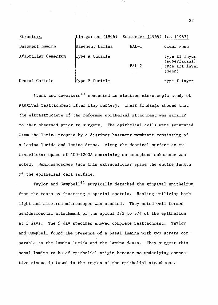
22
Structure List arten (1966) Schroeder (1969) Ito (1967)
Basement Lamina Basement Lamina EAL-1 clear zone
Afibrillar Cementum Type A Cuticle
EAL-2
type II layer (superficial) type III layer (deep)
Dental Cuticle Type B Cuticle type I layer
Frank and coworkers 61 conducted an electron microscopic study of
gingival reattachment after flap surgery. Their findings showed that
the ultrastructure of the reformed epithelial attachment was similar
to that observed prior to surgery. The epithelial cells were separated
from the lamina propria by a distinct basement membrane consisting of
a lamina lucida and lamina densa. Along the dentinal surface an ex-
tracellular space of 400-1200A containing an amorphous substance was
noted. Hemidesmosomes face this extracellular space the entire length
of the epithelial cell surface.
Taylor and Campbell 62 surgically detached the gingival epithelium
from the tooth by inserting a special spatula. Healing utilizing both
light and electron microscopes was studied. They noted well formed
hemidesmosomal attachment of the apical 1/2 to 3/4 of the epithelium
at 3 days. The 5 day specimen showed complete reattachment. Taylor
and Campbell found the presence of a basal lamina with two strata com-
parable to the lamina lucida and the lamina densa. They suggest this
basal lamina to be of epithelial origin because no underlying connec-
tive tissue is found in the region of the epithelial attachment.
23
D. Attachment, Inflammation, Epithelial and connective tissue changes.
1. Attachment
Gottlieb 63'
64' 4 '
65'
66 proposed a concept for the development. of
the dentogingival junction in which he stated that after completion of
enamel deposition, the inner enamel epithelium or ameloblasts produce
a cuticle, the primary enamel cuticle. Following the production of
this cuticle the ameloblasts were thought to degenerate and disappear.
At the time of eruption the cells of the outer enamel epithelium come
in contact with the primary enamel cuticle. Gottlieb felt that the
cells of the outer enamel epithelium transformed to squamous epithe
lial cells and that they produced a keratinized layer which became
structurally united with the primary enamel cuticle. This product of
the squamous epithelial cells was termed the secondary enamel cuticle
and it served as the origin of the epithelial attachment following the
degeneration and loss of cellular layers of the developing dental organ.
Weski 68 stated shortly after Gottlieb's 1921 publications that
the gingival sulcus represents an intraepithelial split. He called
this split a retrocuticular fissure of the epithelium and stated that
as the tooth erupts and pierces the oral epithelium the split appears
in the epithelial layer. Weski felt that the majority of the epithelial
cells kept their connection with the basal layer of the epithelium and
only a few cells remain attached to the cuticle of the enamel.
Becks 69 in conducting a study of humans stated that "when the
epithelium of the mouth becomes fused with the enamel epithelium and

24
the removal of necrotic cell remnants follows, the degeneration of the
enamel epithelium progresses apically and the mouth epithelium prolif
erates downward to cover the defect in the surface ••. concurrently, the
cuticula dentis is left intact on the surface of the teeth". "This
means that the normal pocket is not formed between the cuticula dentis
and the deeper layer of the enamel epithelium or by an intra-epithelial
split, but between the surface of the mouth epithelium, which now repre
sents a part of the pocket epithelium and the enamel epithelium, which
is more or less degenerated. The bottom of the pocket proceeds apically
by this progressive degeneration of the enamel epithelium." In the
presence of injury or inflammation a deepening of the pocket also occurs.
Gottlieb 70 attempted to explain the continued apically growth of
the epithelial attachment by the concept of cementopathia. The con
tinuous deposition of new cementum inside the epithelial attachment
forms the barrier against the apical growth of the epithelial attach
ment. ~%en the cementum layer becomes calcified and ages, without a
new layer being deposited on its surface, its effectiveness against
apically growth ceases. Gottlieb explains such occurences as gingival
recession, pocket formation, pathologic wandering of teeth, and passive
eruption by the concept of cementopathia.
Aisenberg and Aisenberg 71 introduced a fourth concept for pocket
formation. They state that epithelial projections migrate apically
between the existing fiber bundles of the periodontal membrane before
detachment of these fibers from cementum. The epithelial projections
extend between and around the existing fiber bundles a short distance
25
away from cementum and in strands of varying thickness. The authors
state that since proliferating epithelial projections are always ob
served where epithelial lined tissues are involved in the inflammatory
process they should be considered normal extensions of the gingival
epithelium.
Waerhaug 67 by inserting thin steel blades into the sulcus down
to the cemento-enamel junction hypothesized that no epithelial attach
ment of an organic nature existed. Waerhaug felt the term epithelial
cuff described the relationship of the gingival tissue to the enamel.
There have been controversial results by various researchers trying
to duplicate Waerhaug's findings. In any event the light microscope
appears to be an incompetent tool for such judgements since the epithe
lial attachment is beyond the range of resolution of the light micro
scope.
Butcher 72 studied the surface structure of teeth from Rhesus
monkeys following extraction and subsequent reimplantation. He observed
that "a sulcus or crevice forms as an intraepithelial split in the
enamel epithelium". The cause of the split was uncertain and the depth
of the sulcus varied from shallow to deep. Butcher gave recognition to
the existence of primary and secondary enamel cuticles. He found no
cellular extensions of the secondary cuticle into the primary cuticle
or enamel, though he did identify their existance between the primary
cuticle and enamel. Butcher identified the secondary cuticle as the
keratinized product of the superficial layer of cells belonging to the

26
enamel epithelium.
Toto and Sicher 73 studied the jaws of rats and mice as well as
gingival specimen of rats, mice, and humans to determine the nature of
the epithelial attachment. They found a neutral mucopolysaccharide at
the basement membrane, intercellularly, and as a cuticle on the dental
surface of the attached epithelium. This neutral mucopolysaccharide
substance is elaborated by the epithelial cells and renewed as the
epithelial cells undergo mitosis and renewal. It serves as an effec
tive membrane between the epithelium and the tooth and acts as a cement
ing substance.
Wertheimer 7 ~ utilizing various staining techniques attempted to
determine the reactivity similarities between apical cuticles, secondary
dental cuticles, and hyaline bodies. He found that although the deri
vation, function and composition of these three structures was in doubt
there was a consistency in their reactivity to various stains and reac
tions employed. Wertheimer suggests that the epithelium most likely
plays a role in the formation of these structures.
Loe 66 attempting to bolster the concept of Waehaug regarding the
mode of attachment of epithelium to calcified tooth surface stated "the
most convincing evidence against the existence of structural continuity
and in favor of the concept of the dentogingival junctions as a contact
relationship is derived from the study of the dynamic processes taking
place in this area. The continuous loss at the surface and the renewal
of the epithelium imply that the union between the surface cells and the
enamel would have to be continously re-established irrespective of

27
,.,rhether the attachment is mediated by a secondary cuticle on in some
other way." In summarizing his apologea on the morphology, chemistry
and physiology of the dentogingival junction Loe further states that
"following the atrophy and disappearance of the ameloblasts, the epithe
lium facing the tooth surface is not in structural continuity with it
but is kept in close contact with it by the stickiness of the inter
cellular substance of the superficial cells and tonus exerted by the
blood pressure and the connective tissue fibers of the marginal gingiva.
This relationship is adequately expressed by the term epithelial cuff."
2. Inflammation, Epithelial and Connective Tissue Changes.
Goldman 75 in a study on humans of the changes in the pattern of
the gingival fibers in the presence of disease or inflammation found
notable architectural alterations. The gingival crestal fibers were
replaced by dense inflammatory infiltrates. The inflammatory cells
dispersed between the collagen bundles creating fragmentation. The
fiber bundles were generally destroyed in midsection with the cementa!
portion remaining in tact for some period. The inflammatory cells,
primarily identified as plasma cells and lymphocytes, eventually re
placed the connective tissue in the corium permitting apical prolifer
ation of the junctional epithelium with pocket formation as well as
proliferation of the epithelial rete pegs into the connective tissue
corium. Goldman found that epithelial migration ceased where connective
tissue remnants connected to the cementum provided a barrier. General
ly, with increases in the state of inflammation concomitant fiber

28
destruction and replacement by inflammatory cells occurred. In a sub
sequent publication Goldman 76 states that the transseptal fibers of the
periodontal membrane provide a ligamentous-like barrier between adjacent
teeth to prohibit extensive apical migration. In states o! repair where
portions of the transseptal fiber arrangement had previously been des
troyed by inflammation the reformation of transseptal fiber groups are
not regarded as new fiber group formations but as a union of previously
existing periodontal membrane fibers.
Wassermann 77 conducted a study in Sprague Dawley rats which sup
ported the thesis that connective tissue fibrillogenesis was a function
of the fibroblasts. Primary fibrils rather than collagenous fibrils
have an intimate developmental relationship with the fibroblast. The
tight fitting mantle which surrounds the cells and the absence of well
defined cell borders of the fibroblasts suggested to Wassermann that the
primary fibrils along with other cytoplasmic components constituted a
cortical zone of the cell where intercellular growth occurred followed
by extracellular detachment into the ground substance. Fusion of these
primary fibrils for the formation, growth and maturation of collagenous
fibrils occurred within the ground substance.
Grant and Orban 78 suggest that the initial penetration of the bac
terial toxins is via the epithelial attachment. They found in a perio
dontitis study, that the pock~t epithelium became altered by an increase
in size of intercellular spaces encouraging the ingress of the bacterial
toxins and the concomitant egress of polymorphonuclear leukocytes as a

29
defense mechanism. Subsequent alterations in the subepithelial con
nective tissue occurred with the dense infiltration of plasma cells and
lymphocytes. The connective tissue fiber bundles were destroyed which
made imminent the apical proliferation of the epithelial attachment.
The epithelium terminated where dense connective tissue fibers were
still embedded into the cementum.
In a review of repair systems Ratcliff 79 offers three possible
explanations as to why cementum, which has been pathologically exposed
via apical migration of the epithelial attachment, fails to permit new
attachment. He states that the molecular bonding adhesion potential of
epithelial cells to the pathologically exposed cementum is reduced by
the lack of organic components or collagen fibrils to reform a strong
mucopolysaccharide bond. The increased mineral content or the lowered
organic component of the exposed cementum may prohibit the new attachment.
Secondly, Ratcliff suggests that proteolytic enzymes retained within the
porous cementium after exposure to pocket microbial flora would lyse the
mucopolysaccharides elaborated by the young proliferating epithelial
cells thus preventing reattachment. Thirdly, he suggests the possibility
of toxins which have penetrated the porous cementium initiating antigen
antibody reactions and thus interfering with healing and prohibiting
attachment.
Stern 80 studied collagen solubility of human gingiva and found
that in the presence of inflammation the degradation of pre-existing
mature collagen fibers was accompanied by a shift in the percentage of
collagen which was being synthesized and organized on a subfibrillar

30
level. Stern felt this might help explain the finding of collagen deg
radation and epithelial proliferation associated with gingival inflam
mation. Thus the increase in soluble collagen may be due to partial
degradation of pre-existing insoluble collagen or due to an alteration
in the maturation pattern of collagen synthesized in the presence of
inflammation.
Fullmer and coworkers 81 found that pure epithelial cells in culture
and variably inflammed gingival connective tissue free of epithelium were
able to produce the enzyme collagenase in culture. Collagen is the pre
dominant structural protein of the periodontal ligament, alveolar bone,
and cementum; therefore, collagenase is capable of degrading most of
the periodontal tissues. Fullmer suggests that collagenase may be re
sponsible for the normal connective tissue turnover of collagen and the
intensified destruction seen in periodontal disease.
Stanton and coworkers 82 in a study of collagen restoration during
healing projected the time for complete collagen restoration at 49 days
following wounding~via gingivectomy. A productive phase of collagen
repair was noted to last approximately two weeks and this proceeded the
actual collagen reparative phase. Stanton and coworkers found that the
level of collagen noted immediately after the removal of the inflammed
tissue via gingivectomy was more than 50% greater than that found in 6
day specimens and slightly less at 14 days. Strong collagen recovery
was noted at 21 days and 28 days although the 28 day specimen revealed
collagen levels slightly less than the 0 day specimen.

31
Toto and Gargiulo 83 histologically studied the alteration of the
epithelium and connective tissue in the presence of inflammation and
noted that the inflammed gingiva had lost its acid mucopolysaccharide
intercellular cementing substance as well as its desmosomes .. connections.
Edema and polymorphonuclear leukocyte infiltrates were evident. The
lamina propria contained thin walled capillaries, collagen fibers which
had lost their acid mucopolysaccharide coating, and replacement of de
graded collagen fibers by perivascular plasma cells. Alveolar bone
loss occurred by both endosteal and periosteal resorption. Collagen
fibers within the marrow spaces were noted to unravel and disappear.
The neutral polysaccharide of the epithelial attachment was lost.
Ten Cate and Deporter 8 ~ conducted an electron microscopic exam
ination of fibroblasts within the periodontal ligament of functioning
lower first molars of mice. The study revealed the presence of mem
brane-bound intracytoplasmic profiles containing banded collagen. The
study suggests that the fibroblast serves as the cellular basis for
both connective tissue turnover as well as remodeling and that the dis
tinction between the two functions may not be great.
Grant and Bernick85 utilizing thick sections to provide a three
dimensional perspective, studied the nature of epithelial rest cells in
miniature swine. They found that a continuum may exist between the
cells of the reduced enamel epithelium and the epithelial rest cells of
Malassez. This finding was best demonstrated in unerupted or newly
erupted teeth. It was not discernable in older, functional, disease

32
free teeth may be due to the density of the connective tissue bundles
or due to loss of continuity via cell degeneration. The authors found
apically projecting proliferating cords from the epithelial attachment
which "seemed to be continuous with the epithelial rest". They suggest
that the confluence may be initially present, lost, then reestablished
during inflammation and thus may be a factor in the apical progression
of the epithelial attachment and subsequent pocket formation.
Polson, Kennedy, and Zander 86 created periodontitis in squirrel
monkeys and subjected them to thermal injury. They noted that at 6
months the junctional epithelium was apically positioned on the dementum,
a cell-rich collagen-poor connective tissue area existed beneath the
epithelium and that loss of alveolar bone was still eivdent. Recovery
had not occurred.
Polson 87 utilized the same ligation technique to induce perio
dontitis in squirrel monkeys and then subject them to mechanical trauma.
The mechanical trauma created by a wooden wedge driven between two teeth
produced an area of necrosis which was found immediately beneath the
cell-rich collagen-poor area caused by the marginal periodontitis sub
jacent to the junctional epithelium. The lesion extended from the level
of the alveolar crest to the upper half of the periodontal ligament. At
three weeks the necrotic area was replaced by a highly cellular loosely
arranged connective tissue. The periodontal ligament space had increased
dimensionally through alveolar bone resorption. At 8 weeks new bone for
mation was evident. The width of periodontal ligament, cellularity and
orientation of fibers was similar in nature to the periodontal ligament

of a tooth which had not undergone trauma but had an existing perio
dontitis for the same duration.
33
Listgarten88 found epithelial cell rests in approximately 20% of
sections from albino mice jaw block sections. The rest cells were
located in the coronal half of the periodontal ligament in a 3:1 ratio
to the apical half. The rest cells were invariable in close proximity
to the cementa! surface. The cells possessed hemidesmosomes and a basal
lamina to the surrounding connective tissues as would be expected of
typical epithelial-connective tissue junctions. The individual cells
possessed desmosomal, gap and tight junctions along their borders with
adjacent epithelial cells.
Attstrom and coworkers 89 found that in clinically healthy beagle
dogs the normal gingival tissues possesses small numqers of ~solated in
flammatory cells beneath and within the junctional epithelium. The
transmigration of leukocytes was a constant finding which persists in
dependent of the presence or absence of an inflammatory infiltrate.
These findings are fairly consistent with the histologic picture of gin
giva in the clinically healthy human, where the connective tissue ad
jacent to the base of the gingival sulcus always shows some sign of
chronic inflammatory cell infiltration. 90'
91
Schroeder 92 noted a remarkably rapid and complete breakdown of
collagen in beagle dogs with the development of acute exudation and in
flammatory cellular infiltrates. The collagen loss was 60% in some
areas. The pathologic mechanism responsible for this rapid breakdown
is thought to be of an enzyme nature which works through the influx of
34
hydrolytic substances from plague growing in the sulcus or through en
zymes released from host cellular elements.
Aleo and coworkers 93 found that roots of extracted periodontally
involved teeth which had been treated with 45% phenol in H20 at 60°C for
1 hour then washed with 70% ethanol when placed with cultured human gin
gival fibroblasts displayed reattachment. Likewise extracted perio
dontally involved teeth which had their cementum mechanically removed
also demonstrated reattachment in a fibroblast culture. The phenol and
curettement apparently remove the lipopolysaccharide, endotoxin, which
becomes embedded in the porous cementumand serves to prevent attachment.
Novaes and coworkers 94 in a discussion of the development of perio
dontal clefts note that in the presence of the constant inflammation that
exists in the human gingiva various resorptive and proliferative reactions
occur. The inflammatory exudates spread apically through the gingival
connective tissues but also laterally toward the outer aspect of the gin
giva and alveolar mucosa. Collagen and matrical resorption are mediated
via hydrolytic enzyme activity. As the connective tissue is destroyed,
the pocket epithelium proliferates and migrates to fill the voids created
by the loss of the connective tissue. Eventually, anastomosis occurs be
tween the pocket and gingival epithelium as the intervening connective
tissue is lost. This process though relatively slow can lead to cleft
formation and gingival recession.
Grant and coworkers 12 utilizing marmosets as a model studied the
leukocyte migration through the junctional epithelium, the proliferation

35
of the junctional epithelium and sulcular epithelium, the area and density
of inflammatory cell concentrations in the gingival corium, vascular pro
liferation, the area of collagen fiber alteration and loss, and the amount
of alveolar bone loss. They found no correlation between the alveolar
bone loss and the other parameters.

CHAPTER III
l~TERIAL AND METHODS
Patients whose teeth were condemed for periodontal or prosthetic
reasons were invited to volunteer for this study. The experimental pro
cedures were limited to teeth with apparently healthy gingival tissues
or to areas with shallow pockets. An effort was made to select teeth
that had extruded or where alveolectomy was prescribed, in order to
avoid creating deformities in denture bearing tissues.
Four adult patients, tvm male and t\vO females, ranging in age from
42 to 63 years were selected. A thorough examination of the periodontium
was performed on each patient, and each patient was determined by medical
history and examination to be in sound clinical health. Five specimens
obtained from these four patients were utilized in this study. The spe
cimens consisted of three mandibular molar teeth, one maxillary canine
and one mandibular canine.
The experimental surgeries performed in this study attempted to
duplicate specific clinical periodontal surgical procedures. In each
case the procedure was modified by making grooves or notches as landmarks
in the tooth surface to assist in histologic interpretation. Appropriately
these grooves recorded 1) the presurgical level of the gingival margin,
2) the presurgical depth of the sulcus or pocket, 3) the level of the al
veolar crest upon the reflection of a gingivomucoperiosteal flap, and
36
37
4) the post surgical level of the alveolar crest where osteoectomy was
performed (See diagrams 1 and 2).
The following surgical procedures were studied:
1) Apically positioned flaps: full thickness mucoperiosteal
flaps were raised and repositioned apically to cover the
alveolar crestal bone.
2) Apically positioned flaps with osteoectomy: full thickness
mucoperiosteal flaps were raised, alveolar crestal bone was
removed with a surgical curette, and the flap was repositioned
apically to cover the alveolar crestal bone.
For each procedure a horizontal notch was made with a diamond
stone on the labial surface of the experimental tooth at the level of
the gingival crest. Measurements were recorded from the gingival crest
to the bottom of the gingival crevice or pocket utilizing a dull perio
dontal probe prior to the administration of anesthesia. Upon reflection
of the full thickness mucoperiosteal flap a second horizontal notch was
made with a diamond stone on the labial surface of the tooth indicating
the bottom of the gingival crevice previously recorded. For surgical
procedure 1, the surgical curette marked the height of the alveolar
crest, and in surgical procedure 2, the surgical curette was used to
mark the height of the alveolar crest, to remove approximately 1 mm of
alveolar bone in a vertical dimension and then to mark the new height
of the alveolar crest. In each procedure interrupted 4-0 black silk
sutures were utilized to reposition the gingival margin to cover the

38
alveolar bone. Orban's surgical periodontal pack was placed to protect
the wound and removed at one week. No instructions were given and no
attention was paid to the patient's oral hygiene.
Block sections were removed at 60, 90, and 120 days postoperatively
utilizing a technic described by Kohler and Ramfjord. 8 During the sec
tion removal, care was taken to include the labial gingiva, underlying
alveolar process, periodontal ligament, and the tooth as an intact unit.
In order to avoid damage on block removal the vertical incisions for the
experimental area were moved mesially and distally to include the ad
jacent teeth where possible.
The specimens were prepared for microscopic observation by fixing
in 10% formalin, dehydrated, embedded in celloidin, decalcified in nitric
acid-formalin, and processed for nitro-cellulose embedding in the routine
manner. The specimens were sectioned at 10-15 micron intervals for micro
scopic morphometric measurements and stained with hematoxylin and eosin
and Mallory's connectiv~ tissue stain.
A 10 mm square grid was calibrated at lOx magnification for morpho
metric measurements. A conversion factor of .029 was used to convert
units to millimeters. To provide uniformity and standardization one ob
server utilizing the same microscope recorded all measurements.
No attempt was made to access the severity of inflammation by ster
eometric cell count using the Weibel method 9•
10 as proposed by Schroeder 11
because it was found by Grant 12 that the principle of randomnicity upon
which the Weibel technique is based was in fact lacking. Grant deter
mined that the inflammatory process was focal, directional, and
39
chronologically sequential. In addition the chronological sequence of
inflammation may differ between different specimens. Severity of in
flammation was instead related to the distribution of the round cells
present and the evidence of collagen loss. Zachrisson 13 • Oliver, Holm
Pedersen and Loe 1 ~' and Angelopoulos 15 have presented methods of acces
sing round cell inflammatory infiltrates. Each method makes a totally
subjective analysis of the specimens studied. An objective analysis of
round cell infiltrates though desirable is difficult to achieve. In
this study the severity of inflammation was evaluated as mild, moderate,
or severe depending upon the concentration of round cell infiltrates
seen on each specimen and the corresponding appearance of collagen loss.
Although recognition is given to the fact that the degree of collagen
loss will generally vary directly with the quantity of inflammatory cells
present, no definitive statement can be made in accessing areas that
appear to be cell poor and collagen poor, as compared to areas which are
cell rich and collager poor, and as reflections of the status of inflam
mation. Mild inflammation is characterized by sparse distribution of in
flammatory cells, generally seen perivascularly, with little collagen
loss evident. Moderate inflammation is characterized by moderately dense
accummulations of inflammatory cells in isolated areas with sparse dis
tribution noted elsewhere. Areas of collagen dissolution are evident
although some normal fiber organization is present. Severe inflammation
is characterized by dense aggregation of inflammatory cells throughout
the area. Extensive areas of collagen loss are evident, marked by large
areas of dissolution, no organization nor functional orientation of
40
existing fibers.
In order to more accurately access the status of inflammation in
various sections of the same specimen, the dentogingival junction was
compartmentalized into three zones and individual accessments of inflam
mation based on the previously stated criteria were made. Zone I (see
diagram 3) is bordered apically by the apical extent of the functional
epithelium, coronally by the tooth and the outer oral epithelium, and
buccal-lingually it comprises the inner one-third of the gingival corium.
The apically border for zone 2 is determined by dividing the connective
tissue attachment in half as measured from the apical extent of the
junctional epithelium to the alveolar crest. The coronal and lateral
borders for zone 2 are the apical border of zone I, the outer oral epi
thelium, the tooth, and the middle one-third of the gingival corium,
respectively. Zone 3 is bounded by the apically border of zone 2 coro
nally, the alveolar crest apically and the outer oral epithelium, the
tooth, and outer one-third of the gingival corium, laterally. The
zones correspond to the spread of inflammation as observed in microscopic
specimens by Grant. Correlations between the severity of inflammation
and variations in the morphometric analysis will be made.
For each of the five specimens utilized in this study, mean values
and ranges in millimeters will be determined for the sulcus depth, epi
thelial attachment, connective tissue attachment, bone removed and bone
resorbed. Additionally the standard deviation will be determined for the
epithelial and connective tissue attachment. The data will be compared
to the values obtained by Gargiulo and coworkers to determine if the

41
mean values are within the range found by Gargiulo and if a significant
correlation exists between the presurgical and postsurgical dimensions
of the dentogingival junction.
Furthermore an accessment of the degree of inflammation in the
postsurgical specimens will be made and correlated by the specimen age
and surgical procedure to the histologic observations of the healing
process.

CHAPTER IV
OBSERVATIONS
A. Morphometric Analysis:
A total of five cases were utilized in this study. Cases 1 and 2,
consisting of 24 and 19 sections respectively, present the healing re
sponse following full thickness flap reflection and replacement with no
osteoectomy performed. Cases 3, 4, and 5, consisting of 21, 13, and 21
sections respectively, present the healing response following full thick
ness flap reflection and replacement with osteoectomy of the alveolar
crestal bone performed. Mean values are presented for the four or five
parameters investigated depending upon the particular surgical procedure.
Standard deviations are presented for the epithelial and connective
tissue attachments values only. A dimensional analysis of the attachment
apparatus of the dentogingival junction is of primary concern. Mean
values will be compared to the ranges presented in Gargiulo's phase IV
analysis (Table 1).
1. Case 1: Female patient, age 53, mandibular right first molar. The
section was removed at 90 days. Table 2 provides the mean
values for the sulcus depth (A-B), epithelial attachment (B-C),
connective tissue attachment (C-D), and bone resorption (E-D).
No osteoectomy was performed.
47.
43
2. Case 2: Female patient, age 42, mandibular right first molar. The
section was removed at 120 days. Table 3 provides the mean
value determinations. No osteoectomy was performed.
3. Case 3: Male patient, age 50, mandibular left first molar. The
section was removed at 60 days. Table 4 presents the mean
value determinations. Osteoectomy was performed and is
represented by symbol E-F. Bone resorption is represented
by symbol F-D.
4. Case 4: Male patient, age 63, mandibular right cuspid. The section
was removed at 120 days. Osteoectomy was performed.
Table 5 provides the data.
5. Case 5: Same patient as in case 4. Maxillary right cuspid. The
section was removed at 120 days. Osteoectomy was performed
Table 6 provides the data.
Table 7 represents a compilation of the data from the five cases and
provides a comparison of the mean average values to the mean average
values of Gargiulo's phase IV analysis.
B. Assessment of Inflammation
Table 8 presents a compilation of the total number of sections in
each case for which assessments of inflammation based upon the compart
mentalization of the gingiva have been made. Combinations of various
inflammatory patterns, when not observed, have been excluded from the
table. Table 9 presents the frequency by percentage in which severe,
moderate, or mild inflammation is found in compartments 1,2, and 3
44
respectively.
C. Histologic Observation
Marked inflammation was evident in the great majority of sections
studied. Epithelial proliferation, connective tissue dissolution with
inflammatory cell infiltration, edema and vascular dilation and osteo
blastic and osteoclastic activity of the alveolar crestal bone were
generally uniform observations. The more specific histologic observa
tions are indicated below as an analysis of figures 1 through 14.
Figure 1: 90 day section. No osteoectomy was performed. Excessive
vascularity of the gingival crest is noted. In some in
stances the gingival vessels are only one or two cell layers
removed from the gingival sulcus. Calculus formation is
evident in the surgically created notch. The epithelial
attachment was torn in sectioning. The portion of compart
ments 1 and 2, which are sho,vn, are severely inflammed.
Figure 2: 90 day section. No osteoectomy was performed. The osteo
blastic response at the alveolar crest is noted with young
osteophytic bone and osteoid formation. Periodontal ligament
fibers appear to be functionally oriented though still immature
as noted by the excessive cellularity. Reversal lines mark
the areas from which new bone formation occurred. Epithelial
rests are noted near the cementum surface.
Figure 3: 120 day section. No osteoectomy was performed. Osteoblastic
activity is evident on the periodontal ligament side of the
Figu~e 4:
Figure 5:
45
alveolar bone. Osteoblasts line the bone surface on this
side. Crestal alveolar bone regeneration is apparent. The
bone is a highly fibrous woven bone. Cementoid deposition
is evident in the surgically created notch. Cemental frag
ments are surrounded by dense connective tissue. Some ce
mentoid or osteoid deposition is evident. Periodontal liga
ment fibers a~e functionally oriented. Reversal lines mark
the amount of osseous regeneration. Some osteoclastic ac
tivity is still evident on the periosteal surface of the
alveolar bone.
120 day section. No osteoectomy was performed. Osteoblastic
activity evident on periodontal ligament side of the alveolar
bone. Crestal alveolar bone regeneration is marked by reversal
line. Periodontal ligament fibers are seen radiating from the
alveolar bone and are functionally oriented. Dento-alveolar
(dento-periosteal) periodental ligament fibers are noted.
Cementoid deposition is evident in the surgically created
notch as are cemental fragments with osteoid or cementoid de
position. Due to the vascularity in the periodontal ligament
space it is difficult to trace the entire length of the hori
zontal fibers. Mild inflammation is noted in compartment
three.
120 day section. No osteoectomy was performed. The section
shows severe inflammation in compartment 1 with an extensive

Figure 6:
Figure 7:
Figure 8:
46
collagen free area. This suggests that the loss of the
collagen fiber barrier is concomitant with the apical pro
liferation and elongation of the junctional epithelium.
Marked vascularity of the gingival crest is noted as is the
thickness of the outer oral epithelium with elongated epith
elial proliferations into the inflammed lamina propria.
Dense surface keratinization is evident. The remaining dento
gingival fibers appear to have some functional orientation.
The inflammatory state in compartment 2 is classified as
severe. Plaque and calculus formation are apparent in abun
dance.
120 day section. No osteoectomy was performed. The junc
tional epithelium ends where dense dentogingival fibers are
embedded in cementum. Compartments 1 and 2 show inflammation.
120 day section. No osteoectomy was performed. An example
is seen of severe, severe, and mild inflammation in compart
ments 1, 2, and 3 respectively. Very long junctional epithe
lium is noted.
120 day section. No osteoectomy was performed. Section de
picts extensive vascularity at gingival crest. Severe in
flammation in compartments 1 and 2, with little or no evidence
of collagen remnants. Confluence of junctional epithelium
with a proliferating strand from the outer oral epithelium.
The gingival margin is not keratinized. A vacuolated cellu
lar population is seen. Elongation of the epithelial
Figure 9:
attachment is evident. There appears to be an attachment
of the sulcular epithelium to calculus.
47
120 day section. Osteoectomy was performed. Proliferation
of the junctional epithelium into the collagen poor zone of
the gingiva corium is seen. The epithelial attachment ter
minates at the coronal border of the second surgical notch.
Elongated epithelial rete pegs and general thickening of the
outer oral epithelium is noted. The surface epithelium is
heavily keratinized. The junctional epithelium is difficult
to trace. There appears to be an interruption of the attach
ment by connective tissue fibers attached to the cementum
coronally to the first surgical notch. The status of the
inflammation in compartments 1 and 2 is classified as moder
ate and moderate, respectively.
Figure 10: 60 day section. Osteoectomy was performed. Repair of dento-
gingival fibers is noted. Functional orientation of perio
dontal ligament fibers is noted. Cemental fragment is seen,
however, cementoid formation is not apparent in this section.
Minimal osteoid formation is noted on the periodontal ligament
side of the alveolar crest while osteoclastic activity is
apparent on the periosteal surface. Scattered inflammatory
cells are noted. The gingival collagen fibers do not appear
functionally oriented. The inflammation in compartment 3 is
classified as mild.

48
Figure 11: 60 day section. Osteoectomy was performed. Alveolar crest
appears flat or almost concave with active osteoclastic ac
tivity noted. Osteoblasts are lining the periodontal liga
ment surface of the alveolar bone. Osteoid is apparent
though not abundantly. The periodontal surface of the al
veolar bone shows minimal new bone formation. Vascular
channel extends from the gingival corium into the periodontal
ligament space. Periodontal ligament fibers are interrupted
by vascular channels.
Figure 12: 120 day section. Osteoectomy was performed. Osteoblasts
are in evidence lining the periodontal ligament side of the
alveolar bone. Osteoid formation and new bone formation are
evident. The periosteal surface reveals resorptive bays un
dergoing active osteoclasia. The periodontal ligament fibers
appear highly cellular and functionally oriented. Reversal
lines outline the area of osseous regeneration on the perio
dontal ligament surface of the alveolar bone. Osteoid and
osteoblastic activity are apparent in the endosteal marrow
space.
Figure 13: 120 day section. Osteoectomy was performed. An example of
severe, moderate, and mild inflammation in compartments 1,2,
and 3, respectively, is noted. Sequestered cementa! frag
ments can be seen. The extent of the osteoblastic activity
on the periodontal ligament side of the alveolar bone can be

49
discerned. Heavy keratinization and numerous collagen poor
bones are evident.
Figure 14: 120 day section. Osteoectomy was performed. Endosteal bone
apposition is noted within the marrow spaces of the alveolar
bone. Osteoblastic activity is apparent on the periodontal
ligament side. Osteoclastic activity is evident on the perio
steal surface. Epithelial rest cells are noted in the perio
dontal ligament space.

CHAPTER V
DISCUSSION
The results of the morphometric analysis of this study provide for
some interesting comparisons with the work of Gargiulo. Statistical
correlations cannot be drawn between the dimensional variations of the
dentogingival junction as they appear in the normal healthy adult and
as they appear postsurgically in the adult periodontal patient. Com
parisons, however, can be made between the morphometric relationships of
the dentogingival junction as they appear in phase IV of passive tooth
exposure (eruption)(table 1) and the postsurgical specimens which upon
healing resemble phase IV of passive tooth exposure in that in both in
stances the epithelial and connective tissue attachments of the dento
gingival junction are located apical to the cementa-enamel-junction.
The mean values for the sulcus depth, epithelial and connective
attachments as determined in this study fall within the range of values
provided in table 1. The depth of the sulcus in postsurgical specimens
is subject to variation. The range of values noted in the five cases pre
sented is .182 to .695 mm. This approximately one-half millimeter dif
ference can be accounted for as a normal variation due to flap replace
ment, suturing techniques, and pack placement procedures. Further, some
shrinkage may have occurred due to formalin fixation and loss of fluid
in the treatment with ascending alcohol solutions ranging from 70% to 100%.
50

51
A point of interest, however, is the sulcus depth values noted in cases
4 and 5 as a possible reflection of the status of inflammation. A mod
erate inflammatory state was found in compartment 1 in approximately 77%
of the sections for case 4, and in approximately 90% of the sections in
case 5. These values compare to 21%, 0%, and 0% for cases 1, 2, and 3
respectively.
Cases 1 and 2 had no osseous surgery performed. The connective
tissue attachments compare favorably between 90 and 120 days. There was
an almost total regeneration of the resorbed crestal alveolar bone be
tween this time interval. This finding is supported by various research
ers31'34'45' however; other researchers 28 ' 29 ' 23 ' 8 ' 44 ' 47 ' 51 have found
that full thickness flap reflection resulted in a more significant amount
of permanent alveolar crestal bone loss. The epithelial attachment in
case 1 is approximately .5 mm greater in length than in case 2. The in
flammation noted in compartments 1 and 2 of case 2 was severe in approx
imately 85% of the sections observed. The inflammation noted in compart
ment 1 of case 1 was severe in 80% of the sections; however, moderate or
mild inflammation was observed 100% of the time in compartment 2 of case
1. It may well be that the severity of the inflammation in compartment
2 of case 2 has permitted the elongation of the epithelial attachment to
occur, 75 ' 78 however, if such were the case, it does not appear that the
epithelial attachment elongation has been at the expense of the connective
tissue attachment. More likely the variations of a comparative nature be
tween separate specimens are normal. This assumption would be supported
by the range variations provided in the phase IV analysis (table 1).

52
Cases 3, 4, and 5 have all had osteoectomy _performed. The time in
terval between 60 and 120 days appears to be an active period. Morpho
metrically, minimal alveolar crestal bone resorption appears to have
occurred by 60 days; however, between 60 and 120 days marked resorption
of the alveolar crestal bone has occurred. This falls within Wilderman's
functional repair phase23 which lasted between 28 and 185 days. Wilder
man's 1970 52 study revealed an average alveolar crestal bone loss of .8 mm
in human subjects studied up to 6 months. The greatest bone loss occurred
on teeth with initially thin roots. The findings of .649 and .813 mm bone
loss in cases 4 and 5, respectively, is consistent with Wilderman's find
ings. The teeth surgerized in cases 4 and 5 were canine teeth which due
to their natural prominence in the dental arch would be expected to have
a thin labial plate of alveolar bone and which thus would be expected to
have an increased amount of postsurgical resorption.
The connective tissue component for both the osseous and non-osseous
cases demonstrated noteworthy standardization and showed a significant
correlation to the values shown in the phase IV analysis. The epithelial
component of the sulcular and junctional epithelium showed less standard
ization and was subject to more variation. The collective values of the
epithelial and connective tissue components provide a dimensional consis
tency to the dentogingival junction which lends to an accurate assessment
of postsurgical healing.
The fact that no attention was given to the state of the patient's
oral hygiene in this study is a factor which must be considered in

53
assessing the observations. It is well known that histologic specimens
of clinically healthy human gingiva show some degree of chronic inflam
mation subjacent to the junctional epithelium and gingival sulcus. The
severe states of inflammation may certainly have affected our demensional
findings; however, it can be said that in the presence of severe inflam
mation the dentogingival junction postsurgically assumes a functional
morphometric relationship which appears consistent with the phase IV anal
ysis of the disease free human.
Histologic observations reveal cementoid formation in the 90 and
120 days sections; however, this observation was not apparent in the 60
day sections. Although it is generally agreed upon that cementoid forma
tion precedes cementum maturation, various studies 23•
39•
52•
95 have re
vealed a wide chronological variation for initial cementoid formation.
Osteoid formation was evident in the 60, 90 and 120 day sections. This
observation is consistent with studies previously reported. As mentioned,
the period between 60 and 120 days appears to be active from a morpho
metric standpoint. Histologically, the 60 day section (Figure 11) reveals
a flattened, almost concave crestal arrangement of the alveolar bone.
Little osteoid formation is evident on the periodontal ligament side of
the alveolar bone. Although osteoblasts are evident; conversely, on the
periosteal surface marked osteoclastic activity is noticeable. The 120
day sections (Figures 12 and 13) in contradistinction show the crestal
alveolar bone to be undergoing osteoblastic and osteoclastic remodeling
in an attempt to reestablish the rounded physiologic contouring which ex
isted initially.
54
For the osseous and non-osseous surgical cases observed, a func
tional orientation of periodontal ligament fibers appeared initially at
60 and 90 days, respectively (Figures 10 and 2). It can be hypothesized
that had a 60 day section of a nonosseous surgical case been prepared
and studied, the fiber orientation of the periodontal ligament would
also have been functionally oriented. This finding is contrary to the
findings of Morris 97 and Wilderman 7 •
Possible mechanisms for gingival recession are evident in figures
8 and 9. Figure 9 reveals a moderately inflammed gingival corium which
has a broad collagen poor zone. The junctional epithelium has prolif
erated into the gingival corium and is subjacent to the outer oral epith
elium. Confluence of these epithelial structures though not evident in
this figure may likely have occurred in subsequent sections. Figure 8
reveals a cell rich collagen poor gingival corium in which confluence of
the outer oral and the junctional epithelium has occurred. As mentioned
by Novaes 94 this lateral spread of the epithelium with subsequent anasto
mosis may lead to gingival cleft formation or gingival recession. The
anastomosis of the outer oral and junctional epithelium may serve to in
terdict the vascularity to portions of the gingival crestal and sulcular
tissues thus leading to cell death and eventual loss of these cells into
the gingival sulcus.
The degree of surface keratinization evident in figures depicting
the gingival tissues is of interest in that the tissues show heavy kera
tinization in the presence of severe and moderate inflammation. Previous
55
studies 98 52 have revealed that a reduction in gingival keratinization
is associated with increasing gingival inflammation.
The morphometric findings in this study provide insight into the
nature of the healed periodontal tissues following surgical intervention.
The data shows that when mucoperiosteal flaps are reflected osseous
changes via osteoclastic activity will occur in the crestal alveolar bone.
The data further shows that the destructive techniques, currently employed
in an attempt to eliminate pathologic pockets and reestablish the parabolic
osseous contours, which existed in the disease free adult, are not predic
table procedures. It is evident that extensive resorption occurs to the
crestal alveolar tissues following osteoectomy procedures and that a def
inite crestal deformity results from these procedures. The morphometric
data reveals that a definite and rather predictable dimensional relation
ship exists between the tissues of the dentogingival junction. Following
periodontal surgery, apparently regardless of the status of the gingival
inflammation, the connective tissue attachment assumes a definite dimen
sional state. Variations in the epithelial attachment are apparent and
as stated previously this component of the dentogingival junction is sub
ject to greater variations. The question of the mechanism of the reattach
ment of the tissues of the dentogingival junction to the cementum is often
raised. Whether or not a true connective tissue attachment occurs or
whether the attachment is solely via an elongated epithelial attachment is
often questioned. Although greater variation is seen in the epithelial
component, in no instances did an increased epithelial attachment dimension

56
appear to occur at the expense of the connective tissue attachment. The
data suggests that an accurate assessment of the height of the alveolar
bone in the healed surgerized periodontal patient can be made by apply
ing the dimensional values for the dentogingival junction obtained in
this study to an accurate determination of the bottom of the gingival
sulcus. These data have application in the presurgerized periodontal
patient for projection of the consequences of traumatic osseous surgery
can be made.
Although subsequent studies of this nature would be desirable to
establish the possible sequential alterations of the tissues of the dento
gingival junction over a 15 to 180 day period, for example, the inherent
difficulties of working with humans makes the likelihood of such an un
dertaking doubtful.

CHAPTER VI
CONCLUSIONS
1. Full thickness mucoperiosteal flap reflection with osteoectomy re
sults in additional loss of crestal alveolar bone by postsurgical
osteoclastic resorption between 60 and 120 days. Healing of the
alveolar crest with deformity is observed.
2. Dimensional similarities are apparent although statistical correla
tions of the dimensions of the dentogingival junction cannot be made
between disease free adults and the postsurgical results of the adult
periodontal patients.
3. The morphometric analysis of the dentogingival junction in postperio
dontal surgery cases reveals noteworthy standardization of the connec
tive tissue component and shows significant correlation to the values
in phase IV analysis of passive tooth exposure (passive eruption).
4. Despite the presence of severe and moderate inflammation the connec
tive tissue component of the dentogingival junction appears to heal
postsurgically in a predictable manner.
5. The epithelial component of the dentogingival junction showed less
standardization and was subject to more dimensional variation than
the connective tissue component.
6. Full thickness mucoperiosteal flap reflection without osteoectomy fe
sults in crestal alveolar bone alterations via osteoclastic activity
57

58
although significant recovery is noted by 120 days.
7. More meaningful assessment and description of the pattern of inflam
mation and its progression is achieved by the compartmentalization
method utilized in this study.
CHAPTER VII
SUMMARY
A study was undertaken to microscopically determine the morpho
metric dimensions of the dentogingival junction following various
periodontal surgical modalities.
Four adult periodontal patients served as the experimental models.
Two patients providing two block sections had full thickness mucoperio
steal flaps reflected and replaced to cover the alveolar bone. Two
other patients providing three block sections had full thickness muco
periosteal flaps reflected, osteoectomy performed, and the flaps replaced
to cover the alveolar bone. The block sections were obtained at 90 and
120 days for the non-osteoectomy cases, and 60 and 120 day~ for the osteo
ectomy cases. The sections were embedded in celloidin, decalcified,
stained with H & E and sectioned at 10-15 microns providing a total of
98 specimens for morphometric analysis. Presurgical measurements of poc
ket depth were obtained and surgical landmarks were made to mark the pre
surgical height of the gingival crest, the bottom of the gingival sulcus,
the height of the alveolar crest, and the new height of the alveolar
crest following ostectomy. No attention was paid to the patient's oral
hygiene in this study.
The results show that following periodontal flap surgery morpho
metric analysis reveals the dimensional variations of the components of
59
60
the dentogingival junction to fall within the range of values revealed
in Gargiulo's 1961 analysis of disease free humans. The connective
tissue component of the dentogingival junction shows noteworthy standard
ization and appears to correlate significantly to the values revealed in
the phase IV analysis of passive eruption. The epithelial component of
the dentogingival junction showed less standardization and was subject
to greater variation.
A method of assessing the status of inflammation by compartment
alization of the gingival tissues was proposed.

P~FERENCES
1. Gargiulo, A.W., Wentz, F.M., and Orban, B.: Dimensions and Relations of the Dentogingival Junction in Humans. J. Periodont., 32:261, 1961.
2. Schroeder, H.E.: Ultrastructure of the Junctional Epithelium of the Human Gingiva. Helv. Odont. Acta., 13:65, 1969.
3. Listgarten, M.A.: Electron Microscopic Study of the GingivaDental Junction of Man. Am. J. Anat., 119:147, 1966.
4. Schroeder, H.E. and Listgarten, M.A.: Fine Structure of the Developing Epithelial Attachment of Human Teeth. In Vol. 2, Monographs in Developmental Biology, New York: s. Karger, 1971.
5. Morris, M.: Healing of Human Periodontal Tissues Following Surgical Detachment From Vital Teeth: The Position of the Epithelial Attachment. J. Periodont., 32:108, 1961.
6. Marfino, N., Orban, B., and Wentz, F.M: Repair of the Dentogingival Junction Following Surgical Intervention. J. Periodont., 30:180, 1959.
7. Wilderman, M., Pennel, K., King, K., and Barron, J.: Histogenesis of Repair Following Osseous Surgery. J. Perio
. dont., 41:551, 1970.
8. Kohler, C.A. and Ramfjord, S.P.: Healing of Gingival Mucoperiosteal Flaps. Oral Surg., Oral Med., and Oral Path., 13:89, 1960.
9. Weibel, E.R., Kistler, G.S., and Scherle, W.F.: logical Methods for Morphometric Cytology. 30:23, 1966.
Practical StereoJ. Cell Biology,
10. Weibel, E.R.: Stereological Principles for Morphometry in Electron Microscopic Cytology. Int. Rev. Cytol., 26:235, 1969.
11. Schroeder, H.E. and Munzel-Pedrazzdi, S.: Morphometric Analysis Comparing Junctional and Oral Epithelium of Normal Human Gingiva. Helv. Odont. Acta., 14:53, 1970.
61
12. Grant, D., Bernick, B.M., and Levy, B.M.: Histopathologic Changes in Osseous, Collagenous, Vascular and Epithelial Compartments in Gingival Inflammation. Abstract 786 Speical Issue B,; J. Dent. Res., 55:258, 1976.
62
13. Zachrisson, Man.
B.: A Histological Study of Experimental Gingivitis in J. Perio. Res., 3:293, 1968.
14. Oliver, R.C., Holm-Pederen, P., and Loe, H.: The Correlation between Clinical Scoring, Exudate Measurements and Mircoscopic Evaluation of Inflammation in the Gingiva. J. Periodont., 46:201, 1969.
15. Angelopoulos, A.P.: Studies of Mast Cells in the Human Gingiva, II, Topographical Distribution. J. Perio. Res., 8:314, 1973.
16. Skillen, W.S. and Lundquist, G.R.: An Experimental Study of Peridental Membrane Reattachment in Healthy and Pathologic Tissues. J.A.D.A., 24:175, 1937.
17. Beube, F.E.: A Study on Reattachment of the Supporting Structures of the Teeth. J. Periodont., 18:55, 1947.
18. Bordon, S.M.: Histological Study of Healing Following Detachment of Tissue as is Commonly Carried Out in the Vertical Incision For the Surgical Removal of Teeth. J. Can. D.A., 14:510, 1948.
19. Beube, F.E.: The Problem of Reattachment. J. Periodont., 31:310, 1960.
20. Linghorne, W.J. and O'Connell, D.C.: Studies in the Regeneration and Reattachment of the Supporting Structures of the Teeth. I. Soft Tissue Reattachment. J. Dent. Res., 29:419, 1950.
21. Linghorne, W.J. and O'Connell, D.C.: Studies in the Regeneration and Reattachment of the Supporting Structure of the Teeth. II. Regeneration of Alveolar Process. J. Dent. Res., 30:604, 1951.
22. Linghorne, W.J.: Studies in the Reattachment and Regeneration of The Supporting Structures of the Teeth. IV. Regeneration in Epithelized Pockets Following the Organization of a Blood Clot. J. Dent. Res., 36:4, 1957.
23. Wilderman, M., Wentz, F., and Orban, B.: Histogenesis of Repair After Mucogingival Surgery. J. Periodont., 31:283, 1960.

63
24. West, T.L. and Bloom, A.: A Histologic Study of Wound Healing Following Mucogingival Surgery. (Abstract). J. Dent. Res., 40:675, 1961.
25. Staffileno, H., Wentz, F., and Orban, B.: Histologic Study of Healing of Split Thickness Flap Surgery in Dogs. J. Periodont., 33:56, 1962.
26. Wilderman, M.N.: Repair After a Periosteal Retention Procedure. J. Periodont., 34:487, 1963.
27. Lobene, R., and Glickman, I.: The Response of Alveolar Bone to Grinding with Rotary Diamond Stones. J. Periodont., 34:105, 1963.
28. Carranza, F.A., and Carrara, J.J.: Effect of Removal of Periosteum on Postoperative Results of Mucogingival Surgery. J. Periodont. 34:223, 1963.
29. Glickman, I., Smulow, J.B., O'Brien, T., and Tannen, R.: of the Periodontium Following Mucogingival Surgery. Surg., Oral Med., and Oral Path., 16:530, 1963.
Healing Oral
30. Staffileno, H., Levy, S., and Gargiulo, A.: Histologic Study of Cellular Mobilization and Repair Following a Periosteal Retention Operation Via Split Thickness Mucogingival Flap Surgery. J. Periodont., 37:117, 1966.
31. Hiatt, W.H., Stallard, R.E., Butler, E.D., and Badgett, B.: Repair Following Mucoperiosteal Flap Surgery With Full Lingual Retention. J. Periodont., 39:11, 1968.
32. Caffesse, R.G., Ramfjord, S.P., Nasjleti, C.E.: Reverse Bevel Periodontal Flaps in Monkeys. J. Periodont., 39:219, 1968.
33. Henning, F.R.: Epithelial Mitotic Activity After Gingivectomy. J. Perio. Res., 4:319, 1969.
34. Kon, S., Novaes, A.B., Ruben, M.P., and Goldman, H.M.: Visualization of the Microvasculature of the Healing Periodontal Wound. IV. Mucogingival Surgery: Full Thickness Flap. J. Periodont., 40:441, 1969.
35. Noyes, F.B.: A Textbook of Dental Histology and Embryology: Philadelphia, Lea and Febiger, 1912.
36. McCall, J.O.: An Improved Method of Inducing Reattachment of the Gingival Tissue "in Periodontoclasia", Dental Items of Interest, 68:344, 1926.

37. Workman, C.M.: Studies in Reattachment of the Gingivae. M.S. Thesis, Northwestern University, 1947.
38. Schaffer, E.M., and Zander, H.A.: Histological Evidence of Reattachment of Periodontal Pockets, Paradent., 7:101, 1953.
64
39. Morris, M.L.: The Reattachment of Human Periodontal Tissues Following Surgical Detachment: A Clinical and Histological Study, J. 1eriodent., 24:220, 1953.
40. Morris, M.L.: Healing of Human Periodontal Tissues Following Surgical Detachment From Non-Vital Teeth: J. Periodont. 28:222, 1957.
41. Dedolph, T.H., and Clark, H.B.: periosteal Flap Healing.
A Histological Study of MucoJ. Oral Surg., 16:367, 1958.
42. Morris, M.L.: Healing of Human Periodontal Tissues Following Surgical Detachment and Extirpation of Vital Pulps, J. Periodont., 31:23, 1960.
43. Morris, M.L.: Healing of Human Periodontal Tissues Following Surgical Detachment From Vital Teeth: The Position of the Epithelial Attachment. P. Periodont., 32:108, 1961.
44. Donnenfeld, O.W., Marks, R.M., and Blackman, I.: The apically Repositioned Flap-A Clinical Study. J. Periodont., 35:381, 1964.
45. Friedman, N., and Levine, H.L.: Mucogingival Surgery: Current Status. J. Periodont., 35:5, 1964.
46. Pfeiffer, J.S.: The Reaction of Alveolar Bone to Flap Procedures In Man. Periodontics, 3:135, 1965.
47. Pennel, B., King, D., Wilderman, M., and Barron, J.: the Alveolar Process Following Osseous Surgery. 38:426, 1967.
Repair of J. Periodont.,
48. Grant, D.A.: Experimental Periodontal Surgery: Sequestration of Alveolar Bone. J. Periodont., 38:409, 1967.
49. Costich, E.R., and Ramfjord, S.P.: Healing After Partial Denu~ dation of the Alveolar Process. J. Periodont., 39:127, 1968.
50. Ramfjord, S.P., and Costich, E.R.: Healing After Exposure of Periosteum on the Alveolar Process. J. Periodont., 39:199, 1968.

65
51. Tartigian, R.: The Height of the Facial Radicular Alveolar Crest Following Apically Positioned Flap Operations. J. Periodont., 41:412, 1970.
52. Lainson, P.A., and MacKenzie, I.C.: An Examination of the Cytology of Uninflamed and Inflamed Gingiva Using a Filter Imprint Technique. J. Periodont,, 47:477, 1976.
53. Wood, D,L., Hoag, P.M., Donnenfeld, O.W., Rosenfeld, D.D,: Alveolar Crest Reduction Following Full and Partial Thickness Flaps. J. Periodont., 43:141, 1972.
54. Levine, H.L. and Stahl, S.S.: Repair Following Periodontal Flap Surgery with the Retention of the Gingival Fibers. J, Periodont,, 43:99, 1972.
55. Stahl, S.S., Slavkin, H.D,, Yamada, L., and Levine, S.: Speculations About Gingival Repair. J. Periodont., 43:395, 1972.
56. Stahl, S. S. : faces.
The Nature of Healthy and Diseased Root SurJ. Periodont., 46:156, 1975,
57. Ussing, M.J.: The Development of the Epithelial Attachment. Acta Odont, Scanc., 13:123, 1955.
58. Stern, I.B.: An Electron Microscopic Study of the Cementum, Sharpey's Fibers and Periodontal Ligament in the Rat Incisor, Am. J. Anat., 115:377, 1964.
59. Schroeder, H.E. and Theilade, J,: Electron Microscopy of Normal Human Gingival Epithelium, J.Perio Res., 1:95, 1966.
60. Ito, H., Enomoto, S., and Kobayashi, K.: Electron Microscopic Study of the Human Epithelial Attachment, Bull. Tokyo Med, Dent, Univ,, 14:267, 1967.
61, Frank, R., Fiore-Donno, G., Cimasoni, G., and Ogilvie, A.: Gingival Reattachment After Surgery in Man: An Electron Microscopic Study. J. Periodont., 43:597, 1972.
62. Taylor, A.C. and Campbell, M,M.: Reattachment of Gingival Epithelium to the Tooth, J. Periodont., 43:281, 1972.
63. Gottlieb, B.: Der Epithelansatz am Zahme. Otsch Mschr Zahnheilk, 39:142, 1921a.
64. Gottlieb, B.: Atiologie und Prophylaxe der Zahn Karies. Z Stomatol., 19:129, 192lb.
I
65. Listgarten, M.A.: Structure of Surface Coatings on Teeth. A Review •. J. Periodont., 47:139, 1976.
66. Loe, H.: The Structure and Physiology of the Dento-Gingival Junction. Structural and Chemical Organization of Teeth, Vol II. Edited by A.E.W. Miles. Academic Press, New York, 1967.
67, Waerhaug, J.: The Gingival Pocket. Odont. Tidskr., Co. Supp.l, 1952,
68. Weski, D.: Die Chronisch Margin Alem Entzundungen des Alveolarfortsatzes mit besonderer Berucksichtigung der Alveolar Pyorrhoe. Vrtljschr. f. Zahnheilk, 38:1, 1922.
69. Becks, H.: Normal and Pathologic Pocket Formation. JADA, 16:2167, 1929.
70. Gottlieb, B.: The New Concept of Periodontoclasia. J. Periodont., 1:7, 1946.
71. Aisenberg, M.S., and Aisenberg, A.D.: A New Concept of Pocket Formation. Oral Surg., Oral Med., and Oral Path, 1:1047, 1948.
72. Butcher, E.O.: The Cuticles, Epithelial Attachment andReattachment in the Incisors of the Rhesus Monkey. Brit. Dent. J., 94:197, 1953.
73. Toto, P.D. and Sieber, H.: The Epithelial Attachment. Periodontics, 2:154, 1964.
74. Wertheimer, F.W.: A Histologic Comparison of Apical Cuticles, Secondary Dental Cuticles and Hyaline Bodies. J. Periodont., 37:91, 1966.
66
75. Goldman, H.M.: Histologic Topographic Changes of Inflammatory Origin in the Gingival Fibers. J. Periodont., 23:104, 1952.
76. Goldman, H.M.: The Behaviour of Transseptal Fibers in Periodontal Disease. J. Dent. Res., 36:249, 1957.
77. Wassermann, F.: Fibrilogenesis in the Regenerating Rat Tendon with Special Reference to Growth and Composition of the Collagenous Fibril. Am. J. Anat., 94:399, 1954.
78. Grant, D.A. and Orban, F.: Leukocytes in the Epithelial Attachment. J. Periodont., 31:87, 1960,
79. Ratcliff, P.A.: Therapy.
An Analysis of Repair Systems in Periodontal Periodontal Abstracts, 14:57, 1966.
80. Stern, B.: Collagen Solubility of Normal and Inflammed Human Gingiva. Periodontics. 5:167, 1967.
81. Fullmer, H.M, Gibson, W.A., Lazarus, G.S., Bladen, H.A., and Whedon, K.A.: The Origin of Collagenase in Periodontal Tissues of Man. J. Dent. Res., 48:646, 1969.
82. Stanton, G., Levy, M., and Stahl, S.S.: Collagen Restoration in Healing Human Gingiva. J. Dent. Res., 48:27, 1969.
83. Toto, P. and Gargiulo, A.: Epithelial and Connective Tissue Changes in Periodontitis. J. Periodont., 41:587, 1970.
84. Ten Cate, A.R, and Deporter, D.A.: The Role of the Fibroblast in Collagen Turnover in the Functioni~g Periodontal Ligament of the Mouse. Arch. Oral Biol., 19:339, 1974.
85. Grant, D.A., and Bernick, s.: A Possible Continuity Between Epithelial Rests and Epithelial Attachment in Miniature Swine. J. Periodont., 40:87, 1969.
86. Polson, A.M., Kennedy, J.E., and Zander, H.A.: Trauma and Progression of Marginal Periodontitis in Squirrel Monkeys. I. Co-destructive Factor of Periodontitis and Thermally-Produced Injury. J. Perio. Res., 9:100, 1974.
87, Polson, A.M •. : Trauma and Progression of Marginal Periodontitis in Squirrel Monkeys. II. Co-destructive Factors of Periodontitis and Mechanically-Produced Injury. J. Perio. Res., 9:108, 1974.
88. Listgarten, M.: Cells Rests in the Periodontal Ligament of Mouse Molars. J. Periodont., 10:197, 1975.
89. Attstrom, R., Graf-DeBeer, M., and Schroeder, H.E.: Animal and Histologic Characteristics of Normal Gingiva in Dogs. J. Perio Res., 10:115, 1975.
90. Grant, D.A., Stern, I.B., and Everett, F.G.: Orban's Periodontics. 4th ed. The C.V. Mosby Company, Saint Louis, 1972.
91. Goldman, H.M. and Cohen, D,W.: Periodontal Therapy. 5th. Ed. The C.V. Mosby Company, Saint Louis, 1973.
67

92. Schroeder, H.E., Graf-De Beer, M., and Ahstrom, R,: Initial Gingivitis in Dogs. J. Perio Res., 10:128, 1975.
68
93. Aleo, J.J., De Renzis, F.A., and Farber, P.A.: In Vitro Attachment of Human Gingival Fibroblasts to Root Surfaces. J. Periodont., 46:639, 1975.
94, Novaes, A.B., Ruben, M.P., Kon, S., Goldman, H.M., and Novaes, A.B. jr.: The Development of the Periodontal Cleft. J. Periodont., 46:701, 1975,
95, Stallard, R., and Hiatt, W.: The Induction of New Bone and Cementum Formation, I. Retention of Mineralized Fragments within the Flap. J, Periodont., 39:273, 1968,
96. Moskow, B.S.: Calcified Gingival Inclusions. Periodontics. 3:115, 1965.
97. Morris, M.L.: Healing of Human Periodontal Tissues Following Surgical Detachment: The Arrangement of the Fibers of the Periodontal Space, Periodontics. 1:118, 1963.
98. Weiss, M.D., Weinmann, J.P., and Meyer, J.: Degree of Keratinization and Glycogen Content of the noninflamed and Inflamed Gingiva and Oral Mucosa. J. Periodont. 30:208, 1959.

Diagrams
Tables
Figures
APPENDIX
69

Representation of Landmarks used for Morphometric Analysis
in non-osteoectomy Cases
REPRESENTATION OF LANDMARKS USED FOR
MORPHOMETRIC ANALYSIS IN NON-OSTEOECTOMY CASES
GROOVES
2
3
~~----~r-~----------8
~-------------+-~----------C
---------------+~---------E
.---------J---+--------- D
A-8: Sulcus Oeplh
8-C: Ep1lhPI1al Attachment
Connecl1ve T1ssue
Attach men!
8 one Res orbed
DIAGRAM 1
70
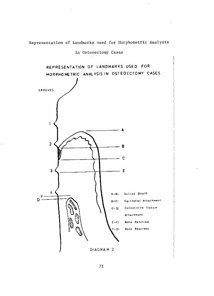
Representation of Landmarks used for Morphometric Analysis
in Osteoectomy Cases
REPRESENTATION 0 F LANDMARKS USE 0 FOR
MORPHOMETRIC ANALYSIS IN OSTEOECTOMY CASES
2
3
4 F--~....._--
0---t---
A
B
c
E
A-B: Sui cus Depth
Epi 1 helial Allachmenl
Conneclive Tissue
Altachmenl
Bone Removed
Bone Resorbed
DIAGRAM 2
71

Compartmentalization of Gingival Tissue For Assessment
of Inflammation
COMPARTMENTALIZATION OF GINGIVAL TISSUE FOR
ASSESSMENT OF INFLAMMATION
Enamel----------~----
Cementum
DIAGRAM 3
72

Table 1
Hean Values of the Components of the Dentogingival
Junction in Phase IV of Passive Eruption
Measurement Range (mm)
A. Sulcus depth 0.00 to 2.25
B. Attached epithelium 0.08 to 2.65
C. Apical point of epithelium attached 0.39 to 6.08 below cementa-enamel junction
D. Bottom of sulcus from cementa -0.03 to 5.84 enamel junction
E. Cementa enamel junction to alveolar bone
F. Deepest point of epithelial attachment to alveolar bone
1.10 to 10.88
0.00 to 6.52
Mean Average
1. 76
o. 71
1.41
-1.14
2.81
1.06
73

74
Table 2
Case 1 - Morphometric Analysis - No osteoectomy was performed
Slide A-B B-C C-D E-F E-D
1 . 377 .29 1.218 .174 2 .29 .29 1.247 .174 3 .377 .348 1.102 .232 4 .58 .377 1.015 .436 5 .377 .377 1.16 .145 6 .406 .406 1.102 .319 7 .406 .377 1.073 .348 8 .493 .377 .957 .377 9 .522 .406 .957 .377
10 .493 .406 .957 . 377 11 .522 .667 .696 .493 12 .638 .493 .754 .493 13 .667 .58 .667 .464 14 .319 .348 1.247 .232 15 .29 .29 1.334 .203 16 .348 .348 1.131 .232 17 .406 .377 1.015 .29 18 .493 .406 .957 .406 19 .406 .319 1.16 .203 20 .667 .493 .754 .493 21 .609 .348 .812 .348 22 .348 . 348 1.247 .174 23 .638 .464 .783 .493 24 .377 .406 1.015 .29
Total 24 11.05 9.54 24.36 7. 77 He an .460 .396 1.015 .324
Range .29 - .667 .29 - .667 .667 - 1.334 .145 - .493 1 S.D. ±.0894 ± .1918
note: all values are in millimeters
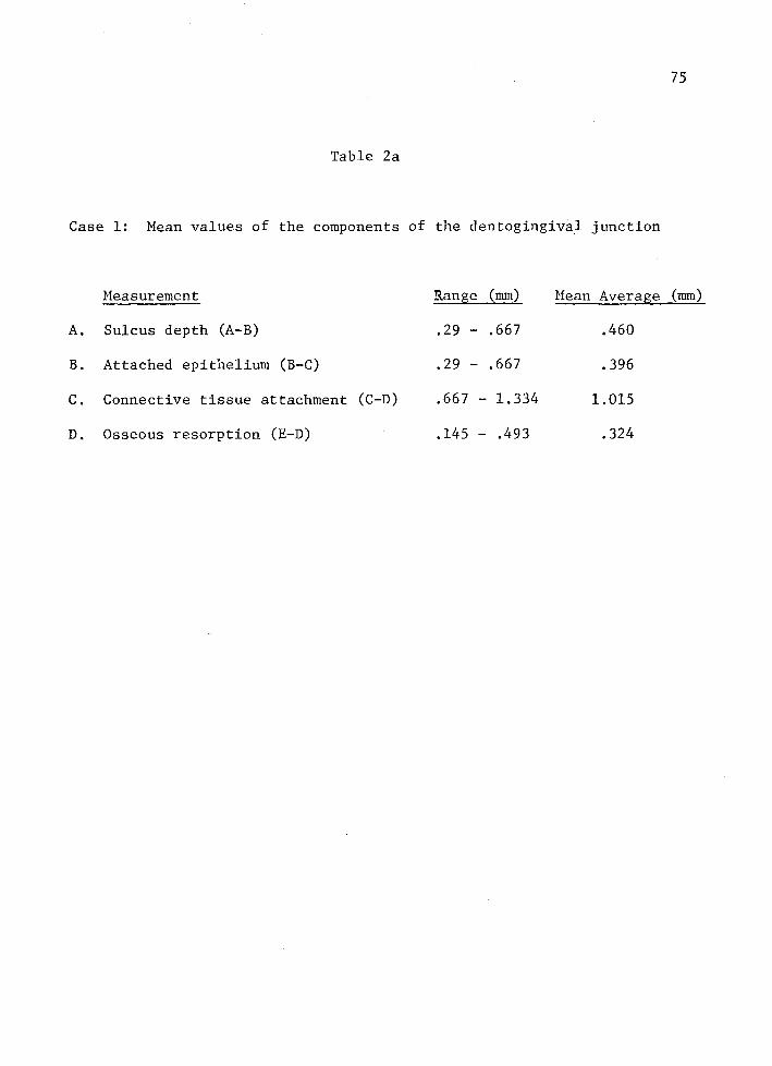
75
Table 2a
Case 1: Mean values of the components of the dentogingiva.l junction
Measurement Range (mm) Mean Average (mm)
A. Sulcus depth (A-B) .29 - .667 .460
B. Attached epithelium (B-C) .29 - .667 .396
c. Connective tissue attachment (C-D) .667 - 1.334 1.015
D. Osseous resorption (E-D) .145 - .493 .324
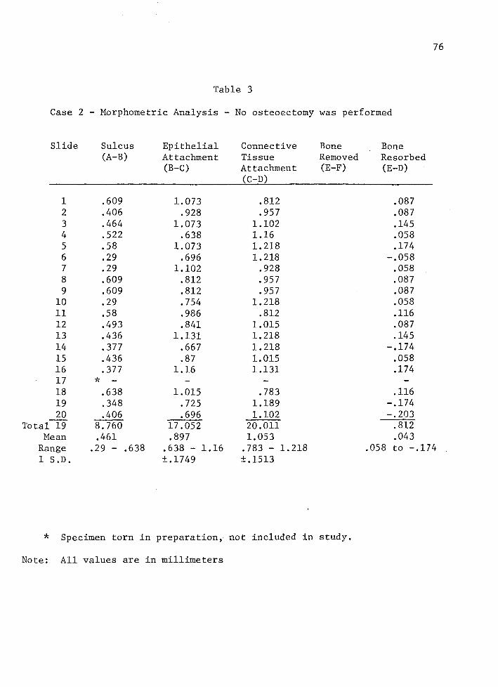
76
Table 3
Case 2 - Morphometric Analysis - No osteoectomy was performed
Slide Sulcus Epithelial Connective Bone Bone (A-B) Attachment Tissue Removed Resorbed
(B-C) Attachment (E-F) (E-D) (C-D)
1 .609 1.073 .812 .087 2 .406 .928 .957 .087 3 .464 1.073 1.102 .145 4 .522 .638 1.16 .058 5 .58 1.073 1.218 .174 6 .29 .696 1.218 -.058 7 .29 1.102 .928 .058 8 .609 .812 .957 .087 9 .609 .812 .957 .087
10 .29 .754 1.218 .058 11 .58 .986 .812 .116 12 .493 .841 1.015 .087 13 .436 1.131 1.218 .145 14 .377 .667 1.218 -.174 15 .436 .87 1.015 .058 16 .377 1.16 1.131 .174 17 * 18 .638 1.015 .783 .116 19 .348 .725 1.189 -.174 20 .406 .696 1.102 -.203
Total 19 8.760 17.052 20.011 .812 Mean .461 .897 1.053 .043
Range .29 - .638 .638 - 1.16 .783- 1.218 .058 to -.174 1 S.D. ±.1749 ± .1513
* Specimen torn in preparation, not included in study.
Note: All values are in millimeters

77
Table 3a
Case 2: Mean values of the components of the dentogingival junction
Measurement Range (mm) Mean Average (mm)
A. Sulcus depth (A-B) .29 - .638 .461
B. Attached epithelium (B-C) .638 - 1.16 .897
c. Connective tissue attachment (C-D) .783 - 1. 218 1.053
D. Osseous resorption (E-D) .058 to -.174 .043
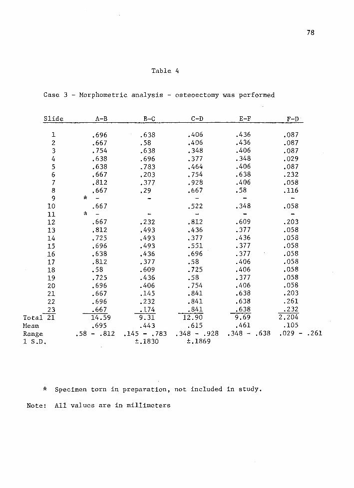
78
Table 4
Case 3 - Morphometric analysis - osteoectomy was performed
Slide A-B B-C C-D E-F F-D
1 .696 .638 .406 .436 .087 2 .667 .58 .406 .436 .087 3 .754 .638 .348 .406 .087 4 .638 .696 .377 .348 .029 5 .638 .783 .464 .406 .087 6 .667 .203 .754 .638 .232 7 .812 .377 .928 .406 .058 8 .667 .29 .667 .58 .116 9 * -
10 .667 .522 .348 .058 11 * -12 .667 .232 .812 .609 .203 13 .812 .493 .436 .377 .058 14 .725 .493 .377 .436 .058 15 .696 .493 .551 .377 .058 16 .638 .436 .696 .377 .058 17 .812 .377 .58 .406 .058 18 .58 .609 .725 .406 .058 19 .725 .436 .58 .377 .058 20 .696 .406 .754 .406 .058 21 .667 .145 .841 .638 .203 22 .696 .232 .841 .638 .261 23 .667 .174 .841 .638 .232
Total 21 14.59 9.31 12.90 9.69 2.204 Mean .695 .443 .615 .461 .105 Range .58 - .812 .145- .783 .348 - .928 .348 - .638 .029 - .261 1 S.D. ±.1830 ±.1869
* Specimen torn in preparation, not included in study.
Note: All values are in millimeters
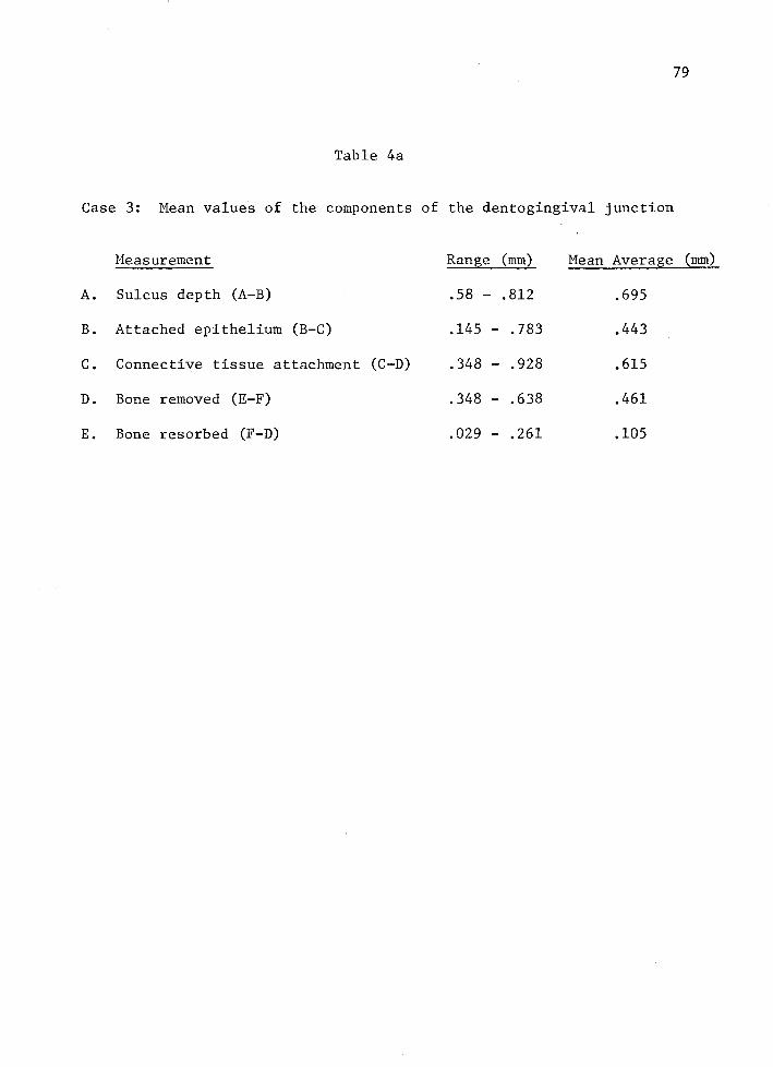
79
Table 4a
Case 3: Mean values of the components of the den to gingival junction
Heasurement Rang;e (mm) Mean Average (mm)
A. Sulcus depth (A-B) .58 - .812 .695
B. Attached epithelium (B-C) .145 - .783 .443
c. Connective tissue attachment (C-D) .348 - .928 .615
D. Bone removed (E-F) .348 - .638 .461
E. Bone resorbed (F-D) .029 - .261 .105

80
Table 5
Case 4: Morphometric Analysis - osteoectomy was performed
Slide A-B B-C C-D E-F F-D
1 .116 .464 .783 1.45 .522 2 .377 .145 1.247 1.682 .609 3 * 4 .174 .203 1.247 1.508 .667 5 .203 .377 1.218 1.624 .667 6 .261 .087 1.363 1.537 .696 7 .261 .145 1.421 1.566 .725 8 .145 .174 1.131 1.479 .638 9 .174 .203 1.189 1.508 .667
10 .145 .203 1.015 1.421 .638 11 .174 .174 1.131 1.363 .696 12 .116 .203 1.073 1.421 .58 13 .145 .232 .986 1.392 .609 14 .203 .116 1.392 1.566 .725
Total 13 2.494 2. 726 15.20 19.52 8.439 Mean .192 .210 1.17 1.50 .649 Range .116-.377 .087-.464 . 783-.1.421 1. 363-1.682 .522-.725 1 S.D. ±.1034 ±.1789
* Specimen torn in preparation, not used in study
Note: All values are in millimeters
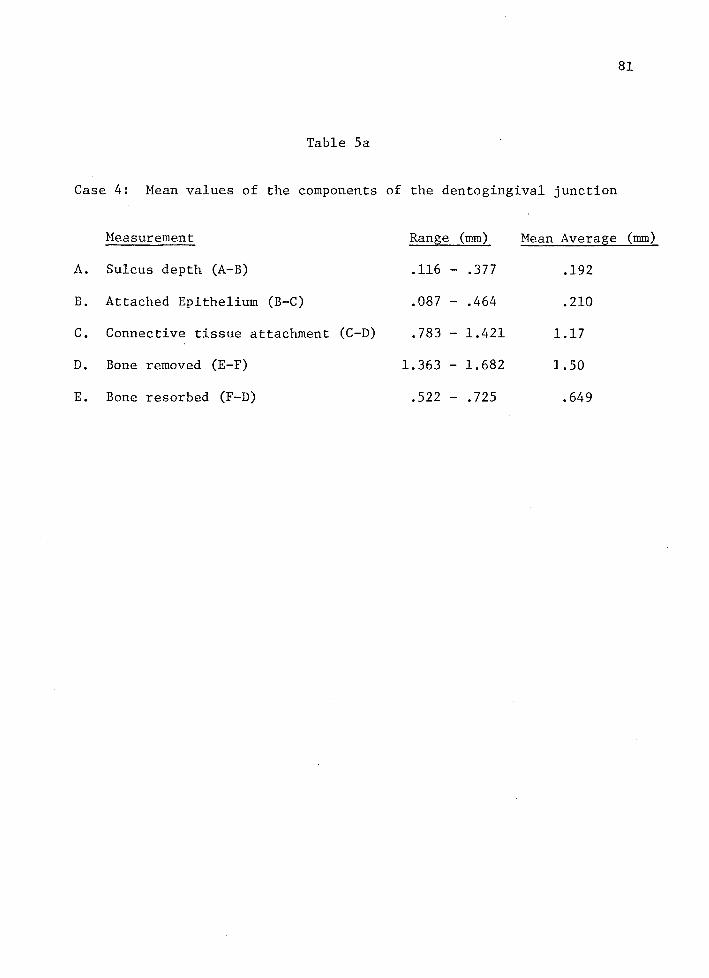
81
Table Sa
Case 4: Mean values of the components of the dentogingival junction
Heasurement Range (mm) Mean Average (mm)
A. Sulcus depth (A-B) .116 - .377 .192
B. Attached Epithelium (B-C) .087 - .464 .210
c. Connective tissue attachment (C-D) .783 - 1.421 1.17
D. Bone removed (E-F) 1.363 - 1.682 1.50
E. Bone resorbed (F-D) .522 - .725 .649

82
Table 6
Case 5: Morphometric Analysis - osteoectomy was performed
Slide A-B B-C C-D
1 .116 .29 1. 044 2 .174 .087 1. 073 3 .116 .203 1.131 4 .087 .29 1.105 5 * 6 .145 .145 1.044 7 .116 .232 1. 044 8 .116 .174 1. 247 9 .261 .232 1. 363
10 .116 .087 1. 247 11 .116 .261 1.102 12 .145 .116 1.131 13 .145 .087 1.305 14 .145 .087 1.073 15 .116 .087 1. 276 16 .145 .116 1. 073 17 .406 .058 1.131 18 .464 .116 1.131 19 .261 .174 1.508 20 .145 .087 1. 247 21 .203 .232 1. 595 22 .29 .174 1.015
Total 21 3.828 3.335 24.795 Mean .182 .159 1.181 Range .087-.464 .058-.29 1. 015-1.595 1 S.D. +.0229 +.160
* Specimen torn in preparation, not used in study NOTE: all values are in millimeters
E-F F-D
1.073 .87 .986 .638
1.363 .667 .928 .58
1.305 .58 .957 .58
1.073 .428 .841 .986
1.073 .928 1.363 .638 1. 247 .928 1.073 .928 1.044 .667 1.044 .928 1.044 .812
.87 .377
.493 1.276
.841 .986 1.073 .87
.87 .957
.812 .957 21.373 17.081 1.018 .813
. 812-1.363 • 377-1.276

83
Table 6a
Case 5: Mean values of the components of the dentogingival junction
Measurement Range (nun) Mean Average (mm)
A. Sulcus depth (A-B) .087 - .464 .182
B. Attached epithelium (B-C) .058 - .29 .159
c. Connective tissue attachment (C-D) 1.015 - 1.595 1.181
D. Bone removed (E-F) .812 - 1. 363 1.018
E. Bone resorbed (F-D) .377 - 1. 276 .813

Table 7. Compilation of data and comparison to phase IV analysis
Case A-B B-C C-D E-F E-D (F-D) B-C + C-D
1 .460 .396 1.015 .324 1.411 (90 da s)
2 .461 .897 1.053 .043 1.950 (120 da s)
3 .695 .443 .615 .461 .105 1.058 (60 da s)
' 4 .192 .210 1.17 1.50 .649 1. 38 (120 da s)
5 .182 .159 1.181 1.018 .813 1.340 (120 d as)
mean .398 .421 1.007 .993 .1835 (Avg. cases 1 & 2) 1.428 average values *(.4155) (.428) (.9925) .993 .522 (Avg. cases 3,4,5) 1.421
mean avg. 1. 76 0.71 1.06 values and ranges for 000-2.25 0.08-2.65 0.00-6.52 phase IV Analysis (1)
* ( ):mean average for the study of 98 total sections
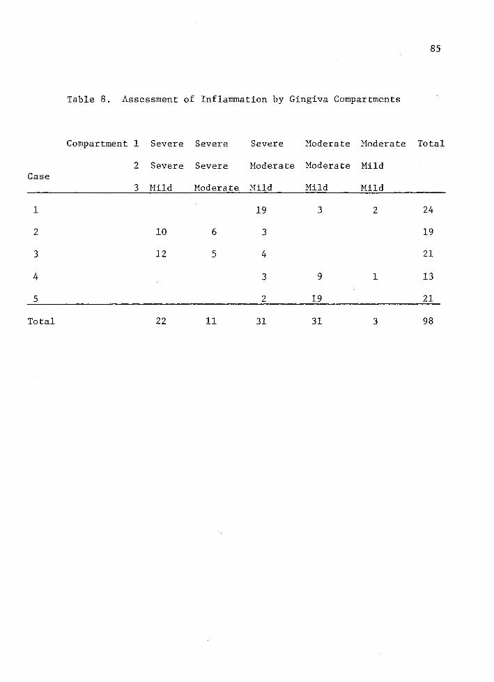
85
Table 8. Assessment of Inflammation by Gingiva Compartments
Compartment 1 Severe Severe Severe Moderate Moderate Total
2 Severe Severe Moderate Moderate Mild Case
3 Hild Moderate Mild Mild Mild
1 19 3 2 24
2 10 6 3 19
3 12 5 4 21
4 3 9 1 13
5 2 19 21
Total 22 11 31 31 3 98

86
Table 9. Degree of Inflammation by Gingival Compartment
Compartment Degree of Inflammation Frequency
1 Severe 65.3%
Moderate 34.7%
Mild 0
2 Severe 33.7%
Moderate 63.3%
Mild 3%
3 Severe 0
Moderate 11.2%
Mild 88.8%
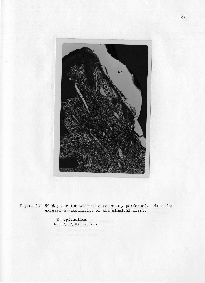
Figure 1: 90 day section with no osteoectomy performed. Note the excessive vascularity of the gingival crest.
E: epithelium GS: gingival sulcus
87

Figure 2: 90 day section with no osteoectomy performed. The osteoblastic response at the alveolar crest is noted.
PL: periodontal ligament OS: osteoid OB: osteophytic bone RL: reversal line
88

. Figure 3: 120 day section with no osteoectomy performed.
PL: periodontal ligament C: cementoid deposition
OB: osteoblasts
89
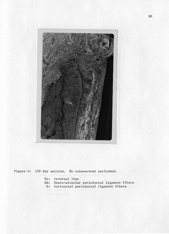
Figure 4: 120 day section. No osteoectomy performed,
RL: reversal line DA: Dento-alveolar periodontal ligament fibers
H: horizontal periodontal ligament fibers
90

Figure 5: 120 day section. No osteoectomy was performed. Note severe inflammation in compartment I.
CF: collagen free area AP: apical proliferating junctional epithelium
D: dentin C: cementum
91
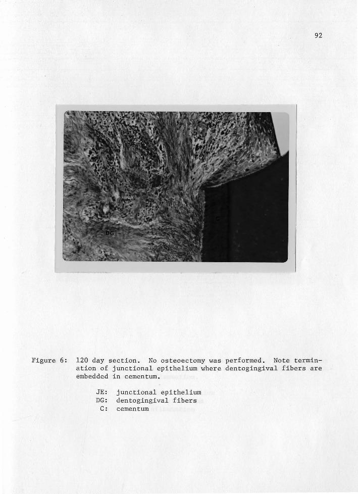
92
Figure 6: 120 day section. No osteoectomy was performed. Note termination of junctional epithelium where dentogingival fibers are embedded in cementum.
JE: junctional epithelium DG: dentogingival fibers
C: cementum

93
Figure 7: 120 day section. No osteoectomy was performed. Note apparent fusion of proliferating epithelium, elongated junctional epithelium and severe inflammation.
PE: proliferating epithelium JE: junctional epithelium I: severe inflammation

Figure 8: 120 day section. No osteoectomy ~-.1as performed. vascularity at gingival crest and proliferation and junctional epithelium.
CA: calculus D: dentin
GC: gingival crest
94
Note excessive at outer oral

95
Figure 9:. 120 day section. Osteoectomy was performed. Note proliferation of junctional epithelium into the collagen poor zone of the gingiva corium.

96
Figure 10: 60 day section. Osteoectomy was performed. Note repair of dento-gingival fibers above alveolar crest.
DG: dento-gingival fibers OC: osteoclast OS: osteoid formation PD: periodontal ligament CF: cemental ~ragment

Figure 11: 60 day section. Osteoectomy was performed. Note vascular channel projecting into periodontal ligament space and osteoclastic activity at alveolar crest.
OS: osteoclast VC: vascular channel PD: periodontal ligament space
97
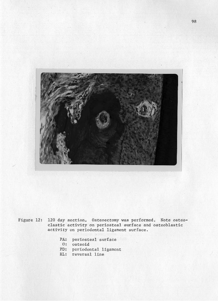
Figure 12: 120 day section. Osteoectomy was performed. Note osteoclastic activity on periosteal surface and osteoblastic activity on periodontal ligament surface.
PA: periosteal surface 0: osteoid
PD: periodontal ligament RL: reversal line
98

Figure 13: 120 day section. Osteoectomy was performed. Note heavy keratinization and numerous collagen poor zone.
CP: collagen poor zone KE: keratinized epithelium
99

Figure 14: 120 day section. Osteoectomy was performed. Endosteal
bone apposition is seen.
EB: endosteal bone apposition OC: osteoclastic activity OB: osteoblastic activity PD: periodontal ligament space
100

APPROVAL SHEET
The thesis/dissertation submitted by Doctor Richard J. Rizzo has been read and approved by the following committee:
Doctor Anthony W. Gargiulo, Director Clinical Professor and Chairman, Periodontics, Loyola
Doctor Patrick D. Toto Professor and Chairman, General and Oral Pathology, Loyola
Doctor Hal D. McReynolds Associate Professor, Histology, Loyola
The final copies have been examined by the director of the thesis/ dissertation and the signature which appears below verifies the fact that any necessary changes have been incorporated and that the thesis/ dissertation is now given final approval by the Committee with reference to content and form.
The thesis/dissertation is therefore accepted in partial fulfillment of the requirements for the degree of Master of Science in Oral Biology.
Date
Related Documents