J Cardiol Curr Res 2014, 1(5): 00028 Journal of Cardiology & Current Research Submit Manuscript | http://medcraveonline.com Introduction Cardiac pacing through a trans-venous catheter implanted in Right Ventricle (RV) usually produces a Left Bundle Branch (LBBB) morphology on surface electrocardiogram (ECG) [1]. There have been instances where a Right bundle branch (RBBB) pattern is seen on RV pacing as well. This raises the suspicion of septal perforation, lead malposition or a coronary sinus (CS) placement of lead. However this can be a variant of true uncomplicated RV pacing. We report a case of RBBB after RV pacing, reiterating the fact that this can be a absolutely benign pattern [2]. Case Report 75 year old man presented with gradually worsening exertional shortness of breath and easy fatigability. Nuclear stress test was abnormal for reversible ischemia and a cardiac catheterization was performed which showed a proximal Left anterior descending artery stenosis which was stented. He also had an atrial Flutter with slow ventricular rate of 50-60 bpm which occasionally dropped to 30 bpm (Figure 1). This was suggestive of significant Atrio-ventricular conduction abnormality. He underwent Flutter ablation after a Trans esophageal echo ruled out any intracardiac thrombus. Post successful ablation he had a HR of 40-50 bpm with complete AV dissociation hence a dual chamber pacemaker was implanted. St.Jude’s pacemaker was used with RV lead fixed to RV apex and Right atrial (RA) lead fixed to RA appendage and mode set at DDDR at lower pacing rate of 70/min. Measurements obtained were RA p- wave 2.7 mv, RA pacing atrial threshold 1mv at 4m.RV lead impedance was 980 ohm. Post-operative chest X Ray confirmed correct lead placement. However the ECG was done which showed a RBBB morphology with mean QRS of -86 degrees (Figure 2). Repeat PA and lateral chest x-ray next day (Figure 3) and a 2D echocardiogram(2D ECHO) (Figure 4) were done to confirm the lead placement as well to r/o any inadvertent position of the lead. All modalities confirmed RV apical lead placement and uncomplicated RV pacing. Following Pacemaker implantation, patient’s breathlessness and Paradoxical Right Bundle Branch Block Morphology in Right Ventricular Apical Pacing- Is it Really Concerning? Case Report Volume 1 Issue 5 - 2014 Ritin Bomb 1 , Marc Del Rosario 2 and Sunil K Jha 1 * 1 Division of cardiovascular medicine, University of Tennessee Health Sciences Center, USA 2 Division of cardiovascular medicine, Methodist University Hospital, USA *Corresponding author: Sunil K Jha, Division of cardiovascular medicine University of Tennessee Health Sciences Center, Memphis, Tennessee, USA, Tel: 901- 734-1325; Email: Received: August 27, 2014 | Published: October 31, 2014 fatigability were markedly improved. He was regularly followed in the clinic initially after 3 months and then every 6 months. He continues to be in an improved state of health. Discussion Commonly RV pacing causes LBBB pattern and LV pacing causes RBBB pattern on ECG [1]. Presence of RBBB in RV pacing raises the suspicion of inadvertent LV lead placement (through Atrial Septal Defect, Patent Foramen Ovale or septal perforation) , coronary sinus placement or transarterial placement crossing aortic valve and entering LV cavity [3,4]. These can have serious consequences and usually requires urgent revision of procedure. A thorough assessment with analysis of ECG, chest X-ray, and 2D ECHO can prove the placement of lead to be in uncomplicated RV apical position and hence prevent unnecessary intervention in such situation. Coman and Trohman [5] and Okmen et al. [6] suggested an algorithm to separate RV and LV pacing morphology by using frontal axis and precordial transition point. After exclusion of pacing from proximal and mid septum, if there is a frontal axis of 0 to 90 degrees and precordial transition is by V3 then uncomplicated RV pacing can be distinguished from LV pacing with a sensitivity, specificity and positive predictive value of 86%,99% and 95% respectively. Our patient had a frontal axis of -86 degrees and transition by V3 hence correlating with proposed algorithm. They also suggested that if V1, V2 are placed one intercostal below than a normal LBBB morphology could be achieved. However our patient continued to show RBBB morphology even one space lower than usual position. Many reasons have been postulated for RBBB pattern in RV pacing. Mower et al suggested that that there could be portions Abstract RBBB pattern on surface electrocardiogram after right ventricular pacing is always concerning for lead malposition in left ventricle. However it can be a variant of normal pacing and careful assessment with chest X-ray, 2 D ECHO and ECG reassures normal pacing and can prevent unnecessary interventions. Keywords Right ventricular pacing; Right bundle branch block pattern; Lead malposition

Welcome message from author
This document is posted to help you gain knowledge. Please leave a comment to let me know what you think about it! Share it to your friends and learn new things together.
Transcript
J Cardiol Curr Res 2014, 1(5): 00028
Journal of Cardiology & Current Research
Submit Manuscript | http://medcraveonline.com
IntroductionCardiac pacing through a trans-venous catheter implanted
in Right Ventricle (RV) usually produces a Left Bundle Branch (LBBB) morphology on surface electrocardiogram (ECG) [1]. There have been instances where a Right bundle branch (RBBB) pattern is seen on RV pacing as well. This raises the suspicion of septal perforation, lead malposition or a coronary sinus (CS) placement of lead. However this can be a variant of true uncomplicated RV pacing. We report a case of RBBB after RV pacing, reiterating the fact that this can be a absolutely benign pattern [2].
Case Report75 year old man presented with gradually worsening
exertional shortness of breath and easy fatigability. Nuclear stress test was abnormal for reversible ischemia and a cardiac catheterization was performed which showed a proximal Left anterior descending artery stenosis which was stented. He also had an atrial Flutter with slow ventricular rate of 50-60 bpm which occasionally dropped to 30 bpm (Figure 1). This was suggestive of significant Atrio-ventricular conduction abnormality. He underwent Flutter ablation after a Trans esophageal echo ruled out any intracardiac thrombus. Post successful ablation he had a HR of 40-50 bpm with complete AV dissociation hence a dual chamber pacemaker was implanted. St.Jude’s pacemaker was used with RV lead fixed to RV apex and Right atrial (RA) lead fixed to RA appendage and mode set at DDDR at lower pacing rate of 70/min. Measurements obtained were RA p- wave 2.7 mv, RA pacing atrial threshold 1mv at 4m.RV lead impedance was 980 ohm.
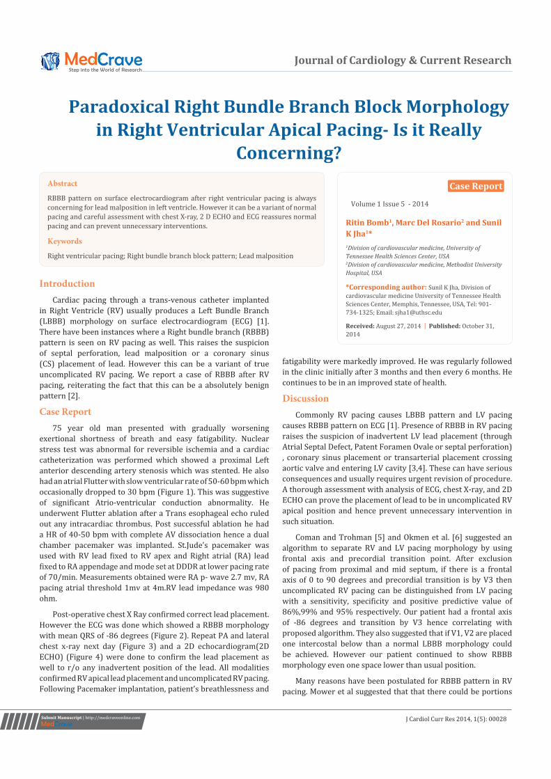
Post-operative chest X Ray confirmed correct lead placement. However the ECG was done which showed a RBBB morphology with mean QRS of -86 degrees (Figure 2). Repeat PA and lateral chest x-ray next day (Figure 3) and a 2D echocardiogram(2D ECHO) (Figure 4) were done to confirm the lead placement as well to r/o any inadvertent position of the lead. All modalities confirmed RV apical lead placement and uncomplicated RV pacing. Following Pacemaker implantation, patient’s breathlessness and
Paradoxical Right Bundle Branch Block Morphology in Right Ventricular Apical Pacing- Is it Really
Concerning?Case Report
Volume 1 Issue 5 - 2014
Ritin Bomb1, Marc Del Rosario2 and Sunil K Jha1*1Division of cardiovascular medicine, University of Tennessee Health Sciences Center, USA2Division of cardiovascular medicine, Methodist University Hospital, USA
*Corresponding author: Sunil K Jha, Division of cardiovascular medicine University of Tennessee Health Sciences Center, Memphis, Tennessee, USA, Tel: 901-734-1325; Email:
Received: August 27, 2014 | Published: October 31, 2014
fatigability were markedly improved. He was regularly followed in the clinic initially after 3 months and then every 6 months. He continues to be in an improved state of health.
DiscussionCommonly RV pacing causes LBBB pattern and LV pacing
causes RBBB pattern on ECG [1]. Presence of RBBB in RV pacing raises the suspicion of inadvertent LV lead placement (through Atrial Septal Defect, Patent Foramen Ovale or septal perforation) , coronary sinus placement or transarterial placement crossing aortic valve and entering LV cavity [3,4]. These can have serious consequences and usually requires urgent revision of procedure. A thorough assessment with analysis of ECG, chest X-ray, and 2D ECHO can prove the placement of lead to be in uncomplicated RV apical position and hence prevent unnecessary intervention in such situation.
Coman and Trohman [5] and Okmen et al. [6] suggested an algorithm to separate RV and LV pacing morphology by using frontal axis and precordial transition point. After exclusion of pacing from proximal and mid septum, if there is a frontal axis of 0 to 90 degrees and precordial transition is by V3 then uncomplicated RV pacing can be distinguished from LV pacing with a sensitivity, specificity and positive predictive value of 86%,99% and 95% respectively. Our patient had a frontal axis of -86 degrees and transition by V3 hence correlating with proposed algorithm. They also suggested that if V1, V2 are placed one intercostal below than a normal LBBB morphology could be achieved. However our patient continued to show RBBB morphology even one space lower than usual position.
Many reasons have been postulated for RBBB pattern in RV pacing. Mower et al suggested that that there could be portions
Abstract
RBBB pattern on surface electrocardiogram after right ventricular pacing is always concerning for lead malposition in left ventricle. However it can be a variant of normal pacing and careful assessment with chest X-ray, 2 D ECHO and ECG reassures normal pacing and can prevent unnecessary interventions.
Keywords
Right ventricular pacing; Right bundle branch block pattern; Lead malposition

Paradoxical Right Bundle Branch Block Morphology in Right Ventricular Apical Pacing- Is it Really Concerning?
Citation: Bomb R, Rosario MD, Jha S (2014) Paradoxical Right Bundle Branch Block Morphology in Right Ventricular Apical Pacing- Is it Really Concerning? J Cardiol Curr Res 1(5): 00028. DOI: 10.15406/jccr.2014.01.00028
Copyright: 2014 Bomb et al.
2/3
Figure 1: Atrial flutter with slow ventricular rate.
Figure 2: Post-operative ECG showing RBBB pattern.
Figure 3: PA and lateral Chest X ray showing normal position of lead.
Paradoxical Right Bundle Branch Block Morphology in Right Ventricular Apical Pacing- Is it Really Concerning?
Citation: Bomb R, Rosario MD, Jha S (2014) Paradoxical Right Bundle Branch Block Morphology in Right Ventricular Apical Pacing- Is it Really Concerning? J Cardiol Curr Res 1(5): 00028. DOI: 10.15406/jccr.2014.01.00028
Copyright: 2014 Bomb et al.
3/3
of interventricular septum which could be anatomical RV but functionally and electrically behave as LV. They also suggested that a pacemaker stimulus may enter the RBBB and can travel in a retrograde direction to the AV junction and down the LBBB. They also proposed that if the screw tip is deeper into the septal plane then it could activate LV early [7]. Barold et al. [8] said that combination of RV activation delay due to severe disease of RV conduction system and early penetration of electrical impulse into LV system could be responsible for this effect.
To conclude we would say that RBBB in a RV pacing could be a variant of normal uncomplicated RV apical pacing and does not necessarily mean a complication. A careful assessment with detailed analysis of ECG, pacing parameters, PA and lateral chest X-Ray and 2D ECHO can further prove the correct position of lead and should be undertaken before subjecting a patient for unnecessary intervention.
References1. Shima T, Ohnishi Y, Inoue T, Yoshida A, Shimizu H, et al. (1998) The
relation between the pacing sites in the right ventricular outflow tract and QRS morphology in the 12-lead ECG. Jpn Circ J 62(6): 399-404.
2. Yang YN, Yin WH, Young MS (2003) Safe right bundle branch block pattern during permanent right ventricular pacing. J Electrocardiol 36(1): 67-71.
3. Raghavan C, Cashion WR, Spencer WH 3rd (1996) Malposition of transvenous pacing lead in the left ventricle. Clin Cardiol 19(4): 335-338.
4. Lugo-Adams F, Rodriguez-Santiago A, Mercado-Crespo J, Rodriguez-Ospina L (2007) Paced right bundle branch block: identification and management of a malpositioned left ventricular pacemaker lead. Bol Asoc Med P R 99(1): 46-50.
5. Coman JA, Trohman RG (1995) Incidence and electrocardiographic localization of safe right bundle branch block configurations during permanent ventricular pacing. Am J Cardiol 76(11): 781-784.
6. Okmen E, Erdinler I, Oguz E, Akyol A, Turek O, et al. (2006) An electrocardiographic algorithm for determining the location of pacemaker electrode in patients with right bundle branch block configuration during permanent ventricular pacing. Angiology 57(5): 623-630.
7. Mower MM, Aranaga CE, Tabatznik B (1967) Unusual patterns of conduction produced by pacemaker stimuli. Am Heart J 74(1): 24-28.
8. Barold SS, Narula OS, Javier RP, Linhart JW, Lister JW, et al. (1969) Significance of right bundle-branch block patterns during pervenous ventricular pacing. Br Heart J 31(3): 285-290.
Figure 4: 2D ECHO-sub costal view showing insertion of RV lead.
Related Documents