REVIEW ARTICLE published: 02 January 2014 doi: 10.3389/fcimb.2013.00110 Pandemic Vibrio parahaemolyticus O3:K6 on the American continent Jorge Velazquez-Roman 1 , Nidia León-Sicairos 1,2 , Lucio de Jesus Hernández-Díaz 3 and Adrian Canizalez-Roman 1,4 * 1 School of Medicine, Autonomous University of Sinaloa, Culiacan, Mexico 2 Pediatric Hospital of Sinaloa, Culiacan, Mexico 3 Programa Regional Para el Doctorado en Biotecnología, FCQB-UAS, Culiacan, México 4 The Sinaloa State Public Health Laboratory, Secretariat of Health, Culiacan, Mexico Edited by: Dongsheng Zhou, Beijing Institute of Microbiology and Epidemiology, China Reviewed by: Maria M. Lleo, Università di Verona, Italy Rohinee Paranjpye, NOAA/NMFS/Northwest Fisheries Science Center, USA *Correspondence: Adrian Canizalez-Roman, Research Unit, School of Medicine, Autonomous University of Sinaloa, Campus 2, Av. Cedros y Calle Sauces, Fracc. Los Fresnos, 80246 Culiacan, Sinaloa, Mexico e-mail: [email protected] Vibrio parahaemolyticus is one of the most important seafood-borne bacterial in recent years and is the leading causal agent of human acute gastroenteritis, primarily following the consumption of raw, undercooked or mishandled marine products. Until 1996, infections caused by V. parahaemolyticus were generally associated with diverse serovars. However, in February 1996, a unique serovar (O3:K6) of V. parahaemolyticus with specific genetic markers (tdh, toxRS/New and/or orf8 ) appeared abruptly in Kolkata, India. In subsequent years, O3:K6 isolates similar to those isolated in Kolkata have been reported from food borne outbreaks in Southeast Asia, as well as in the Atlantic and Gulf coasts of the United States (U.S). More recently, there have been reports in Europe, Africa and Central and South America. Specifically, in the American continent, some countries have reported cases of gastroenteritis due to the pandemic O3:K6 strain and its serovariants; the pandemic strain was first detected in Peru (1996, >100 cases), subsequently spreading to Chile in 1998 (>16,804 human cases), to the U.S. in 1998 (>700 cases), to Brazil in 2001 (>18 cases) and to Mexico in 2004 (>1200 cases). The arrival of the pandemic clone on the American continent may have resulted in a significant shift on the epidemic dynamics of V. parahaemolyticus. However, although O3:K6 is the predominant serovar of the recognized clinical strains in some countries in the Americas, a decrease in clinical cases caused by O3:K6 and an increase in cases associated with a new serotype (O3:K59, Chile) have been recently reported. The emergence and worldwide dissemination of O3:K6 and other pandemic strains since 1996 have come to represent a threat to public health and should concern health authorities. This review focuses on the presence, distribution and virulence factors of the V. parahaemolyticus O3:K6 pandemic clone and its serovariants in clinical and environmental strains on the American continent. Keywords: American continent, pandemic clone, serotypes, Vibrio parahaemolyticus, biosurveillance, public health INTRODUCTION Vibrio parahaemolyticus is a Gram-negative halophilic, mesophilic, non-spore forming, curved rod-shaped bacterium that naturally inhabits marine and estuarine environments worldwide (Baumann et al., 1984). It is commonly found free swimming, attached to underwater surfaces, and commensally associated with various species of shellfish (McCarter, 1999). V. parahaemolyticus has recently been recognized as one of the most important foodborne pathogens and as the leading causal agent of human acute gastroenteritis, primarily following the consumption of raw, undercooked or mishandled seafood and marine products (Okuda et al., 1997; Bag et al., 1999; Depaola et al., 2000; Wong et al., 2000; Ansaruzzaman et al., 2005; Martinez-Urtaza et al., 2005; Cabanillas-Beltran et al., 2006; Su and Liu, 2007; Pal and Das, 2010; Velazquez-Roman et al., 2012). The halophilic bacteria was first identified as a cause of food-borne illness during the fall of 1950 within the southern suburbs of Osaka, Japan, where an outbreak of acute gastroenteritis following the consumption of semidried juvenile sardines sickened 272 and killed 20 individuals (Fujino et al., 1953). V. parahaemolyticus causes two additional major clinical pathologies wound infections and septicemia. The most common syndrome is gastroenteritis, the symptoms of which include diarrhea with abdominal cramps, nausea, vomiting, headache, chills and low-grade fever (Honda and Iida, 1993). The associated diarrhea is occasionally bloody, characterized by reddish watery stools (Qadri et al., 2003) that are unlike those observed in cases of dysentery caused by Shigella species or in cases of amoebiasis. The mean incubation period for V. parahaemolyticus infection is 15 h (range: 4–96 h) (Joseph et al., 1982). The illness is self-limiting and of moderate severity, lasting an average of 3 days in immunocompetent patients (Yeung and Boor, 2004). Because of its self-limiting nature, most cases of infection by V. parahaemolyticus can be can be treated by oral rehydration, alone. Occasionally, treatment with antibiotics such as doxycycline, ciprofloxacin, or erythromycin is necessary Frontiers in Cellular and Infection Microbiology www.frontiersin.org January 2014 | Volume 3 | Article 110 | 1 CELLULAR AND INFECTION MICROBIOLOG Y

Welcome message from author
This document is posted to help you gain knowledge. Please leave a comment to let me know what you think about it! Share it to your friends and learn new things together.
Transcript
REVIEW ARTICLEpublished: 02 January 2014
doi: 10.3389/fcimb.2013.00110
Pandemic Vibrio parahaemolyticus O3:K6 on the AmericancontinentJorge Velazquez-Roman1, Nidia León-Sicairos1,2, Lucio de Jesus Hernández-Díaz3 and
Adrian Canizalez-Roman1,4*
1 School of Medicine, Autonomous University of Sinaloa, Culiacan, Mexico2 Pediatric Hospital of Sinaloa, Culiacan, Mexico3 Programa Regional Para el Doctorado en Biotecnología, FCQB-UAS, Culiacan, México4 The Sinaloa State Public Health Laboratory, Secretariat of Health, Culiacan, Mexico
Edited by:
Dongsheng Zhou, Beijing Institute ofMicrobiology and Epidemiology,China
Reviewed by:
Maria M. Lleo, Università di Verona,ItalyRohinee Paranjpye,NOAA/NMFS/Northwest FisheriesScience Center, USA
*Correspondence:
Adrian Canizalez-Roman, ResearchUnit, School of Medicine,Autonomous University of Sinaloa,Campus 2, Av. Cedros y CalleSauces, Fracc. Los Fresnos, 80246Culiacan, Sinaloa, Mexicoe-mail: [email protected]
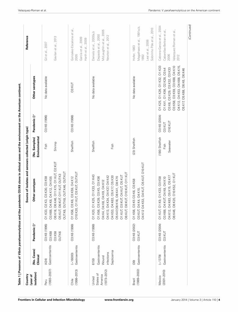
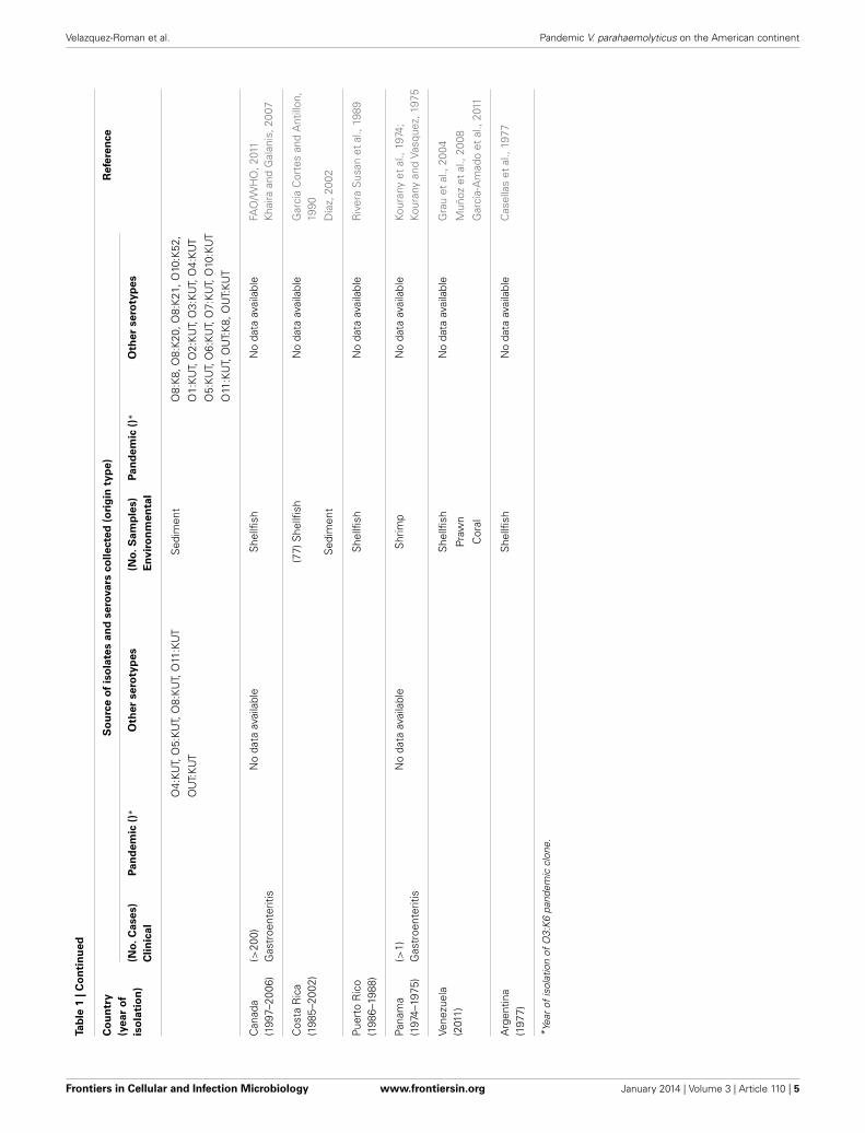
Vibrio parahaemolyticus is one of the most important seafood-borne bacterial in recentyears and is the leading causal agent of human acute gastroenteritis, primarily followingthe consumption of raw, undercooked or mishandled marine products. Until 1996,infections caused by V. parahaemolyticus were generally associated with diverse serovars.However, in February 1996, a unique serovar (O3:K6) of V. parahaemolyticus with specificgenetic markers (tdh, toxRS/New and/or orf8) appeared abruptly in Kolkata, India. Insubsequent years, O3:K6 isolates similar to those isolated in Kolkata have been reportedfrom food borne outbreaks in Southeast Asia, as well as in the Atlantic and Gulf coastsof the United States (U.S). More recently, there have been reports in Europe, Africa andCentral and South America. Specifically, in the American continent, some countries havereported cases of gastroenteritis due to the pandemic O3:K6 strain and its serovariants;the pandemic strain was first detected in Peru (1996, >100 cases), subsequently spreadingto Chile in 1998 (>16,804 human cases), to the U.S. in 1998 (>700 cases), to Brazil in 2001(>18 cases) and to Mexico in 2004 (>1200 cases). The arrival of the pandemic clone on theAmerican continent may have resulted in a significant shift on the epidemic dynamics of V.parahaemolyticus. However, although O3:K6 is the predominant serovar of the recognizedclinical strains in some countries in the Americas, a decrease in clinical cases caused byO3:K6 and an increase in cases associated with a new serotype (O3:K59, Chile) havebeen recently reported. The emergence and worldwide dissemination of O3:K6 and otherpandemic strains since 1996 have come to represent a threat to public health and shouldconcern health authorities. This review focuses on the presence, distribution and virulencefactors of the V. parahaemolyticus O3:K6 pandemic clone and its serovariants in clinical andenvironmental strains on the American continent.
Keywords: American continent, pandemic clone, serotypes, Vibrio parahaemolyticus, biosurveillance, public
health
INTRODUCTIONVibrio parahaemolyticus is a Gram-negative halophilic,mesophilic, non-spore forming, curved rod-shaped bacteriumthat naturally inhabits marine and estuarine environmentsworldwide (Baumann et al., 1984). It is commonly found freeswimming, attached to underwater surfaces, and commensallyassociated with various species of shellfish (McCarter, 1999).V. parahaemolyticus has recently been recognized as one ofthe most important foodborne pathogens and as the leadingcausal agent of human acute gastroenteritis, primarily followingthe consumption of raw, undercooked or mishandled seafoodand marine products (Okuda et al., 1997; Bag et al., 1999;Depaola et al., 2000; Wong et al., 2000; Ansaruzzaman et al.,2005; Martinez-Urtaza et al., 2005; Cabanillas-Beltran et al.,2006; Su and Liu, 2007; Pal and Das, 2010; Velazquez-Romanet al., 2012). The halophilic bacteria was first identified asa cause of food-borne illness during the fall of 1950 withinthe southern suburbs of Osaka, Japan, where an outbreak of
acute gastroenteritis following the consumption of semidriedjuvenile sardines sickened 272 and killed 20 individuals (Fujinoet al., 1953). V. parahaemolyticus causes two additional majorclinical pathologies wound infections and septicemia. The mostcommon syndrome is gastroenteritis, the symptoms of whichinclude diarrhea with abdominal cramps, nausea, vomiting,headache, chills and low-grade fever (Honda and Iida, 1993).The associated diarrhea is occasionally bloody, characterizedby reddish watery stools (Qadri et al., 2003) that are unlikethose observed in cases of dysentery caused by Shigella speciesor in cases of amoebiasis. The mean incubation period for V.parahaemolyticus infection is 15 h (range: 4–96 h) (Joseph et al.,1982). The illness is self-limiting and of moderate severity, lastingan average of 3 days in immunocompetent patients (Yeung andBoor, 2004). Because of its self-limiting nature, most cases ofinfection by V. parahaemolyticus can be can be treated by oralrehydration, alone. Occasionally, treatment with antibioticssuch as doxycycline, ciprofloxacin, or erythromycin is necessary
Frontiers in Cellular and Infection Microbiology www.frontiersin.org January 2014 | Volume 3 | Article 110 | 1
CELLULAR AND INFECTION MICROBIOLOGY
Velazquez-Roman et al. Pandemic V. parahaemolyticus on the American continent
(Qadri et al., 2003). The infection can be fatal for immuno-compromised patients or for those with a preexisting medicalcondition, such as liver disease or diabetes. Although the overallpathogenetic mechanism of marine Vibrio species such as V.parahaemolyticus is not completely understood, these pathogensare known to produce various extracellular products, some ofwhich are known pathogenicity factors (Hasegawa and Hase,2009). Potentially virulent strains are commonly distinguishedfrom virulent strains by the presence of two hemolysins, namelythe thermostable direct hemolysin (tdh) and the Tdh-relatedhemolysin (trh), which exhibits 68% homology with the tdhgene (Zhang and Austin, 2005), and plays a significant role inprecipitating the disease (Takeda, 1983; Shirai et al., 1990; Bejet al., 1999). Pathogenic V. parahaemolyticus can produce eitherTDH, TRH, or both (Honda and Iida, 1993; Nishibuchi andKaper, 1995). An isolate that produces TDH is referred to asKanagawa phenomenon (KP)-positive and can be identified by β
hemolysis on a special agar known as Wagatsuma blood agar (Paland Das, 2010; Canizalez-Roman et al., 2011; Velazquez-Romanet al., 2012). PCR analysis targeting the tdh gene encoding TDHhemolysin and/or the trh gene encoding TRH hemolysin havebeen widely used for the identification of pathogenic isolates ofV. parahaemolyticus (Shirai et al., 1990; Yoh et al., 1995).
PATHOGENIC V. parahaemolyticusAlthough the presence of V. parahaemolyticus is extensive inmarine, estuarine and riverine environments, not all strains ofV. parahaemolyticus are considered pathogenic. Vibrios culturedfrom environmental samples commonly lack the genes codingfor proteins with associated pathogenic functions in humans andmarine animals (e.g., tdh in V. parahaemolyticus) (Deepanjaliet al., 2005; Canizalez-Roman et al., 2011; Gutierrez West et al.,2013). However, few studies have reported the presence of thetdh and trh genes in V. parahaemolyticus strains of environmen-tal origin; only 0–6% of the samples analyzed from the coastsof the U. S. (Kaysner et al., 1990; Depaola et al., 2000), Europe(Hervio-Heath et al., 2002), and Asia contained the tdh and/ortrh genes (Vuddhakul et al., 2000; Wong et al., 2000; Alam et al.,2002). Most of the environmental strains are known to be KP-negative, and only 1–2% of the samples are KP-positive (Josephet al., 1982; Nishibuchi and Kaper, 1995). In molecular epidemi-ological studies, up to 90% of the isolated clinical strains havebeen found to possess the tdh and/or trh gene (the serotype O3:K6strain has increased in prominence) (Okuda et al., 1997; Garciaet al., 2009; Chao et al., 2010; Velazquez-Roman et al., 2012),whereas their presence in environmental isolates is rare (Shiraiet al., 1990; Depaola et al., 1992; Nishibuchi and Kaper, 1995;Islam et al., 2004; Yeung and Boor, 2004; Nair et al., 2007; Chaoet al., 2010). However, in limited areas of Mexico and the U.S.,increased proportions (48–52%) of strains with pathogenic mark-ers (tdh and/or trh) have been detected in environmental isolates(Velazquez-Roman et al., 2012; Gutierrez West et al., 2013).The increased rate of detection of pathogenic V. parahaemolyti-cus within the marine environment might be attributed to adecreased prevalence of total V. parahaemolyticus rather than toan increased abundance of pathogenic strains (Velazquez-Romanet al., 2012).
Currently, a comprehensive understanding of the ability of thisorganism to cause disease remains incomplete. Although the iso-lates that do not contain tdh or those that exhibit a deletion in tdhare still cytotoxic to host cells, the overall mechanism underly-ing the pathogenesis of these tdh-negative strains remains unclear(Boyd et al., 2008). Isolates TDH positive has been shown tohave hemolytic, enterotoxic, cardiotoxic, and cytotoxic activities(Nishibuchi et al., 1992; Raimondi et al., 2000). These cellularevents are caused by bacterial effector proteins, include cytolysins,proteases, lipases, siderophores, exopolysaccharides, and effectorswhich are delivered into intestinal cells (enterotoxic) that directlymodulate the activities of host cell proteins and are secreted andtranslocated into host cells via type III secretion systems (T3SS)(Hiyoshi et al., 2010, 2011; Matsuda et al., 2012) and recently typeVI secretion systems (T6SS) (Yu et al., 2012; Salomon et al., 2013).
The complex interactions between pathogens and the hoststhat they infect have long been believed to represent the pri-mary driving forces that determine the strategies used by bacteriato counter host defenses. However, new evidence suggests thatthe external environment, including other hosts, might play agreater role in the evolution of certain pathogens than previ-ously expected (Wilson and Salyers, 2003). Thus, the pandemicstrains that exhibit certain biological characteristics, such as ele-vated toxin production or the ability to survive within the naturalenvironment, might provide further insights into the mechanismsunderlying the emergence and spread of these strains (Wonget al., 2000). Factors that affect the incidence and distributionof V. parahaemolyticus in the environment are known to includethe water temperature, salt and oxygen concentrations, inter-action with plankton, presence of sediment, organic matter insuspension, fish, and seafood, as well as the incorporation andtidal action of estuarine waters (Cabrera-Garcia et al., 2004).However, how these strains disseminate worldwide remainsunknown.
Vibrio parahaemolyticus O3:K6 IN THE WORLDUntil 1996, infections caused by V. parahaemolyticus were usuallyassociated with diverse serovars (for example, O1:K38, O3:K29,O4:K8, O3:K6, O2:K3, O4:K8, and other serotypes) (Okuda et al.,1997; Wong et al., 2000) and exhibited a localized distribu-tion, emerging in different areas of the world only during thewarmer months of the year. However, the epidemiology of V.parahaemolyticus changed drastically in February 1996, when anatypical increase in V. parahaemolyticus infections occurred inKolkata, a city in the northeastern part of India, resulting inhospitalized patients with diarrheal infections. These infectionswere linked to strains that belonged to a clonal group of theO3:K6 serotype, which exhibited the specific genetic markerstdh, toxRS/New and orf8. These strains had not been previ-ously isolated, but subsequent surveillance in Kolkata identifiedthis serotype as responsible for 50–80% of the infections dur-ing the following months. This clone rapidly spread throughoutthe majority of Southeast Asian countries within a single year(Okuda et al., 1997; Chowdhury et al., 2000). During subse-quent years, O3:K6 isolates similar to those isolated in Kolkatawere reported from foodborne outbreaks and from sporadic casesin the Atlantic and Gulf coasts of the U.S. (Okuda et al., 1997;
Frontiers in Cellular and Infection Microbiology www.frontiersin.org January 2014 | Volume 3 | Article 110 | 2
Velazquez-Roman et al. Pandemic V. parahaemolyticus on the American continent
Chowdhury et al., 2000; Matsumoto et al., 2000). Recently, therehave been similar reports in Europe (Martinez-Urtaza et al.,2005), Africa (Ansaruzzaman et al., 2005), North, Central andSouth America (Daniels et al., 2000a; Gonzalez-Escalona et al.,2005; Velazquez-Roman et al., 2012). The widespread occurrenceof this single V. parahaemolyticus serotype had not previouslybeen reported; thus, it became evident that a pandemic strain hademerged.
Currently several serovariants of the pandemic strains ofO3:K6, included 14 different serotypes (O1:K25, O1:K41,O1:K56, O1:KUT, O3:K6, O3:K58, O3:K68, O3:K75, O4:K8,O4:K12, O4:K68, O4:KUT, O5:KUT, and OUT:K6), althoughonly O3:K6, O4:K68, O1:K25, O1:26, and O1:KUT have beenrecognized as the predominant group responsible for most out-breaks since 1996. These serotypes have been identified as pan-demic clones (Okuda et al., 1997; Matsumoto et al., 2000; Okuraet al., 2003; Chowdhury et al., 2004a; Ansaruzzaman et al., 2005;Mahmud et al., 2007; Nair et al., 2007; Velazquez-Roman et al.,2012). The acquisition of additional serotypes of the pandemicstrain may be a selective response to host immunological pressure(Chowdhury et al., 2004b). Molecular studies based on pulsed-field gel electrophoresis and arbitrarily primed PCR (APPCR)have indicated that the pandemic strains exhibit almost identi-cal fragment patterns (Depaola et al., 2000; Parvathi et al., 2006).Notably, since 1996, a filamentous phage (f 237) (Nasu et al.,2000; Iida et al., 2001) has been reported to be associated withthe pandemic isolates of V. parahaemolyticus (Nasu et al., 2000;Iida et al., 2001). Therefore, from a single O3:K6 serotype, otherserotypes that exhibit identical genotypes and molecular pro-files to the O3:K6 isolates have emerged. These new serotypeshave been collectively called the “serovariants” of O3:K6 isolates(Matsumoto et al., 2000). Generally, pandemic strains belong tothe O3:K6 serotype and contain the orf8 gene. The orf8 gene isbelieved to encode an adherence protein that increases the abilityof V. parahaemolyticus to adhere to host intestinal cells or to thesurfaces of marine plankton (Nasu et al., 2000). Furthermore, sev-eral studies have reported that the toxRS operon of the pandemicstrains contains a unique sequence (toxRS/new) that encodes fortransmembrane proteins involved in the regulation of virulence-associated genes (Chowdhury et al., 2000; Matsumoto et al., 2000;Okura et al., 2003). Characteristics of the O3:K6 pandemic cloneisolates include the O3:K6 antigens, a distinctive toxRS sequence(toxRS/new) (Matsumoto et al., 2000), orf8 (Nasu et al., 2000)and tdh genes, and the absence of the trh gene that is found insome pathogenic strains. Although several polymerase chain reac-tion (PCR)-based methods targeting toxRS/new, group-specificPCR (GS-PCR) or orf8—have been developed, the presence of thetoxRS/new gene is necessary but not always sufficient, whereas theorf8 gene is sufficient but not always necessary for the detectionof pandemic strains (Chowdhury et al., 2000; Okura et al., 2003).In general, however, based on the presence or absence of viru-lence genes, an isolate possessing both tdh and toxRS/new genescan be considered a pandemic strain (Okura et al., 2003). One ofthe other virulence genes, trh, is not specific to pandemic strainsand is rarely present in environmental strains compared to clini-cal strains (Depaola et al., 2000; Parvathi et al., 2006). Pathogenicstrains feature tdh- and/or trh-positivity, whereas non-pathogenic
strains are characteristically tdh- and trh-negative. Some clini-cal isolates have been found to contain both tdh and trh genes,whereas most environmental isolates possess neither (Shirai et al.,1990; Baba et al., 1991; Kishishita et al., 1992; Xu et al., 1994).
PANDEMIC O3:K6 CLONE ON THE AMERICAN CONTINENTThe routes and mechanisms resulting in the dissemination ofthe pandemic strain are still unclear. Moreover, the epidemiol-ogy of V. parahaemolyticus infections remains poorly understood.Recently, there have been occurrences of other extraordinaryevents regarding pathogenic Vibrio species. The transmissionand epidemiology of V. parahaemolyticus infections in placessuch as Kolkata, India, and Bangladesh are entirely distinct fromthe rest of the world because seafood is never eaten raw, andfreshwater fish are preferred over seawater fish by the local pop-ulation (Sarkar et al., 1985). However, in February 1996, theO3:K6 serotype emerged in Kolkata with identical genotypes (tdhpositive and trh negative) and profiles that were indistinguish-able by molecular subtyping techniques (Okuda et al., 1997).In subsequent years, isolates similar to those from Kolkata werereported from foodborne outbreaks and from sporadic cases inseveral sites worldwide (Daniels et al., 2000a; Matsumoto et al.,2000; Ansaruzzaman et al., 2005; Gonzalez-Escalona et al., 2005;Martinez-Urtaza et al., 2005; Leal et al., 2008; Velazquez-Romanet al., 2012), particularly during the warmer months of theyear. Subsequently, the pandemic clone began global dissemina-tion, with many outbreaks. In particular, some countries of theAmerican continent have reported cases of gastroenteritis dueto the pandemic O3:K6 strain and its serovariants, where thepandemic strain was first detected in Peru (>100 cases at approx-imately the same time that it caused an outbreak in Calcutta inFebruary 1996) (Gil et al., 2007). The O3:K6 strain subsequentlyspread to Chile in 1998 (>16,804 human cases), to the U.S. in1998 (>700 cases), to Brazil in 2001 (>18 cases) and to Mexico in2004 (>1200 cases) (Figure 1 and Table 1).
PERUPeru was the first country where the V. parahaemolyticus O3:K6clone was found in marine food products (raw fish or seafoodmarinated with lemon juice, popularly called “cebiche”) (GuevaraDuncan and Del Carpio, 1989) and is where the strain wasfirst associated with gastroenteritis on the American continent(Ibarra and Alvarado, 1999; Gil et al., 2007). Case records from1994–1996 reflect occasional scattered infections restricted to thewarmest months of the year (Martinez-Urtaza et al., 2008). Gilet al. found that the first O3:K6 strains were isolated in February1996 in Trujillo city, indicating that the pandemic strain waspresent in Peru at approximately the same time that it caused theoutbreak in Calcutta in February 1996 (Gil et al., 2007). In 1997,a sudden increase in the number of O3:K6 V. parahaemolyticuscases was observed in Chiclayo, Department of Lambayeque (innorthern Peru) in July, which lasted for 10 months with 2 peaks.Infections were observed along the entire coastline of Peru andspread more than 1500 km within a 4-month period (Martinez-Urtaza et al., 2008). The infections spread in a constant southwarddirection, affecting the Department of Cajamarca in August, LaLibertad in September, and Huaraz in October, finally reaching
Frontiers in Cellular and Infection Microbiology www.frontiersin.org January 2014 | Volume 3 | Article 110 | 3
Velazquez-Roman et al. Pandemic V. parahaemolyticus on the American continent
Tab
le1
|P
resen
ce
of
Vib
rio
para
haem
oly
ticu
san
dth
ep
an
dem
icO
3:K
6clo
ne
inclin
icalcases
an
dth
een
vir
on
men
to
nth
eA
meri
can
co
nti
nen
t.
Co
un
try
(year
of
iso
lati
on
)
So
urc
eo
fis
ola
tes
an
dse
rovars
co
lle
cte
d(o
rig
inty
pe
)R
efe
ren
ce
(No
.C
ases)
Cli
nic
al
Pan
dem
ic()
*O
ther
sero
typ
es
(No
.S
am
ple
s)
En
vir
on
men
tal
Pan
dem
ic()
*O
ther
sero
typ
es
Peru
(199
3–20
07)
(424
)G
astr
oent
eriti
sO
3:K
6(1
996)
O1:
K33
,O2:
K3,
O3:
K30
,O3:
K58
Fish
O3:
K6
(199
6)N
oda
taav
aila
ble
Gil
etal
.,20
07
O3:
K68
O3:
K68
,O4:
K8,
O4:
K12
,O4:
K55
O3:
K58
O6:
K18
,O6:
K46
,O11
:K15
,O1:
KU
T,O
2:K
UT
Shr
imp
Gav
ilan
etal
.,20
13
OU
T:K
6O
5:K
UT,
O6:
KU
T,O
11:K
UT,
OU
T:K
3
OU
T:K
6,O
UT:
K8,
OU
T:K
46,O
UT:
KU
T
Chi
le(1
998–
2013
)(>
1600
0)G
astr
oent
eriti
sO
3:K
6(1
998)
O1:
K56
,O3:
K6,
O3:
K59
,O4:
K12
O10
:K20
,O1:
KU
T,O
3:K
UT,
OU
T:K
UT
She
llfish
O3:
K6
(199
8)O
3:K
UT
Gon
zale
z-E
scal
ona
etal
.,20
05
Gar
cia
etal
.,20
09
Har
thet
al.,
2009
Uni
ted
Sta
tes
ofA
mer
ica
(197
3–20
12)
6159
O3:
K6
(199
8)O
1:K
20,O
1:K
25,O
1:K
33,O
1:K
43S
hellfi
shN
oda
taav
aila
ble
Dan
iels
etal
.,20
00a,
b
Gas
troe
nter
itis
O1:
K56
,O3:
K39
,O3:
K5,
O3:
K56
Dep
aola
etal
.,20
00
Wou
ndO
4:K
4,O
4:K
8,O
4:K
9,O
4:K
12M
cLau
ghlin
etal
.,20
05
infe
ctio
nsO
4:K
13,O
4:K
34,O
4:K
37,O
4:K
42N
ewto
net
al.,
2012
Sep
ticem
iaO
4:K
53,O
4:K
63,O
5:K
17,O
5:K
30Fi
sh
O5:
K47
,O6:
K18
,O8:
K41
,O8:
K70
O1:
KU
T,O
3:K
UT,
O4:
KU
T,O
5:K
UT
O6:
KU
T,O
8:K
UT,
O10
:KU
T,O
11:K
UT
Bra
zil
(198
9–20
02)
(39)
O3:
K6
(200
2)O
1:K
56,O
3:K
5O
3:K
6,O
3:K
58(2
3)S
hellfi
shN
oda
taav
aila
ble
Hof
er,1
983
Gas
troe
nter
itis
O3:
KU
TO
3:K
UT,
O4:
K4,
O4:
K10
,O
4:K
12O
4:K
53,O
4:K
UT,
O5:
KU
T,O
10:K
UT
Mag
alha
eset
al.,
1991
a,b,
1992
Leal
etal
.,20
08
Sob
rinho
Pde
etal
.,20
10
Mex
ico
(200
1–20
10)
(>13
79)
O3:
K6
(200
4)O
1:K
UT,
O1:
K9,
O1:
K33
,O3:
K58
(190
)She
llfish
O3:
K6
(200
4)O
1:K
25,O
1:K
26,O
1:K
32,O
1:K
33C
abre
ra-G
arci
aet
al.,
2004
Gas
troe
nter
itis
O3:
KU
TO
3:K
68,O
4:K
UT,
O4:
K8,
O4:
K10
Fish
O3:
KU
TO
1:K
41,O
1:K
56,O
2:K
28,O
3:k4
Cab
anill
as-B
eltr
anet
al.,
2006
O4:
K12
,O4:
K63
,O5:
K15
,O5:
K17
Sea
wat
erO
10:K
UT
O3:
K6,
O3:
K29
,O3:
K32
,O3:
K33
O6:
K46
,O8:
K20
,O10
:K52
,O1:
KU
TO
3:K
58,O
3:K
59,O
3:K
68,O
4:K
10Ve
lazq
uez-
Rom
anet
al.,
2012
O4:
K12
,O4:
K53
,O4:
K68
,O5:
K15
,
O5:
K17
,O5:
K68
,O6:
K6,
O6:
K46
(Con
tinue
d)
Frontiers in Cellular and Infection Microbiology www.frontiersin.org January 2014 | Volume 3 | Article 110 | 4
Velazquez-Roman et al. Pandemic V. parahaemolyticus on the American continent
Tab
le1
|C
on
tin
ued
Co
un
try
(year
of
iso
lati
on
)
So
urc
eo
fis
ola
tes
an
dse
rovars
co
lle
cte
d(o
rig
inty
pe
)R
efe
ren
ce
(No
.C
ases)
Cli
nic
al
Pan
dem
ic()
*O
ther
sero
typ
es
(No
.S
am
ple
s)
En
vir
on
men
tal
Pan
dem
ic()
*O
ther
sero
typ
es
O4:
KU
T,O
5:K
UT,
O8:
KU
T,O
11:K
UT
Sed
imen
tO
8:K
8,O
8:K
20,O
8:K
21,O
10:K
52,
OU
T:K
UT
O1:
KU
T,O
2:K
UT,
O3:
KU
T,O
4:K
UT
O5:
KU
T,O
6:K
UT,
O7:
KU
T,O
10:K
UT
O11
:KU
T,O
UT:
K8,
OU
T:K
UT
Can
ada
(199
7–20
06)
(>20
0)G
astr
oent
eriti
sN
oda
taav
aila
ble
She
llfish
No
data
avai
labl
eFA
O/W
HO
,201
1K
haira
and
Gal
anis
,200
7
Cos
taR
ica
(198
5–20
02)
(77)
She
llfish
No
data
avai
labl
eG
arci
aC
orte
san
dA
ntill
on,
1990
Sed
imen
tD
íaz,
2002
Puer
toR
ico
(198
6–19
88)
She
llfish
No
data
avai
labl
eR
iver
aS
usan
etal
.,19
89
Pana
ma
(197
4–19
75)
(>1)
Gas
troe
nter
itis
No
data
avai
labl
eS
hrim
pN
oda
taav
aila
ble
Kour
any
etal
.,19
74;
Kour
any
and
Vasq
uez,
1975
Vene
zuel
a(2
011)
She
llfish
No
data
avai
labl
eG
rau
etal
.,20
04
Praw
nM
uñoz
etal
.,20
08
Cor
alG
arci
a-A
mad
oet
al.,
2011
Arg
entin
a(1
977)
She
llfish
No
data
avai
labl
eC
asel
las
etal
.,19
77
* Yea
rof
isol
atio
nof
O3:
K6
pand
emic
clon
e.
Frontiers in Cellular and Infection Microbiology www.frontiersin.org January 2014 | Volume 3 | Article 110 | 5
Velazquez-Roman et al. Pandemic V. parahaemolyticus on the American continent
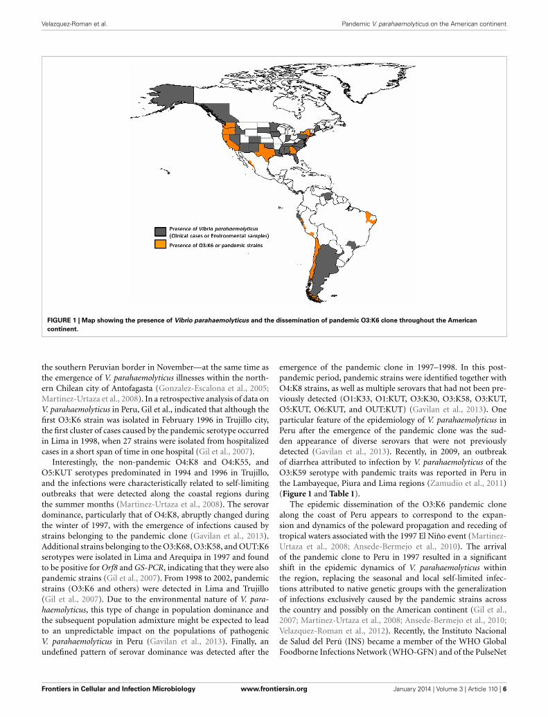
FIGURE 1 | Map showing the presence of Vibrio parahaemolyticus and the dissemination of pandemic O3:K6 clone throughout the American
continent.
the southern Peruvian border in November—at the same time asthe emergence of V. parahaemolyticus illnesses within the north-ern Chilean city of Antofagasta (Gonzalez-Escalona et al., 2005;Martinez-Urtaza et al., 2008). In a retrospective analysis of data onV. parahaemolyticus in Peru, Gil et al., indicated that although thefirst O3:K6 strain was isolated in February 1996 in Trujillo city,the first cluster of cases caused by the pandemic serotype occurredin Lima in 1998, when 27 strains were isolated from hospitalizedcases in a short span of time in one hospital (Gil et al., 2007).
Interestingly, the non-pandemic O4:K8 and O4:K55, andO5:KUT serotypes predominated in 1994 and 1996 in Trujillo,and the infections were characteristically related to self-limitingoutbreaks that were detected along the coastal regions duringthe summer months (Martinez-Urtaza et al., 2008). The serovardominance, particularly that of O4:K8, abruptly changed duringthe winter of 1997, with the emergence of infections caused bystrains belonging to the pandemic clone (Gavilan et al., 2013).Additional strains belonging to the O3:K68, O3:K58, and OUT:K6serotypes were isolated in Lima and Arequipa in 1997 and foundto be positive for Orf8 and GS-PCR, indicating that they were alsopandemic strains (Gil et al., 2007). From 1998 to 2002, pandemicstrains (O3:K6 and others) were detected in Lima and Trujillo(Gil et al., 2007). Due to the environmental nature of V. para-haemolyticus, this type of change in population dominance andthe subsequent population admixture might be expected to leadto an unpredictable impact on the populations of pathogenicV. parahaemolyticus in Peru (Gavilan et al., 2013). Finally, anundefined pattern of serovar dominance was detected after the
emergence of the pandemic clone in 1997–1998. In this post-pandemic period, pandemic strains were identified together withO4:K8 strains, as well as multiple serovars that had not been pre-viously detected (O1:K33, O1:KUT, O3:K30, O3:K58, O3:KUT,O5:KUT, O6:KUT, and OUT:KUT) (Gavilan et al., 2013). Oneparticular feature of the epidemiology of V. parahaemolyticus inPeru after the emergence of the pandemic clone was the sud-den appearance of diverse serovars that were not previouslydetected (Gavilan et al., 2013). Recently, in 2009, an outbreakof diarrhea attributed to infection by V. parahaemolyticus of theO3:K59 serotype with pandemic traits was reported in Peru inthe Lambayeque, Piura and Lima regions (Zamudio et al., 2011)(Figure 1 and Table 1).
The epidemic dissemination of the O3:K6 pandemic clonealong the coast of Peru appears to correspond to the expan-sion and dynamics of the poleward propagation and receding oftropical waters associated with the 1997 El Niño event (Martinez-Urtaza et al., 2008; Ansede-Bermejo et al., 2010). The arrivalof the pandemic clone to Peru in 1997 resulted in a significantshift in the epidemic dynamics of V. parahaemolyticus withinthe region, replacing the seasonal and local self-limited infec-tions attributed to native genetic groups with the generalizationof infections exclusively caused by the pandemic strains acrossthe country and possibly on the American continent (Gil et al.,2007; Martinez-Urtaza et al., 2008; Ansede-Bermejo et al., 2010;Velazquez-Roman et al., 2012). Recently, the Instituto Nacionalde Salud del Perú (INS) became a member of the WHO GlobalFoodborne Infections Network (WHO-GFN) and of the PulseNet
Frontiers in Cellular and Infection Microbiology www.frontiersin.org January 2014 | Volume 3 | Article 110 | 6
Velazquez-Roman et al. Pandemic V. parahaemolyticus on the American continent
Latin American and Caribbean Net (PN-AL & C), with whom itshares the genetic profiles of the isolated pathogenic strains, mak-ing it possible to compare the genotypes of similar strains foundin different countries and to identify the occurrence of epidemicoutbreaks within the region (Zamudio et al., 2011).
CHILEOver the past decade, trends in V. parahaemolyticus illnessesassociated with molluscan shellfish have taken unexpected andunprecedented turns. Pandemic V. parahaemolyticus has nowspread to at least 5 continents and has caused repeated annualoutbreaks in Chile, where V. parahaemolyticus was not evenconsidered a problem when this risk assessment began (Gonzalez-Escalona et al., 2005). The spread of the pandemic clonal O3:K6complex reached the Southern Hemisphere as early as 1998,two years after the strain abruptly appeared in Calcutta, India,in 1996 (Gonzalez-Escalona et al., 2005). An outbreak thatoccurred primarily within the northern city of Antofagasta inChile from November 1997 to March 1998 caused >300 clin-ical cases (Cordova et al., 2002). This city is near the bor-der with Peru. A second outbreak affecting approximately 1500individuals occurred from January to March 2004, predomi-nantly in Puerto Montt, a region that is known for cold coastalwaters. The outbreak in Puerto Montt was likely triggered byhigher-than-normal temperatures during the summer monthswithin the region, which is generally cool during all seasons(Gonzalez-Escalona et al., 2005). This last outbreak had impor-tant economic and social repercussions because this region isone of the main shellfish-producing areas in Chile (Gonzalez-Escalona et al., 2005). The specific infection dynamics foundin Chile, where infections associated with the pandemic clonestarted in 1997–1998 and subsequently in 2004 and where thepandemic clone has been predominant among clinical isolates,clearly contrasts with the epidemiological trend of arrival andthe rapid decline of pandemic V. parahaemolyticus infectionsin Peru.
During the summer of 2005, one of the outbreaks in Chilewas high-magnitude, causing >11000 cases. This is the largestoutbreak documented for this pathogen, most of which havebeen associated with consumption of clams and mussels but notoysters, which reflects the pattern of shellfish consumption inChile (Gonzalez-Escalona et al., 2005; Fuenzalida et al., 2006;Cabello et al., 2007). The large epidemics of diarrhea observedduring the last two austral summers in Puerto Montt, Chile,appears to be directly related to the introduction of the O3:K6serovar pandemic strain to this region (Gonzalez-Escalona et al.,2005; Fuenzalida et al., 2006). Until 2006, every analyzed case inChile was caused by the serovar O3:K6 pandemic strain (Oleaet al., 2005; MINSAL, 2013). In the summer of 2007, among the477 cases reported, 73% corresponded to the pandemic complex(Olea et al., 2005; MINSAL, 2013), among which 40% of the clin-ical cases were associated with the O3:K59 serotype and 27% wereunrelated to the pandemic strain (Garcia et al., 2009; Harth et al.,2009). In the summer of 2008, there was an unexpected increasefrom 477 to 1143 clinical cases that were associated with the pan-demic strain serotype O3:K6 (98%) (Garcia et al., 2009; Harthet al., 2009). According to data from the Ministery of Health of
Chile, more than 3640 cases of gastroenteritis were reported in2008, although the reports do not specify the serotype of the V.parahaemolyticus isolates (MINSAL, 2013). In 2009, clinical casesdecreased to 441, and only 64% were related to the pandemicstrain; the remaining cases were related to non-pandemic tdh-and trh-negative strains (Garcia et al., 2009). Gradually, outbreakshave decreased to fewer than 10 cases in 2010 and 2011 (Garciaet al., 2013). In Chile, the pandemic strain had become a relativelystable bacterial subpopulation of the diverse V. parahaemolyticuspopulation that is present in shellfish (Garcia et al., 2009).
This cycle of proliferation and the disappearance of pathogenicstrains might be the result of a combination of biological andphysical environmental factors, including temperature, bacte-rial solar exposure and interaction with bacteriophages, whichhas been demonstrated in other regions of the world (Kanekoand Colwell, 1973; Kaneko, 1977; Kaysner et al., 1990; Garciaet al., 2013). However, the significant mechanisms of transmissionremain poorly defined.
UNITED STATES OF AMERICAIn the U.S., Vibrio infections result in an estimated 80000 ill-nesses, 500 hospitalizations, and 100 deaths every year (Babaet al., 1991). The United States has been prone to V. para-haemolyticus infections since the identification of this pathogenin 1971 in Maryland, where steamed crabs and crabmeat wereimplicated in the outbreaks (Dadisman et al., 1972). A total offorty V. parahaemolyticus outbreaks associated with consump-tion of raw or cooked seafood cross-contamination occurred inthe U.S. between 1973 and 1998 (Daniels et al., 2000a). Onesuch report was of sporadic Vibrio infections in two coastal areasof Louisiana and Texas in the U.S. between 1992–1993, whencrayfish consumption was reported by 50% of the individualsinfected by V. parahaemolyticus (Bean et al., 1998). Furthermore,three large multistate outbreaks occurred in 1997 and 1998. Anoutbreak linked to the consumption of raw or undercookedshellfish harvested from the waters off the coasts of California,Oregon, Washington, and British Columbia resulted in 1200 ill-nesses in 7 states and Canada (CDC, 1998), between July toAugust 1997. The most common V. parahaemolyticus serotypesisolated from patients involved in this outbreak were O4:K12and O1:K56. The O4:K12 serotype exhibited the highest preva-lence among tested clinical V. parahaemolyticus isolates fromthe U.S. Pacific Coast between 1979 and 1995 (Depaola et al.,2003).
Between May and June 1998, 416 individuals in 13 statesreported experiencing gastroenteritis after having eaten raw oys-ters harvested from Galveston Bay, Tex. All of the 28 availablestool samples yielded V. parahaemolyticus O3:K6 isolates, whichclosely resembled the pandemic Asian O3:K6 isolates by PFGE(Daniels et al., 2000a). During July to September 1998, a smalleroutbreak of O3:K6 V. parahaemolyticus associated with the con-sumption of oysters and clams harvested from Long Island Soundoccurred among residents of Connecticut, New Jersey, and NewYork (CDC, 1999a,b). Interestingly, prior to 1998, outbreaks ofthe O3:K6 V. parahaemolyticus serotype had never been reportedin the U.S. (Daniels et al., 2000a). V. parahaemolyticus has alsobeen detected as far north as Alaska (Vasconcelos et al., 1975).
Frontiers in Cellular and Infection Microbiology www.frontiersin.org January 2014 | Volume 3 | Article 110 | 7
Velazquez-Roman et al. Pandemic V. parahaemolyticus on the American continent
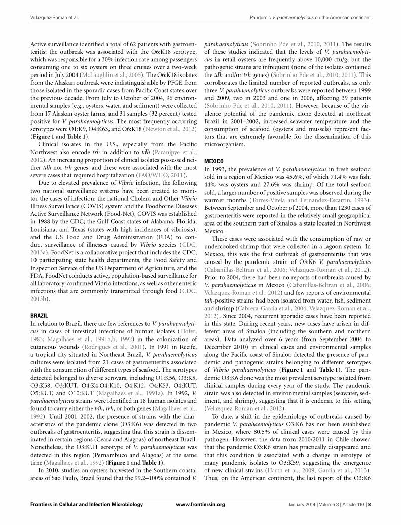
Active surveillance identified a total of 62 patients with gastroen-teritis; the outbreak was associated with the O6:K18 serotype,which was responsible for a 30% infection rate among passengersconsuming one to six oysters on three cruises over a two-weekperiod in July 2004 (McLaughlin et al., 2005). The O6:K18 isolatesfrom the Alaskan outbreak were indistinguishable by PFGE fromthose isolated in the sporadic cases from Pacific Coast states overthe previous decade. From July to October of 2004, 96 environ-mental samples (e.g., oysters, water, and sediment) were collectedfrom 17 Alaskan oyster farms, and 31 samples (32 percent) testedpositive for V. parahaemolyticus. The most frequently occurringserotypes were O1:K9, O4:K63, and O6:K18 (Newton et al., 2012)(Figure 1 and Table 1).
Clinical isolates in the U.S., especially from the PacificNorthwest also encode trh in addition to tdh (Paranjpye et al.,2012). An increasing proportion of clinical isolates possessed nei-ther tdh nor trh genes, and these were associated with the mostsevere cases that required hospitalization (FAO/WHO, 2011).
Due to elevated prevalence of Vibrio infection, the followingtwo national surveillance systems have been created to moni-tor the cases of infection: the national Cholera and Other VibrioIllness Surveillance (COVIS) system and the Foodborne DiseasesActive Surveillance Network (Food-Net). COVIS was establishedin 1988 by the CDC; the Gulf Coast states of Alabama, Florida,Louisiana, and Texas (states with high incidences of vibriosis);and the US Food and Drug Administration (FDA) to con-duct surveillance of illnesses caused by Vibrio species (CDC,2013a). FoodNet is a collaborative project that includes the CDC,10 participating state health departments, the Food Safety andInspection Service of the US Department of Agriculture, and theFDA. FoodNet conducts active, population-based surveillance forall laboratory-confirmed Vibrio infections, as well as other entericinfections that are commonly transmitted through food (CDC,2013b).
BRAZILIn relation to Brazil, there are few references to V. parahaemolyti-cus in cases of intestinal infections of human isolates (Hofer,1983; Magalhaes et al., 1991a,b, 1992) in the colonization ofcutaneous wounds (Rodrigues et al., 2001). In 1991 in Recife,a tropical city situated in Northeast Brazil, V. parahaemolyticuscultures were isolated from 21 cases of gastroenteritis associatedwith the consumption of different types of seafood. The serotypesdetected belonged to diverse serovars, including O1:K56, O3:K5,O3:K58, O3:KUT, O4:K4,O4:K10, O4:K12, O4:K53, O4:KUT,O5:KUT, and O10:KUT (Magalhaes et al., 1991a). In 1992, V.parahaemolyticus strains were identified in 18 human isolates andfound to carry either the tdh, trh, or both genes (Magalhaes et al.,1992). Until 2001–2002, the presence of strains with the char-acteristics of the pandemic clone (O3:K6) was detected in twooutbreaks of gastroenteritis, suggesting that this strain is dissem-inated in certain regions (Ceara and Alagoas) of northeast Brazil.Nonetheless, the O3:KUT serotype of V. parahaemolyticus wasdetected in this region (Pernambuco and Alagoas) at the sametime (Magalhaes et al., 1992) (Figure 1 and Table 1).
In 2010, studies on oysters harvested in the Southern coastalareas of Sao Paulo, Brazil found that the 99.2–100% contained V.
parahaemolyticus (Sobrinho Pde et al., 2010, 2011). The resultsof these studies indicated that the levels of V. parahaemolyti-cus in retail oysters are frequently above 10,000 cfu/g, but thepathogenic strains are infrequent (none of the isolates containedthe tdh and/or trh genes) (Sobrinho Pde et al., 2010, 2011). Thiscorroborates the limited number of reported outbreaks, as onlythree V. parahaemolyticus outbreaks were reported between 1999and 2009, two in 2003 and one in 2006, affecting 39 patients(Sobrinho Pde et al., 2010, 2011). However, because of the vir-ulence potential of the pandemic clone detected at northeastBrazil in 2001–2002, increased seawater temperature and theconsumption of seafood (oysters and mussels) represent fac-tors that are extremely favorable for the dissemination of thismicroorganism.
MEXICOIn 1993, the prevalence of V. parahaemolyticus in fresh seafoodsold in a region of Mexico was 45.6%, of which 71.4% was fish,44% was oysters and 27.6% was shrimp. Of the total seafoodsold, a larger number of positive samples was observed during thewarmer months (Torres-Vitela and Fernandez-Escartin, 1993).Between September and October of 2004, more than 1230 cases ofgastroenteritis were reported in the relatively small geographicalarea of the southern part of Sinaloa, a state located in NorthwestMexico.
These cases were associated with the consumption of raw orundercooked shrimp that were collected in a lagoon system. InMexico, this was the first outbreak of gastroenteritis that wascaused by the pandemic strain of O3:K6 V. parahaemolyticus(Cabanillas-Beltran et al., 2006; Velazquez-Roman et al., 2012).Prior to 2004, there had been no reports of outbreaks caused byV. parahaemolyticus in Mexico (Cabanillas-Beltran et al., 2006;Velazquez-Roman et al., 2012) and few reports of environmentaltdh-positive strains had been isolated from water, fish, sedimentand shrimp (Cabrera-Garcia et al., 2004; Velazquez-Roman et al.,2012). Since 2004, recurrent sporadic cases have been reportedin this state. During recent years, new cases have arisen in dif-ferent areas of Sinaloa (including the southern and northernareas). Data analyzed over 6 years (from September 2004 toDecember 2010) in clinical cases and environmental samplesalong the Pacific coast of Sinaloa detected the presence of pan-demic and pathogenic strains belonging to different serotypesof Vibrio parahaemolyticus (Figure 1 and Table 1). The pan-demic O3:K6 clone was the most prevalent serotype isolated fromclinical samples during every year of the study. The pandemicstrain was also detected in environmental samples (seawater, sed-iment, and shrimp), suggesting that it is endemic to this setting(Velazquez-Roman et al., 2012).
To date, a shift in the epidemiology of outbreaks caused bypandemic V. parahaemolyticus O3:K6 has not been establishedin Mexico, where 80.5% of clinical cases were caused by thispathogen. However, the data from 2010/2011 in Chile showedthat the pandemic O3:K6 strain has practically disappeared andthat this condition is associated with a change in serotype ofmany pandemic isolates to O3:K59, suggesting the emergenceof new clinical strains (Harth et al., 2009; Garcia et al., 2013).Thus, on the American continent, the last report of the O3:K6
Frontiers in Cellular and Infection Microbiology www.frontiersin.org January 2014 | Volume 3 | Article 110 | 8
Velazquez-Roman et al. Pandemic V. parahaemolyticus on the American continent
pandemic clone associated with clinical cases was observed inMexico.
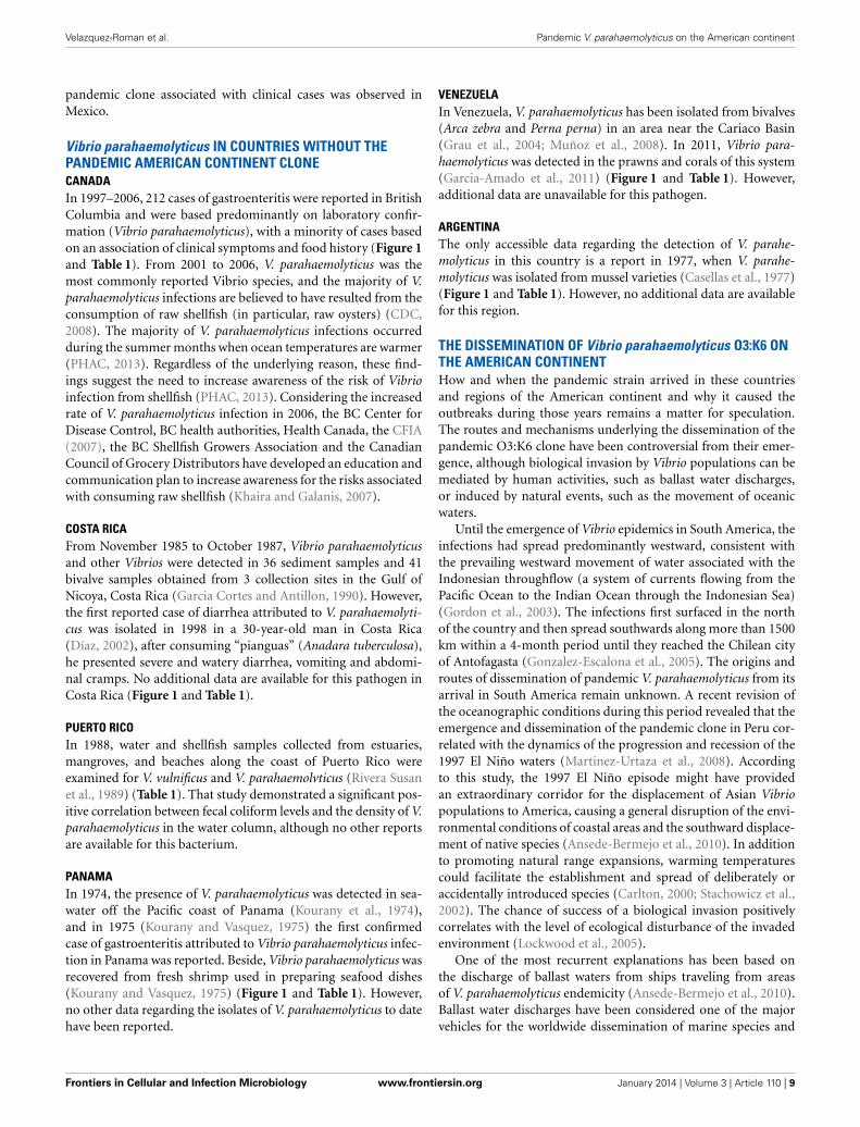
Vibrio parahaemolyticus IN COUNTRIES WITHOUT THEPANDEMIC AMERICAN CONTINENT CLONECANADAIn 1997–2006, 212 cases of gastroenteritis were reported in BritishColumbia and were based predominantly on laboratory confir-mation (Vibrio parahaemolyticus), with a minority of cases basedon an association of clinical symptoms and food history (Figure 1and Table 1). From 2001 to 2006, V. parahaemolyticus was themost commonly reported Vibrio species, and the majority of V.parahaemolyticus infections are believed to have resulted from theconsumption of raw shellfish (in particular, raw oysters) (CDC,2008). The majority of V. parahaemolyticus infections occurredduring the summer months when ocean temperatures are warmer(PHAC, 2013). Regardless of the underlying reason, these find-ings suggest the need to increase awareness of the risk of Vibrioinfection from shellfish (PHAC, 2013). Considering the increasedrate of V. parahaemolyticus infection in 2006, the BC Center forDisease Control, BC health authorities, Health Canada, the CFIA(2007), the BC Shellfish Growers Association and the CanadianCouncil of Grocery Distributors have developed an education andcommunication plan to increase awareness for the risks associatedwith consuming raw shellfish (Khaira and Galanis, 2007).
COSTA RICAFrom November 1985 to October 1987, Vibrio parahaemolyticusand other Vibrios were detected in 36 sediment samples and 41bivalve samples obtained from 3 collection sites in the Gulf ofNicoya, Costa Rica (Garcia Cortes and Antillon, 1990). However,the first reported case of diarrhea attributed to V. parahaemolyti-cus was isolated in 1998 in a 30-year-old man in Costa Rica(Díaz, 2002), after consuming “pianguas” (Anadara tuberculosa),he presented severe and watery diarrhea, vomiting and abdomi-nal cramps. No additional data are available for this pathogen inCosta Rica (Figure 1 and Table 1).
PUERTO RICOIn 1988, water and shellfish samples collected from estuaries,mangroves, and beaches along the coast of Puerto Rico wereexamined for V. vulnificus and V. parahaemolvticus (Rivera Susanet al., 1989) (Table 1). That study demonstrated a significant pos-itive correlation between fecal coliform levels and the density of V.parahaemolyticus in the water column, although no other reportsare available for this bacterium.
PANAMAIn 1974, the presence of V. parahaemolyticus was detected in sea-water off the Pacific coast of Panama (Kourany et al., 1974),and in 1975 (Kourany and Vasquez, 1975) the first confirmedcase of gastroenteritis attributed to Vibrio parahaemolyticus infec-tion in Panama was reported. Beside, Vibrio parahaemolyticus wasrecovered from fresh shrimp used in preparing seafood dishes(Kourany and Vasquez, 1975) (Figure 1 and Table 1). However,no other data regarding the isolates of V. parahaemolyticus to datehave been reported.
VENEZUELAIn Venezuela, V. parahaemolyticus has been isolated from bivalves(Arca zebra and Perna perna) in an area near the Cariaco Basin(Grau et al., 2004; Muñoz et al., 2008). In 2011, Vibrio para-haemolyticus was detected in the prawns and corals of this system(Garcia-Amado et al., 2011) (Figure 1 and Table 1). However,additional data are unavailable for this pathogen.
ARGENTINAThe only accessible data regarding the detection of V. parahe-molyticus in this country is a report in 1977, when V. parahe-molyticus was isolated from mussel varieties (Casellas et al., 1977)(Figure 1 and Table 1). However, no additional data are availablefor this region.
THE DISSEMINATION OF Vibrio parahaemolyticus O3:K6 ONTHE AMERICAN CONTINENTHow and when the pandemic strain arrived in these countriesand regions of the American continent and why it caused theoutbreaks during those years remains a matter for speculation.The routes and mechanisms underlying the dissemination of thepandemic O3:K6 clone have been controversial from their emer-gence, although biological invasion by Vibrio populations can bemediated by human activities, such as ballast water discharges,or induced by natural events, such as the movement of oceanicwaters.
Until the emergence of Vibrio epidemics in South America, theinfections had spread predominantly westward, consistent withthe prevailing westward movement of water associated with theIndonesian throughflow (a system of currents flowing from thePacific Ocean to the Indian Ocean through the Indonesian Sea)(Gordon et al., 2003). The infections first surfaced in the northof the country and then spread southwards along more than 1500km within a 4-month period until they reached the Chilean cityof Antofagasta (Gonzalez-Escalona et al., 2005). The origins androutes of dissemination of pandemic V. parahaemolyticus from itsarrival in South America remain unknown. A recent revision ofthe oceanographic conditions during this period revealed that theemergence and dissemination of the pandemic clone in Peru cor-related with the dynamics of the progression and recession of the1997 El Niño waters (Martinez-Urtaza et al., 2008). Accordingto this study, the 1997 El Niño episode might have providedan extraordinary corridor for the displacement of Asian Vibriopopulations to America, causing a general disruption of the envi-ronmental conditions of coastal areas and the southward displace-ment of native species (Ansede-Bermejo et al., 2010). In additionto promoting natural range expansions, warming temperaturescould facilitate the establishment and spread of deliberately oraccidentally introduced species (Carlton, 2000; Stachowicz et al.,2002). The chance of success of a biological invasion positivelycorrelates with the level of ecological disturbance of the invadedenvironment (Lockwood et al., 2005).
One of the most recurrent explanations has been based onthe discharge of ballast waters from ships traveling from areasof V. parahaemolyticus endemicity (Ansede-Bermejo et al., 2010).Ballast water discharges have been considered one of the majorvehicles for the worldwide dissemination of marine species and
Frontiers in Cellular and Infection Microbiology www.frontiersin.org January 2014 | Volume 3 | Article 110 | 9
Velazquez-Roman et al. Pandemic V. parahaemolyticus on the American continent
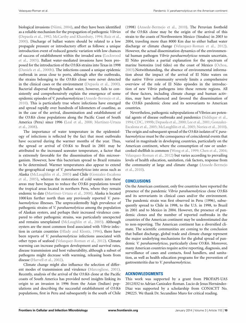
biological invasions (Niimi, 2004), and they have been identifiedas a reliable mechanism for the propagation of pathogenic Vibrios(Depaola et al., 1992; McCarthy and Khambaty, 1994; Ruiz et al.,2000). Discharge of ballast waters should be related to a lowpropagule pressure or introductory effort as follows: a uniqueintroduction event of reduced genetic variation with low chancesof success of establishment within the invaded area (Lockwoodet al., 2005). Ballast water-mediated invasions have been pro-posed for the introduction of the O3:K6 strains into Texas in 1998(Daniels et al., 2000b). The infections caused a unique epidemicoutbreak in areas close to ports, although after the outbreaks,the strains belonging to the O3:K6 clone were never detectedin the clinical cases or the environment (Depaola et al., 2000).Bacterial dispersal through ballast water, however, fails to con-sistently and comprehensively explain the emergence of someepidemic episodes of V. parahaemolyticus (Ansede-Bermejo et al.,2010). This is particularly true where infections have emergedand spread rapidly over hundreds of kilometers of coastline, asin the case of the arrival, dissemination and establishment ofthe O3:K6 clone populations along the Pacific Coast of SouthAmerica (Peru) since 1996 (Leal et al., 2008; Martinez-Urtazaet al., 2008).
The importance of water temperature in the epidemiol-ogy of infections is reflected by the fact that most outbreakshave occurred during the warmer months of the year. Thus,the spread or arrival of O3:K6 to Brazil in 2001 may beattributed to the increased seawater temperature, a factor thatis extremely favorable for the dissemination of this microor-ganism. However, how this bacterium spread to Brazil remainsto be determined. Warmer temperatures also appear to extendthe geographical range of V. parahaemolyticus into areas such asAlaska (McLaughlin et al., 2005) and Chile (Gonzalez-Escalonaet al., 2005), whereas the restoration of cold waters into theseareas may have begun to reduce the O3:K6 populations towardthe tropical areas located in northern Peru, where they remainendemic to date (Martinez-Urtaza et al., 2008). Alaska is located1000 km further north than any previously reported V. para-haemolyticus illnesses. The unprecedentedly high prevalence ofpathogenic strains of V. parahaemolyticus among the populationof Alaskan oysters, and perhaps their increased virulence com-pared to other pathogenic strains, was particularly unexpectedand remains unexplained (McLaughlin et al., 2005). Althoughoysters are the most common food associated with Vibrio infec-tion in certain countries (Hlady and Klontz, 1996), there havebeen reports of V. parahaemolyticus infections associated withother types of seafood (Velazquez-Roman et al., 2012). Climatewarming can increase pathogen development and survival rates,disease transmission and host vulnerability, although a subset ofpathogens might decrease with warming, releasing hosts fromdisease (Harvell et al., 2002).
Climate change might also influence the selection of differ-ent modes of transmission and virulence (Marcogliese, 2001).Recently, analysis of the arrival of the O3:K6 clone at the Pacificcoasts of South America has provided novel insights linking itsorigin to an invasion in 1996 from the Asian (Indian) pop-ulations and describing the successful establishment of O3:K6populations, first in Peru and subsequently in the south of Chile
(1998) (Ansede-Bermejo et al., 2010). The Peruvian footholdof the O3:K6 clone may be the origin of the arrival of thisstrain to the coasts of Northwestern Mexico (Sinaloa) in 2003 to2004, traveling more than 8000 Km due to either ballast waterdischarge or climate change (Velazquez-Roman et al., 2012).However, the actual dissemination dynamics of the environmen-tal human pathogen Vibrio parahaemolyticus remain uncertain.El Niño provides a partial explanation for the spectrum ofmarine biotoxins (red tides) on the coast of Mexico (Ochoa,2003).Notwithstanding, the absence of environmental informa-tion about the impact of the arrival of El Niño waters onthe native Vibrio community severely limits a comprehensiveoverview of the role of El Niño episodes in the introduc-tion of new Vibrio pathogens into these remote regions. Allof these factors, including climate change and human activ-ities, may have influenced and favored the dissemination ofthe O3:K6 pandemic clone and its serovariants to Americancountries.
Nevertheless, pathogenic subpopulations of Vibrios are poten-tial agents of disease outbreaks and pandemics (Siddique et al.,1994; CDC, 1999b; Depaola et al., 2000; Lee et al., 2001; Gonzalez-Escalona et al., 2005; McLaughlin et al., 2005; Noriea et al., 2010).The origin and subsequent spread of the O3:K6 isolates of V. para-haemolyticus must be the consequence of coincidental events thatvaried in magnitude in developing countries, particularly on theAmerican continent, where the consumption of raw or under-cooked shellfish is common (Wong et al., 1999; Chen et al., 2004;Velazquez-Roman et al., 2012) but varies according to prevailinglevels of health education, sanitation, risk factors, response fromthe community at large and climate change (Ansede-Bermejoet al., 2010).
CONCLUSIONSOn the American continent, only five countries have reported thepresence of the pandemic Vibrio parahaemolyticus clone O3:K6and its serovariants in clinical cases and/or the environment.The pandemic strain was first observed in Peru (1996), subse-quently spread to Chile in 1998, to the U.S. in 1998, to Brazilin 2001 and to Mexico in 2004. However, the presence of pan-demic clones and the number of reported outbreaks in thecountries of the American continent may be underestimated dueto non-reporting. The American continent has a diversified cli-mate. The scientific communities are coming to the conclusionthat ballast discharge, global trade and climate change representthe major underlying mechanisms for the global spread of pan-demic V. parahaemolyticus, particularly clone O3:K6. Moreover,many American countries require active reporting, diagnosis, andsurveillance of cases and contacts, food handlers, and sanita-tion, as well as health education programs for the prevention ofgastroenteritis due to V. parahaemolyticus.
ACKNOWLEDGMENTSThis work was supported by a grant from PROFAPI-UAS2012/032 to Adrian Canizalez-Roman. Lucio de Jesus Hernández-Díaz was supported by a scholarship from CONACYT No.290225. We thank Dr. Secundino Muro for critical reading.
Frontiers in Cellular and Infection Microbiology www.frontiersin.org January 2014 | Volume 3 | Article 110 | 10
Velazquez-Roman et al. Pandemic V. parahaemolyticus on the American continent
REFERENCESAlam, M. J., Tomochika, K. I., Miyoshi, S. I., and Shinoda, S. (2002). Environmental
investigation of potentially pathogenic Vibrio parahaemolyticus in the Seto-Inland Sea, Japan. FEMS Microbiol. Lett. 208, 83–87. doi: 10.1111/j.1574-6968.2002.tb11064.x
Ansaruzzaman, M., Lucas, M., Deen, J. L., Bhuiyan, N. A., Wang, X. Y., Safa,A., et al. (2005). Pandemic serovars (O3:K6 and O4:K68) of Vibrio para-haemolyticus associated with diarrhea in Mozambique: spread of the pan-demic into the African continent. J. Clin. Microbiol. 43, 2559–2562. doi:10.1128/JCM.43.6.2559-2562.2005
Ansede-Bermejo, J., Gavilan, R. G., Trinanes, J., Espejo, R. T., and Martinez-Urtaza, J. (2010). Origins and colonization history of pandemic Vibrio para-haemolyticus in South America. Mol. Ecol. 19, 3924–3937. doi: 10.1111/j.1365-294X.2010.04782.x
Baba, K., Shirai, H., Terai, A., Takeda, Y., and Nishibuchi, M. (1991). Analysisof the tdh gene cloned from a tdh gene- and trh gene-positive strain ofVibrio parahaemolyticus. Microbiol. Immunol. 35, 253–258. doi: 10.1111/j.1348-0421.1991.tb01554.x
Bag, P. K., Nandi, S., Bhadra, R. K., Ramamurthy, T., Bhattacharya, S. K.,Nishibuchi, M., et al. (1999). Clonal diversity among recently emerged strainsof Vibrio parahaemolyticus O3:K6 associated with pandemic spread. J. Clin.Microbiol. 37, 2354–2357.
Baumann, P., Furniss, A.l., and Lee, J. V. (1984). “Genus I. Vibrio,” in Bergey’sManual of Systematic Bacteriology, Vol. 1. eds R. G. E. Murray, D. J. Brenner,and M. P. Bryant (Baltimore, MD: Williams and Wilkins), 518.
Bean, N. H., Maloney, E. K., Potter, M. E., Korazemo, P., Ray, B., Taylor,J. P., et al. (1998). Crayfish: a newly recognized vehicle for vibrioinfections. Epidemiol. Infect. 121, 269–273. doi: 10.1017/S0950268898001381
Bej, A. K., Patterson, D. P., Brasher, C. W., Vickery, M. C., Jones, D. D., andKaysner, C. A. (1999). Detection of total and hemolysin-producing Vibrio para-haemolyticus in shellfish using multiplex PCR amplification of tl, tdh and trh.J. Microbiol. Methods 36, 215–225. doi: 10.1016/S0167-7012(99)00037-8
Boyd, E. F., Cohen, A. L., Naughton, L. M., Ussery, D. W., Binnewies, T. T., Stine,O. C., et al. (2008). Molecular analysis of the emergence of pandemic Vibrioparahaemolyticus. BMC Microbiol. 8, 110. doi: 10.1186/1471-2180-8-110
Cabanillas-Beltran, H., LLausás-Magaña, E., Romero, R., Espinoza, A., Garcia-Gasca, A., Nishibuchi, M., et al. (2006). Outbreak of gastroenteritis caused bythe pandemic Vibrio parahaemolyticus O3:K6 in Mexico. FEMS Microbiol. Lett.265, 76–80. doi: 10.1111/j.1574-6968.2006.00475.x
Cabello, F. C., Espejo, R. T., Hernandez, M. C., Rioseco, M. L., Ulloa, J., and Vergara,J. A. (2007). Vibrio parahaemolyticus O3:K6 epidemic diarrhea, Chile, 2005.Emerging Infect. Dis. 13, 655–656. doi: 10.3201/eid1304.06-1152
Cabrera-Garcia, M. E., Vazquez-Salinas, C., and Quinones-Ramirez, E. I. (2004).Serologic and molecular characterization of Vibrio parahaemolyticus strainsisolated from seawater and fish products of the Gulf of Mexico. Appl. Environ.Microbiol. 70, 6401–6406. doi: 10.1128/AEM.70.11.6401-6406.2004
Canadian Food Inspection Agency. (2007). Available online at: http://www.
phac-aspc.gc.ca/publicat/ccdr-rmtc/07vol33/dr3311b-eng.phpCanizalez-Roman, A., Flores-Villasenor, H., Zazueta-Beltran, J., Muro-Amador, S.,
and Leon-Sicairos, N. (2011). Comparative evaluation of a chromogenic agarmedium-PCR protocol with a conventional method for isolation of Vibrioparahaemolyticus strains from environmental and clinical samples. Can. J.Microbiol. 57, 136–142. doi: 10.1139/W10-108
Carlton, J. T. (2000). “Global change and biological invasions in the oceans,” inInvasive Species in a Changing World, eds H. A. Mooneyand R. J. Hobbs (Covelo,CA: Island Press), 31–53.
Casellas, J. M., Caría, M. A., and Gerghi, M. E. (1977). Isolation of Vibrio para-hemolyticus from mussel varieties in Argentina. Rev. Asoc. Argent. Microbiol. 9,41–53.
CDC. (1998). Outbreak of Vibrio parahaemolyticus infections associated with eat-ing raw oysters–Pacific Northwest, 1997. MMWR Morb. Mortal. Wkly. Rep. 47,457–462.
CDC. (1999a). From the centers for disease control and prevention. outbreak ofvibrio parahaemolyticus infection associated with eating raw oysters and clamsharvested from Long Island Sound—Connecticut. New Jersey, and New York,1998. JAMA 281, 603–604.
CDC. (1999b). Outbreak of Vibrio parahaemolyticus infection associated with eat-ing raw oysters and clams harvested from Long Island Sound–Connecticut, New
Jersey, and New York, 1998. MMWR Morb. Mortal. Wkly. Rep. 48, 48–51. doi:10.1001/jama.281.7.603
CDC. (2008). Preliminary FoodNet data on the incidence of infection withpathogens transmitted commonly through food–10 states, 2007. MMWR Morb.Mortal. Wkly. Rep. 57, 366–370.
CDC. (2013a). Cholera and Other Vibrio Illness Surveillance System.[Accessed September 9 2013] Centers for Disease Control and Prevention.Department of Health and Human Services.Available online at:http://www.cdc.gov/nationalsurveillance/cholera_vibrio_surveillance.html
CDC. (2013b). Foodborne Diseases Active Surveillance Network. Foodborne DiseasesActive Surveillance Network (FoodNet). [Accessed September 9]. Available onlineat: http://www.cdc.gov/foodnet/
Chao, G., Jiao, X., Zhou, X., Wang, F., Yang, Z., Huang, J., et al. (2010). Distributionof genes encoding four pathogenicity islands (VPaIs), T6SS, biofilm, and typeI pilus in food and clinical strains of Vibrio parahaemolyticus in China.Foodborne Pathog. Dis. 7, 649–658. doi: 10.1089/fpd.2009.0441
Chen, C. H., Shimada, T., Elhadi, N., Radu, S., and Nishibuchi, M. (2004).Phenotypic and genotypic characteristics and epidemiological significance ofctx+ strains of Vibrio cholerae isolated from seafood in Malaysia. Appl. Environ.Microbiol. 70, 1964–1972. doi: 10.1128/AEM.70.4.1964-1972.2004
Chowdhury, A., Ishibashi, M., Thiem, V. D., Tuyet, D. T., Tung, T. V., Chien,B. T., et al. (2004a). Emergence and serovar transition of Vibrio para-haemolyticus pandemic strains isolated during a diarrhea outbreak in Vietnambetween 1997 and 1999. Microbiol. Immunol. 48, 319–327. doi: 10.1111/j.1348-0421.2004.tb03513.x
Chowdhury, N. R., Stine, O. C., Morris, J. G., and Nair, G. B. (2004b). Assessment ofevolution of pandemic Vibrio parahaemolyticus by multilocus sequence typing.J. Clin. Microbiol. 42, 1280–1282. doi: 10.1128/JCM.42.3.1280-1282.2004
Chowdhury, N. R., Chakraborty, S., Ramamurthy, T., Nishibuchi, M., Yamasaki, S.,Takeda, Y., et al. (2000). Molecular evidence of clonal Vibrio parahaemolyticuspandemic strains. Emerging Infect. Dis. 6, 631–636. doi: 10.3201/eid0606.000612
Cordova, J. L., Astorga, J., Silva, W., and Riquelme, C. (2002).Characterization by PCR of Vibrio parahaemolyticus isolates collectedduring the 1997-1998 Chilean outbreak. Biol. Res. 35, 433–440. doi:10.4067/S0716-97602002000300017
Dadisman, T. A. Jr., Nelson, R., Molenda, J. R., and Garber, H. J. (1972). Vibrioparahaemolyticus gastroenteritis in Maryland. I. Clinical and epidemiologicaspects. Am. J. Epidemiol. 96, 414–426.
Daniels, N. A., Mackinnon, L., Bishop, R., Altekruse, S., Ray, B., Hammond, R. M.,et al. (2000a). Vibrio parahaemolyticus infections in the United States, 1973-1998. J. Infect. Dis. 181, 1661–1666. doi: 10.1086/315459
Daniels, N. A., Ray, B., Easton, A., Marano, N., Kahn, E., McShan, A. L., et al.(2000b). Emergence of a new Vibrio parahaemolyticus serotype in raw oysters:a prevention quandary. JAMA 284, 1541–1545. doi: 10.1001/jama.284.12.1541
Deepanjali, A., Kumar, H. S., and Karunasagar, I. (2005). Seasonal variation inabundance of total and pathogenic Vibrio parahaemolyticus bacteria in oystersalong the southwest coast of India. Appl. Environ. Microbiol. 71, 3575–3580. doi:10.1128/AEM.71.7.3575-3580.2005
Depaola, A., Capers, G. M., Motes, M. L., Olsvik, O., Fields, P. I., Wells, J., et al.(1992). Isolation of Latin American epidemic strain of Vibrio cholerae O1 fromUS Gulf Coast. Lancet 339, 624. doi: 10.1016/0140-6736(92)90917-R
Depaola, A., Kaysner, C. A., Bowers, J., and Cook, D. W. (2000). Environmentalinvestigations of Vibrio parahaemolyticus in oysters after outbreaks inWashington, Texas, and New York (1997 and 1998). Appl. Environ. Microbiol.66, 4649–4654. doi: 10.1128/AEM.66.11.4649-4654.2000
Depaola, A., Ulaszek, J., Kaysner, C. A., Tenge, B. J., Nordstrom, J. L., Wells, J.,et al. (2003). Molecular, serological, and virulence characteristics of Vibrioparahaemolyticus isolated from environmental, food, and clinical sourcesin North America and Asia. Appl. Environ. Microbiol. 69, 3999–4005. doi:10.1128/AEM.69.7.3999-4005.2003
Díaz, J. M. E. (2002). Diarrea por Vibrio parahaemolyticus: primer caso reportadoen Costa Rica. Revista Médica del Hospital Nacional de Niños Dr. Carlos SáenzHerrera 37, 15–17.
FAO/WHO. (2011). Risk assessment of Vibrio parahaemolyticus in seafood:Interpretative summary and Technical report. food and agriculture organiza-tion of the united nations/world health organization. Microbiol. Risk Assess.16, 193.
Fuenzalida, L., Hernandez, C., Toro, J., Rioseco, M. L., Romero, J., and Espejo, R.T. (2006). Vibrio parahaemolyticus in shellfish and clinical samples during two
Frontiers in Cellular and Infection Microbiology www.frontiersin.org January 2014 | Volume 3 | Article 110 | 11
Velazquez-Roman et al. Pandemic V. parahaemolyticus on the American continent
large epidemics of diarrhoea in southern Chile. Environ. Microbiol. 8, 675–683.doi: 10.1111/j.1462-2920.2005.00946.x
Fujino, T., Okuno, Y., Nakada, D., Aoyama, A., Fukai, K., Mukai, T., et al. (1953).On the bacteriological examination of Shirasu food poisoning. Med. J. OsakaUniv. 4, 299–304.
Garcia Cortes, V., and Antillon, F. (1990). [Isolation of enteropathogenic Vibrioin bivalves and mud from the Nicoya Gulf, Costa Rica]. Rev. Biol. Trop. 38,437–440.
Garcia, K., Bastias, R., Higuera, G., Torres, R., Mellado, A., Uribe, P., et al. (2013).Rise and fall of pandemic Vibrio parahaemolyticus serotype O3:K6 in southernChile. Environ. Microbiol. 15, 527–534. doi: 10.1111/j.1462-2920.2012.02883.x
Garcia, K., Torres, R., Uribe, P., Hernandez, C., Rioseco, M. L., Romero, J.,et al. (2009). Dynamics of clinical and environmental Vibrio parahaemolyticusstrains during seafood-related summer diarrhea outbreaks in southern Chile.Appl. Environ. Microbiol. 75, 7482–7487. doi: 10.1128/AEM.01662-09
Garcia-Amado, M. A., Bozo-Hurtado, L., Astor, Y., Suarez, P., and Chistoserdov,A. (2011). Denaturing gradient gel electrophoresis analyses of the vertical dis-tribution and diversity of Vibrio spp. populations in the Cariaco Basin. FEMSMicrobiol. Ecol. 77, 347–356. doi: 10.1111/j.1574-6941.2011.01116.x
Gavilan, R. G., Zamudio, M. L., and Martinez-Urtaza, J. (2013). Molecular epi-demiology and genetic variation of pathogenic Vibrio parahaemolyticus inPeru. PLoS Negl. Trop. Dis. 7:e2210. doi: 10.1371/journal.pntd.0002210
Gil, A. I., Miranda, H., Lanata, C. F., Prada, A., Hall, E. R., Barreno, C. M., et al.(2007). O3:K6 serotype of Vibrio parahaemolyticus identical to the global pan-demic clone associated with diarrhea in Peru. Int. J. Infect. Dis. 11, 324–328. doi:10.1016/j.ijid.2006.08.003
Gonzalez-Escalona, N., Cachicas, V., Acevedo, C., Rioseco, M. L., Vergara, J. A.,Cabello, F., et al. (2005). Vibrio parahaemolyticus diarrhea, Chile, 1998 and2004. Emerging Infect. Dis. 11, 129–131. doi: 10.3201/eid1101.040762
Gordon, A. L., Susanto, R. D., and Vranes, K. (2003). Cool Indonesian through-flow as a consequence of restricted surface layer flow. Nature 425, 824–828. doi:10.1038/nature02038
Grau, C. L. B., Zerpa, A., Silva, S., and Gallardo, O. (2004). Aislamiento de Vibriospp. y evaluacion de la condicion sanitaria de los moluscos bivalvos Arca zebray Perna perna procedentes de la costa nororiental del Edo. Sucre. Venezuela.Revista Cientıfica FCV-LUZ 14, 513–521.
Guevara Duncan, J. P., Del Carpio, E. (1989). Vibrio parahaemolyticus in ‘cebiches’sold by hucksters in Lima Peru. Diagnostico (in Spanish). 24, 23–26
Gutierrez West, C. K., Klein, S. L., and Lovell, C. R. (2013). High frequency ofvirulence factor genes tdh, trh, and tlh in Vibrio parahaemolyticus strains iso-lated from a pristine estuary. Appl. Environ. Microbiol. 79, 2247–2252. doi:10.1128/AEM.03792-12
Harth, E., Matsuda, L., Hernandez, C., Rioseco, M. L., Romero, J., Gonzalez-Escalona, N., et al. (2009). Epidemiology of Vibrio parahaemolyticus out-breaks, southern Chile. Emerging Infect. Dis. 15, 163–168. doi: 10.3201/eid1502.071269
Harvell, C. D., Mitchell, C. E., Ward, J. R., Altizer, S., Dobson, A. P., Ostfeld, R. S.,et al. (2002). Climate warming and disease risks for terrestrial and marine biota.Science 296, 2158–2162. doi: 10.1126/science.1063699
Hasegawa, H., and Hase, C. C. (2009). The extracellular metalloprotease ofVibrio tubiashii directly inhibits its extracellular haemolysin. Microbiology 155,2296–2305. doi: 10.1099/mic.0.028605-0
Hervio-Heath, D., Colwell, R. R., Derrien, A., Robert-Pillot, A., Fournier, J.M., and Pommepuy, M. (2002). Occurrence of pathogenic vibrios in coastalareas of France. J. Appl. Microbiol. 92, 1123–1135. doi: 10.1046/j.1365-2672.2002.01663.x
Hiyoshi, H., Kodama, T., Iida, T., and Honda, T. (2010). Contribution of Vibrioparahaemolyticus virulence factors to cytotoxicity, enterotoxicity, and lethalityin mice. Infect. Immun. 78, 1772–1780. doi: 10.1128/IAI.01051-09
Hiyoshi, H., Kodama, T., Saito, K., Gotoh, K., Matsuda, S., Akeda, Y., et al. (2011).VopV, an F-actin-binding type III secretion effector, is required for Vibrioparahaemolyticus-induced enterotoxicity. Cell Host Microbe 10, 401–409. doi:10.1016/j.chom.2011.08.014
Hlady, W. G., and Klontz, K. C. (1996). The epidemiology of Vibrioinfections in Florida, 1981–1993. J. Infect. Dis. 173, 1176–1183. doi:10.1093/infdis/173.5.1176
Hofer, E. (1983). Primeiro isolamento e identificac,a∼o de Vibrio parahaemolyti-cus no Brasil de infecc,a∼o gastrointestinal humana. Rev. Microbiol. Sa∼o Paulo4, 174–175.
Honda, T., and T., Iida (1993). The pathogenicity of Vibrio parahaemolyticus andthe role of the thermostable direct hemolysin and related hemolysins. Rev. Med.Microbiol. 4, 106–113. doi: 10.1097/00013542-199304000-00006
Ibarra, J. D., A. Alvarado, D. (1999). Non-epdemic vibrios and Vibrio choleraeO1 associated with acute diarrhea from cases given during “El Niño” SouthernOscillation (ENSO) 1998. Hospital Nacional Dos de Mayo. An Fac Med (Peru)(in Spanish) 60, 251–256.
Iida, T., Hattori, A., Tagomori, K., Nasu, H., Naim, R., and Honda, T. (2001).Filamentous phage associated with recent pandemic strains of Vibrio para-haemolyticus. Emerging Infect. Dis. 7, 477–478. doi: 10.3201/eid0703.010325
Islam, M. S., Tasmin, R., Khan, S. I., Bakht, H. B., Mahmood, Z. H., Rahman, M. Z.,et al. (2004). Pandemic strains of O3:K6 Vibrio parahaemolyticus in the aquaticenvironment of Bangladesh. Can. J. Microbiol. 50, 827–834. doi: 10.1139/w04-072
Joseph, S. W., Colwell, R. R., and Kaper, J. B. (1982). Vibrio parahaemolyti-cus and related halophilic Vibrios. Crit. Rev. Microbiol. 10, 77–124. doi:10.3109/10408418209113506
Kaneko, T., and Colwell, R. R. (1973). Ecology of Vibrio parahaemolyticus inChesapeake Bay. J. Bacteriol. 113, 24–32.
Kaneko, T. C., R. R. (1977). The annual cycle of Vibrio parahaemolyticus inChesapeake Bay. Microb. Ecol. 4, 135–155. doi: 10.1007/BF02014284
Kaysner, C. A., Abeyta, C. Jr., Stott, R. F., Lilja, J. L., and Wekell, M. M.(1990). Incidence of urea-hydrolyzing Vibrio parahaemolyticus in Willapa Bay,Washington. Appl. Environ. Microbiol. 56, 904–907.
Khaira, G., and Galanis, E. (2007). Descriptive epidemiology of Vibrio para-haemolyticus and other Vibrio species infections in British Columbia: 2001-2006. Can. Commun. Dis. Rep. 33, 12–22.
Kishishita, M., Matsuoka, N., Kumagai, K., Yamasaki, S., Takeda, Y., andNishibuchi, M. (1992). Sequence variation in the thermostable direct hemolysin related hemolysin (trh) gene of Vibrio parahaemolyticus Appl. Environ.Microbiol.58, 2449–2457.
Kourany, M., Kinney, R. J., and Vasquez, M. A. (1974). Vibrio parahaemolyticus inseawater off the Pacific coast of Panama. Am. J. Trop. Med. Hyg. 23, 714–715.
Kourany, M., and Vasquez, M. A. (1975). The first reported case from Panama ofacute gastroenteritis caused by Vibrio parahaemolyticus. Am. J. Trop. Med. Hyg.24, 638–640.
Leal, N. C., Da Silva, S. C., Cavalcanti, V. O., Figueiroa, A. C., Nunes, V. V.,Miralles, I. S., et al. (2008). Vibrio parahaemolyticus serovar O3:K6 gastroen-teritis in northeast Brazil. J. Appl. Microbiol. 105, 691–697. doi: 10.1111/j.1365-2672.2008.03782.x
Lee, W. C., Lee, M. J., Kim, J. S., and Park, S. Y. (2001). Foodborne illness outbreaksin Korea and Japan studied retrospectively. J. Food Prot. 64, 899–902.
Lockwood, J. L., Cassey, P., and Blackburn, T. (2005). The role of propagulepressure in explaining species invasions. Trends Ecol. Evol. 20, 223–228. doi:10.1016/j.tree.2005.02.004
Magalhaes, M., Magalhaes, V., Antas, M. G., and Tateno, S. (1991a). Isolationof urease-positive Vibrio parahaemolyticus from diarrheal patients in north-east Brazil. Rev. Inst. Med. Trop. Sao Paulo 33, 263–265. doi: 10.1590/S0036-46651991000400004
Magalhaes, V., Lima, R. A., Tateno, S., and Magalhaes, M. (1991b). Human gas-troenteritis associated with Vibrio parahaemolyticus in Recife, Brazil. Rev. Inst.Med. Trop. Sao Paulo 33, 64–68. doi: 10.1590/S0036-46651991000100012
Magalhaes, M., Takeda, Y., Magalhaes, V., and Tateno, S. (1992). Brazilian urease-positive strains of Vibrio parahaemolyticus carry genetic potential to pro-duce the TDH-related hemolysin. Mem. Inst. Oswaldo Cruz 87, 167–168. doi:10.1590/S0074-02761992000100027
Mahmud, Z. H., Neogi, S. B., Kassu, A., Wada, T., Islam, M. S., Nair,G. B., et al. (2007). Seaweeds as a reservoir for diverse Vibrio para-haemolyticus populations in Japan. Int. J. Food Microbiol. 118, 92–96. doi:10.1016/j.ijfoodmicro.2007.05.009
Marcogliese, D. J. (2001). Implications of climate change for parasitism of animalsin the aquatic environment. Can. J. Zool. 79, 1331–1352. doi: 10.1139/z01-067
Martinez-Urtaza, J., Huapaya, B., Gavilan, R. G., Blanco-Abad, V., Ansede-Bermejo,J., Cadarso-Suarez, C., et al. (2008). Emergence of asiatic vibrio diseasesin south america in phase with El Nino. Epidemiology 19, 829–837. doi:10.1097/EDE.0b013e3181883d43
Martinez-Urtaza, J., Simental, L., Velasco, D., Depaola, A., Ishibashi, M.,Nakaguchi, Y., et al. (2005). Pandemic Vibrio parahaemolyticus O3:K6, Europe.Emerging Infect. Dis. 11, 1319–1320. doi: 10.3201/eid1108.050322
Frontiers in Cellular and Infection Microbiology www.frontiersin.org January 2014 | Volume 3 | Article 110 | 12
Velazquez-Roman et al. Pandemic V. parahaemolyticus on the American continent
Matsuda, S., Okada, N., Kodama, T., Honda, T., and Iida, T. (2012). A cytotoxictype III secretion effector of Vibrio parahaemolyticus targets vacuolar H+-ATPase subunit c and ruptures host cell lysosomes. PLoS Pathog. 8:e1002803.doi: 10.1371/journal.ppat.1002803
Matsumoto, C., Okuda, J., Ishibashi, M., Iwanaga, M., Garg, P., Rammamurthy,T., et al. (2000). Pandemic spread of an O3:K6 clone of Vibrio para-haemolyticus and emergence of related strains evidenced by arbitrar-ily primed PCR and toxRS sequence analyses. J. Clin. Microbiol. 38,578–585.
McCarter, L. (1999). The multiple identities of Vibrio parahaemolyticus. J. Mol.Microbiol. Biotechnol. 1, 51–57.
McCarthy, S. A., and Khambaty, F. M. (1994). International dissemination of epi-demic Vibrio cholerae by cargo ship ballast and other nonpotable waters. Appl.Environ. Microbiol. 60, 2597–2601.
McLaughlin, J. B., Depaola, A., Bopp, C. A., Martinek, K. A., Napolilli, N. P.,Allison, C. G., et al. (2005). Outbreak of Vibrio parahaemolyticus gastroen-teritis associated with Alaskan oysters. N. Engl. J. Med. 353, 1463–1470. doi:10.1056/NEJMoa051594
MINSAL, (2013). Vibrio parahaemolyticus. [Accessed 4 September 2013].Departamento de Epidemiologia. Ministerio de Salud. (Departamentode Epidemiología-DIPLAS-MINSAL). Vibrio parahaemolyticus. Availableonline at: http://epi.minsal.cl/vigilancia-epidemiologica/enfermedades-de-notificacion-obligatoria/vibrio-parahaemolyticus/
Muñoz, D., Crucita,G. D. M., Martınez, C., Marjal, H., and Zerpa, A. (2008).Prevalencia de Staphylococcus aureus, Vibrio spp. y enterobacterias en carnede pepitona, Arca zebra, comercializada en Cumana, Venezuela. Zoot. Trop. 26,505–513.
Nair, G. B., Ramamurthy, T., Bhattacharya, S. K., Dutta, B., Takeda, Y.,and Sack, D. A. (2007). Global dissemination of Vibrio parahaemolyticusserotype O3:K6 and its serovariants. Clin. Microbiol. Rev. 20, 39–48. doi:10.1128/CMR.00025-06
Nasu, H., Iida, T., Sugahara, T., Yamaichi, Y., Park, K. S., Yokoyama, K., et al. (2000).A filamentous phage associated with recent pandemic Vibrio parahaemolyticusO3:K6 strains. J. Clin. Microbiol. 38, 2156–2161.
Newton, A., Kendall, M., Vugia, D. J., Henao, O. L., and Mahon, B. E. (2012).Increasing rates of vibriosis in the United States, 1996-2010: review of surveil-lance data from 2 systems. Clin. Infect. Dis. 54(Suppl. 5), S391–S395. doi:10.1093/cid/cis243
Niimi, A. J. (2004). Role of container vessels in the introduction of exotic species.Mar. Pollut. Bull. 49, 778–782. doi: 10.1016/j.marpolbul.2004.06.006
Nishibuchi, M., and Kaper, J. B. (1995). Thermostable direct hemolysin gene ofVibrio parahaemolyticus: a virulence gene acquired by a marine bacterium.Infect. Immun. 63, 2093–2099.
Nishibuchi, M., Fasano, A., Russell, R. G., and Kaper, J. B. (1992).Enterotoxigenicity of Vibrio parahaemolyticus with and withoutgenes encoding thermostable direct hemolysin. Infect. Immun. 60,3539–3545.
Noriea, N. F. 3rd., Johnson, C. N., Griffitt, K. J., and Grimes, D. J. (2010).Distribution of type III secretion systems in Vibrio parahaemolyticus from thenorthern Gulf of Mexico. J. Appl. Microbiol. 109, 953–962. doi: 10.1111/j.1365-2672.2010.04722.x
Ochoa, J. L. (2003). ENSO phenomenon and toxic red tides in Mexico. GeofísicaInt. 42, 505–515.
Okuda, J., Ishibashi, M., Hayakawa, E., Nishino, T., Takeda, Y., Mukhopadhyay, A.K., et al. (1997). Emergence of a unique O3:K6 clone of Vibrio parahaemolyticusin Calcutta, India, and isolation of strains from the same clonal group fromSoutheast Asian travelers arriving in Japan. J. Clin. Microbiol. 35, 3150–3155.
Okura, M., Osawa, R., Iguchi, A., Arakawa, E., Terajima, J., and Watanabe, H.(2003). Genotypic analyses of Vibrio parahaemolyticus and development of apandemic group-specific multiplex PCR assay. J. Clin. Microbiol. 41, 4676–4682.doi: 10.1128/JCM.41.10.4676-4682.2003
Olea, A. M., González, C., Chiu, M.,Vallebuona, C., Labraña, M., Martiniello, F.(2005). Brote de gastroenteritis por Vibrio parahaemolyticus en Chile. Rev.Chile. Salud Pública 9, 51–53.
Pal, D., and Das, N. (2010). Isolation, identification and molecular characteriza-tion of Vibrio parahaemolyticus from fish samples in Kolkata. Eur. Rev. Med.Pharmacol. Sci. 14, 545–549.
Paranjpye, R., Hamel, O. S., Stojanovski, A., and Liermann, M. (2012). Geneticdiversity of clinical and environmental Vibrio parahaemolyticus strains
from the Pacific Northwest. Appl. Environ. Microbiol. 78, 8631–8638. doi:10.1128/AEM.01531-12
Parvathi, A., Kumar, H. S., Bhanumathi, A., Ishibashi, M., Nishibuchi, M.,and Karunasagar, I. (2006). Molecular characterization of thermostabledirect haemolysin-related haemolysin (TRH)-positive Vibrio parahaemolyti-cus from oysters in Mangalore, India. Environ. Microbiol. 8, 997–1004. doi:10.1111/j.1462-2920.2006.00990.x
PHAC. (2013). Descriptive epidemiology of vibrio parahaemolyticus and othervibrio species infections in british columbia: 2001–2006. 33, 1–7. [Accessed 4September 2013].
Qadri, F., Alam, M. S., Nishibuchi, M., Rahman, T., Alam, N. H., Chisti, J., et al.(2003). Adaptive and inflammatory immune responses in patients infectedwith strains of Vibrio parahaemolyticus. J. Infect. Dis. 187, 1085–1096. doi:10.1086/368257
Raimondi, F., Kao, J. P., Fiorentini, C., Fabbri, A., Donelli, G., Gasparini, N.,et al. (2000). Enterotoxicity and cytotoxicity of Vibrio parahaemolyticus ther-mostable direct hemolysin in in vitro systems. Infect. Immun. 68, 3180–3185.doi: 10.1128/IAI.68.6.3180-3185.2000
Rivera Susan, Lugo, T., and Hazen, T. C. (1989). Autecology of Vibrio vulnificusand Vibrio parahaemolyticus in tropical waters. Wat. Res. 23, 923–931. doi:10.1016/0043-1354(89)90018-3
Rodrigues, S. M., Goncalves, E. G., Mello, D. M., Oliveira, E. G., and Hofer, E.(2001). [Identification of Vibrio spp bacteria on skin lesions of fisherman in thecounty of Raposa-MA]. Rev. Soc. Bras. Med. Trop. 34, 407–411.
Ruiz, G. M., Rawlings, T. K., Dobbs, F. C., Drake, L. A., Mullady, T., Huq, A.,et al. (2000). Global spread of microorganisms by ships. Nature 408, 49–50. doi:10.1038/35040695
Salomon, D., Gonzalez, H., Updegraff, B. L., and Orth, K. (2013). Vibrio para-haemolyticus type VI secretion system 1 is activated in marine conditionsto target bacteria, and is differentially regulated from system 2. PLoS ONE8:e61086. doi: 10.1371/journal.pone.0061086
Sarkar, B. L., Nair, G. B., Banerjee, A. K., and Pal, S. C. (1985). Seasonaldistribution of Vibrio parahaemolyticus in freshwater environs and in asso-ciation with freshwater fishes in Calcutta. Appl. Environ. Microbiol. 49,132–136.
Shirai, H., Ito, H., Hirayama, T., Nakamoto, Y., Nakabayashi, N., Kumagai, K., et al.(1990). Molecular epidemiologic evidence for association of thermostable directhemolysin (TDH) and TDH-related hemolysin of Vibrio parahaemolyticuswith gastroenteritis. Infect. Immun. 58, 3568–3573.
Siddique, A. K., Zaman, K., Akram, K., Mutsuddy, P., Eusof, A., andSack, R. B. (1994). Emergence of a new epidemic strain of Vibriocholerae in Bangladesh. An epidemiological study. Trop. Geogr. Med. 46,147–150.
Sobrinho Pde, S., Destro, M. T., Franco, B. D., and Landgraf, M. (2010).Correlation between environmental factors and prevalence of Vibrio para-haemolyticus in oysters harvested in the southern coastal area of Sao PauloState, Brazil. Appl. Environ. Microbiol. 76, 1290–1293. doi: 10.1128/AEM.00861-09
Sobrinho Pde, S., Destro, M. T., Franco, B. D., and Landgraf, M. (2011). Occurrenceand distribution of Vibrio parahaemolyticus in retail oysters in Sao Paulo State,Brazil. Food Microbiol. 28, 137–140. doi: 10.1016/j.fm.2010.09.006
Stachowicz, J. J., Terwin, J. R., Whitlatch, R. B., and Osman, R. W. (2002). Linkingclimate change and biological invasions: ocean warming facilitates nonindige-nous species invasions. Proc. Natl. Acad. Sci. U.S.A. 99, 15497–15500. doi:10.1073/pnas.242437499
Su, Y. C., and Liu, C. (2007). Vibrio parahaemolyticus: a concern of seafood safety.Food Microbiol. 24, 549–558. doi: 10.1016/j.fm.2007.01.005
Takeda, Y. (1983). Thermostable direct hemolysin of Vibrio parahaemolyticus.Pharmacol. Ther. 19, 123–146. doi: 10.1016/0163-7258(82)90044-4
Torres-Vitela, M. R., Fernandez-Escartin, E. (1993). Incidence of V. parahaemolyti-cus in raw fish, oysters and shrimps. Rev. Latinoam. Microbiol. 35, 276–272.
Vasconcelos, G. J., Stang, W. J., and Laidlaw, R. H. (1975). Isolation of Vibrioparahaemolyticus and Vibrio alginolyticus from estuarine areas of SoutheasternAlaska. Appl. Microbiol. 29, 557–559.
Velazquez-Roman, J., Leon-Sicairos, N., Flores-Villasenor, H., Villafana-Rauda,S., and Canizalez-Roman, A. (2012). Association of pandemic Vibrio para-haemolyticus O3:K6 present in the coastal environment of Northwest Mexicowith cases of recurrent diarrhea between 2004 and 2010. Appl. Environ.Microbiol. 78, 1794–1803. doi: 10.1128/AEM.06953-11
Frontiers in Cellular and Infection Microbiology www.frontiersin.org January 2014 | Volume 3 | Article 110 | 13
Velazquez-Roman et al. Pandemic V. parahaemolyticus on the American continent
Vuddhakul, V., Chowdhury, A., Laohaprertthisan, V., Pungrasamee, P.,Patararungrong, N., and Thianmontri, P. (2000). Isolation of a pandemicO3:K6 clone of a Vibrio parahaemolyticus strain from environmental andclinical sources in Thailand. Appl. Environ. Microbiol. 66, 2685–2689. doi:10.1128/AEM.66.6.2685-2689.2000
Wilson, B. A., and Salyers, A. A. (2003). Is the evolution of bacterial pathogensan out-of-body experience? Trends Microbiol. 11, 347–350. doi: 10.1016/S0966-842X(03)00179-3
Wong, H. C., Chen, M. C., Liu, S. H., and Liu, D. P. (1999). Incidence ofhighly genetically diversified Vibrio parahaemolyticus in seafood importedfrom Asian countries. Int. J. Food Microbiol. 52, 181–188. doi: 10.1016/S0168-1605(99)00143-9
Wong, H. C., Liu, S. H., Wang, T. K., Lee, C. L., Chiou, C. S., Liu, D. P., et al. (2000).Characteristics of Vibrio parahaemolyticus O3:K6 from Asia. Appl. Environ.Microbiol. 66, 3981–3986. doi: 10.1128/AEM.66.9.3981-3986.2000
Xu, M., Iida, T., Yamamoto, K., Takarada, Y., Miwatani, T., and Honda, T. (1994).Demonstration and characterization of simultaneous production of a ther-mostable direct hemolysin (TDH/I) and a TDH-related hemolysin (TRHx) bya clinically isolated Vibrio parahaemolyticus strain, TH3766. Infect. Immun. 62,166–171.
Yeung, P. S., and Boor, K. J. (2004). Epidemiology, pathogenesis, and prevention offoodborne Vibrio parahaemolyticus infections. Foodborne Pathog. Dis. 1, 74–88.doi: 10.1089/153531404323143594
Yoh, M., Kawakami, N., Funakoshi, Y., Okada, K., and Honda, T. (1995). Evaluationof two assay kits for thermostable direct hemolysin (TDH) as an indica-tor of TDH-related hemolysin (TRH) produced by Vibrio parahaemolyticus.Microbiol. Immunol. 39, 157–159. doi: 10.1111/j.1348-0421.1995.tb02183.x
Yu, Y., Yang, H., Li, J., Zhang, P., Wu, B., Zhu, B., et al. (2012). Putative type VIsecretion systems of Vibrio parahaemolyticus contribute to adhesion tocultured
cell monolayers. Arch. Microbiol. 194, 827–835. doi: 10.1007/s00203-012-0816-z
Zamudio, M. L., Meza, A., Bailon, H., Martinez-Urtaza, J., and Campos, J. (2011).[Experiences in the epidemiological surveillance of foodborne pathogens bypulsed field gel electrophoresis (PFGE) in Peru]. Rev. Peru. Med. Exp. SaludPublica 28, 128–135. doi: 10.1590/S1726-46342011000100020
Zhang, X. H., and Austin, B. (2005). Haemolysins in Vibrio species. J. Appl.Microbiol. 98, 1011–1019. doi: 10.1111/j.1365-2672.2005.02583.x
Conflict of Interest Statement: The authors declare that the research was con-ducted in the absence of any commercial or financial relationships that could beconstrued as a potential conflict of interest.
Received: 31 October 2013; paper pending published: 14 November 2013; accepted: 15December 2013; published online: 02 January 2014.Citation: Velazquez-Roman J, León-Sicairos N, de Jesus Hernández-Díaz L andCanizalez-Roman A (2014) Pandemic Vibrio parahaemolyticus O3:K6 on theAmerican continent. Front. Cell. Infect. Microbiol. 3:110. doi: 10.3389/fcimb.2013.00110This article was submitted to the journal Frontiers in Cellular and InfectionMicrobiology.Copyright © 2014 Velazquez-Roman, León-Sicairos, de Jesus Hernández-Díaz andCanizalez-Roman. This is an open-access article distributed under the terms of theCreative Commons Attribution License (CC BY). The use, distribution or reproductionin other forums is permitted, provided the original author(s) or licensor are creditedand that the original publication in this journal is cited, in accordance with acceptedacademic practice. No use, distribution or reproduction is permitted which does notcomply with these terms.
Frontiers in Cellular and Infection Microbiology www.frontiersin.org January 2014 | Volume 3 | Article 110 | 14
Related Documents