637 Oral Abstract Session, Fri, 1:30 PM-3:00 PM Randomized Phase III Study of FOLFOX Alone and with Pegilodecakin as Second-line Therapy in Patients with Metastatic Pancreatic Cancer (SEQUOIA). J. Randolph Hecht, Sara Lonardi, Johanna C. Bendell, Hao-Wen Sim, Teresa Macarulla, Charles D. Lopez, Eric Van Cutsem, Andres J. Munoz Martin, Joon Oh Park, Richard Greil, Yong Lin, Sujata Rao, Baek-Yeol Ryoo; David Geffen School of Medicine at UCLA, Santa Monica, Los Angeles, CA; Medical Oncology Unit 1, Clinical and Experimental Oncology Department, Veneto Institute of Oncology IOV–IRCCS, Padua, Italy; Sarah Cannon Research Institute/Tennessee Oncology, Nashville, TN; The Kinghorn Cancer Centre, St Vincent’s Hospital Sydney, Sydney, Australia; Vall d’Hebr´ on University Hospital and Vall d’Hebr´ on Institute of Oncology, Barcelona, Spain; Oregon Health & Science University, Portland, OR; University Hospitals Gasthuisberg Leuven and KU Leuven, Leuven, Belgium; Hospital General Universitario Gregorio Maranon, Madrid, Spain; Samsung Medical Center, Seoul, South Korea; Paracelsus Medical University Salzburg, Salzburg Cancer Research Institute-CCCIT, and Cancer Cluster Salzburg, Salzburg, Austria; Eli Lilly and Company, Indianapolis, IN; Asan Medical Center, University of Ulsan College of Medicine, Seoul, South Korea Background: Effective therapies are limited for advanced metastatic pancreatic ductal adeno- carcinoma (PDAC) patients (pts) who have progressed after 1 st line gemcitabine-based chemo- therapy (Gem). FOLFOX has clinical benefit in Gem-refractory PDAC pts. A phase 1 trial demonstrated promising activity with pegilodecakin (PEG; pegylated IL-10) and FOLFOX in Gem-refractory PDAC pts, providing rationale for the phase 3 trial (SEQUOIA; NCT02923921). Methods: SEQUOIA is a randomized phase 3 study of FOLFOX alone or with PEG in Gem- refractory PDAC pts. Pts were randomized 1:1, excluding pts with prior surgery and radiation, and received FOLFOX (dI-Leucovorin [400 mg/m 2 ], oxaliplatin [85 mg/m 2 ] followed by bolus 5-FU [400 mg/m 2 ], and a 46-48 hr infusion of 5-FU [2400 mg/m 2 ]) on day 1 of a 14-day cycle up to 12 cycles. PEG + FOLFOX arm received PEG (0.4 mg/d if #80kg and 0.8mg/d if . 80 kg) on Days 1-5 then Days 8-12 + FOLFOX. Pts could continue PEG monotherapy (0.8mg/d if # 80 kg and 1.6 mg/d if . 80 kg) after FOLFOX discontinuation. Primary objective was OS. Secondary objectives included PFS, ORR per RECIST 1.1, and safety. Assuming OS HR of 0.74, the study was powered to 85% at 2-sided a = 0.05 with ~566 pts to detect superiority of PEG + FOLFOX. Results: As of Sept 9, 2019, 567 pts were randomized to PEG + FOLFOX (283) or FOLFOX (284). The majority (94.7%) had 1 st line Gem+nab paclitaxel. The mOS was similar between FOLFOX + PEG arm [5.8 months] and FOLFOX arm [6.3 months] with HR = 1.045 (95% CI [0.863, 1.265], p = 0.6565). No statistical difference was observed for PFS, mPFS was 2.1 months in both arms with HR = 0.981, (95% CI [0.808, 1.190], p = 0.8144). ORR was 4.6% on the PEG+FOLFOX arm and 5.6% on the FOLFOX arm. Grade $3 adverse events that were 5% higher on the PEG+FOLFOX arm included thrombocytopenia (25.2% vs. 3.6%), anemia (16.2% vs. 4.0%), neutropenia (29.5% vs. 22.7%), and fatigue (17.6% vs. 10.8%). Conclusions: The addition of PEG to FOLFOX did not improve efficacy (OS, PFS, ORR) in advanced PDAC pts who have progressed after 1st line Gem- containing therapy. Safety findings were consistent with previous data observed from PEG + chemotherapy; toxicity was manageable and tolerable. Clinical trial information: NCT02923921. Research Sponsor: Eli Lilly and Company. © 2020 American Society of Clinical Oncology. Visit gicasym.org and search by abstract for disclosure information. PANCREATIC CANCER

Welcome message from author
This document is posted to help you gain knowledge. Please leave a comment to let me know what you think about it! Share it to your friends and learn new things together.
Transcript
637 Oral Abstract Session, Fri, 1:30 PM-3:00 PM
Randomized Phase III Study of FOLFOX Alone and with Pegilodecakin as Second-line Therapy inPatients with Metastatic Pancreatic Cancer (SEQUOIA).
J. Randolph Hecht, Sara Lonardi, Johanna C. Bendell, Hao-Wen Sim, Teresa Macarulla, Charles D. Lopez, Eric Van Cutsem,Andres J. Munoz Martin, Joon Oh Park, Richard Greil, Yong Lin, Sujata Rao, Baek-Yeol Ryoo; David Geffen School of Medicineat UCLA, Santa Monica, Los Angeles, CA; Medical Oncology Unit 1, Clinical and Experimental Oncology Department, VenetoInstitute of Oncology IOV–IRCCS, Padua, Italy; Sarah Cannon Research Institute/Tennessee Oncology, Nashville, TN; TheKinghorn Cancer Centre, St Vincent’s Hospital Sydney, Sydney, Australia; Vall d’Hebron University Hospital and Vall d’HebronInstitute of Oncology, Barcelona, Spain; Oregon Health & Science University, Portland, OR; University Hospitals GasthuisbergLeuven and KU Leuven, Leuven, Belgium; Hospital General Universitario Gregorio Maranon, Madrid, Spain; Samsung MedicalCenter, Seoul, South Korea; Paracelsus Medical University Salzburg, Salzburg Cancer Research Institute-CCCIT, and CancerCluster Salzburg, Salzburg, Austria; Eli Lilly and Company, Indianapolis, IN; Asan Medical Center, University of Ulsan Collegeof Medicine, Seoul, South Korea
Background: Effective therapies are limited for advanced metastatic pancreatic ductal adeno-carcinoma (PDAC) patients (pts) who have progressed after 1st line gemcitabine-based chemo-therapy (Gem). FOLFOX has clinical benefit in Gem-refractory PDAC pts. A phase 1 trialdemonstrated promising activity with pegilodecakin (PEG; pegylated IL-10) and FOLFOX inGem-refractory PDAC pts, providing rationale for the phase 3 trial (SEQUOIA; NCT02923921).Methods: SEQUOIA is a randomized phase 3 study of FOLFOX alone or with PEG in Gem-refractory PDAC pts. Pts were randomized 1:1, excluding pts with prior surgery and radiation, andreceived FOLFOX (dI-Leucovorin [400 mg/m2], oxaliplatin [85 mg/m2] followed by bolus 5-FU[400 mg/m2], and a 46-48 hr infusion of 5-FU [2400 mg/m2]) on day 1 of a 14-day cycle up to 12cycles. PEG + FOLFOX arm received PEG (0.4 mg/d if#80kg and 0.8mg/d if. 80 kg) on Days 1-5then Days 8-12 + FOLFOX. Pts could continue PEGmonotherapy (0.8mg/d if#80 kg and 1.6 mg/dif . 80 kg) after FOLFOX discontinuation. Primary objective was OS. Secondary objectivesincluded PFS, ORR per RECIST 1.1, and safety. Assuming OS HR of 0.74, the study was powered to85%at 2-sided a = 0.05with ~566 pts to detect superiority of PEG + FOLFOX.Results:As of Sept9, 2019, 567 pts were randomized to PEG + FOLFOX (283) or FOLFOX (284). The majority(94.7%) had 1st line Gem+nab paclitaxel. The mOS was similar between FOLFOX + PEG arm[5.8 months] and FOLFOX arm [6.3 months] with HR = 1.045 (95% CI [0.863, 1.265], p = 0.6565).No statistical difference was observed for PFS, mPFS was 2.1 months in both arms with HR =0.981, (95% CI [0.808, 1.190], p = 0.8144). ORR was 4.6% on the PEG+FOLFOX arm and 5.6%on the FOLFOX arm. Grade $3 adverse events that were 5% higher on the PEG+FOLFOX armincluded thrombocytopenia (25.2% vs. 3.6%), anemia (16.2% vs. 4.0%), neutropenia (29.5% vs.22.7%), and fatigue (17.6% vs. 10.8%). Conclusions: The addition of PEG to FOLFOX did notimprove efficacy (OS, PFS, ORR) in advanced PDAC pts who have progressed after 1st line Gem-containing therapy. Safety findings were consistent with previous data observed from PEG +chemotherapy; toxicity was manageable and tolerable. Clinical trial information: NCT02923921.Research Sponsor: Eli Lilly and Company.
© 2020 American Society of Clinical Oncology. Visit gicasym.org and search by abstract for disclosure information.
PANCREATIC CANCER
638 Oral Abstract Session, Fri, 1:30 PM-3:00 PM
HALO 109-301: A randomized, double-blind, placebo-controlled, phase 3 study of pegvorhyaluronidasealfa (PEGPH20) + nab-paclitaxel/gemcitabine (AG) in patients (pts) with previously untreatedhyaluronan (HA)-high metastatic pancreatic ductal adenocarcinoma (mPDA).
Margaret A. Tempero, Eric Van Cutsem, Darren Sigal, Do-Youn Oh, Nicola Fazio, Teresa Macarulla, Erika Hitre, Pascal Hammel,Andrew Eugene Hendifar, Susan Elaine Bates, Chung-Pin Li, Christelle De La Fouchardiere, Volker Heinemann,Anthony Maraveyas, Nathan Bahary, Laura Layos, Vaibhav Sahai, Lei Zheng, Jill Lacy, Andrea J. Bullock, HALO 109-301Investigators; School of Medicine, University of California, San Francisco, San Francisco, CA; University HospitalsGasthuisberg Leuven and KU Leuven, Leuven, Belgium; Scripps Clinic Cancer Center, La Jolla, CA; Seoul National UniversityHospital, Seoul, South Korea; European Institute of Oncology, IRCCS, Milan, Italy; Vall d’Hebron University Hospital and Valld’Hebron Institute of Oncology, Barcelona, Spain; National Institute of Oncology, Budapest, Hungary; Hopital Beaujon (AP-HP), Clichy, and University Paris VII, Paris, France; Samuel Oschin Comprehensive Cancer Institute, Cedars-Sinai MedicalCenter, Los Angeles, CA; Columbia University Irving Medical Center, New York, NY; Taipei Veterans General Hospital, Taipei,Taiwan; Leon Berard Cancer Centre, Lyon, France; Department of Internal Medicine III and Comprehensive Cancer Center,Klinikum Grosshadern, LMU Munich, Munich, Germany; Joint Centre for Cancer Studies, Hull York Medical School, Castle HillHospital, Cottingham, Hull, United Kingdom; Department of Medical Oncology, UPMC Hillman Cancer Center, University ofPittsburgh School of Medicine, Pittsburgh, PA; Institut Catala d’Oncologia, Hospital Universitari Germans Trias i Pujol,Badalona, Spain; University of Michigan, Ann Arbor, MI; Johns Hopkins University Hospital, Baltimore, MD; Smilow CancerHospital, Yale University, New Haven, CT; Beth Israel Deaconess Medical Center, Boston, MA
Background: HA is a major component of the tumor microenvironment (TME) in PDA. PEGPH20degrades tumor HA, remodeling the TME. In PDA models, PEGPH20 has shown antitumor activityand increased TME delivery of anticancer agents to improve efficacy. A randomized phase 2 studyshowed promising results for PEGPH20+AG (PAG) in mPDA and identified HA accumulation as abiomarker. We present results from a phase 3 study (NCT02715804) of PAG for pts with HA-highmPDA. Methods: Pts $18 years with untreated HA-high mPDA were randomized (stratified bygeographic region) 2:1 to PAG or placebo+AG (AG). HA status was prospectively determined withVENTANA HA RxDx Assay, with HA-high defined as$50% staining of a tumor sample. Treatmentwas administered IV in 4-wk cycles (3 wks on, 1 wk off) until progression or intolerable adverseevents (AEs): PEGPH20 3.0mg/kg twice wkly for Cycle 1 and oncewkly (QW) thereafter, A 125mg/m2
QW and G 1000 mg/m2 QW. Prophylactic enoxaparin 1 mg/kg was given daily for thromboembolism(TE) risk. The primary endpointwas overall survival (OS); secondary endpoints included progression-free survival (PFS), objective response rate (ORR) and safety. Response was independently assessedper RECIST v1.1. The estimated sample size was ~500 pts to detect a hazard ratio (HR) for OS of 0.67(93% power, 2-sided a = 0.05) after 330 deaths. Results: As of 20 May 2019, 494 pts wererandomizedwith492 (327 for PAGand 165 forAG) included in ITT analyses (2 pts excludeddue to siteviolations). Baseline characteristics were balanced for PAG vs AG. After 330 deaths, median OS forPAG vs AGwas 11.2 vs 11.5mo (HR 1.00, 95%CI 0.80–1.27; P = 0.97); median PFSwas 7.1 vs 7.1 mo (HR0.97, 95% CI 0.75–1.26); confirmed ORR was 34% vs 27%. Grade (G) 3+ AEs (PAG vs AG) includedneutropenia (44% vs 47%), thrombocytopenia (21% vs 16%) and fatigue (16% vs 10%); G3+ rateswere 6% vs 7% for TE events, 5% vs 2% for bleeding events and 13% vs 5% for musculoskeletalevents. Conclusions: PAG did not improve clinical outcomes vs AG. The PAG safety profile wasconsistent with that of previous studies. Clinical trial information: NCT02715804. Research Sponsor:Halozyme.
© 2020 American Society of Clinical Oncology. Visit gicasym.org and search by abstract for disclosure information.
PANCREATIC CANCER
639 Rapid Abstract Session, Fri, 7:00 AM-7:45 AM and Poster Session(Board #A4), Fri, 12:00 PM-1:30 PM and 4:30 PM-5:30 PM
A randomized, multicenter, phase II trial of gemcitabine (G), cisplatin (C) +/- veliparib (V) inpatients with pancreas adenocarcinoma (PDAC) and a known germline (g)BRCA/ PALB2 mutation.
Eileen Mary O’Reilly, Jonathan W. Lee, Mark Zalupski, Marinela Capanu, Jennifer Park, Talia Golan, Esther Tahover,Maeve Aine Lowery, Joanne F. Chou, Vaibhav Sahai, Robin Brenner, Hedy L. Kindler, Kenneth H. Yu, Alice Zervoudakis,Shreya Vemuri, Zsofia Kinga Stadler, Richard K. G. Do, Neesha C. Dhani, Alice P. Chen, David Paul Kelsen; Memorial SloanKettering Cancer Center, New York, NY; University of Michigan, Ann Arbor, MI; The Oncology Institute, Sheba Medical Centerat Tel-Hashomer, Tel Aviv University, Tel Aviv, Israel; The Oncology Institute, Shaare Zedek Medical Center, Jerusalem, Israel;Trinity College Dublin, Dublin, NY, Ireland; Memorial Sloan Kettering Cancer Center, New York City, NY; The University ofChicago, Chicago, IL; Memorial Sloan Kettering Cancer Center/Weill Cornell Medical College, New York, NY; WinthropOncology Hematology Associates, Mineola, NY; Princess Margaret Cancer Centre, University Health Network, Toronto, ON,Canada; Developmental Therapeutics Clinic/Early Clinical Trials Development Program, Division of Cancer Treatment andDiagnosis, National Cancer Institute, Bethesda, MD
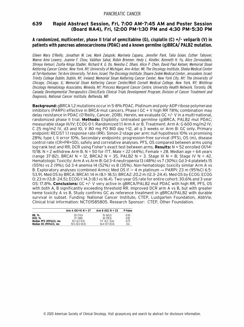
Background: gBRCA 1,2mutations occur in 5-8%PDAC. Platinum and poly-ADP ribose polymeraseinhibitors (PARPi) effective in BRCA-mut cancers. Phase I GC + V high RR 78%; combination maydelay resistance in PDAC (O’Reilly, Cancer, 2018). Herein, we evaluate GC +/- V in a multi-national,randomized phase II trial. Methods: Eligibility: Untreated germline (g)BRCA, PALB2 mut PDAC;measurable stage III/IV; ECOG 0-1. Randomized 1:1 Arm A or B. Treatment: Arm A: G 600mg/m2 IV,C 25 mg/m2 IV, d3 and 10, V 80 mg PO BID day 1-12, all q 3 weeks or Arm B: GC only. Primaryendpoint: RECIST 1.1 response rate (RR). Simon 2-stage per arm: null hypothesis 10% vs promising28%; type I, II error 10%. Secondary endpoints: progression-free survival (PFS), OS (m), diseasecontrol rate (CR+PR+SD), safety and correlative analyses. PFS, OS compared between arms usinglog-rank test and RR, DCR using Fisher’s exact test between arms. Results: N = 52 enrolled 01/14-11/18. N = 2 withdrew Arm B. N = 50 for ITT. Male = 22 (44%), Female = 28. Median age = 64 years(range 37-82). BRCA1 N = 12, BRCA2 N = 35, PALB2 N = 3. Stage III N = 8; Stage IV N = 42.Hematologic Toxicity: ArmA vs Arm B: Gd 3-4 neutropenia 13 (48%) vs 7 (30%); Gd 3-4 platelets 15(55%) vs 2 (9%); Gd 3-4 anemia 14 (52%) vs 8 (35%). Non-hematologic toxicity similar Arm A vsB. Exploratory analyses (combined Arms): Med OS if . 4 m platinum → PARPi: 23 m (95%CI 6.5-53.9). Med OS by BRCA: BRCA1: 14m (8.1- 18.5); BRCA2: 20.2m (12.3- 24.4). Med OS by ECOG: ECOG0: 23m (13.8- 24.5); ECOG 1: 14.3 (8.1 vs 16.4). Two-year OS rate for entire cohort: 30.6% and 3-yearOS: 17.8%. Conclusions: GC +/- V very active in gBRCA/PALB2 mut PDAC with high RR, PFS, OSwith both A, B significantly exceeding threshold RR. Improved DCR arm A vs B, but with greaterheme toxicity A vs B. Study confirms GC as reference treatment in gBRCA/PALB2 with durablesurvival in subset. Funding: National Cancer Institute, CTEP, Lustgarten Foundation, AbbVie.Clinical trial information: NCT01585805. Research Sponsor: CTEP, Other Foundation.
Arm A (GC+V) N = 27 Arm B (GC) N = 23 P-Value
RR, % 20 (74.1) 15 (65.2) 0.55DCR, % 27 (100) 18 (78.3) 0.02Median PFS (95%CI), mo 10.1 (6.7-11.5) 9.7 (4.2- 13.6) 0.73Median OS (95%CI), mo 15.5 (12.2-24.3) 16.4 (11.7-23.4) 0.6
© 2020 American Society of Clinical Oncology. Visit gicasym.org and search by abstract for disclosure information.
PANCREATIC CANCER
640 Poster Session (Board #G15), Fri, 12:00 PM-1:30 PM and4:30 PM-5:30 PM
The clinical utility of the 2017 Fukuoka Guidelines and 2015 American GastroenterologicalAssociation Guidelines for the management of intraductal papillary mucinous neoplasms.
Gandhi Lanke, Donald Campbell, Emmanuel Coronel, Manoop S. Bhutani, Brian Weston, William A. Ross, Michael Paul Kim,Graciela M. Nogueras-Gonzalez, Matthew H. G. Katz, Jeffrey H. Lee; MD Anderson Cancer Center, Houston, TX; The Universityof Texas MD Anderson Cancer Center, Houston, TX; University of Texas MD Anderson Cancer Center, Houston, TX
Background: Mucinous cystadenocarcinoma are malignant and mucinous pancreatic cysts (PC)have malignant potential. The management of PC remains controversial despite consensusguidelines. This study aims to evaluate the clinical utility of the 2017 Fukuoka Guidelines (FG)and 2015 American Gastroenterological Association Guidelines (AGA-G) for the management ofPC.Methods: 212 patients who underwent EUS for PC between 2010 and 2017 were identified. TheFG and AGA-G were used to define worrisome and high-risk cyst features. Receiver OperatingCharacteristic (ROC) curve was used to define sensitivity (SN), specificity (SP), positive predictivevalue (PPV) and negative predictive value (NPV). Results: 141 of 212 patients had IPMNs.EUS-FNAwas performed in 76.5% with no reported complications. Median follow-up was 4.2 years. Themajority of the IPMNswere in the pancreatic head (44.7%) or body (39.7%) while only 15.6%werein the tail. Using the FG, 46.1% had at least one worrisome feature (FG-W) and 7.1% had at least onehigh risk feature (FG-HR). Using the AGA-G, 28.4%had at least one HR feature (AGA-HR1) and 1.4%patients had two or more risk factors (AGA-HR2). A change in cyst character (increase of. 5 mmin 2 years, development of a solid component, or new pancreatic duct dilation) was noted in 43.2%patients. The median time to cyst change was 21 months. For prediction of cyst changes, the FG-Whad a SN of 45.8%, SP of 55.4%, PPV 45%, and NPV 56%. FG-HR had a SN of 14.3%, SP of 53.2%,PPV 1.7%, and NPV 91.8%. AGA-HR1 had a SN of 35.3%, SP of 51.5%, PPV 20%, and NPV 69.9%.AGA-HR2 had a SN of 0%, SP of 54.2%, PPV 0%, and NPV 97.3%. No difference was seen in cystchange or development of high risk or worrisome features with CEA . 192 vs. , 192 (p = 0.99).During follow up, 14 patients died, but only one patient died of pancreatic cancer. Conclusions: FGand AGA-G are difficult to validate because malignant cyst transformation is rare. There was nocorrelation between any cyst characteristics on EUS and cyst changes. FG-W had the bestperformance in predicting changes. Surgical candidates should be carefully selected, as theseguidelines have a limited clinical utility. Research Sponsor: None.
© 2020 American Society of Clinical Oncology. Visit gicasym.org and search by abstract for disclosure information.
PANCREATIC CANCER
641 Poster Session (Board #G16), Fri, 12:00 PM-1:30 PM and4:30 PM-5:30 PM
Determining when endoscopic ultrasound (EUS) changes management for patients withpancreatic cystic neoplasms.
Hasrit Sidhu, Khaola Maher, Dave Farnell, Leo Chen, Ian Gan, Maja Segedi; University of British Columbia Faculty of Medicine,Vancouver, BC, Canada; University of British Columbia, Vancouver, BC, Canada; Department of Surgery, University of BritishColumbia, Vancouver, BC, Canada
Background: Pancreatic cystic neoplasms (PCNs) are being incidentally detected at an increasedrate due to the widespread use of CT and MRI. CT and MRI cannot always differentiate betweenmalignant and benign PCNs. EUS is an emerging tool that provides higher quality descriptions ofpancreatic cysts and can be used to differentiate between benign and malignant features.Considering that EUS is a resource dependent tool, we hope to identify the PCN cases in whichEUS changes management. Methods: We conducted a retrospective case-control chart reviewevaluating patients, who were diagnosed with pancreatic cysts and underwent EUS for analysisbetween January 1, 2010 and December 31, 2017. We determined whether EUS correctly identifiedhigh-risk features (HRFs) relative to CT/MRI and whether EUS upstaged or downstaged the CT/MRIdiagnosis to change overall patient management. Results: EUS was found to have a high spec-ificity (.95%) for all high-risk features identified in theAGAand FG guidelines and a low sensitivity( , 70%) for all high risk features except cyst size . 3cm (82.35%) and mural nodule , 5mm(100%). EUS was found to change management in 29.4% of cases (18.2% upstaged, 11.2%downstaged). EUS screening led to a total of three adenocarcinoma diagnoses, in which twowere reported to be invasive. Conclusions: The high specificity of EUS supports its use in thedifferentiation of high risk PCNs identified on cross-sectional imaging. Its low sensitivity indicatesthat the reliance on operator experience may be a substantial limitation resulting in inconclusivediagnoses. In conclusion, considering that EUS is successful in changing patient management ofPCNs, it should be readily referred when any HRF is identified on cross-sectional imaging. ResearchSponsor: None.
© 2020 American Society of Clinical Oncology. Visit gicasym.org and search by abstract for disclosure information.
PANCREATIC CANCER
642 Poster Session (Board #G17), Fri, 12:00 PM-1:30 PM and4:30 PM-5:30 PM
Evaluation of ICD codes and phecodes for the identification of pancreatic cancer in a largegenomic database.
Chelsea Anne Isom, Eric R. Gamazon, Marcus Chuan Beng Tan; Vanderbilt University Medical Center, Nashville, TN; VanderbiltUniversity, Nashville, TN; Mitchell Cancer Institute, The University of South Alabama, Mobile, AL
Background: Large genomic databases linked to electronic health records promise to shed light onmolecular mechanisms underlying rare diseases, such as pancreatic cancer. However, accuratelyidentifying patients with the desired phenotype can be challenging. This is particularly the case forpancreatic tumors, since ICD codes do not distinguish between pancreatic adenocarcinoma (PDAC)and pancreatic neuroendocrine tumors (pNET). Previous studies have shown that ICD codesaggregated by phenotype, known as “phecodes”, have a higher accuracy in identifying specificphenotypes than ICD codes themselves; however, their performance in identifying cancers of thepancreas has not been studied.Methods: From a large deidentified genomic database, two querieswere performed to identify all adults with pancreatic cancer for a GWAS study, one using ICD-9/10codes and the other using phecodes. The medical records for all patients identified from bothqueries were then reviewed to confirm the presence and histologic type of pancreatic cancer.Results: Of the 91,985 genotyped adults in the database, ICD-9/10 codes identified 1,247 patientswith pancreatic cancer, compared with only 422 patients identified by the phecode query. Allpatients in the phecode cohort were also found in the ICD cohort. Of the 1,247 patients in the ICDcohort, 760 were confirmed to have pancreatic cancer on review of the health records (594 withPDAC, 166 with pNET) whereas in the phecode cohort, only 251 were confirmed to have pancreaticcancer (159 with PDAC, 92 pNET). The positive predictive value (PPV) for PDAC in the ICD querywas 47%, compared with 38% for the phecode cohort. The ICD and phecode cohorts had similarlylow numbers of pre-malignant cystic tumors (5% in each cohort) and other periampullary cancers(3%).Conclusions: In this large genomic database, the use of ICD-9/10 codes for pancreatic cancerwas able to identify nearly three times as many patients with pancreatic cancer and had a higherPPV compared to using phecodes. Therefore, ICD codes, rather than phecodes, should be used toidentify patients with pancreatic cancer for subsequent genotyping analysis, though caution isrequired because the PPV is still low. Research Sponsor: None.
© 2020 American Society of Clinical Oncology. Visit gicasym.org and search by abstract for disclosure information.
PANCREATIC CANCER
643 Poster Session (Board #G18), Fri, 12:00 PM-1:30 PM and4:30 PM-5:30 PM
Weight loss as an untapped early detection marker in pancreatic cancer.
Jonathan J. Hue, Sarah C. Markt, Ravi Kumar Kyasaram, John Shanahan, Goutham Rao, Jordan Michael Winter; UniversityHospitals Cleveland Medical Center, Cleveland, OH; Case Western Reserve University, Cleveland, OH; University HospitalsSeidman Cancer Center, Cleveland, OH
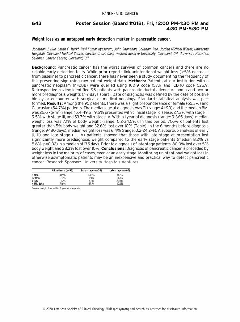
Background: Pancreatic cancer has the worst survival of common cancers and there are noreliable early detection tests. While prior reports link unintentional weight loss (.5% decreasefrom baseline) to pancreatic cancer, there has never been a study documenting the frequency ofthis presenting sign using raw patient weight data. Methods: Patients at our institution with apancreatic neoplasm (n=288) were queried using ICD-9 code 157.9 and ICD-10 code C25.9.Retrospective review identified 95 patients with pancreatic ductal adenocarcinoma and two ormore prediagnosis weights (.7 days apart). Date of diagnosis was defined by the date of positivebiopsy or encounter with surgical or medical oncology. Standard statistical analysis was per-formed. Results: Among the 95 patients, there was a slight preponderance of female (65.3%) andCaucasian (54.7%) patients. Themedian age at diagnosis was 71 (range: 41-90) and themedian BMIwas 25.6 kg/m2 (range: 15.4-49.5). 9.5%presentedwith clinical stage I disease, 27.3%with stage II,9.5%with stage III, and 53.7%with stage IV. Within 1 year of diagnosis (range: 9-365 days), medianweight loss was 7.1% of body weight (range: 0.2-34.5%). In this period, 71.6% of patients lostgreater than 5% body weight and 32.6% lost over 10% (Table). In the 6 months before diagnosis(range: 9-180 days), median weight loss was 6.4% (range: 0.2-24.2%). A subgroup analysis of early(I, II) and late stage (III, IV) patients showed that those with late stage at presentation lostsignificantly more prediagnosis weight compared to the early stage patients (median 8.2% vs5.6%, p=0.02) in amedian of 175 days. Prior to diagnosis of late stage patients, 80.0% lost over 5%body weight and 38.3% lost over 10%. Conclusions: Diagnosis of pancreatic cancer is preceded byweight loss in the majority of cases, even at an early stage. Monitoring unintentional weight loss inotherwise asymptomatic patients may be an inexpensive and practical way to detect pancreaticcancer. Research Sponsor: University Hospitals Ventures.
All patients (n=95) Early stage (n=35) Late stage (n=60)
5-10% 38.9% 34.3% 41.7%10-15% 17.9% 17.1% 18.3%>15% 14.7% 5.7% 20.0%>5%, total 71.6% 57.1% 80.0%
Percent weight loss within 1 year of diagnosis.
© 2020 American Society of Clinical Oncology. Visit gicasym.org and search by abstract for disclosure information.
PANCREATIC CANCER
644 Poster Session (Board #G19), Fri, 12:00 PM-1:30 PM and4:30 PM-5:30 PM
Pathological examination of CT findings of tumor infiltration to the periarterial plexus inpancreatic cancer.
Takashi Miyamoto, Yusuke Yamamoto, Teiichi Sugiura, Yukiyasu Okamura, Takaaki Ito, Ryo Ashida, Katsuhisa Ohgi,Katsuhiko Uesaka; Division of Hepato-Biliary-Pancreatic Surgery, Shizuoka Cancer Center, Nagaizumi, Japan; Division ofHepato-Biliary-Pancreatic Surgery, Shizuoka Cancer Center, Shizuoka, Japan
Background: The radiographic diagnosis of tumor infiltration into the periarterial plexus inpancreatic cancer is important because it is related to the classification, however, it is difficultto distinguish the abnormal shadow along the artery caused by inflammation or cancer infiltration.The aim of this study is to investigate CT values of the abnormal shadow along artery coulddistinguish between inflammation and tumor invasion. Methods: Study 1: Of 26 patients whounderwent DP-CAR between 2009 and 2018, we analyzed 19 patients who had dynamic CT andobtained sagittal slice taken 120 seconds after injection with less than 2.5 mm slice thickness. Atfirst, we measured CT values at upper and lower point of CeA and CHA each sagittal slice using CT.Next, we evaluated tumor invasion at the upper and lower plexus of CeA and CHA in each section ofthe pathological specimen, and evaluated the relationship between the tumor invasion and theCT value. Study 2: Using these 19 patients and 40 patients who underwent DP for PDAC between2010 and 2014, we analyzed the relation between CT value and long-term states. Results: Study 1:CT value was totally measured at the 606 points using 19 patients who underwent DP-CAR. At the490 points, we did not observe cancer infiltration and fibrosis. At the 70 points, we observedfibrosis without cancer cells. At the 46 points, we observed cancer infiltration. CT value wassignificantly higher in the tumor infiltration group than that in the without cancer infiltration andfibrosis group (P, 0.01). Study 2: The best cut-off of CT value of the presence of cancer infiltrationwas 44.9 HU using ROC curve (AUC = 0.861). The median survival time of patients who had thepoints of CT value.44.9HUaround arterieswas significantly shorter than that of patientswho didnot have the points of CT value.44.9HU (2.17 vs. 4.55 years, p = 0.03).Conclusions:The CT valuearound the arteries was significantly higher in the points of pathological tumor infiltration thanthat in the points of fibrosis without cancer cells. The best cut-off CT value of the presence ofcancer infiltration around arteries was 44.9 HU, and the presence of the point of CT value . 44.9HU around arteries was associated with poor survival. Research Sponsor: None.
© 2020 American Society of Clinical Oncology. Visit gicasym.org and search by abstract for disclosure information.
PANCREATIC CANCER
645 Poster Session (Board #G20), Fri, 12:00 PM-1:30 PM and4:30 PM-5:30 PM
Detection of circulating tumor DNA in pancreas cancer.
Daniel King, Ash A. Alizadeh, George A. Fisher; Stanford University, Palo Alto, CA; Stanford Cancer Institute, Stanford, CA;Stanford University, Stanford, CA
Background: Pancreas cancer remains a leading cause of cancer-related death. Improved detec-tion of early relapse or early failure of chemotherapy also has the potential to further improveoutcomes. Exploring circulating tumor DNA (ctDNA) in this setting is an area of active investi-gation. Methods: We previously developed an approach, CAPP-Seq, combining high-depth se-quencing with several strategies of error-suppression to identify minute amounts of circulatingtumor DNA. We then trained and validated a new capture panel for pancreas cancer from 640tumors from three data sources (TCGA, ICGC, UTSW), targeting 265 kb of the genome. We enrolledtwo cohorts of patients with pancreatic cancers at Stanford Cancer Center: (1) patients withlocalized tumors undergoing resection with curative intent, and (2) patients with unresectable ormetastatic disease undergoing systemic therapy. Results: As of August 2019, we recruited 131patients with at least one blood collection, with 63% having resectable disease and 27% havingadvanced disease; 59 patients had 2 or more blood collections. Stage distribution included 34%stage I, 33% stage II, 18% III, 16% IV disease. Approximately 15% had normal CA19-9 levels. Deepsequencing (4,000x unique depth) of an initial set of resected pancreatic tumors and matchedgermline specimens identified 1-6 non-synonymous coding mutations per case (median=3, n=14),with the most frequently mutated genes involving KRAS (79%), TP53 (50%), SMAD4 (29%).Among newly diagnosed treatment-naıve patients with resectable adenocarcinoma (n=9), wedetected ctDNA in 4 patients (44%) prior to surgery including with AFs ranging from 0.27% -0.88%. Subsequent sequencing will compare patients with and without neoadjuvant therapy priorto resection, selection of unresectable patients across a larger range of tumor burden and acrossmultiple timepoints, and integration of large-scale copy number variant detection using low-passwhole-genome sequencing.Conclusions:Circulating tumor DNAmonitoringwith CAPP-Seq showspromise for improved detection of PDAC. Two key applications include early detection of minimalresidual disease after resection and early assessment of response to chemotherapy. ResearchSponsor: Stanford Cancer Institute.
© 2020 American Society of Clinical Oncology. Visit gicasym.org and search by abstract for disclosure information.
PANCREATIC CANCER
646 Poster Session (Board #G21), Fri, 12:00 PM-1:30 PM and4:30 PM-5:30 PM
Impact of biliary metal and plastic stents on preoperative staging for pancreatic cancer.
Hachem Hachem, Sanjay S. Reddy, Jeffrey Tokar, Eileen O’Halloran, Jennifer Higa, Abby Sapp, Albert Civitarese,Michael Bartel; Fox Chase Cancer Center, Philadelphia, PA
Background: Multiple studies have shown the superiority of biliary metal compared with plasticstents for pre-operative (preop) biliary drainage in pancreatic cancer (PDAC). Despite the impor-tance of preop cross-sectional imaging, particularly in the era of neoadjuvant treatment, there is nodata on the impact of such stents on the quality of preop cross-sectional imaging. We hypothesis,that biliary metal stents negatively impact the accuracy of preop cross-sectional imaging inpancreatic cancer, with unknown impact for the adequacy of surgical candidacy.Methods: Data ofall patients undergoing pancreatic resection for PDAC between 1/1/2012 and 1/1/2018 was retro-spectively abstracted. Clinical staging based on preop cross-sectional imaging following biliarystent placement (within 2 months prior surgical resection) was compared with the surgicalpathology (staging gold standard). Accuracy of clinical and surgical pathology staging was com-pared. Logistic regression was performed to control for biliary stent type, neoadjuvant treatmentand patient baseline characteristics including BMI and type of imaging. Results: 312 patientsunderwent pancreatic resections. 118 patients required preop biliary drainage in setting of PDAC,including 92 ERCPs of which 83 were successful (46 plastic and 37 metal stents). 76 patientsunderwent neoadjuvant chemoradiation therapy. Surgical pathology revealed following stages: 0n = 4, 1A n = 5, 1B n = 8, 2A n = 20, 2B n = 24, 3 n = 1, 4 n = 14. 96% underwent preop CT and 4%MRIpancreas protocol imaging. Exact correlation between clinical and surgical pathology was presentin only 48% of cases (57% plastic, 46% metal stent), with 28% of clinical T overstaging, 4%clinical T understaging, 16% clinical N understaging and 4% unable to stage due to artefacts. Moreimportantly, 8% patients were incorrectly staged to be surgical candidates (14% plastic, 6%metal). Controlling for stent type, neoadjuvant treatment and BMI did not impact preop cross-sectional imaging accuracy. Conclusions: Despite their impact on preop cross-imaging biliarymetal stents did not negatively impact the accuracy and patient selection for surgical candidacycompared with biliary plastic stents in PDAC. Research Sponsor: None.
© 2020 American Society of Clinical Oncology. Visit gicasym.org and search by abstract for disclosure information.
PANCREATIC CANCER
647 Poster Session (Board #G22), Fri, 12:00 PM-1:30 PM and4:30 PM-5:30 PM
Efficacy of chemotherapy for patients with unresectable or recurrent pancreatic adenosquamouscarcinoma: A multicenter retrospective analysis.
Yukio Yoshida, Akira Fukutomi, Makoto Ueno, Keita Mori, Kazuo Watanabe, Akihiro Ohba, Satoshi Kobayashi,Mitsuhiro Furuta, Akiko Tsujimoto, Masato Osaka, Naohiro Okano, Kei Yane, Kumiko Umemoto, Yasuyuki Kawamoto,Takeshi Terashima, Hidetaka Tsumura, Keitaro Doi, Kazuhiko Shioji, Akinori Asagi, Junji Furuse; Okinawa Chubu Hospital,Uruma Okinawa, Japan; Division of Gastrointestinal Oncology, Shizuoka Cancer Center, Shizuoka, Japan; Kanagawa CancerCenter, Yokohama, Japan; Clinical Research Center, Shizuoka Cancer Center, Shizuoka, Japan; National Cancer CenterHospital East, Kashiwa, Japan; National Cancer Center Hospital, Tokyo, Japan; Department of Gastroenterology,Hepatobiliary and Pancreatic Medical Oncology Division, Kanagawa Cancer Center, Yokohama, Japan; Department ofGastroenterology, Chiba Cancer Center, Chiba, Japan; Cancer Institute Hospital of JFCR, Tokyo, Japan; Department ofMedical Oncology, Kyorin University Faculty of Medicine, Tokyo, Japan; Center for Gastroenterology, Teine Keijinkai Hospital,Sapporo, Japan; St.Marianna University School of Medicine, Department of Clinical Oncology, Kawasaki, Japan; Departmentof Gastroenterology and Hepatology, Hokkaido University Hospital, Sapporo, Japan; Kanazawa University Hospital,Kanazawa, Japan; Hyogo Cancer Center, Hyogo, Japan; Department of Medical Oncology, Kyoto University GraduateSchool of Medicine, Kyoto, Japan; Niigata Cancer Center Hospital, Niigata, Japan; Department of Gastrointestinal MedicalOncology, National Hospital Organization Shikoku Cancer Center, Matsuyama, Japan; Kyorin University, Tokyo, Japan
Background: Pancreatic adenosquamous carcinoma (PASC) is a rare variant of pancreatic ductaladenocarcinoma (PDAC). Although unresectable or recurrent PASC is usually treated by systemicchemotherapy, there are few reports which show the efficacy of chemotherapy. The aim of thisstudy was to evaluate the efficacy of chemotherapy for patients (pts) with unresectable orrecurrent PASC. Methods: We collected data retrospectively from 24 Japanese institutions.The selection criteria were as follows: 1) histologically or cytologically proven PASC (non-surgicalspecimens were eligible if squamous cell carcinoma (SCC) was detected), 2) unresectable orrecurrent disease treated with 1st line chemotherapy between April 2001 and December 2017.Results: This study included 138 pts with median age of 66 years (range: 36-85). About 60%of ptswere diagnosed with biopsy and only SCC was detected in 13.0% of pts. Median overall survival(mOS) was 6.7 months (M), median progression free survival (mPFS) was 2.8 M, and the 1-yearsurvival rate (1YSR) was 26.7%. For the 102metastatic or distal recurrent pts with PS of 0-1, patientcharacteristics were as follows:$76 years old, 9 (8.8%); PS of 0, 39 (38.2%); number ofmetastaticsites $2, 25 (24.5%). The treatment efficacies (The objective response rates(%)/mPFS(M)/mOS(M)/1YSR(%)) of the 5 major regimens were Gemcitabine(GEM) (n=45, 4.4%/2.2M/4.8M/28.1%), GEM+nab-PTX (n=24, 29.2%/2.9M/7.6M/23.1%), GEM+S-1 (n=9, 11.1%/5.1M/9.9M/25.4%),FOLFIRINOX (n=7, 14.3%/2.5M/7.5M/14.3%), and S-1 (n=7; 28.6%/2.6M/5.0M/28.6%), respec-tively. One patient with liver metastasis underwent conversion surgery after GEM+nab-PTX andachieved long survival. CRP $3.0mg/dl, CA19-9 $1000 U/ml, residual primary site, and mono-therapy had a significant correlation with poor survival in multivariate analysis. Conclusions:Although combination chemotherapy regimens such as FOLFIRINOX and GEM+nab-PTX are nowavailable, the prognosis of metastatic PASC remains poor. Development of more effective treat-ment options is required. Research Sponsor: None.
© 2020 American Society of Clinical Oncology. Visit gicasym.org and search by abstract for disclosure information.
PANCREATIC CANCER
648 Poster Session (Board #H1), Fri, 12:00 PM-1:30 PM and4:30 PM-5:30 PM
Pancreatic cancer (PaC)-specific health-related quality of life (HRQoL) with maintenance olaparib(O) in patients (pts) with metastatic (m) PaC and a germline BRCA mutation (gBRCAm): Phase IIIPOLO trial.
Michael J. Hall, Talia Golan, Pascal Hammel, Michele Reni, Eric Van Cutsem, Teresa Macarulla, Joon Oh Park,Daniel Hochhauser, Dirk Arnold, Do-Youn Oh, Anke C. Reinacher-Schick, Giampaolo Tortora, Hana Algul,Eileen Mary O’Reilly, David McGuinness, Karen Cui, Seongjung Joo, Hyun Kyoo Yoo, Nikunj Patel, Hedy L. Kindler; FoxChase Cancer Center, Philadelphia, PA; The Oncology Institute, Sheba Medical Center at Tel-Hashomer, Tel Aviv University,Tel Aviv, Israel; Hopital Beaujon (AP-HP), Clichy, and University Paris VII, Paris, France; IRCCS Ospedale, San RaffaeleScientific Institute, Milan, Italy; University Hospitals Gasthuisberg Leuven and KU Leuven, Leuven, Belgium; Vall d’HebronUniversity Hospital and Vall d’Hebron Institute of Oncology, Barcelona, Spain; Samsung Medical Center, SungkyunkwanUniversity School of Medicine, Seoul, South Korea; University College London Cancer Institute, London, United Kingdom;Asklepios Tumorzentrum Hamburg AK Altona, Hamburg, Germany; Seoul National University Hospital, Seoul, South Korea; StJosef-Hospital, Ruhr University Bochum, Bochum, Germany; Azienda Ospedaliera Universitaria Integrata Verona, Verona andFondazione Policlinico Universitario Gemelli IRCCS, Rome, Italy; Klinikum Rechts der Isar, Comprehensive Cancer CenterMunich-TUM and Department of Internal Medicine II, Technische Universitat Munchen, Munich, Germany; Memorial SloanKettering Cancer Center, New York, NY; AstraZeneca, Cambridge, United Kingdom; AstraZeneca, Gaithersburg, MD; Merck &Co., Inc., Kenilworth, NJ; The University of Chicago, Chicago, IL
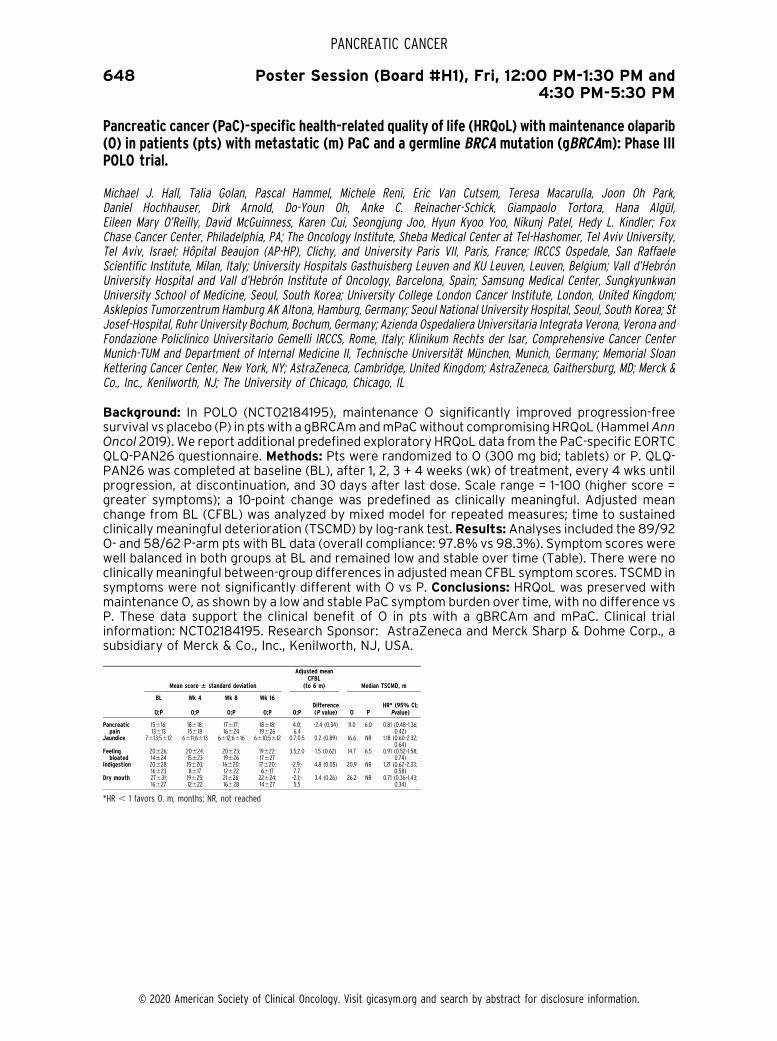
Background: In POLO (NCT02184195), maintenance O significantly improved progression-freesurvival vs placebo (P) in pts with a gBRCAmandmPaCwithout compromisingHRQoL (HammelAnnOncol 2019). We report additional predefined exploratory HRQoL data from the PaC-specific EORTCQLQ-PAN26 questionnaire. Methods: Pts were randomized to O (300 mg bid; tablets) or P. QLQ-PAN26 was completed at baseline (BL), after 1, 2, 3 + 4 weeks (wk) of treatment, every 4 wks untilprogression, at discontinuation, and 30 days after last dose. Scale range = 1–100 (higher score =greater symptoms); a 10-point change was predefined as clinically meaningful. Adjusted meanchange from BL (CFBL) was analyzed by mixed model for repeated measures; time to sustainedclinically meaningful deterioration (TSCMD) by log-rank test.Results: Analyses included the 89/92O- and 58/62 P-arm pts with BL data (overall compliance: 97.8% vs 98.3%). Symptom scores werewell balanced in both groups at BL and remained low and stable over time (Table). There were noclinicallymeaningful between-group differences in adjustedmean CFBL symptom scores. TSCMD insymptoms were not significantly different with O vs P. Conclusions: HRQoL was preserved withmaintenance O, as shown by a low and stable PaC symptom burden over time, with no difference vsP. These data support the clinical benefit of O in pts with a gBRCAm and mPaC. Clinical trialinformation: NCT02184195. Research Sponsor: AstraZeneca and Merck Sharp & Dohme Corp., asubsidiary of Merck & Co., Inc., Kenilworth, NJ, USA.
Mean score 6 standard deviation
Adjusted meanCFBL
(to 6 m) Median TSCMD, m
BL Wk 4 Wk 8 Wk 16
O;P O;P O;P O;P O;PDifference(P value) O P
HR* (95% CI;Pvalue)
Pancreaticpain
15616;13613
18618;15618
17617;16624
18618;19626
4.0;6.4
-2.4 (0.34) 11.0 6.0 0.81 (0.48–1.36;0.42)
Jaundice 7613;5612 6611;6613 6612;6616 6610;5612 0.7;0.5 0.2 (0.89) 16.6 NR 1.18 (0.60–2.32;0.64)
Feelingbloated
20626;14624
20624;15623
20623;19626
19622;17627
3.5;2.0 1.5 (0.62) 14.7 6.5 0.91 (0.52–1.58;0.74)
Indigestion 20628;16623
15620;8617
16620;12622
17620;6617
-2.9;-7.7
4.8 (0.05) 20.9 NR 1.21 (0.62–2.33;0.58)
Dry mouth 27631;16627
19625;12622
21626;16628
22624;14627
-2.1;-5.5
3.4 (0.26) 26.2 NR 0.71 (0.36–1.43;0.34)
*HR , 1 favors O. m, months; NR, not reached
© 2020 American Society of Clinical Oncology. Visit gicasym.org and search by abstract for disclosure information.
PANCREATIC CANCER
649 Poster Session (Board #H2), Fri, 12:00 PM-1:30 PM and4:30 PM-5:30 PM
Scoring model with serum albumin and CA19-9 in advanced pancreatic cancer in second-linetreatment: Results from the NAPOLEON study.
Azusa Komori, Satoshi Otsu, Mototsugu Shimokawa, Taiga Otsuka, Futa Koga, Yujiro Ueda, Junichi Nakazawa, Shiho Arima,Masaru Fukahori, Yoshinobu Okabe, Akitaka Makiyama, Hiroki Taguchi, Takuya Honda, Taro Shibuki, Kenta Nio, Yasushi Ide,Norio Ureshino, Tsuyoshi Shirakawa, Kenji Mitsugi; Department of Medical Oncology and Hematology, Oita University Facultyof Medicine, Yufu, Japan; Clinical Research Institute, National Kyushu Cancer Center, Fukuoka, Japan; Department of MedicalOncology, Saga Medical Center Koseikan, Saga, Japan; Department of Hepatobiliary and Pancreatology, Saga Medical CenterKoseikan, Saga, Japan; Department of Hematology and Oncology, Japanese Red Cross Kumamoto Hospital, Kumamoto,Japan; Department of Medical Oncology, Kagoshima City Hospital, Kagoshima, Japan; Digestive and Lifestyle Diseases,Kagoshima University Graduate School of Medical and Dental Sciences, Kagoshima, Japan; Division of Gastroenterology,Department of Medicine, Kurume University School of Medicine, Kurume, Japan; Department of Hematology/Oncology,Japan Community Health Care Organization Kyushu Hospital, Kitakyushu, Japan; Department of Gastroenterology, SaiseikaiSendai Hospital, Satsumasendai, Japan; Department of Gastroenterology and Hepatology, Nagasaki University GraduateSchool of Biomedical Science, Nagasaki, Japan; Department of Internal Medicine, Imari Arita Kyoritsu Hospital, Nishi-Matsuura, Japan; Department of Medical Oncology, Sasebo Kyosai Hospital, Sasebo, Japan; Department of Internal Medicine,Karatsu Red Cross Hospital, Karatsu, Japan; Department of Medical Oncology, Fukuoka Wajiro Hospital, Fukuoka, Japan;Department of Medical Oncology, Hamanomachi Hospital, Fukuoka, Japan
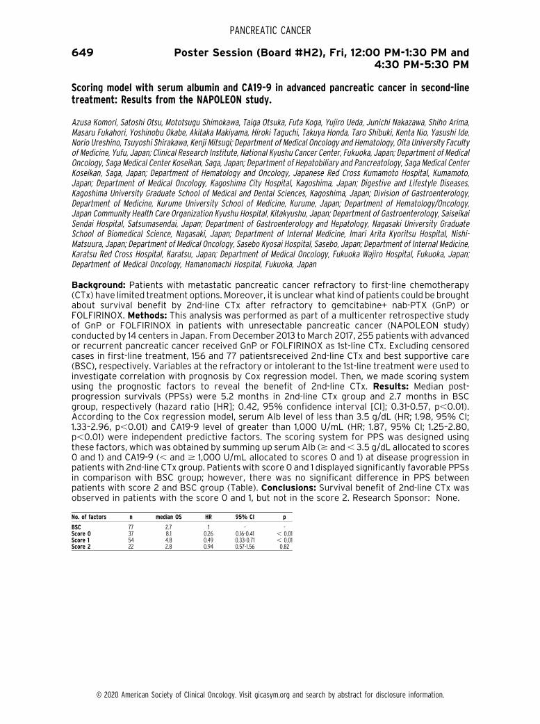
Background: Patients with metastatic pancreatic cancer refractory to first-line chemotherapy(CTx) have limited treatment options. Moreover, it is unclear what kind of patients could be broughtabout survival benefit by 2nd-line CTx after refractory to gemcitabine+ nab-PTX (GnP) orFOLFIRINOX. Methods: This analysis was performed as part of a multicenter retrospective studyof GnP or FOLFIRINOX in patients with unresectable pancreatic cancer (NAPOLEON study)conducted by 14 centers in Japan. FromDecember 2013 toMarch 2017, 255 patients with advancedor recurrent pancreatic cancer received GnP or FOLFIRINOX as 1st-line CTx. Excluding censoredcases in first-line treatment, 156 and 77 patientsreceived 2nd-line CTx and best supportive care(BSC), respectively. Variables at the refractory or intolerant to the 1st-line treatment were used toinvestigate correlation with prognosis by Cox regression model. Then, we made scoring systemusing the prognostic factors to reveal the benefit of 2nd-line CTx. Results: Median post-progression survivals (PPSs) were 5.2 months in 2nd-line CTx group and 2.7 months in BSCgroup, respectively (hazard ratio [HR]; 0.42, 95% confidence interval [CI]; 0.31-0.57, p,0.01).According to the Cox regression model, serum Alb level of less than 3.5 g/dL (HR; 1.98, 95% CI;1.33–2.96, p,0.01) and CA19-9 level of greater than 1,000 U/mL (HR; 1.87, 95% CI; 1.25–2.80,p,0.01) were independent predictive factors. The scoring system for PPS was designed usingthese factors, which was obtained by summing up serum Alb ($ and, 3.5 g/dL allocated to scores0 and 1) and CA19-9 (, and $ 1,000 U/mL allocated to scores 0 and 1) at disease progression inpatients with 2nd-line CTx group. Patients with score 0 and 1 displayed significantly favorable PPSsin comparison with BSC group; however, there was no significant difference in PPS betweenpatients with score 2 and BSC group (Table). Conclusions: Survival benefit of 2nd-line CTx wasobserved in patients with the score 0 and 1, but not in the score 2. Research Sponsor: None.
No. of factors n median OS HR 95% CI p
BSC 77 2.7 1 - -Score 0 37 8.1 0.26 0.16-0.41 , 0.01Score 1 54 4.8 0.49 0.33-0.71 , 0.01Score 2 22 2.8 0.94 0.57-1.56 0.82
© 2020 American Society of Clinical Oncology. Visit gicasym.org and search by abstract for disclosure information.
PANCREATIC CANCER
650 Poster Session (Board #H3), Fri, 12:00 PM-1:30 PM and4:30 PM-5:30 PM
Prognostic factors associated with short-term survival (STS) in advanced pancreatic cancer(APC): A multicenter analysis from the CHORD consortium.
Sakshi Mehta, Miguel Cardoso, Christina Kim, Dawn Elizabeth Armstrong, Ravi Ramjeesingh, Daniel John Renouf,Shahid Ahmed, Yoo-Joung Ko, Mohammed Harb, Shiying Kong, Winson Y. Cheung, Brandon M. Meyers; Juravinski CancerCentre, Hamilton, ON, Canada; Dept of Medical Oncology, CancerCare Manitoba, Winnipeg, MB, Canada; Dr. H. Bliss MurphyCancer Centre, St. John’s, NF, Canada; Nova Scotia Cancer Centre, Dalhousie University, Nova Scotia, NS, Canada; BC CancerAgency, Vancouver, BC, Canada; University of Saskatchewan, Saskatoon, SK, Canada; Odette Cancer Centre, SunnybrookHealth Sciences Centre, Toronto, ON, Canada; Moncton Hospital, Moncton, NB, Canada; Alberta Health Services, Calgary, AB,Canada
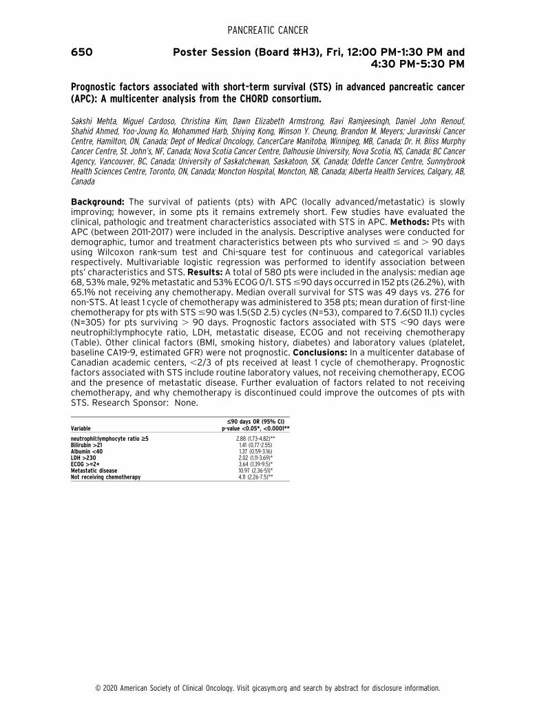
Background: The survival of patients (pts) with APC (locally advanced/metastatic) is slowlyimproving; however, in some pts it remains extremely short. Few studies have evaluated theclinical, pathologic and treatment characteristics associated with STS in APC. Methods: Pts withAPC (between 2011-2017) were included in the analysis. Descriptive analyses were conducted fordemographic, tumor and treatment characteristics between pts who survived # and . 90 daysusing Wilcoxon rank-sum test and Chi-square test for continuous and categorical variablesrespectively. Multivariable logistic regression was performed to identify association betweenpts’ characteristics and STS. Results: A total of 580 pts were included in the analysis: median age68, 53%male, 92%metastatic and 53%ECOG0/1. STS#90 days occurred in 152 pts (26.2%), with65.1% not receiving any chemotherapy. Median overall survival for STS was 49 days vs. 276 fornon-STS. At least 1 cycle of chemotherapy was administered to 358 pts; mean duration of first-linechemotherapy for pts with STS#90 was 1.5(SD 2.5) cycles (N=53), compared to 7.6(SD 11.1) cycles(N=305) for pts surviving . 90 days. Prognostic factors associated with STS ,90 days wereneutrophil:lymphocyte ratio, LDH, metastatic disease, ECOG and not receiving chemotherapy(Table). Other clinical factors (BMI, smoking history, diabetes) and laboratory values (platelet,baseline CA19-9, estimated GFR) were not prognostic. Conclusions: In a multicenter database ofCanadian academic centers, ,2/3 of pts received at least 1 cycle of chemotherapy. Prognosticfactors associated with STS include routine laboratory values, not receiving chemotherapy, ECOGand the presence of metastatic disease. Further evaluation of factors related to not receivingchemotherapy, and why chemotherapy is discontinued could improve the outcomes of pts withSTS. Research Sponsor: None.
Variable£90 days OR (95% CI)
p-value <0.05*, <0.0001**
neutrophil:lymphocyte ratio ‡5 2.88 (1.73-4.82)**Bilirubin >21 1.41 (0.77-2.55)Albumin <40 1.37 (0.59-3.16)LDH >230 2.02 (1.11-3.69)*ECOG >=2+ 3.64 (1.39-9.5)*Metastatic disease 10.97 (2.36-51)*Not receiving chemotherapy 4.11 (2.26-7.5)**
© 2020 American Society of Clinical Oncology. Visit gicasym.org and search by abstract for disclosure information.
PANCREATIC CANCER
651 Poster Session (Board #H4), Fri, 12:00 PM-1:30 PM and4:30 PM-5:30 PM
Improved surgical margins with neoadjuvant versus adjuvant chemotherapy in clinical stage Iresectable pancreatic adenocarcinoma: A National Cancer Database study.
Geoffrey Bellini, Samit Kumar Datta, Nicholas Sich, Maharaj Singh, James Leighton Weese, Federico Augusto Sanchez,Nalini Guda, Wesley Allan Papenfuss, Aaron Chevinsky; Aurora St. Luke’s Medical Center, Milwaukee, WI; Aurora Health Care,Milwaukee, WI; Dental School, Marquette University, Milwaukee, WI; Aurora Cancer Care, Advocate Aurora Health, Milwaukee,WI; GI Associates, Milwaukee, WI
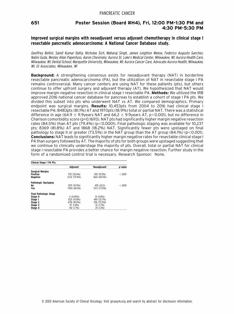
Background: A strengthening consensus exists for neoadjuvant therapy (NAT) in borderlineresectable pancreatic adenocarcinoma (PA), but the utilization of NAT in resectable stage I PAremains controversial. Many cancer centers are using NAT for these patients (pts), but otherscontinue to offer upfront surgery and adjuvant therapy (AT). We hypothesized that NAT wouldimprove margin negative resection in clinical stage I resectable PA. Methods: We utilized the IRBapproved 2016 national cancer database for pancreas to establish a cohort of stage I PA pts. Wedivided this subset into pts who underwent NAT vs AT. We compared demographics. Primaryendpoint was surgical margins. Results: 10,453pts from 2004 to 2016 had clinical stage Iresectable PA: 8483pts (81.1%) AT and 1970pts (18.9%) total or partial NAT. There was a statisticaldifference in age (64.9 6 9.9years NAT and 66.2 6 9.9years AT, p,0.001), but no difference inCharlson comorbidity score (p=0.1693). NAT pts had significantly highermargin negative resectionrates (84.5%) than AT pts (79.4%) (p,0.0001). Final pathologic staging was available for 10,237pts: 8369 (81.8%) AT and 1868 (18.2%) NAT. Significantly fewer pts were upstaged on finalpathology to stage II or greater (73.5%) in the NAT group than the AT group (84.1%) (p,0.001).Conclusions: NAT leads to significantly higher margin negative rates for resectable clinical stage IPA than surgery followed by AT. Themajority of pts for both groups were upstaged suggesting thatwe continue to clinically understage the majority of pts. Overall, total or partial NAT for clinicalstage I resectable PA provides a better chance for margin negative resection. Further study in theform of a randomized control trial is necessary. Research Sponsor: None.
Clinical Stage 1 PA Pts.
Adjuvant Neoadjuvant p-value
Surgical MarginsPositive 1751 (20.6%) 305 (15.5%) ,.0001Negative 6732 (79.4%) 1665 (84.5%)
Pathologic UpstagingNo 1329 (15.9%) 495 (26.5) ,.0001Yes 7040 (84.1%) 1373 (73.5%)
Final Pathologic StageStage 0 4 (0.05%) 15 (0.8%)Stage 1 1325 (15.8%) 480 (25.7%)Stage 2 6781 (81.0%) 1316 (70.5%)Stage 3 117 (1.4%) 32 (1.7%)Stage 4 142 (1.7%) 25 (1.3%)
© 2020 American Society of Clinical Oncology. Visit gicasym.org and search by abstract for disclosure information.
PANCREATIC CANCER
652 Poster Session (Board #H5), Fri, 12:00 PM-1:30 PM and4:30 PM-5:30 PM
An institutional series of early-onset pancreatic cancer (EOPC): Clinical outcomes and geneticand supportive care referral patterns.
Kunal C. Kadakia, Sally Jeanne Trufan, Megan Jagosky, William Mills Worrilow, Bradley W. Harrison, Katherine Broyhill,Nicole Lee Gower, Harris Coley, Jimmy J. Hwang, Laura W. Musselwhite, Reza Nazemzadeh, Seungjean Chai,John Stuart Salmon, Edward S. Kim, Mohamed E. Salem; Levine Cancer Institute, Atrium Health, Charlotte, NC; AtriumHealth, Charlotte, NC; Levine Cancer Institute-Atrium Health, Charlotte, NC; Levine Cancer Institute, Carolinas MedicalCenter, Charlotte, NC; Levine Cancer Institute, Charlotte, NC
Background: The incidence of EOPC is rising and is associated with substantial implications foraffected individuals and their families. Little is known about the extent of physician referrals ofthese patients (pts) to genetic, supportive care, and hospice services. Methods: Pts with EOPC(#50 years) were identified using the institutional tumor registry for years 2011-2018 and retro-spectively reviewed. Clinical data and rates of referral to supportive, genetic and hospice serviceswere retrieved. Descriptive analyses were performed with 25-75% interquartile ranges (IQR)where appropriate. Overall survival (OS) was assessed using Kaplan-Meier curves and Cox Pro-portional Hazards modeling. Results: In total, 113 pts with EOPC and a median age of 47 years(range, 28-50) were analyzed. Of these 113 pts, 43% were female, 27% were black, and 45% hadmetastatic disease at initial presentation. The most commonly administered first line chemother-apywas FOLFIRINOX, with gemcitabine/nab-paclitaxel reserved for the second line. ThemedianOSof ptswithmetastatic diseasewas 5.8 compared to 15.8months for thosewithoutmetastases. Only28% of pts were referred to genetic services, and 72% of these underwent genetic testing. Out ofthe genetically tested pts, pathogenic germline mutations were confirmed for 33%. Of the original113 pts, 41% received concurrent palliative care, which was provided at a median of 2.4 mos. (IQR,0.7-6.8) preceding death. The median time between last chemotherapy administered and deathwas 2 mos. (IQR, 1-4.4), with 23% receiving treatment within the last month of life. Only 55% usedhospice services prior to death for a median duration of 0.5 mos. (IQR, 0.2-1.4). Conclusions: Ourstudy suggests that there is a tendency for late utilization of supportive and hospice care in pts withEOPC, possibly due to the desire of both pts and physicians to be more aggressive given the youngage. Larger studies arewarranted to elucidate barriers to concurrent supportive care, andwhetherformation of specialized young patient supportive care clinics would aid this situation and to avoidthe use of unnecessary chemotherapy near the end of life. Research Sponsor: None.
© 2020 American Society of Clinical Oncology. Visit gicasym.org and search by abstract for disclosure information.
PANCREATIC CANCER
653 Poster Session (Board #H6), Fri, 12:00 PM-1:30 PM and4:30 PM-5:30 PM
Real-world eligibility of advanced pancreatic (APC) patients for maintenance olaparib.
Atul Batra, Winson Y. Cheung, Patricia A. Tang; Tom Baker Cancer Center, Calgary, AB, Canada; Tom Baker Cancer Centre,University of Calgary, Calgary, AB, Canada
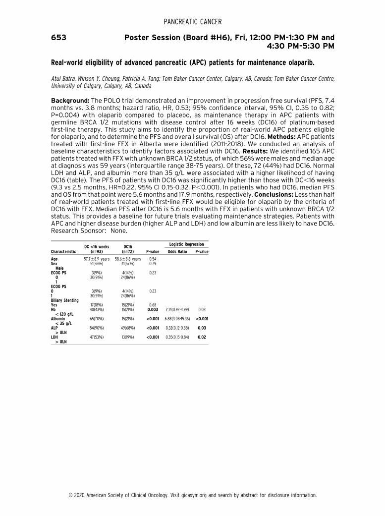
Background: The POLO trial demonstrated an improvement in progression free survival (PFS, 7.4months vs. 3.8 months; hazard ratio, HR, 0.53; 95% confidence interval, 95% CI, 0.35 to 0.82;P=0.004) with olaparib compared to placebo, as maintenance therapy in APC patients withgermline BRCA 1/2 mutations with disease control after 16 weeks (DC16) of platinum-basedfirst-line therapy. This study aims to identify the proportion of real-world APC patients eligiblefor olaparib, and to determine the PFS and overall survival (OS) after DC16.Methods:APC patientstreated with first-line FFX in Alberta were identified (2011-2018). We conducted an analysis ofbaseline characteristics to identify factors associated with DC16. Results: We identified 165 APCpatients treatedwith FFXwith unknownBRCA 1/2 status, of which 56%weremales andmedian ageat diagnosis was 59 years (interquartile range 38-75 years). Of these, 72 (44%) had DC16. NormalLDH and ALP, and albumin more than 35 g/L were associated with a higher likelihood of havingDC16 (table). The PFS of patients with DC16 was significantly higher than those with DC,16 weeks(9.3 vs 2.5 months, HR=0.22, 95% CI 0.15-0.32, P,0.001). In patients who had DC16, median PFSandOS from that point were 5.6months and 17.9months, respectively.Conclusions: Less than halfof real-world patients treated with first-line FFX would be eligible for olaparib by the criteria ofDC16 with FFX. Median PFS after DC16 is 5.6 months with FFX in patients with unknown BRCA 1/2status. This provides a baseline for future trials evaluating maintenance strategies. Patients withAPC and higher disease burden (higher ALP and LDH) and low albumin are less likely to have DC16.Research Sponsor: None.
CharacteristicDC <16 weeks
(n=93)DC16(n=72) P-value
Logistic Regression
Odds Ratio P-value
Age 57.768.9 years 58.668.8 years 0.54SexMale
51(55%) 41(57%) 0.79
ECOG PS01
3(9%)30(91%)
4(14%)24(86%)
0.23
ECOG PS0 3(9%) 4(14%) 0.231 30(91%) 24(86%)Biliary StentingYes 17(18%) 15(21%) 0.68Hb< 120 g/L
40(43%) 15(21%) 0.003 2.14(0.92-4.99) 0.08
Albumin< 35 g/L
65(70%) 15(21%) <0.001 6.88(3.08-15.36) <0.001
ALP> ULN
84(90%) 49(68%) <0.001 0.32(0.12-0.88) 0.03
LDH> ULN
47(53%) 13(19%) <0.001 0.35(0.15-0.84) 0.02
© 2020 American Society of Clinical Oncology. Visit gicasym.org and search by abstract for disclosure information.
PANCREATIC CANCER
654 Poster Session (Board #H7), Fri, 12:00 PM-1:30 PM and4:30 PM-5:30 PM
Safety and efficacy of chemotherapy in older adults with locally advanced and metastaticpancreatic ductal adenocarcinoma (PDAC).
Lorena Ostios-Garcia, Patricia Saade, Joseph Elan Grossman, Leanne Lanniello, Andrea J. Bullock; Beth Israel DeaconessMedical Center, Hematology/Oncology Division, Boston, MA; Brigham And Women’s Hospital, Pulmonary and Critical Care,Department of Medicine, Boston, MA; Beth Israel Deaconess Medical Center, Boston, MA
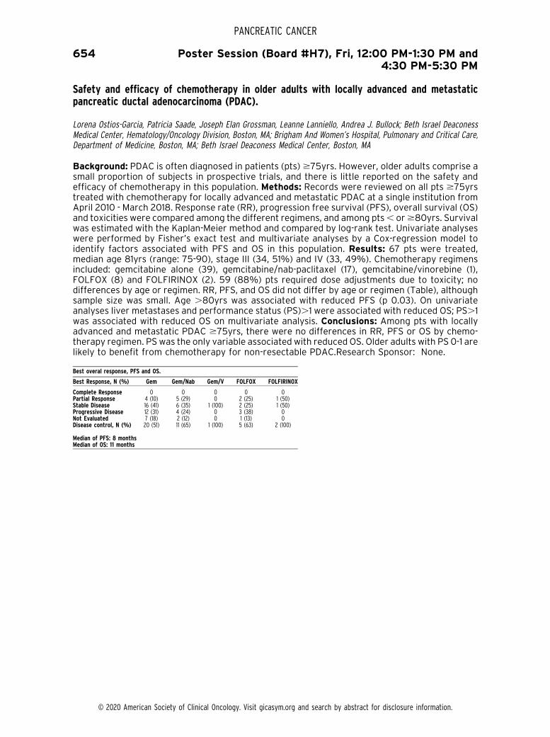
Background: PDAC is often diagnosed in patients (pts)$75yrs. However, older adults comprise asmall proportion of subjects in prospective trials, and there is little reported on the safety andefficacy of chemotherapy in this population. Methods: Records were reviewed on all pts $75yrstreated with chemotherapy for locally advanced and metastatic PDAC at a single institution fromApril 2010 - March 2018. Response rate (RR), progression free survival (PFS), overall survival (OS)and toxicities were compared among the different regimens, and among pts, or$80yrs. Survivalwas estimated with the Kaplan-Meier method and compared by log-rank test. Univariate analyseswere performed by Fisher’s exact test and multivariate analyses by a Cox-regression model toidentify factors associated with PFS and OS in this population. Results: 67 pts were treated,median age 81yrs (range: 75-90), stage III (34, 51%) and IV (33, 49%). Chemotherapy regimensincluded: gemcitabine alone (39), gemcitabine/nab-paclitaxel (17), gemcitabine/vinorebine (1),FOLFOX (8) and FOLFIRINOX (2). 59 (88%) pts required dose adjustments due to toxicity; nodifferences by age or regimen. RR, PFS, and OS did not differ by age or regimen (Table), althoughsample size was small. Age .80yrs was associated with reduced PFS (p 0.03). On univariateanalyses liver metastases and performance status (PS).1 were associated with reduced OS; PS.1was associated with reduced OS on multivariate analysis. Conclusions: Among pts with locallyadvanced and metastatic PDAC $75yrs, there were no differences in RR, PFS or OS by chemo-therapy regimen. PS was the only variable associated with reduced OS. Older adults with PS 0-1 arelikely to benefit from chemotherapy for non-resectable PDAC.Research Sponsor: None.
Best overal response, PFS and OS.
Best Response, N (%) Gem Gem/Nab Gem/V FOLFOX FOLFIRINOX
Complete Response 0 0 0 0 0Partial Response 4 (10) 5 (29) 0 2 (25) 1 (50)Stable Disease 16 (41) 6 (35) 1 (100) 2 (25) 1 (50)Progressive Disease 12 (31) 4 (24) 0 3 (38) 0Not Evaluated 7 (18) 2 (12) 0 1 (13) 0Disease control, N (%) 20 (51) 11 (65) 1 (100) 5 (63) 2 (100)
Median of PFS: 8 monthsMedian of OS: 11 months
© 2020 American Society of Clinical Oncology. Visit gicasym.org and search by abstract for disclosure information.
PANCREATIC CANCER
655 Poster Session (Board #H8), Fri, 12:00 PM-1:30 PM and4:30 PM-5:30 PM
Gemcitabine and nab-paclitaxel in older adults with metastatic pancreatic cancer: Are twodoses per cycle enough?
Arthur Winer, Elizabeth A. Handorf, Efrat Dotan; NYU Langone Medical Center, New York, NY; Fox Chase Cancer Center,Philadelphia, PA
Background: The dosing of Gemcitabine and Nab-Paclitaxel (GA), a frontline regimen to treatmetastatic pancreatic cancer (mPC), is frequently altered from the traditional dosing schedule(TDS) of day 1, 8, and 15 of a 28 day cycle to a modified dosing schedule (MDS) of 2 doses/cycle.Previouswork showed that overall survival (OS) was similar between patients (pts) treatedwith theMDS vs the TDS. We sought to analyze a larger real-world database to assess these trends.Methods:We retrospectively analyzed de-identified pts with mPC$ 65 y/o treated with GA in theFlatiron Health nationwide EHR-derived database. Demographics, treatments (tx), and outcomeswere collected. Pts were grouped as either starting with the TDS or MDS. Analysis included time ontreatment (TOT) as well as OS. A Cox model was used to test non-inferiority of the MDS vs the TDSfor both TOT and OS, adjusting performance status, age, race, gender, and line of therapy (LOT).The upper bound for non-inferiority was a Hazard Ratio (HR) = 1.2. Results: 1497 pts were treatedbetween 1/1/14-5/31/19; 883 pts with the TDS and 614with theMDS. Median TDS agewas 72 (65-85)and MDS was 73 (65-84) (p,0.001). 1237 pts received first- line GA; 60% received the TDS, 40%theMDS. The use of the TDS vs MDS did not vary significantly by LOT, gender, or race, but more ptswith a PS of $2 received the MDS (p=0.03). In the first-line, outcomes were better for the TDS vsthe MDS (unadjusted median TOT 5.3 vs 3.2 mo, p,0.001, OS 9.2 vs 5.3 mo; p,0.001), withconsistent results in the$ second-line. TheMDS did notmeet its non-inferiority boundary: first-lineTOT HR=1.4 [95% CI 1.2-1.6]; second+ line TOT HR=1.3 [95% CI 1.0-1.7]; first-line OS HR=1.6 [95% CI1.4-1.8]; second+ line OS HR=1.3 [95% CI 1.0-1.8]. Results were consistent when additionallystratified by PS 0-1 vs 2+. Conclusions: In this large real-world cohort, first-line GA tx with aMDS did notmeet criteria for non-inferiority for TOT andOS vs a TDS in older adults withmPC.Withthe caveats of potential confounding that exist in a de-identified retrospective database, theseresults suggest that dose intensity may be important in pts with mPC. Further prospective studiesare necessary to ensure we utilize effective tx strategies in older adults with mPC. ResearchSponsor: None.
© 2020 American Society of Clinical Oncology. Visit gicasym.org and search by abstract for disclosure information.
PANCREATIC CANCER
656 Poster Session (Board #H9), Fri, 12:00 PM-1:30 PM and4:30 PM-5:30 PM
Clinical outcomes of FOLFIRINOX and gemcitabine-nab-paclitaxel for metastatic pancreaticcancer in the real-world setting.
Fabio Franco, Jose Ignacio Martin Valades, David Marrupe, Juan Carlos Camara, David Gutierrez Abad, Ana Lopez-Alfonso,Brezo Martinez-Amores, Ana Leon, Mar Perez, Ignacio Juez, Alicia Hurtado, Ana Ruiz-Casado; Hospital Universitario Puertade Hierro-Majadahonda, Madrid, Spain; Fundacion Jimenez Diaz/Universidad Autonoma, Madrid, Spain; Hospital Universi-tario Mostoles, Mostoles, Spain; Hospital Universitario Fundacion de Alcorcon, Madrid, Spain; Hospital Universitario deFuenlabrada, Madrid, Spain; Infanta Leonor Hospital, Madrid, Spain; Hospital Universitario Rey Juan Carlos, Madrid, Spain;Oncology Department and Translational Oncology Division, University Hospital Fundacion Jimenez Diaz, Madrid, Spain;Hospital Universitario Infanta Leonor, Madrid, Spain; Hospital Universitario Fundacion de Alcorcon, Alcorcon, Spain
Background: Randomized clinical trials have established new chemotherapeutic standards of carefor metastatic pancreatic cancer, namely FOLFIRINOX (FFX) and gemcitabine + nab-paclitaxel(GNP) after demonstrating a significant and relevant increase of overall survival. However, thereare some important uncertainties regarding how many patients are candidate to each of the twonew regimens in the real life and how is the pattern of use in the elderly population.Methods: Thisis a retrospective study. Departments of Pharmacy of 7 Spanish hospitals generated the listings ofpatients (pts) treated in first line with these new regimens (FFX or GNP). Non-metastatic patientswere excluded. An exploratory analysis was performed in the elderly population. Results: FromJan 2012 to Dec 2017, a total of 119 pts (M/F 58/42 %) were treated. Med age 63 y (38-83 y), 99%adenocarcinoma. 40% located in the head of pancreas. ECOG 87% 0-1. 89% had liver mets. In the1st line 49.6% were treated with FFX and 50.4% with GNP. 53% of the pts could receive a 2nd line(82% after FFX 75% after GNP). The median OS was 12 months with no statistically significantdifferences between both regimens (12,7m for FFX vs 10,2 m for GNP). Elevated Ca 19.9 levels andNeutrophil-Lymphocyte ratio (NLR) increased the risk of death. Patients who received bothregimens in first/second line had a median OS longer than 15 months whichever the sequence.32 patients (27%) were older than 70 yo. 13 (41%) were treated with FFX and 19 (59%) with GNP.The median OS for patients older than 70 was 9.5m versus 12.3m for patients younger than 70.Conclusions: In our setting the use of FFX and GNP for treating metastatic pancreatic cancer isquite similar. Superiority could not be demonstrated for any of the schemes in first-line. Overallsurvival was determined by basal Ca 19.9 and NLR. Patients receiving both regimens (FFX or GNP)in first/second line whichever the sequence, exhibited the best survival rates. In our series elderlypatients had poor survival rates. Research Sponsor: None.
© 2020 American Society of Clinical Oncology. Visit gicasym.org and search by abstract for disclosure information.
PANCREATIC CANCER
657 Poster Session (Board #H10), Fri, 12:00 PM-1:30 PM and4:30 PM-5:30 PM
Survival outcomes based on sequence of therapy using FOLFIRINOX and nab-paclitaxel + gemcitabinein metastatic pancreatic ductal adenocarcinoma.
Kelsey Baron, Christopher Duane Nevala-Plagemann, Justin Moser, Benjamin Haaland, Xuechen Wang, Ignacio Garrido-Laguna; Division of Internal Medicine, Intermountain Medical Center, Murray, UT; Huntsman Cancer Institute, University ofUtah, Salt Lake City, UT; Honor Health Research Institute, Scottsdale, AZ; University of Utah, Salt Lake City, UT
Background: Optimal sequence of therapy for patients with metastatic pancreatic ductal adeno-carcinoma (mPDAC) is unknown. FOLFIRINOX (FFX) and Gemcitabine + Nab-paclitaxel (AG) arestandard first line (1L) therapies. They have never been prospectively compared. Therefore, weretrospectively compared the overall survival (OS) of patients treated with 1L AG and second line(2L) FFX compared to those treated with 1L FFX and 2L AG.Methods: Patients withmPDAC treatedwith 1L FFX followed by 2L AG, or vice versa were identified using the Flatiron Health EHR-derivednationwide database. To avoid immortal time bias, patients who received no 2L were included. OSfrom the initiation of 1L was comparedwith KaplanMeier curves and log rank analysis. A coxmodel,stratified by deciles of propensity score (PS), was used to estimate the effect of treatment on OSwith adjustment for differences between the groups.Results: 3,042 patients were identified. 2001patients received 1L AG. Among these patients, 1446 received 2L FFX, and 555 received no 2L. 1041patients received 1L FFX. Among these patients, 496 received 2L AG, and 545 received no 2L.Median OS and 1-year OS for those treated with 1L AG followed by 2L FFX or no therapy was 6.1months (95% CI:5.6 – 6.5) and 25% (95% CI: 0.23 – 0.28). Median OS and 1-year OS for patientstreatedwith 1L FFX followed byAG or no therapywas 8.7months (95%CI: 7.9 – 9.2) and 36% (95%CI: 0.33 –0.39). The propensity stratified hazard ratio between these two groups was 0.76 (95%CI:0.69 – 0.83), favoring 1L FFX. Median OS for patients treated with 1L FFX and 2L AG versus 1L AGand 2L FFX was not significantly different (12.0 m vs. 12.5 m; HR 1.04; 95% CI: 0.90 - 1.20).Conclusions: In this analysis of real-world data, 1L FFX was associated with increased OS inpropensity analysis. For patients who received both FFX and AG,median OSwas similar, regardlessof the sequence. Research Sponsor: None.
© 2020 American Society of Clinical Oncology. Visit gicasym.org and search by abstract for disclosure information.
PANCREATIC CANCER
658 Poster Session (Board #H11), Fri, 12:00 PM-1:30 PM and4:30 PM-5:30 PM
Effects of duration of initial treatment on postoperative complications in pancreatic cancer.
Naoya Takeda, Suguru Yamada, Hideki Takami, Fuminori Sonohara, Masamichi Hayashi, Isaku Yoshioka, Kazuto Shibuya,Koshi Matsui, Katsuhisa Hirano, Toru Watanabe, Yuuko Tohmatsu, Nana Kimura, Shozo Hojo, Shigeaki Sawada,Tomoyuki Okumura, Takuya Nagata, Yasuhiro Kodera, Tsutomu Fujii; Department of Surgery and Science, Graduate Schoolof Medicine and Pharmaceutical Sciences, University of Toyama, Toyama, Japan, Toyama, Japan; Nagoya UniversityGraduate School of Medicine, Gastroenterological Surgery, Nagoya, Japan; Department of Surgery and Science, GraduateSchool of Medicine and Pharmaceutical Sciences, University of Toyama, Toyama, Japan; Univ of Toyama, Toyama, Japan
Background: Early studies raised concerns over whether preoperative treatment led to postop-erative complications or even death. In contrast, recent studies have reported that initial treatment(IT) prior to resection of pancreatic ductal adenocarcinoma (PDAC) is safe, with no significantincrease in overall morbidity or mortality, despite evidence for more advanced disease. In thisstudy, we analyzed the clinical impact of chemotherapy or chemoradiotherapy as IT, focusing ontreatment duration, on morbidity and mortality in patients with resected PDAC. Methods: Weenrolled 509 consecutive patients, with 417 in the upfront surgery group and 92 in the IT group.The IT group was subdivided into 72 patients treated for , 8 months and 20 treated $8 months.We compared rates of postoperative Clavien–Dindo grade$III complications between the groups.Multivariate logistic regression analysis was used to find independent predictors of complications.Results: The upfront surgery and IT groups did not significantly differ in overall postsurgicalcomplications. The rate of postoperative pancreatic fistula was significantly less in the IT group.Rates of other complications did not significantly differ, except for severe infection and delayedgastric emptying. Initiation of adjuvant chemotherapy was later in the IT group than in the upfrontsurgery group (43.2 vs 57.8 days, P , 0.001). In contrast, rates of overall complications signif-icantly differed between the , 8 months and $8 months IT groups, although their backgroundclinical factors did not differ. In multivariate analysis, operative procedure (distal pancreatectomyand distal pancreatectomywith celiac axis resection) (odds ratio [OR] 6.950, P = 0.0416) and IT$8months (OR: 4.508, 95%, P = 0.0156) were independent predictive factors for postoperativecomplications. Conclusions: The incidence of postoperative complication was similar between theupfront surgery group and the IT group, however, it was significantly higher in the $8 months ITgroup in patients who underwent PDAC resection. Research Sponsor: None.
© 2020 American Society of Clinical Oncology. Visit gicasym.org and search by abstract for disclosure information.
PANCREATIC CANCER
660 Poster Session (Board #H13), Fri, 12:00 PM-1:30 PM and4:30 PM-5:30 PM
Observational retrospective evaluation of treatment with liposomal irinotecan plus fluorouracil/leucovorin for metastatic pancreatic cancer patients: An Italian large real-world analysis.
Antonio Pellino, Chiara Manai, Valeria Merz, Mario Scartozzi, Michele Milella, Ferdinando De Vita, Lorenzo Antonuzzo,Clizia Zichi, Maria Antonietta Satolli, Michele Panebianco, Silvia Noventa, Guido Giordano, Floriana Nappo, Camilla Zecchetto,Marco Puzzoni, Vanja Vaccaro, Annalisa Pappalardo, Elisa Giommoni, Davide Melisi, Sara Lonardi; Department of Clinical andExperimental Oncology, Medical Oncology 1 Unit, Istituto Oncologico Veneto IOV-IRCCS, Padua, Italy; Unit of MedicalOncology, University of Verona Hospital Trust, Verona, TN, Italy; Medical Oncology Department, University Hospital,University of Cagliari, Cagliari, Italy; Medical Oncology, Azienda Ospedaliera Universitaria Integrata, Verona, Italy; Division ofMedical Oncology, Department of Precision Medicine, University of Study of Campania "L. Vanvitelli", Naples, Italy; MedicalOncology, Azienda Ospedaliero-Universitaria Careggi, Florence, Italy; Department of Oncology, University of Turin, OrdineMauriziano Hospital, Turin, Italy; Department of Medical Oncology, University of Turin, Turin, Italy; Medical Oncology Unit,Clinical Cancer Center, IRCCS-AUSL di Reggio Emilia, Reggio Emilia, Italy; Medical Oncology Unit, Casa di Cura Poliambulanza,Brescia, Italy; Fondazione IRCCS Casa Sollievo della Sofferenza, UO di Oncologia Medica, San Giovanni Rotondo, Italy; Unit ofMedical Oncology, University of Verona Hospital Trust, Verona, Italy; Medical Oncology 1, IRCCS Regina Elena National CancerInstitute, Rome, Italy; Medicine - Digestive Molecular Clinical Oncology Research Unit, University of Verona, Verona, Italy
Background: In the NAPOLI I phase III trial, Nanoliposomal irinotecan (nal-IRI) plus 5-fluorouracil/leucovorin (5-FU/LV) showed better outcome compared to 5FU/LV in patients with metastaticPancreatic Cancer (MPC) progressed to 1st- line gemcitabine-based therapy. Aim of this study is toexplore the real-world efficacy and safety of 5FU/LV-nal-IRI by a compassionate use programmeand to identify potential prognostic factors that could affect survival in this setting.Methods: Thisis a retrospective multi-center analysis including patients with MPC who received 5FU/LV-nal-IRIafter failure of a gemcitabine-based therapy. Survival analyses were carried out by the Kaplan-Meier method. Univariate andmultivariate analyses were performed by using the log-rank test andthe Cox regression. Results: A total of 296 pts (median age, 69 years, range 30-82; 50% male;ECOGPS0, 44%)were treated at 11 Italian institutions fromJune 2016 andNovember 2018. 34%ofthe pts have been previously resected on their primary tumor, and 76% received gemcitabine-nabpaclitaxel as 1st - line treatment. 5FU/LV-nal-IRI has been administered as 2nd - line in 72% ofthe pts, while in 23% of the cases as 3rd - line or more. The median OS was 7.1 months [95%confidence interval (CI) 6.1 - 8.1] and the median PFS was 3.3 months (95% CI 2.9 - 3.6). At sixmonths, OS and PFS rate were 53.4% and 31.4% respectively. ORR was 12% and DCR was 40%.52%of pts receivedmore than 4 cycle with dose reduction in 148 pts (50%). Most common grade 3toxicities were neutropenia (14%), diarrhea (11%), anemia (3%), nausea (3%), fatigue (3%),mucositis (2%) and vomiting (1%). Baseline characteristics associated with better OS were ECOGPS 0, normal CEA, neutrophil-to-lymphocyte ratio #5 and haemoglobin $11 g/dL. Conclusions:These real-world data confirm the efficacy and safety of 5FU/LV-nal-IRI in patients with MPCprogressed to a gemcitabine-based therapy, with outcome comparable to NAPOLI-1 even in a lessselected population and with more active 1st - line combination therapy. In this cohort, well knownprognostic markers has been confirmed, as expected. Research Sponsor: None.
© 2020 American Society of Clinical Oncology. Visit gicasym.org and search by abstract for disclosure information.
PANCREATIC CANCER
661 Poster Session (Board #H14), Fri, 12:00 PM-1:30 PM and4:30 PM-5:30 PM
Efficacy of second-line chemotherapy after standard combination chemotherapy in patientswith metastatic pancreatic cancer: The results from the NAPOLEON study.
Masaru Fukahori, Yoshinobu Okabe, Mototsugu Shimokawa, Taiga Otsuka, Futa Koga, Yujiro Ueda, Junichi Nakazawa,Azusa Komori, Shiho Arima, Akitaka Makiyama, Hiroki Taguchi, Takuya Honda, Tomoyuki Ushijima, Keisuke Miwa,Taro Shibuki, Kenta Nio, Yasushi Ide, Norio Ureshino, Kenji Mitsugi, Tsuyoshi Shirakawa; Division of Gastroenterology,Department of Medicine, Kurume University School of Medicine, Kurume, Japan; National Kyushu Cancer Center, Fukuoka,Japan; Department of Medical Oncology, Saga Medical Center Koseikan, Saga, Japan; Department of Hepatobiliary andPancreatology, Saga Medical Center Koseikan, Saga, Japan; Department of Hematology and Oncology, Japanese Red CrossKumamoto Hospital, Kumamoto, Japan; Department of Medical Oncology, Kagoshima City Hospital, Kagoshima, Japan;Department of Medical Oncology and Hematology, Oita University Faculty of Medicine, Yufu, Japan; Digestive and LifestyleDiseases, Kagoshima University Graduate School of Medical and Dental Sciences, Kagoshima, Japan; Department ofHematology/Oncology, Japan Community Health Care Organization Kyushu Hospital, Kitakyushu, Japan; Department ofGastroenterology, Saiseikai Sendai Hospital, Satsumasendai, Japan; Nagasaki University Hospital, Nagasaki, Japan; KurumeUniversity, Fukuoka, Japan; Kurume University Hospital, Kurume, Japan; National Cancer Center Hospital East, Kashiwa,Chiba, Japan; Department of Medical Oncology, Sasebo Kyosai Hospital, Sasebo, Japan; Department of Internal Medicine,Karatsu Red Cross Hospital, Karatsu, Japan; Department of Medical Oncology, Saga-Ken Medical Center Koseikan, Saga,Japan; Department of Medical Oncology, Hamanomachi Hospital, Fukuoka, Japan; Department of Gastrointestinal andMedical Oncology, National Hospital Organization Kyushu Cancer Center, Fukuoka, Japan
Background: Gemcitabine plus nab-paclitaxel (GnP) and FOLFIRINOX (FFX) have been establishedas standard first-line combination chemotherapy (CTx) for patients with metastatic pancreaticcancer (MPC). However, the efficacy of second-line CTx and the significance of combination CTx inclinical practice are unclear. We therefore investigated the efficacy of second-line CTx in patientswith MPC. Methods: Data were collected from CTx-naive MPC patients treated with first-linecombination CTx at 14 hospitals in the Kyushu area of Japan from December 2013 to June 2018.The median overall survival (mOS) from second-line treatment was compared between patientswho received second-line CTx (CT group) and those who received best supportive care (BSCgroup). Furthermore, in the CT group, the mOS was compared between the patients who receivedcombination CTx and those who received mono-CTx. To control potential bias in the selection ofsecond-line treatment, we also conducted a propensity score-adjusted analysis.Results:A total of255 patients received GnP or FFX as first-line CTx. Of these, there were 156 (61%) in the CT groupand 77 (30%) in the BSC group. The number of patients who received FFX/GnP as first-line CTxwas79 (51%)/77 (49%) in the CT group and 15 (20%)/62 (80%) in the BSC group, respectively (P ,0.01). The mOS in the CT group was significantly longer than that in the BSC group (5.2 vs. 2.7months; hazard ratio [HR] 0.42; 95% confidence interval [CI] 0.31-0.57; p , 0.01 and 5.2 vs. 2.6months; adjusted HR 0.39; 95% CI 0.28-0.55; p , 0.01). In the CT group, 89 (57%) patientsreceived combination CTx, and 67 (43%) received mono-CTx. There was no significant differencein the mOS between the combination CTx and mono-CTx patients (5.5 vs. 4.4 months; HR 0.88;95% CI 0.62-1.26; p = 0.88 and 5.6 vs. 4.4 months; adjusted HR 0.85; 95% CI 0.56-1.30; p = 0.47).Conclusions: Among patients with MPC receiving second-line treatment, the CT group had asignificantly longer mOS than the BSC group, but combination CTx conferred no improvement inthe survival duration compared with mono-CTx. Research Sponsor: None.
© 2020 American Society of Clinical Oncology. Visit gicasym.org and search by abstract for disclosure information.
PANCREATIC CANCER
662 Poster Session (Board #H15), Fri, 12:00 PM-1:30 PM and4:30 PM-5:30 PM
Comparison of clinicopathological characteristics and prognosis of borderline resectablepancreatic cancer according to the location of the primary tumor.
Tsuyoshi Takeda, Takashi Sasaki, Takafumi Mie, Takaaki Furukawa, Ryo Kanata, Akiyoshi Kasuga, Masato Matsuyama,Masato Ozaka, Naoki Sasahira; Cancer Institute Hospital of Japanese Foundation for Cancer Research, Tokyo, Japan
Background: Little is known about the clinicopathological and prognostic differences betweenborderline resectable (BR) pancreatic head (Ph) and pancreatic body/tail (Pbt) cancer. Therefore,we conducted this study to compare the clinicopathological features and prognosis of BR pan-creatic cancer (PC) according to the location of the primary tumor. Methods: We retrospectivelyinvestigated consecutive patients with BR PC who initiated neoadjuvant chemotherapy (NAC)between March 2015 and April 2019. We compared clinicopathological characteristics and prog-nosis between Ph and Pbt cancer. Furthermore, multivariate survival analysis was performed usingcox proportional hazard model. Results: A total of 104 patients with BR PC (median age 68, male49%)were included in this study. The location of the tumor was Ph 72 and Pbt 32, respectively. Theinitial regimen of NAC was nab-paclitaxel/gemcitabine in 102 and gemcitabine in 2, respectively.The median cycle of NAC was 4. Median age, sex, primary tumor size, performance status,neutrophil to lymphocyte ratio, and serum level of carbohydrate antigen 19-9 at the time ofthe initiation of NACwere not significantly different between Ph and Pbt cancer, while themodifiedglasgow prognostic score (mGPS) was lower in Pbt cancer (mGPS = 0; 78% vs. 94%, p = 0.05). R0/R1 resection rate (81% vs. 69%, p = 0.21) and median survival time (928 days vs. NA, p = 0.13) werealso not different between Ph and Pbt cancer. Multivariate survival analysis revealed that R0/R1resection (HR, 0.11; p , 0.01) and Ph (HR, 2.29; p = 0.03) were independent prognostic factors forsurvival in patients with BR PC. Conclusions: Although R0/R1 resection rate was similar betweenBR Ph and Pbt cancer, Pbt cancer had a higher rate of mGPS score of 0 compared to Ph cancer.Furthermore, R0/R1 resection (HR, 0.11) and Ph (HR, 2.29) were independent prognostic factors forsurvival in patients with BR PC. Research Sponsor: None.
© 2020 American Society of Clinical Oncology. Visit gicasym.org and search by abstract for disclosure information.
PANCREATIC CANCER
663 Poster Session (Board #H16), Fri, 12:00 PM-1:30 PM and4:30 PM-5:30 PM
A real-world evidence analysis of periampullary cancers in an academic hospital in Chile.
Luis Villanueva, Gonzalo Navarrete, Ivan Gallegos, Valentina Castillo, Francisco Dodds, Jaime Gonzalez, Olga Barajas,Rodrigo Vasquez, Barbara Nu~nez, Monica Ahumada; Hospital Clinico Universidad de Chile, Santiago, Chile
Background: Periampullary cancers can originate in the pancreas, duodenum, bile duct or struc-tures of the ampullary complex. The treatment of choice in early stages is pancreatoduodenec-tomy. The management post-surgery can depend on the histology pattern, and the overall survivalcan vary in different subgroups. Methods: A retrospective cohort study. We examined patients(pts) with invasive periampullary cancer undergoing pancreatoduodenectomy at the HospitalClinico Universidad de Chile between 2002 to 2018. We analyzed epidemiological, clinical, surgical,and histological data. OS and the hazard ratio (HR) were established by GraphPad Prism 8.0.Results: Thirty-seven cases were registered. Twenty-two (59%) pts were men. The mean age was62.5 (43-83 years). The histological subtypes were: 15 pts (40.5%) intestinal group (IN), 20 pts(54%) pancreatobiliary group (PB), 1 pt (2.7%) mixed and 1 pt (2.7%) signet ring cell type. A fullconcordance between histology and immunohistochemistry (CK20, CK7, CDX2, MUC1, and MUC2)patterns was 66%of the PB group, and 0%of the IN group. The stage IB wasmost frequent in all ofthe group (36,4%). The most frequent stages were IB (66,6%) in the IN type and IIIA (46%) in thePB type. The level of Ca19-9 was higher the PB group than IN group (629.7 versus 41.5 U/ml,respectively). Seven pts received postoperative adjuvant treatment such as FOLFOX, capecitabine,and gemcitabine. The median OS was 133,5 months (mo) in the intestinal group and 32,6 mo in PBgroup (P-value = 0.021). The HR was 0.38 (95% CI of ratio 0.1332 to 1.084). The 5-year OS was75,2% and 45,7% in the IN and PB group, respectively. Conclusions: Periampullary cancerremains very challenging because it is a rare malignancy and present diverse histological pattern.These factors influence the behavior and OS of the disease. Our results showed clinically andstatistically relevant differences in the staging, levels of Ca19-9, and OS of the IN and PB subtypes.Our patients received few post-operatory therapies such as chemotherapy; this factor couldinfluence the OS in the high-risk group. According to our data, a personalized treatment byhistological type should consider in this disease. Research Sponsor: None.
© 2020 American Society of Clinical Oncology. Visit gicasym.org and search by abstract for disclosure information.
PANCREATIC CANCER
664 Poster Session (Board #H17), Fri, 12:00 PM-1:30 PM and4:30 PM-5:30 PM
Comparing survival outcomes for neoadjuvant therapy versus adjuvant therapy in themanagement of stage 1 pancreatic adenocarcinoma: A National Cancer Database study.
Samit Kumar Datta, Geoffrey Bellini, Maharaj Singh, Nicholas Sich, James L. Weese, Federico Augusto Sanchez, Nalini Guda,Wesley Allan Papenfuss, Aaron Chevinsky; Aurora Health Care, Milwaukee, WI; Aurora St. Luke’s Medical Center, Milwaukee,WI; Marquette Dental School, Milwaukee, WI; Advocate Aurora Health, Milwaukee, WI; Aurora Cancer Care, Advocate AuroraHealth, Milwaukee, WI; GI Associates, Milwaukee, WI
Background:We are in the midst of a paradigm shift in the treatment of stage 1 pancreatic ductaladenocarcinoma (PDAC) from surgery first followed by adjuvant therapy (AT) to Neoadjuvanttherapy (NAT) first followed by surgery and this is reflected in the current NCCN guidelines as well.Data comparing these two modalities are limited. AIM: To compare long term survival betweenSurgery + AT and NAT + Surgery in a large National Cancer Database for stage 1 PDAC. Methods:We identified patients with the NCDBwith surgically resected AJCC clinical stage 1, 1A, and 1B PDACbetween 2004-2016. Patients were stratified into two groups to assess outcomes: AT and NAT.Patients with incomplete survival and sequence of therapy were excluded. Baseline demographicdata, 90-Day Mortality, Median survival, and Hazard ratios (HR) for survival was evaluated.Results: 9017 pts with Clinical stage 1, 1A, 1B PDAC between 2004-2016 were identified. Of these7453 pts had surgery followed by AT; and 1564 pts had NAT followed by surgery. There was astatistically significant difference in age (66.069.9 years for AT vs. 64.769.78 years for NAT, p <0.001) but no difference in Charlson Comorbidity Scoring (p = 0.618) or sex (p = 0.073). 90-DayMortality was 0.35% in the AT group compared to 0.83% in the NAT group (p = < 0.001). Mediansurvival was 28.5 (95% CI 26.5-29.9) months in the NAT group compared to 25.4 (95% CI 24.7-26.1) months in the AT group. With AT as the reference group for survival, there was a HR of 0.904(95% CI 0.845-0.968, p = 0.003) for NAT. Conclusions: In this retrospective cohort of patients,NAT was associated with increased overall survival. However, NAT was associated with anincreased 90 day mortality. A randomized, controlled trial is necessary to further support thesuperiority of NAT in the management of stage 1 PDAC. Research Sponsor: None.
© 2020 American Society of Clinical Oncology. Visit gicasym.org and search by abstract for disclosure information.
PANCREATIC CANCER
665 Poster Session (Board #H18), Fri, 12:00 PM-1:30 PM and4:30 PM-5:30 PM
Impact of dose reductions on clinical outcomes among patients (pts) with metastatic pancreaticcancer (mPC) treated with liposomal irinotecan (nal-IRI) in oncology clinics in the United States.
Paul Cockrum, Andy Surinach, George P. Kim, Daniel Mercer, Jim M. Koeller, Rebecca A. Miksad; Ipsen Biopharmaceuticals,Cambridge, MA; Genesis Research, Hoboken, NJ; George Washington University, Division of Hematology & Oncology,Washington, DC; Univ of Texas Health Sci Ctr, San Antonio, TX; Flatiron Health, New York, NY
Background: The recommended starting dose for nal-IRI is 70mg/m2 (free base, equivalent to 80mg/m2 salt-based dosing). This study evaluates the impact of nal-IRI dose reductions on clinicaloutcomes. Methods: Using the nationwide Flatiron Health electronic health record-derived data-base, de-identified data were extracted and analyzed for adult mPC pts treated with nal-IRI Jan2014-Jan 2019 and who initiated treatment at approximately the recommended dose (RD), 70mg/m2 +/- 5mg. Initial dose was derived from structured medication records, prioritizing adminis-trations. The cumulative dose (CD) of nal-IRI over the first six weeks of treatment, the presence ofdose reductions (DR) – (a decrease$ 7mg/m2), overall survival (OS) from treatment initiation, andduration of treatment (DoT) were assessed.Results: 257mPC pts treatedwith nal-IRI (median age:68y, IQR: 61 - 73) were identified initiating therapy at approximately the RD. 26.5% (N = 68) of ptsexperienced a DR during treatment. Mean 6-week CD was 175.8 mg/m2 (SD: 77.9) among pts withnoDR. For ptswith DR,meanCDwas 191.8mg/m2 (53.2). MedianDoTwas 6.1 wks (IQR: 2.1 – 15.3). Ptsthat experienced aDR had a longermedian DoT: 15.1 wks (7.1 – 23.0) vs 4.3. wks (2.1 – 12.1) for pts withno DR. Overall Median OS (mOS) was 4.2 months (95% CI: 3.7 – 5.4). mOS for DR pts was 7.2 mos(95% CI: 5.5 – 9.7) and 3.7 mos (3.0 – 4.1) for pts who did not experience a DR. Conclusions: Thisreal-world analysis suggests that reducing the dose of subsequent administrations of nal-IRI duringtreatment is associated with pts remaining on therapy longer, experiencing a larger CD, and a withlonger OS. Additional real-world prospective studies are necessary to characterize the impact ofnal-IRI dosing on clinical outcomes. Research Sponsor: Ipsen Biopharmaceuticals.
© 2020 American Society of Clinical Oncology. Visit gicasym.org and search by abstract for disclosure information.
PANCREATIC CANCER
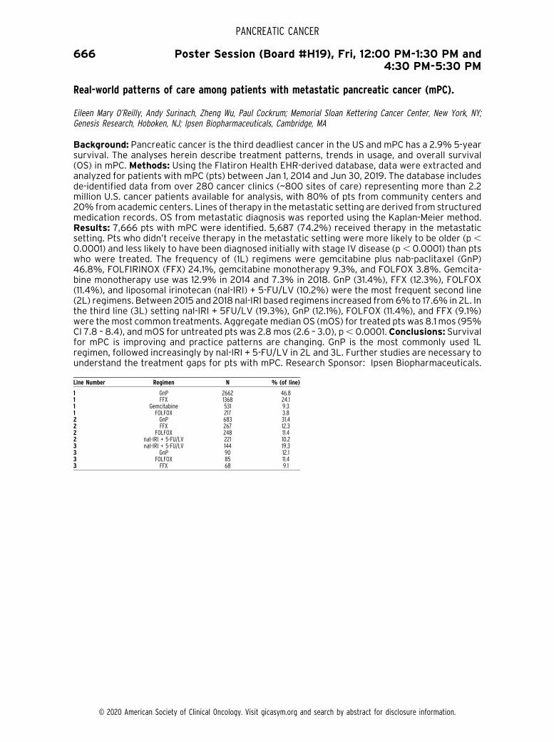
666 Poster Session (Board #H19), Fri, 12:00 PM-1:30 PM and4:30 PM-5:30 PM
Real-world patterns of care among patients with metastatic pancreatic cancer (mPC).
Eileen Mary O’Reilly, Andy Surinach, Zheng Wu, Paul Cockrum; Memorial Sloan Kettering Cancer Center, New York, NY;Genesis Research, Hoboken, NJ; Ipsen Biopharmaceuticals, Cambridge, MA
Background: Pancreatic cancer is the third deadliest cancer in the US and mPC has a 2.9% 5-yearsurvival. The analyses herein describe treatment patterns, trends in usage, and overall survival(OS) in mPC. Methods: Using the Flatiron Health EHR-derived database, data were extracted andanalyzed for patients with mPC (pts) between Jan 1, 2014 and Jun 30, 2019. The database includesde-identified data from over 280 cancer clinics (~800 sites of care) representing more than 2.2million U.S. cancer patients available for analysis, with 80% of pts from community centers and20% from academic centers. Lines of therapy in themetastatic setting are derived from structuredmedication records. OS from metastatic diagnosis was reported using the Kaplan-Meier method.Results: 7,666 pts with mPC were identified. 5,687 (74.2%) received therapy in the metastaticsetting. Pts who didn’t receive therapy in the metastatic setting were more likely to be older (p ,0.0001) and less likely to have been diagnosed initially with stage IV disease (p, 0.0001) than ptswho were treated. The frequency of (1L) regimens were gemcitabine plus nab-paclitaxel (GnP)46.8%, FOLFIRINOX (FFX) 24.1%, gemcitabine monotherapy 9.3%, and FOLFOX 3.8%. Gemcita-bine monotherapy use was 12.9% in 2014 and 7.3% in 2018. GnP (31.4%), FFX (12.3%), FOLFOX(11.4%), and liposomal irinotecan (nal-IRI) + 5-FU/LV (10.2%) were the most frequent second line(2L) regimens. Between 2015 and 2018 nal-IRI based regimens increased from6% to 17.6% in 2L. Inthe third line (3L) setting nal-IRI + 5FU/LV (19.3%), GnP (12.1%), FOLFOX (11.4%), and FFX (9.1%)were themost common treatments. Aggregatemedian OS (mOS) for treated pts was 8.1 mos (95%CI 7.8 – 8.4), and mOS for untreated pts was 2.8 mos (2.6 – 3.0), p, 0.0001. Conclusions: Survivalfor mPC is improving and practice patterns are changing. GnP is the most commonly used 1Lregimen, followed increasingly by nal-IRI + 5-FU/LV in 2L and 3L. Further studies are necessary tounderstand the treatment gaps for pts with mPC. Research Sponsor: Ipsen Biopharmaceuticals.
Line Number Regimen N % (of line)
1 GnP 2662 46.81 FFX 1368 24.11 Gemcitabine 531 9.31 FOLFOX 217 3.82 GnP 683 31.42 FFX 267 12.32 FOLFOX 248 11.42 nal-IRI + 5-FU/LV 221 10.23 nal-IRI + 5-FU/LV 144 19.33 GnP 90 12.13 FOLFOX 85 11.43 FFX 68 9.1
© 2020 American Society of Clinical Oncology. Visit gicasym.org and search by abstract for disclosure information.
PANCREATIC CANCER
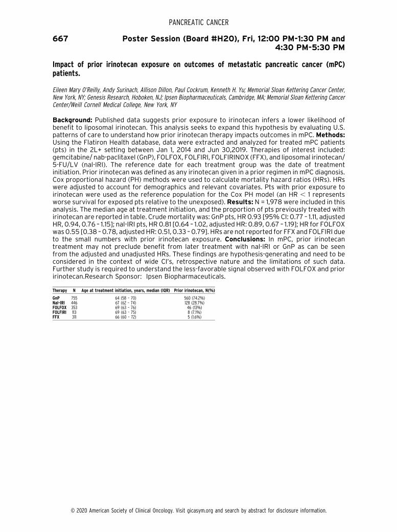
667 Poster Session (Board #H20), Fri, 12:00 PM-1:30 PM and4:30 PM-5:30 PM
Impact of prior irinotecan exposure on outcomes of metastatic pancreatic cancer (mPC)patients.
Eileen Mary O’Reilly, Andy Surinach, Allison Dillon, Paul Cockrum, Kenneth H. Yu; Memorial Sloan Kettering Cancer Center,New York, NY; Genesis Research, Hoboken, NJ; Ipsen Biopharmaceuticals, Cambridge, MA; Memorial Sloan Kettering CancerCenter/Weill Cornell Medical College, New York, NY
Background: Published data suggests prior exposure to irinotecan infers a lower likelihood ofbenefit to liposomal irinotecan. This analysis seeks to expand this hypothesis by evaluating U.S.patterns of care to understand how prior irinotecan therapy impacts outcomes in mPC. Methods:Using the Flatiron Health database, data were extracted and analyzed for treated mPC patients(pts) in the 2L+ setting between Jan 1, 2014 and Jun 30,2019. Therapies of interest included:gemcitabine/ nab-paclitaxel (GnP), FOLFOX, FOLFIRI, FOLFIRINOX (FFX), and liposomal irinotecan/5-FU/LV (nal-IRI). The reference date for each treatment group was the date of treatmentinitiation. Prior irinotecan was defined as any irinotecan given in a prior regimen in mPC diagnosis.Cox proportional hazard (PH) methods were used to calculate mortality hazard ratios (HRs). HRswere adjusted to account for demographics and relevant covariates. Pts with prior exposure toirinotecan were used as the reference population for the Cox PH model (an HR , 1 representsworse survival for exposed pts relative to the unexposed). Results: N = 1,978 were included in thisanalysis. The median age at treatment initiation, and the proportion of pts previously treated withirinotecan are reported in table. Crudemortality was: GnP pts, HR 0.93 [95%CI: 0.77 – 1.11, adjustedHR, 0.94, 0.76 – 1.15]; nal-IRI pts, HR 0.81 [0.64 – 1.02, adjusted HR: 0.89, 0.67 – 1.19]; HR for FOLFOXwas0.55 [0.38 –0.78, adjustedHR: 0.51, 0.33 –0.79]. HRs are not reported for FFX and FOLFIRI dueto the small numbers with prior irinotecan exposure. Conclusions: In mPC, prior irinotecantreatment may not preclude benefit from later treatment with nal-IRI or GnP as can be seenfrom the adjusted and unadjusted HRs. These findings are hypothesis-generating and need to beconsidered in the context of wide CI’s, retrospective nature and the limitations of such data.Further study is required to understand the less-favorable signal observed with FOLFOX and prioririnotecan.Research Sponsor: Ipsen Biopharmaceuticals.
Therapy N Age at treatment initiation, years, median (IQR) Prior irinotecan, N(%)
GnP 755 64 (58 – 70) 560 (74.2%)Nal-IRI 446 67 (62 – 74) 128 (28.7%)FOLFOX 353 69 (63 – 76) 46 (13%)FOLFIRI 113 69 (63 – 75) 8 (7.1%)FFX 311 66 (60 – 72) 5 (1.6%)
© 2020 American Society of Clinical Oncology. Visit gicasym.org and search by abstract for disclosure information.
PANCREATIC CANCER
668 Poster Session (Board #H21), Fri, 12:00 PM-1:30 PM and4:30 PM-5:30 PM
Changes in glucose tolerance after pancreatectomy in patients with pancreatic ductaladenocarcinoma.
Sachiyo Shirakawa, Hirochika Toyama, Shohei Komatsu, Jun Ishida, Masahiro Kido, Takumi Fukumoto; Division of Hepato-Biliary-Pancreatic Surgery, Department of Surgery, Kobe University Graduate School of Medicine, Kobe, Japan; KobeUniversity Hospital, Dept. of Hepato-Biliary Pancreatic Surgery, Kobe, Japan
Background:Diabetesmellitus (DM) is reported to be related to pancreatic ductal adenocarcinoma(PDAC). Long-standing DM is a risk factor for PDAC, meanwhile, quite a few patients with PDACdevelop DM as a paraneoplastic disorder and some papers reported that DM affected prognosis forPDAC. In this study, we investigated pre- and post-operative glucose tolerance in patients withpancreatectomy for PDAC or other disease. Methods: This single-center prospective studyincluded 69 patients with pancreatectomy (40 pancreaticoduodenectomy (PD) and 29 distalpancreatectomy (DP) ) who received 75-g oral glucose tolerance test (OGTT) and glucagon testpre- and one month postoperatively. Plasma glucose, insulin, and C-peptide (CPR) at 0-, 30-, 60-,and 120-min during OGTT; and 0- and 6-min during glucagon test were obtained. These data andsurvival outcomes were analyzed. Results: There were 20 (29%) PDAC patients: 12 (30%) in PDgroup and eight (28%) in DP group. Nine patients with PDAC (45%) and seven patients (18%)without PDAC demonstrated DM type in preoperative OGTT. After pancreatectomy, 11 patients(55%) with PDAC and seven patients (15%) without PDAC experienced improvement in OGTT(P=0.0005). Greater improvement in homeostasis model assessment insulin resistance that wereobtained by OGTT and used to measure insulin resistance, was noted after surgery in PDACpatients compared with non-PDAC patients (-1.4 vs -0.5 in PD group, P=0.07; -0.8 vs +0.06 P=0.05in DP group). Delta CPRs obtained by glucagon test were significantly decreased postoperatively(3.0 to 1.1 ng/mL, P,0.0001 in PD group; 3.3 to 1.8 ng/mL, P,0.0001 in DP group). In survivalanalysis, fasting plasma glucose .110 mg/dL (HR 3.9, 95%CI 1.5-10, P=0.005) and the average ofplasma insulin . 25 mIU/mL during OGTT (HR 0.36, 95%CI 0.14-0.93, P=0.035) were significantprognostic factor for PDAC.Conclusions: Pancreatectomy impaired insulin secretion and improveinsulin resistance especially in PDAC patients. About half of PDAC patients demonstrated theimprovement of glucose tolerance after surgery. Of note, glucose tolerance differed betweenPDAC and other disease, and affect the survival outcome for PDAC patients. Research Sponsor:None.
© 2020 American Society of Clinical Oncology. Visit gicasym.org and search by abstract for disclosure information.
PANCREATIC CANCER
669 Poster Session (Board #H22), Fri, 12:00 PM-1:30 PM and4:30 PM-5:30 PM
A multicenter clinical randomized phase II study of investigating duration of adjuvantchemotherapy with S-1 (six versus 12 months) for patients with resected pancreatic cancer:PACS-1 study.
Yo-ichi Yamashita, Shinji Itoh, Mototsugu Shimokawa, Hiroshi Takamori, Kengo Fukuzawa, Mizuki Ninomiya, Kenichiro Araki,Kiyoshi Kajiyama, Keishi Sugimachi, Hideaki Uchiyama, Yuji Morine, Tohru Utsunomiya, Tadashi Uwagawa, Takashi Maeda,Eisuke Adachi, Yasuharu Ikeda, Tomoharu Yoshizumi, Hideo Baba, Masaki Mori; Department of Gastroenterological Surgery,Kumamoto University, Kumamoto, Japan; Department of Surgery and Science, Graduate School of Medical Sciences, KyushuUniversity, Fukuoka, Japan; Department of Biostatistics, Yamaguchi University, Ube, Japan; Department of Surgery, SaiseikaiKumamoto Hospital, Kumamoto, Japan; Department of Surgery, Oita Red Cross Hospital, Oita, Japan; Department of Surgery,Matsuyama Red Cross Hospital, Matsuyama, Japan; Division of Hepatobiliary and Pancreatic Surgery, Department of GeneralSurgical Science, Gunma University, Maebashi, Japan; Department of Surgery, Iizuka Hospital, Iizuka, Japan; KyushuUniversity Beppu Hospital, Beppu, Japan; Department of Surgery, Saiseikai Fukuoka General Hospital, Fukuoka, Japan;Tokushima University, Tokushima, Japan; Department of Surgery, Oita Prefectural Hospital, Oita, Japan; Department ofHepato-Biliary-Pancreatic Surgery, the Jikei University School of Medicine, Minato-Ku, Tokyo, Japan; Department of Surgery,Hiroshima Red Cross Hospital & Atomic-Bomb Survivors Hospital, Hiroshima, Japan; Department of Surgery, Kyushu CentralHospital, Fukuoka, Japan; Department of Surgery, Fukuoka City Hospital, Fukuoka, Japan; Department of Surgery andScience, Kyushu University, Fukuoka, Japan; Department of Gastroenterological Surgery, Graduate School of MedicalSciences, Kumamoto University, Kumamoto, Japan
Background: Although adjuvant chemotherapy with S-1 has improved overall survival andprogression-free survival (PFS) in patients with resected pancreatic cancer, the duration evalu-ation of adjuvant chemotherapy with S-1 have not established yet.Methods:We did a randomized,multicenter, phase 2 trial undertaken at 15 hospitals in Japan. Patients who were Eastern Coop-erative Oncology Group performance states of 0 or 1 and aged 20 years or older were eligible.Patients with resected pancreatic cancer were randomly assigned (in a 1:1 ratio) to receive S-1 [40mg, 50 mg, or 60 mg according to body-surface area, orally administered twice a day for 28 daysfollowed by a 14 day rest, every 6 weeks [one cycle], for up to four cycles (6 months)] or up to eightcycles (12 months). The primary end point was overall survival rate. Secondary endpoints includedPFS and safety. Results: The population consisted of 82 patients in the S-1 for 6 months groupand 82 patients in the S-1 for 12 months group. The 2-year overall survival rate was 71.4% in the S-1for 6 months group and 65.4% in the S-1 for 12 months, and the median overall survival was 31.0months in the S-1 for 6 months group and 26.3 months in the S-1 for 12 months group [hazard ratio(HR) 1.23, 95%confidence interval (CI) 0.76-1.99, p = 0.377]. The PFS at 2 yearswas 56.8% in the S-1 for 6months group, and 51.2% in the S-1 for 12 months. The HR for recurrence of S-1 for 6months,compared with S-1 for 12 months, was 1.23 (95%CI 0.76-1.99, p = 0.392). Twenty-nine (35.3%)patients in the S-1 for 6 months group and 46 (56.0%) in the S-1 for 12 months group discontinuedtreatment before completion. In regard to patients completed treatment, the S-1 for 12 monthsgroup showed tendency to favorable prognosis on PFS compared with the S-1 for 6 months group(log-rank test; p = 0.175). Conclusions: In patients with resected pancreatic cancer, adjuvantchemotherapy with S-1 for 12months is not superior to that for 6months in terms ofmedian overallsurvival and PFS. For patients who can tolerate adjuvant chemotherapy with S-1 for 6 months well,continuing treatment for up to 12 months may improve the prognosis. Research Sponsor: None.
© 2020 American Society of Clinical Oncology. Visit gicasym.org and search by abstract for disclosure information.
PANCREATIC CANCER
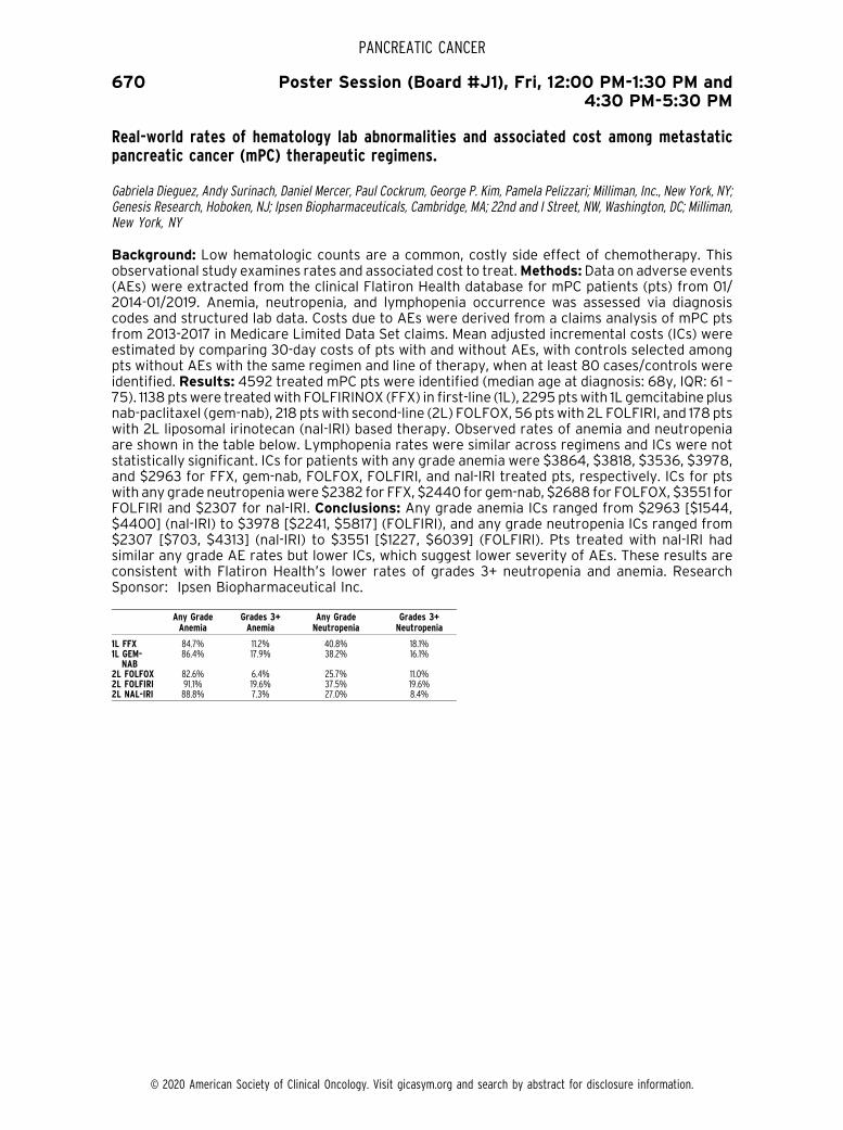
670 Poster Session (Board #J1), Fri, 12:00 PM-1:30 PM and4:30 PM-5:30 PM
Real-world rates of hematology lab abnormalities and associated cost among metastaticpancreatic cancer (mPC) therapeutic regimens.
Gabriela Dieguez, Andy Surinach, Daniel Mercer, Paul Cockrum, George P. Kim, Pamela Pelizzari; Milliman, Inc., New York, NY;Genesis Research, Hoboken, NJ; Ipsen Biopharmaceuticals, Cambridge, MA; 22nd and I Street, NW, Washington, DC; Milliman,New York, NY
Background: Low hematologic counts are a common, costly side effect of chemotherapy. Thisobservational study examines rates and associated cost to treat.Methods:Data on adverse events(AEs) were extracted from the clinical Flatiron Health database for mPC patients (pts) from 01/2014-01/2019. Anemia, neutropenia, and lymphopenia occurrence was assessed via diagnosiscodes and structured lab data. Costs due to AEs were derived from a claims analysis of mPC ptsfrom 2013-2017 in Medicare Limited Data Set claims. Mean adjusted incremental costs (ICs) wereestimated by comparing 30-day costs of pts with and without AEs, with controls selected amongpts without AEs with the same regimen and line of therapy, when at least 80 cases/controls wereidentified. Results: 4592 treated mPC pts were identified (median age at diagnosis: 68y, IQR: 61 –75). 1138 ptswere treatedwith FOLFIRINOX (FFX) in first-line (1L), 2295 ptswith 1L gemcitabine plusnab-paclitaxel (gem-nab), 218 ptswith second-line (2L) FOLFOX, 56 ptswith 2L FOLFIRI, and 178 ptswith 2L liposomal irinotecan (nal-IRI) based therapy. Observed rates of anemia and neutropeniaare shown in the table below. Lymphopenia rates were similar across regimens and ICs were notstatistically significant. ICs for patients with any grade anemia were $3864, $3818, $3536, $3978,and $2963 for FFX, gem-nab, FOLFOX, FOLFIRI, and nal-IRI treated pts, respectively. ICs for ptswith any grade neutropeniawere $2382 for FFX, $2440 for gem-nab, $2688 for FOLFOX, $3551 forFOLFIRI and $2307 for nal-IRI. Conclusions: Any grade anemia ICs ranged from $2963 [$1544,$4400] (nal-IRI) to $3978 [$2241, $5817] (FOLFIRI), and any grade neutropenia ICs ranged from$2307 [$703, $4313] (nal-IRI) to $3551 [$1227, $6039] (FOLFIRI). Pts treated with nal-IRI hadsimilar any grade AE rates but lower ICs, which suggest lower severity of AEs. These results areconsistent with Flatiron Health’s lower rates of grades 3+ neutropenia and anemia. ResearchSponsor: Ipsen Biopharmaceutical Inc.
Any GradeAnemia
Grades 3+Anemia
Any GradeNeutropenia
Grades 3+Neutropenia
1L FFX 84.7% 11.2% 40.8% 18.1%1L GEM-NAB
86.4% 17.9% 38.2% 16.1%
2L FOLFOX 82.6% 6.4% 25.7% 11.0%2L FOLFIRI 91.1% 19.6% 37.5% 19.6%2L NAL-IRI 88.8% 7.3% 27.0% 8.4%
© 2020 American Society of Clinical Oncology. Visit gicasym.org and search by abstract for disclosure information.
PANCREATIC CANCER
671 Poster Session (Board #J2), Fri, 12:00 PM-1:30 PM and4:30 PM-5:30 PM and Poster Walks, Fri, 4:45 PM-5:30 PM
Long-term outcome after combined carbon-ion radiotherapy and chemotherapy for locallyadvanced pancreatic cancer.
Makoto Shinoto, Hidenari Hirata, Hiroaki Suefuji, Shingo Toyama, Minoru Ueda; Ion Beam Therapy Center, SAGA HIMATFoundation, Tosu, Japan
Background: Carbon-ion radiotherapy (C-ion RT) has the potential advantages in terms ofimproved dose localization and enhanced biological effectiveness compared to conventionalradiotherapy or proton therapy. C-ion RT is expected to contribute to the prolongation of survivalin patients with pancreatic ductal adenocarcinoma (PDAC). We started C-ion RT for pancreaticcancer at SAGA HIMAT since April 2014. The aim of this study is to evaluate the long-term clinicalresults of C-ion RT for locally advanced PDAC. Methods: From April 2014 to March 2018, 144patients with pancreatic cancer were treated with definitive C-ion RT. 80 patients who wereconfirmed as unresectable locally advanced PDAC were included in this retrospective analysis.C-ion RT was performed with 55.2 Gy (RBE) at 12 fractions in 3 weeks. Overall survival (OS) andprognostic factors were analyzed. Toxicities were evaluated using the CTCAE ver. 5.0. Results:Themedian follow-up period for survivors was 38 (range 13-59) months from the initiation of C-ionRT. In all patients, planned C-ion RT was completed. Induction chemotherapy was performed in 64patients (80%) and the median duration time was 3 (range 1-19) months. Seventy patients (86%)underwent concurrent chemotherapy with gemcitabine or S-1. 2y-, 3y-, and 4y-OS from C-ion RTwere 47% (95%CI, 37-59%), 25% (95%CI,16-37%), and 18% (95%CI, 10-30%), respectively. 2y-,3y-, and 4y-OS from initial treatment were 55% (95%CI, 45-66%), 33% (95%CI, 24-45%), and20% (95%CI, 12-32%). Multivariate analysis showed that PS 1-2, CA19-9$350, and the absence ofconcurrent chemotherapy were independent prognostic factors on OS. Only seven patients (9%)experienced grade 3 toxicities that were gastrointestinal ulcer/bleeding (n = 4), anorexia (n = 1),leukopenia (n = 1), and neutropenia (n = 1). Therewas no grade4 or 5 toxicity.Conclusions:C-ion RTfor locally advanced PDAC was effective and well-tolerated. Research Sponsor: None.
© 2020 American Society of Clinical Oncology. Visit gicasym.org and search by abstract for disclosure information.
PANCREATIC CANCER
672 Poster Session (Board #J3), Fri, 12:00 PM-1:30 PM and4:30 PM-5:30 PM
Neoadjuvant therapy (NAT) and its role for pancreatic adenocarcinoma (PC) in the current era:Institutional experience.
Emily Walzer, Mengying Deng, Elizabeth A. Handorf, Aryeh Blumenreich, Lauren Pease, Marianna Dorta, Kristen Sorice,Shannon M. Lynch, Namrata Vijayvergia, Efrat Dotan; Temple University Hospital, Philadelphia, PA; Fox Chase Cancer Center,Philadelphia, PA; Lewis Katz School of Medicine at Temple University, Philadelphia, PA
Background: PC affects 57,000 people in the U.S. annually with poor long-term outcomes. NAT forlocalized disease has increasingly been used but lacks robust prospective data. We investigated thedisease course and outcomes for patients (pts) undergoing NAT versus upfront surgery for PC at ahigh-volume academic center. Methods: Utilizing our IRB-approved retrospective database ofmetastatic PC pts (year 2000-2017), we identified pts who presented with localized disease andwere considered for surgery and present the baseline tumor and treatment characteristics here.Fisher’s exact and Wilcoxon Rank-Sum tests were used for categorical data and Kaplan Meier (KM)curves for survival data when comparing those who had upfront surgery versus surgery followingNAT.Results: 352 pts with localized disease at diagnosis were included in our analysis withmedianage of 65 y (range 38-89) and 45% females. NAT was used in 225 (64%) pts while 109 pts (31%)had upfront surgery and 18 pts (5%) received no treatment. Adjuvant therapy was given to 77% ofpts after upfront surgery and 48% of pts after surgery following NAT. NAT regimen consisted ofchemotherapy (CTx) and radiation for 48%, CTx alone for 8% and radiotherapy alone for 44%of pts. Of those receiving CTx, 24% received triple agent while 51% and 25% received dual andsingle agent therapy. Pt factors (age, CCI, gender, BMI, smoking status, race) did not differ betweenthose receiving upfront surgery and surgery following NAT but upfront surgery was associatedwith a lower stage at diagnosis (p, 0.0001). Surgical resection after NAT occurred in 79 pts (35%)with median overall survival of 26.3m vs 19.7m (p = 0.06) in those who had upfront surgery.Survival rates at year 1, 3, and 5 years were 94%, 34%, and 8% for those with NAT followed bysurgery vs 76%, 17%, and 11% for thosewith upfront surgery (p = 0.06).Conclusions:Use of NAT isprevalent, yet only 35% of pts make it to surgical resection. Survival was improved for pts whounderwent resection following NAT versus upfront, although the difference was not statisticallysignificant. Additional research is warranted to define the optimal NAT approach for pts withborderline resectable PC. Research Sponsor: None.
© 2020 American Society of Clinical Oncology. Visit gicasym.org and search by abstract for disclosure information.
PANCREATIC CANCER
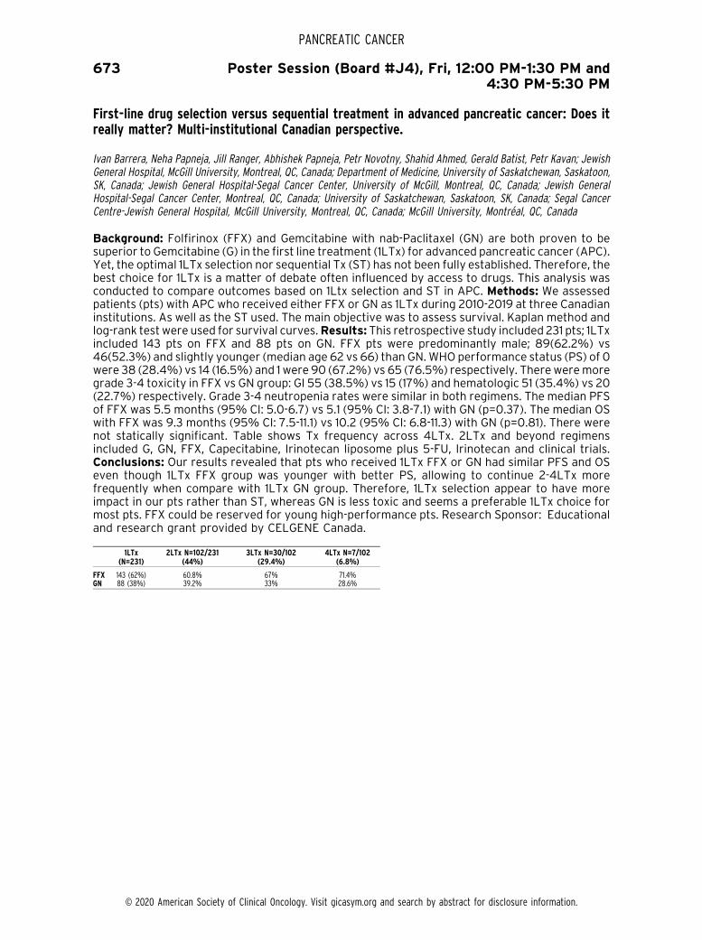
673 Poster Session (Board #J4), Fri, 12:00 PM-1:30 PM and4:30 PM-5:30 PM
First-line drug selection versus sequential treatment in advanced pancreatic cancer: Does itreally matter? Multi-institutional Canadian perspective.
Ivan Barrera, Neha Papneja, Jill Ranger, Abhishek Papneja, Petr Novotny, Shahid Ahmed, Gerald Batist, Petr Kavan; JewishGeneral Hospital, McGill University, Montreal, QC, Canada; Department of Medicine, University of Saskatchewan, Saskatoon,SK, Canada; Jewish General Hospital-Segal Cancer Center, University of McGill, Montreal, QC, Canada; Jewish GeneralHospital-Segal Cancer Center, Montreal, QC, Canada; University of Saskatchewan, Saskatoon, SK, Canada; Segal CancerCentre-Jewish General Hospital, McGill University, Montreal, QC, Canada; McGill University, Montreal, QC, Canada
Background: Folfirinox (FFX) and Gemcitabine with nab-Paclitaxel (GN) are both proven to besuperior to Gemcitabine (G) in the first line treatment (1LTx) for advanced pancreatic cancer (APC).Yet, the optimal 1LTx selection nor sequential Tx (ST) has not been fully established. Therefore, thebest choice for 1LTx is a matter of debate often influenced by access to drugs. This analysis wasconducted to compare outcomes based on 1Ltx selection and ST in APC. Methods: We assessedpatients (pts) with APC who received either FFX or GN as 1LTx during 2010-2019 at three Canadianinstitutions. As well as the ST used. The main objective was to assess survival. Kaplan method andlog-rank test were used for survival curves.Results: This retrospective study included 231 pts; 1LTxincluded 143 pts on FFX and 88 pts on GN. FFX pts were predominantly male; 89(62.2%) vs46(52.3%) and slightly younger (median age 62 vs 66) than GN.WHO performance status (PS) of 0were 38 (28.4%) vs 14 (16.5%) and 1 were 90 (67.2%) vs 65 (76.5%) respectively. There weremoregrade 3-4 toxicity in FFX vs GN group: GI 55 (38.5%) vs 15 (17%) and hematologic 51 (35.4%) vs 20(22.7%) respectively. Grade 3-4 neutropenia rates were similar in both regimens. The median PFSof FFX was 5.5 months (95% CI: 5.0-6.7) vs 5.1 (95% CI: 3.8-7.1) with GN (p=0.37). The median OSwith FFX was 9.3 months (95% CI: 7.5-11.1) vs 10.2 (95% CI: 6.8-11.3) with GN (p=0.81). There werenot statically significant. Table shows Tx frequency across 4LTx. 2LTx and beyond regimensincluded G, GN, FFX, Capecitabine, Irinotecan liposome plus 5-FU, Irinotecan and clinical trials.Conclusions: Our results revealed that pts who received 1LTx FFX or GN had similar PFS and OSeven though 1LTx FFX group was younger with better PS, allowing to continue 2-4LTx morefrequently when compare with 1LTx GN group. Therefore, 1LTx selection appear to have moreimpact in our pts rather than ST, whereas GN is less toxic and seems a preferable 1LTx choice formost pts. FFX could be reserved for young high-performance pts. Research Sponsor: Educationaland research grant provided by CELGENE Canada.
1LTx(N=231)
2LTx N=102/231(44%)
3LTx N=30/102(29.4%)
4LTx N=7/102(6.8%)
FFX 143 (62%) 60.8% 67% 71.4%GN 88 (38%) 39.2% 33% 28.6%
© 2020 American Society of Clinical Oncology. Visit gicasym.org and search by abstract for disclosure information.
PANCREATIC CANCER
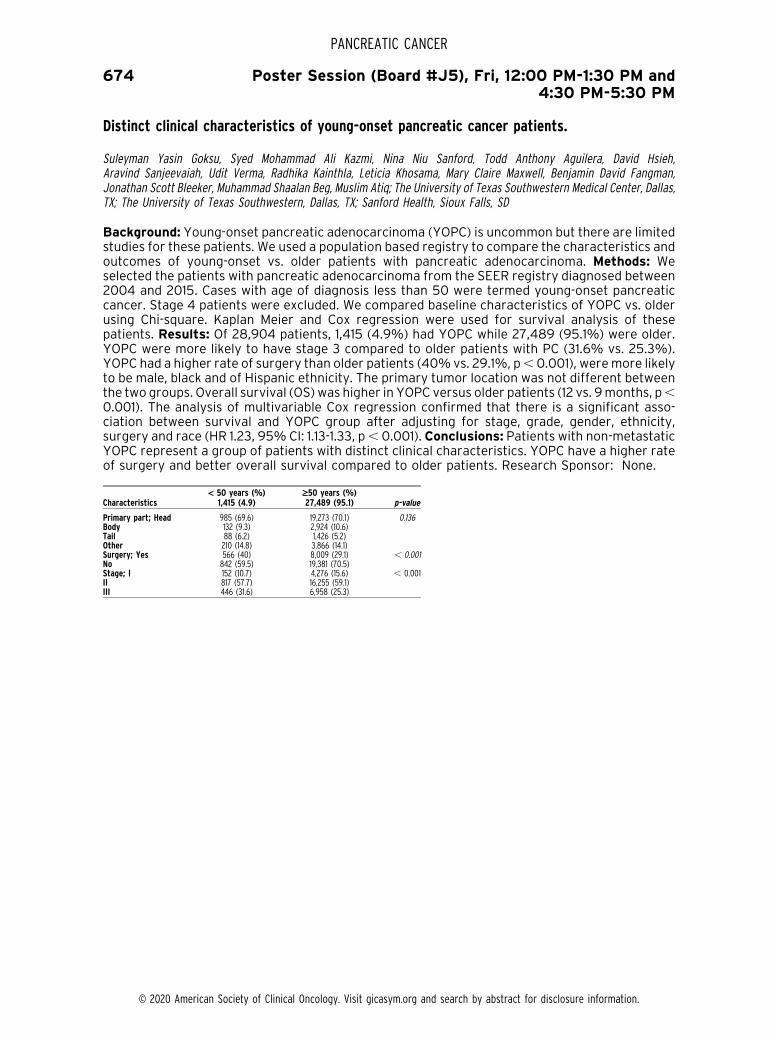
674 Poster Session (Board #J5), Fri, 12:00 PM-1:30 PM and4:30 PM-5:30 PM
Distinct clinical characteristics of young-onset pancreatic cancer patients.
Suleyman Yasin Goksu, Syed Mohammad Ali Kazmi, Nina Niu Sanford, Todd Anthony Aguilera, David Hsieh,Aravind Sanjeevaiah, Udit Verma, Radhika Kainthla, Leticia Khosama, Mary Claire Maxwell, Benjamin David Fangman,Jonathan Scott Bleeker, Muhammad Shaalan Beg, Muslim Atiq; The University of Texas Southwestern Medical Center, Dallas,TX; The University of Texas Southwestern, Dallas, TX; Sanford Health, Sioux Falls, SD
Background: Young-onset pancreatic adenocarcinoma (YOPC) is uncommon but there are limitedstudies for these patients. We used a population based registry to compare the characteristics andoutcomes of young-onset vs. older patients with pancreatic adenocarcinoma. Methods: Weselected the patients with pancreatic adenocarcinoma from the SEER registry diagnosed between2004 and 2015. Cases with age of diagnosis less than 50 were termed young-onset pancreaticcancer. Stage 4 patients were excluded. We compared baseline characteristics of YOPC vs. olderusing Chi-square. Kaplan Meier and Cox regression were used for survival analysis of thesepatients. Results: Of 28,904 patients, 1,415 (4.9%) had YOPC while 27,489 (95.1%) were older.YOPC were more likely to have stage 3 compared to older patients with PC (31.6% vs. 25.3%).YOPC had a higher rate of surgery than older patients (40% vs. 29.1%, p, 0.001), weremore likelyto be male, black and of Hispanic ethnicity. The primary tumor location was not different betweenthe two groups. Overall survival (OS) was higher in YOPC versus older patients (12 vs. 9months, p,0.001). The analysis of multivariable Cox regression confirmed that there is a significant asso-ciation between survival and YOPC group after adjusting for stage, grade, gender, ethnicity,surgery and race (HR 1.23, 95% CI: 1.13-1.33, p, 0.001). Conclusions: Patients with non-metastaticYOPC represent a group of patients with distinct clinical characteristics. YOPC have a higher rateof surgery and better overall survival compared to older patients. Research Sponsor: None.
Characteristics< 50 years (%)
1,415 (4.9)‡50 years (%)27,489 (95.1) p-value
Primary part; Head 985 (69.6) 19,273 (70.1) 0.136Body 132 (9.3) 2,924 (10.6)Tail 88 (6.2) 1,426 (5.2)Other 210 (14.8) 3,866 (14.1)Surgery; Yes 566 (40) 8,009 (29.1) , 0.001No 842 (59.5) 19,381 (70.5)Stage; I 152 (10.7) 4,276 (15.6) , 0.001II 817 (57.7) 16,255 (59.1)III 446 (31.6) 6,958 (25.3)
© 2020 American Society of Clinical Oncology. Visit gicasym.org and search by abstract for disclosure information.
PANCREATIC CANCER
675 Poster Session (Board #J6), Fri, 12:00 PM-1:30 PM and4:30 PM-5:30 PM
Clinicopathological features of pancreatic cancer-related diabetes.
Michael Lee, Hui-Li Wong, Erica S. Tsang, Sean McKay Fenlon Addison, James T. Topham, Joanna Karasinska, Steve Kalloger,Jonathan M. Loree, David F. Schaeffer, Daniel John Renouf; BC Cancer, Vancouver, BC, Canada; Royal Melbourne Hospital,Melbourne, Australia; BC Cancer Agency, Vancouver, BC, Canada; Pancreas Centre BC, Vancouver, BC, Canada; Department ofPathology & Laboratory Medicine Vancouver General Hospital, Vancouver, BC, Canada
Background: Epidemiological studies suggest pancreatic ductal adenocarcinoma (PDAC) may bestrongly interrelated with diabetes. However, little is known about the clinicopathological featuresof pancreatic cancer related diabetes.Methods:A retrospective chart reviewwas undertaken of allpatients with advanced PDAC treated with at least one cycle of palliative chemotherapy at BCCancer, Vancouver between Jan 2012- Dec 2015. Diagnosis of diabetes was determined byconsultation documentation and/or fasting glucose . 7mmol/L or HbA1c . 48mmol/L. Peri-pancreatic diabetes is defined as diabetes diagnosis , 3 years prior to PDAC diagnosis. Results:578 patients were identified with median age 66 (49-81), 54.6% male, 39.5% non-smoker and63.5% ECOG 0/1. 27.3% confirmed diabetics, of which 75.8% (119/157) have peripancreaticdiabetes. At initial diagnosis, 11.2% were deemed upfront resectable, 44.0% borderline/locallyadvanced, and 55.1% metastatic. Median overall survival (OS) for the cohort based on stage ofdisease at initial diagnosis for borderline, locally advanced and metastatic was 22 months (16.1-27.9), 12 months (10.1-13.9) and 6 months (5.0-7.0) respectively. There was no association withdiabetes status and OS noted (p = 0.58). Statistical differences were noted in BMI (24.1 v 26.1, p =0.003), and proportion of Charlson comorbidity index (CCI) of 2 (2.2 v 88.3%, p , 0.01) betweennon-diabetic and diabetic patients respectively. Statistical difference between peripancreaticdiabetes compared to long-term diabetes were noted in resectable status (18.6 v 7.6%, p =0.048), weight loss. 2kg (78.6 v 60.5%, p = 0.035), hypertension (25.9 v 59.8%, p = 0.002) anddyslipidemia (18.5 v 42.7%, p = 0.024). Conclusions: The majority of patients diagnosed withadvanced PDAC with diabetes appeared to develop diabetes within 3 years prior to diagnosis.Further studies to assess the potential role of pancreatic cancer screening investigations in newlydiagnosed diabetics are warranted. Research Sponsor: None.
© 2020 American Society of Clinical Oncology. Visit gicasym.org and search by abstract for disclosure information.
PANCREATIC CANCER
676 Poster Session (Board #J7), Fri, 12:00 PM-1:30 PM and4:30 PM-5:30 PM
Changing the landscape of germline testing in patients with pancreatic adenocarcinoma.
Aly Athens, Lisa Amacker-North, Kelly Warsinske, Kunal C. Kadakia, Edward S. Kim, Mohamed E. Salem, Sara Elrefai; LevineCancer Institute-Atrium Health, Charlotte, NC; University of Michigan Comprehensive Cancer Center, Ann Arbor, MI; LevineCancer Institute, Charlotte, NC; Georgetown Lombardi Comprehensive Cancer Center, Washington, DC
Background: Results from the POLO trial demonstrated the benefits of PARP inhibition in patients(pts) with germline BRCA-mutated metastatic pancreatic cancer (PC). In 2018, ASCO and NCCNupdated their guidelines to recommend that pts with a personal history of PC undergo germlinetesting. We examined referral patterns and frequency of germline pathogenic variants in pts withPC.Methods: A retrospective review was performed of PC pts seen at the Levine Cancer Institute(LCI) Center for Genetics between January 2010 and September 2019. Descriptive analyses werecompleted on demographics and appointment outcomes. Results: A total of 201 PC pts werereferred; 20 canceled and 14 no-showed their appointment. The remaining 167 were seen andincluded in this analysis. Most pts (59%) were referred after July 2018. The median age was 65years (range 32-90) and 19% were , 50 years. The majority of pts were female (61%). Race wasmost often reported as white (72%) followed by black (20%). Reported family histories were asfollows: 28 (17%) claimed at least one first-degree relative with PC; 54 (32%) claimed a first,second, or third-degree relative with PC; 24 (14%) had no known family history of PC; and 95 (57%)claimed a first-degree relative with another cancer (breast [37], prostate [25], colon [18], ovarian[9], uterine [6], and gastric [2]). Germline testing was pursued by 138 (83%) pts: 25 (18%) werefound to have a pathogenic variant and 50 (36%) a variant of uncertain significance. Pathogenicvariants were most commonly identified in ATM (24%), BRCA2 (20%), PALB2 (12%), and CDKN2A(8%). Variants were also observed in DIS3L2, HOXB13, MITF, MUTYH (heterozygote), NTHL1(compound heterozygote), RAD50, PRSS1, and SDHA. Among pts that had a pathogenic variant,cascade testing was performed in 11 families (44%) for 29 individuals. Conclusions: Our datasuggest that the referral of PC pts to genetics has increased following updated ASCO/NCCNguidelines. However, improved adherence to genetic counseling is needed. ATM and BRCA2 werethe most common germline mutations observed. More effort to increase awareness of genetictesting and its potential implications for pts and their families is warranted and might reducecancellations and missed visits. Research Sponsor: None.
© 2020 American Society of Clinical Oncology. Visit gicasym.org and search by abstract for disclosure information.
PANCREATIC CANCER
678 Poster Session (Board #J9), Fri, 12:00 PM-1:30 PM and4:30 PM-5:30 PM
Implementation of systematic genetic counseling (GC) and multigene germline testing (MGT) forpancreatic cancer (PC) patients (pts).
Matthew B. Yurgelun, Anu B. Chittenden, Chinedu I. Ukaegbu, Kimberly Perez, Audrey P. Madigan, Shraddha Gaonkar,James M. Cleary, Andrew Aguirre, Brian M. Wolpin, Sapna Syngal; Dana-Farber Cancer Institute, Boston, MA
Background: MGT identifies cancer susceptibility gene variants in 4-10% of unselected PC pts.Such data have prompted national guidelines to recommend GC and MGT of all PC pts, but thebenefits and barriers to implementing systematic testing are unknown. This study’s aim was tostudy the implementation of universal GC for all PC pts seen in an academic oncology practice.Methods: In 12/2016, all Dana-Farber Cancer Institute (DFCI) gastrointestinal oncologists wererecommended to refer all PC pts for GC andMGT. In 10/2018, workflows were changed such that PCpatients were automatically scheduled for GC consultation on the same day as their initialoncologic evaluation (unless patients opted out), rather than relying on provider referral. Clinicaland germline data were collected on a consecutive cohort of PC pts undergoing GC and MGT from3/1/2017-3/31/2019. Two additional months (4/1/2019-5/31/2019) were collected for clinical qualityassessment purposes. Results: 1305 (48.3/month) PC pts were seen for oncologic new patientvisits, 318 (25.1%; 12.1/month) of whom underwent GC. Rates of GC/MGT increased significantlyafter the 10/2018workflow change (8.2 PC pts/month [17.2%of all newPC pts seen] versus 20.3 PCpts/month, [40.9%of all new PC pts seen]; p,0.01). Of the 318 PC pts who underwent GC, 29 (9.1%;95% CI 6.4-11.9%; 2.2% of all PC pts seen) were found to carry germline PC susceptibility genemutations on MGT. Rates of mutation carrier identification increased after the clinical workflowchange from 0.79mutation carriers/month (1.6% of all new PC pts seen) to 1.75 mutation carriers/month (3.5% of all new PC pts seen). The majority of identified mutation carriers have eitherreceived therapy targeted towards their germline mutation or are undergoing first-line palliativesystemic therapy with potential for future targeted therapy. Conclusions: Clinical implementationof routine GC/MGT in PC pts is feasible and results in the detection ofmutations that are actionablefor PC pts and at-risk family members. Systematized workflows for GC evaluation not reliant onactive referral result in markedly higher uptake of MGT andmutation carrier identification. Clinicaltrial information: NCT03060720. Research Sponsor: U.S. National Institutes of HealthDana-FarberCancer Institute Department of Medical Oncology Research Award.
© 2020 American Society of Clinical Oncology. Visit gicasym.org and search by abstract for disclosure information.
PANCREATIC CANCER
679 Poster Session (Board #J10), Fri, 12:00 PM-1:30 PM and4:30 PM-5:30 PM and Poster Walks, Fri, 4:45 PM-5:30 PM
Development and validation of a pancreatic cancer prediction model from electronic healthrecords using machine learning.
Limor Appelbaum, Jose Pablo Cambronero, Karla Pollick, George Silva, Jennifer P. Stevens, Harvey J. Mamon,Irving D. Kaplan, Martin Rinard; Beth Israel Deaconess Medical Center, Boston, MA; Massachusetts Institute of Technology,Cambridge, MA; Dana-Farber Cancer Institute, Boston, MA
Background: Pancreatic Adenocarcinoma (PDAC) is often diagnosed at an advanced stage. Wesought to develop a model for early PDAC prediction in the general population, using electronichealth records (EHRs) and machine learning. Methods: We used three EHR datasets from Beth-Israel Deaconess Medical Center (BIDMC) and Partners Healthcare (PHC): 1. “BIDMC-Development-Data” (BIDMC-DD) for model development, using a feed-forward neural network (NN) and L2-regularized logistic regression,randomly split (80:20) into training and test groups. We tunedhyperparameters using cross-validation in training, and report performance on the test split. 2.“BIDMC-Large-Data” (BIDMC-LD) to re-fit and calibrate models. 3. “PHC-Data” for external val-idation. We evaluate using Area Under the Receiver Operating Characteristic Curve (AUC) andcompute 95% CI using empirical bootstrap over test data. PDAC patients were selected usingICD9/-10 codes and validated with tumor registries. In contrast to prior work, we did not predefinefeature sets based on known clinical correlates and instead employed data-driven feature selec-tion, specifically importance-based feature pruning, regularization, and manual validation, toidentify diagnostic-based features. Results: BIDMC-DD included demographics, diagnoses, labsand medications for 1018 patients (cases = 509; age-sex paired controls). BIDMC-LD includeddiagnoses for 547,917 patients (cases = 509), and PHC included diagnoses for 160,593 patients(cases = 408). We compared our approach to adapted and re-fitted published baselines. With a365-day lead-time, NN obtained a BIDMC-DD test AUC of 0.84 (CI 0.79 - 0.90) versus the previousbest baseline AUC of 0.70 (CI 0.62 - 0.78). We also validated using BIDMC-DD’s test cancer patientsand BIDMC LD controls. The AUC was 0.71 (CI 0.67 - 0.76) at the 365-day cutoff. NN’s externalvalidation AUC on PHC-Data was 0.71 (CI 0.63 - 0.79), outperforming an existing model’s AUC of0.61 (CI 0.52 - 0.70) (Baecker et al, 2019). Conclusions: Models based on data-driven featureselection outperform models that use predefined sets of known clinical correlates and can help inearly prediction of PDAC development. Research Sponsor: None.
© 2020 American Society of Clinical Oncology. Visit gicasym.org and search by abstract for disclosure information.
PANCREATIC CANCER
680 Poster Session (Board #J11), Fri, 12:00 PM-1:30 PM and4:30 PM-5:30 PM
Case series examining somatic test results for patients with hereditary cancer syndromesassociated with gastrointestinal cancer risk.
Kristen Pauley, Cathryn Koptiuch, Samantha Greenberg, Gammon Amanda, Christopher Nevala-Plagemann, Jennie Vagher,Whitney Espinel, Glynn Weldon Gilcrease, Wendy Kohlmann, Ignacio Garrido-Laguna; Huntsman Cancer Institute, Universityof Utah, Salt Lake City, UT; Huntsman Cancer Institute, Salt Lake City, UT; University of Utah and Huntsman Cancer Institute,Salt Lake City, UT
Background: Somatic tumor testing may identify germline pathogenic variants (PV) associatedwith cancer predisposition syndromes. Labs differ whether they offer somatic only or pairedgermline analysis. Methods used by somatic testing labs, even those that include germline analysis,differ from designated germline labs that have optimized the identification of germline PV.Methods: Chart reviews were performed for patients who had testing through both somaticand designated germline laboratories. Cases with discrepant results in which germline PVwere notdetected by the somatic laboratory are summarized. Results: Nine cases with discrepant results.Five had paired germline testing and 4 somatic testing only. All 9 patients met the criteria toundergo designated germline testing, either for Lynch syndrome (3) or BRCA1/2 testing (6), basedon personal and/or family history. Designated germline testing identified 4 MLH1, 1 BRCA1, 2 ATM,1MUTYH and 1 RAD50 PV not reported by the somatic labs’ tumor or germline analysis; 2 MLH1 PVwere called variants of uncertain significance by somatic testing but classified as PV by ClinVar anddesignated germline labs. Three PV identified by designated germline labs are targets for PARPinhibitors and resulted in different treatment options. Three of the MLH1 PV were identified inpatients meeting Lynch Syndrome test criteria while 1 was identified in a patient meeting BRCA1/2criteria. Among the 5 other patientsmeeting BRCA1/2 test criteria, 3 had PV in breast cancer genes(2 ATM, 1 BRCA1) and 2 had PV in other cancer genes (MUTYH and RAD50) not reported by thesomatic labs, highlighting the importance of panel testing. Conclusions:Methods used by somaticlabs, regardless of inclusion of germline analysis, are not equivalent to those of designatedgermline labs. Overlooked germline PV may miss identification of hereditary syndromes andtargeted therapy opportunities (e.g. Anti-PD1 immunotherapy, PARP inhibitors). Patients meetingcriteria for genetic evaluation should be referred for designated germline testing regardless ofsomatic testing outcomes. Research Sponsor: U.S. National Institutes of Health.
© 2020 American Society of Clinical Oncology. Visit gicasym.org and search by abstract for disclosure information.
PANCREATIC CANCER
681 Poster Session (Board #J12), Fri, 12:00 PM-1:30 PM and4:30 PM-5:30 PM
Survival and quality of life after isolated upper abdominal perfusion with chemofiltration (UAP-F)for stage III and IV pancreatic cancer.
Karl R Aigner, Sabine Gailhofer, Emir Selak, Kornelia Aigner; Medias Klinikum Burghausen, Burghausen, Germany
Background: In order to increase response and survival rates of advanced pancreatic cancer atgood quality of life, UAP-F was clinically tested. We report on feasibility, safety, overall survival andquality of life of 79 patients in stage III and 142 patients in stage IV submitted to UAP withsubsequent chemofiltration for lowering of systemic drug exposure. Methods: This technique isachieved with stop-flow balloon catheters introduced via the femoral artery and vein. The arterialballoon is placed below the celiac axis and proceeded beneath the diaphragm after injection of athree-drug combination consisting of cisplatin (CDDP), adriamycin (ADM) and mitomycin (MMC).Because of tenfold increased cytotoxicity of ADM and MMC, the perfusion is performed underhypoxic condition. Perfusion time is 15 minutes followed by 30 – 45 minutes of chemofiltration.Results:Median survival was 12.1 months and 8.7 months in stage III and IV respectively. One-yearsurvival was 49.4% and 37% for stage III and IV, and 3-years survival 21.7% and 7.7% for stage IIIand IV respectively. Cisplatin levels in the arterial circuit amounted to 60.000 ng/ml and 6.000 ng/ml in the venous line. Resolution of ascites was achieved within two therapies in 33/36 cases withUAP-F. Toxicity was generallymild, not exceedingWHOgrade II and amounted to grade III or IV onlyin patients after prior severe systemic chemotherapy. Conclusions: Upper abdominal perfusionwith chemofiltration is a safe technique for advanced cancers of the pancreas, increasing survivaltime and maintaining quality of life. Research Sponsor: None.
© 2020 American Society of Clinical Oncology. Visit gicasym.org and search by abstract for disclosure information.
PANCREATIC CANCER
682 Poster Session (Board #J13), Fri, 12:00 PM-1:30 PM and4:30 PM-5:30 PM
Extent of lymph node resection and effect on pancreatic cancer overall survival.
Brian Cox, Nicholas Manguso, Humair Quadri, Jessica Crystal, Katelyn Mae Atkins, Jaewon Lee, Andrew Eugene Hendifar,Mitchell Kamrava, Richard Tuli, Jun Gong, Alexandra Gangi; Cedars Sinai Medical Center, Los Angeles, CA; Cedars-SinaiMedical Center, Department of Surgery, Los Angeles, CA; Samuel Oschin Comprehensive Cancer Institute, Cedars-SinaiMedical Center, Los Angeles, CA; Cedars-Sinai Medical Center, Los Angeles, CA; Memorial Sloan Kettering Cancer Center, NewYork, NY; City of Hope, Duarte, CA
Background: Lymph node (LN) metastases affect overall survival (OS) in pancreatic cancer (PC).However, a LN sampling threshold does not exist. We examined the impact of nodal sampling onoverall survival (OS).Methods: Patients with Stage I-III PC$55 years old who underwent curativeresection from 2004-2016 were identified from the National Cancer Database (NCDB). Afteradjusting for age, gender, grade, stage, and Charlson-Deyo score, multiple binomial logisticregression analyses assessed the impact of the LN ratio (LNR) on OS. LNR was defined as thenumber of positive LN over the number of LN examined. Regression analyses, a Cox-Regression,and a Kaplan-Meier survival curve assessed how many LN should be sampled. Results: A total of13,673 patients, median age 69 years (55-90), were included. Most were Caucasian (86.6%) maleswith Charlson-Deyo scores # 1 (90.3%) and moderately to poorly differentiated PC (90.1%).Median number of LN examined was 15 (1-75) with a median of 1 positive LN (0-35). As expected,increased number of positive LNs was associated with reduced OS, p , 0.001. After datanormalization, an increasing LNR was associated with a 12-fold likelihood of death [OR: 11.9,p, 0.001 (CI 6.0, 23.7)]. Subsequent regression models established evaluation of$ 16 LNs as thegreatest predictor of OS. A regression model evaluating, or$ 16 lymph nodes was performed toascertain the effects of age, gender, ethnicity, grade, stage, and LN examined on OS. The logisticregression model correctly classified 74.5% of cases with a specificity of 99.6% (p , 0.001).Examination of , 16 LN, Caucasian race, grade, stage, and higher Charlson-Deyo scores weresignificantly associated with decreased OS. If $ 16 LNs were examined, patients had a 1.5-foldlikelihood of better OS, p, 0.001 (CI 1.4, 1.6). An adjusted Cox Regression showed increased HR of1.2, p, 0.001 (CI 1.1, 1.2) and an unadjusted KaplanMeier survival curve predicted$ 16 LN examinedare associated with an increase in OS of 2.8 months [log-rank: 32.0, p , 0.001]. Conclusions:Patients undergoing curative intent resection for PC should have adequate nodal sampling.Stratification of patients by LNR may provide useful information of OS. Examination of $ 16LNs impacts OS in patients with Stage I-III PC. Research Sponsor: None.
© 2020 American Society of Clinical Oncology. Visit gicasym.org and search by abstract for disclosure information.
PANCREATIC CANCER
683 Poster Session (Board #J14), Fri, 12:00 PM-1:30 PM and4:30 PM-5:30 PM
Goals of care designations in advanced pancreatic cancer patients undergoing palliativechemotherapy.
Matthew Anaka, Minji Lee, Elisa Lim, Sunita Ghosh, Winson Y. Cheung, Jennifer L. Spratlin; University of Alberta, Edmonton,AB, Canada; University of Calgary, Calgary, AB, Canada; Cross Cancer Institute/University of Alberta, Edmonton, AB, Canada;BC Cancer Agency, Vancouver, BC, Canada; Alberta Health Services, Edmonton, AB, Canada
Background: Discussion of goals of care (GoC) is a key part of quality care for patients withpalliative cancer. Numerous studies have shown that documentation of GoC in this populationremains low. In 2014, Alberta Health Services launched a health-system wide initiative to providepatients with physical copies of their GoC designation intended to be available at all health-systeminteractions. Herewe describe rates of GoC documentation in the period surrounding this initiative.Methods: This is a retrospective cohort analysis of 240 patients with locally advanced or met-astatic pancreatic cancer treated with palliative chemotherapy from 2012-2015 in Alberta, Canada.Data were obtained from outpatient electronic medical record documentation and the provincialcancer registry. Results: 63.8% (153/240) of patients had a documented GoC discussion, with60.4% (145/240) receiving a specific GoC designation. 59.6% (143/240) of patients were referredto palliative care, with 32.5% (78/240) seen by palliative care physician. Of 334 individual GoCdiscussions documented, 38.6% (129/334) were by medical oncologists, 2.3% (10/334) were byradiation oncologists, 27.2% (91/334) were by palliative care, and 19.2% (64/334) were by otherinpatient physicians during hospital admissions. At least 9.6% (32/334) referenced discussionsthat occurred prior to initial consultation with an oncology physician.Conclusions: Themajority ofpancreatic cancer patients undergoing palliative chemotherapy had a documented GoC designa-tion during the study period. Providing patients with physical copies of their GoC designation maytherefore represent a simple but effective means of increasing GoC documentation in the out-patient oncology setting. Research Sponsor: None.
© 2020 American Society of Clinical Oncology. Visit gicasym.org and search by abstract for disclosure information.
PANCREATIC CANCER

684 Poster Session (Board #J15), Fri, 12:00 PM-1:30 PM and4:30 PM-5:30 PM
Risk factors for severe neutropenia among pancreatic cancer patients receiving nab-paclitaxeland gemcitabine combination therapy.
Kazuyoshi Kawakami, Genta Ito, Takeshi Aoyama, Takashi Yokokawa, Masashi Nakamura, Masato Ozaka, Naoki Sasahira,Hayato Kizaki, Masayuki Hashiguchi, Toshihiro Hama, Satoko Hori; Department of Pharmacy, Cancer Institute Hospital ofJapanese Foundation for Cancer Research, Tokyo, Japan; Division of Drug Informatics, Faculty of Pharmacy, Keio University,Tokyo, Japan; Department of Gastroenterology, Cancer Institute Hospital of Japanese Foundation for Cancer Research,Tokyo, Japan
Background: Albumin-bound paclitaxel (nab-paclitaxel) and gemcitabine combination therapy(GnP therapy) significantly extends overall survival in patients with metastatic pancreatic cancer,compared to conventional gemcitabine monotherapy. However, severe neutropenia (Grade $ 3)occurred in 67.6%of patients in the Japanese phase I/II trials of GnP therapy, and is often a limitingfactor. The purpose of this study was to identify the risk factors for severe neutropenia inpancreatic patients receiving GnP therapy in clinical settings. Methods: A retrospective studyof 222 consecutive patients with pancreatic cancer who received GnP therapy at the CancerInstitute Hospital from December 2014 to December 2016 was conducted. Univariate and multi-variate analyses were used to compare blood test values and patients’ characteristics betweenpatients with no neutropenia or Grade 1/2 (non-serious) neutropenia and those with Grade $ 3(severe) neutropenia. Results: There were 19 patients (8.6%) with modified FOLFIRINOX in theprevious treatment history. The median doses of nab-paclitaxel and gemcitabine were 192.5 mg(range 134-277.5) and 1,545 mg (range 1,000-2,220), respectively. Severe neutropenia and febrileneutropenia occurred in 118 patients (53.2%) and 15 patients (6.8%), respectively. Multivariatelogistic regression analysis indicated that ANC (absolute neutrophil count) , 3.03 x 10³ /mL (OR:4.806, 95% CI: 2.416-9.558, p = 0.000), T-Bil $ 0.6 mg/dl (OR: 1.964, 95% CI: 1.040-3.708, p =0.037) and CRP , 0.13 mg/dl (OR: 2.607, 95% CI: 1.331-5.106, p = 0.005) were significant riskfactors for severe neutropenia. The incidence rate of severe neutropenia was 85.7% (18/21) inpatients with all three identified factors, while it was 27.7% (13/47) in patients with none of them.Age was not a risk factor in either univariate or multivariate analysis. Conclusions: Low ANC, highT-Bil, and low CRP were found to be risk factors for severe neutropenia in patients receiving GnPtherapy. It would be desirable to monitor patients with these risk factors carefully, even if theirvalues are within the standard ranges. Research Sponsor: None.
© 2020 American Society of Clinical Oncology. Visit gicasym.org and search by abstract for disclosure information.
PANCREATIC CANCER
685 Poster Session (Board #J16), Fri, 12:00 PM-1:30 PM and4:30 PM-5:30 PM
The role of neoadjuvant chemotherapy in elderly patients with borderline or locally advancedpancreatic cancer: Is it safe and feasible?
Atsushi Oba, Christopher Hanyoung Lieu, Cheryl Lauren Meguid, Sarah Lindsey Davis, Alexis Diane Leal, Tom Purcell,Gentry Teng King, Karyn A. Goodman, Tracey E. Schefter, Ana Luiza Gleisner, Steven Arthur Ahrendt, Stephen Leong,Wells A. Messersmith, Richard D. Shulick, Marco Del Chiaro; Department of Surgery, University of Colorado School ofMedicine, Aurora, CO; University of Colorado Cancer Center, Aurora, CO; Albert Einstein Medical Center, Philadelphia, PA;Department of Radiation Oncology, University of Colorado School of Medicine, Aurora, CO
Background: For borderline resectable (BRPC) or locally advanced pancreatic cancer (LAPC),neoadjuvant (NAT) FOLFIRINOX or gemcitabine plus nab-paclitaxel (GnP) are standard treatmentoptions and these regimens have shown a survival advantage over single-agent gemcitabine.However, the role of these modern therapeutic regimens in elderly patients is debatable. In thisanalysis, we evaluated the outcomes of neoadjuvant treatment (NAT) with combination chemo-therapy in elderly patients. Methods: 230 consecutive patients who underwent neoadjuvanttreatment for BRPC/LAPC discussed and planned for NAT at the University of Colorado CancerCenter from January 2011 to March 2019 were reviewed. 214 patients who received FOLFIRINOX(n = 143) or GnP (n = 71) were eligible for analysis. We divided all patients into three groups (, 70,70-74, $75 years) and compared the short-term and long-term outcomes. Results: Of 214patients, patients , 70 (n = 147) received FOLFIRINOX more frequently than the other groups(p,0.001): FOLFIRINOX: 115 cases, GnP: 32 cases, 70-74 years (n = 33): FOLFIRINOX: 15 cases, GnP:18 cases, and $75 years (n = 34): FOLFIRINOX: 13 cases, GnP: 21 cases. Resection rates were notstatistically different between three groups (, 70: 62%, 70-74: 70%,$75 years: 56%, p = 0.504).There was a slight trend towards worse survival in the two older groups (Median Survival Time[MST]:, 70: 23.2 mo., 70-74: 19.5 mo.,$75 years: 17.6 mo., p = 0.075) The FOLFIRINOX group wassuperior to GnP group in all three groups (MST: , 70: 25.6 vs 18.2 mo., p = 0.017; 70-74: 33.2 vs16.1mo., p = 0.029;$75 years: not reached vs 16.1 mo., p = 0.135). There were no toxic deaths or 30day mortality after pancreatectomy in the study population. Conclusions: Neoadjuvant combi-nation chemotherapy regimens were safe and feasible for elderly patients. Neoadjuvant therapywith FOLFIRINOXwas associated with a survival advantage vs GnP and is an good option for fit andelderly patients $75 years. Research Sponsor: None.
© 2020 American Society of Clinical Oncology. Visit gicasym.org and search by abstract for disclosure information.
PANCREATIC CANCER
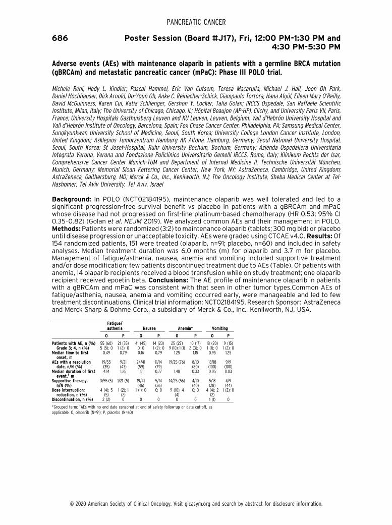
686 Poster Session (Board #J17), Fri, 12:00 PM-1:30 PM and4:30 PM-5:30 PM
Adverse events (AEs) with maintenance olaparib in patients with a germline BRCA mutation(gBRCAm) and metastatic pancreatic cancer (mPaC): Phase III POLO trial.
Michele Reni, Hedy L. Kindler, Pascal Hammel, Eric Van Cutsem, Teresa Macarulla, Michael J. Hall, Joon Oh Park,Daniel Hochhauser, Dirk Arnold, Do-Youn Oh, Anke C. Reinacher-Schick, Giampaolo Tortora, Hana Algul, Eileen Mary O’Reilly,David McGuinness, Karen Cui, Katia Schlienger, Gershon Y. Locker, Talia Golan; IRCCS Ospedale, San Raffaele ScientificInstitute, Milan, Italy; The University of Chicago, Chicago, IL; Hopital Beaujon (AP-HP), Clichy, and University Paris VII, Paris,France; University Hospitals Gasthuisberg Leuven and KU Leuven, Leuven, Belgium; Vall d’Hebron University Hospital andVall d’Hebron Institute of Oncology, Barcelona, Spain; Fox Chase Cancer Center, Philadelphia, PA; Samsung Medical Center,Sungkyunkwan University School of Medicine, Seoul, South Korea; University College London Cancer Institute, London,United Kingdom; Asklepios Tumorzentrum Hamburg AK Altona, Hamburg, Germany; Seoul National University Hospital,Seoul, South Korea; St Josef-Hospital, Ruhr University Bochum, Bochum, Germany; Azienda Ospedaliera UniversitariaIntegrata Verona, Verona and Fondazione Policlinico Universitario Gemelli IRCCS, Rome, Italy; Klinikum Rechts der Isar,Comprehensive Cancer Center Munich-TUM and Department of Internal Medicine II, Technische Universitat Munchen,Munich, Germany; Memorial Sloan Kettering Cancer Center, New York, NY; AstraZeneca, Cambridge, United Kingdom;AstraZeneca, Gaithersburg, MD; Merck & Co., Inc., Kenilworth, NJ; The Oncology Institute, Sheba Medical Center at Tel-Hashomer, Tel Aviv University, Tel Aviv, Israel
Background: In POLO (NCT02184195), maintenance olaparib was well tolerated and led to asignificant progression-free survival benefit vs placebo in patients with a gBRCAm and mPaCwhose disease had not progressed on first-line platinum-based chemotherapy (HR 0.53; 95% CI0.35–0.82) (Golan et al. NEJM 2019). We analyzed common AEs and their management in POLO.Methods:Patients were randomized (3:2) tomaintenance olaparib (tablets; 300mg bid) or placebountil disease progression or unacceptable toxicity. AEswere graded using CTCAE v4.0.Results:Of154 randomized patients, 151 were treated (olaparib, n=91; placebo, n=60) and included in safetyanalyses. Median treatment duration was 6.0 months (m) for olaparib and 3.7 m for placebo.Management of fatigue/asthenia, nausea, anemia and vomiting included supportive treatmentand/or dosemodification; few patients discontinued treatment due to AEs (Table). Of patients withanemia, 14 olaparib recipients received a blood transfusion while on study treatment; one olaparibrecipient received epoetin beta. Conclusions: The AE profile of maintenance olaparib in patientswith a gBRCAm and mPaC was consistent with that seen in other tumor types.Common AEs offatigue/asthenia, nausea, anemia and vomiting occurred early, were manageable and led to fewtreatment discontinuations. Clinical trial information: NCT02184195. Research Sponsor: AstraZenecaand Merck Sharp & Dohme Corp., a subsidiary of Merck & Co., Inc., Kenilworth, NJ, USA.
Fatigue/asthenia Nausea Anemia* Vomiting
O P O P O P O P
Patients with AE, n (%) 55 (60) 21 (35) 41 (45) 14 (23) 25 (27) 10 (17) 18 (20) 9 (15)Grade 3; 4, n (%) 5 (5); 0 1 (2); 0 0; 0 1 (2); 0 9 (10); 1 (1) 2 (3); 0 1 (1); 0 1 (2); 0
Median time to firstonset, m
0.49 0.79 0.16 0.79 1.25 1.15 0.95 1.25
AEs with a resolutiondate, n/N (%)
19/55(35)
9/21(43)
24/41(59)
11/14(79)
19/25 (76) 8/10(80)
18/18(100)
9/9(100)
Median duration of firstevent,† m
4.14 1.25 1.51 0.77 1.48 0.33 0.05 0.03
Supportive therapy,n/N (%)
3/55 (5) 1/21 (5) 19/41(46)
5/14(36)
14/25 (56) 4/10(40)
5/18(28)
4/9(44)
Dose interruption;reduction, n (%)
4 (4); 5(5)
1 (2); 1(2)
1 (1); 0 0; 0 9 (10); 4(4)
0; 0 4 (4); 2(2)
1 (2); 0
Discontinuation, n (%) 2 (2) 0 0 0 0 0 1 (1) 0
*Grouped term; †AEs with no end date censored at end of safety follow-up or data cut-off, asapplicable. O, olaparib (N=91); P, placebo (N=60)
© 2020 American Society of Clinical Oncology. Visit gicasym.org and search by abstract for disclosure information.
PANCREATIC CANCER
687 Poster Session (Board #J18), Fri, 12:00 PM-1:30 PM and4:30 PM-5:30 PM
Outcomes of advanced gastrointestinal (GI) cancer patients in relationship to opioid use: Anindividual patient data pooled analysis from eight clinical trials.
Omar M Abdel-Rahman Abdelsalam, Hatim Karachiwala, Jacob C. Easaw; Cross Cancer Institute, Edmonton, AB, Canada
Background: The current study aims at assessing the patterns of opioid use, and evaluating theimpact of opioid use on survival outcomes among patients with advanced GI cancers who wereincluded in eight clinical trials.Methods:De-identified datasets of eight clinical trials evaluating first-line systemic treatment for advanced GI cancers (NCT01124786; NCT00844649; NCT00290966;NCT00678535; NCT00699374; NCT00272051; NCT00305188; NCT00384176)were accessed fromthe Project Data Sphere platform. These trials evaluated patients with pancreatic, gastric,hepatocellular and colorectal carcinoma. Multivariable logistic regression analysis was usedto evaluate factors predicting the use of opioids. Kaplan-Meier survival estimates were used tocompare survival outcomes in each disease entity among patients who did or did not receiveopioid treatment. Multivariable Cox regression analysis was used to assess the impact of opioiduse on survival outcomes in each disease entity. Results: A total of 3441 participants wereincluded in the current analysis. The following factors predicted a higher probability of opioid usewithin logistic regression analysis: younger age (P = 0.004), non-white race (P = 0.010), higherECOG score (P, 0.001) and pancreatic primary site (P, 0.001). Use of opioids was consistentlyassociated with worse overall survival in Kaplan-Meier survival estimates of each disease entity(for pancreatic cancer: P = 0.008; for gastric cancer: P , 0.001; for hepatocellular carcinoma:P , 0.001 and for colorectal cancer: P , 0.001). Within multivariable Cox regression analysis,opioid use was associated with worse overall survival among patients with pancreatic cancer(HR = 1.245; 95% CI: 1.063-1.459; P = 0.007), gastric cancer (HR = 1.725; 95% CI: 1.403-2.122; P,0.001), hepatocellular carcinoma (HR = 1.841; 95 CI: 1.480-2.290; P , 0.001) and colorectalcancer (HR = 1.651; 95% CI: 1.380-1.975; P , 0.001). Conclusions: Opioid use is consistentlyassociated with worse overall survival among patients with different GI cancers. Further studiesare needed to evaluate the underlying mechanisms of this observation. Research Sponsor: None.
© 2020 American Society of Clinical Oncology. Visit gicasym.org and search by abstract for disclosure information.
PANCREATIC CANCER
688 Poster Session (Board #J19), Fri, 12:00 PM-1:30 PM and4:30 PM-5:30 PM
Perioperative complication rates following neoadjuvant therapy in pancreatic adenocarcinoma.
Christopher R. Deig, Blake Beneville, Amy Liu, Aasheesh Kanwar, Alison Grossblatt-Wait, Brett C. Sheppard, Erin W. Gilbert,Charles D. Lopez, Kevin G. Billingsley, Nima Nabavizadeh, Charles R. Thomas, Aaron Grossberg; Oregon Health & ScienceUniversity, Portland, OR; Nova Southeastern University College Of Osteopathic Medicine, Davie, FL
Background:Whether upfront resection or total neoadjuvant therapy is superior for the treatmentof potentially resectable pancreatic adenocarcinoma (PDAC) remains controversial. The impact ofneoadjuvant treatment on major perioperative complication rates for patients (pts) undergoingresection for PDAC is commonly debated. We hypothesized that rates would be comparable amongpatients receiving neoadjuvant chemoradiation (neo-CRT), neoadjuvant chemotherapy alone (neo-CHT), or upfront surgery. Methods: This is a retrospective study of 208 pts with PDAC whounderwent resection within amultidisciplinary pancreatico-biliary program at an academic tertiaryreferral center between 2011-2018. Data were abstracted from the medical record, an institutionalcancer registry and NSQIP databases. Outcomes were assessed using x2, Fisher’s exact test andtwo-tailed Student’s t-tests. Results: 208 pts were identified: 33 locally advanced, borderline orupfront resectable pts underwent neo-CRT, 35 borderline or resectable pts underwent neoadjuvant-CHT, and 140 resectable pts did not undergo neoadjuvant therapy. There were no statisticallysignificant differences inmajor perioperative complication rates between groups. Overall rateswere36.4%, 34.3%, and 26.4% for pts who underwent neo-CRT, neo-CHT alone, or upfront resection,respectively (p = 0.38). No significant difference were observed in complication rates (35.3% v.26.4%; p = 0.19) or median hospital length of stay (10 days v. 10 days; p = 0.87) in pts who receivedany neoadjuvant therapy versus upfront resection. There were two perioperative deaths in the neo-CRT group (6.1%), zero in the neo-CHT group, and four in the upfront resection group (2.9%); p =0.22. Conclusions: There were no significant differences in major perioperative complication rates,hospital length of stay, or post-operative mortality in pts who underwent neoadjuvant therapy (neo-CRT or neo-CHT alone) versus upfront surgery. Notably, neo-CRT had comparable perioperativecomplication rates to neo-CHT alone, which suggests neoadjuvant radiation therapy may not poseadditional surgical risk. Research Sponsor: None.
© 2020 American Society of Clinical Oncology. Visit gicasym.org and search by abstract for disclosure information.
PANCREATIC CANCER
689 Poster Session (Board #J20), Fri, 12:00 PM-1:30 PM and4:30 PM-5:30 PM
Efficacy and safety of second-line nab-paclitaxel plus gemcitabine (nab-P+GEM) afterprogression on first-line FOLFIRINOX in advanced pancreatic ductal adenocarcinoma (PDAC):Multicenter retrospective analysis.
Changhoon Yoo, Hyehyun Jeong, Heejung Chae, Jaekyung Cheon, Hong Jae Chon, Hyewon Ryu, Il-Hwan Kim, Jae Ho Jeong,Kyu-Pyo Kim, Baek-Yeol Ryoo; Department of Oncology, Asan Medical Center, University of Ulsan College of Medicine, Seoul,South Korea; Division of Hematology and Oncology, Department of Internal Medicine, Ulsan University Hospital, University ofUlsan College of Medicine, Ulsan, South Korea; Department of Medical Oncology, CHA Bundang Medical Center, CHAUniversity, Seongnam, South Korea; Division of Hematology and Oncology, Department of Internal Medicine, ChungnamNational University Hospital, Daejeon, South Korea; Department of Oncology, Haeundae Paik Hospital, Cancer Center, InjeUniversity College of Medicine, Busan, South Korea; Asan Medical Center, University of Ulsan College of Medicine, Seoul,South Korea
Background: FOLFIRINOX is one of standard 1st-line regimens for patients (pts) with advancedPDAC. However, there is no globally established 2nd-line regimen after failure of FOLFIRINOX.Although gemcitabine-based regimens are recommended bymultiple guidelines andwidely used indaily practice, further analysis is needed to reveal the magnitude of clinical benefit with theseregimens. Nab-P+Gem is another standard 1st-line regimen for PDAC, but there are limited data as2nd-line therapy in PDAC. Therefore, we conducted multicenter retrospective analysis of 2nd-linenab-P+Gem after progression on FOLFIRINOX in pts with advanced PDAC.Methods: Between Feb2016 and Feb 2019, a total of 103 pts with histologically documented PDAC who received nab-P+GEMafter progression on 1st-line FOLFIRINOXwere identified among 5 referral cancer centers inSouth Korea. Results: Median age was 60 years and 50 pts (49%) were male. All but one pts hadECOG performance status of 0-1 at the time of nab-P+GEM. At the time of nab-P+GEM, 25 (24%)and 78 (76%) patients had locally advanced and metastatic disease, respectively. Median overallsurvival (OS) and progression-free survival (PFS) with nab-P+GEM was 9.8 months (95% CI: 8.9-10.6) and 4.6 months (95% CI: 3.7-5.5), respectively. Among pts with measurable disease (n = 95),partial response and stable disease were achieved in 8 (8%) and 56 (54%), respectively. MedianOS from the start of 1st-line FOLFIRINOX was 20.9 months (95% CI: 15.2-26.6). Most commonadverse event of all grade was anemia (77%), followed by neutropenia (60%), fatigue (52%),thrombocytopenia (45%), and peripheral neuropathy (30%). Most common grade 3-4 adverseevents were neutropenia (36%), anemia (9%), and peripheral neuropathy (8%). Conclusions: Inmedically fit pts with advanced PDAC who failed on 1st-line FOLFIRINOX, nab-P+GEM was effectiveand well tolerated as 2nd-line therapy. Research Sponsor: Celgene.
© 2020 American Society of Clinical Oncology. Visit gicasym.org and search by abstract for disclosure information.
PANCREATIC CANCER
690 Poster Session (Board #K1), Fri, 12:00 PM-1:30 PM and4:30 PM-5:30 PM
Tumor downsizing following neoadjuvant therapy for borderline-resectable pancreaticadenocarcinoma.
Joseph Arturo Reza, Alberto Monreal, Patrick Hunter Meyer, Swati Patel, Ahmed Zakari, Sebastian de la Fuente; AdventHealthOrlando General Surgery Residency, Orlando, FL; AdventHealth Orlando Institute for Surgical Advancement, Orlando, FL;Florida Hospital Cancer Institute, Orlando, FL; Florida Hospital Cancer Institute-Florida Hospital Medical Group, Orlando, FL
Background: Downstaging of pancreatic adenocarcinoma in patients presenting with nonmeta-static, unresectable disease has proven to be associated with improved clinical outcomes. Effortsat rescuing these patients to become surgical candidates are commonly attempted with a com-bination of systemic and radiation strategies. In this study, we aimed to determine tumor down-sizing in patients that underwent neoadjuvant systemic therapy followed by a curative-intendedsurgical resection. Methods: A retrospective review of consecutive patients that underwentsurgical resection for pancreatic adenocarcinoma following a course of neoadjuvant therapywas performed. Basic demographics, endoscopic ultrasound (EUS) findings, chemotherapy reg-imens and duration, rates of radiotherapy, type of surgical procedure and pathologic results wererecorded. Tumor response to neoadjuvant therapy was established by correlating EUS- to path-ologic tumor dimensions. Analysis of the data was done using Mann-Whitney U test, Pearsoncorrelation and Chi-square when indicated. Results: A total of 97 patients were analyzed; 40 un-derwent neoadjuvant chemotherapy (13 patients also received concurrent radiation therapy). Inthose 57 patients that were resected upfront, EUS tended to underestimate tumor sizes signif-icantly compared to pathologic dimensions, with an average difference between dimensions of0.66 cm (p = 0.0004). Within the group treated with neoadjuvant chemotherapy, 90% of patientshad downsizing at an average of 8% of tumor size. There were no differences in rates of tumordownsizing between FOLFIRINOX or Gemcitabine/Nac-paclitaxel treated patients. In addition,there were no correlations inmargin status (R0) based on chemotherapy used, with both regimensachieving a similar rate of R0 resections (mean 61%). The type of chemotherapy regimen used didnot affect the ratio of positive lymph nodes harvested. Conclusions: In patients that present withborderline resectable pancreatic adenocarcinoma, a course of neoadjuvant therapy results intumor downsizing in a significant number allowing for margin negative resections. These resultswere seen regardless of the chemotherapeutic regimens utilized. Research Sponsor: None.
© 2020 American Society of Clinical Oncology. Visit gicasym.org and search by abstract for disclosure information.
PANCREATIC CANCER
691 Poster Session (Board #K2), Fri, 12:00 PM-1:30 PM and4:30 PM-5:30 PM
The surgical outcomes of borderline resectable or locally advanced pancreatic cancer.
Yukiyasu Okamura, Teiichi Sugiura, Takaaki Ito, Yusuke Yamamoto, Ryo Ashida, Katsuhisa Ohgi, Katsuhiko Uesaka; Divisionof Hepato-Biliary-Pancreatic Surgery, Shizuoka Cancer Center, Shizuoka, Japan
Background: Advances in multidisciplinary treatment for pancreatic cancer (PC) have increasedsurgical opportunities for initially unresectable locally advanced (UR-LA) or borderline resectablePC. In order to obtain a high rate of R0 resection, it is important to select an appropriate approachaccording to the infiltration site of the artery that can determine whether curative surgery ispossible or not at early phase of the operation. Methods: From April 2012 to December 2018, 81patients who were scheduled for curative resection for UR-LA or borderline resectable PC thatcontact the main artery (BR-A). In our institution, if a tumor is in contact with the superiormesenteric artery (SMA), we select the mesenteric approach. And if a tumor is in contact with thecommon hepatic artery (CHA) and/or celiac artery (CA), we open the lesser omentum and thendissect from the cranial side of pancreas to the diaphragm leg to judge the resectability beforedividing the stomach. When arterial plexus infiltration is observed during surgery, we abandonedcurative surgery or we performed combined resection of CHA and reconstruction if possible.Results: There were 69 BR-A and 12 UR-LA patients. Macroscopic curative resection wasperformed in 67 (83%) of 81 patients, and 14 patients were unresectable. Pancreatoduodenectomywas performed in 54 patients, distal pancreatectomy (DP) in 8, and DP with celiac axis resection in7. Therewere 67 patientswith vascular resection / reconstruction. R0 resectionwas obtained in 64of 67 patients among curative resection. The median blood loss, operation time, and length ofhospital stay were 714 mL, 439 minutes, and 19 days, respectively. The complications of Clavien-Dindo grade 3a or higher were observed in 18 patients (27%). There were no post-operative deaths.The 3-year survival rate after surgery was 70.3%, and there was no significant difference betweenBR-A and UR-LA (P = 0.701). The 3-year recurrence-free survival rate after surgery was 34.4%,which was not significantly different between the two groups (P = 0.816). Conclusions: A high R0resection rate (96%) was obtained by an appropriate approach that can determine the resect-ability at early stage of operation, and high R0 rate leads to good outcomes. Research Sponsor:None.
© 2020 American Society of Clinical Oncology. Visit gicasym.org and search by abstract for disclosure information.
PANCREATIC CANCER
693 Poster Session (Board #K4), Fri, 12:00 PM-1:30 PM and4:30 PM-5:30 PM
Phase Ib study of gemcitabine, nab-paclitaxel, and ficlatuzumab in patients with advancedpancreatic cancer.
Kimberly Perez, Colin D. Weekes, James M. Cleary, Thomas Adam Abrams, Lawrence Scott Blaszkowsky, Peter C. Enzinger,Marios Giannakis, Lipika Goyal, Jeffrey A. Meyerhardt, Douglas Adam Rubinson, Matthew B. Yurgelun, Danielle Stonely,Srivatsan Raghavan, Basil Bakir, Koushik Das, Jason Pitarresi, Andrew Aguirre, Michael N. Needle, Anil Rustgi,Brian M. Wolpin; Dana-Farber Cancer Institute, Boston, MA; Hematology/Oncology, Massachusetts General Hospital, Boston,MA; Massachusetts General Hospital, Boston, MA; Dana-Farber Cancer Institute/Partners Cancer Care, Boston, MA; DanaFarber Cancer Institute, Boston, MA; Dana-Farber Cancer Institute, Cambridge, MA; University of Pennsylvania, Philadelphia,PA; Aveo Oncology, Cambridge, MA; Columbia University Medical Center, New York, NY
Background: Paired-related homeodomain transcription factor 1 (Prrx1) isoforms are involved inpancreatic development, pancreatitis, and carcinogenesis. Hepatocyte growth factor (HGF) is atranscriptional target of Prrx1b. Ficlatuzumab is a recombinant humanized HGF antibody, thatneutralizes HGF/c-Met binding and HGF-induced c-Met phosphorylation. In preclinical pancreaticductal adenocarcinoma (PDAC) models, inhibition of Prrx1b-HGF signaling using ficlatuzumab andgemcitabine reduced primary tumor volume and eliminatedmetastatic disease.Methods: Patients(pts) with previously-untreated metastatic PDAC enrolled in a phase Ib dose escalation study with3+3 design and two dose cohorts of ficlatuzumab (10mg/kg and 20mg/kg) administered intrave-nously every other week with gemcitabine (G; 1000mg/m2) and nab-paclitaxel (A; 125mg/m2) given3weeks on and 1 week off. This was followed by an expansion phase at themaximally tolerated dose(MTD) of the combination.Results: 24 pts (sex, 12M:12F; median age, 69 years [range, 51-82 years])were enrolled. No dose-limiting toxicitieswere identified in the phase 1b (N =6pts) and ficlatuzumabat 20mg/kg with GA was advanced to the expansion phase (N = 18 pts). By RECISTv1.1 in the fullstudy population, 7 (29%) pts had partial response, 15 (63%) had stable disease, and 2 (8%) couldnot be evaluated. Median progression-free survival was 8 months (range, 3-16 months), 4 pts arestill on study treatment. The primary toxicities attributed to ficlatuzumab included hypoalbumi-nemia (grade 3, 21%; any grade, 91%) and edema (grade 3, 8%; any grade, 91%). Nine (38%) of the24 pts discontinued study treatment due to these toxicities prior to disease progression.Conclusions: The combination of ficlatuzumab with gemcitabine and nab-paclitaxel is associatedwith durable treatment responses but also significant hypoalbuminemia and edema thatmay impairtreatment tolerability. Serial blood samples were collected for circulating HGF measurements, andmandatory pretreatment biopsies were collected for tumor c-MET pathway activation and 3Dorganoid culture drug sensitivity testing. Clinical trial information: NCT03316599. Research Spon-sor: AVEO Oncology.
© 2020 American Society of Clinical Oncology. Visit gicasym.org and search by abstract for disclosure information.
PANCREATIC CANCER
694 Poster Session (Board #K5), Fri, 12:00 PM-1:30 PM and4:30 PM-5:30 PM
Effects of early discontinuation of adjuvant chemotherapy (EDAC) and the timing of treatment onoutcomes in patients with early-stage pancreatic cancer (ESPC): Result from a population-basedretrospective cohort study.
Javeria Muhammadzai, Michael Moser, Kamal Haider, John Shaw, Haji I. Chalchal, Donald Gardiner, Adnan Zaidi,Osama Ahmed, Yigang Luo, Bryan Brunet, Dueck Dorie-Anna, mussawar iqbal, Gavin Beck, Shahid Ahmed; College ofMedicine, University of Saskatchewan, Saskatoon, SK, Canada; Department of Surgery, University of Saskatchewan,Saskatoon, SK, Canada; Saskatoon Cancer Centre, University of Saskatchewan, Saskatoon, SK, Canada; Allan Blair CancerCentre, Regina, SK, Canada; Saskatoon Cancer Center, Saskatchewan Cancer Agency, Saskatoon, SK, Canada; SaskatchewanCancer Agency, University of Saskatchewan, Saskatoon, SK, Canada; Saskatchewan Cancer Agency, Saskatoon, SK, Canada;Saskachewan Cancer Agency, Saskatoon, SK, Canada; ABCC, Regina, SK, Canada; University of Saskatchewan, Saskatoon, SK,Canada
Background: Although evidence suggests that a delay in initiation of adjuvant chemotherapy (AC)results in inferior outcomes in some cancers, little is known about its detrimental effects in patientswith ESPC. Moreover, it is not known if EDAC has been associated with high risk of recurrence andpoor survival. The current study aims to determine association between timing and completion ofAC and outcomes in ESPC. Methods: Patients with ESPC who were diagnosed from Jan 2007 toDec 2017 and underwent complete resection in the province of Saskatchewan were examined.Kaplan Meier methods and log rank tests were performed for survival analyses. Cox proportionalmultivariate analyses were performed for correlation with recurrence and survival. Results: Atotal 168 patients with ESPC were identified. 97 (57%) patients were excluded as they did notreceive AC, were found to have metastatic disease, did not have curative surgery or had receivedpreoperative chemotherapy. Of 71 eligible patients with median age of 69 years (IQR: 57-73), 52%were male, 31% had WHO performance status of 0 and 92% had a comorbid illness. 78% hadpancreatic head tumor, 66% had T3 tumor and 63% had node-positive disease. Median time tostart of AC from surgery was 73 days (IQR: 59-89). 32%were started ACwithin 60 days of surgery.89% received single-agent chemotherapy and 25% received adjuvant radiation. 69% completedplanned treatment. Median time to recurrence in groupwhich completed treatment was 22months(95%CI:15.8-28.2) vs. 9 months (3.3-14.7) if treatment was discontinued early (P, 0.001). Medianoverall survival of the group that completed treatment was 33 months (17.5-48.5) vs. 16 months(17.5-48.5) if it was stopped early (P , 0.001). On multivariate analysis, EDAC was significantlycorrelatedwith recurrent disease (HR = 3.0; 1.6-5.5), P = 0.0001 and inferior survival (HR = 3.2; 1.68-6.12), P, 0.001. No correlation between AC timing and survival was noted. Conclusions: Althoughtiming of AC does not correlate with inferior outcomes, EDAC has been associated with high risk ofrecurrence and inferior survival in ESPC. Research Sponsor: College of Medicine, University ofSaskatchewan.
© 2020 American Society of Clinical Oncology. Visit gicasym.org and search by abstract for disclosure information.
PANCREATIC CANCER
695 Poster Session (Board #K6), Fri, 12:00 PM-1:30 PM and4:30 PM-5:30 PM
Multicenter retrospective observational study of pancreatic cancer with positive peritoneallavage cytology intended for surgical resection.
Akiko Todaka, Satoshi Nara, Fuyuhiko Motoi, Soichiro Morinaga, Naoki Hama, Ryota Higuchi, Masaru Konishi,Hirofumi Shirakawa, Hidetaka Tsumura, Hiroyuki Okuyama, Hiroto Matsui, Kazuhiko Shioji, Keishi Sugimachi,Akinori Asagi, Nobumasa Mizuno, Akira Fukutomi, Junji Furuse; Division of Gastrointestinal Oncology, Shizuoka CancerCenter, Shizuoka, Japan; National Cancer Center Hospital, Tokyo, Japan; Department of Surgery, Tohoku University GraduateSchool of Medicine, Sendai, Japan; Kanagawa Cancer Center, Yokohama, Japan; National Hospital Organization OsakaNational Hospital, Osaka, Japan; Tokyo Women’s Medical University, Tokyo, Japan; Department of Hepatobiliary andPancreatic Surgery, National Cancer Center Hospital East, Kashiwa, Japan; Department of Hepato-Biliary-Pancreatic Surgery,Tochigi Cancer Center, Utsunomiya, Japan; Hyogo Cancer Center, Hyogo, Japan; Department of Clinical Oncology, KagawaUniversity Faculty of Medicine, Kagawa, Japan; Department of Gastroenterological, Breast and Endocrine Surgery,Yamaguchi University, Ube, Japan; Niigata Cancer Center Hospital, Niigata, Japan; Kyushu University Beppu Hospital,Beppu, Japan; Department of Gastrointestinal Medical Oncology, National Hospital Organization Shikoku Cancer Center,Matsuyama, Japan; Department of Gastroenterology, Aichi Cancer Center Hospital, Nagoya, Japan; Kyorin University, Tokyo,Japan
Background: Although macroscopically curative resection has been performed for pancreaticcancerwith positive peritoneal lavage cytology (CY1), the prognosis is poor inmost reports. In 2013,the JASPAC01 trial showed that S-1 was superior to Gemcitabine (GEM) as adjuvant chemotherapyfor resected pancreatic cancer, and S-1 was also administered to the patients with CY1 who hadundergone macroscopically curative resection. Methods: This is a multicenter retrospectiveobservational study that collected data of the patients with pancreatic adenocarcinoma whowere diagnosed with CY1 between 2007 and 2015 and had no other noncurable factors. Results:One hundred twenty-seven patients were enrolled from 14 institutions, and 3 were excluded due tolivermetastasis or non-adenocarcinoma. Themedian age was 67 years old and almost patients hadPS 0 or 1. Of the 124 patients, 114 underwent macroscopically curative resection and the medianoverall survival (OS) and recurrence free survival (RFS) were 16.7 and 7.2 months. Of the resectedpatients, 80 (70%) had no early recurrence and started postoperative adjuvant chemotherapy.Adjuvant chemotherapy regimens were S-1 in 43 patients (54%), GEM in 31 (39%) and others in 6(7%). The median OS was 21.0 months with S-1 and 19.2 months with GEM (HR: 0.73, 95%CI: 0.44-1.22, P = 0.23), whereas the median RFS was 10.2 and 7.1 months (HR: 0.58, 95%CI: 0.36-0.95, P =0.03), respectively. Conclusions: After the report of JASPAC01, most patients with pancreaticcancer with CY1 received macroscopically curative resection and treated with S-1 as adjuvanttherapy, however the efficacy was insufficient. We should consider appropriate treatment strat-egies for patients with pancreatic cancer with CY1 intended for surgical resection. ResearchSponsor: Shizuoka Cancer Center Medical Fund.
© 2020 American Society of Clinical Oncology. Visit gicasym.org and search by abstract for disclosure information.
PANCREATIC CANCER
696 Poster Session (Board #K7), Fri, 12:00 PM-1:30 PM and4:30 PM-5:30 PM
Use of gastrin vaccine to increase gamma-delta and NKT cells and alter pancreas tumormicroenvironment to improve survival.
Jill P Smith, Nicholas Osborne, Rebecca Sundseth, Hong Cao, Martha D Gay, Robin D. Tucker, Alexander H Kroemer,Lynda Youngpeter Sutton, Allen Cato; Georgetown University, Department of Medicine, Washington, DC; Cato Research,Waltham, MA; Cato Research Ltd, Durham, NC; Georgetown University, Department of Pathology, Washington, DC; MedStarGeorgetown University Hospital, Washington, DC; Cancer Advances, Inc, Durham, NC
Background: Pancreatic cancer (PC) has been called a “cold” tumor as it responds poorly toimmune-based therapies. Strategies to render PC susceptible to immunotherapy are underinvestigation by methods that alter immune cell signatures such as increasing the populationof double negative CD4-CD8-T-cells, or changing the polarization of tumor-associated macro-phages (TAMs). Our current aim was to determine if a gastrin vaccine, Polyclonal AntibodyStimulator (PAS) influences double negative T-cell phenotype and polarization of TAMs to improvesurvival of PC. Methods: Two cohorts of C57BL/6 mice were injected either sc or orthotopicallywith syngeneic mT3 murine pancreatic cancer cells. After 1 week, groups were treated with PBS;PAS (100mg); PD-1 antibody (150mg); or the combination of PAS and PD-1 Ab. PAS was given ip atweeks 0, 1 and 3. Anti-PD-1 was given on days 0, 4, 8, 15 and 21. Spleens were collected from the scexperiment for T-cell surface analysis by flow cytometry. Orthotopic tumors were measured forgrowth rate, metastases, and stained for M1 (inos) and M2 (arginase) polarized TAMs, and mousesurvival was analyzed. Results: PAS therapy increased expression of double negative T-cells. Thepercentage of gamma-delta T-cells in the (CD3+/CD42/CD82/CD442/CD62L2) TEMRA subpop-ulation of mice treated with PAS or combinations of PAS with PD-1 Ab were increased approx-imately 40% compared to PBS-treated controls or PD-1 Ab-treated mice. NKT cells from PAS-vaccinated mice were 2.5-fold higher than controls. M2+ TAMS were significantly decreased intumors of PAS treated mice compared to PBS treated mice (p = 0.017). PAS monotherapy resultedin a nearly 10-fold increase in M1 TAMs relative to controls (p = 0.002). PAS monotherapydecreased tumor metastases by 69% and prolonged survival. Primary tumor growth decreasedwith PAS in combination with PD-1 Ab (p=0.0025). Conclusions: PAS decreases PC tumor growthand metastases by increasing CD4-CD8-, gamma-delta, and NKT T-cell populations. Furthermore,PAS monotherapy significantly increases M1 and decreases M2 TAMs. These immune cell changeswith PAS vaccination may render tumors more susceptible to other cancer therapies and improvesurvival. Research Sponsor: U.S. National Institutes of Health, Pharmaceutical/Biotech Company.
© 2020 American Society of Clinical Oncology. Visit gicasym.org and search by abstract for disclosure information.
PANCREATIC CANCER
697 Poster Session (Board #K8), Fri, 12:00 PM-1:30 PM and4:30 PM-5:30 PM
Phase II study of preoperative chemotherapy with nab-paclitaxel and gemcitabine followed bychemoradiation for borderline resectable or node-positive pancreatic ductal adenocarcinoma.
Emerson Yu-sheng Chen, Garth William Tormoen, Adel Kardosh, Nima Nabavizadeh, Bryan Foster, Skye C. Mayo,Kevin G. Billingsley, Erin W. Gilbert, Christian Lanciault, Aaron Grossberg, Kenneth Gregory Bensch, Erin Maynard,Eric C. Anderson, Brett C. Sheppard, Charles R. Thomas, Charles D. Lopez, Gina M. Vaccaro, OHSU Gastrointestinal CancerDisease Group; Oregon Health & Science University, Portland, OR; Portland VA Medical Center, Portland, OR
Background: Pre-operative therapy for resectable pancreatic ductal adenocarcinoma (PDAC)mayeliminate micro-metastatic disease early and help achieve negative surgical margins. The presentstudy is based on the hypothesis that gemcitabine/nab-paclitaxel chemotherapy followed bychemo-radiation with fluoropyrimidine is a feasible and efficacious pre-operative treatment forborderline resectable or node-positive PDAC. Methods: This is a single-arm phase II trial toevaluate pre-operative treatment with 2 cycles of gemcitabine 1000 mg/m2 and nab-paclitaxel 125mg/m2 on days 1, 8, 15 every 28 days followed by 50.4 Gy of intensity-modulated radiation therapyover 28 fractions with concurrent 5-fluorouracil or capecitabine prior to pancreatic resection.Patients were eligible if they met borderline resectable criteria or had abnormal regional nodesvisible on contrast CT. After surgery, they were eligible to receive up to 4 additional cycles ofgemcitabine/nab-paclitaxel. The primary endpoint was the R0 resection rate. Secondary endpointsincluded response to pre-operative therapy, overall toxicities, relapse-free survival, and overallsurvival. Results: Nineteen of 24 screened patients have been enrolled. Median age was 68, 10(53%) were female, and 4 (21%) were non-Caucasian. Eleven (78%) had head of pancreas cancers,13 (68%) exhibited both arterial and venous involvement, and 12 (63%) had positive clinical nodes.All 19 patients received 2 months of gemcitabine/nab-paclitaxel, of which 17 patients continued tochemo-radiation (1 developedmetastatic disease and 1 moved out of state). In the interval betweenchemo-radiation and surgery, 3 developed metastatic disease, 1 became unresectable, 1 withdrewfrom study, and 1 was deemed too frail for surgery. Nine have undergone successful pancreaticresection, and 2 are pending resection. Conclusions: Pre-operative gemcitabine/nab-paclitaxelfollowed by chemo-radiation with fluoropyrimidine is feasible in patients with borderline resect-able PDAC and represents another strategy to FOLFIRINOX-based therapy. A planned interimanalysis is ongoing. Clinical trial information: NCT02427841. Research Sponsor: CelgeneOtherFoundation.
© 2020 American Society of Clinical Oncology. Visit gicasym.org and search by abstract for disclosure information.
PANCREATIC CANCER
698 Poster Session (Board #K9), Fri, 12:00 PM-1:30 PM and4:30 PM-5:30 PM
Phase II clinical trial of novel agent PBI-05204 in patients with metastatic pancreaticadenocarcinoma (mPDA).
Marc Thomas Roth, Dana Backlund Cardin, Erkut Hasan Borazanci, Margaux Steinbach, Vincent J. Picozzi,Alexander Rosemurgy, Raymond Couric Wadlow, Robert A. Newman, Jordan Berlin; Vanderbilt-Ingram Cancer Center,Nashville, TN; HonorHealth Research Institute, Scottsdale, AZ; Virginia Mason Hospital and Medical Center, Seattle, WA;Florida Hospital Tampa, Tampa, FL; Virginia Cancer Specialists, Fairfax, VA; MD Anderson Cancer Ctr, Houston, TX
Background: Survival statistics for mPDA are dismal and with limited treatment options novelagents are needed to improve disease outcomes. PBI-05204 (Phoenix Biotechnology, Inc., SanAntonio, TX) is a modified supercritical carbon dioxide extract of Nerium oleander leaves.Oleandrin, the extract’s major cytotoxic component, has demonstrated anti-tumor activity invarious tumor cell lines. In a human PDA orthotopic model, this preparation reduced tumor burdenas monotherapy. Pharmacodynamic studies suggest that PBI-05204’s mechanism of action isthrough inhibition of the PI3k/Akt/mTOR pathway. Methods: A phase II single-arm, open-labelstudy to determine the efficacy of PBI-05204 in patients (pts) with mPDA refractory to standardtherapy was conducted. The primary endpoint was overall survival (OS) with the hypothesis that50% of pts would be alive at 4.5 months. Secondary objectives included safety, progression-freesurvival (PFS), and overall response rate. Pts received oral PBI-05204 daily until progressivedisease (PD), unacceptable toxicity, or pt withdrawal. Radiographic response was assessed everytwo cycles.Results: Forty-one pts were enrolled; two never received treatment and one was foundto have a neuroendocrine tumor after pathological re-evaluation, leaving 38 pts for analysis.Median age at time of enrollment was 65.0 years. The median time from initial diagnosis totreatment was 16.9months. The primary reason for withdrawal was PD (45.2%). Ten pts were aliveat 4.5 months (26.3%) with a mPFS of 56 days (corresponding to first restaging). One objectiveresponse (2.6%) was observed for 162 days. Grade $3 treatment-emergent adverse eventsoccurred in 63.2% of pts with the most common attributed to drug (all grades) being fatigue(36.8%), vomiting (23.7%), nausea (18.4%), decreased appetite (18.4%), and diarrhea (15.8%).Conclusions: PBI-05204 did not meet its primary endpoint for OS in this study. Recent preclinicaldata indicate an efficacious role for PBI-05204 against glioblastoma multiforme when combinedwith chemotherapy, such as temozolomide, and radiotherapy. A randomized Phase II trial iscurrently being designed. Clinical trial information: NCT02329717. Research Sponsor: PhoenixBiotechnology, Inc., San Antonio, TX.
© 2020 American Society of Clinical Oncology. Visit gicasym.org and search by abstract for disclosure information.
PANCREATIC CANCER

699 Poster Session (Board #K10), Fri, 12:00 PM-1:30 PM and4:30 PM-5:30 PM
A pilot clinical trial of p53/p16-independent epigenetic therapy for pancreatic ductaladenocarcinoma (PDA).
Davendra Sohal, Smitha S. Krishnamurthi, Rita Tohme, Dale Randall Shepard, Alok A. Khorana, Yogen Saunthararajah;University of Cincinnati, Cincinnati, OH; Cleveland Clinic Cancer Center, Case Comprehensive Cancer Center, Case WesternReserve University, Cleveland, OH; Cleveland Clinic, Cleveland, OH; Cleveland Clinic Taussig Cancer Insitute, Cleveland, OH;Cleveland Clinic- Taussig Cancer Institute, Cleveland, OH
Background: PDA treatment is limited to cytotoxic drugs. A key factor limiting their efficacy isTP53 mutations, omnipresent in PDA, which counter apoptosis-mediated cell kill. We evaluated anovel epigenetic approach using decitabine (Dec) to inhibit DNA methyltransferase 1 (DNMT1) andeffect cancer cell cycle exits by epithelial-differentiation, combined with tetrahydrouridine (THU)to inhibit cytidine deaminase (CDA) and thereby permit oral bioavailability and solid-tissuedistribution of Dec.Methods: Open-label single-arm, IRB-approved clinical trial at Cleveland Clinicand University Hospitals for patients with metastatic PDA that had progressed on prior chemo-therapy, ECOGPS of 0-2. Treatment was oral, weight-based, with Dec 10-20mg, and THU500-1000mg daily, 5 days/week. Primary endpoint was DNMT1 protein levels at 16-week vs baseline biopsies.Results: From Apr to Aug 2017, we enrolled 13 patients. Median age was 65 (range 44-74) years; 7(54%) males; 11 (85%) Caucasians. Median time from diagnosis was 13 (3.9-53.5) months, with amedian of 2 (1-3) prior lines of therapy. Baseline ECOG PS was 0/1 in 12 (92%) patients. All patientsstarted study drugs; median time on treatment was 35 (4-63) days, and on study 72 (25-105) days.The most frequent adverse events attributable to the study drugs were anemia (n=5), andanorexia, dehydration, nausea, fatigue, febrile neutropenia and decreased lymphocyte count, in3 patients each; no deaths. Eight (62%) patients underwent evaluation scans at 8 weeks, showingstable disease in 1 patient and progression in 7. Common reasons for coming off of study drugswere progression (n=6), physician discretion (n=3), and adverse events (n=2). Overall, 6 patientsdied; median survival was 3.1 months, and patients did not reach the 16-week biopsy. Shifts in bloodcounts, a sensitive indicator of Dec systemic activity, were unexpectedly mild, and plasma CDAenzyme activitywas increased versus other cancer and normal controls.Conclusions: This first-of-its-kind study demonstrated feasibility and safety of the novel oral epigenetic therapy. System-ically elevated CDA in these patients requires higher doses of THU; a trial accordingly refined isplanned. Clinical trial information: NCT02847000. Research Sponsor: Case Comprehensive Can-cer Center.
© 2020 American Society of Clinical Oncology. Visit gicasym.org and search by abstract for disclosure information.
PANCREATIC CANCER
700 Poster Session (Board #K11), Fri, 12:00 PM-1:30 PM and4:30 PM-5:30 PM
An exploratory study of metformin (Met) with or without rapamycin (Rapa) as maintenance therapyafter induction chemotherapy in patients (Pts) with metastatic pancreatic adenocarcinoma(PDA).
Katherine M. Bever, Erkut Hasan Borazanci, Elizabeth Thompson, Annie Wu, Jennifer N. Durham, Meizheng Liu, Cara Wilt,Wei Fu, Jeffrey P. Leal, Ana De Jesus-Acosta, Hao Wang, David Cosgrove, Lei Zheng, Daniel A. Laheru, Daniel D. Von Hoff,Elizabeth M. Jaffee, Jonathan Powell, Dung T. Le; The Sidney Kimmel Comprehensive Cancer Center and Bloomberg-KimmelInstitute for Cancer Immunotherapy at Johns Hopkins, Baltimore, MD; HonorHealth Research Institute, Scottsdale, AZ;Department of Biostatistics at the Johns Hopkins University School of Medicine, Baltimore, MD; The Sidney KimmelComprehensive Cancer Center at Johns Hopkins, Baltimore, MD; Vancouver Cancer Center, Compass Oncology, Vancouver,WA; Translational Genomics Research Institute, Phoenix, AZ
Background: Few studies have examined maintenance therapy in unselected pts with metastaticPDA (mPDA).mTOR signaling is central to several oncogenic pathways in PDA and also has a role inT cell differentiation and activation, and we hypothesized a role for mTOR inhibition (mTORi) in themaintenance setting. Methods: This was a randomized open-label study conducted at 2 sites.Eligible pts had mPDA with stable disease for $6 months on chemotherapy and ECOG PS 0/1. Ptswere randomized 1:1 to Met 850mg BID alone (Arm A) or with Rapa 4mg daily (Arm B), stratified byprior FOLFIRINOX. Baseline and on-treatment PET scans and peripheral blood mononuclear cellswere obtained for exploratory analyses. Results: 23 pts were randomized. Median age was 64(range 34-77) and 82% had ECOG PS 1. 12 of 23 received prior FOLFIRINOX; 8 received.1 prior lineof therapy. 22 subjects (11 per arm) were treated per protocol. Treatment related adverse eventsof Grade$3 were seen in 0% vs 27% of pts in Arm A vs B and were all asymptomatic hematologicor electrolyte abnormalities that were not clinically significant. Median PFS/OS were 3.5 (95% CI:2.9-9.2)/13.2 mos (95% CI: 7.8 to not reached) respectively, with 2 yr OS rate of 37% (95% CI: 21-66%); there were no differences between treatment arms. As expected in the maximally debulkedsetting, no responses were observed by RECIST; however, decreases in FDG avidity and/or CA199were observed in several long-term survivors. Better survival was associated with low baselineneutrophil to lymphocyte ratio, baseline lack of assessable disease by PET, and with expansion ofdendritic cells following treatment. Compared to Met alone, Met + Rapa was associated withdecreased mTOR activity on some immune cell subsets and decreased metabolic fitness, but thiswas not correlated with outcome. Conclusions: Met +/- rapa maintenance for mPDA was well-tolerated and several pts achieved stable disease associated with exceptionally long survival.Further prospective studies are needed to clarify the role of mTORi in themaintenance setting andto enhance pt selection for such approaches. Clinical trial information: NCT02048384. ResearchSponsor: Stand Up To Cancer.
© 2020 American Society of Clinical Oncology. Visit gicasym.org and search by abstract for disclosure information.
PANCREATIC CANCER
702 Poster Session (Board #K13), Fri, 12:00 PM-1:30 PM and4:30 PM-5:30 PM
Multicenter phase I/II study of intravenous gemcitabine + nab-paclitaxel combined withintraperitoneal paclitaxel for pancreatic ductal adenocarcinoma patients with peritonealmetastasis.
Suguru Yamada, Tsutomu Fujii, Tomohisa Yamamoto, Hideki Takami, Isaku Yoshioka, So Yamaki, Fuminori Sonohara,Kazuto Shibuya, Fuyuhiko Motoi, Satoshi Hirano, Yoshiaki Murakami, Hitoshi Inoue, Masamichi Hayashi, Yasuhiro Kodera,Mitsugu Sekimoto, Sohei Satoi; Nagoya University Graduate School of Medicine, Gastroenterological Surgery, Nagoya,Japan; Department of Surgery and Science, Graduate School of Medicine and Pharmaceutical Sciences, University ofToyama, Toyama, Japan; Department of Surgery, Kansai Medical University, Hirakata, Japan; Department of Surgery, TohokuUniversity Graduate School of Medicine, Sendai, Japan; Department of Gastroenterological Surgery II, Faculty of Medicine,Hokkaido University, Sapporo, Japan; Department of Surgery, Institute of Biomedical and Health Sciences, HiroshimaUniversity, Hiroshima, Japan; Department of HBP and Breast Surgery, Ehime University Graduate School of Medicine, Ehime,Japan
Background: Pancreatic ductal adenocarcinoma (PDAC) patients with peritoneal metastasis(peritoneal deposits and/or positive peritoneal cytology) have an extremely poor prognosis,and an effective treatment strategy remains elusive. Methods: The aim of this study were todetermine the recommended dose (RD) for a combination of intravenous (IV) gemcitabine,intravenous nab-paclitaxel, and intraperitoneal (IP) paclitaxel in chemotherapy-naive PDAC pa-tients with peritoneal metastasis and to evaluate the clinical efficacy and safety. Gemcitabine andnab-paclitaxel was administered IV combined with paclitaxel IP on days 1, 8 and 15, followed by 1week of rest. The frequency of dose-limiting toxicity was evaluated and the RD was determined.The primary endpoint of the phase II part was 1-year overall survival (OS) rate. The secondaryendpoints were antitumor effect, symptom relief effect, safety and OS.Results: In the phase I part,RD for IV gemcitabine, IV nab-paclitaxel and IP paclitaxel were determined as 800 mg/m2, 75 mg/m2, and 20 mg/m2, respectively. A total of 46 patients were enrolled in the phase II part and drugswere delivered at theRD. All patients had positive intraperitoneal cytology and 29 patients (63.0%)had the peritoneal dissemination. The median treatment period was 6.0 (0-22.6) months. Theresponse rate and disease control rate were 45.7% and 95.7%, respectively. Ascites disappearedin 40.0% and cytology turned negative in 67.4%. Median CA19-9 decrease ratio was 84.4 (16.9-99.1) %. The median survival time was 12.8 (3.1-32.7) months, and the 1-year survival rate was52.2%. Finally, conversion surgery was performed in 8 (17.4%) patients and those who receivedconversion surgery survived significantly longer than those who did not (not reached vs. 11.7months, P = 0.0070). Grade 3/4 hematologic toxicities occurred in 76.0% and nonhematologicadverse events in 15.0%, of which 6.5% were bowel obstructions. Conclusions: This regimen hasshown promising clinical efficacy with acceptable tolerability in chemotherapy-naive PDAC pa-tients with peritoneal metastasis. Clinical trial information: 000018878. Research Sponsor: None.
© 2020 American Society of Clinical Oncology. Visit gicasym.org and search by abstract for disclosure information.
PANCREATIC CANCER
703 Poster Session (Board #K14), Fri, 12:00 PM-1:30 PM and4:30 PM-5:30 PM
Radiation as a single modality treatment in localized pancreatic cancer.
Walid Labib Shaib, Katerina Mary Zakka, Asser Shahin, Fares Yared, Jeffrey M. Switchenko, Christina Wu, Mehmet Akce,Olatunji B. Alese, Pretesh R. Patel, Mark William McDonald, Bassel F. El-Rayes; Winship Cancer Institute of Emory University,Atlanta, GA; Emory University, Atlanta, GA; Department of Medicine, Lebanese University, Beirut, Lebanon; Emory University,Department of Biostatistics and Bioinformatics, Atlanta, GA; Ohio State University Arthur G. James Cancer Hospital andRichard J. Solove Research Institute, Columbus, OH; Winship Cancer Institute, Atlanta, GA; Emory Winship Cancer Institute,Atlanta, GA; Indiana University School of Medicine, Indianapolis, GA; Winship Cancer Institute, Emory University, Atlanta, GA
Background: Locally advanced pancreatic cancer (LAPC) is managed with multimodality therapy.A subset of patients with LAPC are not good candidates for aggressive treatment. The aim here isto evaluate the outcomes of single modality radiation therapy for LAPC using the National CancerDatabase (NCDB). Methods: Data was obtained between years 2004 and 2013. Pancreatic ductaladenocarcinoma (PDAC) patients with unresectable local disease were identified excluding pa-tients who received chemotherapy or surgery. Univariate and multivariable analyses identifiedfactors associated with patient outcome. Kaplan-Meier analysis and Cox proportional hazardsmodels were used for patient characteristics and overall survival (OS). Results: A total of 6,590patients were included; 480 (6.9%) received radiation therapy only and 6470 (93.1%) received notreatment. Mean age was 73.5 (range, 28-90) years, with the majority being White (N = 5685;83.2%) and female (N = 3779; 54.4%). Poorly differentiated histology and tumors $ 4 cm ( . T3stage) accounted for 47.8% and 52.7%, respectively. The median dose of radiation was 39.6 Gy.Stereotactic body radiation (SBRT) was given in 64 patients and external-beam/Intensity mod-ulated radiotherapy (IMRT) in 416 patients. Charlson-Deyo score of +1 was seen in 34.4% ofpatients who received no treatment, 32.8% of patients who received SBRT and in 29.8% ofpatients who received external-beam IMRT. Radiation therapy was associated with improved OScompared to no treatment in univariate and multivariable analyses controlling for sex, Charlson-Deyo score, age, tumor size, amongst other covariates. Median OS for patients who received SBRT,external-beam/IMRT or no radiation was 8.6, 6.7 and 3.4months; respectively (P, 0.001). There isa significant difference in 12-month OS for the SBRT cohort (31.9%; 95% CI 20.9%-43.5%)compared to patients who received no radiation (15.1%; 95% CI 14.2%-16.0%), similarly seenonmultivariable analysis (HR 0.50; 95% CI 0.38-0.65; P, 0.001). Conclusions: The current studyis the first to evaluate the efficacy of radiation as single modality therapy in LAPC. The resultssuggest a potential benefit for radiation therapy alone, in comparison to no treatment. ResearchSponsor: None.
© 2020 American Society of Clinical Oncology. Visit gicasym.org and search by abstract for disclosure information.
PANCREATIC CANCER
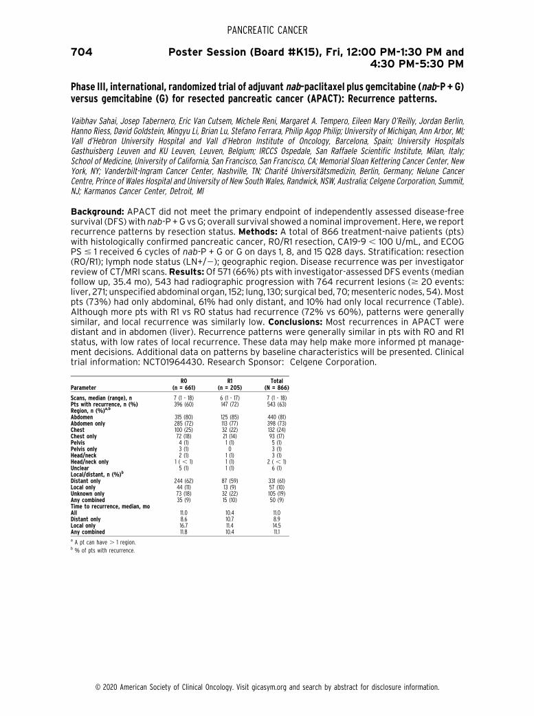
704 Poster Session (Board #K15), Fri, 12:00 PM-1:30 PM and4:30 PM-5:30 PM
Phase III, international, randomized trial of adjuvant nab-paclitaxel plus gemcitabine (nab-P + G)versus gemcitabine (G) for resected pancreatic cancer (APACT): Recurrence patterns.
Vaibhav Sahai, Josep Tabernero, Eric Van Cutsem, Michele Reni, Margaret A. Tempero, Eileen Mary O’Reilly, Jordan Berlin,Hanno Riess, David Goldstein, Mingyu Li, Brian Lu, Stefano Ferrara, Philip Agop Philip; University of Michigan, Ann Arbor, MI;Vall d’Hebron University Hospital and Vall d’Hebron Institute of Oncology, Barcelona, Spain; University HospitalsGasthuisberg Leuven and KU Leuven, Leuven, Belgium; IRCCS Ospedale, San Raffaele Scientific Institute, Milan, Italy;School of Medicine, University of California, San Francisco, San Francisco, CA; Memorial Sloan Kettering Cancer Center, NewYork, NY; Vanderbilt-Ingram Cancer Center, Nashville, TN; Charite Universitatsmedizin, Berlin, Germany; Nelune CancerCentre, Prince of Wales Hospital and University of New South Wales, Randwick, NSW, Australia; Celgene Corporation, Summit,NJ; Karmanos Cancer Center, Detroit, MI
Background: APACT did not meet the primary endpoint of independently assessed disease-freesurvival (DFS) with nab-P + G vs G; overall survival showed a nominal improvement. Here, we reportrecurrence patterns by resection status. Methods: A total of 866 treatment-naive patients (pts)with histologically confirmed pancreatic cancer, R0/R1 resection, CA19-9 , 100 U/mL, and ECOGPS # 1 received 6 cycles of nab-P + G or G on days 1, 8, and 15 Q28 days. Stratification: resection(R0/R1); lymph node status (LN+/2); geographic region. Disease recurrence was per investigatorreview of CT/MRI scans.Results:Of 571 (66%) pts with investigator-assessed DFS events (medianfollow up, 35.4 mo), 543 had radiographic progression with 764 recurrent lesions ($ 20 events:liver, 271; unspecified abdominal organ, 152; lung, 130; surgical bed, 70;mesenteric nodes, 54). Mostpts (73%) had only abdominal, 61% had only distant, and 10% had only local recurrence (Table).Although more pts with R1 vs R0 status had recurrence (72% vs 60%), patterns were generallysimilar, and local recurrence was similarly low. Conclusions: Most recurrences in APACT weredistant and in abdomen (liver). Recurrence patterns were generally similar in pts with R0 and R1status, with low rates of local recurrence. These data may help make more informed pt manage-ment decisions. Additional data on patterns by baseline characteristics will be presented. Clinicaltrial information: NCT01964430. Research Sponsor: Celgene Corporation.
ParameterR0
(n = 661)R1
(n = 205)Total
(N = 866)
Scans, median (range), n 7 (1 - 18) 6 (1 - 17) 7 (1 - 18)Pts with recurrence, n (%) 396 (60) 147 (72) 543 (63)Region, n (%)a,b
Abdomen 315 (80) 125 (85) 440 (81)Abdomen only 285 (72) 113 (77) 398 (73)Chest 100 (25) 32 (22) 132 (24)Chest only 72 (18) 21 (14) 93 (17)Pelvis 4 (1) 1 (1) 5 (1)Pelvis only 3 (1) 0 3 (1)Head/neck 2 (1) 1 (1) 3 (1)Head/neck only 1 ( , 1) 1 (1) 2 ( , 1)Unclear 5 (1) 1 (1) 6 (1)Local/distant, n (%)b
Distant only 244 (62) 87 (59) 331 (61)Local only 44 (11) 13 (9) 57 (10)Unknown only 73 (18) 32 (22) 105 (19)Any combined 35 (9) 15 (10) 50 (9)Time to recurrence, median, moAll 11.0 10.4 11.0Distant only 8.6 10.7 8.9Local only 16.7 11.4 14.5Any combined 11.8 10.4 11.1a A pt can have . 1 region.b % of pts with recurrence.
© 2020 American Society of Clinical Oncology. Visit gicasym.org and search by abstract for disclosure information.
PANCREATIC CANCER
705 Poster Session (Board #K16), Fri, 12:00 PM-1:30 PM and4:30 PM-5:30 PM
Attenuated regimen of biweekly gemcitabine/nab-paclitaxel in patients aged > 65 years withadvanced pancreatic cancer (APC).
Hasan Rehman, Jeffrey Chi, Nausheen Hakim, Shreya Prasad Goyal, Coral Olazagasti, Jyothi Jose, Linda Moriarty,Wasif M. Saif; Northwell Health Cancer Institute, Lake Success, NY
Background: Treatment with gemcitabine/nab-paclitaxel confers a survival benefit over gemci-tabine monotherapy in APC. However, such treatment can be associated with significant toxicitiesespecially in older patients and carries practical disadvantages related to a weekly schedule alongwith financial cost. We retrospectively analyzed patients. 65 years of age with APC who receiveda modified biweekly regimen of gemcitabine/nab-paclitaxel to evaluate efficacy and toxicity.Methods:Patients aged.65 years with chemo-naive APC and ECOGPS, 2were studied. Patientswere treated with a modified regimen of gemcitabine 1000 mg/m2 and nab-paclitaxel 125 mg/m2
every 2 weeks on days 1 and 15 of a 28-day cycle. Patients were evaluated for progression-freesurvival (PFS) and overall survival (OS) with analyses performed using Kaplan-Meier method.Adverse events were recorded on the day of chemotherapy. CA19-9 wasmeasured every cycle andrestaging scans were performed every two cycles. Results: Seventy-three patients (median age:73; range: 66 - 93) were treated with biweekly gemcitabine/nab-paclitaxel as first-line treatment.The median OS and PFS were 9.1 months and 4.8 months respectively. 66% of patients receivedgrowth factor support based on ASCO guidelines and no patients developed neutropenic fever. Theincidence of grade . 3 toxicity for neutropenia, anemia, thrombocytopenia, and neurotoxicitywere 2%, 7%, 3%, and 5% respectively. Dose reductions of gemcitabine/nab-paclitaxel wererequired in 10% and 4% patients respectively. Conclusions: In patients . 65 years of age withAPC, a modified regimen of biweekly gemcitabine/nab-paclitaxel was found to be effective whencompared with historical control from the MPACT study. This regimen allowed for less dosereductions, reduced healthcare costs from additional appointments, travel-related cost, as well asfavorable side effect profile while maintaining efficacy. Though retrospective in nature, this studyunderlines the need for further investigation, particularly in elderly patients with APC and poorperformance status. Better tolerability may allow for combination with a third agent, such as atargeted or immunotherapy. Research Sponsor: None.
© 2020 American Society of Clinical Oncology. Visit gicasym.org and search by abstract for disclosure information.
PANCREATIC CANCER
706 Poster Session (Board #K17), Fri, 12:00 PM-1:30 PM and4:30 PM-5:30 PM
Single institutional analysis of resectable pancreatic cancer.
Jeff Wiisanen, Patrick Navin, Moustafa El Khatib, William R Bamlet, Sean P. Cleary, Robert R. McWilliams, Mark J. Truty,Thanh P. Ho, Mindy L. Hartgers, Siddhartha Yadav, Amit Mahipal; Mayo Clinc, Rochester, MN; Mayo Clinic, Rochester, MN;Beaumont Health, Royal Oak, MI
Background: In recent years, there has been a shift towards neo-adjuvant treatment (NAT) of non-metastatic pancreas cancer in the hopes of improving negativemargin rate, lymph node negativity,recurrence and survival. Even patients deemed resectable based on NCCN criteria are receivingNAT but data for these patients remains limited. This current study evaluated the outcomes ofpatients diagnosed with resectable pancreatic adenocarcinoma. Methods: Patients were retro-spectively identified through the Mayo Clinic, Rochester SPORE pancreatic cancer registry as wellas search of the electronic medical record via Advanced Cohort Explorer from May 2011 to 2016.Baseline demographics, tumor characteristics, treatments rendered, and outcomeswere collected.Variables were analyzed for association with recurrence from time of surgery and survival fromtime of diagnosis using Kaplan-Meier curves and Cox proportional hazards regression. Results: Atotal of 520 patients with resectable pancreatic adenocarcinoma were identified. 72 patientsreceived upfront chemotherapy with 44 (61.1%) proceeding to surgical resection. 62 patientsreceived upfront chemotherapy followed by radiation with 33 (53.2%) proceeding to surgicalresection. 12 patients received upfront radiation alone with 7 (58.3%) proceeding to surgicalresection. 374 patients did not receive any NATwith 293 (78.3%) proceeding to surgical resection.In total, 377 (72.5%) went to resection. Median time to recurrence from surgery was 27.7 monthsvs. 21.7 months for NAT and upfront resection, respectively (HR 0.87, 95% CI 0.60-1.72, p = 0.48).Median overall survival from diagnosis for those receiving NAT was 40.6 months vs. 24.7 monthsfor those receiving upfront resection (HR 0.62, 95% CI 0.41-0.92, p = 0.02). Conclusions: Thisstudy shows an approximate 16 month improvement in overall survival of patients receivingupfront NAT for resectable pancreatic adenocarcinoma. This might be due to a better selection ofpatients. It also highlights that not all patients with resectable cancer undergo resection. Furtherstudies are warranted to identify why resectable patients are not proceeding to resection andwhich specific NAT approaches benefit patients the most. Research Sponsor: None.
© 2020 American Society of Clinical Oncology. Visit gicasym.org and search by abstract for disclosure information.
PANCREATIC CANCER
707 Poster Session (Board #K18), Fri, 12:00 PM-1:30 PM and4:30 PM-5:30 PM
The development of therapeutic cancer vaccine for pancreatic cancer.
Hiroki Yamaue, Motoki Miyazawa, Masahiro Katsuda, Manabu Kawai, Seiko Hirono, Ken-Ichi Okada, Yuji Kitahata, Masaki Ueno,Shinya Hayami; Second Department of Surgery, Wakayama Medical University, School of Medicine, Wakayama, Japan; SecondDepartment of Surgery, Wakayama Medical University, Wakayama, Japan
Background: A previous phase II/III trial using a single cancer peptide vaccine derived fromvascular endothelial growth factor receptor (VEGFR)2 for patients with advanced pancreaticcancer did not demonstrate the overall survival (OS) benefit (Yamaue et al. Cancer Sci 2015).However, for the next trial, we conducted a multicenter phase II study using multipeptide cocktailvaccine named OCV-C01 derived from a novel higher immunogenic antigen KIF20A, VEGFR1 andVEGFR2 combined with gemcitabine in postoperative adjuvant setting. Methods: A single-armmulticenter phase II study was performed on 30 patients with pancreatic ductal carcinoma whounderwent pancreatectomy. At each 28-day treatment cycle, patients received weekly subcuta-neous injection of OCV-C01 for 48 weeks, and gemcitabine was administered intravenously at1,000 mg/m2 on days 1, 8, and 15 for 24 weeks. Patients were followed for 18 months. The primaryendpoint was disease-free survival (DFS) and secondary endpoints included safety, OS andimmunological assays on peptide-specific cytotoxic T lymphocyte (CTL) activity and KIF20Aexpression in resected pancreatic cancer. Results: The median DFS was 15.8 months (95%confidence interval (CI), 11.1-20.6), and the DFS rate at 18 months was 34.6% (95% CI, 18.3-51.6).The median OS was not reached and the OS rate at 18 months was 69.0% (95% CI, 48.8-82.5).The administration of OCV-C01 was well tolerated. In the per protocol set, there were significantdifferences in DFS between patients with and without KIF20A-specific CTL responses (p =0.027), and between patients with and without KIF20A expression in resected pancreatic cancertissues (p = 0.014). In addition, all four patients who underwent R0 resection with KIF20Aexpression had no recurrence of pancreatic cancer with KIF20A-specific CTL responses.Conclusions: OCV-C01 combined with gemcitabine was tolerable with a favorable medianDFS of 15.8 months. In cancer vaccine treatment, positive expression of targeted antigenwas essential, and postoperative adjuvant setting was more suitable than advanced state ofcancer. Clinical trial information: UMIN000007991. Research Sponsor: Health and LabourSciences Research Grants of the Ministry of Health Labour and Welfare in Japan.
© 2020 American Society of Clinical Oncology. Visit gicasym.org and search by abstract for disclosure information.
PANCREATIC CANCER
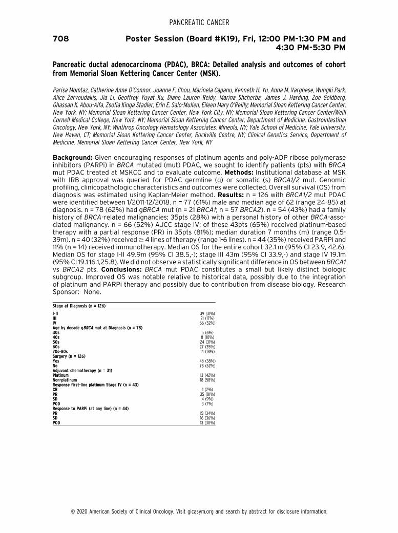
708 Poster Session (Board #K19), Fri, 12:00 PM-1:30 PM and4:30 PM-5:30 PM
Pancreatic ductal adenocarcinoma (PDAC), BRCA: Detailed analysis and outcomes of cohortfrom Memorial Sloan Kettering Cancer Center (MSK).
Parisa Momtaz, Catherine Anne O’Connor, Joanne F. Chou, Marinela Capanu, Kenneth H. Yu, Anna M. Varghese, Wungki Park,Alice Zervoudakis, Jia Li, Geoffrey Yuyat Ku, Diane Lauren Reidy, Marina Shcherba, James J. Harding, Zoe Goldberg,Ghassan K. Abou-Alfa, Zsofia Kinga Stadler, Erin E. Salo-Mullen, Eileen Mary O’Reilly; Memorial Sloan Kettering Cancer Center,New York, NY; Memorial Sloan Kettering Cancer Center, New York City, NY; Memorial Sloan Kettering Cancer Center/WeillCornell Medical College, New York, NY; Memorial Sloan Kettering Cancer Center, Department of Medicine, GastrointestinalOncology, New York, NY; Winthrop Oncology Hematology Associates, Mineola, NY; Yale School of Medicine, Yale University,New Haven, CT; Memorial Sloan Kettering Cancer Center, Rockville Centre, NY; Clinical Genetics Service, Department ofMedicine, Memorial Sloan Kettering Cancer Center, New York, NY
Background: Given encouraging responses of platinum agents and poly-ADP ribose polymeraseinhibitors (PARPi) in BRCA mutated (mut) PDAC, we sought to identify patients (pts) with BRCAmut PDAC treated at MSKCC and to evaluate outcome. Methods: Institutional database at MSKwith IRB approval was queried for PDAC germline (g) or somatic (s) BRCA1/2 mut. Genomicprofiling, clinicopathologic characteristics and outcomeswere collected. Overall survival (OS) fromdiagnosis was estimated using Kaplan-Meier method. Results: n = 126 with BRCA1/2 mut PDACwere identified between 1/2011-12/2018. n = 77 (61%) male and median age of 62 (range 24-85) atdiagnosis. n = 78 (62%) had gBRCA mut (n = 21 BRCA1; n = 57 BRCA2). n = 54 (43%) had a familyhistory of BRCA-related malignancies; 35pts (28%) with a personal history of other BRCA-asso-ciated malignancy. n = 66 (52%) AJCC stage IV; of these 43pts (65%) received platinum-basedtherapy with a partial response (PR) in 35pts (81%); median duration 7 months (m) (range 0.5-39m). n = 40 (32%) received$4 lines of therapy (range 1-6 lines). n =44 (35%) received PARPi and11% (n = 14) received immunotherapy. Median OS for the entire cohort 32.1 m (95% CI 23.9, 42.6).Median OS for stage I-II 49.9m (95% CI 38.5,-); stage III 43m (95% CI 33.9,-) and stage IV 19.1m(95%CI 19.1 16.1,25.8).We did not observe a statistically significant difference in OS betweenBRCA1vs BRCA2 pts. Conclusions: BRCA mut PDAC constitutes a small but likely distinct biologicsubgroup. Improved OS was notable relative to historical data, possibly due to the integrationof platinum and PARPi therapy and possibly due to contribution from disease biology. ResearchSponsor: None.
Stage at Diagnosis (n = 126)
I-II 39 (31%)III 21 (17%)IV 66 (52%)Age by decade gBRCA mut at Diagnosis (n = 78)30s 5 (6%)40s 8 (10%)50s 24 (31%)60s 27 (35%)70s-80s 14 (18%)Surgery (n = 126)Yes 48 (38%)No 78 (62%)Adjuvant chemotherapy (n = 31)Platinum 13 (42%)Non-platinum 18 (58%)Response first-line platinum Stage IV (n = 43)CR 1 (2%)PR 35 (81%)SD 4 (9%)POD 3 (7%)Response to PARPi (at any line) (n = 44)PR 15 (34%)SD 16 (36%)POD 13 (30%)
© 2020 American Society of Clinical Oncology. Visit gicasym.org and search by abstract for disclosure information.
PANCREATIC CANCER
709 Poster Session (Board #K20), Fri, 12:00 PM-1:30 PM and4:30 PM-5:30 PM
A phase I study of olaparib in combination with capecitabine-based chemoradiation (CRT) inpatients (pts) with locally advanced pancreatic cancer (LAPC).
T.R. Jeffry Evans, Martin McKinlay Eatock, Liz-Anne Lewsley, Caroline Kelly, Elaine McCartney, Aileen Duffton, Peter Houston,Claire Harrison, Colin Purcell, Miranda Ashton, Alanna Morton, Jill Graham, Anthony J. Chalmers, Fiona Thomson,Chantevy Pou, Alan Bilsland, Susie Cooke, David McIntosh, Derek B. Grose; University of Glasgow, Beatson West ofScotland Cancer Centre, Glasgow, United Kingdom; Belfast City Hospital, Belfast, United Kingdom; University of Glasgow,Glasgow, United Kingdom; Cancer Research UK Clinical Trials Unit, Glasgow, United Kingdom; Beatson West of ScotlandCancer Centre, Glasgow, United Kingdom; N Ireland Cancer Center, Belfast, United Kingdom; Northern Ireland Cancer Centre,Belfast, United Kingdom; Wolfson Wohl Cancer Research Centre, Institute of Cancer Sciences, University of Glasgow,Glasgow, United Kingdom
Background: Olaparib is a potent inhibitor of PARP-1, which has a critical role in signalling DNAsingle strand breaks (SSB) as part of the base excision repair pathway, and may have radio-sensitizing effects due to impaired resolution of radiation induced SSB. We hypothesize that Omaypotentiate the effects of X-CRT in pts with LAPC.Methods: Eligible pts with LAPC, ECOG, 1, tumordiameter , 6cm, with stable disease (SD) or response after 12 weeks’ induction chemotherapy,were treated with 1 of 4 escalating doses of O given bid po starting on day -3, and then incombination with X (830mg/m2 bid) and radiation (50$4 Gy in 28 fractions) all administered Mon-Fri. Dose limiting toxicities (DLT) were determined on clinical and lab toxicity assessments (NCI-CTCAE v4.03) performedweekly from the start of O until completion of O plus X-CRT (i.e. 6weeks).Dose escalation continued with a rolling-six design until the Maximum Tolerated Dose (MTD) wasreached. Blood samples for PK analyses of O and PD measurement (inhibition of PARP activity)were collected on day -3 (Omonotherapy) and during week 1 of O + X-CRT.Results: 18 pts, (9m, 9 f,ECOG0/1 [n=6/12]), age range 49-81 (median=70) years, with histologic (14) or cytologic (4) provenLAPC, had received induction chemotherapy with gemcitabine [GEM] (n=2), GEM + X (12), orFOLFIRINOX (3) with partial response (n=4) or stable disease (14). Pts received 50 (3), 100 (4), 150(6), or 200 (5)mgs bid of Owith X+CRT. DLTs were observed in 2 pts (both at 200mgs bid): 1 pt withgrade 3 nausea (on optimal anti-emetics) and grade 3 fatigue, 1 pt with grade 3 anorexia. 6 pts weresubsequently recruited at 150mgs bidwith noDLTs. No pts had complete response, 2 pts had partialresponse (1 pt each at 100 and 150 mgs bid) and 1 pt (at 100 mgs bid) had progressive disease; theremaining 14 pts had SD. Conclusions: The recommended dose (RP2) of O is 150mgs bid whengiven in combination with X + CRT in LAPC. Recruitment of up to 12 pts with borderline operableLAPC at the RP2 is ongoing. PK analyses of O, PD studies (PARP inhibition –PBMCs; cytokeratin 18 –
serum; gH2AX foci – hair follicles), and exploratory predictive marker studies (tumor – NGS; RNAexome sequencing) are ongoing. Clinical trial information: ISRCTN10361292. Research Sponsor:Cancer Research UKPharmaceutical/Biotech Company.
© 2020 American Society of Clinical Oncology. Visit gicasym.org and search by abstract for disclosure information.
PANCREATIC CANCER
710 Poster Session (Board #K21), Fri, 12:00 PM-1:30 PM and4:30 PM-5:30 PM
Efficacy of SBP-101, in combination with gemcitabine and nab-paclitaxel, in first-line treatmentof metastatic pancreatic ductal adenocarcinoma.
Dusan Kotasek, Adnan Nagrial, Sumit Lumba, Niall C. Tebbutt, Thomas J. George, Sheri Lynn Smith, Suzanne Gagnon,Michael T. Cullen, Michael J. Walker; University of Adelaide, Adelaide, SA, Australia; Blacktown Hospital Cancer andHaematology Centre, Blacktown, NSW, Australia; John Flynn Private Hospital Cancer and Haematology Centre, Tugun, QLD,Australia; Olivia Newton-John Cancer and Wellness Centre, ~Af A,~A,A Heidelberg Repatriation Hospital, Heidelberg, Australia;University of Florida Health Cancer Center, Gainesville, FL; Courante Oncology, Excelsior, MN; Sun BioPharma Inc,Jamestown, RI; Sun BioPharma, Inc., Waconia, MN
Background: SBP-101, a polyamine metabolic inhibitor, inhibited growth in 6 human pancreaticductal adenocarcinoma (PDA) cell lines and 3murine xenograft tumor models of human PDA. SBP-101 monotherapy in heavily pre-treated PDA patients (. 2 prior regimens, N=4) showed a mediansurvival of 5.9months at the optimal dose level. Purpose: To assess the safety, tolerability, PK, andefficacy of SBP-101 in combination with gemcitabine (G) and nab-paclitaxel (A) in patients withpreviously untreated metastatic PDA. Methods: In a modified 3+3 dose escalation scheme,subcutaneous injections of SBP-101 were dosed at 0.2, 0.4 or 0.6 mg/kg days 1-5 of each 28-day cycle. G (1000mg/m2) and A (125 mg/m2) were administered intravenously on Days 1, 8, and 15of each cycle. Safety and tolerability were evaluated by clinical and laboratory assessments. PKwas evaluated on day 1 of cycle 1. Efficacy was assessed by CA19-9 levels, objective response asassessed by RECIST criteria, progression-free survival (PFS) and overall survival (OS). Results:Fifteen patients have been enrolled in 3 cohorts (1: N=4, 2: N=7, 3: N=4) and received up to 6 cyclesof treatment (7 subjects are ongoing in cohorts 2 and 3). Themost common adverse events relatedto SBP-101 are fatigue (N=4), nausea (N=2) and injection site pain (N=2). There is no evidence ofSBP-101-related bone marrow suppression or peripheral neuropathy. One patient in cohort 2developed grade 3-4 reversible liver enzyme elevation. PK parameters in cohort 1 were below thelimits of detection at most time points, but plasma Cmax and AUC0-t were measurable in cohorts 2and 3. In those cohorts, CA19-9 levels decreased 76-95% in 7 of 8 evaluable subjects (1 additionalsubject TBD), with 5 patients achieving partial responses (4 ongoing) and 1 achieving stabledisease. Median PFS and OS have not yet been reached. Conclusions: Preliminary results suggestSBP-101 is well tolerated when administered with G and A. Signals of efficacy support continueddevelopment of SBP-101 in combination first-line treatment for PDA. Clinical trial information:NCT03412799. Research Sponsor: Sun BioPharma, Inc.
© 2020 American Society of Clinical Oncology. Visit gicasym.org and search by abstract for disclosure information.
PANCREATIC CANCER
711 Poster Session (Board #K22), Fri, 12:00 PM-1:30 PM and4:30 PM-5:30 PM
Modified FOLFIRINOX versus sequential chemotherapy (FOLFIRI/FOLFOX) as second-linetreatment for advanced pancreatic adenocarcinoma: A real-world analysis.
Shun Tezuka, Makoto Ueno, Satoshi Kobayashi, Manabu Morimoto, Shuhei Nagashima, Yusuke Sano, Hiroyuki Asama,Kuniyuki Kawano, Satoshi Tanaka, Taito Fukushima; Kanagawa Cancer Center, Yokohama, Japan; Department of Gastro-enterology, Hepatobiliary and Pancreatic Medical Oncology Division, Kanagawa Cancer Center, Yokohama, Japan; KanagawaCancer Hospital, Yokohama, Japan
Background: Therapy options for second-line treatment of advanced pancreatic adenocarcinoma(PDAC) are limited and preferred regimens have not been established. This study compared theefficacy and safety of modified FOLFIRINOX (mFFX) and sequential chemotherapy (FOLFIRI/FOLFOX) as second-line treatment for advanced PDAC.Methods: This was a retrospective single-center analysis of all patients who received mFFX or sequential chemotherapy between December2014 and May 2019 as second-line treatment for advanced PDAC. The sequential arm included allpatients intended to be treated with sequential chemotherapy even if finished with either FOLFIRIor FOLFOX. For efficacy analysis, progression-free survival (PFS), overall survival (OS), andresponse rate (RR)of all the patients, excluding those with locally advanced pancreatic cancer(LAPC), were evaluated. For safety analysis, we evaluated the incidence of grade $3 adverseevents in all the patients. Results: Seventy-four patients (mFFX 44; sequential 30) were inclu-ded.The patients characteristics (mFFX/sequential) are as follows:median age 64/67 years, per-formance status (ECOG) 0 50%/53%, and LAPC 16%/13%. In contrast to sequential therapy, themFFX group showed better OS and RR. However, there was no significant difference betweenmFFX and sequential therapy in PFS (median 4.4 [95% confidence interval (CI) 1.8-7.9] vs. 4.6[95% CI 2.0-6.2] months; hazard ratio [HR] 0.88 [95% CI 0.51-1.56]; p = 0.657), OS (median 10.6[95% CI 5.9-13.8] vs. 8.5 [95% CI 5.0-12.2] months; HR 0.71 [95% CI 0.37-1.41]; p = 0.318), and RR(8.1%vs. 3.8%; odds ratio 1.94 [95%CI 0.19-19.87]; p = 1.000). In the safety analysis, grade$3 ratesof neutropenia, febrile neutropenia, and anorexia were 36.4%, 6.8%, and 16.0%, respectively, withmFFX and 0%, 0% and 3.3%, respectively, with sequential chemotherapy. Conclusions:Whereascertain trends were observed, there were no significant differences in efficacy between mFFX andsequential chemotherapy. Considering the incidence of grade $3 adverse events with mFFX,sequential chemotherapy could be considered as one of the second-line treatment options foradvanced PDAC. Research Sponsor: None.
© 2020 American Society of Clinical Oncology. Visit gicasym.org and search by abstract for disclosure information.
PANCREATIC CANCER
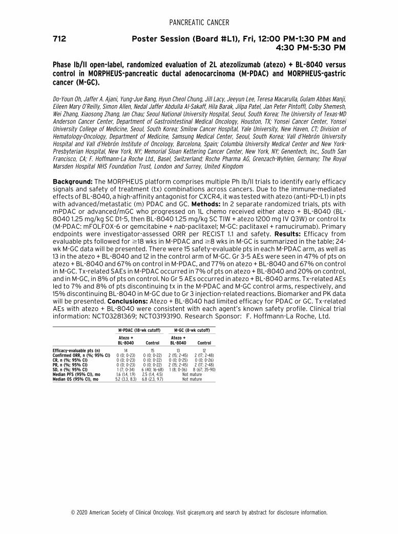
712 Poster Session (Board #L1), Fri, 12:00 PM-1:30 PM and4:30 PM-5:30 PM
Phase Ib/II open-label, randomized evaluation of 2L atezolizumab (atezo) + BL-8040 versuscontrol in MORPHEUS-pancreatic ductal adenocarcinoma (M-PDAC) and MORPHEUS-gastriccancer (M-GC).
Do-Youn Oh, Jaffer A. Ajani, Yung-Jue Bang, Hyun Cheol Chung, Jill Lacy, Jeeyun Lee, Teresa Macarulla, Gulam Abbas Manji,Eileen Mary O’Reilly, Simon Allen, Nedal Jaffer Abdulla Al-Sakaff, Hila Barak, Jilpa Patel, Jan Peter Pintoffl, Colby Shemesh,Wei Zhang, Xiaosong Zhang, Ian Chau; Seoul National University Hospital, Seoul, South Korea; The University of Texas-MDAnderson Cancer Center, Department of Gastrointestinal Medical Oncology, Houston, TX; Yonsei Cancer Center, YonseiUniversity College of Medicine, Seoul, South Korea; Smilow Cancer Hospital, Yale University, New Haven, CT; Division ofHematology-Oncology, Department of Medicine, Samsung Medical Center, Seoul, South Korea; Vall d’Hebron UniversityHospital and Vall d’Hebron Institute of Oncology, Barcelona, Spain; Columbia University Medical Center and New York-Presbyterian Hospital, New York, NY; Memorial Sloan Kettering Cancer Center, New York, NY; Genentech, Inc., South SanFrancisco, CA; F. Hoffmann-La Roche Ltd., Basel, Switzerland; Roche Pharma AG, Grenzach-Wyhlen, Germany; The RoyalMarsden Hospital NHS Foundation Trust, London and Surrey, United Kingdom
Background: The MORPHEUS platform comprises multiple Ph Ib/II trials to identify early efficacysignals and safety of treatment (tx) combinations across cancers. Due to the immune-mediatedeffects of BL-8040, a high-affinity antagonist for CXCR4, it was testedwith atezo (anti-PD-L1) in ptswith advanced/metastatic (m) PDAC and GC. Methods: In 2 separate randomized trials, pts withmPDAC or advanced/mGC who progressed on 1L chemo received either atezo + BL-8040 (BL-8040 1.25 mg/kg SC D1-5, then BL-8040 1.25 mg/kg SC TIW + atezo 1200 mg IV Q3W) or control tx(M-PDAC: mFOLFOX-6 or gemcitabine + nab-paclitaxel; M-GC: paclitaxel + ramucirumab). Primaryendpoints were investigator-assessed ORR per RECIST 1.1 and safety. Results: Efficacy fromevaluable pts followed for$18 wks in M-PDAC and$8 wks in M-GC is summarized in the table; 24-wk M-GC data will be presented. There were 15 safety-evaluable pts in each M-PDAC arm, as well as13 in the atezo + BL-8040 and 12 in the control arm of M-GC. Gr 3-5 AEs were seen in 47% of pts onatezo + BL-8040 and 67%on control inM-PDAC, and 77%on atezo + BL-8040 and 67%on controlinM-GC. Tx-related SAEs inM-PDAC occurred in 7%of pts on atezo + BL-8040 and 20%on control,and inM-GC, in 8%of pts on control. No Gr 5AEs occurred in atezo +BL-8040 arms. Tx-relatedAEsled to 7% and 8% of pts discontinuing tx in the M-PDAC and M-GC control arms, respectively, and15%discontinuing BL-8040 inM-GC due to Gr 3 injection-related reactions. Biomarker and PK datawill be presented. Conclusions: Atezo + BL-8040 had limited efficacy for PDAC or GC. Tx-relatedAEs with atezo + BL-8040 were consistent with each agent’s known safety profile. Clinical trialinformation: NCT03281369; NCT03193190. Research Sponsor: F. Hoffmann-La Roche, Ltd.
M-PDAC (18-wk cutoff) M-GC (8-wk cutoff)
Atezo +BL-8040 Control
Atezo +BL-8040 Control
Efficacy-evaluable pts (n) 14 15 13 12Confirmed ORR, n (%; 95% CI) 0 (0; 0–23) 0 (0; 0–22) 2 (15; 2–45) 2 (17; 2–48)CR, n (%; 95% CI) 0 (0; 0–23) 0 (0; 0–22) 0 (0; 0–25) 0 (0; 0–26)PR, n (%; 95% CI) 0 (0; 0–23) 0 (0; 0–22) 2 (15; 2–45) 2 (17; 2–48)SD, n (%; 95% CI) 1 (7; 0–34) 6 (40; 16–68) 1 (8; 0–36) 8 (67; 35–90)Median PFS (95% CI), mo 1.6 (1.4, 1.9) 2.5 (1.4, 4.5) Not matureMedian OS (95% CI), mo 5.2 (3.3, 8.3) 6.8 (2.3, 9.7) Not mature
© 2020 American Society of Clinical Oncology. Visit gicasym.org and search by abstract for disclosure information.
PANCREATIC CANCER
713 Poster Session (Board #L2), Fri, 12:00 PM-1:30 PM and4:30 PM-5:30 PM
Phase Ia, first-in-human study of AbGn-107, a novel antibody-drug conjugate (ADC), in patientswith gastric, colorectal, pancreatic, or biliary cancers.
Andrew H. Ko, Andrew L. Coveler, Benjamin L. Schlechter, Tanios S. Bekaii-Saab, Brian M. Wolpin, Jeffrey William Clark,Yi-Lin Cheng, Ting-Ying Cheng, Peter J. Langecker, Shih-Yao Lin; UCSF Helen Diller Family Comprehensive Cancer Center, SanFrancisco, CA; Seattle Cancer Care Alliance/University of Washington, Seattle, WA; Beth Israel Deaconess Medical Center,Boston, MA; Mayo Clinic, Phoenix, AZ; Dana-Farber Cancer Institute, Boston, MA; Massachusetts General Hospital, Boston, MA;Abgenomics International, Los Altos, CA; Los Gatos Pharmaceuticals, Monte Sereno, CA; AbGenomics International, Taipei,Taiwan
Background: AbGn-107 is an ADC directed against AG-7 antigen, a Lewis A-like glycol-epitopeexpressed in 24-61% of gastric (G), colorectal (CRC), pancreatic (PDA), and biliary (BIL) cancers.Based on promising antitumor activity of AbGn-107 in both in vitro and in vivo preclinical studies,we performed a Phase Ia trial in pts with the aforementioned GI malignancies.Methods: Standard3+3 dose escalation was used. Key eligibility criteria: locally adv or metastatic G, CRC, PDA, or BILcancer, previously treated, ECOG PS 0-1; positive AG-7 expression was not required. Two dosingintervals were tested: AbGn-107 administered i.v. Q4 weeks (at doses ranging from 0.1-1.2 mg/kg)and Q2 weeks (at doses from 0.8-1.0 mg/kg). DLTs were based on grade 3/4 hematologic and non-heme AEs occurring during the initial 4-week rx window. Pts were treated until dz progression orunacceptable toxicity, with tumor assessments Q8 weeks. 1o objectives: safety and MTD; 2oobjectives: PK, immunogenicity, and efficacy defined by ORR (RECIST 1.1). Results: 35 patientswere enrolled across 6 dose levels (median age 61.5 yo (range 40 –81); G (0)/CRC (12)/PDA (20)/BIL(3); median # lines of prior rx = 3 (range 1-7). Safety: 5 pts experienced Grade 3 or 4 neutropenia, allat higher dose levels, with 1 episode of febrile neutropenia. Other frequent drug-relatedAEs,mostlygrade 1/2, inc. fatigue (29%), nausea (20%), and diarrhea (14%). DLTs include grade 4 CK elevation(n = 1) at 0.8 mg/kg Q4W and grade 3 arthralgias (n = 1) at 1.2 mg/kg Q4W. MTD was not reached ateither 1.2 mg/kg Q4Wor 0.8mg/kg Q2W; the 1.0mg/kg Q2W cohort will complete enrollment in Oct2019. Efficacy: Median duration of treatment = 56 days (range, 8 – 225 days); best responseobserved to date is stable dz lasting. 6 months at 0.8 mg/kg Q4W and Q2W cohorts (n = 1 each).Conclusions: Overall, AbGn-107 appears well-tolerated with encouraging prelim signs of efficacy(prolonged dz control) in non-biomarker selected pts with advanced GI cancers. Pre-screening forhigh AG-7 expression is underway for subjects with G, CRC, PDA, and BIL cancers for the cohortexpansion phase of this study, which will be open across multiple sites in U.S. and Taiwan. Clinicaltrial information: NCT02908451. Research Sponsor: Abgenomics.
© 2020 American Society of Clinical Oncology. Visit gicasym.org and search by abstract for disclosure information.
PANCREATIC CANCER
714 Poster Session (Board #L3), Fri, 12:00 PM-1:30 PM and4:30 PM-5:30 PM
The role of immunotherapy on the survival of pancreatic cancer patients.
Saber Ali Amin, Michael Baine, Jane L. Meza, Chi Lin; University of Nebraska Medical Center, Omaha, NE
Background: Immunotherapy has revolutionized the treatment landscape of many malignancies,but its therapeutic role in pancreatic cancer (PC) remains unclear. The objective of this study is toinvestigate the impact of immunotherapy on the overall survival of PC patients stratified bydefinitive surgery of the pancreas using the National Cancer Database (NCDB).Methods: Patientswith pancreatic adenocarcinomawere identified fromNCDB. Cox proportional hazardmodels wereemployed to assess the impact of immunotherapy on survival after being stratified by surgery andadjusted for age of diagnosis, race, sex, place of living, income, education, treatment facility type,insurance status, year of diagnosis, and treatment types such as chemotherapy and radiationtherapy. Results: Of 252,280 patients who were analyzed, 214,632 (85.08%) had definitivesurgery, and 37,638 (14.92%) did not get definitive surgery of the pancreas. In the surgery group,351 (0.93%) received immunotherapy and 37,287 (99.07%) did not while in the no surgery group,838 (0.39%) received immunotherapy and 213,804 (99.61%) did not. In the multivariable analysis,patients who received immunotherapy had significantly improved OS both in the no surgery group(HR: 0.886, CI: 0.655-0.714; P, 0.0001) and in the surgery group (HR: 0.846, CI: 0.738-0.971; P,0.0001) compared to patients who did not receive immunotherapy. Treatment with chemotherapyplus immunotherapy was associated with significantly improved OS (HR: 0.871, CI: 0.784-0.967;P , 0.009) compared to chemotherapy without immunotherapy in the no surgery group, while itwas not significant in the surgery group. Chemoradiation plus immunotherapy was associated withsignificantly improved OS (HR: 0.787, CI: 0.684-0.906; P , 0.0009) in the no surgery group and(HR: 0.799, CI: 0.681-0.938) in the surgery group compared to chemoradiation alone.Conclusions:In this study, the addition of immunotherapy to chemoradiation therapy was associated withsignificantly improved OS in PC patients with or without definitive surgery. The study warrantsfurther future clinical trials of immunotherapy in PC. Research Sponsor: None.
© 2020 American Society of Clinical Oncology. Visit gicasym.org and search by abstract for disclosure information.
PANCREATIC CANCER
715 Poster Session (Board #L4), Fri, 12:00 PM-1:30 PM and4:30 PM-5:30 PM
Role of para-aortic lymph node sampling in pancreatic cancer surgery.
Yusuke Kazami, Hiromichi Ito, Yoshihiro Ono, Takafumi Sato, Yosuke Inoue, Yu Takahashi; Department of Digestive and HBPSurgery, Cancer Institute Hospital, Japanese Foundation for Cancer Research, Tokyo, Japan; Japanese Foundation forCancer Research, Tokyo, Japan
Background: In the management of pancreatic cancer, para-aortic lymph node (PALN) metastasisis regarded as distant metastasis, and systemic treatment is recommended. However, imagingstudy is not perfect to detect all PALN metastasis and the management of intraoperativelydiscovered PALN has been controversial. We hypothesized that sampling of PALNs on explorationcould allow us to avoid pancreatic resection for patients who would not benefit. In this study, weevaluated the incidence and the effect on the long-term outcomes for patients with potentiallyresectable pancreatic cancer. Methods: Three hundred and ninety-two patients who had PALNssampled upon potentially resectable pancreatic cancer from 2005 through 2014 were included inthe study. All patients were appropriately staged preoperatively with CT/MRI and those withsuspected PALN metastasis were not considered as candidates for resection. The patients whoseresections were aborted because of liver metastasis or peritoneal dissemination discovered onexploration, or those who died within 30-days after the operation were not included. Evaluatedoutcomeswere incidence of PALNmetastasis and their recurrence-free and overall survivals (RFS,OS). Results: The patients’ median age was 74 years, and 58.6% was man. 67.8% had tumors atpancreatic head. Preoperative chemotherapy was given only on 16 patients (3.2%). Among 392patients with PALNs sampled, 53 (13.5%) patients had metastasis; Resection was completed on 40patients and resection was aborted on the rest. Among patients who underwent pancreaticresection, median RFS and OSwere 10 and 12months for patients with PALNmetastasis, comparedto 17 and 26 months for those without PALN metastasis (p, 0.001 for RFS and p, 0.001 for OS).The 5-year-OS rates for patients with/without PALN metastasis were 5.9% and 25% (p , 0.001).Among 53 patients with PALN metastasis, OS were not different between the patients whounderwent resection and those who did not (median 13 months vs 17 months, p = 0.06), and therewere no recurrence-free survivors. Conclusions: PALN sampling and evaluation before commit-ting to resection is useful to identify the patients who can unlikely benefit and to avoid unnecessarymorbid operation. Research Sponsor: None.
© 2020 American Society of Clinical Oncology. Visit gicasym.org and search by abstract for disclosure information.
PANCREATIC CANCER
716 Poster Session (Board #L5), Fri, 12:00 PM-1:30 PM and4:30 PM-5:30 PM
A phase I/II study combining a TMZ-CD40L/4-1BBL-armed oncolytic adenovirus and nab-paclitaxel/gemcitabine chemotherapy in advanced pancreatic cancer: An interim report.
Benjamin Leon Musher, Brandon George Smaglo, Wasif Abidi, Mohamed Othman, Kalpesh Patel, James Jing, Nir Stanietzky,Jinyu (Jim) Lu, Amanda Brisco, Jessica Wenthe, Benjamin Brenner, Emma Eriksson, Ann M. Leen, Susan G. Hilsenbeck,Gustav J. Ullenhag, Eric Keith Rowinsky, Bambj Grilley, Angelica Sara Ingrid Loskog; Baylor College of Medicine, Houston, TX;Georgetown Lombardi Comprehensive Cancer Center, Washington, DC; Uppsala University, Uppsala, Sweden; Smith BreastCenter At BCM, Houston, TX; Akademiska Sjukhus, Uppsala, Sweden; Rgenix, Inc., New York, NY
Background: Pancreatic ductal adenocarcinoma (PDAC) has been highly resistant to immuno-therapeutics to date. LOAd703, an oncolytic adenovirus with transgenes encoding TMZ-CD40Land 4-1BBL, has been shown to lyse tumor cells selectively, induce anti-tumor cytotoxic T-cellresponses, reduce myeloid-derived suppressor cell (MDSC) infiltration, and induce tumor regres-sion in preclinical studies.Methods: In this phase I/II trial, patients with unresectable or metastaticPDAC are treated with LOAd703 intratumoral injections and standard nab-paclitaxel/gemcitabine(nab-P/G) chemotherapy. Starting on cycle 1 day 15 of nab-P/G, LOAd703 is injected with imageguidance into the primary pancreatic tumor or a metastasis every 2 weeks for 6 injections. In theevent of sustained tumor control, subjects are eligible to receive 6 more injections. Three doselevels of LOAd703 are being investigated using a BOIN dose escalation design. Primary endpointsare safety and feasibility. Secondary endpoints include response rate and overall survival.Results:To date, 13 subjects are evaluable for safety and feasibility. Three patients were treated at dose1 (5x10e10 VP), 4 subjects at dose 2 (1x10e11 VP), and 6 subjects at dose 3 (5x10e11 VP). The mostcommon adverse events (AEs) attributed to LOAd703 have been fever, chills, nausea, andincreased transaminases. AEs have been transient and grade 1-2, with the exception of a grade3 transaminase elevation in 1 subject receiving dose 3 (the only dose-limiting toxicity observed thusfar). During protocol treatment, circulating MDSCs decreased in 8/13 subjects while effectormemory T-cells increased in 10/13. ELISPOT analyses showed a rise in tumor antigen-specificT-cells in 10/13 subjects. At the lowest dose level, best response was stable disease, and 6/10patients who received higher LOAd703 doses have had partial responses. Only 1 patient has hadprogressive disease as best response. Conclusions: Adding LOAd703 to nab-P/G has been safe andfeasible. Treatment-emergent immune responses have been demonstrated in most subjects, with anotable proportion having objective anti-tumor responses. Clinical trial information: NCT02705196.Research Sponsor: LOKON pharma, Other Foundation.
© 2020 American Society of Clinical Oncology. Visit gicasym.org and search by abstract for disclosure information.
PANCREATIC CANCER
717 Poster Session (Board #L6), Fri, 12:00 PM-1:30 PM and4:30 PM-5:30 PM
Efficacy and safety of nab-paclitaxel plus S-1(nab-P/S-1) versus nab-paclitaxel plus gemcitabine(nab-P/Gem) for first-line chemotherapy in advanced pancreatic ductal adenocarcinoma(aPDAC): A randomized study.
Yuan Zong, Zhi Peng, Xicheng Wang, Lin Shen, Jun Zhou; Department of Gastrointestinal Oncology, Key Laboratory ofCarcinogenesis and Translational Research (Ministry of Education/Beijing), Peking University Cancer Hospital and Institute,Beijing, China; Department of Gastrointestinal Oncology, Key Laboratory of Carcinogenesis and Translational Research(Ministry of Education), Peking University Cancer Hospital and Institute, Beijing, China; Department of GastrointestinalOncology, Key Laboratory of Carcinogenesis and Translational Research (Ministry of Education), Peking University CancerHospital & Institute, Beijing, China; Beijing Cancer Hospital and Institute, Beijing, China
Background: Nab-P/Gem significantly improved survival compared with gem in patients (pts) withmetastatic PDAC, but the ORR was limited to 23% with increased myelosuppression. Two phase IItrials demonstrated high ORR of 50.0-53.1% with nab-P/S-1 and showed less hematologic toxicity.Methods: A randomized (1:1) phase II trial was conducted. Eligibility required treatment-naıve ptswith unresectable PDAC. Pts received nab-P 125mg/m2on day 1 + S-1 80-120mg orally per day onday 1-7 every 2 weeks or nab-P 125mg/m2+ Gem 1000mg/m2 on days 1,8 every 3 weeks. With anincrease of ORR from 25% to 50%, 100 pts were required for 90% power at a two-sidedsignificance level of 0.05. We enrolled 40 pts for a pilot study. Primary endpoints were ORRand 6-month PFS rate. Secondary endpoints were ORR of primary lesion, DCR, PFS, OS and safety.Results: 40 pts were enrolled between 06/2018 and 06/2019, including locally advanced (27.5%)and metastatic (72.5%) PDACs. 42.5% were male and the median age was 61 (range, 36-75) yearsold. The median duration of treatment was 2.3 months in nab-P/S-1 (n = 20) and 2.7 months in nab-P/Gem (n = 20). In the intention-to-treat (ITT) population, the ORR and DCR were 35.0% vs 25.0%(P= 0.49) and 70.0% vs 70.0%, respectively.The ORR of primary lesion was 30.0% vs 25.0% (P=0.72). In the evaluable pts (nab-P/S-1 n = 18, nab-P/Gem n = 18), the ORR, DCR and the ORR ofprimary lesion were 38.9% vs 27.8%, 77.8% vs 77.8% and 35.3% vs 29.4%, respectively.With themedian follow-up of 5.0 (range 0.3-11.4) months, the median PFS and 6-month PFS rate was 6.3 vs5.7 months and 56.1% vs 36.2%(P= 0.61) for nab-P/S-1 and nab-P/Gem, respectively. The medianOS have not reached. Grade 3/4 toxicities occurred in 30.0% nab-P/S-1 and 30.0% nab-P/Gem:leukopenia/neutropenia(15.0% vs 25.0%), febrile neutropenia (0 vs 5.0%), rash (0 vs 5.0%) anddiarrhea (10.0% vs 0).Conclusions: Compared with nab-P/Gem, nab-P/S-1 had higher ORR, ORR ofprimary lesion and longer PFS without significant difference. Nab-P/S-1 developed a trend towardsless hematologic toxicity. Follow up for survival is ongoing. Clinical trial information: 03636308.Research Sponsor: None.
© 2020 American Society of Clinical Oncology. Visit gicasym.org and search by abstract for disclosure information.
PANCREATIC CANCER
718 Poster Session (Board #L7), Fri, 12:00 PM-1:30 PM and4:30 PM-5:30 PM
Neoadjuvant gemcitabine and nab-paclitaxel followed by concurrent capecitabine/radiation inborderline resectable (BR) pancreatic cancer: A single-institution experience.
Niraj K. Gupta, Kirpal Singh, Murad Aburajab, Vinay Mathavan, Mazen Al-Satie, Timothy Glass, Christopher A. Leagre,Michael Rowe, Thomas Tigges, Robert H. Liebross, Chad Davis, Sweta Tandra, Vas Tumati, Jianmin Tian, Brandon Martinez,Joshua Dowell, Kristen Kay Schmidt, Vincent Flanders, St Vincent Cancer Care; St. Vincent Cancer Care, Carmel, IN; St.Vincent Hospital, Indianapolis, IN; St Vincent Cancer Care, Indianapolis, IN; St Vincent Cancer care, Indianapolis, IN; StVincent Cancer Care, Carmel, IN; Cancer Care Grp, Carmel, IN; St Vincent Care, Indianapolis, IN
Background: Neo-adjuvant therapy is becoming a preferred approach in the management of BRpancreatic cancer patients. There is no consensus on an ideal treatment regimen. We report ourexperience with a combination of nab-Paclitaxel/Gemcitabine followed by concurrent Capecita-bine and radiation treatments in BR pancreatic cancer patients. Methods: A prospectivelymaintained database of patients with BR pancreatic cancer undergoing neo-adjuvant treatmentsat our cancer center between 01/2013- 11/2017 was reviewed. Patients were treated withGemcitabine(1gm/m2) and nab-paclitaxel (125mg/m2) given on D1-8-15 every 28 days. Pts. werere-assessed after two cycles and the responding pts received two additional cycles. Pts. whocontinued to respond after four cycles were treated with capecitabine (825mg/m2) and radiationtreatments(50.4Gy). Results: A total of 32 patients with PS 0/1 were treated. Median age was 59yrs (42-76), 19 Males and 13 females. After 2 cycles of Gem/nab-paclitaxel, none of the pts. hadprogressive disease. Thirty patients (93%) were able to complete all four cycles of Gem/nab-paclitaxel. Twenty nine (90%) received capecitabine and radiation treatments. Imaging to assessresponse was done 4weeks after completing radiation and the results were were; 2 CR, 11 PR, 14 SD,2 PD. Surgery was performed 6-8 weeks after completing radiation. Twenty six (81%) underwentplanned resection, 2 had PD, 3 declined surgery and 1 had significant decline in PS. Twenty two outof Twenty six patients undergoing surgery had a R0 resection (80%). Grade-III/IV toxicities withthe neo-adjuvant treatments were seen in 41% and 7%of the pts., respectively. No thirty day post-op mortality, pancreatic leaks or re-operations were observed. The median PFS among all patientswas 11.7months, 2 yr OS 49%andmedianOSwas 27.6months, compared to 23.4months, 65%andmedianOS not reached, in patientswho underwent surgical resection.Conclusions:Nab-Paclitaxeland Gemcitabine followed by Capecitabine and radiation is an effective neo-adjuvant treatmentstrategy with acceptable toxicity-profile for patients with BR pancreatic cancer. Research Sponsor:None.
© 2020 American Society of Clinical Oncology. Visit gicasym.org and search by abstract for disclosure information.
PANCREATIC CANCER
719 Poster Session (Board #L8), Fri, 12:00 PM-1:30 PM and4:30 PM-5:30 PM
A study of preoperative FOLFIRINOX in potentially curable pancreatic cancer.
Hamed Javan, Tamim Niazi, Tsafrir Vanounou, Jean-Sebastien Pelletier, Richard Dalfen, Magali Lecavalier-Barsoum,Gerald Batist, Petr Kavan; McGill University, Montreal, QC, Canada; Jewish General Hospital, McGill University, Montreal,QC, Canada; Jewish General Hospital, Montreal, QC, Canada; Segal Cancer Centre-Jewish General Hospital, McGill University,Montreal, QC, Canada
Background: Although Folfirinox (FFX) prolonged survival in metastatic and adjuvant setting, therole of preoperative FFX is still controversial. Our aim is to evaluate how surgery after neoadjuvantFFXwith or without radiotherapy (RT) affects the clinical outcome in these patients.Methods: Thisis a single arm, open-label, phase 2 prospective study. Based on resectability criteria (NCCN-V.1.2017), patients prospectively were divided into 3 groups of resectable, borderline resectable(BR), locally advanced (LA). Patients received 6 cycles of preoperative FFX. Patients with adequateresponse, underwent resection. Continuation of chemotherapy or radiation was given to thepatients who were deemed unresectable after 6 cycles. Primary objective is time to progression(TTP), and secondary objectives are safety, R0/R1 resection rate, response rate (RR) and overallsurvival (OS). Results: 20 consecutive patients with pancreatic adenocarcinoma enrolled. Thefrequency of each groupwas 4, 8, 8 patients, respectively. Median agewas 64 years old (range, 49-78). 45% of patients had primary tumor in head or uncinate process. 25% of cases presented withnormal CA 19-9 value. 85% (17/20) of patients completed the preoperative treatment. Folfirinoxwas given within median of 11.5 weeks (range, 8-17) and median of 6 cycles (range, 1-7). Medianrelative dose intensity (RDI) was 85.89%. Grade III-IV (G3+4) adverse event (CTCAE-4.03)observed in 47.4% (9/19). RR (RECIST) was 89% (16/18). Best response was partial response(PR) and stable disease (SD) with 22.2% (4/18) and 66.7% (12/18). Resection rate was 64.3% (9/14,1 case scheduled for resection). R0 and negative lymph node (LN) achieved in 87.50% (7/8) and62.50% (5/8) of patients. Complete pathological response (cPR) was seen in one patient 12.5% (1/8) who preoperatively reported as SD. Patients TTP and OS will be reported during the meeting.Conclusions: Preoperative FFX was associated with high clinical and pathological response ratetranslating in high resection rate inmajority of BRPC and LAPC, and appears to be a safe treatmentstrategy. Patients received higher FFX dose intensity than it was reported in adjuvant setting.However, these results need to be assessed in a randomized trial. Clinical trial information:NCT03167112. Research Sponsor: None.
© 2020 American Society of Clinical Oncology. Visit gicasym.org and search by abstract for disclosure information.
PANCREATIC CANCER
720 Poster Session (Board #L9), Fri, 12:00 PM-1:30 PM and4:30 PM-5:30 PM
Efficacy and safety of mFOLFIRINOX in patients with borderline resectable and locally advancedunresectable pancreatic cancer: Intention-to-treat population analysis.
Inhwan Hwang, Changhoon Yoo, Kyu-Pyo Kim, Jae Ho Byun, Jae Ho Jeong, Heung-Moon Chang, Baek-Yeol Ryoo; Asan MedicalCenter, University of Ulsan, Seoul, South Korea; Department of Oncology, Asan Medical Center, University of Ulsan College ofMedicine, Seoul, South Korea; Department of Radiology, Asan Medical Center, University of Ulsan College of Medicine, Seoul,South Korea; Asan Medical Center, University of Ulsan College of Medicine, Seoul, South Korea
Background: Borderline resectable pancreatic cancer (BRPC) and locally advanced unresectablepancreatic cancer (LAUPC) are heterogeneous disease entity with various prognosis. Based on thephase III PRODIGE trial, (m)FOLFIRINOX has been widely used for themanagement of patients withBRPC and LAUPC. Considering the lack of large phase 3 trial of (m)FOLFIRINOX for BRPC andLAUPC, real-life evidences of (m)FOLFIRINOX are needed.Methods: In this retrospective analysis,199 patients who received at least one dose of (m)FOLFIRINOX between February 2013 andJanuary 2017 were included. Endpoints of this study were objective response rates (ORR), surgicalresection rate, progression-free survival (PFS) and overall survival (OS). Results:Median age was60 years (range, 33-79) and 62.3% of patients were male. Pancreas head (n=112, 56.3%) was themost common primary tumor site, followed by body (n=42, 21.1%) and multifocal (n=34, 17.1%). Byan independent radiology review, patients were classified to BRPC (n=75, 37.7%) and LAUPC(n=124, 62.3%). With median 40.3 months (95% CI, 36.7-43.8) of follow-up duration in survivingpatients, ORR was 26.6% (n=53), median PFS and OS were 10.6 months (95% CI, 9.5-11.7) and 17.1months (95% CI, 13.2-20.9), respectively. There was no difference in PFS and OS between BRPCand LAUPC (median PFS, 11.1 months [95% CI, 8.8-13.5] vs. 10.1 months [95% CI, 8.4-11.8], p=0.47);(median OS, 18.4 months [95%CI, 16.1-20.8] vs. 17.1 months [95% CI, 13.2-20.9], p=0.50). Curative-intent surgery (R0 and R1) was done in 63 patients (33.2%, 49 for R0 and 14 for R1) after treatmentwith (m)FOLFIRINOX. Resection rates were 58.2% in BRPC patients and 19.4% in LAUPC patients(p,0.001). In patients who underwent curative-intent surgery, median disease-free survival sincesurgery was 10.4 months (95% CI, 8.3-12.5 ) and there was no difference according to the baselinedisease extent (BRPC vs. LAUPC): 10.0 months (95% CI, 7.5-12.5) vs. 12.0 months (95% CI, 3.7-20.3), p=0.37. Conclusions: (m)FOLFIRINOX is effective therapy for BRPC and LAUPC patients.Significant proportion of patients could receive curative-intent surgery. Research Sponsor: None.
© 2020 American Society of Clinical Oncology. Visit gicasym.org and search by abstract for disclosure information.
PANCREATIC CANCER
721 Poster Session (Board #L10), Fri, 12:00 PM-1:30 PM and4:30 PM-5:30 PM
Comparing total cost of care for Medicare FFS patients with pancreatic cancer by chemotherapyregimen.
Jared Hirsch, Gabriela Dieguez, Paul Cockrum; Milliman, Inc., New York, NY; Ipsen Biopharmaceuticals, Cambridge, MA
Background: To analyze total cost of care for patients with pancreatic cancer by FDA-Approved/NCCN Category 1 regimen.Methods: Cancer episodes were identified using a methodology similarto the Medicare Oncology Care Model (OCM) in the 2014-2016 100% Medicare Limited Data Set(LDS) claims files. Index dates were established for chemotherapy claims that did not occur within6 months of another chemotherapy claim for all Medicare fee-for-service beneficiaries. Cancerepisodes were defined as the 6-month period following an index date. Each episode was assigned acancer type based on the plurality of cancer ICD 9/10 diagnosis codes that occurred on chemo-therapy claims in the episode. Episode costs were calculated from claim paid amounts, and DMEand other Part B spending was estimated using episodes created in the 5% Medicare LDS filesusing the same methodology. We analyzed total episode costs for three FDA-Approved/NCCNCategory 1 pancreatic cancer regimens: gemcitabine plus nab-paclitaxel (gem-nab), FOLFIRINOX(FFX), and liposomal irinotecan (nal-IRI). Results:We identified 110,618 cancer episodes in 2016, ofwhich 4,018 were for pancreatic cancer (average age at index: 71.3 years). Pancreatic cancerpatients in these episodes were treated with gem-nab (45% of episodes), FFX (14%), and nal-IRI(4%). Themain cost drivers across all regimens were Part B chemotherapy, other Part B drugs andinpatient services. Episode costs were $41,749, $42,086, and $45,851 for patients receiving gem-nab, FFX, and nal-IRI, respectively. Part B chemotherapy costs were $13,065 (gem-nab), $3,095(FFX), and $18,472 (nal-IRI); other Part B drug costs were $7,343 (gem-nab), $17,013 (FFX), and$10,479 (nal-IRI); and inpatient service costs were $9,044 (gem-nab), $9,069 (FFX), and $5,108(nal-IRI). Conclusions: Total episode costs for pancreatic cancer care were similar among threeFDA-Approved/NCCN Category 1 regimens, but the components of cost varied. Episodes with Nal-IRI had the largest Part B chemotherapy costs and the lowest inpatient service costs. Episodes withFFX and gem-nab had similar inpatient service costs, which were higher than episodes with nal-IRI.Episodes with FFX had the highest other Part B drug costs. Research Sponsor: Ipsen Biopharma-ceuticals Inc.
© 2020 American Society of Clinical Oncology. Visit gicasym.org and search by abstract for disclosure information.
PANCREATIC CANCER
722 Poster Session (Board #L11), Fri, 12:00 PM-1:30 PM and4:30 PM-5:30 PM
The impact of pancreatic cancer resection in the era of effective systemic treatment.
Yaacov Richard Lawrence, Ofer Margalit, Galia Jacobson, Liat Hamer, Uri Amit, Naama Halpern, Einat Shacham Shmueli,Ben Boursi, Yuri Goldes, Daniel Azoulay, Talia Golan, Zvi Symon; Sheba Medical Center, Ramat Gan, Israel; Perelman School ofMedicine at the University of Pennsylvania, Philadelphia, PA; Chaim Sheba Medical Center, Ramat Gan, Israel; The OncologyInstitute, Sheba Medical Center at Tel-Hashomer, Tel Aviv University, Tel Aviv, Israel
Background: Surgical resection is the only curative modality in pancreatic cancer, yet the vastmajority of patients undergoing surgery succumb of their disease. No randomized studies havebeen performed to assess the survival impact of the procedure. We hypothesized that in the era ofeffective systemic treatments, the survival advantage of surgical resection would be lessened.Methods: Ameta-analysis of published phase III clinical trials in pancreatic cancer in both the postresection adjuvant setting and the locally advanced metastatic setting, based upon indirectaggregate data. Data was stratified based upon the systemic agents used. Patients from trialsarms for which there were not complementary data sets with/without surgical resection wereexcluded. Primary endpoint was 3 year overall survival (OS). Results: Trials were publishedbetween 1997 and 2018. A total of 2722 patients were included in the data analysis, of whom 1645underwent tumor resection and 814 were metastatic. Median follow-up was 40 months. Analyseswere performed of five systemic options with / without tumor resection. Across the trials averaged3 yr OS was 0%, 0.8%, 0%, and 3.8% for 5FU, gemcitabine, gemcitabine + capecitabine, &FOLFIRINOX respectively; and 18.1%, 30.0%, 37.9%, 42.5%, and 62.5% for the same systemictreatments delivered following surgical resection. Hence the additive impact of surgical resectionon absolute 3 yr OS was only 18.1% in the absence of systemic treatment, but 30.0%, 37.1%, 42.5%and 58.8% in the presence of 5FU, gemcitabine, gemcitabine + capecitabine, FOLFIRINOXrespectively. Conclusions: Within the limitations of this analysis, it appears that our hypothesiswas incorrect, and that the opposite is true. The introduction of effective systemic therapies hasgreatly increased the impact of pancreatic surgery on long-term survival in pancreatic cancer.Consequently, every effort should be made to bring patients to curative resection. ResearchSponsor: None.
© 2020 American Society of Clinical Oncology. Visit gicasym.org and search by abstract for disclosure information.
PANCREATIC CANCER
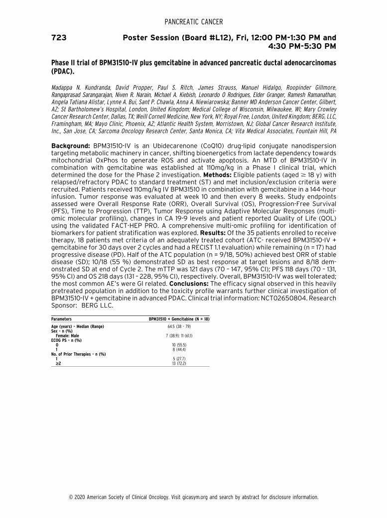
723 Poster Session (Board #L12), Fri, 12:00 PM-1:30 PM and4:30 PM-5:30 PM
Phase II trial of BPM31510-IV plus gemcitabine in advanced pancreatic ductal adenocarcinomas(PDAC).
Madappa N. Kundranda, David Propper, Paul S. Ritch, James Strauss, Manuel Hidalgo, Roopinder Gillmore,Rangaprasad Sarangarajan, Niven R. Narain, Michael A. Kiebish, Leonardo O Rodrigues, Elder Granger, Ramesh Ramanathan,Angela Tatiana Alistar, Lynne A. Bui, Sant P. Chawla, Anna A. Niewiarowska; Banner MD Anderson Cancer Center, Gilbert,AZ; St Bartholomew’s Hospital, London, United Kingdom; Medical College of Wisconsin, Milwaukee, WI; Mary CrowleyCancer Research Center, Dallas, TX; Weill Cornell Medicine, New York, NY; Royal Free, London, United Kingdom; BERG, LLC,Framingham, MA; Mayo Clinic, Phoenix, AZ; Atlantic Health System, Morristown, NJ; Global Cancer Research Institute,Inc., San Jose, CA; Sarcoma Oncology Research Center, Santa Monica, CA; Vita Medical Associates, Fountain Hill, PA
Background: BPM31510-IV is an Ubidecarenone (CoQ10) drug-lipid conjugate nanodispersiontargeting metabolic machinery in cancer, shifting bioenergetics from lactate dependency towardsmitochondrial OxPhos to generate ROS and activate apoptosis. An MTD of BPM31510-IV incombination with gemcitabine was established at 110mg/kg in a Phase I clinical trial, whichdetermined the dose for the Phase 2 investigation. Methods: Eligible patients (aged $ 18 y) withrelapsed/refractory PDAC to standard treatment (ST) and met inclusion/exclusion criteria wererecruited. Patients received 110mg/kg IV BPM31510 in combination with gemcitabine in a 144-hourinfusion. Tumor response was evaluated at week 10 and then every 8 weeks. Study endpointsassessed were Overall Response Rate (ORR), Overall Survival (OS), Progression-Free Survival(PFS), Time to Progression (TTP), Tumor Response using Adaptive Molecular Responses (multi-omic molecular profiling), changes in CA 19-9 levels and patient reported Quality of Life (QOL)using the validated FACT-HEP PRO. A comprehensive multi-omic profiling for identification ofbiomarkers for patient stratification was explored. Results: Of the 35 patients enrolled to receivetherapy, 18 patients met criteria of an adequately treated cohort (ATC- received BPM31510-IV +gemcitabine for 30 days over 2 cycles and had a RECIST 1.1 evaluation) while remaining (n = 17) hadprogressive disease (PD). Half of the ATC population (n = 9/18, 50%) achieved best ORR of stabledisease (SD); 10/18 (55 %) demonstrated SD as best response at target lesions and 8/18 dem-onstrated SD at end of Cycle 2. The mTTP was 121 days (70 – 147, 95% CI); PFS 118 days (70 – 131,95%CI) and OS 218 days (131 – 228, 95%CI), respectively. Overall, BPM31510-IV was well tolerated;the most common AE’s were GI related. Conclusions: The efficacy signal observed in this heavilypretreated population in addition to the toxicity profile warrants further clinical investigation ofBPM31510-IV + gemcitabine in advanced PDAC. Clinical trial information: NCT02650804. ResearchSponsor: BERG LLC.
Parameters BPM31510 + Gemcitabine (N = 18)
Age (years) - Median (Range) 64.5 (38 - 79)Sex - n (%)Female: Male 7 (38.9): 11 (61.1)
ECOG PS - n (%)0 10 (55.5)1 8 (44.4)
No. of Prior Therapies - n (%)1 5 (27.7)‡2 13 (72.2)
© 2020 American Society of Clinical Oncology. Visit gicasym.org and search by abstract for disclosure information.
PANCREATIC CANCER
724 Poster Session (Board #L13), Fri, 12:00 PM-1:30 PM and4:30 PM-5:30 PM
Randomized phase II trial of chemoradiotherapy with S-1 versus combination chemotherapy withgemcitabine and S-1 as neoadjuvant treatment for resectable pancreatic cancer (JASPAC 04).
Hirochika Toyama, Teiichi Sugiura, Akira Fukutomi, Hirofumi Asakura, Yuriko Takeda, Kouji Yamamoto, Satoshi Hirano,Sohei Satoi, Ippei Matsumoto, Shinichiro Takahashi, Soichiro Morinaga, Makoto Yoshida, Yasunaru Sakuma, Hidetaka Iwamoto,Yasuhiro Shimizu, Katsuhiko Uesaka; Division of Hepato-Biliary-Pancreatic Surgery, Kobe University Graduate School of Medicine,Kobe, Japan; Division of Hepato-Biliary-Pancreatic Surgery, Shizuoka Cancer Center, Shizuoka, Japan; Division of GastrointestinalOncology, Shizuoka Cancer Center, Shizuoka, Japan; Radiation and Proton Therapy Center, Shizuoka Cancer Center, Shizuoka,Japan; Department of Biostatistics Yokohama City University School of Medicine, Yokohama, Japan; Department of Gastroen-terological Surgery II, Faculty of Medicine, Hokkaido University, Sapporo, Japan; Department of Surgery, Kansai Medical University,Hirakata, Japan; Department of Surgery, Kindai University, Osaka-Sayama, Japan; National Cancer Center Hospital East, Kashiwa,Japan; Kanagawa Cancer Center, Yokohama, Japan; Department of Medical Oncology, Sapporo Medical University, Sapporo, Japan;Department of Surgery, Jichi Medical University, Tochigi-Shimotsuke, Japan; Division of Metabolism and Biosystemic Science,Department of Medicine, Asahikawa Medical University, Asahikawa, Japan; Department of Gastroenterological Surgery, AichiCancer Center Hospital, Aichi, Japan
Background: Although neoadjuvant treatment (NAT) has been widely employed for resectablepancreatic ductal adenocarcinoma (PDAC), it is still unclear what kind of regimen is recommended.The aim of the study is to investigate which chemoradiotherapy (CRT) with S-1 or combinationchemotherapy with gemcitabine (GEM) and S-1 is more promising as NAT for resectable PDAC interms of effectiveness and safety. Methods: Patients with resectable PDAC were enrolled andrandomly assigned into either CRT group or chemotherapy group. In the CRT group, a totalradiation dose of 50.4 Gy in 28 fractions was administered and S-1, at a dose of 30, 40 or 50 mgaccording to the body surface area, was orally provided twice a day on the same day of irradiation.In the chemotherapy group, GEMwas intravenously administered at a dose of 1000mg/m2 on day 1and 8 and S-1 was orally provided at a dose of 30, 40 or 50 mg according to the body surface areatwice daily on day 1 to 14 followed by one week reset. Patients in the chemotherapy group receivedtwo cycles of this regimen. Surgery was performed between 15 and 56 days after the last day ofNAT. The primary endpoint was 2-year progression-free survival (PFS) rate. With 50 patients ineach group, the study had 80% power assuming a threshold 2-year PFS rate of 25% and anexpected 2-year PFS rate of 40% at 0.05 one-sided alpha. The trial was registered with the UMINClinical Trial Registry as UMIN000014894. Results: From April 2014 and April 2017, 103 patientswere enrolled from 11 institutions in Japan. One was excluded because of ineligibility, therefore 51patients in CRT group and 51 patients in chemotherapy group constituted the intention-to-treatanalysis. The 2-year PFS rate was 45% (90% CI, 33-60%) in the CRT group and 55% (43-65%) inthe chemotherapy group (p = 0.52). The hazard ratio for chemotherapy to CRT was 0.78 (0.46-1.31). Themedian survival timewas 37.7 (95%CI, 30.3-NE) in the CRT group and NE (29.9-NE) in thechemotherapy group (p = 0.30). There was no treatment-related death in both groups.Conclusions: Combination chemotherapy with GEM and S-1 may be more promising comparedwith CRT with S-1 as NAT for resectable PDAC. Clinical trial information: UMIN000014894.Research Sponsor: Pharma-Valley Center, Shizuoka Industrial Foundation.
© 2020 American Society of Clinical Oncology. Visit gicasym.org and search by abstract for disclosure information.
PANCREATIC CANCER
725 Poster Session (Board #L14), Fri, 12:00 PM-1:30 PM and4:30 PM-5:30 PM
Clinical outcome of initially unresectable pancreatic cancer patients: Conversion surgery aftermodified FOLFIRINOX or gemcitabine nab-paclitaxel.
Yuta Ushida, Yosuke Inoue, Hiromichi Ito, Yoshihiro Ono, Takafumi Sato, Yu Takahashi; Japanese Foundation for CancerResearch, Tokyo, Japan
Background: Although pancreatic cancer (PC) is unfavorable clinical entity, the prognosis ofresectable PC has been improving due to perioperative chemotherapy. Meanwhile, the prognosisof unresectable (UR) PC remains poor. In highly selected patients, however, conversion surgery(CS) has been performed with good outcome. Indication criteria of CS remain unestablishedbecause the number of patients who underwent CS was very small in each institution. Methods:From 2014 to 2018, 485 consecutive patients with UR-PC who received modified FOLFIRINOX(mFFX) / Gemcitabine Nab-Paclitaxel (GnP) chemotherapy were reviewed. Among them, patientswith disease control for more than 8 months were enrolled and divided into two groups; patientswho underwent CS (CS group) and patients who did not undergo CS (non-CS group). We comparedclinical characteristics and survival outcomes between groups. Our surgical indications were asfollows: 1) Decreasing trend in CA19-9, 2) With response for chemotherapy in image, 3) Diseasecontrol more than 8 months, 4) Decision in Cancer board as for metastatic cases. Results: In UR-PC patients, 358 patients had distant metastasis (MPC) and 127 patients had locally advanced (LA)PC. The overall survival (OS), progression free survival (PFS) and conversion rate of LAPC weresignificantly better thanMPC (OS; 21 vs. 13months, PFS; 12 vs. 7months, Conversion rate; 16 vs. 5%,p, 0.001). Chemotherapy regimen (mFFX/GnP) had no significant difference in survival outcome.Between CS group (n = 39) with non-CS group (n = 160), age, sex, body mass index, location oflesion, CEA, CA19-9, regimen of chemotherapy and histology had no significant differences. Themedian survival time of CS group was significantly better than that of non-CS group (OS; NA vs. 21months, p,0.001, PFS; 24 vs. 14months, p = 0.01). In CS group,median operative durationwas 509minutes, blood losswas 735ml, hospital staywas 26 days, and therewas no 90-daysmortality case.Conclusions: In our retrospective study, CS for UR-PC can be safely performed, and amongcarefully selected patients, reasonable short and long term outcomes can be obtained withoutacceptable morbidity rate. Research Sponsor: None.
© 2020 American Society of Clinical Oncology. Visit gicasym.org and search by abstract for disclosure information.
PANCREATIC CANCER
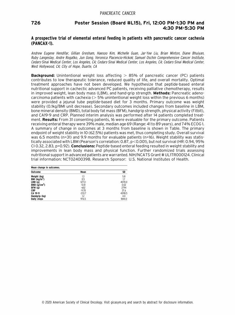
726 Poster Session (Board #L15), Fri, 12:00 PM-1:30 PM and4:30 PM-5:30 PM
A prospective trial of elemental enteral feeding in patients with pancreatic cancer cachexia(PANCAX-1).
Andrew Eugene Hendifar, Gillian Gresham, Haesoo Kim, Michelle Guan, Jar-Yee Liu, Brian Minton, Diane Bhuiyan,Ruby Langeslay, Andre Rogatko, Jun Gong, Veronica Placencio-Hickok; Samuel Oschin Comprehensive Cancer Institute,Cedars-Sinai Medical Center, Los Angeles, CA; Cedars-Sinai Medical Center, Los Angeles, CA; Cedars-Sinai Medical Center,West Hollywood, CA; City of Hope, Duarte, CA
Background: Unintentional weight loss affecting . 85% of pancreatic cancer (PC) patientscontributes to low therapeutic tolerance, reduced quality of life, and overall mortality. Optimaltreatment approaches have not been developed. We hypothesize that peptide-based enteralnutritional support in cachectic advanced PC patients, receiving palliative chemotherapy, resultsin improved weight, lean body mass (LBM), and hand-grip strength. Methods: Pancreatic adeno-carcinoma patients with cachexia (. 5% unintentional weight loss within the previous 6 months)were provided a jejunal tube peptide-based diet for 3 months. Primary outcome was weightstability (0.1kg/BMI unit decrease). Secondary outcomes included changes from baseline in LBM,bonemineral density (BMD), total body fat mass (BFM), handgrip strength, physical activity (Fitbit),and CA19-9 and CRP. Planned interim analysis was performed after 14 patients completed treat-ment. Results: From 31 consenting patients, 16 were evaluable for the primary outcome. Patientsreceiving enteral therapy were 39%male, median age 69 (Range: 41 to 89 years), and 74%ECOG 1.A summary of change in outcomes at 3 months from baseline is shown in Table. The primaryendpoint of weight stability in 10 (62.5%) patients wasmet, thus completing study. Overall survivalwas 6.5 months (n=31) and 9.9 months for evaluable patients (n=16). Weight stability was statis-tically associated with LBM (Pearson’s correlation: 0.87, p,0.001), but not survival (HR: 0.94, 95%CI 0.32, 2.83, p=0.92). Conclusions: Peptide-based enteral feeding resulted in weight stability andimprovements in lean body mass and physical function. Further randomized trials assessingnutritional support in advanced patients arewarranted. NIH/NCATSGrant #UL1TR000124. Clinicaltrial information: NCT02400398. Research Sponsor: U.S. National Institutes of Health.
Mean change in outcomes.
Outcome Mean SD
Weight (kg) 1.3 5.8BMI (kg/m2) 0.5 1.7LBM (g) 1273.1 4078.0BMD (g/cm2) -0.01 0.02BFM (g) -602 2794CRP -9.78 37.6CA 19-9 -233.7 43118.5Handgrip (kg) 1.2 4.0Daily steps 976.5 1844.0
© 2020 American Society of Clinical Oncology. Visit gicasym.org and search by abstract for disclosure information.
PANCREATIC CANCER
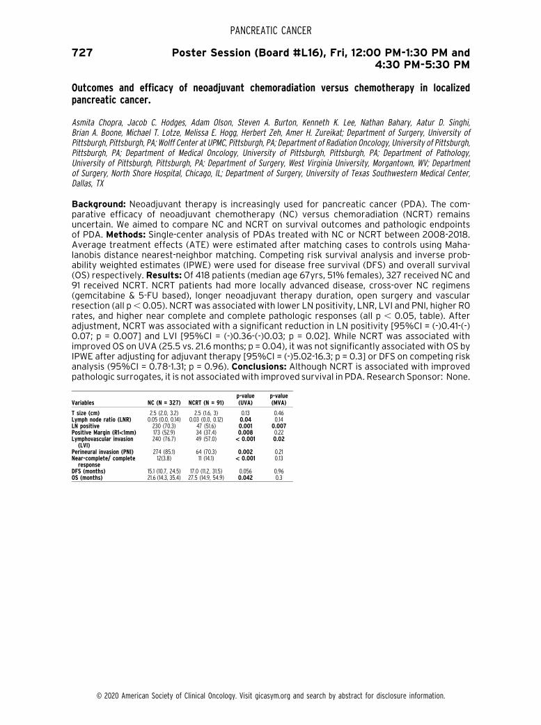
727 Poster Session (Board #L16), Fri, 12:00 PM-1:30 PM and4:30 PM-5:30 PM
Outcomes and efficacy of neoadjuvant chemoradiation versus chemotherapy in localizedpancreatic cancer.
Asmita Chopra, Jacob C. Hodges, Adam Olson, Steven A. Burton, Kenneth K. Lee, Nathan Bahary, Aatur D. Singhi,Brian A. Boone, Michael T. Lotze, Melissa E. Hogg, Herbert Zeh, Amer H. Zureikat; Department of Surgery, University ofPittsburgh, Pittsburgh, PA; Wolff Center at UPMC, Pittsburgh, PA; Department of Radiation Oncology, University of Pittsburgh,Pittsburgh, PA; Department of Medical Oncology, University of Pittsburgh, Pittsburgh, PA; Department of Pathology,University of Pittsburgh, Pittsburgh, PA; Department of Surgery, West Virginia University, Morgantown, WV; Departmentof Surgery, North Shore Hospital, Chicago, IL; Department of Surgery, University of Texas Southwestern Medical Center,Dallas, TX
Background: Neoadjuvant therapy is increasingly used for pancreatic cancer (PDA). The com-parative efficacy of neoadjuvant chemotherapy (NC) versus chemoradiation (NCRT) remainsuncertain. We aimed to compare NC and NCRT on survival outcomes and pathologic endpointsof PDA. Methods: Single-center analysis of PDAs treated with NC or NCRT between 2008-2018.Average treatment effects (ATE) were estimated after matching cases to controls using Maha-lanobis distance nearest-neighbor matching. Competing risk survival analysis and inverse prob-ability weighted estimates (IPWE) were used for disease free survival (DFS) and overall survival(OS) respectively. Results: Of 418 patients (median age 67yrs, 51% females), 327 received NC and91 received NCRT. NCRT patients had more locally advanced disease, cross-over NC regimens(gemcitabine & 5-FU based), longer neoadjuvant therapy duration, open surgery and vascularresection (all p, 0.05). NCRTwas associated with lower LN positivity, LNR, LVI and PNI, higher R0rates, and higher near complete and complete pathologic responses (all p , 0.05, table). Afteradjustment, NCRT was associated with a significant reduction in LN positivity [95%CI = (-)0.41-(-)0.07; p = 0.007] and LVI [95%CI = (-)0.36-(-)0.03; p = 0.02]. While NCRT was associated withimproved OS on UVA (25.5 vs. 21.6 months; p = 0.04), it was not significantly associated with OS byIPWE after adjusting for adjuvant therapy [95%CI = (-)5.02-16.3; p = 0.3] or DFS on competing riskanalysis (95%CI = 0.78-1.31; p = 0.96). Conclusions: Although NCRT is associated with improvedpathologic surrogates, it is not associatedwith improved survival in PDA. Research Sponsor: None.
Variables NC (N = 327) NCRT (N = 91)p-value(UVA)
p-value(MVA)
T size (cm) 2.5 (2.0, 3.2) 2.5 (1.6, 3) 0.13 0.46Lymph node ratio (LNR) 0.05 (0.0, 0.14) 0.03 (0.0, 0.12) 0.04 0.14LN positive 230 (70.3) 47 (51.6) 0.001 0.007Positive Margin (R1<1mm) 173 (52.9) 34 (37.4) 0.008 0.22Lymphovascular invasion(LVI)
240 (76.7) 49 (57.0) < 0.001 0.02
Perineural invasion (PNI) 274 (85.1) 64 (70.3) 0.002 0.21Near-complete/ completeresponse
12(3.8) 11 (14.1) < 0.001 0.13
DFS (months) 15.1 (10.7, 24.5) 17.0 (11.2, 31.5) 0.056 0.96OS (months) 21.6 (14.3, 35.4) 27.5 (14.9, 54.9) 0.042 0.3
© 2020 American Society of Clinical Oncology. Visit gicasym.org and search by abstract for disclosure information.
PANCREATIC CANCER
729 Poster Session (Board #L18), Fri, 12:00 PM-1:30 PM and4:30 PM-5:30 PM
Establishment of patient-derived tumor xenograft (PDX) model as artificially-created livermetastasis using cryopreserved pancreatic ductal adenocarcinoma.
Kenjiro Kimura, Ryota Tanaka, Ryosuke Amano, Shoji Kubo, Hiroaki Tanaka, Kazuya Muguruma, Kosei Hirakawa,Masaichi Ohira; Department of Hepato-Biliary-Pancreatic Surgery, Osaka City University Graduate School of Medicine,Osaka, Japan; Department of Gastroenterological Surgery, Osaka City University Graduate School of Medicine, Osaka, Japan;Kashiwara Municipal Hospital, Kashiwara, Japan
Background: Translational research using patient-derived tumor xenograft (PDX) model is pro-gressing rapidly, and is also becoming widespread in pancreatic cancer research. The purpose ofthis study was to establish the liver transplant PDX model as artificially-created liver metastasiswith cryopreserved primary pancreatic ductal adenocarcinoma(PDAC). Methods: The primaryPDAC from 10 patients were cryopreserved and transplanted into NSG mice using liver pocketmethod. For engraftment and similarity evaluation, H&E staining and immunohistochemicalstaining such as Ki-67, p53, SMAD4, andMUC1 were performed.Results: Patient-derived xenograftwas succeeded in 6 cases (60%), 10 mice (33.3%). Ki-67 index of primary PDAC and the interval ofcryopreservation were significantly related to successful engraftment, respectively (p = 0.003, p =0.007). Conclusions: In this study, we succeeded in establishing a liver transplant PDX mousemodel as a preclinical platform. The factors such as Ki-67 index and the interval of cryopreser-vation would affect the successful establishment. Research Sponsor: None.
© 2020 American Society of Clinical Oncology. Visit gicasym.org and search by abstract for disclosure information.
PANCREATIC CANCER
730 Poster Session (Board #L19), Fri, 12:00 PM-1:30 PM and4:30 PM-5:30 PM
Prognostic and predictive value of circulating tumour DNA (ctDNA) by amplicon-based nextgeneration sequencing (NGS) of advanced pancreatic cancer (APC) in a phase I trial of oxaliplatincapecitabine and irinotecan (OXIRI) triplet chemotherapy.
Amanda Oon Lim Seet, Su Pin Choo, David Wai-Meng Tai, Justina Yick Ching Lam, Tira Jing Ying Tan, Daniel Shao-Weng Tan,Aaron C. Tan, Yvonne Chang, Balram Chowbay, Matthew C.H. Ng; National Cancer Center, Singapore, Singapore; CurieOncology, Singapore, Singapore; National Cancer Center Singapore, Singapore, Singapore; National Cancer Centre,Singapore, Singapore; National Cancer Centre Singapore, Singapore, Singapore
Background: Tumoral KRASmutations (KRASmt) are detected in ~80%ofAPC and associatedwitha negative prognosis. Digital droplet PCR (ddPCR) has high sensitivity for circulating KRASmtbutnarrower gene coverage. NGS using hybrid-capture methods has reported ctDNA KRASmtin ~30%of patients (pt). We previously presented clinical results of the Phase I trial of OXIRI (GI ASCO 18#411). We now present the first prospective evaluation of ctDNA in APC by an amplicon-based NGSapproach in this Phase I trial.Methods: Paired ctDNA and CA19-9 samples were taken at baseline,C2D1, C3D1 and end of trial. A targeted panel with error-correction (Lucence Diagnostics) was usedto detect for mtin KRAS, TP53, SMAD4, CDKN2A, CTNNB1, GNAS, APC and MYC. CT scans wereperformed every 2 cycles. Survival curves by Kaplan Meier were compared by mutational status,ctDNA and CA19-9 response using the log-rank test. Spearman correlation of ctDNA and CA19-9changes was performed. Results: ctDNAmtwas detected at baseline in 19/23 (83%) samples,comprising KRAS 17/23 (73%), TP53 (61%), SMAD4 (48%) and CDKN2A (30%). KRASmtandSMAD4mt conferred a negative prognosis for overall survival with a hazard ratio of 4.2 (CI: 1.6-10.4; p = 0.01) and 2.8 (CI: 0.9-8.65; p = 0.01) respectively. Drop in ctDNA andCA19-9was associatedwith a trend for longer progression free survival at C2D1 (both) and C3D1 (ctDNA only). Radiologicalpartial response (PR) was associated with lower ctDNA in 5/5 pt and CA 19-9 in 4/5 pt. Decrease inctDNA and CA19-9 was associated with disease control (PR/SD) at C2D1 in 11/14 pt and 10/10 pt; atC3D1 in 11/12 pt and 6/7 respectively. No significant correlation between the amplitude of CA19-9and ctDNA changes was found. Conclusions: ctDNA could be detected in 83% of pts of whomKRASmtrates were similar to reports using tissue NGS. Determination of RASmtand SMAD4mtinctDNA may aid in the prognostication of pts and decrease in ctDNA levels may predict fortreatment benefit, similar in extent to CA 19-9. This may be particularly useful in non-CA19-9secreting APC as an adjunct to imaging. Clinical trial information: NCT02368860. ResearchSponsor: National Medical Research Council Singapore.
© 2020 American Society of Clinical Oncology. Visit gicasym.org and search by abstract for disclosure information.
PANCREATIC CANCER
731 Poster Session (Board #L20), Fri, 12:00 PM-1:30 PM and4:30 PM-5:30 PM
DNA-damage repair deficiency (dDDR) and response to nanoliposomal irinotecan (nal-IRI) inmetastatic pancreatic ductal adenocarcinoma (mPDAC).
Sameeha Rau, Andrea P. Espejo Freire, Gretel Terrero, Manisha Jayandra Patel, Benjamin Woodress Rush,Caio Max Sao Pedro Rocha Lima, Peter Joel Hosein; University of Miami Miller School of Medicine, Miami, FL; Universityof Miami-Miller School of Medicine, Jackson Memorial Hospital, Miami, FL; Jackson Health System, Miami, FL; Wake ForestBaptist Health, Hematology and Oncology-Heme Onc Fellowship Program, Winston Salem, NC; NRG Oncology and Wake ForestUniversity Baptist Medical Center, Winston-Salem, NC; University of Miami Sylvester Comprehensive Cancer Center, Miami, FL
Background: Chemotherapy is the standard of care for patients with mPDAC but there are nobiomarkers to aid in treatment selection. Nal-IRI with 5-fluorouracil/folinic acid (FUFA) improvessurvival over FUFA in the second-line treatment ofmPDAC. Nal-IRI is a topoisomerase inhibitor andits action produces DNA damage leading to cell death. We hypothesize that tumors with dDDR, aprocess that is altered in a subset of patients with PDAC, may be more sensitive to the effects ofnal-IRI.Methods: Utilizing the IRB-approved pancreas cancer databases at the University of Miamiand Wake Forest University, we identified patients with mPDAC treated with nal-IRI and FUFA whohad germline and/or somatic mutation testing. We conducted a retrospective chart review toextract demographic and clinical characteristics including treatments received, response, andsurvival. Results: Among 31 patients identified, the median age was 66y and 47% were female.Nine patients had a DDR mutation; 6 germline and 3 somatic. Median progression-free survival(PFS) in patients with any germline or somatic DDR mutation was 3.2m vs 3.9m for those without(log-rank p = 0.7). When restricted to germline DDR mutations only, the median PFS was notreached with germline dDDR vs 4m for those without (log-rank p = 0.22). Presence of dDDR wasassociated with a higher clinical benefit rate (CBR = partial response + stable disease); a DDRmutation was present in 36% of patients who showed clinical benefit vs 15% in those withoutclinical benefit (p = 0.21). Conclusions: DDR mutations appear to define a subset of patients withmPDACwhomay bemore sensitive to nal-IRI and FUFA. The PFS and CBRwere numerically but notstatistically superior, especially in patients with germline DDR mutations. Larger data sets andlonger follow-up are needed to confirm this trend. Research Sponsor: None.
© 2020 American Society of Clinical Oncology. Visit gicasym.org and search by abstract for disclosure information.
PANCREATIC CANCER
732 Poster Session (Board #L21), Fri, 12:00 PM-1:30 PM and4:30 PM-5:30 PM
Homologous recombination deficiency (HRD) by BROCA-HR and survival outcomes after surgeryfor patients (pts) with pancreatic adenocarcinoma (PC): A single institution experience.
Amy E. Chang, David Bing Zhen, Marc Radke, Kelsey K. Baker, Andrew L. Coveler, Kit Man Wong, Venu Gopal Pillarisetty,Mary Weber Redman, Elizabeth Swisher, E. Gabriela Chiorean; University of Michigan, Ann Arbor, MI; University of WashingtonSchool of Medicine, Seattle, WA; Fred Hutchinson Cancer Research Center, Seattle, WA; Seattle Cancer Care Alliance/University of Washington, Seattle, WA; University of Colorado, Denver, CO; University of Washington, Seattle, WA; SWOGStatistical Center; Fred Hutchinson Cancer Research Center, Seattle, WA; University of Washington/Seattle Cancer CareAlliance, Seattle, WA; University of Washington and Fred Hutchinson Cancer Research Center, Seattle, WA
Background: 5-7%of PC pts exhibit deleterious germlinemutations (MUT) in HR tumor suppressorgenes BRCA1 and BRCA2. BROCA-HR is a targeted capture andmassively parallel sequencing assaydesigned to detect all mutation classes including gene rearrangements, copy number variations,and gene aberrations within the Fanconi Anemia-BRCA HR, non-homologous end joining (NHEJ)DNA repair, and DNA mismatch repair pathways. BROCA-HR has been successfully used in breastand ovarian cancer pts for overall prognosis and prediction of response to platinum-basedtherapies. While BRCA1/2 MUT may confer survival advantage for PC pts if treated withplatinum-chemotherapy, the survival impact of HRD is less well defined. Methods: We retrospec-tively identified 100 consecutive pts who underwent surgical resection for suspected PC atUniversity of Washington Medical Center between 1999 and 2008. Formalin-fixed paraffin em-bedded resected tumors were sequenced using BROCA-HR. HRD was grouped based on thefollowing deleterious genetic mutations: 1) BRCA1, BRCA2; 2) core HRD: BARD1, BRIP1, RAD51C,RAD51D, PALB2, CDK12,NBN; 3) non-core HRD: ATM, ATR,ATRX, BAP1, BLM, CHEK1/2, ERCC, FANCA/C/D2/E /F/G/L, MRE11, RAD50/51/51B, RIF1, SLX4; 4) HR proficient. Overall survival (OS) wasmeasured from diagnosis until death or last follow-up.Results: 95 pts had histologically confirmedPC, and 81 pts had adequate tumor DNA for analysis. Six pts (7%) had BRCA1/2 MUT (n = 5), orBRCA1methylation (n = 1), 1 pt (1%) had non-BRCA core HRD (PALB2MUT), 7 pts (9%) had non-coreHRD: ERCC (2), CHEK2 (2), ATR, RAD51D, and FANCA MUT (1 each). Median OS was: all pts 1.93 yrs(95%C.I. 1.53, 2.16), BRCA1/2 pts 3.09 yrs (95%CI 0.41, 12.21), all core HRD pts 1.21 yrs (95%CI 0.41,12.21), all core and non-core HRD pts 1.89 yrs (95%CI 0.57, 4.96), HR proficient pts 1.93 yrs (95%CI1.51, 2.15). There were no OS differences between pts with HRD vs those HR proficient.Conclusions: HRD is common (17%) but does not affect OS for pts with resected PC. Prospectiveclinical trials should test neo/adjuvant therapies including platinum chemotherapy and PARPinhibitors for pts with HRD. Research Sponsor: U.S. National Institutes of Health.
© 2020 American Society of Clinical Oncology. Visit gicasym.org and search by abstract for disclosure information.
PANCREATIC CANCER
733 Poster Session (Board #L22), Fri, 12:00 PM-1:30 PM and4:30 PM-5:30 PM
Loss of Rnf43 to accelerate KRAS-mediated neoplasia in a clinically relevant geneticallyengineered mouse model of pancreatic adenocarcinoma.
Abdel Nasser Hosein, Gita Dangol, Takashi Okumura, Lotfi Abou-Elkacem, Anirban Maitra; University of Texas MD AndersonCancer Center, Houston, TX
Background: The genetic heterogeneity of pancreatic ductal adenocarcinoma (PDAC) demands apersonalized molecular-targeted treatment approach. While activating KRASmutations are a nearubiquitous event in PDAC pathogenesis, 5-10% of cases display deleterious driver mutations in theWnt-signaling negative regulator, ring finger 43 (RNF43). Despite this characterization there areno personalized treatment options for this subset of patients. Methods: We have developed agenetically engineeredmousemodel (GEMM) of PDAC, driven by an activatingmutation inKras anddeletion of Rnf43 under control of a pancreas specific promoter (KRC). Mice were followed forsurvival outcomes and histological changes which were compared to a Kras driven PDAC GEMM(KC). Mice underwent serial magnetic resonance imaging (MRI), with and without dynamic contrastenhanced (DCE) imaging, to evaluate cystic tumor morphology and contrast enhancement duringtumor progression. Single cell RNA sequencing (scRNAseq) was also performed to assess changesin single cell populations during tumor progression. Lastly, we established ex vivo cultures fromKRC and KC tumors and performed bulk RNA-sequencing (RNAseq) and in vitro pharmacologystudies. Results: KRC mice displayed a decrease in overall survival and higher incidence of bothhigh grade pre-neoplastic lesions and invasive PDAC compared to KC mice. Serial MRI revealedincreased cystic morphology of KRCmice during tumor progression with increasing DCE intensity.scRNAseq of KRC tumors from moribund mice displayed two distinct populations of both mac-rophages and fibroblasts, similar to our previous report of Kras/Tp53 and Kras/Ink4a drivenGEMMs. Lastly, primary cultures from KRC tumors demonstrated increased expression of Wnt-related genes by RNAseq and increased sensitivity to small molecule porcupine inhibition relativeto KC cell lines, demonstrating functional Wnt dependence of the KRC system.Conclusions: Rnf43is a bona fide tumor suppressor gene in PDAC. This GEMM is a novel platform for drug discovery inRNF43-mutated PDAC with the eventual goal of implementing a precision oncology approach inthese patients. Research Sponsor: UT MD Anderson Pancreatic Cancer Moonshot Initiative.
© 2020 American Society of Clinical Oncology. Visit gicasym.org and search by abstract for disclosure information.
PANCREATIC CANCER
734 Poster Session (Board #M1), Fri, 12:00 PM-1:30 PM and4:30 PM-5:30 PM
Tumor infiltration and cytokine biomarkers of prostate stem cell antigen (PSCA)-directedGOCAR-T cells in patients with advanced pancreatic tumors.
Joanne Shaw, Brandon Ballard, Xiaohui Yi, Aditya Malankar, Matthew R. Collinson-Pautz, Carlos Roberto Becerra,Joseph Paul Woodard, Aaron E. Foster; Bellicum Pharmaceuticals, Inc., Houston, TX; Baylor University Medical Center,Dallas, TX
Background: PSCA is a cell surface protein overexpressed in approximately 60% of pancreaticcancers. BPX-601 is an autologous GOCAR-T cell therapy engineered to express a PSCA-CD3z CARand the MyD88/CD40 (iMC) costimulatory domain activated by rimiducid (Rim), designed to boostCAR-T performance in solid tumors. The safety and activity of BPX-601 activatedwith Rim in PSCA+
metastatic pancreatic cancer is being assessed in a Phase 1/2 clinical trial, BP-012 (NCT02744287).Methods: Phase 1 of BP-012 is a 3+3 dose escalation of BPX-601 (1.25-5 x10⁶/kg) administered onDay 0 with a single, fixed-dose of Rim (0.4 mg/kg) on Day 7 in subjects with previously treatedPSCA+ metastatic pancreatic cancer. All 5 subjects in cohort 5B received Flu/Cy lymphodepletionfollowed by BPX-601 (5 x10⁶/kg) and Rim. BPX-601 kinetics, PBMC phenotype, and serum cytokineswere assayed by qPCR, flow cytometry, and cytokine multiplex, respectively. Baseline and on-treatment biopsies were evaluated by RNAscope in situ hybridization. Results: BPX-601 cellsexpanded in all subjects and persisted up to 9 months (median 42 days). Transient reduction inBPX-601 vector copy number and total T cell count concurrent with Rim infusion, supportsmargination of activated BPX-601 cells. Increased serum cytokines, such as IFN-g and GM-CSF,were observed following BPX-601 infusion with further elevation after Rim activation. All subjectswith evaluable on-treatment biopsies had infiltration of BPX-601 cells (n = 3) proximal to tumorcells 7-15 days after Rim, but not in an end of treatment biopsy . 200 days after Rim (n = 1).Stratification by best response (RECIST 1.1) revealed stable disease in 3 subjects and progressivedisease in 2 subjects was potentially associated with distinct cytokine signatures. Conclusions:BPX-601 GOCAR-T cells expand and persist in patients with PSCA+ metastatic pancreatic cancerand infiltrate metastatic lesions. A peripheral cytokine signature was observed following BPX-601infusion. Select cytokines were enhanced after GOCAR-T cell activation and may correlate withclinical response. A cohort of subjects exploring serial administration of Rim is open for enrollment.Clinical trial information: NCT02744287. Research Sponsor: Bellicum Pharmaceuticals, Inc.
© 2020 American Society of Clinical Oncology. Visit gicasym.org and search by abstract for disclosure information.
PANCREATIC CANCER
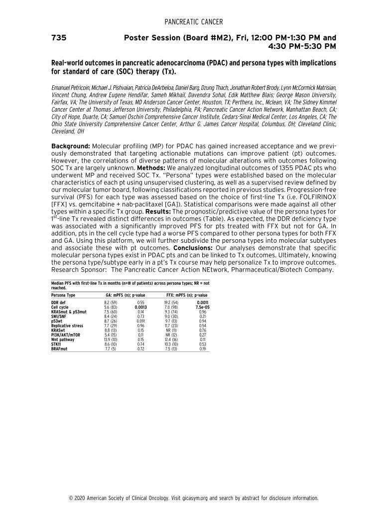
735 Poster Session (Board #M2), Fri, 12:00 PM-1:30 PM and4:30 PM-5:30 PM
Real-world outcomes in pancreatic adenocarcinoma (PDAC) and persona types with implicationsfor standard of care (SOC) therapy (Tx).
Emanuel Petricoin, Michael J. Pishvaian, Patricia DeArbeloa, Daniel Barg, Dzung Thach, Jonathan Robert Brody, LynnMcCormickMatrisian,Vincent Chung, Andrew Eugene Hendifar, Sameh Mikhail, Davendra Sohal, Edik Matthew Blais; George Mason University,Fairfax, VA; The University of Texas, MD Anderson Cancer Center, Houston, TX; Perthera, Inc., Mclean, VA; The Sidney KimmelCancer Center at Thomas Jefferson University, Philadelphia, PA; Pancreatic Cancer Action Network, Manhattan Beach, CA;City of Hope, Duarte, CA; Samuel Oschin Comprehensive Cancer Institute, Cedars-Sinai Medical Center, Los Angeles, CA; TheOhio State University Comprehensive Cancer Center, Arthur G. James Cancer Hospital, Columbus, OH; Cleveland Clinic,Cleveland, OH
Background: Molecular profiling (MP) for PDAC has gained increased acceptance and we previ-ously demonstrated that targeting actionable mutations can improve patient (pt) outcomes.However, the correlations of diverse patterns of molecular alterations with outcomes followingSOC Tx are largely unknown.Methods:We analyzed longitudinal outcomes of 1355 PDAC pts whounderwent MP and received SOC Tx. “Persona” types were established based on the molecularcharacteristics of each pt using unsupervised clustering, as well as a supervised review defined byourmolecular tumor board, following classifications reported in previous studies. Progression-freesurvival (PFS) for each type was assessed based on the choice of first-line Tx (i.e. FOLFIRINOX[FFX] vs. gemcitabine + nab-paclitaxel [GA]). Statistical comparisons were made against all othertypes within a specific Tx group.Results: The prognostic/predictive value of the persona types for1st-line Tx revealed distinct differences in outcomes (Table). As expected, the DDR deficiency typewas associated with a significantly improved PFS for pts treated with FFX but not for GA. Inaddition, pts in the cell cycle type had a worse PFS compared to other persona types for both FFXand GA. Using this platform, we will further subdivide the persona types into molecular subtypesand associate these with pt outcomes. Conclusions: Our analyses demonstrate that specificmolecular persona types exist in PDAC pts and can be linked to Tx outcomes. Ultimately, knowingthe persona type/subtype early in a pt’s Tx course may help personalize Tx to improve outcomes.Research Sponsor: The Pancreatic Cancer Action NEtwork, Pharmaceutical/Biotech Company.
Median PFS with first-line Tx in months (n=# of patients) across persona types; NR = notreached.
Persona Type GA: mPFS (n); p-value FFX: mPFS (n); p-value
DDR def 8.2 (59) 0.55 19.2 (54) 0.0011Cell cycle 5.6 (83) 0.0013 7.0 (98) 7.5e-05KRASmut & p53mut 7.5 (60) 0.14 9.3 (74) 0.96SWI/SNF 8.4 (24) 0.73 9.0 (30) 0.21p53wt 8.7 (26) 0.091 9.7 (13) 0.94Replicative stress 7.7 (29) 0.96 11.7 (23) 0.54KRASwt 8.8 (13) 0.15 NR (11) 0.76PI3K/AKT/mTOR 5.4 (15) 0.11 NR (12) 0.27Wnt pathway 13.9 (10) 0.15 12.4 (16) 0.11STK11 8.6 (10) 0.74 10.3 (10) 0.53BRAFmut 7.7 (5) 0.72 7.5 (13) 0.19
© 2020 American Society of Clinical Oncology. Visit gicasym.org and search by abstract for disclosure information.
PANCREATIC CANCER
736 Poster Session (Board #M3), Fri, 12:00 PM-1:30 PM and4:30 PM-5:30 PM
Gene expression profiling of unresectable pancreatic cancer patients treated with gemcitabine,nab-paclitaxel, metformin, and dietary supplements (DS).
Vincent Chung, Paul Henry Frankel, Stephen Shibata, Isa Mambetsariev, Holly Yin, Tamara Mirzapoiazova, Bolot Mambetsariev,Prakash Kulkarni, Dean Lim, Daneng Li, Joseph Chao, Marwan Fakih, Andrea Bild, Raju K. Pillai, Orn Adalsteinsson, Steven Hirsh,Ravi Salgia; City of Hope, Duarte, CA; City of Hope Comprehensive Cancer Center, Duarte, CA; City of Hope Natl Medcl Ctr, Duarte,CA; City of Hope National Medical Center, Duarte, CA; Life Extension, Fort Lauderdale, FL
Background: Pancreatic cancer commonly causes weight loss and many patients take supple-ments to improve nutrition but also for any potential anticancer properties. We conducted a pilottrial evaluating a standardized combination of dietary supplements, metformin and gemcitabineplus nab-paclitaxel chemotherapy. Methods: The supplements were custom designed by a nutri-tionally trained oncologist. 17 supplements were given as 12 pills and 2 smoothie packets dividedtwice per day. Patients began metformin on day (D)-6 to evaluate GI tolerance before starting theDS on D-3. On D1, gemcitabine and nab-paclitaxel chemotherapy was administered at standarddoses. Serum samples were collected D-6, C1D1, C3D1 and at the end of treatment. RNA wasextracted from PBMC samples and a total of 48 samples were analyzed from 19 patients.NanoString Human Immune Profiling and PanCancer Pathways were performed to evaluatechanges in gene expression with treatment. Results: The differential expression of genes wassmall with dietary supplements alone and in combination with chemotherapy. After 2 months oftreatment, 17 genes were upregulated while 4 genes were down regulated. At the time of tumorresistance, 33 genes were upregulated while 17 genes were downregulated. We observed down-regulation of tumor suppressor gene expression with upregulation in growth factor pathways.Interestingly, SPRY2 mRNA expression was also up regulated which functions as a negativefeedback regulator of multiple receptor tyrosine kinases. Conclusions: Increasing number ofgenes were upregulated with continued treatment. The most common pathway affected was cellcycle and apoptosis. During the initial supplement run-in period from D-6 to C1D1, IL-8 mRNAexpression was upregulated the most. IL-8 is a neutrophil chemotactic factor secreted by cellsinvolved in the innate immune response as well as pancreatic cancer cells associated with a pro-inflammatory state. Treatment with gemcitabine and nab-paclitaxel decreased the IL-8 mRNAexpression. Additional studies with longer course treatment with supplements alone would berequired to explore its impact on differential gene expression. Research Sponsor: BiomedicalResearch and Longevity Society, Inc.
© 2020 American Society of Clinical Oncology. Visit gicasym.org and search by abstract for disclosure information.
PANCREATIC CANCER
737 Poster Session (Board #M4), Fri, 12:00 PM-1:30 PM and4:30 PM-5:30 PM
Comparison of gemcitabine delivery and tumor response in a pressurized pancreatic retrogradevenous infusion versus systemic infusion in an orthotopic murine model.
Diego Vicente, Jayanth Shankara Narayanan, Partha Ray, Louis F. Chai, Suna Erdem, Matthew Carr, Benedict Capacio,Bryan Cox, David Jaroch, Steven C. Katz, Rebekah Ruth White; Moores Cancer Center, San Diego, CA; Roger Williams CancerCenter, Providence, RI; TriSalus Life Sciences, Westminster, CO
Background: Pancreatic ductal adenocarcinoma (PDAC) is associated with limited response tosystemic therapy (ST). Elevated tumor interstitial fluid pressures (IFP) inhibit penetration of ST.Regional Pressure Enabled Drug Delivery has recently demonstrated improved response for livertumors in a clinical trial. However, this delivery method has not been evaluated in PDAC. Wecompared gemcitabine (Gem) by systemic delivery vs. a novel pressurized Pancreatic RetrogradeVenous Infusion (PRVI) method in an orthotopic PDAC mouse model. Methods: PDAC murine cellline (KPC4580P) tumors were transplanted onto the pancreatic tail of C57BL/6J mice. Groups of15 mice were randomly assigned to PRVI Gem, PRVI saline (Control), or intraperitoneal Gem(Systemic) groups. Fivemice from the PRVI and Systemic groups were randomly selected after onehour post infusion to evaluate Gem tumor concentrations by liquid chromatography - tandemmassspectrometry (ng/mg), and the remainder of mice were euthanized after 7 days to evaluatetreatment response. Results: Tumor concentrations of Gem were significantly higher followingPRVI compared to Systemic (128 vs. 19, p , 0.01) at one hour after treatment. Seven days aftertreatment, PRVI Gemmice demonstrated lowermean tumor volume (mm3) than Systemic GemandControl mice (274 vs. 857 vs. 629, p , 0.01), respectively. Histologic evaluation of tumorsdemonstrated decreased cellularity in the PRVI Gem mice compared to Systemic and Controlmice (35 vs. 78 vs. 71%, p = 0.01), respectively. No differences were seen in Ki67% or immune cellinfiltrate between groups. Conclusions: PRVI delivery resulted in increased PDAC Gem concen-trations and improved treatment responses with decreased tumor burden and cellularity. Thesefindings suggest that pressurized regional chemotherapy infusion overcomes the elevated PDACIFP and justifies additional translational pre-clinical studies with other chemotherapeutics (in-cluding immunomodulating antibodies) with different physicochemical properties. Research Spon-sor: TriSalus Life Sciences, Westminster, CO.
© 2020 American Society of Clinical Oncology. Visit gicasym.org and search by abstract for disclosure information.
PANCREATIC CANCER
738 Poster Session (Board #M5), Fri, 12:00 PM-1:30 PM and4:30 PM-5:30 PM
Pharmacogenomic blood-based assay to predict chemotherapy response and survival inpancreatic cancer.
Kenneth H. Yu, Brian McCarthy, William H. Isacoff, Brandon Cooper, Andrew Bartlett, Jennifer Park, Fay Purcell,Devan McCarthy, Eileen Mary O’Reilly; Memorial Sloan Kettering Cancer Center/Weill Cornell Medical College, New York,NY; Adera Biolabs, Germantown, MD; Univ of California, Los Angeles, CA; University of Maryland Greenebaum ComprehensiveCancer Center, Baltimore, MD; Adera Biooncology, LLC, Germantown, MD; Memorial Sloan Kettering Cancer Center, New York,NY; The Pancreatic Cancer Center of Los Angeles, Los Angeles, CA
Background:Pancreatic adenocarcinoma (PDAC) is formost patients a refractory disease. Moderncytotoxic chemotherapeutics (C) are not yet optimal for inducing responses and extending life. Weare developing a blood-based pharmacogenomic (PGx) assay profiling circulating tumor andinvasive cells (CTICs) to predict sensitivity and resistance to C. Methods: The PGx assay wasstudied in two cohorts of patients (pts) presenting for frontline C therapy of metastatic PDAC.Cohort 1 is from an ongoing prospective study (planned n=80); pts are enrolled prior to receivingeither FOLFIRINOX or gemcitabine (Gem)/nab-paclitaxel (Nab-P). Cohort 2 (n=50) consists of ptsenrolled prior to receiving bespoke combinations of C agents informed by the assay. 6 mL ofperipheral blood was collected from pts at baseline and while on C therapy. CTICs were isolated bypreviously described collagen invasion assay, total RNA was extracted and gene-expressionanalysis was performed. PGxmodels for seven C agents used in PDACwere applied, and correlatedto treatment received. Pts were classified as sensitive if C received were predicted to be effectiveand resistant if not. Objective endpoints were PFS and OS.Results: Cohort 1 patients who receivedsensitive first-line C combinations experienced significantly longer time to progression (TTP) vresistant (Table). In Cohort 2, the PGx assay was predictive of TTP and OS when used acrossmultiple lines of therapy, with a two-year survival of 38%. Greater OS was observed in Cohort 2 ptsreceiving heterogenous C regimens more highly correlated to those predicted by the PGx assay.Conclusions: The PGx assay has promising predictive performance in both standard and person-alized C regimens. A prospective, directed trial comparing these approaches is warranted. Clinicaltrial information: NCT03033927. Research Sponsor: U.S. National Institutes of Health.
Cohort 1 (n=45) Cohort 2 (n=50)
Median age 65 Median age 62Gender (M/F) 25/22 Gender (M/F) 26/24Treatment Frontline regimen containsFOLFIRINOX 17 5-FU 43 Oxaliplatin 21Gem/Nab-P 30 Mitomycin-C 16 Nab-P 31
TTP, frontline (mo) Irinotecan 23 Gem 20Sensitive 7.6 Cisplatin 11Resistant 4.3 p=0.03 TTP, any line (mo)
OS (mo) Sensitive 5.3Sensitive 14.9 Resistant 3.3 p=0.03Resistant 11.8 NS OS (mo)
Sensitive 24.8Resistant 15.8 p=0.05
© 2020 American Society of Clinical Oncology. Visit gicasym.org and search by abstract for disclosure information.
PANCREATIC CANCER
740 Poster Session (Board #M7), Fri, 12:00 PM-1:30 PM and4:30 PM-5:30 PM
Translational analysis from SCALOP trial: CCL5 as a prognostic biomarker and a potentiallyactionable target in locally advanced pancreatic cancer (LAPC).
Somnath Mukherjee, Simone Lanfredini, Catrin Cox, Asmita Thapa, Sophie Hughes, Fiona Bangs, Frances Willenbrock,Charlotte Wilhelm-Benartzi, Aswin George Abraham, Rob Owens, Ahmad Sabbagh, Tim Maughan, Chris Hurt, Eric O’Neill;University of Oxford, Oxford, United Kingdom; Centre for Trials Research, Cardiff, United Kingdom; Oxford University, Oxford,United Kingdom; Wales Cancer Trials Unit, Cardiff, United Kingdom; Oxford University Hospital NHS Trust, Oxford, UnitedKingdom; Weston Park Hospital, Sheffield, United Kingdom; Centre for Trials Research, Cardiff University, Cardiff, UnitedKingdom
Background: SCALOPwas amulti-centre phase II RCT where 114 patients with LAPC were received3 cycles of Gemcitabine and Capecitabine (GEMCAP) and thosewith stable/responding disease (n =74) were randomised to Gem-RT or Cap-RT. The trial showed superiority of Cap-RT. Baseline bloodsamples of randomised patients were analysed for 35 circulating biomarkers. In vivo study wasundertaken with candidate biomarker (CCL5) to test actionability. Methods: Patient bloods weretested using R&Dmultiplexedmagnetic Luminex assays and IGF-1, TGF-b1 and b-NGF DuoSet ELISA.Orthotropic KrasG12D;P53R172H;PDXcre (KPC) tumors were implanted in Bl6-mice and treated withGem, CCR5-inhibitor (CCR5i) maraviroc (MV), PD1 inhibitor (PD1i), PD1i+MV alone and in combi-nation with MRI guided small animal Radiotherapy (RT). Immunophenotyping was performed byIHC and Aurora Cytek spectral flow cytometry. Results: Baseline biomarker data was available on63/74 randomised patients. Of the 35 biomarkers tested, only CCL5 was found to be significantlyassociated with OSwith amedian OS of 18.5 (95%CI: 11.76-21.32) vs 11.3 (9.86-15.51) months (low vshigh), and HR 1.37 (95% CI:1.04-3.65; p = 0.037) in the Cox multivariable model. Treatment oforthotopic KPC tumors revealed that combination of MV+PD1i+RT resulted in tumour growthinhibition and a switch of tumour macrophages from M2 to M1 accompanied by increase ininfiltration of cytotoxic CD8+ Tcells and NK cells. Conclusions: Previous pre-clinical studiesreported CCL5-CCR5 axis as a poor prognostic marker and a possible cause of immune-resistance in pancreatic cancer. Herein we have demonstrated in prospectively collected clinicaltrial blood samples that high circulating CCL5 is associated with poor prognosis in LAPC. CCR5inhibitor in combination with RT+PD1i may overcome immune-resistance, and should be tested inclinical trials. Clinical trial information: 96169987. Research Sponsor: SCALOP trial.
© 2020 American Society of Clinical Oncology. Visit gicasym.org and search by abstract for disclosure information.
PANCREATIC CANCER
741 Poster Session (Board #M8), Fri, 12:00 PM-1:30 PM and4:30 PM-5:30 PM
Homologous recombination deficiency (HRD) scoring in pancreatic ductal adenocarcinoma(PDAC) and response to chemotherapy.
Grainne M. O’Kane, Rob Denroche, Sarah Louise Picardo, Amy Zhang, Spring Holter, Robert C. Grant, Michael Allen,Yifan Wang, Anna Dodd, Stephanie Ramotar, Shawn Hutchinson, Mustapha Tehfe, James Joseph Biagi, Julie Wilson,Faiyaz Notta, Sandra Fischer, George Zogopoulos, Steven Gallinger, Jennifer J. Knox; Princess Margaret Cancer Centre,Toronto, ON, Canada; Ontario Institute for Cancer Research, Toronto, ON, Canada; Department of Medical Oncology,Beaumont Hospital, Dublin, Ireland; Ontario institute for Cancer Research, Toronto, ON, Canada; Samuel Lunenfeld ResearchInstitute, Toronto, ON, Canada; Division of Medical Oncology and Hematology, Princess Margaret Cancer Centre, Toronto, ON,Canada; McGill University, Montreal, QC, Canada; Princess Margaret Cancer Centre, University Health Network, Toronto, ON,Canada; Centre Hospitalier Universite de Montreal- Hopital Notre Dame, Montreal, QC, Canada; Queen’s University, CancerCentre of Southeastern Ontario, Kingston, ON, Canada; Toronto General Hospital, University Health Network, Toronto, ON,Canada; McGill University Health Centre, Montreal, QC, Canada; Toronto General Hospital, Toronto, ON, Canada
Background:Whole genome sequencing (WGS) can reveal patterns of substitution base signaturesand structural variation consistent with tumours deficient in homologous recombination repair.Weevaluated the published HRDetect score and a novel HRD hallmark score (HS) in patients receivingcombination chemotherapy (cCT) on the COMPASS trial for advanced PDAC. Methods: The HRD-HS incorporates 10 genomic characteristics of HRD-PDAC with a score$ 4 defining HRD. HRD-HSand an HRDetect score $0.7 were applied to WGS data and overall survival (OS) and response(ORR) evaluated. Sensitivity and specificity were ascertained. Results: As of 05/19, 205 eligiblepatients (pts) were enrolled and 186 received cCT includingmodified FOLFIRINOX n = 108 (58%) orcisplatin/gemcitabine n = 2 (1%) and gemcitabine/nab-paclitaxel n = 76 (41%). HRD-HS had asensitivity of 87.5% and specificity of 100% in detecting HRD-PDAC. In contrast, HRDetect ($0.7)had sensitivity of 51.9% and specificity of 100%; sensitivity increased to 73.7%when using a cutoffscore of $0.99. 23/186 (12%) pts were classified as HRdetecthi and median OS was 15.3months(mo) vs 8.7mo in HRDetectlo pts (HR 0.44 95% CI 0.27-.70, p = 0.009). In platinum treated pts,median OS was 18.1mo (HRDetecthi) vs 9.3mo (HRDetectlo) (HR 0.38 95%CI 0.21-0.69, p = 0.02).HRD-HS predicted the longest median OS for platinum of 21.0mths. ORR in HRDetecthi was notdifferent to HRDetectlo pts treated with cCT, however in those receiving platinum the ORR was50% vs 19% respectively (p , 0.001). Of the false positives by HRDetect, 46% had a non-BRCA1tandem duplicator phenotype (TDP). The TDP group comprised 8% of all patients enrolled. HRD-PDAC was caused by inactivation of BRCA1/2, PALB2, RAD51C and XRCC2; all germline variantswere pathogenic. Pathogenic ATM and CHEK2 germline variants were present in 3 pts withevidence of a second somatic hit or LOH, none of these identified as HRD by either classifiernor considered a TDP. Conclusions: HRD-HS most correctly identified HRD-PDAC however theHRDetect score classifies additional patients sensitive to cCT, especially platinum. The TDP cohortmay be responsive to DNA damaging agents warranting further evaluation. Clinical trial infor-mation: NCT02750657. Research Sponsor: Ontario Institute Cancer Research, Pancreatic CancerCanada.
© 2020 American Society of Clinical Oncology. Visit gicasym.org and search by abstract for disclosure information.
PANCREATIC CANCER
742 Poster Session (Board #M9), Fri, 12:00 PM-1:30 PM and4:30 PM-5:30 PM
Clinical significance of MUC4 isoforms in pancreatic cancer patients.
Christopher M Thompson, Andrew Cannon, Prakash Kshirsagar, Rakesh Bhatia, Surinder Batra, Sushil Kumar; Univ of NEMedical Center, Omaha, NE; University of Nebraska Medical Center, Omaha, NE
Background: Pancreatic adenocarcinoma (PC) is a highly aggressive cancer with a 5-year survivalrate around 9% with majority of patients diagnosed at advanced stage. Prior studies describetranscriptomic alterations during tumorigenesis, of which novel and progressive expression ofmucins are significant. Mucins are large secreted or membrane-tethered glycoproteins that havebeen shown to be of pathogenic importance in PC. Methods: We explored differential expressionand survival outcomes based on mucin expression using TCGA PC patients (n = 150). RNA-Seq.reads were realigned to all known mucin splice variant (SV) sequences. Hazard ratios (HR) werecalculated for all SVs (n = 123), and SVs with significant HRs were plotted on Kaplan-Maier survivalcurves comparing expression about the median. The MUC4 SV (MUC4D6) was selected forvalidation in patient tumor samples (n = 17) due to PC tumor cell-specific expression and in-frame deletion of a single exon. After discovery of significant mucin SVs, we designed a gold-nanoparticle (GNP) assay to specifically detect MUC4D6 in circulation from PC patient plasma.Results: In the absence of significant mucin-based survival differences, we expanded our analysisto include mucin SV transcripts. Through hazard and survival analyses, we identified 3 MUC1 SVswith better survival (SV1 HR = .61, p = .033; SV2 HR = .64, p = .05; SV3 HR = .62, p = .04), and oneeach of MUC4 (HR = 1.93, p = .028) and MUC16 (HR = 1.90, p = .027) with worse prognosis. In avalidation cohort, we found 10 samples had a high cellularity (HC) gene signature. Expression ofMUC4D6 was 4804.7 copies/100,000 GPI copies in the HC population and expressors above themedian had amedian survival of 397 days compared to1964.5 days (p = .191) in low expressors. Ournovel GNP assay detected MUC4Δ6 transcripts at minimum concentrations of 100 fM with asynthetic RNA. Uniquely, our assay detects theMUC4Δ6SV but not wild-type variant.Conclusions:We were able to determine that expression of specific mucin SV are prognostic in PC patients. Wedeveloped technology to detect MUC4D6 transcript in circulation using a novel GNP assay. Futurestudies will seek to stabilize the nanoparticles andmodify them for potential diagnostic purposes ina clinical setting. Research Sponsor: U.S. National Institutes of Health.
© 2020 American Society of Clinical Oncology. Visit gicasym.org and search by abstract for disclosure information.
PANCREATIC CANCER
743 Poster Session (Board #M10), Fri, 12:00 PM-1:30 PM and4:30 PM-5:30 PM
Whole genome and transcriptome analysis and the link between insulin receptor aberration anddiabetes in PDAC.
Michael Lee, James T. Topham, Steve Kalloger, Shehara Ramyalini Mendis, Erica S. Tsang, Joanna Karasinska,Jonathan M. Loree, David F. Schaeffer, Daniel John Renouf; BC Cancer, Vancouver, BC, Canada; Pancreas Centre BC,Vancouver, BC, Canada; Department of Pathology & Laboratory Medicine Vancouver General Hospital, Vancouver, BC,Canada; BC Cancer Agency, Vancouver, BC, Canada
Background: Pancreatic ductal adenocarcinoma’s (PDAC) association with diabetes developmentremains poorly understood. The insulin receptor (INSR) can divert insulin signaling frommetabolicto oncogenic pathway activation through alternative splicing of INSR in several cancer types.Methods:54 treatment naıve patients withmetastatic PDAC underwent fresh tumour biopsy in theBC Cancer Personalized Oncogenomics (POG) and PanGen studies (NCT02155621, NCT02869802)for whole genome (WGA) and transcriptome analysis (RNASeq). Copy status and expression ofINSR were correlated with T2DM status, Moffitt subtypes, and overall survival (OS). The findingswere then correlated with 92 resected PDAC from the International Cancer Genome Consortium(ICGC). Results: 13/54 (24%) had confirmed T2DM at enrollment, and had poorer OS compared tonon-diabetic PDAC patients, independent of Moffitt subtype, HR 3.2 (1.5-6.5), p =0.001. Diabeticswere more likely to have hypertension (64 v 11%, p,0.001), dyslipidemia (57 v 16%, p=0.013), andto be older (61.5 v 58 years, p=0.014) and smokers (71.4 v 21.6%, p=0.015). WGA revealedsignificant enrichment of heterozygous INSR copy loss in T2DM (69%) compared to all otherpatients (24%; p=0.03) and an enrichment of INSR copy loss for metastatic PDAC relative toresected PDAC in ICGC (35 v 18%, p=0.03). Heterozygous INSR copy loss (n = 17/54) was anindependent predictor of worse OS (10.8 v 15.1 months, HR 2.29 (1.20-4.36), p=0.012), and itinteracted with diabetes status (p=0.023). Moffitt basal (vs. classical) subtype (n = 17/54, of which8/17 have INSR copy loss) was also an independent predictor of OS with HR 4.3 (2.1-8.7), p,0.001.Whilst there was no interaction between INSR status and Moffitt subtype on OS (p=0.727), INSRexpression is lower in basal subtype, p,0.001. Conclusions: Presence of T2DM in our cohort is anindependent predictor of worse OS, consistent with published literature. Alteration in the insulinsignalling pathway with heterozygous copy loss of INSR was associated with poorer prognosis,diabetes development and overlapped with Moffitt basal subtype. Research Sponsor: Terry FoxResearch Institute, BC Cancer Foundation, Pancreas Centre BC.
© 2020 American Society of Clinical Oncology. Visit gicasym.org and search by abstract for disclosure information.
PANCREATIC CANCER
744 Poster Session (Board #M11), Fri, 12:00 PM-1:30 PM and4:30 PM-5:30 PM
Pancreatic cancer intratumoral microbiome and characteristics within paired patient samples.
Sonal Suresh Noticewala, Daniel Lin, Ramez Kouzy, Anirban Maitra, Lauren Elizabeth Colbert, Cullen M. Taniguchi; Universityof Texas MD Anderson Cancer Center, Houston, TX; The University of Texas MD Anderson Cancer Center, Houston, TX
Background: While most studies evaluating the microbiome in gastrointestinal cancers analyzestool, little is known about themicrobiota of the peri-tumoral and intra-tumoral environment. Here,we evaluated the intra-tumoral and peri-tumoral (duodenumand normal pancreas)microbiome forpaired duodenal, normal pancreas and resected tumor specimens from pancreatic cancer patients.The purpose of this studywas to describe the similarities and differenceswithin patientmicrobiota.Methods: Fifteen specimens from 5 patients with pancreatic cancer were collected during surgicalresection. Genomic bacterial DNA was extracted from these specimens and underwent 16S rRNAsequencing. Alpha (Inverse Simpson) and beta diversity were calculated, and relative abundancesof individual bacterial species were compared. Sorensen distancewas used the evaluate the spreadin beta diversity between paired sample types. Results: Of the five patients who underwentresection, the following baseline characteristics were obtained: median age = 65 years (range 55-80 years), 2/5 patients were treated with gemcitabine/abraxane, 3/5 patients were treated withoxaliplatin, irinotecan, fluorouracil, and leucovorin (FOLFIRINOX); 4/5 patients received pre-operative radiation. 16s sequencing analysis of the pancreatic tumor showed the dominant genusto be Escherichia/Shigella (10.6%). Bradyrhizobium (10.1 %) was dominant in the normal pancreas.Escherichia/Shigella (14.3%) was abundant in the duodenum. There was a trend towards higheralpha diversity in tumor vs. normal duodenum/ pancreas (p = 0.12). Sorensen distance wasstatistically different between sample types (p = 0.004), with duodenal samples most consistent(distance = 67.82), and tumor vs. normal pancreas (81.86) and tumor vs. other tumor samples themost heterogeneous (78.5). Conclusions: This pilot data suggests that the pancreatic tumormicrobiome is distinct from the normal pancreas and duodenal microbiome, which indicates tumorspecific bacteria should be studied. In future studies, intra-tumoral microbiome may be morerelevant to associations with outcomes and treatment response than stool or intestinal micro-biome studies. Research Sponsor: None.
© 2020 American Society of Clinical Oncology. Visit gicasym.org and search by abstract for disclosure information.
PANCREATIC CANCER
745 Poster Session (Board #M12), Fri, 12:00 PM-1:30 PM and4:30 PM-5:30 PM
Clinical significance of monitoring KRAS in tissue and plasma of pancreatic cancer patients.
Fumiaki Watanabe, Koichi Suzuki, Toshiki Rikiyama; Jichi Medical University, Saitama, Japan
Background: KRASmonitoringprovides valuable information for early diagnosis and prediction oftreatment outcome in colorectal cancer. KRASmutation is observed in only half of colon cancerpatients, whereas it is detected in 90% of pancreatic cancer patients. Therefore, investigatingtumor DNA in plasma by KRASmonitoringmay be evenmore valuablein pancreatic cancerpatients.In this study, we elucidated the clinicalsignificance of KRASmonitoring in pancreatic cancerpatients during treatment. Methods: KRASin tumor tissues was analyzed for mutations by Scor-pion ARMS or RASKET in 83 patients with pancreatic tumors. KRASin plasma was analyzed formutations(G12D, G12V, G12C, G12A, G12S, G12R, G13D, Q61L, and Q61H) using droplet digital poly-merase chain reaction in 88 patients who underwent the curative surgery (N = 45) or thechemotherapy (N = 33) and who had KRAS mutation in their tissues. Results: KRASmutationin tumor tissues was detected in 74 of 83 patients (89.2%). These 74 patients showed significantlypoorer prognosis (MST; 32) than the seven patients without mutation (p = 0.03), whose MST were193. Monitoring ofKRASin plasma revealed KRASmutation in 35 of 88 patients (39.8%). In patientswho underwent the chemotherapy (N = 33), 2years OS of patients who detected KRASmutation inplasma (N = 23) was 16.4% and them which not detected it (N = 10) was 53.3% (p = 0.18). But in thecurative resection group (N = 45), 3years OS of patients who detected KRASmutation in plasma(N = 12) was 16.7% and them which not detected it (N = 33) was 68.2% (p = 0.00). Conclusions:KRASmutation in tissue and plasma could be a valuable predictive and prognostic biomarker inpancreatic cancer patients. Research Sponsor: Jichi medical university saitama medical center.
© 2020 American Society of Clinical Oncology. Visit gicasym.org and search by abstract for disclosure information.
PANCREATIC CANCER
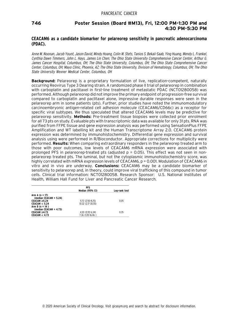
746 Poster Session (Board #M13), Fri, 12:00 PM-1:30 PM and4:30 PM-5:30 PM
CEACAM6 as a candidate biomarker for pelareorep sensitivity in pancreatic adenocarcinoma(PDAC).
Anne M. Noonan, Jacob Yount, Jason David, Mindy Hoang, Colin W. Stets, Tanios S. Bekaii-Saab, Ying Huang, Wendy L. Frankel,Cynthia Dawn Timmers, John L. Hays, James Lin Chen; The Ohio State University Comprehensive Cancer Center, Arthur G.James Cancer Hospital, Columbus, OH; The Ohio State University, Columbus, OH; The Ohio State Comprehensive CancerCenter, Columbus, OH; Mayo Clinic, Phoenix, AZ; The Ohio State University, Division of Hematology, Columbus, OH; The OhioState University Wexner Medical Center, Columbus, OH
Background: Pelareorep is a proprietary formulation of live, replication-competent, naturallyoccurring Reovirus Type 3 Dearing strain. A randomized phase II trial of pelareorep in combinationwith carboplatin and paclitaxel in first-line treatment of metastatic PDAC (NCT01280058) wasperformed. Although pelareorep did not improve the primary endpoint of progression-free survivalcompared to carboplatin and paclitaxel alone, impressive durable responses were seen in thepelareorep arm in some patients (pts). Further, prior studies have noted the immunomodulatorycarcinoembryonic antigen-related cell adhesion molecule (CEACAM6/CD66c) as a receptor forspecific viral subtypes. We thus speculated that altered CEACAM6 levels may be predictive forpelareorep sensitivity. Methods: Pre-treatment tissue biopsies were collected prior enrolmentfor all 73 pts on study. Evaluable pts with transcriptomic data was available for only 31 pts. RNAwaspurified from FFPE tissue and gene expression analysis was performed using SensationPlus FFPEAmplification and WT labelling kit and the Human Transcriptome Array 2.0. CEACAM6 proteinexpression was determined by immunohistochemistry. Differential gene expression and survivalanalysis using were performed in R/Bioconductor. Appropriate corrections for multiplicity wereperformed. Results: When comparing extraordinary responders in the pelareorep treated arm tothose with poor outcomes, low levels of CEACAM6 mRNA expression were associated withprolonged PFS in pelareorep-treated pts (adjusted p = 0.05). This effect was not seen in non-pelareorep treated pts. The luminal, but not the cytoplasmic immunohistochemistry score, washighly correlated with mRNA expression levels of CEACAM6, p = 0.001. Modulation of CEACAM6 invitro and in vivo are underway. Conclusions: CEACAM6 may be a candidate biomarker ofsensitivity to pelareorep and, in theory, could improve viral trafficking of this compound in tumorcells. Clinical trial information: NCT01280058. Research Sponsor: U.S. National Institutes ofHealth, William Hall Fund for Liver and Pancreatic Cancer Research.
PFSMedian (95% CI) Log-rank test
Arm A (n = 17)(median CEACAM = 5.24)
CEACAM ‡5.24 5.72 (2.50-8.25) 0.05CEACAM < 5.24 10.32 (2.17-30.55)Arm B (n = 14 )(median CEACAM = 4.73)
CEACAM ‡4.73 4.30 (0.92-6.34) 0.35CEACAM < 4.73 7.36 (1.05-16.46 )
© 2020 American Society of Clinical Oncology. Visit gicasym.org and search by abstract for disclosure information.
PANCREATIC CANCER
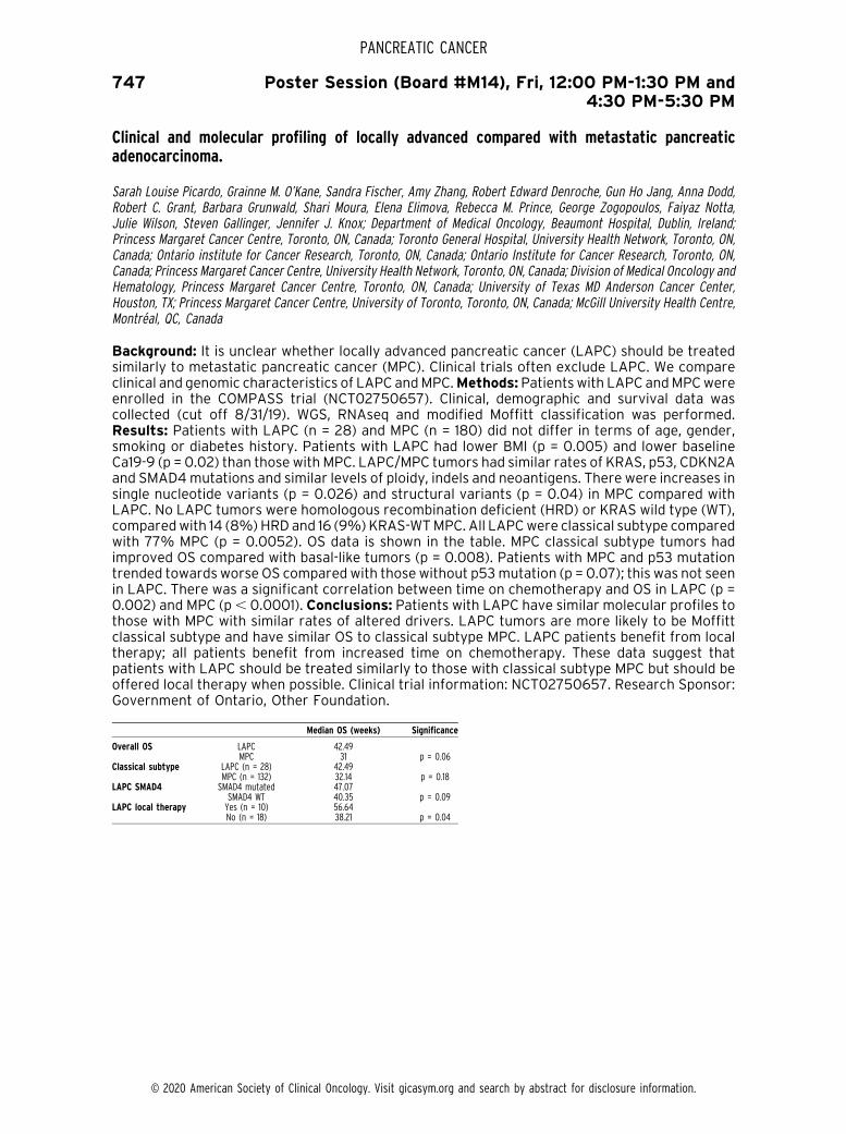
747 Poster Session (Board #M14), Fri, 12:00 PM-1:30 PM and4:30 PM-5:30 PM
Clinical and molecular profiling of locally advanced compared with metastatic pancreaticadenocarcinoma.
Sarah Louise Picardo, Grainne M. O’Kane, Sandra Fischer, Amy Zhang, Robert Edward Denroche, Gun Ho Jang, Anna Dodd,Robert C. Grant, Barbara Grunwald, Shari Moura, Elena Elimova, Rebecca M. Prince, George Zogopoulos, Faiyaz Notta,Julie Wilson, Steven Gallinger, Jennifer J. Knox; Department of Medical Oncology, Beaumont Hospital, Dublin, Ireland;Princess Margaret Cancer Centre, Toronto, ON, Canada; Toronto General Hospital, University Health Network, Toronto, ON,Canada; Ontario institute for Cancer Research, Toronto, ON, Canada; Ontario Institute for Cancer Research, Toronto, ON,Canada; Princess Margaret Cancer Centre, University Health Network, Toronto, ON, Canada; Division of Medical Oncology andHematology, Princess Margaret Cancer Centre, Toronto, ON, Canada; University of Texas MD Anderson Cancer Center,Houston, TX; Princess Margaret Cancer Centre, University of Toronto, Toronto, ON, Canada; McGill University Health Centre,Montreal, QC, Canada
Background: It is unclear whether locally advanced pancreatic cancer (LAPC) should be treatedsimilarly to metastatic pancreatic cancer (MPC). Clinical trials often exclude LAPC. We compareclinical and genomic characteristics of LAPC andMPC.Methods:Patients with LAPC andMPCwereenrolled in the COMPASS trial (NCT02750657). Clinical, demographic and survival data wascollected (cut off 8/31/19). WGS, RNAseq and modified Moffitt classification was performed.Results: Patients with LAPC (n = 28) and MPC (n = 180) did not differ in terms of age, gender,smoking or diabetes history. Patients with LAPC had lower BMI (p = 0.005) and lower baselineCa19-9 (p = 0.02) than those with MPC. LAPC/MPC tumors had similar rates of KRAS, p53, CDKN2Aand SMAD4mutations and similar levels of ploidy, indels and neoantigens. There were increases insingle nucleotide variants (p = 0.026) and structural variants (p = 0.04) in MPC compared withLAPC. No LAPC tumors were homologous recombination deficient (HRD) or KRAS wild type (WT),comparedwith 14 (8%)HRD and 16 (9%)KRAS-WTMPC. All LAPCwere classical subtype comparedwith 77% MPC (p = 0.0052). OS data is shown in the table. MPC classical subtype tumors hadimproved OS compared with basal-like tumors (p = 0.008). Patients with MPC and p53 mutationtrended towards worse OS compared with those without p53mutation (p = 0.07); this was not seenin LAPC. There was a significant correlation between time on chemotherapy and OS in LAPC (p =0.002) and MPC (p, 0.0001). Conclusions: Patients with LAPC have similar molecular profiles tothose with MPC with similar rates of altered drivers. LAPC tumors are more likely to be Moffittclassical subtype and have similar OS to classical subtype MPC. LAPC patients benefit from localtherapy; all patients benefit from increased time on chemotherapy. These data suggest thatpatients with LAPC should be treated similarly to those with classical subtype MPC but should beoffered local therapy when possible. Clinical trial information: NCT02750657. Research Sponsor:Government of Ontario, Other Foundation.
Median OS (weeks) Significance
Overall OS LAPC 42.49MPC 31 p = 0.06
Classical subtype LAPC (n = 28) 42.49MPC (n = 132) 32.14 p = 0.18
LAPC SMAD4 SMAD4 mutated 47.07SMAD4 WT 40.35 p = 0.09
LAPC local therapy Yes (n = 10) 56.64No (n = 18) 38.21 p = 0.04
© 2020 American Society of Clinical Oncology. Visit gicasym.org and search by abstract for disclosure information.
PANCREATIC CANCER
748 Poster Session (Board #M15), Fri, 12:00 PM-1:30 PM and4:30 PM-5:30 PM
The potential of Tenascin C in the tumor-nerve microenvironment to enhance perineuralinvasion and correlate with locoregional recurrence-related poor prognosis in pancreatic ductaladenocarcinoma.
Satoru Furuhashi, Takanori Sakaguchi, Ryuta Muraki, Ryo Kitajima, Mayu Fukushima, Makoto Takeda, Yoshifumi Morita,Hirotoshi Kikuchi, Mitsutoshi Setou, HIROYA TAKEUCHI; Hamamatsu University School of Medicine, Hamamatsu, Japan
Background: Perineural invasion (PNI) is commonly seen in pancreatic ductal adenocarcinoma(PDAC) and worsens the postoperative prognosis. However, the detail mechanisms of PNI in PDACremain unclear. Tenascin C (TNC), an extracellular matrix glycoprotein, is abundant in cancerstroma and modulates tumor progression. In this study, we hypothesized that TNC could enhancePNI in PDAC. The aim of this study was to investigate the roles of TNC in the tumor-nervemicroenvironment of PDAC.Methods:We immunohistochemically examined TNC expression in 78resected PDAC specimens. TNC staining intensity in perineural sites at the invasive front wasclassified as low or high, by comparison with adjacent non-cancerous tissues in the same section.The relationships between TNC expression and clinicopathological features were retrospectivelyanalyzed. Furthermore, interactions between cancer cells and nerves after supplementation withTNC were investigated using in vitro co-culture model with a PDAC cell line and neonatal mousedorsal root ganglion (DRG). Results: High perineural TNC expression at the invasive front, seen in30 (38%) patients, was associated with the presence of PNI (p = 0.006), pathological T stage $ 3(p = 0.01), and postoperative locoregional recurrence (p = 0.002). It was independently associatedwith postoperative, poor recurrence-free survival in multivariate analysis (p = 0.045). In the invitro co-culture model, TNC supplementation significantly enhanced both neurotropism of PDACcells and tumor tropism of a DRG. On the other hand, when PDAC cells and a DRG were culturedseparately, TNC did not affect cancer cell proliferation or neural outgrowth. Furthermore, theknockdown of Annexin A2, which is known to be a receptor for TNC, cancelled the neurotropism ofPDAC cell toward DRGs. Conclusions: Strong perineural TNC expression was a prognostic indi-cator of locoregional recurrence-related poor prognosis. The neurotropism of PDAC induced byTNC and TNC-Annexin A2 signaling pathway could be the potential therapeutic target for PDAC byregulating PNI. Research Sponsor: kaken.
© 2020 American Society of Clinical Oncology. Visit gicasym.org and search by abstract for disclosure information.
PANCREATIC CANCER
749 Poster Session (Board #M16), Fri, 12:00 PM-1:30 PM and4:30 PM-5:30 PM
Molecular genetic changes in solid pseudopapillary neoplasms (SPN) of the pancreas.
Elisa M. Rodriguez-Matta, Gerardo Colon-Otero, Amanda Hemmerich, Eric Allan Severson; Univeristy of Puerto Rico School ofMedicine, San Juan, PR; Mayo Clinic, Jacksonville, FL; Foundation Medicine, Inc., Cambridge, MA
Background: Solid Pseudopapillary Neoplasms (SPNs) of the pancreas are rare accounting for 1-2%of all pancreatic tumors. Previous studies had shown that a pathogenicmutation of theCTNNB1gene is present in over 90% of the SPN tumors with very limited information available on thespecific molecular changes present in SPN. Here we report the results of next generation genetictesting of a large series of SPN tumors from the Foundation Medicine database. Methods:Foundation Medicine database from 07/2012 to 04/2019 was reviewed. A total of 31 cases ofSPN tumors were identified out of 12,892 cases of pancreatic cancers. Information collectedincluded demographic information on the patients as well as tumor analysis for next generationgenetic sequencing of 315 genes associated with cancer with FoundationOne or 324 genesassociated with FoundationOneCDx. Microsatellite stability status and tumor mutation burdenwere determined. Results: Twenty nine out of 31 cases (93%) had a CTNNB1 mutation. Seventypercent of the CTNNB1 mutations were on position 32 (14 cases, 41%) or in position 37 (9 cases,29%). Other sites of CTNNB1 mutations included position 34 (3 cases, 9.6%), position 2 (2 cases,6.4%) and position 38 (1 case, 3.2%). Two cases did not have a CTNNB1 mutation, one had aCDKN2Amutation and the other had no detectablemutations. Most cases had additionalmutationsaside from the CTNNB1, the most common were TP53 (3 cases, 9.6%) and LRP1B (2 cases, 6.4%).Other accompanying mutations were seen just once. Twenty-five percent of these cases hadactionable gene mutations, each found in one case including: MSH2, BRCA2, ATM, XRCC3, ATRX,PTEN, ESR1, CDKN2, and PIK3CA.Conclusions:Next generation genetic testing of SPN tumors is ofclinical benefit since it identifies actionable mutations in 29% of the cases. Research Sponsor:None.
© 2020 American Society of Clinical Oncology. Visit gicasym.org and search by abstract for disclosure information.
PANCREATIC CANCER
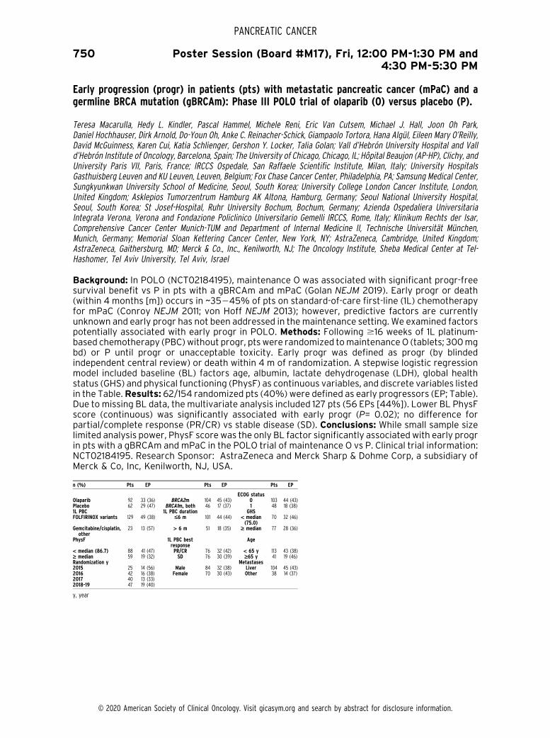
750 Poster Session (Board #M17), Fri, 12:00 PM-1:30 PM and4:30 PM-5:30 PM
Early progression (progr) in patients (pts) with metastatic pancreatic cancer (mPaC) and agermline BRCA mutation (gBRCAm): Phase III POLO trial of olaparib (O) versus placebo (P).
Teresa Macarulla, Hedy L. Kindler, Pascal Hammel, Michele Reni, Eric Van Cutsem, Michael J. Hall, Joon Oh Park,Daniel Hochhauser, Dirk Arnold, Do-Youn Oh, Anke C. Reinacher-Schick, Giampaolo Tortora, Hana Algul, Eileen Mary O’Reilly,David McGuinness, Karen Cui, Katia Schlienger, Gershon Y. Locker, Talia Golan; Vall d’Hebron University Hospital and Valld’Hebron Institute of Oncology, Barcelona, Spain; The University of Chicago, Chicago, IL; Hopital Beaujon (AP-HP), Clichy, andUniversity Paris VII, Paris, France; IRCCS Ospedale, San Raffaele Scientific Institute, Milan, Italy; University HospitalsGasthuisberg Leuven and KU Leuven, Leuven, Belgium; Fox Chase Cancer Center, Philadelphia, PA; Samsung Medical Center,Sungkyunkwan University School of Medicine, Seoul, South Korea; University College London Cancer Institute, London,United Kingdom; Asklepios Tumorzentrum Hamburg AK Altona, Hamburg, Germany; Seoul National University Hospital,Seoul, South Korea; St Josef-Hospital, Ruhr University Bochum, Bochum, Germany; Azienda Ospedaliera UniversitariaIntegrata Verona, Verona and Fondazione Policlinico Universitario Gemelli IRCCS, Rome, Italy; Klinikum Rechts der Isar,Comprehensive Cancer Center Munich-TUM and Department of Internal Medicine II, Technische Universitat Munchen,Munich, Germany; Memorial Sloan Kettering Cancer Center, New York, NY; AstraZeneca, Cambridge, United Kingdom;AstraZeneca, Gaithersburg, MD; Merck & Co., Inc., Kenilworth, NJ; The Oncology Institute, Sheba Medical Center at Tel-Hashomer, Tel Aviv University, Tel Aviv, Israel
Background: In POLO (NCT02184195), maintenance O was associated with significant progr-freesurvival benefit vs P in pts with a gBRCAm and mPaC (Golan NEJM 2019). Early progr or death(within 4 months [m]) occurs in ~35245% of pts on standard-of-care first-line (1L) chemotherapyfor mPaC (Conroy NEJM 2011; von Hoff NEJM 2013); however, predictive factors are currentlyunknown and early progr has not been addressed in themaintenance setting. We examined factorspotentially associated with early progr in POLO. Methods: Following $16 weeks of 1L platinum-based chemotherapy (PBC) without progr, pts were randomized tomaintenanceO (tablets; 300mgbd) or P until progr or unacceptable toxicity. Early progr was defined as progr (by blindedindependent central review) or death within 4 m of randomization. A stepwise logistic regressionmodel included baseline (BL) factors age, albumin, lactate dehydrogenase (LDH), global healthstatus (GHS) and physical functioning (PhysF) as continuous variables, and discrete variables listedin the Table.Results: 62/154 randomized pts (40%) were defined as early progressors (EP; Table).Due to missing BL data, the multivariate analysis included 127 pts (56 EPs [44%]). Lower BL PhysFscore (continuous) was significantly associated with early progr (P= 0.02); no difference forpartial/complete response (PR/CR) vs stable disease (SD). Conclusions: While small sample sizelimited analysis power, PhysF score was the only BL factor significantly associated with early progrin pts with a gBRCAm and mPaC in the POLO trial of maintenance O vs P. Clinical trial information:NCT02184195. Research Sponsor: AstraZeneca and Merck Sharp & Dohme Corp, a subsidiary ofMerck & Co, Inc, Kenilworth, NJ, USA.
n (%) Pts EP Pts EP Pts EP
ECOG statusOlaparib 92 33 (36) BRCA2m 104 45 (43) 0 103 44 (43)Placebo 62 29 (47) BRCA1m, both 46 17 (37) 1 48 18 (38)1L PBC 1L PBC duration GHSFOLFIRINOX variants 129 49 (38) £6 m 101 44 (44) < median
(75.0)70 32 (46)
Gemcitabine/cisplatin,other
23 13 (57) > 6 m 51 18 (35) ‡ median 77 28 (36)
PhysF 1L PBC bestresponse
Age
< median (86.7) 88 41 (47) PR/CR 76 32 (42) < 65 y 113 43 (38)‡ median 59 19 (32) SD 76 30 (39) ‡65 y 41 19 (46)Randomization y Metastases2015 25 14 (56) Male 84 32 (38) Liver 104 45 (43)2016 42 16 (38) Female 70 30 (43) Other 38 14 (37)2017 40 13 (33)2018-19 47 19 (40)
y, year
© 2020 American Society of Clinical Oncology. Visit gicasym.org and search by abstract for disclosure information.
PANCREATIC CANCER
751 Poster Session (Board #M18), Fri, 12:00 PM-1:30 PM and4:30 PM-5:30 PM
Pancreatic adenosquamous carcinoma: Clinical associations, treatment, and outcomes.
Mindy L. Hartgers, William Bamlet, Rondell P. Graham, Amit Mahipal, Taofic Mounajjed, Sean P. Cleary, Robert R. McWilliams;Mayo Clinic, Rochester, MN
Background: Pancreatic adenosquamous carcinomas (PASC) are rare malignancies, with limitedevidence regarding best treatment options. The Mayo Clinic Pancreatic Cancer SPORE Registrywas utilized to compare/contrast outcomes for pancreatic cancers with a squamous component topancreatic ductal adenocarcinoma (PDAC). Methods: Patients were identified from the SPORERegistry (2000-2019), were reviewed and confirmed by expert pathologists. Demographic andclinical information was ascertained from medical records and risk factor questionnaires. A casecontrol study of patients with PASC vs PDAC was constructed. PASC patients were also followedfor outcome and treatment records were extracted. Results: Of 2,584 total patients with pan-creatic cancers, 45 cases of PASC and 2,438 with PDACwere identified. There were no differencesin age (median 69 vs 67 years, p=0.42), sex (male 64.4vs 56.6%, p= 0.29), BMI (27.41 vs 27.78 kg/m2, P=0.50), or ever-smoking (61.0 vs 55.3%, P=0.47), with a borderline association with reporteddiabetes (17.8 vs 29.9%, p =0.08). Compared to PDAC, PASCsweremore likely to involve the body/tail (48.9 vs 33.2%, p= 0.02) and had poorer overall survival, adjusted for age, gender, and stage(median 7.1 m vs 12.8m, HR 1.89, p=0.0004). Of 9 PASC pts treated with neoadjuvant intent, 4 weresurgically resected, median survival was 7 months. Eleven pts underwent upfront surgery, withvariable adjuvant treatments. Median OS post- surgery was 18m (range 7-51). Of 14 patientspresenting with metastatic disease, median survival was 4.5 m (range 1-22). With regard tosystemic chemotherapy, for neoadjuvant or metastatic disease median duration of treatmentwas 3 months (range 0.5-7m) for Gemcitabine + nab-paclitaxel (N=9) and also 3 months (range 2-6m) for FOLFIRINOX (N-13). Conclusions: The diagnosis of PASC carries an even poorer outcomethan pancreatic adenocarcinoma. Tumors are more likely to arise in the distal pancreas, andpatients may be less likely to report associated diabetes. Limited antitumor activity was noted withmulti-agent chemotherapeutic regimens. Prospective trials will be needed to clarify choice ofregimen in the future. Research Sponsor: None.
© 2020 American Society of Clinical Oncology. Visit gicasym.org and search by abstract for disclosure information.
PANCREATIC CANCER
752 Poster Session (Board #M19), Fri, 12:00 PM-1:30 PM and4:30 PM-5:30 PM
Prognostic utility of inflammation biomarkers in a PDAC trial involving the human anti-CTGFantibody pamrevlumab.
Mark D. Sternlicht, Dongxia Li, Viet-Tam Nguyen, Mairead Carney, Duo Zhou, Kenneth E. Lipson, Elias Kouchakji,Vincent J. Picozzi, Ewa Carrier, Todd W. Seeley; FibroGen, Inc., San Francisco, CA; Virginia Mason Hospital and MedicalCenter, Seattle, WA
Background: Pancreatic ductal adenocarcinomas (PDAC) often exhibit desmoplasia, elevatedCTGF (connective tissue growth factor) expression and inflammation. The influence of inflamma-tion on patient outcomes was examined in a dose-ranging trial of the anti-CTGF antibodypamrevlumab in combination with a fixed regimen of gemcitabine and erlotinib in locally advancedor metastatic PDAC patients (NCT01181245; Picozzi et al. J Cancer Clin Trials 2017 2:123; n = 75).Methods: The prognostic utility of pre-treatment plasma levels of C-reactive protein (CRP),transforming growth factor b1 (TGFb1), albumin, CTGF and CA 19-9 were assessed by univariateand multivariate Cox analysis. Demographic parameters, treatment cohort, and pamrevlumabexposure determined on treatment day 15 were also evaluated, as were changes in biomarkerlevels over the first four weeks of treatment. Results: Elevated baseline CTGF and CRP wereprognostic for shorter overall survival (OS) by univariate analysis (HR = 3.2 for CRP. 10 mg/L, p=0.00002 and HR = 1.6 for CTGF. 10 ng/mL, p= 0.045). In a five-factor multivariate Coxmodel thatincluded CRP and TGFb1 as continuous Ln-transformed variables, performance status, age, andpamrevlumab treatment cohort, cohort assignments associated with increasing pamrevlumabexposure predicted improved OS (HR = 0.87, p= 0.03). Removing CRP from this model reduced theprognostic utility of pamrevlumab cohort assignment and exposure, indicating an importantcontribution of inflammation to interpretation of treatment outcome. Changes in inflammationbiomarkers over the course of treatment were also evaluated, but were not prognostic in thisstudy.Conclusions: In multivariate Coxmodels, assessment of pre-treatment CRP levels improvedability to detect significant differences in PDAC patient survival outcomes associated withpamrevlumab treatment. Our results emphasize the utility of accounting for pre-treatmentCRP levels as an independent prognostic factor in PDAC treatment effect models. ResearchSponsor: FibroGen.
© 2020 American Society of Clinical Oncology. Visit gicasym.org and search by abstract for disclosure information.
PANCREATIC CANCER
753 Poster Session (Board #M20), Fri, 12:00 PM-1:30 PM and4:30 PM-5:30 PM and Poster Walks, Fri, 4:45 PM-5:30 PM
Noninvasive comprehensive genomic profiling from plasma ctDNA in pancreatic cancer patients.
Danielle Sara Bitterman, Kristin Sedgwick Price, Emily E. Van Seventer, Jeffrey William Clark, Jill N. Allen,Lawrence Scott Blaszkowsky, David P. Ryan, Christine Elissa Eyler, Jennifer Yon-Li Wo, Theodore S. Hong,Ryan David Nipp, Eric Roeland, Janet E. Murphy, Ryan Bruce Corcoran, Colin D. Weekes, Aparna Raj Parikh; Departmentof Radiation Oncology, Massachusetts General Hospital, Boston, MA; Guardant Health, Inc., Redwood City, CA; MassachusettsGeneral Hospital, Boston, MA; Hematology/Oncology, Massachusetts General Hospital, Boston, MA; Massachusetts GeneralHospital Cancer Center, Boston, MA
Background: The use of comprehensive genomic profiling (CGP) is increasing in pancreatic ductaladenocarcinoma (PDAC) as knowledge improves regardingmolecular drivers of tumorigenesis andeffective targeted therapies emerge. However, adequate tissue sampling is often limited. Plasma-based CGP offers a non-invasive approach to assess biomarkers that may impact treatmentdecisions. Methods: We retrospectively evaluated genomic and clinical data from 97 PDACpatients with circulating tumor DNA (ctDNA) testing from 9/2016-8/2019 (Guardant Health,Inc.). ctDNA analysis included single nucleotide variants (SNV), fusions, indels and copy numbervariations (CNV) of up to 74 genes. ctDNA results were assessed across clinical variables. Weevaluated for actionable alterations. Results: A total of 114 samples were obtained from 97patients for ctDNA testing. ctDNA alterations were detected in 82% (93/114) of all samples,including 90% (18/20) at diagnosis, 88% (59/67) at progression, and 56% (10/18) while on stabletherapy. ctDNA alterations were found at each stage of PDAC: in 25% (1/4) of samples withresectable disease, 75% (3/4) with borderline resectable disease, 82% (9/11) with locally advanceddisease, and 85% (81/95) with metastatic disease. One or more KRAS alterations were detected in55% (51/93) of patients with alterations present. The median maximum mutant allele frequencywas similar between the cohort of patients with KRAS detected (0.55%) versus not detected(0.70%). 8% (8/97) of patients had potentially actionable alterations (2 activating BRAF SNVs, 1ERBB2 CNV, 1 ERBB2 activating SNV, 1 KRAS G12C, and 3 indels in Homologous RecombinationDeficiency genes). Median turnaround time was 8 days. 51% (49/97) of patients had both plasma-based CGP and tissue-based CGP. Of these patients, tissue-based CGP showed $ 1 alterationsdetected in 82% (40/49), test failure in 14% (7/49), and no alterations detected in 4% (2/49).Conclusions:Plasma-based CGP detected ctDNA alterations in 90%of samples tested at diagnosisand 82% of all samples. Potentially actionable mutations were found in 8% of patients, withprompt processing time allowing for rapid decision making. Research Sponsor: None.
© 2020 American Society of Clinical Oncology. Visit gicasym.org and search by abstract for disclosure information.
PANCREATIC CANCER
754 Poster Session (Board #M21), Fri, 12:00 PM-1:30 PM and4:30 PM-5:30 PM
Exceptional responses to ipilimumab/nivolumab (ipi/nivo) in patients (pts) with refractorypancreatic ductal adenocarcinoma (PDAC) and germline BRCA or RAD51 mutations.
Gretel Terrero, Terri Pollack, Daniel A. Sussman, Albert Craig Lockhart, Peter Joel Hosein; Jackson Health System, Miami, FL;Sylvester Comprehensive Cancer Center, University of Miami Miller School of Medicine, Miami, FL; University of Miami MillerSchool of Medicine, Miami, FL; University of Miami Sylvester Cancer Center, Miami, FL; University of Miami SylvesterComprehensive Cancer Center, Miami, FL
Background: Immune checkpoint inhibitors (ICI’s) have not shown meaningful clinical activity inunselected pts with PDAC. BRCA-deficient tumors have increased genomic instability, includingincreased tumor mutation burden (TMB), more tumor-infiltrating immune cells, and enrichment ofa T cell-inflamed signature. We hypothesized that pts withmutations in BRCA or other homologousrecombination repair genes may be sensitive to ICI’s. Methods: Utilizing the IRB-approved PDACdatabase at the University of Miami, we identified pts with relapsed/refractory PDAC with path-ogenic germline mutations who were treated with combination ICI’s (ipi 1mg/kg and nivo 3mg/kgevery 21 days followed by nivo 240mg every 2 weeks). Results: Five pts were identified (1 BRCA1, 2BRCA2, 1 RAD51C and 1 RAD51D). Among the 3 evaluable pts, there was one complete response(CR), one partial response (PR) and one had progressive disease (PD). The pt with a CR had BRCA1;he had resection followed by adjuvant gem/cape and had a biopsy-proven recurrence in the lungand retroperitoneum 1y after the end of adjuvant therapy. He received ipi/nivo at recurrence andachieved a CR, ongoing for 17m on nivo maintenance. The patient with a PR had RAD51C; he wasdiagnosed with mPDAC and received FOLFIRINOX for 6m, followed by olaparib on a trial for 12m.Upon PD, the disease quickly progressed on 5FU/liposomal irinotecan, gemcitabine/nab-paclitaxel/cisplatin and FOLFIRINOX. He then started ipi/nivo with immediate improvement inpain and tumor markers. A radiological PR was seen after 2 doses and is ongoing for 3m withcontinued clinical and tumor marker improvement. The 3rd evaluable pt had BRCA2 and had PDwith an exponential rise in tumormarkers accompanied by clinical deterioration. Response data onthe final two pts were pending at the time of submission and will be presented at the meeting.Conclusions: In this biomarker selected cohort, 2 out of 3 evaluable pts with PDAC had impressiveresponses to ipi/nivo. PDAC has generally been refractory to ICI therapy but this series suggeststhat this subgroup may be responsive to ICI’s. Further evaluation is warranted. Research Sponsor:None.
© 2020 American Society of Clinical Oncology. Visit gicasym.org and search by abstract for disclosure information.
PANCREATIC CANCER
755 Poster Session (Board #M22), Fri, 12:00 PM-1:30 PM and4:30 PM-5:30 PM
Patterns of failure after adjuvant stereotactic body radiation therapy in patients withpancreatic cancer with close or positive margins.
Ankur Patel, Joshua L. Rodrıguez-Lopez, Nathan Bahary, Amer H. Zureikat, Steven A. Burton, Dwight Earl Heron,Adam C. Olson; Department of Radiation Oncology, UPMC Hillman Cancer Center, University of Pittsburgh School ofMedicine, Pittsburgh, PA; Department of Medical Oncology, UPMC Hillman Cancer Center, University of Pittsburgh School ofMedicine, Pittsburgh, PA; Department of Surgical Oncology, UPMC Hillman Cancer Center, University of Pittsburgh School ofMedicine, Pittsburgh, PA
Background: There is no consensus on treatment volumes for stereotactic body radiation (SBRT)in patients with pancreatic cancer (PCa). Herein, we report patterns of failure following adjuvantSBRT for close/positive margins in patients with pancreatic cancer, which may inform appropriatetarget volume design for SBRT. Methods: An IRB-approved retrospective review of patients withPCa treated with adjuvant SBRT for close/positive margins from 2009-2018 was conducted.Patterns of failurewere assessed by reviewof imaging andwere defined as local (LF), regional (RF),local and regional (LRF), or distant (DF). The Kaplan-Meier method was used to calculate long-termfailure rates. In-field failures were defined as LFs completely within the PTV (planning targetvolume). The location of LFs was compared to the RTOG consensus volumes for adjuvanttreatment of PCa to determine if conventional radiation volumes would have included the LF.Results: Seventy-six patients were treated with adjuvant SBRT for close (51.3%) or positive(48.7%)margins, with amedian follow-up of 17.0 months (interquartile range [IQR] 7.4-28.3 mos.).Adjuvant SBRT was delivered at a median of 2.2 months after surgery (IQR 1.7-3.0 mos.). Mostpatients (81.6%) received 36 Gy in 3 fractions. The median PTV volume was 17.8 cc (IQR 12.3-25.2cc). Upon examination of first failure sites, crude rates of isolated LF, isolated RF, isolated LRF, andDF +/- LF or RF were 9.2%, 6.6%, 2.6%, and 56.6% respectively; 2-year rates were 12.4%, 11.5%,7.0%, and 66.5%, respectively. Thirty-two patients (42.1%) developed a LF at some point duringfollow-up. Of 28 LFs with available plans and imaging, 21.4% were in-field failures, while theremainder were completely outside (60.1%) or partially outside (17.9%) the PTV. Most LFs outsidethe PTV (90.9%) would have been encompassed by the RTOG consensus target volumes forpostoperative conventional radiation. Conclusions: In patients with PCa who receive adjuvantSBRT for close/positive margins, the majority of LFs are outside the PTV. Future trials involvingSBRT or hypofractionated radiation should consider expansion of treatment volumes if feasible.Research Sponsor: None.
© 2020 American Society of Clinical Oncology. Visit gicasym.org and search by abstract for disclosure information.
PANCREATIC CANCER
756 Poster Session (Board #N1), Fri, 12:00 PM-1:30 PM and4:30 PM-5:30 PM
Evaluation of the ratio of plasma fibrinogen to platelet in resectable pancreatic cancer.
Yusuke Arakawa, Mitsuo Shimada, Yuji Morine, Satoru Imura, Tetsuya Ikemoto, Yu Saito, Shinichiro Yamada,Masato Yoshikawa, Katsuki Miyazaki; Tokushima University, Tokushima, Japan; Department of Surgery, TokushimaUniversity, Tokushima, Japan
Background: Several prognostic factors were reported in pancreatic cancer such as neutrophillymphocyte ratio (NLR), platelet lymphocyte ration (PLR) modified GPS. Fibrinogen platelet ratio(FPR) was reported as one of the prognostic factor of resectable gastric cancer (Surgery today2019). In this report, the FPR was evaluated in patients with resectable pancreatic cancer.Methods: Between 2004 and 2019, one hundred and sixty-three patients in our institution withcurative resection for pancreatic cancer were enrolled in this retrospective study. The cases ofnon-curative resection were excluded. The FPR was calculated with the preoperative plasmafibrinogen and the platelet counts. Cut-off value was decided with ROC curve. The patients weredivided into high and low FPR group according to cut-off value. Results: The cut-off value of FPRwas 25.51. In age, gender, BMI, operative factors including operative type, amount of blood loss andoperative time, there was no significant difference between these two groups. Patients in low FPRgroup had significantly better overall survival (OS) rates and relapse-free survival (RFS) ratescomparedwith high FPR group (p, 0.05). Onmultivariate analysis, the high FPR, CA19-9. 300U/ml and receipt of adjuvant chemotherapy were independent risk factor of post-operative recur-rence. Conclusions: The FPR might be a prognostic factor of patients with resectable pancreaticcancer. Research Sponsor: None.
© 2020 American Society of Clinical Oncology. Visit gicasym.org and search by abstract for disclosure information.
PANCREATIC CANCER
757 Poster Session (Board #N2), Fri, 12:00 PM-1:30 PM and4:30 PM-5:30 PM
PD-L1 expression, tumor mutational burden, and microsatellite instability status in 746pancreas ductal adenocarcinomas.
Amanda Hemmerich, Claire I. Edgerly, Daniel Duncan, Richard Huang, Natalie Danziger, Garrett M. Frampton,Julia Andrea Elvin, Jo-Anne Vergilio, Jonathan Keith Killian, Douglas I. Lin, Erik Williams, Siraj Mahamed Ali,Prasanth Reddy, Vincent A. Miller, Clarence Owens, Charlotte Brown, Brian M Alexander, Jeffrey S. Ross,Eric Allan Severson, Shakti H. Ramkissoon; Foundation Medicine, Inc., Cambridge, MA; Foundation Medicine,Cambridge, MA; Foundation Medicine, Inc, Morrisville, NC; Foundation Medicine Inc., Cambridge, MA; FoundationMedicine, Inc, Cambridge, MA
Background:Pancreas ductal adenocarcinomas (PDA) has a 5-year survival rate of 6%with a needfor new therapeutic options. The approval of pembrolizumab for some gastrointestinal cancersshows the potential of immunotherapy (IMT) in PDA. We evaluated the IMT-associated biomarkersof PD-L1 expression, tumor mutational burden (TMB), microsatellite instability (MSI) and PD-L1amplification in PDAs. Methods: 746 formalin-fixed paraffin embedded samples were evaluatedfor PD-L1 IHC using the Dako 22C3 pharmDx assay and scored using tumor proportion score (TPS).The cases had comprehensive genomic profiling (CGP) via DNA sequencing, using a hybrid-capturenext-generation sequencing assay (FoundationOne and FoundationOneCDx) for genomic alter-ations (GAs), TMB, andMSI.Results: PD-L1 was positive (TPS$ 1%) in 29% (214/746) and negativein 71% (532/746). 43/214 (20%) of positive cases were high positive (TPS $ 50%). TMB (590cases) had a mean of 3.20, 3.46, and 3.61 mutations/Mb for PD-L1 negative, positive, and highpositive groups. 3 hypermutated (TMB$ 20) were negative for PD-L1 expression. 3/581 cases wereMSI-high with a high TMB score (average 23.53 mutations/Mb). 2 MSI-high cases were negative forPD-L1 and 1 was high positive. PD-L1 amplification was not detected (0/746). Only BCOR wassignificantly different between PD-L1 high positive and PD-L1 negative tumors (Table).Conclusions: Of 729 PDA cases, 29% were positive (TPS $ 1%) for PD-L1 expression while only6% of all cases showed a high level of PD-L1 expression on tumor cells. TMB high (3/729) and MSI-High (3/729) cases were rare. Only 2 of the TMB high cases were also MSI-high. PD-L1 amplificationwas not detected. Comparing GAs in PD-L1 high positive vs negative cases was only significantlydifferent for BCOR. Further investigation is needed to see if a combined positive score of PD-L1expression may identify a subset of patients with PDA who are more likely to respond to IMT.Research Sponsor: None.
GeneHigh Positive PD-L1(TPS ‡50%, n= 43)
Negative PD-L1(TPS <1%, n=532) p-value
KRAS 41 499 1TP53 36 422 0.5608CDKN2A 27 320 0.8715CDKN2B 17 133 0.0463MTAP 12 100 0.161ARID1A 7 57 0.3082SMAD4 8 176 0.0608AKT2 5 22 0.0432BCOR 5 7 0.001DNMT3A 3 29 0.7246
© 2020 American Society of Clinical Oncology. Visit gicasym.org and search by abstract for disclosure information.
PANCREATIC CANCER
758 Poster Session (Board #N3), Fri, 12:00 PM-1:30 PM and4:30 PM-5:30 PM
Analysis of pancreatobiliary adenocarcinoma (PBC) treatment response and resistance utilizingcirculating tumor DNA (ctDNA).
Madhulika Banerjee, Alejandro Recio Boiles, Sumana Veeravelli, Jorge Andres Leiva, Kathylynn Saboda, Denise Roe,Ali McBride, Hani M. Babiker; University of Arizona College of Medicine, Tucson, AZ; University of Arizona Medical Center,Tucson, AZ; Strake Jesuit College, Houston, TX; University of Arizona Cancer Center, Tucson, AZ; University of Arizona,Tucson, AZ
Background:Accurate diseasemonitoring in PBC is instrumental for optimal therapeutic decision-making. CA 19-9 is the most utilized biomarker, though it has limited sensitivity/specificity andcannot be used in CA 19-9 non-secretors (n-S). ctDNA is a potentially helpful monitoring aid andsurrogate for PBC n-S. Serial ctDNA could identify emerging resistant driver mutations. Our studyprospectively examined ctDNA in PBC patients receiving treatment and retrospectively correlatedit with clinical response. Methods: We performed genomic testing of ctDNA from metastatic PBCpatients’ plasma from 11/2016 to 08/2019. This included 77 patients, of those, 18 had .1 ctDNAmeasurement with 49 correlative data points in total. Demographics, serial CA 19-9 levels andimaging results were collected. ctDNA analysis by parallel sequencing of amplified target genes(74) using Guardant360 was obtained. We correlated imaging and CA 19-9 responses withmolecular alterations in patients receiving systemic chemotherapy. Descriptive statistics andlogistic regression of the data was performed. Results: Of those included, median age was 66 yo,50% male, and 92% pancreatic ductal adenocarcinoma. Baseline ctDNA showed 103 mutationsincluding TP53 12.6%, KRAS 9.7%,MET 6.8%, APC, ARID1A and NF1 4.8% each, and others, 3%.44% of patients were n-S with 75% having both TP53 and KRASmutations. APC, ARID1A, and NF1were only present in n-S. 91% vs 90% KRAS and 84% vs 78% TP53 of n-S and secretors (S),respectively, had correlation between ctDNA levels and imaging response. S TP53 and KRASmutations correlated to CA19-9 levels and scans in 78% and 70% responses. New TP53 subclonalvariant mutations were the most common resistance mutations for all progressions (75%). Alogistic regressionmodel of imaging progression on change in CA19-9 secretion and TP53 or KRASexpression was not statistically significant. Conclusions: Baseline ctDNA level changes (TP53 andKRAS) can potentially act as a biomarker of response in PBC, specifically in n-S. TP53 subclonalmutations were the most common resistant alterations at progression and can be explored asfuture targets. This is being explored in larger prospective trials. Research Sponsor: None.
© 2020 American Society of Clinical Oncology. Visit gicasym.org and search by abstract for disclosure information.
PANCREATIC CANCER
759 Poster Session (Board #N4), Fri, 12:00 PM-1:30 PM and4:30 PM-5:30 PM
Impact of CDKN2A/b status in pancreatic cancer (PC).
Kaitlin Annunzio, Claire Griffiths, Igli Arapi, Matthew Lasowski, Arun K. Singavi, Kulwinder Dua, Abdul H. Khan, Paul S. Ritch,Mandana Kamgar, James P. Thomas, William Adrian Hall, Beth Erickson, Susan Tsai, Kathleen K. Christians, Douglas B. Evans,Raul Urrutia, Aniko Szabo, Ben George; Medical College of Wisconsin, Milwaukee, WI; Medical Colllege of Wisconsin,Milwaukee, WI; Medical College of Wisconsin and Clement J. Zablocki Veterans Affairs Medical Center, Milwaukee, WI;Froedtert & the Medical College of Wisconsin, Milwaukee, WI
Background: PC is a lethal disease with limited treatment options. We utilized ComprehensiveGenomic Profiling (CGP) to identify putative prognostic and/or predictive biomarkers. Methods:We retrospectively reviewed PC patients (pts) at our institution who underwent CGP utilizing theFoundation One assay. CGP was performed on hybrid-capture, adaptor ligation-based libraries forup to 315 genes plus 47 introns from 19 genes frequently rearranged in cancer. PC pts werecategorized by clinical stage – localized (resectable and borderline resectable PC; LPC), locallyadvanced (LAPC) and metastatic (mPC). Effect of gene alterations (GAs) with at least 10%prevalence were analyzed. The marginal effect of each gene on radiographic response andsurvival outcomes was estimated using proportional odds and multivariate Cox regression anal-ysis, respectively, adjusting for stage. Results: Ninety-three pts were identified - median age was63, 55%weremale, and 50%were smokers. Clinical stage at diagnosis was LPC, LAPC andmPC in42 (45%), 23 (25%) and 28 (30%) pts, respectively. Themost commonly altered genes were KRAS(94%), TP53 (75%), CDKN2A (41.2%) and SMAD4 (32.9%). All patients were microsatellite stableand the median tumor mutational burden was 1.7. 5-FU (52%) or Gemcitabine (46%) basedchemotherapy combinations were utilized as the first systemic therapy. Median overall survival forpatients with LPC, LAPC and mPC were 30.7, 28.8 and 9.6 months respectively. Thirty-eight (91%)pts with LPC underwent curative intent surgery compared to 15 (65%) pts with LAPC (p = 0.019).Thirty-five (95%) pts with wild type (WT) CDKN2A and 47 (94%) pts with WT CDKN2B underwentcurative intent surgery compared to 13 (65%) and 1(14%) pt(s) with GAs in CDKN2A and CDKN2Brespectively (p = 0.003 and p , 0.0001 respectively). The response to chemotherapy wasstatistically significantly higher in pts with WT CDKN2A (53%) and CDKN2B (48%) comparedto pts with GAs in CDKN2A (19%) and CDKN2B (12%) (p = 0.03 and p = 0.05, respectively).Conclusions: GAs in CDKN2A/B may have a predictive and possibly a prognostic impact. Theclinical validity and biological relevance of these findings need to be further explored in largerstudies. Research Sponsor: U.S. National Institutes of Health.
© 2020 American Society of Clinical Oncology. Visit gicasym.org and search by abstract for disclosure information.
PANCREATIC CANCER
760 Poster Session (Board #N5), Fri, 12:00 PM-1:30 PM and4:30 PM-5:30 PM
Utilization of somatic comprehensive genomic profiling (CGP) to identify patients (pts) withpancreatic cancer (PC) that harbor germline DNA damage repair (DDR) gene alterations.
Matthew Lasowski, Samantha Stachowiak, Igli Arapi, Kulwinder Dua, Abdul H. Khan, William Adrian Hall, Beth Erickson,Susan Tsai, Kathleen K. Christians, Douglas B. Evans, Raul Urrutia, Paul S. Ritch, Mandana Kamgar, James P. Thomas,Ben George; Medical College of Wisconsin, Milwaukee, WI; Medical Colllege of Wisconsin, Milwaukee, WI; Medical College ofWisconsin and Clement J. Zablocki Veterans Affairs Medical Center, Milwaukee, WI; Froedtert & the Medical College ofWisconsin, Milwaukee, WI
Background: Somatic and germline DDR gene alterations in PC have been postulated to positivelypredict response to DNA damaging cytotoxic agents. Due to the relatively high prevalence ofgermline DDR gene alterations, germline testing is recommended in all pts with PC. We examinedwhether somatic CGP can be used to reliably identify PC pts that merit germline testing.Methods:We retrospectively reviewed the electronicmedical records of PC pts who underwent both somaticCGP (utilizing the Foundation One assay) and germline testing. DDR gene mutations were cate-gorized as somatic-pathogenic, somatic-variant of uncertain significance (VUS), germline-pathogenic and germline-VUS. For somatic testing, DNAwas extracted from formalin fixed paraffinembedded (FFPE) clinical specimens and CGP was done on hybrid-capture, adaptor ligation basedlibraries to a mean coverage depth of . 600 for up to 315 genes plus 47 introns from 19 genesfrequently rearranged in cancer. Germline genetic testing was performed on submitted blood orsaliva samples, utilizing commercial assays; next generation or Sanger sequencing of all codingregions and adjacent intronic nucleotides were performed. Results: Ninety-three pts had somaticCGP data, 51 (55%) pts had both somatic CGP and germline data available. Among the 51 pts withboth germline and somatic data available, DDR gene alterations that were somatic-pathogenic,germline-pathogenic, somatic-VUS and germline-VUS were present in 7 (13.7%), 7 (13.7%), 23(45.1%) and 16 (31.4%) pts, respectively. Of the 7 pts with somatic-pathogenic alterations, 5 (71%)had a concordant germline alteration and of the 7 pts with germline-pathogenic alterations, 5(71%) had a concordant somatic alteration. Of the 23 pts with somatic-VUSs, 12 (52%) had aconcordant germline VUS and of the 16 pts with germline-VUSs, 12 (75%) had a concordant somaticVUS.Conclusions: Both somatic and germline DDR gene alterations are common in PC pts. Despitethe relatively high concordance rate between somatic and germline pathogenic DDR gene alter-ations, somatic CGP will miss approximately one fourth of the germline DDR gene alterations.Research Sponsor: None.
© 2020 American Society of Clinical Oncology. Visit gicasym.org and search by abstract for disclosure information.
PANCREATIC CANCER
761 Poster Session (Board #N6), Fri, 12:00 PM-1:30 PM and4:30 PM-5:30 PM
Association of SMAD4 loss with response to neoadjuvant chemotherapy with the autophagyinhibitor hydroxychloroquine in patients with pancreatic adenocarcinoma.
Naomi Fei, Sijin Wen, Pavan Rao, Rajesh Ramanathan, Melissa E. Hogg, Amer H. Zureikat, Michael T. Lotze, Nathan Bahary,Aatur D. Singhi, Herbert Zeh, Brian A. Boone; West Virginia University Hospital, Morgantown, WV; West Virginia UniversitySchool of Public Health, Department of Biostatistics, Morgantown, WV; Division of Surgical Oncology, Department of Surgery,West Virginia University, Morgantown, WV; Division of Surgical Oncology, Department of Surgery, University of Pittsburgh,Pittsburgh, PA; Department of Surgery, North Shore Hospital, Chicago, IL; Department of Surgical Oncology, UPMC HillmanCancer Center, University of Pittsburgh School of Medicine, Pittsburgh, PA; Hillman Cancer Center, Pittsburgh, PA;Department of Medical Oncology, UPMC Hillman Cancer Center, University of Pittsburgh School of Medicine, Pittsburgh, PA;University of Pittsburgh Medical Center, Pittsburgh, PA; West Virginia University, Morgantown, WV
Background: SMAD4, a tumor suppressor gene, is inactivated or deleted in 60-90% of pancreaticadenocarcinomas (PDA). Loss of SMAD4 allows tumor progression by limiting cell cycle arrest andapoptosis and increasing metastases. SMAD4 deficient PDA cells are resistant to radiotherapy byupregulation of autophagy, a cell survival mechanism that allows intracellular recycling of mac-romolecules and organelles. Hydroxychloroquine (HCQ) is a known autophagy inhibitor, suggest-ing that HCQ treatment in SMAD4 deficient PDA may prevent therapeutic resistance induced byautophagy upregulation.Methods:We retrospectively analyzed the SMAD4 status of PDA patientsenrolled in two prospective clinical trials evaluating preoperative HCQ. The first dose escalationtrial demonstrated the safety of preoperative gemcitabine with HCQ (NCT01128296). More re-cently, a randomized trial of gemcitabine/nab-paclitaxel +/- HCQ evaluated Evans Grade histo-pathologic response (NCT01978184). Immunohistochemistry of resected specimens for SMAD4was previously performed. Patients not treated at themax HCQ dose (n = 5), not resected (n = 2) orwith SMAD4 staining unavailable were excluded (n = 10). The effect of SMAD4 loss on response toHCQ and chemotherapy was studied for association with clinical outcome. Fisher’s exact test andlog-rank test were used to assess response and survival. Results: 52 patients receiving HCQ withneoadjuvant chemotherapy and 24 patients receiving neoadjuvant chemotherapy alone werestudied. Of the HCQ group, 25 patients had SMAD4 loss (48%), compared with 15 control patients(63%, p = 0.32). 76% of HCQ treated patients with SMAD4 loss obtained a histopathologicresponse$2A, compared to only 37%with SMAD4 intact (p = 0.006). In the control group, loss ofSMAD4 was associated with a nonsignificant detriment in 3 year OS (25% vs. 78%, p = 0.3) thatwas less apparent in patients treated with HCQ (46% vs. 47%, p = 0.18). Conclusions: The additionof HCQ to neoadjuvant chemotherapy in PDA may improve treatment response in patients withSMAD4 loss. Further study of the relationship between SMAD4, autophagy and treatment out-comes in PDA is warranted. Research Sponsor: U.S. National Institutes of Health.
© 2020 American Society of Clinical Oncology. Visit gicasym.org and search by abstract for disclosure information.
PANCREATIC CANCER
762 Poster Session (Board #N7), Fri, 12:00 PM-1:30 PM and4:30 PM-5:30 PM
Analysis of associations of CXCR3 ligands with immune microenvironment and aggressiveness inmurine and human pancreatic ductal adenocarcinoma.
Andrew Cannon, Christopher M Thompson, Pranita Atri, Rakesh Bhatia, Sushil Kumar, Surinder Batra; University of NebraskaMedical Center, Omaha, NE; Univ of NE Medical Center, Omaha, NE
Background: The complex milieu of cytokines within pancreatic ductal adenocarcinoma (PDAC)promotes tumor progression and immune suppression thereby contributing to the dismal prog-nosis of patients with PDAC. However, the roles of many cytokines, including CXCR3 ligands, inPDAC have not been thoroughly investigated. Methods: Bioinformatics analyses of PDAC micro-array and TCGA datasets were used to identify cytokines overexpressed in PDAC, their associationwith patient survival as well as the expression of cognate cytokine receptors. Comparative analysisof cytokine expression in KrasLSL-G12D-P53LSL-R172H-Pdx1-Cre (KPC) and KrasLSL-G12D-Pdx1-Cre (KC)murine PDACmodels were used to validate these findings. Pathway and CIBERSORT analyses wereemployed to determine mechanistic basis of altered survival associated with cytokines of interest.Results: Of the 149 cytokines analyzed, CXCR3 ligands CXCL9 and CXCL10 were highly andconsistently overexpressed in PDAC datasets. Concurrently, CXCL9, CXCL10 and PF4 were over-expressed in the aggressive KPCmurinemodel compared to the indolent KCmodel. CXCR3 showedrobust expression in PDAC inmicroarray, TCGA and IHC analyses. Interestingly, high expression ofCXCR3 ligands was associated with shorter overall survival (p = 0.04 for CXCL9, 10 and 11 and p =0.02 for PF4) while high expression of CXCR3 was associated with increased overall survival (p =0.03). Pathway analysis of genes correlated with CXCR3 and/or its ligands showed that CXCR3ligands may promote T-cell exhaustion (p , 0.001). Finally, CIBERSORT analysis of TCGA datademonstrated that high CXCR3 expression was associated with increased CD8 T-cell and naıve B-cell signatures and loss of plasma cells signatures. High CXCR3 ligand expression was associatedwith increased CD8 T-cell, and M1 macrophage, and loss of NK-cell signatures(p , 0.05).Conclusions: CXCR3 ligands are overexpressed in PDAC and are associated with poor survival,likely related to alterations in tumor immune infiltrate/activity and may represent targets toaugment anti-tumor immunity. Research Sponsor: U.S. National Institutes of Health.
© 2020 American Society of Clinical Oncology. Visit gicasym.org and search by abstract for disclosure information.
PANCREATIC CANCER
763 Poster Session (Board #N8), Fri, 12:00 PM-1:30 PM and4:30 PM-5:30 PM
Serum glycoproteomic-based liquid biopsy for the detection of pancreatic ductaladenocarcinoma.
Pashtoon Murtaza Kasi, Ling Shen, Prasanna Ramachandran, Kaitlynn Moser, Gege Xu, Padraig Buckley, Daniel Serie,Carlito Lebrilla, Hui Xu, Carlos Hou Fai Chan; Mayo Clinic, Jacksonville, FL; InterVenn Biosciences, Redwood City, CA; VennBiosciences Corporation, Redwood City, CA; Brigham and Women’s Hosp, Brookline, MA
Background: Non-invasive biomarkers with high sensitivity and specificity would be of great valuefor patients with Pancreatic Ductal Adenocarcinoma (PDAC). This would aid in early detection andserve other purposes that ’liquid biopsies’ are being explored in. Besides circulating tumor DNA(ctDNA) and methylation markers, glycosylation markers hold great potential promise. We devel-oped a novel workflow using high-resolution quantification of site-specific protein glycosylation byliquid chromatography and tandem mass spectrometry to evaluate the clinical utility of glyco-protemics signature for patients with PDAC.Methods: Serum samples fromnewly diagnosedPDACpatients and controls were obtained from a commercial biobank (Indivumed, Hamburg, Germany),and a panel of 504 glycan motifs, representing 73 previously reported proteomic markers, wasdetermined. Age-adjusted generalized linear regression models were used to evaluate the differ-ential abundance of eachmarker, and stepwise variable selection was used formodel construction.Results: We analyzed 45 PDAC and 136 control samples. PDAC patients (60% male) had a meanage of 67 (611) years. with 4.4%, 71.1%, 4.4%, and 20% at stage 1, 2, 3,4, respectively. Controlswere women with benign histology after pelvic mass surgery; with a mean age of 61 (611) years.Twenty-six glycoproteomic markers showed statistically highly significant differential abundanceamong cases and controls (p,1e-4 each) and were highly reproducible(Pearson’s r. 0.85), whichwere glycoforms in proteins that have previously been found to be associated with PDAC. Fourteenof these markers displayed .0.8 area under the curve of the receiver operating characteristic(AUC). Multivariate logistic regression modeling with backward selection yielded a classificationmodel with an AUC of 0.94 (95% CI: 0.89-0.99), sensitivity of 91% (95% CI: 75-97%) andspecificity of 86% (95% CI: 81-92%). Conclusions: Circulating glycoproteomic biomarkers maybe useful in the early detection and clinical management of PDAC patients; offering a new platformto explore and validate. Research Sponsor: InterVenn.
© 2020 American Society of Clinical Oncology. Visit gicasym.org and search by abstract for disclosure information.
PANCREATIC CANCER
764 Poster Session (Board #N9), Fri, 12:00 PM-1:30 PM and4:30 PM-5:30 PM
The effect of TGF-b on PD-L1 expression on PDAC TAMs.
Katarzyna Trebska-McGowan, Liza Makowski, Marcus A. Alvarez, Rita Kansal, S. Mazher Husain, Ajeeth K. Pingili,Evan Scott Glazer; University of Tennessee Health Science Center, Memphis, TN
Background:Pancreatic Ductal Adenocarcinoma (PDAC) has less than a 10% five year survival andwill become the second leading cause of US cancer mortality in the next decade. Immunotherapy,such as checkpoint inhibition against anti-Programmed death-ligand 1 (PD-L1) has not beensuccessful in the treatment of PDAC patients. Both tumor associated macrophages (TAMs) andthe TGF-b protein are ubiquitous in PDAC tumors. We hypothesize that TGF-b increases the overallnumber of TAMs and degree of PD-L1 expression of TAMs in PDAC.Methods: Our lab has a mousepancreatic cancer cell line derived from a genetically engineered mouse model (KPC mice thatspontaneously form PDAC tumors that are similar to human PDAC). We orthotopically implantedthis cell line into the pancreas of immunocompetent C57BL/6 (B6) mice. In groups of 5 each, micewere treated with saline (control) or TGF-b. We investigated tumor burden, the number of TAMs(CD45+, CD11b+F4/80hi, Ly6C2, Ly6G2/lo) in the tumors with flow cytometry and the percentage ofTAMs expressing PD-L1 in the pancreas andmetastatic lesions in the liver.Results: As a percent ofleukocytes in the tumor, PDAC liver metastases had more TAMs compared to tumors in thepancreas (33 6 5% vs 10 6 4%, P = 0.001). Compared to controls, TGF-b treatment significantlyincreased the percent of PD-L1 expressing TAMs (326 6% vs 126 5%, P = 0.013, see Figure) in thepancreas but no effect was evident on TAM density. In liver metastases, treatment with TGF-bdecreased the overall TAM density (P = 0.039) but did not affect the number of PD-L1 positiveTAMs.Conclusions: TGF-b plays pivotal role in the progression of PDAC and demonstrates contextdependent activity. Our results suggest that an immunosuppressive effect mediated by PD-L1expression on TAMs may be initiated by TGF-b. Future investigations will focus on understandingthe role of the PDAC – TAM interaction to develop effective immune therapies for PDAC patients.Research Sponsor: SSAT - Society for Surgery of Alimentary Track.
© 2020 American Society of Clinical Oncology. Visit gicasym.org and search by abstract for disclosure information.
PANCREATIC CANCER
765 Poster Session (Board #N10), Fri, 12:00 PM-1:30 PM and4:30 PM-5:30 PM
Genomic associations in metastatic pancreatic cancer (mPC).
Anil Kumar Rengan, Efrat Dotan, Karthik Devarajan, Namrata Vijayvergia; Temple University Hospital, Philadelphia, PA; FoxChase Cancer Center, Philadelphia, PA
Background: mPC is an aggressive cancer, and molecular profiling provides further insight intopathogenesis and treatment. We sought to illustrate the molecular landscape of mPC in relation topost-metastatic survival and VTE incidence. Methods: With IRB approval, we retrospectivelyanalyzed charts of mPC patients who underwent molecular profiling. Fisher’s exact test (cate-gorical) and Mann-Whitney test (continuous) were used to compare groups. Log-rank test, Coxproportional hazards model and weighted Cox regression were used for survival analysis.Results:Between 2009 and 2018, 98 out of 502 mPC patients (19.5%) underwent molecular testing. Ofthese, 70% were tested after 2015. The most common mutations were in KRAS (79.2%), TP53(59.0%), CDKN2A (35.9%), BRCA (16.4%), PIK3CA (9.4%) and SMAD4 (7.4%). Concurrent KRASand TP53 mutations were found in 62.3% of patients. KRAS mutation was positively associatedwith Jewish ancestry (OR 2.3; p , 0.05), and TP53 mutation was positively associated withyounger age at diagnosis (63.5 vs. 67 years; p, 0.05). KRAS and TP53 mutations were negativelyassociated with tobacco use (OR 0.43 and 0.3, respectively; p , 0.05). There was no associationbetween any mutation and metastatic site. Both KRAS and TP53 mutations were positivelyassociated with VTE (OR 2.55 and 2.71, respectively; p , 0.05). Interestingly, TP53 mutationportends worse survival (HR 3.85; p, 0.05) whereas CDKN2Amutation confers improved survival(HR 0.55; p , 0.05). No patient received targeted therapy for their specific mutations.Conclusions: Molecular testing is becoming more prevalent in the mPC treatment paradigm.Among our cohort, concurrent KRAS and TP53 mutations were frequently identified. Incidencerates of the most common mutations were similar to those observed in prior literature. KRAS andTP53mutations were associated with higher incidence of VTE. TP53mutation was associated withpoorer prognosis. Research Sponsor: None.
© 2020 American Society of Clinical Oncology. Visit gicasym.org and search by abstract for disclosure information.
PANCREATIC CANCER
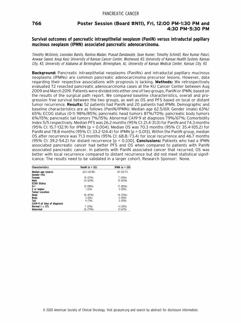
766 Poster Session (Board #N11), Fri, 12:00 PM-1:30 PM and4:30 PM-5:30 PM
Survival outcomes of pancreatic intraepithelial neoplasm (PanIN) versus intraductal papillarymucinous neoplasm (IPMN) associated pancreatic adenocarcinoma.
Timothy McGinnis, Leonidas Bantis, Rashna Madan, Prasad Dandawate, Sean Kumer, Timothy Schmitt, Ravi Kumar Paluri,Anwaar Saeed, Anup Kasi; University of Kansas Cancer Center, Westwood, KS; University of Kansas Health System, KansasCity, KS; University of Alabama at Birmingham, Birmingham, AL; University of Kansas Medical Center, Kansas City, KS
Background: Pancreatic intraepithelial neoplasms (PanINs) and intraductal papillary mucinousneoplasms (IPMNs) are common pancreatic adenocarcinoma precursor lesions. However, dataregarding their respective associations with prognosis is lacking. Methods: We retrospectivelyevaluated 72 resected pancreatic adenocarcinoma cases at the KU Cancer Center between Aug2009 andMarch 2019. Patients were divided into either one of two groups, PanIN or IPMN, based onthe results of the surgical path report. We compared baseline characteristics, overall and pro-gression free survival between the two groups, as well as OS and PFS based on local or distanttumor recurrence. Results: 52 patients had PanIN and 20 patients had IPMN. Demographic andbaseline characteristics are as follows (PanIN/IPMN): Median age 62.5/69; Gender (male) 63%/65%; ECOG status (0-1) 98%/85%; pancreatic head tumors 87%/70%; pancreatic body tumors6%/15%; pancreatic tail tumors 7%/15%; Abnormal CA19-9 at diagnosis 79%/67%; ComorbidityIndex 5/5 respectively. Median PFSwas 26.2months (95%CI: 21.4-31.0) for PanIN and 74.3months(95% CI: 15.7-132.9) for IPMN [p = 0.004]. Median OS was 70.3 months (95% CI: 35.4-105.2) forPanIN and 78.8 months (95% CI: 33.2-124.4) for IPMN [p = 0.013]. Within the PanIN group, medianOS after recurrence was 71.3 months (95% CI: 68.8.-73.4) for local recurrence and 46.7 months(95% CI: 39.2-54.2) for distant recurrence [p = 0.330]. Conclusions: Patients who had a IPMNassociated pancreatic cancer had better PFS and OS when compared to patients with PanINassociated pancreatic cancer. In patients with PanIN associated cancer that recurred, OS wasbetter with local recurrence compared to distant recurrence but did not meet statistical signif-icance. The results need to be validated in a larger cohort. Research Sponsor: None.
Characteristics PanIN (n = 52) IPMN (n = 20)
Median age (years) 62.5 (42-85) 69 (54-77)Gender (%)Female 19 (37%) 7 (35%)Male 33 (63%) 13 (65%)ECOG Status0-1 51 (98%) 17 (85%)2 or higher 1 (2%) 3 (15%)Tumor LocationHead 45 (87%) 14 (70%)Body 3 (6%) 3 (15%)Tail 4 (7%) 3 (15%)CA19-9 at time of diagnosisNormal ( < 37) 7 (21%) 4 (33%)Abnormal 26 (79%) 8 (67%)
© 2020 American Society of Clinical Oncology. Visit gicasym.org and search by abstract for disclosure information.
PANCREATIC CANCER
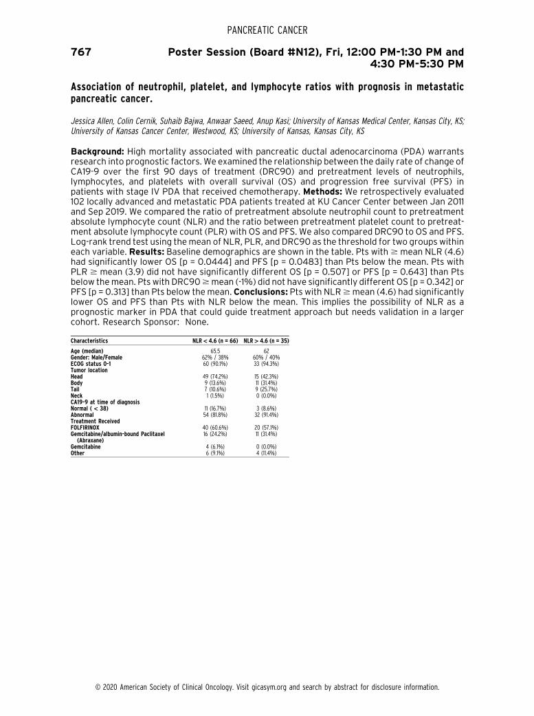
767 Poster Session (Board #N12), Fri, 12:00 PM-1:30 PM and4:30 PM-5:30 PM
Association of neutrophil, platelet, and lymphocyte ratios with prognosis in metastaticpancreatic cancer.
Jessica Allen, Colin Cernik, Suhaib Bajwa, Anwaar Saeed, Anup Kasi; University of Kansas Medical Center, Kansas City, KS;University of Kansas Cancer Center, Westwood, KS; University of Kansas, Kansas City, KS
Background: High mortality associated with pancreatic ductal adenocarcinoma (PDA) warrantsresearch into prognostic factors. We examined the relationship between the daily rate of change ofCA19-9 over the first 90 days of treatment (DRC90) and pretreatment levels of neutrophils,lymphocytes, and platelets with overall survival (OS) and progression free survival (PFS) inpatients with stage IV PDA that received chemotherapy. Methods: We retrospectively evaluated102 locally advanced and metastatic PDA patients treated at KU Cancer Center between Jan 2011and Sep 2019. We compared the ratio of pretreatment absolute neutrophil count to pretreatmentabsolute lymphocyte count (NLR) and the ratio between pretreatment platelet count to pretreat-ment absolute lymphocyte count (PLR) with OS and PFS. We also compared DRC90 to OS and PFS.Log-rank trend test using themean of NLR, PLR, and DRC90 as the threshold for two groups withineach variable. Results: Baseline demographics are shown in the table. Pts with$mean NLR (4.6)had significantly lower OS [p = 0.0444] and PFS [p = 0.0483] than Pts below the mean. Pts withPLR $ mean (3.9) did not have significantly different OS [p = 0.507] or PFS [p = 0.643] than Ptsbelow themean. Pts with DRC90$mean (-1%) did not have significantly different OS [p = 0.342] orPFS [p = 0.313] than Pts below themean.Conclusions: Pts with NLR$mean (4.6) had significantlylower OS and PFS than Pts with NLR below the mean. This implies the possibility of NLR as aprognostic marker in PDA that could guide treatment approach but needs validation in a largercohort. Research Sponsor: None.
Characteristics NLR < 4.6 (n = 66) NLR > 4.6 (n = 35)
Age (median) 65.5 62Gender: Male/Female 62% / 38% 60% / 40%ECOG status 0-1 60 (90.1%) 33 (94.3%)Tumor locationHead 49 (74.2%) 15 (42.3%)Body 9 (13.6%) 11 (31.4%)Tail 7 (10.6%) 9 (25.7%)Neck 1 (1.5%) 0 (0.0%)CA19-9 at time of diagnosisNormal ( < 38) 11 (16.7%) 3 (8.6%)Abnormal 54 (81.8%) 32 (91.4%)Treatment ReceivedFOLFIRINOX 40 (60.6%) 20 (57.1%)Gemcitabine/albumin-bound Paclitaxel(Abraxane)
16 (24.2%) 11 (31.4%)
Gemcitabine 4 (6.1%) 0 (0.0%)Other 6 (9.1%) 4 (11.4%)
© 2020 American Society of Clinical Oncology. Visit gicasym.org and search by abstract for disclosure information.
PANCREATIC CANCER
768 Poster Session (Board #N13), Fri, 12:00 PM-1:30 PM and4:30 PM-5:30 PM
Impact of surveillance among patients with resected pancreatic cancer following adjuvantchemotherapy.
Selina Wong, Lovedeep Gondara, Daniel John Renouf, Howard John Lim, Sharlene Gill; BC Cancer Agency, Vancouver, BC,Canada; British Columbia Cancer Agency, Vancouver, BC, Canada
Background: Pancreatic adenocarcinoma carries a poor prognosis and high risk of recurrenceeven after surgery and adjuvant chemotherapy (AC). Guidelines recommend against routinesurveillance imaging due to lack of evidence supporting a survival benefit. With current first-line chemotherapy options, it is unclear whether surveillance scans allow for early detection ofasymptomatic disease and therefore an opportunity to offer fit patients chemotherapy. Wedescribe the patterns of surveillance in patients followed at a Canadian provincial cancer agencyand determine whether routine imaging after AC is associated with receipt of palliative chemo-therapy (PC). Methods: A retrospective review was completed to identify patients treated atBritish Columbia (BC) Cancer centres between January 1, 2010 and December 31, 2016 who hadundergone curative intent resection and received at least one cycle of AC. Baseline characteristics,number of scans done after completing AC to recurrence, and PC were collected. Logisticregression analysis was performed. Results: A total of 151 patients followed at BC Cancer wereidentified. Patients who recurred within 28 days after AC were excluded, leaving 142 patients, ofwhich 115 patients had recurrence. We defined 2 cohorts based on number of scans done betweencompletion of AC and recurrence: those with 0-1 scans were “symptomatic” recurrences (22patients, median age 68y, 64% female, and 91% node-positive) and those with . 1 scan were“surveillance” recurrences (93 patients, median age 64y, 43% female, and 81% node-positive).Patients who underwent surveillance scans were more likely to receive PC at time of recurrence,though statistical significance was not reached (OR 2.11, 95% CI 0.75-6.58, p = 0.17). Conclusions:Despite guidelines, the majority of patients treated in BC underwent surveillance imaging. Withinthe limits of our sample size, we demonstrated a trend towards increased likelihood of receiving PCin patients who receive surveillance scans following AC. With efficacious PC options available,studies to determine whether receipt of PC in asymptomatic recurrences detected on imagingtranslates into improved survival and/or quality of life are warranted. Research Sponsor: None.
© 2020 American Society of Clinical Oncology. Visit gicasym.org and search by abstract for disclosure information.
PANCREATIC CANCER
769 Poster Session (Board #N14), Fri, 12:00 PM-1:30 PM and4:30 PM-5:30 PM
Clinical outcomes of first-line FOLFIRINOX versus gemcitabine plus nab-paclitaxel in metastaticpancreatic cancer at the Yale Smilow Healthcare System.
Timil Patel, Thejal Srikumar, Joseph Anthony Miccio, Jill Lacy, Stacey Stein, Jeremy S. Kortmansky, Carol Staugaard,Melissa Gambaccini, Alfredo Axtmayer, Michael Cecchini; Yale School of Medicine, New Haven, CT; Department of TherapeuticRadiology, Yale School of Medicine, New Haven, CT; Yale New Haven Hospital, New Haven, CT
Background: FOLFIRINOX (FFX) and Gemcitabine plus nab-paclitaxel (GN) are established first line(1L) therapies for metastatic pancreatic cancer (MPC) but real-world data on their comparativeeffectiveness is limited.Methods:All cases ofMPC treatedwith 1L FFX or GN at Yale SmilowCancerHospital and the affiliated community care centers (CCC) from January 2011 – April 2019 werereviewed. Patient (pt) demographics, prior therapy, initial and subsequent dose reductions (DR),time to treatment discontinuation (TTD), overall survival (OS), and second line (2L) treatment datawere manually abstracted from the electronic medical record. Categorical and continuous var-iables were compared between 1L FFX and GN cohorts via the Chi-squared and Wilcoxon rank-sumtests. Median OS was calculated by the Kaplan-Meier method. Results:We identified 363 MPC ptstreated with 1L FFX or GN; 269 (74%) pts were treated with FFX and 94 (26%) with GN as 1Ltherapy. 204 (56%) pts were treated at the main campus and 159 (44%) at a CCC. Demographicsand baseline characteristics (FFX/GN) were as follows: gender (male) 55% / 41%; race (white) 82% /77%; age, 76 90% / 71% (P, 0.001). 332 (91%) of pts received no prior therapy; 21 (6%) had priorsurgery plus adjuvant gemcitabine and 10 (3%) had surgery alone. 98% of FFX-treated pts weretreated with upfront DR, compared to 78% of GN-treated pts (P= 0.003). 78% and 53% ofFFX-treated and GN-treated pts, respectively, had subsequent DR (P, 0.001). Median TTD was4.8 months with FFX and 3.4 months with GN (P= 0.0029) and the median OS was 11.3 months withFFX versus 7.2 months with GN (P , 0.0001). After 1L, 33% and 61% of FFX- and GN-treated pts,respectively, received no further chemotherapy (P, 0.001). Conclusions: In the largest manuallyabstracted retrospective analysis to date, MPC pts treated with 1L FFX were younger, more likely toreceive 2L therapy, and had increased survival compared to pts treated with GN. The OS of ptstreated with FFX was similar to the OS reported by Conroy et al despite upfront dose attenuations in98% of pts. A randomized trial is needed to confirm optimal sequencing of chemotherapy in MPC.Research Sponsor: None.
© 2020 American Society of Clinical Oncology. Visit gicasym.org and search by abstract for disclosure information.
PANCREATIC CANCER
770 Poster Session (Board #N15), Fri, 12:00 PM-1:30 PM and4:30 PM-5:30 PM
Timing and location of palliative care consultation in metastatic pancreatic cancer: Aretrospective, single-center observational study.
Timil Patel, Joseph Anthony Miccio, Thejal Srikumar, Elizabeth Horn Prsic, Kerin B. Adelson, Dmitry Kozhevnikov, Komal Patel,Jill Lacy, Michael Cecchini; Yale School of Medicine, New Haven, CT; Department of Therapeutic Radiology, Yale School ofMedicine, New Haven, CT; Yale New Haven Hospital, New Haven, CT
Background: Patients (pts) receiving treatment for metastatic pancreatic cancer (MPC) experi-ence significant symptoms, treatment related side effects and psychosocial burdens. The ASCOguidelines recommend early palliative care consultation (PCC) to improve quality of life andsurvival. However, limited real-world data is available regarding the timing of PCC, hospiceenrollment and location of death (LOD) in pts with MPC. Methods: We conducted a retrospectiveobservational analysis of pts treated with chemotherapy for MPC at the Yale Smilow CancerHospital and affiliated community care centers (CCC) from January 2011 to April 2019. Patientdemographics, treatment dates, initial PCC, enrollment of hospice at the time of death and LODweremanually abstracted from the electronic medical record. Univariate andmultivariable logisticregression analyses were conducted to predict for PCC and death outside the hospital.Results:Of363 pts identified with MPC who received chemotherapy, 38% (138) had a PCC. 67% (93) ofpatients’ initial PCC was in the hospital versus 33% (45) in the outpatient setting. The median timefrom the start of first-line chemotherapy to the first PCCwas 5.2months (interquartile range [IQR]1.2 – 12.9). The median time from the first PCC to death was 1.5 months (IQR 0.5 – 4.42). At the timeof our analysis, 300 pts had died and of those 76% (229) were enrolled on hospice at the time ofdeath while 24% (71) were not. With respect to LOD, 47% (139) of pts died at home with hospice,31% (94) at an inpatient hospice facility and 22% (67) died in the hospital. Female gender wasassociated with an increased likelihood of a PCC (HR 1.78, 95%CI 1.07-2.94, P = 0.026). Pts treatedat a CCC were less likely to have a PCC (HR 0.21, 95% CI 0.12-0.36, P , 0.001). A PCC was notassociated with a higher likelihood of death outside the hospital (HR 1.31, 95% CI 0.75-2.29, P =0.346).Conclusions:Althoughmost pts with MPC enroll in hospice, PCC is generally underutilized.In fact, many pts receive PCC near the end of life and in the hospital. Further studies are warrantedto determine how best to incorporate early PCC to maximize supportive care for pts with MPC.Research Sponsor: None.
© 2020 American Society of Clinical Oncology. Visit gicasym.org and search by abstract for disclosure information.
PANCREATIC CANCER
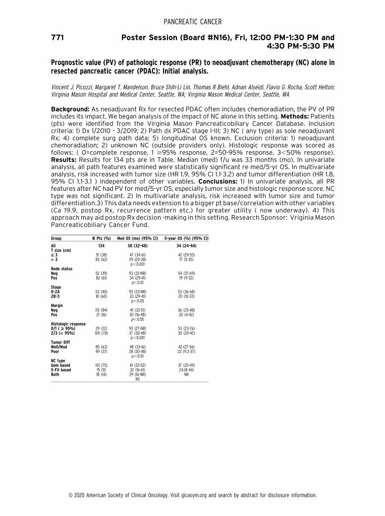
771 Poster Session (Board #N16), Fri, 12:00 PM-1:30 PM and4:30 PM-5:30 PM
Prognostic value (PV) of pathologic response (PR) to neoadjuvant chemotherapy (NC) alone inresected pancreatic cancer (PDAC): Initial analysis.
Vincent J. Picozzi, Margaret T. Mandelson, Bruce Shih-Li Lin, Thomas R Biehl, Adnan Alseidi, Flavio G. Rocha, Scott Helton;Virginia Mason Hospital and Medical Center, Seattle, WA; Virginia Mason Medical Center, Seattle, WA
Background: As neoadjuvant Rx for resected PDAC often includes chemoradiation, the PV of PRincludes its impact. We began analysis of the impact of NC alone in this setting.Methods: Patients(pts) were identified from the Virginia Mason Pancreaticobiliary Cancer Database. Inclusioncriteria: 1) Dx 1/2010 - 3/2019; 2) Path dx PDAC stage I-III; 3) NC ( any type) as sole neoadjuvantRx; 4) complete surg path data; 5) longitudinal OS known. Exclusion criteria: 1) neoadjuvantchemoradiation; 2) unknown NC (outside providers only). Histologic response was scored asfollows: ( 0=complete response, 1 $95% response, 2=50-95% response, 3,50% response).Results: Results for 134 pts are in Table. Median (med) f/u was 33 months (mo). In univariateanalysis, all path features examined were statistically significant re med/5-yr OS. In multivariateanalysis, risk increased with tumor size (HR 1.9, 95% CI 1.1-3.2) and tumor differentiation (HR 1.8,95% CI 1.1-3.1 ) independent of other variables. Conclusions: 1) In univariate analysis, all PRfeatures after NC had PV for med/5-yr OS, especially tumor size and histologic response score. NCtype was not significant. 2) In multivariate analysis, risk increased with tumor size and tumordifferentiation.3) This data needs extension to a bigger pt base/correlation with other variables(Ca 19.9, postop Rx, recurrence pattern etc.) for greater utility ( now underway). 4) Thisapproachmay aid postop Rx decision -making in this setting. Research Sponsor: Vriginia MasonPancreaticobiliary Cancer Fund.
Group # Pts (%) Med OS (mo) (95% CI) 5-year OS (%) (95% CI)
All 134 38 (32-48) 34 (24-44)T size (cm)£ 3 51 (38) 47 (34-61) 42 (29-55)> 3 83 (62) 29 (20-38) 17 (5-35)
p,0.001Node statusNeg 52 (39) 93 (33-NR) 54 (37-69)Pos 82 (61) 34 (29-41) 19 (9-32)
p,0.01Stage0-2A 53 (40) 93 (33-NR) 53 (36-68)2B-3 81 (60) 33 (29-41) 20 (10-33)
p,0.05MarginNeg 113 (84) 41 (33-51) 36 (25-48)Pos 21 (16) 30 (16-48) 20 (4-42)
p,0.05Histologic response0/1 ( ‡ 95%) 29 (22) 93 (27-NR) 53 (23-76)2/3 (< 95%) 105 (78) 37 (30-48) 30 (20-42)
p,0.001Tumor DiffWell/Mod 85 (63) 48 (33-61) 42-(27-56)Poor 49 (37) 28 (30-48) 22 (9.3-37)
p,0.01NC typeGem based 101 (75) 41 (33-52) 37 (25-49)5-FU based 15 (11) 32 (16-61) 24.(8-44)Both 18 (14) 29 (16-NR) NR
NS
© 2020 American Society of Clinical Oncology. Visit gicasym.org and search by abstract for disclosure information.
PANCREATIC CANCER
772 Poster Session (Board #N17), Fri, 12:00 PM-1:30 PM and4:30 PM-5:30 PM
Prognostic significance of familial pancreatic cancer after surgery.
Koji Tezuka, Yukiyasu Okamura, Teiichi Sugiura, Takaaki Ito, Yusuke Yamamoto, Ryo Ashida, Katsuhisa Ohgi,Katsuhiko Uesaka; Division of Hepato-Biliary-Pancreatic Surgery, Shizuoka Cancer Center, Shizuoka, Japan
Background: Familial pancreatic cancer (FPC) is defined as two first-degree relatives with pan-creatic cancer. It is known that the risk of developing pancreatic cancer increases in those whohave a family history of pancreatic cancer in first-degree relatives. However, prognostic signif-icance of FPC after surgery is not fully understood. Methods: Patients who underwent pancre-atectomy for pancreatic ductal carcinoma between January 2008 and December 2016 wereretrospectively reviewed. The prognostic significance of FPCwas analyzed.Results:A total of 423patients underwent pancreatectomy for pancreatic ductal carcinoma. FPC was identified in 32(7.6%) patients. Recurrence occurred in 72% of all resected cases and in 88% of resected FPCcases. Multivariate analysis revealed FPC (hazard ratio [HR] 1.60; P=0.026), CA19-9 $300 U/ml(HR 1.54; P=0.001), lymph node metastasis (HR 2.10; P,0.001), microscopic venous invasion (HR1.64; P,0.001), nerve plexus invasion (HR 1.39; P=0.010), R1 resection (HR 1.65; P=0.010), and lackof adjuvant chemotherapy (HR 2.27; P,0.001) as independent predictors for recurrence-freesurvival (RFS). The univariate analysis revealed that FPC is significantly associated with worseoverall survival (OS) (P=0.018). Themultivariate analysis showed that FPC was not an independentpredictor of OS. This cohort was divided into 314 patients (FPC: 18 patients, non-FPC: 296 patients)who received adjuvant chemotherapy (AC group) and 109 patients (FPC: 14 patients, non-FPC: 95patients) received no adjuvant chemotherapy (no AC group). In AC group, FPC is an independentpredictor for RFS (HR 3.03; P,0.001) and OS (HR 2.23; P=0.018). In no AC group, FPC is not apredictor for RFS and OS. Conclusions: This study may show that FPC has a significant impact onRFS and OS after resection in patients who received adjuvant chemotherapy. Research Sponsor:None.
© 2020 American Society of Clinical Oncology. Visit gicasym.org and search by abstract for disclosure information.
PANCREATIC CANCER
773 Poster Session (Board #N18), Fri, 12:00 PM-1:30 PM and4:30 PM-5:30 PM
An assessment of the total cost of pancreatic cancer using real-world evidence.
Vincent J. Picozzi, Victoria G. Manax, Kelly Feehan, Zachary Wintrob, Michele Korfin, Giuseppe Del Priore, Robert Goldberg;Virginia Mason Hospital and Medical Center, Seattle, WA; PanCan, Manhattan Beach, CA; ROAKETIN Inc., Oneonta, NY; Tyme,New York, NY; Morehouse School of Medicine, Atlanta, GA; Center for Medicine in the Public Interest, New York, NY
Background: The aggregate health economic implications of pancreatic cancer are poorly un-derstood, especially from the patient perspective. As a preliminary effort, we sought to betterunderstand changes in type and quantity of medical expenditures over time, along with quality oflife related costs, from this perspective. This preliminary research is part of a larger effort tounderstand how the introduction of new treatments affect both the outcome and costs ofpancreatic cancer associated with care, patients, survivors, their families, and their communities.Methods:We analyzed patient-level data from the Medical Expenditure Panel Survey (MEPS, 1996-2017). All analyses were performed using R version 3.6.1 on Ubuntu 19.04. Averages werecomputed for the total health care costs, including prescription drug costs. Average individualannual cost estimates for the second year excluded individuals that were identified as having diedprior to the first round of data collection in the second year. The individual patient level ratios ofprescription drug cost to othermedical expenses was also computed. All expenditures are adjustedfor inflation using 2017 US dollars. Included subjects, N= 80 had a diagnosis of pancreatic cancerand available prescription data. Individual age and employment status were accounted for ascovariates. Results: Between 1997 and 2017 inflation adjusted first and second year non-medication spending on pancreatic cancer care averaged $66,999.96 and $105,308.60 respec-tively. However, inflation-adjusted first and second year charges for hospitalizations and emer-gency visits fell between 2007-2017. Prescription drug as a proportion of total spendingprescription drugs increased during the same time period. Lost work/school days declined between2007 and 2017. Conclusions: Total inflation adjusted pancreatic cancer care expenses declinedover the past decade even as drug costs increased. Quality of life costs declined as well. Furtheranalysis is needed to evaluate the relationship between drug spending, total cost of care andquality of life. Research Sponsor: Tyme Inc.
© 2020 American Society of Clinical Oncology. Visit gicasym.org and search by abstract for disclosure information.
PANCREATIC CANCER
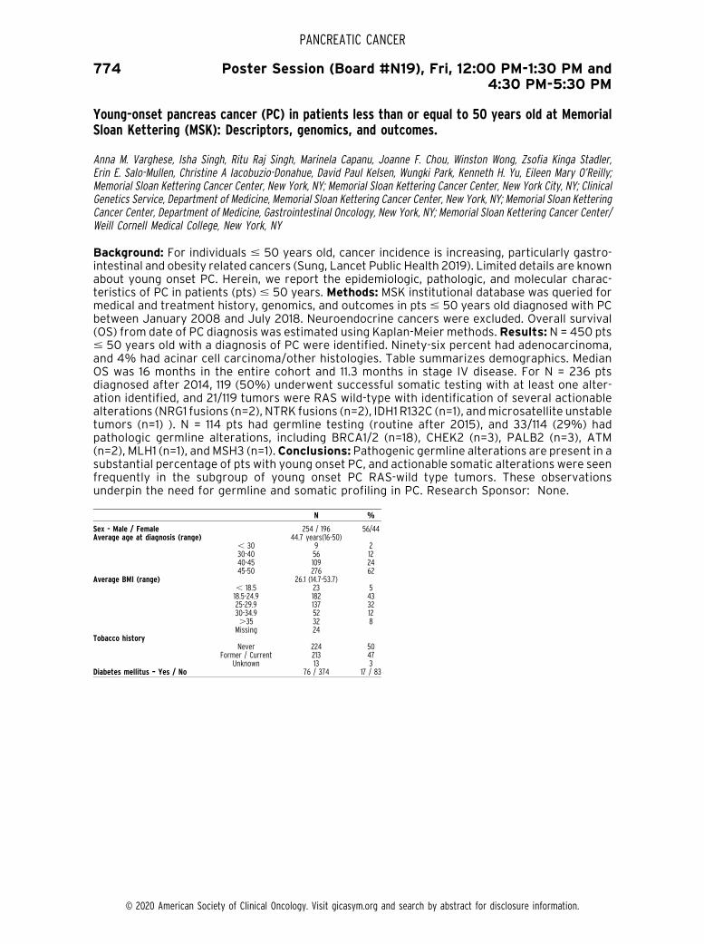
774 Poster Session (Board #N19), Fri, 12:00 PM-1:30 PM and4:30 PM-5:30 PM
Young-onset pancreas cancer (PC) in patients less than or equal to 50 years old at MemorialSloan Kettering (MSK): Descriptors, genomics, and outcomes.
Anna M. Varghese, Isha Singh, Ritu Raj Singh, Marinela Capanu, Joanne F. Chou, Winston Wong, Zsofia Kinga Stadler,Erin E. Salo-Mullen, Christine A Iacobuzio-Donahue, David Paul Kelsen, Wungki Park, Kenneth H. Yu, Eileen Mary O’Reilly;Memorial Sloan Kettering Cancer Center, New York, NY; Memorial Sloan Kettering Cancer Center, New York City, NY; ClinicalGenetics Service, Department of Medicine, Memorial Sloan Kettering Cancer Center, New York, NY; Memorial Sloan KetteringCancer Center, Department of Medicine, Gastrointestinal Oncology, New York, NY; Memorial Sloan Kettering Cancer Center/Weill Cornell Medical College, New York, NY
Background: For individuals # 50 years old, cancer incidence is increasing, particularly gastro-intestinal and obesity related cancers (Sung, Lancet Public Health 2019). Limited details are knownabout young onset PC. Herein, we report the epidemiologic, pathologic, and molecular charac-teristics of PC in patients (pts) # 50 years. Methods: MSK institutional database was queried formedical and treatment history, genomics, and outcomes in pts # 50 years old diagnosed with PCbetween January 2008 and July 2018. Neuroendocrine cancers were excluded. Overall survival(OS) from date of PC diagnosis was estimated using Kaplan-Meier methods.Results: N = 450 pts# 50 years old with a diagnosis of PC were identified. Ninety-six percent had adenocarcinoma,and 4% had acinar cell carcinoma/other histologies. Table summarizes demographics. MedianOS was 16 months in the entire cohort and 11.3 months in stage IV disease. For N = 236 ptsdiagnosed after 2014, 119 (50%) underwent successful somatic testing with at least one alter-ation identified, and 21/119 tumors were RAS wild-type with identification of several actionablealterations (NRG1 fusions (n=2), NTRK fusions (n=2), IDH1 R132C (n=1), andmicrosatellite unstabletumors (n=1) ). N = 114 pts had germline testing (routine after 2015), and 33/114 (29%) hadpathologic germline alterations, including BRCA1/2 (n=18), CHEK2 (n=3), PALB2 (n=3), ATM(n=2), MLH1 (n=1), and MSH3 (n=1).Conclusions: Pathogenic germline alterations are present in asubstantial percentage of pts with young onset PC, and actionable somatic alterations were seenfrequently in the subgroup of young onset PC RAS-wild type tumors. These observationsunderpin the need for germline and somatic profiling in PC. Research Sponsor: None.
N %
Sex - Male / Female 254 / 196 56/44Average age at diagnosis (range) 44.7 years(16-50)
, 30 9 230-40 56 1240-45 109 2445-50 276 62
Average BMI (range) 26.1 (14.7-53.7), 18.5 23 518.5-24.9 182 4325-29.9 137 3230-34.9 52 12.35 32 8Missing 24
Tobacco historyNever 224 50
Former / Current 213 47Unknown 13 3
Diabetes mellitus – Yes / No 76 / 374 17 / 83
© 2020 American Society of Clinical Oncology. Visit gicasym.org and search by abstract for disclosure information.
PANCREATIC CANCER
775 Poster Session (Board #N20), Fri, 12:00 PM-1:30 PM and4:30 PM-5:30 PM
Does aberrant hepatic arterial anatomy impact the complication rate or survival followingresection of pancreatic adenocarcinoma?
Nicketti M Handy, Angelena Crown, Kimberly A. Bertens, Jesse Clanton, Adnan Alseidi, Thomas Biehl, William Scott Helton,Flavio G. Rocha; Virginia Mason Medical Center, Seattle, WA; University of Ottawa, Ottawa, ON, Canada; West VirginiaUniversity, Charleston, WV; Virginia Mason Hospital and Medical Center, Seattle, WA
Background: Patients with aberrant hepatic arterial anatomy (AHAA) are susceptible to tumorinvasion and/or ligation during resection of the pancreatic head. The purpose of this study is todetermine if AHAA negatively impacts perioperative outcomes or survival. Methods: All patientswho underwent either pancreaticoduodenectomy or total pancreatectomy for pancreatic ductaladenocarcinoma (PDAC) between 2005 and 2014 at our center were retrospectively reviewed.Univariate logistic regression was used to compare outcomes between patients with conventionalhepatic arterial anatomy to those with AHAA. Survival analysis was performed by Kaplan-Meiermethod with log rank test.Results: During the study period, 330 patients underwent resection forPDAC, 69 (20.9%) with aberrant hepatic arterial anatomy. The presence of AHAA does notsignificantly increase operative time (p= 0.110) or length of stay (p=0.518). The overall frequency ofcomplications (49.3% vs 37.9%, p=0.088) was higher in the AHAA group, but not significantly so.Certain postoperative complications are more common in the AHAA group, namely superficialsurgical site infection (18.8%vs. 8.8%, p=0.018) and pancreatic fistula (18.8%vs. 10.0%, p=0.042).However, deep SSI, need for blood transfusion, respiratory failure, DGE, bleed from GDA/pseudoaneurysm, biliary fistula, chyle leak, PV thrombus, fascial dehiscence, and reoperationare not statistically different between the two groups. There is a trend for reduced overall survivalin the AHAA group that is not statistically significant (p=0.11). Conclusions: Aberrant hepaticarterial anatomy is encountered in greater than 20% of pancreatic surgery patients, and itspresence may increase the rate of certain postoperative complications such as superficial surgicalsite infection and pancreatic fistula. Research Sponsor: None.
© 2020 American Society of Clinical Oncology. Visit gicasym.org and search by abstract for disclosure information.
PANCREATIC CANCER
776 Poster Session (Board #N21), Fri, 12:00 PM-1:30 PM and4:30 PM-5:30 PM
Retrospective analysis of institutional outcomes with FOLFIRINOX versus nab-paclitaxel plusgemcitabine in metastatic pancreatic cancer.
Amisha Singh, Rachna T. Shroff, Ali McBride; University of Arizona College of Medicine, Tucson, AZ; University of ArizonaCancer Center, Tucson, AZ
Background: With an estimated six percent five-year survival, metastatic pancreatic adenocar-cinoma is one of the most lethal cancers in the United States. Previously, treatments withFOLFIRINOX (FFX) or gemcitabine plus nab-paclitaxel (G+A) have demonstrated improved overallsurvival versus gemcitabine alone. The purpose of this study is to compare institutional outcomesfor the two regimens, FFX vs. G+A in metastatic pancreatic cancer. Methods: We conducted aretrospective review of medical records of all metastatic pancreatic cancer patients from 2010 to2018 who received first line treatment with FFX or G+A at University of Arizona Cancer Center.Results: Thirty-five patients received combination treatment with G+A and 29 patients receivedFFX. Patient demographics: median age was 66 years in FFX vs 70 years in the G+A group; baselineCA-19-9 was 791 in FFX vs 738 in the G+A group. Median ECOG score was 1 for both groups. Medianoverall survival was 11.5 months in the FFX group (range 0-39 mos) vs 5 months in the G+A group(range 0-37 mos). Overall survival at 6 months was 75.9% vs 51.4% in FFX vs G+A groupsrespectively. Median progression-free survival was 6 months with FFX (range 0-25 mos) and 3months with G+A (range 0-24 mos). Twenty-one of 29 patients in the FFX group pursued secondline treatment, compared to 12 of 35 patients in the G+A group. Time to next treatment was 6months in the FFX group vs 5months in the G+A group. More adverse effects were noted with FFX,the most common being neuropathy and neutropenia, leading to treatment discontinuation due toadverse effects in 31% of patients, compared to 3% of patients in the G+A group. FFX patientsrequired a higher median no. of office visits (35 visits vs 18 visits in the G+A group). Conclusions:FFX showed improved progression-free survival vs G+A; however, this could be due to morepatients pursuing second line treatment in the FFX group. Compared to G+A, FFX patients hadhigher rates of treatment discontinuation due to adverse effects. FFX patients also required moreoffice visits and further analysis is necessary to assess whether this resulted in poorer quality oflife and increased total cost of care for patients treated with FFX. Research Sponsor: None.
© 2020 American Society of Clinical Oncology. Visit gicasym.org and search by abstract for disclosure information.
PANCREATIC CANCER
TPS777 Trials in Progress Poster Session (Board #P20),Fri, 12:00 PM-1:30 PM and 4:30 PM-5:30 PM
The impact of early palliative care on the quality of life of patients with advanced pancreaticcancer: The IMPERATIVE study.
Stephanie Lelond, Harvey Chochinov, Paul Joseph Daeninck, Benjamin Adam Goldenberg, Lisa Lix, Susan McClement,Christina Kim; CancerCare Manitoba, Winnipeg, MB, Canada; University of Manitoba, Winnipeg, MB, Canada; Dept of MedicalOncology, CancerCare Manitoba, Winnipeg, MB, Canada; University of Manitoba, Manitoba Palliative Care Research Unit,Winnipeg, MB, Canada
Background:Pancreatic cancer is lethal. Chemotherapy can improve survival bymonths; however,many patients experience an overwhelming burden of cancer-associated symptoms and poorquality of life (QOL). Early palliative care (EPC) alongside standard oncologic care results inimproved QOL and survival in patients with lung cancer. Although international guidelines rec-ommend EPC for patients with advanced pancreatic cancer (PANC), the benefit is not known.Objectives: The primary objective is to test for change in QOL between baseline (BL) and 16 weeks(wk). Secondary objectives are to test for change between BL and 16wk in (a) symptom control; and(b) depression and anxiety.Methods: This prospective case-crossover study of patients with PANCprovides EPC plus standard oncologic care. Primary oncology clinics refer patients to an EPC teamled by a palliative care physician and a clinical nurse specialist. BL questionnaires are completedprior to initial EPC assessment, then every 4 wk until wk 16. EPC visits are every 2 wk for the firstmonth, every 4 wk until wk 16, and then as needed. QOL, symptom control, anxiety and depressionare measured using the FACT-Hep tool, ESAS-r, HADS and PHQ-9, respectively. A generalizedlinear model will test for statistically significant change in scores between BL and 16 wk; chemo-therapy (yes/no) is included as a confounding covariate; model fit will be assessed. A sample size of20 patients provides 80%power after controlling for covariate effects. 40 patients will be enrolledto account for missing data. To date, 28 patients have enrolled and 17 have completed theintervention. Significance: The benefit of EPC for patients with PANC is not known, however, EPC isincreasingly recognized internationally by patients and stakeholders as a critical interventionwhich may improve both QOL and satisfaction with care. The Canadian Partnership AgainstCancer’s report on the patient experience states “the best possible patient experience means allpeople with cancer have equitable access to high-quality person-centered palliative care”. Thisstudy offers access to EPC and provides an environment in which the benefit of an integratedapproach is evaluated. Research Sponsor: CancerCare Manitoba FoundationPharmaceutical/Biotech Company.
© 2020 American Society of Clinical Oncology. Visit gicasym.org and search by abstract for disclosure information.
PANCREATIC CANCER
TPS778 Trials in Progress Poster Session (Board #P21),Fri, 12:00 PM-1:30 PM and 4:30 PM-5:30 PM
Demoralization and depression in pancreatic cancer patients.
Jar-Yee Liu, Veronica Placencio-Hickok, Brian Anderson, Paul Eliezer Oberstein, Andrew L. Coveler, Crystal S. Denlinger,Jun Gong, Andrew Eugene Hendifar; Cedars-Sinai Medical Center, Los Angeles, CA; Samuel Oschin Comprehensive CancerInstitute, Cedars-Sinai Medical Center, Los Angeles, CA; University of California San Francisco, San Francisco, CA; Laura andIsaac Perlmutter Cancer Center, NYU Langone Health, New York, NY; Seattle Cancer Care Alliance/University of Washington,Seattle, WA; Fox Chase Cancer Center, Philadelphia, PA; City of Hope, Duarte, CA
Background: Demoralization is a maladaptive coping response to stressful situations character-ized by thoughts of hopelessness, helplessness, and loss of meaning and purpose. Psychometri-cally, it is measured using the Demoralization-Scale II (DS-II), a validated questionnaire that yields apatient-reported quantification (scale 0-32) of demoralization. Previous studies involving patientswith progressive disease have uncovered a strong positive correlation between demoralizationand depression, respectively measured by the DS-II and Patient Health Questionnaire-9 (PHQ-9)surveys. Here, we aim to characterize demoralization and its relationship to depression in pan-creatic cancer patients, a unique patient population in terms of its poor prognosis. We hypothesizethat demoralization is highly prevalent in the pancreatic cancer patient population and stronglycorrelated with depression.Methods: Eligible patients with an active pancreatic cancer diagnosis,after consenting to an IRB approved protocol, will be administered the DS-II and PHQ-9 surveys toyield psychometric measurements for analysis. The primary objective of this project is to deter-mine the association between demoralization (DS-II) and depression (PHQ-9) in pancreatic cancerpatients. Secondary objectives include associations between demoralization and ethnicity, sexualorientation, suicidal ideation, education, cancer stage, and disease progression. Data will beanalyzed via simple linear regression. An ANOVA will also be conducted using DS-II groups asthe categorical variable and PHQ-9 scores as the continuous variable, and vice versa. This is amulti-institutional study to be conducted at Cedars-Sinai Medical Center, New York University,University of Washington, UC San Francisco, and Lewis Katz Schools of Medicine. ResearchSponsor: None.
© 2020 American Society of Clinical Oncology. Visit gicasym.org and search by abstract for disclosure information.
PANCREATIC CANCER
TPS779 Trials in Progress Poster Session (Board #P22),Fri, 12:00 PM-1:30 PM and 4:30 PM-5:30 PM
Improving cascade genetic testing for families with inherited pancreatic cancer (PDAC) risk: TheGENetic Education, Risk Assessment and TEsting (GENERATE) study.
Matthew B. Yurgelun, Chinedu I. Ukaegbu, C. Sloane Furniss, Barbara Kenner, Alison Klein, Andrew M. Lowy, Florencia McAllister,Maureen E Mork, Scott H. Nelson, Alison Robertson, Jill E. Stopfer, Meghan Underhill, Allyson J. Ocean, Lisa Madlensky,Gloria M. Petersen, Judy Ellen Garber, Scott Michael Lippman, Michael Goggins, Anirban Maitra, Sapna Syngal; Dana-Farber Cancer Institute, Boston, MA; Kenner Family Research Fund, New York, NY; The Sidney Kimmel ComprehensiveCancer Center at Johns Hopkins, Baltimore, MD; UCSD Moores Cancer Center, La Jolla, CA; The University of Texas MDAnderson Cancer Center, Houston, TX; Mayo Clinic, Rochester, MN; Center for Cancer Genetics and Prevention, Dana-Farber Cancer Institute, Boston, MA; Weill Cornell Medical College, New York, NY; University of California San Diego, LaJolla, CA; Johns Hopkins University, Baltimore, MD; University of Texas MD Anderson Cancer Center, Houston, TX
Background: 4-10% of PDAC patients harbor pathogenic germline variants in cancer susceptibilitygenes, including APC, ATM, BRCA1, BRCA2, CDKN2A, EPCAM, MLH1, MSH2, MSH6, PALB2, PMS2,STK11, and TP53. For families with such pathogenic variants, the greatest potential impact ofgermline testing is to identify relatives with the same variant (cascade testing), thereby providingthe opportunity for early detection and interception of PDAC and other associated cancers.Numerous factors limit cascade testing in real-world practice, including family dynamics, wide-spread geographic distribution of relatives, access to genetic services, and misconceptions aboutthe importance of germline testing, such that the preventive benefits of cascade testing are oftennot fully realized. The primary aim of this study is to analyze two alternative strategies for cascadetesting in families with inherited PDAC risk.Methods: 1000 individuals with a confirmed pathogenicgermline variant in any of the above genes in a 1st/2nd degree relative and a 1st/2nd degree relativewith PDAC will be remotely enrolled through the study website (www.GENERATEstudy.org) andrandomized between two methods of cascade testing (individuals with prior genetic testing will beineligible): Arm 1 will undergo pre-test genetic education with a pre-recorded video and liveinteractive session with a genetic counselor via a web-based telemedicine platform (Doxy.me),followed by germline testing through Color Genomics; Arm 2will undergo germline testing throughColor Genomics without dedicated pre-test genetic education. Color Genomics will disclose resultsto study personnel and directly to participants in both arms. All participants will have the option ofpursuing additional telephone-based genetic counseling through Color Genomics. The primaryoutcome will be uptake of cascade testing. Secondary outcomes will include self-reported geneticknowledge, cancer worry, distress, decisional preparedness, familial communication, and screen-ing uptake, which will be measured via longitudinal surveys. Enrollment is underway nationwide asof May, 2019. Clinical trial information: NCT03762590. Research Sponsor: Stand Up To Cancer-Lustgarten Foundation Pancreatic Cancer Interception Translational Cancer Research Grant(Grant Number: SU2C-AACR-DT25-17).
© 2020 American Society of Clinical Oncology. Visit gicasym.org and search by abstract for disclosure information.
PANCREATIC CANCER
TPS780 Trials in Progress Poster Session (Board #Q1),Fri, 12:00 PM-1:30 PM and 4:30 PM-5:30 PM
A phase I study of nanoliposomal irinotecan and 5-fluorouracil/folinic acid in combination withinterleukin-1-alpha antagonist for advanced pancreatic cancer patients with cachexia (OnFX).
Katelyn Mae Atkins, Jun Gong, Mourad Tighiouart, Samuel J. Klempner, Richard Tuli, Veronica Placencio-Hickok,Andrew Eugene Hendifar; Samuel Oschin Comprehensive Cancer Institute, Cedars-Sinai Medical Center, Los Angeles,CA; Cedars-Sinai Medical Center, Los Angeles, CA; Massachusetts General Hospital, Boston, MA; Memorial Sloan KetteringCancer Center, New York City, NY
Background: Patients with pancreatic cancer have the highest rate of weight loss among alladvanced cancers. Of which, the majority develop cachexia, characterized by progressive andinvoluntary loss of weight and skeletal muscle mass. In preclinical studies, interleukin-1-alpha (IL-1a) antagonism has been found to neutralize tumor angiogenesis and onco-inflammation. Earlyphase single-agent studies in cancer cachexia have demonstrated increased lean body mass anddecreased fatigue, pain, and appetite loss. The present study aims to establish the safety of an IL-1aantagonist (Bermekimab) in combination with nanoliposomal irinotecan (Nal-Iri) and 5-fluorouracil(5FU)/folinic acid (FA) in patients with advanced pancreatic adenocarcinoma and cachexia whohave failed gemcitabine-based chemotherapy.Methods: This is a single arm, single center, phase Istudy. The primary objective is to assess the maximum tolerated dose (MTD) of Bermekimab incombination with Nal-Iri and 5FU/FA. MTD is defined as the dose such that the probability of dose-limiting toxicities at MTD is u = 0.33. The first cohort of up to 3 patients will receive 7.5 mg/kgBermekimab, 50 mg/m2 nanoliposomal irinotecan, 2000 mg/m2 5FU, and 400mg/m2 FA and thesubsequent doses will be determined by the escalation with overdose control (EWOC) algorithm.Treatment is administered on days 1 and 15 of each 28-day cycle. Key inclusion criteria include:advanced or locally advanced pancreatic adenocarcinoma that has progressed through or intol-erant of gemcitabine-based chemotherapy, cachexia defined as . 5% unexplained weight loss 6months prior to screening or as documented by physician, ECOG PS 0-2 or KPS$ 60%, and normalorgan and marrow function. Secondary objectives are to assess weight, lean body mass, inflam-matory cytokines, overall survival, progression free survival, patient quality of life, and functionalstatus. Since January 2019, 23 patients have been screened and 21 enrolled. Clinical trial infor-mation: NCT03207724. Research Sponsor: Ipsen and XBiotech, Inc.
© 2020 American Society of Clinical Oncology. Visit gicasym.org and search by abstract for disclosure information.
PANCREATIC CANCER
TPS781 Trials in Progress Poster Session (Board #Q2),Fri, 12:00 PM-1:30 PM and 4:30 PM-5:30 PM
Apartinib in combination with S-1 for the second-line treatment of advanced pancreatic cancer(APC).
Junjie Hang, Lixia Wu, Ruohan Yin, Muhan Liu, Kequn Xu; Changzhou No.2 People’s Hospital, Changzhou, China; ShanghaiZhabei District Central Hospital, Shanghai, China
Background: The prognosis for patients with advanced pancreatic cancer (APC) is extremelydismal. First-line treatment for APC is gemcitabine/5-FU-based chemotherapy with no standardsecond-line treatment. Anti-angiogenic therapy combined with chemotherapy have showed itseffects in improving the outcomes in a variety of cancers. Apatinib is an oral tyrosine kinaseinhibitor that selectively targets VEGFR2. Some preclinical studies and several case reportsshowed the anti-tumor effect of apatinib in pancreatic cancer, but there is no evidence fromclinical trial to confirm it. This study aims to evaluate the efficacy and safety of apartinib incombination with S-1 as the second-line therapy for patients with APC.Methods: In this open-label,single-arm, randomized phase II study, we will recruit 30 patients with pathologically provenadvanced pancreatic cancer after the failure of first-line chemotherapy. All patients are aged 18-70years with ECOG PS 0-2 and will receive apatinib at an initial dose of 500mg/d on a continuousbasis, and oral S-1 (60mg/d for BSA , 1.25m2, 80mg/d for 1.25,BSA , 1.5m2, and 100mg for BSA.1.5m2, orally) twice a day on days 1-14 of a 21-day cycle. Primary endpoint is PFS. Secondaryendpoints include OS, duration of response, ORR and DCR. The safety of apartinib + S-1 will beevaluated by CTCAE v4.0. Translational research will be performed in blood (before and on-treatment): cytokine panel to explore predictive and prognostic biomarkers. Clinical trial infor-mation: NCT03662035. Research Sponsor: CSCO-Henrui Funding for Cancer Research.
© 2020 American Society of Clinical Oncology. Visit gicasym.org and search by abstract for disclosure information.
PANCREATIC CANCER
TPS782 Trials in Progress Poster Session (Board #Q3),Fri, 12:00 PM-1:30 PM and 4:30 PM-5:30 PM
A pilot study of intratumoral SD-101 (toll-like receptor 9 agonist), nivolumab, and radiotherapyfor treatment of chemotherapy-refractory metastatic pancreatic adenocarcinoma.
Justin Chen, Jasmine Huynh, Arta Monjazeb, May Thet Cho, Edward Jae-Hoon Kim; University of California DavisComprehensive Cancer Center, Sacramento, CA; UC Davis Comprehensive Cancer Center, Sacramento, CA; University ofCalifornia Davis, Sacramento, CA
Background: Pancreatic adenocarcinoma is an aggressive disease projected to be the secondleading cause of cancer-related death. A majority of patients have advanced disease on diagnosis.Combination chemotherapy is first-line for advanced disease but limited by toxicity and mediansurvival under 1 year. SD-101 is a toll-like receptor 9 agonist that is injected intratumorally toincrease immunogenicity in the tumormicroenvironment. Localized radiation can further enhancethis via antigen release and potential abscopal effects. Immunotherapy has revolutionized care forvarious solid organ malignancies but not yet for pancreatic cancer. Therefore, the combination ofSD-101, localized radiation, and checkpoint inhibitor is a promising therapeutic strategy formetastatic pancreatic adenocarcinoma. Methods: Six patients with chemotherapy-refractory,liver-metastatic pancreatic adenocarcinoma will be evaluated for combination SD-101, radiation,and nivolumab. SD-101 is injected intratumorally into a liver metastasis on days 1, 8, 15, 29 withoptional dosing days 43 and 57. Localized radiation (6-10 Gy per fraction) to the injected lesion willbe given on days 1, 3, 5, 8, and 10. Nivolumab will be given at 240 mg every 2 weeks starting day 2until progression or unacceptable toxicity. Blood samples will be collected at baseline and atregular intervals while on treatment. Biopsies will be obtained at baseline and on day 29. Primaryobjectives are to evaluate safety and tolerability, defined so if $5 patients reach day 29 withoutexperiencing grade $3 treatment-related toxicity. Secondary objectives include preliminary ef-ficacy as defined by disease control rate, duration of response, progression-free survival, andoverall survival. Exploratory objectives include objective response rate and biomarker correlatives(T-cell clonality, tumor mutational burden, tumor infiltrative immune cell subsets, and immune-related gene expression profile). Blood and biopsy specimens will be analyzed using flow cytom-etry, qRT-PCR, RNA sequencing, andwhole exome sequencing including immunohistochemistry onbiopsy specimens. Clinical trial information: NCT04050085. Research Sponsor: Private, individualdonorDrug-only support from Bristol-Myers Squibb and Dynavax.
© 2020 American Society of Clinical Oncology. Visit gicasym.org and search by abstract for disclosure information.
PANCREATIC CANCER
TPS783 Trials in Progress Poster Session (Board #Q4),Fri, 12:00 PM-1:30 PM and 4:30 PM-5:30 PM
TRYbeCA-1: A randomized, phase III study of eryaspase in combination with chemotherapyversus chemotherapy alone as second-line treatment in patients with pancreatic adenocarcinoma(NCT03665441).
Pascal Hammel, Rossana Berardi, Eric Van Cutsem, Jaime Feliu, Richard Greil, Harpreet Singh Wasan, Jean-Philippe Metges,Peter Nygren, Pia J. Osterlund, Marcus Smith Noel, Thomas Seufferlein, Geert-Yan Creemers, Anu Gupta, Sophie Salesse,Nigel Biswas-Baldwin, Hedy Dion, Hagop Youssoufian, Felix Tomas Garzon, Iman El-Hariry, Manuel Hidalgo; Hopital Beaujon(AP-HP), Clichy, and University Paris VII, Paris, France; Clinical Oncology, Polytechnic University of Marche, AOU OspedaliRiuniti, Ancona, Italy; University Hospitals Gasthuisberg Leuven and KU Leuven, Leuven, Belgium; Translational OncologyUnit, Madrid, Spain; Department of Internal Medicine III with Hematology, Medical Oncology, Hemostaseology, InfectiousDiseases, Rheumatology, Oncologic Center, Paracelsus Medical University, Salzburg, Austria; Hammersmith Hospital,Imperial College Health Care Trust, London, United Kingdom; Centre Hospitalier Regional Universitaire (CHRU) deBrest–Hopital Morvan, Brest, France; Uppsala University, Uppsala, Sweden; Helsinki University Central Hospital, Helsinki,Finland; James P. Wilmot Cancer Institute, University of Rochester, Rochester, NY; Department of Medicine I, Hospital of theUniversity Ulm, Ulm, Germany; Catharina Hospital, Noord-Brabant Holland, Netherlands; Erytech Pharma Inc., Cambridge, MA;Erytech Pharma Inc., Lyon, France; Erytech, Boston, MA; Centro Nacional de Investigaciones Oncologicas, Madrid, Spain
Background: Second-line treatment options for advanced pancreatic adenocarcinoma are cur-rently limited. Eryaspase, asparaginase (ASNase) encapsulated in red blood cells (RBCs) is aninvestigational product under development. Following infusion, asparagine and glutamine areactively transported into RBCs where they are hydrolyzed by the encapsulated ASNase. We haverecently reported the outcome of a randomized Phase 2b study inpatients with advanced pan-creatic cancer whose disease progressed following first-line treatment. Eryaspase in combinationwith gemcitabine monotherapy or FOLFOX combination therapy improved overall survival (OS)and progression free survival (PFS). The safety profile of eryaspase was acceptable. The results ofthis Phase 2b study provided a rationale for initiating this confirmatory Phase 3 pivotal trial(TRYbeCA-1). Methods: TRYbeCA-1 is a randomized, open-label Phase 3 trial (N = ~500) oferyaspase combined with chemotherapy in patients with adenocarcinoma of the pancreas whohave failed only one prior line of systemic anti-cancer therapy for advanced pancreatic cancer andhave measurable disease. Patients are randomized in a 1:1 ratio to receive gemcitabine/Abraxaneor irinotecan-based therapy (FOLFIRI [FOLinic acid-Fluorouracil-IRInotecan regimen] or irinotecanliposome injection/5-fluorouracil/leucovorin) with or without eryaspase, administered as IV infu-sion on Day 1 and Day 15 of each 4-week cycle. Key eligibility criteria include performance status 0or 1; stage III-IV disease; documented evidence of disease progression; available tumor tissue; andadequate organ function. The primary endpoint is OS. Key secondary endpoints include PFS andobjective response rate, safety, quality of life, pharmacokinetics and pharmacodynamics, andbiomarker research. A hazard ratio in OS of 0.725 is being targeted which represents a conser-vative estimate based on the Phase 2b data and is viewed as being highly clinically relevant. AnIDMC will be established to review safety at regular intervals andto review efficacy data at theplanned interim and final analyses. Clinical trial information: NCT03665441. Research Sponsor:Erytech.
© 2020 American Society of Clinical Oncology. Visit gicasym.org and search by abstract for disclosure information.
PANCREATIC CANCER
TPS784 Trials in Progress Poster Session (Board #Q5),Fri, 12:00 PM-1:30 PM and 4:30 PM-5:30 PM
Vitamin D receptor agonist paricalcitol plus gemcitabine and nab-paclitaxel in patients withmetastatic pancreatic cancer.
Kimberly Perez, James M. Cleary, Thomas Benjamin Karasic, Srivatsan Raghavan, Osama E. Rahma, Jonathan Nowak,Erkut Hasan Borazanci, Michael Downes, Jeffrey A. Drebin, David A. Tuveson, David Tsai Ting, Richard Moffitt, Jen Jen Yeh,Andrew Aguirre, Ronald Evans, Daniel D. Von Hoff, Peter J. Odwyer, Brian M. Wolpin; Dana-Farber Cancer Institute, Boston,MA; University of Pennsylvania Abramson Cancer Center, Philadelphia, PA; Dana-Farber Cancer Institute, Cambridge, MA;Brigham and Women’s Hospital, Boston, MA; HonorHealth Research Institute, Scottsdale, AZ; Salk Institute, La Jolla, CA;University of Pennsylvania, Philadelphia, PA; Cold Spring Harbor Laboratory, Cold Spring Harbor, NY; Massachusetts GeneralHospital, Boston, MA; SUNY Stonybrook, Stonybrook, NY; UNC Chapel Hill Lineberger Comprehensive Cancer Center, ChapelHill, NC; Translational Genomics Research Institute, Phoenix, AZ
Background: Patients(pts) with metastatic pancreatic cancer (PC) have a median survival of lessthan one year even with use of multiagent chemotherapy programs. Pancreatic tumors arecomposed of multiple cell types and a dense extracellular matrix that may support cancer cellproliferation and impede chemotherapy delivery. Cancer-associated fibroblasts (CAF’s) in thetumor microenvironment secrete pro-inflammatory factors and components of the extracellularmatrix. In PC laboratory models, engagement of the vitamin D receptor (VDR) by VDR agonistsshifts CAFs toward a more quiescent phenotype with reduced tumor growth and improvedchemotherapy penetration (Sherman. Cell, 2014). Paricalcitol is a synthetic VDR agonist usedin patients with secondary hyperparathyroidism due to chronic kidney disease. A prior pilot studyevaluated IV paricalcitol with gemcitabine (G) and nab-paclitaxel (A) before surgical resection inpatients with resectable PC (NCT02030860). Methods: Pts with previously-untreated metastaticPC will be enrolled in a two-stage study consisting of a safety run-in and a randomized phase 2study (NCT03520790). In the run-in stage, 36 pts will be randomized 1:1:1 to G (1000 mg/m2) and A(125 mg/m2) given 3 weeks on and 1 week off plus: (a) paricalcitol 25mcg IV thrice weekly, (b)paricalcitol 16mcg oral daily, or (c) placebo oral daily. Grade 3/4 hypercalcemia or genitourinarystones will be considered dose limiting toxicities.Pts will undergo paired pre- and on-treatmenttumor biopsies to examine pharmacodynamic (PD)markers by bulk and single cell RNA sequencingand multiplex immunofluoresence.Assuming safety and supportive PD assessments, the phase 2study will randomize an additional 76 pts to two treatment arms with GA plus: (a) paricalcitol or (b)placebo.Paricalcitol formulation (IV or oral) will be determined based on data from the run-instage.The primary endpoint of the phase 2 study is overall survival, with a total of 100 pts needed toidentify a hazard ratio of 0.6 with 80% power and one-sided alpha of 0.10.Secondary endpointsinclude safety, response rate, and progression free survival.Trial funding provided by SU2C,CRUK,Lustgarten Foundation, and AACR. Clinical trial information: NCT03520790. ResearchSponsor: SU2C, Lustgarten Foundation, and AACR.
© 2020 American Society of Clinical Oncology. Visit gicasym.org and search by abstract for disclosure information.
PANCREATIC CANCER
TPS785 Trials in Progress Poster Session (Board #Q6),Fri, 12:00 PM-1:30 PM and 4:30 PM-5:30 PM
PCRT16-001: Phase II study of PEGPH20 plus pembrolizumab for patients (pts) with hyaluronan(HA)-high refractory metastatic pancreatic ductal adenocarcinoma (mPDA).
E. Gabriela Chiorean, Paul S. Ritch, David Bing Zhen, Elizabeth Poplin, Ben George, Andrew Eugene Hendifar, Tomislav Dragovich,Andrew L. Coveler, Amy C. Stoll-D’Astice, Stephanie Edwards, Adam Rosenthal, Shelley M. Thorsen, Sunil R. Hingorani; Universityof Washington and Fred Hutchinson Cancer Research Center, Seattle, WA; Medical College of Wisconsin, Milwaukee, WI;University of Michigan, Ann Arbor, MI; Rutgers Cancer Institute of New Jersey, New Brunswick, NJ; Froedtert & theMedical College of Wisconsin, Milwaukee, WI; Samuel Oschin Comprehensive Cancer Institute, Cedars-Sinai MedicalCenter, Los Angeles, CA; Banner MD Anderson Cancer Center, Gilbert, AZ; Seattle Cancer Care Alliance/University ofWashington, Seattle, WA; Cancer Research and Biostatistics, Seattle, WA; Fred Hutchinson Cancer Research Center,Seattle, WA
Background: PDA is characterized by invasiveness and therapeutic resistance in part due to adesmoplastic stroma and an immunosuppressive microenvironment (Provenzano PP, Hingorani S.Br J Cancer 2013). PD1/PD-L1 inhibitors have no single agent activity in PDA, except for pts withmismatch repair defects. There is high need to overcome resistance to immune targeted therapiesand develop biomarkers for pts selection. Stromal HA poses a physical barrier and protects tumorcells from immune surveillance (Kultti A, et al Biomed Res Int 2014). By remodeling the tumorstroma, PEGPH20 allows infiltration of cytotoxic T lymphocytes, and improves delivery of che-motherapy and PD1/PD-L1 antibodies (Singha NC, et al Mol Cancer Ther 2015). mPDA pts refractoryto 1st line therapy have median overall survival (OS) of 6 mos. We hypothesize that stromaremodeling with PEGPH20 sensitizes PDA to immune therapy, and stroma and immunologicbiomarkers will identify pts most likely to benefit. In this trial we will evaluate the efficacy, safetyand translational biomarkers of PEGPH20 plus pembrolizumab in HA-high refractory mPDA.Methods: Eligible pts have ECOG PS 0-1, # 2 prior therapies for mPDA, life expectancy $ 12wks, able/willing to have tumor biopsies at baseline and after 6 wks of treatment. PEGPH20 dosingis 3 mg/kg iv QW and pembrolizumab 200mg iv Q3W (2-4 hrs after PEGPH20 onwk 1) in 3-wk cycles.All pts receive prophylactic low molecular weight heparin. Primary endpoint: progression-freesurvival (PFS). Secondary endpoints: safety, OS, response rates. Translational endpoints: flowcytometry of peripheral and intratumoral immune cells, T-cell receptor sequencing, immune tran-scriptome, immune subsets IHC, circulating cytokines, serial plasma and tumor HA levels. For theprimary endpoint of PFS, with a sample size of 31 evaluable pts, a one-sideda-level of 0.05, assuming12 mos of accrual and 6 mos of follow-up, this study has 80% power to detect a difference betweenthe null hypothesis median PFS 3 mos, versus the alternative hypothesis median PFS 6 mos. Thestudywas activated inMay 2019 and is open to accrual; 6 ptswere enrolled as of 24Sept 2019. Clinicaltrial information: NCT03634332. Research Sponsor: Merck, Halozyme.
© 2020 American Society of Clinical Oncology. Visit gicasym.org and search by abstract for disclosure information.
PANCREATIC CANCER
TPS786 Trials in Progress Poster Session (Board #Q7),Fri, 12:00 PM-1:30 PM and 4:30 PM-5:30 PM and Poster Walks,
Fri, 4:45 PM-5:30 PM
Stereotactic MR-guided on-table adaptive radiation therapy (SMART) for locally advancedpancreatic cancer.
Parag Parikh, Daniel Low, Olga L. Green, Percy P. Lee; Henry Ford Cancer Institute, Detroit, MI; University of California LosAngeles, Los Angeles, CA; Washington University in St. Louis, Department of Radiation Oncology, St. Louis, MO; UCLA JonssonComprehensive Cancer Center, Los Angeles, CA
Background: Standard dose radiation therapy has been unsuccessful in inoperable pancreaticcancer; with a negative study (LAP07) for conventional chemoradiation and dropping of thestereotactic body radiation therapy arm in Alliance A021501. Recently, reports of using high doseablative radiation therapy has been associated with increased survival in retrospective studies.Moreover, technological advances with MRI-guided radiation therapy offer improved targeting andthe ability to change the radiation delivery on a daily fashion; allowing ablative radiation doses overone week. However, it is not clear whether this can be done safely on a multiinstitutional basis.Methods: We are conducting the largest prospective study of ablative radiation therapy inpancreatic cancer. The study is a single arm, multi-institutional phase II, industry sponsored studyto investigate the safety and efficacy of Stereotactic, MR guided, on-table-Adaptive RadiationTherapy (SMART). Eligibility criteria include locally advanced and borderline resectable pancreaticcancer patients with ECOG PS of 0 or 1; who have non-metastatic disease after a minimum of 3months of any systemic therapy; including investigational agents. Patients will receive MR-guidedradiation therapy to a dose of 50 Gy / 5 fractions; with maximum tumor coverage delivered eachfraction that allows keeping the gastrointestinal organs at risk to a dose of 33 Gy or less. Primaryendpoint is grade 3 of higher gastrointestinal toxicity at 90 days. Secondary endpoints are overallsurvival at 2 years, distant progression free survival at 6 months, and changes in patient relatedquality of life at 3 and 12 months. Target sample size was calculated to show at a significance level0.05, a reduction of the toxicity rate to 8% or lower by using SMART compared with 15.8%, thetoxicity rate of conventionally delivered chemoradiation at a power level 0.8. Given an expected15% drop-out, the enrollment goal is 133. Descriptive statistics will be used for secondary objec-tives. The study opened in January, 2019 and is currently opened at 4 centers; with other US andinternational sites pending. Sponsored by Viewray, Inc. Clinical trial information: NCT03621644.Research Sponsor: Viewray, Inc.
© 2020 American Society of Clinical Oncology. Visit gicasym.org and search by abstract for disclosure information.
PANCREATIC CANCER
TPS788 Trials in Progress Poster Session (Board #Q9),Fri, 12:00 PM-1:30 PM and 4:30 PM-5:30 PM
A phase I study to evaluate the safety and tolerability of AB680 combination therapy inparticipants with gastrointestinal malignancies.
Johanna C. Bendell, Gulam Abbas Manji, Shubham Pant, Dominic W. Lai, Jill Colabella, Wade Berry, Melissa Constance Paoloni,William J. Grossman, Eileen Mary O’Reilly; Sarah Cannon Research Institute/Tennessee Oncology, Nashville, TN; ColumbiaUniversity Medical Center and New York-Presbyterian Hospital, New York, NY; University of Texas MD Anderson CancerCenter, Houston, TX; Arcus Biosciences, Inc., Hayward, CA; Arcus Biosciences, Hayward, CA; Memorial Sloan Kettering CancerCenter, New York, NY
Background:Metastatic pancreatic ductal adenocarcinoma (mPDAC) expresses very high levels ofCD73 among tumor types, and CD73 expression level is a known poor prognostic factor in PDAC.Adenosine, a product of AMP breakdown by CD73, is highly immunosuppressive against effector T& NK cells in the tumor microenvironment. AB680 is the first clinical-stage small-molecule CD73inhibitor, which is highly potent, pharmacodynamically active, and safe in healthy volunteer doseescalation studies. Targeting the adenosine pathway in combination with standard of care reg-imensmay have amore profound effect on activating and inducing sustained anti-tumor immunity.Methods: This is a Phase 1/1b, open-label, dose-escalation, and dose-expansion study to evaluatethe safety, tolerability, pharmacokinetics, pharmacodynamics, and clinical activity of AB680 incombination with AB122 (anti-PD-1 antibody) and standard chemotherapy (nab-paclitaxel [NP] andgemcitabine [Gem]) in participants with first line (1L) mPDAC. In the dose-escalation Ph1 portion,increasing dose levels of AB680 are administered every 2 weeks (Q2W) in combination with AB122(240 mg Q2W) and NP/Gem (Gem 1000 mg/m2 + NP 125 mg/m2 IV on Days 1, 8, and 15 of each 28-day cycle). Up to 30 participants may be evaluated in Ph1 dose-escalation. In the dose-expansionPh1b portion, AB680 will be administered at the recommended dose for expansion in combinationwith AB122 and NP/Gem in up to 40 participants. Adverse events will be graded according to NCICTCAE 5.0 and antitumor activity assessed using RECIST v1.1. Conclusions: This Ph1/1b study willbe the first to target the adenosine axis using a highly potent small-molecule inhibitor of CD73,AB680, in 1L mPDAC in combination with standard of care chemotherapy (NP/Gem) and a PD-1antibody (AB122). Future results will be shared in upcoming scientific conferences. Clinical trialinformation: NCT04104672. Research Sponsor: Arcus Biosciences, INc.
© 2020 American Society of Clinical Oncology. Visit gicasym.org and search by abstract for disclosure information.
PANCREATIC CANCER
TPS789 Trials in Progress Poster Session (Board #Q10),Fri, 12:00 PM-1:30 PM and 4:30 PM-5:30 PM
Tyme-88-Panc Part 2: A randomized phase II/III of SM-88 with MPS as third-line in metastaticPDAC.
Shubham Pant, Sant P. Chawla, Vincent Chung, Giuseppe Del Priore, Dae Won Kim, Marcus Smith Noel, Paul Eliezer Oberstein,Allyson J. Ocean, Philip Agop Philip, Vincent J. Picozzi, Diane M Simeone, Andrea Wang-Gillam; University of Texas MDAnderson Cancer Center, Houston, TX; Sarcoma Oncology Research Center, Santa Monica, CA; City of Hope, Duarte, CA;Morehouse School of Medicine, Atlanta, GA; The University of Texas MD Anderson Cancer Center, Houston, TX; James P.Wilmot Cancer Institute, University of Rochester, Rochester, NY; Laura and Isaac Perlmutter Cancer Center, NYU LangoneHealth, New York, NY; Weill Cornell Medical College, New York, NY; Karmanos Cancer Institute, Detroit, MI; Virginia MasonHospital and Medical Center, Seattle, WA; NYU Langone Health, New York, NY; Washington University School of Medicine in St.Louis, St. Louis, MO
Background:Patients withmetastatic pancreatic cancer who have progressed on two prior lines oftherapy have a poor prognosis with an overall survival in the range of 2-2.5months. (Manax, et al. JClin Oncol 37, 2019 suppl 4; abstr 226). There is currently no standard of care for these patientsthat has demonstrated improved outcomes. SM-88 (D,L-alpha-metyrosine; racemetyrosine[USAN]) is a proprietary dysfunctional tyrosine derivative and is the backbone of SM-88 usedwithMPS (Methoxsalen 10mg, Phenytoin 50mg and Sirolimus 0.5mg; all administered daily). SM-88monotherapy was relatively well tolerated, with improvement in survival in select patients withheavily pretreated PDAC who achieved stable disease on therapy (HR 0.08, p = 0.02). Circulatingtumor cells (CTC’s) were prognostic and decreased on therapy with SM-88 potentially identifying asubgroup of PDAC that may be most likely to benefit from therapy (Noel et al. Annal Oncol V30,Suppl 4, 2019). Preliminary radiomic analysis of the largest metastases at baseline suggested thesame benefits including a correlation with baseline CTCs, changes in CTCs on therapy and OS(Ocean et al, Annal Oncol, V30, Suppl 5, 2019). Here, we describe a randomized, open-label, phase2/3 trial evaluating the efficacy of SM-88 + MPS vs physician’s choice treatment as third linetherapy for patients with metastatic PDAC. Methods: This is a multi-center Phase 3 study ofpatients $18 years with metastatic PDAC that progressed after 2 lines of chemotherapy (gemci-tabine [gem] and 5-fluorouracil [5-FU] based) with an ECOG,2. Randomization will be 1:1 with 250patients being stratified by site, ECOG, and choice of chemotherapy. SM-88 will be administered ata dose of 460mg twice daily (920 mg/day). Primary end point is Overall Survival (OS). Secondaryend points include progression free survival, response rate, duration of response, pharmacoki-netics, safety and CTCs. Clinical trial information: NCT03512756. Research Sponsor: Tyme Inc.
© 2020 American Society of Clinical Oncology. Visit gicasym.org and search by abstract for disclosure information.
PANCREATIC CANCER
TPS790 Trials in Progress Poster Session (Board #Q11),Fri, 12:00 PM-1:30 PM and 4:30 PM-5:30 PM
A phase II, open-label pilot study evaluating the safety and activity of Nal-IRI in combinationwith 5-FU and oxaliplatin in preoperative treatment of pancreatic adenocarcinoma (NEO-Nal-IRIStudy) (NCT03483038).
Hiral D. Parekh, Jessica L. Cioffi, Kathryn Hitchcock, Ji-Hyun Lee, Z. Hugh Fan, Carmen Joseph Allegra, Steven J. Hughes,Jose Gilberto Trevino, David L. DeRemer, Thomas J. George; University of Florida Health Cancer Center, Gainesville, FL;University of Florida, Gainesville, FL; NSABP Foundation, Inc., and The University of Florida, Gainesville, FL; Georgia RegentsUniv, Augusta, GA
Background: Neoadjuvant treatment for borderline resectable pancreatic cancer (PCa) is increas-ing in acceptability, but a standard regimen has yet to be established. Multiple studies havedemonstrated feasibility and effectiveness of the FOLFIRINOX (5-fluorouracil, leucovorin, oxali-platin and irinotecan) regimen in the perioperative setting. However, FOLFIRINOX often requiresdose modifications, delays and growth factor support due to excessive toxicity which can com-plicate care delivery when given neoadjuvantly. Irinotecan liposomal injection (Nal-IRI) is FDAapproved with a well-tolerated safety profile in relapsed, refractory metastatic PCa. The currentstudy aims to substitute Nal-IRI for traditional irinotecan in the standard FOLFIRINOX regimen andto demonstrate safe and effective neoadjuvant delivery. Methods: This phase 2, open-label,multicenter single-arm study focuses on patients (pts) with borderline resectable PCa withoutmetastatic disease. Other key eligibility criteria include age $18 years, resectability confirmed bymultiD GI tumor board, adequate cardiac, renal, hepatic function and ECOG performance status of0 to 1. Pts receive FOLFNal-IRINOX regimen as per Table every 2weeks for fourmonths followed bydisease reassessment. Pts who remain surgical candidates will undergo surgical resection within 4to 8weeks following last dose of therapy. The primary endpoint is to assess safety and feasibility ofregimen in perioperative setting. Secondary endpoints include R0 resection rate, clinical, bio-chemical and radiological response rate and patient-reported quality of life during treatment asmeasured by the NCI validated FACT-G scale. Enrollment continues to a maximum of 28 evaluablepts to demonstrate a reduction in historical 30 day postoperative complication rate. Clinical trialinformation: NCT03483038. Research Sponsor: IPSEN.
FOLFNal-IRINOX Regimen components given every 14 days.
Agent Dose Route/Duration
Nal-IRI 50 mg/m2 IV over 90 minutesOxaliplatin 60 mg/m2 IV over 120 minutesLeucovorin 400 mg/m2 IV over 120 minutes5-fluorouracil infusion 2400 mg/m2 IV continuous infusion for 46 hours
© 2020 American Society of Clinical Oncology. Visit gicasym.org and search by abstract for disclosure information.
PANCREATIC CANCER
TPS791 Trials in Progress Poster Session (Board #Q12),Fri, 12:00 PM-1:30 PM and 4:30 PM-5:30 PM
A phase II trial of pharmacological ascorbate, gemcitabine, and nabpaclitaxel for metastaticpancreatic cancer.
Pashtoon Murtaza Kasi, Kellie Bodeker, Daniel James Berg, Chandrikha Chandrasekharan, Saima Sharif, Brian J. Smith,[email protected] Vollstedt, Heather Brown, Meghan Chandler, Joseph J. Cullen; Mayo Clinic, Jacksonville, FL; TheUniversity of Iowa, Iowa City, IA; University of Iowa Hospitals and Clinics, Iowa City, IA; University of Iowa, Iowa City, IA;University of Iowa Hospitals and Clinics, Holden Comprehensive Cancer Center, Iowa City, IA; Department of Biostatistics,The University of Iowa, Iowa City, IA
Background: FOLFIRINOX or gemcitabine/nab-paclitaxel are both frontline chemotherapy optionsfor patients with metastatic pancreas cancer. For most who cannot tolerate the triplet, the latterdoublet is the preferred option. Through previous work by our group, pharmacologic ascorbate isknown to synergize with gemcitabine; preliminary in vitro data from our group suggests a similarsynergistic response with paclitaxel. Though ascorbate has been utilized in cancer therapy, fewrobust trials have investigated intravenous delivery of ascorbate to deliver plasma concentrationsthat are cytotoxic to tumor cells. Our prior studies have demonstrated ascorbate induces oxidativestress and cytotoxicity in pancreatic cancer cells; this cytotoxicity appears to be greater in tumorvs. normal cells. We hypothesize that production of hydrogen peroxide mediates the increasedsusceptibility of pancreatic cancer cells to ascorbate-induced metabolic oxidative stress, resultingin improved treatment outcomes, which has led to the development of the clinical trial(NCT02905578). Methods: All participants receive gemcitabine (1000 mg/m2 weekly) andnab-paclitaxel (125mg/m2) on cycle days 1, 8, and 15 of a 28-day cycle. Participants are randomizedto 6 pharmacologic ascorbate (75-gram infusion 3x weekly) in addition to chemotherapy. Studytherapy continues until tumor progression. The primary objective is to determine overall survival inpatients when treated with combination gemcitabine, nab-Paclitaxel and high-dose ascorbic acidcompared to gemcitabine and nab-paclitaxel in patients with non-resectable pancreatic cancer.Secondary objectives include determining objective response rate as well as progression freesurvival using RECIST 1.1 criteria employing a blinded reviewer for RECIST measurements. Thestudy opened to accrual in 2018 with a goal of enrolling 65 participants. Oversight.Study isconducted under IND 105715 (J. Cullen, sponsor). The University of Iowa Biomedical IRB (IRB-01)serves as the IRB of record. Clinical trial information: NCT02905578. Research Sponsor: U.S.National Institutes of Health.
© 2020 American Society of Clinical Oncology. Visit gicasym.org and search by abstract for disclosure information.
PANCREATIC CANCER
TPS792 Trials in Progress Poster Session (Board #Q13),Fri, 12:00 PM-1:30 PM and 4:30 PM-5:30 PM
PANOVA-3: A phase III study of tumor treating fields with nab-paclitaxel and gemcitabine forfront-line treatment of locally advanced pancreatic adenocarcinoma (LAPC).
Vincent J. Picozzi, Teresa Macarulla, Philip Agop Philip, Carlos Roberto Becerra, Tomislav Dragovich; Virginia Mason Hospitaland Medical Center, Seattle, WA; Vall d’Hebron University Hospital and Vall d’Hebron Institute of Oncology, Barcelona, Spain;Karmanos Cancer Institute, Detroit, MI; Baylor University Medical Center, Dallas, TX; Banner MD Anderson Cancer Center,Gilbert, AZ
Background: Tumor Treating Fields (TTFields) are a non-invasive, regional antimitotic treatmentmodality, which has been approved for the treatment of glioblastoma. TTFields at specificfrequency (150-200 kHz) are delivered via transducer arrays placed on the skin in proximity tothe tumor site. TTFields predominantly act by disrupting the formation of themitotic spindle duringmetaphase. TTFields were effective in multiple preclinical models of pancreatic cancer. The Phase2 PANOVA study, the first trial testing TTFields in pancreatic cancer patients, demonstrated thesafety and preliminary efficacy of TTFields when combined with nab-paclitaxel and gemcitabine inboth metastatic and LAPC. The Phase 3 PANOVA-3 trial (NCT03377491) is designed to test theefficacy and safety of adding TTFields to nab-paclitaxel and gemcitabine combination in LAPC.Methods: Patients (N = 556) with unresectable, LAPC (per NCCN guidelines) will be enrolled in thisprospective, randomized trial. Patients should have an ECOG score of 0-2 and no prior progressionor treatment. Patients will be stratified based on their performance status and geographical region,and will be randomized 1:1 to TTFields plus nab-paclitaxel and gemcitabine or to nab-paclitaxel andgemcitabine alone. Chemotherapy will be administered at standard dose of nab-paclitaxel (125mg/m2) and gemcitabine (1000 mg/m2 once weekly). TTFields (150 kHz) will be delivered at least 18hours/day until local disease progression per RECIST Criteria V1.1. Follow upwill be performed q8w,including a CT scan of the chest and abdomen. Following local disease progression, patients will befollowed monthly for survival. Overall survival will be the primary endpoint and progression-freesurvival, objective response rate, rate of resectability, quality of life and toxicity will all besecondary endpoints. Sample size was calculated using a log-rank test comparing time to eventin patients treated with TTFields plus chemotherapy with control patients on chemotherapy alone.PANOVA-3 is designed to detect a hazard ratio 0.75 in overall survival. Type I error is set to 0.05(two-sided) and power to 80%. Clinical trial information: NCT03377491. Research Sponsor:Novocure.
© 2020 American Society of Clinical Oncology. Visit gicasym.org and search by abstract for disclosure information.
PANCREATIC CANCER
TPS793 Trials in Progress Poster Session (Board #Q14),Fri, 12:00 PM-1:30 PM and 4:30 PM-5:30 PM
A pilot study of liposomal irinotecan plus 5-FU/LV combined with paricalcitol in patients withadvanced pancreatic cancer progressed on gemcitabine-based therapy.
Patrick Grierson, Andrea Wang-Gillam, Haeseong Park, Katrina Pedersen, Benjamin R. Tan, Manik A. Amin, Rama Suresh,Nikolaos Trikalinos, Jingxia Liu; Washington University in St. Louis, St. Louis, MO; Washington University School of Medicinein St. Louis, St. Louis, MO; Washington University School of Medicine, St. Louis, MO; Mayo School of Graduate MedicalEducation, Rochester, MN
Background: Pancreatic ductal adenocarcinoma (PDAC) is predicted to be the second leadingcause of cancer-related death by 2030, and is characterized by resistance to chemo- and radio-therapy and a highly fibrotic tumor microenvironment. Front-line therapies for advanced PDACinclude FOLFIRINOX and gemcitabine/nab-paclitaxel withmedian overall survival ranging from8.5to 11 months. After progression on gemcitabine-containing therapy, 5-FU/LV/liposomal irinotecanis a standard second-line option, however outcomes are still poor. Retrospective studies demon-strate superior survival of advanced PDAC in patients with high serum levels of 25(OH) vitamin D.Notably, the PDAC tumor microenvironment is enriched in cancer-associated fibroblasts thatfavorably respond to vitamin D, prolonging survival in combination with chemotherapy in mousemodels. Furthermore, vitamin D suppresses catabolism of irinotecan in gastrointestinal cancercells, potentiating its efficacy. Therefore, we are conducing an investigator-initiated study of 5FU/LV/liposomal irinotecan with paricalcitol as second-line therapy in advanced PDAC.Methods: Thisis a pilot study of 5FU/LV/liposomal irinotecan combined with paricalcitol in patients withadvanced PDAC progressed on gemcitabine-based therapy. All patients receive liposomal irino-tecan, LV, 5-FU and paricalcitol. Liposomal irinotecan is given at 70 mg/m2 IV over 90minutes, LVat 400mg/m2 IV over 30 minutes, and 5-FU at 2400mg/m2 continuous IV infusion over 46 hours,on Day 1 of each 14-day cycle. Paricalcitol IV infusion will precede the above, given according toassigned cohort (75 mcg weekly or 7 mcg/kg weekly). The primary objective of this study is todetermine the tolerability between two different dose levels of paricalcitol added to the combi-nation regimen of 5-FU/LV/liposomal irinotecan in patients with advanced PDAC. Secondaryobjectives aremeasures of efficacy (ORR, PFS, OS, CA19-9 biochemical response rate). Clinical trialinformation: NCT03883919. Research Sponsor: IPSEN.
© 2020 American Society of Clinical Oncology. Visit gicasym.org and search by abstract for disclosure information.
PANCREATIC CANCER
Related Documents