RESEARCH ARTICLE Pancreatic cancer escape variants that evade immunogene therapy through loss of sensitivity to IFNg-induced apoptosis G Mazzolini 1 , I Narvaiza 1 , LA Martinez-Cruz 1,2 , A Arina 1 , M Barajas 1 , JC Galofre ´ 1 , C Qian 1 , JM Mato 1,2 , J Prieto 1 and I Melero 1 1 Gene Therapy Unit, Department of Medicine, School of Medicine, University of Navarra, Pamplona, Spain; and 2 Genomic and Proteomic Unit, Department of Medicine, School of Medicine, University of Navarra, Pamplona, Spain Combined injections into experimental tumor nodules of adenovirus encoding IL-12 and certain chemokines are capable to induce immune-mediated complete regressions. In this study, we found that the combination of two adenoviruses, one encoding IL-12 and other MIP3a (Ad- CMVIL-12+AdCMVMIP3a) was very successful in treating CT-26-derived colon carcinomas. However, in experimental tumors generated from the pancreatic carcinoma cell line Panc02 such combined treatment induces 50% of macro- scopic complete regressions, although local relapses within 1 week are almost constant. We derived cell lines from such relapsing tumors and found that experimental malignancies derived from their inoculum were not amenable to treatment in any case with AdCMVIL-12+AdCMVMIP-3a. Importantly, relapsing cell lines were insensitive to in vitro induction of apoptosis by IFNg, in clear contrast with the original Panc02 cells. Comparative analyses by cDNA arrays of relapsing cell lines versus wild-type Panc02 were performed revealing an important number of genes (383) whose expression levels were modified more than two-fold. These changes grouped in certain gene ontology categories should harbor the mechanistic explanations of the acquired selective resis- tance to IFNg. Gene Therapy (2003) 10, 1067–1078. doi:10.1038/sj.gt.3301957 Keywords: IFNg; IL-12; MIP3-a; apoptosis; tumor-escape-variants Introduction So far, the most promising gene therapy strategies for cancer are those based upon augmenting the immune response towards tumor antigens. 1,2 Adenovirus- mediated gene transfer of a number of cytokines inside the malignant tissue induces tumor regressions in many models. 1,3 Combinations of more than one of the immunopotentiating genes achieve better results. 1,4–12 Some of such synergistic combinations encompass a chemokine plus a T-cell-activating cytokine to mediate attraction and activation of infiltrating inflammatory cells. 5–7,13 Gene transfer of IL-12 has shown potent antitumor activity by itself 3,14,15 that can be enhanced upon combination with lymphotactin, 16 IP-10 5 or Mig. 5,6 Macrophage inflammatory protein 3a (MIP-3a) is a chemokine mainly secreted by activated macrophages, which interacts with its cognate receptor CCR6 to attract a number of leukocyte types to inflammatory foci with selectivity for tisular dendritic cells (DCs). 17,18 Gene transfer of MIP-3a into experimental tumors has been reported to display antitumor activity and to induce early infiltration by DCs. 19 The most important effector mechanism in these approaches is the generation of cytotoxic T lymphocytes (CTLs) capable of destroying tumor cells. To achieve this goal, CTLs need to recognize specific antigens presented by MHC class-I molecules 20 and to acquire molecular mechanisms capable of destroying tumor cells, such as perforin, Fas-L, as well as secretion of cytokines that induce apoptosis of malignant cells. 21,22 However, the same CTL response can select, among tumor cells, CTL- insensitive variants that have lost antigens, 23,24 antigen- presenting molecules 25 or their susceptibility to the lytic mechanisms. 26,27 Herein, we describe an experimental pancreatic cancer that relapses because it has lost its susceptibility to IFN- g-induced apoptosis as a mechanism of escape to gene transfer treatment with IL-12+MIP-3a. This fact under- scores the importance of IFN-g as a major antitumor effector mechanism. 28,29 Results Intratumor gene transfer of IL-12+MIP-3a induces intensely immunotherapeutic results In previous reports by our group and others, 1,3,5–12,34 the activity of intratumoral injections of first-generation recombinant adenovirus encoding IL-12 (AdCMVIL-12) has been extensively described. For our combination studies, a similar adenovirus was constructed encoding Received 6 June 2002; accepted 14 November 2002 Correspondence: Dr G Mazzolini, Gene Therapy Unit, Department of Medicine, School of Medicine, University of Navarra, Avda. Pio XII s/n. 31008 Pamplona, Spain Gene Therapy (2003) 10, 1067–1078 & 2003 Nature Publishing Group All rights reserved 0969-7128/03 $25.00 www.nature.com/gt

Welcome message from author
This document is posted to help you gain knowledge. Please leave a comment to let me know what you think about it! Share it to your friends and learn new things together.
Transcript
RESEARCH ARTICLE
Pancreatic cancer escape variants that evadeimmunogene therapy through loss of sensitivity toIFNg-induced apoptosis
G Mazzolini1, I Narvaiza1, LA Martinez-Cruz1,2, A Arina1, M Barajas1, JC Galofre1, C Qian1, JM Mato1,2,
J Prieto1 and I Melero1
1Gene Therapy Unit, Department of Medicine, School of Medicine, University of Navarra, Pamplona, Spain; and 2Genomic andProteomic Unit, Department of Medicine, School of Medicine, University of Navarra, Pamplona, Spain
Combined injections into experimental tumor nodules ofadenovirus encoding IL-12 and certain chemokines arecapable to induce immune-mediated complete regressions.In this study, we found that the combination of twoadenoviruses, one encoding IL-12 and other MIP3a (Ad-CMVIL-12+AdCMVMIP3a) was very successful in treatingCT-26-derived colon carcinomas. However, in experimentaltumors generated from the pancreatic carcinoma cell linePanc02 such combined treatment induces 50% of macro-scopic complete regressions, although local relapses within 1week are almost constant. We derived cell lines from suchrelapsing tumors and found that experimental malignancies
derived from their inoculum were not amenable to treatmentin any case with AdCMVIL-12+AdCMVMIP-3a. Importantly,relapsing cell lines were insensitive to in vitro induction ofapoptosis by IFNg, in clear contrast with the original Panc02cells. Comparative analyses by cDNA arrays of relapsing celllines versus wild-type Panc02 were performed revealing animportant number of genes (383) whose expression levelswere modified more than two-fold. These changes groupedin certain gene ontology categories should harbor themechanistic explanations of the acquired selective resis-tance to IFNg.Gene Therapy (2003) 10, 1067–1078. doi:10.1038/sj.gt.3301957
Keywords: IFNg; IL-12; MIP3-a; apoptosis; tumor-escape-variants
Introduction
So far, the most promising gene therapy strategies forcancer are those based upon augmenting the immuneresponse towards tumor antigens.1,2 Adenovirus-mediated gene transfer of a number of cytokines insidethe malignant tissue induces tumor regressions in manymodels.1,3 Combinations of more than one of theimmunopotentiating genes achieve better results.1,4–12
Some of such synergistic combinations encompass achemokine plus a T-cell-activating cytokine to mediateattraction and activation of infiltrating inflammatorycells.5–7,13 Gene transfer of IL-12 has shown potentantitumor activity by itself3,14,15 that can be enhancedupon combination with lymphotactin,16 IP-105 or Mig.5,6
Macrophage inflammatory protein 3a (MIP-3a) is achemokine mainly secreted by activated macrophages,which interacts with its cognate receptor CCR6 to attracta number of leukocyte types to inflammatory foci withselectivity for tisular dendritic cells (DCs).17,18 Genetransfer of MIP-3a into experimental tumors has beenreported to display antitumor activity and to induceearly infiltration by DCs.19
The most important effector mechanism in theseapproaches is the generation of cytotoxic T lymphocytes(CTLs) capable of destroying tumor cells. To achieve thisgoal, CTLs need to recognize specific antigens presentedby MHC class-I molecules20 and to acquire molecularmechanisms capable of destroying tumor cells, such asperforin, Fas-L, as well as secretion of cytokines thatinduce apoptosis of malignant cells.21,22 However, thesame CTL response can select, among tumor cells, CTL-insensitive variants that have lost antigens,23,24 antigen-presenting molecules25 or their susceptibility to the lyticmechanisms.26,27
Herein, we describe an experimental pancreatic cancerthat relapses because it has lost its susceptibility to IFN-g-induced apoptosis as a mechanism of escape to genetransfer treatment with IL-12+MIP-3a. This fact under-scores the importance of IFN-g as a major antitumoreffector mechanism.28,29
Results
Intratumor gene transfer of IL-12+MIP-3a inducesintensely immunotherapeutic resultsIn previous reports by our group and others,1,3,5–12,34 theactivity of intratumoral injections of first-generationrecombinant adenovirus encoding IL-12 (AdCMVIL-12)has been extensively described. For our combinationstudies, a similar adenovirus was constructed encodingReceived 6 June 2002; accepted 14 November 2002
Correspondence: Dr G Mazzolini, Gene Therapy Unit, Department ofMedicine, School of Medicine, University of Navarra, Avda. Pio XII s/n.31008 Pamplona, Spain
Gene Therapy (2003) 10, 1067–1078& 2003 Nature Publishing Group All rights reserved 0969-7128/03 $25.00
www.nature.com/gt
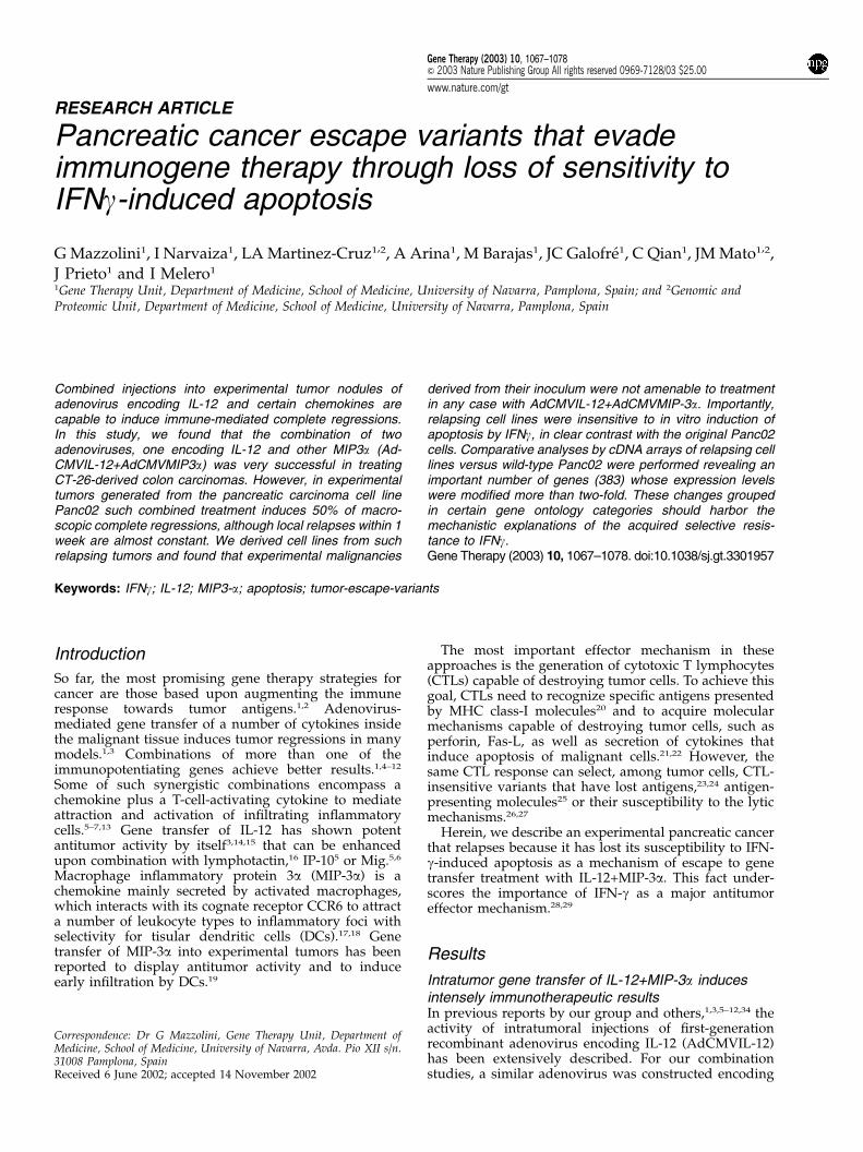
for MIP-3a cDNA under the control of the immediate/early CMV promoter (AdCMVMIP-3a), as schematicallyrepresented in Figure 1a. CT26 cells infected withAdCMVMIP-3a at different MOIs released into the tissueculture supernatant a polypeptide recognized by aspecific mAb in enzyme immune assays (Figure 1b).
According to the hypothesis of attraction/activa-tion,7,13 we have combined intratumor IL-12 genetransfer with gene therapy with cDNA encoding fordifferent chemokines. In CT26-derived tumors that hadgrown for 8 days in syngenic BALB/c mice, intratumorinjection of 5� 108 PFU of AdCMVMIP-3a induced onlyone out of six complete regressions (Figure 2a). In orderto test a possible synergistic effect with IL-12 genetransfer, we used an intratumoral suboptimal dose5
(7.5� 107 PFU) of AdCMVIL-12 that induced regressionin three out of five tumors. Combined injection ofidentical doses of both adenoviruses eradicated everytumor (six out of six), indicating the potency of thetherapeutic combination, that was confirmed in arepeated experiment shown in Figure 2b in whichAdCMVIL-12 (7.5� 107 PFU)+AdCMVMIP3-a (5�108 PFU) caused four out of five tumors to regress,whereas a similar dose of AdCMVIL-12 cured only oneout of four. In those two experimental groups receiving
subtherapeutical i.t. doses of AdCMVIL-12, IL-12 in-tratumor concentrations were comparable ranging from2 to 11 pg/mg of tumor tissue at day 4 after treatment. Inorder to exclude transgene-unrelated antitumoral effectscaused by adenovirus combinations, we performed (datanot shown) an additional set of experiments combiningAdCMVIL-12 (7.5� 107 PFU)+AdCMVlacZ (5� 108 PFU)that results in the similarly poor antitumoral effectsobserved with AdCMVIL-12 alone, thus confirmingresults that we have published in a previous workperformed in the same model.5
Intratumor injection of AdCMVIL-12+AdCMVMIP-3ainduces transient macroscopic complete regressionsof experimental pancreatic carcinomas derived fromPanc02 cell lineIn our hands, CT26 shows relatively high immunogeni-city, since its progression in Rag1�/� BALB/c mice is
Figure 1 Construction of AdCMVMIP3-a and in vitro production of thetransgene. (a) RT-PCR was used to clone mMIP3-a cDNA from mousethymus. MIP3-a cDNA was cloned downstream the CMV promoter andupstream polyA signal (An) into adenoviral shuttle vector pSQ1containing the adenoviral packaging signal (C). pSQ1/MIP-3a andpJM17 were cotransfected into 293 cells by calcium phosphate precipita-tion generating AdCMVMIP3-a. (b) Direct EIA analysis of MIP3-aproduction of cell culture supernatants from untransfected CT26 cells(control), infected with AdCMVlacZ (MOI: 10 000) or AdCMVMIP3-a ata MOI of 100, 1000, 5000 and 10 000 as indicated.
Figure 2 Intratumor injection of AdCMVIL-12+AdCMVMIP3-a dis-plays a potent and synergistic antitumoral effect on CT26 colorectalcarcinomas. BALB/c mice bearing subcutaneous CT26 tumor nodules (4–5 mm in diameter) were injected intratumorally with saline buffer ascontrol, 7.5� 107 PFU of AdCMVIL-12, 1�108 PFU of AdCMVMIP3-a,or a combination of both adenoviruses at identical doses. Data representfollow up of the mean diameter of tumor nodules was assessed every 3 days.The fraction of tumor-free surviving mice 3 months after treatment isgiven in each panel. Panels a and b represents two different experimentssimilarly done.
IFNg-resistant tumors escape IL12+MIP3-a gene therapyG Mazzolini et al
1068
Gene Therapy
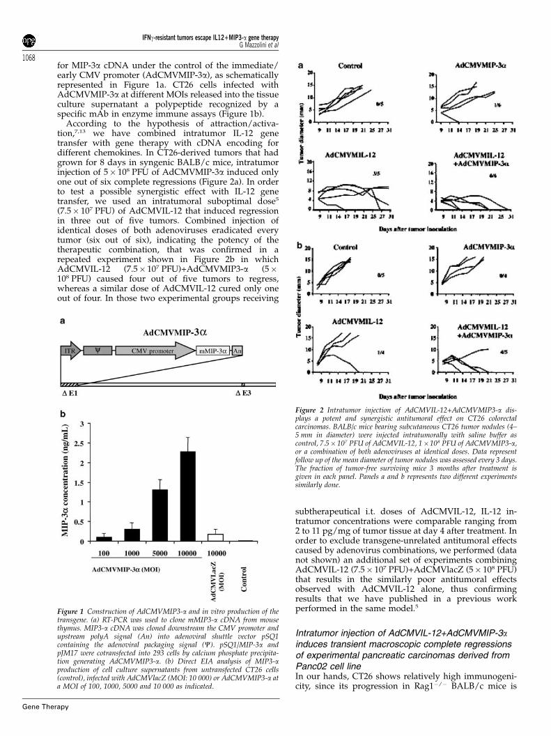
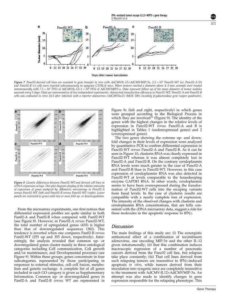
much faster than in BALB/c wild-type mice (Tirapu et al,manuscript in preparation). Therefore, we moved ontothe Panc02 tumor model, which shows lower intrinsicimmunogenicity in similar experiments. In Panc02-derived tumors, neither AdCMVIL-12 nor AdCMVMIP-3a displayed any detectable therapeutic activity (Figure3). By contrast, combination of both adenovirusesinduces complete regressions in six out of 12 tumors(three out of six tumors in experiment represented inFigure 3 and three out of six tumors represented inFigure 7). Surprisingly, although tumors remainedundetectable by careful palpation and inspection, re-lapses took place within approximately 1 week afterregression in every case. In C57BL/6 Rag1�/� mice,devoid of T and B-cells, this inhibition of growth neverdid take place (0/6), indicating that the effect wasmediated by the adaptative immune system (not shown).
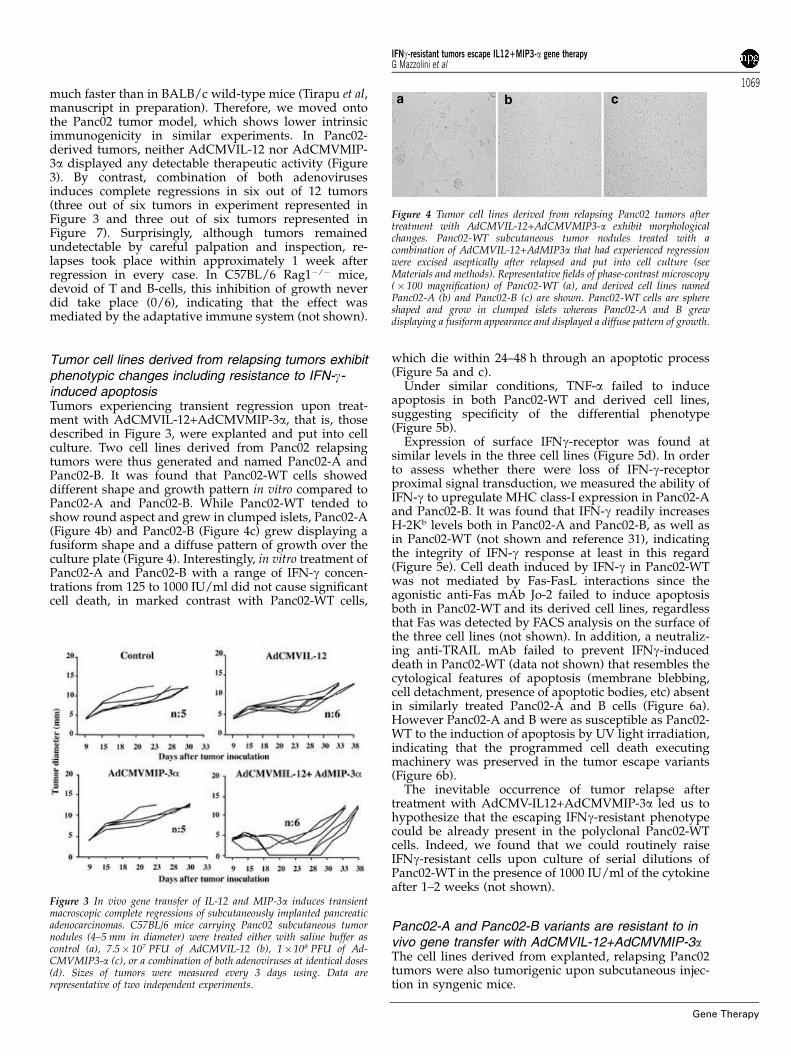
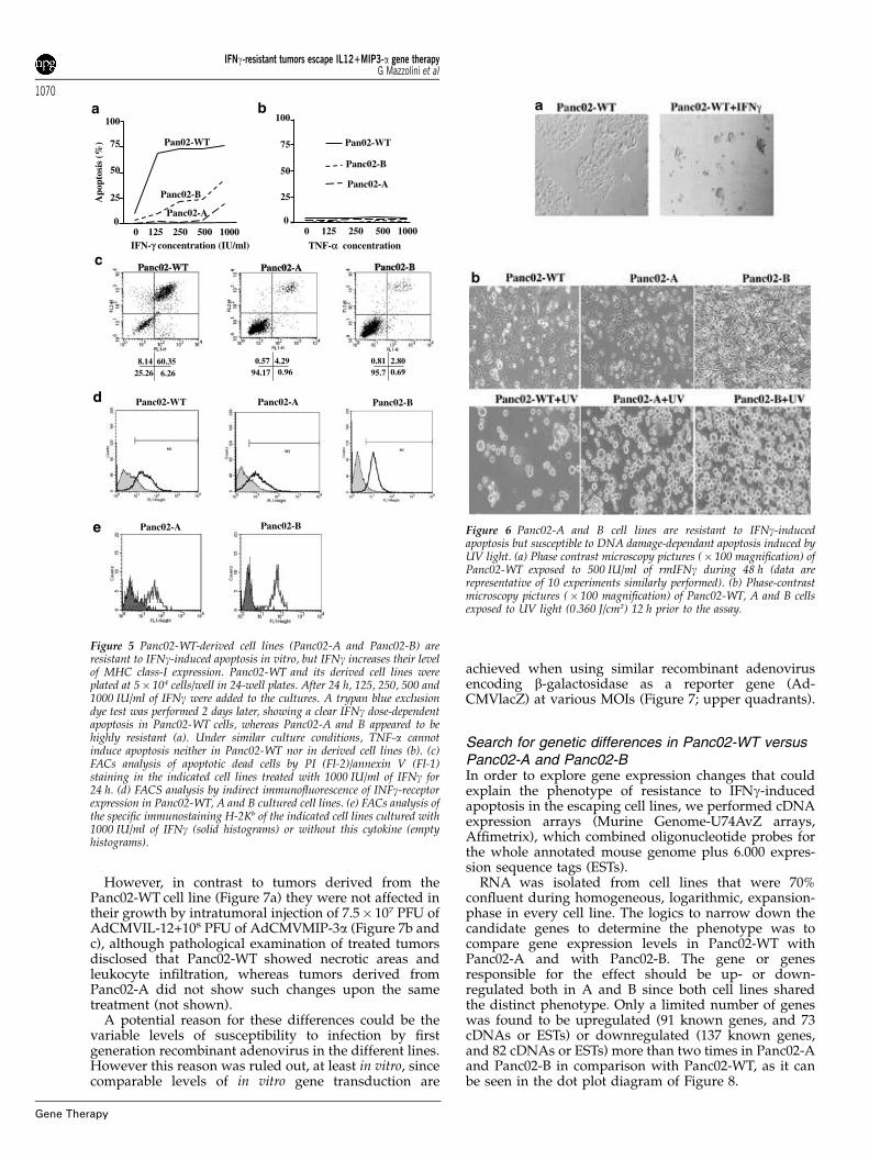
Tumor cell lines derived from relapsing tumors exhibitphenotypic changes including resistance to IFN-g-induced apoptosisTumors experiencing transient regression upon treat-ment with AdCMVIL-12+AdCMVMIP-3a, that is, thosedescribed in Figure 3, were explanted and put into cellculture. Two cell lines derived from Panc02 relapsingtumors were thus generated and named Panc02-A andPanc02-B. It was found that Panc02-WT cells showeddifferent shape and growth pattern in vitro compared toPanc02-A and Panc02-B. While Panc02-WT tended toshow round aspect and grew in clumped islets, Panc02-A(Figure 4b) and Panc02-B (Figure 4c) grew displaying afusiform shape and a diffuse pattern of growth over theculture plate (Figure 4). Interestingly, in vitro treatment ofPanc02-A and Panc02-B with a range of IFN-g concen-trations from 125 to 1000 IU/ml did not cause significantcell death, in marked contrast with Panc02-WT cells,
which die within 24–48 h through an apoptotic process(Figure 5a and c).
Under similar conditions, TNF-a failed to induceapoptosis in both Panc02-WT and derived cell lines,suggesting specificity of the differential phenotype(Figure 5b).
Expression of surface IFNg-receptor was found atsimilar levels in the three cell lines (Figure 5d). In orderto assess whether there were loss of IFN-g-receptorproximal signal transduction, we measured the ability ofIFN-g to upregulate MHC class-I expression in Panc02-Aand Panc02-B. It was found that IFN-g readily increasesH-2Kb levels both in Panc02-A and Panc02-B, as well asin Panc02-WT (not shown and reference 31), indicatingthe integrity of IFN-g response at least in this regard(Figure 5e). Cell death induced by IFN-g in Panc02-WTwas not mediated by Fas-FasL interactions since theagonistic anti-Fas mAb Jo-2 failed to induce apoptosisboth in Panc02-WT and its derived cell lines, regardlessthat Fas was detected by FACS analysis on the surface ofthe three cell lines (not shown). In addition, a neutraliz-ing anti-TRAIL mAb failed to prevent IFNg-induceddeath in Panc02-WT (data not shown) that resembles thecytological features of apoptosis (membrane blebbing,cell detachment, presence of apoptotic bodies, etc) absentin similarly treated Panc02-A and B cells (Figure 6a).However Panc02-A and B were as susceptible as Panc02-WT to the induction of apoptosis by UV light irradiation,indicating that the programmed cell death executingmachinery was preserved in the tumor escape variants(Figure 6b).
The inevitable occurrence of tumor relapse aftertreatment with AdCMV-IL12+AdCMVMIP-3a led us tohypothesize that the escaping IFNg-resistant phenotypecould be already present in the polyclonal Panc02-WTcells. Indeed, we found that we could routinely raiseIFNg-resistant cells upon culture of serial dilutions ofPanc02-WT in the presence of 1000 IU/ml of the cytokineafter 1–2 weeks (not shown).
Panc02-A and Panc02-B variants are resistant to invivo gene transfer with AdCMVIL-12+AdCMVMIP-3aThe cell lines derived from explanted, relapsing Panc02tumors were also tumorigenic upon subcutaneous injec-tion in syngenic mice.
Figure 3 In vivo gene transfer of IL-12 and MIP-3a induces transientmacroscopic complete regressions of subcutaneously implanted pancreaticadenocarcinomas. C57BL/6 mice carrying Panc02 subcutaneous tumornodules (4–5 mm in diameter) were treated either with saline buffer ascontrol (a), 7.5� 107 PFU of AdCMVIL-12 (b), 1�108 PFU of Ad-CMVMIP3-a (c), or a combination of both adenoviruses at identical doses(d). Sizes of tumors were measured every 3 days using. Data arerepresentative of two independent experiments.
a b c
Figure 4 Tumor cell lines derived from relapsing Panc02 tumors aftertreatment with AdCMVIL-12+AdCMVMIP3-a exhibit morphologicalchanges. Panc02-WT subcutaneous tumor nodules treated with acombination of AdCMVIL-12+AdMIP3a that had experienced regressionwere excised aseptically after relapsed and put into cell culture (seeMaterials and methods). Representative fields of phase-contrast microscopy(� 100 magnification) of Panc02-WT (a), and derived cell lines namedPanc02-A (b) and Panc02-B (c) are shown. Panc02-WT cells are sphereshaped and grow in clumped islets whereas Panc02-A and B grewdisplaying a fusiform appearance and displayed a diffuse pattern of growth.
IFNg-resistant tumors escape IL12+MIP3-a gene therapyG Mazzolini et al
1069
Gene Therapy
However, in contrast to tumors derived from thePanc02-WT cell line (Figure 7a) they were not affected intheir growth by intratumoral injection of 7.5� 107 PFU ofAdCMVIL-12+108 PFU of AdCMVMIP-3a (Figure 7b andc), although pathological examination of treated tumorsdisclosed that Panc02-WT showed necrotic areas andleukocyte infiltration, whereas tumors derived fromPanc02-A did not show such changes upon the sametreatment (not shown).
A potential reason for these differences could be thevariable levels of susceptibility to infection by firstgeneration recombinant adenovirus in the different lines.However this reason was ruled out, at least in vitro, sincecomparable levels of in vitro gene transduction are
achieved when using similar recombinant adenovirusencoding b-galactosidase as a reporter gene (Ad-CMVlacZ) at various MOIs (Figure 7; upper quadrants).
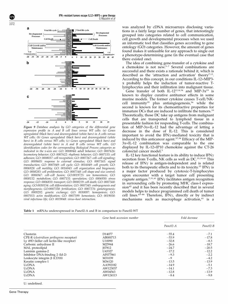
Search for genetic differences in Panc02-WT versusPanc02-A and Panc02-BIn order to explore gene expression changes that couldexplain the phenotype of resistance to IFNg-inducedapoptosis in the escaping cell lines, we performed cDNAexpression arrays (Murine Genome-U74AvZ arrays,Affimetrix), which combined oligonucleotide probes forthe whole annotated mouse genome plus 6.000 expres-sion sequence tags (ESTs).
RNA was isolated from cell lines that were 70%confluent during homogeneous, logarithmic, expansion-phase in every cell line. The logics to narrow down thecandidate genes to determine the phenotype was tocompare gene expression levels in Panc02-WT withPanc02-A and with Panc02-B. The gene or genesresponsible for the effect should be up- or down-regulated both in A and B since both cell lines sharedthe distinct phenotype. Only a limited number of geneswas found to be upregulated (91 known genes, and 73cDNAs or ESTs) or downregulated (137 known genes,and 82 cDNAs or ESTs) more than two times in Panc02-Aand Panc02-B in comparison with Panc02-WT, as it canbe seen in the dot plot diagram of Figure 8.
0
25
50
75
100
IFN-γ concentration (IU/ml)
Apo
ptos
is (
%) Pan02-WT
Panc02-B
Panc02-A
0 125 250 500 1000TNF-α concentration
0
25
50
75
100
Pan02-WT
Panc02-B
Panc02-A
0 125 250 500 1000
0.57 4.2994.17 0.96
8.14 60.3525.26 6.26
0.81 2.8095.7 0.69
a b
c
d
Panc02-Be Panc02-A
Panc02-BPanc02-APanc02-WT
Figure 5 Panc02-WT-derived cell lines (Panc02-A and Panc02-B) areresistant to IFNg-induced apoptosis in vitro, but IFNg increases their levelof MHC class-I expression. Panc02-WT and its derived cell lines wereplated at 5� 104 cells/well in 24-well plates. After 24 h, 125, 250, 500 and1000 IU/ml of IFNg were added to the cultures. A trypan blue exclusiondye test was performed 2 days later, showing a clear IFNg dose-dependentapoptosis in Panc02-WT cells, whereas Panc02-A and B appeared to behighly resistant (a). Under similar culture conditions, TNF-a cannotinduce apoptosis neither in Panc02-WT nor in derived cell lines (b). (c)FACs analysis of apoptotic dead cells by PI (Fl-2)/annexin V (Fl-1)staining in the indicated cell lines treated with 1000 IU/ml of IFNg for24 h. (d) FACS analysis by indirect immunofluorescence of INFg-receptorexpression in Panc02-WT, A and B cultured cell lines. (e) FACs analysis ofthe specific immunostaining H-2Kb of the indicated cell lines cultured with1000 IU/ml of IFNg (solid histograms) or without this cytokine (emptyhistograms).
Figure 6 Panc02-A and B cell lines are resistant to IFNg-inducedapoptosis but susceptible to DNA damage-dependant apoptosis induced byUV light. (a) Phase contrast microscopy pictures (� 100 magnification) ofPanc02-WT exposed to 500 IU/ml of rmIFNg during 48 h (data arerepresentative of 10 experiments similarly performed). (b) Phase-contrastmicroscopy pictures (� 100 magnification) of Panc02-WT, A and B cellsexposed to UV light (0.360 J/cm2) 12 h prior to the assay.
IFNg-resistant tumors escape IL12+MIP3-a gene therapyG Mazzolini et al
1070
Gene Therapy
From the microarray experiments, one first notices thatdifferential expression profiles are quite similar in bothPan02-A and Pan02-B when compared with Pan02-WT(see Figure 8). However, in Panc02-A versus Panc02-WT,the total number of upregulated genes (435) is higherthan that of downregulated sequences (382). Thistendency is inverted when one compares Pan02-B versusPan02-WT (255 up and 355 down, respectively). Inter-estingly, the analysis revealed that common up- ordownregulated genes cluster mainly in three ontologicalcategories including Cell communication, Cell growthand/or maintenance, and Developmental processes (seeFigure 9). Within these groups, genes concentrate in foursubcategories, represented by those participating inresponses to external stimulus, cell–cell fusion, metabo-lism and genetic exchange. A complete list of all genesincluded in each GO category is given as SupplementaryInformation. Common up- or downregulated genes inPan02-A and Pan02-B versus WT are represented in
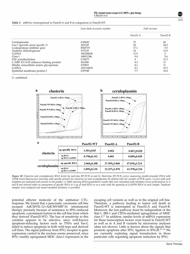
Figure 9c (left and right, respectively) in which geneswere grouped according to the Biological Process inwhich they are involved33 (Figure 9). The identity of thegenes with the highest changes in the relative levels ofexpression in Panc02-WT versus Panc02-A and B ishighlighted in Tables 1 (underexpressed genes) and 2(overexpressed genes).
The two genes showing the extreme up- and down-fold changes in their levels of expression were analyzedby quantitative PCR to confirm differential expression inPanc02-WT versus Panc02-A and Panc02-B. As it can beseen in Figure 10, clusterin RNA was clearly expressed inPanc02-WT whereas it was almost completely lost inPanc02-A and Panc02-B. On the contrary ceruloplasminRNA levels were much greater in the case of Panc02-Aand Panc02-B than in Panc02-WT. However, in this caseexpression of ceruloplasmin RNA was also detected inPanc02-WT at levels comparable to the housekeepingenzyme GAPDH RNA. In other words, ceruloplasminseems to have been overexpressed during the transfor-mation of Panc02-WT cells into the escaping variantsfrom basal levels. In the case of clusterin results arecompatible with a nearly complete loss of expression.The intensity of the observed changes with clusterin andceruloplasmin RNA concentrations, that are fully con-sistent with the cDNA microarray data, suggest a role forthese molecules in the apoptotic response to IFNg.
Discussion
The main findings of this study are: (i) The synergisticantitumoral effect of a combination of recombinantadenovirus, one encoding MIP-3a and the other IL-12given intratumorally; (ii) that this combination inducesmacroscopic regression of a number of pancreaticcancers derived from the Panc02 cell line, but relapsestake place constantly; (iii) That cell lines derived fromsuch relapsing tumors are insensitive to IFNg-inducedapoptosis in vitro, while tumors derived from theirinoculation into syngenic mice are completely insensitiveto the treatment with AdCMV-IL12+AdCMVMIP-3a. Anattempt was also made to identify changes in geneexpression responsible for the relapsing phenotype. This
Figure 7 Panc02-derived cell lines are resistant to gene transfer in vivo with AdCMVIL-12+AdCMVMIP-3a. 2.5� 105 Panc02-WT (a), Panc02-A (b)and Panc02-B (c) cells were injected subcutaneously in syngenic C57BL/6 mice. When tumors reached a diameter above 4–5 mm, animals were treatedintratumorally with 7.5� 107 PFU of AdCMVIL-12+1�108 PFU of AdCMVMIP3-a. Data represent follow up of the mean diameter of tumor nodulesassessed every 3 days. Data are representative of two independent experiments. Adenoviral transduction efficiency in Panc02-WT, Panc02-A and Panc02-Bcells was evaluated in vitro 24 h after infection with a reporter adenovirus (AdCMVlacZ) (MOI: 500) encoding b-galactosidase gene (upper quadrants).
a b
c d
Pan
c02-
A
Pan
c02-
BP
anc0
2-B
Pan
c02-
A
Panc02-WT
Panc02-WT
Panc02-WT
Panc02-WT
Figure 8 Genetic differences between Panc02-WT and derived cell lines incDNA expression arrays. Dot-plot diagram display of the relative intensityof expression of genes analyzed by Affimetrix microarrays in Panc02-Aversus Panc02-WT (left) and Panc02-B versus Panc02-WT (right). Lowerpanels are restricted to genes with two or more fold up- or downregulation.
IFNg-resistant tumors escape IL12+MIP3-a gene therapyG Mazzolini et al
1071
Gene Therapy
was analyzed by cDNA microarrays disclosing varia-tions in a fairly large number of genes, that interestinglygrouped into categories related to cell communication,cell growth and developmental processes when we usedan informatic tool that classifies genes according to geneontology (GO) categories. However, the amount of genesfound makes it unfeasible for any approach to single outa phenotype-determining gene (in the eventual case thatthere existed one).
The idea of combining gene-transfer of a cytokine anda chemokine is not new.5–7 Several combinations aresuccessful and there exists a rationale behind it, which isdescribed as the ‘attraction and activation’ theory7,13.According to this concept, in our conditions IL-12+MIP3-a probably helps the induction of tumor-reactive T-lymphocytes and their infiltration into malignant tissue.
Gene transfer of both IL-123,14,34 and MIP-3a19 isknown to display curative antitumor effects in somemurine models. The former cytokine causes T-cell/NK-cell immunity35 plus antiangiogenesis,36 while thesecond is known for its chemoattractive properties forimmature DCs that are induced to infiltrate the tumors.19
Theoretically, those DC take up antigens from malignantcells that are transported to lymphoid tissue in apresentable fashion for responding T-cells. The combina-tion of MIP-3a+IL-12 had the advantage of letting adecrease in the dose of IL-12. This is consideredimportant to avoid the IFNg-mediated toxicity that isinduced by this anticancer agent.37,38 Efficacy of the MIP-3a+IL-12 combination was comparable to the onedisplayed by IL-12+IP10 chemokine against the CT-26colorectal cancer model.5
IL-12 key functional feature is its ability to induce IFNgsecretion from T-cells, NK cells as well as DC.35,39,40 Thisrelease of IFNg is antigen-independent and is relatedboth to its therapeutic effects and to its toxicity.35 IFNg isa major factor produced by cytotoxic-T-lymphocytesupon encounter with a target tumor cell presentingcognate antigen.7,31,41 IFNg facilitates antigen recognitionin surrounding cells by promoting MHC class-I expres-sion42 and it has been recently described that in severalmodels helps to induce programmed cell death of tumorcell lines.43–46 Therefore, IFNg, directly or by indirectmechanisms such as macrophage activation,41 is a
b
a
c
Figure 9 Database analysis by GO categories of the differential geneexpression profile in A and B cell lines versus WT cells: (a) Genesupregulated (black bars) and downregulated (white bars) in A cells versusWT cells; (b) Genes upregulated (black bars) and downregulated (whitebars) in B cells versus WT cells; (c) Genes upregulated (black bars) anddownregulated (white bars) in A and B cells versus WT cells. GOidentification codes for the corresponding Biological Process categories asindicated in the x-axis are: GO: 0008044: adult behavior; GO: 0007626:locomotory behavior; GO: 0007622: rhythmic behavior; GO: 0007155: celladhesion; GO: 0008037: cell recognition; GO: 0007267: cell–cell signaling;GO: 0009605: response to external stimulus; GO: 0007165: signaltransduction; GO: 0007049: cell cycle; GO: 0016049: cell growth; GO:0006928: cell motility; GO: 0016043: cell organization and biogenesis;GO: 0008283: cell proliferation; GO: 0007148: cell shape and size control;GO: 0006947: cell–cell fusion; GO:0019725: ion homeostasis; GO:0008152: metabolism; GO: 0007151: sporulation; GO: 0006950: stressresponse; GO: 0006810: transport; GO: 0008219: cell death; GO: 0007568:aging; GO:0030154: cell differentiation; GO: 0007345: embryogenesis andmorphogenesis; GO:0007338: fertilization; GO: 0007276: gametogenesis;GO: 0009292: genetic exchange; GO: 0030097: hemopoiesis; GO:0007013: actin modification; GO: 0007599: hemostasis; GO: 0019058:viral infectious life; GO: 0019048: virus–host interaction.
Table 1 mRNAs underexpressed in Panc02-A and B in comparison to Panc02-WT
Gene bank accession number Fold decrease
Panc02-A Panc02-B
Clusterin D14077 �55.4 �7.1CPE-R (clostridium perfingrens receptor) AB000713 �53.9 �17.8Ly 49D (killer cell lectin-like receptor) U10090 �32.8 �8.3Carbonic anhydrase II M25944 �26.6 �18.7MAL proteolipid J07812 �24.7 �20.5Laminin gamma-2 U43327 �17.2 �12.2Inhibitor DNA-binding 2 (Id-2) AF077861 �9.3 �2.2Leukocyte integrin b (CD18) M31039 �9 �4.2Keratin complex-1 M36120 �13.5 �13.9UcDNA AA592182 �14.9 �5.1UcDNA AW123037 �14.1 �7.3UcDNA AW04563 �12.8 �13.9UcDNA AW124113 �8.4 �9.8
U: undefined.
IFNg-resistant tumors escape IL12+MIP3-a gene therapyG Mazzolini et al
1072
Gene Therapy
potential effector molecule of the antitumor CTL-response. We found that a pancreatic carcinoma cell lineescaped AdCMVIL-12+AdCMVMIP-3a intratumoraltherapy precisely because of resistance to IFNg-inducedapoptosis, a prominent feature in the cell line from whichthey derived (Panc02-WT). The loss of sensitivity to thiscytokine appears to be selective, since well-knownapoptosis-inducing factors such as TNFa and Fas-Lfailed to induce apoptosis in both wild-type and derivedcell lines. The signal pathway from IFNg receptor to geneexpression control in the nucleus seems preserved, sinceIFNg readily upregulated MHC class-I expression in the
escaping cell variants as well as in the original cell line.Therefore, a pathway leading to tumor cell death inPanc02-WT is interrupted in Panc02-A and Panc0-B.Moreover, the lost pathway must be independent of theStat-1, IRS-1 and CIITA-mediated upregulation of MHCclass I.41 In addition, similar levels of mRNA expressionfor those transcription factors were found in Panc02-WTas well as in A and B variants by microarray analyses(data not shown). Little is known about the signals thatpromote apoptosis after IFNg ligation to IFNgR.47–50 Weare currently exploring signal transduction in theseparticular cells regarding apoptosis induction by IFNg.
Table 2 mRNAs overexpressed in Panc02-A and B in comparison to Panc02-WT
Gene bank accession number Fold increase
Panc02-A Panc02-B
Ceruloplasmin U49430 73 60Gas-1 (growth arrest specific 1) X65128 30 24.9Leukoprotease inhibitor gene F002719 17.1 5.5Xanthine dehydrogenase X75129 14 13.9UcDNA AW208628 11.9 5Clast-1 AB031386 9.4 5.6PAF acetylhydrolase U34277 9 11.3C/EBP (CCAAT enhancer binding protein) X61800 8.3 11Fibulin extracellular matrix glycoprotein X70853 8.1 11UcDNA AW124470 8.1 10.3Epithelial membrane protein-3 U87948 7.7 10.4
U: undefined.
a bclusterin ceruloplasmin
Panc02-WT Panc02-BPanc02-A
clusterin
ceruloplasmin
ng specific RNA
ng RNA/ng RNA GADPH
ng specific RNA
ng RNA/ng RNA GADPH
c
1.501+0.05
0.798+0.152
2.068+0.208
1.099+0.110
0.002
0.001
37.995+3.068
23.257+1.878
0.067+0.020
0.089+0.028
47.039+3.214
61.978+4.234
Panc02-WT-RNA (50ng)
Panc02-A-RNA (50ng)
Panc02-B-RNA (50ng)
Panc02-WT-RNA (10ng)
Panc02-B-RNA (10ng)
Panc02-A-RNA (10ng)
Panc02-WT-RNA (50ng)
Panc02-A-RNA (50ng)Panc02-B-RNA (50ng)
Panc02-B-RNA (10ng)
Panc02-A-RNA (10ng)
Panc02-WT-RNA (10ng)
Figure 10 Clusterin and ceruloplasmin RNA levels by real-time RT-PCR (a and b). Real-time RT-PCR curves measuring double-stranded DNA withSYBR Green fluorescence intensity with specific-primers for clusterin (a) and ceruloplasmin (b) plotted with the number of PCR cycles. Curves with eachtemplates are indicated with arrows in the graphs. (c) Table showing RNA-quantitative results that were calculated with standard curves performed as in Aand B and referred either as nanograms of specific RNA in 5 mg of total RNA or as a ratio with the quantity of GADPH RNA in each sample. Triplicatesamples were analyzed and mean+standard deviation is provided.
IFNg-resistant tumors escape IL12+MIP3-a gene therapyG Mazzolini et al
1073
Gene Therapy
It is possible that the escaping mutant variants werealready present in the original Panc02-WT populationsince in vitro culture of these cells in the presence of IFNgrendered in week-time colonies growing in the presenceof 1000 IU/ml of IFNg. This favors the interpretation thatthe in vivo immunotherapeutic treatment selects out theescaping variants that were already present as a minorityamong Panc02-WT cells.
To identify candidate changes in genes or in proteinsthat could explain the resistant phenotype observed inPanc02-derived cell lines, we reasoned that such varia-tions should be overexpressed or underexpressed in bothof the escaping cell lines, compared to the wild type. Thetools used for this study included analysis of gene-expression from cDNA microarrays encompassingprobes for the whole-annotated mouse genome plus alarge number of ESTs. This approach narrowed down thenumber of genes, but still an important number ofcandidates51 with significant variation of expressioncommon to both escaping cell lines was substantiated(up¼164 and down¼219), hence precluding individualstudy. In a sense the phenotype could be the result ofonly one of such changes or alternatively the result fromthe sum of a number of them. We favor the secondinterpretation and we analyzed the genes by groupingthem into ontological categories according to theBiological Process in which they are involved33 (Marti-nez-Cruz et al, manuscript in preparation that describesthe informatics of algorithms functioning on the data-bases). Similar classification criteria have been used toanalyze complete genomes52 and changes of geneexpression profiles in transgenic mice.53
From the list of genes with up- or downregulation inPanc02-A and Panc02-B versus Panc02-WT some of themhad striking fold changes and are the subject of ourcurrent and future research. For instance, among under-expressed genes we find clusterin (fold changes A versusWT: �55.4; B versus WT: �7.1). This gene has beeninvolved both in promotion and protection from apop-tosis with a still elusive molecular mechanism.54,55
Curiously the receptor for Clostridium perfringens enter-otoxin (CPE-R) is decreased in its expression (�53.9-foldin A and �17.8-fold in B). This gene is a target forpancreatic cancer therapy and its function as a putativetransmembrane channel makes it very interesting sinceits physiological role is still unknown.56 Carbonicanhydrase II, MAL protelipid, laminin g2 were alsogenes with more than 10-fold downregulation in bothescaping cell lines and deserve future study for theirpotential role at changing IFNg susceptibility and/or cellshape of the tumor cells.
On the contrary, other genes were drastically upregu-lated among them: ceruloplasmin (73-fold and 60-fold inA and B, respectively), Gas 1 (growth arrest specific 1)(fold 30/24.9), xanthine deshydrogenase (14/13.9). Thesegenes have not obvious involvement in the observedphenotype, but novel functions could be discovered forthem. Furthermore, ceruloplasmin and xanthine dehy-drogenase have a role in the defense against oxidativestress57 that may be involved in the observed resistanceto apoptosis. Besides, platelet activatory factor (PAF)acetylhydrolase is also upregulated (nine times in A/11.3times in B); this is also an interesting finding since it hasbeen recently proposed that this molecule might haveimplications in tumor biology.58
It has been recently shown that small changes inexpression results in severe disease.59 Therefore, exces-sive focusing in the highlighted changes could bemisleading since it cannot be excluded that genes withsmall changes may play key roles in promotion and/orprotection from apoptosis. Nonetheless the role playedby those extreme fold changes in the levels of RNAencoding for clusterin and ceruloplasmin, that have beenconfirmed by quantitative PCR, is the matter of ourcurrent research efforts.
We think that other alterations not involving changesin mRNA expression, such as point mutations that mightalter the function due to production of nonfunctionalproteins, should not be overlooked. These changes arenot addressed by the analysis of cDNA arrays. Inaddition, an important number of sequences correspond-ing to ESTs and cDNAs without assigned function weredifferentially expressed in Panc02-WT and escapingmalignant cells. It is possible that a key piece ofinformation is still missing due to insufficient genefunction knowledge.
In conclusion, beyond describing a successful immu-notherapeutic gene combination, this study raises a newconcern for immunity-based gene therapy, since it adds anovel mechanism of tumor escape through the acquisi-tion of resistance to IFNg-induced apoptosis. Ourattempt to study the gene expression profile of escapevariants showed variation in multiple genes that tend togroup in certain functional categories. Our ongoing andfuture research tries to identify the master switchchanges in gene expression that would hierarchicallyexplain the phenotype of the escaping-malignant cells.
Material and methods
Mice and cell linesAt 5- to 8-week-old C57BL/6 (H2b) and BALB/c (H2d)female mice were purchased from Harlan (Barcelona,Spain) and were housed according to institutionalguidelines. The 293 cell line was obtained throughAmerican Type Culture Collection (ATCC) (Manassas,VA, USA). CT26 tumor cell line is an undifferentiatedmurine colorectal adenocarcinoma.14 The mouse pan-creatic cancer Panc02 was established in C57BL/6 miceinduced by 3-methylcholantrene and presented as aductal pancreatic adenocarcinoma.30,31 The subcutaneousPanc02 tumor tissue was obtained from the NationalCancer Institute, DCTDC Tumor Repository (Frederick,MD, USA). A cell line was isolated from trypsination ofPanc02 tumor tissue. Panc02 and CT26 cells weremaintained in RPMI 1640 medium containing 10% FCS,2 mM L-glutamine, 100 U/ml streptomycin, 10 mg/mlciprofloxacin and 100 mg/ml penicillin. Cell culturereagents were from Life Technologies (Basel, Switzer-land).
Adenovirus constructionThe mouse MIP-3a cDNA was generated by an RT-PCRtechnique using total RNA extracted from mousethymus as template with primers (sense: 50-AC-CATGGCCTGCGGTGGCAAGCGT-30 and antisense: 50-GATATCTTACATCTTCTTGACTCTTAG-30) and directlycloned into pCR2.1-TOPO (Invitrogen, Carlsbad, CA,USA). The sequence was confirmed by DNA sequencing.
IFNg-resistant tumors escape IL12+MIP3-a gene therapyG Mazzolini et al
1074
Gene Therapy
The MIP-3a sequence was cloned into adenoviral shuttlevector pSQ1 with CMV immediate/early promoter up-stream and polyA sequence downstream of the insertionsite to form pSQ1/MIP-3a. pSQ1/MIP-3a and pJM17were cotransfected into 293 cells by calcium phosphateprecipitation to obtain AdCMVMIP-3a according tostandard procedures.14 Recombinant adenoviruses carry-ing the p40 and p35 genes of IL-12 (AdCMVIL-12) or b-galactosidase (AdCMVlacZ) under the control of CMVpromoter have been previously described.32 Adeno-viruses were purified by double cesium chloride ultra-centrifugation and extensively dialyzed against 10 mM
Tris/1 mM MgCl2 prior to be stored at �801C in thepresence of 10% (v/v) glycerol. Titration was made byplaque assay.32
In vitro MIP-3a productionSupernatants from AdCMVMIP-3a-infected CT26 cells(3� 105 cell/well in 12-well plates) at MOIs 100 and10 000, as well as noninfected CT26 cells, were collected48 h after infection. MIP-3a levels were measured bydirect enzyme immunoabsorbent assay (EIA). A 96-wellflat-bottomed microtitration plate (Titertek, ICN Bio-chemicals INC. Aurora, OH, USA) were coated with100 ml of different supernatants and standards inidentical medium (recombinant murine MIP-3a, R&DSystems, Minneapolis, MN, USA) and maintained at 41Covernight. The plates were washed five times with 100 mlphosphate-buffered saline (PBS)-Tween (0.05%) andblocked with 100 ml of PBS containing 10% fetal bovineserum (FBS) for 2 h at room temperature. After blocking,the plates were washed in PBS-Tween and incubated for2 h at room temperature with 100 ml of anti-mouse MIP-3a (R&D Systems) at final concentration of 2 mg/ml. Theplates were washed five times and further incubated inPBS-Tween with 10% FBS containing 100 ml of 1:2000diluted peroxidase-conjugated anti-goat IgG (SIGMA,Madrid, Spain). After 2 h of incubation at roomtemperature, the plates were washed 10 times with100 ml of PBS-Tween. Color was developed with 100 ml oftetramethylbenzidine substrate (BD Biosciences Phar-mingen, San Diego CA) followed by 50 ml of stopsolution (2 N H2SO4). Absorbance (OD) of samples wasread by a Titertek Multiscan II (Flow Laboratories)microplate reader at 450 nm. Results were determinedin duplicate and expressed as the mean7s.d.
Establishment of subcutaneous model in vivoPrior to administration of treatment, BALB/c andC57BL/6 mice were inoculated s.c. in the right hindflank with 5� 105 CT26 or 2.5� 105 Panc02-WT tumorcells in 50 ml of PBS. For treatment, when tumors reach adiameter above 4–5 mm, subcutaneous tumors wereinjected with 50 ml of PBS containing 7.5� 107 PFU ofAdCMVIL-12, 5� 108 PFU of AdCMVMIP-3a or both.Similarly, tumors derived from Panc02-A and Panc02-Bcell lines were treated with 7.5� 107 PFU of AdCMVIL-12+5� 108 PFU of AdMIP3a or saline. Tumor size (meandiameter) was assessed using a precision caliper.
Tumor cell lines derived from relapsing tumorsPanc02-WT tumors treated with 7.5� 107 PFU of Ad-CMVIL-12+5� 108 PFU of AdMIP3a that experiencedregression and subsequent relapse were excised asepti-cally and single-cell suspensions were obtained passing
the cells through a mesh and then put into cell culture.Culture medium was RPMI 1640 supplemented with10% FCS, 2 mM L-glutamine, 100 U/ml streptomycin,10 mg/ml ciprofloxacin and 100 mg/ml penicillin. Thetwo cell lines derived were named Panc02-A and Panc02-B. Cell culture reagents were from Life Technologies(Basel, Switzerland).
In vitro IFN-g-induced apoptosisPanc02-WT, Panc02-A and Panc02-B cells were plated at5� 104 cells/well onto 24-well plates (Greiner Labortech-nik) and cultured in RPMI 1640 medium to sub-confluence. After 24 h, cells were incubated at differentconcentrations (100–1000 IU/ml) of mrIFN-g (Peprotech.UK). 36 hs after exposure to IFNg, Tripan blue dyeexclusion test was carried out. UV light induction ofapoptosis was performed by exposure at 0.360 J/cm2
during 60 s in a DNA cross linker device (UVItecCL508G).
Detection of major histocompatibility complex (MHC)class-I expressionSuspensions of Panc02-WT, Panc02-A and Panc02-B weretested for H-2Kb expression by flow cytometry analysis.Cells were washed with PBS and stained with FITC-tagged mAb anti-H-2Kb (Pharmingen, San Diego, CA,USA). Indirect immunofluorescence staining with anti-IFNg-receptor mAb (Pharmingen) was developed withFITC-tagged polyclonal anti-rat IgG (Sigma, Barcelona,Spain). Control staining with irrelevant FITC-tagged ratmAb was also used. After washing, cells were analyzedusing flow cytometer (Facscallibur, Becton-Dickinson).
FACS analysis of apoptotic cells by annexin-V stainingTo detect and quantify apoptosis of both Panc02-WT andderived cell lines (Panc02-A and Panc02-B) after ex-posure to rmIFNg at the indicated concentrations cellswere stained with Anexin-V-FLUOS Staining kit (Boher-inger Mannheim, Germany) according to the manufac-turer instructions.
In vitro transduction of Panc02-WT, Panc02-A andPanc02-B cell lines. X-gal stainingWe evaluated the in vitro transduction efficiency of therecombinant adenovirus in Panc02-WT, Panc02-A andPanc02-B cells using AdCMVlacZ vector. 105 cells in 1 mlof complete RPMI 1640 medium were seeded in 24-wellplates. After 24 h, cells were infected at different multi-plicities of infection (100, 500 and 1000) with Ad-CMVlacZ and incubated at 371C and 5% CO2 for 48 h.For X-gal staining, cells were fixed in 24-well plates withglutaraldehyde (0.5%) and stained with 5-bromo-4-chloro-3-indoyl-beta-galactosidase (X-Gal) as previouslydescribed.32 The experiment was done twice andtriplicates were included.
Gene expression analysis by microarraysMurine pancreatic cancer Panc02-WT and its derivedPanc02-A and Panco2-B cell lines were grown asmonolayer in RPMI 1640 supplemented with 10% FCS,2 mM L-glutamine, 100 U/ml streptomycin, 10 mg/mlciprofloxacin and 100 mg/ml penicillin. Cells were grownat 371C in humidified 5% CO2 atmosphere, placed in 15-cm plates and harvested when at 70–80% confluence.
IFNg-resistant tumors escape IL12+MIP3-a gene therapyG Mazzolini et al
1075
Gene Therapy
Total RNA was isolated using the Trizol RNA isolationmethod (Gibco/BRL) and purified with the QiagenRNeasy Mini Kit spin columns (Qiagen). RNA concen-tration was determined using a spectrophotometer. RNAintegrity was confirmed on a 1% agarose gel electro-phoresis.
Target preparationDouble-strand DNA was synthesized using the Super-script Choice System (Gibco/BRL). In all, 10 mg of totalRNA was used in a reverse transcription reaction tosynthesize negative strand cDNA with a primer contain-ing poly-T and T7 RNA polymerase promoter sequences.Double-stranded cDNA was phenol–chloroform ex-tracted and ethanol precipitated. The cDNA was resus-pended in 12 ml of RNase-free water and 5 ml of thedouble-stranded cDNA was used as a template for invitro transcription, in the presence of biotinylated UTPand CTP, to generate labeled antisense RNA. The in vitrotranscription reaction was performed using the EnzoBioArray High Yield RNA Transcript Labeling Kit (EnzoDiagnostics). Labeled RNA was purified with the QiagenRNeasy Mini Kit spin columns (Qiagen) and quantifiedspectrophotometrically.
Array hybridization and scanningLabeled cRNA was fragmented in fragmentation buffer(5� buffer: 200 mM Tris-acetate (pH 8.1)/500 mM
KOAc/150 mM MgOAc) and hybridized to the micro-arrays in 200 ml of hybridization solution containing15 mg of labeled target in 1� Mes buffer (0.1 M Mes/1.0 M NaCl/0.01% Tween20/20 mM EDTA) and 0.1 mg/ml herring sperm DNA. Test2 arrays (Affymetrix) werehybridized to check the cRNA integrity before theexpression arrays and in all cases the GADPH 30/50 ratiowas below 1.1. Samples were then hybridized on MurineGenome-U74Av2 Arrays (Affymetrix, USA). This arrayrepresents all sequences (B6000) in the Mouse UniGenedatabase (Build 74) that have been functionally char-acterized and B6000 EST clusters. Arrays were rotated at60 rpm for 16 h at 451C. Following hybridization, thearrays were washed with 6� SSPE-T (0.9 M NaCl/60 mM NaH2PO4/6 mM EDTA/0.01% Tween20) at 251Con a fluidics station (Affymetrix) for 10� 2 cycles, andsubsequently with 0.1 M Mes/0.1 M NaCl/0.01%Tween20 at 501C for 4� 15 cycles. The arrays were thenstained with a streptavidin–phycoerythrin conjugate(Molecular Probes), followed by 10� 4 wash cycles. Toenhance the signals, the arrays were further stained withAnti-streptavidin antibody for 10 min, followed by a 10-min staining with a streptavidin–phycoerythrin conju-gate. After 15� 4 additional wash cycles, the arrays werescanned using a confocal scanner (Affymetrix). Theimage data were analyzed by Microarray Suite 4.0(Affymetrix).
Genechip analysisUsing oligonucleotide microarrays, we studied geneexpression profiles of Panc02-A and Panc02-B cell linesversus Panc02-WT cells, and analyzed those genessharing a common up- or downregulation behavior inthe microarray experiments. Those differentially ex-pressed genes for which a GO code33 is assigned in theUnigene database (http://www.ncbi.nlm.nih.gov/Lo-cuslink), were classified according to the Biological
Process in which they participate (Martinez-Cruz et al,a manuscript in preparation that describes the infor-matics of algorithms functioning on the databases). Onlyentries showing an expression fold change equal orbigger than 2.0 were considered.
A complete list of all genes included in each GOcategory is given as Supplementary Information.
Real-time quantitative RT-PCR5 mg of total RNA from Panc02-Wt, Panc02-A andPanc02-B cell lines were DNase I treated (1.2 U/mgRNA) before cDNA synthesis. One mg of DNase I treatedRNA was used for cDNA synthesis (20 ml) with Super-Scriptt Rnase H-Reverse Transcriptase (Invitrogen)(400 U/mg RNA) and 100 pmol oligodT.
Sequences for forward and reverse primers for eachgene are the following: 50CGACTCGCTCCAGGTGGCCGAGAGGC30 and 50CCACCTCAGTGACACGGGAGGGGACCTCTGAGT30 for clusterin; 50CCTGGTAATCCTACAGATGGACAGAGCAAC30 and 50TGGTTTTAGAGCTTTATTGTAGGAAGGCAG30 for ceruloplasmin; 50AAGGCTGGGGCTCACCTGAA30 and 50GGCATGGACTGTGGTCATGAG 30 for GAPDH.
A standard curve, with serial dilutions of cDNA, wasconstructed for each sequence. Template concentrationsfor reactions in the relative standard were given arbitraryvalues of 10, 5, 2.5, 1.25, 0.625, 0.3125 and 0.15625.
Real time PCR reactions were prepared using Light-Cycler-FastStart DNA master SYBR Green I kit (Roche)according to the manufacturer instructions. The ampli-fication program consisted of 1 cycle of 951C with 1 minhold (‘hot start’) followed by 45 cycles of 951C(denaturation) with 10-s hold, 601C (annealing) with 5-shold, 721C (amplification and acquisition) with 10-s hold.Melting curve analysis using the program run for onecycle at 951C with 0-s hold, 651C with 15-s hold, and 951Cwith 0-s hold at the step acquisition mode followedamplification.
The average of triplicate data of each sample was usedto calculate the relative change in gene expression afternormalization target concentration to GAPDH.
Acknowledgements
We thank Dr Bendandi and Tirapu for critical readingand helpful discussion. Juan Percaz, Javier Guillen,Izaskun Gabari and Celia Gomar are acknowledged fortheir technical assistance. The team at Medplant Geneticsis acknowledged for assistance with the microarraytechnology. Financial support was from CICYT (SAF99/0039), Gobierno de Navarra and grants from Funda-cion Ramon Areces and Ines Bemberg.
References
1 Melero I et al. IL-12 gene therapy for cancer: in synergy withother immunotherapies. Trends Immunol 2001; 22: 113–115.
2 Dranoff GM, Mulligan RC. Gene transfer as cancer therapy. AdvImmunol 1995; 58: 417–454.
3 Bramson JL et al. Direct intratumoral injection of an adenovirusexpressing interleukin-12 induces regression and long-lastingimmunity that is associated with highly localized expression ofinterleukin-12. Hum Gene Ther 1995; 7: 1995–2002.
IFNg-resistant tumors escape IL12+MIP3-a gene therapyG Mazzolini et al
1076
Gene Therapy
4 Emtage PC et al. A double recombinant adenovirus expressingthe costimulatory molecule B7-1 (murine) and human IL-2induces complete tumor regression in a murine breast adeno-carcinoma model. J Immunol 1998; 160: 2531–2538.
5 Narvaiza I et al. Intratumoral coinjection of two adenoviruses,one encoding the chemokine IFN-gamma-inducible protein-10and another encoding IL-12, results in marked antitumoralsynergy. J Immunol 2000; 164: 3112–3122.
6 Palmer K et al. Combined CXC chemokine and interleukin-12gene transfer enhances antitumor immunity. Gene Ther 2001; 8:282–290.
7 Dilloo D et al. Combined chemokine and cytokine gene transferenhances antitumor immunity. Nat Med 1996; 2: 1090–1095.
8 Putzer B et al. Interleukin 12 and B7-1 costimulatory moleculeexpressed by an adenovirus vector act synergistically to facilitatetumor regression. Proc Natl Acad Sci USA 1997; 94: 10889–10894.
9 Putzer BM et al. Large nontransplanted hepatocellular carcinomain woodchucks: treatment with adenovirus-mediated delivery ofinterleukin 12/B7.1 genes. J Natl Cancer Inst 2001; 93: 472–479.
10 Martinet O et al. Immunomodulatory gene therapy withinterleukin 12 and 4-1BB ligand: long-term remission of livermetastases in a mouse model. J Natl Cancer Inst 2000; 92: 931–936.
11 Zitvogel L et al. Interleukin-12 and B7.1 co-stimulation cooperatein the induction of effective antitumor immunity and therapy ofestablished tumors. Eur J Immunol 1996; 26: 1335–1341.
12 Addison CL et al. Intratumoral coinjection of adenoviral vectorsexpressing IL-2 and IL-12 results in enhanced frequency ofregression of injected and untreated distal tumors. Gene Ther1998; 5: 1400–1409.
13 Levitsky HI. The best cytokine for the job. Nat Med 1997; 3: 126.14 Mazzolini G et al. Regression of colon cancer and induction of
antitumor immunity by intratumoral injection of adenovirusexpressing interleukin-12. Cancer Gene Ther 1999; 6: 514–522.
15 Barajas M et al. Gene therapy of orthotopic hepatocellularcarcinoma in rats using adenovirus coding for interleukin 12.Hepatology 2001; 33: 52–61.
16 Emtage PC et al. Adenoviral vectors expressing lymphotactinand interleukin 2 or lymphotactin and interleukin 12 synergizeto facilitate tumor regression in murine breast cancer models.Hum Gene Ther 1999; 10: 697–709.
17 Luster AD. Chemokines – chemotactic cytokines that mediateinflammation. N Engl J Med 1998; 338: 436–445.
18 Greaves DR et al. CCR6, a CC chemokine receptor that interactswith macrophage inflammatory protein 3alpha and is highlyexpressed in human dendritic cells. J Exp Med 1997; 186: 837–844.
19 Fushimi T, Kojima A, Moore MA, Crystal RG. Macrophageinflammatory protein 3alpha transgene attracts dendritic cells toestablished murine tumors and suppresses tumor growth. J ClinInvest 2000; 105: 1383–1393.
20 Hellstrom I, Hellstrom KE. T cell immunity to tumor antigens.Crit Rev Immunol 1998; 18: 1–6.
21 Berke G. Unlocking the secrets of CTL and NK cells. ImmunolToday 1995; 16: 343–346.
22 Yagita H et al. Role of perforin in lymphocyte-mediatedcytolysis. Adv Immunol 1992; 51: 215–242.
23 Seliger B, Maeurer MJ, Ferrone S. Antigen-processing machinerybreakdown and tumor growth. Immunol Today 2000; 21: 455–464.
24 Marincola FM, Jaffee EM, Hicklin DJ, Ferrone S. Escape ofhuman solid tumors from T-cell recognition: molecular mechan-isms and functional significance. Adv Immunol 2000; 74: 181–273.
25 Garrido F et al. Implications for immunosurveillance of alteredHLA class I phenotypes in human tumours. Immunol Today 1997;18: 89–95.
26 Bergmann-Leitner ES, Abrams SI. Influence of interferon gammaon modulation of Fas expression by human colon carcinomacells and their subsequent sensitivity to antigen-specific CD8+cytotoxic T lymphocyte attack. Cancer Immunol Immunother 2000;49: 193–207.
27 Bergmann-Leitner ES, Abrams SI. Positive and negative con-sequences of soluble Fas ligand produced by an antigen-specificCD4(+) T cell response in human carcinoma immune interac-tions. Cell Immunol 2001; 209: 49–62.
28 Kaplan DH et al. Demonstration of an interferon gamma-dependent tumor surveillance system in immunocompetentmice. Proc Natl Acad Sci USA 1998; 95: 7556–7561.
29 Shankaran V et al. IFNgamma and lymphocytes prevent primarytumour development and shape tumour immunogenicity. Nature2001; 410: 1107–1111.
30 Corbett TH et al. Induction and chemotherapeutic response oftwo transplantable ductal adenocarcinomas of the pancreas inC57BL/6 mice. Cancer Res 1984; 44: 717–726.
31 Clary BM et al. Inhibition of established pancreatic cancersfollowing specific active immunotherapy with interleukin-2gene-transduced tumor cells. Cancer Gene Ther 1997; 4: 97–104.
32 Qian C, Bilbao R, Bruna O, Prieto J. Induction of sensitivity toganciclovir in human hepatocellular carcinoma cells by adeno-virus-mediated gene transfer of herpes simplex virus thymidinekinase. Hepatology 1995; 22: 118–123.
33 Ashburner M et al. Gene ontology: tool for the unification ofbiology. The Gene Ontology Consortium. Nat Genet 2000; 25: 25–9.
34 Caruso M et al. Adenovirus-mediated interleukin-12 genetherapy for metastatic colon carcinoma. Proc Natl Acad Sci USA1996; 93: 11302–11306.
35 Trinchieri G. Interleukin-12: a cytokine at the interface ofinflammation and immunity. Adv Immunol 1998; 70: 83–243.
36 Sgadari C, Angiolillo AL, Tosato G. Inhibition of angiogenesis byinterleukin-12 is mediated by the interferon-inducible protein 10.Blood 1996; 87: 3877–3882.
37 Robertson MJ et al. Immunological effects of interleukin 12administered by bolus intravenous injection to patients withcancer. Clin Cancer Res 1999; 5: 9–16.
38 Leonard JP et al. Effects of single-dose interleukin-12 exposureon interleukin-12-associated toxicity and interferon-gammaproduction. Blood 1997; 90: 2541–8.
39 Grohmann U et al. Positive regulatory role of IL-12 inmacrophages and modulation by IFN-gamma. J Immunol 2001;167: 221–227.
40 Munder M, Mallo M, Eichmann K, Modolell M. Murinemacrophages secrete interferon gamma upon combined stimula-tion with interleukin (IL)-12 and IL-18: a novel pathway ofautocrine macrophage activation. J Exp Med 1998; 187: 2103–2108.
41 Sad S, Marcotte R, Mosmann TR. Cytokine-induced differentia-tion of precursor mouse CD8+ T cells into cytotoxic CD8+ T cellssecreting Th1 or Th2 cytokines. Immunity 1995; 2: 271–279.
42 Boehm U, Klamp T, Groot M, Howard JC. Cellular responses tointerferon-gamma. Annu Rev Immunol 1997; 15: 749–795.
43 Shyu RY, Su HL, Yu JC, Jiang SY. Direct growth suppressiveactivity of interferon-alpha and -gamma on human gastriccancer cells. J Surg Oncol 2000; 75: 122–130.
44 Ruiz-Ruiz C, Munoz-Pinedo C, Lopez-Rivas A. Interferon-gamma treatment elevates caspase-8 expression and sensitizeshuman breast tumor cells to a death receptor-induced mitochon-dria-operated apoptotic program. Cancer Res 2000; 60: 5673–5680.
45 Wu AJ et al. Interferon-gamma induced cell death in a culturedhuman salivary gland cell line. J Cell Physiol 1996; 167: 297–304.
46 Suk K et al. Interferon gamma (IFNgamma) and tumor necrosisfactor alpha synergism in ME-180 cervical cancer cell apoptosisand necrosis. IFNgamma inhibits cytoprotective NF-kappa Bthrough STAT1/IRF-1 pathways. J Biol Chem 2001; 276: 13153–13159.
47 Veldman RJ, Klappe K, Hoekstra D, Kok JW. Interferon-gamma-induced differentiation and apoptosis of HT29 cells: dissociationof (glucosyl)ceramide signaling. Biochem Biophys Res Commun1998; 247: 802–808.
IFNg-resistant tumors escape IL12+MIP3-a gene therapyG Mazzolini et al
1077
Gene Therapy
48 Lee KY, Anderson E, Madani K, Rosen GD. Loss of STAT1expression confers resistance to IFN-gamma-induced apoptosisin ME180 cells. FEBS Lett 1999; 459: 323–326.
49 Jiang MC et al. IRF-1-mediated CAS expression enhancesinterferon-gamma-induced apoptosis of HT-29 colon adenocar-cinoma cells. Mol Cell Biol Res Commun 2001; 4: 353–358.
50 Naujokat C, Sezer O, Possinger K. Tumor necrosis factor-alphaand interferon-gamma induce expression of functional Fasligand on HT29 and MCF7 adenocarcinoma cells. BiochemBiophys Res Commun 1999; 264: 813–819.
51 Wu TD. Analysing gene expression data from DNA microarraysto identify candidate genes. J Pathol 2001; 195: 53–65.
52 Dwight SS et al. Saccharomyces Genome Database (SGD)provides secondary gene annotation using the Gene Ontology(GO). Nucleic Acids Res 2001; 30: 69–72.
53 Martınez-Chantar ML et al. Spontaneous oxidative stress andliver tumors in mice lacking methionine adenosyltransferase 1A.FASEB J 2002; 16: 1292–2004.
54 Han BH et al. Clusterin contributes to caspase-3-independentbrain injury following neonatal hypoxia-ischemia. Nat Med 2001;7: 338–343.
55 McLaughlin L et al. Apolipoprotein J/clusterin limits theseverity of murine autoimmune myocarditis. J Clin Invest 2000;106: 1105–1113.
56 Michl P et al. Claudin-4: a new target for pancreatic cancertreatment using Clostridium perfringens enterotoxin. Gastro-enterology 2001; 121: 678–684.
57 de Silva DM, Aust SD. Ferritin and ceruloplasmin in oxidativedamage: review and recent findings. Can J Physiol Pharmacol1993; 71: 715–720.
58 Bussolati B et al. PAF produced by human breast cancer cellspromotes migration and proliferation of tumor cells and neo-angiogenesis. Am J Pathol 2000; 157: 1713–1725.
59 Yan H et al. Small changes in expression affect predisposition totumorigenesis. Nat Genet 2002; 30: 25–6.
IFNg-resistant tumors escape IL12+MIP3-a gene therapyG Mazzolini et al
1078
Gene Therapy
Related Documents