Tjomsland et al. BMC Cancer 2010, 10:87 http://www.biomedcentral.com/1471-2407/10/87 Open Access RESEARCH ARTICLE © 2010 Tjomsland et al; licensee BioMed Central Ltd. This is an Open Access article distributed under the terms of the Creative Com- mons Attribution License (http://creativecommons.org/licenses/by/2.0), which permits unrestricted use, distribution, and reproduc- tion in any medium, provided the original work is properly cited. Research article Pancreatic adenocarcinoma exerts systemic effects on the peripheral blood myeloid and plasmacytoid dendritic cells: an indicator of disease severity? Vegard Tjomsland 1 , Per Sandström 2 , Anna Spångeus 3 , Davorka Messmer 4 , Johan Emilsson 1 , Ursula Falkmer 5 , Sture Falkmer 6 , Karl-Eric Magnusson 7 , Kurt Borch 2 and Marie Larsson* 1 Abstract Background: Dendritic cells (DCs) isolated from tumor bearing animals or from individuals with solid tumors display functional abnormalities and the DC impairment has emerged as one mechanism for tumor evasion from the control of the immune system. Ductal pancreatic adenocarcinoma (PDAC), the most common pancreatic cancer, is recognized as a very aggressive cancer type with a mortality that almost matches the rate of incidence. Methods: We examined the systemic influence ductal pancreatic adenocarcinoma (PDAC) exerted on levels of peripheral blood DCs and inflammatory mediators in comparison to the effects exerted by other pancreatic tumors, chronic pancreatitis, and age-matched controls. Results: All groups examined, including PDAC, had decreased levels of myeloid DCs (MDC) and plasmacytoid DCs (PDC) and enhanced apoptosis in these cells as compared to controls. We found elevated levels of PGE2 and CXCL8 in subjects with PDAC, and chronic pancreatitis. Levels of these inflammatory factors were in part restored in PDAC after tumor resection, whereas the levels of DCs were impaired in the majority of these patients ~12 weeks after tumor removal. Our results prove that solid pancreatic tumors, including PDAC, systemically affect blood DCs. The impairments do not seem to be tumor-specific, since similar results were obtained in subjects with chronic pancreatitis. Furthermore, we found that PDAC patients with a survival over 2 years had significant higher levels of blood DCs compared to patients with less than one year survival. Conclusions: Our findings points to the involvement of inflammation in the destruction of the blood MDCs and PDCs. Furthermore, the preservation of the blood DCs compartment in PDAC patients seems to benefit their ability to control the disease and survival. Background Pancreatic duct adenocarcinoma (PDAC) is a lethal human cancer, with a five year survival rate of less than 5% [1,2]. PDAC is the tenth most common cancer, repre- senting about 2% [3] of all cases of cancer, the grim prog- nosis makes it the number four when it comes to cancer deaths in the western world [2-4]. Despite all research efforts during the last 50 years, there are still no effective therapies for PDAC, except for surgical resection which has a minor impact on the long term survival rate [5]. Consequently, it is of great importance to acquire a deeper knowledge about the development and progres- sion of PDAC in order to develop new treatment strate- gies for this aggressive cancer. Increasing evidence points to a systemic impairment of the immune system in individuals with different types of cancers [6-8] putatively promoting tumor progression and development. Dendritic cells (DCs) are professional antigen presenting cells equipped for activation of naïve T cells and central memory T cells [9,10]. The DCs are ubiquitously distributed within the body and constitute less than 1% of peripheral blood mononuclear cells (PBMCs) [11,12]. Two distinct subtypes of DCs exist in the peripheral blood, i.e. the myeloid DCs (MDCs) and plasmacytoid DCs (PDCs). They share several common * Correspondence: [email protected] 1 Division of Molecular Virology, Department of Clinical and Experimental Medicine, Linköping University, Sweden

Welcome message from author
This document is posted to help you gain knowledge. Please leave a comment to let me know what you think about it! Share it to your friends and learn new things together.
Transcript
Tjomsland et al. BMC Cancer 2010, 10:87http://www.biomedcentral.com/1471-2407/10/87
Open AccessR E S E A R C H A R T I C L E
Research articlePancreatic adenocarcinoma exerts systemic effects on the peripheral blood myeloid and plasmacytoid dendritic cells: an indicator of disease severity?Vegard Tjomsland1, Per Sandström2, Anna Spångeus3, Davorka Messmer4, Johan Emilsson1, Ursula Falkmer5, Sture Falkmer6, Karl-Eric Magnusson7, Kurt Borch2 and Marie Larsson*1
AbstractBackground: Dendritic cells (DCs) isolated from tumor bearing animals or from individuals with solid tumors display functional abnormalities and the DC impairment has emerged as one mechanism for tumor evasion from the control of the immune system. Ductal pancreatic adenocarcinoma (PDAC), the most common pancreatic cancer, is recognized as a very aggressive cancer type with a mortality that almost matches the rate of incidence.
Methods: We examined the systemic influence ductal pancreatic adenocarcinoma (PDAC) exerted on levels of peripheral blood DCs and inflammatory mediators in comparison to the effects exerted by other pancreatic tumors, chronic pancreatitis, and age-matched controls.
Results: All groups examined, including PDAC, had decreased levels of myeloid DCs (MDC) and plasmacytoid DCs (PDC) and enhanced apoptosis in these cells as compared to controls. We found elevated levels of PGE2 and CXCL8 in subjects with PDAC, and chronic pancreatitis. Levels of these inflammatory factors were in part restored in PDAC after tumor resection, whereas the levels of DCs were impaired in the majority of these patients ~12 weeks after tumor removal. Our results prove that solid pancreatic tumors, including PDAC, systemically affect blood DCs. The impairments do not seem to be tumor-specific, since similar results were obtained in subjects with chronic pancreatitis. Furthermore, we found that PDAC patients with a survival over 2 years had significant higher levels of blood DCs compared to patients with less than one year survival.
Conclusions: Our findings points to the involvement of inflammation in the destruction of the blood MDCs and PDCs. Furthermore, the preservation of the blood DCs compartment in PDAC patients seems to benefit their ability to control the disease and survival.
BackgroundPancreatic duct adenocarcinoma (PDAC) is a lethalhuman cancer, with a five year survival rate of less than5% [1,2]. PDAC is the tenth most common cancer, repre-senting about 2% [3] of all cases of cancer, the grim prog-nosis makes it the number four when it comes to cancerdeaths in the western world [2-4]. Despite all researchefforts during the last 50 years, there are still no effectivetherapies for PDAC, except for surgical resection whichhas a minor impact on the long term survival rate [5].Consequently, it is of great importance to acquire a
deeper knowledge about the development and progres-sion of PDAC in order to develop new treatment strate-gies for this aggressive cancer.
Increasing evidence points to a systemic impairment ofthe immune system in individuals with different types ofcancers [6-8] putatively promoting tumor progressionand development. Dendritic cells (DCs) are professionalantigen presenting cells equipped for activation of naïveT cells and central memory T cells [9,10]. The DCs areubiquitously distributed within the body and constituteless than 1% of peripheral blood mononuclear cells(PBMCs) [11,12]. Two distinct subtypes of DCs exist inthe peripheral blood, i.e. the myeloid DCs (MDCs) andplasmacytoid DCs (PDCs). They share several common
* Correspondence: [email protected] Division of Molecular Virology, Department of Clinical and Experimental
Medicine, Linköping University, Sweden
© 2010 Tjomsland et al; licensee BioMed Central Ltd. This is an Open Access article distributed under the terms of the Creative Com-mons Attribution License (http://creativecommons.org/licenses/by/2.0), which permits unrestricted use, distribution, and reproduc-tion in any medium, provided the original work is properly cited.

Tjomsland et al. BMC Cancer 2010, 10:87http://www.biomedcentral.com/1471-2407/10/87
Page 2 of 14
features, such as the expression of high levels of MHCclass II molecules (HLA-DR) and lack of lineage specificmarkers (CD3, CD14, CD16, CD19, CD20, and CD56)[13]. MDCs express high levels of CD11c, BDCA1, andBDCA3 and myeloid related surface molecules, whereasPDCs lack the myeloid markers including CD11c, butthey express the IL-3 receptor (CD123) [13]. These twoDC subtypes also differ in their distribution throughoutthe body. MDCs are traveling from the bone marrow intothe peripheral blood and/or out in peripheral tissues. Theencounter of pathogens by tissue MDCs initiate their dif-ferentiation into mature DCs with the ability to migrateto lymphatic tissue and activate naïve T cells [11]. PDCsmigrate from the bone marrow to the peripheral blood,but in contrast to MDCs, they relocate directly from theblood into secondary lymphoid tissue without encounter-ing any antigen and PDC is the main producer of IFN-a inthe body upon activation [13,14].
Several types of solid and blood cancers, such as pan-creatic, breast, prostate, hepatocellular, lung, leukemiaand squamous cell head and neck carcinomas, are accom-panied by impaired function and reduced numbers ofDCs [15-20]. This imbalance in the circulating DC pool isnot just exclusively a finding in cancer, but is alsoobserved in patients with chronic infections, such asHIV-1, hepatitis B, and hepatitis C, atopic dermatitis, andin autoimmune diseases, such as psoriasis arthritis, andrheumatoid arthritis [12,21-23].
The connection between these medical conditions issome degree of chronic inflammation, caused either bythe tumor mass, infectious agents, or by autoreactiveimmune cells. The immune system serves to counteractthe attack; which for a short period of time has beneficialconsequences and under normal circumstances promotesthe healing. However, it can be harmful when an inflam-mation becomes chronic and cause tumor escape fromthe immune surveillance [24,25], for instance as a resultof dysfunctional immune cells.
In the present study, we investigated how the PDACaffect the MDCs and PDCs existing in peripheral blood.In addition, we wanted to study whether these popula-tions of DCs return to normal after the tumor resection,which should be expected if the tumor was the only causeof the inflammation. We found that the PDAC, and othercancers located in the pancreas, such as biliary duct ade-nocarcinoma (BDAC), ampullary carcinoma (AC), andendocrine carcinoma (EC), all exerted systemic effects onthe MDCs and PDCs, resulting in both reduced numbersand enhanced apoptosis. Incidentally, chronic inflamma-tion of the pancreas, i.e. chronic pancreatitis, had thesame effect on the DCs as the different tumors implicat-ing chronic inflammation as a factor involved in thisimpairment. This could indicate that inflammation doesnot only directly support the development of the tumor,
for instance by releasing growth stimulatory factors, butalso indirectly by impairing the ability of DCs to activateimmune response directed against the tumor. Of, note apreservation of the blood DCs compartment in PDACpatients seems to benefit the patients’ ability to managethe disease as PDAC patients with a survival over 2 yearshad significant higher levels of blood DCs compared topatients with less than one year survival.
MethodsPatients and controls involved in the studyTwenty ml heparinized peripheral whole blood sampleswere obtained from controls, at one occasion, and frompatients at two time points, one week prior surgicalremoval of the tumor (Whipple resection) and 8-12weeks after the surgery. The age matched controls wererecruited randomly from department of TransfusionMedicine at Linköping University Hospital (Linköping,Sweden) and from the senior division of Linköping orien-teering club. Subjects were consecutively recruited fromthe list of patients planned for pancreatic resection afterpreoperative radiological evaluation at Linköping Univer-sity Hospital. The final diagnosis was histologically con-firmed by two pathologists, independently investigatingthe samples. The patient group in this study referred to asbillary duct adenocarcinoma (BDAC), are tumors histo-logically confirmed arising from the distal part of the bil-lary duct located inside the pancreas. The patients withpancreatic disease did not receive chemo/radiotherapyduring the time period of the pre or post blood samplecollection and had no long term treatment with cortisoneor NSAID. All samples were coded to protect the identi-ties of the subjects participating in this study. The studyprotocol and patient consent documents were approvedby the Regional Ethics committee in Linköping, Sweden(Dnr. M38-06). The PDACs were staged according to the1997 International Union against Cancer classification(TNM = Tumor, Node, Metastasis).
Separation of peripheral blood mononuclear cellsPeripheral blood mononuclear cells (PBMCs) were iso-lated from heparin treated whole blood by Ficoll-PaquePLUS (GE Healthcare, Uppsala, Sweden) density gradientcentrifugation. The plasma layer was collected after thedensity centrifugation, aliquoted in cryogenic-tubes andstored at -70°C until analysis. The cellular interface con-taining the PBMCs was harvested and washed two timesin Dulbecco's PBS without Ca2+ and Mg2+ (PAA Labora-tories GmbH, Germany). The PBMCs were resuspendedin PBS supplemented with 0.2% bovine serum albumin(PAA Laboratories GmbH, Germany) and the cell quan-tity and viability measured by staining with Trypan blue(Fisher Scientific, Västra Frölunda, Sweden). The PBMCswere diluted to 5 × 10⁶ cells/ml and 5 × 10⁵ cells were
Tjomsland et al. BMC Cancer 2010, 10:87http://www.biomedcentral.com/1471-2407/10/87
Page 3 of 14
added to the wells of a 96-wells U-bottom plate for exam-ining the DC frequency and phenotype (see below). Theremaining cells were spun down and re-suspended infreezing media (fetal bovine serum containing 8% DMSO:(Sigma-Aldrich, Schnelldorf, Germany) and cryogenicpreserved in a liquid nitrogen freezer.
Flow cytometry monoclonal antibodiesPeripheral blood DC subsets were identified using FITCconjugated lineage (Lin) cocktail (CD3, CD14, CD16,CD19, CD20 and CD56), HLA-DR (PerCP), CD11c(APC), and CD123 (PE) monoclonal antibodies (mab)(Becton Dickinson, Stockholm, Sweden). Detection ofapoptotic cells in peripheral blood was done by stainingwith Annexin V (APC) protein (Becton Dickinson, Stock-holm, Sweden) in combination with FITC conjugated Lincocktail, HLA DR PerCP and PE CD123 for PDCs or PECD11c for MDCs.
Flow cytometry acquisition and analysisPBMCs (5 × 10⁵) were suspended in PBS supplementedwith 0.2% BSA (FACS wash) and labeled with lineagecocktail, HLA-DR, CD11c, and CD123 mabs to detectMDCs and PDCs. The antibody straining was carried outat 4°C for 40 min. After the incubation unbound antibodywas removed by spinning down the samples and replac-ing the supernatant with new FACS wash. This procedurewas repeated 3 times. Detection of apoptotic DCs in thePBMCs was done by staining with Lin cocktail, HLA-DR,CD11c (for MDC) and Lin cocktail, HLA-DR, CD123 (forPDC) followed by incubation both sets with Annexin Vprotein for 15 min at 4°C. Four color flow cytometry wasperformed using a FACS Calibur flow cytometer (BectonDickinson, San Jose, CA), analyzing 5 × 10⁵ PBMCs fordetection of apoptotic MDCs and PDCs and 2 × 10⁵
PBMCs for determining the quantity of MDCs and PDCs.The acquired data were analyzed using the FLOW-JOsoftware, v7.0 (Tree Star Inc, Ashland, OR).
Cytokine array and ELISAPlasma cytokine profiles were analyzed by Bio-Plex™Human cytokine 27-plex panel (Biorad, Laboratories,Inc.). The plasma was thawed, processed, and analyzed asrecommended by the manufacturer. The cytokine panelwas analyzed using Luminex 100™ (Luminex, Inc) platereader and data processed using the corresponding pro-gram. Concentrations of plasma PGE2 metabolites (Cay-man Chemicals Company, Ann Arbor, USA) and TGF-β(EBioscience, Inc. San Diego, USA) were measured byEIA and ELISA, respectively, according to the manufac-ture protocols.
StatisticsAll groups were tested using Kruskal-Wallis one-wayanalysis of variance by ranks and when they were found
significant followed by Mann-Whitney U test. P values <0.05 were considered to be statistically significant. Corre-lation analysis of the data was performed using the Spear-man rank correlation of nonparametric data.
ResultsCharacteristics of patients and controls52 patients and 20 age matched controls were recruitedto participate in this study. The cancer patients weredivided according to cancer type, such as pancreatic ductadenocarcinoma (PDAC) (N = 25), ampullary carcinoma(AC) (N = 6), billary duct adenocarcinoma (BDAC) (N =4) and endocrine carcinoma (EC) (N = 5). Furthermore,seven patients that underwent surgery for suspectedtumor in the pancreas and turned out to have chronicpancreatitis (CP) (N = 7) were also included in this study.In six individuals the pancreatic tumor was deemed noneresectable at laparotomy however they were included inthe pre surgery group and termed none resectable pan-creatic tumor (NRPT) (N = 6). Detailed characteristics ofthe different patient groups and controls are summarizedin Table 1. The patients that fulfilled the criteria for surgi-cal resection of the tumor mass donated peripheral bloodaround one week before (pre) resection and 8-12 weeksafter (post) resection. The results obtained from individ-uals with PDAC and other pancreatic cancers andchronic pancreatitis were compared with twenty ran-domly selected age matched controls.
Peripheral blood MDCs and PDCs are diminished in patients with pancreatic tumors, including PDAC, and chronic pancreatitisSeveral solid cancers display impaired function and num-bers of blood DCs [15-20,26,27]. In the case of pancreaticcancer Yanagimoto et al found that both the circulatingMDC numbers and their function were impaired [15,26].We examined both MDCs and PDCs in peripheral bloodfrom patients diagnosed with PDAC and compared thisto levels found in other tumors located in the pancreasand age matched controls. The frequency of DCs was dis-tinguished by flow cytometry by gating on HLA DR posi-tive (gate R2) and lineage negative cells (i.e. to excludeother cell types). This population contains two DC sub-types, which can be distinguished from each other by gat-ing on cells positive for CD11c which correlate to MDCs(gate R3) or for CD123 which correlate to PDCs (gate R4)(Figure 1A: representative data from one healthy controland one individual with PDAC). The frequency of MDCsand PDCs in blood was measured as the percentage oftotal PBMCs. This may not give the exact same levels as ifanalyzed as total numbers of PBMCs per ml blood butgives accurate values of the decrease in DCs occurring inindividuals with pancreatic cancer compared to agematched healthy controls. Individuals with PDAC, NRPT,
Tjomsland et al. BMC Cancer 2010, 10:87http://www.biomedcentral.com/1471-2407/10/87
Page 4 of 14
and CP all had significant decreased levels of MDCs com-pared to controls (Figure 1B), whereas the levels of PDCswere significantly reduced in PDAC, and NRPT as com-pared to controls (Figure 1B). Our age matched controlshad equivalent levels of MDCs and PDCs as documentedpreviously for this age group [28,29]. The MDCs consti-tute a larger population than the PDCs in a healthy indi-vidual. This relationship was altered in PDAC with agreater loss among MDCs than the PDCs, which broughtabout equal frequencies of these cells within the PBMCs(Figure 1C). In a few patients with PDAC the bloodMDCs and PDCs were almost gone (Figure 1A; lowerpanel) indicating that this disease can exert systemicimpairing effects on immune cells important for main-taining a functional immunity. Of note, we did not seeany correlation between tumor differentiation grade orstage and the levels of blood DCs (data not shown). Ourfindings confirm the decreased frequencies measured insubjects with pancreatic cancers and other types of can-cers [16-20]. The reasons why the MDCs and PDCs are
more afflicted in PDAC patients than BDAC, AC, and ECare unclear, but could be due to the level of inflammationcaused by the different tumors or to behavioral differ-ences of the tumors.
The blood MDCs and PDCs impairment persist in the majority of patients with PDAC and other cancers in the pancreas 12 weeks after tumor removalSurgical removal of primary tumors can reverse tumorinduced immunosuppression [30]. The recovery seems totake time as normalized levels of MDCs were only foundin PDAC subjects that had been disease free 12 monthsafter the tumor removal, whereas patients with recurrentdisease or metastasis had no significant increase in thesecells at this time point [26]. Of note, a significant decreasein blood DCs was seen initially six weeks post the breastcancer surgery [31]. To evaluate if the resection of thetumor in patients with PDAC or other cancers in the pan-creas restored or lowered the blood DC levels, the levelsof MDCs and PDCs were assessed 8 to 12 weeks after
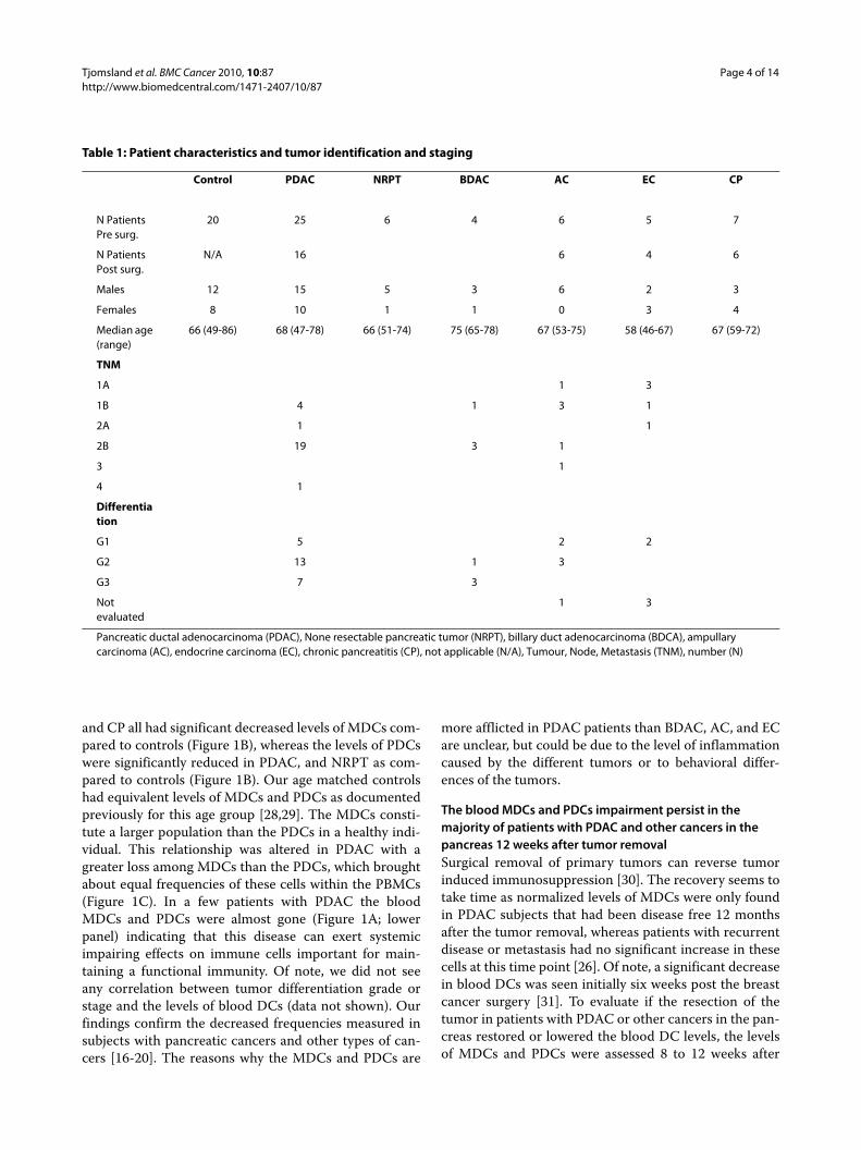
Table 1: Patient characteristics and tumor identification and staging
Control PDAC NRPT BDAC AC EC CP
N Patients Pre surg.
20 25 6 4 6 5 7
N Patients Post surg.
N/A 16 6 4 6
Males 12 15 5 3 6 2 3
Females 8 10 1 1 0 3 4
Median age (range)
66 (49-86) 68 (47-78) 66 (51-74) 75 (65-78) 67 (53-75) 58 (46-67) 67 (59-72)
TNM
1A 1 3
1B 4 1 3 1
2A 1 1
2B 19 3 1
3 1
4 1
Differentiation
G1 5 2 2
G2 13 1 3
G3 7 3
Not evaluated
1 3
Pancreatic ductal adenocarcinoma (PDAC), None resectable pancreatic tumor (NRPT), billary duct adenocarcinoma (BDCA), ampullary carcinoma (AC), endocrine carcinoma (EC), chronic pancreatitis (CP), not applicable (N/A), Tumour, Node, Metastasis (TNM), number (N)

Tjomsland et al. BMC Cancer 2010, 10:87http://www.biomedcentral.com/1471-2407/10/87
Page 5 of 14
Figure 1 Characterization and quantification of MDCs and PDCs. (A) PBMCs isolated from individuals with pancreatic duct adenocarcinoma (PDAC) (1 week pre surgery) and healthy age matched volunteers were analyzed for MDCs and PDCs levels by flow cytometry. The PBMCs stained with different direct conjugated mabs to distinguish the MDC and PDC from the rest of the cells. The gating was set to exclude debris (R1) and second gate (R2) was set on Lineage (FITC) negative and HLA DR (PerCP) positive cells. The MDCs were identified as Lin-HLA DR+ CD11c+ cells (R3) and PDCs as Lin-HLA DR+ CD123+ cells (R4). Top panel shows the dot plots from a healthy volunteer and the bottom panel shows the dot plots from an indi-vidual with PDAC. (B) Percentage of MDCs (top panel) and PDCs (lower panel) prior tumor resection in healthy controls, PDAC, none resectable pan-creatic tumor (NRPT), billary duct adenocarcinoma (BDAC), ampullary carcinoma (AC), endocrine carcinoma (EC), and chronic pancreatitis (CP). (C) Correlation of MDC and PDCs numbers (% out of PBMCs) between controls and PDAC. Control: Solid line and black circles r2 = 0.50. PDAC: striped line and white circles r2 = 0.098. Statistically significant differences between individuals with pancreatic disease and healthy controls are indicated as; * = p < 0.05, ** = p < 0.005, *** = p < 0.001
A B
Healthy control
R3
PDAC
FCS
SS
C
R1
Lin
age
FIT
C
HLA-DR PerCP
R2
CD
11c
AP
C
HLA DR PerCP
CD
123
PE
MDC
PDC R4
R3
R1
FCS
SS
C
R2Lin
age
FIT
C
HLA-DR PerCP
CD
11c
AP
C
R3
HLA-DR PerCP
R4 PDC
CD
123
PE
HLA-DR PerCP
HLA-DR PerCP
MDC
0,22%
0,10%
0,53%
1,12%Pre Surgery
0.0
0.5
1.0
1.5
MD
Cs
of
To
tal P
BM
Cs
0.0
0.2
0.4
0.6
0.8
1.0
PD
Cs
of
To
tal P
BM
Cs
***
******
***
*C
on
tro
ls
PD
AC
NR
PT
BD
AC
AC
EC
CP
C
ControlsPDAC
0.0 0.5 1.0 1.50.0
0.2
0.4
0.6
0.8
1.0
MDCs of Total PBMCs
PD
Cs
of T
ota
l PB
MC
s
r2=0.50
r2=0.098
Tjomsland et al. BMC Cancer 2010, 10:87http://www.biomedcentral.com/1471-2407/10/87
Page 6 of 14
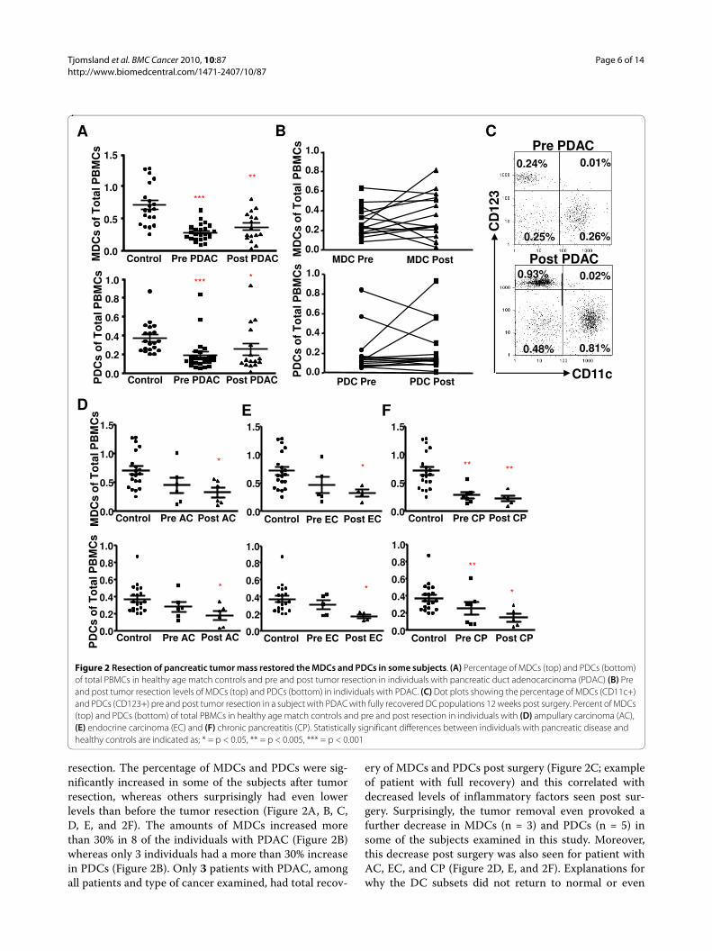
resection. The percentage of MDCs and PDCs were sig-nificantly increased in some of the subjects after tumorresection, whereas others surprisingly had even lowerlevels than before the tumor resection (Figure 2A, B, C,D, E, and 2F). The amounts of MDCs increased morethan 30% in 8 of the individuals with PDAC (Figure 2B)whereas only 3 individuals had a more than 30% increasein PDCs (Figure 2B). Only 3 patients with PDAC, amongall patients and type of cancer examined, had total recov-
ery of MDCs and PDCs post surgery (Figure 2C; exampleof patient with full recovery) and this correlated withdecreased levels of inflammatory factors seen post sur-gery. Surprisingly, the tumor removal even provoked afurther decrease in MDCs (n = 3) and PDCs (n = 5) insome of the subjects examined in this study. Moreover,this decrease post surgery was also seen for patient withAC, EC, and CP (Figure 2D, E, and 2F). Explanations forwhy the DC subsets did not return to normal or even
Figure 2 Resection of pancreatic tumor mass restored the MDCs and PDCs in some subjects. (A) Percentage of MDCs (top) and PDCs (bottom) of total PBMCs in healthy age match controls and pre and post tumor resection in individuals with pancreatic duct adenocarcinoma (PDAC) (B) Pre and post tumor resection levels of MDCs (top) and PDCs (bottom) in individuals with PDAC. (C) Dot plots showing the percentage of MDCs (CD11c+) and PDCs (CD123+) pre and post tumor resection in a subject with PDAC with fully recovered DC populations 12 weeks post surgery. Percent of MDCs (top) and PDCs (bottom) of total PBMCs in healthy age match controls and pre and post resection in individuals with (D) ampullary carcinoma (AC), (E) endocrine carcinoma (EC) and (F) chronic pancreatitis (CP). Statistically significant differences between individuals with pancreatic disease and healthy controls are indicated as; * = p < 0.05, ** = p < 0.005, *** = p < 0.001
D E F
MD
Cs
of
To
tal P
BM
Cs
PD
Cs
of
To
tal P
BM
Cs
Post ECControl Pre EC
*
0.0
0.2
0.4
0.6
0.8
1.0
Control Pre EC Post EC0.0
0.5
1.0
1.5
*
Control Pre AC Post AC
*
0.0
0.5
1.0
1.5
Post ACControl Pre AC
*
0.0
0.2
0.4
0.6
0.8
1.0
Control Pre CP Post CP
**
*
0.0
0.2
0.4
0.6
0.8
1.0
Control Pre CP Post CP
** **
0.0
0.5
1.0
1.5
MD
Cs
of
To
tal P
BM
Cs
Control Pre PDAC Post PDAC
***
**
0.0
0.5
1.0
1.5
Control Pre PDAC Post PDAC
*** *
0.0
0.2
0.4
0.6
0.8
1.0
PD
Cs
of
To
tal P
BM
Cs
CA
0.0
0.2
0.4
0.6
0.8
1.0
MDC Pre MDC Post
MD
Cs
of
To
tal P
BM
Cs
0.0
0.2
0.4
0.6
0.8
1.0
PDC Pre PDC Post
PD
Cs
of
To
tal P
BM
Cs
B
CD
123
0.24%
0.26%0.25%
0.01%
0.48%
0.93%
0.81%
0.02%
CD11c
Post PDAC
Pre PDAC
CD
123

Tjomsland et al. BMC Cancer 2010, 10:87http://www.biomedcentral.com/1471-2407/10/87
Page 7 of 14
decreased post surgery could be due to, the high recur-rence rate seen among surgical treated PDAC patients,that the surgically procedure itself induced a setting withmore inflammation or that the initial inflammation hadnot been cleared. The immune system by it self shouldnot require longer time to recover as the levels and func-tions of DCs in most of HIV-1 infected individuals startsto recover soon after the viral load is abolish [21]. In con-clusion, our results point to an induction of a systemicimpairment of the immune system and its cells, i.e. bloodMDCs and PDCs, by the tumor mass and/or fibroticmass. This impairment was reversed in some individualswhen the primary tumor was removed and the inflamma-tion resolved.
Increased numbers of circulating Lin-HLA DR+ CD123- CD11c- cells in peripheral blood from individuals with ductal pancreatic adenocarcinomaAccumulation of lineage- (lin-) HLA DR+CD11c-CD123-blood cells (non DC) coincides with a reduction in theCD11c+ DC and/or CD123+ DCs in subjects with breastcancer, prostate cancer, and malignant glioma [32]. Wenoticed that absolute numbers of the non DCs were com-parable between controls and the different pancreaticcancer subjects with the exception for BDAC, which hada decrease in the total numbers of these cells (data notshown). Of note, even if the absolute numbers of thesecells remained the same did the composition of the lin-HLA DR+ population change with significantly increasedfrequency of non DCs, and decreased PDCs and MDCsfor PDAC, NRPT, AC, EC, and CP (Figure 3A). Theresection of the tumor mass did not diminish the elevatedlevels of non DCs in the lin- HLA DR+ cell population(Figure 3B). The type of cell or cell progenitor that thenon DC population corresponds to needs further evalua-tion, however we can exclude leukocytes such as normalmonocytes, macrophages, B cells, T cells, NK cells, neu-trophils, eosinophils, basophils, MDCs, and PDCs. Inter-estingly, Pinzon-Charry et al showed that this non DCpopulation increases with metastatic disease compare tolocal disease and controls suggesting that there is an asso-ciation with disease augmentation [32]. The reasons forwhy this cell population did not decline to a higher extentin our cohort could be due to their disease status or tooshort time frame from the surgery to have restored theblood composition.
Elevated levels of apoptotic blood MDCs and PDCs in PDAC and chronic pancreatitisImmune cells circulating in peripheral blood such asMDCs, PDCs, and T cells in individuals with breast can-cer, melanoma and head and neck cancer are affected bythe solid tumors as they display an increased spontane-ous programmed cell death, i.e. apoptosis [33-35]. We
used the Annexin V protein to detect apoptotic cells inthe PBMCs from healthy controls or individuals withPDAC, other cancers in the pancreas and CP (Figure 4A).The amounts of apoptotic MDCs and PDCs were signifi-cantly higher pre surgery in all pancreatic cancers besidesfor EC (Figure 4A, B, C, D, and 4E). The levels of apopto-sis in MDCs and PDCs from PDAC patients post surgerywere similar to the levels seen pre surgery. In contrast,apoptosis of PDCs and MDCs increased after surgery inEC (Figure 4D) and AC showed the same tendency (Fig-ure 4C). Notably, the individuals with CP had the highestlevel of MDCs and PDCs apoptosis both pre and postsurgery (Figure 4E). The reasons behind the increase inapoptotic cells post surgery could be enhanced and/orchanged composition in inflammatory factors during the
Figure 3 The non DC in lin-HLA DR+ cell population is increased in individuals with different types of pancreatic cancers and chronic pancreatitis. Percentage of non DC (lin-HLA DR+ CD11c-CD123-) within the lin-HLA DR+ cell population in healthy age match controls and pancreatic duct adenocarcinoma (PDAC), none resect-able pancreatic tumor (NRPT), billary duct adenocarcinoma (BDAC), ampullary carcinoma (AC), endocrine carcinoma (EC), and chronic pan-creatitis (CP) prior surgery (A) and after surgery (B). The proportion of non DC was estimated as the mean percentage of the total amount of lin-HLA-DR+ cells. Statistically significant differences between individ-uals with pancreatic disease and healthy controls are indicated as; * = p < 0.05, ** = p < 0.005, *** = p < 0.001.
A
B
Pre Surgery
Post Surgery
*** ** **
Co
ntr
ol
PD
AC
NR
PT
BD
AC
AC EC
CP
0
20
40
60
% N
on
DC
in li
n-
HL
A-D
R+
cell
po
pu
lati
on
****
***
Co
ntr
ol
PD
AC
AC
EC CP0
20
40
60
% N
on
DC
in li
n-
HL
A-D
R+
cell
po
pu
lati
on

Tjomsland et al. BMC Cancer 2010, 10:87http://www.biomedcentral.com/1471-2407/10/87
Page 8 of 14
healing process. We found a significant negative correla-tion of the levels of PDCs found in peripheral blood priorand after surgery with the levels of apoptosis among thesecells from individuals with PDAC (Figure 4F, and 4G),whereas this correlation for MDCs only was significantfor the post surgery samples (Figure 4F, and 4G). Thisfinding may indicate that the decrease in DC in individu-als with PDAC and other types of diseases in the pancreascould be due to increased apoptosis.
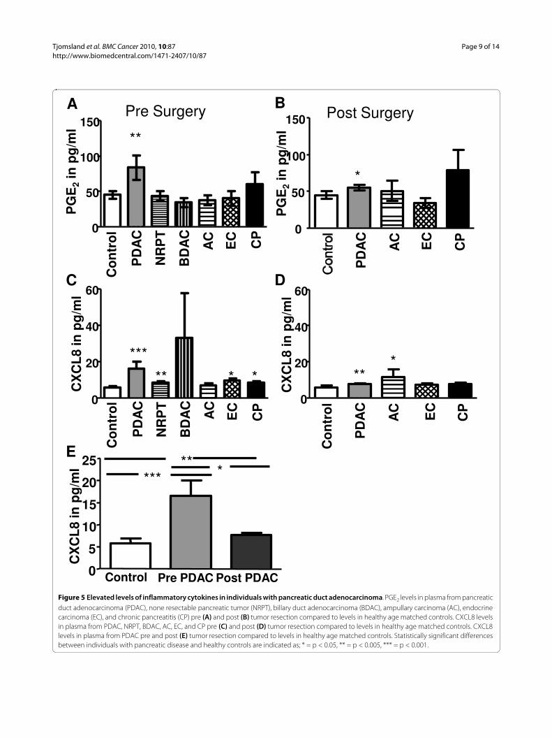
Elevated PGE2 and CXCL8 in plasma from individuals with PDACResults points to that the COX-2 product PGE2 mediatedPDAC cellular invasiveness through an ERK/Ets-1-
dependent induction of MMP-2 expression and activity[36]. We found here significantly elevated PGE2 levels inindividuals with PDAC both pre- and post surgery even ifthe levels decreased after surgery (Figure 5A, B, C, and5D). The PGE2 levels were not elevated pre or post sur-gery in the other pancreatic tumors, i.e. BDAC, AC, andEC (Figure 5A, B, C, and 5D). Incidentally, CP had alsoelevated PGE2 levels but was not found statistically signif-icant (Figure 5A). Other immune regulatory factors havebeen found elevated, i.e. IL-10, which have beendescribed as negative indicator for survival of patientswith different types of cancers. IL-10 in serum frompatients with hepatocellular carcinoma correlates with
Figure 4 Increased spontaneous apoptosis among the MDCs and PDCs in individuals with PDAC and other types pancreatic cancers. (A) Percentage apoptotic MDCs (top panel) and PDCs (lower panel) of total MDCs and PDCs prior tumor resection in controls, pancreatic duct adenocar-cinoma (PDAC), none resectable pancreatic tumor (NRPT), billary duct adenocarcinoma (BDAC), ampullary carcinoma (AC), endocrine carcinoma (EC), and chronic pancreatitis (CP). Percent apoptotic MDCs (top) and PDCs (bottom) of total MDCs and PDCs in healthy age match controls and pre and post tumor resection in individuals with PDAC (B), AC (C), EC (D) and CP (E). Correlation between percent apoptotic MDCs and level of MDCs (left) and correlation between percent apoptotic PDCs and level of PDCs (right) in individuals with PDAC pre tumor resection (F) and post tumor resection (G). The numbers of Annexin V positive MDCs and PDCs were measured as the percentage of Annexin V+ cells out of the total amount of MDCs or PDCs. Statistically significant differences between individuals with pancreatic disease and healthy controls are indicated as; * = p < 0.05, ** = p < 0.005, *** = p < 0.001.
G
F
PR
EP
OS
T
0 10 20 30 400.00.20.40.60.81.0
% Apoptotic PDCs% P
DC
s o
f to
tal P
BM
Cs
0 5 10 150.00.20.4
0.60.81.0
% M
DC
s o
f to
tal P
BM
Cs
% Apoptotic MDCs0 10 20 300.0
0.20.40.60.81.0
% P
DC
s o
f to
tal P
BM
Cs
% Apoptotic PDCs
% Apoptotic MDCs% M
DC
s o
f to
tal P
BM
Cs
0 5 10 15 20 250.00.20.40.60.81.0
r2=0,13 * r2=0,10
r2=0,45**r2=0,52
A B
% A
po
pto
tic
PD
Cs
Control Pre PDAC
Post PDAC
**
***
0
10
20
30
40
Control Pre PDAC
Post PDAC
***
***
0
5
10
15
20
25
% A
po
pto
tic
MD
Cs
Pre Surgery
Co
ntr
ol
PD
AC
NR
PT
BD
AC
AC
EC
CP
0
**
* **
**
***
**
**
****
***
0
5
10
15
20
25
% A
po
pto
tic
PD
Cs
10
20
30
40
% A
po
pto
tic
MD
Cs
C D E
***
**
Control Pre CP
Post CP
0
10
20
30
40
**
*
Control Pre AC
Post AC
% A
po
pto
tic
PD
Cs
0
5
10
15
20
Control Pre AC
Post AC
***
0
5
10
15
% A
po
pto
tic
MD
Cs
Control Pre CP
Post CP
**
**
0
10
20
30
**
Control Pre EC
Post EC
0
5
10
15
20
Control Pre EC
Post EC
**
0
5
10
15
Tjomsland et al. BMC Cancer 2010, 10:87http://www.biomedcentral.com/1471-2407/10/87
Page 9 of 14
Figure 5 Elevated levels of inflammatory cytokines in individuals with pancreatic duct adenocarcinoma. PGE2 levels in plasma from pancreatic duct adenocarcinoma (PDAC), none resectable pancreatic tumor (NRPT), billary duct adenocarcinoma (BDAC), ampullary carcinoma (AC), endocrine carcinoma (EC), and chronic pancreatitis (CP) pre (A) and post (B) tumor resection compared to levels in healthy age matched controls. CXCL8 levels in plasma from PDAC, NRPT, BDAC, AC, EC, and CP pre (C) and post (D) tumor resection compared to levels in healthy age matched controls. CXCL8 levels in plasma from PDAC pre and post (E) tumor resection compared to levels in healthy age matched controls. Statistically significant differences between individuals with pancreatic disease and healthy controls are indicated as; * = p < 0.05, ** = p < 0.005, *** = p < 0.001.
Pre SurgeryA B
C Co
ntr
ol
PD
AC
BD
AC
AC
EC
CP
**
0
50
100
150
PG
E2
in p
g/m
l
NR
PT
Post Surgery
PD
AC
AC
EC
CP
*
0
50
100
150
PG
E2
in p
g/m
l
Co
ntr
ol
PD
AC
AC
EC
CP
0
20
40
60C
XC
L8
in p
g/m
l
******
0
20
40
60
CX
CL
8 in
pg
/ml
** **
Co
ntr
ol
PD
AC
BD
AC
AC
EC
CP
NR
PT
0
5
10
15
20
25*** *
**
Control Pre PDAC Post PDAC
CX
CL
8 in
pg
/ml
D
E
Tjomsland et al. BMC Cancer 2010, 10:87http://www.biomedcentral.com/1471-2407/10/87
Page 10 of 14
decreased number of DCs and immature DCs subsets[16]. However, we could not observe any significantincrease in IL-10 levels in our samples compared to con-trols (data not shown). Similar inflammatory componentsand downstream effectors have been found to be elevatedin CP and PDAC, such as CXCL8, an activator of inflam-matory NF-kB cascade and associated with tumorigene-sis by promoting angiogenesis and metastasis [37]. Inaddition, mRNA CXCL8 are over expressed in ~80% ofPDAC tissue samples compared to normal surroundingtissue [38]. The individuals with PDAC, NRPT, EC, andCP had significantly elevated levels of serum CXCL8 presurgery (Figure 5C). Post surgery, PDAC and AC were theonly diseases that had elevated levels of CXCL8 (Figure5D). Interestingly, the removal of the tumor mass or sus-pected to be tumor mass (massive fibrosis) lowered theCXCL8 levels for PDAC, EC, and CP but only signifi-cantly for PDAC (P = 0.008) (Figure 5E). We investigatedthe levels of TGF-β and several other inflammatory fac-tors in our samples and could not see any significantincrease or decrease of these factors in any of the individ-uals with different tumors in the pancreas (data notshown). In summary, our findings point to systemiceffects exerted by the presence of solid tumor and chronicinflammation in the pancreas that create elevated levelsof immunoregulatory soluble factors such as CXCL8 inperipheral blood.
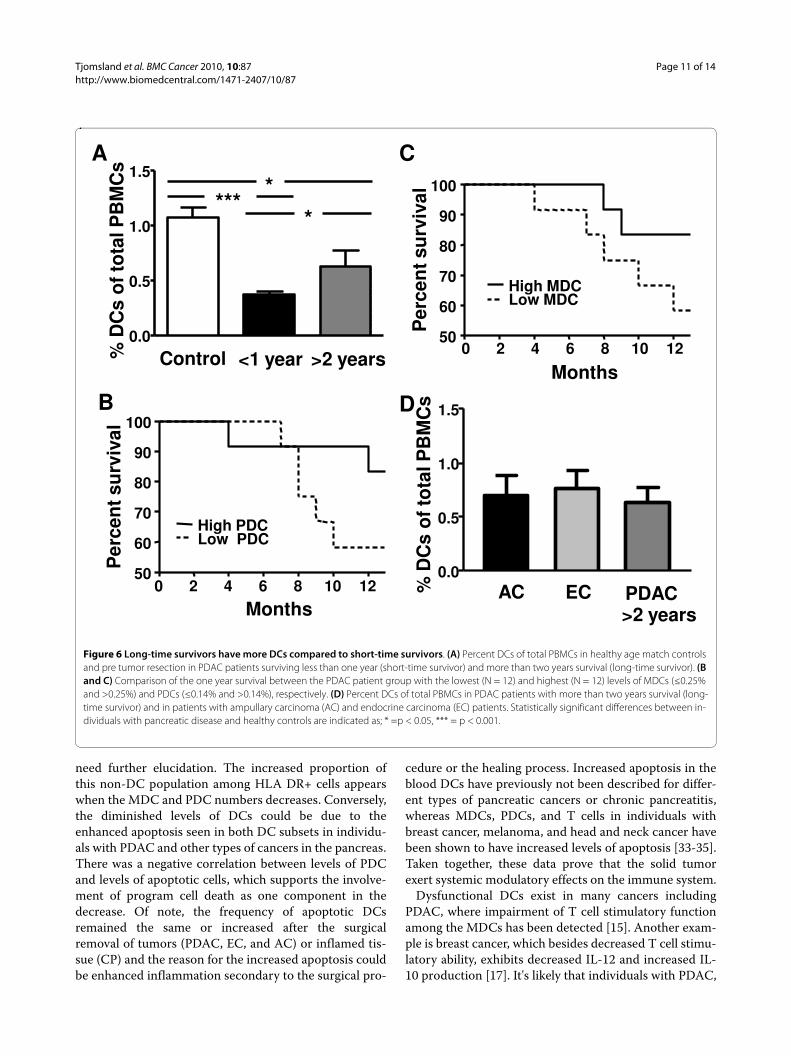
Long time PDAC survivors are presented with more circulating DCs than short time survivorsThe total amount of blood DCs pre surgery were com-pared between patients with PDAC surviving less thanone year (n = 7) (short time survivors) and patients sur-viving more than two years (n = 6) (long time survivors)after surgery. The rest of our patient cohort had less timethan 2 years since their surgery so they did not fit thesecategories for long and short time survivors. Data fromour patient cohort showed a significant difference in thelevels of blood DCs between the group with short (0.38%)and long time (0.64%) survival (Figure 6A). Of note, bothgroups had decreased amount of total blood DCs com-pared to healthy controls (1.08%) (Figure 6A). To investi-gate the importance of the amount of circulating MDCsand PDCs for patient survival, the PDAC patients weredivided into two groups; one group with the 12 lowestand one group with the 12 highest levels of blood MDC((≤0.25% and >0.25% MDCs) and PDC (≤0.14% and>0.14% PDCs)). The low MDC and PDC groups had asimilar one year survival rate of 58%, compared to 83% forthe high MDC and PDC groups (Figure 6B, and 6C).
Some of the other tumors found in pancreas, i.e. amp-ullary and endocrine carcinoma have higher 5 years sur-vival rates (60%) [39,40] and both carcinoma groups werefound with similar DC levels pre surgery (0.73% and
0.76%, respectively) as the patients with PDAC with longtime survival (0.64%) (Figure 6D). Hitherto, we have notfound any correlations between the patients' survivaltimes and the level of inflammatory factors CXCL8 andPGE2 in their peripheral blood. Our data show the con-nection of longer survival time and level of DCs in theblood compartment in patients with PDAC and this needto be addressed in a bigger cohort of patients and as far aswe know has this not been shown previously and needfurther elucidation.
DiscussionDCs have been recognized as the main initiators of theadaptive immune response and play the pivotal role oftumor surveillance in healthy individuals. The number ofperipheral blood DCs appears to be decreased in severaltypes of cancer, including pancreatic cancer [15-20,31,32]. An opposing finding has been shown for levelsof DC in individuals with melanoma, which hadincreased levels of both MDCs and PDCs with the high-est in stage I disease but even stage IV had elevated levels[41]. Findings from several types of solid cancers implythat anti-tumor immunity may be related to the numberand/or functions of DCs [15-17,19,20,26]. Our resultsindicate that there is a significant decrease in the amountof MDCs and PDCs in peripheral blood from patientswith different types of pancreatic cancers, includingPDAC, but also for chronic inflammation in pancreas, i.e.chronic pancreatitis. A decrease in the MDC and anincrease in the PDC subsets have previously beendescribed for PDAC [15]. The latter observation are con-tradicting the results we have obtained in the presentstudy for PDAC and other pancreatic cancer as well as inprevious findings for different adenocarcinoma[17,20,31]. These differences are probably due to the defi-nition of PDCs as CD11c-/lin-/HLA DR+ without usingany specific marker for PDCs, which will include a nonPDC population besides the PDCs [15].
The peripheral Lin- HLA DR+ cell population containsMDCs, PDCs, and cells that are lin-HLA-DR+CD123-
CD11c- (non DC). Our findings show a significantlyincreased frequency of non DCs among the Lin- HLADR+ population in patients with PDAC. This corrobo-rates a previous study by Pinzon-Charry et al displayingan increase in this population in breast cancer, prostatecancer, and malignant glioma [32]. These lin- HLA-DR+CD11c-CD123- cells might be a specific pre DCpopulation but they have less efficient antigen presentingfunction and generated an inadequate immune responsecompared to MDCs and PDCs [32]. Whether these cellsare MDCs and PDCs, that have impaired phenotype andfunctions due to the systemic effect by the disease, or ifthey comprise a separate cell population of different ori-gin or a progenitor for one of the blood lymphocyte will
Tjomsland et al. BMC Cancer 2010, 10:87http://www.biomedcentral.com/1471-2407/10/87
Page 11 of 14
need further elucidation. The increased proportion ofthis non-DC population among HLA DR+ cells appearswhen the MDC and PDC numbers decreases. Conversely,the diminished levels of DCs could be due to theenhanced apoptosis seen in both DC subsets in individu-als with PDAC and other types of cancers in the pancreas.There was a negative correlation between levels of PDCand levels of apoptotic cells, which supports the involve-ment of program cell death as one component in thedecrease. Of note, the frequency of apoptotic DCsremained the same or increased after the surgicalremoval of tumors (PDAC, EC, and AC) or inflamed tis-sue (CP) and the reason for the increased apoptosis couldbe enhanced inflammation secondary to the surgical pro-
cedure or the healing process. Increased apoptosis in theblood DCs have previously not been described for differ-ent types of pancreatic cancers or chronic pancreatitis,whereas MDCs, PDCs, and T cells in individuals withbreast cancer, melanoma, and head and neck cancer havebeen shown to have increased levels of apoptosis [33-35].Taken together, these data prove that the solid tumorexert systemic modulatory effects on the immune system.
Dysfunctional DCs exist in many cancers includingPDAC, where impairment of T cell stimulatory functionamong the MDCs has been detected [15]. Another exam-ple is breast cancer, which besides decreased T cell stimu-latory ability, exhibits decreased IL-12 and increased IL-10 production [17]. It's likely that individuals with PDAC,
Figure 6 Long-time survivors have more DCs compared to short-time survivors. (A) Percent DCs of total PBMCs in healthy age match controls and pre tumor resection in PDAC patients surviving less than one year (short-time survivor) and more than two years survival (long-time survivor). (B and C) Comparison of the one year survival between the PDAC patient group with the lowest (N = 12) and highest (N = 12) levels of MDCs (≤0.25% and >0.25%) and PDCs (≤0.14% and >0.14%), respectively. (D) Percent DCs of total PBMCs in PDAC patients with more than two years survival (long-time survivor) and in patients with ampullary carcinoma (AC) and endocrine carcinoma (EC) patients. Statistically significant differences between in-dividuals with pancreatic disease and healthy controls are indicated as; * =p < 0.05, *** = p < 0.001.
0.0
0.5
1.0
1.5
<1 year >2 years
****
*
Control% D
Cs
of t
ota
l PB
MC
s
0 2 4 6 8 10 1250
60
70
80
90
100
Low PDCHigh PDC
Months
Per
cen
t su
rviv
al
B
0.0
0.5
1.0
1.5
PDAC>2 years
AC EC% D
Cs
of t
ota
l PB
MC
s
0 2 4 6 8 10 1250
60
70
80
90
100
High MDCLow MDC
Months
Per
cen
t su
rviv
al
A
D
C
Tjomsland et al. BMC Cancer 2010, 10:87http://www.biomedcentral.com/1471-2407/10/87
Page 12 of 14
and other pancreatic cancers, with decreased amounts ofMDCs and PDCs and increased level of the non DCs pro-vide a setting were the DCs in blood and tissue are inade-quate to initiate a sufficient immune response against thetumor. Surgical removal of primary tumors can reversethe tumor induced immunosuppression [30], whichpoints to the tumor and surrounding stroma cells as gen-eral sources of inflammation inflicting impairment in theMDCs and PDCs. So, when resecting this inflammatorycatalyst the immune system should get a second chanceto reorganize and possibly kill remaining tumor cells.Unfortunately, this seems to have taken place only in aminority of the PDAC patients in a timely manner but onthe other hand, these patients seem to have a completerecovery of the circulating DC populations. This failureto normalize MDC in subjects with PDAC was also seenin a recent study 2.5 to 13 weeks post surgery [26] andPinzon-Charry et al even found a further decrease inblood DCs six weeks post breast cancer surgery [31].
Tumors have been referred to as wounds that neverheal, due to their ability to create an inflammatorymicroenvironment [42]. Resection of the tumor shouldprincipally rescue the body from this chronic wound, butit seems likely that the healing process after surgery byitself contributes to the impairment of the circulatingDCs as we found increased apoptosis in all pancreaticcancer groups and CP and even a further decrease in thefrequency of DCs in many of subjects. Patients with earlybreast cancer disease showed minimally reduced DC lev-els at diagnosis but displayed a prolonged period (oneyear) of marked DC suppression after tumor resection[31]. For the majority of PDAC patients this time frame istoo long since the disease gives them a shorter mean sur-vival span than it takes for the DCs to recover.
Production of residual inflammatory and/or additionalfactors induced and produced during the healing processcould be blamed for the sustained negative effect exertedon the DCs. Cyclin E1, epithelial growth factor (EGF)[37], IL-6, CXCL8, IL-10, and IL-1RA have all beenshown to be elevated in individuals with pancreatic can-cer [43,44] and high IL-6 or IL-10 levels correlated topoor survival [43]. Over expression of CXCL8 mRNAwas found ~80% of PDAC tissues compared to corre-sponding normal surrounding tissue [38] and CXCL8 wasalso present in CP [37]. We found elevated levels ofplasma CXCL8 in PDAC, EC, and CP pre surgery andthat CXCL8 decreased after tumor resection. Of note, ithas been shown that CXCL8 in PDAC is associated withtumor genesis by promoting angiogenesis and metastasis[38] but not with survival [43]. Furthermore, CXCL8 is achemoattractant that will attract many different cell typesexpressing CXCR1 or CXCR2 including DCs [45] and thepresence of CXCL8 in blood may affect their ability toexit the blood stream and affect their viability. Our find-
ings do not show correlation between the increasedplasma levels of CXCL8 and the amount of MDCs andPDCs, neither to the level of apoptosis or patient survival.The levels of CXCL8 decreased significantly in the bloodfrom PDAC patients post surgery but this had no directeffects on the post surgery levels of MDCs or PDCs.Taken together, our findings indicate that CXCL8 is notdirectly involved in the depletion of blood DCs in PDACor other pancreatic tumor patients but do not excludethat the recovery of DCs may take longer time than thenormalization of inflammatory factors, such as CXCL8,in blood.
COX-2 enzyme expression is found in several cancersincluding PDAC and it's involved in cancer differentia-tion, apoptosis, metastasis, and angiogenesis [46-50]. Wefound the COX-2 metabolite PGE2 to be elevated signifi-cantly only in PDAC patients and tumor resection low-ered the levels to almost normal. Increased levels of PGE2in plasma seem to be a specific feature for PDAC and apossible marker for distinguish PDAC from other tumorsin the pancreas, but further test must be performed. Ofnote, PGE2 is known to affect the DC function including,antigen presentation, maturation, and T cell activation[51] and this could be true for the MDCs and PDCs inPDAC. Furthermore, patients with chronic pancreatitishad also elevated levels but the PGE2 did not normalizeafter surgery. The increased plasma levels of PGE2 did notcorrelate to the amount of MDCs and PDCs, level ofapoptosis or patient survival. However, the high expres-sion of PGE2 and CXCL8 in PDAC patients could reflectthe severity of this tumor compared to other pancreatictumors [39,40].
Our findings show that patients surviving more than 2years are represented with more circulating DCs thanshort time survivors. Some of the other tumors in thepancreas, i.e. ampullary and endocrine carcinoma havehigh 5 years survival rates (60%) [39,40] and both groupswere found with similar DC levels presurgery (0.73% and0.76%, respectively) as the patients with PDAC with longtime survival (0.64%). Moreover, the one year survivalwas 83% in the group of PDAC patients with the highestMDC and PDC levels compared to only 58% in the groupof PDAC patients with the lowest MDC and PDC levels.These findings indicate that the total levels of DCs presurgery could predict patient survival. Moreover, ourfindings indicate that patients with un-affected blood DCsubsets pre and post, or with normalized DC numberspost surgery seems to have a survival benefit compared toindividuals with impaired numbers of DCs. The fewpatients fitting these characteristics had a survival over30 months post surgery which is above the mean survivalafter pancreaticoduodenectomy for PDAC which is

Tjomsland et al. BMC Cancer 2010, 10:87http://www.biomedcentral.com/1471-2407/10/87
Page 13 of 14
between 14 to15 months [52-54] indicating the impor-tance of an intact blood DC compartment.
ConclusionsIndividuals with cancer or chronic inflammation in thepancreas have blood DCs characterized by both reducednumbers and enhanced apoptosis. The post-surgery fol-low up revealed a DC compartment still impaired in thevast majority of individuals examined, but a few individu-als with cancer had normalized the circulating DC com-partment. The preservation of the blood DCscompartment in PDAC patients seems to benefit theirability to control the disease and survival. If the inabilityto restore the DC compartment depends on irreversibleeffects exerted by the PDAC or a slow recovery of theimmune system in this type of pancreatic cancer willneed further evaluation seeing that our findings indicatethat the levels of MDCs and PDCs correlate with survival.
Competing interestsThe authors declare that they have no competing interests.
Authors' contributionsVT carried out experiments, analyzed data and helped writing this manuscript.PS, AS, KB, and DS contributed with ideas, crucial patients and/or analyzeddata, and edited the paper. UF, SF and KM contributed with ideas and editedthe paper. ML conceived, designed, and supervised the study and wrote themanuscript. All authors read and approved the final manuscript.
AcknowledgementsThis work has been supported by grants from: ML: The Swedish Research Council AI52731, The Swedish International Development Cooperation Agency; SIDA, VINNMER (Vinnova), Medical Research Council of Southeast Sweden, and Swedish Society of Medicine.
Author Details1Division of Molecular Virology, Department of Clinical and Experimental Medicine, Linköping University, Sweden, 2Division of Surgery, Linköping University, Sweden, 3Division of Internal Medicine, Department of Medical and Health Science, Linköping University, Sweden, 4Moores Cancer Institute, University of California, USA, 5Department of Oncology and Radiotherapy, Jönköping Hospital, Sweden, 6Morphology/Pathology Unit, Department of Laboratory Medicine, Jönköping Hospital, Sweden and 7Division of Medical Microbiology, Department of Clinical and Experimental Medicine, Linköping University, Sweden
References1. Ferlay J, Autier P, Boniol M, Heanue M, Colombet M, Boyle P: Estimates of
the cancer incidence and mortality in Europe in 2006. Ann Oncol 2007, 18:581-592.
2. Jemal A, Siegel R, Ward E, Murray T, Xu J, Thun MJ: Cancer statistics, 2007. CA Cancer J Clin 2007, 57:43-66.
3. Verslype C, Van Cutsem E, Dicato M, Cascinu S, Cunningham D, Diaz-Rubio E, Glimelius B, Haller D, Haustermans K, Heinemann V, et al.: The management of pancreatic cancer. Current expert opinion and recommendations derived from the 8th World Congress on Gastrointestinal Cancer, Barcelona, 2006. Ann Oncol 2007, 18(Suppl 7):vii1-vii10.
4. Bachmann J, Heiligensetzer M, Krakowski-Roosen H, Buchler MW, Friess H, Martignoni ME: Cachexia worsens prognosis in patients with resectable pancreatic cancer. J Gastrointest Surg 2008, 12:1193-1201.
5. Li D, Xie K, Wolff R, Abbruzzese JL: Pancreatic cancer. Lancet 2004, 363:1049-1057.
6. Groh V, Wu J, Yee C, Spies T: Tumour-derived soluble MIC ligands impair expression of NKG2D and T-cell activation. Nature 2002, 419:734-738.
7. Shimauchi T, Kabashima K, Nakashima D, Sugita K, Yamada Y, Hino R, Tokura Y: Augmented expression of programmed death-1 in both neoplastic and non-neoplastic CD4+ T-cells in adult T-cell leukemia/lymphoma. Int J Cancer 2007, 121:2585-2590.
8. Gross S, Walden P: Immunosuppressive mechanisms in human tumors: why we still cannot cure cancer. Immunol Lett 2008, 116:7-14.
9. Chehimi J, Campbell DE, Azzoni L, Bacheller D, Papasavvas E, Jerandi G, Mounzer K, Kostman J, Trinchieri G, Montaner LJ: Persistent decreases in blood plasmacytoid dendritic cell number and function despite effective highly active antiretroviral therapy and increased blood myeloid dendritic cells in HIV-infected individuals. J Immunol 2002, 168:4796-4801.
10. Vakkila J, Thomson AW, Vettenranta K, Sariola H, Saarinen-Pihkala UM: Dendritic cell subsets in childhood and in children with cancer: relation to age and disease prognosis. Clin Exp Immunol 2004, 135:455-461.
11. Steinman RM: Lasker Basic Medical Research Award. Dendritic cells: versatile controllers of the immune system. Nat Med 2007, 13:1155-1159.
12. Hashizume H, Horibe T, Yagi H, Seo N, Takigawa M: Compartmental imbalance and aberrant immune function of blood CD123+ (plasmacytoid) and CD11c+ (myeloid) dendritic cells in atopic dermatitis. J Immunol 2005, 174:2396-2403.
13. Cao W, Liu YJ: Innate immune functions of plasmacytoid dendritic cells. Curr Opin Immunol 2007, 19:24-30.
14. Liu YJ: IPC: professional type 1 interferon-producing cells and plasmacytoid dendritic cell precursors. Annu Rev Immunol 2005, 23:275-306.
15. Yanagimoto H, Takai S, Satoi S, Toyokawa H, Takahashi K, Terakawa N, Kwon AH, Kamiyama Y: Impaired function of circulating dendritic cells in patients with pancreatic cancer. Clin Immunol 2005, 114:52-60.
16. Beckebaum S, Zhang X, Chen X, Yu Z, Frilling A, Dworacki G, Grosse-Wilde H, Broelsch CE, Gerken G, Cicinnati VR: Increased levels of interleukin-10 in serum from patients with hepatocellular carcinoma correlate with profound numerical deficiencies and immature phenotype of circulating dendritic cell subsets. Clin Cancer Res 2004, 10:7260-7269.
17. Satthaporn S, Robins A, Vassanasiri W, El-Sheemy M, Jibril JA, Clark D, Valerio D, Eremin O: Dendritic cells are dysfunctional in patients with operable breast cancer. Cancer Immunol Immunother 2004, 53:510-518.
18. Mohty M, Jarrossay D, Lafage-Pochitaloff M, Zandotti C, Briere F, de Lamballeri XN, Isnardon D, Sainty D, Olive D, Gaugler B: Circulating blood dendritic cells from myeloid leukemia patients display quantitative and cytogenetic abnormalities as well as functional impairment. Blood 2001, 98:3750-3756.
19. Hoffmann TK, Muller-Berghaus J, Ferris RL, Johnson JT, Storkus WJ, Whiteside TL: Alterations in the frequency of dendritic cell subsets in the peripheral circulation of patients with squamous cell carcinomas of the head and neck. Clin Cancer Res 2002, 8:1787-1793.
20. Sciarra A, Lichtner M, Autran GA, Mastroianni C, Rossi R, Mengoni F, Cristini C, Gentilucci A, Vullo V, Di Silverio F: Characterization of circulating blood dendritic cell subsets DC123+ (lymphoid) and DC11C+ (myeloid) in prostate adenocarcinoma patients. Prostate 2007, 67:1-7.
21. Pacanowski J, Kahi S, Baillet M, Lebon P, Deveau C, Goujard C, Meyer L, Oksenhendler E, Sinet M, Hosmalin A: Reduced blood CD123+ (lymphoid) and CD11c+ (myeloid) dendritic cell numbers in primary HIV-1 infection. Blood 2001, 98:3016-3021.
22. Kunitani H, Shimizu Y, Murata H, Higuchi K, Watanabe A: Phenotypic analysis of circulating and intrahepatic dendritic cell subsets in patients with chronic liver diseases. J Hepatol 2002, 36:734-741.
23. Jongbloed SL, Lebre MC, Fraser AR, Gracie JA, Sturrock RD, Tak PP, McInnes IB: Enumeration and phenotypical analysis of distinct dendritic cell subsets in psoriatic arthritis and rheumatoid arthritis. Arthritis Res Ther 2006, 8:R15.
24. Aggarwal BB, Shishodia S, Sandur SK, Pandey MK, Sethi G: Inflammation and cancer: how hot is the link? Biochem Pharmacol 2006, 72:1605-1621.
25. Sadun RE, Sachsman SM, Chen X, Christenson KW, Morris WZ, Hu P, Epstein AL: Immune signatures of murine and human cancers reveal unique mechanisms of tumor escape and new targets for cancer immunotherapy. Clin Cancer Res 2007, 13:4016-4025.
Received: 11 October 2009 Accepted: 9 March 2010 Published: 9 March 2010This article is available from: http://www.biomedcentral.com/1471-2407/10/87© 2010 Tjomsland et al; licensee BioMed Central Ltd. This is an Open Access article distributed under the terms of the Creative Commons Attribution License (http://creativecommons.org/licenses/by/2.0), which permits unrestricted use, distribution, and reproduction in any medium, provided the original work is properly cited.BMC Cancer 2010, 10:87
Tjomsland et al. BMC Cancer 2010, 10:87http://www.biomedcentral.com/1471-2407/10/87
Page 14 of 14
26. Takahashi K, Toyokawa H, Takai S, Satoi S, Yanagimoto H, Terakawa N, Araki H, Kwon AH, Kamiyama Y: Surgical influence of pancreatectomy on the function and count of circulating dendritic cells in patients with pancreatic cancer. Cancer Immunol Immunother 2006, 55:775-784.
27. Maecker B, Mougiakakos D, Zimmermann M, Behrens M, Hollander S, Schrauder A, Schrappe M, Welte K, Klein C: Dendritic cell deficiencies in pediatric acute lymphoblastic leukemia patients. Leukemia 2006, 20:645-649.
28. Shodell M, Siegal FP: Circulating, interferon-producing plasmacytoid dendritic cells decline during human ageing. Scand J Immunol 2002, 56:518-521.
29. Della Bella S, Bierti L, Presicce P, Arienti R, Valenti M, Saresella M, Vergani C, Villa ML: Peripheral blood dendritic cells and monocytes are differently regulated in the elderly. Clin Immunol 2007, 122:220-228.
30. Danna EA, Sinha P, Gilbert M, Clements VK, Pulaski BA, Ostrand-Rosenberg S: Surgical removal of primary tumor reverses tumor-induced immunosuppression despite the presence of metastatic disease. Cancer Res 2004, 64:2205-2211.
31. Pinzon-Charry A, Ho CS, Maxwell T, McGuckin MA, Schmidt C, Furnival C, Pyke CM, Lopez JA: Numerical and functional defects of blood dendritic cells in early- and late-stage breast cancer. Br J Cancer 2007, 97:1251-1259.
32. Pinzon-Charry A, Ho CS, Laherty R, Maxwell T, Walker D, Gardiner RA, O'Connor L, Pyke C, Schmidt C, Furnival C, Lopez JA: A population of HLA-DR+ immature cells accumulates in the blood dendritic cell compartment of patients with different types of cancer. Neoplasia 2005, 7:1112-1122.
33. Pinzon-Charry A, Maxwell T, McGuckin MA, Schmidt C, Furnival C, Lopez JA: Spontaneous apoptosis of blood dendritic cells in patients with breast cancer. Breast Cancer Res 2006, 8:R5.
34. Saito T, Dworacki G, Gooding W, Lotze MT, Whiteside TL: Spontaneous apoptosis of CD8+ T lymphocytes in peripheral blood of patients with advanced melanoma. Clin Cancer Res 2000, 6:1351-1364.
35. Hoffmann TK, Dworacki G, Tsukihiro T, Meidenbauer N, Gooding W, Johnson JT, Whiteside TL: Spontaneous apoptosis of circulating T lymphocytes in patients with head and neck cancer and its clinical importance. Clin Cancer Res 2002, 8:2553-2562.
36. Ito H, Duxbury M, Benoit E, Clancy TE, Zinner MJ, Ashley SW, Whang EE: Prostaglandin E2 enhances pancreatic cancer invasiveness through an Ets-1-dependent induction of matrix metalloproteinase-2. Cancer Res 2004, 64:7439-7446.
37. Farrow B, Sugiyama Y, Chen A, Uffort E, Nealon W, Mark Evers B: Inflammatory mechanisms contributing to pancreatic cancer development. Ann Surg 2004, 239:763-769. discussion 769-771
38. Li M, Zhang Y, Feurino LW, Wang H, Fisher WE, Brunicardi FC, Chen C, Yao Q: Interleukin-8 increases vascular endothelial growth factor and neuropilin expression and stimulates ERK activation in human pancreatic cancer. Cancer Sci 2008, 99:733-737.
39. Figueiredo FA, Giovannini M, Monges G, Charfi S, Bories E, Pesenti C, Caillol F, Delpero JR: Pancreatic Endocrine Tumors: A Large Single-Center Experience. Pancreas 2009.
40. Morris-Stiff G, Alabraba E, Tan YM, Shapey I, Bhati C, Tanniere P, Mayer D, Buckels J, Bramhall S, Mirza DF: Assessment of survival advantage in ampullary carcinoma in relation to tumour biology and morphology. Eur J Surg Oncol 2009, 35:746-750.
41. McCarter MD, Baumgartner J, Escobar GA, Richter D, Lewis K, Robinson W, Wilson C, Palmer BE, Gonzalez R: Immunosuppressive dendritic and regulatory T cells are upregulated in melanoma patients. Ann Surg Oncol 2007, 14:2854-2860.
42. Dvorak HF: Tumors: wounds that do not heal. Similarities between tumor stroma generation and wound healing. N Engl J Med 1986, 315:1650-1659.
43. Ebrahimi B, Tucker SL, Li D, Abbruzzese JL, Kurzrock R: Cytokines in pancreatic carcinoma: correlation with phenotypic characteristics and prognosis. Cancer 2004, 101:2727-2736.
44. Wigmore SJ, Fearon KC, Sangster K, Maingay JP, Garden OJ, Ross JA: Cytokine regulation of constitutive production of interleukin-8 and -6 by human pancreatic cancer cell lines and serum cytokine concentrations in patients with pancreatic cancer. Int J Oncol 2002, 21:881-886.
45. Feijoo E, Alfaro C, Mazzolini G, Serra P, Penuelas I, Arina A, Huarte E, Tirapu I, Palencia B, Murillo O, et al.: Dendritic cells delivered inside human
carcinomas are sequestered by interleukin-8. Int J Cancer 2005, 116:275-281.
46. Wang MT, Honn KV, Nie D: Cyclooxygenases, prostanoids, and tumor progression. Cancer Metastasis Rev 2007, 26:525-534.
47. Shamma A, Yamamoto H, Doki Y, Okami J, Kondo M, Fujiwara Y, Yano M, Inoue M, Matsuura N, Shiozaki H, Monden M: Up-regulation of cyclooxygenase-2 in squamous carcinogenesis of the esophagus. Clin Cancer Res 2000, 6:1229-1238.
48. Fosslien E: Molecular pathology of cyclooxygenase-2 in neoplasia. Ann Clin Lab Sci 2000, 30:3-21.
49. Molina MA, Sitja-Arnau M, Lemoine MG, Frazier ML, Sinicrope FA: Increased cyclooxygenase-2 expression in human pancreatic carcinomas and cell lines: growth inhibition by nonsteroidal anti-inflammatory drugs. Cancer Res 1999, 59:4356-4362.
50. Ma X, Kundu N, Rifat S, Walser T, Fulton AM: Prostaglandin E receptor EP4 antagonism inhibits breast cancer metastasis. Cancer Res 2006, 66:2923-2927.
51. Ahmadi M, Emery DC, Morgan DJ: Prevention of both direct and cross-priming of antitumor CD8+ T-cell responses following overproduction of prostaglandin E2 by tumor cells in vivo. Cancer Res 2008, 68:7520-7529.
52. Herman JM, Swartz MJ, Hsu CC, Winter J, Pawlik TM, Sugar E, Robinson R, Laheru DA, Jaffee E, Hruban RH, et al.: Analysis of fluorouracil-based adjuvant chemotherapy and radiation after pancreaticoduodenectomy for ductal adenocarcinoma of the pancreas: results of a large, prospectively collected database at the Johns Hopkins Hospital. J Clin Oncol 2008, 26:3503-3510.
53. Ueda M, Endo I, Nakashima M, Minami Y, Takeda K, Matsuo K, Nagano Y, Tanaka K, Ichikawa Y, Togo S, et al.: Prognostic Factors After Resection of Pancreatic Cancer. World J Surg 2008.
54. Conlon KC, Klimstra DS, Brennan MF: Long-term survival after curative resection for pancreatic ductal adenocarcinoma. Clinicopathologic analysis of 5-year survivors. Ann Surg 1996, 223:273-279.
Pre-publication historyThe pre-publication history for this paper can be accessed here:http://www.biomedcentral.com/1471-2407/10/87/prepub
doi: 10.1186/1471-2407-10-87Cite this article as: Tjomsland et al., Pancreatic adenocarcinoma exerts sys-temic effects on the peripheral blood myeloid and plasmacytoid dendritic cells: an indicator of disease severity? BMC Cancer 2010, 10:87
http://www.ncbi.nlm.nih.gov/entrez/query.fcgi?cmd=Retrieve&db=PubMed&dopt=Abstract&list_uids=3537791
Related Documents