Protocol for the Examination of Specimens From Patients With Carcinoma of the Endocrine Pancreas Protocol applies to all neuroendocrine tumors of the pancreas. Based on AJCC/UICC TNM, 7th edition Protocol web posting date: June 2012 Procedures • Excisional Biopsy (Enucleation) • Partial Pancreatectomy • Pancreaticoduodenectomy (Whipple Resection) • Total Pancreatectomy Authors Laura H. Tang, MD, PhD* Department of Pathology, Memorial Sloan-Kettering Cancer Center, New York, NY Jordan Berlin, MD Department of Medicine, Vanderbilt University Medical Center, Nashville, TN Philip Branton, MD, FCAP Department of Pathology, Inova Fairfax Hospital, Falls Church, VA Lawrence J. Burgart, MD, FCAP Allina Laboratories, Abbott Northwestern Hospital, Minneapolis, MN David K. Carter, MD, FCAP Department of Pathology, St. Mary’s/Duluth Clinic Health System, Duluth, MN Carolyn C. Compton, MD, PhD, FCAP Critical Path Institute, Tucson, AZ Patrick Fitzgibbons, MD, FCAP Department of Pathology, St. Jude Medical Center, Fullerton, CA Wendy Frankel, MD, FCAP Department of Pathology, Ohio State University Medical Center, Columbus, OH John Jessup, MD Division of Cancer Treatment and Diagnosis, National Cancer Institute, Bethesda, MD Sanjay Kakar, MD, FCAP Department of Pathology, University of California San Francisco and the Veterans Affairs Medical Center, San Francisco, CA Bruce Minsky, MD Department of Radiation Oncology, University of Chicago, Chicago, IL

Welcome message from author
This document is posted to help you gain knowledge. Please leave a comment to let me know what you think about it! Share it to your friends and learn new things together.
Transcript
Protocol for the Examination of Specimens From Patients With Carcinoma of the Endocrine Pancreas Protocol applies to all neuroendocrine tumors of the pancreas.
Based on AJCC/UICC TNM, 7th editionProtocol web posting date: June 2012
Procedures• Excisional Biopsy (Enucleation)• Partial Pancreatectomy• Pancreaticoduodenectomy (Whipple Resection)• Total Pancreatectomy
AuthorsLaura H. Tang, MD, PhD*
Department of Pathology, Memorial Sloan-Kettering Cancer Center, New York, NYJordan Berlin, MD
Department of Medicine, Vanderbilt University Medical Center, Nashville, TNPhilip Branton, MD, FCAP
Department of Pathology, Inova Fairfax Hospital, Falls Church, VALawrence J. Burgart, MD, FCAP
Allina Laboratories, Abbott Northwestern Hospital, Minneapolis, MNDavid K. Carter, MD, FCAP
Department of Pathology, St. Mary’s/Duluth Clinic Health System, Duluth, MNCarolyn C. Compton, MD, PhD, FCAP
Critical Path Institute, Tucson, AZPatrick Fitzgibbons, MD, FCAP
Department of Pathology, St. Jude Medical Center, Fullerton, CAWendy Frankel, MD, FCAP
Department of Pathology, Ohio State University Medical Center, Columbus, OHJohn Jessup, MD
Division of Cancer Treatment and Diagnosis, National Cancer Institute, Bethesda, MDSanjay Kakar, MD, FCAP
Department of Pathology, University of California San Francisco and the Veterans Affairs Medical Center, San Francisco, CA
Bruce Minsky, MDDepartment of Radiation Oncology, University of Chicago, Chicago, IL
Raouf Nakhleh, MD, FCAPDepartment of Pathology, Mayo Clinic, Jacksonville, FL
Kay Washington, MD, PhD, FCAP†Department of Pathology, Vanderbilt University Medical Center, Nashville, TN
For the Members of the Cancer Committee, College of American Pathologists
* Denotes primary author. † Denotes senior author. All other contributing authors are listed alphabetically.
Endocrine • Pancreas (Neuroendocrine Tumor)PancreasEndocrine 3.2.0.0
© 2012 College of American Pathologists (CAP). All rights reserved.
The College does not permit reproduction of any substantial portion of these protocols without its written authorization. The College hereby authorizes use of these protocols by physicians and other health care providers in reporting on surgical specimens, in teaching, and in carrying out medical research for nonprofit purposes. This authorization does not extend to reproduction or other use of any substantial portion of these protocols for commercial purposes without the written consent of the College.
The CAP also authorizes physicians and other health care practitioners to make modified versions of the Protocols solely for their individual use in reporting on surgical specimens for individual patients, teaching, and carrying out medical research for non-profit purposes.
The CAP further authorizes the following uses by physicians and other health care practitioners, in reporting on surgical specimens for individual patients, in teaching, and in carrying out medical research for non-profit purposes: (1) Dictation from the original or modified protocols for the purposes of creating a text-based patient record on paper, or in a word processing document; (2) Copying from the original or modified protocols into a text-based patient record on paper, or in a word processing document; (3) The use of a computerized system for items (1) and (2), provided that the Protocol data is stored intact as a single text-based document, and is not stored as multiple discrete data fields.
Other than uses (1), (2), and (3) above, the CAP does not authorize any use of the Protocols in electronic medical records systems, pathology informatics systems, cancer registry computer systems, computerized databases, mappings between coding works, or any computerized system without a written license from CAP. Applications for such a license should be addressed to the SNOMED Terminology Solutions division of the CAP.
Any public dissemination of the original or modified Protocols is prohibited without a written license from the CAP.
The College of American Pathologists offers these protocols to assist pathologists in providing clinically useful and relevant information when reporting results of surgical specimen examinations of surgical specimens. The College regards the reporting elements in the “Surgical Pathology Cancer Case Summary” portion of the protocols as essential elements of the pathology report. However, the manner in which these elements are reported is at the discretion of each specific pathologist, taking into account clinician preferences, institutional policies, and individual practice.
The College developed these protocols as an educational tool to assist pathologists in the useful reporting of relevant information. It did not issue the protocols for use in litigation, reimbursement, or other contexts. Nevertheless, the College recognizes that the protocols might be used by hospitals, attorneys, payers, and others. Indeed, effective January 1, 2004, the Commission on Cancer of the American College of Surgeons mandated the use of the required data elements of the protocols as part of its Cancer Program Standards for Approved Cancer Programs. Therefore, it becomes even more important for pathologists to familiarize themselves with these documents. At the same time, the College cautions that use of the protocols other than for their intended educational purpose may involve additional considerations that are beyond the scope of this document.
The inclusion of a product name or service in a CAP publication should not be construed as an endorsement of such product or service, nor is failure to include the name of a product or service to be construed as disapproval.
2
Endocrine • Pancreas (Neuroendocrine Tumor)PancreasEndocrine 3.2.0.0
CAP Pancreas (Endocrine) Protocol Revision History
Version CodeThe definition of the version code can be found at www.cap.org/cancerprotocols.
Version: PancreasEndocrine 3.2.0.0
Summary of ChangesThe following changes have been made since the February 2011 release.
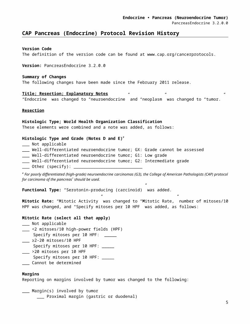
Title; Resection; Explanatory Notes“Endocrine” was changed to “neuroendocrine” and “neoplasm” was changed to “tumor.”
Resection
Histologic Type; World Health Organization ClassificationThese elements were combined and a note was added, as follows:
Histologic Type and Grade (Notes D and E)#
___ Not applicable___ Well-differentiated neuroendocrine tumor; GX: Grade cannot be assessed___ Well-differentiated neuroendocrine tumor; G1: Low grade ___ Well-differentiated neuroendocrine tumor; G2: Intermediate grade___ Other (specify): ____________________________# For poorly differentiated (high-grade) neuroendocrine carcinomas (G3), the College of American Pathologists (CAP) protocol for carcinoma of the pancreas1 should be used.
Functional Type: “Serotonin-producing (carcinoid)” was added.
Mitotic Rate: “Mitotic Activity” was changed to “Mitotic Rate,” number of mitoses/10 HPF was changed, and “Specify mitoses per 10 HPF” was added, as follows:
Mitotic Rate (select all that apply) ___ Not applicable___ <2 mitoses/10 high-power fields (HPF)
Specify mitoses per 10 HPF: ________ ≥2-20 mitoses/10 HPF
Specify mitoses per 10 HPF: ________ >20 mitoses per 10 HPF
Specify mitoses per 10 HPF: ________ Cannot be determined
MarginsReporting on margins involved by tumor was changed to the following:
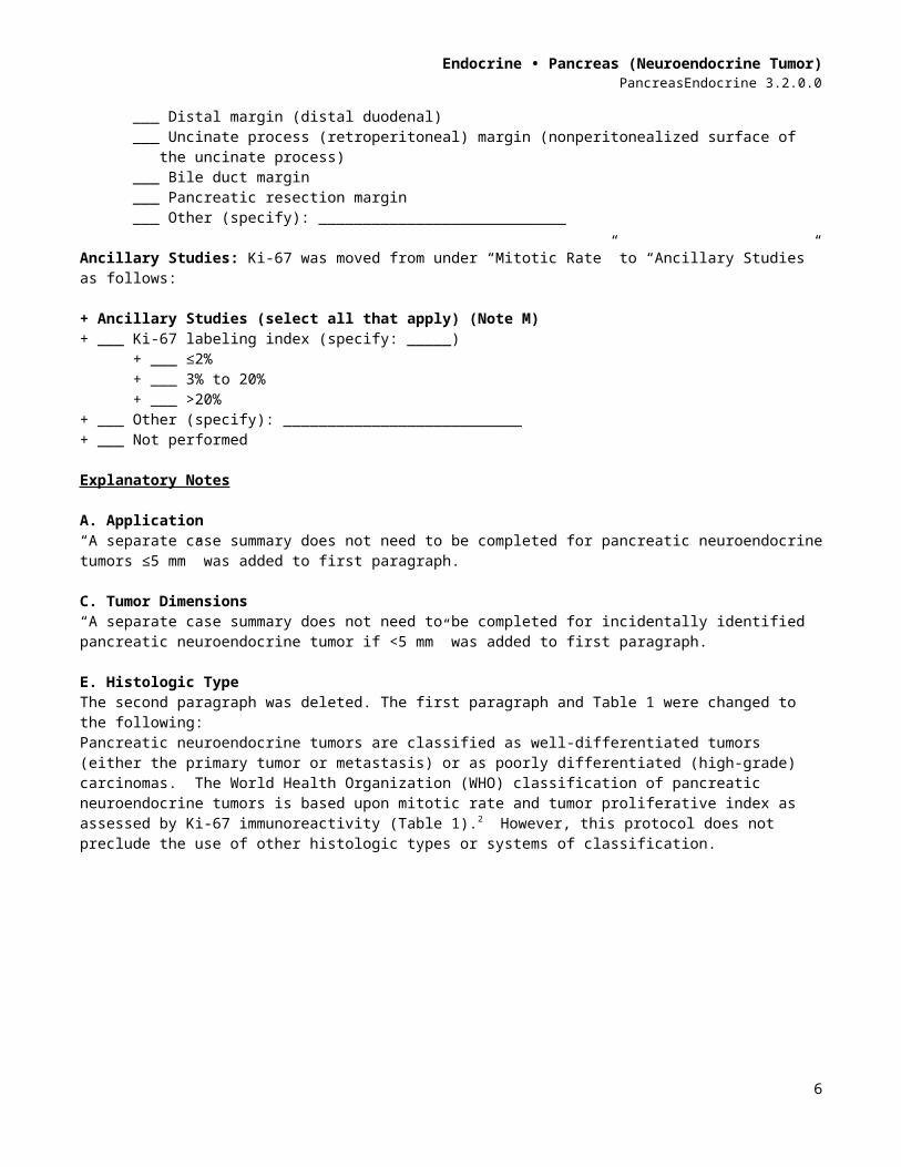
___ Margin(s) involved by tumor___ Proximal margin (gastric or duodenal)___ Distal margin (distal duodenal)___ Uncinate process (retroperitoneal) margin (nonperitonealized surface of the
uncinate process)___ Bile duct margin___ Pancreatic resection margin___ Other (specify): ____________________________
3
Endocrine • Pancreas (Neuroendocrine Tumor)PancreasEndocrine 3.2.0.0
Ancillary Studies: Ki-67 was moved from under “Mitotic Rate” to “Ancillary Studies” as follows:
+ Ancillary Studies (select all that apply) (Note M)+ ___ Ki-67 labeling index (specify: _____)
+ ___ ≤2%+ ___ 3% to 20%+ ___ >20%
+ ___ Other (specify): ___________________________+ ___ Not performed
Explanatory Notes
A. Application“A separate case summary does not need to be completed for pancreatic neuroendocrine tumors ≤5 mm” was added to first paragraph.
C. Tumor Dimensions“A separate case summary does not need to be completed for incidentally identified pancreatic neuroendocrine tumor if <5 mm” was added to first paragraph.
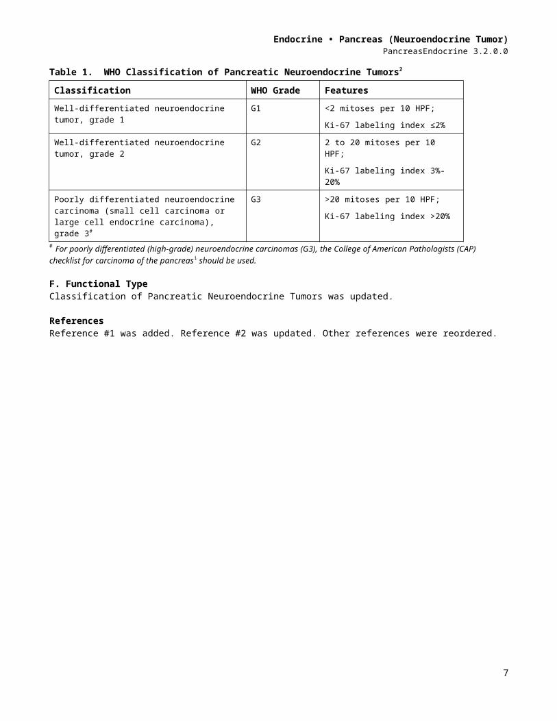
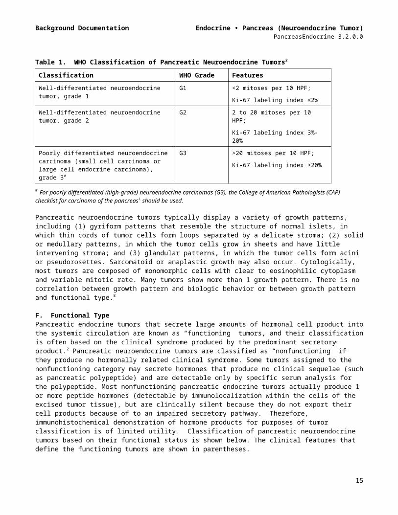
E. Histologic TypeThe second paragraph was deleted. The first paragraph and Table 1 were changed to the following:Pancreatic neuroendocrine tumors are classified as well-differentiated tumors (either the primary tumor or metastasis) or as poorly differentiated (high-grade) carcinomas. The World Health Organization (WHO) classification of pancreatic neuroendocrine tumors is based upon mitotic rate and tumor proliferative index as assessed by Ki-67 immunoreactivity (Table 1).2 However, this protocol does not preclude the use of other histologic types or systems of classification.
Table 1. WHO Classification of Pancreatic Neuroendocrine Tumors2
Classification WHO Grade Features Well-differentiated neuroendocrine tumor, grade 1
G1 <2 mitoses per 10 HPF; Ki-67 labeling index ≤2%
Well-differentiated neuroendocrine tumor, grade 2
G2 2 to 20 mitoses per 10 HPF;Ki-67 labeling index 3%-20%
Poorly differentiated neuroendocrine carcinoma (small cell carcinoma or large cell endocrine carcinoma), grade 3#
G3 >20 mitoses per 10 HPF; Ki-67 labeling index >20%
# For poorly differentiated (high-grade) neuroendocrine carcinomas (G3), the College of American Pathologists (CAP) checklist for carcinoma of the pancreas1 should be used.
F. Functional TypeClassification of Pancreatic Neuroendocrine Tumors was updated.
ReferencesReference #1 was added. Reference #2 was updated. Other references were reordered.
4
CAP Approved Endocrine • Pancreas (Neuroendocrine Tumor)PancreasEndocrine 3.2.0.0
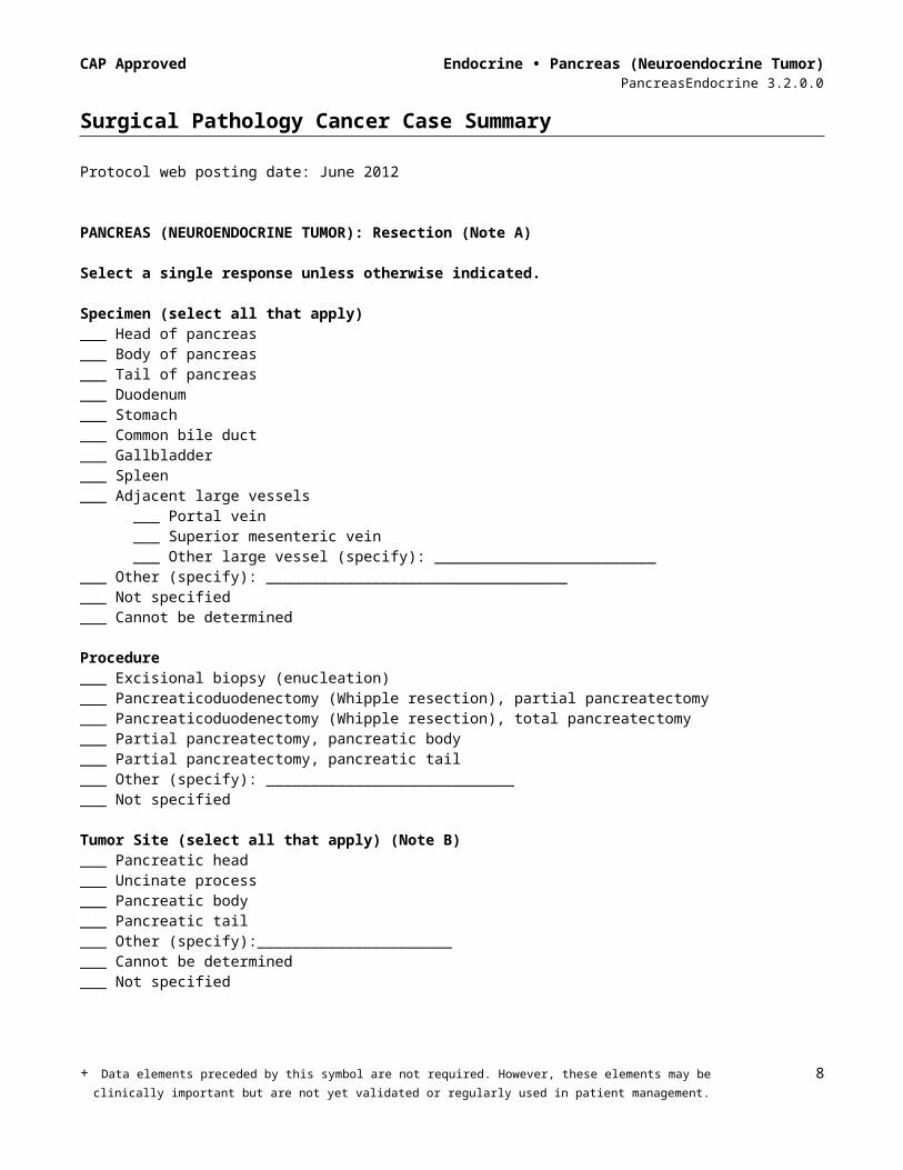
Surgical Pathology Cancer Case Summary
Protocol web posting date: June 2012
PANCREAS (NEUROENDOCRINE TUMOR): Resection (Note A)
Select a single response unless otherwise indicated.
Specimen (select all that apply)___ Head of pancreas___ Body of pancreas___ Tail of pancreas ___ Duodenum___ Stomach___ Common bile duct___ Gallbladder___ Spleen___ Adjacent large vessels
___ Portal vein___ Superior mesenteric vein___ Other large vessel (specify): _________________________
___ Other (specify): _____________________________________ Not specified___ Cannot be determined
Procedure___ Excisional biopsy (enucleation)___ Pancreaticoduodenectomy (Whipple resection), partial pancreatectomy___ Pancreaticoduodenectomy (Whipple resection), total pancreatectomy___ Partial pancreatectomy, pancreatic body ___ Partial pancreatectomy, pancreatic tail___ Other (specify): _______________________________ Not specified
Tumor Site (select all that apply) (Note B)___ Pancreatic head___ Uncinate process___ Pancreatic body___ Pancreatic tail___ Other (specify):_________________________ Cannot be determined___ Not specified
Tumor Size (Note C)Greatest dimension: ___ cm (specify size of largest tumor if multiple tumors are present)+ Additional dimensions: ___ x ___ cm___ Cannot be determined (see Comment)
Tumor Focality (Note D)___ Unifocal___ Multifocal (specify number of tumors: ______________________)___ Cannot be determined___ Not specified+ Data elements preceded by this symbol are not required. However, these elements may be
clinically important but are not yet validated or regularly used in patient management. 5
CAP Approved Endocrine • Pancreas (Neuroendocrine Tumor)PancreasEndocrine 3.2.0.0
Histologic Type and Grade (Note E)#
___ Not applicable___ Well-differentiated neuroendocrine tumor; GX: Grade cannot be assessed___ Well-differentiated neuroendocrine tumor; G1: Low grade ___ Well-differentiated neuroendocrine tumor; G2: Intermediate grade___ Other (specify): ____________________________# For poorly differentiated (high-grade) neuroendocrine carcinomas (G3), the College of American Pathologists (CAP) protocol for carcinoma of the pancreas1 should be used.
+ Functional Type (select all that apply) (Note F)+ ___ Cannot be assessed+ ___ Pancreatic neuroendocrine tumor, functional
(correlation with clinical syndrome and elevated serum levels of hormone product) + ___ Insulin-producing (insulinoma)+ ___ Glucagon-producing (glucagonoma)+ ___ Somatostatin-producing (somatostatinoma)+ ___ Gastrin-producing (gastrinoma) + ___ Vasoactive intestinal polypeptide (VIP)-producing (VIPoma)+ ___ Serotonin-producing (carcinoid)+ ___ Other (specify): __________________________
+ ___ Pancreatic neuroendocrine tumor, nonfunctional+ ___ Pancreatic neuroendocrine tumor, functional status unknown
Mitotic Rate (select all that apply) (Note G)___ Not applicable___ <2 mitoses/10 high-power fields (HPF)
Specify mitoses per 10 HPF: ________ ≥2-20 mitoses/10 HPF
Specify mitoses per 10 HPF: ________ >20 mitoses per 10 HPF
Specify mitoses per 10 HPF: ________ Cannot be determined
+ Tumor Necrosis (Note H)+ ___ Not identified+ ___ Present+ ___ Not applicable+ ___ Cannot be determined
Microscopic Tumor Extension (select all that apply) ___ Cannot be determined___ No evidence of primary tumor___ Tumor is confined to pancreas___ Tumor invades ampulla of Vater___ Tumor invades common bile duct___ Tumor invades duodenal wall ___ Tumor invades peripancreatic soft tissues ___ Tumor invades other adjacent organs or structures (specify): ______________________
Margins (select all that apply) (Note I)___ Cannot be assessed___ Margins uninvolved by tumor
Distance of tumor from closest margin: ___ mm or ___ cm+ Specify margin (if possible): ____________________________
+ Data elements preceded by this symbol are not required. However, these elements may be clinically important but are not yet validated or regularly used in patient management.
6
CAP Approved Endocrine • Pancreas (Neuroendocrine Tumor)PancreasEndocrine 3.2.0.0
___ Margin(s) involved by tumor___ Proximal margin (gastric or duodenal)___ Distal margin (distal duodenal)___ Uncinate process (retroperitoneal) margin (nonperitonealized surface of the
uncinate process)___ Bile duct margin___ Pancreatic resection margin
+ ___ Tumor involves posterior retroperitoneal surface of pancreas
Lymph-Vascular Invasion (Note J)___ Not identified___ Present___ Indeterminate
Perineural Invasion (Note K)___ Not identified___ Present___ Indeterminate
Pathologic Staging (pTNM) (Note L)
TNM Descriptors (required only if applicable) (select all that apply)___ m (multiple primary tumors)___ r (recurrent)___ y (posttreatment)
Primary Tumor (pT)___ pTX: Cannot be assessed___ pT0: No evidence of primary tumor___ pT1: Tumor limited to the pancreas, 2 cm or less in greatest dimension___ pT2: Tumor limited to the pancreas, more than 2 cm in greatest dimension___ pT3: Tumor extends beyond the pancreas but without involvement of the celiac axis or the
superior mesenteric artery___ pT4: Tumor involves the celiac axis or the superior mesenteric artery
Regional Lymph Nodes (pN)___ pNX: Cannot be assessed___ pN0: No regional lymph node metastasis___ pN1: Regional lymph node metastasis
___ No nodes submitted or found
Number of Lymph Nodes ExaminedSpecify: _______ Number cannot be determined (explain): ______________________
Number of Lymph Nodes InvolvedSpecify: _______ Number cannot be determined (explain): ______________________
Distant Metastasis (pM)___ Not applicable___ pM1: Distant metastasis
+ Specify site(s), if known: ____________________________
+ Data elements preceded by this symbol are not required. However, these elements may be clinically important but are not yet validated or regularly used in patient management.
7
CAP Approved Endocrine • Pancreas (Neuroendocrine Tumor)PancreasEndocrine 3.2.0.0
+ Additional Pathologic Findings (select all that apply) + ___ None identified+ ___ Chronic pancreatitis+ ___ Acute pancreatitis+ ___ Adenomatosis (multiple neuroendocrine tumors, each less than 5 mm in greatest dimension)+ ___ Other (specify): ____________________________
+ Ancillary Studies (select all that apply) (Note M)+ ___ Ki-67 labeling index (specify: _____)
+ ___ ≤2%+ ___ 3% to 20%+ ___ >20%
+ ___ Other (specify): ___________________________+ ___ Not performed
+ Clinical History (select all that apply) (Note N)+ ___ von Hippel-Lindau disease+ ___ Multiple endocrine neoplasia type 1+ ___ Familial pancreatic cancer syndrome+ ___ Hypoglycemic syndrome+ ___ Necrolytic migratory erythema+ ___ Watery diarrhea+ ___ Hypergastrinemia+ ___ Zollinger-Ellison syndrome + ___ Other (specify): ______________________________ + ___ Not specified
+ Comment(s)
+ Data elements preceded by this symbol are not required. However, these elements may be clinically important but are not yet validated or regularly used in patient management.
8
Background Documentation Endocrine • Pancreas (Neuroendocrine Tumor)PancreasEndocrine 3.2.0.0
Explanatory Notes
A. ApplicationThis protocol applies to neuroendocrine tumors of the pancreas. Use of the protocol is not required for incidentally identified pancreatic neuroendocrine tumors ≤5 mm in specimens removed for other indications. Pancreatic neuroendocrine tumors are also known as “islet cell tumors,” but this terminology is considered to be outdated and misleading because these tumors are not derived from pancreatic islets. Rather, they are believed to arise from pluripotential cells in the pancreatic ducts that have the capacity to differentiate along neuroendocrine lines.
Currently, there are no definitive histopathologic criteria for differentiating benign from malignant neuroendocrine tumors of the pancreas, and the presence of metastasis is the only absolute criterion for malignancy. Thus, in the absence of known metastasis or gross local invasion, it is suggested that the term “neuroendocrine tumor” be used rather than definitive terms such as “adenoma” or “carcinoma,” which connote certainty about the biologic nature of the neoplasm.
Fewer than 5% to l0% of malignant tumors of the pancreas are neuroendocrine carcinomas. Surgical resection remains the only potentially curative approach for these tumors. The prognosis of pancreatic neuroendocrine carcinomas is primarily dependent on the functional subtype, the completeness of the surgical resection, and the anatomic extent of disease.2 The TNM staging system for carcinomas of the exocrine pancreas is also applied to pancreatic neuroendocrine tumors.3,4
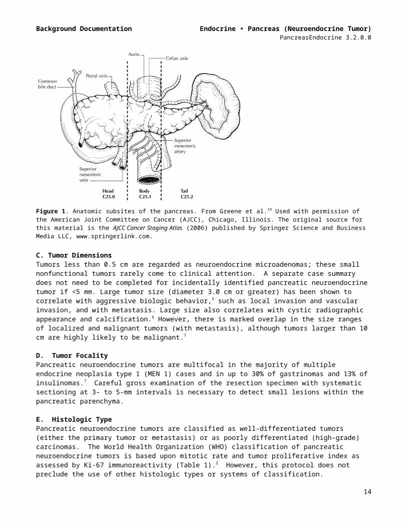
B. Tumor Site: Definition of LocationThe anatomic subdivisions defining location of tumors of the pancreas (Figures 1 and 2) are as follows4: Tumors of the head of the pancreas are those arising to the right of the left border of the
superior mesenteric vein. The uncinate process is part of the head. Tumors of the body of the pancreas are those arising between the left border of the superior
mesenteric vein and the left border of the aorta. Tumors of the tail of the pancreas are those arising between the left border of the aorta and
the hilum of the spleen.
9
Background Documentation Endocrine • Pancreas (Neuroendocrine Tumor)PancreasEndocrine 3.2.0.0
Figure 1. Anatomic subsites of the pancreas. From Greene et al.19 Used with permission of the American Joint Committee on Cancer (AJCC), Chicago, Illinois. The original source for this material is the AJCC Cancer Staging Atlas (2006) published by Springer Science and Business Media LLC, www.springerlink.com.
C. Tumor DimensionsTumors less than 0.5 cm are regarded as neuroendocrine microadenomas; these small nonfunctional tumors rarely come to clinical attention. A separate case summary does not need to be completed for incidentally identified pancreatic neuroendocrine tumor if <5 mm. Large tumor size (diameter 3.0 cm or greater) has been shown to correlate with aggressive biologic behavior,5 such as local invasion and vascular invasion, and with metastasis. Large size also correlates with cystic radiographic appearance and calcification.6 However, there is marked overlap in the size ranges of localized and malignant tumors (with metastasis), although tumors larger than 10 cm are highly likely to be malignant.7
D. Tumor FocalityPancreatic neuroendocrine tumors are multifocal in the majority of multiple endocrine neoplasia type 1 (MEN 1) cases and in up to 30% of gastrinomas and 13% of insulinomas.7 Careful gross examination of the resection specimen with systematic sectioning at 3- to 5-mm intervals is necessary to detect small lesions within the pancreatic parenchyma.
E. Histologic TypePancreatic neuroendocrine tumors are classified as well-differentiated tumors (either the primary tumor or metastasis) or as poorly differentiated (high-grade) carcinomas. The World Health Organization (WHO) classification of pancreatic neuroendocrine tumors is based upon mitotic rate and tumor proliferative index as assessed by Ki-67 immunoreactivity (Table 1).2 However, this protocol does not preclude the use of other histologic types or systems of classification.
10

Background Documentation Endocrine • Pancreas (Neuroendocrine Tumor)PancreasEndocrine 3.2.0.0
Table 1. WHO Classification of Pancreatic Neuroendocrine Tumors2
Classification WHO Grade Features Well-differentiated neuroendocrine tumor, grade 1
G1 <2 mitoses per 10 HPF; Ki-67 labeling index ≤2%
Well-differentiated neuroendocrine tumor, grade 2
G2 2 to 20 mitoses per 10 HPF;Ki-67 labeling index 3%-20%
Poorly differentiated neuroendocrine carcinoma (small cell carcinoma or large cell endocrine carcinoma), grade 3#
G3 >20 mitoses per 10 HPF; Ki-67 labeling index >20%
# For poorly differentiated (high-grade) neuroendocrine carcinomas (G3), the College of American Pathologists (CAP) checklist for carcinoma of the pancreas1 should be used.
Pancreatic neuroendocrine tumors typically display a variety of growth patterns, including (1) gyriform patterns that resemble the structure of normal islets, in which thin cords of tumor cells form loops separated by a delicate stroma; (2) solid or medullary patterns, in which the tumor cells grow in sheets and have little intervening stroma; and (3) glandular patterns, in which the tumor cells form acini or pseudorosettes. Sarcomatoid or anaplastic growth may also occur. Cytologically, most tumors are composed of monomorphic cells with clear to eosinophilic cytoplasm and variable mitotic rate. Many tumors show more than 1 growth pattern. There is no correlation between growth pattern and biologic behavior or between growth pattern and functional type.8
F. Functional TypePancreatic endocrine tumors that secrete large amounts of hormonal cell product into the systemic circulation are known as “functioning” tumors, and their classification is often based on the clinical syndrome produced by the predominant secretory product.2 Pancreatic neuroendocrine tumors are classified as “nonfunctioning” if they produce no hormonally related clinical syndrome. Some tumors assigned to the nonfunctioning category may secrete hormones that produce no clinical sequelae (such as pancreatic polypeptide) and are detectable only by specific serum analysis for the polypeptide. Most nonfunctioning pancreatic endocrine tumors actually produce 1 or more peptide hormones (detectable by immunolocalization within the cells of the excised tumor tissue), but are clinically silent because they do not export their cell products because of to an impaired secretory pathway. Therefore, immunohistochemical demonstration of hormone products for purposes of tumor classification is of limited utility. Classification of pancreatic neuroendocrine tumors based on their functional status is shown below. The clinical features that define the functioning tumors are shown in parentheses.
11
Background Documentation Endocrine • Pancreas (Neuroendocrine Tumor)PancreasEndocrine 3.2.0.0
Classification of Pancreatic Neuroendocrine Tumors2
Pancreatic neuroendocrine microadenoma (<0.5 cm and nonfunctional)Neuroendocrine tumor (NET) (nonfunctional)
NET G1NET G2
Neuroendocrine carcinoma (NEC)Large cell NECSmall cell NEC
Pancreatic neuroendocrine tumor, functionalEC cell, serotonin-producing NET (carcinoid) (carcinoid syndrome, flashing, diarrhea); rarely
encountered as primary in the pancreasGastrin-secreting (gastrinoma) (abdominal pain, ulcer disease, diarrhea, gastrointestinal
bleeding)Glucagon-secreting (glucagonoma) (diabetes, skin rash [necrolytic migratory erythema,
stomatitis)Insulin-secreting (insulinoma) (hypoglycemia, neuropsychiatric disturbances)Somatostatin-secreting (somatostatinoma) (diabetes, steatorrhea, achlorhydria); rarely
encountered.Vasoactive intestinal polypeptide (VIP)-secreting (VIPoma#) (watery diarrhea, hypokalemia,
achlorhydria)Mixed ductal-endocrine carcinoma##
Mixed acinar-endocrine carcinoma##
# Sometimes known as Verner-Morrison tumors.
## Biphasic tumors containing a significant proportion (greater than 25% to 30%) of tumor cells with differentiation along ductal or acinar cell lines are classified separately as subtypes of pancreatic endocrine carcinoma. The neuroendocrine component in such tumors is often high grade.
G. Mitotic RateHigh mitotic rate, a high degree of pleomorphism, and tumor necrosis have all been shown to correlate strongly with malignant potential.7 The WHO classification2 and others9 (see Note E) use mitoses per 10 or per 50 HPF as one of the criteria for potential for aggressive behavior. However, a low mitotic rate is of little prognostic value, and many malignant tumors show little to no mitotic activity. Mitotic rate has also been proposed as the basis for a grading scheme for foregut endocrine tumors, including pancreatic neuroendocrine tumors.10
Mitotic rate should be based upon counting 50 HPF (40x objective) and in the area of highest mitotic activity, and reported as number of mitoses per 10 HPF.
Ki-67 index is reported as percentage of positive tumor cells in area of highest nuclear labeling. It has been recommended that 2000 tumor cells be counted to determine the Ki-67 index10; however, this practice may not be practical for routine clinical purposes, and it is acceptable to estimate the labeling index.
H. Tumor Necrosis Tumor necrosis is uncommon in low-grade pancreatic neuroendocrine tumors but is generally regarded as a malignancy-associated feature. When possible, a distinction should be made between nonischemic necrosis (usually punctate or geographic), which is associated with higher tumor grade, and ischemic necrosis.
I. MarginsFor enucleation procedures, the periphery of the resection specimen tissue may be inked, and radial sections at the closest approach of tumor can be examined microscopically.
12
Background Documentation Endocrine • Pancreas (Neuroendocrine Tumor)PancreasEndocrine 3.2.0.0
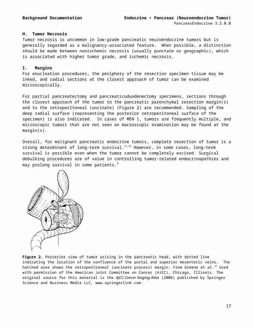
For partial pancreatectomy and pancreaticoduodenectomy specimens, sections through the closest approach of the tumor to the pancreatic parenchymal resection margin(s) and to the retroperitoneal (uncinate) (Figure 2) are recommended. Sampling of the deep radial surface (representing the posterior retroperitoneal surface of the specimen) is also indicated. In cases of MEN 1, tumors are frequently multiple, and microscopic tumors that are not seen on macroscopic examination may be found at the margin(s).
Overall, for malignant pancreatic endocrine tumors, complete resection of tumor is a strong determinant of long-term survival.11,12 However, in some cases, long-term survival is possible even when the tumor cannot be completely excised. Surgical debulking procedures are of value in controlling tumor-related endocrinopathies and may prolong survival in some patients.2
Figure 2. Posterior view of tumor arising in the pancreatic head, with dotted line indicating the location of the confluence of the portal and superior mesenteric veins. The hatched area shows the retroperitoneal (uncinate process) margin. From Greene et al.19 Used with permission of the American Joint Committee on Cancer (AJCC), Chicago, Illinois. The original source for this material is the AJCC Cancer Staging Atlas (2006) published by Springer Science and Business Media LLC, www.springerlink.com.
J. Blood Vessel Invasion The presence of blood vessel invasion,13 perineural invasion, or both have been regarded by some authors as histopathologic criteria for malignancy. Invasion of blood vessels (particularly veins within the tumor capsule) or perineural spaces have been observed in 90% of cases with distant metastases in some studies.14
K. Perineural Invasion Perineural invasion has been associated with malignancy and with shortened survival in some series16 of pancreatic endocrine tumors.
L. Pathologic StagingThe same TNM staging system of the American Joint Committee on Cancer (AJCC) and the International Union Against Cancer (UICC) is recommended for staging both carcinoma of the exocrine pancreas and pancreatic endocrine tumors, as shown below.4 The post-resection prognosis of a patient with pancreatic carcinoma is primarily determined by the anatomic extent of disease as defined by the TNM stage groupings. According to AJCC/UICC convention, the designation “T” refers to a primary tumor that has not been previously treated. The symbol “p” refers to the pathologic classification of the TNM, as
13
Background Documentation Endocrine • Pancreas (Neuroendocrine Tumor)PancreasEndocrine 3.2.0.0
opposed to the clinical classification, and is based on gross and microscopic examination. pT entails a resection of the primary tumor or biopsy adequate to evaluate the highest pT category, pN entails removal of nodes adequate to validate lymph node metastasis, and pM implies microscopic examination of distant lesions. Clinical classification (cTNM) is usually carried out by the referring physician before treatment during initial evaluation of the patient or when pathologic classification is not possible.
Pathologic staging is usually performed after surgical resection of the primary tumor. Pathologic staging depends on pathologic documentation of the anatomic extent of disease, whether or not the primary tumor has been completely removed. If a biopsied tumor is not resected for any reason (eg, when technically infeasible) and if the highest T and N categories or the M1 category of the tumor can be confirmed microscopically, the criteria for pathologic classification and staging have been satisfied without total removal of the primary cancer.
TNM DescriptorsFor identification of special cases of TNM or pTNM classifications, the “m” suffix and “y,” “r,” and “a” prefixes are used. Although they do not affect the stage grouping, they indicate cases needing separate analysis.
The “m” suffix indicates the presence of multiple primary tumors in a single site and is recorded in parentheses: pT(m)NM.
The “y” prefix indicates those cases in which classification is performed during or after initial multimodality therapy (ie, neoadjuvant chemotherapy, radiation therapy, or both chemotherapy and radiation therapy). The cTNM or pTNM category is identified by a “y” prefix. The ycTNM or ypTNM categorizes the extent of tumor actually present at the time of that examination. The “y” categorization is not an estimate of tumor before multimodality therapy (ie, before initiation of neoadjuvant therapy).
The “r” prefix indicates a recurrent tumor when staged after a documented disease-free interval and is identified by the “r” prefix: rTNM.
The “a” prefix designates the stage determined at autopsy: aTNM.
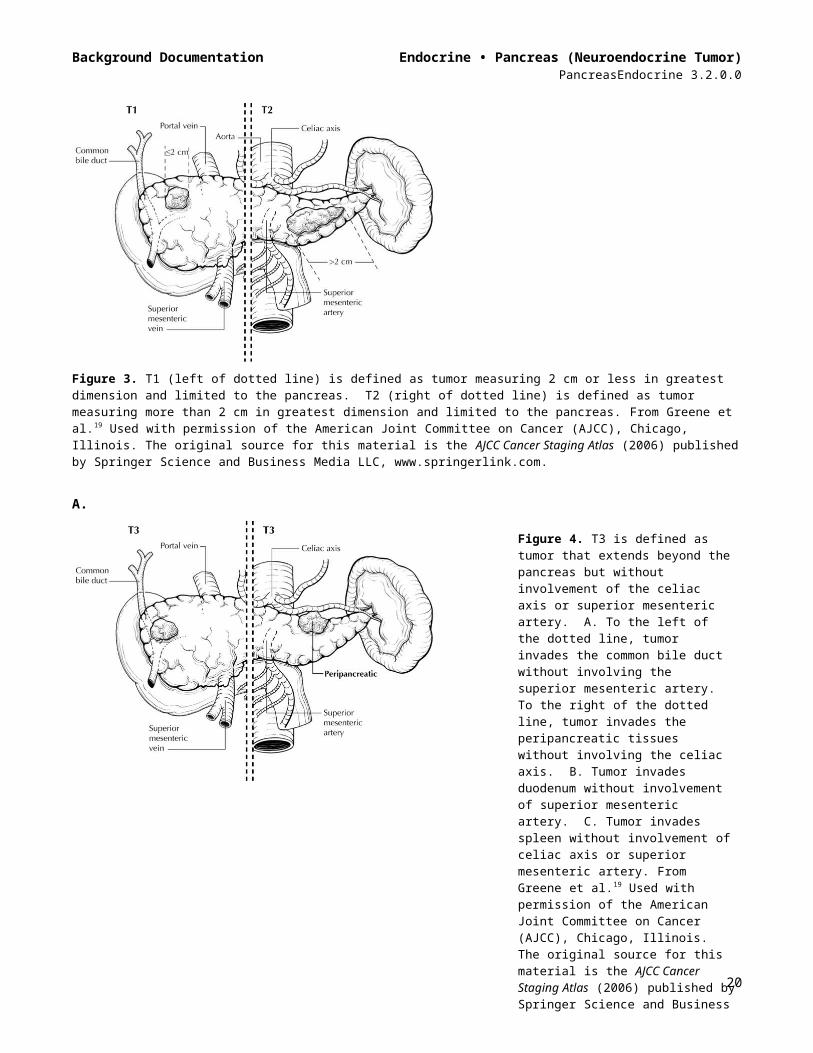
Primary Tumor# (T) (Figures 3 through 5)TX Primary tumor cannot be assessedT0 No evidence of primary tumorT1 Tumor limited to the pancreas, 2 cm or less in greatest dimension##
T2 Tumor limited to the pancreas, more than 2 cm in greatest dimension##
T3 Tumor extends beyond the pancreas but without involvement of the celiac axis or the superior mesenteric artery###
T4 Tumor involves the celiac axis or the superior mesenteric artery (unresectable primary tumor)^
# If more than 1 tumor is present in the pancreas, the tumor with the highest T category should be classified according to the pT definitions, and either the multiplicity (“m”) or the actual number of simultaneous multiple tumors (eg, “3”) should be indicated in parentheses after the T category of the primary tumor (eg, pT3[m] or pT3[2]). The “m” designation applies only to grossly recognizable, synchronous primary carcinomas and not to a single, grossly detected tumor with multiple separate microscopic foci.## Tumor size has been shown to have independent prognostic significance.5,7
### For T3, extension beyond the pancreas may include invasion of soft tissues adjacent to the pancreas, the common bile duct, and/or duodenum (including the ampulla of Vater). Specifically,
14
Background Documentation Endocrine • Pancreas (Neuroendocrine Tumor)PancreasEndocrine 3.2.0.0
peripancreatic tissues include the surrounding retroperitoneal fat (retroperitoneal soft tissue), including mesentery (mesenteric fat), mesocolon, greater and lesser omentum, and peritoneum.
^ Invasion of the portal vein also has been shown to have independent prognostic significance as an adverse factor.4
Figure 3. T1 (left of dotted line) is defined as tumor measuring 2 cm or less in greatest dimension and limited to the pancreas. T2 (right of dotted line) is defined as tumor measuring more than 2 cm in greatest dimension and limited to the pancreas. From Greene et al.19 Used with permission of the American Joint Committee on Cancer (AJCC), Chicago, Illinois. The original source for this material is the AJCC Cancer Staging Atlas (2006) published by Springer Science and Business Media LLC, www.springerlink.com.
A.
15
Figure 4. T3 is defined as tumor that extends beyond the pancreas but without involvement of the celiac axis or superior mesenteric artery. A. To the left of the dotted line, tumor invades the common bile duct without involving the superior mesenteric artery. To the right of the dotted line, tumor invades the peripancreatic tissues without involving the celiac axis. B. Tumor invades duodenum without involvement of superior mesenteric artery. C. Tumor invades spleen without involvement of celiac axis or superior mesenteric artery. From Greene et al.19 Used with permission of the American Joint Committee on Cancer (AJCC), Chicago, Illinois. The original source for this material is the AJCC Cancer Staging Atlas (2006) published by Springer Science and Business Media LLC, www.springerlink.com.
Background Documentation Endocrine • Pancreas (Neuroendocrine Tumor)PancreasEndocrine 3.2.0.0
B.
C.
Figure 5. T4 tumor involves the celiac axis (above dotted line) or the superior mesenteric artery (below dotted line). T4 tumors are considered unresectable and are rarely encountered in surgical pathology specimens. From Greene et al.19 Used with permission of the American Joint Committee on Cancer (AJCC),
16
Background Documentation Endocrine • Pancreas (Neuroendocrine Tumor)PancreasEndocrine 3.2.0.0
Chicago, Illinois. The original source for this material is the AJCC Cancer Staging Atlas (2006) published by Springer Science and Business Media LLC, www.springerlink.com.
Regional Lymph Nodes (N)# (Figures 6 and 7)NX Regional lymph nodes cannot be assessed N0 No regional lymph node metastasis N1 Regional lymph node metastasis##
The regional nodes may be subdivided as follows#:Superior Lymph nodes superior to head and body of pancreasInferior Lymph nodes inferior to head and body of pancreasAnterior Anterior pancreaticoduodenal, pyloric, and proximal mesenteric lymph nodesPosterior Posterior pancreaticoduodenal, common bile duct or pericholedochal, and proximal
mesenteric nodesSplenic (For tumors in body and tail only) Nodes of the splenic hilum and tail of pancreas.# The following lymph nodes are also considered regional: hepatic artery nodes, infrapyloric nodes (for tumors in head only), subpyloric nodes (for tumors in head only), celiac nodes (for tumors in head only), superior mesenteric nodes, pancreaticolieno nodes (for tumors in body and tail only), splenic nodes (for tumors in body and tail only), retroperitoneal nodes, and lateral aortic nodes. Tumor involvement of other nodal groups is considered distant metastasis.## The presence of lymph node metastases has been shown to have independent prognostic significance as an adverse factor.3,12 The minimum number of lymph nodes needed for adequate staging for pancreatic endocrine tumors in pancreaticoduodenectomy specimens has not been determined, although a minimum of 12 lymph nodes has been suggested for pancreatic adenocarcinoma specimens.
Figure 6. Regional lymph nodes of the pancreas (anterior view). From Greene et al.18 Used with permission of the American Joint Committee on Cancer (AJCC), Chicago, Illinois. The original source for this material is the AJCC Cancer Staging Atlas (2006) published by Springer Science and Business Media LLC, www.springerlink.com.
17

Background Documentation Endocrine • Pancreas (Neuroendocrine Tumor)PancreasEndocrine 3.2.0.0
Figure 7. Regional lymph nodes of the pancreas (anterior view with pancreatic body removed to reveal retroperitoneal vessels and lymph nodes). From Greene et al.19 Used with permission of the American Joint Committee on Cancer (AJCC), Chicago, Illinois. The original source for this material is the AJCC Cancer Staging Atlas (2006) published by Springer Science and Business Media LLC, www.springerlink.com.
Distant Metastasis (M) M0 No distant metastasisM1 Distant metastasis#
# The most common site of distant metastasis is liver. In many cases, metastasis is found only in the liver, without regional lymph node metastasis.7
Anatomic Stage/Prognostic GroupingsStage IA T1 N0 M0Stage IB T2 N0 M0Stage IIA T3 N0 M0Stage IIB T1 N1 M0
T2 N1 M0T3 N1 M0
Stage III T4 Any N M0Stage IV Any T Any N M1
Additional Descriptors
Residual Tumor (R)Tumor remaining in a patient after therapy with curative intent (eg, surgical resection for cure) is categorized by a system known as R classification, shown below.
RX Presence of residual tumor cannot be assessedR0 No residual tumorR1 Microscopic residual tumorR2 Macroscopic residual tumor
For the surgeon, the R classification may be useful to indicate the known or assumed status of the completeness of a surgical excision. For the pathologist, the R classification is relevant to the status of the margins of a surgical resection specimen. That is, tumor involving the resection margin on pathologic examination may be assumed to correspond to residual tumor in the patient
18
Background Documentation Endocrine • Pancreas (Neuroendocrine Tumor)PancreasEndocrine 3.2.0.0
and may be classified as macroscopic or microscopic according to the findings at the specimen margin(s).
Lymph-Vascular InvasionLymph-vascular invasion (LVI) indicates whether microscopic lymph-vascular invasion is identified in the pathology report. LVI includes lymphatic invasion, vascular invasion, or lymph-vascular invasion. By AJCC/UICC convention, LVI does not affect the T category indicating local extent of tumor unless specifically included in the definition of a T category.
M. Ancillary StudiesMost pancreatic neuroendocrine tumors are strongly positive for synaptophysin and chromogranin A. Some investigators,15,16 but not all,17 have found that expression of cytokeratin 19, which is found in normal pancreatic ductal cells but not in pancreatic endocrine cells, is strongly predictive of poor outcome. It is hypothesized that CK19 positivity in pancreatic neuroendocrine tumors may indicate differentiation along pancreatic ductal lines, thus accounting for the poorer outcome.
Ki-67 is used routinely by some investigators to assess proliferative activity in pancreatic neuroendocrine tumors,2,10,15-18 but it is unclear if use of the Ki-67 index is superior to assessment of mitotic activity in routinely stained sections.
Immunohistochemical studies to determine production of hormonal products are not indicated for routine assessment, because determination of tumor functionality is made on the basis of presence or absence of clinical syndromes.
N. Clinical History The etiology of most sporadic endocrine tumors of the pancreas is not known. However, MEN 1, von Hippel-Lindau disease, and, more rarely, neurofibromatosis type 12 are associated with pancreatic endocrine tumors. It is important to know whether the patient has a history of a genetic syndrome because tumors from such patients are more likely to be multifocal.
Knowledge of the clinical history is important for determining whether a pancreatic endocrine tumor is associated with a functional syndrome, which is an important predictor of malignancy (see Note F). In particular, insulinomas behave in a benign fashion, probably because they are discovered early due to the production of a hypoglycemic state. Other functioning tumors are generally malignant.
References1. Washington K, Berline J, Branton P, et al. Protocol for the examination of specimens from patients
with carcinoma of the exocrine pancreas. In: Amin MB et al, eds. Reporting on Cancer Specimens: Case Summaries and Background Documentation. Northfield, IL: College of American Pathologists; 2011.
2. Bosman FT, Carneiro F, Hruban RH, Theise ND, eds. WHO Classification of Tumours of the Digestive System. Geneva, Switzerland: WHO Press; 2010.
3. Bilimoria KY, Bentrem DJ, Merkow RP, et al. Application of the pancreatic adenocarcinoma staging system to pancreatic neuroendocrine tumors. J Am Coll Surg. 2007;205(4):558-563.
4. Edge SB, Byrd DR, Carducci MA, Compton CC, eds. AJCC Cancer Staging Manual. 7th ed. New York, NY: Springer; 2009.
5. Panzuto F, Nasoni S, Falconi M, et al. Prognostic factors and survival in endocrine tumor patients: comparison between gastrointestinal and pancreatic localization. Endocr Relat Cancer. 2005;12(4):1083-1092.
19
Background Documentation Endocrine • Pancreas (Neuroendocrine Tumor)PancreasEndocrine 3.2.0.0
6. Buetow PC, Parrino TV, Buck JL, et al. Islet cell tumors of the pancreas: pathologic-imaging correlation among size, necrosis and cysts, calcification, malignant behavior, and functional status. AJR Am J Roentgenol. Nov 1995;165(5):1175-1179.
7. Hruban RH, Pitman MB, Klimstra DS. Tumors of the Pancreas. Fourth series, Fascicle 6 ed. Washington, DC: Armed Forces Institute of Pathology; 2007.
8. Heitz PU, Kasper M, Polak JM, Kloeppel G. Pancreatic endocrine tumors: immunocytochemical analysis of 125 tumors. Hum Pathol. 1982;13:263-271.
9. Ferrone CR, Tang LH, Tomlinson J, et al. Determining prognosis in patients with pancreatic endocrine neoplasms: can the WHO classification system be simplified? J Clin Oncol. Dec 2007;25(35):5609-5615.
10. Rindi G, Kloppel G, Ahlman H, et al. TNM staging of foregut (neuro)endocrine tumors: a consensus proposal including a grading system. Virchows Archiv. Oct 2006;449(4):395-401.
11. Chung JC, Choi DW, Jo SH, Heo JS, Choi SH, Kim YI. Malignant nonfunctioning endocrine tumors of the pancreas: predictive factors for survival after surgical treatment. World J Surg. 2007;31(3):579-585.
12. Tomassetti P, Campana D, Piscitelli L, et al. Endocrine pancreatic tumors: factors correlated with survival. Ann Oncol. Nov 2005;16(11):1806-1810.
13. Kazanjian KK, Reber HA, Hines OJ. Resection of pancreatic neuroendocrine tumors: results of 70 cases. Arch Surg. 2006;141(8):765-769; discussion 769-770.
14. La Rosa S, Sessa F, Capella C, et al. Prognostic criteria in nonfunctioning pancreatic endocrine tumours. Virchows Arch. 1996;429(6):323-333.
15. Schmitt AM, Anlauf M, Rousson V, et al. WHO 2004 criteria and CK19 are reliable prognostic markers in pancreatic endocrine tumors. Am J Surg Pathol. Nov 2007;31(11):1677-1682.
16. La Rosa S, Rigoli E, Uccella S, Novario R, Capella C. Prognostic and biological significance of cytokeratin 19 in pancreatic endocrine tumours. Histopathology. Apr 2007;50(5):597-606.
17. Deshpande V, Fernandez-del Castillo C, Muzikansky A, et al. Cytokeratin 19 is a powerful predictor of survival in pancreatic endocrine tumors. Am J Surg Pathol. Sep 2004;28(9):1145-1153.
18. Bordi C, D'Adda T, Azzoni C, et al. Criteria for malignancy in gastrointestinal endocrine tumors. Endocr Pathol. 2006;17(2):119-129.
19. Greene FL, Compton, CC, Fritz AG, et al, eds. AJCC Cancer Staging Atlas. New York, NY: Springer; 2006.
20
Related Documents


