REVIEW Pan-cancer patterns of DNA methylation Tania Witte, Christoph Plass and Clarissa Gerhauser * Abstract The comparison of DNA methylation patterns across cancer types (pan-cancer methylome analyses) has revealed distinct subgroups of tumors that share similar methylation patterns. Integration of these data with the wealth of information derived from cancer genome profiling studies performed by large international consortia has provided novel insights into the cellular aberrations that contribute to cancer development. There is evidence that genetic mutations in epigenetic regulators (such as DNMT3, IDH1/2 or H3.3) mediate or contribute to these patterns, although a unifying molecular mechanism underlying the global alterations of DNA methylation has largely been elusive. Knowledge gained from pan-cancer methylome analyses will aid the development of diagnostic and prognostic biomarkers, improve patient stratification and the discovery of novel druggable targets for therapy, and will generate hypotheses for innovative clinical trial designs based on methylation subgroups rather than on cancer subtypes. In this review, we discuss recent advances in the global profiling of tumor genomes for aberrant DNA methylation and the integration of these data with cancer genome profiling data, highlight potential mechanisms leading to different methylation subgroups, and show how this information can be used in basic research and for translational applications. A remaining challenge is to experimentally prove the functional link between observed pan-cancer methylation patterns, the associated genetic aberrations, and their relevance for the development of cancer. * Correspondence: [email protected] Division of Epigenomics and Cancer Risk Factors, German Cancer Research Center (DKFZ), Im Neuenheimer Feld 280, 69120 Heid elberg, Germany Introduction Ongoing molecular characterizations of large cohorts of cancer patients using tumor samples from all major organs have made available a wealth of genomic, epigenomic, tran- scriptomic and proteomic data, enabling integrated analysis across different tumor types - so called pan-cancer analyses. These studies aim to identify genomic and epigenomic similarities and differences among distinct cancer types, independent of their tissue of origin [1]. The large number of available tumor sample datasets increases statistical power, allowing researchers to detect molecular aberra- tions that otherwise would have been missed. From these integrated analyses, mutational landscapes are emerging that have revealed novel oncogenic signatures and cancer driver mutations [2-4]. Cancer is no longer seen as solely a genetic disease; epigenetic alterations are now being taken into account as additional layers in the regulation of gene expression. Epigenetic modifications, including DNA methylation, non-coding RNAs, histone modifications and nucleosome positioning, modify chromatin structure and hence gene transcription. These mechanisms act coordinately to form an epigenetic landscape regulated by various enzymes, either es- tablishing (writers), interpreting (readers), modifying (editors) or removing (erasers) epigenetic marks (reviewed in [5]). DNA methylation is by far the best characterized epi- genetic modification and is involved in the regulation of gene expression, genome stability and developmental pro- cesses (reviewed in [6]). High-throughput techniques, in- cluding array and sequencing-based technologies, now provide genome-scale DNA methylation maps (also called methylomes), which have confirmed aberrant methylation as a hallmark of all cancer types and are used to identify novel methylation-based cancer biomarkers. Multidisciplinary international consortia such as The Cancer Genome Atlas (TCGA) or the International Cancer Genome Consortium (ICGC) have produced methylomes for thousands of samples from at least 15 cancer types (Box 1). Integrative data analyses have revealed that methy- lomes in subgroups within one tumor type might differ more than between distinct cancer types. Even within the same tumor, regional differences in DNA methylation © 2014 Witte et al.; licensee BioMed Central; licensee BioMed Central Ltd. The licensee has exclusive rights to distribute this article, in any medium, for 12 months following its publication. After this time, the article is available under the terms of the Creative Commons Attribution License (http://creativecommons.org/licenses/by/4.0), which permits unrestricted use, distribution, and reproduction in any medium, provided the original work is properly credited. The Creative Commons Public Domain Dedication waiver (http://creativecommons.org/publicdomain/zero/1.0/) applies to the data made available in this article, unless otherwise stated. Witte et al. Genome Medicine 2014, 6:66 http://genomemedicine.com/content/6/1/66

Welcome message from author
This document is posted to help you gain knowledge. Please leave a comment to let me know what you think about it! Share it to your friends and learn new things together.
Transcript

Witte et al. Genome Medicine 2014, 6:66http://genomemedicine.com/content/6/1/66
REVIEW
Pan-cancer patterns of DNA methylationTania Witte, Christoph Plass and Clarissa Gerhauser*
Abstract
The comparison of DNA methylation patterns acrosscancer types (pan-cancer methylome analyses) hasrevealed distinct subgroups of tumors that share similarmethylation patterns. Integration of these data with thewealth of information derived from cancer genomeprofiling studies performed by large internationalconsortia has provided novel insights into the cellularaberrations that contribute to cancer development.There is evidence that genetic mutations in epigeneticregulators (such as DNMT3, IDH1/2 or H3.3) mediate orcontribute to these patterns, although a unifyingmolecular mechanism underlying the global alterationsof DNA methylation has largely been elusive. Knowledgegained from pan-cancer methylome analyses will aid thedevelopment of diagnostic and prognostic biomarkers,improve patient stratification and the discovery of noveldruggable targets for therapy, and will generatehypotheses for innovative clinical trial designs based onmethylation subgroups rather than on cancer subtypes.In this review, we discuss recent advances in the globalprofiling of tumor genomes for aberrant DNAmethylation and the integration of these data withcancer genome profiling data, highlight potentialmechanisms leading to different methylationsubgroups, and show how this information can be usedin basic research and for translational applications. Aremaining challenge is to experimentally prove thefunctional link between observed pan-cancermethylation patterns, the associated genetic aberrations,and their relevance for the development of cancer.
as a hallmark of all cancer types and are used to identify
* Correspondence: [email protected] of Epigenomics and Cancer Risk Factors, German Cancer ResearchCenter (DKFZ), Im Neuenheimer Feld 280, 69120 Heid elberg, Germany
© 2014 Witte et al.; licensee BioMed Central; lin any medium, for 12 months following its publCommons Attribution License (http://creativecomreproduction in any medium, provided the origin(http://creativecommons.org/publicdomain/zero/
IntroductionOngoing molecular characterizations of large cohorts ofcancer patients using tumor samples from all major organshave made available a wealth of genomic, epigenomic, tran-scriptomic and proteomic data, enabling integrated analysisacross different tumor types - so called pan-cancer analyses.These studies aim to identify genomic and epigenomicsimilarities and differences among distinct cancer types,independent of their tissue of origin [1]. The large numberof available tumor sample datasets increases statisticalpower, allowing researchers to detect molecular aberra-tions that otherwise would have been missed. From theseintegrated analyses, mutational landscapes are emergingthat have revealed novel oncogenic signatures and cancerdriver mutations [2-4].Cancer is no longer seen as solely a genetic disease;
epigenetic alterations are now being taken into accountas additional layers in the regulation of gene expression.Epigenetic modifications, including DNA methylation,non-coding RNAs, histone modifications and nucleosomepositioning, modify chromatin structure and hence genetranscription. These mechanisms act coordinately to form anepigenetic landscape regulated by various enzymes, either es-tablishing (writers), interpreting (readers), modifying (editors)or removing (erasers) epigenetic marks (reviewed in [5]).DNA methylation is by far the best characterized epi-
genetic modification and is involved in the regulation ofgene expression, genome stability and developmental pro-cesses (reviewed in [6]). High-throughput techniques, in-cluding array and sequencing-based technologies, nowprovide genome-scale DNA methylation maps (also calledmethylomes), which have confirmed aberrant methylation
novel methylation-based cancer biomarkers.Multidisciplinary international consortia such as The
Cancer Genome Atlas (TCGA) or the International CancerGenome Consortium (ICGC) have produced methylomesfor thousands of samples from at least 15 cancer types(Box 1). Integrative data analyses have revealed that methy-lomes in subgroups within one tumor type might differmore than between distinct cancer types. Even within thesame tumor, regional differences in DNA methylation
icensee BioMed Central Ltd. The licensee has exclusive rights to distribute this article,ication. After this time, the article is available under the terms of the Creativemons.org/licenses/by/4.0), which permits unrestricted use, distribution, andal work is properly credited. The Creative Commons Public Domain Dedication waiver1.0/) applies to the data made available in this article, unless otherwise stated.

Box 1 The International Cancer Genome Consortium:characterizing cancer genomes in different tumor types
Cancer genomes are complex. The integration of comprehensive
catalogues of genomic, transcriptomic, epigenomic and proteomic
data is a promising strategy for tackling this complexity. Institutions
from across the globe have joined forces to achieve this ambitious
goal. In 2006, The Cancer Genome Atlas (TCGA) Research Network
was launched in the USA with the aim of generating molecular
profiles of thousands of samples from more than 25 distinct tumor
types [2]. A year later, the International Cancer Genome Consortium
(ICGC) was created, with the goal of characterizing genomes from
50 different cancer types and subtypes worldwide [103]. By 2013,
TCGA – now an ICGC member – produced comprehensive
molecular profiles of more than 7,000 samples from 27 types of
cancer [2]. All the data generated by these research networks are
publicly available via the ICGC [104], TCGA [105] and the cancer
genomics hub [106] data portals.
To make these data comparable, the ICGC aims to standardize the
collection, processing and analysis of samples across multiple
institutions. Infinium HumanMethylation27 and
HumanMethylation450 BeadChips have been used by ICGC to
produce genome-wide DNA methylation profiles. From at least 15
cancer methylomes generated so far, the breast cancer methylome
comprises the largest number of samples, followed by serous ovarian
and kidney renal clear cell carcinoma (Table 1). Moreover, whole-
genome bisulfite sequencing (WGBS) will be applied for some tumors
and has already been used to generate the methylomes of pediatric
brain tumors and chronic lymphocytic leukemia (CLL).
Witte et al. Genome Medicine 2014, 6:66 Page 2 of 18http://genomemedicine.com/content/6/1/66
alterations have been identified, associated with intrinsictumor heterogeneity [7].The TCGA Pan-Cancer project was launched in 2012
with the goal of collecting, analyzing and interpretingdata across distinct tumor types and of making these re-sources publically available [2]. One of the aims of thisproject is to define pan-cancer methylation patterns andto integrate them with genomic, transcriptomic andproteomic data. A remarkable initial finding was thattumor samples cluster largely according to their tissue oforigin [1]. Analyses of single tumor entities revealed thatcolorectal, gastric and endometrial cancers have similarhighly methylated subgroups that are associated with tu-mors with microsatellite instability and hypermethyla-tion of the MLH1 promoter. Subtypes of breast, serousendometrial, high-grade serous ovarian, colorectal andgastric carcinomas are associated with high chromo-somal instability as well as with recurrent TP53 muta-tions and share patterns of low methylation. Moreover,emerging evidence shows that cancer genomes exhibit
frequent mutations in epigenetic regulators, suggesting aclose interplay between epigenomic and genomic events(reviewed in [8]). Identifying commonalities betweentumor entities might help to identify therapeutic regi-mens that are in place for one tumor type as being ofuse for another, less well characterized one, and willallow better patient stratification [1]. Deciphering themechanisms underlying methylation patterns will facili-tate the identification of novel therapeutic targets.In this review, we aim to highlight recent findings
from genome-wide DNA methylation profiling studies.We describe DNA methylation subgroups in 11 distincttumor entities and analyses across cancer types, and dis-cuss the potential mechanisms underlying the differentmethylation subgroups. We also explore the potentialuse of DNA methylation as a biomarker for diagnostic,prognostic and treatment response, and as a target for epi-genetic therapy.
Definition and function of DNA methylationDNA methylation usually occurs at cytosine-guanine(CpG) dinucleotides, where DNA methyltransferases(DNMTs) catalyze the transfer of a methyl group to pos-ition 5 of a cytosine, generating 5-methylcytosine (5mC).DNMT1 maintains the patterns of DNA methylationafter cell division using hemi-methylated DNA as a tem-plate [9], while the de novo methyltransferases DNMT3Aand DNMT3B establish cytosine methylation during earlydevelopment [10]. For a long time, it was believed thatmethyl groups are only removed passively after cell repli-cation. However, active mechanisms of DNA demethyla-tion were recently identified. For instance, DNA repairpathways have an essential role in the active removal of5mC, involving proteins such as GADD45 (reviewed in[11]). Another mechanism implicates the ten-eleven trans-location (TET) family of proteins, which catalyze the hy-droxylation of 5mC to 5-hydroxymethylcytosine (5hmC)[12]. Subsequent studies showed that 5hmC can be furtherconverted to 5-formylcytosine and/or 5-carboxylcytosine,which can then be excised by thymine-DNA glycosylase [13].The location and distribution of 5mCs across the gen-
ome have important implications for understanding theroles of DNA methylation [6]. In mammalian genomesCpGs are unevenly distributed: they are depleted on a glo-bal scale but enriched at short CpG-rich DNA stretchesknown as CpG islands (CGIs), which are preferentiallylocated at transcription start sites of gene promoters(reviewed in [14]). In normal cells, cytosines withinCGIs are generally protected from DNA methylation, incontrast to the vast majority of CpGs, which are usuallymethylated (that is, at non-coding regions and repetitiveelements) [15]. Flanking regions of CGIs (±2 kilobases),referred to as CGI shores, show tissue-specific DNAmethylation and are associated with gene silencing [16].
Table 1 International Cancer Genome Consortiumprojects with methylomes generated by InfiniumBeadChips
Tumor type Project and countryidentification
Number ofmethylomes
Breast BRCA-US 971
Ovary OV-US 572
Kidney KIRC-US 491
Head andneck
THCA-US 488
Uterus UCEC-US 481
Lung LUAD-US 460
Colorectal COAD-US 414
Lung LUSC-US 410
Head andneck
HNSC-US 407
Brain GBM-US 393
Skin SKCM-US 338
Stomach STAD-US 328
Brain LGG-US 293
Bladder BLCA-US 198
Prostate PRAD-US 196
Blood LAML-US 194
Pancreas PACA-AU 167
Blood CLLE-ES 159
Colorectal READ-US 150
Liver LIHC-US 149
Kidney KIRP-US 142
Cervix CESC-US 127
Brain PBCA-DE 115
Ovary OV-AU 93
Pancreas PAAD-US 72
Pancreas PAEN-AU 23
Modified from the International Cancer Genome Consortium data portal [104].AU, Australia; DE, Germany; ES, Spain; US, United States.
Witte et al. Genome Medicine 2014, 6:66 Page 3 of 18http://genomemedicine.com/content/6/1/66
The patterns of DNA methylation observed in normalgenomes change during tumorigenesis. The first epigen-etic alteration reported in cancer cells was a widespreadloss of 5mC [17], which has been recently confirmedin single-base-resolution methylomes of colorectal can-cer, chronic lymphocytic leukemia (CLL) and medullo-blastoma [18-20]. Loss of DNA methylation occursmainly at repetitive sequences, centromeric DNA andgene bodies, leading to genomic instability, reactivationof transposable elements or loss of imprinting, whichultimately contribute to tumor initiation or progression[21]. Hypomethylation can also lead to transcriptionalactivation of normally silenced genes such as oncogenes(reviewed in [22]). Additionally, whole-genome bisulfitesequencing (WGBS) analyses have shown that global
hypomethylation usually coincides with large partiallymethylated domains (PMDs) that are associated with latereplication lamina-associated domains and might lead tolong-range epigenetic silencing through repressive chro-matin domain formation [23,24]. Recent studies have alsorevealed that hypomethylation occurs at more localizedregions, termed DNA methylation valleys (DMVs), whichare enriched for developmental genes and may regulatetissue-specific expression [20,25]. Global or localized DNAhypomethylation in cancer cells is often accompanied byfocal hypermethylation of CGIs (Figure 1), which contrib-utes to carcinogenesis by transcriptional silencing of genesincluding tumor suppressor genes (TSGs) [26].
DNA methylation subgroups according to tumortypesIt has long been thought that each tumor type has acharacteristic DNA methylation pattern. For example, aspecific pattern of high methylation at CGIs, defined asthe CpG island methylator phenotype (CIMP), was firstdiscovered in colorectal cancer [27], even before theomics era. Now, genome-wide sequencing projects haveconfirmed the existence of this and additional DNAmethylation subgroups in multiple cancer types. The ques-tion remains as to what extent these DNA methylation pat-terns are unique for a specific tumor type or comparableacross different types of cancers. The comprehensive mo-lecular catalogs generated by the TCGA might help to shedlight on this (summarized in Table 2).However, a caveat should be noted: the methylation
data underlying these reports were derived from 27 k and450 k Illumina platforms. Only CpG sites covered on bothplatforms were considered and filtered for sites overlap-ping with single-nucleotide polymorphisms, resulting inaround 10,000 eligible CpGs. From these, the most vari-able CpG sites were used for cluster analyses. The inter-pretation of these datasets is to a certain extent biased, as27 k arrays mainly cover sites located within CGIs, whileinformation on additional regulatory regions (for example,shores, intra- and intergenic enhancers) is missing. Also,information on larger genomic domains such as PMDsand DMVs cannot be determined from these datasets.
Colorectal and gastric cancerAccording to the degree of methylation, colorectal can-cer is currently divided into four DNA-methylation sub-groups with specific genetic and clinical features [28,29];that is, CIMP high (CIMP-H), CIMP low (CIMP-L) andtwo non-CIMP subgroups. CIMP-H is associated withhypermethylation of the repair gene MLH1, the activatingBRAFV600E mutation and microsatellite instability (MSI).Tumors in this subgroup are often derived from the right/ascending colon, show high mutation rates (hypermuta-tion) and low somatic copy-number alterations (SCNAs).
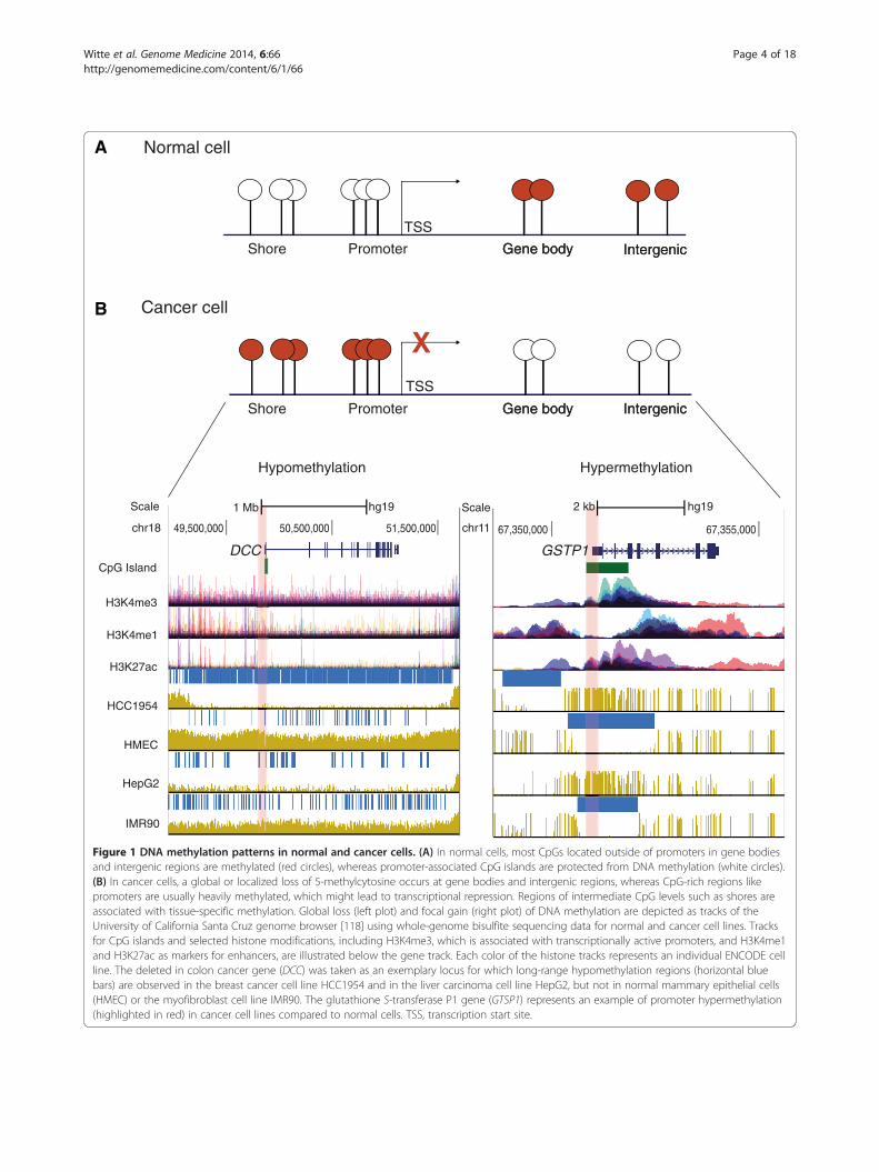
Figure 1 DNA methylation patterns in normal and cancer cells. (A) In normal cells, most CpGs located outside of promoters in gene bodiesand intergenic regions are methylated (red circles), whereas promoter-associated CpG islands are protected from DNA methylation (white circles).(B) In cancer cells, a global or localized loss of 5-methylcytosine occurs at gene bodies and intergenic regions, whereas CpG-rich regions likepromoters are usually heavily methylated, which might lead to transcriptional repression. Regions of intermediate CpG levels such as shores areassociated with tissue-specific methylation. Global loss (left plot) and focal gain (right plot) of DNA methylation are depicted as tracks of theUniversity of California Santa Cruz genome browser [118] using whole-genome bisulfite sequencing data for normal and cancer cell lines. Tracksfor CpG islands and selected histone modifications, including H3K4me3, which is associated with transcriptionally active promoters, and H3K4me1and H3K27ac as markers for enhancers, are illustrated below the gene track. Each color of the histone tracks represents an individual ENCODE cellline. The deleted in colon cancer gene (DCC) was taken as an exemplary locus for which long-range hypomethylation regions (horizontal bluebars) are observed in the breast cancer cell line HCC1954 and in the liver carcinoma cell line HepG2, but not in normal mammary epithelial cells(HMEC) or the myofibroblast cell line IMR90. The glutathione S-transferase P1 gene (GTSP1) represents an example of promoter hypermethylation(highlighted in red) in cancer cell lines compared to normal cells. TSS, transcription start site.
Witte et al. Genome Medicine 2014, 6:66 Page 4 of 18http://genomemedicine.com/content/6/1/66

Table 2 Pan-cancer patterns of DNA methylation
Tumor type(number ofmethylationgroups)
Methylationsubgroup
Genomicaberrations
Methylationpattern*
Comments References
AML HighIDH1/2 or TET2mutations
A Associated with patients presenting with anintermediate-risk karyotype
[43,51,107]
Co-occurrence of IDH1/2 and NPM1 mutations isassociated with good clinical outcome
Bladder urothelial(3)
High RB1 mutations Smoking-pack years as predictor of CIMPphenotypeFrequent mutations in chromatin regulatorssuch as MLL2, ARID1A, KDM6A, and EP300†Mutations inchromatin regulators were more frequent than in anyother TCGA tumor
[35]Low
↑ TP53 mutations B
Breast(5)
B-CIMP
↓ mutation rate Luminal ER/PR-positive tumors
[31,32]
Low metastatic risk and better clinical outcome
Enriched for genes targeted by the PRC2 (e.g. SUZ12and EZH2)
B-CIMP-negative↑ TP53 mutations B Basal-like tumors (ER/PR-negative)
High metastatic risk and poor clinical outcome
Cholangiocarcinoma HighIDH1 and/or IDH2mutations
A Longer survival[47]
Chondrosarcoma HighIDH1 and/or IDH2mutations
A[46,64]
Colorectal(4)
CIMP-H
MLH1hypermethylation
C MSI
[29,108]
Right/ascending colonic region↑ mutation rate
↑ BRAFV600E
mutationGood prognosis
↑ BRAFV600E
mutation
CIMP-LKRAS mutations CIN (non-MSI)
Poor prognosis
Two non-CIMP↑ TP53 mutations B Anatomic origins distinct from CIMP groups
↑ SCNAs
Endometrial(4)
High
MLH1hypermethylation
C MSI
[33]
ARID5B mutations↑ mutation rate
Low↑ TP53 mutations B Serous-like tumors
↑ SCNAs Poor prognosis
Two non-methylated
↑ POLE mutations Endometrioid tumors
↑ SCNAs ARID1A and PTEN mutations were present in all groupswithout high TP53 mutations
Gastric(4) EBV-CIMP
↑ PIK3CA, ARID1Aand BCORmutations
EBV-positive tumorsHighest frequency ofhypermethylation events among TCGA tumors
[30]CDKN2Ahypermethylation
Amplifications ofJAK2, CD274 andPDCD1LG2
Witte et al. Genome Medicine 2014, 6:66 Page 5 of 18http://genomemedicine.com/content/6/1/66
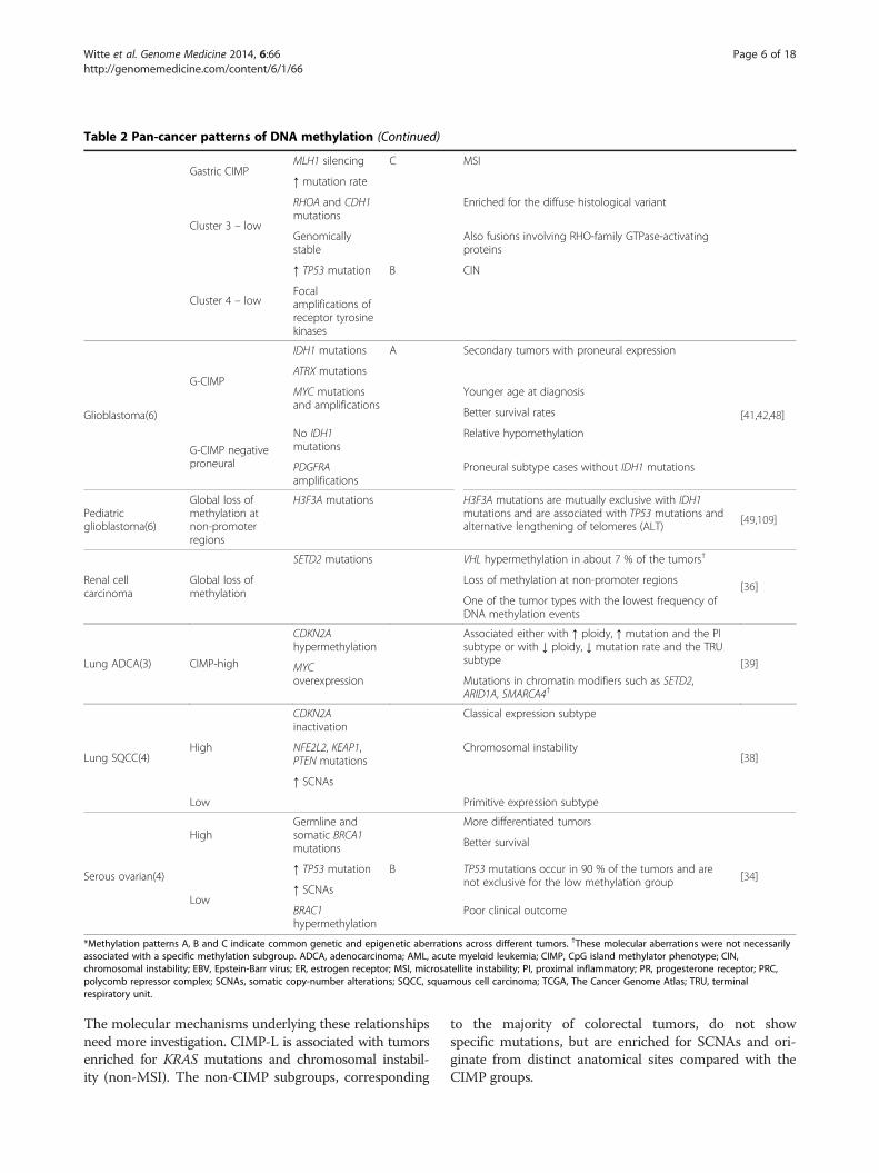
Table 2 Pan-cancer patterns of DNA methylation (Continued)
Gastric CIMPMLH1 silencing C MSI
↑ mutation rate
Cluster 3 – low
RHOA and CDH1mutations
Enriched for the diffuse histological variant
Genomicallystable
Also fusions involving RHO-family GTPase-activatingproteins
Cluster 4 – low
↑ TP53 mutation B CIN
Focalamplifications ofreceptor tyrosinekinases
Glioblastoma(6)
G-CIMP
IDH1 mutations A Secondary tumors with proneural expression
[41,42,48]
ATRX mutations
MYC mutationsand amplifications
Younger age at diagnosis
Better survival rates
G-CIMP negativeproneural
No IDH1mutations
Relative hypomethylation
PDGFRAamplifications
Proneural subtype cases without IDH1 mutations
Pediatricglioblastoma(6)
Global loss ofmethylation atnon-promoterregions
H3F3A mutations H3F3A mutations are mutually exclusive with IDH1mutations and are associated with TP53 mutations andalternative lengthening of telomeres (ALT)
[49,109]
Renal cellcarcinoma
Global loss ofmethylation
SETD2 mutations VHL hypermethylation in about 7 % of the tumors†
[36]Loss of methylation at non-promoter regions
One of the tumor types with the lowest frequency ofDNA methylation events
Lung ADCA(3) CIMP-high
CDKN2Ahypermethylation
Associated either with ↑ ploidy, ↑ mutation and the PIsubtype or with ↓ ploidy, ↓ mutation rate and the TRUsubtype [39]MYC
overexpression Mutations in chromatin modifiers such as SETD2,ARID1A, SMARCA4†
Lung SQCC(4)High
CDKN2Ainactivation
Classical expression subtype
[38]NFE2L2, KEAP1,PTEN mutations
Chromosomal instability
↑ SCNAs
Low Primitive expression subtype
Serous ovarian(4)
HighGermline andsomatic BRCA1mutations
More differentiated tumors
[34]
Better survival
Low
↑ TP53 mutation B TP53 mutations occur in 90 % of the tumors and arenot exclusive for the low methylation group
↑ SCNAs
BRAC1hypermethylation
Poor clinical outcome
*Methylation patterns A, B and C indicate common genetic and epigenetic aberrations across different tumors. †These molecular aberrations were not necessarilyassociated with a specific methylation subgroup. ADCA, adenocarcinoma; AML, acute myeloid leukemia; CIMP, CpG island methylator phenotype; CIN,chromosomal instability; EBV, Epstein-Barr virus; ER, estrogen receptor; MSI, microsatellite instability; PI, proximal inflammatory; PR, progesterone receptor; PRC,polycomb repressor complex; SCNAs, somatic copy-number alterations; SQCC, squamous cell carcinoma; TCGA, The Cancer Genome Atlas; TRU, terminalrespiratory unit.
Witte et al. Genome Medicine 2014, 6:66 Page 6 of 18http://genomemedicine.com/content/6/1/66
The molecular mechanisms underlying these relationshipsneed more investigation. CIMP-L is associated with tumorsenriched for KRAS mutations and chromosomal instabil-ity (non-MSI). The non-CIMP subgroups, corresponding
to the majority of colorectal tumors, do not showspecific mutations, but are enriched for SCNAs and ori-ginate from distinct anatomical sites compared with theCIMP groups.
Witte et al. Genome Medicine 2014, 6:66 Page 7 of 18http://genomemedicine.com/content/6/1/66
Epstein-Barr virus (EBV)-positive gastric tumors displayan extreme EBV-CIMP profile [30], with hypermethyla-tion of CDKN2A but not of MLH1. This phenotype hasthe highest frequency of DNA hypermethylation whencompared with other cancer types reported by TCGA[30]. In contrast, gastric CIMP tumors showed hyper-mutation, MSI and epigenetic silencing of MLH1.
Breast, endometrial and ovarian carcinomasA breast CpG island methylator phenotype (B-CIMP)was first reported in 2011 [31]. B-CIMP is enriched inestrogen and progesterone receptor (ER/PR)-positive tu-mors and is associated with good survival rates and lowmetastatic risk. It is characterized by high methylation ofgenes targeted by the polycomb repressor complex 2(PRC2), including SUZ12 and EZH2 [31]. In contrast,the B-CIMP-negative group shows high metastatic riskand poor clinical outcome. TCGA analyses confirmedthese findings, although they defined five distinct DNAmethylation subgroups. The high methylation groupoverlapped with luminal B tumors (ER/PR-positive) andhad a low rate of mutations. Conversely, the methylation-low group had a high TP53 mutation rate and was enrichedin basal-like tumors (ER/PR-negative) [32].In endometrial carcinomas, TCGA identified four DNA
methylation subtypes. Similar to colorectal cancer, thehigh methylator phenotype was mainly composed ofhypermutated MSI tumors showing extensive MLH1promoter hypermethylation and an under-representationof TP53 mutations [33].Four DNA methylation clusters were defined for serous
ovarian cancer. This cancer type has a 90 % prevalence ofTP53 mutations. TCGA identified a methylation-highgroup enriched for highly differentiated tumors withgermline BRCA1 mutations. BRCA1 mutations weremutually exclusive with BRCA1 hypermethylation, which ischaracteristic of methylation-low tumors with high SCNAs.Survival analysis showed that cases with hypermethylatedBRCA1 had a poorer clinical outcome compared to tu-mors with BRCA1/2 mutations [34].
Bladder urothelial and kidney renal clear cell carcinomasBladder urothelial carcinomas were divided into threeDNA methylation subgroups; one of these groups had aCIMP-like hypermethylation profile and was enrichedfor tumors with RB1 mutations. Similar to the low methy-lation groups in breast, endometrial, gastric and colorectaltumors, the methylation-low group had the highest per-centage of TP53 mutations, suggesting a common mo-lecular mechanism of epigenetic regulation. Interestingly,chromatin regulators such as the histone methyltrans-ferase MLL2, the chromatin remodeling gene ARID1A,the histone demethylase KDM6A and the histone
acetyltransferase EP300 were frequently mutated in thiscancer type [35].For renal clear cell carcinoma, the most common type
of kidney cancer, TCGA identified epigenetic silencing ofthe tumor suppressor VHL in about 7 % of the tumors,which was mutually exclusive with VHL mutations. In-creased promoter methylation was linked to tumors witha higher grade and stage. Tumors with a widespread lossof DNA methylation were associated with mutations ofthe H3K36 methyltransferase SETD2, in contrast tomethylation-low subgroups in other cancer types [36].
Lung adenocarcinoma and squamous cell carcinomaNon-small-cell lung carcinoma (NSCLC), the most com-mon type of lung cancer, is divided into three subtypes:adenocarcinoma, squamous cell carcinoma (SQCC), andlarge cell carcinoma [37]. Methylation analysis of SQCCidentified four groups with distinct DNA methylationpatterns. The methylation-high group overlapped withtumors from the so-called classical subtype, which arecharacterized by chromosomal instability. Moreover, theTSG CDKN2A was inactivated in 72 % of cases, 21 % ofwhich were due to epigenetic silencing [38].Recent results for adenocarcinoma revealed three differ-
ent methylation subgroups: CIMP-H, a subgroup withintermediate methylation levels, and CIMP-L. Remarkably,these methylation subgroups were not specifically relatedto genomic, transcriptomic or histopathological subtypes.CIMP-H subtypes were either associated with tumors withhigh ploidy and a high mutation rate and were classified asproximal inflammatory (previously known as squamoid),or were associated with tumors presenting with low ploidyand a low mutation rate and were classified as terminal re-spiratory unit (formerly bronchioid). Moreover, an associ-ation between tumors enriched for SETD2 and CDKN2Amethylation was found, suggesting an interaction betweenSETD2 mutations and altered chromatin structure forthese tumors [39].
GlioblastomaAberrant DNA methylation has been widely describedfor glioblastoma multiforme (GBM) – the most commonadult brain tumor. In 2008, TCGA chose GBM as thefirst cancer to be comprehensively characterized, reveal-ing an important association between MGMT methyla-tion, mutations in mismatch repair genes and responseto therapy [40]. Subsequently, TCGA identified threeDNA methylation groups, one of which showed hyper-methylation at a large number of loci and was termedG-CIMP [41]. This group was enriched in secondary tu-mors with proneural expression and somatic mutationsof the isocitrate dehydrogenase 1 (IDH1) gene [42]. Thisgain-of-function mutation results in increased catalysisof α-ketoglutarate to D-2-hydroxyglutarate (2-HG), which
Witte et al. Genome Medicine 2014, 6:66 Page 8 of 18http://genomemedicine.com/content/6/1/66
inhibits the activity of TET and KDM proteins, affectingchromatin remodeling and leading to an increase in DNAmethylation. IDH1/2 mutations are also common inhematopoietic malignancies, including acute myeloidleukemia (AML) [43], myelodysplastic syndromes (MDS),myeloproliferative neoplasms [44] and T-cell lymphomas[45], as well as in solid tumors such as chondrosarcoma[46] and cholangiocarcinoma [47].The G-CIMP group is associated with better survival
compared with G-CIMP-negative tumors. The survival ad-vantage of G-CIMP tumors was confirmed by a follow-upTCGA study characterizing more than 500 GBM tumors[48]. In this study, six DNA methylation clusters, includingthe G-CIMP subgroup, were identified. Additionally, theG-CIMP phenotype was associated with a younger ageat diagnosis, enrichment for mutations in the chromatinremodeling gene ATRX, and MYC alterations.The landscape of DNA methylation and genomic aber-
rations in pediatric GBM varies. Instead of having a hyper-methylator phenotype, these tumors show a global loss of5mC, which is mainly associated with extensive changesin histone modifications caused by mutations in H3F3A(reviewed in [8]). This was defined by Sturm et al., whofound six epigenetic subgroups harboring specific muta-tions, SCNAs and transcriptome patterns [49]. Twomethylation subgroups specifically correlated with hotspotmutations in H3F3A, namely at K27 and G34, andwere associated with a younger age at diagnosis. Strikingly,the G34 tumors showed a global loss of methylationoccurring mainly at chromosome ends. The presenceof IDH1 mutations was mutually exclusive with H3F3Amutations.
Acute myeloid leukemiaAML is a highly heterogeneous myeloid disorder andthe most common acute leukemia in adults. AML pa-tients from the normal or intermediate cytogenetic riskcategory frequently have mutations in epigenetic regulatorssuch as IDH1/2, DNMT3 and TET enzymes (reviewed in[50]). Similar to GBM, AML with a DNA hypermethylationphenotype is associated with IDH1/2 mutations [43]. Thesemutations are mutually exclusive with mutations in thedemethylating enzyme TET2, suggesting a complementaryrole. It might be that DNA methylation is a consequence ofmutant IDH expression and that this phenotype contributesto AML development. The association of IDH1/2 muta-tions with the hypermethylation phenotype in AML wasconfirmed by a recent TCGA study. Gain of DNA methyla-tion was mainly observed at CpG-sparse regions of the gen-ome. Other subtypes of tumors were associated with asubstantial loss of DNA methylation and with the presenceof MLL fusion genes or co-occurring mutations in NPM1,DNMT3A or FLT3 [51].
Potential mechanisms leading to DNA methylationsubgroupsThe observation that many tumor types carry numerousmutations in enzymes regulating epigenetic patterns sug-gests that these defects contribute to the global alter-ations seen in cancer genomes [5,8]. However, despitethis expected molecular link, there are currently onlyreports associating methylome subgroups with genemutations [29,49], rather than detailed molecular stud-ies. Exceptions are studies on the histone H3.3 muta-tion H3F3A(K27M), which inactivates EZH2 in thePRC2 complex [52-54]. In addition, introduction of anIDH1 mutant, R132H, into astrocytes induces a specificmethylome pattern [55]. Mutations in IDH1/2 cause accu-mulation of the oncometabolite 2-HG, which disturbs theDNA demethylation process, causing hypermethylation [43].Epigenetic subgroups might also represent preexisting
epigenetic states. For example, PRC2 target genes arecommonly hypermethylated in cancer, and EZH2 is up-regulated in various cancer subtypes. These changeswere associated with gene amplifications, and alterationsin the regulation of gene expression by noncoding RNAsand mutations (reviewed in [56]). Apart from mutationsaffecting epigenetic modifiers, other genes are certainlyalso affected. Colorectal CIMP is tightly associated withBRAF mutations, although it appears that these muta-tions do not drive the hypermethylation phenotype [28].Methylation subgroups might reflect the survival advan-tage of cell populations that have acquired early defectsin DNA repair genes (for example, MLH1, MGMT andBRCA1). Distinct methylation clusters might also repre-sent a common cell type of origin. As an example, thebasal breast cancer subgroup shares characteristics of lowmethylation, high TP53 mutations and high chromosomalinstability with serous endometrial and serous ovariancancer subgroups [33]. Different epigenetic subgroupshave been suggested to represent differences in tumor eti-ology induced by environmental factors, such as recentlyshown for EBV in gastric cancer [30].Again, the question of whether there is a causal relation-
ship between epigenetic changes and cancer or whetherthese associations represent changes in the methylomethat are non-functional events and thus do not contributeto the carcinogenic process (passengers) rather thanmethylation events that drive the carcinogenic process(drivers) remains open. However, there are some generalobservations that extend across studies. First, mutations inepigenetic enzymes such as IDH1/2 are causally linked tothe pathogenesis of subtypes of GBM and AML, as well asto the formation of CIMP. Second, mutations in the geneH3F3A encoding the histone variant H3.3 are associatedwith global loss of methylation, especially in sub-telomericregions, and with the alternative lengthening of telomeresphenotype that is characteristic of a fraction of cancer

Witte et al. Genome Medicine 2014, 6:66 Page 9 of 18http://genomemedicine.com/content/6/1/66
cells, for example in pediatric GBM. Third, mutationsin chromatin regulatory factors such as SETD2, ARID1,SMARCA4, KDM6A, EP300 and MLL are emerging invarious cancer types [57] but, so far, only a few have beenlinked to altered methylome patterns. Many of these fac-tors act in protein complexes, indicating that mutations inany of these could disrupt the function of the complex.Fourth, current cancer epigenome research points to thefact that methylation of polycomb group targets (PCGTs)is detectable even in pre-neoplastic lesions and could rep-resent a risk factor for neoplastic transformation [58].Fifth, recent reports have described particular methylationpatterns related to infectious agents such as EBV or hu-man papilloma virus (HPV), which can initiate carcino-genesis [30,59]; whether these methylation alterations areprimarily useful biomarkers for patient stratification orwhether there is a causal relationship to carcinogenesishas yet to be demonstrated. Last, similarities in methyla-tion patterns across tumor types could indicate the accu-mulation of as yet unidentified, low frequency molecularaberrations that lead to a common phenotype and con-tribute to cancer development. Future research will haveto address these points to draw clear conclusions.
Methylome analyses across different cancer typesThe genome-wide methylation profiles generated byTCGA and others have shown that aberrant methylomesare a hallmark of cancer, and are useful for classifyingtumor subgroups as well as for identifying novel clinicalbiomarkers. Currently, efforts are being made to integratedifferent methylomes and to determine common andtissue-specific DNA methylation patterns across multipletumor entities (pan-cancer). These integrative analysesmight also help to distinguish the driver methylationevents (that contribute to the carcinogenic process) fromthe passenger methylation events (which do not contrib-ute to the carcinogenic process).In 2013, TCGA published the first integrative analysis
of genomic data across 12 cancer types. In this study,SCNAs, somatic mutations and DNA methylation wereintegrated, although methylation changes were limitedto a selection of 13 epigenetically silenced genes. Fromthese genes, MGMT, GSTP1, MLH1 and CDKN2A werefound to be aberrantly methylated in a large number ofsamples in different types of tumors. Hypermethylationof MLH1 was associated with the so-called ‘M class’,characterized by recurrent mutations, whereas BRCA1hypermethylation correlated with the ‘C class’ of tumorsenriched for SCNAs [3]. These findings confirm the pre-vious TCGA reports for single tumor entities. However,by using this selected panel of genes, the results of thisinvestigation might not reflect the actual similarities anddifferences in DNA methylation patterns across distincttumor types, as for example shown in Figure 2.
By combining the methylomes of ten distinct tumorentities, Kim et al. found that aberrant DNA methylationaffects similar biological pathways across the cancertypes analyzed [60]. Over 50 % of the hypermethylationevents were involved in early development and morpho-genesis, including neurogenesis and embryonic develop-ment, whereas the remaining hypermethylation changeswere related to transcription factor activity. A significantoverlap between those pathways and PCGT genes wasobserved. Among the pan-cancer hypermethylated genestargeted by PRC2 were several members of the HOXfamily as well as the TSG CDKN2A. This finding is inagreement with previous studies reporting that methyla-tion of PCGT genes is frequent in distinct cancer types(reviewed in [61]).The integration of genome-wide DNA methylation
data across four different gynecological tumors, namelybreast, ovarian, endometrial and cervical carcinomas, re-vealed similar results [62]. This study additionally inves-tigated the dynamics of DNA methylation throughdifferent stages of cervical carcinogenesis (that is, nor-mal, invasive and metastatic stages). Hypermethylationat stem-cell PCGT genes was found to occur in cyto-logically normal cervical cells 3 years before the appear-ance of the first neoplastic alterations. Moreover, a lossof DNA methylation in CpGs termed ‘methylated em-bryonic stem-cell loci’ was predominantly observed ininvasive tissues, suggesting that hypomethylation atthese CpG sites might constitute a poor prognostic sig-nature for these four gynecological tumor entities.In contrast to these findings, a comparative analysis of
methylomes from seven different tissue types revealedthat hypermethylated genes tend to be already repressedin precancerous tissues and that aberrant methylationdoes not contribute to cancer progression under theclassical model of epigenetic silencing [63]. It was sug-gested that pan-cancer patterns of hypermethylationoccur owing to the variable gene expression profiles inthe corresponding normal tissues. Hypermethylation ofspecific genes might then account for passenger methy-lation events rather than for driver events.Apart from analyzing pan-cancer methylomes, integra-
tive analyses of different tumors harboring mutations incommon epigenetic regulators might provide clues aboutthe molecular mechanisms affecting DNA methylation.Guilhamon et al. performed an exemplary meta-analysisof the DNA methylation profiles of tumors with IDHmutations and intrinsic high methylator phenotypes –namely AML, low-grade GBM, cholangiocarcinomasand chondrosarcomas [64]. The retinoic acid receptorpathway, which is usually dysregulated in the early stepsof tumorigenesis, was enriched in the four tumor types.The early B-cell factor 1 (EBF1) was identified as a novelinteraction partner of the dioxygenase TET2, suggesting
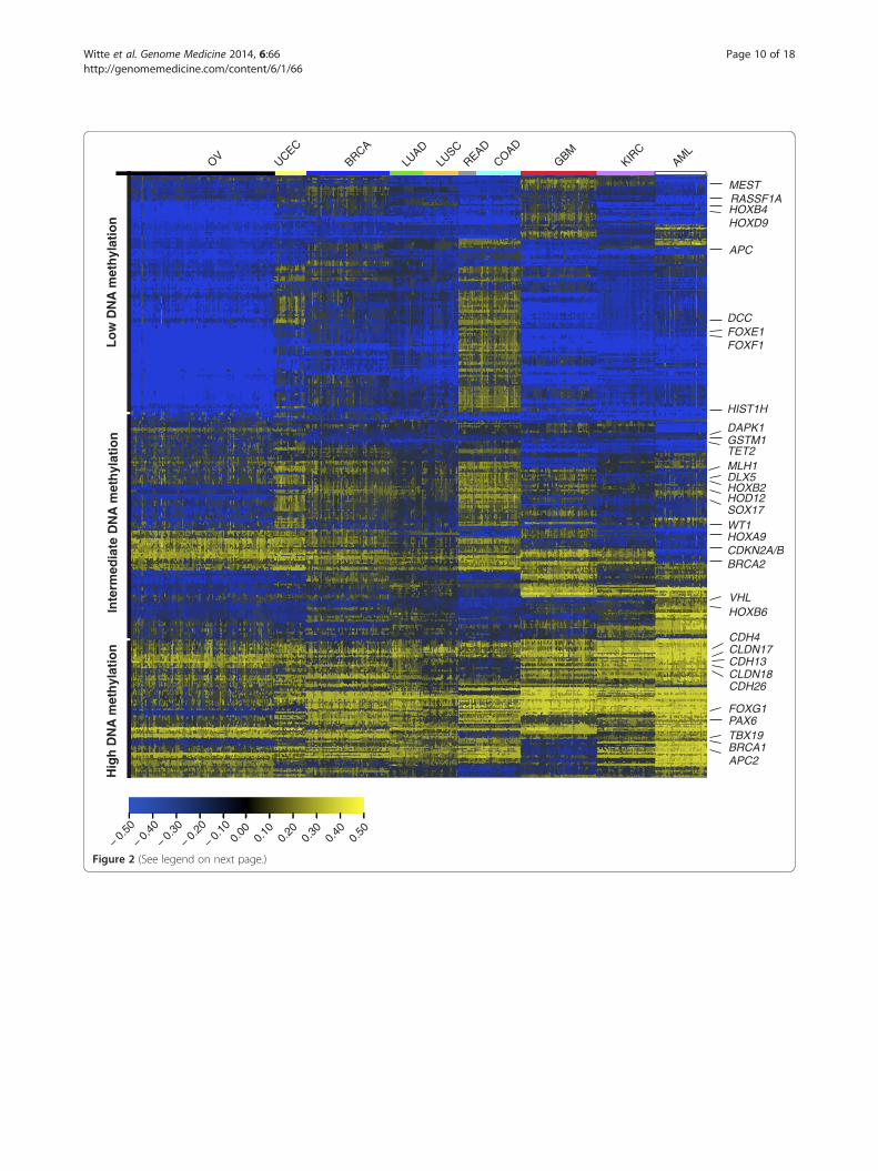
Figure 2 (See legend on next page.)
Witte et al. Genome Medicine 2014, 6:66 Page 10 of 18http://genomemedicine.com/content/6/1/66
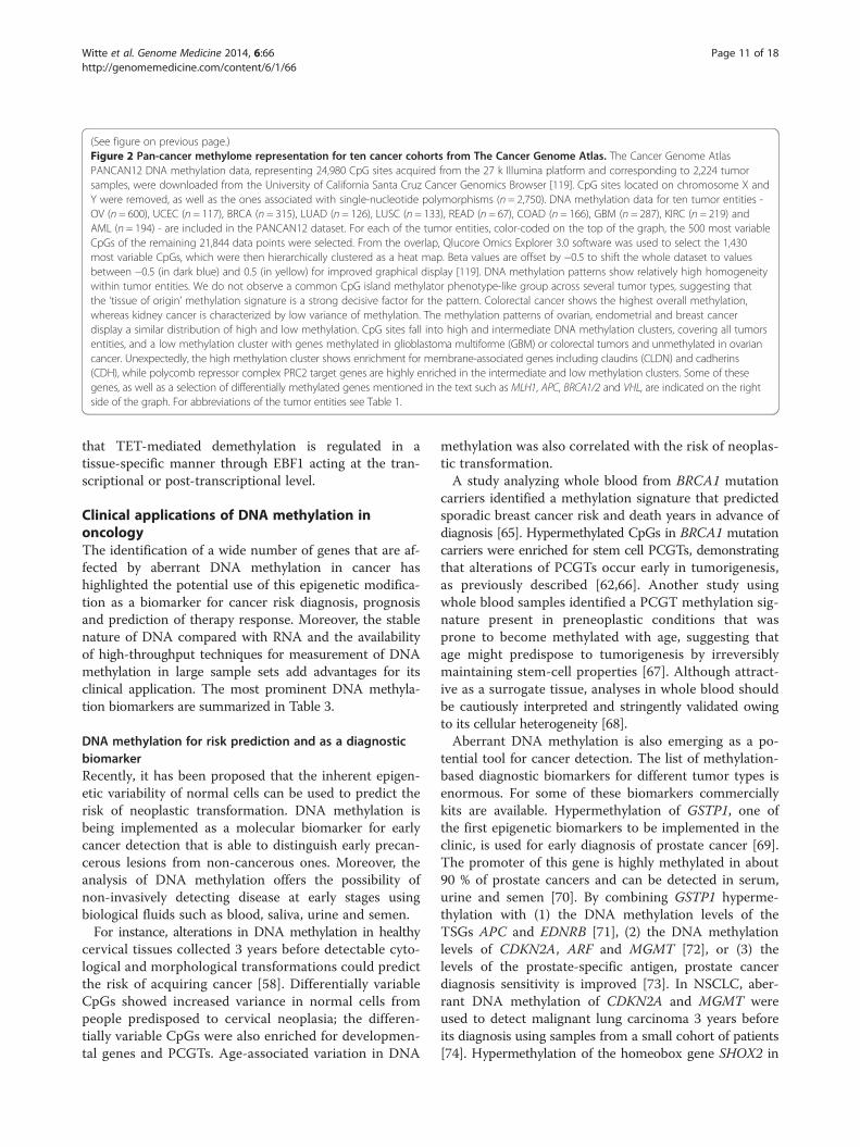
(See figure on previous page.)Figure 2 Pan-cancer methylome representation for ten cancer cohorts from The Cancer Genome Atlas. The Cancer Genome AtlasPANCAN12 DNA methylation data, representing 24,980 CpG sites acquired from the 27 k Illumina platform and corresponding to 2,224 tumorsamples, were downloaded from the University of California Santa Cruz Cancer Genomics Browser [119]. CpG sites located on chromosome X andY were removed, as well as the ones associated with single-nucleotide polymorphisms (n = 2,750). DNA methylation data for ten tumor entities -OV (n = 600), UCEC (n = 117), BRCA (n = 315), LUAD (n = 126), LUSC (n = 133), READ (n = 67), COAD (n = 166), GBM (n = 287), KIRC (n = 219) andAML (n = 194) - are included in the PANCAN12 dataset. For each of the tumor entities, color-coded on the top of the graph, the 500 most variableCpGs of the remaining 21,844 data points were selected. From the overlap, Qlucore Omics Explorer 3.0 software was used to select the 1,430most variable CpGs, which were then hierarchically clustered as a heat map. Beta values are offset by −0.5 to shift the whole dataset to valuesbetween −0.5 (in dark blue) and 0.5 (in yellow) for improved graphical display [119]. DNA methylation patterns show relatively high homogeneitywithin tumor entities. We do not observe a common CpG island methylator phenotype-like group across several tumor types, suggesting thatthe ‘tissue of origin’ methylation signature is a strong decisive factor for the pattern. Colorectal cancer shows the highest overall methylation,whereas kidney cancer is characterized by low variance of methylation. The methylation patterns of ovarian, endometrial and breast cancerdisplay a similar distribution of high and low methylation. CpG sites fall into high and intermediate DNA methylation clusters, covering all tumorsentities, and a low methylation cluster with genes methylated in glioblastoma multiforme (GBM) or colorectal tumors and unmethylated in ovariancancer. Unexpectedly, the high methylation cluster shows enrichment for membrane-associated genes including claudins (CLDN) and cadherins(CDH), while polycomb repressor complex PRC2 target genes are highly enriched in the intermediate and low methylation clusters. Some of thesegenes, as well as a selection of differentially methylated genes mentioned in the text such as MLH1, APC, BRCA1/2 and VHL, are indicated on the rightside of the graph. For abbreviations of the tumor entities see Table 1.
Witte et al. Genome Medicine 2014, 6:66 Page 11 of 18http://genomemedicine.com/content/6/1/66
that TET-mediated demethylation is regulated in atissue-specific manner through EBF1 acting at the tran-scriptional or post-transcriptional level.
Clinical applications of DNA methylation inoncologyThe identification of a wide number of genes that are af-fected by aberrant DNA methylation in cancer hashighlighted the potential use of this epigenetic modifica-tion as a biomarker for cancer risk diagnosis, prognosisand prediction of therapy response. Moreover, the stablenature of DNA compared with RNA and the availabilityof high-throughput techniques for measurement of DNAmethylation in large sample sets add advantages for itsclinical application. The most prominent DNA methyla-tion biomarkers are summarized in Table 3.
DNA methylation for risk prediction and as a diagnosticbiomarkerRecently, it has been proposed that the inherent epigen-etic variability of normal cells can be used to predict therisk of neoplastic transformation. DNA methylation isbeing implemented as a molecular biomarker for earlycancer detection that is able to distinguish early precan-cerous lesions from non-cancerous ones. Moreover, theanalysis of DNA methylation offers the possibility ofnon-invasively detecting disease at early stages usingbiological fluids such as blood, saliva, urine and semen.For instance, alterations in DNA methylation in healthy
cervical tissues collected 3 years before detectable cyto-logical and morphological transformations could predictthe risk of acquiring cancer [58]. Differentially variableCpGs showed increased variance in normal cells frompeople predisposed to cervical neoplasia; the differen-tially variable CpGs were also enriched for developmen-tal genes and PCGTs. Age-associated variation in DNA
methylation was also correlated with the risk of neoplas-tic transformation.A study analyzing whole blood from BRCA1 mutation
carriers identified a methylation signature that predictedsporadic breast cancer risk and death years in advance ofdiagnosis [65]. Hypermethylated CpGs in BRCA1 mutationcarriers were enriched for stem cell PCGTs, demonstratingthat alterations of PCGTs occur early in tumorigenesis,as previously described [62,66]. Another study usingwhole blood samples identified a PCGT methylation sig-nature present in preneoplastic conditions that wasprone to become methylated with age, suggesting thatage might predispose to tumorigenesis by irreversiblymaintaining stem-cell properties [67]. Although attract-ive as a surrogate tissue, analyses in whole blood shouldbe cautiously interpreted and stringently validated owingto its cellular heterogeneity [68].Aberrant DNA methylation is also emerging as a po-
tential tool for cancer detection. The list of methylation-based diagnostic biomarkers for different tumor types isenormous. For some of these biomarkers commerciallykits are available. Hypermethylation of GSTP1, one ofthe first epigenetic biomarkers to be implemented in theclinic, is used for early diagnosis of prostate cancer [69].The promoter of this gene is highly methylated in about90 % of prostate cancers and can be detected in serum,urine and semen [70]. By combining GSTP1 hyperme-thylation with (1) the DNA methylation levels of theTSGs APC and EDNRB [71], (2) the DNA methylationlevels of CDKN2A, ARF and MGMT [72], or (3) thelevels of the prostate-specific antigen, prostate cancerdiagnosis sensitivity is improved [73]. In NSCLC, aber-rant DNA methylation of CDKN2A and MGMT wereused to detect malignant lung carcinoma 3 years beforeits diagnosis using samples from a small cohort of patients[74]. Hypermethylation of the homeobox gene SHOX2 in
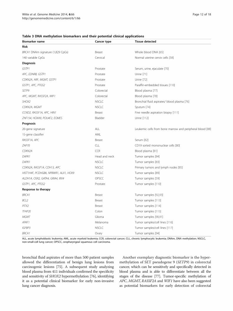
Table 3 DNA methylation biomarkers and their potential clinical applications
Biomarker name Cancer type Tissue detected
Risk
BRCA1 DNAm signature (1,829 CpGs) Breast Whole blood DNA [65]
140 variable CpGs Cervical Normal uterine cervix cells [58]
Diagnosis
GSTP1 Prostate Serum, urine, ejaculate [70]
APC, EDNRB, GSTP1 Prostate Urine [71]
CDKN2A, ARF, MGMT, GSTP1 Prostate Urine [72]
GSTP1, APC, PTGS2 Prostate Paraffin-embedded tissues [110]
SETP9 Colorectal Blood plasma [77]
APC, MGMT, RASSF2A, WIF1 Colorectal Blood plasma [78]
SHOX2 NSCLC Bronchial fluid aspirates/ blood plasma [76]
CDKN2A, MGMT NSCLC Sputum [74]
CCND2, RASSF1A, APC, HIN1 Breast Fine needle aspiration biopsy [111]
ZNF154, HOXA9, POU4F2, EOMES Bladder Urine [112]
Prognosis
20-gene signature ALL Leukemic cells from bone marrow and peripheral blood [88]
15-gene classifier AML
RASSF1A, APC Breast Serum [82]
ZAP70 CLL CD19 sorted mononuclear cells [80]
CDKN2A CCR Blood plasma [81]
DAPK1 Head and neck Tumor samples [84]
DAPK1 NSCLC Tumor samples [83]
CDKN2A, RASSF1A, CDH13, APC NSCLC Primary tumors and lymph nodes [85]
HIST1H4F, PCDHGB6, NPBWR1, ALX1, HOX9 NSCLC Tumor samples [89]
ALDH1A, OSR2, GATA4, GRIA4, IRX4 OPSCC Tumor samples [59]
GSTP1, APC, PTGS2 Prostate Tumor samples [110]
Response to therapy
BRCA1 Breast Tumor samples [92,93]
BCL2 Breast Tumor samples [113]
PITX2 Breast Tumor samples [114]
TFAP2E Colon Tumor samples [115]
MGMT Glioma Tumor samples [90,91]
APAF1 Melanoma Tumor samples/cell lines [116]
IGFBP3 NSCLC Tumor samples/cell lines [117]
BRCA1 Ovary Tumor samples [94]
ALL, acute lymphoblastic leukemia; AML, acute myeloid leukemia; CCR, colorectal cancer; CLL, chronic lymphocytic leukemia; DNAm, DNA methylation; NSCLC,non-small-cell lung cancer; OPSCC, oropharyngeal squamous cell carcinoma.
Witte et al. Genome Medicine 2014, 6:66 Page 12 of 18http://genomemedicine.com/content/6/1/66
bronchial fluid aspirates of more than 500 patient samplesallowed the differentiation of benign lung lesions fromcarcinogenic lesions [75]. A subsequent study analyzingblood plasma from 411 individuals confirmed the specificityand sensitivity of SHOX2 hypermethylation [76], identifyingit as a potential clinical biomarker for early non-invasivelung cancer diagnosis.
Another exemplary diagnostic biomarker is the hyper-methylation of SET pseudogene 9 (SETP9) in colorectalcancer, which can be sensitively and specifically detected inblood plasma and is able to differentiate between all thestages of the disease [77]. Tumor-specific methylation ofAPC,MGMT, RASSF2A andWIF1 have also been suggestedas potential biomarkers for early detection of colorectal

Witte et al. Genome Medicine 2014, 6:66 Page 13 of 18http://genomemedicine.com/content/6/1/66
cancer [78]. Moreover, a recent genome-wide screen usingDNA methylation data from more than 700 colorectalcancer samples identified hypermethylation of the throm-bin receptor THBD and of C9orf50 as novel blood-basedbiomarkers for colorectal cancer detection [79].
DNA methylation as a prognosis biomarkerIn addition to its diagnostic applications, aberrant DNAmethylation could help to predict and stratify patientswith risks of distinct clinical outcomes. Studies using DNAmethylation as a prognostic biomarker have identified moreaggressive tumors and predicted overall survival and riskof disease progression and/or recurrence. Initially, studiescombined clinical characteristics with aberrant DNA methy-lation at single or multiple genes, but genome-wide DNAmethylation profiling of thousands of CpG sites is now lead-ing to the identification of prognostic signatures.In CLL, DNA methylation of a single CpG within the
zeta-chain-associated protein kinase 70 (ZAP70) gene pro-moter predicted disease outcome better than current geneticapproaches [80]. Examples of other hypermethylated genesused to predict poor clinical prognosis include CDKN2A incolorectal cancer [81], RASSF1A and APC in breast cancer[82], the apoptosis-associated gene DAPK1 in lung andhead and neck cancers [83,84], and CDKN2A, RASSF1A,cadherin 13 (CDH13) and APC in stage I NSCLC [85].The first studies characterizing DNA methylation at
a genome-wide scale and using large cohorts of patientsto investigate prognostic signatures were performed onhematopoietic malignancies. In AML, the methylomesof 344 patients were used to classify 16 distinct AMLsubgroups. From these, 5 subgroups defined new AMLsubtypes without any reported cytogenetic, molecular orclinical features. This study also revealed a 15-gene methy-lation classifier that predicted overall survival [86]. A recentinvestigation that focused on cytogenetically normal AMLpatients identified a seven-gene score which combinedDNA methylation and gene expression and was associatedwith patient outcome [87]. In childhood acute lymphoblasticleukemia (ALL), distinct biological ALL subtypes wereidentified, as well as a group of genes whose DNAmethylation levels correlated with a higher risk of relapse[88]. Another study in HPV-driven oropharyngeal squamouscell carcinoma defined a DNA methylation score of fivegenes (ALDH1A2, OSR2, GATA4, GRIA4 and IRX4), whichwas associated with clinical outcome [59]. Moreover, DNAhypermethylation of five genes (HIST1H4F, PCDHGB6,NPBWR1, ALX1 and HOXA9) was used to classify high-and low-risk stage I NSCLC and patients with shorterrelapse-free survival [89]. Apart from these studies, theefforts of TCGA have shown that methylomes could beused to stratify tumors with distinct biological and clin-ical characteristics, as mentioned earlier.
DNA methylation as a biomarker to predicttreatment responseThe individual response of each patient to chemotherapeuticdrugs is quite heterogeneous and, hence, biomarkers thatpredict response to therapy as well as the developmentof drug resistance are urgently required. DNA methyla-tion has proven to be a suitable biomarker to predicttreatment outcome in various types of tumors. Sucha marker was identified in GBM, where hypermethyla-tion of the DNA repair gene MGMT predicted treat-ment response. Silencing of MGMT diminishes DNArepair activity and removal of alkyl lesions, and thus pre-dicts responsiveness to chemotherapeutic agents such astemozolomide and carmustine [90,91]. TCGA confirmedthese findings and further identified that MGMT hyper-methylation in GBM patients might predict respondersfrom non-responders more accurately than the classicalexpression subgroups [48].Hypermethylation of the DNA repair gene BRCA1 in
sporadic triple-negative breast tumors has also beenproposed as a biomarker to predict sensitivity of breastcancers to the cross-linking agent cisplatin [92] and tothe poly(ADP)-ribose polymerase inhibitor olaparib [93].Similar results were observed in ovarian tumors withBRCA1/2 mutations, where BRCA1 hypermethylationpredicted better response to poly(ADP)-ribose polymeraseinhibitor treatment [94].
Therapeutic useOwing to its reversible nature in comparison to geneticalterations, aberrant DNA methylation can also be thera-peutically targeted. Epigenetic drugs such as the histonedeacetylase (HDAC) inhibitors, DNA demethylating agentsor small molecule inhibitors of the BET family of bromodo-main proteins have been shown to modify chromatinstructure and modify DNA methylation patterns acrossthe genome [95,96]. DNMT inhibitors can be incorpo-rated into the DNA or RNA of replicating cells, blockingthe catalytic domain of DNMTs and thus inhibiting themaintenance of DNA methylation after cell division.The DNMT inhibitors azacitidine (5-azacytidine) anddecitabine (5-aza-2'-deoxycytidine) have been tested inclinical trials for hematopoietic malignancies and wereapproved by the US Food and Drug Administration forthe treatment of MDS and AML [97,98]. Moreover, azaci-tidine in combination with an HDAC inhibitor has beenused as a treatment regimen in a phase II clinical trialfor solid tumors including NSCLC, breast cancer andcolorectal cancer [95,99]. The results obtained for NSCLCshowed durable responses and better patient survival,suggesting that combined epigenetic therapy may haveclinical benefits for the treatment of this and other solidtumor types.

Witte et al. Genome Medicine 2014, 6:66 Page 14 of 18http://genomemedicine.com/content/6/1/66
Conclusions and future perspectivesThe integration of genome-wide DNA methylation profileswith genomic and other omic profiles is just emerging, andfurther efforts are needed to complete cross-tumor ana-lyses, which will then help us to understand the molecularmechanisms responsible for the epigenetic defects that canresult from aberrant DNA methylation. Several interestingfindings have been revealed. Subgroups of cancers withhigh methylation (including CIMP), are associated with in-dividual genomic aberrations underlying these patterns, andhave been identified in various cancer entities. At present,however, there is no evidence for a unifying mechanismleading to these high methylation phenotypes.Moreover, several tumor types, such as basal breast,
high-grade serous ovarian and subtypes of serous endo-metrial, gastric and colorectal carcinomas, related to fre-quent TP53 mutations and high levels of SCNAs, sharea pattern of low methylation in CGIs. Apparently, in thesetumor subtypes, CGIs retain the low methylation patternsobserved in normal tissues and are protected from methyla-tion or are subjected to active demethylation. Again, themolecular mechanism underlying these observations is notknown. We hypothesize that in this case structural genomicalterations are sufficient to drive carcinogenesis.Although still in its infancy, pan-cancer methylome
analyses have provided some interesting insights into themechanisms of cancer development. First, it is becomingmore apparent that multiple cancer types are affected bymutations in genes encoding epigenetic regulatory enzymes,histone variants and chromatin regulatory factors. Some ofthese have been experimentally shown to contribute to al-terations in methylation patterns. Comparing methylomesacross cancer types might now help to identify novel non-recurrent mutations converging on common biologicalpathways that might lead to the development of alteredmethylation phenotypes in specific subgroups of cancers.Second, hypermethylation of PCGTs is apparent in basicallyevery tumor type and can even be observed in preneoplastictissues. Third, the influence of environmental factors onDNA methylomes might have been underestimated untilnow. For example, infectious agents have been recentlylinked to specific methylation patterns.However, pan-cancer methylome analyses still need to
overcome some challenges. First, in the past, DNAmethylation data were generated on two different plat-forms for some tumor types. Integration of these datarestricts the output to overlapping CpG sites, mostlyrepresenting CGIs, and strongly reduces the genome-wide coverage. With the generation of larger datasetsderived from the 450 k platform, these limitations willbe overcome in the future. Second, comparing datasetsderived from different platforms, and from samples pro-vided by various centers, is intrinsically prone to system-atic batch effects that need to be carefully monitored.
Third, some tumor types are characterized by high tumorheterogeneity that is difficult to control and might leadto false positive results. Also, high tumor purity is animportant prerequisite for correct data interpretation,but is often difficult to achieve. Enrichment of certain celltypes by sorting or laser capture microdissection prior toanalysis might be desirable. Fourth, for the developmentof clinical predictive, diagnostic or prognostic biomarkersand stratification of patient subgroups, the availability ofwell documented clinical data is essential. Last, integrativeand comparative analyses of multi-platform datasets requirepowerful bioinformatic and biostatistical algorithms. Dedi-cated computational centers have to develop and rigorouslytest and validate these tools.The epigenetic field is rapidly evolving, and in the near
future more single-base resolution methylomes for alarge number of tumors will be available. The generationof such methylomes is now affordable due to a consider-able reduction in next-generation sequencing costs, im-proved computational expertise and emerging technologiesthat use lower DNA input, such as tagmentation-basedWGBS. This method is used for WGBS library preparation,and is based on the enzymatic activity of a transposase tosimultaneously fragment and tag DNA with adapters [100].High-resolution methylation maps will provide additionalinformation to the current methylomes, especially regard-ing cytosine methylation in a non-CpG context, long-rangemethylation interactions, and better assessment of allele-specific DNA methylation (reviewed in [101]). In addition,high sequencing coverage will accurately quantify DNAmethylation in genomic regions such as enhancers, insula-tors, intergenic regions and repetitive elements, which arecurrently not included in pan-cancer methylome analyses.In the longer term, novel technologies will also allow
genomic and epigenomic analyses of single cells. Theseanalyses will generate more precise datasets by avoidingthe problems associated with tissue impurities or hetero-geneity, and will allow a direct link between the methy-lome and the transcriptome [102]. However, the broadapplication of single-cell analyses still requires meth-odological development to reduce technical artefacts.To fully understand the interplay between the genome,epigenome and transcriptome, existing datasets needto be integrated with information about additionalmechanisms of epigenomic regulation, including theemerging non-coding transcriptome and higher-orderchromatin organization. Importantly, hypotheses generatedfrom these combined efforts need to be experimentallytested to prove their functional relevance.Finally, in terms of translation to the clinic, an essential
aspect is to use the knowledge generated by methylomeanalyses as well as from the integration of methylation datawith other omic data to identify novel clinical markers thatshould be able to stratify patients better and to define

Witte et al. Genome Medicine 2014, 6:66 Page 15 of 18http://genomemedicine.com/content/6/1/66
molecular signatures across different tumor types. On thebasis of these molecular markers, novel epigenetic therapiescould be developed, setting the stage for better clinical trialstrategies across cancer types as well as for personalizedmedicine based on next-generation sequencing data. Already,pan-cancer analyses have revealed molecular similarities thatwill allow existing therapies to be applied to different cancertypes.
Abbreviations2-HG: D-2-hydroxyglutarate; 5hmC: 5-hydroxymethylcytosine; 5mC:5-methylcytosine; ALL: acute lymphoblastic leukemia; AML: acute myeloidleukemia; CGI: CpG island; CIMP: CpG island methylator phenotype;CLL: chronic lymphocytic leukemia; DMV: DNA methylation valley;DNMT: DNA methyltransferase; EBF1: early B-cell factor 1; EBV: Epstein-Barrvirus; ER: estrogen receptor; GBM: glioblastoma multiforme; HDCA: histonedeacetylase; HPV: human papilloma virus; ICGC: International Cancer GenomeConsortium; MDS: myelodysplastic syndrome; MSI: microsatellite instability;NSCLC: non-small-cell lung carcinoma; PCGT: polycomb group target;PMD: partially methylated domain; PR: progesterone receptor;PRC: polycomb repressor complex; SCNA: somatic copy-number alteration;SQCC: squamous cell carcinoma; TCGA: The Cancer Genome Atlas;TET: ten-eleven translocation; TSG: tumor suppressor gene; WGBS: whole-genomebisulfite sequencing.
Competing interestsThe authors declare that they have no competing interests.
AcknowledgementsThis work was supported in part by the German Federal Ministry ofEducation and Science through the program for medical genome research(FKZ: 01KU1001A) to CP and CG and by the German Funding Agency (DFGSPP1463) to TW and CP. TW is supported by a PhD scholarship provided bythe Helmholtz International Graduate School for Cancer Research at theGerman Cancer Research Center.
References1. Cline MS, Craft B, Swatloski T, Goldman M, Ma S, Haussler D, Zhu J:
Exploring TCGA Pan-Cancer data at the UCSC Cancer Genomics Browser.Sci Rep 2013, 3:2652.
2. Cancer Genome Atlas Research Network, Weinstein JN, Collisson EA, MillsGB, Shaw KR, Ozenberger BA, Ellrott K, Shmulevich I, Sander C, Stuart JM:The Cancer Genome Atlas Pan-Cancer analysis project. Nat Genet 2013,45:1113–1120.
3. Ciriello G, Miller ML, Aksoy BA, Senbabaoglu Y, Schultz N, Sander C:Emerging landscape of oncogenic signatures across human cancers.Nat Genet 2013, 45:1127–1133.
4. Alexandrov LB, Nik-Zainal S, Wedge DC, Aparicio SA, Behjati S, Biankin AV,Bignell GR, Bolli N, Borg A, Borresen-Dale AL, Boyault S, Burkhardt B, ButlerAP, Caldas C, Davies HR, Desmedt C, Eils R, Eyfjord JE, Foekens JA, GreavesM, Hosoda F, Hutter B, Ilicic T, Imbeaud S, Imielinski M, Jager N, Jones DT,Jones D, Knappskog S, Kool M, et al: Signatures of mutational processes inhuman cancer. Nature 2013, 500:415–421.
5. Shen H, Laird PW: Interplay between the cancer genome and epigenome.Cell 2013, 153:38–55.
6. Jones PA: Functions of DNA methylation: islands, start sites, gene bodiesand beyond. Nat Rev Genet 2012, 13:484–492.
7. Brocks D, Assenov Y, Minner S, Bogatyrova O, Simon R, Koop C, Oakes C,Zucknick M, Lipka DB, Weischenfeldt J, Feuerbach L, Cowper-Sal Lari R,Lupien M, Brors B, Korbel J, Schlomm T, Tanay A, Sauter G, Gerhauser C,Plass C, ICGC Early Onset Prostate Cancer Project: Intratumor DNAmethylation heterogeneity reflects clonal evolution in aggressiveprostate cancer. Cell Rep 2014, 8:798–806.
8. Plass C, Pfister SM, Lindroth AM, Bogatyrova O, Claus R, Lichter P: Mutationsin regulators of the epigenome and their connections to globalchromatin patterns in cancer. Nat Rev Genet 2013, 14:765–780.
9. Hermann A, Goyal R, Jeltsch A: The Dnmt1 DNA-(cytosine-C5)-methyltransferasemethylates DNA processively with high preference for hemimethylatedtarget sites. J Biol Chem 2004, 279:48350–48359.
10. Okano M, Bell DW, Haber DA, Li E: DNA methyltransferases Dnmt3a andDnmt3b are essential for de novo methylation and mammaliandevelopment. Cell 1999, 99:247–257.
11. Niehrs C, Schafer A: Active DNA demethylation by Gadd45 and DNArepair. Trends Cell Biol 2012, 22:220–227.
12. Tahiliani M, Koh KP, Shen Y, Pastor WA, Bandukwala H, Brudno Y, Agarwal S,Iyer LM, Liu DR, Aravind L, Rao A: Conversion of 5-methylcytosine to5-hydroxymethylcytosine in mammalian DNA by MLL partner TET1.Science 2009, 324:930–935.
13. He YF, Li BZ, Li Z, Liu P, Wang Y, Tang Q, Ding J, Jia Y, Chen Z, Li L, Sun Y, Li X, DaiQ, Song CX, Zhang K, He C, Xu GL: Tet-mediated formation of 5-carboxylcytosineand its excision by TDG in mammalian DNA. Science 2011, 333:1303–1307.
14. Deaton AM, Bird A: CpG islands and the regulation of transcription.Genes Dev 2011, 25:1010–1022.
15. Bird AP: CpG-rich islands and the function of DNA methylation.Nature 1986, 321:209–213.
16. Irizarry RA, Ladd-Acosta C, Wen B, Wu Z, Montano C, Onyango P, Cui H,Gabo K, Rongione M, Webster M, Ji H, Potash JB, Sabunciyan S, Feinberg AP:The human colon cancer methylome shows similar hypo- andhypermethylation at conserved tissue-specific CpG island shores.Nat Genet 2009, 41:178–186.
17. Gama-Sosa MA, Slagel VA, Trewyn RW, Oxenhandler R, Kuo KC, Gehrke CW,Ehrlich M: The 5-methylcytosine content of DNA from human tumors.Nucleic Acids Res 1983, 11:6883–6894.
18. Kulis M, Heath S, Bibikova M, Queiros AC, Navarro A, Clot G, Martinez-TrillosA, Castellano G, Brun-Heath I, Pinyol M, Barberan-Soler S, Papasaikas P, JaresP, Bea S, Rico D, Ecker S, Rubio M, Royo R, Ho V, Klotzle B, Hernandez L,Conde L, Lopez-Guerra M, Colomer D, Villamor N, Aymerich M, Rozman M,Bayes M, Gut M, Gelpi JL, et al: Epigenomic analysis detects widespreadgene-body DNA hypomethylation in chronic lymphocytic leukemia.Nat Genet 2012, 44:1236–1242.
19. Hansen KD, Timp W, Bravo HC, Sabunciyan S, Langmead B, McDonald OG,Wen B, Wu H, Liu Y, Diep D, Briem E, Zhang K, Irizarry RA, Feinberg AP:Increased methylation variation in epigenetic domains across cancertypes. Nat Genet 2011, 43:768–775.
20. Hovestadt V, Jones DT, Picelli S, Wang W, Kool M, Northcott PA, Sultan M,Stachurski K, Ryzhova M, Warnatz HJ, Ralser M, Brun S, Bunt J, Jager N,Kleinheinz K, Erkek S, Weber UD, Bartholomae CC, von Kalle C, Lawerenz C, EilsJ, Koster J, Versteeg R, Milde T, Witt O, Schmidt S, Wolf S, Pietsch T, Rutkowski S,Scheurlen W, et al: Decoding the regulatory landscape of medulloblastomausing DNA methylation sequencing. Nature 2014, 510:537–541.
21. Hoffmann MJ, Schulz WA: Causes and consequences of DNAhypomethylation in human cancer. Biochem Cell Biol 2005, 83:296–321.
22. Sandoval J, Esteller M: Cancer epigenomics: beyond genomics. Curr OpinGenet Dev 2012, 22:50–55.
23. Berman BP, Weisenberger DJ, Aman JF, Hinoue T, Ramjan Z, Liu Y,Noushmehr H, Lange CP, van Dijk CM, Tollenaar RA, Van Den Berg D, LairdPW: Regions of focal DNA hypermethylation and long-rangehypomethylation in colorectal cancer coincide with nuclearlamina-associated domains. Nat Genet 2012, 44:40–46.
24. Hon GC, Hawkins RD, Caballero OL, Lo C, Lister R, Pelizzola M, Valsesia A,Ye Z, Kuan S, Edsall LE, Camargo AA, Stevenson BJ, Ecker JR, Bafna V,Strausberg RL, Simpson AJ, Ren B: Global DNA hypomethylation coupledto repressive chromatin domain formation and gene silencing in breastcancer. Genome Res 2012, 22:246–258.
25. Jeong M, Sun D, Luo M, Huang Y, Challen GA, Rodriguez B, Zhang X,Chavez L, Wang H, Hannah R, Kim SB, Yang L, Ko M, Chen R, Gottgens B,Lee JS, Gunaratne P, Godley LA, Darlington GJ, Rao A, Li W, Goodell MA:Large conserved domains of low DNA methylation maintained byDnmt3a. Nat Genet 2014, 46:17–23.
26. Esteller M: Cancer epigenomics: DNA methylomes and histone-modificationmaps. Nat Rev Genet 2007, 8:286–298.
27. Toyota M, Ahuja N, Ohe-Toyota M, Herman JG, Baylin SB, Issa JP: CpG islandmethylator phenotype in colorectal cancer. Proc Natl Acad Sci USA 1999,96:8681–8686.
28. Hinoue T, Weisenberger DJ, Lange CP, Shen H, Byun HM, Van Den Berg D, Malik S,Pan F, Noushmehr H, van Dijk CM, Tollenaar RA, Laird PW: Genome-scale analysisof aberrant DNA methylation in colorectal cancer. Genome Res 2012, 22:271–282.

Witte et al. Genome Medicine 2014, 6:66 Page 16 of 18http://genomemedicine.com/content/6/1/66
29. The Cancer Genome Atlas Network: Comprehensive molecular characterizationof human colon and rectal cancer. Nature 2012, 487:330–337.
30. The Cancer Genome Atlas Research Network: Comprehensive molecularcharacterization of gastric adenocarcinoma. Nature 2014, doi:10.1038/nature13480.
31. Fang F, Turcan S, Rimner A, Kaufman A, Giri D, Morris LG, Shen R, Seshan V,Mo Q, Heguy A, Baylin SB, Ahuja N, Viale A, Massague J, Norton L, VahdatLT, Moynahan ME, Chan TA: Breast cancer methylomes establish anepigenomic foundation for metastasis. Sci Transl Med 2011, 3:75ra25.
32. The Cancer Genome Atlas Network: Comprehensive molecular portraits ofhuman breast tumours. Nature 2012, 490:61–70.
33. The Cancer Genome Atlas Research Network: Integrated genomiccharacterization of endometrial carcinoma. Nature 2013, 497:67–73.
34. The Cancer Genome Atlas Research Network: Integrated genomic analysesof ovarian carcinoma. Nature 2011, 474:609–615.
35. The Cancer Genome Atlas Research Network: Comprehensive molecularcharacterization of urothelial bladder carcinoma. Nature 2014, 507:315–322.
36. The Cancer Genome Atlas Research Network: Comprehensive molecularcharacterization of clear cell renal cell carcinoma. Nature 2013, 499:43–49.
37. Hensing T, Chawla A, Batra R, Salgia R: A personalized treatment for lungcancer: molecular pathways, targeted therapies, and genomiccharacterization. Adv Exp Med Biol 2014, 799:85–117.
38. The Cancer Genome Atlas Research Network: Comprehensive genomiccharacterization of squamous cell lung cancers. Nature 2012,489:519–525.
39. The Cancer Genome Atlas Research Network: Comprehensive molecularprofiling of lung adenocarcinoma. Nature 2014, 511:543–550.
40. The Cancer Genome Atlas Research Netowrk: Comprehensive genomiccharacterization defines human glioblastoma genes and core pathways.Nature 2008, 455:1061–1068.
41. Noushmehr H, Weisenberger DJ, Diefes K, Phillips HS, Pujara K, Berman BP,Pan F, Pelloski CE, Sulman EP, Bhat KP, Verhaak RG, Hoadley KA, Hayes DN,Perou CM, Schmidt HK, Ding L, Wilson RK, Van Den Berg D, Shen H,Bengtsson H, Neuvial P, Cope LM, Buckley J, Herman JG, Baylin SB, Laird PW,Aldape K, The Cancer Genome Atlas Research Network: Identification of aCpG island methylator phenotype that defines a distinct subgroup ofglioma. Cancer Cell 2010, 17:510–522.
42. Parsons DW, Jones S, Zhang X, Lin JC, Leary RJ, Angenendt P, Mankoo P,Carter H, Siu IM, Gallia GL, Olivi A, McLendon R, Rasheed BA, Keir S,Nikolskaya T, Nikolsky Y, Busam DA, Tekleab H, Diaz LA Jr, Hartigan J, SmithDR, Strausberg RL, Marie SK, Shinjo SM, Yan H, Riggins GJ, Bigner DD,Karchin R, Papadopoulos N, Parmigiani G, et al: An integrated genomicanalysis of human glioblastoma multiforme. Science 2008, 321:1807–1812.
43. Figueroa ME, Abdel-Wahab O, Lu C, Ward PS, Patel J, Shih A, Li Y, BhagwatN, Vasanthakumar A, Fernandez HF, Tallman MS, Sun Z, Wolniak K, PeetersJK, Liu W, Choe SE, Fantin VR, Paietta E, Lowenberg B, Licht JD, Godley LA,Delwel R, Valk PJ, Thompson CB, Levine RL, Melnick A: Leukemic IDH1 andIDH2 mutations result in a hypermethylation phenotype, disrupt TET2function, and impair hematopoietic differentiation. Cancer Cell 2010, 18:553–567.
44. Kosmider O, Gelsi-Boyer V, Slama L, Dreyfus F, Beyne-Rauzy O, Quesnel B,Hunault-Berger M, Slama B, Vey N, Lacombe C, Solary E, Birnbaum D,Bernard OA, Fontenay M: Mutations of IDH1 and IDH2 genes in earlyand accelerated phases of myelodysplastic syndromes andMDS/myeloproliferative neoplasms. Leukemia 2010, 24:1094–1096.
45. Cairns RA, Iqbal J, Lemonnier F, Kucuk C, de Leval L, Jais JP, Parrens M,Martin A, Xerri L, Brousset P, Chan LC, Chan WC, Gaulard P, Mak TW: IDH2mutations are frequent in angioimmunoblastic T-cell lymphoma. Blood2012, 119:1901–1903.
46. Amary MF, Bacsi K, Maggiani F, Damato S, Halai D, Berisha F, Pollock R,O'Donnell P, Grigoriadis A, Diss T, Eskandarpour M, Presneau N,Hogendoorn PC, Futreal A, Tirabosco R, Flanagan AM: IDH1 and IDH2mutations are frequent events in central chondrosarcoma and centraland periosteal chondromas but not in other mesenchymal tumours.J Pathol 2011, 224:334–343.
47. Wang P, Dong Q, Zhang C, Kuan PF, Liu Y, Jeck WR, Andersen JB, Jiang W,Savich GL, Tan TX, Auman JT, Hoskins JM, Misher AD, Moser CD, YourstoneSM, Kim JW, Cibulskis K, Getz G, Hunt HV, Thorgeirsson SS, Roberts LR, Ye D,Guan KL, Xiong Y, Qin LX, Chiang DY: Mutations in isocitratedehydrogenase 1 and 2 occur frequently in intrahepaticcholangiocarcinomas and share hypermethylation targets withglioblastomas. Oncogene 2013, 32:3091–3100.
48. Brennan CW, Verhaak RG, McKenna A, Campos B, Noushmehr H, Salama SR,Zheng S, Chakravarty D, Sanborn JZ, Berman SH, Beroukhim R, Bernard B,Wu CJ, Genovese G, Shmulevich I, Barnholtz-Sloan J, Zou L, Vegesna R,Shukla SA, Ciriello G, Yung WK, Zhang W, Sougnez C, Mikkelsen T, Aldape K,Bigner DD, Van Meir EG, Prados M, Sloan A, Black KL, et al: The somaticgenomic landscape of glioblastoma. Cell 2013, 155:462–477.
49. Sturm D, Witt H, Hovestadt V, Khuong-Quang DA, Jones DT, Konermann C,Pfaff E, Tonjes M, Sill M, Bender S, Kool M, Zapatka M, Becker N, Zucknick M,Hielscher T, Liu XY, Fontebasso AM, Ryzhova M, Albrecht S, Jacob K, WolterM, Ebinger M, Schuhmann MU, van Meter T, Fruhwald MC, Hauch H, PekrunA, Radlwimmer B, Niehues T, von Komorowski G, et al: Hotspot mutationsin H3F3A and IDH1 define distinct epigenetic and biological subgroupsof glioblastoma. Cancer Cell 2012, 22:425–437.
50. Im AP, Sehgal AR, Carroll MP, Smith BD, Tefferi A, Johnson DE, Boyiadzis M:DNMT3A and IDH mutations in acute myeloid leukemia and othermyeloid malignancies: associations with prognosis and potentialtreatment strategies. Leukemia 2014. doi:10.1038/leu.2014.124.
51. The Cancer Genome Atlas Research Network: Genomic and epigenomiclandscapes of adult de novo acute myeloid leukemia. N Engl J Med 2013,368:2059–2074.
52. Bender S, Tang Y, Lindroth AM, Hovestadt V, Jones DT, Kool M, Zapatka M,Northcott PA, Sturm D, Wang W, Radlwimmer B, Hojfeldt JW, Truffaux N,Castel D, Schubert S, Ryzhova M, Seker-Cin H, Gronych J, Johann PD, Stark S,Meyer J, Milde T, Schuhmann M, Ebinger M, Monoranu CM, Ponnuswami A,Chen S, Jones C, Witt O, Collins VP, et al: Reduced H3K27me3 and DNAhypomethylation are major drivers of gene expression in K27M mutantpediatric high-grade gliomas. Cancer Cell 2013, 24:660–672.
53. Bjerke L, Mackay A, Nandhabalan M, Burford A, Jury A, Popov S, Bax DA,Carvalho D, Taylor KR, Vinci M, Bajrami I, McGonnell IM, Lord CJ, Reis RM,Hargrave D, Ashworth A, Workman P, Jones C: Histone H3.3 mutationsdrive pediatric glioblastoma through upregulation of MYCN. CancerDiscov 2013, 3:512.
54. Lewis PW, Muller MM, Koletsky MS, Cordero F, Lin S, Banaszynski LA, GarciaBA, Muir TW, Becher OJ, Allis CD: Inhibition of PRC2 activity by again-of-function H3 mutation found in pediatric glioblastoma. Science2013, 340:857–861.
55. Turcan S, Rohle D, Goenka A, Walsh LA, Fang F, Yilmaz E, Campos C, Fabius AW,Lu C, Ward PS, Thompson CB, Kaufman A, Guryanova O, Levine R, Heguy A,Viale A, Morris LG, Huse JT, Mellinghoff IK, Chan TA: IDH1 mutation issufficient to establish the glioma hypermethylator phenotype.Nature 2012, 483:479–483.
56. Sauvageau M, Sauvageau G: Polycomb group proteins: multi-facetedregulators of somatic stem cells and cancer. Cell Stem Cell 2010,7:299–313.
57. Gonzalez-Perez A, Jene-Sanz A, Lopez-Bigas N: The mutational landscapeof chromatin regulatory factors across 4,623 tumor samples. Genome Biol2013, 14:r106.
58. Teschendorff AE, Jones A, Fiegl H, Sargent A, Zhuang JJ, Kitchener HC,Widschwendter M: Epigenetic variability in cells of normal cytology isassociated with the risk of future morphological transformation. GenomeMed 2012, 4:24.
59. Kostareli E, Holzinger D, Bogatyrova O, Hielscher T, Wichmann G, Keck M,Lahrmann B, Grabe N, Flechtenmacher C, Schmidt CR, Seiwert T, Dyckhoff G,Dietz A, Hofler D, Pawlita M, Benner A, Bosch FX, Plinkert P, Plass C,Weichenhan D, Hess J: HPV-related methylation signature predictssurvival in oropharyngeal squamous cell carcinomas. J Clin Invest 2013,123:2488–2501.
60. Kim JH, Karnovsky A, Mahavisno V, Weymouth T, Pande M, Dolinoy DC, RozekLS, Sartor MA: LRpath analysis reveals common pathways dysregulated viaDNA methylation across cancer types. BMC Genomics 2012, 13:526.
61. Kalari S, Pfeifer GP: Identification of driver and passenger DNA methylationin cancer by epigenomic analysis. Adv Genet 2010, 70:277–308.
62. Zhuang J, Jones A, Lee SH, Ng E, Fiegl H, Zikan M, Cibula D, Sargent A,Salvesen HB, Jacobs IJ, Kitchener HC, Teschendorff AE, WidschwendterM: The dynamics and prognostic potential of DNA methylationchanges at stem cell gene loci in women's cancer. PLoS Genet 2012,8:e1002517.
63. Sproul D, Kitchen RR, Nestor CE, Dixon JM, Sims AH, Harrison DJ,Ramsahoye BH, Meehan RR: Tissue of origin determines cancer-associatedCpG island promoter hypermethylation patterns. Genome Biol 2012,13:R84.
Witte et al. Genome Medicine 2014, 6:66 Page 17 of 18http://genomemedicine.com/content/6/1/66
64. Guilhamon P, Eskandarpour M, Halai D, Wilson GA, Feber A, TeschendorffAE, Gomez V, Hergovich A, Tirabosco R, Fernanda Amary M, Baumhoer D,Jundt G, Ross MT, Flanagan AM, Beck S: Meta-analysis of IDH-mutantcancers identifies EBF1 as an interaction partner for TET2. Nat Commun2013, 4:2166.
65. Anjum S, Fourkala EO, Zikan M, Wong A, Gentry-Maharaj A, Jones A, Hardy R,Cibula D, Kuh D, Jacobs IJ, Teschendorff AE, Menon U, Widschwendter M: ABRCA1-mutation associated DNA methylation signature in blood cells predictssporadic breast cancer incidence and survival. Genome Med 2014, 6:47.
66. Maegawa S, Hinkal G, Kim HS, Shen L, Zhang L, Zhang J, Zhang N, Liang S,Donehower LA, Issa JP: Widespread and tissue specific age-related DNAmethylation changes in mice. Genome Res 2010, 20:332–340.
67. Teschendorff AE, Menon U, Gentry-Maharaj A, Ramus SJ, Weisenberger DJ,Shen H, Campan M, Noushmehr H, Bell CG, Maxwell AP, Savage DA,Mueller-Holzner E, Marth C, Kocjan G, Gayther SA, Jones A, Beck S, WagnerW, Laird PW, Jacobs IJ, Widschwendter M: Age-dependent DNAmethylation of genes that are suppressed in stem cells is a hallmark ofcancer. Genome Res 2010, 20:440–446.
68. Michels KB, Binder AM, Dedeurwaerder S, Epstein CB, Greally JM, Gut I,Houseman EA, Izzi B, Kelsey KT, Meissner A, Milosavljevic A, Siegmund KD,Bock C, Irizarry RA: Recommendations for the design and analysis ofepigenome-wide association studies. Nat Methods 2013, 10:949–955.
69. Esteller M, Corn PG, Urena JM, Gabrielson E, Baylin SB, Herman JG:Inactivation of glutathione S-transferase P1 gene by promoterhypermethylation in human neoplasia. Cancer Res 1998, 58:4515–4518.
70. Goessl C, Krause H, Muller M, Heicappell R, Schrader M, Sachsinger J, MillerK: Fluorescent methylation-specific polymerase chain reaction forDNA-based detection of prostate cancer in bodily fluids. Cancer Res 2000,60:5941–5945.
71. Rogers CG, Gonzalgo ML, Yan G, Bastian PJ, Chan DY, Nelson WG, PavlovichCP: High concordance of gene methylation in post-digital rectalexamination and post-biopsy urine samples for prostate cancerdetection. J Urol 2006, 176:2280–2284.
72. Hoque MO, Topaloglu O, Begum S, Henrique R, Rosenbaum E, VanCriekinge W, Westra WH, Sidransky D: Quantitative methylation-specificpolymerase chain reaction gene patterns in urine sediment distinguishprostate cancer patients from control subjects. J Clin Oncol 2005,23:6569–6575.
73. Sunami E, Shinozaki M, Higano CS, Wollman R, Dorff TB, Tucker SJ, MartinezSR, Mizuno R, Singer FR, Hoon DS: Multimarker circulating DNA assay forassessing blood of prostate cancer patients. Clin Chem 2009, 55:559–567.
74. Palmisano WA, Divine KK, Saccomanno G, Gilliland FD, Baylin SB, HermanJG, Belinsky SA: Predicting lung cancer by detecting aberrant promotermethylation in sputum. Cancer Res 2000, 60:5954–5958.
75. Schmidt B, Liebenberg V, Dietrich D, Schlegel T, Kneip C, Seegebarth A,Flemming N, Seemann S, Distler J, Lewin J, Tetzner R, Weickmann S, Wille U,Liloglou T, Raji O, Walshaw M, Fleischhacker M, Witt C, Field JK: SHOX2 DNAmethylation is a biomarker for the diagnosis of lung cancer based onbronchial aspirates. BMC Cancer 2010, 10:600.
76. Kneip C, Schmidt B, Seegebarth A, Weickmann S, Fleischhacker M,Liebenberg V, Field JK, Dietrich D: SHOX2 DNA methylation is a biomarkerfor the diagnosis of lung cancer in plasma. J Thorac Oncol 2011,6:1632–1638.
77. Warren JD, Xiong W, Bunker AM, Vaughn CP, Furtado LV, Roberts WL, FangJC, Samowitz WS, Heichman KA: Septin 9 methylated DNA is a sensitiveand specific blood test for colorectal cancer. BMC Med 2011, 9:133.
78. Lee BB, Lee EJ, Jung EH, Chun HK, Chang DK, Song SY, Park J, Kim DH:Aberrant methylation of APC, MGMT, RASSF2A, and Wif-1 genes inplasma as a biomarker for early detection of colorectal cancer. ClinCancer Res 2009, 15:6185–6191.
79. Lange CP, Campan M, Hinoue T, Schmitz RF, van der Meulen-de Jong AE,Slingerland H, Kok PJ, van Dijk CM, Weisenberger DJ, Shen H, Tollenaar RA,Laird PW: Genome-scale discovery of DNA-methylation biomarkers forblood-based detection of colorectal cancer. PLoS ONE 2012, 7:e50266.
80. Claus R, Lucas DM, Stilgenbauer S, Ruppert AS, Yu L, Zucknick M, Mertens D,Buhler A, Oakes CC, Larson RA, Kay NE, Jelinek DF, Kipps TJ, Rassenti LZ,Gribben JG, Dohner H, Heerema NA, Marcucci G, Plass C, Byrd JC:Quantitative DNA methylation analysis identifies a single CpGdinucleotide important for ZAP-70 expression and predictive ofprognosis in chronic lymphocytic leukemia. J Clin Oncol 2012,30:2483–2491.
81. Lecomte T, Berger A, Zinzindohoue F, Micard S, Landi B, Blons H, Beaune P,Cugnenc PH, Laurent-Puig P: Detection of free-circulating tumor-associatedDNA in plasma of colorectal cancer patients and its association withprognosis. Int J Cancer 2002, 100:542–548.
82. Muller HM, Widschwendter A, Fiegl H, Ivarsson L, Goebel G, Perkmann E, MarthC, Widschwendter M: DNA methylation in serum of breast cancer patients:an independent prognostic marker. Cancer Res 2003, 63:7641–7645.
83. Tang X, Khuri FR, Lee JJ, Kemp BL, Liu D, Hong WK, Mao L:Hypermethylation of the death-associated protein (DAP) kinasepromoter and aggressiveness in stage I non-small-cell lung cancer. J NatlCancer Inst 2000, 92:1511–1516.
84. Sanchez-Cespedes M, Esteller M, Wu L, Nawroz-Danish H, Yoo GH, KochWM, Jen J, Herman JG, Sidransky D: Gene promoter hypermethylation intumors and serum of head and neck cancer patients. Cancer Res 2000,60:892–895.
85. Brock MV, Hooker CM, Ota-Machida E, Han Y, Guo M, Ames S, Glockner S,Piantadosi S, Gabrielson E, Pridham G, Pelosky K, Belinsky SA, Yang SC, BaylinSB, Herman JG: DNA methylation markers and early recurrence in stage Ilung cancer. N Engl J Med 2008, 358:1118–1128.
86. Figueroa ME, Lugthart S, Li Y, Erpelinck-Verschueren C, Deng X, Christos PJ,Schifano E, Booth J, van Putten W, Skrabanek L, Campagne F, Mazumdar M,Greally JM, Valk PJ, Lowenberg B, Delwel R, Melnick A: DNA methylationsignatures identify biologically distinct subtypes in acute myeloidleukemia. Cancer Cell 2010, 17:13–27.
87. Marcucci G, Yan P, Maharry K, Frankhouser D, Nicolet D, Metzeler KH,Kohlschmidt J, Mrozek K, Wu YZ, Bucci D, Curfman JP, Whitman SP, EisfeldAK, Mendler JH, Schwind S, Becker H, Bar C, Carroll AJ, Baer MR, Wetzler M,Carter TH, Powell BL, Kolitz JE, Byrd JC, Plass C, Garzon R, Caligiuri MA, StoneRM, Volinia S, Bundschuh R, et al: Epigenetics meets genetics in acutemyeloid leukemia: clinical impact of a novel seven-gene score. J ClinOncol 2014, 32:548–556.
88. Milani L, Lundmark A, Kiialainen A, Nordlund J, Flaegstad T, Forestier E,Heyman M, Jonmundsson G, Kanerva J, Schmiegelow K, Soderhall S,Gustafsson MG, Lonnerholm G, Syvanen AC: DNA methylation for subtypeclassification and prediction of treatment outcome in patients withchildhood acute lymphoblastic leukemia. Blood 2010, 115:1214–1225.
89. Sandoval J, Mendez-Gonzalez J, Nadal E, Chen G, Carmona FJ, Sayols S,Moran S, Heyn H, Vizoso M, Gomez A, Sanchez-Cespedes M, Assenov Y,Muller F, Bock C, Taron M, Mora J, Muscarella LA, Liloglou T, Davies M, PollanM, Pajares MJ, Torre W, Montuenga LM, Brambilla E, Field JK, Roz L, LoIacono M, Scagliotti GV, Rosell R, Beer DG, et al: A prognostic DNAmethylation signature for stage I non-small-cell lung cancer. J Clin Oncol2013, 31:4140–4147.
90. Esteller M, Garcia-Foncillas J, Andion E, Goodman SN, Hidalgo OF, VanaclochaV, Baylin SB, Herman JG: Inactivation of the DNA-repair gene MGMT and theclinical response of gliomas to alkylating agents. N Engl J Med 2000,343:1350–1354.
91. Hegi ME, Diserens AC, Gorlia T, Hamou MF, de Tribolet N, Weller M, Kros JM,Hainfellner JA, Mason W, Mariani L, Bromberg JE, Hau P, Mirimanoff RO,Cairncross JG, Janzer RC, Stupp R: MGMT gene silencing and benefit fromtemozolomide in glioblastoma. N Engl J Med 2005, 352:997–1003.
92. Silver DP, Richardson AL, Eklund AC, Wang ZC, Szallasi Z, Li Q, Juul N, LeongCO, Calogrias D, Buraimoh A, Fatima A, Gelman RS, Ryan PD, Tung NM, DeNicolo A, Ganesan S, Miron A, Colin C, Sgroi DC, Ellisen LW, Winer EP,Garber JE: Efficacy of neoadjuvant Cisplatin in triple-negative breastcancer. J Clin Oncol 2010, 28:1145–1153.
93. Veeck J, Ropero S, Setien F, Gonzalez-Suarez E, Osorio A, Benitez J, HermanJG, Esteller M: BRCA1 CpG island hypermethylation predicts sensitivity topoly(adenosine diphosphate)-ribose polymerase inhibitors. J Clin Oncol2010, 28:e563–e564.
94. Fong PC, Yap TA, Boss DS, Carden CP, Mergui-Roelvink M, Gourley C, DeGreve J, Lubinski J, Shanley S, Messiou C, A'Hern R, Tutt A, Ashworth A,Stone J, Carmichael J, Schellens JH, de Bono JS, Kaye SB: Poly(ADP)-ribosepolymerase inhibition: frequent durable responses in BRCA carrierovarian cancer correlating with platinum-free interval. J Clin Oncol 2010,28:2512–2519.
95. Azad N, Zahnow CA, Rudin CM, Baylin SB: The future of epigenetictherapy in solid tumours - lessons from the past. Nat Rev Clin Oncol 2013,10:256–266.
96. Chung CW, Coste H, White JH, Mirguet O, Wilde J, Gosmini RL, Delves C,Magny SM, Woodward R, Hughes SA, Boursier EV, Flynn H, Bouillot AM,
Witte et al. Genome Medicine 2014, 6:66 Page 18 of 18http://genomemedicine.com/content/6/1/66
Bamborough P, Brusq JM, Gellibert FJ, Jones EJ, Riou AM, Homes P, MartinSL, Uings IJ, Toum J, Clement CA, Boullay AB, Grimley RL, Blandel FM,Prinjha RK, Lee K, Kirilovsky J, Nicodeme E: Discovery and characterizationof small molecule inhibitors of the BET family bromodomains. J MedChem 2011, 54:3827–3838.
97. Issa JP, Garcia-Manero G, Giles FJ, Mannari R, Thomas D, Faderl S, BayarE, Lyons J, Rosenfeld CS, Cortes J, Kantarjian HM: Phase 1 study oflow-dose prolonged exposure schedules of the hypomethylating agent5-aza-2'-deoxycytidine (decitabine) in hematopoietic malignancies. Blood2004, 103:1635–1640.
98. Silverman LR, Demakos EP, Peterson BL, Kornblith AB, Holland JC,Odchimar-Reissig R, Stone RM, Nelson D, Powell BL, DeCastro CM, Ellerton J,Larson RA, Schiffer CA, Holland JF: Randomized controlled trial ofazacitidine in patients with the myelodysplastic syndrome: a study ofthe cancer and leukemia group B. J Clin Oncol 2002, 20:2429–2440.
99. Juergens RA, Wrangle J, Vendetti FP, Murphy SC, Zhao M, Coleman B,Sebree R, Rodgers K, Hooker CM, Franco N, Lee B, Tsai S, Delgado IE, RudekMA, Belinsky SA, Herman JG, Baylin SB, Brock MV, Rudin CM: Combinationepigenetic therapy has efficacy in patients with refractory advancednon-small cell lung cancer. Cancer Discov 2011, 1:598–607.
100. Wang Q, Gu L, Adey A, Radlwimmer B, Wang W, Hovestadt V, Bahr M, Wolf S,Shendure J, Eils R, Plass C, Weichenhan D: Tagmentation-based whole-genomebisulfite sequencing. Nat Protoc 2013, 8:2022–2032.
101. Stirzaker C, Taberlay PC, Statham AL, Clark SJ: Mining cancer methylomes:prospects and challenges. Trends Genet 2014, 30:75–84.
102. Smallwood SA, Lee HJ, Angermueller C, Krueger F, Saadeh H, Peat J,Andrews SR, Stegle O, Reik W, Kelsey G: Single-cell genome-wide bisulfitesequencing for assessing epigenetic heterogeneity. Nat Methods 2014,11:817–820.
103. The International Cancer Genome Consortium: International network ofcancer genome projects. Nature 2010, 464:993–998.
104. International Cancer Genome Consortium. In [http://ICGC.org/]105. The Cancer Genome Atlas. In [http://cancergenome.nih.gov]106. Cancer Genomics Hub. In [http://cghub.ucsc.edu/]107. Patel JP, Gonen M, Figueroa ME, Fernandez H, Sun Z, Racevskis J, Van
Vlierberghe P, Dolgalev I, Thomas S, Aminova O, Huberman K, Cheng J,Viale A, Socci ND, Heguy A, Cherry A, Vance G, Higgins RR, Ketterling RP,Gallagher RE, Litzow M, van den Brink MR, Lazarus HM, Rowe JM, Luger S,Ferrando A, Paietta E, Tallman MS, Melnick A, Abdel-Wahab O, et al:Prognostic relevance of integrated genetic profiling in acute myeloidleukemia. N Engl J Med 2012, 366:1079–1089.
108. Issa JP: Colon cancer: it's CIN or CIMP. Clin Cancer Res 2008, 14:5939–5940.109. Schwartzentruber J, Korshunov A, Liu XY, Jones DT, Pfaff E, Jacob K, Sturm
D, Fontebasso AM, Quang DA, Tonjes M, Hovestadt V, Albrecht S, Kool M,Nantel A, Konermann C, Lindroth A, Jager N, Rausch T, Ryzhova M, KorbelJO, Hielscher T, Hauser P, Garami M, Klekner A, Bognar L, Ebinger M,Schuhmann MU, Scheurlen W, Pekrun A, Fruhwald MC, et al: Drivermutations in histone H3.3 and chromatin remodelling genes inpaediatric glioblastoma. Nature 2012, 482:226–231.
110. Bastian PJ, Ellinger J, Wellmann A, Wernert N, Heukamp LC, Muller SC, vonRuecker A: Diagnostic and prognostic information in prostate cancerwith the help of a small set of hypermethylated gene loci. Clin CancerRes 2005, 11:4097–4106.
111. Jeronimo C, Monteiro P, Henrique R, Dinis-Ribeiro M, Costa I, Costa VL, FilipeL, Carvalho AL, Hoque MO, Pais I, Leal C, Teixeira MR, Sidransky D:Quantitative hypermethylation of a small panel of genes augments thediagnostic accuracy in fine-needle aspirate washings of breast lesions.Breast Cancer Res Treat 2008, 109:27–34.
112. Reinert T, Modin C, Castano FM, Lamy P, Wojdacz TK, Hansen LL, Wiuf C,Borre M, Dyrskjot L, Orntoft TF: Comprehensive genome methylationanalysis in bladder cancer: identification and validation of novelmethylated genes and application of these as urinary tumor markers.Clin Cancer Res 2011, 17:5582–5592.
113. Stone A, Cowley MJ, Valdes-Mora F, McCloy RA, Sergio CM, Gallego-OrtegaD, Caldon CE, Ormandy CJ, Biankin AV, Gee JM, Nicholson RI, Print CG, ClarkSJ, Musgrove EA: BCL-2 hypermethylation is a potential biomarker ofsensitivity to antimitotic chemotherapy in endocrine-resistant breastcancer. Mol Cancer Ther 2013, 12:1874–1885.
114. Maier S, Nimmrich I, Koenig T, Eppenberger-Castori S, Bohlmann I, ParadisoA, Spyratos F, Thomssen C, Mueller V, Nahrig J, Schittulli F, Kates R, Lesche R,Schwope I, Kluth A, Marx A, Martens JW, Foekens JA, Schmitt M, Harbeck N:
DNA-methylation of the homeodomain transcription factor PITX2 reliablypredicts risk of distant disease recurrence in tamoxifen-treated,node-negative breast cancer patients - technical and clinical validationin a multi-centre setting in collaboration with the European Organisationfor Research and Treatment of Cancer (EORTC) PathoBiology group. Eur JCancer 2007, 43:1679–1686.
115. Ebert MP, Tanzer M, Balluff B, Burgermeister E, Kretzschmar AK, Hughes DJ,Tetzner R, Lofton-Day C, Rosenberg R, Reinacher-Schick AC, Schulmann K,Tannapfel A, Hofheinz R, Rocken C, Keller G, Langer R, Specht K, Porschen R,Stohlmacher-Williams J, Schuster T, Strobel P, Schmid RM: TFAP2E-DKK4and chemoresistance in colorectal cancer. N Engl J Med 2012, 366:44–53.
116. Soengas MS, Capodieci P, Polsky D, Mora J, Esteller M, Opitz-Araya X,McCombie R, Herman JG, Gerald WL, Lazebnik YA, Cordon-Cardo C, Lowe SW:Inactivation of the apoptosis effector Apaf-1 in malignant melanoma.Nature 2001, 409:207–211.
117. Cortes-Sempere M, Moratilla C, Machado-Pinilla R, Rodriguez-Fanjul V,Manguan-Garcia C, Cejas P, Lopez-Rios F, Paz-Ares L, de CastroCarpeno J,Nistal M, Belda-Iniesta C, Perona R: IGFBP-3 hypermethylation-deriveddeficiency mediates cisplatin resistance in non-small-cell lung cancer.Oncogene 2010, 29:1681–1690.
118. UCSC Genome Bioinformatics. In [http://genome.ucsc.edu/]119. UCSC Cancer Genomics Browser. In [https://genome-cancer.ucsc.edu/]
doi:10.1186/s13073-014-0066-6Cite this article as: Witte et al.: Pan-cancer patterns of DNA methylation.Genome Medicine 2014 6:66.
Related Documents