Pain and non-pain processing during hypnosis: A thulium-YAG event-related fMRI study A. Vanhaudenhuyse a , M. Boly a,b , E. Balteau a , C. Schnakers a , G. Moonen b , A. Luxen a , M. Lamy d , C. Degueldre a , J.F. Brichant d , P. Maquet a,b , S. Laureys a,b, ⁎, M.E. Faymonville c a Coma Science Group, Cyclotron Research Center, University of Liège, Belgium b Department of Neurology, University Hospital of Liège, Belgium c Clinic of Pain, University Hospital of Liège, Belgium d Department of Anesthesiology, University Hospital of Liège, Belgium abstract article info Article history: Received 15 January 2009 Revised 5 May 2009 Accepted 8 May 2009 Available online 19 May 2009 The neural mechanisms underlying the antinociceptive effects of hypnosis still remain unclear. Using a parametric single-trial thulium-YAG laser fMRI paradigm, we assessed changes in brain activation and connectivity related to the hypnotic state as compared to normal wakefulness in 13 healthy volunteers. Behaviorally, a difference in subjective ratings was found between normal wakefulness and hypnotic state for both non-painful and painful intensity-matched stimuli applied to the left hand. In normal wakefulness, non- painful range stimuli activated brainstem, contralateral primary somatosensory (S1) and bilateral insular cortices. Painful stimuli activated additional areas encompassing thalamus, bilateral striatum, anterior cingulate (ACC), premotor and dorsolateral prefrontal cortices. In hypnosis, intensity-matched stimuli in both the non-painful and painful range failed to elicit any cerebral activation. The interaction analysis identified that contralateral thalamus, bilateral striatum and ACC activated more in normal wakefulness compared to hypnosis during painful versus non-painful stimulation. Finally, we demonstrated hypnosis-related increases in functional connectivity between S1 and distant anterior insular and prefrontal cortices, possibly reflecting top-down modulation. © 2009 Elsevier Inc. All rights reserved. Introduction Nociceptive processing results from the interaction of sensory and affective components of pain, i.e. interactions between sensory characteristics of the stimulus and the state of the nervous system based on past experiences and on cognitive as well as emotional processes of the organism at the time of sensory input (Price et al., 1999; Wall, 1992). Sensory processes refer to the quality, intensity, and spatio-temporal characteristics of the sensation while affective- motivational processes relate to its negative valence and aversiveness (Melzack and Wall, 1965). Since pain is a multidimensional experi- ence, its cerebral correlate is best described in terms of neural circuits or networks, referred to as the “neuromatrix” for pain processing, and not as a localized “pain center” (Jones et al., 1991; Peyron et al., 2000). Neuroimaging studies have brought evidence for two distinct cerebral networks involved in the perception of pain. The sensory component of pain would involve the somatosensory thalamus (lateral thalamic nuclei) and its projections to the primary and secondary somatosen- sory cortices. The affective component would involve the medial thalamic nuclei and its projections to the anterior cingulate and pre- frontal cortices (Hofbauer et al., 2001; Rainville et al., 1997) with the insula playing an intermediate position between both components of processing (Augustine, 1996; Craig et al., 1994). Under most circumstances, the sensory and affective components of pain are highly correlated; as pain increases, it usually becomes more unpleasant. However, in some situations such as in hypnosis, these components are dissociated (Faymonville et al., 1995, 1997, 1999, 2003; Rainville et al., 1997, 1999a). Hypnosis can be defined as “a procedure during which a health professional or researcher suggests that a patient or subject experience changes in sensations, percep- tions, thoughts, or behavior” (The Executive Committee of the American Psychological Association, 1994). The hypnotic context is generally established by an induction procedure including suggestions for relaxation (Faymonville et al., 1997). Since 1992, the University Hospital of Liège has used “hypnoseda- tion”, a combination of hypnosis with local anesthesia and minimal conscious sedation, in over 6500 patients (Vanhaudenhuyse et al., 2008). Hypnosedation was shown to be a valuable, safe and efficient alternative to general anesthesia in thyroid or parathyroid surgery (Defechereux et al., 1999, 2000; Meurisse et al., 1996; Meurisse, 1999), plastic surgery (Faymonville et al., 1995, 1997, 1999) and in the NeuroImage 47 (2009) 1047–1054 ⁎ Corresponding author. Coma Science Group, Cyclotron Research Center, University of Liège, Sart Tilman-B30, 4000 Liège, Belgium. Fax: +32 4 366 29 46. E-mail address: [email protected] (S. Laureys). URL: http://www.comascience.org (S. Laureys). 1053-8119/$ – see front matter © 2009 Elsevier Inc. All rights reserved. doi:10.1016/j.neuroimage.2009.05.031 Contents lists available at ScienceDirect NeuroImage journal homepage: www.elsevier.com/locate/ynimg

Welcome message from author
This document is posted to help you gain knowledge. Please leave a comment to let me know what you think about it! Share it to your friends and learn new things together.
Transcript
NeuroImage 47 (2009) 1047–1054
Contents lists available at ScienceDirect
NeuroImage
j ourna l homepage: www.e lsev ie r.com/ locate /yn img
Pain and non-pain processing during hypnosis: A thulium-YAG event-relatedfMRI study
A. Vanhaudenhuyse a, M. Boly a,b, E. Balteau a, C. Schnakers a, G. Moonen b, A. Luxen a, M. Lamy d,C. Degueldre a, J.F. Brichant d, P. Maquet a,b, S. Laureys a,b,⁎, M.E. Faymonville c
a Coma Science Group, Cyclotron Research Center, University of Liège, Belgiumb Department of Neurology, University Hospital of Liège, Belgiumc Clinic of Pain, University Hospital of Liège, Belgiumd Department of Anesthesiology, University Hospital of Liège, Belgium
⁎ Corresponding author. Coma Science Group, Cyclotrof Liège, Sart Tilman-B30, 4000 Liège, Belgium. Fax: +3
E-mail address: [email protected] (S. LaureysURL: http://www.comascience.org (S. Laureys).
1053-8119/$ – see front matter © 2009 Elsevier Inc. Aldoi:10.1016/j.neuroimage.2009.05.031
a b s t r a c t
a r t i c l e i n f oArticle history:Received 15 January 2009Revised 5 May 2009Accepted 8 May 2009Available online 19 May 2009
The neural mechanisms underlying the antinociceptive effects of hypnosis still remain unclear. Using aparametric single-trial thulium-YAG laser fMRI paradigm, we assessed changes in brain activation andconnectivity related to the hypnotic state as compared to normal wakefulness in 13 healthy volunteers.Behaviorally, a difference in subjective ratings was found between normal wakefulness and hypnotic state forboth non-painful and painful intensity-matched stimuli applied to the left hand. In normal wakefulness, non-painful range stimuli activated brainstem, contralateral primary somatosensory (S1) and bilateral insularcortices. Painful stimuli activated additional areas encompassing thalamus, bilateral striatum, anteriorcingulate (ACC), premotor and dorsolateral prefrontal cortices. In hypnosis, intensity-matched stimuli in boththe non-painful and painful range failed to elicit any cerebral activation. The interaction analysis identifiedthat contralateral thalamus, bilateral striatum and ACC activated more in normal wakefulness compared tohypnosis during painful versus non-painful stimulation. Finally, we demonstrated hypnosis-related increasesin functional connectivity between S1 and distant anterior insular and prefrontal cortices, possibly reflectingtop-down modulation.
© 2009 Elsevier Inc. All rights reserved.
Introduction
Nociceptive processing results from the interaction of sensory andaffective components of pain, i.e. interactions between sensorycharacteristics of the stimulus and the state of the nervous systembased on past experiences and on cognitive as well as emotionalprocesses of the organism at the time of sensory input (Price et al.,1999;Wall, 1992). Sensory processes refer to the quality, intensity, andspatio-temporal characteristics of the sensation while affective-motivational processes relate to its negative valence and aversiveness(Melzack and Wall, 1965). Since pain is a multidimensional experi-ence, its cerebral correlate is best described in terms of neural circuitsor networks, referred to as the “neuromatrix” for pain processing, andnot as a localized “pain center” (Jones et al., 1991; Peyron et al., 2000).Neuroimaging studies have brought evidence for two distinct cerebralnetworks involved in the perception of pain. The sensory componentof pain would involve the somatosensory thalamus (lateral thalamicnuclei) and its projections to the primary and secondary somatosen-
on Research Center, University2 4 366 29 46.).
l rights reserved.
sory cortices. The affective component would involve the medialthalamic nuclei and its projections to the anterior cingulate and pre-frontal cortices (Hofbauer et al., 2001; Rainville et al., 1997) with theinsula playing an intermediate position between both components ofprocessing (Augustine, 1996; Craig et al., 1994).
Under most circumstances, the sensory and affective componentsof pain are highly correlated; as pain increases, it usually becomesmore unpleasant. However, in some situations such as in hypnosis,these components are dissociated (Faymonville et al., 1995,1997,1999,2003; Rainville et al., 1997, 1999a). Hypnosis can be defined as “aprocedure during which a health professional or researcher suggeststhat a patient or subject experience changes in sensations, percep-tions, thoughts, or behavior” (The Executive Committee of theAmerican Psychological Association, 1994). The hypnotic context isgenerally established by an induction procedure including suggestionsfor relaxation (Faymonville et al., 1997).
Since 1992, the University Hospital of Liège has used “hypnoseda-tion”, a combination of hypnosis with local anesthesia and minimalconscious sedation, in over 6500 patients (Vanhaudenhuyse et al.,2008). Hypnosedation was shown to be a valuable, safe and efficientalternative to general anesthesia in thyroid or parathyroid surgery(Defechereux et al., 1999, 2000; Meurisse et al., 1996; Meurisse, 1999),plastic surgery (Faymonville et al., 1995, 1997, 1999) and in the

1048 A. Vanhaudenhuyse et al. / NeuroImage 47 (2009) 1047–1054
severely burned (Frenay et al., 2001; Sharar et al., 2008). It has alsobeen used as a treatment for chronic pain patients (Elkins et al., 2007;Grondahl and Rosvold, 2008; Jensen et al., 2008; Oneal et al., 2008).
The aim of this study was to investigate how the hypnotic statechanges non-painful and painful laser-induced brain activation ascompared to normal wakefulness by means of event-related fMRI.
Material and methods
Subjects
Thirteen healthy volunteers (8 males, age 24±2 years; mean±standard deviation) without history of neurological or psychiatricdisease, gave written informed consent to participate in the studyapproved by the Ethics Committee of the Medical School of theUniversity of Liège. It was performed in accordance with theDeclaration of Helsinki (1997) and the International Association forthe Study of Pain (IASP) Ethical Guidelines for Pain Research inHumans (Charlton, 1995). All subjects were instructed to take neitheralcohol nor analgesic drugs during the 24 h preceding the experiment.Subjects were selected because they were highly hypnotizable (score8 of 12 on the Stanford Hypnotic Susceptibility Scale-Form, Hilgardet al., 1963). During the selection procedure, which took place severalweeks before the experiment, detailed information about pleasant lifeexperiences that the subject wanted to use during hypnosis wasobtained through a semi-structured interview as described elsewhere(Faymonville et al., 2003).
Stimuli and task
The stimulation procedure has been described elsewhere (Bolyet al., 2007; Buchel et al., 2002). In short, we used a thulium-YAG laser(Baasel Lasertech, Starnberg, Germany) to apply computer-controlledbrief radian pulses to the dorsum of the left hand of the subjects. Thethulium-YAG laser emits calibrated near-infrared radiation (wave-length, 1.92 μm; spot diameter, 5 mm; pulse duration, 1 ms, distance20 cm from the left hand) with a penetration depth of 360 μm into thehuman skin. The laser stimulus allows a precise restriction of theemitted heat energy to the termination area of primary nociceptiveafferents without damaging the epidermis or affecting the subcuta-neous tissue. It delivers brief (1ms) stimuli with defined energy levelsand activates only nociceptors, not the vibrotactile sensory system(Ploner et al., 2000). MEG studies have shown that vibrotactile stimulievoke short-latency responses in the primary somatosensory cortex,while the latencies of laser-evoked responses in the primary soma-tosensory cortex were three times longer, without an initial short-latency component (Ploner et al., 2000; Timmermann et al., 2001).The temperature rise in the superficial skin after laser stimuli is fastenough to elicit activation of thinly myelinated A delta nociceptorsand unmyelinated C nociceptors. At low intensities, laser stimuli elicita sensation of light touch. At higher intensities, the induced sensationis similar to a pinprick. The stimulation site was changed slightly aftereach stimulus to avoid sensitization and habituation.
Two sessions were performed on 2 different days: one duringnormal wakefulness and one during hypnotic state. The order of the 2sessions was randomized. The hypnotic state was induced in the sameway as it is in our patients during surgery (Faymonville et al., 1995,1997, 1999) and as in our previous PET studies on hypnosis (Maquetet al., 1999; Faymonville et al., 2000, 2003). The hypnotic instructionencompassed a 3-min induction procedure involving progressivemuscle relaxation and eye fixation. Subjects were then invited toreexperience their pleasant autobiographical memory. As in clinicalconditions, permissive and indirect suggestions were used to developand deepen the hypnotic state. They were continuously given cues formaintaining a hypnotic state. The exact words and details of theinduction technique and specific suggestions and details during the
course of the induction varied depending upon the experimenter's(M.E.F.) observation of subject behavior, and on her judgment ofsubject's needs (in a similar way to her extensive clinical experiencewith hypnosedation; for review see Vanhaudenhuyse et al. (2008)).However, during scanning, the experimenter remained silent and thehypnotic state was considered to be present when subjects respondedby a prearranged footmovement that he/she felt in the hypnotic state.The subjects were instructed to interrupt the experiment if they weregoing out of the hypnotic state.
During each session, 200 laser stimuli were administered withtarget intensities randomly ranging around 300, 400, 500 and 600 mJ(Buchel et al., 2002) and with a randomized inter-stimulus intervalranging from 8 to 12 s. After each stimulus, subjects rated their sen-sory perception on a five-point scale as described elsewhere (Bolyet al., 2007; Buchel et al., 2002): P0, no perception; P1, perceived notpainful; P2, mild; P3, moderate and P4, severe pain. The subjectindicated their ratings on a keyboard with their right hand. To betterseparate motor response from non-painful- or pain-related activity,subjects were prompted by a tone (600 Hz sine wave, 250 ms) torespond at a randomized interval of 3–5 s after the laser stimulation.
fMRI data acquisition
Functional MRI time series were acquired on a 3 T head-onlyscanner (Magnetom Allegra, Siemens Medical Solutions, Erlangen,Germany) operated with the standard transmit-receive quadraturehead coil. Multislice T2⁎-weighted functional images were acquiredwith a gradient-echo planar imaging sequence using axial sliceorientation and covering the whole brain (32 slices, FoV=220×220 mm2, voxel size 3.4×3.4×3 mm3, 30% interslice gap, matrix size64×64×32, TR=2130 ms, TE=40 ms, FA=90°). The three initialvolumes were discarded to avoid T1 saturation effects. For anatomicalreference, a high-resolution T1-weighted image was acquired for eachsubject (T1-weighted 3Dmagnetization-prepared rapid gradient-echosequence, TR=1960 ms, TE=4.43 ms, inversion time (TI)=1100 ms,FoV=230×173 mm2, matrix size=256×192×176, voxel size=0.9×0.9×0.9 mm3).
Data analysis
Laser intensities dataStudent's t-tests were used to compare laser intensities between
normal wakefulness and the hypnosis sessions. Differences were con-sidered significant at pb0.05. Data were analyzed using the Statis-tical Package for the Social Sciences (SPSS 14.0). No differences werefound for non-painful laser intensities in normal wakefulness and inhypnotic state (mean laser intensity±standard deviation 294±30 mJ; 298±23 mJ; respectively; t=0.7), as well as for painful laserintensities (534±8 mJ; 532±14 mJ; t=0.4).
Behavioral dataA repeated measures ANOVA compared sensation scores in normal
wakefulness and hypnotic state, separating stimuli into low non-painful (i.e., b450 mJ) and high noxious range (i.e., ≥450 mJ)intensities. Data were analyzed using SPSS 14.0.
fMRI dataFunctional data were preprocessed and analyzed using Statis-
tical Parametric Mapping software SPM5 (www.fil.ion.ucl.ac.uk/spm/software/spm5/; Wellcome Department of Imaging Neuroscience,London, U.K.). The first three fMRI volumes were discarded to allowfor signal equilibration. Preprocessing steps included realignment andadjustment for movement-related effects, spatial normalization intostandard stereotactic Montreal Neurological Institute (MNI) space,and spatial smoothing with a Gaussian kernel of 8 mm full width athalf maximum (Friston et al., 1995a). Functional images were then
1049A. Vanhaudenhuyse et al. / NeuroImage 47 (2009) 1047–1054
analyzed with a mixed-effects model, aiming at showing stereotypicaleffect in the population fromwhich the subjects are drawn (Penny andHolmes, 2003). The mixed-effects model was implemented in twoprocessing steps accounting for fixed and random effects, respectively.
fMRI analysis was not based on the subject's perception but onphysical properties of the stimulus, due to the fact that in hypnoticstate subjects don't have perception of pain sensation. To evaluateperception of painful stimulations during hypnosis we hence, for eachsubject in normal wakefulness, retrospectively sorted laser stimuli insuch a way that laser energy for perceived and unperceived stimuli(P0 and P1 scores) were intensity-matched (user-independent auto-mated procedure performed using Matlab). A similar procedurewas applied for the pain intensity study (P2 and P3 scores) (as des-cribed in Boly et al. (2007)). We found that non-painful sensationswere matched with intensities under 450 mJ, while painful sensa-tions were matched with intensities ≥450 mJ. In the second step, wecould compare sensation scores in normal wakefulness and hypnoticstate, separating stimuli into low non-painful (i.e., b450 mJ) and highnoxious range (i.e., ≥450 mJ) intensities (Fig. 1).
For each subject and each condition (normal state and hypnoticstate), a first-level intra-individual fixed effects analysis aimed atmodeling the data to partition the observed neurophysiological res-ponses into components of interest, confounds, and errors by using ageneral linear model (Friston et al., 1995b). The onsets of laser stimu-lations in the low and high intensity ranges were convolved with thecanonical hemodynamic response function as implemented in SPM5.Laser stimuli were thus incorporated as regressors of interest in thedesign matrix. Motor responses were modeled in a supplementaryregressor. Movement parameters derived from realignment of thefunctional volumes (translations in the x, y, and z directions androtations around the x, y, and z axes) were included as covariates of nointerest in the design matrix. High-pass filtering using a cut-off periodof 128 (Desseilles et al., 2009; Majerus et al., 2008)) between activityin the right primary somatosensory (S1) cortex and distant brainregions involved in non-painful and painful processing, as a functionof the subjects state (i.e., normal wakefulness versus hypnosis). Usingpsychophysiological interaction (PPI), we determined whether thecorrelations between activity in S1 and other brain regions differedbetween states (Friston et al., 1997; Gitelman et al., 2003). The choiceof S1 was driven by results obtained by previous studies on sensoryperception in hypnosis (Derbyshire et al., 2004; Raij et al., 2005;Rainville et al., 1997). Activity from S1 was obtained by extracting thefirst eigenvalue of a 6 mm radius sphere around the peak coordinatesobtained by our functional localizer in sensory stimulation in normalwakefulness at the group level (i.e., x=46 y=−38 z=52) as
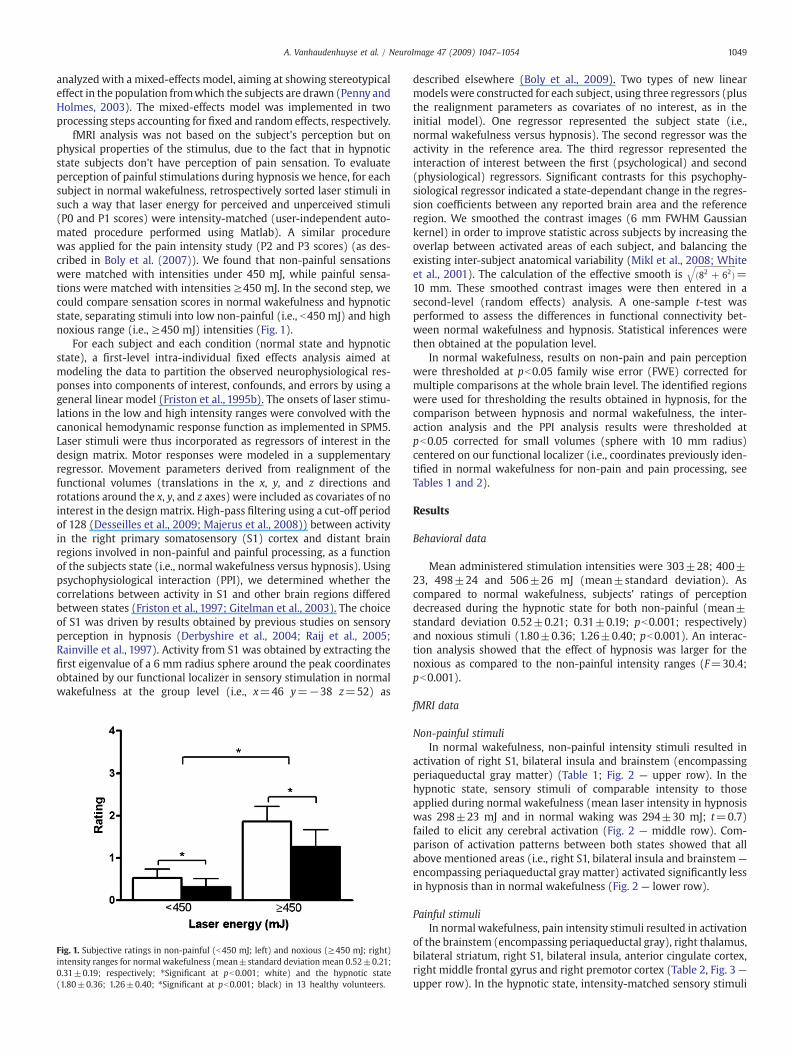
Fig. 1. Subjective ratings in non-painful (b450 mJ; left) and noxious (≥450 mJ; right)intensity ranges for normal wakefulness (mean±standard deviation mean 0.52±0.21;0.31±0.19; respectively; ⁎Significant at pb0.001; white) and the hypnotic state(1.80±0.36; 1.26±0.40; ⁎Significant at pb0.001; black) in 13 healthy volunteers.
described elsewhere (Boly et al., 2009). Two types of new linearmodels were constructed for each subject, using three regressors (plusthe realignment parameters as covariates of no interest, as in theinitial model). One regressor represented the subject state (i.e.,normal wakefulness versus hypnosis). The second regressor was theactivity in the reference area. The third regressor represented theinteraction of interest between the first (psychological) and second(physiological) regressors. Significant contrasts for this psychophy-siological regressor indicated a state-dependant change in the regres-sion coefficients between any reported brain area and the referenceregion. We smoothed the contrast images (6 mm FWHM Gaussiankernel) in order to improve statistic across subjects by increasing theoverlap between activated areas of each subject, and balancing theexisting inter-subject anatomical variability (Mikl et al., 2008; Whiteet al., 2001). The calculation of the effective smooth is
ffiffiffiffiffiffiffiffiffiffiffiffiffiffiffiffiffiffiffiffiffiffið82 + 62Þ
q=
10 mm. These smoothed contrast images were then entered in asecond-level (random effects) analysis. A one-sample t-test wasperformed to assess the differences in functional connectivity bet-ween normal wakefulness and hypnosis. Statistical inferences werethen obtained at the population level.
In normal wakefulness, results on non-pain and pain perceptionwere thresholded at pb0.05 family wise error (FWE) corrected formultiple comparisons at the whole brain level. The identified regionswere used for thresholding the results obtained in hypnosis, for thecomparison between hypnosis and normal wakefulness, the inter-action analysis and the PPI analysis results were thresholded atpb0.05 corrected for small volumes (sphere with 10 mm radius)centered on our functional localizer (i.e., coordinates previously iden-tified in normal wakefulness for non-pain and pain processing, seeTables 1 and 2).
Results
Behavioral data
Mean administered stimulation intensities were 303±28; 400±23, 498±24 and 506±26 mJ (mean±standard deviation). Ascompared to normal wakefulness, subjects' ratings of perceptiondecreased during the hypnotic state for both non-painful (mean±standard deviation 0.52±0.21; 0.31±0.19; pb0.001; respectively)and noxious stimuli (1.80±0.36; 1.26±0.40; pb0.001). An interac-tion analysis showed that the effect of hypnosis was larger for thenoxious as compared to the non-painful intensity ranges (F=30.4;pb0.001).
fMRI data
Non-painful stimuliIn normal wakefulness, non-painful intensity stimuli resulted in
activation of right S1, bilateral insula and brainstem (encompassingperiaqueductal gray matter) (Table 1; Fig. 2 — upper row). In thehypnotic state, sensory stimuli of comparable intensity to thoseapplied during normal wakefulness (mean laser intensity in hypnosiswas 298±23 mJ and in normal waking was 294±30 mJ; t=0.7)failed to elicit any cerebral activation (Fig. 2 — middle row). Com-parison of activation patterns between both states showed that allabove mentioned areas (i.e., right S1, bilateral insula and brainstem—
encompassing periaqueductal gray matter) activated significantly lessin hypnosis than in normal wakefulness (Fig. 2 — lower row).
Painful stimuliIn normal wakefulness, pain intensity stimuli resulted in activation
of the brainstem (encompassing periaqueductal gray), right thalamus,bilateral striatum, right S1, bilateral insula, anterior cingulate cortex,right middle frontal gyrus and right premotor cortex (Table 2, Fig. 3 —
upper row). In the hypnotic state, intensity-matched sensory stimuli

Table 2Peak voxels of pain activation in normal wakefulness and in the hypnotic state andregions showing significant differences between normal wakefulness and hypnosis.
Regions x(mm)
y(mm)
z(mm)
z-value p-value
Activation in normal wakefulnessBrainstem 0 −22 −14 5.91 b0.0001a
Right thalamus 6 −18 16 5.55 0.004a
Left striatum −14 10 8 5.33 0.012a
Right striatum 16 6 −2 5.67 0.002a
Left insula −60 −34 10 5.52 0.004a
Right insula 40 −14 14 5.28 0.01a
Right primary somatosensory cortex 48 −34 52 5.60 0.003a
Anterior cingulate cortex −6 18 44 5.41 0.008a
Right middle frontal gyrus 34 48 12 5.37 0.01a
Right premotor cortex 36 14 60 5.19 0.025a
Activation in the hypnotic stateNo brain activation was identified
Regions less activated in hypnosis compared to normal wakefulnessBrainstem 8 −18 14 4.07 0.003b
Right thalamus 10 −18 14 4.21 0.001b
Left striatum −14 12 10 5.15 b0.0001b
Right striatum 18 8 2 5.16 b0.0001b
Left insula −54 −40 6 3.89 0.003b
Right insula 34 −14 12 3.90 0.001b
Right primary somatosensory cortex 46 −40 52 3.87 0.003b
Anterior cingulate cortex −4 24 46 4.01 b0.001b
Right middle frontal gyrus 36 50 12 4.98 b0.0001b
Right premotor cortex 36 6 54 3.99 0.001b
Regions less activated in normal wakefulness compared to hypnosisNo brain activation was identified
a Family-wise error (FWE) corrected for multiple comparisons for the whole brainvolume.
b Small volume correction (10 mm sphere) for multiple comparisons based oncoordinates identified in normal wakefulness.
Table 1Peak voxels of non-painful activation in normal wakefulness and in the hypnotic stateand regions showing significant differences between wakefulness and hypnosis.
Regions x(mm)
y(mm)
z(mm)
z-value p-value
Activation in normal wakefulnessBrainstem −6 −22 −12 5.15 0.031a
Left insula −58 −34 12 5.86 0.001a
Right insula 46 −16 −4 5.55 b0.0001a
Right primary somatosensory cortex 46 −38 52 4.77 b0.0001b
Activation in the hypnotic stateNo brain activation was identified
Regions less activated in hypnosis compared to normal wakefulnessBrainstem 0 −22 −16 4.61 b0.0001b
Left insula −54 −42 8 4.34 b0.0001b
Right insula 46 −14 −6 4.83 b0.0001b
Right primary somatosensory cortex 48 −36 52 3.13 0.011b
Regions less activated in normal wakefulness compared to hypnosisNo brain activation was identified
a Family-wise error (FWE) corrected for multiple comparisons for the whole brainvolume.
b Small volume correction (10 mm sphere) for multiple comparisons based oncoordinates identified in normal wakefulness.
1050 A. Vanhaudenhuyse et al. / NeuroImage 47 (2009) 1047–1054
failed to elicit any cerebral activation (Fig. 3 — middle row).Comparison of activation patterns between both states showed thatall above mentioned areas (i.e., brainstem, right thalamus, bilateralstriatum, right S1, bilateral insula, anterior cingulate cortex, rightmiddle frontal gyrus and right premotor cortex) activated significantlyless in hypnosis than in normal wakefulness (Fig. 3 — lower row).
Interaction analysesBrain regions activating more in normal wakefulness as compared
to hypnosis during painful as compared to non-painful stimulationwere identified in thalamus, bilateral striatum and ACC (Table 3).Inversely, no brain area showed higher activity in hypnosis comparedto normal wakefulness in painful versus non-painful stimulation.
Psychophysiological interaction analysesFunctional connectivity was shown to be higher in hypnosis as
compared to normal wakefulness between right S1 and left insularand right prefrontal cortices (Table 4). Inversely, S1 functional con-nectivity was not higher in normal wakefulness as compared tohypnosis with any brain region.
Discussion
In the present study, differences between hypnotic and controlconditions should be considered. We here placed subjects into ahypnotic trance in the same way as our patients during surgery(Faymonville et al., 1995, 1997, 1999) but did not deliver anysuggestions for pain relief. However, presentation of the word‘hypnosis’ may automatically trigger lay beliefs and expectationsthat may have influenced our observed behavior (Gandhi and Oakley,2005; Hylands-White and Derbyshire, 2007). Even if we did notsuggest pain relief due to the hypnotic state, it cannot be excluded thatthis might have occurred implicitly. Another critical issue is thathypnosis was internally generated and that no output was requiredfrom our subjects. In consequence, we had to find a solution toascertain the presence of the hypnotic state. First, there was nodifference between clinical appearance of our subjects and patientsundergoing surgical interventions or burn debridement underhypnosis (Faymonville et al., 1995, 1997, 1999). Second, before startingof EPI scan sequences, subjects were requested to manifest, via aprearranged footmovement, that they actually felt themselves to be inhypnosis. Finally, they were interviewed afterwards about theirhypnotic experience. All subjects reported that they fell into ahypnotic state starting from the foot movement and remained in
this state throughout the scanning procedure. Subjects reportedhaving experienced vivid, detailed, and colorful revivifications ofpleasant memories, having actually mentally reenacted them. Beha-viorally, the effect of hypnosis on sensory perceptionwas significantlymore pronounced for pain (i.e., 38% decrease compared to normalwakefulness) as compared to its effect on non-pain ratings (i.e., 33%decrease). These results differ from Rainville et al. (1997) whoobserved no change induced by hypnosis on ratings of either painintensity or unpleasantness. They reported that only specific sugges-tion of increased or decreased unpleasantness changed the painratings. Our hypnosis induction technique decreased non-pain andpain ratings without using explicit suggestions related to the stimuli.
The fMRI data showed that stimuli in the non-painful rangeactivated brainstem, contralateral S1 and bilateral insular cortices. Inline with previous studies, painful range stimuli activated additionalareas encompassing right thalamus, bilateral striatum, anteriorcingulate, contralateral premotor and dorsolateral prefrontal cortices(for review see e.g., Tracey (2008)). In the hypnotic state, both non-painful and painful intensity-matched stimuli failed to elicit anycerebral activation. The interaction analysis identified that contral-ateral thalamus, bilateral striatum and ACC activated more in normalwakefulness versus hypnosis during painful as compared to non-painful stimulation. These results confirm the known role of ACC inpain perception (e.g., Coghill et al., 1994; Craig et al., 1996; Derbyshireet al., 1997; Laureys et al., 2002; Ploghaus et al., 1999) as well as itsmodulatory role in hypnosis-induced analgesia (Rainville et al., 1997,1999a,b; Faymonville et al., 2000, 2003). Our data also point tosubcortical targets of “hypnoanalgesia” in thalamus and striatum.Because the thalamus represents the major cortical relay for afferentpain fibers, its involvement is predicted by theories of hypnosisthat hypothesize inhibition of afferent sensory pain transmission(Faymonville et al., 2003). The thalamus (and ACC) was also shown tocorrelate with the magnitude of placebo-induced analgesia (Wageret al., 2004). Villemure and Bushnell (2009) recently showed

Fig. 2. Upper row. Brain regions showing significant (pb0.05) activation during non-painful stimulation (b450 mJ) in normal wakefulness (mean laser intensity 294±30 mJ). Fordisplay, results were thresholded at uncorrected pb0.00001 (note that all identified regions except S1 survived FWE correction for multiple comparisons). Middle row. In thehypnotic state, sensory stimuli of comparable intensity to those applied during normal wakefulness failed to elicit any cerebral activation. Lower row. Brain regions showingsignificant differences with activation induced by identical stimuli in the hypnotic state (mean laser intensity 298±23 mJ). For display, results were thresholded at uncorrectedpb0.001 (note that all identified regions survived SVC correction for multiple comparisons based on a priori knowledge obtained in normal wakefulness).
1051A. Vanhaudenhuyse et al. / NeuroImage 47 (2009) 1047–1054
involvement of thalamus and ACC in mood-related analgesia. Sincethe induction technique used in the present study in addition to beingdistracting, probably also alters emotions and attention, the observedchanges in brain activation could be a combination of modulation bymood and by attention. The basal ganglia are known to encode andinitiate basic movement patterns expressed through premotor path-ways and are frequently reported in neuroimaging studies of pain(Coghill et al., 1994; Derbyshire et al., 1997; Derbyshire and Jones,1998; Jones et al., 1991). They have also been proposed to support abasic attentional mechanism facilitating the calling up of motor pro-grams and thoughts (Brown and Marsden, 1998). Joint with theobserved decreases in premotor cortices activation in hypnosis, ourresults suggest that hypnosis, may diminish anxiety, defensive andemotional reactions to pain by reducing the activation of both cortical(encompassing ACC) and subcortical areas (encompassing thalamusand striatum) (Faymonville et al., 1997).
Finally, we demonstrated that hypnosis, compared to normalwakefulness, increased functional connectivity between S1 anddistant insular and prefrontal cortices, possibly reflecting top-down
modulation. The insula shows the most consistent activation infunctional imaging studies on pain perception (Coghill et al., 1994;Craig et al., 1996; Derbyshire et al., 1997; Jones et al., 1991; Laureyset al., 2002; Ploghaus et al., 1999) and takes an intermediate positionbetween the lateral (sensory-discriminative) and medial (affective-emotional) components of pain processing (Mesulam and Mufson,1982). Increased S1-insular modulation during hypnosis is in linewith its proposed role in pain affect (Rainville et al., 1999b) and painintensity coding (Craig et al., 2000). However, it is important to stressthat the used correlation analyses do not guarantee that the identi-fied S1-insular connectivity is direct (i.e., a third area, which showscontext-sensitive responses, may be providing input to the two areasimplicated in the PPI) nor does it inform on the directionality of thismodulation. The observed prefrontal areas may indicate distributedassociative processes of cognitive appraisal, attention or memory ofperceived stimuli (Courtney et al., 1998).Widespread frontal increasesin rCBF have previously been demonstrated in the hypnotic state(Faymonville et al., 2000; Maquet et al., 1999; Rainville et al., 1999b).Neuropsychological studies suggest that the efficiency of the frontal
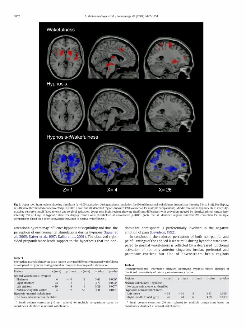
Fig. 3. Upper row. Brain regions showing significant (pb0.05) activation during noxious stimulation (≥450 mJ) in normal wakefulness (mean laser intensity 534±8mJ). For display,results were thresholded at uncorrected pb0.00001 (note that all identified regions survived FWE correction for multiple comparisons). Middle row. In the hypnotic state, intensity-matched sensory stimuli failed to elicit any cerebral activation. Lower row. Brain regions showing significant differences with activation induced by identical stimuli (mean laserintensity 532±14 mJ) in hypnotic state. For display, results were thresholded at uncorrected pb0.001 (note that all identified regions survived SVC correction for multiplecomparisons based on a priori knowledge obtained in normal wakefulness).
1052 A. Vanhaudenhuyse et al. / NeuroImage 47 (2009) 1047–1054
attentional systemmay influence hypnotic susceptibility and thus, theperception of environmental stimulations during hypnosis (Egner etal., 2005; Kaiser et al., 1997; Kallio et al., 2001). The observed right-sided preponderance lends support to the hypothesis that the non-
Table 3Interaction analysis identifying brain regions activated differently in normal wakefulnessas compared to hypnosis during painful as compared to non-painful stimulation.
Regions x (mm) y (mm) z (mm) z-value p-value
Normal wakefulnessNhypnosisThalamus 4 −10 12 3.43 0.009a
Right striatum 24 2 −6 3.76 0.004a
Left striatum −20 8 6 3.29 0.007a
Anterior cingulate cortex 4 12 58 3.31 0.019a
HypnosisNnormal wakefulnessNo brain activation was identified
a Small volume correction (10 mm sphere) for multiple comparisons based oncoordinates identified in normal wakefulness.
dominant hemisphere is preferentially involved in the negativeemotion of pain (Davidson, 1992).
In conclusion, the reduced perception of both non-painful andpainful ratings of the applied laser stimuli during hypnotic state com-pared to normal wakefulness is reflected by a decreased functionalactivation of not only anterior cingulate, insular, prefrontal andpremotor cortices but also of downstream brain regions
Table 4Psychophysiological interaction analyses identifying hypnosis-related changes infunctional connectivity of primary somatosensory cortex.
Regions x (mm) y (mm) z (mm) z-value p-value
Normal wakefulnessNhypnosisNo brain activation was identified
HypnosisNnormal wakefulnessLeft insula −64 −30 8 3.37 0.013a
Right middle frontal gyrus 26 48 6 3.09 0.033a
a Small volume correction (10 mm sphere) for multiple comparisons based oncoordinates identified in normal wakefulness.
1053A. Vanhaudenhuyse et al. / NeuroImage 47 (2009) 1047–1054
encompassing brainstem, thalamus, striatum and primary somato-sensory cortex. Hypnosis-induced analgesia related to a decreasedactivation of ACC, striatal and thalamic areas in pain as compared tonon-pain stimulation. Our findings point to a critical role for thecortical pain neuromatrix (encompassing ACC) but also of hierarchi-cally “lower-level” brain areas (encompassing the thalamus and basalganglia) in a hypnosis-induced decrease of sensory, affective,cognitive and behavioral aspects of sensory perception. Functionalconnectivity studies suggest an increased top-down modulation fromanterior insular and prefrontal areas on primary somatosensorycortex. It reinforces the idea that not only pharmacological but alsopsychological strategies for relieving pain can modulate the cortico-cortical networks that participate in the processing of sensory externalstimuli.
Acknowledgments
This research was funded by the Belgian National Funds forScientific Research (FNRS), the European Commission, the JamesMcDonnell Foundation, the Mind Science Foundation, the FrenchSpeaking Community Concerted Research Action (ARC-06/11-340),the Fondation Médicale Reine Elisabeth and the University of Liège. A.V. was funded by ARC 06/11-340, M.B. and E.B are research fellow atthe FNRS, S.L., P.M. and C.P. are senior research associate at the FNRS.
References
Augustine, J.R., 1996. Circuitry and functional aspects of the insular lobe in primatesincluding humans. Brain Res. Rev. 22, 229–244.
Boly, M., Balteau, E., Schnakers, C., Degueldre, C., Moonen, G., Luxen, A., Phillips, C.,Peigneux, P., Maquet, P., Laureys, S., 2007. Baseline brain activity fluctuations predictsomatosensory perception in humans. Proc. Natl. Acad. Sci. U. S. A.104,12187–12192.
Boly,M., Tshibanda, L., Vanhaudenhuyse,A.,Noirhomme,Q., Schnakers, C., Ledoux,D., et al.,2009. Functional connectivity in the default network during resting state is preservedin a vegetative but not in a brain dead patient. Hum. Brain Mapp. (April 6).
Brown, P., Marsden, C.D., 1998. What do the basal ganglia do? Lancet 351, 1801–1804.Buchel, C., Bornhovd, K., Quante, M., Glauche, V., Bromm, B., Weiller, C., 2002. Disso-
ciable neural responses related to pain intensity, stimulus intensity, and stimulusawareness within the anterior cingulate cortex: a parametric single-trial laserfunctional magnetic resonance imaging study. J. Neurosci. 22, 970–976.
Charlton, E., 1995. Ethical guidelines for pain research in humans. Committee onEthical Issues of the International Association for the Study of Pain. Pain 63,277–278.
Coghill, R.C., Talbot, J.D., Evans, A.C., Meyer, E., Gjedde, A., Bushnell, M.C., Duncan, G.H.,1994. Distributed processing of pain and vibration by the human brain. J. Neurosci.14, 4095–4108.
Courtney, S.M., Petit, L., Haxby, J.V., Ungerleider, L.G., 1998. The role of prefrontal cortexinworking memory: examining the contents of consciousness. Philos. Trans. R. Soc.Lond. B. Biol. Sci. 353, 1819–1828.
Craig, A.D., Bushnell, M.C., Zhang, E.T., Blomqvist, A., 1994. A thalamic nucleus specificfor pain and temperature sensation. Nature 372, 770–773.
Craig, A.D., Reiman, E.M., Evans, A., Bushnell, M.C., 1996. Functional imaging of anillusion of pain. Nature 384, 258–260.
Craig, A.D., Chen, K., Bandy, D., Reiman, E.M., 2000. Thermosensory activation of insularcortex. Nat. Neurosci. 3, 184–190.
Davidson, R.J., 1992. Anterior cerebral asymmetry and the nature of emotion. BrainCogn. 20, 125–151.
Defechereux, T., Meurisse, M., Hamoir, E., Gollogly, L., Joris, J., Faymonville, M.E., 1999.Hypnoanesthesia for endocrine cervical surgery: a statement of practice. J. Altern.Complement. Med. 5, 509–520.
Defechereux, T., Degauque, C., Fumal, I., Faymonville, M.E., Joris, J., Hamoir, E., Meurisse,M., 2000. Hypnosedation, a new method of anesthesia for cervical endocrinesurgery. Prospective randomized study. Ann. Chir. 125, 539–546.
Derbyshire, S.W., Jones, A.K., 1998. Cerebral responses to a continual tonic pain stimulusmeasured using positron emission tomography. Pain 76, 127–135.
Derbyshire, S.W., Jones, A.K., Gyulai, F., Clark, S., Townsend, D., Firestone, L.L., 1997. Painprocessing during three levels of noxious stimulation produces differential patternsof central activity. Pain 73, 431–445.
Derbyshire, S.W., Whalley, M.G., Stenger, V.A., Oakley, D.A., 2004. Cerebral activationduring hypnotically induced and imagined pain. NeuroImage 23 (1), 392–401.
Desseilles, M., Balteau, E., Sterpenich, V., Dang-Vu, T.T., Darsaud, A., Vandewalle, G.,Albouy, G., Salmon, E., Peters, F., Schmidt, C., Schabus, M., Gais, S., Degueldre, C.,Phillips, C., Luxen, A., Ansseau, M., Maquet, P., Schwartz, S., 2009. Abnormal neuralfiltering of irrelevant visual information in depression. J. Neurosci. 29, 1395–1403.
Egner, T., Jamieson, G., Gruzelier, J., 2005. Hypnosis decouples cognitive control fromconflict monitoring processes of the frontal lobe. NeuroImage 27, 969–978.
Elkins, G., Jensen, M.P., Patterson, D.R., 2007. Hypnotherapy for the management ofchronic pain. Int. J. Clin. Exp. Hypn. 55, 275–287.
Faymonville, M.E., Fissette, J., Mambourg, P.H., Roediger, L., Joris, J., Lamy, M., 1995.Hypnosis as adjunct therapy in conscious sedation for plastic surgery. Reg. Anesth.20, 145–151.
Faymonville, M.E., Mambourg, P.H., Joris, J., Vrijens, B., Fissette, J., Albert, A., Lamy, M.,1997. Psychological approaches during conscious sedation. Hypnosis versus stressreducing strategies: a prospective randomized study. Pain 73, 361–367.
Faymonville, M.E., Meurisse, M., Fissette, J., 1999. Hypnosedation: a valuable alternativeto traditional anaesthetic techniques. Acta Chir. Belg. 99, 141–146.
Faymonville, M.E., Laureys, S., Degueldre, C., DelFiore, G., Luxen, A., Franck, G., Lamy, M.,Maquet, P., 2000. Neural mechanisms of antinociceptive effects of hypnosis.Anesthesiology 92, 1257–1267.
Faymonville, M.E., Roediger, L., Del Fiore, G., Delgueldre, C., Phillips, C., Lamy, M., Luxen,A., Maquet, P., Laureys, S., 2003. Increased cerebral functional connectivityunderlying the antinociceptive effects of hypnosis. Cogn. Brain Res. 17, 255–262.
Frenay, M.C., Faymonville, M.E., Devlieger, S., Albert, A., Vanderkelen, A., 2001.Psychological approaches during dressing changes of burned patients: a prospec-tive randomised study comparing hypnosis against stress reducing strategy. Burns27, 793–799.
Friston, K.J., Ashburner, J., Frith, C., Poline, J., Heather, J., Frackowiak, R., 1995a. Spatialrealignment and normalization of images. Hum. Brain Mapp. 2, 165–189.
Friston, K.J., Holmes, A.P., Poline, J.B., Grasby, P.J., Williams, S.C., Frackowiak, R.S., Turner,R., 1995b. Analysis of fMRI time-series revisited. NeuroImage 2, 45–53.
Friston, K.J., Buechel, C., Fink, G.R., Morris, J., Rolls, E., Dolan, R.J., 1997. Psychophysio-logical and modulatory interactions in neuroimaging. NeuroImage 6, 218–229.
Gandhi, B., Oakley, D.A., 2005. Does ‘hypnosis’ by any other name smell as sweet? Theefficacy of ‘hypnotic’ inductions depends on the label ‘hypnosis’. Conscious Cogn.14, 304–315.
Gitelman, D.R., Penny, W.D., Ashburner, J., Friston, K.J., 2003. Modeling regional andpsychophysiologic interactions in fMRI: the importance of hemodynamic decon-volution. NeuroImage 19, 200–207.
Grondahl, J.R., Rosvold, E.O., 2008. Hypnosis as a treatment of chronic widespread painin general practice: a randomized controlled pilot trial. BMCMusculoskelet. Disord.9 (124).
Hilgard, E.R., Lauer, L.W., Morgan, A.H., 1963. Manual for Standard Profile Scales ofHypnotic Susceptibility. Forms I and II, Palo Alto, CA.
Hofbauer, R.K., Rainville, P., Duncan, G.H., Bushnell, M.C., 2001. Cortical representationof the sensory dimension of pain. J. Neurophysiol. 86, 402–411.
Hylands-White, N., Derbyshire, S., 2007. Modifying pain perception: is it better to behypnotisable or feel that you are hypnotised? Contemp. Hypn. 24, 143–153.
Jensen, M.P., Barber, J., Hanley, M.A., Engel, J.M., Romano, J.M., Cardenas, D.D., Kraft, G.H.,Hoffman, A.J., Patterson, D.R., 2008. Long-term outcome of hypnotic-analgesiatreatment for chronic pain in persons with disabilities. Int. J. Clin. Exp. Hypn. 56,156–169.
Jones, A.K., Brown, W.D., Friston, K.J., Qi, L.Y., Frackowiak, R.S., 1991. Cortical andsubcortical localization of response to pain in man using positron emission tomo-graphy. Proc. Biol. Sci. 244, 39–44.
Kaiser, J., Barker, R., Haenschel, C., Baldeweg, T., Gruzelier, J.H., 1997. Hypnosis andevent-related potential correlates of error processing in a stroop-type paradigm: atest of the frontal hypothesis. Int. J. Psychophysiol. 27, 215–222.
Kallio, S., Revonsuo, A., Hamalainen, H., Markela, J., Gruzelier, J., 2001. Anterior brainfunctions and hypnosis: a test of the frontal hypothesis. Int. J. Clin. Exp. Hypn. 49,95–108.
Laureys, S., Faymonville, M.E., Peigneux, P., Damas, P., Lambermont, B., Del Fiore, G.,Degueldre, C., Aerts, J., Luxen, A., Franck, G., Lamy, M., Moonen, G., Maquet, P., 2002.Cortical processing of noxious somatosensory stimuli in the persistent vegetativestate. NeuroImage 17, 732–741.
Majerus, S., Belayachi, S., De Smedt, B., Leclercq, A.L., Martinez, T., Schmidt, C., Weekes,B., Maquet, P., 2008. Neural networks for short-termmemory for order differentiatehigh and low proficiency bilinguals. NeuroImage 42, 1698–1713.
Maquet, P., Faymonville, M.E., Degueldre, C., Delfiore, G., Franck, G., Luxen, A., Lamy, M.,1999. Functional neuroanatomy of hypnotic state. Biol. Psychiatry 45, 327–333.
Melzack, R., Wall, P.D., 1965. Pain mechanisms: a new theory. Science 150, 971–979.Mesulam, M.M., Mufson, E.J., 1982. Insula of the old world monkey. III: Efferent cortical
output and comments on function. J. Comp. Neurol. 212, 38–52.Meurisse, M., 1999. Thyroid and parathyroid surgery under hypnosis: from fiction to
clinical application. Bull. Mem. Acad. R. Med. Belg. 154, 142–150 (discussion150–144).
Meurisse, M., Faymonville, M.E., Joris, J., Nguyen Dang, D., Defechereux, T., Hamoir, E.,1996. Endocrine surgery by hypnosis. From fiction to daily clinical application. Ann.Endocrinol. (Paris) 57, 494–501.
Mikl, M., Marecek, R., Hlustik, P., Pavlicova, M., Drastich, A., Chlebus, P., et al., 2008.Effects of spatial smoothing on fMRI group inferences. Magn. Reson. Imaging 26 (4),490–503.
Oneal, B.J., Patterson, D.R., Soltani, M., Teeley, A., Jensen, M.P., 2008. Virtual realityhypnosis in the treatment of chronic neuropathic pain: a case report. Int. J. Clin. Exp.Hypn. 56, 451–462.
Penny, W., Holmes, A., 2003. In: Frackowiak R.F.K., Frith C., Dolan R., Price C., Zeki S.,Ashburner J., Penny W. (Academic, London) (Ed.), Hum. Brain Funct., p. 1114.
Peyron, R., Laurent, B., Garcia-Larrea, L., 2000. Functional imaging of brain responses topain. A review and meta-analysis. Neurophysiol. Clin. 30, 263–288.
Ploghaus, A., Tracey, I., Gati, J.S., Clare, S., Menon, R.S., Matthews, P.M., Rawlins, J.N.,1999. Dissociating pain from its anticipation in the human brain. Science 284,1979–1981.
Ploner, M., Schmitz, F., Freund, H.J., Schnitzler, A., 2000. Differential organization oftouch and pain in human primary somatosensory cortex. J. Neurophysiol. 83,1770–1776.

1054 A. Vanhaudenhuyse et al. / NeuroImage 47 (2009) 1047–1054
Price, D.D., Milling, L.S., Kirsch, I., Duff, A., Montgomery, G.H., Nicholls, S.S., 1999. Ananalysis of factors that contribute to the magnitude of placebo analgesia in anexperimental paradigm. Pain 83, 147–156.
Raij, T.T., Numminen, J., Narvanen, S., Hiltunen, J., Hari, R., 2005. Brain correlates ofsubjective reality of physically and psychologically induced pain. Proc. Natl. Acad.Sci. U. S. A. 102 (6), 2147–2151.
Rainville, P., Duncan, G.H., Price, D.D., Carrier, B., Bushnell, M.C.,1997. Pain affect encodedin human anterior cingulate but not somatosensory cortex. Science 277, 968–971.
Rainville, P., Carrier, B., Hofbauer, R.K., Bushnell, M.C., Duncan, G.H., 1999a. Dissociation ofsensoryand affective dimensions of painusing hypnoticmodulation. Pain 82,159–171.
Rainville, P.,Hofbauer, R.K., Paus, T.,Duncan,G.H., Bushnell,M.C., Price,D.D.,1999b. Cerebralmechanisms of hypnotic induction and suggestion. J. Cogn. Neurosci. 11, 110–125.
Sharar, S.R., Miller, W., Teeley, A., Soltani, M., Hoffman, H.G., Jensen, M.P., Patterson, D.R.,2008. Applications of virtual reality for pain management in burn-injured patients.Expert Rev. Neurother. 8, 1667–1674.
The Executive Committee of the American Psychological Association — Division ofPsychological Hypnosis, 1994. Definition and description of hypnosis. Contemp.Hypn. 11, 142–162.
Timmermann, L., Ploner, M., Haucke, K., Schmitz, F., Baltissen, R., Schnitzler, A., 2001.Differential coding of pain intensity in the human primary and secondary somato-sensory cortex. J. Neurophysiol. 86, 1499–1503.
Tracey, I., 2008. Imaging pain. Br. J. Anaesth. 101, 32–39.Vanhaudenhuyse, A., Boly, M., Laureys, S., Faymonville, M.E., 2008. Neurophysiological
correlates of hypnotic analgesia. Contemp. Hypn. 25 (3).Villemure, C., Bushnell, M.C., 2009. Mood influences supraspinal pain processing sepa-
rately from attention. J. Neurosci. 29, 705–715.Wager, T.D., Rilling, J.K., Smith, E.E., Sokolik, A., Casey, K.L., Davidson, R.J., Kosslyn, S.M.,
Rose, R.M., Cohen, J.D., 2004. Placebo-induced changes in FMRI in the anticipationand experience of pain. Science 303, 1162–1167.
Wall, P.D., 1992. The placebo effect: an unpopular topic. Pain 51, 1–3.White, T., O'Leary, D., Magnotta, V., Arndt, S., Flaum, M., Andreasen, N.C., 2001. Anatomic
and functional variability: the effects of filter size in group fMRI data analysis.NeuroImage 13 (4), 577–588.
World Medical Association declaration of Helsinki, 1997. Recommendations guidingphysicians in biomedical research involving human subjects. JAMA 277,925–926.
Related Documents


![VISULAS YAG III...000000-1272-175-Inhalt0-GB-060613 Contents User manual VISULAS YAG III [000000 -1272 175 GA GB 060613]Appendix VISULAS YAG III stand-alone operation - Quick instructions](https://static.cupdf.com/doc/110x72/606e9018c22f4860603c0faf/visulas-yag-iii-000000-1272-175-inhalt0-gb-060613-contents-user-manual-visulas.jpg)