international Journal of Systematic Bacteriology (1 999), 49, 239-246 Printed in Great Britain Paenibacillus dendritiformis sp. nov., proposal for a new pattern-formingspecies and its localization within a phylogenetic cluster Marianna Tcherpakov,l Eshel Ben-Jacob’ and David L. Gutnick’ Author for correspondence: David Gutnick. Tel: +972 3 640 9834. Fax: +972 3 642 5786. e-mail: [email protected] Department of Molecular Microbiology and Biotechnology, George 5. Wise Faculty of Life Sciences’, and School of Physics and Astronomy, Raymond and Beverly Sackler Faculty of Exact Sciences*, Tel-Aviv University, Tel-Aviv 69978, Israel A new strain capable of forming distinctive patterns during colony development was identified by using a combination of phenotypic characterization,fatty acid analysis and analysis of the 16s rRNA gene sequence. The strain formed either a branched, tip-splitting colony morphology (referred to as the T morphotype)or a chiral pattern exhibiting thinner branches with distinctive curling patterns (referred to as the C morphotype). Isolates of the T morphotype exhibited sequence identities greater than 97 O/o to Paenibacillus thiaminolyticus JCM 7540. Phylogenetic analysis placed the T morphotypewithin the Paenibacillus cluster on a phylogenetictree. On the basis of unique colony morphology and distinctive phenotypiccharacteristics, it is proposed that the pattern-forming isolates should be placed within a new species of Paenibacillus, Paenibacillus dendritiformis sp. nov., the type strain of which is T168l(= 30A13. Keywords: Paenibacillus dendritiformis sp. nov., pattern formation, tip-splitting, chiral morphology INTRODUCTION Certain species among the Bacillaceae have been shown to produce a variety of complex patterns during colony development (Ben-Jacob, 1997 ; Ben-Jacob & Garick, 1990; Ben-Jacob et al., 1992,1994, 1995,1997, 1998a, b; Fox & Rossler, 1991 ; Kanzawa et al., 1995; Matsushita & Fujikawa, 1990; Matsushita et al., 1995; Matsuyama & Matsushita, 1993; Matsuyama et al., 1993). Although microbial patterns are often remark- ably similar to those observed in abiotic systems, such as snowflake formation or electrodeposition (Ben- Jacob, 1997; Ben-Jacob et al., 1994), the spectrum of shapes and forms of microbial colonies is much larger and more sensitive to changes in conditions. The major difference between the microbial system and abiotic examples stems from the fact that growing organisms modify their environment as they develop co- operatively, excreting various metabolites and sig- nalling molecules, changing the pH, extracting nu- trients from the surface substrate and so on. In order to develop working hypotheses regarding the bio- logical processes governing pattern formation, a gen- eric modelling approach has been developed that is based on close inspection of microbial behaviour and includes features related to cell growth, generation time and production of extracellular wetting fluid that allows the cells to move across the very hard agar surface, as well as various types of cell-cell signalling and communication (Ben-Jacob et al., 1994, 1998a, b; Gutnick & Ben-Jacob, 1998). The term morphotype is employed to describe the pattern-forming capacity of micro-organisms (Fujikawa & Matsushita, 1989 ; Ben- Jacob, 1997; Gutnick & Ben-Jacob, 1998). However, in order to gain a deeper understanding of the biological basis of pattern formation by various morphotypes, it is necessary to define the particular genes involved in pattern formation, their organization and control. This in turn depends on the identification of morphotypes. In this paper, the identification of two such pattern-forming isolates, their location on a phylogenetic tree and their phenotypic characteriz- ation are reported. METHODS The EMBL accession number for the 165 rRNA gene sequence of isolate T168T is Y16128. Bacterial strains and growth conditions. The pattern-forming isolates used in this study were termed T168T(tip-splitting 00811 0 1999 IUMS 239

Welcome message from author
This document is posted to help you gain knowledge. Please leave a comment to let me know what you think about it! Share it to your friends and learn new things together.
Transcript
Downloaded from www.microbiologyresearch.org by
IP: 23.22.50.124
On: Mon, 06 Jun 2016 23:23:05
international Journal of Systematic Bacteriology (1 999), 49, 239-246 Printed in Great Britain
Paenibacillus dendritiformis sp. nov., proposal for a new pattern-forming species and its localization within a phylogenetic cluster
Marianna Tcherpakov,l Eshel Ben-Jacob’ and David L. Gutnick’
Author for correspondence: David Gutnick. Tel: +972 3 640 9834. Fax: +972 3 642 5786. e-mail: [email protected]
Department of Molecular Microbiology and Biotechnology, George 5. Wise Faculty o f Life Sciences’, and School of Physics and Astronomy, Raymond and Beverly Sackler Faculty of Exact Sciences*, Tel-Aviv University, Tel-Aviv 69978, Israel
A new strain capable of forming distinctive patterns during colony development was identified by using a combination of phenotypic characterization, fatty acid analysis and analysis of the 16s rRNA gene sequence. The strain formed either a branched, tip-splitting colony morphology (referred to as the T morphotype) or a chiral pattern exhibiting thinner branches with distinctive curling patterns (referred to as the C morphotype). Isolates of the T morphotype exhibited sequence identities greater than 97 O/o to Paenibacillus thiaminolyticus JCM 7540. Phylogenetic analysis placed the T morphotype within the Paenibacillus cluster on a phylogenetic tree. On the basis of unique colony morphology and distinctive phenotypic characteristics, it is proposed that the pattern-forming isolates should be placed within a new species of Paenibacillus, Paenibacillus dendritiformis sp. nov., the type strain of which is T168l(= 30A13.
Keywords: Paenibacillus dendritiformis sp. nov., pattern formation, tip-splitting, chiral morphology
INTRODUCTION
Certain species among the Bacillaceae have been shown to produce a variety of complex patterns during colony development (Ben-Jacob, 1997 ; Ben-Jacob & Garick, 1990; Ben-Jacob et al., 1992,1994, 1995,1997, 1998a, b; Fox & Rossler, 1991 ; Kanzawa et al., 1995; Matsushita & Fujikawa, 1990; Matsushita et al., 1995; Matsuyama & Matsushita, 1993; Matsuyama et al., 1993). Although microbial patterns are often remark- ably similar to those observed in abiotic systems, such as snowflake formation or electrodeposition (Ben- Jacob, 1997; Ben-Jacob et al., 1994), the spectrum of shapes and forms of microbial colonies is much larger and more sensitive to changes in conditions. The major difference between the microbial system and abiotic examples stems from the fact that growing organisms modify their environment as they develop co- operatively, excreting various metabolites and sig- nalling molecules, changing the pH, extracting nu- trients from the surface substrate and so on. In order to develop working hypotheses regarding the bio-
logical processes governing pattern formation, a gen- eric modelling approach has been developed that is based on close inspection of microbial behaviour and includes features related to cell growth, generation time and production of extracellular wetting fluid that allows the cells to move across the very hard agar surface, as well as various types of cell-cell signalling and communication (Ben-Jacob et al., 1994, 1998a, b; Gutnick & Ben-Jacob, 1998). The term morphotype is employed to describe the pattern-forming capacity of micro-organisms (Fujikawa & Matsushita, 1989 ; Ben- Jacob, 1997; Gutnick & Ben-Jacob, 1998). However, in order to gain a deeper understanding of the biological basis of pattern formation by various morphotypes, it is necessary to define the particular genes involved in pattern formation, their organization and control. This in turn depends on the identification of morphotypes. In this paper, the identification of two such pattern-forming isolates, their location on a phylogenetic tree and their phenotypic characteriz- ation are reported.
METHODS
The EMBL accession number for the 165 rRNA gene sequence of isolate T168T is Y16128.
Bacterial strains and growth conditions. The pattern-forming isolates used in this study were termed T168T (tip-splitting
00811 0 1999 IUMS 239
Downloaded from www.microbiologyresearch.org by
IP: 23.22.50.124
On: Mon, 06 Jun 2016 23:23:05
M. Tcherpakov, E. Ben-Jacob and D. L. Gutnick
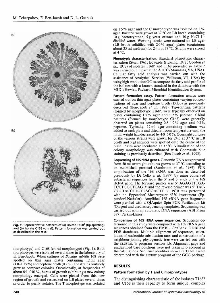
Fig. 1. Representative patterns of (a) isolate T1 68T (tip-splitting) and (b) isolate C168 (chiral). Pattern formation was carried out as described in the text.
morphotype) and C168 (chiral morphotype) (Fig. 1). Both morphotypes were isolated several times in the laboratory of E. Ben-Jacob. When cultures of Bacillus subtilis 168 were spotted on thin agar plates containing 12ml agar (1-0-1-75 YO) and peptone broth (0.2 YO), the strains routinely grew as compact colonies. Occasionally, at frequencies of about 0.1-0.01 O/O, bursts of growth exhibiting a new colony morphology emerged. Cells were picked from this new region of growth and restreaked on LB plates several times in order to purify isolates. The T morphotype was isolated
on 1.5 % agar and the C morphotype was isolated on 1 YO agar. Bacteria were grown at 37 "C on LB broth, containing 10 g bactotryptone, 5 g yeast extract and l o g NaCl 1-' distilled water. Working stocks were cultured on LB agar (LB broth solidified with 2.0% agar) plates (containing about 25 ml medium) for 24 h at 37 "C. Strains were stored at 4 "C. Phenotypic characterization. Standard phenotypic charac- terization (Steel, 1961 ; Edwards & Ewing, 1972; Gordon et al., 1973) of isolates T168T and C168 presented in Table 2 was carried out in part at the ATCC (Manassas, VA, USA). Cellular fatty acid analysis was carried out with the assistance of Analytical Services (Williston, VT, USA) by using high-resolution GC to compare the fatty acid profile of the isolates with a known standard in the database with the MIDI/Hewlett Packard Microbial Identification System. Pattern formation assay. Pattern formation assays were carried out on thin agar plates containing varying concen- trations of agar and peptone broth (Difco) as previously described (Ben-Jacob et al., 1992). Tip-splitting patterns (formed by morphotype T168T) were typically observed on plates containing 1.5% agar and 0.2% peptone. Chiral patterns (formed by morphotype C 168) were generally observed on plates containing 0-8-1-2% agar and 0.2% peptone. Typically, 12 ml agar-containing medium was added to each plate and dried at room temperature until the initial weight had decreased by 4-0-5.0 % . Overnight cultures of the various strains were grown for 24 h at 37 "C in LB broth and 5 pl aliquots were spotted onto the centre of the plate. Plates were incubated at 37 "C. Visualization of the colony morphology was enhanced with Coomassie blue staining as previously described (Ben-Jacob et al., 1992). Sequencing of 165 rRNA genes. Genomic DNA was prepared from 50 ml overnight cultures grown at 37 "C according to an established protocol (Sambrook et al., 1989). PCR amplification of the 16s rRNA was done as described previously by Di Cello et al. (1997) by using conserved eubacterial sequences from the 5' and 3' ends of the 16s rRNA gene. The forward primer was 5' AGAGTTTGA- TCCTGGCTCAG 3' and the reverse primer was 5' TAC- GGCTACCTTGTTACGACTT 3'. PCR was performed with an Eppendorf Mastercycler 5330 instrument (Ep- pendorf-Netheler). Amplified 16s rRNA gene fragments were purified with a QIAquick Spin PCR Purification kit (Qiagen) and used as sequencing templates. Sequencing was carried out with an automatic DNA sequencer (ABI Prism 377; Perkin-Elmer). Comparison of 165 rRNA gene sequences. Sequences de- termined in this study were compared with 16s rRNA gene sequences obtained from the EMBL, GenBank, DDBJ and PDB databases. Multiple alignment of sequences, calcu- lation of nucleotide substitution rates and construction of a neighbour-joining phylogenetic tree were carried out with the CLUSTAL w program version 1.6. Alignment gaps and unidentified base positions were not taken into account in the calculations. Sequence identities shown in Table 1 were determined with the BESTFIT program of the GCG package.
RESULTS
Pattern formation by T and C morphotypes
The distinguishing characteristic of the isolates TI 68T and C168 is their capacity to form unique, complex
240 International Journal of Systematic Bacteriology 49
Downloaded from www.microbiologyresearch.org by
IP: 23.22.50.124
On: Mon, 06 Jun 2016 23:23:05
Identification of pattern-forming morphotypes
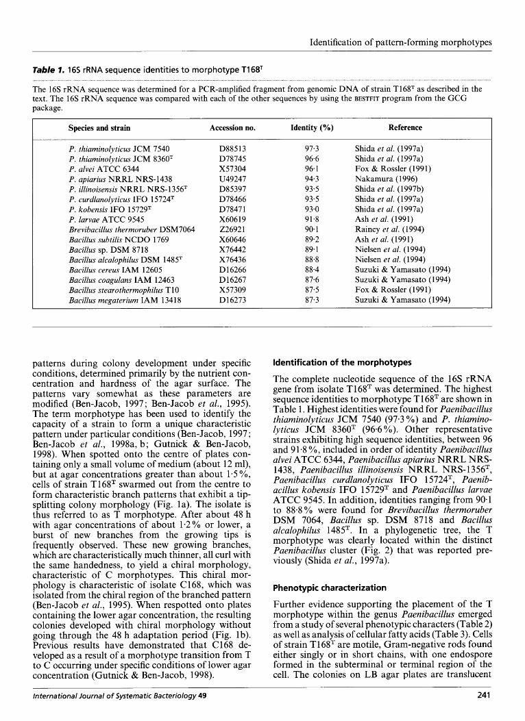
Table 1. 165 rRNA sequence identities to morphotype T1 68T
The 16s rRNA sequence was determined for a PCR-amplified fragment from genomic DNA of strain T16gT as described in the text. The 16s rRNA sequence was compared with each of the other sequences by using the BESTFIT program from the GCG package.
I Species and strain Accession no. Identity (%) Reference I P. thiaminolyticus JCM 7540 P. thiaminolyticus JCM 8360T P. alvei ATCC 6344 P. apiarius NRRL NRS-1438 P. illinoisensis NRRL NRS- 1 356T P. curdlanolyticus I F 0 1 5724T P. kobensis I F 0 15729T P. larvae ATCC 9545 Brevibacillus thermoruber DSM7064 Bacillus subtilis NCDO 1769 Bacillus sp. DSM 8718 Bacillus alcalophilus DSM 1485T Bacillus cereus IAM 12605 Bacillus coagulans IAM 12463 Bacillus stearothermophilus T 10 Bacillus megaterium IAM 1341 8
D885 13 D78745 X57304 U49247 D85397 D78466 D7847 1 X606 19 226921 X60646 X76442 X76436 D 16266 D 16267 X57309 D16273
97.3 96.6 96.1 94.3 93.5 93.5 93-0 91.8 90- 1 89.2 89.1 88-8 8 8-4 87-6 87-5 87.3
Shida et al. (1 997a) Shida et al. (1997a) Fox & Rossler (1991) Nakamura (1 996) Shida et al. (1 997b) Shida et al. (1997a) Shida et al. (1997a) Ash et al. (1991) Rainey et al. (1994) Ash et al. (1991) Nielsen et al. (1994) Nielsen et al. (1994) Suzuh & Yamasato (1994) Suzuki & Yamasato (1994) Fox & Rossler (1991) Suzuki & Yamasato (1994)
patterns during colony development under specific ldentif ication of the morphotypes conditions, determined- primarily by the nutrient con- centration and hardness of the agar surface. The patterns vary somewhat as these parameters are modified (Ben-Jacob, 1997; Ben-Jacob et al., 1995). The term morphotype has been used to identify the capacity of a strain to form a unique characteristic pattern under particular conditions (Ben-Jacob, 1997 ; Ben-Jacob et al., 1998a, b; Gutnick & Ben-Jacob, 1998). When spotted onto the centre of plates con- taining only a small volume of medium (about 12 ml), but at agar concentrations greater than about 1-5%, cells of strain T168T swarmed out from the centre to form characteristic branch patterns that exhibit a tip- splitting colony morphology (Fig. la). The isolate is thus referred to as T morphotype. After about 48 h with agar concentrations of about 1.2% or lower, a burst of new branches from the growing tips is frequently observed. These new growing branches, which are characteristically much thinner, all curl with the same handedness, to yield a chiral morphology, characteristic of C morphotypes. This chiral mor- phology is characteristic of isolate C168, which was isolated from the chiral region of the branched pattern (Ben-Jacob et al., 1995). When respotted onto plates containing the lower agar concentration, the resulting colonies developed with chiral morphology without going through the 48 h adaptation period (Fig. lb). Previous results have demonstrated that C 168 de- veloped as a result of a morphotype transition from T to C occurring under specific conditions of lower agar concentration (Gutnick & Ben-Jacob, 1998).
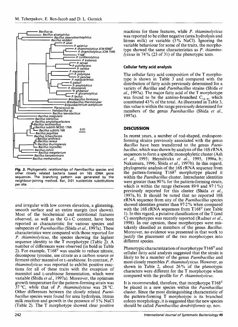
The complete nucleotide sequence of the 16s rRNA gene from isolate T168T was determined. The highest sequence identities to morphotype T 1 68T are shown in Table 1. Highest identities were found for Paenibacillus thiarninolyticus JCM 7540 (97-3 YO) and P. thiarnino- lyticus JCM 8360T (96.6 YO). Other representative strains exhibiting high sequence identities, between 96 and 91.8 %, included in order of identity Paenibacillus alvei ATCC 6344, Paenibacillus apiarius NRRL NRS- 1438, Paenibacillus illinoisensis NRRL NRS-1 35fiT, Paenibacillus curdlanolyticus I F 0 1 5724T, Paenib- acillus kobensis I F 0 1 5729T and Paenibacillus larvae ATCC 9545. In addition, identities ranging from 90-1 to 88-8 YO were found for Brevibacillus therrnoruber DSM 7064, Bacillus sp. DSM 8718 and Bacillus alcalophilus 1485T. In a phylogenetic tree, the T morphotype was clearly located within the distinct Paenibacillus cluster (Fig. 2) that was reported pre- viously (Shida et al., 1997a).
Phenotypic characterization
Further evidence supporting the placement of the T morphotype within the genus Paenibacillus emerged from a study of several phenotypic characters (Table 2) as well as analysis of cellular fatty acids (Table 3). Cells of strain T168T are motile, Gram-negative rods found either singly or in short chains, with one endospore formed in the subterminal or terminal region of the cell. The colonies on LB agar plates are translucent
International Journal of Systematic Bacteriology 49 241
Downloaded from www.microbiologyresearch.org by
IP: 23.22.50.124
On: Mon, 06 Jun 2016 23:23:05
M. Tcherpakov, E. Ben-Jacob and D. L. Gutnick
I? alvei I? apiarius I? thiaminolyticus JCM 8360T
F! thiaminolyticus JCM 7540
I I? macerans
I? glucanolyticus
Planococcus sp. Halobacillus sp. Ba ci llus la e vola cticus
0.0 1 Bacillus subtilis 168 - Bacillus cereus Baci!lus thuringiensis Bacillus mycoides
Bacillus cohnii Bacillus megaterium
Bacillus benzeovorans c Bacillus methanolicus
. . . . . . I.. . . . . . . . . . . . . . . . . . . . . . . . . . . . . . . . . . . . . . . . . . . . , . . . . . . . . . . . . . . . . . . . . . . . . . . . . . . . . . . . . . . . . . . . . . . . . . . . . . . . . . . . . . . . . . . . . . . . . . . . . . . . . . . . . . . . . . . . . Fig. 2. Phylogenetic relationships of Paenibacillus species and other closely related bacteria based on 165 rDNA gene sequences. The branching pattern was generated by the neighbour-joining method. Bar, 0.01 nucleotide substitutions per site.
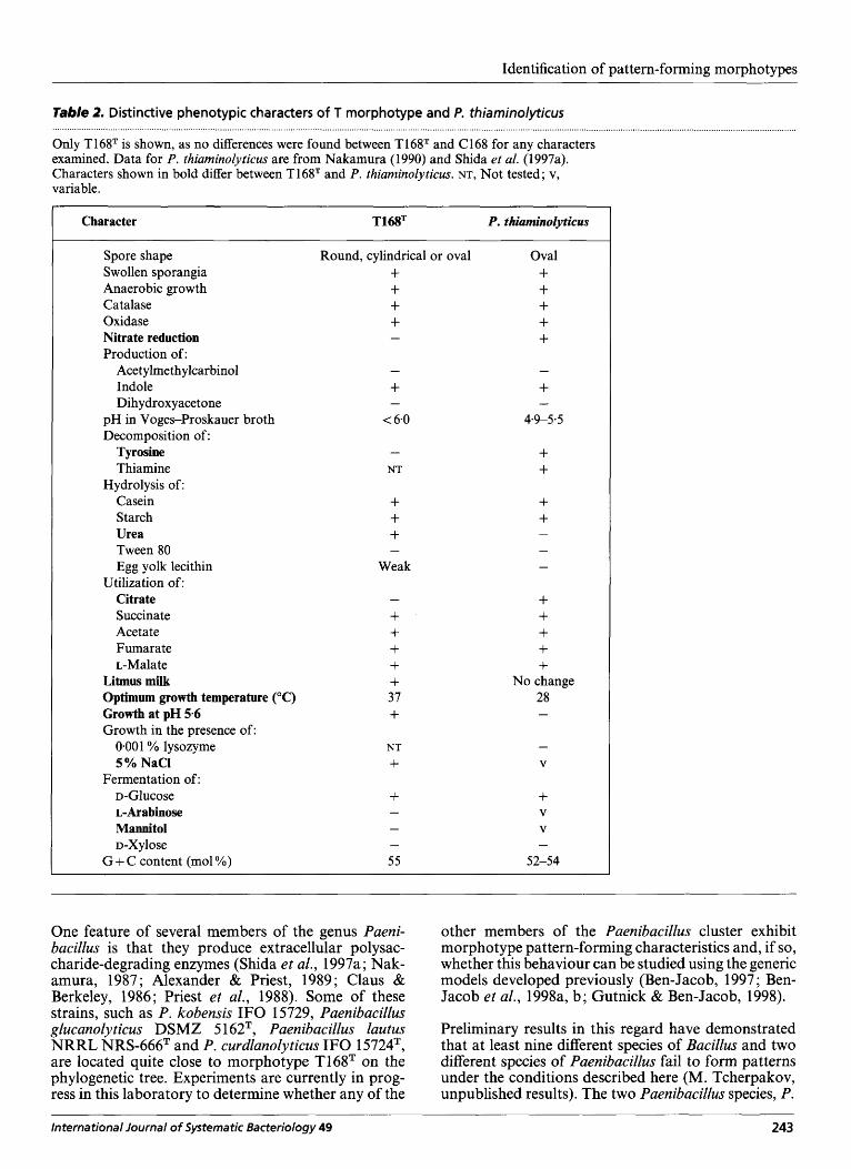
and irregular with low convex elevation, a glistening, smooth surface and an entire margin (not shown). Most of the biochemical and nutritional features observed, as well as the G + C content, have been reported as characteristic for various species and subspecies of Paenibacillus (Shida et al., 1997a). These characteristics were compared with those reported for P. thiarninolyticus, the species showing the highest sequence identity to the T morphotype (Table 2). A number of differences were observed (in bold in Table 2). For example, T168T was unable to reduce nitrate, decompose tyrosine, use citrate as a carbon source or ferment either mannitol or L-arabinose. In contrast, P. thiarninolyticus was reported to exhibit positive reac- tions for all of these traits with the exception of mannitol and L-arabinose fermentation, which were variable (Shida et al., 1997a). Moreover, the optimum growth temperature for the pattern-forming strain was 37 "C, while that of P. thiarninolyticus was 28 "C. Other differences between the closely related Paeni- bacillus species were found for urea hydrolysis, litmus milk reaction and growth in the presence of 5 % NaCl (Table 2). The T morphotype showed clear positive
reactions for these features, while P. thiarninolyticus was reported to be either negative (urea hydrolysis and litmus milk) or variable ( 5 % NaCl). Ignoring the variable behaviour for some of the traits, the morpho- type showed the same characteristics as P. thiamino- lyticus in 74 % (23 of 31) of the phenotypic tests.
Cellular fatty acid analysis
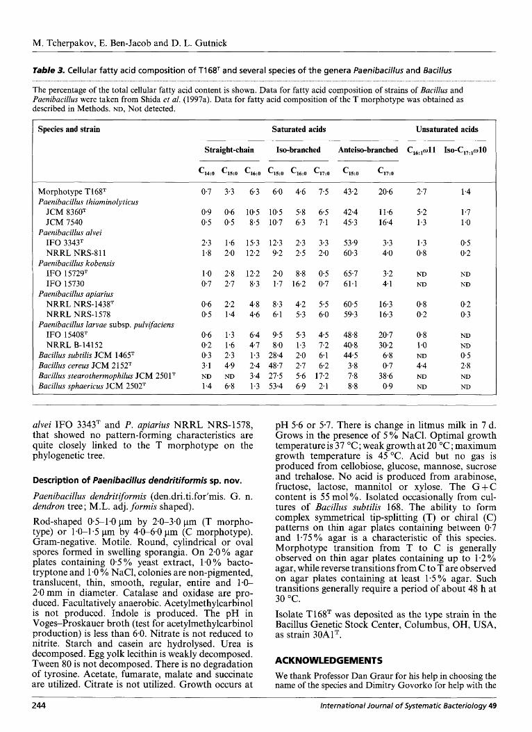
The cellular fatty acid composition of the T morpho- type is shown in Table 3 and compared with the distribution of fatty acids previously determined for a variety of Bacillus and Paenibacillus strains (Shida et al., 1997a). The major fatty acid of the T morphotype was found to be the anteiso-branched which constituted 43 % of the total. As illustrated in Table 3, this value is within the range previously determined for members of the genus Paenibacillus (Shida et al., 1997a).
DISCUSSION
In recent years, a number of rod-shaped, endospore- forming strains previously associated with the genus Bacillus have been transferred to the genus Paeni- bacillus, which was shown by analysis of the 16s rRNA sequences to form a specific monophyletic cluster (Ash et al., 1993; Heyndrickx et al., 1995, 1996a, b; Nakamura, 1996; Shida et al., 1997b). In this regard, phylogenetic analysis of the 16s rRNA sequence from the pattern-forming T16tIT morphotype placed it within the Paenibacillus cluster. Intracluster identities were greater than 90 % for the pattern-forming strain, which is within the range (between 89.9 and 97.1 %) previously reported for this cluster (Shida et al., 1997a, b). It should be noted that no reported 16s rRNA sequence from any of the Paenibacillus species showed identities greater than 97.2 % when compared with the 16s rRNA sequences from T168T (see Table 1). In this regard, a putative classification of the T (and C) morphotypes was recently reported (Rudner et al., 1998). In our opinion, these morphotypes were mis- takenly classified as members of the genus Bacillus. Moreover, no evidence was presented in that work to justify the placement of the two morphotypes into different species. Phenotypic characterization of morphotype TI 6ST and cellular fatty acid analysis suggested that the strain is likely to be a member of the genus Paenibacillus and most closely resembles P. thiaminolyticus. However, as shown in Table 2, about 26% of the phenotypic characters were different for the T morphotype when compared with the profile for P. thiarninolyticus.
It is recommended, therefore, that morphotype T 1 68T be placed in a new species within the Paenibacillus cluster. Since the most distinguishing characteristic of the pattern-forming T morphotype is its branched colony morphology, it is suggested that the new species should be called Paenibacillus dendritiforrnis sp. nov.
242 International Journal of Systematic Bacteriology 49
Downloaded from www.microbiologyresearch.org by
IP: 23.22.50.124
On: Mon, 06 Jun 2016 23:23:05
Identification of pattern-forming morphotypes
Table 2. Distinctive phenotypic characters of T morphotype and P. thiaminolyticus
Only T168T is shown, as no differences were found between T16gT and C168 for any characters examined. Data for P. thiaminoZyticus are from Nakamura (1990) and Shida et al. (1997a). Characters shown in bold differ between T168T and P. thiaminolyticus. NT, Not tested; v, variable.
1 Character T168= P. thiaminolyticus
Spore shape Round, cylindrical or oval Swollen sporangia + Anaerobic growth + Catalase + Oxidase + Nitrate reduction -
Production of: Ace t ylme t h ylcarbinol Indole + Dihydroxyacetone -
Tyrosine -
-
pH in Voges-Proskauer broth Decomposition of:
< 6-0
Thiamine NT Hydrolysis of:
Casein + Starch + Urea + Tween 80 -
Egg yolk lecithin Weak
Citrate -
Succinate + Acetate + Fumarate + L-Malate +
Litmus milk + Optimum growth temperature ("C) 37 Growth at pH 5.6 +
Utilization of:
Growth in the presence of: 0.001 % lysozyme NT 5% NaCl + D-Glucose + L-Arabinose - Mannitol - D-Xylose -
55
Fermentation of:
G + C content (mol %)
Oval + + + + +
+ + + + -
+ + + + +
No change 28 -
-
V
+ V
V -
52-54
One feature of several members of the genus Paeni- bacillus is that they produce extracellular polysac- charide-degrading enzymes (Shida et al., 1997a; Nak- amura, 1987; Alexander & Priest, 1989; Claus & Berkeley, 1986; Priest et al., 1988). Some of these strains, such as P. kobensis IF0 15729, Paenibacillus glucanolyticus DSMZ 5 1 62T, Paenibacillus lautus NRRL NRS-666T and P. curdlanolyticus IF0 1 5724T, are located quite close to morphotype T16ST on the phylogenetic tree. Experiments are currently in prog- ress in this laboratory to determine whether any of the
other members of the Paenibacillus cluster exhibit morphotype pattern-forming characteristics and, if so, whether this behaviour can be studied using the generic models developed previously (Ben-Jacob, 1997 ; Ben- Jacob et al., 1998a, b; Gutnick & Ben-Jacob, 1998).
Preliminary results in this regard have demonstrated that at least nine different species of Bacillus and two different species of Paenibacillus fail to form patterns under the conditions described here (M. Tcherpakov, unpublished results). The two Paenibacillus species, P.
International Journal of Systematic Bacteriology 49 243
Downloaded from www.microbiologyresearch.org by
IP: 23.22.50.124
On: Mon, 06 Jun 2016 23:23:05
M. Tcherpakov, E. Ben-Jacob and D. L. Gutnick
Table 3. Cellular fatty acid composition of T168T and several species of the genera Paenibacillus and Bacillus
The percentage of the total cellular fatty acid content is shown. Data for fatty acid composition of strains of Bacillus and Paenibacillus were taken from Shida et al. (1997a). Data for fatty acid composition of the T morphotype was obtained as described in Methods. ND, Not detected.
Species and strain Saturated acids Unsaturated acids
Straight-chain Iso-branched
'14:O '15:O '16:O '15:O '16:O '17:O
Anteiso-branched
Morphotype T168T Paenibacillus thiam inoly t icus
JCM 8360T JCM 7540
IF0 3343T Paenibacillus alvei
NRRL NRS-8 1 1 Paenibacillus kobensis
IF0 15729T IF0 15730
Paenibacillus apiarius NRRL NRS-1438T NRRL NRS- 1578
Paenibacillus larvae subsp. pulvifaciens IF0 15408T NRRL B-14152
Bacillus subtilis JCM 1465T Bacillus cereus JCM 2 1 52T Bacillus stearothermophilus JCM 250 lT Bacillus sphaericus JCM 2502T
0.7 3.3 6.3 6.0 4.6 7-5
0.9 0.6 10.5 10.5 5.8 6.5 0.5 0.5 8-5 10.7 6.3 7.1
2.3 1.6 15.3 12.3 2.3 3.3 1.8 2.0 12.2 9.2 2.5 2-0
1.0 2.8 12.2 2.0 8.8 0.5 0.7 2.7 8.3 1.7 16.2 0.7
0.6 2.2 4.8 8.3 4.2 5.5 0.5 1.4 4.6 6.1 5.3 6.0
0.6 1.3 6.4 9.5 5.3 4.5 0.2 1.6 4-7 8.0 1.3 7.2 0.3 2.3 1.3 28.4 2.0 6.1 3.1 4.9 2.4 48.7 2-7 6.2
1.4 6.8 1.3 53.4 6-9 2.1 ND ND 3.4 27.5 5.6 17.2
43-2 20.6
42.4 11.6 45.3 16.4
53.9 3.3 60.3 4.0
65.7 3.2 61.1 4.1
60.5 16.3 59.3 16.3
48.8 20.7 40.8 30.2 44.5 6.8
3.8 0-7 7.8 38-6 8.8 0.9
2.7 1.4
5.2 1.7 1.3 1.0
1.3 0.5 0.8 0.2
ND ND ND ND
0.8 0.2 0.2 0.3
0.8 ND 1.0 ND ND 0.5 4-4 2.8 ND ND ND ND
alvei I F 0 3343T and P. apiarius NRRL NRS-1578, that showed no pattern-forming characteristics are quite closely linked to the T morphotype on the phylogenetic tree.
pH 5.6 or 5.7. There is change in litmus milk in 7 d. Grows in the presence of 5 % NaCl. Optimal growth temperature is 37 "C; weak growth at 20 "C; maximum growth temperature is 45 "C. Acid but no gas is produced from cellobiose, glucose, mannose, sucrose
Description of Paenibadhs dendritiformis sp. nov. and trehalose. No acid is produced from arabinose, fructose, lactose, mannitol or xylose. The G + C
Paenibacillus dendritiformis (den.dri.ti.for'mis. G. n. dendron tree; M.L. adj. formis shaped).
content -is 55 mol %. Isolated occ&ionally from cul- tures of Bacillus subtilis 168. The ability to form
Rod-shaped 0-5-1.0 pm by 2.0-3.0 pm (T morpho- type) or 1-0-1.5 pm by 4.0-6.0 pm (C morphotype). Gram-negative. Motile. Round, cylindrical or oval spores formed in swelling sporangia. On 2.0% agar plates containing 0.5 % yeast extract, 1-0 % bacto- tryptone and 1.0 YO NaCl, colonies are non-pigmented, translucent, thin, smooth, regular, entire and 1.0- 2.0 mm in diameter. Catalase and oxidase are pro- duced. Facultatively anaerobic. Acetylmethylcarbinol is not produced. Indole is produced. The pH in Voges-Proskauer broth (test for acetylmethylcarbinol production) is less than 6-0. Nitrate is not reduced to nitrite. Starch and casein are hydrolysed. Urea is decomposed. Egg yolk lecithin is weakly decomposed. Tween 80 is not decomposed. There is no degradation of tyrosine. Acetate, fumarate, malate and succinate are utilized. Citrate is not utilized. Growth occurs at
complex symmetrical tip-splitting (T) or -chiral (C) patterns on thin agar plates containing between 0.7 and 1.75% agar is a characteristic of this species. Morphotype transition from T to C is generally observed on thin agar plates containing up to 1.2% agar, while reverse transitions from C to T are observed on agar plates containing at least 1.5% agar. Such transitions generally require a period of about 48 h at 30 "C. Isolate T168T was deposited as the type strain in the Bacillus Genetic Stock Center, Columbus, OH, USA, as strain 30AlT.
ACKNOWLEDGEMENTS
We thank Professor Dan Graur for his help in choosing the name of the species and Dimitry Govorko for help with the
244 In tern a tion a I lo urn a I of Systematic Bacteriology 49
Downloaded from www.microbiologyresearch.org by
IP: 23.22.50.124
On: Mon, 06 Jun 2016 23:23:05
Identification of pattern-forming morphotypes
CLUSTAL program. We acknowledge the excellent technical assistance of Rina Avigad and Inna Brainis. Thanks are also extended to Inon Cohen for his outstanding contributions to the early development of morphotype characterization and modelling. This work was supported by grant no. 95-00410 from the US-Israel Binational Science Foundation and grant no. 593-95 from the Israel Science Foundation.
REFERENCES
Alexander, B. & Priest, F. G. (1989). Bacillus glucanolyticus, a new species that degrades a variety of P-glucans. Int J Syst Bacteriol
Ash, C., Farrow, J. A. E., Wallbanks, 5. & Collins, M. D. (1991). Phylogenetic heterogeneity of the genus Bacillus revealed by comparative analysis of small subunit ribosomal RNA se- quences. Lett A p p f Microbioll3, 202-206. Ash, C., Priest, F. G. & Collins, M. D. (1993). Molecular iden- tification of rRNA group 3 bacilli (Ash, Farrow, Wallbanks, and Collins) using a PCR probe test. Proposal for the creation of a new genus Paenibacillus. Antonie Leeuwenhoek 64,253-260. Ben-Jacob, E. (1997). From snowflake formation to growth of bacterial colonies. Part 11. Cooperative formation of colonial patterns. Contemp Phvs 38, 205-241. Ben-Jacob, E. & Garik, P. (1990). The formation of patterns in non-equilibrium growth. Nature 343, 523-530. Ben-Jacob, E., Shmueli, H., Shochet, 0. & Tenenbaum, A. (1992). Adaptive self-organization during growth of bacterial colonies. Physica A 187, 378424. Ben-Jacob, E., Shochet, 0.. Tenenbaum, A., Cohen, I., Czirok, A. & Vicsek, T. (1 994). Generic modelling of cooperative growth patterns in bacterial colonies. Nature 368, 4 W 9 . Ben-Jacob, E., Cohen, I., Shochet, O., Czirok, A. & Vicsek, T. (1 995). Cooperative formation of chiral patterns during growth of bacterial colonies. Phys Rev Lett 75, 2899-2902. Ben-Jacob, E., Cohen, I., Czirok, A., Vicsek, T. & Gutnick, D. L. (1 997). Chemomodulation of cellular movement, collective formation of vortices by swarming bacteria, and colonial development. Phjqsica A 238, 18 1-197. Ben-Jacob, E., Cohen, 1. & Gutnick, D. L. (1998a). Cooperative organization of bacterial colonies : from genotype to morpho- type. Annu Rev Microbiol52, 779-806. Ben-Jacob, E., Cohen, 1. & Gutnick, D. L. (1998b). Chemotaxis- based self-organization during colonial morphogenesis. Com- ments Mol Cell Biophjls (in press). Claus, D. & Berkeley, R. C. W. (1986). Genus Bacillus Cohn 1872. In Bergey’s Manual of Systematic Bacteriology, vol. 2, pp. 1105-1140. Edited by P. H. A. Sneath, N. S. Mair, M. E. Sharpe & J. G. Holt. Baltimore: Williams & Wilkins. Di Cello, F., Pepi, M., Baldi, F. & Fani, R. (1997). Molecular characterization of an n-alkane-degrading bacterial community and identification of a new species, Acinetobacter venetianus. Res Microbiol 148, 237-249. Edwards, P. R. & Ewing, W. H. (1972). Identzjication of Entero- bacteriaceae. Minneapolis : Burgess. Fox, G. E. & Rossler, D. (1991). Phylogenetic diversity in the genus Bacillus as seen by 16s RNA sequencing studies. Syst Appl Microbioll4, 266269. Fujikawa, H. & Matsushita, M. (1989). Fractal growth of Bacillus subtilis on agar plates. J Phys Soc Jpn 58, 3875-3878. Gordon, R. E., Haynes, W. C. & Pang, C. H. (1973). The Genus
39, 112-115.
In terna tiona I lo urna I of Systematic Bacteriology 49
Bacillus. Agriculture Handbook no. 427. Washington, DC : US Department of Agriculture. Gutnick, D. L. & Ben-Jacob, E. (1998). Complex pattern formation and cooperative organization of bacterial colonies. In Microbial Ecology and Infectious Diseases. Edited by E. Rosenberg. Washington, DC : American Society for Microbiology (in press). Heyndrickx, M., Vandemeulebroecke, K., Scheldeman, P. and 7 other authors (1 995). Paenibacillzrs (formerly Bacillus) gordonae (Pichinoty et al. 1986) Ash et al. 1994 is a later subjective synonym of Paenibacillus (formerly Bacillus) validus (Nak- amura 1984) Ash et al. 1994: emended description of P. validus. Int J Syst Bacteriol45, 661-669. Heyndrickx, M., Vandemeulebroecke, K., Hoste, B., Janssen, P., Kersters, K., De Vos, P., Logan, N. A., Ali, N. & Berkeley, R. C. W. (1 996a). Reclassification of Paenibacillus (formerly Bacillus) pulvifaciens (Nakamura 1984) Ash et al. 1994, a later subjective synonym of Paenibncillus (formerly Bacillus) larvae (White 1906) Ash et al. 1994, as a subspecies of P. larvae, with emended descriptions of P. larvae as P. larvae subsp. larvae and P. larvae subsp. pulvifaciens. Int J Syst Bacteriof 46, 270-279. Heyndrickx, M., Vandemeulebroecke, K., Scheldeman, P., Kersters, K., De Vos, P., Logan, N. A,, Aziz, A. M., Ali, N. & Berkeley, R. C. W. (1996b). A polyphasic reassessment of the genus Paen ibacillus, reclassification of Bacillus Iau t us (Na ka- mura 1984) as Paenibacillus lautus comb. nov. and of Bacillus peoriae (Montefusco et al. 1993) as Paenibacilluspeoriae comb. nov., and emended descriptions of P. lautus and of P. peoriae. Int J Syst Bacteriol46, 988-1003. Kanzawa, Y., Harada, A., Takeuchi, M., Yokota, A. & Harada, T. (1 995). Bacillus curdlanolyticus sp. nov. and Bacillus kobensis sp. nov., which hydrolyse resistant curdlan. Int J Syst Bacteriol45,
Matsushita, M. & Fujikawa, H. (1990). Diffusion-limited growth in bacterial colony formation. Physica A 168, 498-506. Matsushita, M., Wakita, J.4. & Matsuyama, T. (1995). Growth and morphological changes of bacterial colonies. In Spatio- Temporal Patterns in Nonequilibrium Complex Systems, pp. 609-618. Santa-Fe Institute Studies in the Sciences of Com- plexity. Edited by P. E. Cladis & P. Palffy-Muhoray. New York: Addison-Weseley. Matsuyama, T. & Matsushita, M. (1 993). Fractal morphogenesis by a bacterial cell population. Crit Rev Microbiol 19, 117-135. Matsuyama, T., Harshey, R. M. & Matsushita, M. (1993). Self- similar colony morphogenesis by bacteria as the experimental model of fractal growth by a cell population. Fractclls 1,
Nakamura, L. K. (1987). Bacillus alginolyticus sp. nov. and Bacillus chondroitinus sp. nov., two alginate-degrading species. Int J Syst Bacteriol37, 284-286. Nakamura, L. K. (1990). Bacillus thiarninolyticus sp. nov., nom. rev. Int J Syst Bacteriol40, 242-246. Nakamura, L. K. (1996). Paenibacillus apiarius sp. nov. Int J Syst Bacteriol46, 688-693. Nielsen, P., Rainey, F. A., Outtrup, H., Priest, F. G. & Fritze, D. (1994). Comparative 16s rDNA sequence analysis of some alkaliphilic bacilli and the establishment of a sixth rRNA group within the genus Bacillus. FEMS Microbiol Lett 117, 61-66. Priest, F. G., Goodfellow, M. & Todd, C. (1988). A numerical classification of the genus Bacillus. J Gen Microbiol 134,
Rainey, F. A., Fritze, D. & Stackebrandt, E. (1994). The phylo-
245
515-521.
302-3 1 1 .
1847-1 882.
Downloaded from www.microbiologyresearch.org by
IP: 23.22.50.124
On: Mon, 06 Jun 2016 23:23:05
M. Tcherpakov, E. Ben-Jacob and D. L. Gutnick
genetic diversity of thermophilic members of the genus Bacillus as revealed by 16s rDNA analysis. FEMS Microbiol Lett 115,
Rudner, R., Martsinkevich, O., Leung, W. & Jarvis, E. D. (1998). Classification and genetic characterization of pattern-forming bacilli. Mol Microbiol27, 687-703. Sambrook, J., Fritsch, E. F. & Maniatis, T. (1989). Molecular Cloning: a Laboratory Manual, 2nd edn. Cold Spring Harbor, NY : Cold Spring Harbor Laboratory. Shida, O., Takagi, H., Kadowaki, K., Nakamura, L. K. & Komagata, K. (1 997a). Transfer of Bacillus alginolyticus, Bacillus chondro- it in us, Bacillus cur dlan oly ticus, Bacillus glucanoly ticus, Bacillus
205-21 1.
kobensis, and Bacillus thiaminolyticus to the genus Paenibacillus and emended description of the genus Paenibacillus. Int J Syst Bacteriol47, 289-298. Shida, O., Takagi, H., Kadowaki, K., Nakamura, L. K. & Komagata, K. (1997b). Emended description of Paenibacillus amylolyticus and description of Paenibacillus illinoisensis sp. nov. and Paenibacillus chibensis sp. nov. Int J Syst Bacteriol47,299-306. Steel, K. 1. (1961). The oxidase reaction as a taxonomic tool. J Gen Microbiol25, 297-306. Suzuki, T. & Yamasato, K. (1994). Phylogeny of spore-forming lactic acid bacteria based on 16s rRNA gene sequence. FEMS Microbiol Lett 115, 13-17.
246 International Journal of Systematic Bacteriology 49
Related Documents