1 Paediatric Dentistry Fifth Stage Dr. Suhair W. Abbood Lec. 4 The Dental Caries in Child and Adolescent Dental caries continues to be a major problem in dentistry and should receive significant attention in everyday practice, not only from the standpoint of restorative procedures but also in terms of preventive practices designed to reduce the problem. It can be said that the major work of the dental profession is controlled by this disease process and yet many clinicians have a poor understanding of the mechanisms by which caries is initiated, how to identify patients at risk, and how to put management plans in place to ensure that the disease does not progress. Too often only the outcomes of the carious process are treated and not the cause of the disease itself. It is a multifactorial disease, resulting from the interplay between environmental, behavioural and genetic factors. Although we have observed a decline in caries prevalence for many years, it is clear that dental caries still remains the most prevalent disease afflicting humans. In general and according to WHO (World Health Organization), the dental caries define as a bacterial disease of the dental hard tissues, it begins with acid demineralization of the outer enamel surface, and if not arrested or treated, the dissolution of the enamel continues into the dentin and pulp increasing cavitation and loss of tooth substance. Dental caries is a process that may take place on any tooth surface in the oral cavity where dental plaque is allowed to develop over a period of time. Fermentable carbohydrate and cariogenic plaque need to be present on a tooth surface for acid to form. The acid is produce by bacterial metabolism of the carbohydrate substrate.

Welcome message from author
This document is posted to help you gain knowledge. Please leave a comment to let me know what you think about it! Share it to your friends and learn new things together.
Transcript
1
Paediatric Dentistry
Fifth Stage
Dr. Suhair W. Abbood
Lec. 4
The Dental Caries in Child and Adolescent
Dental caries continues to be a major problem in dentistry and should receive
significant attention in everyday practice, not only from the standpoint of restorative
procedures but also in terms of preventive practices designed to reduce the problem.
It can be said that the major work of the dental profession is controlled by this
disease process and yet many clinicians have a poor understanding of the
mechanisms by which caries is initiated, how to identify patients at risk, and how to
put management plans in place to ensure that the disease does not progress. Too often
only the outcomes of the carious process are treated and not the cause of the disease
itself.
It is a multifactorial disease, resulting from the interplay between
environmental, behavioural and genetic factors. Although we have observed a decline
in caries prevalence for many years, it is clear that dental caries still remains the most
prevalent disease afflicting humans.
In general and according to WHO (World Health Organization), the dental
caries define as a bacterial disease of the dental hard tissues, it begins with acid
demineralization of the outer enamel surface, and if not arrested or treated, the
dissolution of the enamel continues into the dentin and pulp increasing cavitation and
loss of tooth substance.
Dental caries is a process that may take place on any tooth surface in the oral
cavity where dental plaque is allowed to develop over a period of time. Fermentable
carbohydrate and cariogenic plaque need to be present on a tooth surface for acid to
form. The acid is produce by bacterial metabolism of the carbohydrate substrate.
2
Today, all experts on dental caries generally agree that it is an infections and
communicable disease and that multiple factors influence the initiation and
progression of the disease.
The disease is recognized to require :-
1- A host (tooth in the oral environment)
2- A dietary substrate.
3- Aciduric bacteria.
Over time the presence of the substrate serves as a nutrient for the bacteria, and
the bacteria produce acids that can demineralise the tooth.
The main features of the caries process are :-
1-Fermentation of carbohydrate to organic acids by micro-organism in plaque
on the tooth surface.
2-Rapid acid formation, which lowers the pH at the enamel surface below the
level (the critical pH) at which enamel will dissolve.
3-When carbohydrate is no longer available to the plaque micro-organisms, the
pH within plaque will raise due to the outward diffusion of acids and their
metabolism and neutralization in plaque, so that remineralization of enamel can
occur.
4-Dental caries progresses only when demineralization is greater than
remineralization. The realization that demineralization and remineralization is
equilibrium is the key to understanding the dynamic of the carious lesion and
its prevention.
One of the interesting features of an early carious lesion of the enamel is that
the lesion is subsurface, because, the outer surface of enamel is far more resistant to
3
demineralization by acid than the deeper portion of enamel is. That is, most of the
mineral loss occurs 10 to 15 µm beneath a relatively intact enamel surface.
The explanation for the intact surface layer in enamel caries seems to lie in
diffusion dynamics, the layer of dental plaque on the tooth surface acting as a partial
barrier to diffusion.
This contrasts strongly with the histological appearance of enamel after a clean
tooth surface has been exposed to acid, where the surface is etched and there is no
subsurface lesion. This dissolution of the surface of enamel, or etching, is a feature of
enamel erosion caused, among other things, by dietary acids. Further erosion occurs
at much lower pH (< 4) than caries.
Dental plaque forms on uncleaned tooth surfaces, it may be present on all teeth,
whether susceptible or immune to dental caries, this film that exists primarily in the
susceptible areas of the teeth has received a great deal of attention.
The dental plaque is readily apparent if tooth brushing is stopped for 2-3 days.
Contrary to popular opinion, plaque does not consist of food debris, but comprises 70
% microorganisms, about 100 million organisms per milligram of plaque. When
plaque is young, cocci predominate but, as plaque age the proportions of filamentous
organisms and veillonellae increase.
Diet influences the composition of the plaque flora considerably, with mutans
streptococci much more numerous when the diet is rich in sugar and other
carbohydrates, and these organisms are particularly good at metabolizing sugars to
acids.
Knowledge of the dental caries process increased considerably with the
development of pH electrodes, particularly microelectrodes that could be inserted into
plaque before, during, and after the ingestion of various foods. The pioneer of this
area of research was Robert Stephan, and the plot of plaque pH against time has
become known as the Stephan curve. Within 2-3 min of eating sugar or rinsing with a
sugar solution, plaque pH falls from an average of about 6.8 to near pH 5, taking
about 40 min to return to its original value. Below pH 5.5 demineralization of the
enamel occurs, this is known as the critical pH.
The clinical appearance of these early lesions is now well recognized, they
appears as a white area that coincides with the distribution of plaque. This might be
around the gingival margin, or between the teeth. If the process of dental caries
4
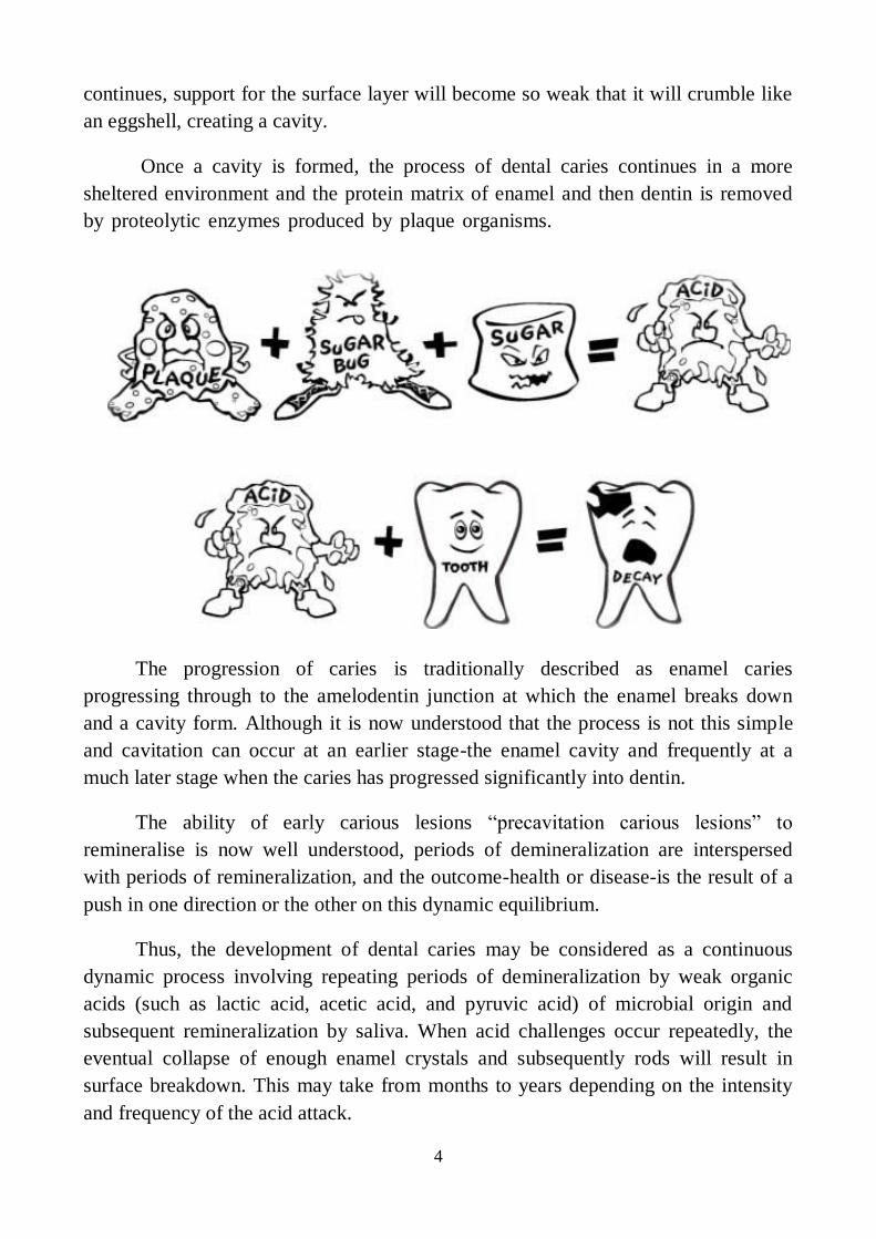
continues, support for the surface layer will become so weak that it will crumble like
an eggshell, creating a cavity.
Once a cavity is formed, the process of dental caries continues in a more
sheltered environment and the protein matrix of enamel and then dentin is removed
by proteolytic enzymes produced by plaque organisms.
The progression of caries is traditionally described as enamel caries
progressing through to the amelodentin junction at which the enamel breaks down
and a cavity form. Although it is now understood that the process is not this simple
and cavitation can occur at an earlier stage-the enamel cavity and frequently at a
much later stage when the caries has progressed significantly into dentin.
The ability of early carious lesions “precavitation carious lesions” to
remineralise is now well understood, periods of demineralization are interspersed
with periods of remineralization, and the outcome-health or disease-is the result of a
push in one direction or the other on this dynamic equilibrium.
Thus, the development of dental caries may be considered as a continuous
dynamic process involving repeating periods of demineralization by weak organic
acids (such as lactic acid, acetic acid, and pyruvic acid) of microbial origin and
subsequent remineralization by saliva. When acid challenges occur repeatedly, the
eventual collapse of enough enamel crystals and subsequently rods will result in
surface breakdown. This may take from months to years depending on the intensity
and frequency of the acid attack.
5
This means that in all mouths (as most mouths will contain some cariogenic
bacteria) there is continual demineralization and remineralization of enamel,
therefore, an individual is never free of dental caries, and the term “caries free” often
used to describe a child with no visible decay is best changed to the term “caries
inactive” to more accurately reflect this clinical reality.
The dental caries typically begins in enamel, in the early stage the progression
of the disease is slowly and the cavitation of the tooth structure is quite a late stage of
the disease. Prior to cavitation, the progress of the disease may be arrested and/ or
reversed if a favorable oral environment can be achieved.
The process of enamel demineralization and remineralization is constantly
cycling between net loss and gain of mineral. It is only when the balance leans
towards net loss that clinically identifiable signs of the process become apparent. For
the balance to be maintained there should be sufficient time between cariogenic
challenges for the remineralization process to take place. When these challenges
become too frequent, or occur when salivary flow is reduced, the rate of
demineralization and subsequent tooth breakdown will increase.
The time required for remineralization to replace the hydroxyapatite lost during
demineralization (the long-term outcome of this cycling) is determined by :-
1-The composition, the amount, and the age of the plaque.
2-The nature of the carbohydrate consumed, sugar consumption (frequency and
timing).
3-The presence or absence of fluoride (fluoride exposure).
4-Salivary flow and quality.
5-Enamel quality.
6-Immune response.
For example, it has been suggested that in the presence of dental plaque that
has developed for 12 hours or less, the enamel demineralization resulting from a
single exposure to sucrose will be remineralised by saliva within about 10 minutes. In
contrast, a period of at least 4 hours is required by saliva to repair the damage to
enamel resulting from a similar exposure to sucrose in the presence of dental plaque
that is 48 or more hours old.
The shorter the time during which plaque covered teeth are exposed to acid
attack and the longer the time remineralization can occur, the greater is the
opportunity for a carious lesion to heal. Satisfactory healing of the carious lesion can
only occur if the surface layer is unbroken, and this is why the “precavitation” stage
6
in the process of dental caries is so relevant to preventive dentistry. Once the surface
has been broken and a cavity has formed, it is usually necessary to restore the tooth
surface with a filling.
The carious process is driven by the plaque on the surface and therefore it is
possible to arrest the caries by effective removal of plaque even after cavitation has
occurred. In the arrested lesions, if the pulp is not involved and if the cavitation area
is open enough to be self-cleansing (plaque-free), the caries process can heal and
become an (arrested lesion). The arrested carious lesions typically exhibit :-
1- Much coronal destruction.
2- The remaining exposed dentine is hard and usually very dark.
3- There is no evidence of pulpal damage.
4- The patient has no pain.
However, the lost tissue cannot be replaced. The first stage of dental caries to
be visible is the “white spot” precavitation lesion stage. This can occur within a few
weeks if conditions are favourable to its development. In the general population,
though, it commonly takes 2-4 years for caries to progress through enamel into dentin
at proximal sites.
It is important to know that the treatment of a carious tooth by providing a
restoration does not cure the disease, because if the unfavourable oral condition that
cause the cavity persists, this will mean the continuity of the caries progression and
more restoration will be required in time.
Thus, the treatment of the dental caries will additionally need:-
1- Reducing the number of cariogenic microorganisms
2- Establishing a favourable oral environment to promote predominantly
remineralization of tooth structures over time that by turn may stop the
caries process and cure the disease.
Curing the disease currently requires modifications by the patient and/ or
caretaker and relies on their compliance in making the necessary modifications.
Research efforts are on-going to find a feasible method of achieving caries immunity
that would be far less dependent on patient compliance.
A number of microorganisms can produce enough acid to decalcify tooth
structures, particularly aciduric streptococci, lactobacilli, diphtheroids, yeasts,
staphylococci, and certain strains of sarcinae.
7
Streptococcus mutans has been implicated as one of the major and most
virulent of the caries producing organisms. The mother is the most common source of
transmission of the bacteria to the child, as many investigations found that reducing
the numbers of oral S. mutans in mothers will delay the colonisation of the organisms
in the mouth of their children, while when the earlier transmission occurs, the higher
risk of caries progression present.
Regarding the field of bacteriology, it is found that children who consumed
sweetened beverages in their baby bottle were four times more likely to have mutans
streptococci than children who only consumed milk. The acids that initially decalcify
the enamel have a pH of 5.2 to 5.5 or less and are formed in the plaque material,
which has been described as an organic nitrogenous mass of microorganisms firmly
attached to the tooth structure.
This film that exists primarily in the susceptible areas of the teeth, has received
a great deal of attention. Considerable emphasis is currently being given to plaque
and its relationship to the oral disease. The acids involved in the initiation of the
caries process are normal metabolic by-products of the microorganisms and are
generated by the metabolism of carbohydrates.
The most important of the natural defenses against dental caries is saliva. If
salivary flow is impaired, dental caries can progress very rapidly. Saliva has many
functions; the presence of food in the mouth is a powerful stimulus to salivation, with
strong-tasting acid foods being the best stimulants. Saliva is excreted at different rates
and with different constituents depending on the presence or absence of stimulatory
factors.
Saliva stimulated by chewing has increased calcium and phosphate ion
concentrations. A gustatory effect, such as that induced by some food acids, has been
shown to stimulate a higher flow rate of saliva than stimulation by mechanical
chewing. Fast-flowing saliva is alkaline-reaching pH values of 7.5-8.0, and is vitally
important in raising the pH of dental plaque previously lowered by exposure to sugar
and carbohydrates.
Saliva not only physically removes dietary substrates and acids produced by
plaque from the mouth, but it has a most important role in buffering the pH in saliva
and within plaque. By removing substrate and buffering plaque acid, saliva helps to
balance the caries process and has a critical role in remineralization as it provides a
stabilized supersaturated solution of calcium and phosphate ions as well as fluoride
ions from extrinsic sources. Because teeth consist largely of calcium and phosphate,
8
the concentration of calcium and phosphate in saliva and plaque is thought to be
important in determining the progression or regression of caries.
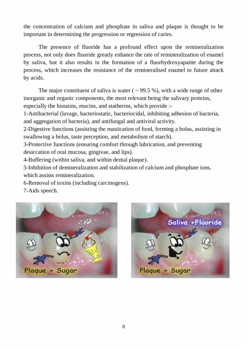
The presence of fluoride has a profound effect upon the remineralization
process, not only does fluoride greatly enhance the rate of remineralization of enamel
by saliva, but it also results in the formation of a fluorhydroxyapatite during the
process, which increases the resistance of the remineralised enamel to future attack
by acids.
The major constituent of saliva is water ( ~ 99.5 %), with a wide range of other
inorganic and organic components, the most relevant being the salivary proteins,
especially the histatins, mucins, and statherins, which provide :-
1-Antibacterial (lavage, bacteriostatic, bacteriocidal, inhibiting adhesion of bacteria,
and aggregation of bacteria), and antifungal and antiviral activity.
2-Digestive functions (assisting the mastication of food, forming a bolus, assisting in
swallowing a bolus, taste perception, and metabolism of starch).
3-Protective functions (ensuring comfort through lubrication, and preventing
desiccation of oral mucosa, gingivae, and lips).
4-Buffering (within saliva, and within dental plaque).
5-Inhibition of demineralization and stabilization of calcium and phosphate ions,
which assists remineralization.
6-Removal of toxins (including carcinogens).
7-Aids speech.
9
Secondary Factors in Causing the Dental Caries :-
Clinical observation and laboratory investigation often support the theory that
dental caries is influenced by a number of secondary factors, which are:-
1-Anatomic Characteristics of the Teeth:- The teeth of many patients, particularly
permanent teeth, seem predisposed to dental caries and may show evidence of the
attack almost coincident with their eruption into the oral cavity. Because enamel
calcification is incomplete at the time of eruption of the teeth and an additional period
of about 2 years is required for the calcification process to be completed by exposure
to saliva, the teeth are especially susceptible to caries formation during the first 2
years after eruption.
First permanent molars often have incompletely coalesced pits and fissures that
allow the dental plaque material to be retained at the base of the defect in contact
with exposed dentin. These defects or anatomic characteristics can readily be seen if
the tooth is dried and the debris and plaque material are removed with a sharp
explorer point.
Lingual pits on the maxillary first permanent molars, buccal pits on the mandibular
first permanent molars, and lingual pits on the maxillary incisors are vulnerable areas
in which the process of dental caries can proceed rapidly.
2-Arrangment of the Teeth in the Arch:- Crowded and irregular teeth are not
readily cleansed during the natural masticatory process. It is likewise difficult for the
patient to clean the mouth properly with a toothbrush if the teeth are crowded or
overlapped. This condition therefore may contribute to the problem of dental
caries.
3-Presence of Dental Appliances:- Partial dentures, space maintainers, and
orthodontic appliances often encourage the retention of food debris and plaque
material and have been shown to result in an increase in the bacterial population. Few
patients keep their mouths meticulously clean, and even those who make an attempt
may be hampered by the presence of dental appliances that retain plaque material
between brushings.
Patients who have had moderate dental caries activity in the past might be expected
to have increased caries activity after the placement of appliances in the mouth unless
they practice unusually good oral hygiene.
4-Hereditary Factors:- Although parents of children with excessive or rampant
caries have a tendency to blame the condition on hereditary factors or tendencies,
there is little scientific evidence to support this contention.
The fact that children acquire their dietary habits, oral hygiene habits, and oral
microflora from their parents makes dental caries more an environmental than a
hereditary disease. Although tooth morphology and enamel defects tend to follow a
10
familial pattern, and therefore heredity may play an indirect role since caries
susceptible surfaces (anatomically) may be genetically produced.
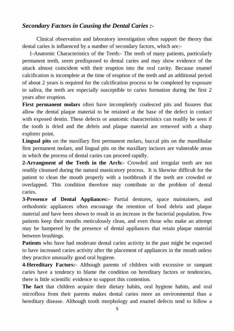
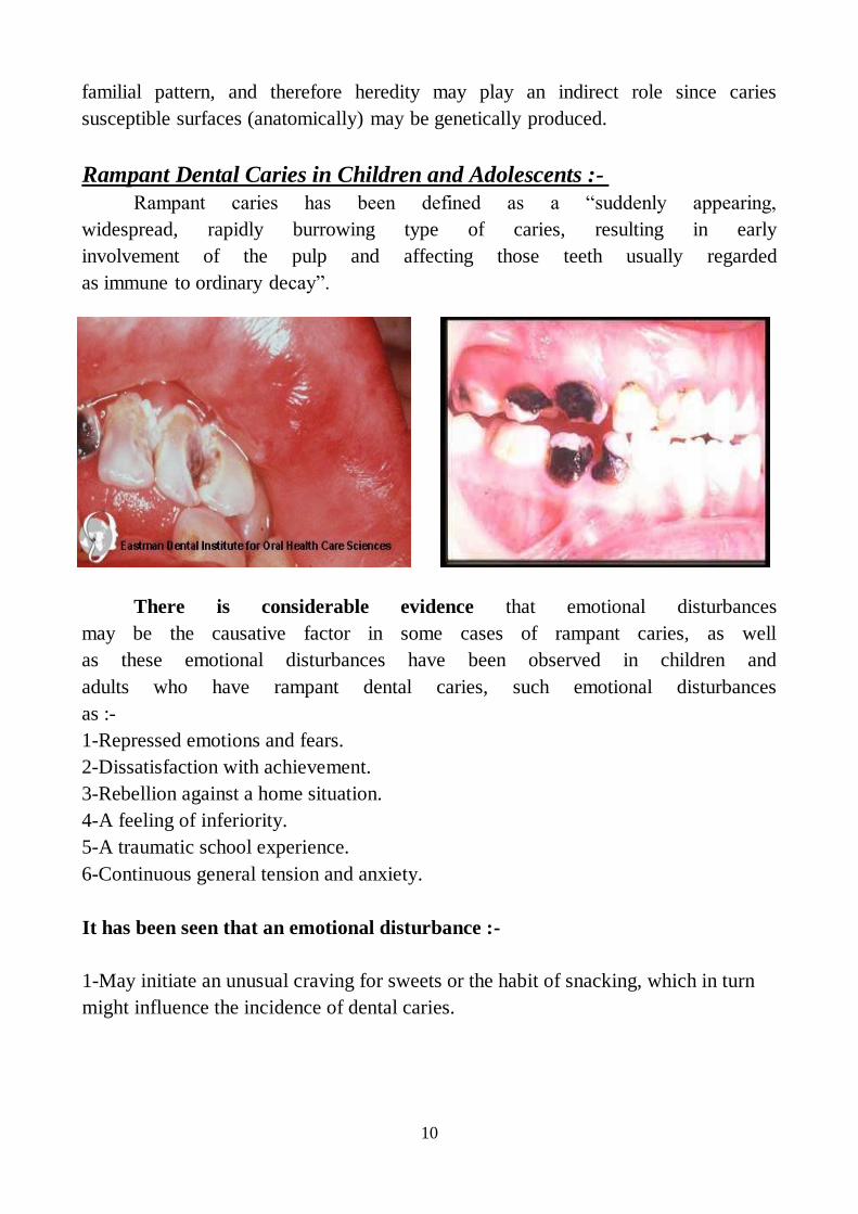
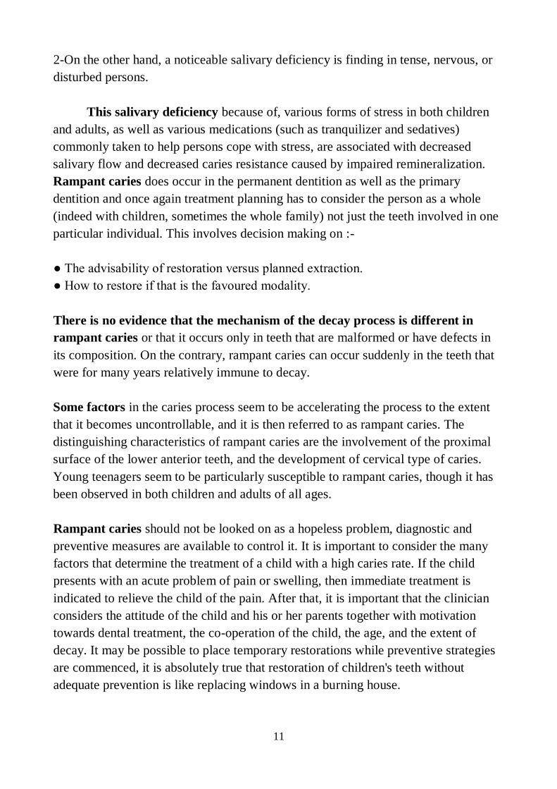
-Rampant Dental Caries in Children and Adolescents :
Rampant caries has been defined as a “suddenly appearing,
widespread, rapidly burrowing type of caries, resulting in early
involvement of the pulp and affecting those teeth usually regarded
as immune to ordinary decay”.
There is considerable evidence that emotional disturbances
may be the causative factor in some cases of rampant caries, as well
as these emotional disturbances have been observed in children and
adults who have rampant dental caries, such emotional disturbances
as :-
1-Repressed emotions and fears.
2-Dissatisfaction with achievement.
3-Rebellion against a home situation.
4-A feeling of inferiority.
5-A traumatic school experience.
6-Continuous general tension and anxiety.
It has been seen that an emotional disturbance :-
1-May initiate an unusual craving for sweets or the habit of snacking, which in turn
might influence the incidence of dental caries.
11
2-On the other hand, a noticeable salivary deficiency is finding in tense, nervous, or
disturbed persons.
This salivary deficiency because of, various forms of stress in both children
and adults, as well as various medications (such as tranquilizer and sedatives)
commonly taken to help persons cope with stress, are associated with decreased
salivary flow and decreased caries resistance caused by impaired remineralization.
Rampant caries does occur in the permanent dentition as well as the primary
dentition and once again treatment planning has to consider the person as a whole
(indeed with children, sometimes the whole family) not just the teeth involved in one
particular individual. This involves decision making on :-
● The advisability of restoration versus planned extraction.
● How to restore if that is the favoured modality.
There is no evidence that the mechanism of the decay process is different in
rampant caries or that it occurs only in teeth that are malformed or have defects in
its composition. On the contrary, rampant caries can occur suddenly in the teeth that
were for many years relatively immune to decay.
Some factors in the caries process seem to be accelerating the process to the extent
that it becomes uncontrollable, and it is then referred to as rampant caries. The
distinguishing characteristics of rampant caries are the involvement of the proximal
surface of the lower anterior teeth, and the development of cervical type of caries.
Young teenagers seem to be particularly susceptible to rampant caries, though it has
been observed in both children and adults of all ages.
Rampant caries should not be looked on as a hopeless problem, diagnostic and
preventive measures are available to control it. It is important to consider the many
factors that determine the treatment of a child with a high caries rate. If the child
presents with an acute problem of pain or swelling, then immediate treatment is
indicated to relieve the child of the pain. After that, it is important that the clinician
considers the attitude of the child and his or her parents together with motivation
towards dental treatment, the co-operation of the child, the age, and the extent of
decay. It may be possible to place temporary restorations while preventive strategies
are commenced, it is absolutely true that restoration of children's teeth without
adequate prevention is like replacing windows in a burning house.
12
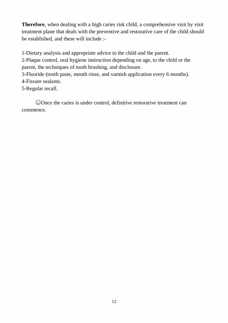
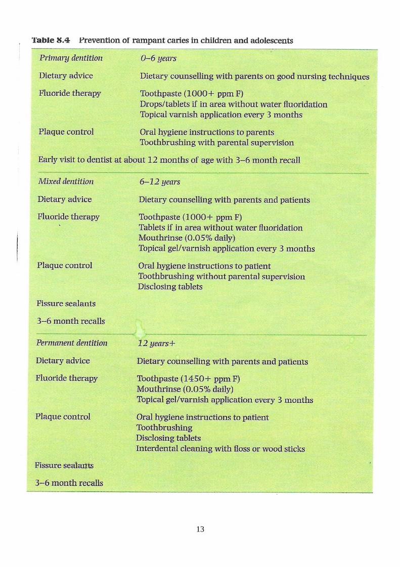
Therefore, when dealing with a high caries risk child, a comprehensive visit by visit
treatment plane that deals with the preventive and restorative care of the child should
be established, and these will include :-
1-Dietary analysis and appropriate advice to the child and the parent.
2-Plaque control, oral hygiene instruction depending on age, to the child or the
parent, the techniques of tooth brushing, and disclosure.
3-Fluoride (tooth paste, mouth rinse, and varnish application every 6 months).
4-Fissure sealants.
5-Regular recall.
☺Once the caries is under control, definitive restorative treatment can
commence.
13
14
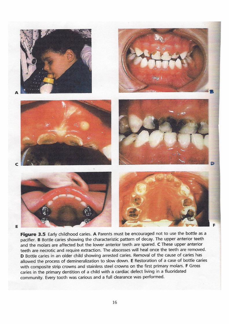
-Early Childhood Dental Caries :
Early childhood caries (ECC) is a term used to describe dental caries
presenting in the primary dentition of young children. Terms such as “nursing bottle
mouth”, “bottle mouth caries”, or “nursing caries” are used to describe a particular
pattern of dental caries in which the upper primary incisors and upper first primary
molars are usually most severely affected, the lower first primary molars are also
often carious, canines are affected less than the first molars because of later eruption,
but the lower primary incisors are usually spared, being either entirely caries free or
only mildly affected.
The sparing of the lower incisors seen in nursing caries is thought to result from the
shielding of the lower incisors by the tongue during sucking, whilst at the same time
they are being bathed in saliva from the sublingual and submandibular ducts. While
the upper incisors on the other hand, are bathed in fluid from the bottle / feeder
provides an excellent culture medium for acidogenic microorganisms.
Some children present with extensive caries that does not follow the “nursing caries”
pattern. Such children often have multiple carious teeth and may be slightly older (3
or 4 years of age) at initial presentation. This presentation of caries is sometimes
called “rampant caries”. There is however, no clear distinction between rampant
caries and nursing caries, and the term “early childhood caries” has been suggested as
a suitable, all-encompassing term.
In many cases, early childhood caries is related to the frequent consumption of a
drink containing sugars from a bottle or “dinky” type comforters (these have a small
reservoir that can be filled with a drink), and it is one of the causes of early childhood
caries or rampant caries in young children is allowing infants and toddlers to sleep
with a bottle.
Research has shown that children who tend to fall asleep with the bottle in their
mouths are most likely to get ECC, and this is probably a reflection of the dramatic
reduction in salivary flow that occurs as a child falls asleep, and clearance of the
liquid from the oral cavity is slowed. The child who falls asleep while nursing should
be burped and then placed in bed.
The reported prevalence ranges from 2.5 % to 15 %. Also in recent years it has
been recognized that prolonged bottle feeding, beyond the usual time when the child
is weaned from the bottle and introduced to solid food, may result in early and
rampant caries. However, other habits such as “grazing” (snacking on food
15
constantly) also puts many children at risk as does the use of feeding cups and sipper
bottles that toddlers walk around with.
The bottle caries occurs in all socioeconomic groups and as such often reflects the
social dynamics of the family. Children who are difficult sleepers or have colic are
often pacified with a bottle. The bottle can contain any liquid with fermentable
carbohydrate; even milk, commonly, drinks and juices containing vitamin C are used.
Fruit-based drinks are most commonly associated with nursing caries, even many of
those claiming to have “low sugar” or “no added sugar” appears to be capable of
causing caries.
However, the link between bottle habits and ECC is not absolute and studies have
suggested that other factors, such as parental history of active and untreated caries-
particularly in the mother, linear enamel defects, and malnutrition may play an
important role in the etiology of this condition.
Nursing caries can be prevented and managed by :-
● Early counseling of the parents. This is one reason for suggesting that children
receive their first dental examination between 6 and 12 months of age, when nursing
caries will not likely have developed.
● The parents should start brushing the child's teeth as soon as they erupt and
discontinue nursing as soon as the child can drink from a cup at approximately 12
months of age.
● Cessation of habit.
● Dietary advice.
● Possible use of antimicrobial products.
● Fluoride application.
● Bulid-up of restorable teeth. This may consist of glass ionomer restorations,
composite resin-strip crowns and / or stainless steel crowns.
● Extractions if required. Loss of the upper anterior teeth will not result in space loss
if the canines have erupted. Speech will develop normally. If posterior teeth have to
be extracted, the parents will need to be informed about possible space loss, and an
assessment should be carried out to determine if a space maintainer is appropriate.
16
1
Paediatric Dentistry
Fifth Stage
Dr. Suhair W. Abbood
Lec. 5
Treatment of the Deep Carious Lesion, and Pulp Therapy for
Primary and Immature Permanent Teeth
Treatment of the Deep Carious Lesion:-
Children and young adults who have not received early and adequate dental
care and optimal systemic fluoride often have deep carious lesions in the primary and
permanent teeth. Many of the lesions appear radiographically to be dangerously
close to the pulp or to actually involve the dental pulp. Approximately 75% of the
teeth with deep caries have been found from clinical observations to have pulpal
exposures. Also over 90% of the asymptomatic teeth with deep carious lesions could
be successfully treated without pulp exposure using indirect pulp therapy techniques.
The dentist cannot initially predict with certainty the state of health of the
pulp. When dealing with a deep cavity, however, the dentist can probably be assured
that the caries has invaded the reparative dentin. Therefore the dentist should take
every precaution to minimize the trauma of the operative procedure, for, in the
presence of established pulpal pathosis resulting from caries, the addition of operative
trauma can provide sufficient irritation to compound the pathosis. This can lead to the
establishment of irreversible pulpal lesions.
In view of the direct relationship between caries depth and pulpal pathosis,
early excavation of what appear to be superficial caries in the dentin is advocated as
sound preventive treatment to minimize pulpal irritation. If a carious exposure
discovered at the time of the initial caries excavation could be routinely treated with
consistently good results; a major problem in dentistry would be solved.
Unfortunately, the treatment of vital exposures, especially in primary teeth, has not
been entirely successful. For this reason, care must be taken to prevent pulp exposure
during the removal of deep caries.
2
Indirect Pulp Treatment (Gross Caries Removal or Indirect Pulp
Therapy):-
The procedure in which only the gross caries or soft caries is removed from the
lesion and the cavity is sealed for a time with a radioopaque, biocompatible base, and
bactericidal agent over the remaining carious dentin to stimulate healing and repair is
referred to as indirect pulp
treatment.
Only teeth with deep caries that are free of symptoms of painful pulpitis
should be selected for this procedure. In the majority of circumstances, carious
lesions can and should be fully excavated before tooth restoration. A clinical dilemma
is presented by a deep lesion in a vital, symptom-free tooth where complete removal
of softened dentin on the pulpal floor is likely to result in frank
exposure.
The advancing front of carious lesion contains very few cariogenic bacteria,
provided the bulk of infected overlying dentin is removed, a small amount of
softened dentin may often be left in the deepest part of the preparation without
endangering the pulp.
Sealing off the remnants of the advancing carious lesion from the oral environment,
produces a bacteriostatic response within the body of the lesion, and promotes pulpal
healing with the formation of reactionary dentin. This is the basis for indirect pulp
capping in both the primary and permanent dentition, and is also known as caries
control. Indirect pulp capping is also the basis for the atraumatic restorative technique
(ART).
The teeth selected had to have deep carious lesions and to fulfill the following
criteria:-
1-No history of spontaneous, unprovoked toothache (The tooth may have had a
history of toothache associated with eating, as long as pain subsided immediately
after removal of the stimulus).
2-No tenderness to percussion.
3-No abnormal mobility.
4-No radiographic evidence of radicular disease.
5-No radiographic evidence of abnormal internal or external root resorption.
3
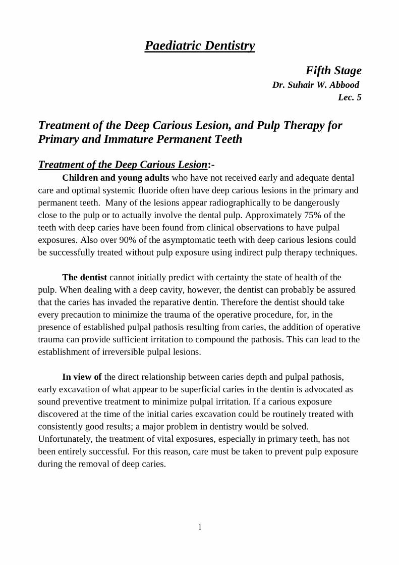
The clinical procedure of indirect pulp capping involves:-
1-It is advisable to use local anesthesia because the procedure usually results in some
discomfort to the child. The placement of a rubber dam is a further
advantage.
2-The removal of the gross caries
with large round burs or sharp spoon excavators, allowing sufficient caries to remain
over the pulp horn to avoid exposure of the pulp
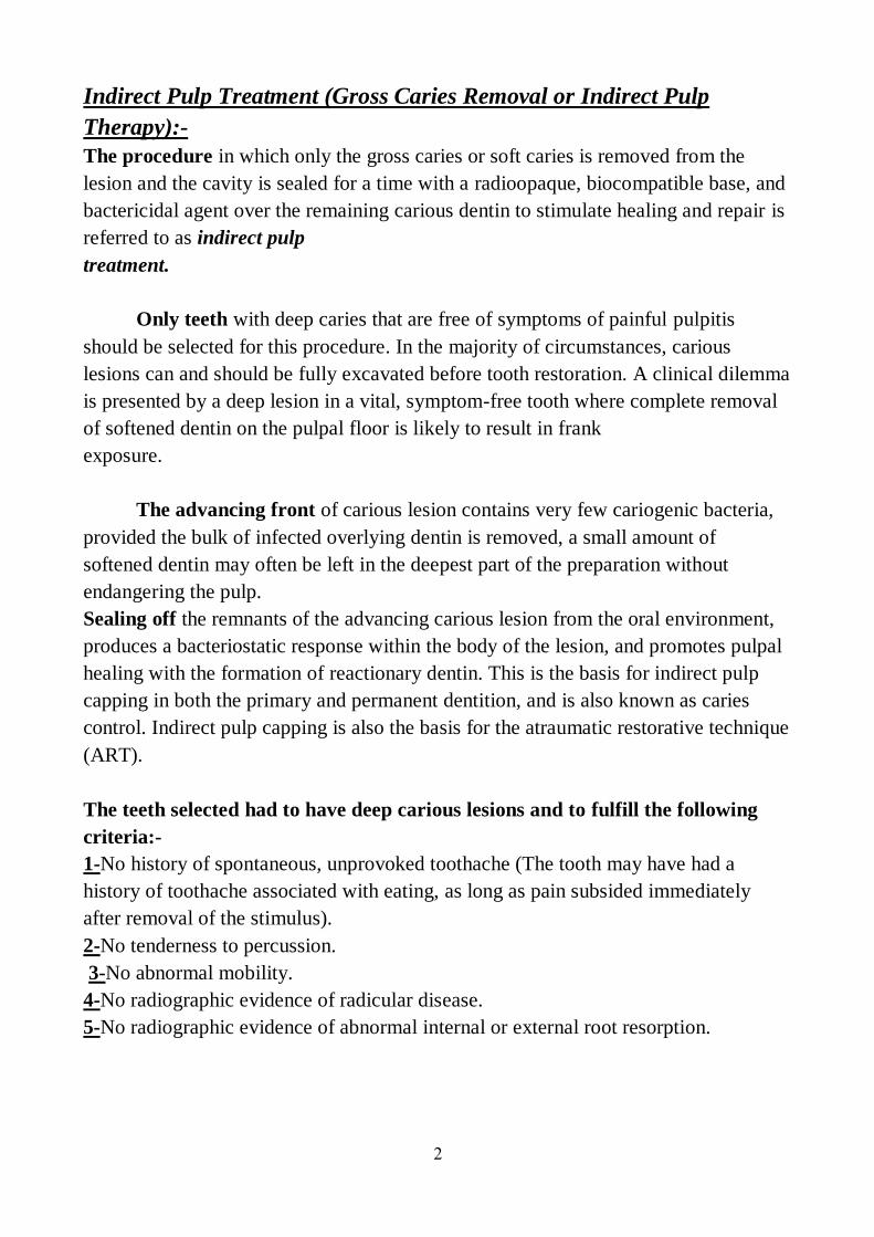
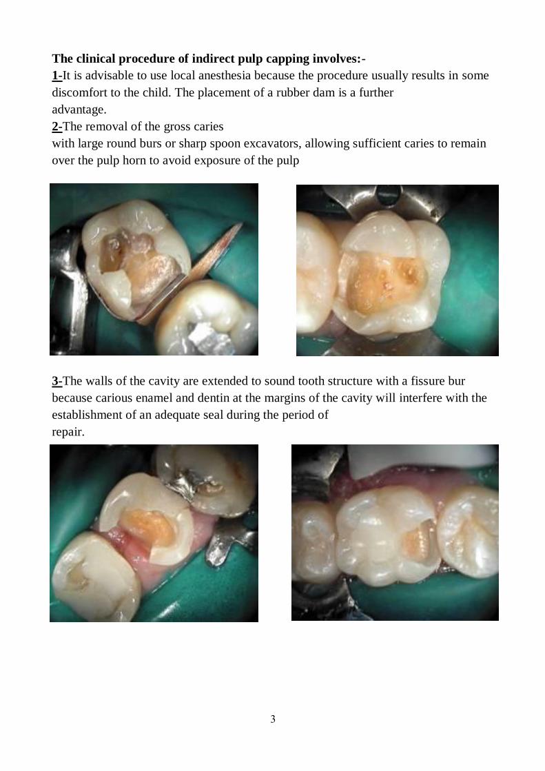
3-The walls of the cavity are extended to sound tooth structure with a fissure bur
because carious enamel and dentin at the margins of the cavity will interfere with the
establishment of an adequate seal during the period of
repair.
4
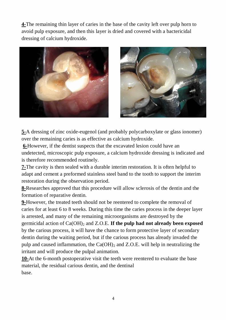
4-The remaining thin layer of caries in the base of the cavity left over pulp horn to
avoid pulp exposure, and then this layer is dried and covered with a bactericidal
dressing of calcium hydroxide.
5-A dressing of zinc oxide-eugenol (and probably polycarboxylate or glass ionomer)
over the remaining caries is as effective as calcium hydroxide.
6-However, if the dentist suspects that the excavated lesion could have an
undetected, microscopic pulp exposure, a calcium hydroxide dressing is indicated and
is therefore recommended routinely.
7-The cavity is then sealed with a durable interim restoration. It is often helpful to
adapt and cement a preformed stainless steel band to the tooth to support the interim
restoration during the observation period.
8-Researches approved that this procedure will allow sclerosis of the dentin and the
formation of reparative dentin.
9-However, the treated teeth should not be reentered to complete the removal of
caries for at least 6 to 8 weeks. During this time the caries process in the deeper layer
is arrested, and many of the remaining microorganisms are destroyed by the
germicidal action of Ca(OH)2 and Z.O.E. If the pulp had not already been exposed
by the carious process, it will have the chance to form protective layer of secondary
dentin during the waiting period, but if the carious process has already invaded the
pulp and caused inflammation, the Ca(OH)2 and Z.O.E. will help in neutralizing the
irritant and will produce the pulpal animation.
10-At the 6-month postoperative visit the teeth were reentered to evaluate the base
material, the residual carious dentin, and the dentinal
base.
5
Treatment was judged successful if:-
1-The restoration was intact.
2-The tooth had normal mobility.
3-The tooth was not sensitive to percussion.
4-The tooth had no history of pain after treatment.
5-There was no radiographic evidence of abnormal root resorption.
6-There was no radiographic evidence of radicular disease.
7-There was no clinical evidence of direct pulp exposure when the tooth was
reentered and the residual carious dentin was examined or excavated.
11-Careful removal of the remaining carious material, now somewhat sclerotic, may
reveal a sound base of dentin without an exposure of the
pulp.
12-If a second layer of dentin covers the pulp, a liner material containing calcium
hydroxide is applied, the cavity preparation is completed, and the tooth is restored in
a conventional manner.
If a small pulp exposure is encountered, a different type of treatment, based
on the clinical signs and symptoms and local conditions, must be used. In all cases of
successful treatment the base material and the residual carious dentin were observed
to do dry on reentry and clinical examination.
Indirect pulp therapy has been proved to be a valuable therapeutic procedure
in treating asymptomatic teeth with deep carious lesions. The procedure reduces the
risk of direct pulp exposure and preserves pulp vitality. It is uncertain whether the
carious lesion in dentin will become sterile and remineralize, or if it merely becomes
quiescent with the potential to reactive if there is leakage around the final restoration,
hence there is debate over the necessity of reentering the tooth to remove the residual
caries once there is clinical and radiographic evidence of pulpal healing.
One may question the need to reenter the tooth if it has been properly
selected and monitored, if a durable restoration is placed initially, if no adverse signs
or symptoms develop, and since the returning to the operative site to complete caries
removal increases the risk of pulp exposure, also because of the known service life of
the primary tooth, there is no indication for re-entering the primary tooth to remove
residual caries when the clinical response is favourable, many clinicians are
successfully practicing indirect pulp treatment without reentry after the initial caries
excavation. However, the inexperienced dentist should perform the treatment in two
appointments until confidence in proper case selection has been achieved.
Dental treatments are constantly evolving. One such innovation, ozone
therapy (heal ozone) has hit the media headlines, spiking much public interest. The
technology is available and costly devices for delivery of ozone for dental purposes
6
exist, but as yet the superiority of this modality over conventional treatment has not
been proven with properly conducted clinical trials. Ozone and silver fluoride have
both been proposed as adjunctive antimicrobial agents in conjunction with indirect
pulp capping. At present there is a lack of evidence to support their superiority over
sealing the lesion with standard restorative materials.
Although the routine practice of indirect pulp therapy in properly selected
teeth will significantly reduce the number of pulp therapy encountered, all dentist
who treat severe caries in children will occasionally be faced with treatment decisions
related to the management of pulp therapy. Dental caries, trauma, and iatrogenic
effects of conservative dental treatment, all provoke a biological response in the
pulpodentinal complex.
Pulp Therapy for Primary and Immature Permanent Teeth
The pulp therapy is concerned with cascade of therapeutic interventions used to
promote an adaptive biological response in the pulpodentinal complex of treated
tooth, the optimize subsequent growth and development. Contemporary advances in
primary prevention have reduced dental disease in the developed world, but there is
no room for complacency.
Dental caries and traumatic dental injuries are still prevalent and treatment
of the damage they cause is still a major component of pediatric dental practice. The
principal goals of pediatric operative dentistry are to prevent the extension of dental
disease and to restore damaged teeth to healthy function. To this end, a range of
conservative endodontic procedures can provide alternatives to extraction for many
pulpally compromised primary teeth. Therapeutic efforts are directed towards the
retention of carious or traumatized teeth, maintaining normal function, with the
resolution of, or freedom from, clinical symptoms. They are within the grasp of all
practitioners and are central to the practice of pediatric dentistry.
The Dental Pulp :-
Dental pulp is the living, soft tissue structure which resides in the coronal pulp
chamber and root canals of primary and permanent teeth. Histologically, it is
composed of loose connective tissue, surrounded on its periphery by a continuous
layer of specialized secretory cells, the odontoblasts. Odontoblasts are unique to the
dental pulp and are responsible for dentin deposition. Blood vessels and nerves enter
the pulp through the apical foramen and occasionally through lateral or accessory
root canals. The pulps of primary and young permanent teeth, especially those with
incomplete apices, have a very rich blood supply.
7
The most important function of the pulp is to lay down dentin which forms
the basic structure of teeth, defines their general morphology, and provides them with
mechanical strength and toughness. Dentin deposition commences many months
(primary teeth) or years (permanent teeth) before tooth eruption and while the crown
of a newly erupted tooth has a mature external form, the pulp within still has
considerable work to do in completing tooth development.
The newly erupted teeth have short roots, their apices are wide and often
diverging, and the dentin walls of the entire tooth are thin and relatively weak.
Provided the pulp remains healthy, dentin deposition will continue during the
posteruptive year for primary teeth. One of the key goals of pediatric dentistry is
therefore to protect and preserve the pulps of teeth in a healthy state at least until this
critical phase of tooth development is complete.
The Role of Primary Teeth :-
The primary teeth play an integral role in the development of the occlusion.
Premature loss of a primary tooth through trauma or infection has the potential to
destabilize the developing occlusion with space loss, arch collapse, and premature,
delayed or ectopic eruption of the permanent successor teeth. In general, the effects
of early extraction of primary teeth are more profound in the buccal segments than in
the anterior dentition.
Effective pulpal therapy in the primary dentition must not only stabilize the
affected primary tooth, but also create a favourable environment for normal
exfoliation of the primary tooth, without harm to the developing enamel or
interference with the normal eruption of its permanent successor. Where these
outcomes cannot reasonably be achieved over the clinical life of the primary tooth, it
is appropriate to extract the affected tooth and consider alternative strategies for
occlusal guidance and maintenance of arch integrity.
The Immature Permanent Teeth :-
The permanent teeth are still immature when they erupt. In addition to the
important phase of posteruptive enamel maturation, the roots of newly erupted
permanent teeth will take up to 5 years before their growth is completed. During this
period, the roots are short, the root apex is wide open, the dentin is relatively thin,
and the dentin tubules are relatively wide increasing the permeability of dentin to
bacteria. The open apex is associated with excellent pulpal vascularity and the
potential for a favourable healing response.
8
Therapeutic efforts are directed towards preserving the vitality of the
pulpodentinal complex to facilitate normal root development and maturation. If pulp
necrosis occurs prior to root maturation, the affected tooth can still be preserved
using nonvital endodontics strategies, but will be compromised with regard to
strength, root length, and apical development. Retention of a compromised immature
permanent tooth with a poor long-term prognosis may still be beneficial for arch
integrity and normal alveolar development during the period of dentofacial
growth.
The Vital Pulp Therapy :-
The treatment of the dental pulp exposed by the caries process, by accident during
cavity preparation, or even as a result of injury and fracture of the tooth has long
presented a challenge in treatment.
Diagnostic Aids in the Selection of Teeth for Vital Pulp Therapy and the
Evaluation of Treatment Prognosis before Pulp Therapy :-
The diagnostic process of selecting teeth that are good candidates for vital pulp
therapy has at least two dimensions :-
☻First, the dentist must decide that the tooth has a good chance of responding
favorably to the pulp therapy procedure indicated.
☻Second, the advisability of performing the pulp therapy and restoring the tooth
must be weighed against extraction and space management.
For example, nothing is gained by successful pulp therapy if the crown of the
involved tooth is nonrestorable or the periodontal structures are irreversibly diseased.
By the same rationale, a dentist is likely to invest more time and effort to save a
pulpally involved second primary molar in a 4-year-old child with unerupted first
permanent molars than to save a pulpally involved first primary molar in an 8-year-
old child.
Other factors to consider include the following:-
1-The level of patient and parent cooperation and motivation in receiving the
treatment.
2-The level of patient and parent desire and motivation in maintaining oral health and
hygiene.
3-The caries activity of the patient and the overall prognosis of oral rehabilitation.
4-The stage of dental development of the patient.
9
5-The degree of difficulty anticipated in adequately performing the pulp therapy
(instrumentation) in the particular case.
6-Space management considerations resulting from:-
a-Previous extractions.
b-Preexisting malocclusion.
c-Ankylosis.
d-Congenitally missing teeth.
e-Space loss caused by the extensive carious destruction of teeth and subsequent
drifting.
7-Excessive extrusion of the pulpally involved tooth resulting from the missing
opposing teeth.
These examples, in any combination, illustrate the almost infinite number of
treatment considerations that could be important in an individual patient with pulpal
pathosis.
Clinical Assessment and General Considerations of Teeth for Vital Pulp
Therapy
1-The History of Pain :- The history of either presence or absence of pain may not be
as reliable in the differential diagnosis of the condition of the exposed primary pulp
as it is in permanent teeth. Young patients frequently vary in their reporting of pain. It
is often not until their pain is sever and prolonged when parents might become aware.
Degeneration of primary pulps even to the point of abscess formation without the
child recalling pain or discomfort is not uncommon.
Nevertheless, the history of a toothache should be the first consideration in
the selection of teeth for vital pulp therapy. A toothache coincident with or
immediately after a meal may not mean extensive pulpal inflammation. The pain may
be caused by an accumulation of food within the carious lesion, by pressure, or by a
chemical irritation to the vital pulp protected by only a thin layer of intact dentin. In a
study of teeth with painful pulpitis the severity of pain and the extent of pulp
involvement were not correlated. Subjective complaints of pain from the intake of hot
foods or drink were indicative of pulpitis, but they were not as reliable as careful tests
made by dentists who need to do so. No real difference in response to heat or cold
was detected. Testing showed most patients to be sensitive to both.
Further observed demonstrating that most teeth with a sever toothache at
night usually means extensive degeneration of the pulp and calls for more than a
conservative type of pulp therapy. A spontaneous toothache of more than momentary
10
duration occurring at any time usually means that pulpal disease has progressed too
far for treatment with even a pulpotomy. Symptoms of severe, prolonged,
spontaneous or nocturnal pain suggest irreversible pulpitis or a dental abscess. A
history of repeated need for analgesics is also suggestive of pulp necrosis. Dental
pain will frequently resolve once a sinus tract establishes drainage, and thus relieves
pressure. In these cases, the underlying pathology is still present and must be resolved
despite the lack of obvious discomfort.
2-The Clinical Signs and Symptoms :- Effective pulpal therapy requires the correct
assessment and interpretation of clinical signs and symptoms, leading to an accurate
diagnosis of the pulpal condition. Ineffective or inappropriate pulp therapy is
associated with both acute and chronic clinical signs and symptoms. Unfortunately,
there are no objective or definitive tests to determine the health of the pulpodentinal
complex in the primary or immature permanent tooth. The clinical signs and
symptoms are poorly correlated with actual pulp histology.
The acute signs and symptoms include :-
● Pain. ● Mobility. ● Periapical abscess. ● Facial cellulitis or progression to
spreading infections of the neck (Ludwig,s angina).
Antibiotic usage to control acute infection may temporarily resolve some or all of
these clinical signs, but will not resolve the underlying pathology.
The chronic signs and symptoms include :-
● Persistent infection. ● Discharging sinus. ● Enamel dysplasia (turner,s tooth).
● Infected follicular cyst. ● Failure of exfoliation of primary teeth. ● Apical
fenestration. ● Ectopic permanent teeth.
Abnormal tooth mobility is a clinical sign that may indicate a severely
diseased pulp. When such a tooth is evaluated for mobility, the manipulation may
elicit localized pain in the area, but this is not always the case.
Sensitivity to percussion or pressure even though thickening of the apical
periodontal membranes was not evident radiographically, is a clinical symptom
suggestive of at least some degree of pulpal disease, but the degenerative stage of the
pulp is probably of the acute inflammatory type.
Tooth mobility or sensitivity to percussion or pressure may be a clinical
signal of other dental problems as well, such as a high restoration or advanced
periodontal disease, also the pathologic mobility must be distinguished from normal
mobility in primary teeth near exfoliation.
11
A gingival abscess or draining fistula associated with a tooth with a deep
carious lesion is an obvious clinical sign of an irreversibly diseased pulp. Such
infection can be resolved only by successful endodontic therapy or extraction of the
tooth.
Other clinical signs that could be seen by carful clinical examination of teeth, and
can reveal useful diagnostic information are :-
● Coronal discoloration is suggestive of pulp necrosis.
● Marginal ridge fracture in primary tooth is suggestive of carious pulpal
involvement in contact point caries.
● Fracture of the occlusal triangular ridges or carious undermining of the cusps in pit
and fissure caries also suggests carious involvement.
However, when these clinical informations are identified in a child and is
associated with a tooth having a deep carious lesion, the problem is most likely to be
from pulpal disease and possible inflammatory involvement of the periodontal
ligament. A primary tooth that cannot be saved requires extraction despite potential
future orthodontic complication.
3-The Radiographic Interpretation :- Unfortunately, the external appearance of the
carious lesion can in some cases be misleading. Persistent symptoms occurring soon
after placement of a restoration indicate pulpal pathology. Lack of coronal seal will
inevitably lead to pulpal pathology.
Radiographic examination is essential to supplement clinical findings and
enhance diagnostic accuracy, but keep in mind that the radiographic interpretation in
children is more difficult than adults.
Longitudinal radiographs showing normal dentin deposition within the pulp
chamber and the roots, suggests pulpal health. Irregular pulp calcification or pulpal
obliteration suggests pulpal dystrophy, while failure of physiological pulp regression
or arrested root development suggests pulpal necrosis. In a single radiographic
examination, individual teeth can be compared with their intimate to identify
asymmetry.
Clinical signs or symptoms suggesting carious involvement of the pulp
require radiographic investigation. Radiographs will show the extent of the carious
lesion, the position and proximity of pulp horns, the presence and position of the
permanent successor, the status of the roots and of their surrounding bone. These
conditions rule out treatment other than an endodontic procedure or extraction of the
tooth. The presence of caries in the furcation, internal or external root resorption
including physiological root resorption, and periapical or furcation bone lesions, are

12
all contraindications to endodontic treatment in the primary dentition, primary teeth
with these radiographic signs should be extracted.
The proximity of carious lesions to the pulp cannot always be determined
accurately in the X-ray film. What often appears to be an intact barrier of secondary
dentin protecting the pulp may actually be a perforated mass of irregularly calcified
and carious material. The pulp beneath this material may have extensive
inflammation.
The permanent teeth may have incompletely formed root ends, giving an
impression of periapical radiolucency, and the roots of the primary teeth undergoing
even normal physiologic resorption often present a misleading picture or one
suggestive of pathologic change.
Radiographic examination should be considered mandatory before
undertaking endodontic procedures. A recent X-ray film must be available to
examine for evidence of per-radicular or periapical changes, such as thickening of the
periodontal ligament or rarefaction of the supporting bone.
4-The Pulp Sensibility Testing :- Standard techniques of pulp sensibility testing are
of limited value in children. These techniques rely on patient feedback response to
thermal and electrical stimulation. The value of the electric pulp test in determining
the condition of the pulp of primary teeth is questionable, though it will give an
indication of whether the pulp is vital. The test does not give reliable evidence of the
degree of inflammation of the pulp. A complicating factor is the occasional positive
response to the test in a tooth with a necrotic pulp if the content of the canal is liquid.
In the immature permanent tooth, raised response thresholds to electrical
stimuli are observed. These decrease to normal levels with root maturation and apical
closure.
Thermal test do not seem to be reliable in the primary dentition either, the
lack of reliability is possibly related to the young child's inability to understand the
tests.
In the primary dentition, it is likely that children will not have achieved the
cognitive development necessary to respond reliably to a potentially painful stimulus
and response challenge. The reliability of the pulp test for the young child can also be
questioned because of the child's apprehension associated with the test itself.
5-The Physical Condition of the Patient :- Although the local observation are of
extreme importance in the selection of cases for vital pulp therapy, the dentist must
also consider the physical condition of the patient.
As pulp therapy necessarily relies on the adaptive healing response after
treatment, so patients with a significantly compromised immune system are
13
considered poor candidates for endodontic therapy. In the case of seriously ill
children, extraction of the involved tooth, after proper premedication with antibiotics,
rather than pulp therapy should be the treatment of choice. In the congenital cardiac
disease, patients who are considered to be at risk of subacute bacterial endocarditis
should be free of oral infection. Any primary tooth with clinical signs of infection
should be extracted.
Children with nephritis, leukemia solid tumors, idiopathic cyclic neutropenia,
or any condition that causes cyclic or chronic depression of granulocyte and
polymorphonuclear leukocyte counts (immunosuppressed patients
"immunodeficiency") should not be subjected to the possibility of an acute infection
resulting from failed pulp therapy.
Occasionally pulp therapy may be justified in a tooth of chronically ill child,
as child with bleeding disorders and coagulopathies, and child with oligodontia
"ectodermal dysplasia", but only after careful consideration is given to the prognosis
of the child's general condition, the prognosis of the endodontic therapy, and the
relative importance of retaining the involved tooth.
1
Paediatric Dentistry
Fifth Stage
Dr. Suhair W. Abbood
Lec. 6
Treatment of the Deep Carious Lesion, and Pulp Therapy for
Primary and Immature Permanent Teeth
1-Direct Pulp Capping :-
The pulp-capping procedure has been widely practiced for years and is still the
favorite method of many dentists treating vital pulp exposures. Although pulp
capping has been condemned by some; others report that if the teeth are carefully
selected, excellent results are obtained. The valuable observations in the diagnosing
the conditions of the primary pulp are:-
1-The size of the exposure.
2-The appearance of the pulp.
3-The amount of bleeding.
For this reason the use of a rubber dam to isolate the tooth is extremely
important; in addition, with the rubber dam the area can be kept clean and the work
can be done more efficiently. The most favorable condition for vital pulp therapy is
the small pinpoint exposure surrounded by sound dentin. However, a true carious
exposure, even of pinpoint size, will be accompanied by inflammation of the pulp,
the degree of which is usually directly related to the size of the exposure.
A large exposure-the type that is encountered when a mass of leathery dentin
is removed- is often associated with a watery exudates or pus at the exposure site.
These conditions are indicative of advanced pulp degeneration and often of internal
resorption in the pulp canal. Additionally, excessive hemorrhage at the point of
carious exposure or during pulp amputation is invariably associated with hyperemia
and generalized inflammation of the pulp. When a generalized inflammation of the
pulp is observed, endodontic therapy or extraction of the tooth is the treatment of
choice.
It is generally agreed that pulp-capping procedures should be limited to:-
1-Small exposures that have been accidentally produced by trauma.
2
2-During cavity preparation.
3-To true pinpoint carious exposures those are surrounded by sound dentin.
4-Pulp capping should be considered only for teeth in which there is an absence of
pain, with the possible exception of discomfort caused by the intake of food.
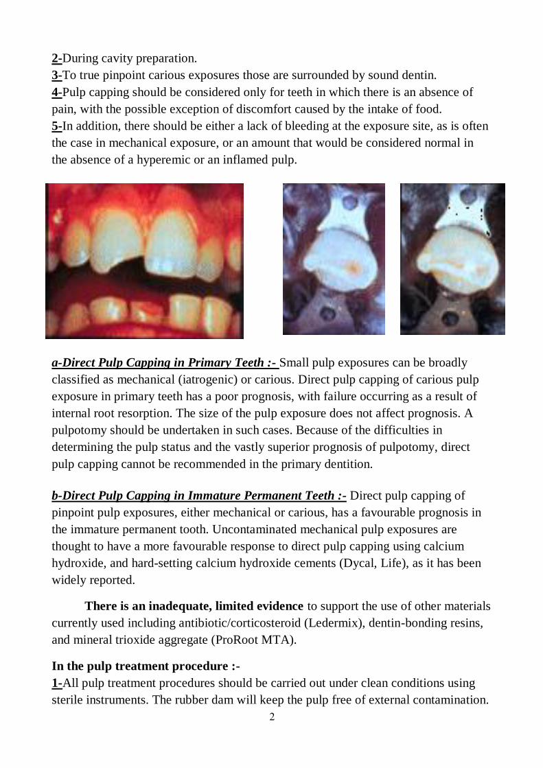
5-In addition, there should be either a lack of bleeding at the exposure site, as is often
the case in mechanical exposure, or an amount that would be considered normal in
the absence of a hyperemic or an inflamed pulp.
a-Direct Pulp Capping in Primary Teeth :- Small pulp exposures can be broadly
classified as mechanical (iatrogenic) or carious. Direct pulp capping of carious pulp
exposure in primary teeth has a poor prognosis, with failure occurring as a result of
internal root resorption. The size of the pulp exposure does not affect prognosis. A
pulpotomy should be undertaken in such cases. Because of the difficulties in
determining the pulp status and the vastly superior prognosis of pulpotomy, direct
pulp capping cannot be recommended in the primary dentition.
b-Direct Pulp Capping in Immature Permanent Teeth :- Direct pulp capping of
pinpoint pulp exposures, either mechanical or carious, has a favourable prognosis in
the immature permanent tooth. Uncontaminated mechanical pulp exposures are
thought to have a more favourable response to direct pulp capping using calcium
hydroxide, and hard-setting calcium hydroxide cements (Dycal, Life), as it has been
widely reported.
There is an inadequate, limited evidence to support the use of other materials
currently used including antibiotic/corticosteroid (Ledermix), dentin-bonding resins,
and mineral trioxide aggregate (ProRoot MTA).
In the pulp treatment procedure :-
1-All pulp treatment procedures should be carried out under clean conditions using
sterile instruments. The rubber dam will keep the pulp free of external contamination.
3
2-All peripheral carious tissue should be excavated before one begins to excavate the
portion of the carious dentin most likely to result in pulp exposure. Thus most of the
bacterially infected tissue will have been removed before actual pulp exposure
occurs.
3-Caustic solution should not be used to sterilize the exposure or the exposed pulp
tissue before capping, because it will lead to pulp injury. Only non irritant solution,
such as normal saline, should be used to clean the region.
4-Calcium hydroxide is the material of choice for pulp capping normal vital pulp
tissue. The possibility of its stimulating the repair reaction is good.
5-Then the procedure is completed by application of temporary filling or cement.
2-Pulpotomy :-
The suffix "otomy" means "to cut", so pulpotomy is "to cut the pulp". The removal of
the coronal portion of the pulp is an accepted procedure for treating both primary and
permanent teeth with carious pulp exposures. Careful selection of teeth for this
procedure is important because of failure. The justification for this procedure is that
the coronal pulp tissue, which is adjacent to the carious exposure, usually contains
microorganisms and shows evidence of inflammation and degenerative change. The
abnormal tissue can be removed, and the healing can be allowed to take place at the
entrance of the pulp canal in an area of essentially normal pulp. Even the pulpotomy
procedure is likely to result in a high percentage of failures unless the teeth are
carefully selected.
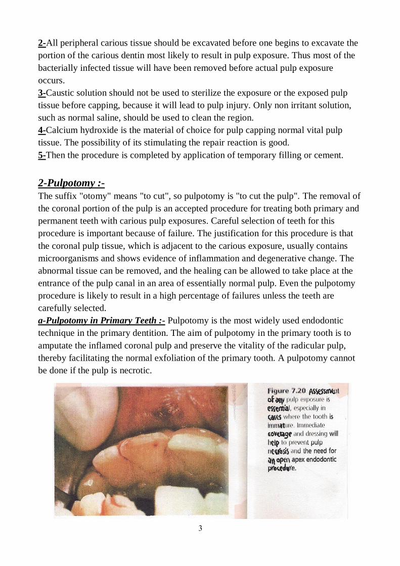
a-Pulpotomy in Primary Teeth :- Pulpotomy is the most widely used endodontic
technique in the primary dentition. The aim of pulpotomy in the primary tooth is to
amputate the inflamed coronal pulp and preserve the vitality of the radicular pulp,
thereby facilitating the normal exfoliation of the primary tooth. A pulpotomy cannot
be done if the pulp is necrotic.
4
The contemporary pulpotomy traces its origins to nineteenth-century
techniques for the mummification of painful, inflamed or putrescent pulpal tissue.
Over the twentieth century, the pulpotomy technique changed with fewer stages and
reduced duration of application and concentration of medicament. Emphasis is now
placed on the preservation of healthy radicular pulp rather than mummification.
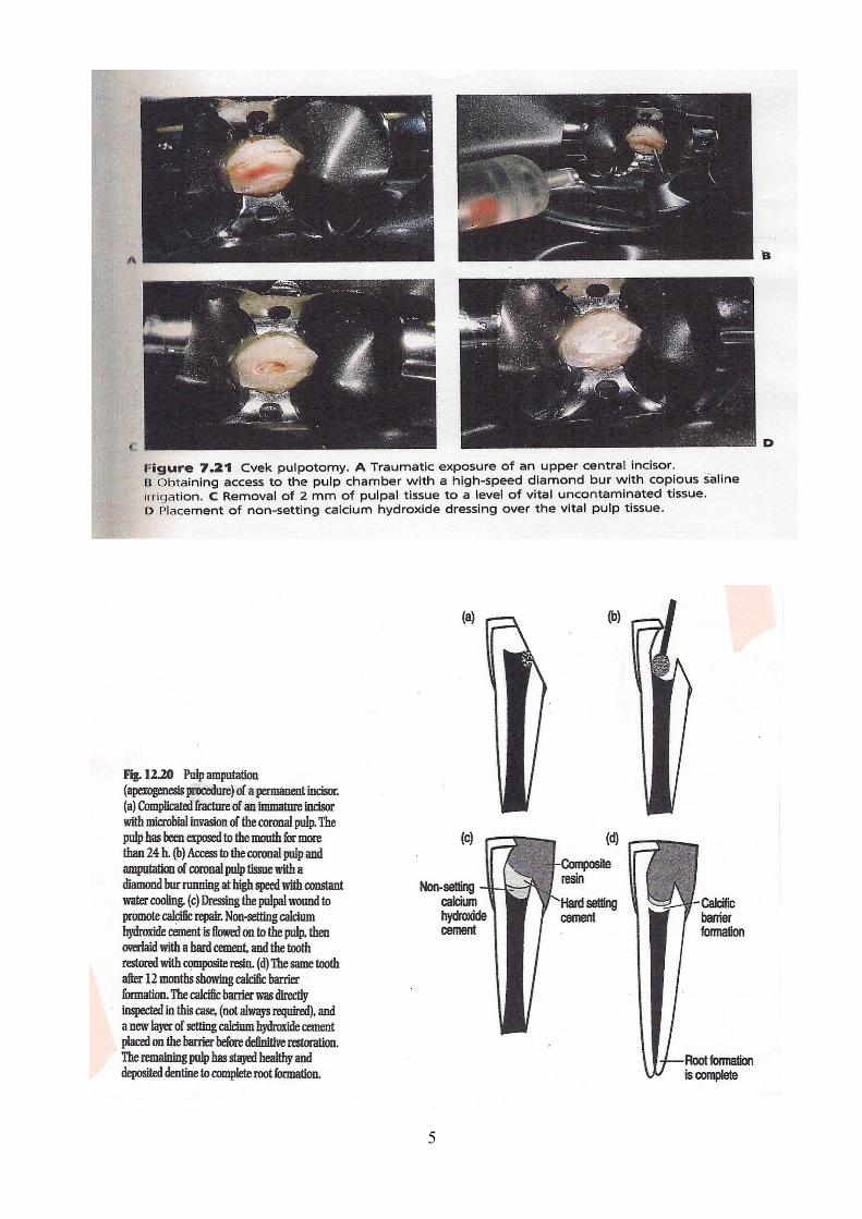
b-Pulpotomy in the Immature Permanent Teeth :- The aim of pulpotomy in the
immature permanent tooth is to amputate the inflamed coronal pulp and preserve the
vitality of the remaining pulp to promote apexogenesis. Apexogenesis involves the
continued normal development of the radicular pulp below the pulpotomy site,
resulting in normal root length, thickness of radicular dentin and apical closure.
Apexogenesis optimizes root anatomy and strength. The main risk of
apexogenesis is the potential for dystrophic pulp calcification in the event that
subsequent pulpectomy is required. The biomechanical properties of the root are
more favourable after apexogenesis than after apexification, but apexification is the
only option once pulp necrosis has occurred in the immature permanent tooth.
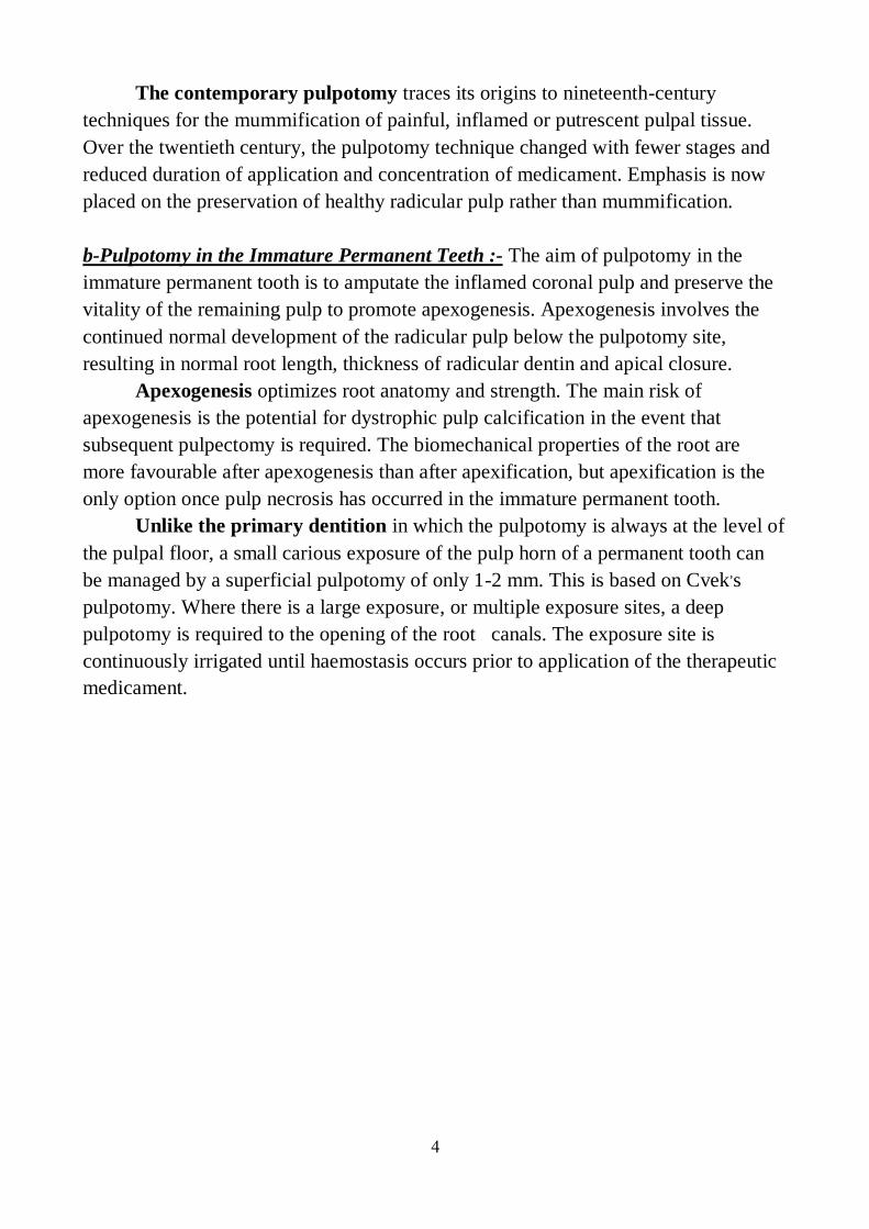
Unlike the primary dentition in which the pulpotomy is always at the level of
the pulpal floor, a small carious exposure of the pulp horn of a permanent tooth can
be managed by a superficial pulpotomy of only 1-2 mm. This is based on Cvek,s
pulpotomy. Where there is a large exposure, or multiple exposure sites, a deep
pulpotomy is required to the opening of the root canals. The exposure site is
continuously irrigated until haemostasis occurs prior to application of the therapeutic
medicament.
5
6
Generally in the pulpotomy procedure :-
1-The tooth should first be anesthetized and isolated with rubber dam.
2-A surgically clean technique should be used
throughout the procedure.
3-All remaining dental caries should be planed back
to provide good access to coronal pulp.
4-The overhanging enamel should be planed back to
provide good access to coronal pulp.
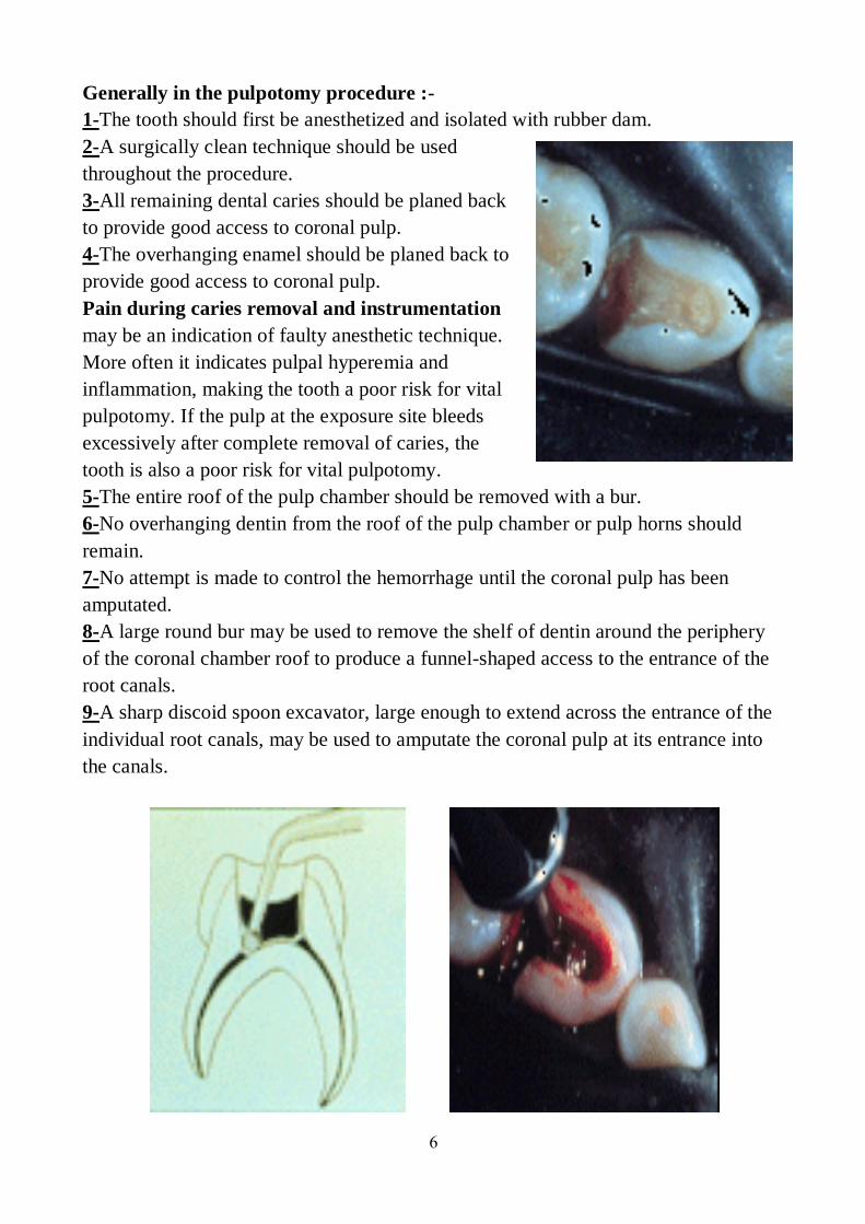
Pain during caries removal and instrumentation
may be an indication of faulty anesthetic technique.
More often it indicates pulpal hyperemia and
inflammation, making the tooth a poor risk for vital
pulpotomy. If the pulp at the exposure site bleeds
excessively after complete removal of caries, the
tooth is also a poor risk for vital pulpotomy.
5-The entire roof of the pulp chamber should be removed with a bur.
6-No overhanging dentin from the roof of the pulp chamber or pulp horns should
remain.
7-No attempt is made to control the hemorrhage until the coronal pulp has been
amputated.
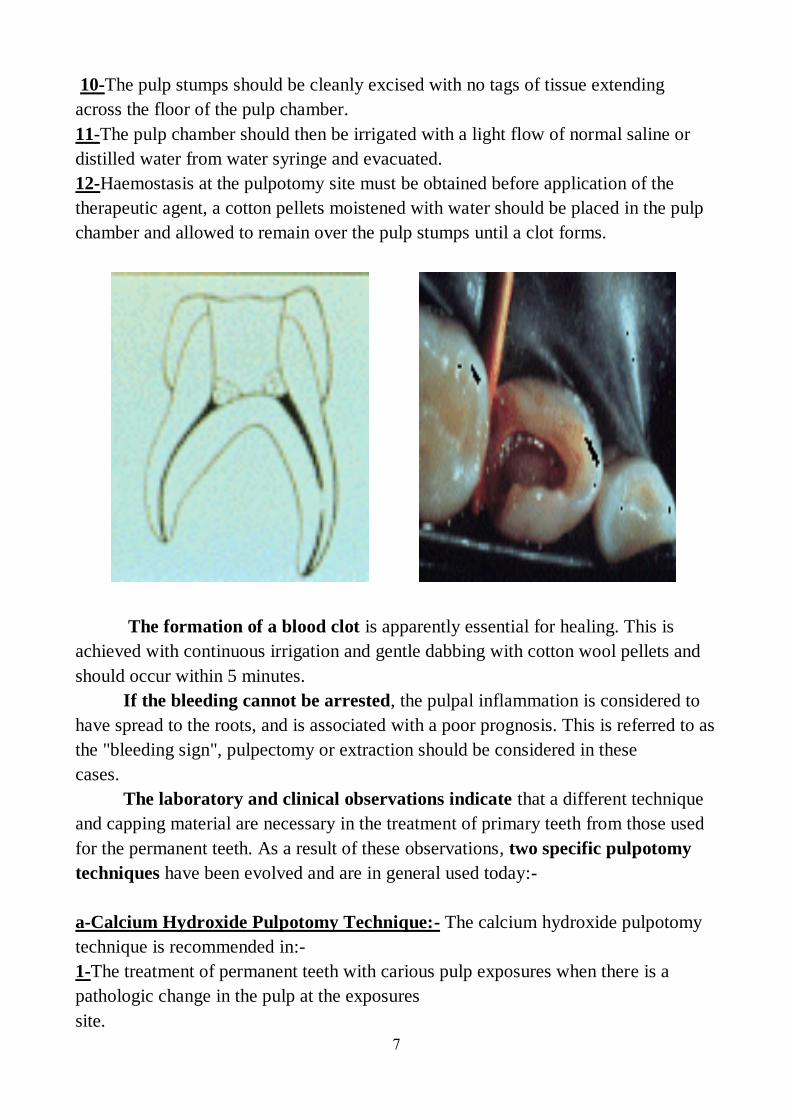
8-A large round bur may be used to remove the shelf of dentin around the periphery
of the coronal chamber roof to produce a funnel-shaped access to the entrance of the
root canals.
9-A sharp discoid spoon excavator, large enough to extend across the entrance of the
individual root canals, may be used to amputate the coronal pulp at its entrance into
the canals.
7
10-The pulp stumps should be cleanly excised with no tags of tissue extending
across the floor of the pulp chamber.
11-The pulp chamber should then be irrigated with a light flow of normal saline or
distilled water from water syringe and evacuated.
12-Haemostasis at the pulpotomy site must be obtained before application of the
therapeutic agent, a cotton pellets moistened with water should be placed in the pulp
chamber and allowed to remain over the pulp stumps until a clot forms.
The formation of a blood clot is apparently essential for healing. This is
achieved with continuous irrigation and gentle dabbing with cotton wool pellets and
should occur within 5 minutes.
If the bleeding cannot be arrested, the pulpal inflammation is considered to
have spread to the roots, and is associated with a poor prognosis. This is referred to as
the "bleeding sign", pulpectomy or extraction should be considered in these
cases.
The laboratory and clinical observations indicate that a different technique
and capping material are necessary in the treatment of primary teeth from those used
for the permanent teeth. As a result of these observations, two specific pulpotomy
techniques have been evolved and are in general used today:-
a-Calcium Hydroxide Pulpotomy Technique:- The calcium hydroxide pulpotomy
technique is recommended in:-
1-The treatment of permanent teeth with carious pulp exposures when there is a
pathologic change in the pulp at the exposures
site.
8
2-This procedure is particularly indicated for permanent teeth with immature root
development but with healthy pulp tissue in the root
canals.
3-It is also indicated for a permanent tooth with a pulp exposure, resulting from
crown fracture when the trauma has also produced a root fracture of the same
tooth.
The technique is completed during a single appointment. Only teeth free of
symptoms of painful pulpitis are considered for treatment.
The procedure involves:-
1-The amputation of the coronal portion of the pulp.
2-The control of hemorrhage.
3-The placement of a calcium hydroxide capping material over the pulp tissue
remaining in the canals.
4-A protective layer of hard-setting cement is placed over the calcium hydroxide to
provide an adequate seal.
5-Then the tooth is prepared for all
coverage.
However, if the tissue in the pulp canals appears hyperemic after the amputation of
the coronal tissue, a pulpotomy should no longer be considered. Endodontic treatment
is indicated if the tooth is to be saved.
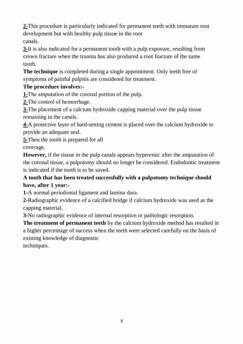
A tooth that has been treated successfully with a pulpotomy technique should
have, after 1 year:-
1-A normal periodontal ligament and lamina dura.
2-Radiographic evidence of a calcified bridge if calcium hydroxide was used as the
capping material.
3-No radiographic evidence of internal resorption or pathologic resorption.
The treatment of permanent teeth by the calcium hydroxide method has resulted in
a higher percentage of success when the teeth were selected carefully on the basis of
existing knowledge of diagnostic
techniques.
9
Failure after Vital Pulp Therapy
Failure in the formation of a calcified bridge across the vital pulp has often been
related to:-
1-The age of the patient.
2-Degree of surgical trauma.
3-Sealing pressure.
4-Improper choice of capping material.
5-Low threshold of host resistance.
6-Presence of microorganisms with subsequent infection.
Research provided further evidence that the success of vital pulp therapy depends
on the adherence to a surgically aseptic technique. So the need for a good surgical
technique and the placement of a restoration that will provide the best possible seal.
b-Formocresol Pulpotomy Technique:- The formocresol pulpotomy technique is
recommended in:-
1-The treatment of primary teeth with carious exposures because they do not respond
as favorably to the calcium hydroxide technique.
2-Can be used for permanent teeth as temporary treatment, which should be changed
sooner or later to endodontic treatment.
The same diagnostic criteria recommended for the selection of permanent teeth for
the calcium hydroxide pulpotomy should be used in the selection of primary teeth for
the formocresol pulpotomy technique.
The formocresol technique is also completed during a single appointment:-
1-A surgically clean technique should be used.
2-The coronal portion of the pulp should be amputated as described previously.
3-The debris should be removed from the chamber, and the hemorrhage should be
controlled.
If there is evidence of hyperemia after the removal of the coronal pulp,
indicating that inflammation is present in the tissue beyond the coronal portion of the
pulp; the technique should be abandoned in favor of the partial pulpectomy or the
removal of the tooth. If the hemorrhage is controlled readily and the pulp stumps
appear normal, it may be assumed that the pulp tissue in the canals is normal, and it is
possible to proceed with the pulpotomy.
4-The pulp chamber is dried with sterile cotton pellets.

10
5-Next, a pellet of cotton moistened with a 1:6 concentration of Buckley's
formocresol and blotted on sterile gauze to remove the excess is placed in contact
with the pulp stumps and is allowed to remain for 5 minutes. Since formocresol is
caustic, care must be taken to avoid contact with the gingival tissues.
6-The pellets are then removed, and the pulp chamber is dried with new pellets, and
checking must be done for pulp fixation, black color formation in the floor of the
cavity. That means the pulp tissue remaining in the canal has been fixed, so, the
procedure can be completed in a single appointment.
7-A thick paste of hard-setting zinc oxide-eugenol is prepared and placed over the
pulp stumps.
8-The tooth is then restored with a stainless steel
crown.

11
If the pulp tissue still after the removal of formocresol cotton pellet bleeds:-
1-Treatment should be changed into a two appointment technique, means leaving the
cotton pellet with formocresol.
2-In the second appointment the cotton pellet with formocresol is removed.
3-A thick paste of hard-setting zinc oxide-eugenol is prepared and placed over the
pulp stumps.
4-The tooth is then restored with a stainless steel crown.
Some dentists prefer to make the pulp-capping material by mixing the zinc oxide
powder with equal part of eugenol and formocresol. There are no proved
contraindications to adding formocresol to the mixture; however, there are no proved
benefits.
12
13
14
3-Partial Pulpectomy:-
A partial pulpectomy may be performed on:-
1-Primary teeth when coronal pulp tissue and the tissue entering the pulp canals are
vital but show clinical evidence of hyperemia.
2-The tooth may or may not have a history of painful pulpitis, but the contents of the
root canals should not show evidence of necrosis (suppuration).
3-In addition, there should not be radiographic evidence of a thickened periodontal
ligament or of radicular disease. If any of these conditions are present, a complete
pulpectomy, or an extraction, should be performed.
The partial pulpectomy technique, which may be completed in one appointment,
involves:-
1-The removal of the coronal pulp as described for the pulpotomy technique.
2-The pulp filaments from the root canals are removed with a fine barbed broach;
there will be considerable hemorrhage at this point.
3-The file removes tissue only as it is withdrawn and penetrates readily with a
minimum of resistance. Care should be taken to avoid penetrating the apex of the
tooth .
4-After the pulp tissue has been removed from the canals, a syringe is used to irrigate
them with 3% hydrogen peroxide followed by sodium hypochlorite. The canals
should then be dried with sterile paper points.
5-When hemorrhaging is controlled and the canals remain dry, a thin mix of zinc
oxide-eugenol paste may be prepared (without setting accelerators) and paper points
covered with the material are used to coat the root canal walls.
6-Small files may be used to file the paste into the walls.
7-The excess thin paste may be removed with paper points.
8-A thick mix of the treatment paste should then be prepared, rolled into a point, and
carried into the canal.
9-Root canal pluggers may be used to condense the filling material into the canals.
10-An X-ray film may be necessary to allow evaluation of the success in completely
filling the canals.
11-Further condensation may be carried out if required.
12-The tooth should be restored with full coverage.
1
Paediatric Dentistry
Fifth Stage
Dr. Suhair W. Abbood
Lec. 7
Endodontic Treatment for Primary Teeth and Immature Permanent
Teeth ( The Complete Pulpectomy ) The complete pulpectomy is the complete removal of all pulpal tissue from the tooth.
a-The Complete Pulpectomy in the Primary Teeth:-
It is unwise to maintain untreated infected primary teeth in the mouth, they may be
opened for drainage and often remain asymptomatic for an indefinite period of time.
However, they are a source of infection and should be treated or
removed.
Primary molars with abscesses are usually indicated for extractions.
Persistent and chronic infection in primary molars can cause damage to the
developing permanent tooth germs and such foci of infection should be removed.
Any evidence of root resorption is an indication for extraction. Severe infections
including acute facial cellulitis associated with primary teeth do not respond well to
pulpectomy, extraction is usually recommended in these cases.
Also the morphology of the root canals in primary teeth makes endodontic
treatment difficult and often impractical. The first primary molar canals are often so
small that they are inaccessible even to the smallest barbed broach, if the canal cannot
be properly cleansed of necrotic material, sterilized, and adequately filled, endodontic
therapy is not indicated. In some cases the non-vital primary molars or ones with a
chronic discharging sinus might need to be retained. Some of the reasons for this
could be :-
● Orthodontic.
● Medical, where extraction is not appropriate, such as in severe haemophiliacs.
● Parents refusal to accept extraction.
In such cases these teeth can be retained by carrying out the pulpectomy
procedure, the pulpectomy procedure can only be considered for primary teeth that
have intact roots. Sometimes there is reluctance among many dentists to carry out a
pulpectomy as it is perceived to be difficult in a young child, with extraction being
2
preferred, but this is a misconception, this technique should be at least learnt by all
pediatric dentists, as it can often save the child from trauma of GA for extraction of
primary teeth. A pulpectomy should be considered whenever it is essential to
preserve a primary tooth that cannot be treated with other means, such as a
pulpotomy, both primary molars and incisors can be treated with a pulpectomy
technique.
The pulpectomy involves accessing the root canal system of primary molars,
cleaning them as best as is possible, and then using an appropriate material, usually
pure zinc oxide eugenol, to obturate the root canals. Obturation of the root canal
space in a primary tooth must not interfere with normal exfoliation of the permanent
successor, this requires a resorbable paste root filling. The exception to this would be
where it is planned to retain a primary tooth that does not have a permanent
successor.
Suitable material for obturation include unreinforced zinc oxide eugenol
cement (pure zinc oxide eugenol) is preferred as it is entirely resorbable and is easily
removed as the roots of the primary teeth undergo resorption also, if it is extruded
through the apices, it gets completely resorbed by the apical tissues. Other materials
such as Iodoform paste (Kri, Diapex), and even calcium hydroxide paste (pulpdent,
Ultracal) are also sometimes used.
Although the root canal morphology of primary incisors is relatively simple,
the root canal morphology of multi-rooted primary teeth is more complex than
permanent teeth, with fins, ramifications and inter-canal communications. These
anatomical factors inhibit complete chemo-mechanical debridement of the root canal
space. The anatomical apex may be up to 3mm from the radiographic apex, and
frequently occurs on the lateral surface of the root, making it difficult to determine
the true working length. Over-instrumentation of the primary tooth root canal has the
potential to damage the underlying permanent tooth. Electronic measurement of the
root canal can assist with the location of the anatomical apex of a primary tooth.
On the other hand the number of root canal of primary molars is quite similar
to that of permanent molars with either three or four root canals present. In the lower
primary molars there are always two mesial root canals (mesio-buccal and mesio-
lingual), with one or sometimes two distal root canals. In upper primary molars there
are three root canals (mesio-buccal, disto-buccal, and palatal).
3
The indication for complete pulpectomy procedure for the treatment of primary
teeth with necrotic pulp are :-
1-Pulp necrosis in any primary tooth (irreversible pulpitis involving both the coronal
and radicular pulp), and if the canals are accessible.
2-Non-vital primary molars or incisors that need to be maintained in the arch. For
example, if the second primary molar is lost before the eruption of the first permanent
molar, the dentist is confronted with difficult problem of preventing the first
permanent molar from drifting mesially during its eruption, therefore, a special effort
should be made to treat and retain the second primary molar even if it has a necrotic
pulp.
3-Abscessed primary molars, there is radiographic evidence of furcation pathology,
but the preoperative radiograph confirms intact non-resorbed root, and there is
evidence of essentially normal supporting bone.
The technique for complete pulpectomy involved :-
1-The rubber dam is applied.
2-The roof of the pulp chamber should be removed to gain access to the root canal as
described previously in the pulpotomy technique, and identify the root canals.
3-The contents of the pulp chamber and all debris from the occlusal third of the
canals should be removed, with care taken to avoid forcing any of the infected
contents through the apical foramen.
4-A moistened pellet of camphorated monochlorophenol (CMCP) or Buckley's
formocresol, with excess moisture blotted, should be placed in the pulp chamber, and
the chamber may be sealed with zinc oxide eugenol.
5-At the second appointment, several days later, if the tooth has remained
asymptomatic during the interval, the remaining contents of the canals should be
removed, the tooth should be isolated with a rubber dam and the treatment pellet
removed.
6-Debride root canals gently with hedstrom files and copious irrigation with normal
saline or 0.5% solution of sodium hypochlorite. With the help of a good preoperative
radiograph, care should be taken to keep files 2-3 mm short of apex to avoid damage
to developing tooth germ, prepare canals to no more than file size 30.
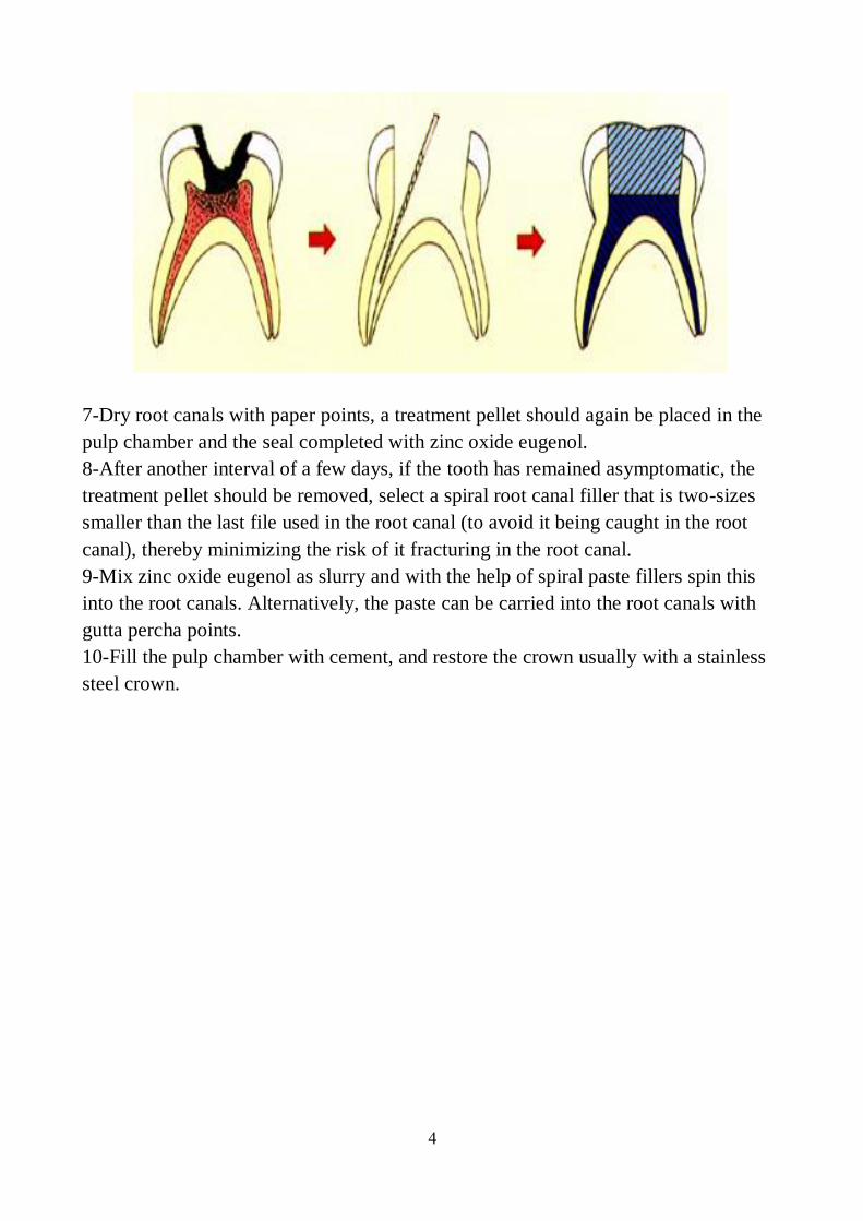
4
7-Dry root canals with paper points, a treatment pellet should again be placed in the
pulp chamber and the seal completed with zinc oxide eugenol.
8-After another interval of a few days, if the tooth has remained asymptomatic, the
treatment pellet should be removed, select a spiral root canal filler that is two-sizes
smaller than the last file used in the root canal (to avoid it being caught in the root
canal), thereby minimizing the risk of it fracturing in the root canal.
9-Mix zinc oxide eugenol as slurry and with the help of spiral paste fillers spin this
into the root canals. Alternatively, the paste can be carried into the root canals with
gutta percha points.
10-Fill the pulp chamber with cement, and restore the crown usually with a stainless
steel crown.
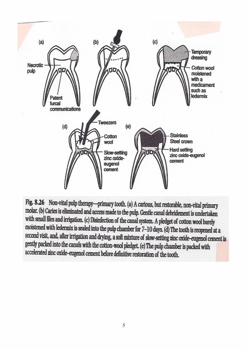
5
6
Follow-up and Review :-
Though the pulpectomy technique carries a good prognosis, the outcome is not as
good as a vital pulpotomy. Clinical follow-up augmented by one periapical
radiograph on a yearly basis is required. The following clinical and radiographic
parameters can be taken as indications of success :-
a-Clinical :-
● Alleviation of acute symptoms.
● Tooth free from pain and mobility.
b-Radiographic :-
● Improvement or no further deterioration of bone condition in the furcation area.
Complete pulpectomy (root canal) treatment of primary incisors :-
The technique of complete pulpectomy that described before can also be used to treat
non-vital or abscessed primary incisors. Increasingly, parents are reluctant to have
their child's upper anterior teeth extracted. In a modern society, where a child's self-
esteem is important, it is the duty of the dentists to maintain aesthetics wherever
possible.
Many primary incisors with abscesses that are exerted can be retained with the help
of a pulpectomy technique, and the root canal morphology is such that this can easily
be performed, the only limiting factor being the child's co-operation. Indications for a
pulpectomy in primary incisors include carious or traumatized primary incisors with
pulp exposures or acute or chronic abscesses.
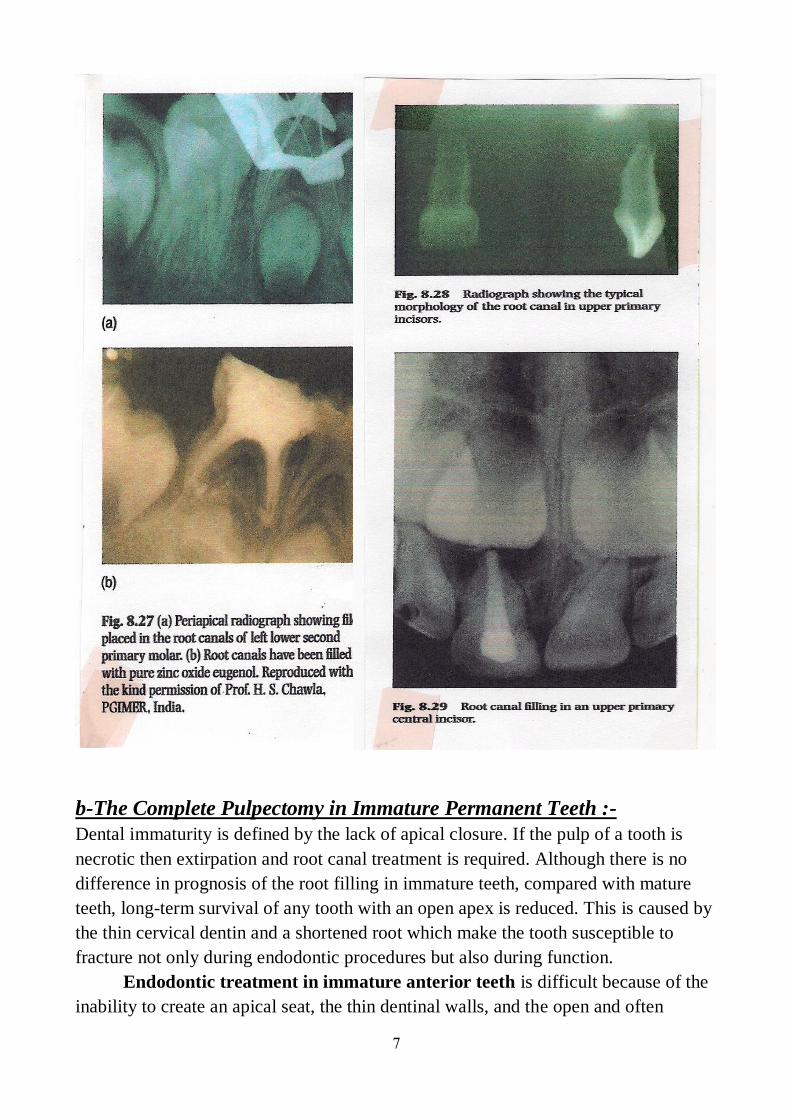
7
b-The Complete Pulpectomy in Immature Permanent Teeth :- Dental immaturity is defined by the lack of apical closure. If the pulp of a tooth is
necrotic then extirpation and root canal treatment is required. Although there is no
difference in prognosis of the root filling in immature teeth, compared with mature
teeth, long-term survival of any tooth with an open apex is reduced. This is caused by
the thin cervical dentin and a shortened root which make the tooth susceptible to
fracture not only during endodontic procedures but also during function.
Endodontic treatment in immature anterior teeth is difficult because of the
inability to create an apical seat, the thin dentinal walls, and the open and often
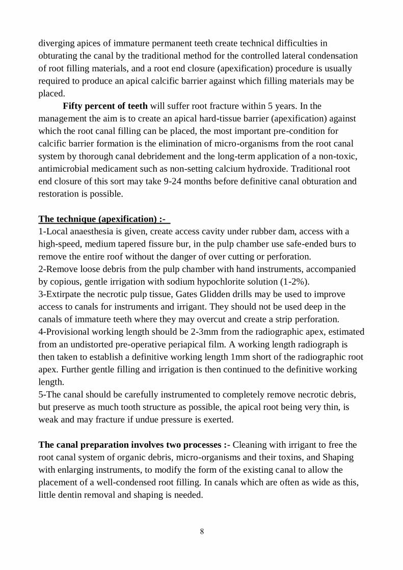
8
diverging apices of immature permanent teeth create technical difficulties in
obturating the canal by the traditional method for the controlled lateral condensation
of root filling materials, and a root end closure (apexification) procedure is usually
required to produce an apical calcific barrier against which filling materials may be
placed.
Fifty percent of teeth will suffer root fracture within 5 years. In the
management the aim is to create an apical hard-tissue barrier (apexification) against
which the root canal filling can be placed, the most important pre-condition for
calcific barrier formation is the elimination of micro-organisms from the root canal
system by thorough canal debridement and the long-term application of a non-toxic,
antimicrobial medicament such as non-setting calcium hydroxide. Traditional root
end closure of this sort may take 9-24 months before definitive canal obturation and
restoration is possible.
The technique (apexification) :-
1-Local anaesthesia is given, create access cavity under rubber dam, access with a
high-speed, medium tapered fissure bur, in the pulp chamber use safe-ended burs to
remove the entire roof without the danger of over cutting or perforation.
2-Remove loose debris from the pulp chamber with hand instruments, accompanied
by copious, gentle irrigation with sodium hypochlorite solution (1-2%).
3-Extirpate the necrotic pulp tissue, Gates Glidden drills may be used to improve
access to canals for instruments and irrigant. They should not be used deep in the
canals of immature teeth where they may overcut and create a strip perforation.
4-Provisional working length should be 2-3mm from the radiographic apex, estimated
from an undistorted pre-operative periapical film. A working length radiograph is
then taken to establish a definitive working length 1mm short of the radiographic root
apex. Further gentle filling and irrigation is then continued to the definitive working
length.
5-The canal should be carefully instrumented to completely remove necrotic debris,
but preserve as much tooth structure as possible, the apical root being very thin, is
weak and may fracture if undue pressure is exerted.
The canal preparation involves two processes :- Cleaning with irrigant to free the
root canal system of organic debris, micro-organisms and their toxins, and Shaping
with enlarging instruments, to modify the form of the existing canal to allow the
placement of a well-condensed root filling. In canals which are often as wide as this,
little dentin removal and shaping is needed.
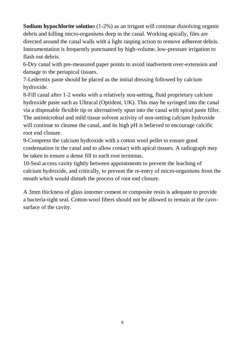
9
Sodium hypochlorite solution (1-2%) as an irrigant will continue dissolving organic
debris and killing micro-organisms deep in the canal. Working apically, files are
directed around the canal walls with a light rasping action to remove adherent debris.
Instrumentation is frequently punctuated by high-volume, low-pressure irrigation to
flash out debris.
6-Dry canal with pre-measured paper points to avoid inadvertent over-extension and
damage to the periapical tissues.
7-Ledermix paste should be placed as the initial dressing followed by calcium
hydroxide.
8-Fill canal after 1-2 weeks with a relatively non-setting, fluid proprietary calcium
hydroxide paste such as Ultracal (Optident, UK). This may be syringed into the canal
via a disposable flexible tip or alternatively spun into the canal with spiral paste filler.
The antimicrobial and mild tissue solvent activity of non-setting calcium hydroxide
will continue to cleanse the canal, and its high pH is believed to encourage calcific
root end closure.
9-Compress the calcium hydroxide with a cotton wool pellet to ensure good
condensation in the canal and to allow contact with apical tissues. A radiograph may
be taken to ensure a dense fill to each root terminus.
10-Seal access cavity tightly between appointments to prevent the leaching of
calcium hydroxide, and critically, to prevent the re-entry of micro-organisms from the
mouth which would disturb the process of root end closure.
A 3mm thickness of glass ionomer cement or composite resin is adequate to provide
a bacteria-tight seal. Cotton-wool fibers should not be allowed to remain at the cavo-
surface of the cavity.
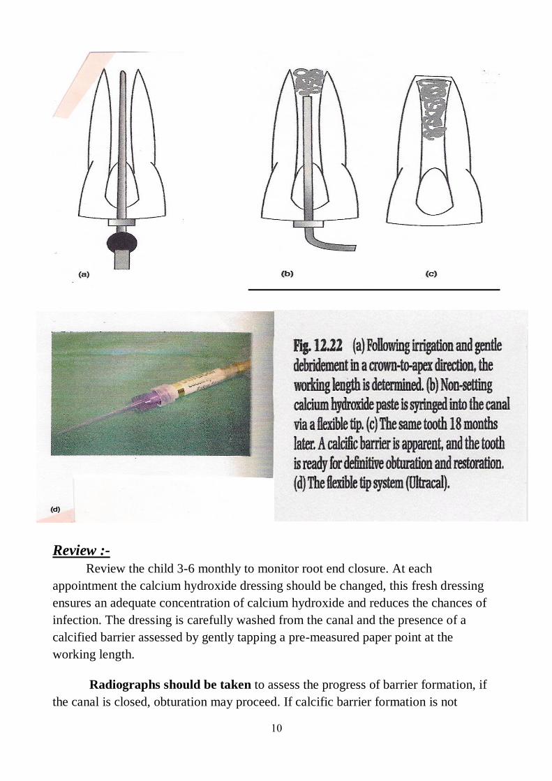
10
Review :-
Review the child 3-6 monthly to monitor root end closure. At each
appointment the calcium hydroxide dressing should be changed, this fresh dressing
ensures an adequate concentration of calcium hydroxide and reduces the chances of
infection. The dressing is carefully washed from the canal and the presence of a
calcified barrier assessed by gently tapping a pre-measured paper point at the
working length.
Radiographs should be taken to assess the progress of barrier formation, if
the canal is closed, obturation may proceed. If calcific barrier formation is not
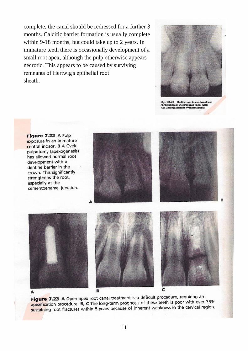
11
complete, the canal should be redressed for a further 3
months. Calcific barrier formation is usually complete
within 9-18 months, but could take up to 2 years. In
immature teeth there is occasionally development of a
small root apex, although the pulp otherwise appears
necrotic. This appears to be caused by surviving
remnants of Hertwig,s epithelial root
sheath.
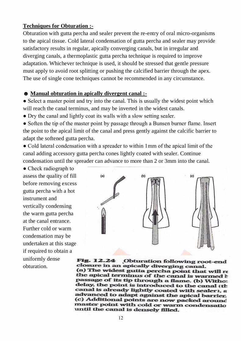
12
Techniques for Obturation :-
Obturation with gutta percha and sealer prevent the re-entry of oral micro-organisms
to the apical tissue. Cold lateral condensation of gutta percha and sealer may provide
satisfactory results in regular, apically converging canals, but in irregular and
diverging canals, a thermoplastic gutta percha technique is required to improve
adaptation. Whichever technique is used, it should be stressed that gentle pressure
must apply to avoid root splitting or pushing the calcified barrier through the apex.
The use of single cone techniques cannot be recommended in any circumstance.
☻ Manual obturation in apically divergent canal :-
● Select a master point and try into the canal. This is usually the widest point which
will reach the canal terminus, and may be inverted in the widest canals.
● Dry the canal and lightly coat its walls with a slow setting sealer.
● Soften the tip of the master point by passage through a Bunsen burner flame. Insert
the point to the apical limit of the canal and press gently against the calcific barrier to
adapt the softened gutta percha.
● Cold lateral condensation with a spreader to within 1mm of the apical limit of the
canal adding accessory gutta percha cones lightly coated with sealer. Continue
condensation until the spreader can advance to more than 2 or 3mm into the canal.
● Check radiograph to
assess the quality of fill
before removing excess
gutta percha with a hot
instrument and
vertically condensing
the warm gutta percha
at the canal entrance.
Further cold or warm
condensation may be
undertaken at this stage
if required to obtain a
uniformly dense
obturation.
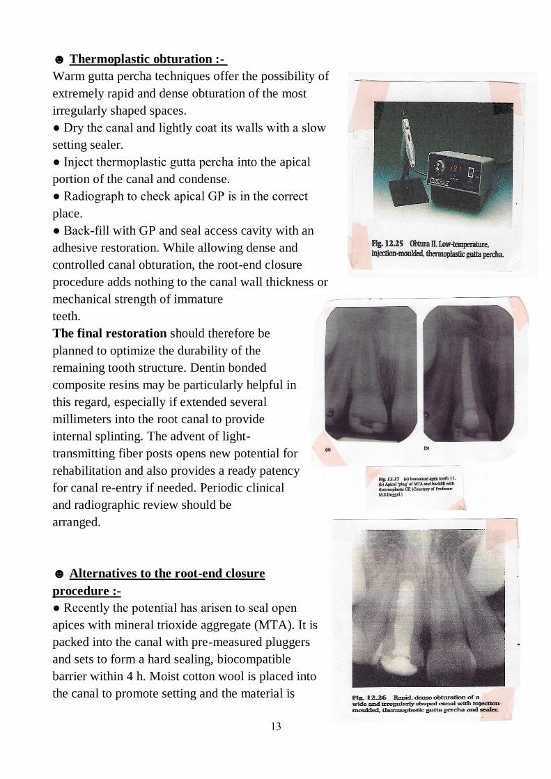
13
☻ Thermoplastic obturation :-
Warm gutta percha techniques offer the possibility of
extremely rapid and dense obturation of the most
irregularly shaped spaces.
● Dry the canal and lightly coat its walls with a slow
setting sealer.
● Inject thermoplastic gutta percha into the apical
portion of the canal and condense.
● Radiograph to check apical GP is in the correct
place.
● Back-fill with GP and seal access cavity with an
adhesive restoration. While allowing dense and
controlled canal obturation, the root-end closure
procedure adds nothing to the canal wall thickness or
mechanical strength of immature
teeth.
The final restoration should therefore be
planned to optimize the durability of the
remaining tooth structure. Dentin bonded
composite resins may be particularly helpful in
this regard, especially if extended several
millimeters into the root canal to provide
internal splinting. The advent of light-
transmitting fiber posts opens new potential for
rehabilitation and also provides a ready patency
for canal re-entry if needed. Periodic clinical
and radiographic review should be
arranged.
☻ Alternatives to the root-end closure
procedure :-
● Recently the potential has arisen to seal open
apices with mineral trioxide aggregate (MTA). It is
packed into the canal with pre-measured pluggers
and sets to form a hard sealing, biocompatible
barrier within 4 h. Moist cotton wool is placed into
the canal to promote setting and the material is
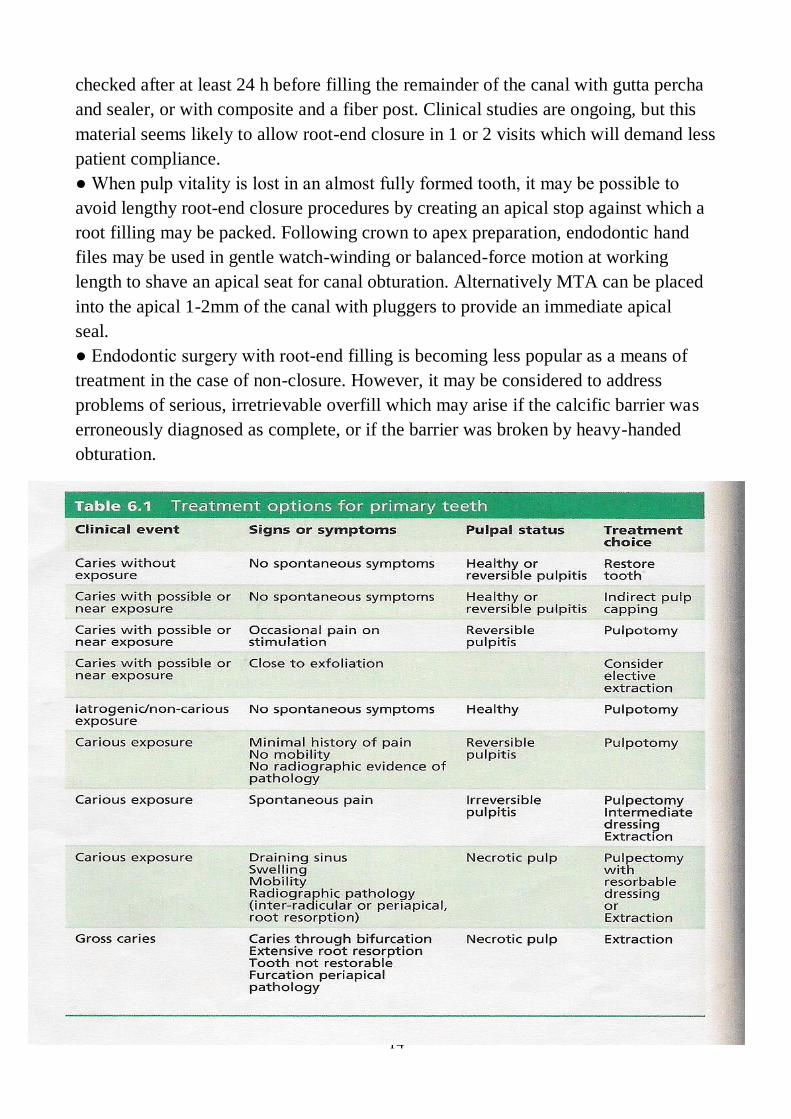
14
checked after at least 24 h before filling the remainder of the canal with gutta percha
and sealer, or with composite and a fiber post. Clinical studies are ongoing, but this
material seems likely to allow root-end closure in 1 or 2 visits which will demand less
patient compliance.
● When pulp vitality is lost in an almost fully formed tooth, it may be possible to
avoid lengthy root-end closure procedures by creating an apical stop against which a
root filling may be packed. Following crown to apex preparation, endodontic hand
files may be used in gentle watch-winding or balanced-force motion at working
length to shave an apical seat for canal obturation. Alternatively MTA can be placed
into the apical 1-2mm of the canal with pluggers to provide an immediate apical
seal.
● Endodontic surgery with root-end filling is becoming less popular as a means of
treatment in the case of non-closure. However, it may be considered to address
problems of serious, irretrievable overfill which may arise if the calcific barrier was
erroneously diagnosed as complete, or if the barrier was broken by heavy-handed
obturation.
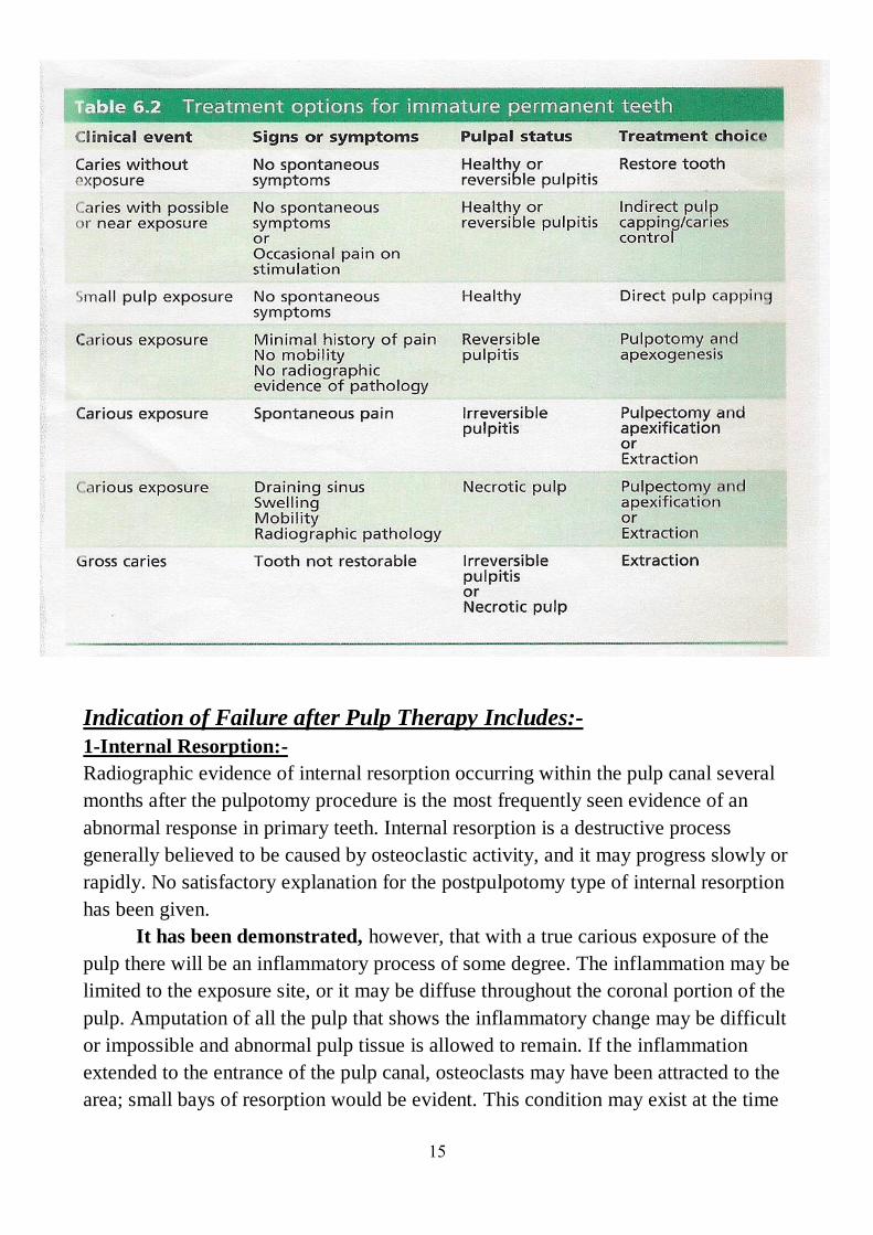
15
Indication of Failure after Pulp Therapy Includes:-
1-Internal Resorption:-
Radiographic evidence of internal resorption occurring within the pulp canal several
months after the pulpotomy procedure is the most frequently seen evidence of an
abnormal response in primary teeth. Internal resorption is a destructive process
generally believed to be caused by osteoclastic activity, and it may progress slowly or
rapidly. No satisfactory explanation for the postpulpotomy type of internal resorption
has been given.
It has been demonstrated, however, that with a true carious exposure of the
pulp there will be an inflammatory process of some degree. The inflammation may be
limited to the exposure site, or it may be diffuse throughout the coronal portion of the
pulp. Amputation of all the pulp that shows the inflammatory change may be difficult
or impossible and abnormal pulp tissue is allowed to remain. If the inflammation
extended to the entrance of the pulp canal, osteoclasts may have been attracted to the
area; small bays of resorption would be evident. This condition may exist at the time
16
of pulp therapy, though there is no way to detect it. The only indication would be the
clinical evidence of a hyperemic pulp.
All the pulp-capping materials in use today are irritating to some extent and
produce at least some degree of inflammation. Inflammatory cells attracted to the
area as a result of the placement of an irritating capping material might well attract
the osteoclastic cells and initiate the internal resorption. This may explain the
occurrence of internal resorption even though the pulp is normal at the time of
treatment.
Because the roots of primary teeth are undergoing normal physiologic resorption;
vascularity of the apical region is increased. There is osteoclastic activity in the area.
This may predispose the tooth to internal resorption when an irritant in the form of a
pulp-capping material is placed on the
pulp.
2-Alveolar Abscess:-
An alveolar abscess occasionally develops some months after pulp therapy has been
completed. The tooth usually remains asymptomatic, and the child is unaware of the
infection, which may be present in the bone surrounding the root apices or in the area
of the root bifurcation. A fistulous opening may be present, indicating the chronic
condition of the infection. Primary teeth that show evidence of an alveolar abscess
should be removed.
Permanent teeth that have previously been treated by pulp capping or by
pulpotomy and later show evidence of pulpal necrosis and apical infection may be
considered for endodontic treatment.
3-Early Exfoliation or Over-Retention of Primary Teeth with Pulp Treatments:-
Occasionally a pulpally treated tooth previously believed to be successfully managed
will loosen and exfoliate (or require extraction) prematurely for no apparent reason. It
is believed that such a condition results from low-grade, chronic, asymptomatic
localized infection. Usually abnormal and incomplete root resorption patterns of the
affected teeth are also observed. When this occurs, space management must be
considered.
Another sequela requiring close observation is the tendency for primary
teeth having successful pulpotomy or pulpectomy to be over-retained. This situation
may have the untoward result of interfering with the normal eruption of permanent
teeth and adversely affection of developing occlusion. Close periodic observation of
pulpally treated teeth is necessary to intercept such a developing problem at the
proper time.

17
Extraction of the primary tooth is usually sufficient, this phenomenon occurs
when normal physiologic exfoliation is delayed by the bulky amount of cement
contained in the pulp chamber. Even though the material is resorbable, its resorption
is slowed significantly when large quantities are present.
1
Paediatric Dentistry
Fifth Stage
Dr. Suhair W. Abbood
Lec. 8
The Restorative Dentistry for Children
The Operative Treatment of Dental Caries in the Primary Dentition :-
Over the years the treatment of dental caries in children has been discussed
and many attempts made to rationalize the management of the disease. it is
unfortunate that many children still suffer from the disease and its consequences.
Hence there is a need to consider operative treatment to prevent the breakdown of the
dentition.
Huge numbers of different techniques and materials have been advocated
over the years. Unfortunately, most treatments are advocated on the basis of dentists'
clinical impressions and there have been very few objective studies that have
attempted to discover which treatments succeed and which do not. Treatment can be
a stressful experience for the child, the parent, and the dentist. It is important that
there is a positive health gain from any treatment that is provided.
What is the Philosophy of the Dental Care :-
Children are the future dental patients and, therefore, the dental care that they
receive should promote positive dental experiences, which in turn would promote
positive dental attitudes. A treatment philosophy which believe is effective in the
management of caries in children is the five-point treatment philosophy for the
provision of high quality dental care for children, which are :-
1-Gain co-operation and trust of the child and parent.
2-Make an accurate diagnosis and devise a treatment plan appropriate to the child's
need.
3-Comprehensive preventive care.
4-Deliver care in a manner the child finds acceptable.
5-Use treatment methods and restorative techniques, which produce a coast-effective,
long-lasting result.
Remove, Restore, or Leave :-
There are certain situations where the clinician might decide not to carry out invasive
restorative procedures in primary teeth and instead use a rigorous preventive
2
approach. Such an approach can be justified where it is likely that remineralization
would occur or the tooth maintained in a state, free from pain or infection until
exfoliation. Recently it has been proposed that it should be possible to divide lesions
into those for which preventive care is advised (PCA) and those for which operative
care is advised (OCA). More work is required on this concept but the following
sections discuss conflicting reasons to treat or not to treat particular carious lesions.
A- Reasons not to Treat :-
These can be divided into several distinct categories :-
1. The Damage done by Treatment to :-
a- The affected tooth :- However conservative the technique it is inevitable
that some sound tooth tissue has to be removed when operative treatment is
undertaken. This weakens the tooth and makes it more likely that problems
such as cracking of the tooth or loss of vitality of the pulp may occur in the
future.
b- The adjacent tooth :- It is almost inevitable when treating an approximal
lesion that the adjacent tooth will be damaged. The outer surface has far
higher fluoride content than the rest of the enamel so that even a slight nick
of the intact surface will remove this reservoir of fluoride. Additionally, it
has been shown that early lesions that remineralise are less susceptible to
caries than intact surfaces and these areas of the tooth are all too easily
removed when preparing an adjacent tooth
c- The periodontal tissues :- Dental treatment can cause both acute and long-term
damage to the periodontium. It is virtually impossible to avoid damaging the
interdental papillae when treating approximal caries. The papillae can be protected by
using rubber dam and/or wedges and if well-fitting restorations are placed the tissues
will heal fairly rapidly, but long-term damage can be more critical. Many adults can
be seen to be suffering from overenthusiastic treatment of approximal caries in their
youth; and while the relative importance of poor margins compared to bacterial
plaque can be debated, the potential damage from approximal restorations is
sufficient reason to avoid treatment unless a definite indication is present.
d- The occlusion :- Poor restoration of the teeth can, over time, lead to considerable
alteration of the occlusion. It is tempting when restoring occlusal surfaces to leave the
material well clear of the opposing teeth to avoid difficulties, or to be unconcerned if
the filling is slightly 'high'. However, this can allow the teeth to erupt into contact
again or the interocclusal position to change and alter the occlusion. Often this is felt
to be of little concern, but there are a large number of adults where the cumulative
effect of many poorly restored teeth has severely disturbed the occlusion, thus
making further treatment difficult, time consuming, and expensive.
3
2. The difficulty of diagnosis :- It is well known that it is difficult to diagnose dental
caries accurately, there is wide variation between examiners. It is not just variations
between examiners that need to be considered as there is also a marked difference
between the same examiners on different occasions. The implications need to be
considered in relation to the decision to treat or not.
3. The slow rate of caries attack :- Caries usually progresses relatively slowly,
although some individuals will show more rapid development than others. The
majority of children and adolescents will have a low level of caries and progress of
carious lesions will be slow. In general, the older the child at the time that the caries
is first diagnosed the slower the progression of the lesion. However, a substantial
group of children will have caries that develops rapidly.
4. The fact that remineralization can arrest and repair enamel caries :- It has
long been known that early, smooth surface lesions are reversible. In addition, it is
now accepted that the chief mechanism whereby fluoride reduces caries is by
encouraging remineralization, and that the remineralised early lesion is more resistant
to caries than intact enamel. Although it is difficult to show reversal of lesions on
radiographs, many studies have demonstrated that a substantial proportion of early
enamel lesions do not progress over many years.
5. The short life of dental restorations :- Surveys of dental treatment have often
shown a rather disappointing level of success. In general, 50% of amalgam
restorations in permanent teeth can be expected to fail during the 10 years following
placement. Some studies have shown an even poorer success rate when looking at
primary teeth, and this has been put forward as a reason for not treating these teeth.
B- Reasons to Treat :-
1. Adverse effects of neglect :- The fact that the treatment of proximal caries can
cause damage to the affected tooth, the adjacent tooth, the periodontium, and the
occlusion is a valid reason to think twice before putting bur to tooth. But, of course, a
case could equally well be made that the neglect of treatment will cause as much or
more damage. Lack of treatment can, and all too often does, lead to loss of contact
with adjacent and opposing teeth, exposure of the pulp resulting in the development
of periapical infection, and/or loss of the tooth. At worst, the child may end up having
a general anaesthetic for the removal of one or more teeth. A procedure, which has
significant morbidity, and mortality.
4
2. Unpredictability of the speed of attack :- While it is true that the rate of attack is
usually slow, it is quite possible for the rate in any one individual to be rapid so that
any delay in treatment would not then be in the best interests of the child.
3. Difficulty in assessing if a lesion is arrested or not :- Because of the normally
slow rate of attack it is difficult to be sure if a lesion is arrested or merely developing
very slowly. It is true that remineralization will arrest and repair early enamel lesions,
but there is, in fact, little evidence that remineralization of the dentine or the late
enamel lesion is common.
4. Success when careful treatment is provided :- The majority of published studies
show that class II amalgam restorations in primary teeth have a poor life expectancy,
but this is not the experience of the careful dentist. Some of these dentists have
published their results, which show that the great majority of their restorations in
primary teeth survive without further attention until they exfoliate.
The treatment procedures used are not particularly difficult in comparison to others
that dentists attempt on adults, and it is difficult to avoid the conclusion that the
reasons for poor results in some studies are due to poor patient management and lack
of attention to detail. It should be the aim of the profession to develop better and
more effective ways of treating the disease rather than throwing our hands up in
surrender.
5. Early treatment is more successful than late :- Small restorations are more
successful than large, and therefore if a carious lesion is going to need treatment it is
better treated early rather than late. This was the rationale behind the early
suggestions of Hyatt of a 'prophylactic filling' for pits and fissures and for the modern
versions in the form of fissure sealants and preventive resin restorations. The fact that
small restorations are often more successful makes for difficult decisions when the
management of caries involves preventive procedures, which need both time to work
and time to assess whether they have been effective.
C- Remove or Restore :-
Once a decision has been made to treat a carious tooth a further decision has to
be made as to whether to remove or restore it. This decision should take into account
the following :-
1. The child :- Each child is an individual and treatment should be planned to
provide the best that is possible for that individual. Too often treatment is given
which is the most convenient for the parent or, more likely, the dentist. Is it really in
the best interest of the child to remove a tooth which could be saved? In the United
5
Kingdom, general anaesthesia is still widely used for removing the teeth of young
children despite the risks of death, its unpleasantness, and the cost involved.
2. The tooth :- It is not usually in a child's interest for a permanent tooth to be
removed. However, if the pulp of a carious permanent tooth is exposed then a
considerable amount of treatment may be required to retain it, and the prognosis for
the tooth would still be poor. It may therefore be in the child's long-term interest to
lose it and to allow another tooth to take its place, either by natural drift or with
orthodontic assistance.
Primary teeth are often considered by parents and some dentists as being disposable
items because there comes a time when they will be exfoliated naturally. However, it
is an unusual child who thinks the same way! Loss of a tooth before its time has a
considerable significance in a child's life. Losing a tooth early gives a message to the
child that teeth are not valuable and not worth looking after. It can then be difficult to
persuade a child to care for their teeth. A well-restored primary dentition can be a
source of pride to young children and an encouragement for them to look after the
succeeding teeth.
3. The stage of the disease :- It is easier for both child and dentist to restore teeth at
an early stage of decay. Later the pulp may become involved and subsequent
restoration difficult, making loss of the tooth more likely.
4. The extent of the disease :- A large number of teeth requiring treatment may put a
strain on a young child and, less importantly, on the parent and dentist. Caries in
children is significantly less than it was 20 years ago, and it would be good to think
that the dental profession would be able to restore the reduced number of decayed
teeth that now present.
Important points in relation to remineralisation :-
1. Early smooth surface lesions are reversible in the right conditions.
2. There is little evidence to suggest that remineralization occurs in lesions already
into dentine.
3. The rate of caries progression is usually slow but can be rapid in some individuals,
particularly younger children. In general, the older the child is at diagnosis of a
carious lesion the slower the progress of the lesion, assuming constancy of other risk
factors.
4. The remineralised tissue of early caries is less susceptible to further caries.
5. Small restorations are generally more successful than large, so a balance has to be
struck, allowing preventive procedures adequate time to function, against the risk of
6
lesion enlargement.
The progression rate of proximal caries can vary from tooth to tooth within the
same mouth. It is thought that if the circumstances for remineralization are
favourable, clinicians should use the modality, as opposed to a restoration that has a
finite but limited lifespan. Remineralization sources available are :-
● fluoride rinse.
● fluoride varnish.
● chlorhexidine thymol varnish.
● oral hygiene measures.
● adjacent glass ionomer restorations.
Determination of the most effective method to retard the progression of approximal
caries requires not only identification of the most effective remineralising agent but
also the frequency with which to employ it. Existing studies indicate that fluoride
varnishes, solutions, and toothpastes all provide a significant effect on the
progression of approximal caries in permanent molars when assessed
radiographically.
Progress of caries through the enamel seems to be fairly slow but once the dentine is
reached it accelerates. So as a rule of thumb, restore proximal surfaces once the
lesion reaches the enamel/ dentine interface.
Important points in relation to treatment :-
1. Gaining access to the caries inevitably means destruction of sound tooth tissue.
The operator must keep this to a minimum, consistent with complete caries
eradication.
2. Once the operator places an initial restoration, he or she cannot 'undo' it and that
tooth will inevitably require further restoration in its lifetime.
3. Every time an operator places a restoration, he or she destroys more of the original
tooth structure, thereby weakening the tooth.
4. Even though the occlusion in a young person changes as growth occurs and teeth
erupt, it is important to realize, that when the operator places restorations, he or she
must replicate the original occlusal contacts in the tooth. Although, it may be
tempting to keep the restoration totally out of the occlusion, teeth will move back into
the occlusion, which will thereafter be slightly different and the cumulative effect of
a lot of little changes can severely disrupt the occlusion in the long term.
5. When treating an approximal lesion on one tooth with an adjacent neighbour, the
operator will almost certainly damage the latter. The important surface layer of the
7
neighbouring tooth, which contains the highest level of fluoride, is the most resistant,
so damage inflicted increases the chances of the adjacent surface of the neighbouring
tooth becoming carious. It also creates an area of roughness on that surface, which in
turn will accumulate more plaque, thereby increasing the risk of further
decalcification.
6. When placing an interproximal restoration it is inevitable that there is some
damage to the periodontal tissues. There is the transient damage caused by placement
of the matrix band and wedge, and there is also an enduring effect caused by the
presence of the restoration margin.
Why Restore the Primary Teeth :-
Our child patients deserve the best dental treatment that clinicians can provide as any
treatment -prevention or restorative- will shape their dental future. The objective of
any restorative treatment is to :-
1-Repair or limit the damage of dental
caries.
2-Protect and preserve remaining the pulp and remaining tooth structure.
3-Ensure adequate function.
4-Restore aesthetics (where applicable).
5-Provide ease in maintaining good oral hygiene.
In addition restoring primary teeth ensures that the natural spaces in the child's
primary dentition are retained for the developing permanent dentition.
The Pit and Fissure Caries:-
Pit and fissure caries is less of a problem in primary teeth than in permanent ones.
The fissures are usually much shallower and less susceptible to decay, so the
presence of a cavity in the occlusal surface of a primary molar is a sign of high caries
activity. Because of this it is quite likely that the children who require treatment of
these surfaces will be young. However, treatment is not difficult and can usually be
accomplished without problem. For restoration, silver amalgam has not so far been
bettered in clinical trial because occlusal caries in the primary dentition indicates high
caries activity, the material of choice may be a resin-modified glass ionomer cement
with its possible caries preventive properties.
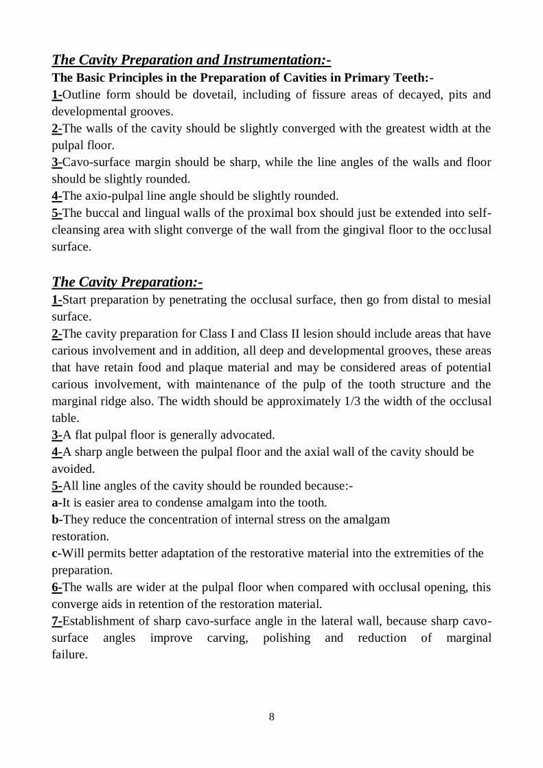
8
The Cavity Preparation and Instrumentation:-
The Basic Principles in the Preparation of Cavities in Primary Teeth:-
1-Outline form should be dovetail, including of fissure areas of decayed, pits and
developmental grooves.
2-The walls of the cavity should be slightly converged with the greatest width at the
pulpal floor.
3-Cavo-surface margin should be sharp, while the line angles of the walls and floor
should be slightly rounded.
4-The axio-pulpal line angle should be slightly rounded.
5-The buccal and lingual walls of the proximal box should just be extended into self-
cleansing area with slight converge of the wall from the gingival floor to the occlusal
surface.
The Cavity Preparation:- 1-Start preparation by penetrating the occlusal surface, then go from distal to mesial
surface.
2-The cavity preparation for Class I and Class II lesion should include areas that have
carious involvement and in addition, all deep and developmental grooves, these areas
that have retain food and plaque material and may be considered areas of potential
carious involvement, with maintenance of the pulp of the tooth structure and the
marginal ridge also. The width should be approximately 1/3 the width of the occlusal
table.
3-A flat pulpal floor is generally advocated.
4-A sharp angle between the pulpal floor and the axial wall of the cavity should be
avoided.
5-All line angles of the cavity should be rounded because:-
a-It is easier area to condense amalgam into the tooth.
b-They reduce the concentration of internal stress on the amalgam
restoration.
c-Will permits better adaptation of the restorative material into the extremities of the
preparation.
6-The walls are wider at the pulpal floor when compared with occlusal opening, this
converge aids in retention of the restoration material.
7-Establishment of sharp cavo-surface angle in the lateral wall, because sharp cavo-
surface angles improve carving, polishing and reduction of marginal
failure.
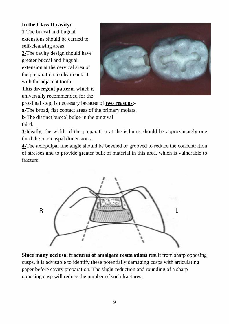
9
In the Class II cavity:-
1-The buccal and lingual
extensions should be carried to
self-cleansing areas.
2-The cavity design should have
greater buccal and lingual
extension at the cervical area of
the preparation to clear contact
with the adjacent tooth.
This divergent pattern, which is
universally recommended for the
proximal step, is necessary because of two reasons:-
a-The broad, flat contact areas of the primary molars.
b-The distinct buccal bulge in the gingival
third.
3-Ideally, the width of the preparation at the isthmus should be approximately one
third the intercuspal dimensions.
4-The axiopulpal line angle should be beveled or grooved to reduce the concentration
of stresses and to provide greater bulk of material in this area, which is vulnerable to
fracture.
Since many occlusal fractures of amalgam restorations result from sharp opposing
cusps, it is advisable to identify these potentially damaging cusps with articulating
paper before cavity preparation. The slight reduction and rounding of a sharp
opposing cusp will reduce the number of such fractures.
L B
10
The Anterior teeth :-
The treatment of decayed primary incisors depends on the stage of decay and
the age and co-operation of the patient. In the preschool child, caries of the upper
primary incisors is usually as a result of 'nursing caries syndrome'.
In 'nursing caries' the progression of decay is rapid, commencing on the labial
surfaces and quickly encircling the teeth. It is impossible to prepare satisfactory
cavities for restoration and after a comprehensive preventive programme the most
suitable form of restoration is the 'strip crown technique'. This utilizes celluloid
crown forms and a light-cured composite resin to restore crown morphology. Either
calcium hydroxide or glass ionomer cement can be used as a lining and the high
polishability of modern hybrid composites make them aesthetically, as well as
physically, suitable for this task.
In older children over 3 or 4 years of age new lesions of primary incisors, although
not usually associated with the use of pacifiers, do indicate high caries activity. Such
lesions do not progress so rapidly and usually appear on the mesial and distal
surfaces, here a glass ionomer cement or composite resin can be used for restoration.
Glass ionomer lacks the translucency of composite resin but has the useful
advantages of being adhesive and releasing fluoride.
Fractures of the incisal edges in primary teeth, as in permanent teeth, should be
restored with composite resin.
Strip Crowns (3M ESPEE) are a useful aid in the restoration of primary incisors,
these crowns are excellent for building primary incisors where extensive tooth tissue
has been lost due to either caries or trauma. The technique for their use is similar to
that of such crowns used in permanent teeth; the crowns are easily trimmed with
sharp scissors, filled with composite, and seated on a prepared and conditioned tooth.
The celluloid crown form can be stripped off after the composite has been cured.
The Operative Treatment of Dental Caries in the Young Permanent
Dentition :-
Caries is still a considerable problem in children and adolescents. The first permanent
teeth erupt into the mouth at approximately 6 years of age, but may appear as early as
the age of 4. The eruption of the anterior teeth usually causes great excitement, as it is
associated with 'the fluttering of tooth fairy wings'. However, the eruption of the first
permanent molars largely goes unnoticed until there is a problem. The mean eruption
time for first permanent molars has been determined as, 6.1 years in girls and 6.3
years in boys, but there is a tremendous variation in both the time of eruption and the
11
time it takes for the tooth to emerge into the mouth. It takes 12-18 months for a first
or second molar to erupt fully. The occlusal surfaces of these molar teeth account for
about 90% of caries in children.
Restoration of the young permanent dentition is part of a continuum and cannot be
regarded in isolation. The restoration is only one small part of the child's treatment
and is the 'surgery' to remove the carious infected area of the tooth and replace it with
a suitable restorative material. It does nothing to cure the disease and must form part
of a much wider treatment modality, which includes identification of the risk factors
contributing to the disease followed by introduction of specific prevention counter
measures. Efforts must be applied to all of these areas to attempt to provide the
optimum conditions for future tooth survival. The clinician must always give
consideration to whether it is better either to treat a carious lesion or remineralise it.
The Management of Occlusal Caries in Permanent Teeth :-
It is of great importance, is the preservation of tooth structure. The placement of
unnecessarily large amalgam restorations undermines the marginal ridges and
weakens the cusps which will eventually fracture. The tooth then will require even
larger restorations with the risk of pulp disease, root canal treatment and finally full
coverage restoration. There must be a different approach to the management of
permanent teeth that have not been previously restored compared with those teeth
which require replacement of restorations.
Amalgam is an inappropriate material for the restoration of early lesions on the
occlusal surfaces of permanent teeth. Here, the preventive resin restoration is more
desirable. Minimal tooth structure is lost in cavity preparation and has the advantage
that the occlusal table is protected by a fissure sealant.
The Occlusal Caries :-
Where the dentist has established a diagnosis that a stained fissure is a carious
lesion into dentine, restorative treatment is indicated. If the lesion is limited to areas
of the tooth not bearing occlusal loads then a PRR (Preventive Resin Restoration) is
appropriate. Due to its superior wear resistance and superior mechanical properties,
composite resin materials rather than glass ionomers are the material of choice for the
treatment of early occlusal caries in permanent teeth. The development of preventive
resin restorations has changed the management of occlusal caries dramatically in
young patients.
The indications of PRR in enamel-only lesions, incipient lesions just into dentin,
and small class I lesions. The durability of preventive resin restoration has been
12
proved to be as good as occlusal amalgam restorations and can be achieved with
significantly less removal of sound tooth tissue. If the lesion is more extensive, then
the clinician should consider a composite or an amalgam restoration.
The Occlusal restorations in young permanent teeth:- If caries affects most of the occlusal fissure system, the clinician should place a
classical class I restoration. The choice of material for this restoration is dependent on
the operator and appropriately informed parent. The plethora of available tooth
coloured materials together with the continuing development and introduction of new
materials makes choice both extensive and difficult.
In small occlusal restorations the only difference needed in the tooth preparation
between composite and amalgam is that when an amalgam is to be placed,
undermined enamel must be removed. In both cases a resin sealant material should be
placed over the margins of the restoration and the remaining fissure system.
Researchers report very high success rates when amalgam is used in this manner.
The approximal Caries :-
In children caries occurs more often occlusally than approximally, but as they
progress to adulthood, the relative level of proximal caries increases. The authors
advocate managing occlusal caries immediately by sealing or PRRs. They also
support remineralization techniques as an early intervention approach in approximal
caries, where the lesion has not reached the dentine.
Whichever way the clinician chooses to restore approximal caries, it will always
entail loss of some sound tooth tissue. In approximal restorations, sufficient tooth
preparation just to gain access to the carious dentine is necessary. Shape the outline
form only to include the carious dentine and to remove demineralized enamel. Finish
the cavo surface margins to remove unsupported enamel.
Amalgam works well in these situations but clinicians are equally using composite
resins more frequently in approximal restorations of young permanent teeth.
Although there are some studies reporting good success rates, the overall consensus
seems to be that tooth coloured restorations are prone to earlier failure than amalgam
restorations. Operators should inform parents of this proviso when discussing the
choice of restorative material.
The Basic Principles in the Preparation of Class II Cavities in
Permanent Molars:-
The principles of cavity preparation for permanent molars, as presented many years
ago, are generally still advocated today. However, as the result of extensive
13
laboratory and clinical research, modifications of the original preparation have been
recommended. The most obvious difference is a reduction in the dimensions of the
cavity preparation, made possible by the smaller burs available today and the
precision methods of cutting tooth structure.
The following basic principles will serve as guides in the preparation of Class II
cavities in permanent molars:-
1-All fissured grooves in the occlusal surface that appear caries susceptible should be
extended and included in the preparation to prevent caries recurrence. However, they
should be kept at a minimum width.
2-The proximal portion of the restoration should be self-retentive.
3-The proximal outline will be determine by the extent of the lesion and by the
morphology of the adjacent tooth; the preparation is carried buccolingually to an area
not quite touching the adjacent tooth to allow cleansing by the patient.
4-The proximal outline should converge occlusally to a slight degree in the form of a
mortise, generally following the buccal and lingual contour of the
tooth.
5-The gingival margin should be extended cervically to break contact with the
adjacent tooth.
Thus the outline of the cavity for the
amalgam restoration is determined
by:-
1-The size of the carious lesion.
2-The need for extension for prevention.
3-The occlusal and proximal anatomy of the tooth.
Extensive cutting of the natural sound tooth structure will only weaken the tooth and
the final restoration.
The Anterior Caries :-
Caries of permanent anterior teeth in childhood and adolescence, is not that common
and usually occurs either where there is defect in the formation of the teeth, which
●1a) gingival floor position
●1b) box is perpendicular to
long axis
● 1c) rounded angles
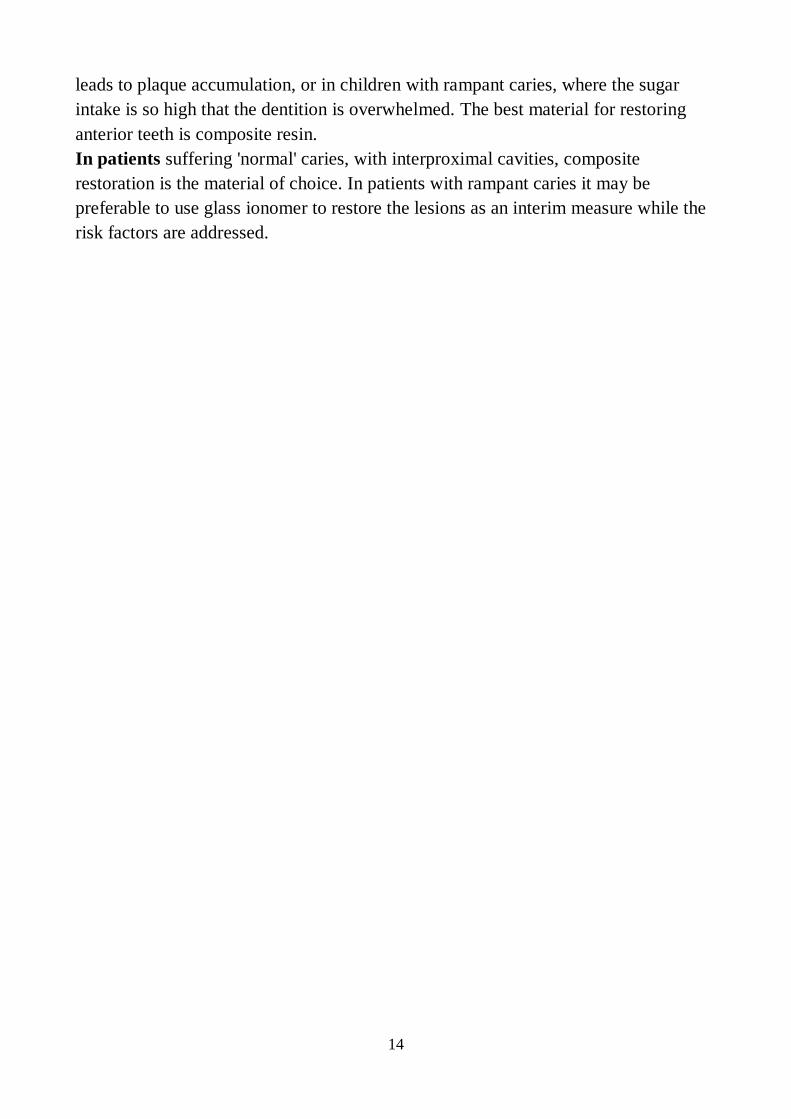
14
leads to plaque accumulation, or in children with rampant caries, where the sugar
intake is so high that the dentition is overwhelmed. The best material for restoring
anterior teeth is composite resin.
In patients suffering 'normal' caries, with interproximal cavities, composite
restoration is the material of choice. In patients with rampant caries it may be
preferable to use glass ionomer to restore the lesions as an interim measure while the
risk factors are addressed.
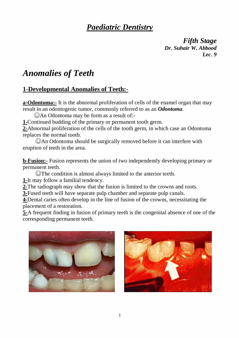
1
Paediatric Dentistry
Fifth Stage Dr. Suhair W. Abbood
Lec. 9
Anomalies of Teeth
1-Developmental Anomalies of Teeth:-
a-Odontoma:- It is the abnormal proliferation of cells of the enamel organ that may
result in an odontogenic tumor, commonly referred to as an Odontoma.
☺An Odontoma may be form as a result of:-
1-Continued budding of the primary or permanent tooth germ.
2-Abnormal proliferation of the cells of the tooth germ, in which case an Odontoma
replaces the normal tooth.
☺An Odontoma should be surgically removed before it can interfere with
eruption of teeth in the area.
b-Fusion:- Fusion represents the union of two independently developing primary or
permanent teeth.
☺The condition is almost always limited to the anterior teeth.
1-It may follow a familial tendency.
2-The radiograph may show that the fusion is limited to the crowns and roots.
3-Fused teeth will have separate pulp chamber and separate pulp canals.
4-Dental caries often develop in the line of fusion of the crowns, necessitating the
placement of a restoration.
5-A frequent finding in fusion of primary teeth is the congenital absence of one of the
corresponding permanent teeth.
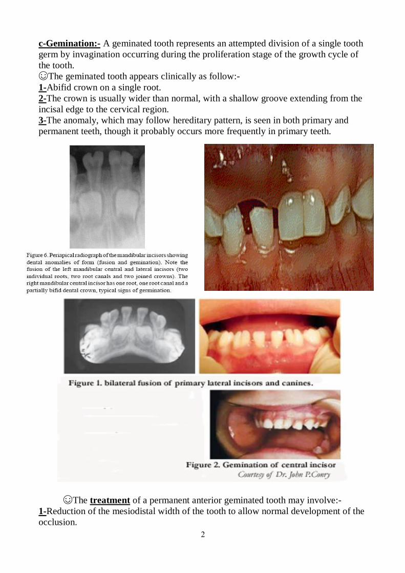
2
c-Gemination:- A geminated tooth represents an attempted division of a single tooth
germ by invagination occurring during the proliferation stage of the growth cycle of
the tooth.
☺The geminated tooth appears clinically as follow:-
1-Abifid crown on a single root.
2-The crown is usually wider than normal, with a shallow groove extending from the
incisal edge to the cervical region.
3-The anomaly, which may follow hereditary pattern, is seen in both primary and
permanent teeth, though it probably occurs more frequently in primary teeth.
☺The treatment of a permanent anterior geminated tooth may involve:-
1-Reduction of the mesiodistal width of the tooth to allow normal development of the
occlusion.
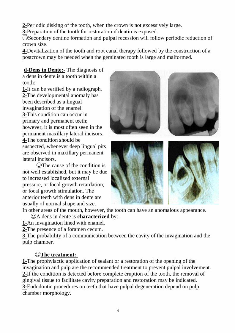
3
2-Periodic disking of the tooth, when the crown is not excessively large.
3-Preparation of the tooth for restoration if dentin is exposed.
☺Secondary dentine formation and pulpal recession will follow periodic reduction of
crown size.
4-Devitalization of the tooth and root canal therapy followed by the construction of a
postcrown may be needed when the geminated tooth is large and malformed.
d-Dens in Dente:- The diagnosis of
a dens in dente is a tooth within a
tooth:-
1-It can be verified by a radiograph.
2-The developmental anomaly has
been described as a lingual
invagination of the enamel.
3-This condition can occur in
primary and permanent teeth;
however, it is most often seen in the
permanent maxillary lateral incisors.
4-The condition should be
suspected, whenever deep lingual pits
are observed in maxillary permanent
lateral incisors.
☺The cause of the condition is
not well established, but it may be due
to increased localized external
pressure, or focal growth retardation,
or focal growth stimulation. The
anterior teeth with dens in dente are
usually of normal shape and size.
In other areas of the mouth, however, the tooth can have an anomalous appearance.
☺A dens in dente is characterized by:-
1-An invagination lined with enamel.
2-The presence of a foramen cecum.
3-The probability of a communication between the cavity of the invagination and the
pulp chamber.
☺The treatment:-
1-The prophylactic application of sealant or a restoration of the opening of the
invagination and pulp are the recommended treatment to prevent pulpal involvement.
2-If the condition is detected before complete eruption of the tooth, the removal of
gingival tissue to facilitate cavity preparation and restoration may be indicated.
3-Endodontic procedures on teeth that have pulpal degeneration depend on pulp
chamber morphology.
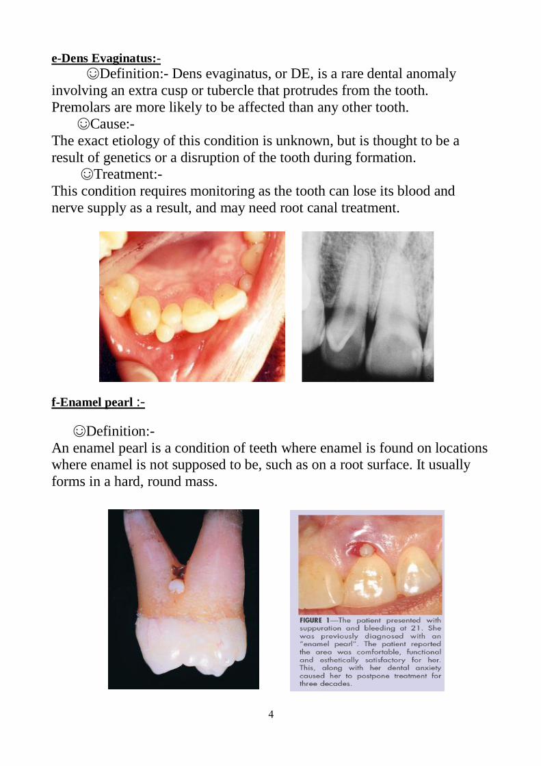
4
e-Dens Evaginatus:-
☺Definition:- Dens evaginatus, or DE, is a rare dental anomaly
involving an extra cusp or tubercle that protrudes from the tooth.
Premolars are more likely to be affected than any other tooth. ☺Cause:-
The exact etiology of this condition is unknown, but is thought to be a
result of genetics or a disruption of the tooth during formation. ☺Treatment:-
This condition requires monitoring as the tooth can lose its blood and
nerve supply as a result, and may need root canal treatment.
f-Enamel pearl :-
☺Definition:-
An enamel pearl is a condition of teeth where enamel is found on locations where enamel is not supposed to be, such as on a root surface. It usually
forms in a hard, round mass.
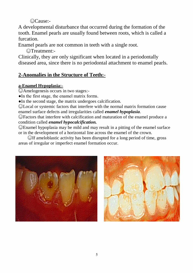
5
☺Cause:-
A developmental disturbance that occurred during the formation of the
tooth. Enamel pearls are usually found between roots, which is called a furcation.
Enamel pearls are not common in teeth with a single root.
☺Treatment:- Clinically, they are only significant when located in a periodontally
diseased area, since there is no periodontal attachment to enamel pearls.
2-Anomalies in the Structure of Teeth:-
a-Enamel Hypoplasia:-
☺Amelogenesis occurs in two stages:-
●In the first stage, the enamel matrix forms.
●In the second stage, the matrix undergoes calcification.
☺Local or systemic factors that interfere with the normal matrix formation cause
enamel surface defects and irregularities called enamel hypoplasia.
☺Factors that interfere with calcification and maturation of the enamel produce a
condition called enamel hypocalcification.
☺Enamel hypoplasia may be mild and may result in a pitting of the enamel surface
or in the development of a horizontal line across the enamel of the crown.
☺If ameloblastic activity has been disrupted for a long period of time, gross
areas of irregular or imperfect enamel formation occur.
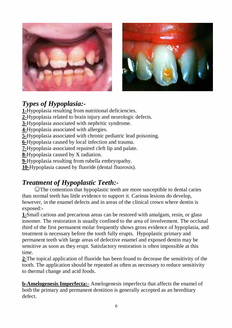
6
Types of Hypoplasia:- 1-Hypoplasia resulting from nutritional deficiencies.
2-Hypoplasia related to brain injury and neurologic defects.
3-Hypoplasia associated with nephritic syndrome.
4-Hypoplasia associated with allergies.
5-Hypoplasia associated with chronic pediatric lead poisoning.
6-Hypoplasia caused by local infection and trauma.
7-Hypoplasia associated repaired cleft lip and palate.
8-Hypoplasia caused by X radiation.
9-Hypoplasia resulting from rubella embryopathy.
10-Hypoplasia caused by fluoride (dental fluorosis).
Treatment of Hypoplastic Teeth:- ☺The contention that hypoplastic teeth are more susceptible to dental caries
than normal teeth has little evidence to support it. Carious lesions do develop,
however, in the enamel defects and in areas of the clinical crown where dentin is
exposed:-
1-Small carious and precarious areas can be restored with amalgam, resin, or glass
ionomer. The restoration is usually confined to the area of involvement. The occlusal
third of the first permanent molar frequently shows gross evidence of hypoplasia, and
treatment is necessary before the tooth fully erupts. Hypoplastic primary and
permanent teeth with large areas of defective enamel and exposed dentin may be
sensitive as soon as they erupt. Satisfactory restoration is often impossible at this
time.
2-The topical application of fluoride has been found to decrease the sensitivity of the
tooth. The application should be repeated as often as necessary to reduce sensitivity
to thermal change and acid foods.
b-Amelogenesis Imperfecta:- Amelogenesis imperfecta that affects the enamel of
both the primary and permanent dentition is generally accepted as an hereditary
defect.
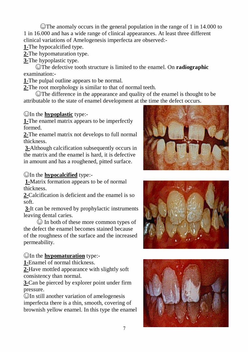
7
☺The anomaly occurs in the general population in the range of 1 in 14.000 to
1 in 16.000 and has a wide range of clinical appearances. At least three different
clinical variations of Amelogenesis imperfecta are observed:-
1-The hypocalcified type.
2-The hypomaturation type.
3-The hypoplastic type.
☺The defective tooth structure is limited to the enamel. On radiographic
examination:-
1-The pulpal outline appears to be normal.
2-The root morphology is similar to that of normal teeth.
☺The difference in the appearance and quality of the enamel is thought to be
attributable to the state of enamel development at the time the defect occurs.
☺In the hypoplastic type:-
1-The enamel matrix appears to be imperfectly
formed.
2-The enamel matrix not develops to full normal
thickness.
3-Although calcification subsequently occurs in
the matrix and the enamel is hard, it is defective
in amount and has a roughened, pitted surface.
☺In the hypocalcified type:-
1-Matrix formation appears to be of normal
thickness.
2-Calcification is deficient and the enamel is so
soft.
3-It can be removed by prophylactic instruments
leaving dental caries.
☺ In both of these more common types of
the defect the enamel becomes stained because
of the roughness of the surface and the increased
permeability.
☺In the hypomaturation type:-
1-Enamel of normal thickness.
2-Have mottled appearance with slightly soft
consistency than normal.
3-Can be pierced by explorer point under firm
pressure.
☺In still another variation of amelogenesis
imperfecta there is a thin, smooth, covering of
brownish yellow enamel. In this type the enamel
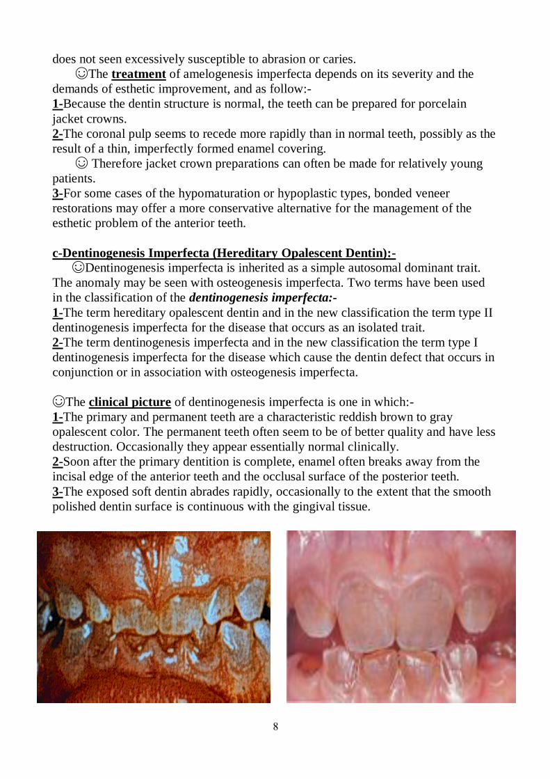
8
does not seen excessively susceptible to abrasion or caries.
☺The treatment of amelogenesis imperfecta depends on its severity and the
demands of esthetic improvement, and as follow:-
1-Because the dentin structure is normal, the teeth can be prepared for porcelain
jacket crowns.
2-The coronal pulp seems to recede more rapidly than in normal teeth, possibly as the
result of a thin, imperfectly formed enamel covering.
☺ Therefore jacket crown preparations can often be made for relatively young
patients.
3-For some cases of the hypomaturation or hypoplastic types, bonded veneer
restorations may offer a more conservative alternative for the management of the
esthetic problem of the anterior teeth.
c-Dentinogenesis Imperfecta (Hereditary Opalescent Dentin):-
☺Dentinogenesis imperfecta is inherited as a simple autosomal dominant trait.
The anomaly may be seen with osteogenesis imperfecta. Two terms have been used
in the classification of the dentinogenesis imperfecta:-
1-The term hereditary opalescent dentin and in the new classification the term type II
dentinogenesis imperfecta for the disease that occurs as an isolated trait.
2-The term dentinogenesis imperfecta and in the new classification the term type I
dentinogenesis imperfecta for the disease which cause the dentin defect that occurs in
conjunction or in association with osteogenesis imperfecta.
☺The clinical picture of dentinogenesis imperfecta is one in which:-
1-The primary and permanent teeth are a characteristic reddish brown to gray
opalescent color. The permanent teeth often seem to be of better quality and have less
destruction. Occasionally they appear essentially normal clinically.
2-Soon after the primary dentition is complete, enamel often breaks away from the
incisal edge of the anterior teeth and the occlusal surface of the posterior teeth.
3-The exposed soft dentin abrades rapidly, occasionally to the extent that the smooth
polished dentin surface is continuous with the gingival tissue.
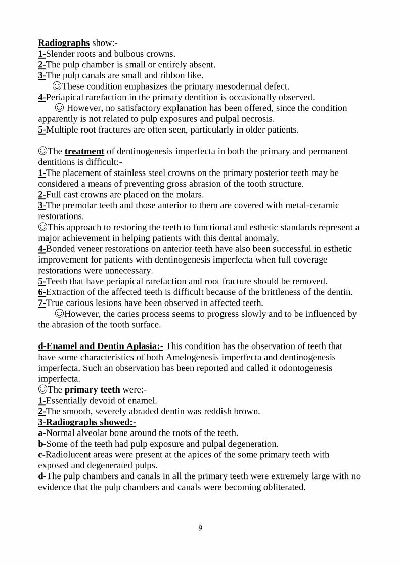
9
Radiographs show:-
1-Slender roots and bulbous crowns.
2-The pulp chamber is small or entirely absent.
3-The pulp canals are small and ribbon like.
☺These condition emphasizes the primary mesodermal defect.
4-Periapical rarefaction in the primary dentition is occasionally observed.
☺ However, no satisfactory explanation has been offered, since the condition
apparently is not related to pulp exposures and pulpal necrosis.
5-Multiple root fractures are often seen, particularly in older patients.
☺The treatment of dentinogenesis imperfecta in both the primary and permanent
dentitions is difficult:-
1-The placement of stainless steel crowns on the primary posterior teeth may be
considered a means of preventing gross abrasion of the tooth structure.
2-Full cast crowns are placed on the molars.
3-The premolar teeth and those anterior to them are covered with metal-ceramic
restorations.
☺This approach to restoring the teeth to functional and esthetic standards represent a
major achievement in helping patients with this dental anomaly.
4-Bonded veneer restorations on anterior teeth have also been successful in esthetic
improvement for patients with dentinogenesis imperfecta when full coverage
restorations were unnecessary.
5-Teeth that have periapical rarefaction and root fracture should be removed.
6-Extraction of the affected teeth is difficult because of the brittleness of the dentin.
7-True carious lesions have been observed in affected teeth.
☺However, the caries process seems to progress slowly and to be influenced by
the abrasion of the tooth surface.
d-Enamel and Dentin Aplasia:- This condition has the observation of teeth that
have some characteristics of both Amelogenesis imperfecta and dentinogenesis
imperfecta. Such an observation has been reported and called it odontogenesis
imperfecta.
☺The primary teeth were:-
1-Essentially devoid of enamel.
2-The smooth, severely abraded dentin was reddish brown.
3-Radiographs showed:-
a-Normal alveolar bone around the roots of the teeth.
b-Some of the teeth had pulp exposure and pulpal degeneration.
c-Radiolucent areas were present at the apices of the some primary teeth with
exposed and degenerated pulps.
d-The pulp chambers and canals in all the primary teeth were extremely large with no
evidence that the pulp chambers and canals were becoming obliterated.
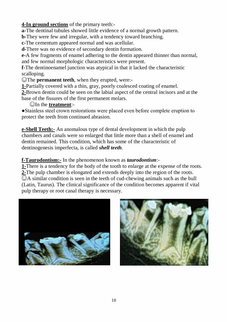
10
4-In ground sections of the primary teeth:-
a-The dentinal tubules showed little evidence of a normal growth pattern.
b-They were few and irregular, with a tendency toward branching.
c-The cementum appeared normal and was acellular.
d-There was no evidence of secondary dentin formation.
e-A few fragments of enamel adhering to the dentin appeared thinner than normal,
and few normal morphologic characteristics were present.
f-The dentinoenamel junction was atypical in that it lacked the characteristic
scalloping.
☺The permanent teeth, when they erupted, were:-
1-Partially covered with a thin, gray, poorly coalesced coating of enamel.
2-Brown dentin could be seen on the labial aspect of the central incisors and at the
base of the fissures of the first permanent molars.
☺In the treatment:-
●Stainless steel crown restorations were placed even before complete eruption to
protect the teeth from continued abrasion.
e-Shell Teeth:- An anomalous type of dental development in which the pulp
chambers and canals were so enlarged that little more than a shell of enamel and
dentin remained. This condition, which has some of the characteristic of
dentinogenesis imperfecta, is called shell teeth.
f-Taurodontism:- In the phenomenon known as taurodontism:-
1-There is a tendency for the body of the tooth to enlarge at the expense of the roots.
2-The pulp chamber is elongated and extends deeply into the region of the roots.
☺A similar condition is seen in the teeth of cud-chewing animals such as the bull
(Latin, Taurus). The clinical significance of the condition becomes apparent if vital
pulp therapy or root canal therapy is necessary.
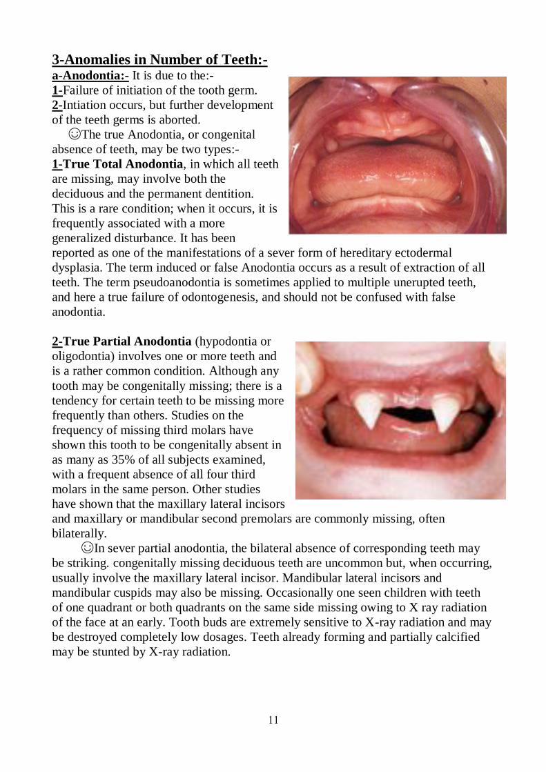
11
3-Anomalies in Number of Teeth:- a-Anodontia:- It is due to the:-
1-Failure of initiation of the tooth germ.
2-Intiation occurs, but further development
of the teeth germs is aborted.
☺The true Anodontia, or congenital
absence of teeth, may be two types:-
1-True Total Anodontia, in which all teeth
are missing, may involve both the
deciduous and the permanent dentition.
This is a rare condition; when it occurs, it is
frequently associated with a more
generalized disturbance. It has been
reported as one of the manifestations of a sever form of hereditary ectodermal
dysplasia. The term induced or false Anodontia occurs as a result of extraction of all
teeth. The term pseudoanodontia is sometimes applied to multiple unerupted teeth,
and here a true failure of odontogenesis, and should not be confused with false
anodontia.
2-True Partial Anodontia (hypodontia or
oligodontia) involves one or more teeth and
is a rather common condition. Although any
tooth may be congenitally missing; there is a
tendency for certain teeth to be missing more
frequently than others. Studies on the
frequency of missing third molars have
shown this tooth to be congenitally absent in
as many as 35% of all subjects examined,
with a frequent absence of all four third
molars in the same person. Other studies
have shown that the maxillary lateral incisors
and maxillary or mandibular second premolars are commonly missing, often
bilaterally.
☺In sever partial anodontia, the bilateral absence of corresponding teeth may
be striking. congenitally missing deciduous teeth are uncommon but, when occurring,
usually involve the maxillary lateral incisor. Mandibular lateral incisors and
mandibular cuspids may also be missing. Occasionally one seen children with teeth
of one quadrant or both quadrants on the same side missing owing to X ray radiation
of the face at an early. Tooth buds are extremely sensitive to X-ray radiation and may
be destroyed completely low dosages. Teeth already forming and partially calcified
may be stunted by X-ray radiation.
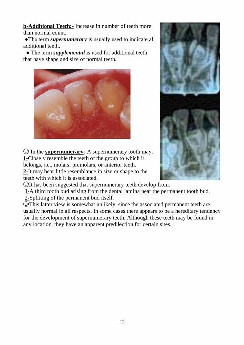
12
b-Additional Teeth:- Increase in number of teeth more
than normal count.
●The term supernumerary is usually used to indicate all
additional teeth.
● The term supplemental is used for additional teeth
that have shape and size of normal teeth.
☺ In the supernumerary:-A supernumerary tooth may:-
1-Closely resemble the teeth of the group to which it
belongs, i.e., molars, premolars, or anterior teeth.
2-It may bear little resemblance in size or shape to the
teeth with which it is associated.
☺It has been suggested that supernumerary teeth develop from:-
1-A third tooth bud arising from the dental lamina near the permanent tooth bud.
2-Splitting of the permanent bud itself.
☺This latter view is somewhat unlikely, since the associated permanent teeth are
usually normal in all respects. In some cases there appears to be a hereditary tendency
for the development of supernumerary teeth. Although these teeth may be found in
any location, they have an apparent predilection for certain sites.
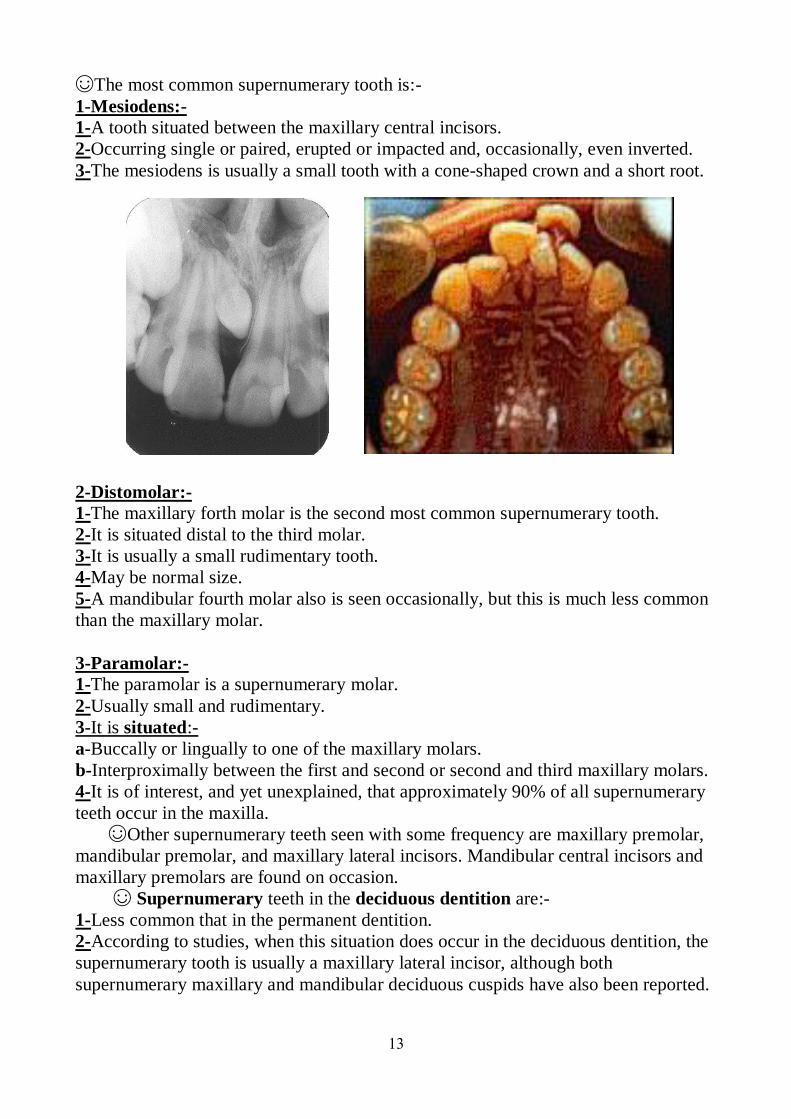
13
☺The most common supernumerary tooth is:-
1-Mesiodens:-
1-A tooth situated between the maxillary central incisors.
2-Occurring single or paired, erupted or impacted and, occasionally, even inverted.
3-The mesiodens is usually a small tooth with a cone-shaped crown and a short root.
2-Distomolar:-
1-The maxillary forth molar is the second most common supernumerary tooth.
2-It is situated distal to the third molar.
3-It is usually a small rudimentary tooth.
4-May be normal size.
5-A mandibular fourth molar also is seen occasionally, but this is much less common
than the maxillary molar.
3-Paramolar:-
1-The paramolar is a supernumerary molar.
2-Usually small and rudimentary.
3-It is situated:-
a-Buccally or lingually to one of the maxillary molars.
b-Interproximally between the first and second or second and third maxillary molars.
4-It is of interest, and yet unexplained, that approximately 90% of all supernumerary
teeth occur in the maxilla.
☺Other supernumerary teeth seen with some frequency are maxillary premolar,
mandibular premolar, and maxillary lateral incisors. Mandibular central incisors and
maxillary premolars are found on occasion.
☺ Supernumerary teeth in the deciduous dentition are:-
1-Less common that in the permanent dentition.
2-According to studies, when this situation does occur in the deciduous dentition, the
supernumerary tooth is usually a maxillary lateral incisor, although both
supernumerary maxillary and mandibular deciduous cuspids have also been reported.
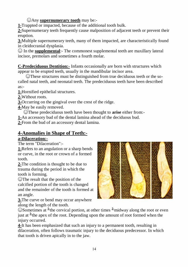
14
☺Any supernumerary tooth may be:-
1-Trappted or impacted, because of the additional tooth bulk.
2-Supernumerary teeth frequently cause malposition of adjacent teeth or prevent their
eruption.
3-Multiple supernumerary teeth, many of them impacted, are characteristically found
in cleidocranial dysplasia.
☺ In the supplemental:- The commonest supplemental teeth are maxillary lateral
incisor, premolars and sometimes a fourth molar.
C-Predeciduous Dentition:- Infants occasionally are born with structures which
appear to be erupted teeth, usually in the mandibular incisor area.
☺These structures must be distinguished from true deciduous teeth or the so-
called natal teeth, and neonatal teeth. The predeciduous teeth have been described
as:-
1-Hornified epithelial structures.
2-Without roots.
3-Occurring on the gingival over the crest of the ridge.
4-May be easily removed.
☺These predeciduous teeth have been thought to arise either from:-
1-An accessory bud of the dental lamina ahead of the deciduous bud.
2-From the bud of an accessory dental lamina.
4-Anomalies in Shape of Teeth:- a-Dilaceration:-
The term "Dilaceration":-
1-Refers to an angulation or a sharp bends
or curve, in the root or crown of a formed
tooth.
2-The condition is thought to be due to
trauma during the period in which the
tooth is forming.
☺The result that the position of the
calcified portion of the tooth is changed
and the remainder of the tooth is formed at
an angle.
3-The curve or bend may occur anywhere
along the length of the tooth.
☺Sometimes at 1-
the cervical portion, at other times 2-
midway along the root or even
just at 3-
the apex of the root. Depending upon the amount of root formed when the
injury occurred.
4-It has been emphasized that such an injury to a permanent tooth, resulting in
dilaceration, often follows traumatic injury to the deciduous predecessor. In which
that tooth is driven apically in to the jaw.
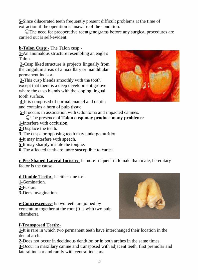
15
5-Since dilacerated teeth frequently present difficult problems at the time of
extraction if the operation is unaware of the condition.
☺The need for preoperative roentgenograms before any surgical procedures are
carried out is self-evident.
b-Talon Cusp:- The Talon cusp:-
1-An anomalous structure resembling an eagle's
Talon.
2-Cusp liked structure is projects lingually from
the cingulum areas of a maxillary or mandibular
permanent incisor.
3-This cusp blends smoothly with the tooth
except that there is a deep development groove
where the cusp blends with the sloping lingual
tooth surface.
4-It is composed of normal enamel and dentin
and contains a horn of pulp tissue.
5-It occurs in association with Odontoma and impacted canines.
☺The presence of Talon cusp may produce many problems:-
1-Interfere with occlusion.
2-Displace the teeth.
3-The cusps or opposing teeth may undergo attrition.
4-It may interfere with speech.
5-It may sharply irritate the tongue.
6-The affected teeth are more susceptible to caries.
c-Peg Shaped Lateral Incisor:- Is more frequent in female than male, hereditary
factor is the cause.
d-Double Teeth:- Is either due to:-
1-Gemination.
2-Fusion.
3-Dens invagination.
e-Concrescence:- Is two teeth are joined by
cementum together at the root (It is with two pulp
chambers).
f-Transposed Teeth:-
1-It is rare in which two permanent teeth have interchanged their location in the
dental arch.
2-Does not occur in deciduous dentition or in both arches in the same times.
3-Occur in maxillary canine and transposed with adjacent teeth, first premolar and
lateral incisor and rarely with central incisors.
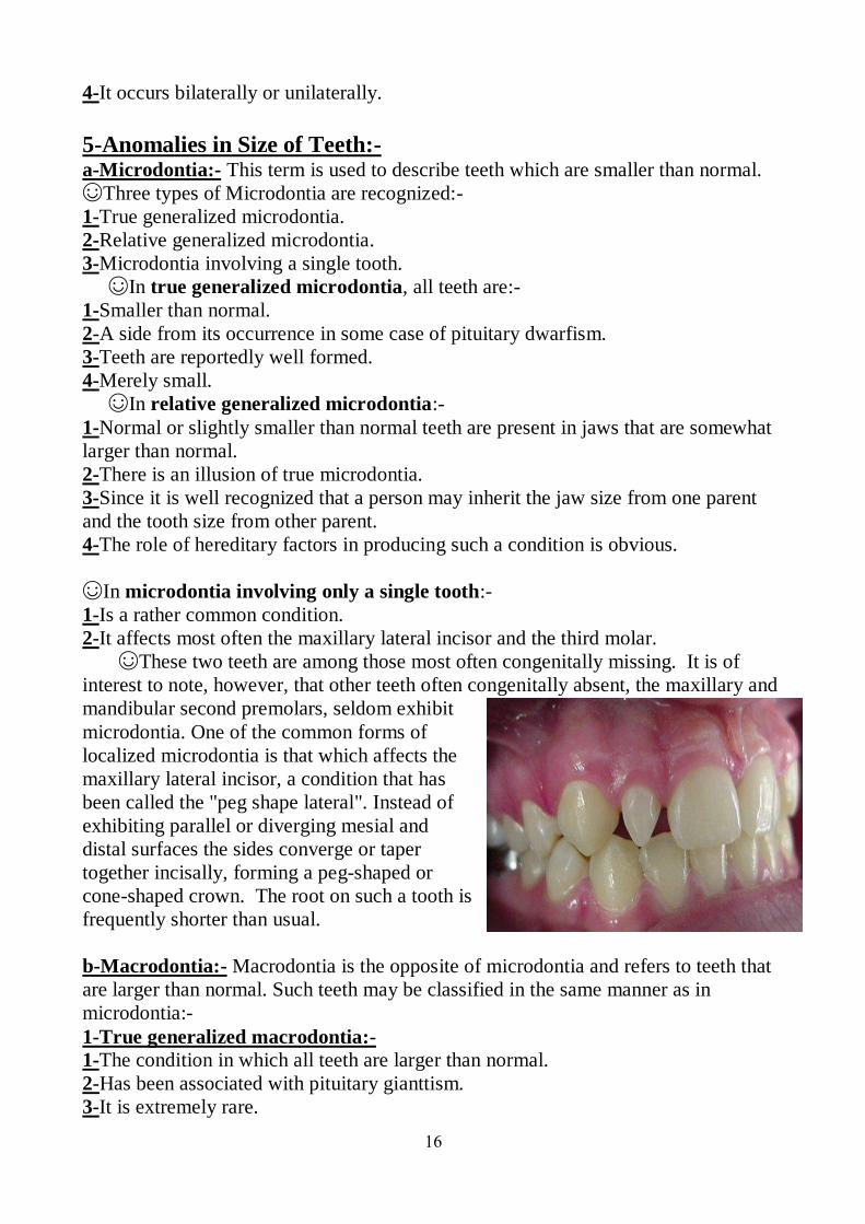
16
4-It occurs bilaterally or unilaterally.
5-Anomalies in Size of Teeth:- a-Microdontia:- This term is used to describe teeth which are smaller than normal.
☺Three types of Microdontia are recognized:-
1-True generalized microdontia.
2-Relative generalized microdontia.
3-Microdontia involving a single tooth.
☺In true generalized microdontia, all teeth are:-
1-Smaller than normal.
2-A side from its occurrence in some case of pituitary dwarfism.
3-Teeth are reportedly well formed.
4-Merely small.
☺In relative generalized microdontia:-
1-Normal or slightly smaller than normal teeth are present in jaws that are somewhat
larger than normal.
2-There is an illusion of true microdontia.
3-Since it is well recognized that a person may inherit the jaw size from one parent
and the tooth size from other parent.
4-The role of hereditary factors in producing such a condition is obvious.
☺In microdontia involving only a single tooth:-
1-Is a rather common condition.
2-It affects most often the maxillary lateral incisor and the third molar.
☺These two teeth are among those most often congenitally missing. It is of
interest to note, however, that other teeth often congenitally absent, the maxillary and
mandibular second premolars, seldom exhibit
microdontia. One of the common forms of
localized microdontia is that which affects the
maxillary lateral incisor, a condition that has
been called the "peg shape lateral". Instead of
exhibiting parallel or diverging mesial and
distal surfaces the sides converge or taper
together incisally, forming a peg-shaped or
cone-shaped crown. The root on such a tooth is
frequently shorter than usual.
b-Macrodontia:- Macrodontia is the opposite of microdontia and refers to teeth that
are larger than normal. Such teeth may be classified in the same manner as in
microdontia:-
1-True generalized macrodontia:-
1-The condition in which all teeth are larger than normal.
2-Has been associated with pituitary gianttism.
3-It is extremely rare.
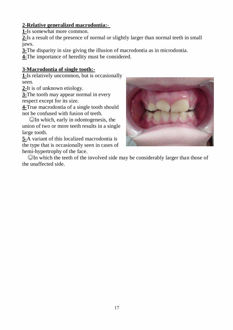
17
2-Relative generalized macrodontia:-
1-Is somewhat more common.
2-Is a result of the presence of normal or slightly larger than normal teeth in small
jaws.
3-The disparity in size giving the illusion of macrodontia as in microdontia.
4-The importance of heredity must be considered.
3-Macrodontia of single tooth:-
1-Is relatively uncommon, but is occasionally
seen.
2-It is of unknown etiology.
3-The tooth may appear normal in every
respect except for its size.
4-True macrodontia of a single tooth should
not be confused with fusion of teeth.
☺In which, early in odontogenesis, the
union of two or more teeth results in a single
large tooth.
5-A variant of this localized macrodontia is
the type that is occasionally seen in cases of
hemi-hypertrophy of the face.
☺In which the teeth of the involved side may be considerably larger than those of
the unaffected side.
1
Paediatric Dentistry
Fifth Stage
Dr. Suhair W. Abbood
Lec. 10
Maintenance of a Clean, a Dry Field and Application of the Rubber
Dam :-
The maintenance of a clean operating field during cavity preparation and placement
of the restoration material will help ensure efficient operating and development of a
serviceable restoration that will maintain the tooth and the integrity of the developing
occlusion.
In fact, once mastered, the technique makes dental care for children easier and a
higher standard of care can be achieved in less time than would otherwise be
required. The time spent in placing the rubber dam is negligible, provided that the
dentist works out a definite routine and uses a chairside assistant. The time required
for the placement of the rubber dam will invariably be made up and additional time
saved through the elimination of rinsing and spitting of the pediatric patient. In
addition, it isolates the child from the operative field making treatment less invasive
of their personal space.
The benefits can be divided into three main categories as shown below :-
1-Safety :-
a-Damage of soft tissues :- The risks of operative treatment include damage to the
soft tissues of the mouth from rotary and hand instruments and the medicaments used
in the provision of endodontic and other care. Rubber dam will go a long way to
preventing damage of this type.
b-Risk of swallowing or inhalation :- There is also the risk that these items may be
lost in the patient's mouth and swallowed or even inhaled and there are reports in the
literature to substantiate this risk. A rubber dam also prevents the small child in a
reclining position from swallowing or aspirating foreign objects and materials.
c-Risk of cross-infection :- The use of the rubber dam will prevent foreign objects
from coming into contact with oral structures. When filling material, debris, or
medicaments are dropped into the mouth, salivary flow is stimulated and interferes
with the operative or restorative procedure. In addition, there is considerable risk that
the use of high-speed rotary instruments distributes an aerosol of the patient's saliva
around the operating room, putting the dentist and staff at risk of infection, again a
risk that has been substantiated in the literature.
2
d-Nitrous oxide sedation :- If this is used it is quite likely that mouth breathing by
the child will increase the level of the gas in the environment, again putting dentist
and staff at risk. The use of rubber dam in this situation will make sure that exhaled
gas is routed via the scavenging system attached to the nose piece. Usually less
nitrous oxide will be required for a sedative effect, increasing the safety and
effectiveness of the procedure.
2-Benefits to the Child :-
a-Isolation :- One of the reasons that dental treatment causes anxiety in patients is
that the operative area is very close to and involved with all the most vital functions
of the body such as sight, hearing, breathing, and swallowing. When operative
treatment is being preformed, all these vital functions are put at risk and any sensible
child would be concerned. It is useful to discuss these fears with child patients and
explain how the risks can be reduced or eliminated.
Glasses should be used to protect the eyes and rubber dam to protect the airways and
the oesophagus. By doing this, and provided that good local analgesia has been
obtained, the child can feel themselves distanced from the operation.
Sometimes it is even helpful to show the child their isolated teeth in a mirror. The
view is so different from what they normally see in the mirror that they can divorce
themselves from the reality of the situation.
b-Relaxation :- A few explanatory words and reference to the rubber dam as a "rain
coat" for the tooth or as a "Halloween mask" will help allay the child's anxiety. It has
been found through experience that apprehensive or otherwise uncooperative children
can often be controlled more easily with a rubber dam in place.
The isolation of the operative area from the child will very often cause the child to
become considerably relaxed-always provided that there is good pain control. It is
common for both adult and child patients to fall asleep while undergoing treatment
involving the use of rubber dam-a situation that rarely occurs without. This is a
function of the safety perceived by the patient and the relaxed way in which the
dental team can work with its assistance.
3-Benefits to the dentist :-
a-Reduced stress :- As noted previously, once rubber dam has been placed the child
will be at less risk from the procedures that will be used to restore their teeth. This
reduces the effort required by the operator to protect the soft tissues of the mouth and
the airways.
Treatment can be carried out in a more relaxed and controlled manner, therefore
lessening the stress of the procedure on the dental team. Also the parents are always
3
interested in the work that has been accomplished for their child. While the rubber
dam is in place, the dentist can conveniently show parents the completed work after
an operative procedure. The rubber dam creates the feeling that the dentist has
complete control of the situation and that a conscientious effort has been made to
provide the highest type of service.
b-Retraction of tongue and cheeks :- Correctly placed rubber dam will gently pull
the cheeks and tongue away from the operative area allowing the operator a better
view of the area to be treated, and since the rubber dam efficiently controls the
tongue and the lips, the dentist has greater freedom for completing the operative
procedure.
c-Retraction of gingival tissue :- Rubber dam will gently pull the gingival tissues
away from the cervical margin of the tooth, making it much easier to see the extent of
any caries close to the margin and often bringing the cervical margin of a prepared
cavity above the level of the gingival margin thus, making restoration considerably
easier.
Interdentally, this retraction should be assisted by placing a wedge firmly between
the adjacent teeth as soon as the dam has been placed. This wedge is placed
horizontally below the contact area and above the dam thus, compressing the
interdental gingivae against the underlying bone. Proximal cavities can then be
prepared, any damage from rotary instruments being inflicted on the wedge rather
than the child's gingival tissue.
d-Moisture control :- Control of saliva is an extremely important consideration
when one is completing an ideal cavity preparation for primary teeth. The margin of
error is appreciably reduced when a cavity is prepared in a primary tooth that has a
large pulp and extensive carious involvement.
Small pulp exposures may be more easily detected when the tooth is well isolated. It
is equally important to observe the true extent of the exposure and the degree and
type of hemorrhage from the pulp tissue. Thus, the rubber dam aids the dentist in
evaluating teeth that are being considered for vital pulp therapy.
Also the silver amalgam is probably the only restorative material that has any
tolerance to being placed in a damp environment, and there is no doubt that it and all
other materials will perform much more satisfactorily if placed in a dry field. Rubber
dam is the only technique that readily ensures a dry field.
Armamentarium for Rubber Dam Placement :-
The armamentarium consists of:-
1-5×5 inch sheets of medium latex.
2-A rubber dam punch.
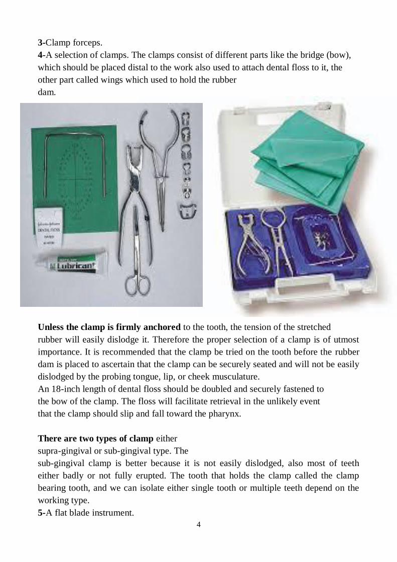
4
3-Clamp forceps.
4-A selection of clamps. The clamps consist of different parts like the bridge (bow),
which should be placed distal to the work also used to attach dental floss to it, the
other part called wings which used to hold the rubber
dam.
Unless the clamp is firmly anchored to the tooth, the tension of the stretched
rubber will easily dislodge it. Therefore the proper selection of a clamp is of utmost
importance. It is recommended that the clamp be tried on the tooth before the rubber
dam is placed to ascertain that the clamp can be securely seated and will not be easily
dislodged by the probing tongue, lip, or cheek musculature.
An 18-inch length of dental floss should be doubled and securely fastened to
the bow of the clamp. The floss will facilitate retrieval in the unlikely event
that the clamp should slip and fall toward the pharynx.
There are two types of clamp either
supra-gingival or sub-gingival type. The
sub-gingival clamp is better because it is not easily dislodged, also most of teeth
either badly or not fully erupted. The tooth that holds the clamp called the clamp
bearing tooth, and we can isolate either single tooth or multiple teeth depend on the
working type.
5-A flat blade instrument.
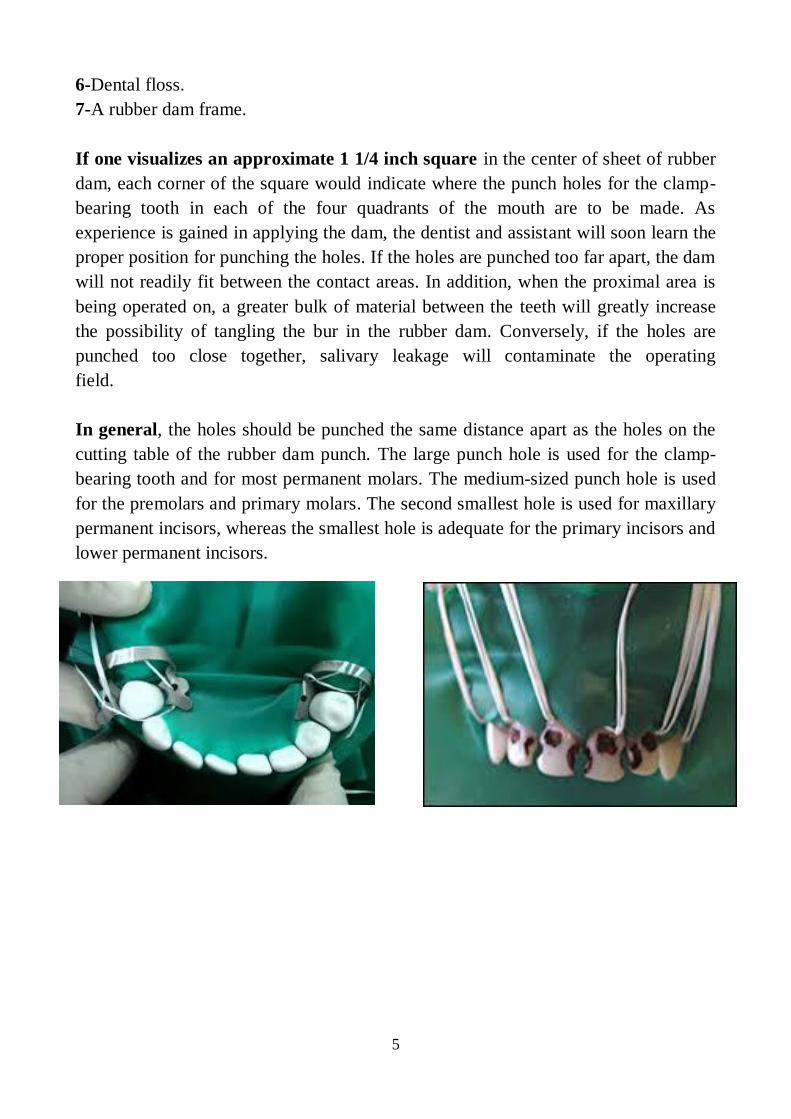
5
6-Dental floss.
7-A rubber dam frame.
If one visualizes an approximate 1 1/4 inch square in the center of sheet of rubber
dam, each corner of the square would indicate where the punch holes for the clamp-
bearing tooth in each of the four quadrants of the mouth are to be made. As
experience is gained in applying the dam, the dentist and assistant will soon learn the
proper position for punching the holes. If the holes are punched too far apart, the dam
will not readily fit between the contact areas. In addition, when the proximal area is
being operated on, a greater bulk of material between the teeth will greatly increase
the possibility of tangling the bur in the rubber dam. Conversely, if the holes are
punched too close together, salivary leakage will contaminate the operating
field.
In general, the holes should be punched the same distance apart as the holes on the
cutting table of the rubber dam punch. The large punch hole is used for the clamp-
bearing tooth and for most permanent molars. The medium-sized punch hole is used
for the premolars and primary molars. The second smallest hole is used for maxillary
permanent incisors, whereas the smallest hole is adequate for the primary incisors and
lower permanent incisors.
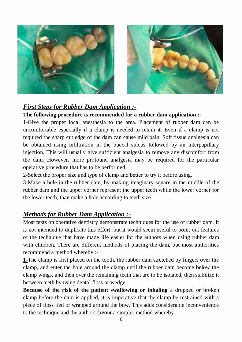
6
First Steps for Rubber Dam Application :-
The following procedure is recommended for a rubber dam application :-
1-Give the proper local anesthesia to the area. Placement of rubber dam can be
uncomfortable especially if a clamp is needed to retain it. Even if a clamp is not
required the sharp cut edge of the dam can cause mild pain. Soft tissue analgesia can
be obtained using infiltration in the buccal sulcus followed by an interpapillary
injection. This will usually give sufficient analgesia to remove any discomfort from
the dam. However, more profound analgesia may be required for the particular
operative procedure that has to be performed.
2-Select the proper size and type of clamp and better to try it before using.
3-Make a hole in the rubber dam, by making imaginary square in the middle of the
rubber dam and the upper corner represent the upper teeth while the lower corner for
the lower teeth, than make a hole according to teeth size.
Methods for Rubber Dam Application :-
Most texts on operative dentistry demonstrate techniques for the use of rubber dam. It
is not intended to duplicate this effort, but it would seem useful to point out features
of the technique that have made life easier for the authors when using rubber dam
with children. There are different methods of placing the dam, but most authorities
recommend a method whereby :-
1-The clamp is first placed on the tooth, the rubber dam stretched by fingers over the
clamp, and enter the hole around the clamp until the rubber dam become below the
clamp wings, and then over the remaining teeth that are to be isolated, then stabilize it
between teeth by using dental floss or wedge.
Because of the risk of the patient swallowing or inhaling a dropped or broken
clamp before the dam is applied, it is imperative that the clamp be restrained with a
piece of floss tied or wrapped around the bow. This adds considerable inconvenience
to the technique and the authors favour a simpler method whereby :-
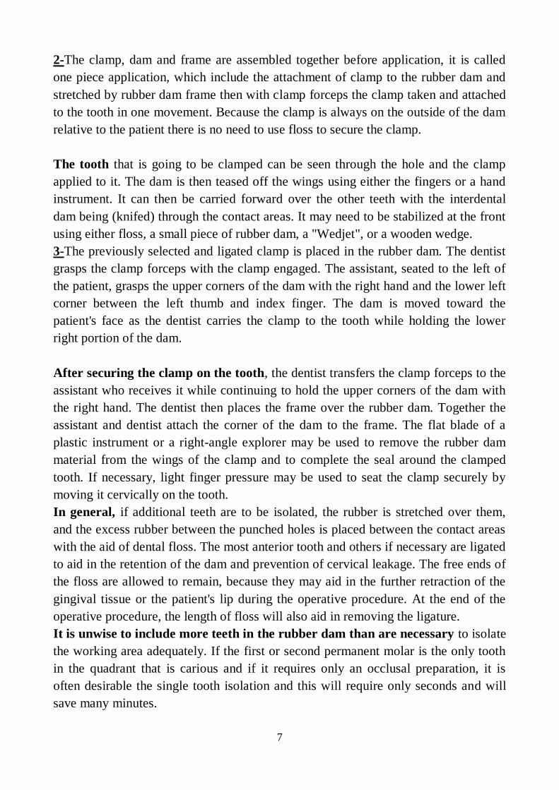
7
2-The clamp, dam and frame are assembled together before application, it is called
one piece application, which include the attachment of clamp to the rubber dam and
stretched by rubber dam frame then with clamp forceps the clamp taken and attached
to the tooth in one movement. Because the clamp is always on the outside of the dam
relative to the patient there is no need to use floss to secure the clamp.
The tooth that is going to be clamped can be seen through the hole and the clamp
applied to it. The dam is then teased off the wings using either the fingers or a hand
instrument. It can then be carried forward over the other teeth with the interdental
dam being (knifed) through the contact areas. It may need to be stabilized at the front
using either floss, a small piece of rubber dam, a "Wedjet", or a wooden wedge.
3-The previously selected and ligated clamp is placed in the rubber dam. The dentist
grasps the clamp forceps with the clamp engaged. The assistant, seated to the left of
the patient, grasps the upper corners of the dam with the right hand and the lower left
corner between the left thumb and index finger. The dam is moved toward the
patient's face as the dentist carries the clamp to the tooth while holding the lower
right portion of the dam.
After securing the clamp on the tooth, the dentist transfers the clamp forceps to the
assistant who receives it while continuing to hold the upper corners of the dam with
the right hand. The dentist then places the frame over the rubber dam. Together the
assistant and dentist attach the corner of the dam to the frame. The flat blade of a
plastic instrument or a right-angle explorer may be used to remove the rubber dam
material from the wings of the clamp and to complete the seal around the clamped
tooth. If necessary, light finger pressure may be used to seat the clamp securely by
moving it cervically on the tooth.
In general, if additional teeth are to be isolated, the rubber is stretched over them,
and the excess rubber between the punched holes is placed between the contact areas
with the aid of dental floss. The most anterior tooth and others if necessary are ligated
to aid in the retention of the dam and prevention of cervical leakage. The free ends of
the floss are allowed to remain, because they may aid in the further retraction of the
gingival tissue or the patient's lip during the operative procedure. At the end of the
operative procedure, the length of floss will also aid in removing the ligature.
It is unwise to include more teeth in the rubber dam than are necessary to isolate
the working area adequately. If the first or second permanent molar is the only tooth
in the quadrant that is carious and if it requires only an occlusal preparation, it is
often desirable the single tooth isolation and this will require only seconds and will
save many minutes.
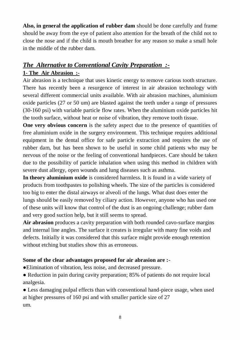
8
Also, in general the application of rubber dam should be done carefully and frame
should be away from the eye of patient also attention for the breath of the child not to
close the nose and if the child is mouth breather for any reason so make a small hole
in the middle of the rubber dam.
The Alternative to Conventional Cavity Preparation :-
1- The Air Abrasion :-
Air abrasion is a technique that uses kinetic energy to remove carious tooth structure.
There has recently been a resurgence of interest in air abrasion technology with
several different commercial units available. With air abrasion machines, aluminium
oxide particles (27 or 50 um) are blasted against the teeth under a range of pressures
(30-160 psi) with variable particle flow rates. When the aluminium oxide particles hit
the tooth surface, without heat or noise of vibration, they remove tooth tissue.
One very obvious concern is the safety aspect due to the presence of quantities of
free aluminium oxide in the surgery environment. This technique requires additional
equipment in the dental office for safe particle extraction and requires the use of
rubber dam, but has been shown to be useful in some child patients who may be
nervous of the noise or the feeling of conventional handpieces. Care should be taken
due to the possibility of particle inhalation when using this method in children with
severe dust allergy, open wounds and lung diseases such as asthma.
In theory aluminium oxide is considered harmless. It is found in a wide variety of
products from toothpastes to polishing wheels. The size of the particles is considered
too big to enter the distal airways or alveoli of the lungs. What dust does enter the
lungs should be easily removed by ciliary action. However, anyone who has used one
of these units will know that control of the dust is an ongoing challenge; rubber dam
and very good suction help, but it still seems to spread.
Air abrasion produces a cavity preparation with both rounded cavo-surface margins
and internal line angles. The surface it creates is irregular with many fine voids and
defects. Initially it was considered that this surface might provide enough retention
without etching but studies show this as erroneous.
Some of the clear advantages proposed for air abrasion are :-
●Elimination of vibration, less noise, and decreased pressure.
● Reduction in pain during cavity preparation; 85% of patients do not require local
analgesia.
● Less damaging pulpal effects than with conventional hand-piece usage, when used
at higher pressures of 160 psi and with smaller particle size of 27
um.
9
● Less fracture and crazing of enamel and dentine during cavity
preparation.
● Root canal access through porcelain crowns without fracturing porcelain.
Air abrasion has been proposed for :-
●Cleaning and removing stains and incipient caries from pits and fissures prior to
sealant and PRRs.
● Small class I, III, IV, and V cavity preparations and selected class II preparations
and porcelain restorations.
● Cleaning and preparation of castings, orthodontic bands, and brackets prior to
cementation.
What it cannot do is remove leathery dentinal caries or prepare extensive cavities
requiring classical retentive form.
To use it successfully, the clinician must learn a new technique as the tip does not
touch the tooth and therefore there is no tactile feedback. The tip width and the tip to
tooth distance seem to have most influence on the cavity width and depth. Increasing
the distance produces larger shallower cuts. Increasing the tip diameter produces
larger deeper cuts.
Therefore, the most precise removal of tooth tissue is achieved with a small inner
diameter tip (0.38 mm), held 2 mm from the tooth surface. If cutting a class II cavity,
it is essential to protect the adjacent tooth. Care must also be taken around the soft
tissues to prevent surgical emphysema. Glass/mirror surfaces may be damaged by the
dust.
In conclusion ,air abrasion may be useful in preparation of small cavities with
reduced patient discomfort, when combined with acid etching to obtain a good bond
with adhesive materials, and when correctly and carefully used. However, the dust is
a practical problem.
2- The Lasers-Assisted Dentistry :-
Laser is an acronym for Light Amplification by Stimulated Emission Radiation.
Dental lasers are devices that use the energy generated by atomic electron shifts
producing coherent monochromatic electromagnetic radiation between the ultraviolet
and the far infrared section of the electromagnetic spectrum.
10
The photo-biological effects of the lasers most commonly used in dentistry are :-
●Laser-induced fluorescence(caries/calculus detection).
● Photo-acoustics causing disruption and ablation (soft-and hard-tissue treatments).
● Photo-thermal effect inducing coagulation and vaporization (soft-tissue
treatments).
Bio-stimulation and photochemical effects induced by short-wavelength laser for
treatments including wound healing, analgesia and tissue growth will become more
commonplace in time. Laser-assisted fluoride and bleaching treatment also show
promising application.
Erbium lasers display bio-resonant properties on neural tissue causing Na+/K
+ pump
blockade and polarization of the A delta fibers and possibly C fibres. For many
applications, local anaesthesia can be reduced and occasionally eliminated due to the
analgesic properties of the lasers themselves.
The public perception of lasers in dentistry is that they can do remarkable things
painlessly, so obviously this appeal to a greater number of people. However, the
number of dentists offering lasers as an option in their practices is still small. The
cost of equipment is obviously a significant factor, but as with all new technologies it
is important that each dentist considers the proven clinical outcomes, that is, what the
recorded literature states regarding the safety, efficacy, and effectiveness.
With lasers this is further complicated by the fact that there are many different types
of lasers, with different uses and new types and applications being produced
constantly.
● Carbon dioxide lasers, Soft tissue incision/ablation, Gingival troughing, Aesthetic
contouring of gingivae, Treatment of oral ulcers, Fraenectomy and gingivactomy, De-
epithelization of gingival tissue during periodontal regenerative procedures.
● Nd : YAG Similar to above plus removal of incipient caries but because of the
depth of penetration there is a greater risk of collateral damage than with dioxide
lasers.
● Er : YAG Caries removal Cavity preparation in both enamel and dentine
Preparation of root canals.
● Argon laser, Resin curing, Tooth bleaching, Treatment of ulcers, Aesthetic gingival
contouring, Fraenectomy and gingivactomy. The argon laser has a major advantage
over the other lasers in that the wavelength at which it operates is absorbed by
haemoglobin and therefore provides excellent haemostasis.
Lasers produce light energy within a narrow frequency range. They are named after
the active element within them, which determines the wavelength of the light emitted.
So some of the commoner lasers have the following characteristics :-
11
● Neodymium : yttrium-aluminium-garnet (Nd : YAG) wavelength = 1.064um
● Carbon dioxide lasers wavelength = 10.6um.
● Erbium : YAG = 2.94um.
● Argon = 457-502 nm.
● Gallium-Arsenide (diode) = 904 nm.
● Holmium : YAG = 2.1um.
The wavelength of light is the primary determinant of the degree to which the target
material absorbs light. The deeper the laser energy penetrates, the more it scatters and
distributes throughout the tissue, for example, carbon dioxide laser penetrates 0.01-
0.03 mm into the tissue while Nd : YAG laser penetrates 2-5 mm. The light from
dental lasers is absorbed and converted to heat, while the thermal effects caused
depend on the tissue composition and the time the beam is focused on the target
tissue.
The increase in temperature may cause the tissue to change in structure and
composition, for example, denaturation, vapourization, carbonization, and melting
followed by recrystallization.
The Hard-Tissue Application :- The two lasers most commonly used for dental
hard-tissue treatments are in the 2790nm (ErCr:YSGG "Erbium-Chromium:Yttrium-
Scandium-Gallium-Garnet"), and 2940nm (Er:YAG "Erbium-doped Yttrium
Aluminium Garnet") wavelengths. The tissue is removed by a non-contact beam that
ablates based on the photo-acoustic affect on water molecules. The water content of
the treated tissue and the power density of the laser beam affect the cutting efficiency.
Hard-tissue applications include cavity preparation, caries and calculus removal,
endodontic treatments, desensitization and bone surgery.
The advantages of lasers include :-
1-Ability to selectively remove only carious dental tissue, results in clean sharp
margins in enamel and dentin. The pulp is protected and safe as the depth of energy
penetration is negligible.
2-Ability to cut dental tissue without the need for local anaesthesia (in some cases),
patient report little or no pain with the use of Er:YAG laser in cavity preparation.
3-Limited noise, no vibration, and time taken for cavity preparation is short,
therefore, laser can be extremely useful for nervous patients.

12
The disadvantages :-
1-They are expensive and care must be taken during use to ensure that excess heat is
not generated, which may be detriment to the pulp tissue.
2-The need to learn a new technique in which there is no proprioceptive feedback
since the laser tip does not impinge dental tissue.
3- The Ozone Therapy :-
Dental treatments are constantly evolving. One such innovation, ozone therapy
(healozone) has hit the media headlines, spiking much public interest. The technology
is available and costly devices for delivery of ozone for dental purposes exist, but as
yet the superiority of this modality over conventional treatment has not been proven
with properly conducted clinical trials.
The theory of the action of ozone is that it kills micro-organisms, by oxidizing their
cell walls to rupture their cytoplasmic membranes, that is, it is bactericidal. In
laboratories it has been shown that ozone can substantially reduce the numbers of
micro-organisms within carious dentine on short exposures of 10-20 s. Ozone may
also promote remineralization by oxidization of the lactate-propionate buffering
system (pH = 4) within the body of the carious lesion to bicarbonate and water. The
depth of residual caries can be no greater than 2mm when ozone is applied, as ozone
will not penetrate more than 2mm into carious dentin.
However, the clinical significance of this has not been established. It has been
postulated that the use of ozone together with a remineralising regime of fluoride
paste and rinse, oral hygiene instruction, and dietary advice would be beneficial and
that it would arrest primary root caries to a greater extent than remineralising regime
alone. It has also been suggested that ozone treatment can stabilize pit and fissure
caries preventing further deterioration.
1
Paediatric Dentistry
Fifth Stage Dr. Suhair W. Abbood
Lec. 11
Conventional Restorative Materials
☺The choice of restoration especially for primary teeth is based upon the
degree of carious involvement, whether the marginal ridge is intact or not and the
length of time that will elapse before exfoliation. The decision regarding the type of
restoration to be used is therefore based on the diagnosis of the extent of the dental
caries.
☺Many different materials have been advocated over the years, but very little
research has been carried out to find out which ones might be the most useful.
Therefore the popularity of any particular material has depended on clinical
impression and fashion.
Silver amalgam is the standard material against which the -Silver Amalgam :-1success of alternative materials is often judged. Amalgam has a known track record.
Dentists have used it for restoring teeth for more than 150 years. When looking at the
literature it must be remembered that amalgam technology has evolved over a very
long period and those amalgam alloys available today are probably very different in
composition to those used even as recently as 15 years ago. Amalgam has many
useful properties :-
1-It is relatively easy to handle.
2-Has good durability.
3-It has relatively yet to be bettered as a material for economically restoring posterior
teeth.
4-It exhibits reducing micro-leakage with time (high copper amalgams can take up to
2 years for a marginal seal to be produced, double the time for low copper amalgams,
but high copper amalgams are not as susceptible to corrosion phenomena and
resulting porosity and therefore retain their strength).
5. It is less technique sensitive to poor handling compared with other restorative
materials, and it is tolerant of operator error. In clinical trials and retrospective
studies, no intracoronal material has so far performed more successfully than
amalgam, but it is still important to control moisture as excess moisture causes
delayed expansion particularly in zinc-containing alloys, and for this reason rubber
dam should always be used if possible.
☺Despite these good properties, amalgam has two main disadvantages (1) it is
not aesthetic and (2) it contains mercury, a known poison. Little can be done to
combat the poor aesthetics. Remembering to polish amalgams does improve
2
characteristics, including appearance and leads to a significant reduction in their
replacement. Clinicians concerned about the toxicity of silver amalgam seek re-
assurance on the continuing use of the alloy. There are four main areas of concern :-
1- Inhalation of mercury vapour or amalgam dust.
2- The ingestion of amalgam.
3- Allergy to mercury.
4- Environmental considerations.
☺Inhalation of amalgam dust is most likely to occur during removal of a
previous restoration. This effect is transient and the effects minimized, if the operator
uses rubber dam and high speed aspiration. It is not in dispute that mercury is
released from amalgam restorations, during placement, polishing, chewing, and
removal, but the amounts are very small and come nowhere near the amounts
ingested from other daily sources, for example, air, water, and diet. True allergy to
amalgam is rare. There have been only 50 cases reported in 100 years. Many
countries are trying to reduce all industrial uses of mercury for environmental reasons
and better mercury hygiene in dental practice is one of the areas targeted.
☺Failure of amalgam itself as well as faults in the cavity design have been the
most commonly reported causes of failure of approximal restorations especially in
primary teeth. Attempts to overcome these deficiencies and to improve durability
have come through alteration in cavity design and the choice of material used. A
reduction in the size of the occlusal lock, rounded line angles, and minimum
extension for prevention all result in less destruction of sound tooth tissue. In
addition, the 'minimal' approximal cavity with no occlusal 'dovetail' has been
described for both amalgam and adhesive restorations, and incorporates some
mechanical retention in the form of small internal resistance grooves placed with a
very small round bur just inside the enamel-dentine junction. It is unlikely that the
'perfect cavity design' exists for an amalgam restoration in primary molars due to
certain anatomical features :-
1. Widened contact areas make a narrow box difficult to achieve.
2. Thin enamel means that cracking and fracture of parts of the crown are more
common.
3. Primary teeth may undergo considerable wear under occlusal stress themselves and
this in turn will affect the restorations.
☺It is therefore necessary to investigate other materials for use in restoring the
primary dentition.
2-Composite Resins :- Resin-based composites (along with photopolymerization)
have revolutionized clinical dentistry. Many dentists advocate the use of composite as
a restorative in the treatment of children, they are being increasingly used in
combination with GICs in a "sandwich"-style aesthetic restoration, and the results are
generally acceptable. Cavity design is usually with beveling of the margins to
increase the amount of enamel available for etching and bonding.
3
☺Placement of these materials is highly technique sensitive, the use of rubber
dam is essential if a dry field is to be achieved, and this adequate moisture isolation,
and patient compliance can prove difficult in the younger, more challenging child
patient, these facts together with the material's relative expense probably reflects the
lack of widespread use of composite resin in many countries.
☺Composite resins came on the market in the early 1970s and have been
modified since then in an attempt to improve their properties, although problems
related to wear resistance, water absorption and polymerization contraction can limit
their use in larger restorations in the posterior permanent dentition, current materials
are still best applied to anterior teeth and small restorations in posterior teeth. The
development of acid etching at the time that these materials were introduced has
ensured that they have performed reasonably well in terms of marginal seal. They are
sensitive to variations in technique and take longer to place than equivalent amalgam
restorations. They must be placed in a dry field. The long-term success of composite
resins is jeopardized by their instability in water. The best materials have maximum
inorganic filler levels and low water absorption, but will deteriorate over time.
☺Abrasive wear of many composite systems is comparable to that of silver
amalgam in the region of 10-20 um/year, and colour stability is now excellent
compared with earlier materials. After placement and occlusal adjustment of the
restorative material, the operator should place a layer of sealant on the finished
surface to fill any micro-cracks within the surface of the resin, followed by curing the
resin to ensure maximal polymerization. Before making decisions concerning the
most appropriate restorative material in the treatment of children, the clinician should
consider :-
1. Moisture exclusion. Is it realistic for this patient?
2. Patient compliance. Will the patient sit still through the restoration?
3. The size of the cavity. Lesion extent determines operative duration.
4. Patient compliance after the procedure. Will he or she return for monitoring and
review?
☺As long as the clinician allows due consideration in relation to these provisos
concerning use of the material, it will be appropriate to employ it restoratively, since
its inherent properties make it an excellent choice in the treatment of children for
occlusal cavities. As long as the responses to questions 1, 2, and 4 are affirmative and
the restoration is relatively small, the composite can be used with confidence.
3-Glass Ionomer Cements :- One of the most significant advances in
contemporary pediatric dental practice has been the development of GICs. A glass
ionomer consists of a basic glass and an acidic water-soluble powder that sets by
acid-base reaction between the two components. Glass ionomer cements came on to
the market in the late 1970s and have also been modified since then in order to
enhance their properties.
4
☺Current materials are much improved and have some advantages over
composite resins. Although they tend to be more brittle than composites, this group of
materials has the advantage of adherence to both enamel and dentine without etching.
They do not suffer from polymerization shrinkage. The coefficient of expansion of
glass ionomer is very close to that of dentine and once set, these materials remain
dimensionally stable in the mouth despite constantly changing moisture and
temperature levels. Their biggest advantage over composites is that being made from
glasses with a high fluoride content they not only provide a sustained release over an
extended period of time but also act as a rechargeable reservoir of fluoride, which
may protect adjacent surfaces from caries progression. Their lack of strength limits
their use in the permanent dentition but they can be used in PRRs where there is no
occlusal load and as an interim restoration while caries is brought under control. They
are also the authors' choice of material for cementing stainless-steel crowns. Similarly
to composite resins it is imperative that they are placed in a dry field.
☺Recently, a number of new materials have come on to the market which aim
to maximize the best qualities of both composite resins and glass ionomers. Some of
these show promise and should be considered for the restoration of children's teeth.
They can be classified according to whether they retain the essential acid-base
reaction of the glass ionomers or not.
4-Resin-Modified Glass Ionomer :- Resin-modified glass ionomers were
developed to overcome the problems of moisture sensitivity and low initial
mechanical strength. They consist of a GIC along with a water-based resin system
which allows the material to set quickly using light or chemical catalysts (or both)
while allowing the acid-base reaction of the glass ionomer to take place, this reaction
will occurs within the light polymerized resin framework. Thus, the materials will set,
albeit rather slowly, without the need for the resin system and essential qualities of a
glass ionomer cement should be retained.
☺The reinforcement of glass ionomer with resin has been used to produce a
fast setting cement but these materials require etching prior to placement. On
modifying the materials, fracture toughness/resistance and abrasion resistance
improve, and they still retain biocompatibility, fluoride ion hydrodynamics,
favourable thermal expansion and contraction characteristics, and most important of
all, they retain physico-chemical bonding to tooth structure.
5-Compomer (polyacid-modified resin-based composite) :- Polyacid-
modified resin composite resins or compomers are material that contain a calcium
aluminium fluorosilicate glass filter and polyacid components. They contain either or
both essential components of a GIC, and have a much higher content of resin.
However, they are not water-based and therefore no acid-base reaction can occur. As
such, they cannot strictly be described as glass ionomers. They set by resin
photopolymerization. The acid-base reaction does occur in the moist intra-oral
5
environment and allows fluoride release from the material.
☺These materials are a combination of composite and ionomer. They have
better aesthetics than glass ionomer as a single material and have the advantage of
some fluoride release, but there is still a need to etch during the restorative procedure.
However, it would appear that they suffer from the disadvantages of loss of retention
together with gap formation between the material and tooth substance. Successful
adhesion requires the use of dentin-bonding primers before placement.
☺Compomers are now widely used in general dental practice for the restoration
of approximal lesions in primary teeth. After good initial results, longer follow-up
periods have shown that this material indeed lived up to its early promise and good
survival rates have been reported for restorations in primary molars. However, it must
be placed in cavities prepared to the usual principles of cavity design for a most
favourable outcome. Recently published work has shown compomer to be as durable
as amalgam after 3 years in approximal cavities in primary molars. There is also a
success rate in using compomer in stress bearing restorations in permanent posterior
teeth, and further studies will clarify the issue.
The Cavity Bases and Varnishes:- ☺Deep cavities resulting from the removal of extensive caries should receive a
base before the placement of a restoration. If a small pulp exposure may be present.
One of the hard-setting calcium hydroxide-containing bases should be used. Only the
deepest portion of the cavity should be filled, allowing the restoration to be supported
by sound dentin. There is also evidence that the use of a calcium hydroxide-
containing material will favorably influence the formation of secondary dentin.
☺Rapid-setting zinc oxide-eugenol, poly carboxylate, or glass ionomer cement
bases are acceptable when there is no danger of a pulp exposure. It is important to
avoid carrying the base onto the cavity wall margin of the preparation because this
would result in deterioration of the material and marginal leakage. The base selected
should be sufficiently strong to resist displacement and fracture during the
condensing of an amalgam restoration. The routine use of a cavity varnish before
placement of an amalgam restoration :-
Will reduce the possibility of discoloration of the dentin.-1
Will help prevent marginal leakage. -2
drying -The application of two or three thin layers of quick-3
varnish to freshly cut dentin will also reduce sensitivity after the placement of the
restoration.
4-A fresh Copalite varnish applied in a thin uniform coating over zinc oxide-eugenol
or calcium hydroxide fulfills all the requirements of a good liner (the base and
varnish combined).
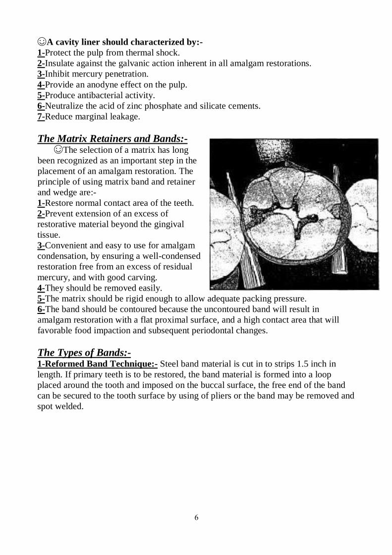
6
☺A cavity liner should characterized by:-
1-Protect the pulp from thermal shock.
2-Insulate against the galvanic action inherent in all amalgam restorations.
3-Inhibit mercury penetration.
4-Provide an anodyne effect on the pulp.
5-Produce antibacterial activity.
6-Neutralize the acid of zinc phosphate and silicate cements.
7-Reduce marginal leakage.
-The Matrix Retainers and Bands: ☺The selection of a matrix has long
been recognized as an important step in the
placement of an amalgam restoration. The
principle of using matrix band and retainer
and wedge are:-
1-Restore normal contact area of the teeth.
2-Prevent extension of an excess of
restorative material beyond the gingival
tissue.
3-Convenient and easy to use for amalgam
condensation, by ensuring a well-condensed
restoration free from an excess of residual
mercury, and with good carving.
4-They should be removed easily.
5-The matrix should be rigid enough to allow adequate packing pressure.
The band should be contoured because the uncontoured band will result in -6
amalgam restoration with a flat proximal surface, and a high contact area that will
favorable food impaction and subsequent periodontal changes.
-The Types of Bands:
1-Reformed Band Technique:- Steel band material is cut in to strips 1.5 inch in
length. If primary teeth is to be restored, the band material is formed into a loop
placed around the tooth and imposed on the buccal surface, the free end of the band
can be secured to the tooth surface by using of pliers or the band may be removed and
spot welded.
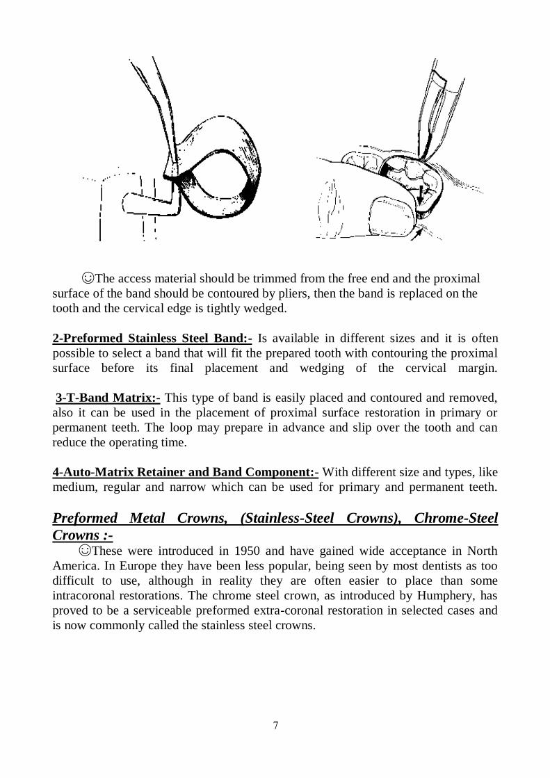
7
☺The access material should be trimmed from the free end and the proximal
surface of the band should be contoured by pliers, then the band is replaced on the
tooth and the cervical edge is tightly wedged.
often Is available in different sizes and it is -Preformed Stainless Steel Band:-2
possible to select a band that will fit the prepared tooth with contouring the proximal
surface before its final placement and wedging of the cervical margin.
3-T-Band Matrix:- This type of band is easily placed and contoured and removed,
also it can be used in the placement of proximal surface restoration in primary or
permanent teeth. The loop may prepare in advance and slip over the tooth and can
reduce the operating time.
With different size and types, like -Matrix Retainer and Band Component:-Auto-4
medium, regular and narrow which can be used for primary and permanent teeth.
Preformed Metal Crowns, (Stainless-Steel Crowns), Chrome-Steel
Crowns :- ☺These were introduced in 1950 and have gained wide acceptance in North
America. In Europe they have been less popular, being seen by most dentists as too
difficult to use, although in reality they are often easier to place than some
intracoronal restorations. The chrome steel crown, as introduced by Humphery, has
proved to be a serviceable preformed extra-coronal restoration in selected cases and
is now commonly called the stainless steel crowns.
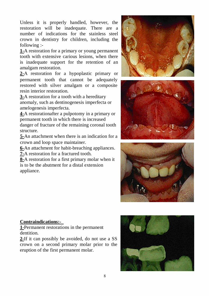
8
Unless it is properly handled, however, the
restoration will be inadequate. There are a
number of indications for the stainless steel
crown in dentistry for children, including the
following :-
1-A restoration for a primary or young permanent
tooth with extensive carious lesions, when there
is inadequate support for the retention of an
amalgam restoration.
2-A restoration for a hypoplastic primary or
permanent tooth that cannot be adequately
restored with silver amalgam or a composite
resin interior restoration.
3-A restoration for a tooth with a hereditary
anomaly, such as dentinogenesis imperfecta or
amelogenesis imperfecta.
4-A restorationafter a pulpotomy in a primary or
permanent tooth in which there is increased
danger of fracture of the remaining coronal tooth
structure.
An attachment when there is an indication for a -5
crown and loop space maintainer.
ng appliances. breachi-An attachment for habit-6
tured tooth. A restoration for a frac-7
A restoration for a first primary molar when it -8
is to be the abutment for a distal extension
appliance.
-Contraindications:
Permanent restorations in the permanent -1
dentition.
2-If it can possibly be avoided, do not use a SS
crown on a second primary molar prior to the
eruption of the first permanent molar.
9
The advantages of the SSC are :-
1-Single visit for placement.
2-Relitively quick and simple procedure
3-Usually reduce sensitivity totally, because they cover the whole tooth.
4-Inexpensive compared with cast restorations.
5-They are economically more attractive over the long term, since the rate of
placement of these restorations is low (3% compared with 15% for class II amalgam
restorations), but they are relatively expensive in relation to both time and money in
the short term.
6-Good retention rate.
The disadvantages of the SSC are :-
1-They may be considered unaesthetic, require a significant amount of tooth
preparation more than cast preparations, and invariably local anaesthesia.
2. Once a tooth has been prepared for a stainless-steel crown, it will need a full
coverage restoration eventually. It has been suggested that placing orthodontic
separators 1 or 2 weeks prior to preparation reduces the amount of tissue requiring
removal. However, some reduction is usually necessary.
3. Gingival margins are sub-gingival.
Operative Technique in Preparation of the Tooth :- 1-A local anaesthetic should be administered and a rubber
dam placed as for other restorative procedures.
Irrespective of whether the tooth to be restored is vital or
non-vital, local anaesthesia should be used when placing
a stainless steel crown because of the soft-tissue
manipulation. Rubber dam, although sometimes difficult
to place in the broken down dentition, should be used
where possible. If rubber dam not being used, place gauze
safety net to avoid aspiration and/or swallowing of the
crown.
2-Select the crown size :-
The smallest crown that completely covers the -1
preparation should be chosen.
☺There are two important principles that will help
to consistently produce well-adapted stainless steel
crowns.
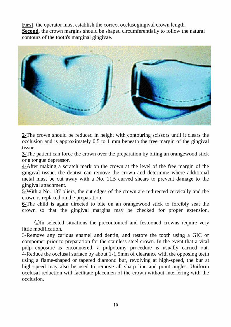
10
First, the operator must establish the correct occlusogingival crown length.
Second, the crown margins should be shaped circumferentially to follow the natural
contours of the tooth's marginal gingivae.
2-The crown should be reduced in height with contouring scissors until it clears the
occlusion and is approximately 0.5 to 1 mm beneath the free margin of the gingival
tissue.
3-The patient can force the crown over the preparation by biting an orangewood stick
or a tongue depressor.
4-After making a scratch mark on the crown at the level of the free margin of the
gingival tissue, the dentist can remove the crown and determine where additional
metal must be cut away with a No. 11B curved shears to prevent damage to the
gingival attachment.
5-With a No. 137 pliers, the cut edges of the crown are redirected cervically and the
crown is replaced on the preparation.
6-The child is again directed to bite on an orangewood stick to forcibly seat the
crown so that the gingival margins may be checked for proper extension.
☺In selected situations the precontoured and festooned crowns require very
little modification.
3-Remove any carious enamel and dentin, and restore the tooth using a GIC or
compomer prior to preparation for the stainless steel crown. In the event that a vital
pulp exposure is encountered, a pulpotomy procedure is usually carried out.
4-Reduce the occlusal surface by about 1-1.5mm of clearance with the opposing teeth
using a flame-shaped or tapered diamond bur, revolving at high-speed, the bur at
high-speed may also be used to remove all sharp line and point angles. Uniform
occlusal reduction will facilitate placemen of the crown without interfering with the
occlusion.
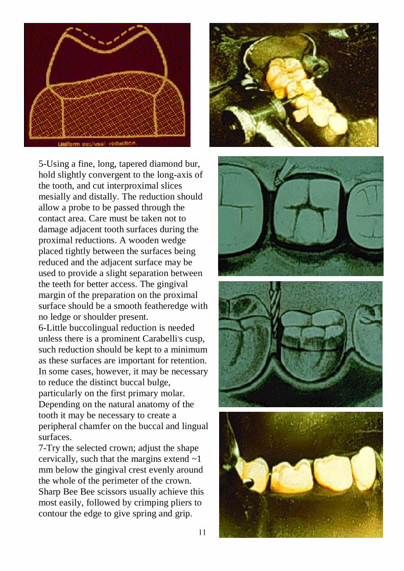
11
5-Using a fine, long, tapered diamond bur,
hold slightly convergent to the long-axis of
the tooth, and cut interproximal slices
mesially and distally. The reduction should
allow a probe to be passed through the
contact area. Care must be taken not to
damage adjacent tooth surfaces during the
proximal reductions. A wooden wedge
placed tightly between the surfaces being
reduced and the adjacent surface may be
used to provide a slight separation between
the teeth for better access. The gingival
margin of the preparation on the proximal
surface should be a smooth featheredge with
no ledge or shoulder present.
6-Little buccolingual reduction is needed
unless there is a prominent Carabelli,s cusp,
such reduction should be kept to a minimum
as these surfaces are important for retention.
In some cases, however, it may be necessary
to reduce the distinct buccal bulge,
particularly on the first primary molar.
Depending on the natural anatomy of the
tooth it may be necessary to create a
peripheral chamfer on the buccal and lingual
surfaces.
7-Try the selected crown; adjust the shape
cervically, such that the margins extend ~1
mm below the gingival crest evenly around
the whole of the perimeter of the crown.
Sharp Bee Bee scissors usually achieve this
most easily, followed by crimping pliers to
contour the edge to give spring and grip.
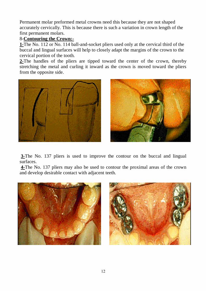
12
Permanent molar preformed metal crowns need this because they are not shaped
accurately cervically. This is because there is such a variation in crown length of the
first permanent molars.
8-Contouring the Crown:-
1-The No. 112 or No. 114 ball-and-socket pliers used only at the cervical third of the
buccal and lingual surfaces will help to closely adapt the margins of the crown to the
cervical portion of the tooth.
2-The handles of the pliers are tipped toward the center of the crown, thereby
stretching the metal and curling it inward as the crown is moved toward the pliers
from the opposite side.
3-The No. 137 pliers is used to improve the contour on the buccal and lingual
surfaces.
4-The No. 137 pliers may also be used to contour the proximal areas of the crown
and develop desirable contact with adjacent teeth.
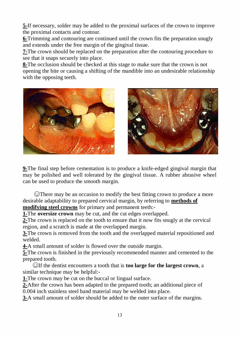
13
5-If necessary, solder may be added to the proximal surfaces of the crown to improve
the proximal contacts and contour.
6-Trimming and contouring are continued until the crown fits the preparation snugly
and extends under the free margin of the gingival tissue.
7-The crown should be replaced on the preparation after the contouring procedure to
see that it snaps securely into place.
8-The occlusion should be checked at this stage to make sure that the crown is not
opening the bite or causing a shifting of the mandible into an undesirable relationship
with the opposing teeth.
9-The final step before cementation is to produce a knife-edged gingival margin that
may be polished and well tolerated by the gingival tissue. A rubber abrasive wheel
can be used to produce the smooth margin.
☺There may be an occasion to modify the best fitting crown to produce a more
desirable adaptability to prepared cervical margin, by referring to methods of
modifying steel crowns for primary and permanent teeth:-
1-The oversize crown may be cut, and the cut edges overlapped.
2-The crown is replaced on the tooth to ensure that it now fits snugly at the cervical
region, and a scratch is made at the overlapped margin.
3-The crown is removed from the tooth and the overlapped material repositioned and
welded.
4-A small amount of solder is flowed over the outside margin.
5-The crown is finished in the previously recommended manner and cemented to the
prepared tooth.
☺If the dentist encounters a tooth that is too large for the largest crown, a
similar technique may be helpful:-
1-The crown may be cut on the buccal or lingual surface.
2-After the crown has been adapted to the prepared tooth; an additional piece of
0.004 inch stainless steel band material may be welded into place.
3-A small amount of solder should be added to the outer surface of the margins.
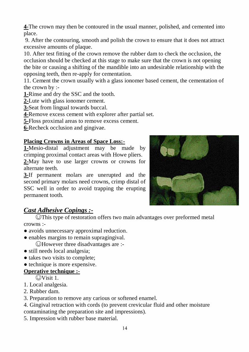
14
4-The crown may then be contoured in the usual manner, polished, and cemented into
place.
9. After the contouring, smooth and polish the crown to ensure that it does not attract
excessive amounts of plaque.
10. After test fitting of the crown remove the rubber dam to check the occlusion, the
occlusion should be checked at this stage to make sure that the crown is not opening
the bite or causing a shifting of the mandible into an undesirable relationship with the
opposing teeth, then re-apply for cementation.
11. Cement the crown usually with a glass ionomer based cement, the cementation of
the crown by :-
1-Rinse and dry the SSC and the tooth.
2-Lute with glass ionomer cement.
3-Seat from lingual towards buccal.
4-Remove excess cement with explorer after partial set.
5-Floss proximal areas to remove excess cement.
6-Recheck occlusion and gingivae.
Placing Crowns in Areas of Space Loss:-
1-Mesio-distal adjustment may be made by
crimping proximal contact areas with Howe pliers.
2-May have to use larger crowns or crowns for
alternate teeth.
3-If permanent molars are unerupted and the
second primary molars need crowns, crimp distal of
SSC well in order to avoid trapping the erupting
permanent tooth.
Cast Adhesive Copings :- ☺This type of restoration offers two main advantages over preformed metal
crowns :-
● avoids unnecessary approximal reduction.
● enables margins to remain supragingival.
☺However three disadvantages are :-
● still needs local analgesia;
● takes two visits to complete;
● technique is more expensive.
Operative technique :-
☺Visit 1.
1. Local analgesia.
2. Rubber dam.
3. Preparation to remove any carious or softened enamel.
4. Gingival retraction with cords (to prevent crevicular fluid and other moisture
contaminating the preparation site and impressions).
5. Impression with rubber base material.
15
6. Temporization if much tooth tissue has been removed.
☺The casting is constructed in the laboratory, and the fit surface is sand blasted.
☺Visit 2.
7. Local analgesia.
8. Rubber dam.
9. Tooth is brushed with pumice, washed, and dried.
10. Casting is tried in to check marginal adaptation and fit.
11. Casting is re-sandblasted to obtain optimum conditions for bonding.
12. Tooth is etched, washed, and dried.
13. Cement is applied to fit surface of casting ensuring there are no bubbles.
14. The casting is held in position under pressure for 3 min.
15. Excess cement is removed.
16. Oxygen inhibiting material (oxyguard) is applied over the margins of the casting
and maintained in position for a further 3 min.
17. The oxyguard is removed by washing; margins rechecked; and occlusion
checked.
The Commonly Used Therapeutic Capping Materials for Pulp Therapy
and the Reaction of the Pulp with Them :- ☺A diverse range of chemicals have been used as pulpotomy agents. As most of
these have not been subject to rigorous clinical trials, their use has been based on
expert opinion and retrospective studies. There is no reliable evidence supporting the
superiority of one type of treatment for pulpally involved primary molars. No
conclusions can be made as to the optimum treatment or techniques for pulpally
involved primary molar teeth due to the scarcity of reliable scientific research.
☺High quality RCTs (Randomized Controlled Trials), with appropriate unit of
randomization and analysis are needed. The available evidence suggests that
formocresol, ferric sulphate, electrocautery and MTA have similar efficacy. Calcium
hydroxide appears to have a consistently lower success rate in vital pulpotomy in
deciduous teeth than these four agents. There are a number of other materials that are
of historical significance, or have regional usage, and a number of experimental
techniques including bone morphogenic protein and growth factors. All current
therapeutic agents have toxic effects and must be correctly handled within their
therapeutic range. Clinicians should carefully read the Materials Safety Data Sheet
for these agents. Cases should be carefully selected within the guidelines
recommended.
Before calcium hydroxide come into common use, the -Zinc Oxide Eugenol :-1zinc oxide eugenol had been used more often than any other pulp-capping material.
Many dentists have apparently had good clinical results with the use of zinc oxide
eugenol, but it is no longer recommended as a direct pulp capping material.
16
Herman first introduced calcium hydroxide as a -Calcium Hydroxide :-2biologic dressing. Because of its alkalinity (pH of 12), it is so caustic that when
placed in contact with vital pulp tissue, the reaction produces a superficial necrosis of
the pulp. The superficial necrotic area in the pulp that develops beneath the calcium
hydroxide is demarcated from the healthy pulp tissue below by a new, deeply staining
zone comprising basophilic elements of the calcium hydroxide dressing.
☺The original proteinate zone is still present. However, against this zone is a
new area of coarse fibrous tissue likened to a primitive type of bone. On the
periphery of the new fibrous tissue, cells resembling odontoblasts appear to be lining
up.
☺Calcium hydroxide was associated with the formation of a dentin bridge and
the complete healing of the amputated pulp. The irritant qualities seem to be related
to its ability to stimulate development of a calcified barrier. One month after the
capping procedure, a calcified bridge is evident radiographically. This bridge
continues to increase in thickness during the next 12-month period. The pulp tissue
beneath the calcified bridge remains vital and is essentially free of inflammatory
cells.
☺Thus, calcium hydroxide currently serves as the standard or control material
for experimentation related to pulp-capping agents. Calcium hydroxide is the material
of choice for direct pulp-capping or vital pulpotomy techniques in permanent teeth.
3-Glutaraldehyde :- Glutaraldehyde continues to receive attention as a potential
pulp-capping agent for pulpotomy techniques in primary teeth. It is an excellent
bactericidal agent and seems to offer some advantages when compared to
formocresol. Research has reported that glutaraldehyde seems to be superior to
formaldehyde preparations for pulp therapy in the following ways :-
1-Formaldehyde reactions are reversible but glutaraldehyde reactions are not.
2-Formaldehyde is a small molecule that penetrates the apical foramen, whereas
glutaraldehyde is a larger molecule that does not.
3-Formaldehyde requires a long reaction time and an excess of solution to fix tissue,
whereas glutaraldehyde fixes tissue instantly and an excess of solution is
unnecessary.
try Formocresol has been used in dentis -Preparation Containing Formalin :-4for over 100 years, and for vital pulpotomy in deciduous teeth for over 80 years. Its
efficacy has been extensively studied, with clinical success rates ranging from 70% to
100%, making it the standard against which newer techniques are compared.
☺The formaldehyde component of formocresol is strongly bactericidal and
reversibly inhibits many enzymes in the inflammatory process. Originally, the aim of
using formocresol was to completely mummify (fix) all residual pulpal tissue and
necrotic material within the root canal. Current techniques however, aim to create a
very superficial layer of fixation while preserving the vitality of the deeper radicular
17
pulp. Contemporary pulpotomy is explicitly contraindicated in the presence of
radicular pulpitis or pulp necrosis.
☺Formocresol is applied to the pulpotomy site on a cotton wool pledget. Any
excess material should be blotted off the pledget prior to application. Traditionally, a
5-minute application time has been recommended, however, contact times of only a
few seconds are probably equally effective. It is prudent to limit both dose and
contact time. Formocresol should only be applied to the pulpotomy site after
haemostasis has been obtained. It should never be applied to bleeding tissue.
☺In 2004, the International Agency for Research on Cancer (IARC) concluded
that chronic exposure to high levels of formaldehyde causes nasopharyngeal cancer in
humans. In assessing the potential risks of using formocresol clinically, however, it is
important to consider the pharmacokinetics of formaldehyde. Formaldehyde is an
important intermediate in normal cellular metabolism. It serves as a building block
for the synthesis of purines, pyrimidines, many amino acids and lipids, and is a key
molecule in one-carbon metabolism.
☺Endogenous formaldehyde is present at low levels in body fluids, with a
concentration of 2-3 mg/L in human blood. Application of formocresol results in
systemic absorption of formaldehyde, however, the absorbed formaldehyde is rapidly
metabolized to formate and carbon dioxide with a half-life of 1-2 minutes. The use of
formocresol in dentistry falls within the current permitted exposure limits, and short-
term exposure limits for formaldehyde, formaldehyde does not bioaccumulate.
Research on the use of formocresol as a pulp-capping agent has continued. The 1:5
concentration of formocresol is currently recommended for initial treatment of pulps
of primary teeth in the vital pulpotomy technique.
5-Ferric Sulphate :- Ferric sulphate is widely used in dentistry as a haemostatic
agent (Astringident). It was initially used in pulpotomy as an aid to haemostasis prior
to placement of calcium hydroxide. However, as an independent therapeutic agent,
ferric sulphate pulpotomy has a success rate of 74-99%.
☺Ferric sulphate is thought to react with the pulp tissue, forming a superficial
protective layer of iron-protein complex. The predominant mode of failure is the
result of internal resorption. Ferric sulphate is burnished onto the pulp stumps
(pulpotomy site) using a microbrush for 15 seconds, than rinsed off with water and
dried. Persistent bleeding after the application of ferric sulphate is an indication for
pulpectomy or extraction.
☺According to criteria of Worksafe Australia, ferric sulphate is a hazardous,
corrosive liquid, which has the potential to cause severe injury. Ferric sulphate
decomposes to form sulphuric acid, which can cause superficial tissue burns if it is
not confined to the pulpotomy site.
18
6-Electrosurgery :- Electrosurgery uses radiofrequency energy to produce a
controlled superficial tissue burn. It is both haemostatic and antibacterial. Excessive
energy or contact time causes a deep tissue burn with necrosis of the radicular pulp
and subsequent internal root resorption. Electrosurgical pulpotomy has a success rate
of 70-94%.
☺The electrosurgery unit should be set to coagulate, with a low power setting. A
small ball or round-ended tip is applied to the pulpotomy site and briefly activated.
The site should immediately be flooded with water to remove excess heat. Each pulp
stump is treated in turn. If necessary, electrocoagulation can be repeated to control
persistent bleeding, until the total cumulative application time is 2 seconds. Persistent
bleeding after this time is an indication for pulpectomy or extraction.
☺Electrosurgical equipment has the potential to interfere with pacemakers and
implanted electronics. The patient must be correctly grounded with a dispersive plate
to prevent earth leakage burns, which can occur in the extremities, a long way from
the surgical site. Electrosurgical equipment should be set up, maintained and used
according to the manufacture's directions.
7-Mineral Trioxide Aggregate :- MTA is a mixture of tricalcium silicate,
bismuth oxide, dicalcium silicate, tricalcium aluminate and calcium sulphate. It is
chemically similar to standard cement mix. MTA powder reacts water to form a
paste, which is highly alkaline (pH=13) during the setting phase, then sets to form an
inert mass.
☺Clinical success rates for MTA pulpotomy are similar to formocresol and ferric
sulphate. The MTA powder is mixed with water immediately prior to use. The
resultant paste is applied to the pulpotomy site using a proprietary carrier or a plastic
instrument and is left in situ to set. It is covered with a suitable base material prior to
restoration of the tooth. The paste should only be applied after haemostasis has been
obtained. Persistent bleeding from the pulpotomy site is an indication for pulpectomy
or extraction.
☺ Exposure to MTA dust can cause respiratory irritation, ocular damage and skin
irritation. Dry powder contacting wet skin or exposure to moist or wet material may
cause more severe skin effects including chemical burns due to its caustic nature
while setting. Exposed persons may not feel discomfort until hours after the exposure
and, in this case, significant injury may have already occurred. ProRoot MTA root
canal repair material may contain trace amounts of free crystalline silica. Prolonged
exposure to respirable free crystalline silica may aggravate other lung conditions. It
also may cause delayed lung injury including silicosis, a disabling and potentially
fatal lung disease, and/or other disease. The IARC has determined that silica is a
known human carcinogen.
19
8-Other Experimental Capping Materials :- Pulp-capping experiments in
animals have tested a variety of antibiotics and corticosteroids, alone or in
combination with calcium hydroxide. The capping material containing corticosteroids
in combination with antibiotics, although seems to produce clinical success, but when
the pulp evaluated we will find:-
Microscopical degeneration process happens within the pulp-1
Inhibition of dentinogenesis. -2
☺The capping materials containing antibiotics have been used in dentistry with
considerable attention, because of sensitivity reaction toward certain types of
antibiotics. Other has tested vancomycin in combination with calcium hydroxide as a
pulp-capping agent in monkeys. The results of tests, in a relatively small sample,
suggested that the combination of these drugs was somewhat more successful in
stimulating regular reparative dentin bridges than calcium hydroxide alone. However,
this work has not been expanded or repeated by others.
☺Interest in pulp-capping research shifted to other experimental materials.
Tricalcium phosphate has been evaluated by several investigators. Other has
evaluated a crystalline form of pure calcium hydroxyapatite, and experimental
synthetic hydroxyapatite used in combination with chlorhexidine gluconate solution
and distilled water as vehicles. None of these prove to be as satisfactory as calcium
hydroxide as a pulp-capping material. In addition, they are somewhat difficult to
manipulate in their present forms.
1
Paediatric Dentistry
Fifth Stage
Dr. Suhair W. Abbood
Lec. 12
The Premedication Practice, the Analgesics, and the Antibiotic usage in
Paediatric Dentistry
The goals of premedication in children’s dentistry are to allay excessive
apprehension, and to prevent resistance to treatment efforts. When incorporated with
proper psychological approaches, premedication may enable the anxious child to
accept his first dental experiences without excessive emotional confusion or it may
often allow outpatient treatment of very young "pre-cooperative" children where the
only alternative might be hospitalization and general anesthesia. Careful employment
of drugs or behavior management it's dependent on the training, experience, and
judgment of the operator.
A regimen of premedicant drugs and dosages is presented which may serve as
base line guidance or more successful management of the difficult child patient.
The dentists' places into one of three categories with respect to the views they hold on
premedication:-
1. Those who use premedication rarely and only in exceptional cases.
2. Those who favor routine use in all patients.
3. Those who find premedication valuable in behavior problems.
The inappropriate behavior on the part of the child patient is usually the
obvious manifestation of an anxiety state. Depending on the degree of anxiety and the
child’s ability to cope with it, this behavior may range in a spectrum from reluctant
cooperation to violent physical resistance. There are those children who cannot or
will not cooperate as dental patients even when pain is eliminated from their office
experiences.
This inability is, in many cases attributed to a lack of reasoning ability due to
age (under three years) or mental deficiency, or due to specific fears and reactions
that cannot be controlled by the patient with the usual psychological management, or
due to a general emotional instability. In other cases, there are children with physical
problems which make cooperation difficult.
2
A number of drugs are available which can aid the patient in overcoming these
problems. The prescribing or administration of these drugs to prepare the patient for a
planned procedure has been termed "premedication." No handful of specific drugs or
drug-types will be considered here. Each clinician should select his own drugs from
the plethora available through study and experience. However, no drug should be
prescribed or administered without a thorough understanding of its effects, side
reactions, safe dosage margins, and contra-indications.
In planning dosages for premedication for children, one must bear in mind,
particularly in the area of psychic sedation or narcosis, the child-patient is in a
different environment and subject to different stresses in the dental office than at
home or in a hospital bed. It should also be remembered that a child's metabolism
changes with age and varies per individual. When more than one drug is planned to
be used, the possibility of synergistic and antagonistic effects should be considered.
Occasionally, the child patient is encountered who is routinely taking a drug
which might influence the effectiveness of a proposed drug. These are merely a few
of the factors which must be considered in planning dosages beyond the child's height
and weight. It is important, of course, that each child's physical history and present
health be evaluated before considering premedication. As with local anesthesia,
emergency drugs and oxygen should be available to manage any drug reactions that
might occur.
Any child who is subjected to psychic sedation is unable to react with normal
reflexes and cannot protect himself normally. This child must be watched and
protected by the dentist during any procedure and by the parent following the
procedure. Parents must be definitely warned and advised of this situation. Many of
the drugs that might be considered are available in pleasant preparations which are
acceptable to small children. An oral route of administration is recommended
whenever feasible. The time of maximum effect as per route of administration should
always be taken into consideration.
In prescribing drugs to be taken at home before appointments, the parent
should be evaluated for intelligence, reliability, and cooperation. With the number of
considerations mentioned, it might seem that planned premedication is too involved
to be practical in the office. For the clinician, experience will resolve many of the
decisions and considerations into second-nature.
3
The sedation dentistry :-
The sedation dentistry provides safe, comfortable dental treatment for children
with special needs. Children who have a level of anxiety that prevents good coping
skills or are very young and do not understand how to cope in a cooperative fashion
for the delivery of dental care should be sedated.
Your child can benefit from sedation dentistry if he/she experiences one or more of
the following:-
High fear of dental care
Complex dental problems
Traumatic dental experiences
Fear of needles and shots
Trouble getting numb
Sensitive teeth
The three types of sedation dentistry we offer are:-
1. Inhalation Sedation
2. Conscious Sedation (oral sedation)
3. IV Sedation
1-Inhalation Sedation :-
Inhalation sedation uses Nitrous Oxide and Oxygen (laughing gas) to help
calm a child’s fear of the dental visit. This is perhaps the safest form of sedation used
in dentistry today. It is non-addictive, mild, easily taken, and quickly eliminated by
the body. Your child remains fully conscious, keeps all natural reflexes when
breathing Nitrous Oxide and Oxygen gas. When inhaled, it is absorbed by the body
and has a calming effect. Normal breathing eliminates Nitrous Oxide/Oxygen from
the body.
Your child will smell a sweet, pleasant aroma and experience a sense of well-
being and relaxation. If your child is worried by the sights, sounds, or sensations of
dental treatment, he or she may respond more positively with the use of Nitrous
Oxide and Oxygen. Every service is modified to your child as an individual. Nitrous
Oxide and Oxygen is not effective for some children, especially those who have
severe anxiety, nasal congestion, extensive treatment needs, or discomfort wearing a
nasal mask.
2-Conscious Sedation :-
Conscious sedation is a behavior management technique that uses medications
to help children cope with fear and anxiety and to cooperate with dental treatment.
4
Children who have a level of anxiety that prevents good coping skills or are very
young and do not understand how to cope in a cooperative fashion for the delivery of
dental care should be sedated, especially if the amount of dental work required is
extensive.
It aids in allowing a child to cope better with dental treatment. This can help
prevent injury to the child from patient movement and promote a better environment
for providing dental care. Many different oral medications can be used for conscious
sedation. Your pediatric dentist will discuss different options for your child. The
medications and dosages are selected that are unlikely to cause loss of consciousness
in the patient.
Through this type of sedation your child will experience deep relaxation while
still being conscious. When the medication takes effect, your child will be able to
speak and respond to external requests.
3-Intravenous Sedation :-
Intravenous sedation is a deeper level sedation which has a rapid onset,
diminishes fear and anxiety and produces a pleasant state of relaxation. This
technique will enable your child to undergo treatment in a very calm and relaxed
manner. The medications are given through intravenous route.
This type of sedation significantly reduces or eliminates moderate to severe
apprehension levels. With this technique, it allows the dentist to perform a variety of
procedures, which would otherwise take several appointments, during one sitting. A
fully trained and qualified anesthesiologist administers the anesthetic, monitors vital
functions and maintains patient comfort during the dental procedure.
Paediatric Oral Conscious Sedation :-
Managing the dental needs of the pediatric population imparts a unique
challenge to the dental practitioner. It is not only be skilled at diagnosing and treating
the deciduous and succedaneous dentition, but also be able to assess and address the
emotional and behavioral status of children. Many prekindergarten children are not
capable of sitting for protracted periods. This fact makes them poor candidates for
restorative dental procedures and represents a troubling issue. These children, ranging
in age from 2 to 4 years, are developmentally at a pre-cooperative age. The typical
attention period of such a young child is four to eight minutes. In addition, many find
restorative dentistry emotionally stressful; and research has shown that children of
5
this age, when placed in emotionally stressful situations, tend to regress, further
impeding the ability of the dentist to perform dentistry effectively.
Some of these young patients with extensive dental caries require general
anesthesia to facilitate treatment. Others, with no complicating medical conditions,
can be ideal candidates for intravenous sedation. IV sedation and general anesthesia
are expensive, ranging in cost from hundreds to thousands of dollars, before a penny
is spent restoring the mouth. In many instances, these financial issues further block
access to care.
Oral conscious sedation is another, significantly less expensive, option. An
appropriately trained and permitted dentist can safely perform this procedure in the
office. Children, who may not be able to accept restorative procedures with nitrous
oxide-oxygen and local anesthesia alone, are frequently ideal candidates. Oral
sedation enables the dentist to perform the necessary dental treatment with minimal
stress to the patient and dental team. In some instances, it may not be the treatment of
choice; but when appropriate, oral sedation may make the difference between
treatment and no treatment at all.
Preoperative Evaluation :-
Not all children are good candidates for oral sedation. Successful management
of pediatric dental patients requires the dentist to have an understanding of age-
dependent behavior, medical conditions that could complicate sedation, and the
complexity of the anticipated dental treatment.
Behavioral Evaluation :-
During the initial exam, it is important for the dentist to evaluate and classify
behavior so that an estimate of the child’s cooperative ability can be determined. This
determination will assist the treating dentist in deciding whether the patient is a
candidate for non-pharmacologic intervention, oral conscious sedation, IV sedation,
or general anesthesia. Numerous systems have been developed to accomplish this
task. Two prominent classification systems have been developed by Wright and
Frankl.
Wright’s system places children into one of three categories based upon their
behavior:
* Cooperative;
* Lacking in cooperative ability; or
* Potentially cooperative.
6
Frankl’s behavior rating scale divides behavior into four categories:
* Rating 1 -- definitely negative;
* Rating 2 -- negative;
* Rating 3 -- positive; and
* Rating 4 -- definitely positive.
Some dentists develop their own scale and use it to evaluate the behavior of
young children in the dental setting. No matter what system a practitioner uses, the
essential issue is that he or she documents preoperative behavior and considers it
when formulating the treatment plan.
Medical Evaluation :-
The medical condition of the pediatric patient can have a profound effect on
the dental treatment plan. To be considered for oral sedation in the dental office
setting, children should be free of systemic disease (ASA Class I) or have a well-
controlled medical condition such as mild asthma or diabetes (ASA Class II).
Because most cases of morbidity and mortality associated with pediatric oral
conscious sedation involve airway and/or respiratory complications, it is necessary
that special attention be paid to these areas. The most common acute medical
condition affecting young children is the upper-respiratory tract infection or common
cold. Preschool-aged children suffering from an upper-respiratory tract infection are
more prone to complications because they frequently are obligate nose breathers.
The hyper-secretion and edema associated with an upper-respiratory tract
infection can dramatically diminish their ability to keep their airway clear, especially
after having received a sedative and local anesthetic. Additionally, nitrous oxide-
oxygen administered via a nasal hood, will have little effect on the child with nasal
congestion. In this instance, treatment should be postponed for two weeks from the
ending of symptoms.
Dental Evaluation :-
Necessary dental procedures should be categorized based upon the anticipated
time needed for their completion. The authors believe that the child whose dental
work can be completed in an hour or less makes the best candidate for oral sedation.
Children requiring significantly more chair-time might be better served by IV
sedation, general anesthetic, or additional appointments using oral sedation.
Standardized treatment protocols that consider the child’s behavioral, medical, and
dental evaluation can aid the practitioner in determining the best course of treatment.
7
Premedicating Agents :-
Successful premedication aims at controlling or diminishing anxiety, thereby
effecting behavior which will facilitate treatment procedures. That premedication is
as much an art as a science is reflected by the many different drugs or combinations
of drugs which have been proposed by various authors. In general though, most
commonly used agents may be grouped within the broad pharmaceutical categories of
hypnotics, anti-anxiety agents, and narcotics.
The hypnotic class of drugs, when used in appropriate dosages, produces sedative
effects through a depressant action on the sensory cortex.
The anti-anxiety drugs seem to hold promise as effective psychotherapeutic agents
in dentistry, but their usefulness has not been evaluated as extensively as the
hypnotics and narcotics. Most are relatively new drugs which are said to reduce
"psychic sedation" or "quiescence," but their pharmaceutical-dynamics are still
obscure.
Of the many agents in this category, two have emerged as popular choices for
premedication purposes. They are hydroxyzine as either the hydrochloride (Atarax),
or pamoic salt suspension (Vistaril), and diazepam (Valium).
The narcotic analgesic, morphine, has been employed in dentistry for premedication
purposes, but recently the newer synthetic narcotics seem to offer advantages which
make them better choices. Meperidine (Demerol) and Alphaprodine (Nisentil) been
mentioned most often as useful and effective narcotics for premedication. Their value
in behavior control lays not so much in their analgesic properties as in the production
of a euphoric state in the patient.
It is for this reason that the non-narcotic analgesics such as Propoxyphene and
Ethopheptazine citrate have little or no application in behavior management
problems. In addition to euphoric and analgesic properties, the synthetic narcotics
also possess some sedative properties and tend to potentiate the action of sedatives
taken concurrently.
Agents and Techniques :-
Many agents and techniques have been used to sedate the pediatric patient. The
administration of oral sedative medications is generally well-tolerated by children.
While most agents are unpleasant to taste, when mixed with sweetened drink powders
or juice, they are generally palatable to patients, particularly when thirsty from
8
preoperative fasts. While effective, oral sedation is much less predictable than
intravenous sedation. When a sedative agent is administered intravenously, the
plasma concentration rises quickly to obtain an immediate dose-dependent response.
The same agent, when administered orally, may be subject to deactivation in the
highly acidic environment of the stomach. Upon passing into the small intestine,
there is a generally rapid uptake of the agent into the portal circulation.
In the liver, a significant portion is metabolized by the cytochrome complex
(phase I metabolism), conjugated with glucuronic acid (phase II metabolism), and
transported to the kidneys, where it is excreted in the urine. Consequently, after a
considerable delay, only a fraction of the administered agent enters the plasma.
A list of agents commonly used for pediatric oral conscious sedation in
dentistry is given in Table 1. Of the agents listed, chloral hydrate has been and
continues to be a popular sedative. Developed in 1832 by Leibig, it is currently
available in capsule, syrup, and suppository form. The sedative properties of chloral
hydrate are attributed to the active metabolite trichloroethanol. An alcohol, it follows
zero-order kinetics and as such, has no definitive half-life.
Consequently, the duration of the sedative effect can be highly variable and
unpredictable when compared to agents that follow first-order kinetics. The duration
of the sedative effect can be significantly longer than the working time. Patients who
may have moved excessively at the end of a procedure may become quite tired when
the stimulation of treatment has ceased. This is especially true for patients having
received chloral hydrate and is a significant disadvantage in the outpatient setting,
where apparently alert patients are discharged into their parent’s care.
Nitrous Oxide-Oxygen Sedation, and Relaxation :-
For many of children, dental procedures are difficult to accomplish without the
help of special medications. The very young, and/or very frightened child requires
medications which make them sedated or sleepy enough to wisely treat them. Older
and/or less frightened children need other medications which relax them, but not
sedate them, so that the dental treatment is easier for them to accept. These children
remain fully awake during the dental procedures. Some children benefit from nitrous
oxide/oxygen inhalation (happy air).
Nitrous oxide/oxygen inhalation is used to calm and distract child during
difficult portions of the dental visit. Not only does nitrous oxide relax a child, it also
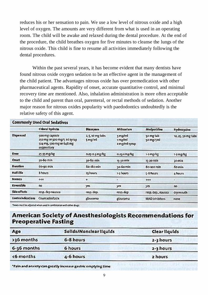
9
reduces his or her sensation to pain. We use a low level of nitrous oxide and a high
level of oxygen. The amounts are very different from what is used in an operating
room. The child will be awake and relaxed during the dental procedure. At the end of
the procedure, the child breathes oxygen for five minutes to cleanse the lungs of the
nitrous oxide. This child is fine to resume all activities immediately following the
dental procedures.
Within the past several years, it has become evident that many dentists have
found nitrous oxide oxygen sedation to be an effective agent in the management of
the child patient. The advantages nitrous oxide has over premedication with other
pharmaceutical agents. Rapidity of onset, accurate quantitative control, and minimal
recovery time are mentioned. Also, inhalation administration is more often acceptable
to the child and parent than oral, parenteral, or rectal methods of sedation. Another
major reason for nitrous oxides popularity with paedodontics undoubtedly is the
relative safety of this agent.
10
It is advocate of inhalation analgesia, and pointed out the absence of serious
untoward effects of nitrous oxide when it is used in appropriate analgesic
concentration. A special application of nitrous oxide-oxygen is suggested in the
management of certain handicapped children. In addition to the sedating and
analgesic effects, nitrous oxide is able to decrease the muscular spasticity and
uncoordinated movements of the cerebral palsied child, and thus make outpatient
dental care possible in many instances.
It is commented that nitrous oxide sedation is a valuable accessory in treating
most children but that it is not always an equivalent substitute for other forms of
premedication. Some degree of cooperation is required initially from the child for it
to be effective and success is rare if any force or strength is employed by the dentist.
It is claimed that routine premedication with hydroxizine will result in a greater
percentage of children who will willingly accept the nitrous oxide nose-piece. The
possible advantages of combining nitrous oxide-oxygen sedation with other forms of
premedication have not been adequately evaluated.
Nitrous Oxide with Atarax (Hydroxizine) Premedication :-
For some children who are anxious or have a severe gag reflex, nitrous oxide is
not enough to relax them for dental procedures. For these patients, we offer nitrous
oxide with a prescription for Atarax. The generic name for Atarax is Hydroxizine. It
is a liquid antihistamine, similar to Benadryl, which has three nice properties:
1-It has an anti-anxiety effect. It can reduce anxiety, and calm the child.
2-It has a drying effect, which can help reduce gagging, and reduce the need to use
many cotton rolls to keep a tooth dry during treatment.
3-It has an anti-emetic (anti-nausea) effect, this helps reduce gagging, vomiting.
4-In large doses Atarax can have a sedative effect, so this medication must be gives
as directed.
General Anesthesia :-
For patients with very high levels of anxiety, severe dental restorative needs, special
healthcare needs, special behavioral needs and sedation dentistry is not effective, we
offer general anesthesia. With general anesthesia, an anesthesiologist at the hospital
delivers the anesthesia to put a child to sleep. The child is completely unconscious and
the dental treatment is performed by doctors in an operating room setting. The
appointment will be scheduled early in the morning. It is important that child not eat or
drink anything after midnight the night before. Once the procedure is complete, a
11
recovery period is necessary. The child will be able to return home on the same day.
Dental Antibiotic Premedication :-
There are certain medical conditions which require the patient to take a dose of
antibiotics prior to their dental appointment. This is done to help prevent any bacteria
introduced from the dental procedure, from causing an infection in another part of the
body, such as the heart lining, called bacterial endocarditis.
This subject is of great discuss, because there is conflicting evidence whether antibiotic
dental premedication is needed at all, and the over prescription of antibiotics can lead
to antibiotic resistant strains of bacteria.
Health Conditions which may Require Premedication :-
Artificial Heart Valves
History of Rheumatic Fever
History of Infective Endocarditis
Kidney Dialysis
Mitral Valve Prolapse with Valvular Regurgitation
Certain Congenital Heart Conditions
Cardiac Transplants
Dental Procedures which Require Premedication :-
All procedures which involve manipulation of gingival tissue or the
periapical region of teeth or perforation of the oral mucosa, such as :-
1-Extraction.
2-Periodontal Surgary.
3-Endodontic Surgery.
4-Root canal therapy.
5-Periodontal Cleaning.
While the following procedures and events do not need prophylaxis:-
1-Routine anesthetic injections through non infected tissue.
2-Taking dental radiographs.
3-Placement and removal of removable prosthetics or orthodontic appliances.
4-Adjustment of orthodontic appliances.
5-Placement of orthodontic brackets.
6-Shedding of primary teeth.
7-Bleeding from trauma to the lips or oral mucosa.
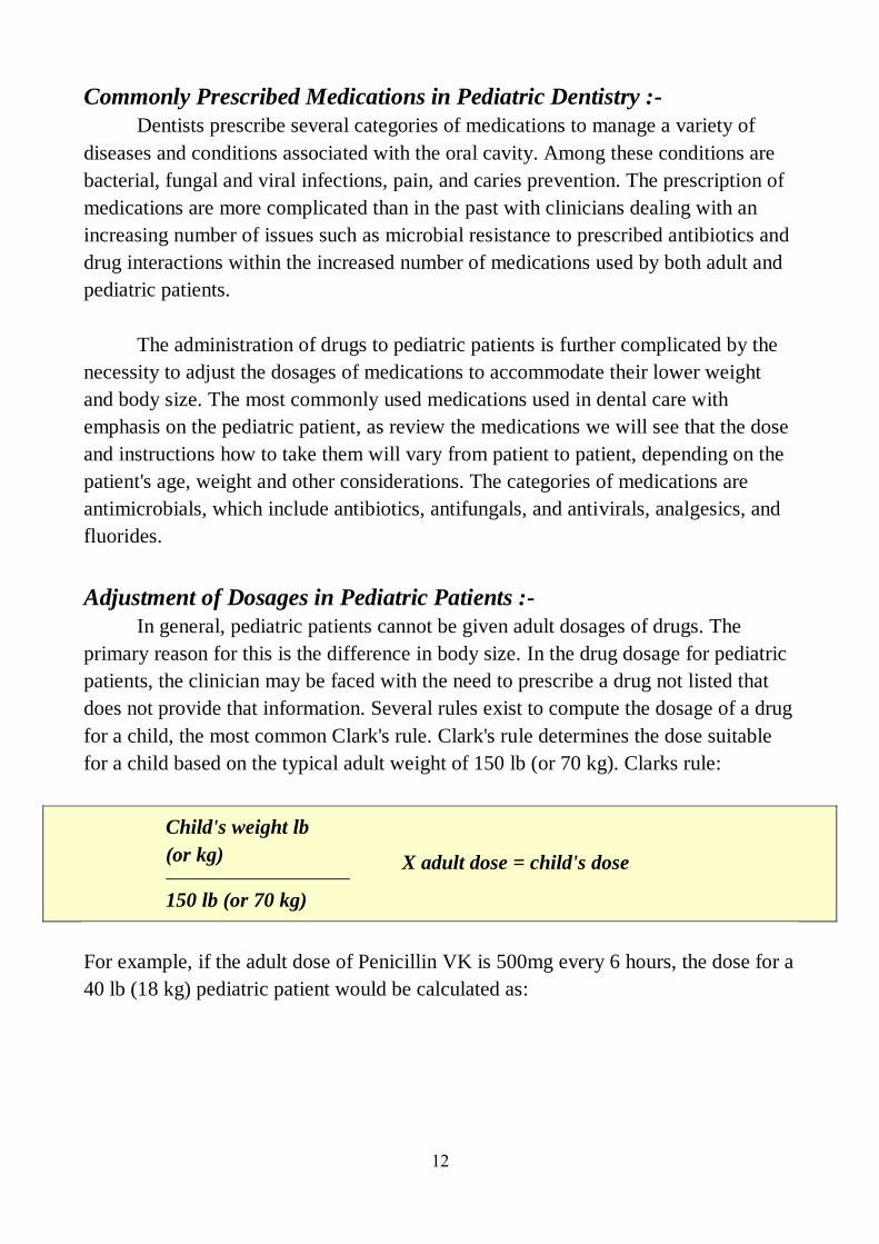
12
Commonly Prescribed Medications in Pediatric Dentistry :-
Dentists prescribe several categories of medications to manage a variety of
diseases and conditions associated with the oral cavity. Among these conditions are
bacterial, fungal and viral infections, pain, and caries prevention. The prescription of
medications are more complicated than in the past with clinicians dealing with an
increasing number of issues such as microbial resistance to prescribed antibiotics and
drug interactions within the increased number of medications used by both adult and
pediatric patients.
The administration of drugs to pediatric patients is further complicated by the
necessity to adjust the dosages of medications to accommodate their lower weight
and body size. The most commonly used medications used in dental care with
emphasis on the pediatric patient, as review the medications we will see that the dose
and instructions how to take them will vary from patient to patient, depending on the
patient's age, weight and other considerations. The categories of medications are
antimicrobials, which include antibiotics, antifungals, and antivirals, analgesics, and
fluorides.
Adjustment of Dosages in Pediatric Patients :-
In general, pediatric patients cannot be given adult dosages of drugs. The
primary reason for this is the difference in body size. In the drug dosage for pediatric
patients, the clinician may be faced with the need to prescribe a drug not listed that
does not provide that information. Several rules exist to compute the dosage of a drug
for a child, the most common Clark's rule. Clark's rule determines the dose suitable
for a child based on the typical adult weight of 150 lb (or 70 kg). Clarks rule:
Child's weight lb
(or kg) X adult dose = child's dose
150 lb (or 70 kg)
For example, if the adult dose of Penicillin VK is 500mg every 6 hours, the dose for a
40 lb (18 kg) pediatric patient would be calculated as:
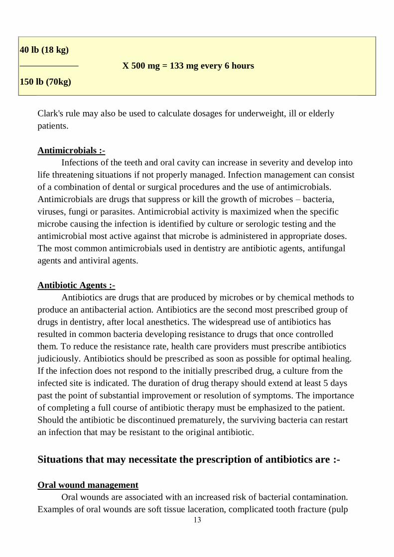
13
40 lb (18 kg)
X 500 mg = 133 mg every 6 hours
150 lb (70kg)
Clark's rule may also be used to calculate dosages for underweight, ill or elderly
patients.
Antimicrobials :-
Infections of the teeth and oral cavity can increase in severity and develop into
life threatening situations if not properly managed. Infection management can consist
of a combination of dental or surgical procedures and the use of antimicrobials.
Antimicrobials are drugs that suppress or kill the growth of microbes – bacteria,
viruses, fungi or parasites. Antimicrobial activity is maximized when the specific
microbe causing the infection is identified by culture or serologic testing and the
antimicrobial most active against that microbe is administered in appropriate doses.
The most common antimicrobials used in dentistry are antibiotic agents, antifungal
agents and antiviral agents.
Antibiotic Agents :-
Antibiotics are drugs that are produced by microbes or by chemical methods to
produce an antibacterial action. Antibiotics are the second most prescribed group of
drugs in dentistry, after local anesthetics. The widespread use of antibiotics has
resulted in common bacteria developing resistance to drugs that once controlled
them. To reduce the resistance rate, health care providers must prescribe antibiotics
judiciously. Antibiotics should be prescribed as soon as possible for optimal healing.
If the infection does not respond to the initially prescribed drug, a culture from the
infected site is indicated. The duration of drug therapy should extend at least 5 days
past the point of substantial improvement or resolution of symptoms. The importance
of completing a full course of antibiotic therapy must be emphasized to the patient.
Should the antibiotic be discontinued prematurely, the surviving bacteria can restart
an infection that may be resistant to the original antibiotic.
Situations that may necessitate the prescription of antibiotics are :-
Oral wound management
Oral wounds are associated with an increased risk of bacterial contamination.
Examples of oral wounds are soft tissue laceration, complicated tooth fracture (pulp
14
exposure), severe tooth displacement (including avulsion), gingivectomy, and severe
ulcerations. If the oral wound seems to have been contaminated by extraoral bacteria,
antibiotics therapy should be considered.
Dental infection :-
Bacteria can gain access to pulpal tissue through caries, exposed dentin tubules
and defective restorations resulting in acute dental infection. Treatment of acute
dental infection is accomplished by immediate dental treatment (pulpotomy,
pulpectomy or extraction). Antibiotic therapy is usually not indicated if the infection
is contained within the pulpal tissue or the immediately surrounding tissue and the
patient does not exhibit systemic signs of infection (fever and facial swelling).
Patients presenting with facial swelling secondary to a dental infection receive dental
treatment accompanied by antibiotic therapy.
If the clinician is able to achieve adequate local anesthesia, the dentist may
render immediate treatment followed by a regimen of antibiotics. If the infection is of
such severity that achievement of adequate local anesthesia is questionable, then
prescription of antibiotics for a period of 5-10 days should be considered before
rendering treatment. While oral antibiotics may be the simplest route of
administration, in cases of severe infection hospitalization and intravenous
administration may be necessary.
Paediatric periodontal diseases :-
Prolonged antibiotic therapy may be prescribed for the management of chronic
periodontal disease, especially in patients with an immunodeficiency disease. In
pediatric periodontal diseases (neutropenias, Papillon-Fevere syndrome, leukocyte
adhesion deficiency) the immune system is unable to control the growth of
periodontal microbes. As this is a chronic rather than an acute condition, effective
drug selection may be accomplished by culture and susceptibility testing
Viral diseases :-
Antibiotics should not be prescribed for viral conditions (acute primary
herpetic gingivostomatitis) unless there is strong evidence to suggest that a secondary
infection exists.
Causes and Treatment of Odontogenic Infections :-
Most orofacial infections are of odontogenic origin. Dental pulp infection, as a
result of caries, is the leading cause of odontogenic infection. The major pathogens
identified in dental caries are members of the viridens (alpha-hemolytic) streptococci
15
family including Streptococcus mutans, Streptococcus sobrinis and Streptococcus
milleri.
Once the bacterial cells invade the dental pulp an inflammatory reaction, they
results in necrosis and a lower tissue oxidation-reduction potential. At this stage, the
bacterial flora changes from predominately aerobic to more anaerobic flora.
Anaerobic gram-positive cocci (Peptostreptococcus) and anaerobic gram negative
rods (Bacteroides, Prevotella, Porphromonas and Fusobacterium) predominate. The
infection progresses forming an abscess at the apex of the root, resulting in bone
destruction.
Depending on host resistance and bacteria virulence the infection may spread
into the marrow, perforate the cortical plate and spread to the surrounding tissues.
Additionally, the anaerobic bacteria inhabiting the periodontal tissues may provide an
additional source of odontogenic infection. The most common anaerobes are
Actinobacillus actinomycetemcomitans, Prevotella intermediud, Porphyrommonas
gingivalis, Fusobacterium nucleatum, and Eikenella corrodens.
Most odontogenic infections (70%) contain mixed aerobic and anaerobic
bacteria. Pure aerobic infections have less than a 5% incidence. Pure anaerobic
infections have a 25% incidence. The agreement by researchers is that in early
odontogenic infections, bacteria are aerobic with gram-positive, alpha-hemolytic
streptococci (S. viridens) predominating. As the infection matures and increases in
severity the microbial flora becomes a mix of aerobes and anaerobes. The anaerobes
found are determined by the site of origin; pulpal or periodontal. As the host defenses
begin to control the infection process the flora becomes predominately anaerobic.
Thus, the choice of antibiotic is influenced by a number of factors: Stage of
infection development and the ability of the patient to take the antibiotic – medical
conditions or allergy. Antibiotics may also be categorized by their method of attack.
Bactericidal antibiotics actually kill microorganisms, while bacteriostatic antibiotics
slow bacterial growth and depend on the host immune system to eliminate the
microorganism.
An antibiotic may be bactericidal for one microorganism and bacteriostatic for
another. Bactericidals are preferable over bacteristatics in most situations.
Bacteriostatics should not be administered to immunocompromised patients whose
compromised immune system may be unable to assist in clearance of the
microorganism. Common bactericidals used in dentistry are the penicillins and
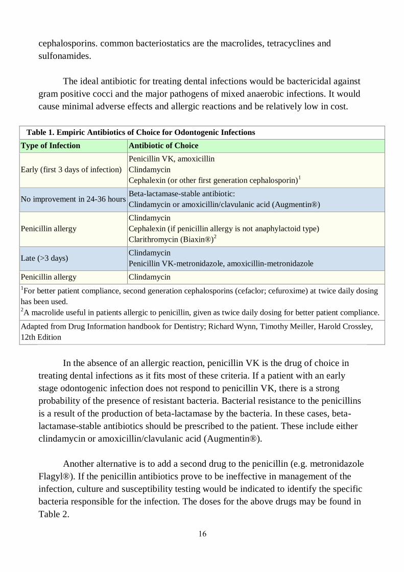
16
cephalosporins. common bacteriostatics are the macrolides, tetracyclines and
sulfonamides.
The ideal antibiotic for treating dental infections would be bactericidal against
gram positive cocci and the major pathogens of mixed anaerobic infections. It would
cause minimal adverse effects and allergic reactions and be relatively low in cost.
Table 1. Empiric Antibiotics of Choice for Odontogenic Infections
Type of Infection Antibiotic of Choice
Early (first 3 days of infection)
Penicillin VK, amoxicillin
Clindamycin
Cephalexin (or other first generation cephalosporin)1
No improvement in 24-36 hours Beta-lactamase-stable antibiotic:
Clindamycin or amoxicillin/clavulanic acid (Augmentin®)
Penicillin allergy
Clindamycin
Cephalexin (if penicillin allergy is not anaphylactoid type)
Clarithromycin (Biaxin®)2
Late (>3 days) Clindamycin
Penicillin VK-metronidazole, amoxicillin-metronidazole
Penicillin allergy Clindamycin
1For better patient compliance, second generation cephalosporins (cefaclor; cefuroxime) at twice daily dosing
has been used. 2A macrolide useful in patients allergic to penicillin, given as twice daily dosing for better patient compliance.
Adapted from Drug Information handbook for Dentistry; Richard Wynn, Timothy Meiller, Harold Crossley,
12th Edition
In the absence of an allergic reaction, penicillin VK is the drug of choice in
treating dental infections as it fits most of these criteria. If a patient with an early
stage odontogenic infection does not respond to penicillin VK, there is a strong
probability of the presence of resistant bacteria. Bacterial resistance to the penicillins
is a result of the production of beta-lactamase by the bacteria. In these cases, beta-
lactamase-stable antibiotics should be prescribed to the patient. These include either
clindamycin or amoxicillin/clavulanic acid (Augmentin®).
Another alternative is to add a second drug to the penicillin (e.g. metronidazole
Flagyl®). If the penicillin antibiotics prove to be ineffective in management of the
infection, culture and susceptibility testing would be indicated to identify the specific
bacteria responsible for the infection. The doses for the above drugs may be found in
Table 2.
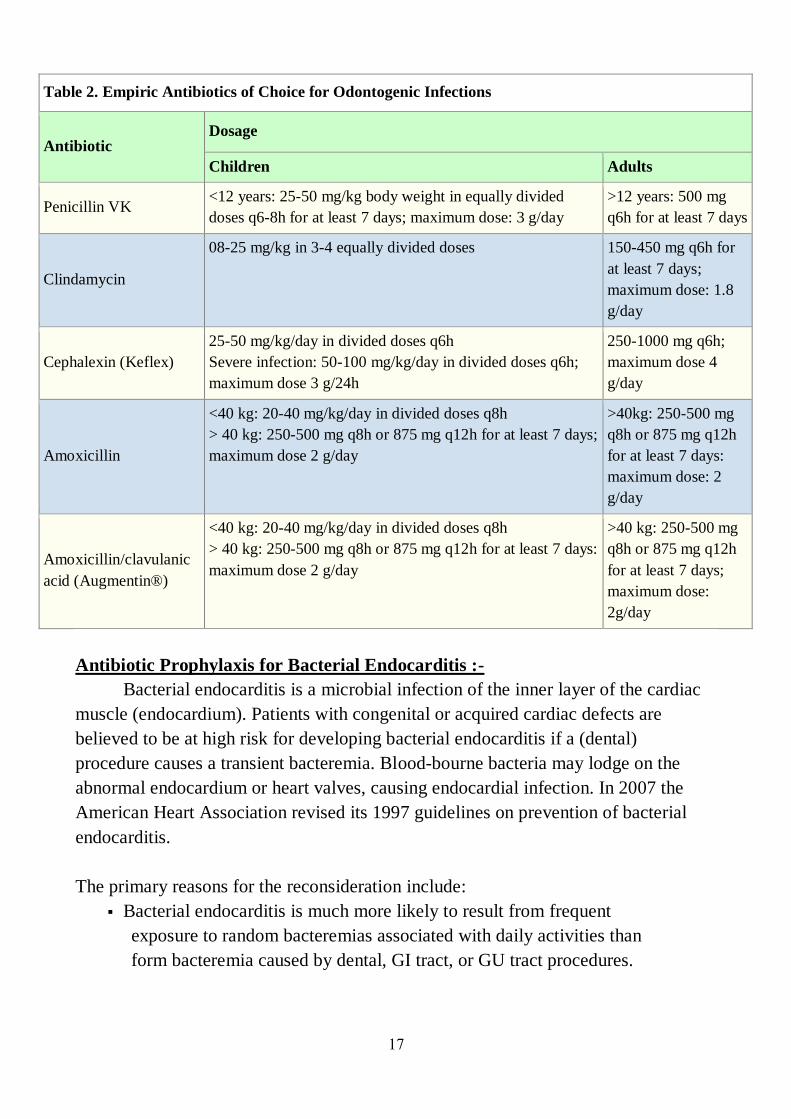
17
Table 2. Empiric Antibiotics of Choice for Odontogenic Infections
Antibiotic Dosage
Children Adults
Penicillin VK <12 years: 25-50 mg/kg body weight in equally divided
doses q6-8h for at least 7 days; maximum dose: 3 g/day
>12 years: 500 mg
q6h for at least 7 days
Clindamycin
08-25 mg/kg in 3-4 equally divided doses 150-450 mg q6h for
at least 7 days;
maximum dose: 1.8
g/day
Cephalexin (Keflex)
25-50 mg/kg/day in divided doses q6h
Severe infection: 50-100 mg/kg/day in divided doses q6h;
maximum dose 3 g/24h
250-1000 mg q6h;
maximum dose 4
g/day
Amoxicillin
<40 kg: 20-40 mg/kg/day in divided doses q8h
> 40 kg: 250-500 mg q8h or 875 mg q12h for at least 7 days;
maximum dose 2 g/day
>40kg: 250-500 mg
q8h or 875 mg q12h
for at least 7 days:
maximum dose: 2
g/day
Amoxicillin/clavulanic
acid (Augmentin®)
<40 kg: 20-40 mg/kg/day in divided doses q8h
> 40 kg: 250-500 mg q8h or 875 mg q12h for at least 7 days:
maximum dose 2 g/day
>40 kg: 250-500 mg
q8h or 875 mg q12h
for at least 7 days;
maximum dose:
2g/day
Antibiotic Prophylaxis for Bacterial Endocarditis :-
Bacterial endocarditis is a microbial infection of the inner layer of the cardiac
muscle (endocardium). Patients with congenital or acquired cardiac defects are
believed to be at high risk for developing bacterial endocarditis if a (dental)
procedure causes a transient bacteremia. Blood-bourne bacteria may lodge on the
abnormal endocardium or heart valves, causing endocardial infection. In 2007 the
American Heart Association revised its 1997 guidelines on prevention of bacterial
endocarditis.
The primary reasons for the reconsideration include:
Bacterial endocarditis is much more likely to result from frequent
exposure to random bacteremias associated with daily activities than
form bacteremia caused by dental, GI tract, or GU tract procedures.
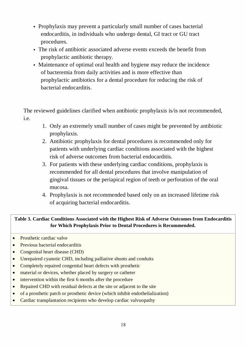
18
Prophylaxis may prevent a particularly small number of cases bacterial
endocarditis, in individuals who undergo dental, GI tract or GU tract
procedures.
The risk of antibiotic associated adverse events exceeds the benefit from
prophylactic antibiotic therapy.
Maintenance of optimal oral health and hygiene may reduce the incidence
of bacteremia from daily activities and is more effective than
prophylactic antibiotics for a dental procedure for reducing the risk of
bacterial endocarditis.
The reviewed guidelines clarified when antibiotic prophylaxis is/is not recommended,
i.e.
1. Only an extremely small number of cases might be prevented by antibiotic
prophylaxis.
2. Antibiotic prophylaxis for dental procedures is recommended only for
patients with underlying cardiac conditions associated with the highest
risk of adverse outcomes from bacterial endocarditis.
3. For patients with these underlying cardiac conditions, prophylaxis is
recommended for all dental procedures that involve manipulation of
gingival tissues or the periapical region of teeth or perforation of the oral
mucosa.
4. Prophylaxis is not recommended based only on an increased lifetime risk
of acquiring bacterial endocarditis.
Table 3. Cardiac Conditions Associated with the Highest Risk of Adverse Outcomes from Endocarditis
for Which Prophylaxis Prior to Dental Procedures is Recommended.
Prosthetic cardiac valve
Previous bacterial endocarditis
Congenital heart disease (CHD)
Unrepaired cyanotic CHD, including palliative shunts and conduits
Completely repaired congenital heart defects with prosthetic
material or devices, whether placed by surgery or catheter
intervention within the first 6 months after the procedure
Repaired CHD with residual defects at the site or adjacent to the site
of a prosthetic patch or prosthetic device (which inhibit endothelialization)
Cardiac transplantation recipients who develop cardiac valvuopathy
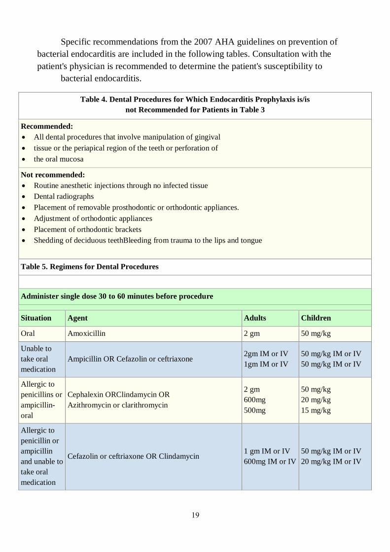
19
Specific recommendations from the 2007 AHA guidelines on prevention of
bacterial endocarditis are included in the following tables. Consultation with the
patient's physician is recommended to determine the patient's susceptibility to
bacterial endocarditis.
Table 4. Dental Procedures for Which Endocarditis Prophylaxis is/is
not Recommended for Patients in Table 3
Recommended:
All dental procedures that involve manipulation of gingival
tissue or the periapical region of the teeth or perforation of
the oral mucosa
Not recommended:
Routine anesthetic injections through no infected tissue
Dental radiographs
Placement of removable prosthodontic or orthodontic appliances.
Adjustment of orthodontic appliances
Placement of orthodontic brackets
Shedding of deciduous teethBleeding from trauma to the lips and tongue
Table 5. Regimens for Dental Procedures
Administer single dose 30 to 60 minutes before procedure
Situation Agent Adults Children
Oral Amoxicillin 2 gm 50 mg/kg
Unable to
take oral
medication
Ampicillin OR Cefazolin or ceftriaxone 2gm IM or IV
1gm IM or IV
50 mg/kg IM or IV
50 mg/kg IM or IV
Allergic to
penicillins or
ampicillin-
oral
Cephalexin ORClindamycin OR
Azithromycin or clarithromycin
2 gm
600mg
500mg
50 mg/kg
20 mg/kg
15 mg/kg
Allergic to
penicillin or
ampicillin
and unable to
take oral
medication
Cefazolin or ceftriaxone OR Clindamycin 1 gm IM or IV
600mg IM or IV
50 mg/kg IM or IV
20 mg/kg IM or IV
20
Antifungals
Oral fungal infections occur from alterations in oral flora as a result of the
extensive use of broad spectrum antibiotics, steroids, chemotherapy immune-
suppression, and inadequate oral hygiene and nutrition. The most common fungal
infection found in children is candidiasis. The clinical variations of candidiasis most
commonly found in children are pseudomembranous candidiasis, angular cheilitis,
erythematous candidiasis and mucocutaneous candidiasis.
Treatment of candidiasis is accomplished through the topical application of
nystatin, clotrimazole and amphotericin and systemic administration of ketoconazole,
fluconazole, itraconazole when topical treatment is ineffective. When prescribing
antifungals, the clinician must closely monitor and re-evaluate the patient's response
every two weeks. If the response is inadequate, the diagnosis, choice of medication
and dosage should be reevaluated. The chosen form of administration is dependent on
the child's ability and maturity to follow directions. The drugs are administered until
2 days after symptoms disappear.
Antiviral and Ulcerative Lesions
Advances in the pharmacological treatment of viral infections lag behind the
treatment of bacterial or fungal infections. The reason is due to the difficulty in
attaining adequate degrees of selective toxicity. Since virus replication uses the same
metabolic mechanisms essential for the function of normal cells, it was difficult to
find drugs that would inhibit viral growth without killing the host. However recent
advances in the research of viral replication have lead to discovery of agents useful in
antiviral activity in the oral cavity. The agents are not highly effective and are best
used as soon as symptoms first appear. Systemic supportive therapy should be
administered in conjunction with antivirals which includes forced fluids, high
concentration protein, vitamin and mineral food supplements and rest. Viral
infections may become secondarily infected with bacteria requiring antibiotics.
Oral viral infections are most commonly caused by the herpes simplex virus.
The herpes zoster or herpes varicella–zoster virus can cause similar viral eruptions
involving the oral mucosa. Diagnosis of oral viral infections begins by evaluation of
presenting signs and symptoms. A distinction must be made between lesions
associated with herpes and aphthous ulcers which do not have a viral etiology.
Viral lesions (Herpetic gingivostomatitis) are characterized by an initial acute
onset of vesicular eruptions on the soft tissues that quickly rupture into small
ulcerations that are covered by a yellowish gray pseudomembrane surrounded by an
21
erythematous halo. The ulcers may coalesce to form larger irregular ulcerations. The
lesions are found on the gingival, tongue, palate lips (labialis), buccal mucosa, tonsils
and posterior pharynx. The ulcers gradually heal over 7-10 days without scarring.
The disease is accompanied by high fever, malaise, irritability, headache and pain in
the mouth during the first three days of onset. It usually appears in children between
the ages of six months and four years. Treatment consists of administration of
acyclovir and supportive therapy.
Caries Prevention
Among the reasons for the significant reduction in dental caries in children
over the past several decades has been the increased availability of fluoride. When
administered in the appropriate dosage, fluoride is a highly safe and effective method
for the prevention and control of caries. Although the precise mechanisms by which
fluorides act are not fully understood, three mechanisms are generally accepted:
1. increasing the resistance of tooth structure to demineralization;
2. enhancing the process of remineralization; and
3. reducing the cariogenic potential of dental plaque.
The effects of fluoride are classified as either systemic or topical. Sources of
systemic fluoride include drinking water from home and school, beverages such as
soda, juice, and infant formula, prepared food, professionally prescribed fluoride
products and ingestion of toothpaste. The sources of topical effects are available from
previously mentioned systemic sources contacting the teeth during ingestion,
toothpastes containing fluoride and professionally applied or self-applied
concentrated forms of fluoride.
Systemic Fluoride
Systemically administered fluoride supplements should be considered for all
children older than 6 months drinking fluoride deficient (<0.6 ppm) water. As
fluorosis of the teeth has been associated with chronic ingestion of greater than
recommended amounts of fluoride, when prescribing systemic fluorides it is
important the dental professional take into account all the possible sources of
fluoride, as described above, that the patient ingests to determine the correct level of
supplementation. On the other hand, in areas with optimal fluoridation, some families
avoid the use community fluoridated water supplies for drinking and cooking because
of fear of chemical and bacterial contamination. The bottled water the family uses
may not contain optimal levels of fluoride and thus fluoride supplementation should
be considered.

22
After investigation of the patient's dietary intake of fluoride and a risk
assessment for the patient's tendency for caries, the clinician can refer to the Dietary
Fluoride Supplementation Schedule as recommended by the American Academy of
Pediatric Dentistry and the American Academy of Pediatrics (Table 7).
Contraindications: Hypersensitivity to fluoride, tartizine or any component of the
formulation; when fluoride content of water exceeds 0.7 ppm.
Warnings/Precaution: Prolonged ingestion of excessive doses may result in dental
fluorosis and osseous changes.
Table 7. Dietary Fluoride Supplementation Schedule
Age <0.3 ppm F 0.3-0.6 ppm F >0.6 ppm F
Birth – 6 months 0 0 0
6 months – 3 years 0.25 mg 0 0
3 – 6 years 0.50 mg 0.25 mg 0
6 years to 16 years 1.00 mg 0.50 mg 0
Systemic fluoride supplements are available as liquid or chewable tablets. The
fluoride in most dietary fluoride supplements is incorporated as sodium fluoride
(NaF). One milligram of fluoride is equivalent to approximately 2.2 mg of sodium
fluoride. When prescribing fluoride the clinician should specify the dose to be
dispensed in terms of fluoride ion, sodium fluoride, or both. Fluoride drops and
tablets are available as sodium fluoride 0.5mg (0.25 mg fluoride), 1.1 mg (0.5 mg
fluoride), and 2.2 mg (1mg fluoride). When prescribing chewable tablets it is
recommended that the child suck on the tablets before chewing and swallowing to
maximize the contact time of the fluoride with the tooth enamel.
Concentration Topical Fluoride for Home Use
In patients with a high caries rate or high risk assessment for caries
(undergoing orthodontic therapy), concentrated topical fluorides may be prescribed
for home use. Topical concentrated agents for home use include 0.5% acidulated
phosphate fluoride, 1.1% sodium fluoride gel and 0.4% stannous fluoride gel. The gel
is available without a prescription. The patient is instructed to brush a small amount
of gel on the child's teeth before bedtime. Expectoration is encouraged however no
23
eating or drinking for 30 minutes. Depending on the formulation and concentration of
the product, application is daily or weekly.
Safety and Toxicity :-
When used as directed, fluoride can improve the oral health of children.
However, when used improperly it can produce chronic (fluorosis) and acute
problems. Therefore, it is very important that the practitioner instruct parents and
patients about the proper storage and use of fluoride products.
Accidental ingestion of excessive amounts of fluoride can result in acute
toxicity. Acute fluoride toxicity usually manifests itself as nausea and vomiting but
death has been reported. The amount of ingested fluoride necessary to produce acute
toxicity is in proportion to the child's weight. For example, the lethal dose of fluoride
for a 25 pound three year old is approximately 500mg but would be proportionately
less for a child of lesser weight.
To reduce the possibility of ingestion of large amounts of fluoride, it is
recommended that no more than 120mg of supplemental fluoride be prescribed at any
one time. Preparations of concentrated topical fluoride preparations (0.5% fluoride
gels containing 5mg fluoride/ml) should be limited to 30 to 40 ml. The recommended
treatment if a child ingests excessive amounts of fluoride is to call local poison
control for verification, induce vomiting as quickly as possible and give milk every
four hours to slow absorption.
1
Paediatric Dentistry
Fifth Stage
Dr. Suhair W. Abbood
Lec. 1
History, Examination and Treatment Planning
The provision of dental care of children presents some of the greatest challenges
(and rewards) in clinical dental practice. High on the list of challenges is the need to
devise a comprehensive yet realistic treatment plan for these young patients.
Historically, dental care for children has been designed primarily to prevent :-
1-Oral pain and infection.
2-The occurrence and progress of dental caries.
3-The premature loss of primary teeth.
4-The loss of arch length.
5-The association between fear and dental care.
Consent to examination, investigation, or treatment is fundamental to the
provision of dental care. The most important element of the consent procedure is
ensuring that the patient / parent understand the nature and purpose of the proposed
treatment, together with any alternative available, and potential benefits and risks.
It is important that “informed consent” be obtained. The clinician must carefully
explain all the procedures planned using lay language as appropriate. All potential
risks need to be mentioned, discussed and documented. When completing the
sections on standard forms on the nature of the operation, be specific, do not use
abbreviations and include all the procedures planned. Where appropriate use simple
terminology to describe the operation.
In an emergency, it is justifiable to treat a child without the consent of the
person with parental responsibility if the treatment is vital to the health of the child.
For example, while it may be acceptable to replant an avulsed permanent incisor, the
parent should be contacted before proceeding to other forms of treatment.
If parents reject a portion or all of the recommendations, the dentist has at least
fulfilled the obligation of educating the child and the parents about the importance of
the recommended procedures. Parents of even moderate income will usually find the
2
means to have oral health care completed if the dentist explains to them that the
child’s future oral health and even general health are related to the correction of oral
defects.
Throughout treatment the dentist is responsible for :-
1-Guiding the child and parents.
2-Resolving oral disorders before they can affect health and dental alignment.
3-Preventing oral disease.
A dentist is traditionally taught to :-
1-Preform a complete oral examination of the patient.
2-Develop a treatment plan from the examination findings.
3-Makes a case presentation to the patient or parents outlining the recommended
course of treatment.
These procedures should be the development and presentation of a prevention
plan that outlines an ongoing comprehensive oral health care program for the patient.
The prevention plan should include :-
1-Recommendations designed to correct existing oral problems (or prevent their
progression).
2-Prevent anticipated future problems.
The successful practice of pediatric dentistry is not merely the completion of any
operative procedure but also ensuring a positive dental outcome for the future oral
health behavior of that individual and family. To this end an understanding of child
development-physical, cognitive and psychosocial is paramount. The clinician must
be comfortable and skilled in talking to children, and interpersonal skills are
essential. It will not usually be the child’s fault if the clinician cannot work with child.
History :-
A clinical history should be taken in a logical and systematic way for each
patient and should be updated regularly. Thorough history taking is time consuming
and requires practice. However, it is an opportunity to get to know the child and
family.
Furthermore, the history facilitates the diagnosis of many conditions even
before the hands-on examination. Because, there are often specific questions related
to a child’s medical history that will be relevant to their management, it is desirable
that parents be present. The understanding of medical conditions that can
compromise treatment is essential.
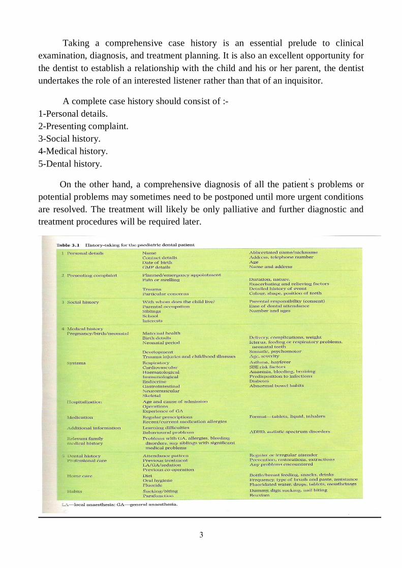
3
Taking a comprehensive case history is an essential prelude to clinical
examination, diagnosis, and treatment planning. It is also an excellent opportunity for
the dentist to establish a relationship with the child and his or her parent, the dentist
undertakes the role of an interested listener rather than that of an inquisitor.
A complete case history should consist of :-
1-Personal details.
2-Presenting complaint.
3-Social history.
4-Medical history.
5-Dental history.
On the other hand, a comprehensive diagnosis of all the patient’s problems or
potential problems may sometimes need to be postponed until more urgent conditions
are resolved. The treatment will likely be only palliative and further diagnostic and
treatment procedures will be required later.
4
The Preliminary Medical and Dental History :-
It is important for the dentist to be familiar with the medical and dental history
of all pediatric patient. Familial history may also be relevant to the patient’s oral
condition and may provide important diagnostic information in some hereditary
disorders.
Information regarding the child’s social and psychologic development is
important. Accurate information reflecting a child's learning, behavioral or
communication problems is sometimes difficult to obtain initially, especially if the
parents are aware of their child’s development disorder but reluctant to discuss it.
Behavior problems in the dental office are often related to the child’s inability to
communicate with the dentist and follow instructions. This inability may be
attributable to a low mental capacity.
While the history is being taken, the clinician should also be making an
“unofficial” assessment of the child's likely level of co-operation in order that the
most appropriate approach for examination can be adopted right from the start
(hopefully saving both time and tears). Broadly speaking, prospective young patients
may fall into one of the following categories :-
● Happy and confident :- This child is likely to hop into the chair for a check-
up without further coaxing.
● A little anxious or shy but displaying some relationship with the dental
team :- This child will probably allow an examination after some simple
acclimatization and reassurance (if the child is very young, the option of sitting
on the mother’s knee could be given).
● Very frightened, crying, clutching their parent, avoiding eye contact, or
not responding to direct questions :- This child is unlikely to accept a
conventional examination at this visit (though the child may allow a brief
examination while sitting on a non-dental chair, perhaps even in the waiting
room), further acclimatization will be required before a thorough examination
can be undertaken.
● Sever behavioral problem or learning disability :- In a few cases, this may
prevent the child from ever voluntarily accepting an examination, restraint
(with or without pharmacological management) may be indicated to facilitate
an intraoral examination.
Examination :-
The first impressions :- The purpose of the examination is not only to check for
caries or periodontal disease, as pediatric dentistry covers all areas of growth and
5
development. Having the opportunity to see the child regularly, the dentist can often
be the first to recognize significant disease and anomalies.
An initial impression of the child’s overall health and development can be
gained as soon as he or she is welcomed in waiting room or enters the surgery. In
particular, it is useful to know that most facts needed for a comprehensive oral
diagnosis in the young patient are obtained by note :-
● General health-does the child look well?
● Overall physical and mental development-does it seem appropriate for the child's
chronological age?
● Weight-is the child grossly under-or overweight?. The first clue to malnutrition
may come from observing a patient’s abnormal size or stature.
● Co-ordination-does the child have an abnormal gait or obvious motor impairment?.
The severity of a child's illness, even if oral in origin, may be recognized by
observing the weak, unsteady gait of tiredness and sickness as the patient walks into
the office.
The Extraoral Examination :-
The General Examination :- Before carrying out a detailed examination of the
craniofacial structures, a more general physical assessment should be undertaken.
Valuable information about a child's overall health, development, or even habits can
often be determined by noting :-
● Height :- Is the child very tall or very small for their age?. In a few cases, it may be
appropriate to take an accurate height measurement and plot data on a standard
growth chart. Children whose height lies below the third centile, above the ninety-
seventh centile, or who exhibit less than 3-5 cm growth per year should be referred to
a pediatrician for further investigation.
● Weight :- Could there be an underlying eating disorder?. Is general anaesthesia
contraindicated due to child's obesity?. Is there an underlying endocrine problem?.
● Skin :- Look for any notable bruising or injury on exposed arms or legs.
● Hands :- Assess for evidence of digit sucking or nail biting, warts, finger clubbing
abnormal nail, or finger morphology. The dentist may first detect an elevated
temperature by holding the patient’s hand. Cold, clammy hands or bitten fingernails
may be the first indication of abnormal anxiety in the child. In addition to that a
callused or unusually clean digit suggests a persistent sucking habit. Clubbing of the
fingers or a bluish color in the nail beds suggests congenital heart disease that may
require special precautions during dental treatment.

6
The Head and Neck :- During the examination of the head and neck, the following
structures should be briefly assessed :-
● Hair :- Note if sparse (look out for head lice).
● Eyes :- Is there any visual impairment or abnormality of the sclera?.
● Ears :- Record any abnormal morphology or presence of hearing aids.
● Skin :- Document any scars, bruising, laceration, pallor, birthmarks and be aware
of contagious infections, such as ringworm or impetigo. Proper referral is indicated
immediately, if a contagious condition is identified, but when the child also has a
dental emergency, the dentist must take appropriate precautions to prevent spread of
the disease to others while the emergency is alleviated. Further treatment should be
postponed until the contagious condition is controlled.
● Temporomandibular joint (TMJ) :- Is there is any pain, crepitus (any crepitus
that may be heard or identified by palpation, or any other abnormal sounds, should be
noted), deviation (movements of the condyles or jaw that are not smooth flowing or
deviate from the expected normal should be noted), or restricted opening (palpating
the head of each mandibular condyle and observing the patient while the mouth is
closed “teeth clenched”, at rest, and in various open positions).
● Lymph nodes :- Palpate for enlarged submandibular or cervical lymph nodes (bear
in mind that lymphadenopathy is not uncommon in children, due to frequent viral
infections).
● Lips :- Note the presence of cold sores, swelling, or abnormal colouring. Any
positive findings should be recorded carefully. Clinical photographs or annotated
sketches may be very helpful for future reference, particularly with respect to
medico-legal purposes, or in cases of suspected child physical abuse. Obviously,
when the child presents with a specific problems, such as a facial swelling, a more
thorough examination of the presenting condition is needed.
The Intraoral Examination :-
The intraoral examination of a pediatric patient should be comprehensive. There
is a temptation to look first for obvious carious lesions. Certainly controlling carious
lesions is important, but the dentist should first evaluate the condition of the oral soft
tissues and the status of the developing occlusion.
If the soft tissues and the occlusion are not observed early in the examination,
the dentist may become so engrossed in charting carious lesions and in planning for
their restoration that other important anomalies in the mouth are overlooked. A
systemic approach should be adopted for the intraoral examination. The following is
a suggested order :-
7
● Soft tissues.
● Gingival and periodontal tissues.
●Teeth.
● Occlusion.
The soft tissues :- An abnormal appearance of the oral soft tissues may be indicative
of an underlying systemic disease or nutritional deficiency. In addition, a variety of
oral pathologies may be seen in children. It is therefore important to carefully
examine the tongue, palate, throat, and cheeks, noting any color changes, ulceration,
swelling, or other pathology.
The tongue and oropharynx should be closely inspected. Enlarged tonsils
accompanied by purulent exudates may be the initial sign of a streptococcal infection
leading to rheumatic fever, when streptococcal throat infection is suspected
immediate referral to the child's physician is indicated.
It is also sensible to check for abnormal frenal attachment or tongue-tie, which
may have functional implications. If a tongue-tie or abnormal tongue function is
observed, some consideration should be given to the child's speech. During
examination of the soft tissues any unusual breath odors should be noted, an overall
impression of salivary flow rate and consistency should also be gained.
The gingival and periodontal tissues :- A visual examination of the gingival tissues
is usually all that is indicated for young children, as periodontal disease is very
uncommon in this age group. The presence of color change (redness), swelling,
ulceration, spontaneous bleeding, or recession should be carefully noted, and the
etiology sought.
During inspection of the gingival issues, an assessment of oral cleanliness
should also be made, and the presence of any plaque or calculus deposits noted. A
number of simple oral hygiene indices have been developed to provide an objective
record of oral cleanliness. One such index, the oral debris index (Green and
Vermillion, 1964), requires disclosing prior to an evaluation of the amount of plaque
on selected teeth (first permanent molars, and upper right and lower left central
incisors).
The presence of profound gingival inflammation in the absence of gross plaque
deposits, lateral periodontal abscesses, prematurely exfoliating teeth, or mobile
permanent teeth may indicate a more serious underlying problem, warranting further
investigation.
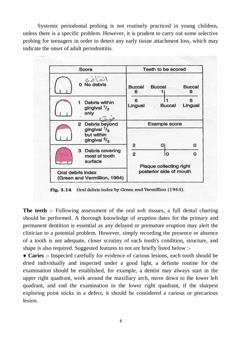
8
Systemic periodontal probing is not routinely practiced in young children,
unless there is a specific problem. However, it is prudent to carry out some selective
probing for teenagers in order to detect any early tissue attachment loss, which may
indicate the onset of adult periodontitis.
The teeth :- Following assessment of the oral soft tissues, a full dental charting
should be performed. A thorough knowledge of eruption dates for the primary and
permanent dentition is essential as any delayed or premature eruption may alert the
clinician to a potential problem. However, simply recording the presence or absence
of a tooth is not adequate, closer scrutiny of each tooth's condition, structure, and
shape is also required. Suggested features to not are briefly listed below :-
● Caries :- Inspected carefully for evidence of carious lesions, each tooth should be
dried individually and inspected under a good light, a definite routine for the
examination should be established, for example, a dentist may always start in the
upper right quadrant, work around the maxillary arch, move down to the lower left
quadrant, and end the examination in the lower right quadrant, if the sharpest
exploring point sticks in a defect, it should be considered a carious or precarious
lesion.
9
See if the carious lesion is active / arrested, identification of carious lesions is
important in patients of all ages but is especially critical in young patients because the
lesions may progress rapidly in children if not controlled. In patients with severe
dental caries, caries activity tests and diet analysis may contribute to the diagnostic
process by helping to define specific etiologic factors. These procedures probably
have an even greater value in helping the patient or parents understand the carious
disease process and in motivating those to make the behavioral changes needed to
control the disease.
See if the carious lesion restorable / unrestorable, eliminating the carious
activity and restoring the teeth as needed will prevent pain and the spread of infection
and also contribute to the stability of the developing occlusion. Check also for the
presence of a chronic sinus associated with grossly carious teeth.
● Restorations :- Are they intact / deficient?.
● Fissure sealants :- Are they intact / deficient?.
● Tooth surface loss :- Note any erosion / attrition, site, extent.
● Trauma :- Note extent, site, or signs of loss of vitality.
● Tooth structure :- Record any enamel opacities / hypoplasia (are defects localized
/ generalized?). Morphological defects and incomplete coalescence of enamel at the
base pits and fissures in molar teeth can often be detected readily by visual
examination after the teeth have been cleaned and dried.
● Tooth shape / size :- Note presence of double teeth, conical teeth, macrodontia /
microdontia, talon cusps, deep cingulum pits, and any hereditary or acquired
anomalies.
● Tooth number :- The teeth should also be counted and identified individually to
ensure recognition of extra teeth (supernumerary teeth) or any missing teeth.
● Tooth mobility :- Is it physiological or pathological?.
● Tooth eruption :- Are there any impactions, infraoccluded primary molars, or
ectopically erupting first permanent molars?.
The occlusion :- Clearly, a full orthodontic assessment is not indicated every time a
child is examined. After thoroughly examining the oral soft tissues, the dentist should
inspect the occlusion and note any dental or skeletal irregularities. The dentition and
resulting occlusion may undergo considerable change during childhood and early
adolescence.
Monitoring the patient's facial profile and symmetry, molar, canine, and
anterior segment relationships, dental midlines, and arch length to tooth mass
comparisons should be routinely included in the clinical examination. However, tooth
alignment and occlusion should be briefly considered, as these may provide an early
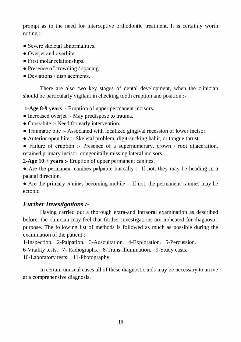
10
prompt as to the need for interceptive orthodontic treatment. It is certainly worth
noting :-
● Severe skeletal abnormalities.
● Overjet and overbite.
● First molar relationships.
● Presence of crowding / spacing.
● Deviations / displacements.
There are also two key stages of dental development, when the clinician
should be particularly vigilant in checking tooth eruption and position :-
1-Age 8-9 years :- Eruption of upper permanent incisors.
● Increased overjet :- May predispose to trauma.
● Cross-bite :- Need for early intervention.
● Traumatic bite :- Associated with localized gingival recession of lower incisor.
● Anterior open bite :- Skeletal problem, digit-sucking habit, or tongue thrust.
● Failure of eruption :- Presence of a supernumerary, crown / root dilaceration,
retained primary incisor, congenitally missing lateral incisors.
2-Age 10 + years :- Eruption of upper permanent canines.
● Are the permanent canines palpable buccally :- If not, they may be heading in a
palatal direction.
● Are the primary canines becoming mobile :- If not, the permanent canines may be
ectopic.
Further Investigations :-
Having carried out a thorough extra-and intraoral examination as described
before, the clinician may feel that further investigations are indicated for diagnostic
purpose. The following list of methods is followed as much as possible during the
examination of the patient :-
1-Inspection. 2-Palpation. 3-Auscultation. 4-Exploration. 5-Percussion.
6-Vitality tests. 7- Radiographs. 8-Trans-illumination. 9-Study casts.
10-Laboratory tests. 11-Photography.
In certain unusual cases all of these diagnostic aids may be necessary to arrive
at a comprehensive diagnosis.
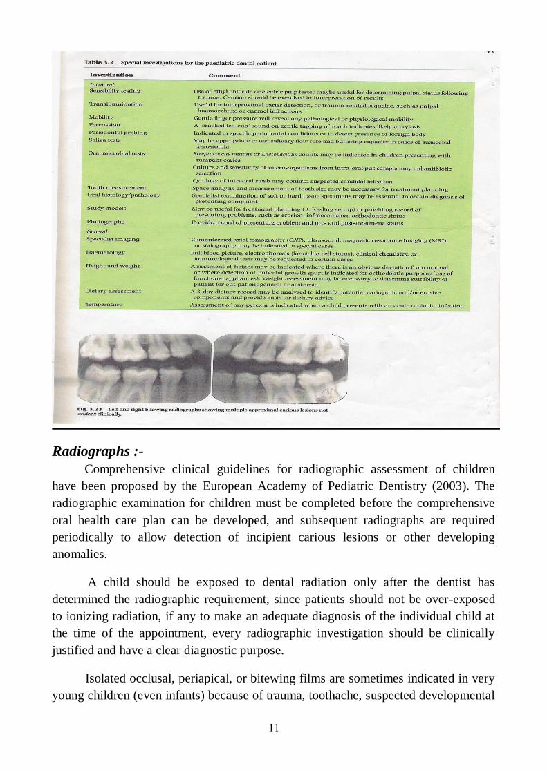
11
Radiographs :-
Comprehensive clinical guidelines for radiographic assessment of children
have been proposed by the European Academy of Pediatric Dentistry (2003). The
radiographic examination for children must be completed before the comprehensive
oral health care plan can be developed, and subsequent radiographs are required
periodically to allow detection of incipient carious lesions or other developing
anomalies.
A child should be exposed to dental radiation only after the dentist has
determined the radiographic requirement, since patients should not be over-exposed
to ionizing radiation, if any to make an adequate diagnosis of the individual child at
the time of the appointment, every radiographic investigation should be clinically
justified and have a clear diagnostic purpose.
Isolated occlusal, periapical, or bitewing films are sometimes indicated in very
young children (even infants) because of trauma, toothache, suspected developmental
12
disturbances, or proximal caries. Routine radiographic screening is certainly not
indicated for children.
However, radiographs may be indicated in order to facilitate :-
● Caries diagnosis.
● Trauma diagnosis.
● Orthodontic treatment planning.
● Identification of any abnormalities in dental development.
● Detection of any bony or dental pathology.
The caries diagnosis :- Bitewing radiographs are invaluable for the detection
of early interproximal carious lesions, or occult occlusal lesions. Indeed, bitewing
radiography will increase the identification of interproximal lesions by a factor of
between 2 and 8, compared to visual assessment alone.
Bitewing radiographs are usually recommended for all new patients, especially
high caries risk individuals, to provide a baseline caries assessment, but, we should
keep in mind that the carious lesion always appears smaller on the radiograph than it
actually is, likewise, microscopic observation of ground sections of teeth reveal that
progress of the lesion through the enamel and dentin is more extensive than is evident
on the radiograph.
However, they may not be necessary for very young patients with open
primary molar contacts. The bitewing radiograph is the view of choice for
interproximal carious detection, but it does require a reasonable degree of patient co-
operation. For patient unable to tolerate intraoral films, the lateral oblique radiograph
provides a useful alternative. This view has the added advantage of including the
developing permanent dentition.
Following the initial radiographic investigation of caries, a decision should be
made regarding the frequency of any future assessment. The interval will depend on
the patient's individual caries risk as follows :-
● High caries risk :- repeat bitewings in 12 months.
● Low caries risk :- repeat bitewings in 24-36 months.
Thus, the radiographic examination is important for recognizing hidden
incipient lesions in the practice of preventive dentistry.
The trauma assessment :- The radiographs may be indicated for patients who
have sustained facial or dental trauma.
13
The orthodontic treatment planning :- A panoral radiograph is usually
mandatory prior to any orthodontic treatment. The need for other views, such as an
upper standard occlusal or lateral cephalometric radiograph, is dependent on the
individual clinical situation.
The dental development :- The need for radiographic assessment of the
developing dentition may be prompted by any of the following clinical features :-
1-Delayed / premature dental development.
2-Suspected missing / extra teeth.
3-Potential ectopic tooth position (especially upper maxillary canines).
4-First permanent molars of poor prognosis-in cases where first permanent molars are
to be extracted it is mandatory to check for the radiographic presence of all other
permanent teeth, including third molars, and to assess the stage of dental
development of the lower second permanent molars in order to determine the
optimum time for any first permanent molar extractions.
The panoramic oral radiograph provides the optimum view for an overall
assessment of normal or abnormal dental development. Furthermore, accurate
determination of chronologic age can be achieved by calculating dental age, using a
panoral radiograph and a technique for dental aging. A panoramic oral radiograph
may be supplemented with an intraoral radiograph, such as an upper standard
occlusal, when an abnormality presents in the anterior maxilla. The combination of
these two views provides the opportunity to confirm the exact position of any
unerupted maxillary canines or supernumerary teeth, using the vertical parallax
technique.
The detection of pathology :- Selected radiographs may be required in cases of
suspected pathology. The actual view is obviously dictated by the presenting
complaint, but, a periapical radiographs is frequently indicated for localized
pathologies, such as :-
● Periapical or interadicular infection (primary molars) associated with non-vital
teeth.
● Periodontal conditions.
● Trauma-related sequel, such as root resorption.
A panoramic oral view is particularly valuable where the pathology involves
more than one quadrant or has extensive bony involvement. A sectional panoramic
oral radiograph may be prescribed in some situations since this approach helps to
reduce ionizing exposure.
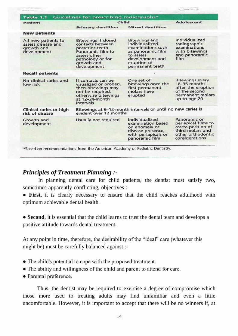
14
Principles of Treatment Planning :-
In planning dental care for child patients, the dentist must satisfy two,
sometimes apparently conflicting, objectives :-
● First, it is clearly necessary to ensure that the child reaches adulthood with
optimum achievable dental health.
● Second, it is essential that the child learns to trust the dental team and develops a
positive attitude towards dental treatment.
At any point in time, therefore, the desirability of the “ideal” care (whatever this
might be) must be carefully balanced against :-
● The child's potential to cope with the proposed treatment.
● The ability and willingness of the child and parent to attend for care.
● Parental preference.
Thus, the dentist may be required to exercise a degree of compromise which
those more used to treating adults may find unfamiliar and even a little
uncomfortable. However, it is important to accept that there will be no winners if, at
15
the outset, a treatment plan is unrealistic or insufficiently flexible to allow
modification, should this become necessary, as treatment progresses.
From the foregoing comments, it should be evident that it is not possible to
take a “one size fits all” approach to treatment planning, very different treatment
plans may be drawn up for children who present with very similar problems.
1-Management of acute dental problems :- The management of acute
problems present at the time of the child's first visit is undoubtedly a priority.
However, it is important that any treatment that is provided sits well in the context of
a holistic treatment plan and does not jeopardize its completion. In most cases,
therefore, pain relief should be provided without recourse to extraction.
2-Prevention :- Any treatment plan relies for its support on a (spine) of
prevention. Restorations placed in a mouth in which the caries process is still active
are prone to failure, repeated restorations may be detrimental to the child's ability to
co-operate and the dentist-parent relationship (as well as frustrating to the dental
team).
Likewise, managing a child's grossly carious teeth by multiple extractions
without ensuring that he or she receives appropriate preventive input does nothing to
assist that child in maintaining dental health in the future. An added advantage of a
“prevention first approach” is its importance in behaviour management, acclimatizing
the child to future treatment.
Procedures such as fluoride varnish applications or disclosing are good
confidence-building steps. Preventive advice, whether this is in relation to diet, oral
hygiene, fluoride supplementation, or even the prevention of dental trauma, should be
realistic and specifically tailored to the individual child and parent. Any preventive
strategy should be dictated by an individual's risk assessment, for instance low caries
risk children do not routinely require fissure sealants. The delivery of preventive
advice and interventions should not be restricted to the commencement of treatment.
Rather, prevention should be reinforced as treatment progresses, modifications being
incorporated should these become necessary.
Clearly, prevention is not simply a job for the members of the dental team, it
demands the creation of a partnership in which both the child and parent are key
players, though the relative role and prominence of each will differ with age of the
child. In the case of young children, parents are (or, at least, should be) responsible
for food choices and oral hygiene, though the latter responsibility is not infrequently
abdicated before the child has sufficient manual dexterity to brush adequately alone.
16
As the child approaches the teenage years (and particularly when he or she
enters secondary schooling), parental control inevitably decreases. Any discussion of
the proposed treatment plan should, therefore, include an agreement as to what is
required of the child and / or parent as well as what will be offered by various
members of the dental team (including professionals complementary to dentistry). It
may be helpful to document this agreement in the form of a written “contract”.
3-Stabilization :- Where a child has open cavities, a phase of stabilization
should precede the provision of definitive treatment, whether this is to be entirely
restorative in nature or a combination of restorations and extractions. In this process,
no attempt is made to render the cavities caries free, rather, minimal tissue is
removed without local anesthesia, allowing placement of an appropriate temporary
dressing. The inclusion of such a phase in all-inclusive treatment plan reduces the
overall bacterial load and slows caries progression, renders the child less likely to
present with pain and sepsis, and buys time for the implementation of preventive
measures and for the child to be acclimatized to treatment.
However, one word of caution is offered, it is essential that the parent
understands the purpose of stabilization and that what have been provided are not
permanent restorations. Otherwise, it is possible that they will perceive that treatment
is failing to progress. Following stabilization, the child's response to acclimatization
and compliance with the suggested preventive regime should be assessed. This is
particularly important before proceeding with definitive treatment. For example, in a
scenario in which a child has not responded to acclimatization and has either refused
stabilization or accepted this only with extreme difficulty, the dentist may be entirely
justified in considering extractions. This will allow the child and his or her family to
enjoy a period where no active treatment is required and in which prevention can be
established (always provided, of course, they return for continuing care).
4-Scheduling operative treatment :- In any treatment plan, it is necessary to
give careful consideration to the order which items of operative care are provided.
The following are general rules of thumb :-
● Small, simple restorations should be completed first.
● Maxillary teeth should be treated before mandibular ones (since it is usually easier
to administer local anesthesia in the upper jaw).
● Posterior teeth should be treated before anterior (this usually ensures that the
patient returns for treatment).
● Quadrant dentistry should be practiced where ever possible (this reduces the
number of visits to a minimum), but only if the time in chair is not excessive for a
very young patient.
17
● Endodontic treatment should follow completion of simple restorative treatment.
● Extractions should be the last items of operative care (at this stage, patient co-
operative can more reliably by assured), unless the patient presents with an acute
problem mid-treatment.
5-Recall :- Treatment planning (in its broadest sense) clearly does not end with
the completion of one treatment journey. The determination of a recall schedule
tailored to the needs of the individual child is an essential part of the treatment-
planning process. It is generally accepted that children should receive a dental
assessment more frequently than adults since :-
● There is evidence that the rate of progression of dental caries can be more rapid in
children than in adults.
● The rate of progression of erosive tooth wear is faster in primary than in permanent
teeth.
● Periodic assessment of orofacial growth and the developing occlusion is required.
In the latter context, there is considerable importance in ensuring that recall
examinations match with particular milestones in dental development, for example,
around 6, 9, and 12 years. Generally speaking, recall intervals of no more than 12
months offer the dentist the opportunity to deliver and reinforce preventive advice
during the crucial period when a child is establishing the basis for their future dental
health. However, the exact recall interval (3, 6, 9, or 12 month) should be design to
meet and vary with the child's needs. This requires an assessment of disease levels as
well as risk of / from dental disease.
6-Treatment planning for general anesthesia :- Is an extremely complex
area that merits special mention. It is sufficient to emphasize here that, in this
context; a comprehensive approach must be taken. Providing treatment under general
anesthesia for a child who has been shown to be unable to cope with operative dental
care under local anesthesia (with or without the support of conscious sedation) will
do absolutely nothing to improve his or her future co-operation. Such treatment
should, therefore, include the restoration or extraction (as appropriate) of all carious
teeth.
The practice of extracting only the most grossly carious or symptomatic teeth
(and assuming that other carious teeth can be restored under local anesthetic at a later
stage) predisposes to a high rate of repeat general anesthesia and should be
discouraged.
The orthodontic implications of any proposed treatment should always be
considered. This is particularly so when the loss of one or more permanent units is to
18
be included in the treatment plan. In such cases, the latter should ideally be drawn up
in consultation with a specialist in orthodontics.
Treatment under general anesthesia, irrespective of whether this includes
restorative treatment or is limited to extractions, should be followed with an
appropriate preventive programme. Failure to provide this almost inevitably leads to
the child undergoing further treatment (usually extraction) under general anesthesia.
7-Treatment planning for complex cases :- The clinician should always have
a clear long-term “vision” for the management of the individual patient. In creating
this, appropriate specialist input to treatment planning should be sought where
indicated.
At the simplest level, an orthodontic opinion should be obtained committing a
child to multiple visits to restore first permanent molars of poor prognosis. However,
it is in the treatment planning of complex cases (such as those presenting with
generalized defects of enamel or dentin formation, hypodontia, or clefts of lip and
palate) that interdisciplinary specialist input is essential. For example, such input may
result in :-
● The retention of anterior roots to maintain alveolar bone in preparation for future
implants.
● The use of preformed metal crowns to maintain clinical crown height in preparation
for definitive crowns.
● The use of direct / laboratory-formed composite veneers in preparation for
porcelain veneers when growth (and any orthodontic treatment) is complete.
The one over-riding consideration in this, management in early adulthood
should never be compromised by inappropriate treatment at young age. In general the
treatment planning for young patients should not only address current needs but
should plan ahead for those of the future, thus ensuring that every child reaches
adulthood with a healthy, functional, and aesthetic dentition as well as positive
attitudes towards dentistry.
Careful history-taking, clinical examination, and risk assessment contribute to
the decision-making process, but one should never lose sight of what is realistic and
practical for the child in the context of his or her environment. To do otherwise not
only judges non-compliance but also fails to recognize the most important aspect of
all-a child's individuality.
19
The treatment plan should be present to parents by inform them the following :-
1- The dental need of their child.
2- The restorative procedures required.
3- The amount of time required to perform the projected procedures.
4- The total cost of the services.
5- Preventive measures necessary to maintain the completed treatment.
The dentist also should keep in mind that the discussion of child dental problems
within the parents need to be done in the following manner :-
1- Point out the problem using cast, radiograph or the patient mouth.
2- State the probable cause of the problem such as plaque or insufficient arch
length.
3- State the outcome of the problem such as its effect on function, health or
appearance.
4- Show how the problem will be resolved.
5- Stress the benefit that the patient will receive from the treatment.
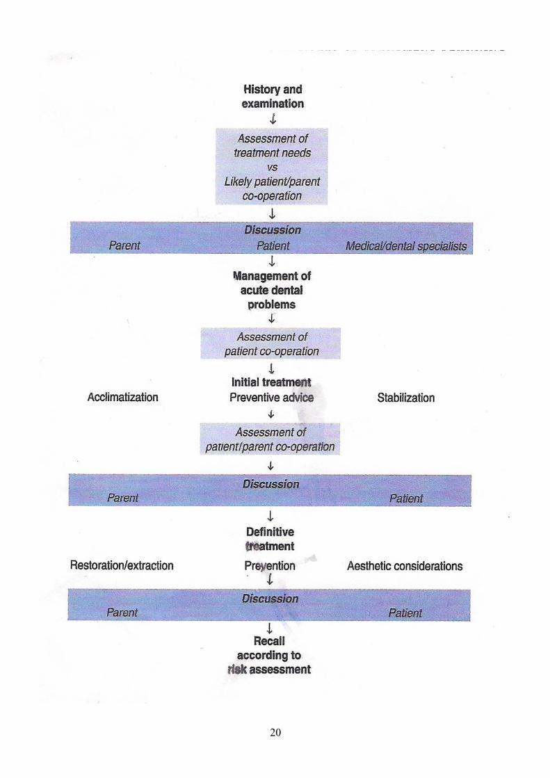
20
1
Paediatric Dentistry
Fifth Stage
Dr. Suhair W. Abbood
Lec. 2
Generally the paediatric dentistry is one of dentistry branch that dealing with
children from birth through adolescence. It is an age-defined specialty that provides
both primary and comprehensive preventive and therapeutic oral health care for
infants and children through adolescence, including those with special health care
needs.
Paediatric dentists are committed to the oral health of children from infancy
through the teen years. They have the experience and qualifications to care for a
child’s teeth, gums, and mouth throughout the various stages of childhood. They are
also serving as educational resources for parents.
Children begin to get their baby teeth during the first 6 months of life. By age 6
or 7 years, they start to lose their first set of teeth, which eventually are replaced by
secondary, permanent teeth. Without proper dental care, children face possible oral
decay and disease that can cause a lifetime of pain and complications.
It is recommended that a dental visit should occur within six months after the
presence of the first tooth or by a child's first birthday. It is important to establish a
comprehensive and accessible ongoing relationship between the dentist and patient
referring to this as the patient's "dental home". This is because early oral examination
aids in the detection of the early stages of tooth decay.
Early detection is essential to maintain oral health, modify aberrant habits, and
treat as needed and as simply as possible. Furthermore, parents are given a program
of preventative home care (brushing/flossing/fluorides), a caries risk assessment,
information on finger, thumb, and pacifier habits, advice on preventing injuries to the
mouth and teeth of children, diet counselling , and information on growth and
development.
2
Development and Eruption of Teeth
The Development of Teeth :-
Tooth development or odontogenesis is the complex process by which teeth
form from embryonic cells, grow, and erupt into the mouth (start as early as 28 days
of IUL and continues to the end of eruption of permanent molars). For human teeth to
have a health oral environment, all parts of the tooth must develop during appropriate
stages of fetal development.
The primary teeth (deciduous teeth-baby teeth) start to form during (either
the fifth and sixth-or sixth and eight) week of embryonic life in utero. The lower front
teeth are formed first followed by the upper front teeth, and this process continues
after birth until the full set of ten upper and ten lower teeth have been formed.
The permanent teeth usually begin forming during the fourth or fifth month
(16th
or 20th
week) in utero. The lower front teeth are formed first followed by the
upper front teeth, and the development continues after birth until 16 upper and lower
teeth have been formed.
If teeth do not start to develop at or near these times, they will not develop at
all, resulting in hypodontia or anodontia. Generally the process of teeth formation
continues until the roots of the third permanent molars are completed at about the age
of 20 years, the deciduous teeth will take 2-3 years to form, while the permanent teeth
will take 9-10 years.
The stages of teeth formation are the same whether the teeth are of the primary
or the permanent dentition, although, obviously, the teeth develop at different times.
The development of teeth passes through the following stages :-
1- Development in the prenatal period: in this period three overlapping phases
occur:-
a- Beginning of the deciduous dentition bud development: The
development of teeth starts at 3rd
week of IUL and then the odontogenic
epithelium proliferates in the 5th
week to form the dental laminas, which
form invaginations that develop into tooth buds.
b- Initiation of the permanent dentition bud: It is initiation in the 4th
month
of IUL.
c- The formation of the successional lamina: It is the lingual extension of
the dental lamina develops in the 5th
months of IUL (permanent central
incisor) to 10th
months of age (2nd
premolar).
3
2- Status of development at birth: the teeth are in different stages of
development at birth.
3- Development in the postnatal period: it shows completion of the crowns of
all primary teeth and initiation of root formation. The permanent teeth
continue to develop in different stages till their root formation is completed.
The Eruption of Teeth:-
The term eruption describes the movement that takes a tooth from its
developmental position in its crypt to its occlusal contact with opposing dentition. It
is the process of a tooth moving through the alveolar bone (intrabony phase) into the
oral cavity (intraoral phase). Each tooth starts to move toward occlusion at
approximately the time of crown completion, and the interval from crown completion
and the beginning of eruption until the tooth is in full occlusion is approximately 5
years. The teeth of girls erupt earlier than that of boys. By the time of clinical
emergence approximately three fourth of root formation had occurred. Teeth reach
occlusion before the root development is completed, and the not fully formed root
appears funnel in shape.
The formation and eruption of teeth are two essential processes, which may
influenced by :-
1- Genetic factors. 2- Environmental factors. 3- Hormonal factors.
Pattern of tooth movement:-
Different phases of movement that tooth pass through are required to bring the
teeth to the occlusal level and then into functional occlusion.
Phases of tooth movement :-
1- Pre-eruptive phase: includes all movements of the deciduous and permanent
tooth germs within the tissues of the jaw, from the time of early initiation
and formation to the time of crown completion and this phase terminates
with the initiation of root development, during this phase the growing tooth
moves in two directions to maintain its position in the expanding jaws
(outward and upward in the mandible and outward and downward in the
maxilla).
- Bodily movement : This occurs continuously as the jaw grows by which
the movement of entire tooth germ cause bone resorption at the direction
of tooth movement and bone apposition behind it.
- Eccentric movement : Here one part of the tooth germ remains fixed
while the rest continuous to grow causing a shift in the center of the
tooth germ.
4
2- Eruptive phase: it is the axial movement of the tooth from its crypt within
the bone of the jaw to its functional position in occlusion (to occlude with
its antagonist).
3- Post-eruptive phase: it occurs primarily to maintain the position of the
erupted tooth while the jaw continuous to grow and to compensate for the
occlusal and proximal wears. This movement occurs in axial direction.
Theories of normal tooth eruption :-
There are several possible explanations to account for the phenomenon of tooth
eruption. Some possibilities are more likely than others to play a part in the process,
and some earlier theories have been largely discounted. The developmental processes
and factors that have been related to the eruption of teeth include:-
1- Root formation theory: It was believed that root formation is an obvious
cause of tooth eruption, because it causes an overall increase in length of
the tooth that must accommodated by the growth of the root into the bone,
an increase in jaw height or by the occlusal movement of the crown
(eruption). The root elongates when the crown does not increase in size.
Root growth theory suggested the presence of the cushion hammock
(hanging bed-a hanging bed made of canvas or netting and suspended
between two supports) ligament at the base of the socket that transmits the
force to cause eruption this ligament histologically was not found.
However, the observational and experimental studies have shown that the
theory root formation is not necessarily required for eruption of teeth, as the
rootless teeth keep erupt into functional occlusion and some of teeth erupt
even after root formation to a greater distance more than the length of their
roots.
2- Bone remodeling theory: Remodeling of dento-alveolar bone has been
proposed as a mechanism for tooth eruption, but the part it plays is difficult
to assess. An inherent growth pattern of the maxilla and mandible
supposedly moves teeth by selective deposition and resorption of bone.
Bone resorption and formation has to occur as teeth erupt, as a result of
eruptive forces applied by tooth over the bone, and it is hard to know
whether the remodeling is a cause of tooth eruption or whether it is simply
in response to the eruption of the teeth.
3- Vascular pressure theory: The vascular pressure theory supposes that a
local increase in tissue fluid or blood pressure in the periapical region is
sufficient to move the tooth. But the surgical excision of the growing root
and associated tissues although it eliminates the periapical vasculature, the
5
tooth eruption occurs without stopping. This making the theory of local
vessels pressure is not absolutely necessary for tooth eruption.
4- Periodontal ligament and dental follicle theory: Eruptive force resides in
the dental follicle-periodental ligament complex. Formation and renewal of
the PDL has been considered a factor in tooth eruption because its
fibroblasts have traction power that could be capable of elevating the tooth
root and pull the tooth out during eruption.
On the other hand, it is most likely that agents responsible for tooth eruption
lay within the dental follicle itself, rather than the tooth. The connective
tissue of the dental follicle is a rich source of factors that are responsible for
the local mediation of bone deposition and resorption to accommodate tooth
movement. Thus, it seems probable that the dental follicle has a major part
to play in the process of tooth eruption.
The Time of Eruption is important :-
1-For the dentist to aid in the diagnosis and treatment plan.
2-For the dental epidemiologist to make survey and study, as example dental
caries.
3-For the physician where under nourished children may have delayed eruption
teeth or retained primary teeth.
4-For orthodontist (the ugly duckling stage should be differentiated from other
conditions).
5-For psychologist (to determine whether it is important to replace the missing
teeth).
6-For forensic odontologist.
7-In the anthropologist.
Eruption Difficulties :-
In most children the eruption of primary teeth is preceded by :-
1-Increased salivation, and the child will want to put the hand and fingers into
the mouth. This observation may be the only indication that the teeth will soon
erupt.
2-Some young children become restless, with loss of appetite, and fretful
during the time of eruption of the primary teeth. In the past, many conditions
including, croup, diarrhea, fever, convulsions, primary herpetic
gingivostomatitis, and even death were incorrectly attributed to eruption.
Because the eruption of teeth is a normal physiologic process, the association
with fever and systemic disturbances is not justified. A fever or respiratory
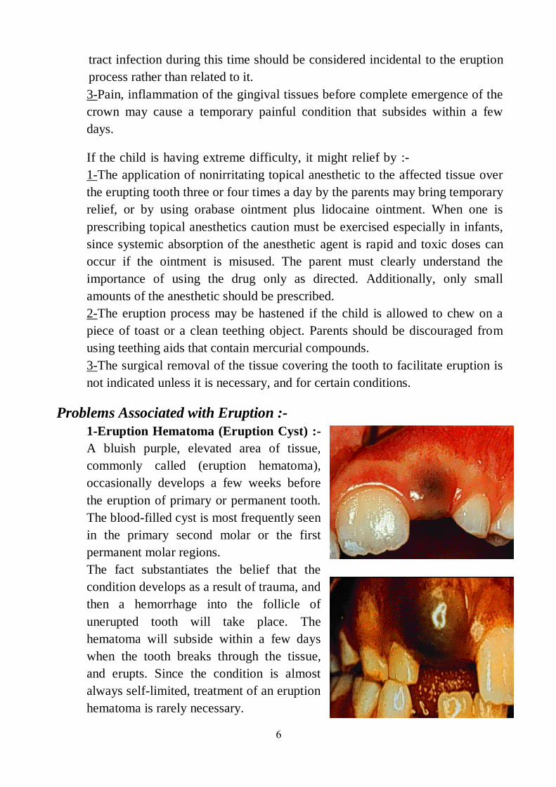
6
tract infection during this time should be considered incidental to the eruption
process rather than related to it.
3-Pain, inflammation of the gingival tissues before complete emergence of the
crown may cause a temporary painful condition that subsides within a few
days.
If the child is having extreme difficulty, it might relief by :-
1-The application of nonirritating topical anesthetic to the affected tissue over
the erupting tooth three or four times a day by the parents may bring temporary
relief, or by using orabase ointment plus lidocaine ointment. When one is
prescribing topical anesthetics caution must be exercised especially in infants,
since systemic absorption of the anesthetic agent is rapid and toxic doses can
occur if the ointment is misused. The parent must clearly understand the
importance of using the drug only as directed. Additionally, only small
amounts of the anesthetic should be prescribed.
2-The eruption process may be hastened if the child is allowed to chew on a
piece of toast or a clean teething object. Parents should be discouraged from
using teething aids that contain mercurial compounds.
3-The surgical removal of the tissue covering the tooth to facilitate eruption is
not indicated unless it is necessary, and for certain conditions.
Problems Associated with Eruption :-
1-Eruption Hematoma (Eruption Cyst) :-
A bluish purple, elevated area of tissue,
commonly called (eruption hematoma),
occasionally develops a few weeks before
the eruption of primary or permanent tooth.
The blood-filled cyst is most frequently seen
in the primary second molar or the first
permanent molar regions.
The fact substantiates the belief that the
condition develops as a result of trauma, and
then a hemorrhage into the follicle of
unerupted tooth will take place. The
hematoma will subside within a few days
when the tooth breaks through the tissue,
and erupts. Since the condition is almost
always self-limited, treatment of an eruption
hematoma is rarely necessary.
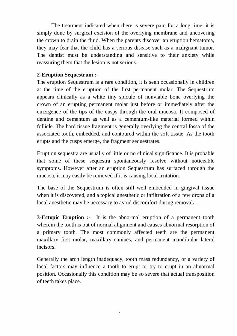
7
The treatment indicated when there is severe pain for a long time, it is
simply done by surgical excision of the overlying membrane and uncovering
the crown to drain the fluid. When the parents discover an eruption hematoma,
they may fear that the child has a serious disease such as a malignant tumor.
The dentist must be understanding and sensitive to their anxiety while
reassuring them that the lesion is not serious.
2-Eruption Sequestrum :-
The eruption Sequestrum is a rare condition, it is seen occasionally in children
at the time of the eruption of the first permanent molar. The Sequestrum
appears clinically as a white tiny spicule of nonviable bone overlying the
crown of an erupting permanent molar just before or immediately after the
emergence of the tips of the cusps through the oral mucosa. It composed of
dentine and cementum as well as a cementum-like material formed within
follicle. The hard tissue fragment is generally overlying the central fossa of the
associated tooth, embedded, and contoured within the soft tissue. As the tooth
erupts and the cusps emerge, the fragment sequestrates.
Eruption sequestra are usually of little or no clinical significance. It is probable
that some of these sequestra spontaneously resolve without noticeable
symptoms. However after an eruption Sequestrum has surfaced through the
mucosa, it may easily be removed if it is causing local irritation.
The base of the Sequestrum is often still well embedded in gingival tissue
when it is discovered, and a topical anesthetic or infiltration of a few drops of a
local anesthetic may be necessary to avoid discomfort during removal.
3-Ectopic Eruption :- It is the abnormal eruption of a permanent tooth
wherein the tooth is out of normal alignment and causes abnormal resorption of
a primary tooth. The most commonly affected teeth are the permanent
maxillary first molar, maxillary canines, and permanent mandibular lateral
incisors.
Generally the arch length inadequacy, tooth mass redundancy, or a variety of
local factors may influence a tooth to erupt or try to erupt in an abnormal
position. Occasionally this condition may be so severe that actual transposition
of teeth takes place.
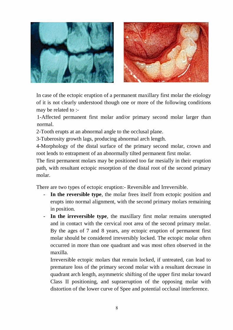
8
In case of the ectopic eruption of a permanent maxillary first molar the etiology
of it is not clearly understood though one or more of the following conditions
may be related to :-
1-Affected permanent first molar and/or primary second molar larger than
normal.
2-Tooth erupts at an abnormal angle to the occlusal plane.
3-Tuberosity growth lags, producing abnormal arch length.
4-Morphology of the distal surface of the primary second molar, crown and
root lends to entrapment of an abnormally tilted permanent first molar.
The first permanent molars may be positioned too far mesially in their eruption
path, with resultant ectopic resorption of the distal root of the second primary
molar.
There are two types of ectopic eruption:- Reversible and Irreversible.
- In the reversible type, the molar frees itself from ectopic position and
erupts into normal alignment, with the second primary molars remaining
in position.
- In the irreversible type, the maxillary first molar remains unerupted
and in contact with the cervical root area of the second primary molar.
By the ages of 7 and 8 years, any ectopic eruption of permanent first
molar should be considered irreversibly locked. The ectopic molar often
occurred in more than one quadrant and was most often observed in the
maxilla.
Irreversible ectopic molars that remain locked, if untreated, can lead to
premature loss of the primary second molar with a resultant decrease in
quadrant arch length, asymmetric shifting of the upper first molar toward
Class II positioning, and supraeruption of the opposing molar with
distortion of the lower curve of Spee and potential occlusal interference.

9
Early assessment with intraoral or panoramic films approximating the timing
of first permanent molar eruption is thus critical to identification of the
problem and provides an opportunity to intercept potential sequelae.
The treatment of such ectopic eruption is by :-
1-Self-corrective (jump-type) :- 66%of the ectopically erupted molars finally
erupted into an acceptable position without corrective treatment. If the problem
is detected at 5 to 6 years of age, an observation approach of (watchful waiting)
with appropriate monitoring may be indicated, given the two-third potential for
self-correction.
2-Treatment method can vary based on clinical examination, extent of
entrapment, and space analysis. The objective is to distalised the permanent
molar from entrapment and provide it with eruptive guidance. With self-
correction being unlikely as the child approaches 7 years of age, continued
(locking) of the first molar with advanced resorption of primary second molar
usually warrants intervention. Another timing clue is that when the opposing
molar reaches the level of the lower occlusal plane, intervention is indicated to
establish proper vertical control and prevent supraeruption.
3-In some cases, the primary second molar is extracted, the permanent molar is
allowed to erupt and then distalised to a normal position. Some methods of
treatment are brass ligature wire, and stainless steel crown.
The Early Eruption
1- Natal and Neonatal Teeth :-
The prevalence of natal teeth (teeth present at birth) and neonatal teeth (teeth
that erupt within the first month of life) is low. Approximately one in 2000-
3000 live births is so affected. About 85% of natal or neonatal teeth are lower
primary incisors, and only a small percentage has been observed to be
supernumerary teeth. It is common for natal and neonatal teeth to occur in
pairs. Natal and neonatal molars are. Most studies suggest that the etiology for
the premature eruption or the appearance of natal and neonatal teeth is
multifactorial.
A possible factor involving the early eruption of primary teeth seems to be
familial, due to inheritance as an autosomal-dominant trait. Also it has been
suggested that this condition is a result of an ectopic position of the tooth-germ
during fetal life. Evidence of a relationship between early eruption and a
systemic condition or a syndrome is not conclusive, but this possibility should
be considered, since the natal or the neonatal teeth may also be seen in
10
association with some syndromes including pachyonychia congenital, Ellis-
Van Creveld syndrome, and Hallermann-Streiff syndrome.
Occasionally maxillary central incisors or the first molars may appear as natal
teeth. Babies with posterior natal teeth should be carefully investigated for
other systemic conditions that may be associated with syndromes or other
disease. The vast majority of cases represent premature eruption of a tooth of
the normal sequence. Eruption of teeth during the neonatal period presents less
of a problem, since these teeth can usually be maintained even though root
development is limited.
However, in the diagnosis and treatment of a natal or neonatal tooth, a
radiograph should be made to determine the amount or root development and
the relationship of a prematurely erupted tooth to its adjacent teeth. One of the
parents can hold the X-ray film in the infant's mouth during the exposure.
The treatment:-
1- Most prematurely erupted teeth (immature type), the crowns may be
abnormal in form and the enamel may be poorly formed or thinner than
normal. They are hyper mobile because of the limited root development; the
mobility of the tooth frequently also causes inflammation of the surrounding
gingivae. Some teeth may be mobile to the extent that there is danger of
displacement of the tooth and possible aspiration, in such case the removal of
the tooth is indicated.
2- In exceptionally rare cases in which the sharp incisal edge of the tooth may
cause laceration on the lingual surface of the tongue, this trauma to the lingual
surface of the tongue may cause ulceration, a local measure such as smoothing
of the sharp edges of the tooth with a rubber cone in a dental handpiece may
help resolve the ulceration. In a number of cases, if the tooth is markedly loose
it should be extracted.
3- A retained natal or neonatal tooth may cause difficulty for a mother who
wishes to breast-feed her infant, the most important point to consider is
whether the nursing mother can adequately establish breast-feeding. If the
nipples are getting traumatized, the tooth should be removed. Another
approach if the breast-feeding is too painful for the mother, initially the use of
a breast pump and storing the milk are recommended. However, the infant may
be conditioned not to bite during suckling in a relatively short time if the
mother persists with breast-feeding, it seems that the infants senses the
mother's discomfort and learns to avoid causing it.
11
If tooth removal is indicated care should be taken to extract the entire tooth, as
the crown only may be removed leaving behind the pulpal tissue. If this is the
case, the dentin and a root will form subsequently, the root will then require
removal at a later date. Before extraction check the medical history for
significant jaundice which may be predispose to postoperative bleeding.
During extraction always protect the airway when removing these teeth by
placing gauze in the back of the mouth.
The teeth are easily dislodged or dropped. A pair of Spencer Wells forceps or
similar will provide a firm grip on the tooth to be removed, followed by minor
local curettage to remove remains of the developing tooth-germ at that site.
Careful should be taken by which the permanent teeth should be unaffected by
extraction of the primary tooth.
4- The preferable approach, however, is to leave the tooth in place and to
explain to the parents the desirability of maintaining this tooth in the mouth
because of its importance in the growth and uncomplicated eruption of the
adjacent teeth. Within a relatively short time the prematurely erupted tooth will
become stabilized as the root continues to develop, and the other teeth in the
arch will erupt.
2- Epstein Pearls, Bohn Nodules, and Dental Lamina Cysts :-
Small, white or grayish white lesions on the alveolar mucosa of the newborn
may on rare occasions be incorrectly diagnosed as natal teeth. The lesions are
usually multiple but do not increase in size. No treatment is indicated, since the
lesions are spontaneously shed a few weeks after birth.
The following three types of inclusion cysts are :-
1- Epstein Pearls :- Are formed along the mid-palatine raphe. They are
considered remnants of epithelial tissue trapped along the raphe as the fetus
grow.
2- Bohn Nodules :- Are formed along the buccal and lingual aspects of the
dental ridges and on the palate away from the raphe. The nodules are
considered remnants of mucous gland tissue and histologically different from
Epstein Pearls.
3- Dental Lamina Cysts :- Are found on the crest of the maxillary and
mandibular dental ridges. The cysts apparently originated from remnants of the
dental lamina.
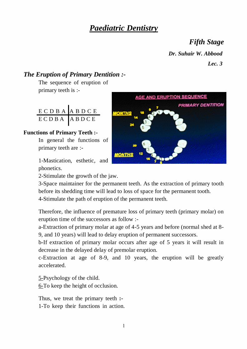
1
Paediatric Dentistry
Fifth Stage
Dr. Suhair W. Abbood
Lec. 3
The Eruption of Primary Dentition :- The sequence of eruption of
primary teeth is :-
E C D B A A B D C E
E C D B A A B D C E
Functions of Primary Teeth :-
In general the functions of
primary teeth are :-
1-Mastication, esthetic, and
phonetics.
2-Stimulate the growth of the jaw.
3-Space maintainer for the permanent teeth. As the extraction of primary tooth
before its shedding time will lead to loss of space for the permanent tooth.
4-Stimulate the path of eruption of the permanent teeth.
Therefore, the influence of premature loss of primary teeth (primary molar) on
eruption time of the successors as follow :-
a-Extraction of primary molar at age of 4-5 years and before (normal shed at 8-
9, and 10 years) will lead to delay eruption of permanent successors.
b-If extraction of primary molar occurs after age of 5 years it will result in
decrease in the delayed delay of premolar eruption.
c-Extraction at age of 8-9, and 10 years, the eruption will be greatly
accelerated.
5-Psychology of the child.
6-To keep the height of occlusion.
Thus, we treat the primary teeth :-
1-To keep their functions in action.
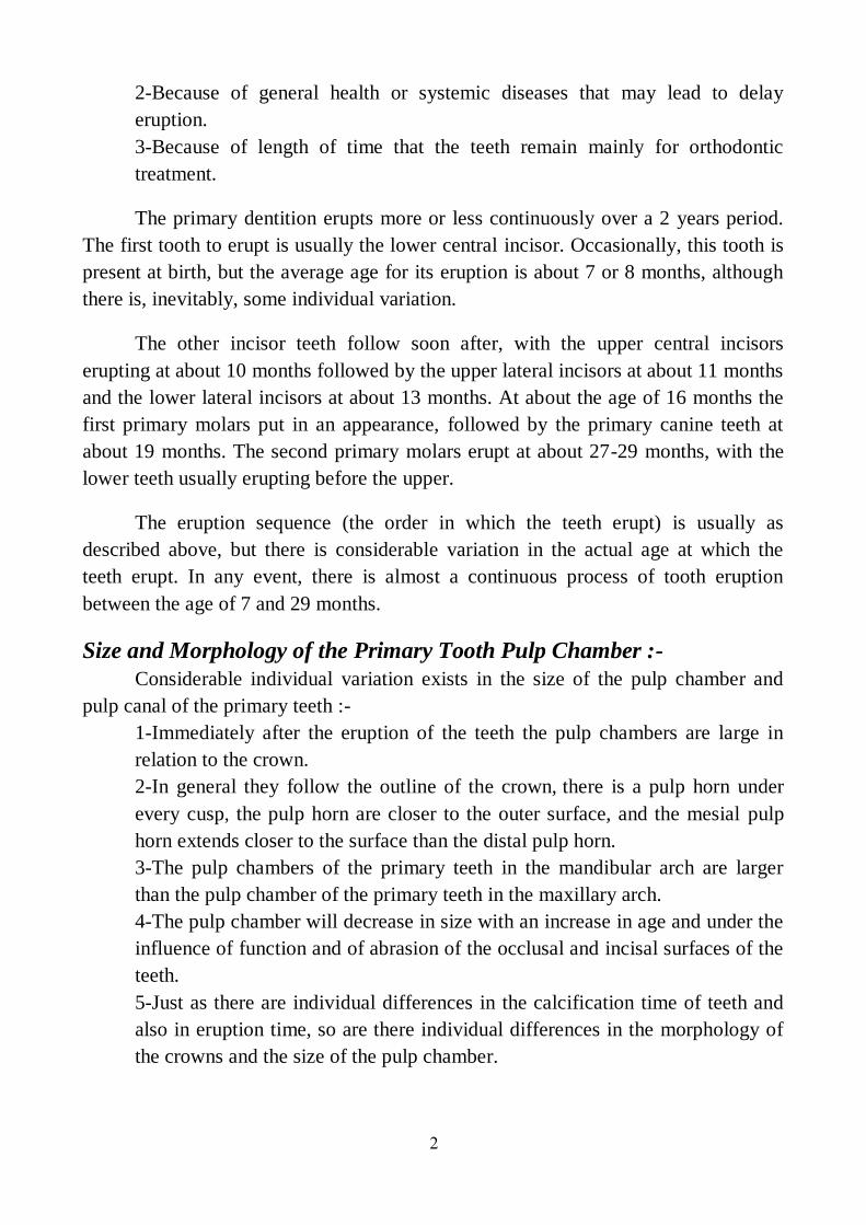
2
2-Because of general health or systemic diseases that may lead to delay
eruption.
3-Because of length of time that the teeth remain mainly for orthodontic
treatment.
The primary dentition erupts more or less continuously over a 2 years period.
The first tooth to erupt is usually the lower central incisor. Occasionally, this tooth is
present at birth, but the average age for its eruption is about 7 or 8 months, although
there is, inevitably, some individual variation.
The other incisor teeth follow soon after, with the upper central incisors
erupting at about 10 months followed by the upper lateral incisors at about 11 months
and the lower lateral incisors at about 13 months. At about the age of 16 months the
first primary molars put in an appearance, followed by the primary canine teeth at
about 19 months. The second primary molars erupt at about 27-29 months, with the
lower teeth usually erupting before the upper.
The eruption sequence (the order in which the teeth erupt) is usually as
described above, but there is considerable variation in the actual age at which the
teeth erupt. In any event, there is almost a continuous process of tooth eruption
between the age of 7 and 29 months.
Size and Morphology of the Primary Tooth Pulp Chamber :- Considerable individual variation exists in the size of the pulp chamber and
pulp canal of the primary teeth :-
1-Immediately after the eruption of the teeth the pulp chambers are large in
relation to the crown.
2-In general they follow the outline of the crown, there is a pulp horn under
every cusp, the pulp horn are closer to the outer surface, and the mesial pulp
horn extends closer to the surface than the distal pulp horn.
3-The pulp chambers of the primary teeth in the mandibular arch are larger
than the pulp chamber of the primary teeth in the maxillary arch.
4-The pulp chamber will decrease in size with an increase in age and under the
influence of function and of abrasion of the occlusal and incisal surfaces of the
teeth.
5-Just as there are individual differences in the calcification time of teeth and
also in eruption time, so are there individual differences in the morphology of
the crowns and the size of the pulp chamber.
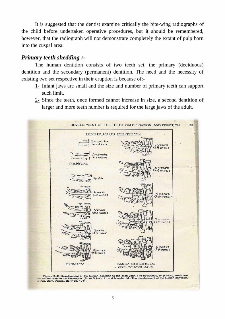
3
It is suggested that the dentist examine critically the bite-wing radiographs of
the child before undertaken operative procedures, but it should be remembered,
however, that the radiograph will not demonstrate completely the extant of pulp horn
into the cuspal area.
Primary teeth shedding :-
The human dentition consists of two teeth set, the primary (deciduous)
dentition and the secondary (permanent) dentition. The need and the necessity of
existing two set respective in their eruption is because of:-
1- Infant jaws are small and the size and number of primary teeth can support
such limit.
2- Since the teeth, once formed cannot increase in size, a second dentition of
larger and more teeth number is required for the large jaws of the adult.
4
Therefore, a physiological process term by shedding or exfoliation that
resulting in the elimination of the deciduous dentition is occurred to replace the
primary dentition with permanent set to accommodate the growth changes.
Pattern of shedding :-
The result of progressive resorption of the roots of teeth and their supporting
tissues, is the shedding of deciduous teeth. In general the pressure generated by the
growing and erupting permanent tooth dictates the pattern of deciduous tooth
resorption.
Resorption of Anterior teeth :-
The permanent anterior tooth germ position is lingual to the apical third of the
roots of the primary tooth hence the resorption is in occluso-labial direction, which
corresponds to the movements of the permanent tooth germ. Later the resorption
proceed horizontally because the crown of the permanent tooth lies directly apical to
the root of primary tooth, and this horizontal resorption allows the permanent tooth to
erupt into the position of the primary tooth.
Resorption of Posterior teeth :-
Initially, the growing crowns of the premolars are situated between the roots of
the primary molars and so the root resorption of the posterior primary teeth will
started at the inter-redicular bone area followed by resorption of the adjacent surfaces
of the root. Meanwhile, the alveolar process is growing to compensate for the
lengthening roots of the permanent tooth. As this occurs, the primary molars move
occlusally, this allows the premolars crowns to be more apical. The premolars
continue to erupt until the primary molars roots are entirely resorbed and the teeth
exfoliate. The premolars then appear in place of the primary molars.
Mechanism of resorption and shedding :-
It is not fully understood, however, it is clear that the pressure from the
erupting successional tooth plays a key role because the odontoclasts differentiate at
predicted sites of pressure. The most likely sequence of events in resorption of dental
hard tissue by odontoclasts is an initial removal of the mineral followed by
extracellular dissolution of the organic matrix (mainly collagen). Forces of
mastication are also synergistically involved in the mechanism of shedding. Due to
growth and increased loading of jaws those forces far exceed the limit that the
deciduous periodontal ligament can withstand, thereby causing trauma to the
ligament and initiation of resorption.
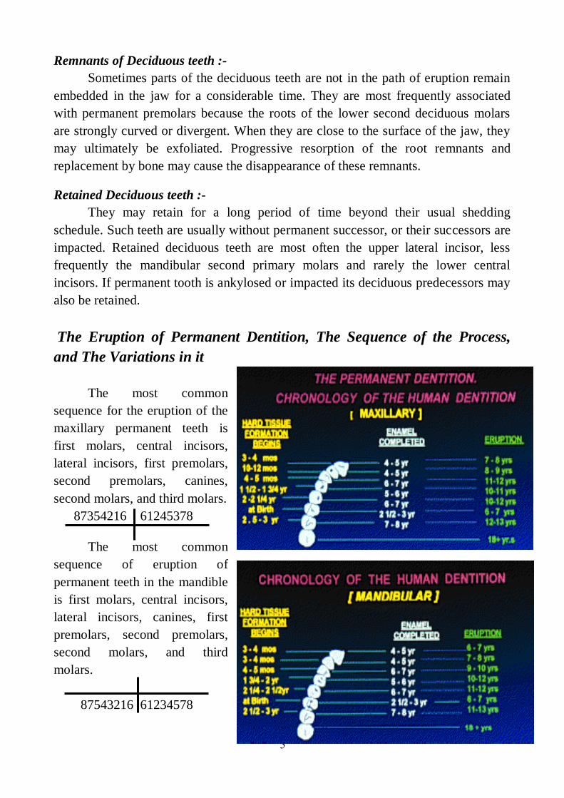
5
Remnants of Deciduous teeth :-
Sometimes parts of the deciduous teeth are not in the path of eruption remain
embedded in the jaw for a considerable time. They are most frequently associated
with permanent premolars because the roots of the lower second deciduous molars
are strongly curved or divergent. When they are close to the surface of the jaw, they
may ultimately be exfoliated. Progressive resorption of the root remnants and
replacement by bone may cause the disappearance of these remnants.
Retained Deciduous teeth :-
They may retain for a long period of time beyond their usual shedding
schedule. Such teeth are usually without permanent successor, or their successors are
impacted. Retained deciduous teeth are most often the upper lateral incisor, less
frequently the mandibular second primary molars and rarely the lower central
incisors. If permanent tooth is ankylosed or impacted its deciduous predecessors may
also be retained.
The Eruption of Permanent Dentition, The Sequence of the Process,
and The Variations in it
The most common
sequence for the eruption of the
maxillary permanent teeth is
first molars, central incisors,
lateral incisors, first premolars,
second premolars, canines,
second molars, and third molars.
87354216 61245378
The most common
sequence of eruption of
permanent teeth in the mandible
is first molars, central incisors,
lateral incisors, canines, first
premolars, second premolars,
second molars, and third
molars.
87543216 61234578
6
The dentition, however, erupts in two stages as first the incisors teeth and the
first permanent molars erupt, then the other teeth in the buccal segments. The lower
central incisors and the first permanent molars erupt at about the age of 6 years. The
upper central incisors and the lower lateral incisors erupt at about the age of 7 years
and the upper lateral incisors at about the age of 8 years.
As with the primary teeth, while some variation in the timing of tooth
eruption is only to be expected, this eruption sequence should not vary. In
particular, the upper central incisors should erupt before the upper lateral
incisors, if the upper lateral incisors erupts before the central then, almost
certainly, there is something impeding the eruption of the central incisors, for
example a supernumerary tooth, or dilacerations of the root of the central
incisors.
There is interval of rest in eruption sequence that occurs between eruptions of
two successive permanent the upper first premolars and the upper lateral incisors, it is
the largest time that is about 1.5 years.
The lower canines and the first premolars teeth are the next to erupt, at about
10 years of age, followed by the upper canines and the second premolars teeth at
about the age of 11 years and the second molars teeth at about the age of 12 years.
Third molars teeth start to erupt from about the age of 16 years onwards, but the
eruption of third molars is very variable, not uncommonly, these teeth are impacted
against their neighbours and fail to erupt at all.
The ugly duckling stage :-
Also called Broadbent phenomenon, it is self-correcting malocclusion that is
seen around 9-11 years of age or during eruptions of canine. As the permanent
canines erupt they displace the roots of lateral incisors mesially. This force is
transmitted to the central incisors and their roots are also displaced mesially. Thus the
resultant force causes the distal divergence of the crown in an opposite direction,
leading to midline spacing (diastema in the incisor region).
The term ugly duckling stage indicates the esthetic appearance of the child
during this stage. This condition correct itself after the canines have erupted when it
apply pressure on the crowns of the incisors thereby causing them to shift back to
original positions. No orthodontic treatment should be attempted at this stage as there
is danger of deflecting the canine from its normal path of eruption.
7
Variation in These Sequences of Eruption :-
1-The mandibular canines erupt before the first and second premolars. This
sequence will aid in maintaining adequate arch length and in preventing lingual
tipping of the incisors.
2-Lingual tipping of the incisors not only will cause a loss of arch length but
also will allow the development of an increased overbite. An abnormal lip
musculature or an oral habit that causes a greater force on the lower incisors
than can be compensated by the tongue will allow a collapse of the anterior
segment. For this reason a passive lingual arch appliance is often indicated
when the primary canines have been lost prematurely or when the sequence of
eruption is undesirable.
3-A deficiency in arch length can occur if the mandibular second permanent
molars develop and erupt before the second premolars. Eruption of second
permanent molar out of sequence will exert a strong force on the first
permanent molar and will cause its mesial migration and encroachment on the
space needed for the second premolars.
4-In the maxillary arch the first premolar ideally should erupt before the
second premolars, and they should be followed by the canines.
5-The untimely loss of primary molars in the maxillary arch, allowing the first
permanent molar to drift and tip mesially, will result in the permanent canines
being blocked out of the arch, usually to the labial side.
6-The position of the developing second permanent molars in the maxillary
arch and its relationship to the first permanent molars should be given special
attention, its eruption before the premolars and canines can cause a loss of arch
length, just as in the mandibular arch.
7-The eruption of maxillary canines often delayed because of an abnormal
position or devious eruption path, this delayed eruption should be considered
along with its possible effect on the alignment of the maxillary arch.
8-Lingual eruption of mandibular permanent incisors, and here we can see the
following :-
a-It is common for mandibular permanent incisors to erupt lingually to the
retained primary mandibular incisors, and this pattern should be considered
essentially normal.
The primary teeth may have undergone extensive root resorption and
may be held only by soft tissues. In other instances the roots may not have
undergone normal resorption and the teeth remain solidly in place.
8
b-It is seen both in patients with an obvious arch length inadequacy and in
those with a desirable amount of spacing of the primary incisors.
In either case the molding action of the tongue and the lips, and the
continued alveolar growth seen to play an important role in influencing the
permanent incisors by improving their relationship into a more normal position
within a few months time.
The spontaneous correction of lingually erupted permanent incisors is
likely to occur given enough time, particularly in cases where there is not
severe crowding; therefore, a watchful waiting approach may be justified,
especially when the patient is first seen in the dentist's office for this specific
problem.
c-Even when mandibular permanent incisors erupt uneventful, they often
appear rotated and staggered in position.
Certain Consideration in the Treatment of the Lingually Erupted
Permanent Mandibular Incisors :- 1-Although there may be insufficient room in the arch for the newly erupted
permanent tooth, its position will improve over several months. In some cases
there is justification for removal of the corresponding primary tooth.
2-Extraction of other primary teeth in the area, however, is not recommended
because it will only temporarily relieve the crowding and may even contribute
to the development of a more sever arch length inadequacy.
3-If the condition is identified before 7 1/2 years of age it is unnecessary to
subject the child to the trauma of removing the primary teeth because the
problem is almost always self-correcting within a few months.
4-However when lingually erupted permanent mandibular incisors are seen in
an older child and the radiograph shows no root resorption of the primary teeth,
self-correction has not been achieved and the corresponding primary teeth
should be removed.
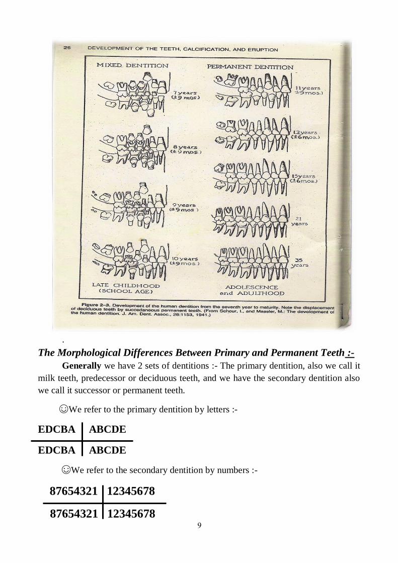
9
.
The Morphological Differences Between Primary and Permanent Teeth :-
Generally we have 2 sets of dentitions :- The primary dentition, also we call it
milk teeth, predecessor or deciduous teeth, and we have the secondary dentition also
we call it successor or permanent teeth.
☺We refer to the primary dentition by letters :-
EDCBA ABCDE
EDCBA ABCDE
☺We refer to the secondary dentition by numbers :-
87654321 12345678
87654321 12345678
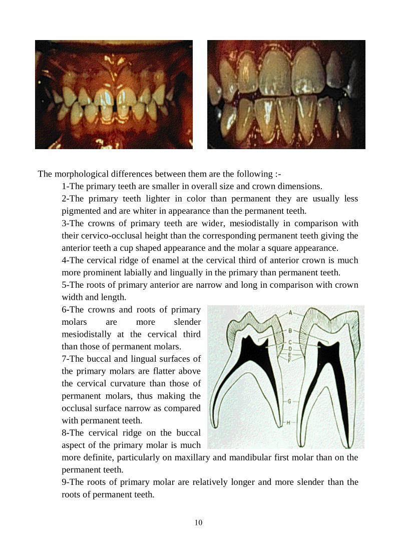
10
The morphological differences between them are the following :-
1-The primary teeth are smaller in overall size and crown dimensions.
2-The primary teeth lighter in color than permanent they are usually less
pigmented and are whiter in appearance than the permanent teeth.
3-The crowns of primary teeth are wider, mesiodistally in comparison with
their cervico-occlusal height than the corresponding permanent teeth giving the
anterior teeth a cup shaped appearance and the molar a square appearance.
4-The cervical ridge of enamel at the cervical third of anterior crown is much
more prominent labially and lingually in the primary than permanent teeth.
5-The roots of primary anterior are narrow and long in comparison with crown
width and length.
6-The crowns and roots of primary
molars are more slender
mesiodistally at the cervical third
than those of permanent molars.
7-The buccal and lingual surfaces of
the primary molars are flatter above
the cervical curvature than those of
permanent molars, thus making the
occlusal surface narrow as compared
with permanent teeth.
8-The cervical ridge on the buccal
aspect of the primary molar is much
more definite, particularly on maxillary and mandibular first molar than on the
permanent teeth.
9-The roots of primary molar are relatively longer and more slender than the
roots of permanent teeth.
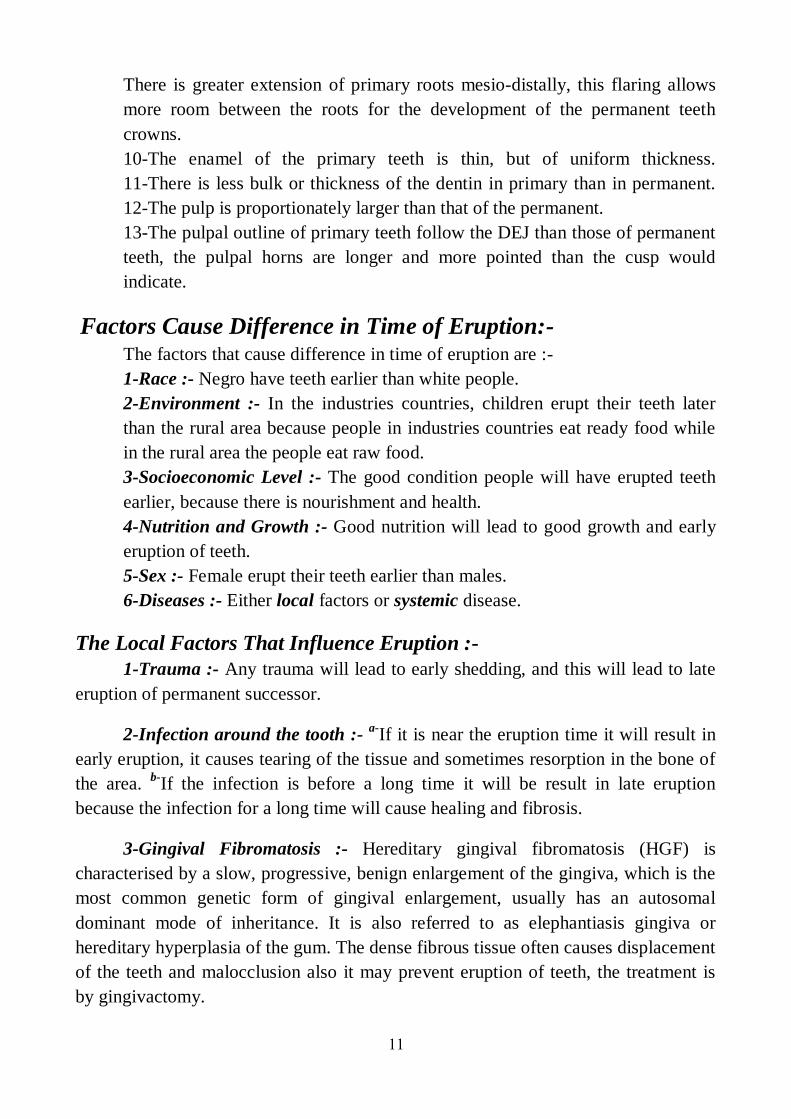
11
There is greater extension of primary roots mesio-distally, this flaring allows
more room between the roots for the development of the permanent teeth
crowns.
10-The enamel of the primary teeth is thin, but of uniform thickness.
11-There is less bulk or thickness of the dentin in primary than in permanent.
12-The pulp is proportionately larger than that of the permanent.
13-The pulpal outline of primary teeth follow the DEJ than those of permanent
teeth, the pulpal horns are longer and more pointed than the cusp would
indicate.
Factors Cause Difference in Time of Eruption:- The factors that cause difference in time of eruption are :-
1-Race :- Negro have teeth earlier than white people.
2-Environment :- In the industries countries, children erupt their teeth later
than the rural area because people in industries countries eat ready food while
in the rural area the people eat raw food.
3-Socioeconomic Level :- The good condition people will have erupted teeth
earlier, because there is nourishment and health.
4-Nutrition and Growth :- Good nutrition will lead to good growth and early
eruption of teeth.
5-Sex :- Female erupt their teeth earlier than males.
6-Diseases :- Either local factors or systemic disease.
The Local Factors That Influence Eruption :-
1-Trauma :- Any trauma will lead to early shedding, and this will lead to late
eruption of permanent successor.
2-Infection around the tooth :- a-
If it is near the eruption time it will result in
early eruption, it causes tearing of the tissue and sometimes resorption in the bone of
the area. b-
If the infection is before a long time it will be result in late eruption
because the infection for a long time will cause healing and fibrosis.
3-Gingival Fibromatosis :- Hereditary gingival fibromatosis (HGF) is
characterised by a slow, progressive, benign enlargement of the gingiva, which is the
most common genetic form of gingival enlargement, usually has an autosomal
dominant mode of inheritance. It is also referred to as elephantiasis gingiva or
hereditary hyperplasia of the gum. The dense fibrous tissue often causes displacement
of the teeth and malocclusion also it may prevent eruption of teeth, the treatment is
by gingivactomy.
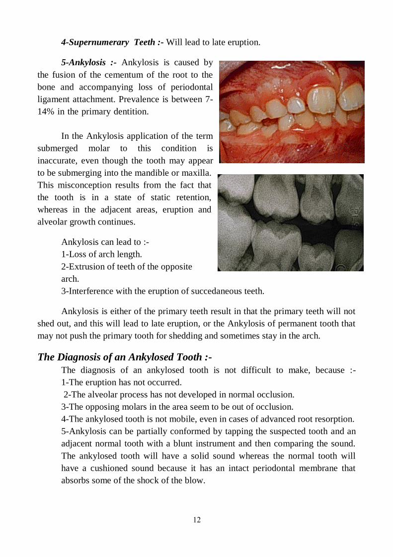
12
4-Supernumerary Teeth :- Will lead to late eruption.
5-Ankylosis :- Ankylosis is caused by
the fusion of the cementum of the root to the
bone and accompanying loss of periodontal
ligament attachment. Prevalence is between 7-
14% in the primary dentition.
In the Ankylosis application of the term
submerged molar to this condition is
inaccurate, even though the tooth may appear
to be submerging into the mandible or maxilla.
This misconception results from the fact that
the tooth is in a state of static retention,
whereas in the adjacent areas, eruption and
alveolar growth continues.
Ankylosis can lead to :-
1-Loss of arch length.
2-Extrusion of teeth of the opposite
arch.
3-Interference with the eruption of succedaneous teeth.
Ankylosis is either of the primary teeth result in that the primary teeth will not
shed out, and this will lead to late eruption, or the Ankylosis of permanent tooth that
may not push the primary tooth for shedding and sometimes stay in the arch.
The Diagnosis of an Ankylosed Tooth :-
The diagnosis of an ankylosed tooth is not difficult to make, because :-
1-The eruption has not occurred.
2-The alveolar process has not developed in normal occlusion.
3-The opposing molars in the area seem to be out of occlusion.
4-The ankylosed tooth is not mobile, even in cases of advanced root resorption.
5-Ankylosis can be partially conformed by tapping the suspected tooth and an
adjacent normal tooth with a blunt instrument and then comparing the sound.
The ankylosed tooth will have a solid sound whereas the normal tooth will
have a cushioned sound because it has an intact periodontal membrane that
absorbs some of the shock of the blow.
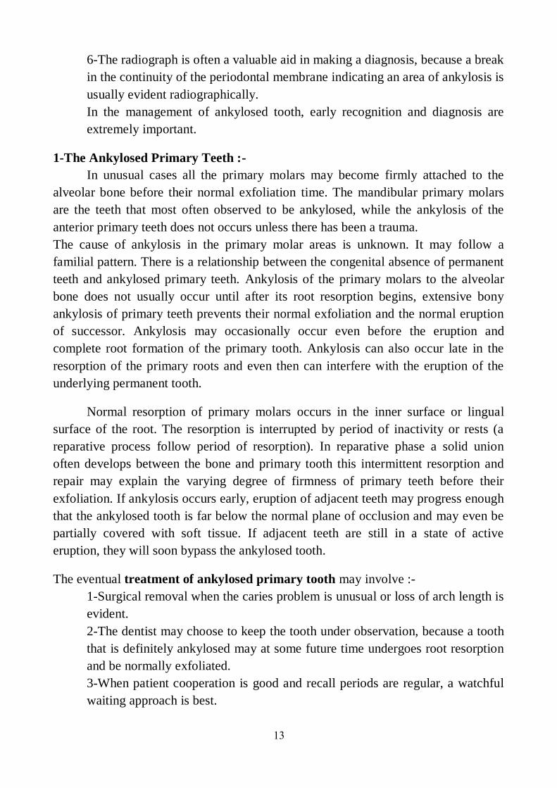
13
6-The radiograph is often a valuable aid in making a diagnosis, because a break
in the continuity of the periodontal membrane indicating an area of ankylosis is
usually evident radiographically.
In the management of ankylosed tooth, early recognition and diagnosis are
extremely important.
1-The Ankylosed Primary Teeth :-
In unusual cases all the primary molars may become firmly attached to the
alveolar bone before their normal exfoliation time. The mandibular primary molars
are the teeth that most often observed to be ankylosed, while the ankylosis of the
anterior primary teeth does not occurs unless there has been a trauma.
The cause of ankylosis in the primary molar areas is unknown. It may follow a
familial pattern. There is a relationship between the congenital absence of permanent
teeth and ankylosed primary teeth. Ankylosis of the primary molars to the alveolar
bone does not usually occur until after its root resorption begins, extensive bony
ankylosis of primary teeth prevents their normal exfoliation and the normal eruption
of successor. Ankylosis may occasionally occur even before the eruption and
complete root formation of the primary tooth. Ankylosis can also occur late in the
resorption of the primary roots and even then can interfere with the eruption of the
underlying permanent tooth.
Normal resorption of primary molars occurs in the inner surface or lingual
surface of the root. The resorption is interrupted by period of inactivity or rests (a
reparative process follow period of resorption). In reparative phase a solid union
often develops between the bone and primary tooth this intermittent resorption and
repair may explain the varying degree of firmness of primary teeth before their
exfoliation. If ankylosis occurs early, eruption of adjacent teeth may progress enough
that the ankylosed tooth is far below the normal plane of occlusion and may even be
partially covered with soft tissue. If adjacent teeth are still in a state of active
eruption, they will soon bypass the ankylosed tooth.
The eventual treatment of ankylosed primary tooth may involve :-
1-Surgical removal when the caries problem is unusual or loss of arch length is
evident.
2-The dentist may choose to keep the tooth under observation, because a tooth
that is definitely ankylosed may at some future time undergoes root resorption
and be normally exfoliated.
3-When patient cooperation is good and recall periods are regular, a watchful
waiting approach is best.
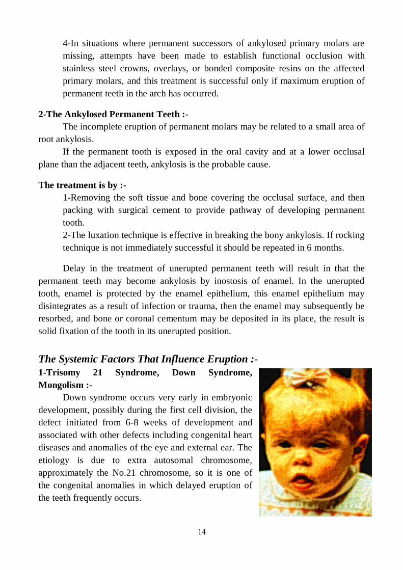
14
4-In situations where permanent successors of ankylosed primary molars are
missing, attempts have been made to establish functional occlusion with
stainless steel crowns, overlays, or bonded composite resins on the affected
primary molars, and this treatment is successful only if maximum eruption of
permanent teeth in the arch has occurred.
2-The Ankylosed Permanent Teeth :-
The incomplete eruption of permanent molars may be related to a small area of
root ankylosis.
If the permanent tooth is exposed in the oral cavity and at a lower occlusal
plane than the adjacent teeth, ankylosis is the probable cause.
The treatment is by :-
1-Removing the soft tissue and bone covering the occlusal surface, and then
packing with surgical cement to provide pathway of developing permanent
tooth.
2-The luxation technique is effective in breaking the bony ankylosis. If rocking
technique is not immediately successful it should be repeated in 6 months.
Delay in the treatment of unerupted permanent teeth will result in that the
permanent teeth may become ankylosis by inostosis of enamel. In the unerupted
tooth, enamel is protected by the enamel epithelium, this enamel epithelium may
disintegrates as a result of infection or trauma, then the enamel may subsequently be
resorbed, and bone or coronal cementum may be deposited in its place, the result is
solid fixation of the tooth in its unerupted position.
The Systemic Factors That Influence Eruption :-
1-Trisomy 21 Syndrome, Down Syndrome,
Mongolism :-
Down syndrome occurs very early in embryonic
development, possibly during the first cell division, the
defect initiated from 6-8 weeks of development and
associated with other defects including congenital heart
diseases and anomalies of the eye and external ear. The
etiology is due to extra autosomal chromosome,
approximately the No.21 chromosome, so it is one of
the congenital anomalies in which delayed eruption of
the teeth frequently occurs.
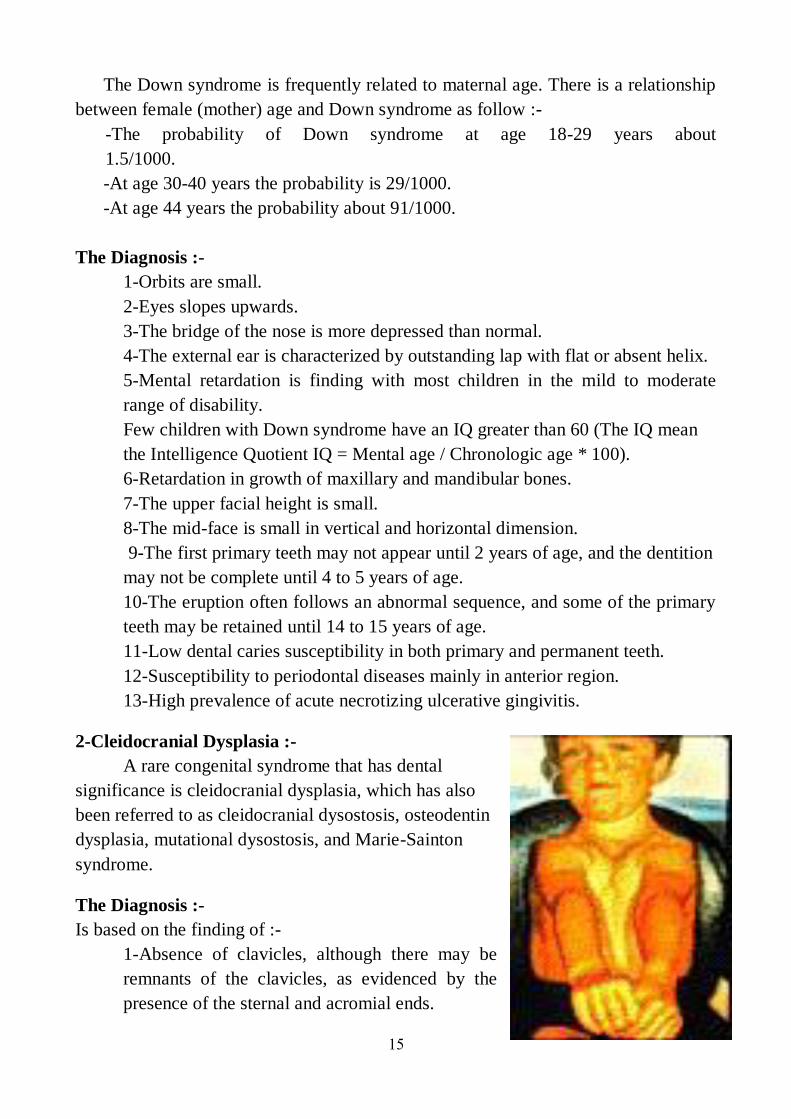
15
The Down syndrome is frequently related to maternal age. There is a relationship
between female (mother) age and Down syndrome as follow :-
-The probability of Down syndrome at age 18-29 years about
1.5/1000.
-At age 30-40 years the probability is 29/1000.
-At age 44 years the probability about 91/1000.
The Diagnosis :-
1-Orbits are small.
2-Eyes slopes upwards.
3-The bridge of the nose is more depressed than normal.
4-The external ear is characterized by outstanding lap with flat or absent helix.
5-Mental retardation is finding with most children in the mild to moderate
range of disability.
Few children with Down syndrome have an IQ greater than 60 (The IQ mean
the Intelligence Quotient IQ = Mental age / Chronologic age * 100).
6-Retardation in growth of maxillary and mandibular bones.
7-The upper facial height is small.
8-The mid-face is small in vertical and horizontal dimension.
9-The first primary teeth may not appear until 2 years of age, and the dentition
may not be complete until 4 to 5 years of age.
10-The eruption often follows an abnormal sequence, and some of the primary
teeth may be retained until 14 to 15 years of age.
11-Low dental caries susceptibility in both primary and permanent teeth.
12-Susceptibility to periodontal diseases mainly in anterior region.
13-High prevalence of acute necrotizing ulcerative gingivitis.
2-Cleidocranial Dysplasia :-
A rare congenital syndrome that has dental
significance is cleidocranial dysplasia, which has also
been referred to as cleidocranial dysostosis, osteodentin
dysplasia, mutational dysostosis, and Marie-Sainton
syndrome.
The Diagnosis :-
Is based on the finding of :-
1-Absence of clavicles, although there may be
remnants of the clavicles, as evidenced by the
presence of the sternal and acromial ends.
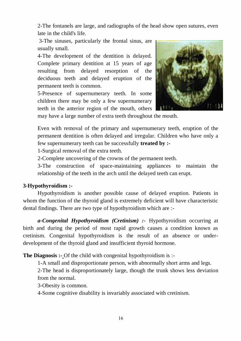
16
2-The fontanels are large, and radiographs of the head show open sutures, even
late in the child's life.
3-The sinuses, particularly the frontal sinus, are
usually small.
4-The development of the dentition is delayed.
Complete primary dentition at 15 years of age
resulting from delayed resorption of the
deciduous teeth and delayed eruption of the
permanent teeth is common.
5-Presence of supernumerary teeth. In some
children there may be only a few supernumerary
teeth in the anterior region of the mouth, others
may have a large number of extra teeth throughout the mouth.
Even with removal of the primary and supernumerary teeth, eruption of the
permanent dentition is often delayed and irregular. Children who have only a
few supernumerary teeth can be successfully treated by :-
1-Surgical removal of the extra teeth.
2-Complete uncovering of the crowns of the permanent teeth.
3-The construction of space-maintaining appliances to maintain the
relationship of the teeth in the arch until the delayed teeth can erupt.
3-Hypothyroidism :-
Hypothyroidism is another possible cause of delayed eruption. Patients in
whom the function of the thyroid gland is extremely deficient will have characteristic
dental findings. There are two type of hypothyroidism which are :-
a-Congenital Hypothyroidism (Cretinism) :- Hypothyroidism occurring at
birth and during the period of most rapid growth causes a condition known as
cretinism. Congenital hypothyroidism is the result of an absence or under-
development of the thyroid gland and insufficient thyroid hormone.
The Diagnosis :- Of the child with congenital hypothyroidism is :-
1-A small and disproportionate person, with abnormally short arms and legs.
2-The head is disproportionately large, though the trunk shows less deviation
from the normal.
3-Obesity is common.
4-Some cognitive disability is invariably associated with cretinism.
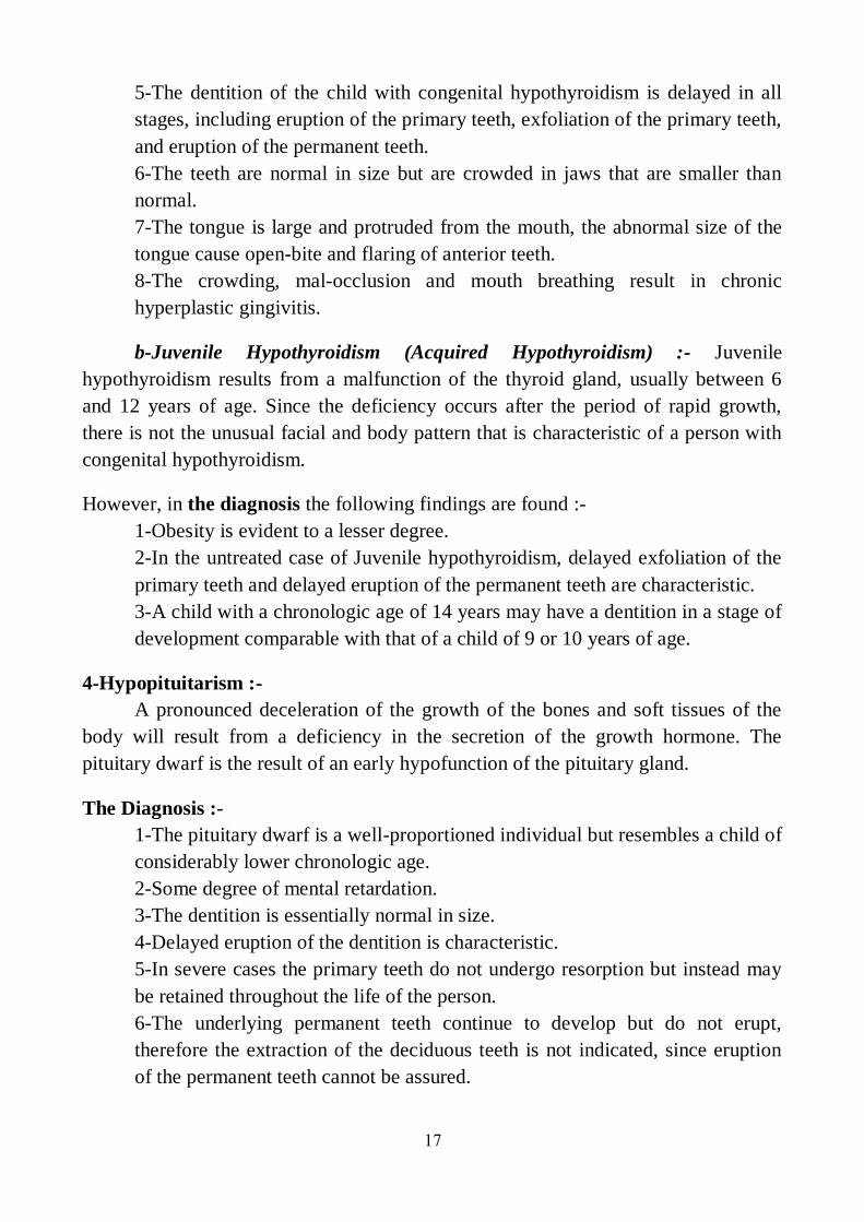
17
5-The dentition of the child with congenital hypothyroidism is delayed in all
stages, including eruption of the primary teeth, exfoliation of the primary teeth,
and eruption of the permanent teeth.
6-The teeth are normal in size but are crowded in jaws that are smaller than
normal.
7-The tongue is large and protruded from the mouth, the abnormal size of the
tongue cause open-bite and flaring of anterior teeth.
8-The crowding, mal-occlusion and mouth breathing result in chronic
hyperplastic gingivitis.
b-Juvenile Hypothyroidism (Acquired Hypothyroidism) :- Juvenile
hypothyroidism results from a malfunction of the thyroid gland, usually between 6
and 12 years of age. Since the deficiency occurs after the period of rapid growth,
there is not the unusual facial and body pattern that is characteristic of a person with
congenital hypothyroidism.
However, in the diagnosis the following findings are found :-
1-Obesity is evident to a lesser degree.
2-In the untreated case of Juvenile hypothyroidism, delayed exfoliation of the
primary teeth and delayed eruption of the permanent teeth are characteristic.
3-A child with a chronologic age of 14 years may have a dentition in a stage of
development comparable with that of a child of 9 or 10 years of age.
4-Hypopituitarism :-
A pronounced deceleration of the growth of the bones and soft tissues of the
body will result from a deficiency in the secretion of the growth hormone. The
pituitary dwarf is the result of an early hypofunction of the pituitary gland.
The Diagnosis :-
1-The pituitary dwarf is a well-proportioned individual but resembles a child of
considerably lower chronologic age.
2-Some degree of mental retardation.
3-The dentition is essentially normal in size.
4-Delayed eruption of the dentition is characteristic.
5-In severe cases the primary teeth do not undergo resorption but instead may
be retained throughout the life of the person.
6-The underlying permanent teeth continue to develop but do not erupt,
therefore the extraction of the deciduous teeth is not indicated, since eruption
of the permanent teeth cannot be assured.
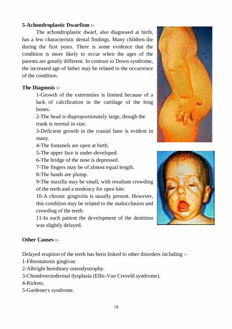
18
5-Achondroplastic Dwarfism :-
The achondroplastic dwarf, also diagnosed at birth,
has a few characteristic dental findings. Many children die
during the first years. There is some evidence that the
condition is more likely to occur when the ages of the
parents are greatly different. In contrast to Down syndrome,
the increased age of father may be related to the occurrence
of the condition.
The Diagnosis :-
1-Growth of the extremities is limited because of a
lack of calcification in the cartilage of the long
bones.
2-The head is disproportionately large, though the
trunk is normal in size.
3-Deficient growth in the cranial base is evident in
many.
4-The fontanels are open at birth.
5-The upper face is under-developed.
6-The bridge of the nose is depressed.
7-The fingers may be of almost equal length.
8-The hands are plump.
9-The maxilla may be small, with resultant crowding
of the teeth and a tendency for open bite.
10-A chronic gingivitis is usually present. However,
this condition may be related to the malocclusion and
crowding of the teeth.
11-In such patient the development of the dentition
was slightly delayed.
Other Causes :-
Delayed eruption of the teeth has been linked to other disorders including :-
1-Fibromatosis gingivae.
2-Albright hereditary osteodystrophy.
3-Chondroectodermal dysplasia (Ellis-Van Creveld syndrome).
4-Rickets.
5-Gardener,s syndrome.
Related Documents