The Open Pathology Journal, 2010, 4, 11-22 11 1874-3757/10 2010 Bentham Open Open Access p53 and Cell Cycle Proteins Participate in Spinal Motor Neuron Cell Death in ALS Srikanth Ranganathan 1,§ and Robert Bowser *,1,2,3 1 Department of Pathology, 2 Center for ALS Research, and 3 Pittsburgh Institute for Neurodegenerative Diseases, Pittsburgh, PA, USA § Current Address: Neuronal Survival Unit, Department of Experimental Medical Science, Wallenberg Neuroscience Center, Lund University, Sweden Abstract: Apoptosis has been implicated in many neurodegenerative diseases, including amyotrophic lateral sclerosis (ALS). We previously demonstrated a role for G1 to S phase cell cycle regulators in ALS with increased levels of hyperphosphorylated retinoblastoma (ppRb) and E2F-1 in ALS spinal cord motor neurons. In this study we examined the levels of the cell cycle checkpoint tumor suppressor protein p53 with concurrent changes in cell death markers during ALS. Expression and subcellular distribution of p53, retinoblastoma, Bax, Fas, and caspases were explored by immunoblot, immunohistochemistry and double-label confocal microscopy in the spinal cord and motor cortex of ALS and control subjects. We identified elevated levels of p53 in ALS spinal cord motor neurons but not neurons in the motor cortex. In addition, there was an increase in Bax, Fas, caspases-8 and -3 proteins in ALS spinal motor neurons. While caspase-3 and TUNEL labeled neurons were positive for ppRb, E2F-1 and p53 in spinal motor neurons, and Fas co- localized with caspase-8 in spinal motor neurons, we failed to observe these results in large neurons in the motor cortex of ALS subjects. We have linked p53 and activation of G1 to S phase cell cycle regulators to an apoptotic mode of cell death ALS spinal cord motor neurons. Keywords: Amyotrophic lateral sclerosis, p53, retinoblastoma, caspase. INTRODUCTION The control of cell proliferation or growth arrest is intimately linked to the control of programmed cell death or apoptosis. Cell cycle components such as ppRb, E2F and p53 function in both cell cycle progression and apoptosis [1- 3]. In fact the promoters of p53, p73, human cytochrome c1 and apoptotic protease activating factor 1 (APAF1) contain E2F-1 binding sites suggesting an E2F-1-mediated transcriptional regulation [3-6]. -amyloid-mediated toxicity in PC12 cells was found to be mediated by a E2F-1 - p53 - Bax pathway and the over-expression of E2F-1 in these cells led to DNA fragmentation in addition to the induction of p53 (by E2F-1) and Bax (by p53) [7]. These studies directly link E2F-1 to p53 and apoptosis. In models of acute and chronic neurological disease, a role for aberrant cell cycle activation and subsequent neuronal apoptosis has been established. These studies include cell death mediated by -amyloid, presenilin-1, DNA damage, oxidative stress, membrane depolarization, and 6- hydroxydopamine (6-OHDA) [8-12]. DNA damage induced phosphorylation of p53 has been shown to activate cell cycle proteins leading to neuronal cell death [13]. Inhibition of p53 through the use of antisense and dominant negative constructs protects neurons from this wide array of toxic insults [14, 15]. *Address correspondence to this author at the Department of Pathology, University of Pittsburgh School of Medicine, BST S-420, 200 Lothrop St., Pittsburgh, PA 15261, USA; Tel: 412-383-7819; Fax : 412-648-1916; E-mail: [email protected] Motor neurons of ALS patients exhibit characteristics of apoptosis in post-mortem tissues [16], and apoptosis is believed to be the mode of cell death in ALS [17]. Increased levels of p53 with concomitant elevation in DNA binding activity has been demonstrated in motor neurons and astroglia of the affected regions of ALS patients but not in controls [18]. This study was corroborated in ALS transgenic mice over-expressing mutant G86R SOD1 protein [19]. Defective DNA repair and enhanced DNA damage are also evident in ALS motor neurons [20-23]. Cell death induced by DNA damage of cultured motor neurons has been linked to Fas and p53 activation [24]. Increased p53 gene expression has been observed in degenerative spinal cords from the wobbler mouse model and in the ventral horns of ALS patients [25]. In the current study we examined the expression and distribution of p53, markers of apoptosis and G1 to S phase cell cycle regulators. We hypothesized that alterations in the cell cycle regulator, p53, will result in concurrent changes in cell cycle proteins and apoptotic death markers during ALS. We present evidence for increased accumulation of p53 in the nucleus of ALS spinal cord motor neurons but not neurons of the motor cortex. Laser scanning confocal microscopy demonstrates that the G1 to S phase cell cycle regulators co-localize with markers of apoptosis (TUNEL and Caspase-3) and p53. Taken together, these results suggest a role for p53 and caspase activation in conjunction with aberrant re-activation of cell cycle regulators during sporadic ALS (SALS).

Welcome message from author
This document is posted to help you gain knowledge. Please leave a comment to let me know what you think about it! Share it to your friends and learn new things together.
Transcript
The Open Pathology Journal, 2010, 4, 11-22 11
1874-3757/10 2010 Bentham Open
Open Access
p53 and Cell Cycle Proteins Participate in Spinal Motor Neuron Cell Death in ALS
Srikanth Ranganathan1,§
and Robert Bowser*,1,2,3
1Department of Pathology,
2Center for ALS Research, and
3Pittsburgh Institute for Neurodegenerative Diseases,
Pittsburgh, PA, USA
§Current Address: Neuronal Survival Unit, Department of Experimental Medical Science, Wallenberg Neuroscience
Center, Lund University, Sweden
Abstract: Apoptosis has been implicated in many neurodegenerative diseases, including amyotrophic lateral sclerosis
(ALS). We previously demonstrated a role for G1 to S phase cell cycle regulators in ALS with increased levels of
hyperphosphorylated retinoblastoma (ppRb) and E2F-1 in ALS spinal cord motor neurons. In this study we examined the
levels of the cell cycle checkpoint tumor suppressor protein p53 with concurrent changes in cell death markers during
ALS. Expression and subcellular distribution of p53, retinoblastoma, Bax, Fas, and caspases were explored by
immunoblot, immunohistochemistry and double-label confocal microscopy in the spinal cord and motor cortex of ALS
and control subjects. We identified elevated levels of p53 in ALS spinal cord motor neurons but not neurons in the motor
cortex. In addition, there was an increase in Bax, Fas, caspases-8 and -3 proteins in ALS spinal motor neurons. While
caspase-3 and TUNEL labeled neurons were positive for ppRb, E2F-1 and p53 in spinal motor neurons, and Fas co-
localized with caspase-8 in spinal motor neurons, we failed to observe these results in large neurons in the motor cortex of
ALS subjects. We have linked p53 and activation of G1 to S phase cell cycle regulators to an apoptotic mode of cell death
ALS spinal cord motor neurons.
Keywords: Amyotrophic lateral sclerosis, p53, retinoblastoma, caspase.
INTRODUCTION
The control of cell proliferation or growth arrest is intimately linked to the control of programmed cell death or apoptosis. Cell cycle components such as ppRb, E2F and p53 function in both cell cycle progression and apoptosis [1-3]. In fact the promoters of p53, p73, human cytochrome c1 and apoptotic protease activating factor 1 (APAF1) contain E2F-1 binding sites suggesting an E2F-1-mediated transcriptional regulation [3-6]. -amyloid-mediated toxicity in PC12 cells was found to be mediated by a E2F-1 - p53 - Bax pathway and the over-expression of E2F-1 in these cells led to DNA fragmentation in addition to the induction of p53 (by E2F-1) and Bax (by p53) [7]. These studies directly link E2F-1 to p53 and apoptosis.
In models of acute and chronic neurological disease, a role for aberrant cell cycle activation and subsequent neuronal apoptosis has been established. These studies include cell death mediated by -amyloid, presenilin-1, DNA damage, oxidative stress, membrane depolarization, and 6-hydroxydopamine (6-OHDA) [8-12]. DNA damage induced phosphorylation of p53 has been shown to activate cell cycle proteins leading to neuronal cell death [13]. Inhibition of p53 through the use of antisense and dominant negative constructs protects neurons from this wide array of toxic insults [14, 15].
*Address correspondence to this author at the Department of Pathology,
University of Pittsburgh School of Medicine, BST S-420, 200 Lothrop St.,
Pittsburgh, PA 15261, USA; Tel: 412-383-7819; Fax : 412-648-1916;
E-mail: [email protected]
Motor neurons of ALS patients exhibit characteristics of apoptosis in post-mortem tissues [16], and apoptosis is believed to be the mode of cell death in ALS [17]. Increased levels of p53 with concomitant elevation in DNA binding activity has been demonstrated in motor neurons and astroglia of the affected regions of ALS patients but not in controls [18]. This study was corroborated in ALS transgenic mice over-expressing mutant G86R SOD1 protein [19]. Defective DNA repair and enhanced DNA damage are also evident in ALS motor neurons [20-23]. Cell death induced by DNA damage of cultured motor neurons has been linked to Fas and p53 activation [24]. Increased p53 gene expression has been observed in degenerative spinal cords from the wobbler mouse model and in the ventral horns of ALS patients [25].
In the current study we examined the expression and distribution of p53, markers of apoptosis and G1 to S phase cell cycle regulators. We hypothesized that alterations in the cell cycle regulator, p53, will result in concurrent changes in cell cycle proteins and apoptotic death markers during ALS. We present evidence for increased accumulation of p53 in the nucleus of ALS spinal cord motor neurons but not neurons of the motor cortex. Laser scanning confocal microscopy demonstrates that the G1 to S phase cell cycle regulators co-localize with markers of apoptosis (TUNEL and Caspase-3) and p53. Taken together, these results suggest a role for p53 and caspase activation in conjunction with aberrant re-activation of cell cycle regulators during sporadic ALS (SALS).
12 The Open Pathology Journal, 2010, Volume 4 Ranganathan and Bowser
MATERIALS AND METHODOLOGY
Source of Tissue Samples
The lumbar spinal cord and pre/post-central gyrus (motor/sensory) region from eighteen cases of clinically diagnosed (using El Escorial criteria) and neuropathologically confirmed sporadic ALS (SALS), and ten non-ALS age-matched controls, were utilized to examine protein expression and distribution as previously detailed [26]. All autopsy tissues were obtained from the University of Pittsburgh ALS Tissue Bank. Human tissue samples were obtained at the time of autopsy using University of Pittsburgh Internal Review Board approved consent forms. Approval for use of autopsy human tissues was obtained from the University of Pittsburgh Committee for Oversight of Research Involving the Dead (CORID). Neuropathologic assessment was used to confirm the clinical diagnosis of each subject.
Antibodies
Monoclonal antibodies were used to detect E2F-1 (KH95; Santa Cruz Biotechnology, Santa Cruz, CA, USA), p53 (DO-1; Santa Cruz), and Fas (B10; Santa Cruz). Polyclonal antibodies were used to detect ppRb (NEN Biolabs, Ipswich, MA), actin (Millipore, Billerica, MA, USA), BAX (BD Biosciences, San Jose, CA, USA), caspase-3 (Cell Signaling Technology, Danvers, MA, USA), caspase-8 (Biomeda Corp, Foster City, CA), p73 (H79; Santa Cruz). The antibody concentrations for immunoblotting were 1:600 (caspase-8), 1:700 (caspase-3), 1:750 (p53, p73, Fas), 1:1000 (BAX) and 1:1500 (Actin). For light microscopy, these antibodies were used at dilutions of 1:100 (caspase-8), 1:150 (p53, caspase-3, Fas), 1:200 (BAX). For confocal laser scanning microscopy, we used primary antibody concentrations at 1:80 (ppRb, p53, caspase-3, caspase-8, Fas), and 1:60 (E2F-1).
Light and Confocal Microscopy
Paraffin-embedded tissue sections from lumbar or cervical spinal cord and mid-motor cortical regions were used as per published protocols [26]. The dorsal horn of the spinal cord and sensory cortex were the internal controls for immunohistochemistry. The study was conducted in a blinded fashion using eighteen ALS and ten age-matched control cases.
For laser scanning confocal microscopy, tissues were processed as for light microscopy with the following modifications. Primary antibodies were used at a higher concentration (ppRb; and the slides incubated overnight at 4
oC. After incubations with biotinylated secondary
antibodies (1:1000), the signal was further amplified using the tyramide amplification kit as per manufacturer’s protocol. The sections were finally incubated with a 1:500 dilution of streptavidin-Alexa Green (green) or streptavidin-Cy5/Cy3 (red) at a dilution of 1:500. A total of 70-80 and 20-30 motor neurons in the spinal cord and motor cortex, respectively, were counted at 400X magnification. The confocal study was conducted in a blinded fashion. Single labeling and other control experiments were performed to determine optimal concentrations of primary and fluorescently labeled secondary antibodies. Omission of the primary antibody resulted in absence of fluorescent signal.
Crossover control experiments were also performed to determine the specificity of the secondary antibody.
Protein Extraction
Lumbar spinal cord and motor cortical (mid- and superior-motor cortical regions) frozen tissue from controls and ALS cases were utilized for immunoblotting and DNA binding assays. For total cell lysates, tissue samples were homogenized using polytron homogenizer (PGC Scientific, Gaithersburg, MD) set at 15000 rpm for 45 secs. It was carried out in lysis buffer containing 25mmol/L HEPES (pH 7.4), 50mmol/L NaCl, protease inhibitor cocktail II (SIGMA), and 1% triton-X 100. The homogenized product was spun at 14000 rpm in a cold microfuge and the supernatant saved as the total cell lysate. Nuclear and post-nuclear extracts were extracted as described previously [26]. Briefly, protein extracts were prepared by detergent lysis on ice (0.1% Nonidet P-40, 10 mM Tris (pH 8.0), 10 mM MgCl2, 15 mM NaCl, 0.5 mM phenylmethylsulfonyl fluoride, 2 μg/ml pepstatin A, and 1 μg/ml leupeptin). The nuclei were collected by low speed centrifugation at 800x g for 5 mins. The supernatant was saved as the post-nuclear supernatant and the pellet containing the nuclei was further extracted with high salt buffer (0.42 mol/l NaCl, 20 mmol/L HEPES (pH 7.9), 20% glycerol, 0.5 mmol/L phenylmethylsulfonyl fluoride, 2 μg/ml pepstatin A, and 1 μg/ml leupeptin) on ice for 10 mins. Residual insoluble material was removed by centrifugation at 14,000 x g for 5 mins. The resulting supernatant fraction was collected and termed the "nuclear extract". Protein concentrations were determined by Bio-Rad assay (Bio-Rad, Richmond, CA, USA).
Immunoblotting
Total cell lysates, nuclear extracts and post-nuclear supernatants were fractionated by electrophoresis on an 8, 10 or 12% sodium dodecyl sulfate-polyacrylamide gels. The proteins were transferred to polyvinylidene difluoride nylon membranes (NEN Biolabs) and blocked in 5% nonfat milk/1X PBS or 0.5% BSA / 0.15% glycine in 1X PBS overnight at 4°C. The blots were probed individually with the primary antibodies overnight at 4°C in 0.5% milk/PBS. The blots were washed three times in PBS/0.1% Tween-20 for 15 minutes. Isotype-specific horseradish peroxidase-conjugated secondary antibodies (Chemicon; goat anti-mouse; 1:2000, goat anti-rabbit; 1:2000) were added for 2 hrs at room temperature. The secondary antibodies were washed extensively in PBS / 0.1% Tween-20 (three times for 20 mins). The final reaction products were visualized using enhanced chemiluminescence (ECL; Pierce) and the band intensities were within the linear range of detection. Actin was used to normalize protein levels within each sample. The density of bands was measured using the NIH Image software version 1.58 (National Institutes of Health, USA).
Electrophoretic Mobility Shift Assay (EMSA)
Gel mobility shift assays were performed as described earlier [26] with the following modifications. The sequences used to probe for p53 specific DNA binding activity were: WT p53 5’-TACAGAACATGTCTAAGCATGCTGGGGA CT -3’; MT p53 5’- TACAGAATCGCTCTAAGCATGCT GGGGACT -3’. For competition
reactions, unlabeled p53
p53 and Cell Cycle Proteins in ALS The Open Pathology Journal, 2010, Volume 4 13
competitor (3, 30, and 100ng) or unlabeled unrelated competitor (5’-GATCATTCAGGTCATGACCTGA-3’; 100, and 300ng) oligonucleotides were pre-incubated with
the
protein for 5 mins on ice prior to addition of labeled probe.
The positive control for p53 DNA binding activity was camptothecin-treated SHYSY5 neuroblastoma cells.
Terminal Deoxynucleotidyl Transferase-Mediated Biotinylated UTP Nick End Labeling Staining (TUNEL)
DNA fragmentation was assayed using the fluorescein based apoptosis detection system as per manufacturer’s instructions (Promega, USA). Briefly, paraffin embedded sections were processed as for immunohistochemistry and post-blocking were treated with Proteinase K (20μg/ml) for 15 mins. They were then incubated with TdT enzyme/nucleotide mix for 1 hr at 37
oC. For double labeling
with confocal laser scanning microscopy, the sections were immunolabeled with p53 or ppRb after TUNEL labeling as described earlier. Negative (no enzyme) and positive (DNase I-treated) samples were used. Motor neurons positive for TUNEL and cell cycle proteins (ppRb, p53) were quantified. A total of 30-40 and 20-30 motor neurons in the spinal cord and motor cortex, respectively, were counted at X400 magnification. The study was conducted in a blinded fashion using 11 ALS and 6 age-matched control cases.
Statistical Analysis
Comparisons between any two groups of data were done using the single-factorial analysis of variance (ANOVA). A p-value of 0.05 was considered statistically significant. Numerical data were expressed as means ± the standard deviation with “n” signifying the number of experiments or cases.
RESULTS
Altered Levels of Cell Death Markers and Cell Cycle
Proteins in ALS
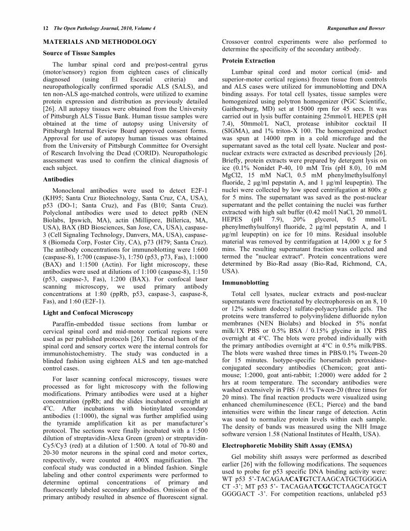
We first examined levels of numerous proteins involved in cell death pathways and the cell cycle by immunoblot analysis, using total tissue lysates from lumbar spinal cord and motor cortex of eighteen SALS and ten age-matched control subjects. Representative results are shown in Fig. (1A). The levels of activated p53 were significantly increased in the total lysates of ALS lumbar spinal cord (Fig. 1) and nuclear extracts (data not shown). E2F-1 can also activate other members of the p53 family such p73. Therefore we performed immunoblotting to analyze p73 levels in ALS tissues but found its levels unchanged in ALS spinal cord tissues (Fig. 1). Since p53 can transactivate pro-apoptotic molecules such as Bax, we examined the levels of Bax and other pro-death proteins including Fas, and active caspase-3 and -8. Levels of Bax, Fas, active caspase-3 and -8 were elevated in the total lysates of ALS spinal cord (Fig. 1B). In addition, we observed two immunoreactive bands at 19 and 21 kDa with the Bax antibody. In contrast to results from lumbar spinal cord, p53 was undetectable in total lysates prepared from motor cortex (data not shown).
Fas, caspase-3 and caspase-8 also exhibited no significant changes in protein levels within the motor cortex of ALS and control subjects.
Fig. (1). Increased p53 and cell death markers in ALS spinal cord.
Immunoblot analysis of lumbar spinal cord tissue extracts. A) Top
panel: Lane assignments for p53, p73 and Bax are as follows: “+”
indicates positive control, lanes 1 to 5 are control cases and lanes 6
to 12 are ALS cases. The arrows on the BAX blot indicate
immunoreactive bands of 19- and 21-kDa. Bottom panel: Lanes for
Fas and Caspase immunoblots are as follows: “+” indicates positive
control, lanes 1 to 3 are control cases and lanes 4 to 9 are ALS
cases. B) Quantification (mean +/- SEM) of the immunoblots.
Black bars represent control cases (n=10) and the gray bars are ALS
cases (n=18). Statistical analysis was performed using single-factor
ANOVA and asterisks (**) indicate p 0.05.
Increased p53 in ALS Spinal Cord But Not Motor Cortex
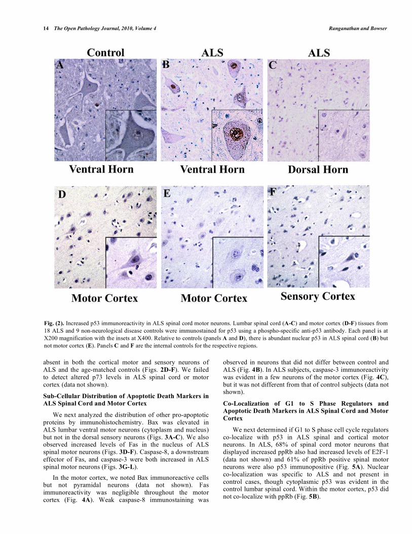
Immunoblotting of tissue extracts presents the caveat of having a mixed population of cells. Therefore we utilized immunohistochemistry to analyze cell specific and sub-cellular localization of p53 in ALS tissues. Paraffin-embedded sections from lumbar spinal cord and motor cortex from the same cases were analyzed by light microscopy.
In control subjects, 38% of lumbar spinal cord motor neurons were p53 immunoreactive, and the protein was predominately located in the cytoplasm (Fig. 2A). Approximately 84% (from a total of 200) of the lumbar spinal cord motor neurons in ALS cases exhibited abundant nuclear accumulation of p53 (Fig. 2B). There was negligible or absent p53 immunostaining in the dorsal horn sensory neurons (Fig. 2C). In addition, p53 immunoreactivity was
14 The Open Pathology Journal, 2010, Volume 4 Ranganathan and Bowser
absent in both the cortical motor and sensory neurons of ALS and the age-matched controls (Figs. 2D-F). We failed to detect altered p73 levels in ALS spinal cord or motor cortex (data not shown).
Sub-Cellular Distribution of Apoptotic Death Markers in ALS Spinal Cord and Motor Cortex
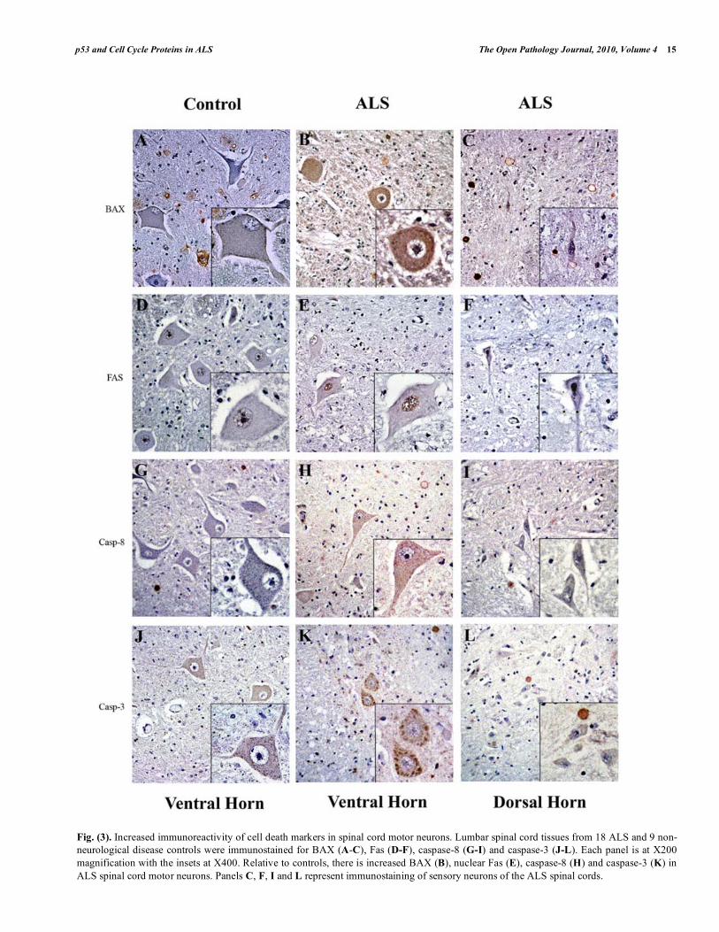
We next analyzed the distribution of other pro-apoptotic proteins by immunohistochemistry. Bax was elevated in ALS lumbar ventral motor neurons (cytoplasm and nucleus) but not in the dorsal sensory neurons (Figs. 3A-C). We also observed increased levels of Fas in the nucleus of ALS spinal motor neurons (Figs. 3D-F). Caspase-8, a downstream effector of Fas, and caspase-3 were both increased in ALS spinal motor neurons (Figs. 3G-L).
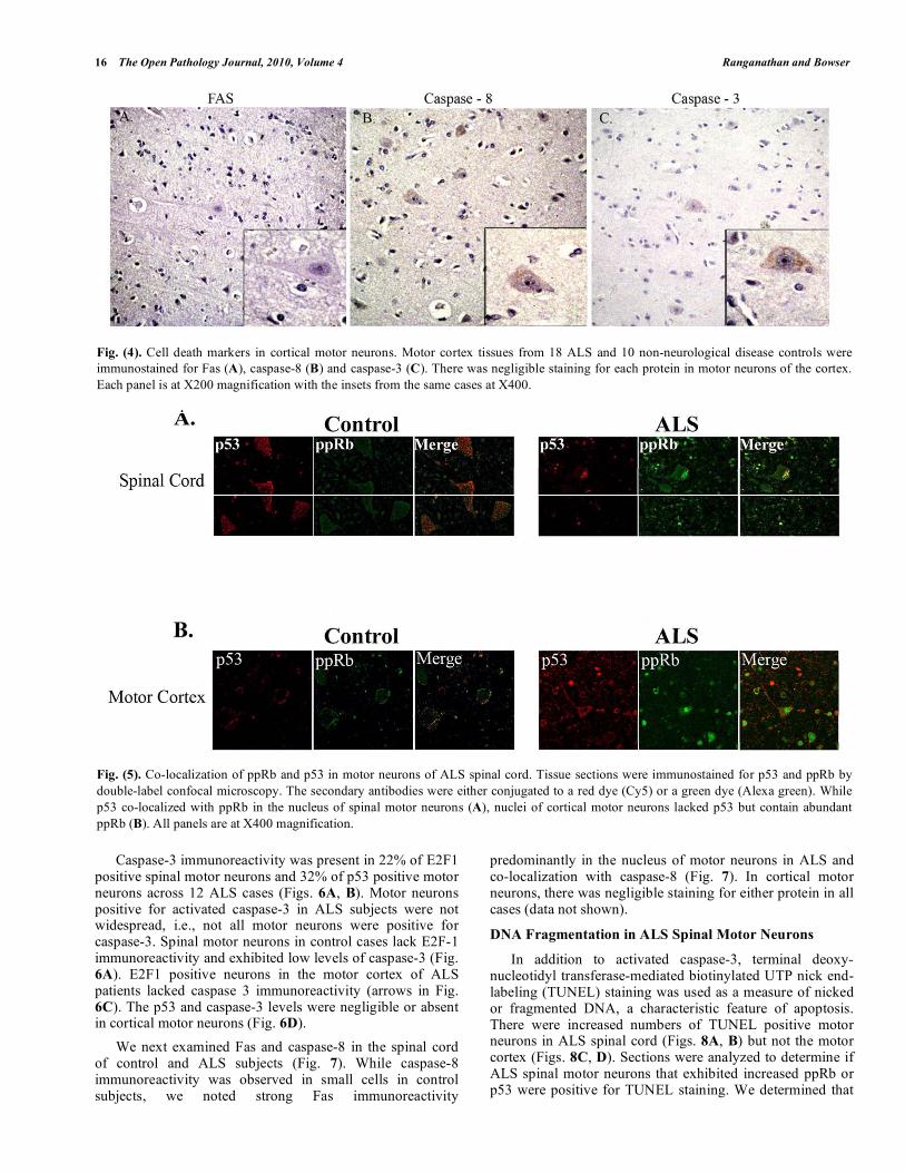
In the motor cortex, we noted Bax immunoreactive cells but not pyramidal neurons (data not shown). Fas immunoreactivity was negligible throughout the motor cortex (Fig. 4A). Weak caspase-8 immunostaining was
observed in neurons that did not differ between control and ALS (Fig. 4B). In ALS subjects, caspase-3 immunoreactivity was evident in a few neurons of the motor cortex (Fig. 4C), but it was not different from that of control subjects (data not shown).
Co-Localization of G1 to S Phase Regulators and Apoptotic Death Markers in ALS Spinal Cord and Motor
Cortex
We next determined if G1 to S phase cell cycle regulators co-localize with p53 in ALS spinal and cortical motor neurons. In ALS, 68% of spinal cord motor neurons that displayed increased ppRb also had increased levels of E2F-1 (data not shown) and 61% of ppRb positive spinal motor neurons were also p53 immunopositive (Fig. 5A). Nuclear co-localization was specific to ALS and not present in control cases, though cytoplasmic p53 was evident in the control lumbar spinal cord. Within the motor cortex, p53 did not co-localize with ppRb (Fig. 5B).
Fig. (2). Increased p53 immunoreactivity in ALS spinal cord motor neurons. Lumbar spinal cord (A-C) and motor cortex (D-F) tissues from
18 ALS and 9 non-neurological disease controls were immunostained for p53 using a phospho-specific anti-p53 antibody. Each panel is at
X200 magnification with the insets at X400. Relative to controls (panels A and D), there is abundant nuclear p53 in ALS spinal cord (B) but
not motor cortex (E). Panels C and F are the internal controls for the respective regions.
p53 and Cell Cycle Proteins in ALS The Open Pathology Journal, 2010, Volume 4 15
Fig. (3). Increased immunoreactivity of cell death markers in spinal cord motor neurons. Lumbar spinal cord tissues from 18 ALS and 9 non-
neurological disease controls were immunostained for BAX (A-C), Fas (D-F), caspase-8 (G-I) and caspase-3 (J-L). Each panel is at X200
magnification with the insets at X400. Relative to controls, there is increased BAX (B), nuclear Fas (E), caspase-8 (H) and caspase-3 (K) in
ALS spinal cord motor neurons. Panels C, F, I and L represent immunostaining of sensory neurons of the ALS spinal cords.
16 The Open Pathology Journal, 2010, Volume 4 Ranganathan and Bowser
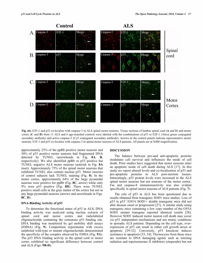
Caspase-3 immunoreactivity was present in 22% of E2F1 positive spinal motor neurons and 32% of p53 positive motor neurons across 12 ALS cases (Figs. 6A, B). Motor neurons positive for activated caspase-3 in ALS subjects were not widespread, i.e., not all motor neurons were positive for caspase-3. Spinal motor neurons in control cases lack E2F-1 immunoreactivity and exhibited low levels of caspase-3 (Fig. 6A). E2F1 positive neurons in the motor cortex of ALS patients lacked caspase 3 immunoreactivity (arrows in Fig. 6C). The p53 and caspase-3 levels were negligible or absent in cortical motor neurons (Fig. 6D).
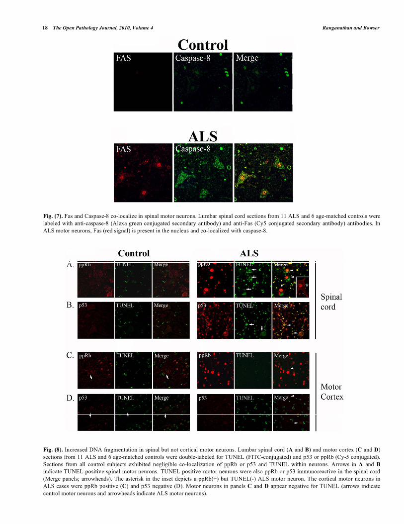
We next examined Fas and caspase-8 in the spinal cord of control and ALS subjects (Fig. 7). While caspase-8 immunoreactivity was observed in small cells in control subjects, we noted strong Fas immunoreactivity
predominantly in the nucleus of motor neurons in ALS and co-localization with caspase-8 (Fig. 7). In cortical motor neurons, there was negligible staining for either protein in all cases (data not shown).
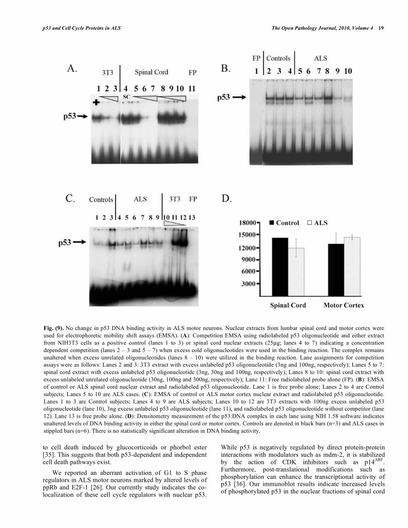
DNA Fragmentation in ALS Spinal Motor Neurons
In addition to activated caspase-3, terminal deoxy-nucleotidyl transferase-mediated biotinylated UTP nick end-labeling (TUNEL) staining was used as a measure of nicked or fragmented DNA, a characteristic feature of apoptosis. There were increased numbers of TUNEL positive motor neurons in ALS spinal cord (Figs. 8A, B) but not the motor cortex (Figs. 8C, D). Sections were analyzed to determine if ALS spinal motor neurons that exhibited increased ppRb or p53 were positive for TUNEL staining. We determined that
Fig. (4). Cell death markers in cortical motor neurons. Motor cortex tissues from 18 ALS and 10 non-neurological disease controls were
immunostained for Fas (A), caspase-8 (B) and caspase-3 (C). There was negligible staining for each protein in motor neurons of the cortex.
Each panel is at X200 magnification with the insets from the same cases at X400.
Fig. (5). Co-localization of ppRb and p53 in motor neurons of ALS spinal cord. Tissue sections were immunostained for p53 and ppRb by
double-label confocal microscopy. The secondary antibodies were either conjugated to a red dye (Cy5) or a green dye (Alexa green). While
p53 co-localized with ppRb in the nucleus of spinal motor neurons (A), nuclei of cortical motor neurons lacked p53 but contain abundant
ppRb (B). All panels are at X400 magnification.
p53 and Cell Cycle Proteins in ALS The Open Pathology Journal, 2010, Volume 4 17
approximately 25% of the ppRb positive motor neurons and 30% of p53 positive motor neurons had fragmented DNA detected by TUNEL (arrowheads in Fig. 8A, B, respectively). We also identified ppRb or p53 positive but TUNEL negative ALS motor neurons (asterisk in Fig. 8A inset). Approximately 75% of the spinal motor neurons that exhibited TUNEL also contain nuclear p53. Motor neurons of control subjects lack TUNEL staining (Fig. 8). In the motor cortex, approximately 64% of the large pyramidal neurons were positive for ppRb (Fig. 8C, arrow) while only 9% were p53 positive (Fig. 8D). There were TUNEL positive small cells in the gray matter of the cortex but not in any large pyramidal neurons (arrows and arrowheads in Figs. 8C, D).
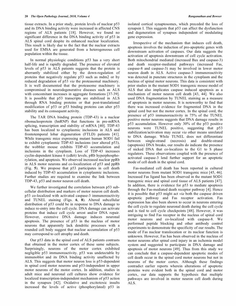
DNA-Binding Activity of p53
To determine the functional status of p53 in ALS, DNA binding activity was analyzed using nuclear extracts of spinal cord and motor cortex and radiolabeled oligonucleotide containing the consensus p53 binding site. DNA binding was monitored by gel mobility shift assays (EMSA) (Fig. 9). Competition experiments with excess unlabeled wild-type or mutant oligonucleotide demonstrated the specificity of the complex observed by EMSA (Fig. 9A). The p53 DNA binding activity in the spinal cord or motor cortex exhibited no significant difference between control and ALS (Figs. 9B-D).
DISCUSSION
The balance between pro-and anti-apoptotic proteins modulates cell survival and influences the mode of cell death. Prior studies have suggested that motor neurons enter an apoptotic mode of cell death during ALS [17]. In this study we report altered levels and co-localization of p53 and pro-apoptotic proteins in ALS post-mortem tissues. Interestingly, p53 protein levels were increased in the ALS spinal motor neurons but not neurons of the motor cortex. Fas and caspase-8 immunoreactivity was also evident specifically in spinal motor neurons of ALS patients (Fig. 7).
The role of p53 in ALS has been questioned due to results obtained from transgenic SOD1 mice studies. Loss of p53 in p53
-/-/G93A SOD1+ double transgenic mice did not
alter disease onset or progression [27]. A similar study using transgenic mice containing a low copy number of the G93A SOD1 mutant transgene reported identical results [28]. However SOD1 induced motor neuron cell death may occur via p53 independent mechanisms and not mimic conditions in sporadic ALS patients. Depending on the cell type, over-expression of p53 can result in either cell growth arrest or apoptosis [29-32]. Conversely, p53 knockout induces resistance to apoptosis [33, 34]. Thymocytes from these mice are resistant to DNA damaging agents such as ionizing radiation and topoisomerase II inhibitors (etoposide) but not
Fig. (6). E2F-1 and p53 co-localize with caspase-3 in ALS spinal motor neurons. Tissue sections of lumbar spinal cord (A and B) and motor
cortex (C and D) from 11 ALS and 6 age-matched controls were labeled with the combinations of p53 or E2F-1 (Alexa green conjugated
secondary antibody) and active caspase-3 (Cy5 conjugated secondary antibody). Arrows in the control panels indicate representative motor
neurons. E2F-1 and p53 co-localize with caspase-3 in spinal motor neurons of ALS patients. All panels are at X400 magnification.
18 The Open Pathology Journal, 2010, Volume 4 Ranganathan and Bowser
Fig. (7). Fas and Caspase-8 co-localize in spinal motor neurons. Lumbar spinal cord sections from 11 ALS and 6 age-matched controls were
labeled with anti-caspase-8 (Alexa green conjugated secondary antibody) and anti-Fas (Cy5 conjugated secondary antibody) antibodies. In
ALS motor neurons, Fas (red signal) is present in the nucleus and co-localized with caspase-8.
Fig. (8). Increased DNA fragmentation in spinal but not cortical motor neurons. Lumbar spinal cord (A and B) and motor cortex (C and D)
sections from 11 ALS and 6 age-matched controls were double-labeled for TUNEL (FITC-conjugated) and p53 or ppRb (Cy-5 conjugated).
Sections from all control subjects exhibited negligible co-localization of ppRb or p53 and TUNEL within neurons. Arrows in A and B
indicate TUNEL positive spinal motor neurons. TUNEL positive motor neurons were also ppRb or p53 immunoreactive in the spinal cord
(Merge panels; arrowheads). The asterisk in the inset depicts a ppRb(+) but TUNEL(-) ALS motor neuron. The cortical motor neurons in
ALS cases were ppRb positive (C) and p53 negative (D). Motor neurons in panels C and D appear negative for TUNEL (arrows indicate
control motor neurons and arrowheads indicate ALS motor neurons).
p53 and Cell Cycle Proteins in ALS The Open Pathology Journal, 2010, Volume 4 19
to cell death induced by glucocorticoids or phorbol ester [35]. This suggests that both p53-dependent and independent cell death pathways exist.
We reported an aberrant activation of G1 to S phase regulators in ALS motor neurons marked by altered levels of ppRb and E2F-1 [26]. Our currently study indicates the co-localization of these cell cycle regulators with nuclear p53.
While p53 is negatively regulated by direct protein-protein interactions with modulators such as mdm-2, it is stabilized by the action of CDK inhibitors such as p14
ARF.
Furthermore, post-translational modifications such as phosphorylation can enhance the transcriptional activity of p53 [36]. Our immunoblot results indicate increased levels of phosphorylated p53 in the nuclear fractions of spinal cord
Fig. (9). No change in p53 DNA binding activity in ALS motor neurons. Nuclear extracts from lumbar spinal cord and motor cortex were
used for electrophoretic mobility shift assays (EMSA). (A): Competition EMSA using radiolabeled p53 oligonucleotide and either extract
from NIH3T3 cells as a positive control (lanes 1 to 3) or spinal cord nuclear extracts (25μg; lanes 4 to 7) indicating a concentration
dependent competition (lanes 2 – 3 and 5 – 7) when excess cold oligonucleotides were used in the binding reaction. The complex remains
unaltered when excess unrelated oligonucleotides (lanes 8 – 10) were utilized in the binding reaction. Lane assignments for competition
assays were as follows: Lanes 2 and 3: 3T3 extract with excess unlabeled p53 oligonucleotide (3ng and 100ng, respectively); Lanes 5 to 7:
spinal cord extract with excess unlabeled p53 oligonucleotide (3ng, 30ng and 100ng, respectively); Lanes 8 to 10: spinal cord extract with
excess unlabeled unrelated oligonucleotide (30ng, 100ng and 300ng, respectively); Lane 11: Free radiolabeled probe alone (FP). (B): EMSA
of control or ALS spinal cord nuclear extract and radiolabeled p53 oligonucleotide. Lane 1 is free probe alone; Lanes 2 to 4 are Control
subjects; Lanes 5 to 10 are ALS cases. (C): EMSA of control or ALS motor cortex nuclear extract and radiolabeled p53 oligonucleotide.
Lanes 1 to 3 are Control subjects; Lanes 4 to 9 are ALS subjects; Lanes 10 to 12 are 3T3 extracts with 100ng excess unlabeled p53
oligonucleotide (lane 10), 3ng excess unlabeled p53 oligonucleotide (lane 11), and radiolabeled p53 oligonucleotide without competitor (lane
12). Lane 13 is free probe alone. (D): Densitometry measurement of the p53:DNA complex in each lane using NIH 1.58 software indicates
unaltered levels of DNA binding activity in either the spinal cord or motor cortex. Controls are denoted in black bars (n=3) and ALS cases in
stippled bars (n=6). There is no statistically significant alteration in DNA binding activity.
20 The Open Pathology Journal, 2010, Volume 4 Ranganathan and Bowser
tissue extracts. In a prior study, protein levels of nuclear p53 and its DNA binding activity were increased in affected CNS regions of ALS patients [18]. However, we found no significant difference in the DNA binding activity of p53 in ALS spinal cord despite its enhanced nuclear localization. This result is likely due to the fact that the nuclear extracts used for EMSA are generated from a heterogeneous cell population within the tissue.
In normal physiologic conditions p53 has a very short half-life and is rapidly degraded. The presence of elevated levels of p53 in ALS patients suggests that the protein is aberrantly stabilized either by the down-regulation of proteins that negatively regulate p53 such as mdm2 or by reduced degradation of p53 via the proteasomal machinery. It is well documented that the proteasome machinery is compromised in neurodegenerative diseases such as ALS with concomitant increases in aggregate formations [37-39]. It is possible that p53 transcripts are stabilized in ALS through RNA binding proteins or that post-translational modification of p53 or p53 binding proteins can alter p53 stability and its consequent activity.
The TAR DNA binding protein (TDP-43) is a nuclear ribonucleoprotein (hnRNP) that functions in pre-mRNA splicing, transcription and stability of mRNA [40]. TDP-43 has been localized to cytoplasmic inclusions in ALS and frontotemporal lobar degeneration (FTLD) patients [41]. While transgenic mice expressing mutant human SOD1 fail to exhibit cytoplasmic TDP-43 inclusions (nor altered p53), the wobbler mouse exhibits TDP-43 accumulation and inclusions in the cytoplasm. Loss of TDP-43 increases cyclin-dependent kinase 6 (cdk6) levels, pRb hyperphospho-rylation, and apoptosis. We observed increased nuclear ppRb in ALS motor neurons and co-localization of p53 and ppRb (Fig. 5). We propose that p53 participates in cell death induced by TDP-43 accumulation in cytoplasmic inclusions. Further studies are required to examine the link between TDP-43, p53 and motor neuron cell death.
We further investigated the correlation between p53 sub-cellular distribution and markers of motor neuron cell death. p53 co-localized with activated caspase-3 immunoreactivity and TUNEL staining (Figs. 6, 8). Altered subcellular distribution of p53 could be in response to DNA damage to induce re-entry into the cell cycle. DNA damage can activate proteins that induce cell cycle arrest and/or DNA repair. However, extensive DNA damage induces neuronal apoptosis. The presence of p53 in the nucleus of motor neurons that apparently lack dendritic processes with a rounded cell body suggest that nuclear accumulation of p53 may correspond to cell atrophy and death.
Our p53 data in the spinal cord of ALS patients contrasts that obtained in the motor cortex of these same subjects. Surprisingly, neurons of the motor cortex exhibited negligible p53 immunoreactivity. p53 was undetectable by immunoblot and its DNA binding activity unaffected by ALS. This suggests that motor neuron loss is p53-dependent in spinal cord motor neurons and p53-independent in upper motor neurons of the motor cortex. In addition, studies in adult mice and neuronal cell cultures show evidence for localized transcription-independent mechanism of p53 action in the synapses [42]. Oxidative and excitotoxic insults increased the levels of active (phosphorylated) p53 in
isolated cortical synaptosomes, which preceded the loss of synapsin I. This suggests that p53 can affect the dysfunction and degeneration of synapses independent of modulating gene expression.
Irrespective of the mechanism that induces cell death, apoptosis involves the induction of pro-apoptotic genes with downstream activation of caspases. Our data suggests the activation of apoptosis downstream of cell cycle activation. Both mitochondrial mediated (increased Bax and caspase-3) and death receptor-mediated pathways (increased Fas, caspase-8 and caspase-3) may be involved in lower motor neuron death in ALS. Active caspase-3 immunoreactivity was detected in punctate structures in the cytoplasm and the nucleus of spinal motor neurons. This data is consistent with prior studies in the mutant SOD1 transgenic mouse model of ALS that also implicates caspase induced apoptosis as a mechanism of motor neuron cell death [43, 44]. We also used DNA fragmentation by TUNEL staining as a measure of apoptosis in motor neurons. It is noteworthy to find that there was increased evidence for fragmented DNA in the spinal cord but not the motor cortex. In the spinal cord, the presence of p53 immunoreactivity in 75% of the TUNEL positive motor neurons suggests that DNA damage results in p53 accumulation. However only 30% of the p53 positive neurons were TUNEL positive, suggesting that p53 stabilization/activation may occur via other means unrelated to DNA damage. While TUNEL does not differentiate between single-strand (necrosis) and double-strand (apoptosis) DNA breaks, our results do indicate the presence of nicked DNA that co-localizes to the G1 to S phase regulators. These observations coupled with the presence of activated caspase-3 lend further support for an apoptotic mode of cell death in the spinal cord.
Fas-mediated cell death has been reported in cultured motor neurons from mutant SOD1 transgenic mice [45, 46]. Increased Fas ligand has been observed in the mutant SOD1 transgenic mice and spinal cord tissue of ALS patients [47]. In addition, there is evidence for p53 to mediate apoptosis through the Fas-mediated death receptor pathway [4]. Hence it is possible that p53 might act via both the caspase driven apoptotic pathway and Fas receptor activation. Fas expression has also been shown to occur in neurons entering the cell cycle to regulate neuronal death during the cell cycle and is tied to cell cycle checkpoints [48]. However, it was intriguing to find Fas receptor in the nucleus of spinal cord motor neurons and co-localized with caspase-8. We performed peptide blocking and other antibody control experiments to demonstrate the specificity of our results. The mode of Fas nuclear translocation or its nuclear function is unknown. However, Fas has been observed in the nucleus of motor neurons after spinal cord injury in an ischemia model system and suggested to participate in DNA damage and apoptosis of motor neurons [49]. Thus from this study we can conclude that a caspase-dependent apoptotic modes of cell death occur in the spinal cord motor neurons but not in neurons of the motor cortex. Although these findings contradict earlier reports wherein alterations of apoptotic proteins were evident both in the spinal cord and motor cortex, our data supports the hypothesis that multiple pathways are involved in motor neuron cell death during ALS.
p53 and Cell Cycle Proteins in ALS The Open Pathology Journal, 2010, Volume 4 21
In conclusion, we report alterations in the G1 to S phase transition of cell cycle with concomitant increases in p53 levels in the spinal cord of ALS patients that may induce an apoptotic mode of spinal motor neuron cell death involving both caspase-3 and Fas. We failed to observe similar alterations of this apoptotic mechanism in the motor cortex. Therefore, these findings suggest differential mechanisms of neuronal cell death in ALS spinal cord and motor cortex.
ACKNOWLEDGEMENT
We gratefully acknowledge funding support from NIH award NS042724 to RB.
ABBREVIATIONS
ALS = Amyotrophic lateral sclerosis
EMSA = Electrophoretic mobility shift assay
TUNEL = Terminal TdT transferase nick-end labeling
REFERENCES
[1] Dasgupta P, Padmanabhan J, Chellappan S. Rb function in the
apoptosis and senescence of non-neuronal and neuronal cells: role in oncogenesis. Curr Mol Med 2006; 6: 719-29.
[2] DeGregori J, Johnson DG. Distinct and overlapping roles for E2F family members in transcription, proliferation and apoptosis. Curr
Mol Med 2006; 6: 739-48. [3] Nahle Z, Polakoff J, Davuluri RV, et al. Direct coupling of the cell
cycle and cell death machinery by E2F. Nat Cell Biol 2002; 4: 859-64.
[4] Stiewe T, Putzer BM. Role of the p53-homologue p73 in E2F1-induced apoptosis. Nature Genet 2000; 26: 464-9.
[5] Moroni MC, Hickman ES, Denchi EL, et al. Apaf-1 is a transcriptional target for E2F and p53. Nat Cell Biol 2001; 3: 552-
8. [6] Luciakova K, Barath P, Li R, Zaid A, Nelson BD. Activity of the
human cytochrome c1 promoter is modulated by E2F. Biochem J 2000; 351: 251-6.
[7] Ramalho RM, Ribeiro PS, Sola S, Castro RE, Steer CJ, Rodrigues CM. Inhibition of the E2F-1/p53/Bax pathway by
tauroursodeoxycholic acid in amyloid -peptide-induced apoptosis of PC12 cells. J Neurochem 2004; 90: 567-75.
[8] Blum D, Wu Y, Nissou MF, Arnaud S, Alim Louis B, Verna JM. p53 and Bax activation in 6-hydroxydopamine-induced apoptosis
in PC12 cells. Brain Res 1997; 751: 139-42. [9] Chan SL, Culmsee C, Haughey N, Klapper W, Mattson MP.
Presenilin-1 mutations sensitize neurons to DNA damage-induced death by a mechanism involving perturbed calcium homeostasis
and activation of calpains and caspase-12. Neurobiol Dis 2002; 11: 2-19.
[10] Daily D, Barzilai A, Offen D, Kamsler A, Melamed E, Ziv I. The involvement of p53 in dopamine-induced apoptosis of cerebellar
granule neurons and leukemic cells overexpressing p53. Cell Mol Neurobiol 1999; 19: 261-76.
[11] Inamura N, Araki T, Enokido Y, Nishio C, Aizawa S, Hatanaka H. Role of p53 in DNA strand break-induced apoptosis in organotypic
slice culture from the mouse cerebellum. J Neurosci Res 2000; 60: 450-7.
[12] O'Hare MJ, Hou ST, Morris EJ, et al. Induction and modulation of cerebellar granule neuron death by E2F-1. J Biol Chem 2000; 275:
25358-64. [13] Kruman II, Wersto RP, Cardozo-Pelaez F, et al. Cell cycle
activation linked to neuronal cell death initiated by DNA damage. Neuron 2004; 41: 549-61.
[14] Tieu K, Ashe PC, Zuo DM, Yu PH. Inhibition of 6-hydroxydopamine-induced p53 expression and survival of
neuroblastoma cells following interaction with astrocytes. Neuroscience 2001; 103: 125-32.
[15] Jordan J, Galindo MF, Gonzalez-Garcia C, Cena V. Role and regulation of p53 in depolarization-induced neuronal death.
Neuroscience 2003; 122: 707-15.
[16] Martin LJ. Neuronal death in amyotrophic lateral sclerosis is
apoptosis: Possible contributions of a programmed cell death mechanism. J Neuropathol Exp Neurol 1999; 58: 459-71.
[17] Sathasivam S, Ince PG, Shaw PJ. Apoptosis in amyotrophic lateral sclerosis: A review of the evidence. Neuropathol Appl Neurobiol
2001; 27: 257-74. [18] Martin LJ. p53 is abnormally elevated and active in the CNS of
patients with amyotrophic lateral sclerosis. Neurobiol Dis 2000; 7: 613-22.
[19] Gonzalez de Aguilar J-L, Gordon JW, Rene F, et al. Alteration of the Bcl-x/Bax ratio in a transgenic mouse model of amyotrophic
lateral sclerosis: evidence for the implication of the p53 signaling pathway. Neurobiol Dis 2000; 7: 406-15.
[20] Bogdanov M, Brown RH, Matson WR, et al. Increased oxidative damage to DNA in ALS patients. Free Radic Biol Med 2000; 29:
652-8. [21] Kikuchi H, Furuta A, Nishioka K, Suzuki SO, Nakabeppu Y, Iwaki
T. Impairment of mitochondrial DNA repair enzymes against accumulation of 8-oxo-guanine in the spinal motor neurons of
amyotrophic lateral sclerosis. Acta Neuropathol 2002; 103: 408-14. [22] Shaikh AY, Martin LJ. DNA base-excision repair enzyme
apurinic/apyrimidinic endonuclease/redox factor-1 is increased and competent in the brain and spinal cord of individuals with
amyotrophic lateral sclerosis. Neuromol Med 2002; 2: 47-60. [23] Aguirre N, Beal MF, Matson WR, Bogdanov MB. Increased
oxidative damage to DNA in an animal model of amyotrophic lateral sclerosis. Free Radic Res 2005; 39: 383-8.
[24] Martin LJ, Chen K, Liu Z. Adult motor neuron apoptosis is mediated by nitric oxide and Fas death receptor linked by DNA
damage and p53 activation. J Neurosci 2005; 25: 6449-59. [25] Eve DJ, Dennis JS, Citron BA. Transcription factor p53 in
degenerating spinal cords. Brain Res 2007; 1150: 174-81. [26] Ranganathan S, Bowser R. Alterations in G1 to S phase cell-cycle
regulators during amyotrophic lateral sclerosis. Am J Pathol 2003; 162: 823-35.
[27] Kuntz C, Kinoshita Y, Beal MF, Donehower LA, Morrison RS. Absence of p53: No effect in a transgenic mouse model of familial
amyotrophic lateral sclerosis. Exp Neurol 2000; 165: 184-90. [28] Prudlo J, Koenig J, Graser J, et al. Motor neuron cell death in a
mouse model of FALS is not mediated by the p53 cell survival regulator. Brain Res 2000; 879: 183-7.
[29] Eliyahu D, Michalovitz D, Eliyahu S, Pinhasi-Kimhi O, Oren M. Wild-type p53 can inhibit oncogene-mediated focus formation.
Proc Natl Acad Sci USA 1989; 86: 8763-7. [30] Finlay CA, Hinds PW, Levine AJ. The p53 proto-oncogene can act
as a suppressor of transformation. Cell 1989; 57: 1083-93. [31] Yonish-Rouach E, Resnitzky D, Lotem J, Sachs L, Kimchi A, Oren
M. Wild-type p53 induces apoptosis of myeloid leukaemic cells that is inhibited by interleukin-6. Nature 1991; 352: 345-7.
[32] Shaw P, Bovey R, Tardy S, Sahli R, Sordat B, Costa J. Induction of apoptosis by wild-type p53 in a human colon tumor-derived cell
line. Proc Natl Acad Sci USA 1992; 89: 4495-9. [33] Donehower LA, Harvey M, Slagle BL, et al. Mice deficient for p53
are developmentally normal but susceptible to spontaneous tumours. Nature 1992; 356: 215-21.
[34] Harvey M, McArthur MJ, Montgomery CA Jr, Bradley A, Donehower LA. Genetic background alters the spectrum of tumors
that develop in p53-deficient mice. FASEB J 1993; 7: 938-43. [35] Clarke AR, Purdie CA, Harrison DJ, et al. Thymocyte apoptosis
induced by p53-dependent and independent pathways. Nature 1993; 362: 849-52.
[36] Ryan KM, Phillips AC, Vousden KH. Regulation and function of the p53 tumor suppressor protein. Curr Opin Cell Biol 2001; 13:
332-7. [37] Urushitani M, Kurisu J, Tsukita K, Takahashi R. Proteasomal
inhibition by misfolded mutant superoxide dismutase I induces selective motor neuron death in FALS. J Neurochem 2002; 83:
1030-42. [38] Kabashi E, Agar JN, Taylor DM, Minotti S, Durham HD. Focal
dysfunction of the proteasome: a pathogenic factor in a mouse model of amyotrophic lateral sclerosis. J Neurochem 2004; 89:
1325-35. [39] Tsuji S, Kikuchi S, Shinpo K, et al. Proteasome inhibition induces
selective motor neuron death in organotypic slice cultures. J Neurosci Res 2005; 82: 443-51.
22 The Open Pathology Journal, 2010, Volume 4 Ranganathan and Bowser
[40] Ou SH, Wu F, Harrich D, Garcia-Martinez LF, Gaynor RB.
Cloning and characterization of a novel cellular protein, TDP-43, that binds to human immunodeficiency virus type 1 TAR DNA
sequence motifs. J Virol 1995; 69: 3584-96. [41] Davidson Y, Kelley T, Mackenzie IR, et al. Ubiquitinated
pathological lesions in frontotemporal lobar degeneration contains the TAR DNA-binding protein, TDP-43. Acta Neuropathol 2007;
113: 521-33. [42] Gilman CP, Chan SL, Guo Z, Zhu X, Greig N, Mattson MP. p53 is
present in synapses where it mediates mitochondrial dysfunction and synaptic degeneration in response to DNA damage, and
oxidative and excitotoxic insults. Neuromol Med 2003; 3: 159-72. [43] Pasinelli P, Houseweart MK, Brown RHJ, Cleveland DW.
Caspase-1 and -3 are sequentially activated in motor neuron death in Cu,Zn superoxide dismutase-mediated familial amyotrophic
lateral sclerosis. Proc Natl Acad Sci USA 2000; 97: 13901-6. [44] Chi L, Ke Y, Luo C, Gozal D, Liu R. Depletion of reduced
glutathione enhances motor neuron degeneration in vitro and in vivo. Neuroscience 2007; 144: 991-1003.
[45] Raoul C, Estevez AG, Nishimune H, et al. Motoneuron death
triggered by a specific pathway downstream of Fas. potentiation by ALS-linked SOD1 mutations. Neuron 2002; 35: 1067-83.
[46] Raoul C, Buhler E, Sadeghi C, et al. Chronic activation in presymptomatic amyotrophic lateral sclerosis (ALS) mice of a
feedback loop involving Fas, Daxx, and FasL. Proc Natl Acad Sci USA 2006; 103: 6007-12.
[47] Kiaei M, Petri S, Kipiani K, et al. Thalidomide and lenalidomide extend survival in a transgenic mouse model of amyotrophic lateral
sclerosis. J Neurosci 2006; 26: 2467-73. [48] Cheema ZF, Santillano DR, Wade SB, Newman JM, Miranda RC.
The extracellular matrix, p53 and estrogen compete to regulate cell-surface Fas/Apo-1 suicide receptor expression in proliferating
embryonic cerebral cortical precursors, and reciprocally, Fas-ligand modifies estrogen control of cell-cycle proteins. BMC Neurosci
2004; 5: 11. [49] Sakurai M, Hayashi T, Abe K, Sadahiro M, Tabayashi K. Delayed
selective motor neuron death and Fas antigen induction after spinal cord ischemia in rabbits. Brain Res 1998; 797: 23-8.
Received: February 16, 2010 Revised: February 18, 2010 Accepted: February 19, 2010
© Ranganathan and Bowser; Licensee Bentham Open.
This is an open access article licensed under the terms of the Creative Commons Attribution Non-Commercial License (http: //creativecommons.org/licenses/by-nc/
3.0/) which permits unrestricted, non-commercial use, distribution and reproduction in any medium, provided the work is properly cited.
Related Documents