Ozone-Induced Programmed Cell Death in the Arabidopsis radical-induced cell death1 Mutant 1 Kirk Overmyer 2 , Mikael Brosche ´, Riikka Pellinen 3 , Tero Kuittinen, Hannele Tuominen 4 , Reetta Ahlfors, Markku Keina ¨nen 5 , Mart Saarma, Dierk Scheel, and Jaakko Kangasja ¨rvi* Department of Biological and Environmental Sciences (K.O., M.B., R.P., H.T., R.A., M.K., J.K.) and Institute of Biotechnology (K.O., R.P., T.K., H.T., M.K., M.S., J.K.), University of Helsinki, FIN–00014 Helsinki, Finland; and Department of Stress and Developmental Biology, Leibniz Institute of Plant Biochemistry, D–06120 Halle/Saale, Germany (D.S.) Short, high-concentration peaks of the atmospheric pollutant ozone (O 3 ) cause the formation of cell death lesions on the leaves of sensitive plants. Numerous similarities between the plant responses to O 3 and pathogens suggest that O 3 triggers hypersensitive response-like programmed cell death (PCD). We examined O 3 and superoxide-induced cell death in the O 3 -sensitive radical-induced cell death1 (rcd1) mutant. Dying cells in O 3 -exposed rcd1 exhibited several of the typical morphol- ogical characteristics of the hypersensitive response and PCD. Double-mutant analyses indicated a requirement for salicylic acid and the function of the cyclic nucleotide-gated ion channel AtCNGC2 in cell death. Furthermore, a requirement for ATPases, kinases, transcription, Ca 21 flux, caspase-like proteolytic activity, and also one or more phenylmethylsulfonyl fluoride- sensitive protease activities was shown for the development of cell death lesions in rcd1. Furthermore, mitogen-activated protein kinases showed differential activation patterns in rcd1 and Columbia. Taken together, these results directly demonstrate the induction of PCD by O 3 . Ozone (O 3 ) is an atmospheric pollutant that is phyto- toxic via its breakdown in the apoplast to form reac- tive oxygen species (ROS). Short, high-concentration peaks, so-called acute O 3 , cause visible damage in sensitive plants (Wohlgemuth et al., 2002). Although accumulating evidence has deepened our understand- ing of oxidative stress and antioxidant defenses in O 3 responses (Kangasja ¨rvi et al., 1994; Sandermann et al., 1998; Overmyer et al., 2003), the mechanisms involved in O 3 -induced cell death are still relatively unknown. Due to the strong chemical reactivity of O 3 , its toxicity has previously been attributed to an ability to form toxic ROS that directly damage membranes (for re- view, see Heath and Taylor, 1997). However, the view of O 3 has recently shifted, where it is now regarded in many cases not as a toxin but rather as an elicitor of cell death (Sandermann et al., 1998). O 3 -induced plant responses resemble on several levels the hypersensitive response (HR), normally seen as the result of challenge by an avirulent patho- gen (for review, see Rao and Davis, 2001; Langebartels and Kangasja ¨rvi, 2004). Common to these two pro- cesses are the induction of a biphasic oxidative burst, salicylic acid (SA) accumulation, ion fluxes, the depo- sition of cell wall-strengthening phenolic compounds, induction of defense genes such as Phe ammonia lyase, pathogenesis-related protein-1 (PR-1), and glutathione S-transferase (GST), as well as induction of local and systemic pathogen resistance. This has led to the view that O 3 misfires HR-like cell death and defense pro- grams via mimicry of the oxidative burst induced by avirulent pathogens. The HR is genetically regulated, and a form of programmed cell death (PCD; Dangl et al., 1996), where ROS have a well-established role in the in- duction and propagation of cell death signals, both generally and in spontaneous lesion mimic mutants (Vranova ´ et al., 2002). As has often been stated (Kangasja ¨rvi et al., 1994; Pell et al., 1997; Sandermann et al., 1998; Rao et al., 2000a), the similarities between the plant responses during the HR and under O 3 suggest that O 3 -induced cell death is executed by the same mechanisms as the HR. This makes O 3 a conve- nient probe for the study of ROS-induced PCD and implies that radical hypersensitive mutants, such as radical-induced cell death1 (rcd1; Overmyer et al., 2000; 1 This work was supported by the Academy of Finland (grant nos. 43671 and 37995), by the Finnish Centre of Excellence Programme (2000–2005), and by an Academy of Finland/German Academic Exchange Service grant (SA10256/313–SF–PPP–pz). R.P. was sup- ported by the Finnish Graduate program in Environmental Physi- ology, Molecular Biology, and Ecotechnology, the University of Kuopio, and The Finnish Graduate School in Environmental Science and Technology, A ˚ bo Akademi. 2 Present address: Biology Department, CB No. 3280, University of North Carolina, Chapel Hill, NC 27599–3280. 3 Present address: A.I. Virtanen Institute, University of Kuopio, FIN–70211 Kuopio, Finland. 4 Present address: Umea ˚ Plant Science Centre, Department of Plant Physiology, Umea ˚ University, SE–90187 Umea ˚, Sweden. 5 Present address: Department of Biology, University of Joensuu, PO Box 111, FIN–80101 Joensuu, Finland. * Corresponding author; e-mail jaakko.kangasjarvi@helsinki.fi; fax 358–9–191–59552. Article, publication date, and citation information can be found at www.plantphysiol.org/cgi/doi/10.1104/pp.104.055681. This article is published in Plant Physiology Online, Plant Physiology Preview Section, which publishes manuscripts accepted for publication after they have been edited and the authors have corrected proofs, but before the final, complete issue is published online. Early posting of articles reduces normal time to publication by several weeks. Plant Physiology Preview, www.aspb.org Ó 2005 American Society of Plant Biologists 1 of 13 www.plant.org on July 21, 2015 - Published by www.plantphysiol.org Downloaded from Copyright © 2005 American Society of Plant Biologists. All rights reserved.

Welcome message from author
This document is posted to help you gain knowledge. Please leave a comment to let me know what you think about it! Share it to your friends and learn new things together.
Transcript
Ozone-Induced Programmed Cell Death in theArabidopsis radical-induced cell death1 Mutant1
Kirk Overmyer2, Mikael Brosche, Riikka Pellinen3, Tero Kuittinen, Hannele Tuominen4, Reetta Ahlfors,Markku Keinanen5, Mart Saarma, Dierk Scheel, and Jaakko Kangasjarvi*
Department of Biological and Environmental Sciences (K.O., M.B., R.P., H.T., R.A., M.K., J.K.) and Institute ofBiotechnology (K.O., R.P., T.K., H.T., M.K., M.S., J.K.), University of Helsinki, FIN–00014 Helsinki, Finland;and Department of Stress and Developmental Biology, Leibniz Institute of Plant Biochemistry, D–06120Halle/Saale, Germany (D.S.)
Short, high-concentration peaks of the atmospheric pollutant ozone (O3) cause the formation of cell death lesions on the leavesof sensitive plants. Numerous similarities between the plant responses to O3 and pathogens suggest that O3 triggershypersensitive response-like programmed cell death (PCD). We examined O3 and superoxide-induced cell death in theO3-sensitive radical-induced cell death1 (rcd1) mutant. Dying cells in O3-exposed rcd1 exhibited several of the typical morphol-ogical characteristics of the hypersensitive response and PCD. Double-mutant analyses indicated a requirement for salicylicacid and the function of the cyclic nucleotide-gated ion channel AtCNGC2 in cell death. Furthermore, a requirement forATPases, kinases, transcription, Ca21 flux, caspase-like proteolytic activity, and also one or more phenylmethylsulfonyl fluoride-sensitive protease activities was shown for the development of cell death lesions in rcd1. Furthermore, mitogen-activatedprotein kinases showed differential activation patterns in rcd1 and Columbia. Taken together, these results directly demonstratethe induction of PCD by O3.
Ozone (O3) is an atmospheric pollutant that is phyto-toxic via its breakdown in the apoplast to form reac-tive oxygen species (ROS). Short, high-concentrationpeaks, so-called acute O3, cause visible damage insensitive plants (Wohlgemuth et al., 2002). Althoughaccumulating evidence has deepened our understand-ing of oxidative stress and antioxidant defenses in O3responses (Kangasjarvi et al., 1994; Sandermann et al.,1998; Overmyer et al., 2003), the mechanisms involvedin O3-induced cell death are still relatively unknown.Due to the strong chemical reactivity of O3, its toxicityhas previously been attributed to an ability to formtoxic ROS that directly damage membranes (for re-
view, see Heath and Taylor, 1997). However, the viewof O3 has recently shifted, where it is now regarded inmany cases not as a toxin but rather as an elicitor of celldeath (Sandermann et al., 1998).
O3-induced plant responses resemble on severallevels the hypersensitive response (HR), normallyseen as the result of challenge by an avirulent patho-gen (for review, see Rao and Davis, 2001; Langebartelsand Kangasjarvi, 2004). Common to these two pro-cesses are the induction of a biphasic oxidative burst,salicylic acid (SA) accumulation, ion fluxes, the depo-sition of cell wall-strengthening phenolic compounds,induction of defense genes such as Phe ammonia lyase,pathogenesis-related protein-1 (PR-1), and glutathioneS-transferase (GST), as well as induction of local andsystemic pathogen resistance. This has led to the viewthat O3 misfires HR-like cell death and defense pro-grams via mimicry of the oxidative burst induced byavirulent pathogens.
The HR is genetically regulated, and a form ofprogrammed cell death (PCD; Dangl et al., 1996),where ROS have a well-established role in the in-duction and propagation of cell death signals, bothgenerally and in spontaneous lesion mimic mutants(Vranova et al., 2002). As has often been stated(Kangasjarvi et al., 1994; Pell et al., 1997; Sandermannet al., 1998; Rao et al., 2000a), the similarities betweenthe plant responses during the HR and under O3suggest that O3-induced cell death is executed by thesame mechanisms as the HR. This makes O3 a conve-nient probe for the study of ROS-induced PCD andimplies that radical hypersensitive mutants, such asradical-induced cell death1 (rcd1; Overmyer et al., 2000;
1 This work was supported by the Academy of Finland (grant nos.43671 and 37995), by the Finnish Centre of Excellence Programme(2000–2005), and by an Academy of Finland/German AcademicExchange Service grant (SA10256/313–SF–PPP–pz). R.P. was sup-ported by the Finnish Graduate program in Environmental Physi-ology, Molecular Biology, and Ecotechnology, the University ofKuopio, and The Finnish Graduate School in Environmental Scienceand Technology, Abo Akademi.
2 Present address: Biology Department, CB No. 3280, Universityof North Carolina, Chapel Hill, NC 27599–3280.
3 Present address: A.I. Virtanen Institute, University of Kuopio,FIN–70211 Kuopio, Finland.
4 Present address: Umea Plant Science Centre, Department ofPlant Physiology, Umea University, SE–90187 Umea, Sweden.
5 Present address: Department of Biology, University of Joensuu,PO Box 111, FIN–80101 Joensuu, Finland.
* Corresponding author; e-mail [email protected];fax 358–9–191–59552.
Article, publication date, and citation information can be found atwww.plantphysiol.org/cgi/doi/10.1104/pp.104.055681.
This article is published in Plant Physiology Online, Plant Physiology Preview Section, which publishes manuscripts accepted for
publication after they have been edited and the authors have corrected proofs, but before the final, complete issue is published
online. Early posting of articles reduces normal time to publication by several weeks.
Plant Physiology Preview, www.aspb.org � 2005 American Society of Plant Biologists 1 of 13 www.plant.org on July 21, 2015 - Published by www.plantphysiol.orgDownloaded from
Copyright © 2005 American Society of Plant Biologists. All rights reserved.

Ahlfors et al., 2004a), represent a new class of lesionmimic mutants.
Although it is generally stated that O3 can inducePCD (Rao et al., 2000a), the morphological and bio-chemical hallmarks of PCD in O3-exposed plants haveonly been described in tobacco (Nicotiana tabacum;Pasqualini et al., 2003). In this article, we have deep-ened the view with the use of various O3-sensitive andhormonal mutants of Arabidopsis (Arabidopsis thali-ana), such as rcd1, jasmonate resistant1 (jar1), nonex-presser of PR proteins1 (npr1), and transgenic NahGplants, to study the mechanisms of O3-induced celldeath. In addition, we have addressed in more detailthe question of O3-induced PCD by characterizingmorphological similarities to the HR and PCD andshowing the requirement for active metabolism in theROS-induced cell death in rcd1.
RESULTS
The Morphology of O3-Induced Cell Death
Two alleles of rcd1 have been found in ROS-relatedmutant screens and have been introduced previously(Overmyer et al., 2000; Ahlfors et al., 2004a; Fujibeet al., 2004). rcd1 is sensitive to O3, which already canbe measured as increased ion leakage at 2 h andmaximum at 12 to 24 h after the beginning of an O3exposure (Overmyer et al., 2000). The morphology ofO3-induced cell death was further studied in rcd1 andparental wild-type Columbia (Col-0) 24 h after thebeginning of an O3 exposure (250 nL L21, 8 h), whenthe difference in cell death between these two geno-types is maximal. In Col-0 plants, no visible damagewas apparent (Fig. 1A) when rcd1 formed macroscopiccell death lesions (Fig. 1E). Lesions in rcd1 take theform of either defined HR-like foci or large confluentareas of dry, collapsed tissue that resemble diseasesymptoms.
Although free of macroscopic lesions, O3-exposedCol-0 exhibited a small number (10–30/leaf) of micro-scopic lesions. These individual or small groups ofcells accumulated autofluorescent phenolics (Fig. 1B,arrow) and were frequently in the vicinity of the vas-cular bundles. In contrast, O3-induced lesions in rcd1exhibited a strong accumulation of autofluorescentphenolic compounds (Fig. 1F, asterisk). Lesion induc-tion and autofluorescent phenolic accumulation werespecific, O3-triggered events in these experiments, asall clean-air control samples did not display thesesymptoms.
One of the biochemical characteristics of PCD is thecontrolled degradation of nuclear DNA into internu-cleosomal fragments (Dangl et al., 1996), which can bedetected with the terminal-transferase dUTP nuclearend label (TUNEL) assay via labeling of the free 3#ends liberated in nuclei undergoing DNA degrada-tion. The vast majority of O3-exposed Col-0 leaf sec-tions examined were free of TUNEL staining (Fig. 1C).
However, consistent with the low level of microscopiccell death observed by fluorescence microscopy inO3-exposed Col-0 leaves, a few TUNEL-positive nu-clei were found especially in or around the vascularbundles in Col-0 (data not shown) when O3-exposedrcd1 had a high proportion of TUNEL-positive nuclei(Fig. 1G). All parallel clean-air control sections ofboth genotypes and controls lacking the terminal trans-ferase were free of TUNEL-positive nuclei, indicatingthe specificity of the assay. At higher magnification(data not shown), the TUNEL-negative nuclei in rcd1appeared larger and the 4#6-diamino-phenylindole(DAPI) stain more even and diffuse. In contrast,
Figure 1. O3-induced HR-like lesions exhibit PCD hallmarks. O3-exposed Col-0 (A–D) and rcd1 (E–H) leaves at 24 h (A, B, E, and F) and8 h (C, D, G, and H) after the beginning of the exposure (250 nL L21,8 h). Leaves show the absence in Col-0 (A) and the presence in rcd1 (E)of O3-induced lesions. Autofluorescence micrographs visualize thedeposition of phenolic compounds indicating the activation of HR-likecell death and defenses seen in a few individual cells (arrow) exhibitingmicroscopic HR-like cell death in Col-0 (B) and in large tissues of rcd1(F, asterisk). DAPI stain (D and H) depicts all nuclei for comparison toTUNEL-stained sections (C and G), illustrating the absence in Col-0 (C)and the presence in rcd1 (yellow-green stain, G) of nuclei undergoingDNA fragmentation.
Overmyer et al.
2 of 13 Plant Physiology Preview www.plant.org on July 21, 2015 - Published by www.plantphysiol.orgDownloaded from
Copyright © 2005 American Society of Plant Biologists. All rights reserved.
TUNEL-positive nuclei were smaller and containedbrightly stained foci, indicative of nuclear shrinkageand chromatin condensation, two additional charac-teristics of PCD.
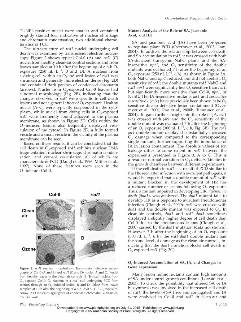
The ultrastructure of rcd1 nuclei undergoing celldeath was examined by transmission electron micros-copy. Figure 2 shows typical Col-0 (A) and rcd1 (C)nuclei from healthy clean-air control sections and fromleaves sampled at 10 h after the beginning of a 6-h O3exposure (250 nL L21; B and D). A nucleus froma dying cell within an O3-induced lesion of rcd1 wasshrunken and generally more electron dense (Fig. 2D)and contained dark patches of condensed chromatin(arrows). Nuclei from O3-exposed Col-0 leaves hada normal morphology (Fig. 2B), indicating that thechanges observed in rcd1 were specific to cell deathlesions and not a general effect of O3 exposure. Healthynuclei (A–C) were typically suspended in the cyto-plasm, while nuclei from dying cells in O3-exposedrcd1 were frequently found adjacent to the plasmamembrane, as shown in Figure 2D. Cells within theO3-induced lesions also frequently displayed vesi-culation of the cytosol. In Figure 2D, a fully formedvesicle and a small vesicle in the vicinity of the plasmamembrane can be seen.
Based on these results, it can be concluded that thecell death in O3-exposed rcd1 exhibits nuclear DNAfragmentation, nuclear shrinkage, chromatin conden-sation, and cytosol vesiculation, all of which arecharacteristic of PCD (Dangl et al., 1996; Mittler et al.,1997). None of these features were seen in theO3-tolerant Col-0.
Mutant Analysis of the Role of SA, Jasmonic
Acid, and HR
SA and jasmonic acid (JA) have been proposedto regulate plant PCD (Overmyer et al., 2003; Lam,2004). To address the relationship between cell deathand SA accumulation in rcd1, it was crossed with bothSA-deficient transgenic NahG plants and the SA-insensitive npr1, and O3 sensitivity of the doublemutants was evaluated 7 h after the beginning of anO3 exposure (250 nL L21, 6 h). As shown in Figure 3A,both NahG and npr1 reduced, but did not abolish, O3sensitivity of rcd1; the double mutants rcd1 NahG andrcd1 npr1 were significantly less O3 sensitive than rcd1,but significantly more sensitive than Col-0, npr1, orNahG. The JA-insensitive mutants jar1 and coronativeinsensitive 1 (coi1) have previously been shown to be O3sensitive due to defective lesion containment (Over-myer et al., 2000; Rao et al., 2000b; Tuominen et al.,2004). To gain further insight into the role of JA, rcd1was crossed with jar1 and the O3 sensitivity of thedouble mutant was evaluated 7 h after the beginningof an O3 exposure (300 nL L21, 6 h; Fig. 3B). The rcd1jar1 double mutant displayed substantially increasedO3 damage when compared to the correspondingsingle mutants, further supporting the importance ofJA in lesion containment. The absolute values of ionleakage differ to some extent in rcd1 between theexperiments presented in Figure 3, A to C. This isa result of normal variation in O3 delivery kinetics inthe growth chambers between different experiments.
If the cell death in rcd1 is a result of PCD similar tothe HR seen after infection with avirulent pathogens, itwould be expected that a double mutant of rcd1 witha mutant blocked in the development of HR hasa reduced number of lesions following O3 exposure.Thus, a mutant impaired in developing HR, defense, nodeath (dnd1), was analyzed. The dnd1 mutant fails todevelop HR as a response to avirulent Pseudomonasinfection (Clough et al., 2000). rcd1 was crossed withdnd1 and the double mutant was exposed to O3. Inclean-air controls, dnd1 and rcd1 dnd1 sometimesdisplayed a slightly higher degree of cell death thanCol-0 due to the spontaneous lesions (Clough et al.,2000) caused by the dnd1 mutation (data not shown).However, 7 h after the beginning of an O3 exposure(300 nL L21, 6 h), the rcd1 dnd1 double mutant hadthe same level of damage as the clean-air controls, in-dicating that the dnd1 mutation blocks cell death inO3-exposed rcd1 (Fig. 3C).
O3-Induced Accumulation of SA, JA, and Changes inGene Expression
Many lesion mimic mutants contain high amountsof SA under control growth conditions (Lorrain et al.,2003). To check the possibility that altered SA or JAbiosynthesis was involved in the increased cell deathof rcd1, the levels of SA (free and conjugated) and JAwere analyzed in Col-0 and rcd1 in clean-air and
Figure 2. rcd1 nuclear morphology. Transmission electron micro-graphs of Col-0 (A and B) and rcd1 (C and D) nuclei. A and C, Nucleifrom healthy leaves in the clean-air controls. B, Typical nucleus fromO3-exposed Col-0. D, Nucleus in a rcd1 cell undergoing PCD fromsection through an O3-induced lesion. B and D, Taken from leavessampled at 10 h after the beginning of a 6-h, 250 nL L21 O3 exposure.Arrow in D indicates aggregates of condensed chromatin. v, Vesicles;cw, cell wall.
Ozone-Induced Programmed Cell Death
Plant Physiology Preview 3 of 13 www.plant.org on July 21, 2015 - Published by www.plantphysiol.orgDownloaded from
Copyright © 2005 American Society of Plant Biologists. All rights reserved.

O3-exposed samples. As previously reported (Raoet al., 2000b), O3 exposure increased the levels of SAand JA in wild-type Col-0 (the latter is masked by thehuge increase of JA in rcd1). In contrast to several otherlesion mimic mutants, rcd1 had normal levels of SAand JA under control conditions (Fig. 4). However,in the O3-exposed rcd1 plants, the levels of both freeand conjugated SA and JA were higher than in Col-0(Fig. 4).
SA and JA are known regulators of defense geneexpression. Thus, defense gene expression was inves-tigated in Col-0 and rcd1 8 h after the beginning of an
Figure 3. O3 sensitivity of double mutants. Plants of the indicatedgenotypes were exposed to a single 6-h pulse of 250 nL L21 of O3 (A) or300 nL L21 (B and C), and cell death was monitored as ion leakage at 7h after the beginning of the exposure. NahG plants express a bacterialsalicylate hydroxylase gene and thus are unable to accumulate SA.Mutant npr1 is SA insensitive and jar1-1 is JA insensitive. The dnd1mutant does not develop HR as a response to avirulent Pseudomonasinfection. Experiments have been replicated at least twice with similarresults; one representative experiment is shown. All data points aremean 6 SD (n 5 5–10). Bars labeled with a different letter differsignificantly (P , 0.01) by Tukey’s honestly significant difference post-hoc test.
Figure 4. SA and JA levels in O3-exposed rcd1 and Col-0. O3 inducedaccumulation of SA and JA. Free SA (A), conjugated SA (B), and JA (C)were measured in whole rosettes of Col-0 and rcd1 in response toa single 6-h pulse O3 exposure of 300 nL L21. The results representmeans 6 SE (n5 5). The analysis was repeated twice with similar resultsfor the different genotypes.
Overmyer et al.
4 of 13 Plant Physiology Preview www.plant.org on July 21, 2015 - Published by www.plantphysiol.orgDownloaded from
Copyright © 2005 American Society of Plant Biologists. All rights reserved.
O3 exposure (250 nL L21, 6 h) using a customizedmacroarray (Table I). In accordance with the increasedlevels of SA (Fig. 4A) and ethylene (Overmyer et al.,2000; Tuominen et al., 2004) during O3 exposure, eth-ylene and SA-regulated genes, such as wall-associatedkinase1 (WAK1), PR-1, and GST (SA markers) and1-aminocyclopropane-1 carboxylic acid (ACC) oxi-dase, hevein-like protein, and basic chitinase (ethylenemarkers), had substantially higher mRNA levels in theO3-exposed plants. Transcript levels for PDF1.2a, acombined ethylene/JA marker, also increased. Formost genes, the differences in expression betweenrcd1 and Col-0 were rather limited, with a few ex-ceptions. ACC oxidase, hevein-like protein, and basicchitinase gene expression were increased 2 to 3 timesin rcd1 compared to Col-0. This likely reflected thehigher ethylene emission (Overmyer et al., 2000) fromrcd1 during O3 exposure.
The Role of Proteases in ROS-Induced Cell Death of rcd1
Proteases have both degenerative and signaling rolesduring PCD. In mammals, caspases (Cys aspartic pro-teases) are central to the regulation of PCD. Plants donot possess classic mammalian caspases; instead, theyuse vacuolar processing enzymes (VPEs), proteaseswith caspase activity, as regulators of PCD (Hatsugaiet al., 2004; Rojo et al., 2004). To study the role ofvarious types of proteases, in vitro experiments wereperformed. Col-0 and rcd1 leaves were incubated withknown protease inhibitors, summarized in Table II,with and without the exogenous superoxide generat-ing system, xanthine and xanthine oxidase (XXO; Jabset al., 1996), which has previously been shown to have
a similar effect on rcd1 as O3 (Overmyer et al., 2000). Asseen in Figure 5, both z-VAD-fmk (general caspaseinhibitor 1; Garcia-Calvo et al., 1998) and phenylme-thylsulfonyl fluoride (Ser-protease inhibitor) reducedthe level of XXO-induced ion leakage in rcd1 to ap-proximately the levels of the Col-0 plants. In contrast,pepstatin, an aspartic protease inhibitor, and E-64, aCys-protease inhibitor, had no effect. In control experi-ments with XXO-treated Col-0, the same inhibitorshad no effect (data not shown). Therefore, it can beconcluded that Ser- and caspase-like protease activ-ities were required for execution of the superoxide-induced cell death in rcd1.
Cell Death Induced by O3 and ROS Requires
Active Metabolism
Inhibitors of active metabolism (Table II) were ap-plied in planta by spraying intact rcd1 and Col-0prior to a 250-nL L21 O3 exposure. Cell death was moni-tored as ion leakage over a short time course at 0, 3, and6 h. At these time points, ion leakage in plants thatreceived the inhibitor treatments alone (in clean air) didnot deviate from control values in Col-0 or rcd1 (datanot shown), indicating that the inhibitors were non-toxic. As shown in Figure 6A, after 3 h of O3, the calciumchannel blocker lanthanum, the transcriptional inhib-itor a-amanitin, the Tyr kinase inhibitor herbimycin A,and the Ser/Thr kinase inhibitor K252a caused a statis-tically significant reduction (P , 0.05) in ion leakagein rcd1 as compared to O3 alone. Furthermore, at 6 h,herbimycin A, K252a, lanthanum, and a-amanitin pre-treatments significantly diminished O3-induced ionleakage in rcd1. In Col-0, pretreatment with herbimycin
Table I. Expression of selected stress- and defense-related genes in wild-type Col-0 and rcd1
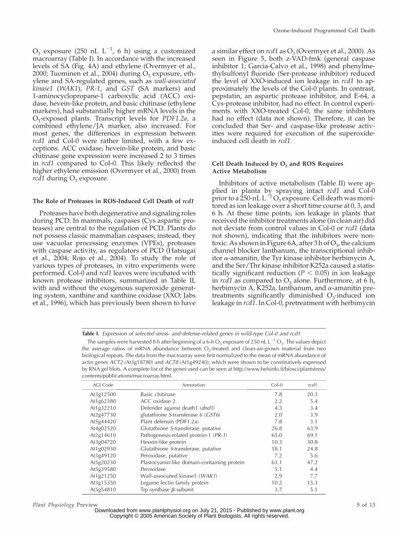
The samples were harvested 8 h after beginning of a 6-h O3 exposure of 250 nL L21 O3. The values depictthe average ratios of mRNA abundance between O3-treated and clean-air-grown material from twobiological repeats. The data from the macroarray were first normalized to the mean of mRNA abundance ofactin genes ACT2 (At3g18780) and ACT8 (At1g49240), which were shown to be constitutively expressedby RNA gel blots. A complete list of the genes used can be seen at http://www.helsinki.fi/biosci/plantstress/contents/publications/macroarray.html.
AGI Code Annotation Col-0 rcd1
At3g12500 Basic chitinase 7.8 20.3At1g62380 ACC oxidase 2 2.2 5.4At1g32210 Defender against death1 (dnd1) 4.3 3.4At2g47730 glutathione S-transferase 6 (GST6) 2.0 3.9At5g44420 Plant defensin (PDF1.2a) 7.8 3.1At4g02520 Glutathione S-transferase, putative 26.8 63.9At2g14610 Pathogenesis-related protein-1 (PR-1) 65.0 69.1At3g04720 Hevein-like protein 10.3 30.8At1g02930 Glutathione S-transferase, putative 18.1 24.8At3g49120 Peroxidase, putative 7.2 5.6At5g20230 Plastocyanin-like domain-containing protein 63.1 47.2At5g39580 Peroxidase 5.1 4.4At1g21250 Wall-associated kinase1 (WAK1) 2.9 7.7At3g15350 Legume lectin family protein 10.2 15.3At5g54810 Trp synthase b-subunit 3.7 5.1
Ozone-Induced Programmed Cell Death
Plant Physiology Preview 5 of 13 www.plant.org on July 21, 2015 - Published by www.plantphysiol.orgDownloaded from
Copyright © 2005 American Society of Plant Biologists. All rights reserved.

A, K252a, lanthanum,a-amanitin, or metavanadate didnot result in significant deviation from fumigation withO3 alone (Fig. 6B). In similar in vitro experiments usingXXO instead of O3 as the death-inducing stimulus,comparable results were obtained with both Col-0 andrcd1 (data not shown).
The fact that inhibition of protein kinases withK252a and herbimycin A reduced cell death in rcd1prompted us to assess the effect of the protein phos-phatase inhibitor calyculin A. Table III shows thattreatment with calyculin A triggered a 5-fold increasein cell death in rcd1. In Col-0, calyculin A causeda slight, but statistically nonsignificant, increase in ionleakage.
Calcium- and ROS-Induced Cell Death
We have shown above that O3- and superoxide-induced cell death was attenuated by the calciumchannel blocker lanthanum (Fig. 6A). To further elu-cidate the role of calcium, the effect of increasedcalcium flux was tested in XXO-challenged leaveswith calcium ionophore A23187 and increased extra-cellular calcium levels (2 mM CaCl2). These treatments,or the control treatment with Mg21, did not causestatistically significant changes in XXO-induced ionleakage in rcd1 (Fig. 7A) or Col-0 (Fig. 7B) leaves. Areduction in calcium fluxes either by the chelation ofextracellular calcium with EGTA or the use of calciumchannel blockers lanthanum and gadolinium, how-ever, caused a significant reduction in the XXO-induced ion leakage in rcd1 (Fig. 7A), suggesting thatcalcium influx from the apoplast is involved in theregulation of cell death in rcd1. In the ROS-tolerant
Col-0, differences in cell death after the restriction ofcalcium flux were not statistically significant (Fig. 7B).
O3 Induces Rapid Activation of Mitogen-ActivatedProtein Kinases
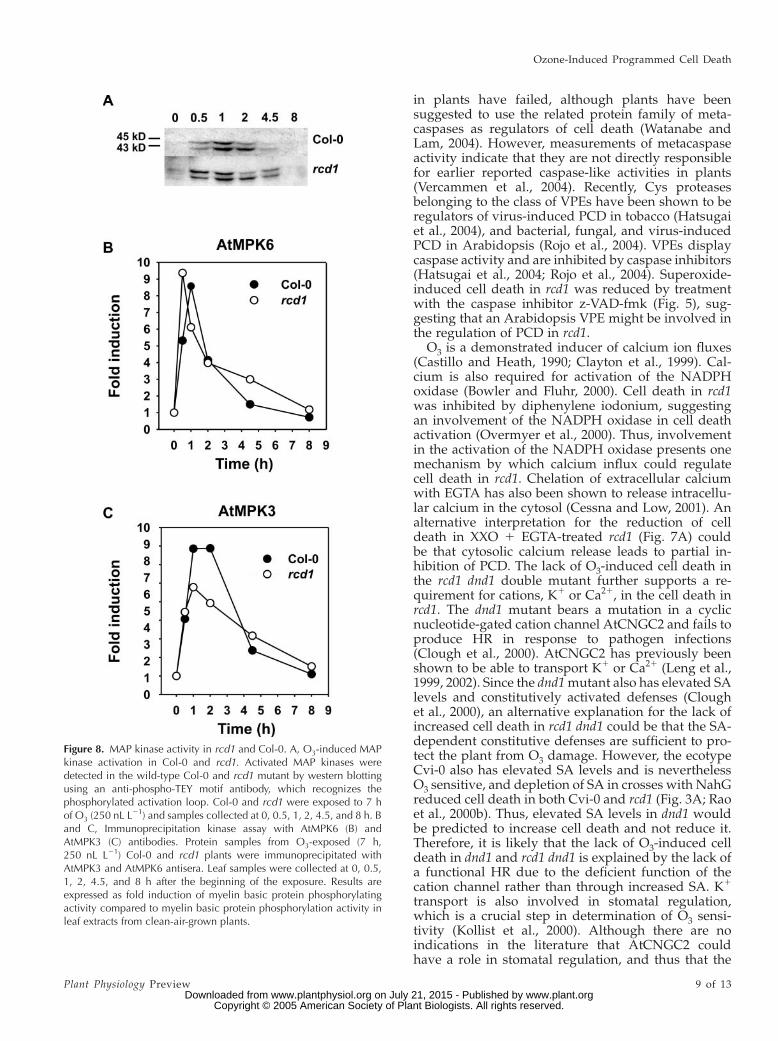
Application of the Ser/Thr kinase inhibitor K252adecreased O3-induced cell death in rcd1 (Fig. 6). SinceK252a acts as a competitive inhibitor of ATP forvarious kinases, including mitogen-activated protein(MAP) kinases, we assessed MAP kinase activation inrcd1 and Col-0 during O3 exposure. Protein extractsfrom O3-exposed rcd1 and Col-0 were analyzed withan antibody (anti-phospho-TEY) that detects the acti-vated forms of both mammalian and plant MAPkinases where the Thr and Tyr residues in the acti-vation loop are phosphorylated (Chang and Karin,2001; Asai et al., 2002). Two putative MAP kinasesof approximately 43 and 45 kD were activated inO3-treated tissue when compared to clean-air-grownplants (Fig. 8A). These two kinases, which have beenidentified with specific antibodies as AtMPK3 andAtMPK6 (Ahlfors et al., 2004b), were activated tran-siently during O3 exposure with slight differencesbetween Col-0 and rcd1; in Col-0, 45-kD kinase showedthe highest phosphorylation level 1 h after the start ofthe exposure, whereas in rcd1, stronger phosphoryla-tion of 45-kD kinase was already evident 0.5 h after thestart of the exposure. Immunoprecipitation kinaseassays with the specific antibodies against AtMPK3and AtMPK6 revealed that, indeed, rcd1 had an earlierpeak activity of AtMPK6 (at 0.5 h) and a slightly lowerpeak activity of AtMPK3 (at 1–2 h) when comparedto Col-0. When the AtMPK3 and AtMPK6 protein
Table II. Inhibitors used in this studya
Symbolb Inhibitor/Reagentc Target/Effectd Infiltrate XXO Spray O3
mMe
mMf
Van Metavanadate ATPases 50,000 100,000Lan LaCl3 Ca21 channels 1,000 2,000Ama a-Amanitin Transcription 2.2 4.4Hba Herbimycin A Tyr kinases 1,000 2,000K25 K252a Ser/Thr kinases 2.2 4.4Pep Pepstatin Asp proteases 1.0E-64 E-64 Cys proteases 1.0VAD z-VAD-fmk Caspases 50PMSF PMSF Ser proteases 500Ion A23187 Ca21 ionophore 10Ca CaCl2 Increased Ca21 2,000Mg MgCl2 Control 2,000EGTA EGTA Decreased Ca21 1,000Gd Gd(III)Cl3 Ca21 channels 1,000
aInhibitors used in this study with their respective abbreviations, concentrations used, and proposedtargets. bAbbreviations used in the figures for each inhibitor. cFull name of all inhibitors andreagents used. dProposed inhibitor target or the expected effect of the treatments. eConcentrationsused for in vitro coinfiltration experiments with XXO as the radical source. fConcentrations used forpretreating plants by spraying intact plants with the inhibitor 1 h prior to O3 exposure.
Overmyer et al.
6 of 13 Plant Physiology Preview www.plant.org on July 21, 2015 - Published by www.plantphysiol.orgDownloaded from
Copyright © 2005 American Society of Plant Biologists. All rights reserved.

accumulation was analyzed, no differences betweenrcd1 and Col-0 were found (data not shown).
DISCUSSION
Morphological Markers of PCD Are Induced by O3
The atmospheric pollutant O3 induces cell death insensitive individuals of several species (Koch et al.,2000; Wohlgemuth et al., 2002; Pasqualini et al., 2003).Typically, Arabidopsis mutants, in which a componentrequired for either antioxidative capacity or a processthat controls cell death is deficient, are sensitive to O3(Rao et al., 2000a; Overmyer et al., 2003; Langebartelsand Kangasjarvi, 2004). One such Arabidopsis mutant,rcd1, was isolated based on the extensive cell death seenin middle-aged rosette leaves as a result of O3 exposure(Overmyer et al., 2000). RCD1 encodes a protein thatmost likely is involved in interactions between hor-monal signaling cascades in abiotic stress (Ahlfors et al.,2004a). In this study, we used different experimentalapproaches to dissect processes involved in O3-induced PCD using the rcd1 mutant. O3-inducedlesions in rcd1 were large, whereas the wild-typeCol-0 had microscopic cell death. In both accessions,O3 caused an accumulation of autofluorescent phenoliccompounds in and around the dying cells (Fig. 1). Thisresponse is also triggered by wounding or a resistance
gene (R-gene) product upon recognition of an avirulentpathogen (Dietrich et al., 1994). In addition, the celldeath in O3-exposed rcd1 exhibits nuclear DNA frag-mentation, nuclear shrinkage, chromatin condensation,and cytosol vesiculation (Figs. 1 and 2), all of which aremorphological and functional characteristics of PCD.
PCD is an orderly and highly regulated disassemblyof cellular functions. It requires active cellular pro-cesses, such as energy production, signal transduction,ion fluxes, transcription, and translation. Active celldeath has been demonstrated with the use of pharma-cological inhibitors in response to pathogens (He et al.,
Figure 6. Effect of pharmacological inhibitors on O3-induced celldeath. A, rcd1 and B, Col-0 plants were pretreated 1 h prior to exposureby spraying intact plants with inhibitor solutions followed by anexposure to 250 nL L21 O3, and cell death was monitored as ionleakage from leaves collected at 3 and 6 h after the beginning of the 6-hexposure. Inhibitors used, their abbreviations, and targets were asfollows: Hba, herbimycin A, Tyr-kinases; K25, K252a, Ser/Thr-kinases;Lan, lanthanum chloride, calcium channels; Ama, a-amanitin, tran-scription; Van, sodium metavanadate, ATPases. Inhibitor informationwith concentrations used is summarized in Table II. Experiments havebeen replicated twice with similar results; one representative experi-ment is shown. All data points are mean 6 SD (n5 5). Bars marked withan asterisk (*) or double asterisks (**) were significantly different fromthe control at the P, 0.05 or P, 0.01 level, respectively, according toTukey’s honestly significant difference post-hoc test.
Figure 5. Effect of protease inhibitors on superoxide-induced celldeath. Col-0 and rcd1 plants were treated in vitro with a panel ofprotease inhibitors in the presence and absence of a XXO superoxide-generating system. Cell death was monitored as ion leakage. Inhibitorsused, their abbreviations, and targets were as follows: Pep, pepstatin,aspartic proteases; E-64, Cys proteases; VAD, z-VAD-fmk (caspaseinhibitor 1), caspases; PMSF, phenylmethylsulfonyl fluoride, Ser pro-teases. Inhibitor information with concentrations used is summarized inTable II. Inhibitor treatment of Col-0 plants resulted in no deviationfrom control and XXO-treated values (data not shown). Experimentshave been replicated twice with similar results; one representativeexperiment is shown. All data points are mean 6 SD (n 5 5). Barslabeled with a different letter differ significantly (P , 0.01), accordingto Tukey’s honestly significant difference post-hoc test.
Ozone-Induced Programmed Cell Death
Plant Physiology Preview 7 of 13 www.plant.org on July 21, 2015 - Published by www.plantphysiol.orgDownloaded from
Copyright © 2005 American Society of Plant Biologists. All rights reserved.

1993), H2O2 (Levine et al., 1996), high light in antisensecatalase tobacco (Dat et al., 2003), and O3 (this study).The inhibitors used, however, are not generally spe-cific for only one process and the unambiguousdemonstration that a process is used (e.g. in PCD)requires additional ways of verification, such as mu-tant analysis.
The Role of Hormones
SA accumulation is a requirement for the execu-tion of HR-like cell death and for the development ofsystemic acquired resistance (Durner et al., 1997).O3-exposed rcd1 had increased SA concentrationwhen compared to Col-0 (Fig. 4A), and SA was re-quired for O3 lesion formation in rcd1, since compro-mised SA signaling in rcd1 npr1 and rcd1 NahG doublemutants diminished symptom development signifi-cantly, but not completely (Fig. 3A). This suggeststhat O3-induced cell death in rcd1 comprises bothSA-dependent and SA-independent components. SA-dependent cell death may be taken as further evidenceof O3-induced HR-like PCD. Cell death in severallesion mimic mutants is reduced in double mutantswith compromised SA signaling, similarly suggestinga role for SA in lesion development (Lorrain et al.,2003).
JA has a proposed role in lesion containment duringO3 exposure (Overmyer et al., 2003; Tuominen et al.,2004). Treatment of O3-sensitive accessions (Arabidop-sis mutant rcd1 and the ecotype Cape Verdi Islands(Cvi-0); tobacco Bel-W3) with methyl jasmonate re-duced or abolished O3-induced cell death, and JA-insensitive or biosynthesis mutants (jar1, coi1, andfad3/7/8) displayed lesions following O3 exposure(Orvar et al., 1997; Overmyer et al., 2000; Rao et al.,2000b; Tuominen et al., 2004). JA levels increaseddramatically in O3-exposed rcd1 (Fig. 4B). It has beenproposed that the increase in JA accumulation in O3-exposed plants is a result of the cell death processitself, which causes a release of a substrate for JAbiosynthesis from the membranes of the damaged cells(Vahala et al., 2003; Tuominen et al., 2004). This wouldform an autocatalytic containment mechanism for thelesion propagation where the magnitude of cell deathwould also determine the strength of signal for lesioncontainment by JA. By this mechanism, decreased JAsensitivity will also increase sensitivity to O3, whichwas apparent in the rcd1 jar1 double mutant that
displayed higher lesion formation than either parent.A similar result was observed when the lesion mimicmutant hypersensitive response-like lesions1 (hrl1) wascrossed with coi1; the resulting double mutant wasunable to contain lesions and had exaggerated celldeath (Devadas et al., 2002).
Inhibitor Studies
The inhibitor studies indicated a role for caspasesand calcium in the induction of cell death by ROS inrcd1 (Figs. 5–7). Caspases are central to the regulationof PCD in mammals. Attempts to find similar proteins
Figure 7. Effect of altered calcium flux on superoxide-induced celldeath. A, rcd1 and B, Col-0 plants were treated in vitro with reagents toalter calcium flux in the presence and absence of a XXO superoxide-generating system. Cell death was monitored as ion leakage. Reagentsused, their abbreviations, and targets were as follows: Ion, A23187,calcium ionophore; Ca, calcium chloride, increased extracellularcalcium; Mg, magnesium chloride, divalent cation control; EGTA,chelator of extracellular calcium; Gd, gadolinium(III) chloride, calciumchannel blocker; La, lanthanum chloride, calcium channel blocker.Inhibitor and reagent information and concentrations used are sum-marized in Table II. Experiments have been replicated twice withsimilar results; one representative experiment is shown. All data pointsare mean 6 SD (n 5 5). Bars marked with an asterisk (*) or doubleasterisks (**) were significantly different from the water control at theP , 0.05 or P , 0.01 level, respectively, according to Tukey’s honestlysignificant difference post-hoc test.
Table III. Induction of cell death by calyculin A
Controla Calyculin Aa
Col-0 7.95 6 1.46b 14.19 6 3.80b
rcd1 5.90 6 1.13b 30.74 6 10.49c
aValues given are percent ion leakage 6 SD (n 5 5) induced by100 mM calyculin A measured at 18 h posttreatment. b,cValuesfollowed by the same letter do not differ significantly for each other(P , 0.05), according to Tukey’s honestly significant post-hoc test.
Overmyer et al.
8 of 13 Plant Physiology Preview www.plant.org on July 21, 2015 - Published by www.plantphysiol.orgDownloaded from
Copyright © 2005 American Society of Plant Biologists. All rights reserved.
in plants have failed, although plants have beensuggested to use the related protein family of meta-caspases as regulators of cell death (Watanabe andLam, 2004). However, measurements of metacaspaseactivity indicate that they are not directly responsiblefor earlier reported caspase-like activities in plants(Vercammen et al., 2004). Recently, Cys proteasesbelonging to the class of VPEs have been shown to beregulators of virus-induced PCD in tobacco (Hatsugaiet al., 2004), and bacterial, fungal, and virus-inducedPCD in Arabidopsis (Rojo et al., 2004). VPEs displaycaspase activity and are inhibited by caspase inhibitors(Hatsugai et al., 2004; Rojo et al., 2004). Superoxide-induced cell death in rcd1 was reduced by treatmentwith the caspase inhibitor z-VAD-fmk (Fig. 5), sug-gesting that an Arabidopsis VPE might be involved inthe regulation of PCD in rcd1.
O3 is a demonstrated inducer of calcium ion fluxes(Castillo and Heath, 1990; Clayton et al., 1999). Cal-cium is also required for activation of the NADPHoxidase (Bowler and Fluhr, 2000). Cell death in rcd1was inhibited by diphenylene iodonium, suggestingan involvement of the NADPH oxidase in cell deathactivation (Overmyer et al., 2000). Thus, involvementin the activation of the NADPH oxidase presents onemechanism by which calcium influx could regulatecell death in rcd1. Chelation of extracellular calciumwith EGTA has also been shown to release intracellu-lar calcium in the cytosol (Cessna and Low, 2001). Analternative interpretation for the reduction of celldeath in XXO 1 EGTA-treated rcd1 (Fig. 7A) couldbe that cytosolic calcium release leads to partial in-hibition of PCD. The lack of O3-induced cell death inthe rcd1 dnd1 double mutant further supports a re-quirement for cations, K1 or Ca21, in the cell death inrcd1. The dnd1 mutant bears a mutation in a cyclicnucleotide-gated cation channel AtCNGC2 and fails toproduce HR in response to pathogen infections(Clough et al., 2000). AtCNGC2 has previously beenshown to be able to transport K1 or Ca21 (Leng et al.,1999, 2002). Since the dnd1 mutant also has elevated SAlevels and constitutively activated defenses (Cloughet al., 2000), an alternative explanation for the lack ofincreased cell death in rcd1 dnd1 could be that the SA-dependent constitutive defenses are sufficient to pro-tect the plant from O3 damage. However, the ecotypeCvi-0 also has elevated SA levels and is neverthelessO3 sensitive, and depletion of SA in crosses with NahGreduced cell death in both Cvi-0 and rcd1 (Fig. 3A; Raoet al., 2000b). Thus, elevated SA levels in dnd1 wouldbe predicted to increase cell death and not reduce it.Therefore, it is likely that the lack of O3-induced celldeath in dnd1 and rcd1 dnd1 is explained by the lack ofa functional HR due to the deficient function of thecation channel rather than through increased SA. K1
transport is also involved in stomatal regulation,which is a crucial step in determination of O3 sensi-tivity (Kollist et al., 2000). Although there are noindications in the literature that AtCNGC2 couldhave a role in stomatal regulation, and thus that the
Figure 8. MAP kinase activity in rcd1 and Col-0. A, O3-induced MAPkinase activation in Col-0 and rcd1. Activated MAP kinases weredetected in the wild-type Col-0 and rcd1 mutant by western blottingusing an anti-phospho-TEY motif antibody, which recognizes thephosphorylated activation loop. Col-0 and rcd1 were exposed to 7 hof O3 (250 nL L21) and samples collected at 0, 0.5, 1, 2, 4.5, and 8 h. Band C, Immunoprecipitation kinase assay with AtMPK6 (B) andAtMPK3 (C) antibodies. Protein samples from O3-exposed (7 h,250 nL L21) Col-0 and rcd1 plants were immunoprecipitated withAtMPK3 and AtMPK6 antisera. Leaf samples were collected at 0, 0.5,1, 2, 4.5, and 8 h after the beginning of the exposure. Results areexpressed as fold induction of myelin basic protein phosphorylatingactivity compared to myelin basic protein phosphorylation activity inleaf extracts from clean-air-grown plants.
Ozone-Induced Programmed Cell Death
Plant Physiology Preview 9 of 13 www.plant.org on July 21, 2015 - Published by www.plantphysiol.orgDownloaded from
Copyright © 2005 American Society of Plant Biologists. All rights reserved.
dnd1 mutation could affect stomatal conductance, thisremains a possibility and requires further study.
O3-Induced MAP Kinase Activation
Plant MAP kinase cascades are activated by a largenumber of stimuli, including pathogen infection and O3(Zhang and Klessig, 2001; Ahlfors et al., 2004b). Con-stitutive MAP kinase activation has been shown to leadto HR-like cell death, suggesting that MAP kinasesignaling is a part of the ROS-induced PCD (Ren et al.,2002). Inhibition of Ser/Thr kinases (including MAPkinases) with K252a suppressed cell death and phos-phatase inhibitors increased cell death in rcd1 (Fig. 6;Table III), indicating that kinase activation is neededfor the early phases of cell death in rcd1. However,when the timing and magnitude of cell death in rcd1and Col-0 (Overmyer et al., 2000) are compared with theAtMPK6 and AtMPK3 activation (Fig. 8), it is likely thatcell death and kinase activation are not directly linked;Col-0 had a high induction of AtMPK3 and AtMPK6activity but little cell death when compared to rcd1.Furthermore, the O3-sensitive jar1 has similar MAPkinase activity compared to Col-0 (Ahlfors et al., 2004b).However, it is possible that ROS production andAtMPK6 activation could be linked. The more openstomata of rcd1 (Ahlfors et al., 2004a) allow more O3 toenter the plant leaf and to react with the components ofthe cell wall and plasma membranes, creating moreROS directly from O3 degradation. This higher oxida-tive load could also cause the earlier AtMPK6 peakactivation in O3-exposed rcd1. The protein phosphataseinhibitor calyculin A, which increased cell death in rcd1(Table III), has previously been shown to increase eth-ylene evolution and ACC synthase activity in tomatosignificantly without an inductive treatment (Spanuet al., 1994; Tuomainen et al., 1997). In O3-exposedplants, ethylene is required for the active ROS pro-duction responsible for lesion propagation (Overmyeret al., 2000; Moeder et al., 2002). In tobacco, the in-duction of ethylene biosynthesis takes place throughSIPK, the tobacco homolog of Arabidopsis AtMPK6(Kim et al., 2003), and in Arabidopsis, AtMPK6 directlyactivates ethylene synthesis by phosphorylating theACC synthases AtACS6 and AtACS2 (Liu and Zhang,2004). Thus, the fast and high induction of ethylenebiosynthesis involved in the formation of O3 lesions inrcd1 (Overmyer et al., 2000) is most likely affected by theearlier peak activity of AtMPK6, sinceAtACS6 was alsospecifically activated by O3 in rcd1 (Overmyer et al.,2000). Whether the AtMPK3/6 activation is a result ofthe increased cell death, or vice versa, requires furtherstudy.
Could Multiple Modes of Cell Death Occur in rcd1?
The inhibitor treatments reduced, but did not fullyblock, cell death in ROS-treated rcd1. This could be dueto the efficacy of the inhibitor treatments, since theaction of a given inhibitor is unlikely to completely
block its target pathway(s). Another interpretation isthat both PCD and necrotic cell death may take place.It has been suggested that both death by rampantoxidation and PCD may occur, depending on themagnitude of O3-induced oxidative stress (Pell et al.,1997). Furthermore, Rao and Davis (1999) have pre-sented evidence of both O3-induced necrotic and HR-like cell death, where the mechanism was dependenton genotype. Both rcd1, and to a smaller extent Col-0,displayed TUNEL-positive nuclei (Fig. 1), but since theTUNEL assay does not discriminate between randomand programmed DNA fragmentation (Collins et al.,1992; Dangl et al., 1996; Pasqualini et al., 2003), it ispossible that mosaics of apoptotic and necrotic cellscan occur in the same O3-exposed tissue. Mixtures ofcells bearing signs of different modes of death withinthe same tissue have been described in the study of celldeath in mammals (Levin et al., 1999) and have re-cently been proposed to take place also in plants(Greenberg and Yao, 2004). It could be that signalsemanating from the few cells undergoing necrotic celldeath by rampant oxidation by O3-derived ROS maytrigger surrounding cells to die by PCD, resulting inlarge areas of affected tissue and lesion propagation.This is analogous to the penumbra of secondary celldeath seen at the periphery of acute hypoxic or tra-umatic lesions, which has been described extensivelyin mammals (Jacobson et al., 1997).
A mixture of necrotic and PCD provides a model forsome of the results seen in this study and for themechanism by which O3 lesion initiation could takeplace in the oxidative cell death cycle (Overmyer et al.,2003). The higher initial stomatal conductance and thedelay in the O3-induced stomatal closure in rcd1(Ahlfors et al., 2004a) allow more O3 to enter the leafduring the initial phase of the exposure. This couldcause more or larger lesion initiations by the directaction of the O3-derived ROS. However, the presence ofcharacteristic biochemical and morphological markersof PCD in O3-exposed rcd1 suggest that the lesion pro-pagation takes placed by hormone-controlled PCD,where the increased activation of MAP kinases is in-volved in the regulation of hormone biosynthesis.Thus, only lesion propagation would be dependent onactive metabolism, ion fluxes, and SA signaling (as in-dicated by the inhibitor and double-mutant experi-ments), while the primary cell death observed would benecrotic, caused by the ROS from O3 degradation. Thishypothesis requires further study, especially the role ofstomatal function during the early phases of O3 chal-lenge.
MATERIALS AND METHODS
Plants and Growth Conditions
Arabidopsis (Arabidopsis thaliana) was grown on a 1:1 mixture of peat (Type
B2; Kekkila, Tuusula, Finland) and vermiculite at 22�C/18�C (day/night) with
70%/90% relative humidity under short-day (12 h, 250 mmol m22 s21)
irradiance with 5 plants/8 3 8-cm pot. The isolation and genetics of the
Overmyer et al.
10 of 13 Plant Physiology Preview www.plant.org on July 21, 2015 - Published by www.plantphysiol.orgDownloaded from
Copyright © 2005 American Society of Plant Biologists. All rights reserved.

rcd1-1 allele used throughout this study has been described previously
(Overmyer et al., 2000; Ahlfors et al., 2004a). Twenty-one- to 27-d-old plants
(prior to bolting) were used for all experiments. Plants for O3 exposure and
clean-air controls were grown side by side under identical conditions and
were selected at random for separation into the two experimental groups.
Mutant seeds were obtained from the Arabidopsis Biological Resource Center
(ABRC; http://www.arabidopsis.org/abrc/) and the Nottingham Arabidop-
sis Stock Centre (NASC; http://nasc.nott.ac.uk). Seeds of the transgenic NahG
(line B15) in the Col-0 background were a kind gift from Syngenta (Research
Triangle Park, NC). rcd1 NahG, rcd1 npr1, rcd1 jar1, and rcd1 dnd1 double-
mutant lines were created by crossing the respective plants with a glabrous
rcd1 line as the pollen acceptor. F1 generation plants were confirmed as true
crosses by the presence of trichomes and allowed to self-pollinate. Appropri-
ate homozygous lines were identified from the F2 populations by PCR
screening with dCAPS markers for rcd1-1, jar1-1, dnd1, and npr1-1, and by
kanamycin resistance screening on plates for the presence of the NahG
transgene. All genotypes were confirmed in the F3 generation.
Superoxide and O3 Treatments
Extracellular superoxide (XXO in sodium phosphate buffer, 10 mM, pH 7.0)
was applied by vacuum infiltration into the apoplast of detached leaves as
described (Jabs et al., 1996; Overmyer et al., 2000). Three completely expanded
middle-aged leaves from each plant were used. Treatments lasted 18 to 20 h at
22�C in closed 50-mL tubes. The following reagents were included: xanthine
(100 mM), xanthine oxidase (0.05 units mL21), plus inhibitors or other reagents
at the concentrations given in Table I. Standard O3 exposure, 250 nL L21 or
300 nL L21 O3 for 6 h, unless otherwise noted, with parallel clean-air controls
at all time points was as described (Overmyer et al., 2000). At the times
indicated, cell death was measured by ion leakage of 2 detached leaves into
5 mL, or whole rosettes into 1 mL, milli-Q water for 1 h, followed by
quantification with a conductivity meter (Mettler Toledo, Greifensee, Switzer-
land). Data are expressed as a percentage of total ions (determined after killing
plants by boiling) and are the means of 5 to 10 replicates.
Histological Procedures
For detection of autofluorescent phenolic deposits, plants were cleared by
boiling 3 min in alcoholic lactophenol (2:1, 95% ethanol:lactophenol), rinsed in
50% ethanol, and then rinsed twice in water. Cleared leaves were mounted and
viewed as by Dietrich et al. (1994). Control samples were microscopically free of
autofluorescence in all experiments. For the TUNEL assay, samples were
vacuum infiltrated and fixed overnight in 4% paraformaldehyde in phosphate-
buffered saline and cryoprotected for 24 h each at 4�C in 15% and then 30% Suc
in phosphate-buffered saline. Leaf samples were then submerged in Tissue-Tek
OTC Compound 4583 (Miles, Elkhart, IN) and snap frozen in liquid nitrogen
prior to cryosectioning (Leitz Kryostat 1720; Ernst Leitz Wetzlar, Wetzlar,
Germany). Sections were mounted on Superfrost Plus slides (Menzel-Glaser,
Erie Scientific Company, Portsmouth, NH) and stained with the In Situ Cell
Death Detection kit (Roche Applied Science, Espoo, Finland), according to the
manufacturer’s instructions, and counterstained with DAPI. Sections were
examined by epifluorescent microscopy (Olympus Provis AX70; Olympus
Optical, Tokyo) with standard DAPI and fluorescent filter sets.
Electron Microscopy
O3-exposed and control leaf samples for electron microscopy were fixed in
2.5% glutaraldehyde in 0.1 M sodium phosphate buffer, pH 7.0, by vacuum
infiltration and then overnight at 4�C. Samples were washed and stored at 4�Cuntil further analysis. Samples were then postfixed in 1% osmium tetroxide
(EMS, Washington Park, PA), dehydrated through an ethanol series, and
embedded in Epon XL 112 (Ladd Research Industries, Williston, VT) and
polymerized. Blocks were sectioned on a Reichert ultracut microtome using
a diamond knife (Diatome, Bienne, Switzerland) and mounted on copper grid
slots. Sections were examined with a transmission electron microscope (JEOL
JEM-1200EX; JEOL, Tokyo) at an accelerating voltage of 60 kV.
Hormone Measurements
JA and SA were extracted and quantified with [1,2-13C]JA and [13C1]SA as
internal standards as described by Baldwin et al. (1997), with the modifica-
tions described by Vahala et al. (2003).
Gene Expression Profiling
Expression of 127 defense-related genes was studied by a cDNA macro-
array analysis using cDNA clones and expressed sequence tag clones from the
ABRC (Columbus, Ohio). All clones were resequenced. Seventy-five nano-
grams of each PCR-amplified sample were blotted onto Hybond N1 mem-
branes with a 96-well dot-blot device; 75 ng of oligo(dT)21 and pSPORT and
pBS plasmids provided the negative controls. The 2 constitutively expressed
genes, ACT2 (At3g18780) and ACT8 (At1g49240), were each applied to the
membrane 4 times. Hybridization and detection were according to Overmyer
et al. (2000), except that 33P-dCTP was used for probe labeling. RNA for the
analysis was extracted from plants 8 h after the beginning of a 6-h, 250-nL L21
O3 exposure. The results were normalized by reference to the mean hybrid-
ization signals for ACT2 and ACT8. Genes with expression levels below
a numerical value of 0.001 in any of the samples were excluded from this
analysis. Hybridizations were performed at least twice, and the results
represent the mean of the duplicate signals.
Inhibitor Treatments
Inhibitors were used at the concentrations stated in Table I. In XXO
experiments, inhibitors were coinfiltrated with the radical-generating system.
In O3 experiments, plants were pretreated 1 h prior to exposure by spraying
intact plants with inhibitor solutions. All inhibitor solutions for spraying were
dissolved in water with 0.05% Tween 20 to mediate surface wetting. The
solvent for stock solutions for K252a, herbimycin A, pepstatin, z-VAD-fmk,
and A23187 was DMSO; for E-64 and PMSF, it was ethanol; and the remaining
inhibitors were in aqueous solutions. Where appropriate, controls were con-
ducted by adding solvent and Tween 20 to the spray solution or adding sol-
vent to incubation media at the concentrations resulting from dilution of
stocks into working solutions. All reagents were from Sigma Aldrich Chem-
icals (St. Louis), except K252a, E-64, and z-VAD-fmk, which were from Cal-
biochem (San Diego).
One-way ANOVA tests were performed with two-sided Dunnet’s or
Tukey’s honestly significant difference post-hoc tests as indicated using
SPSS 8.0.
MAP Kinase Activity Measurements
Protein extractions, TEY westerns, and immunoprecipitation kinase assays
were performed as described previously (Kroj et al., 2003; Ahlfors et al.,
2004b).
ACKNOWLEDGMENTS
We are grateful to Prof. Ian Baldwin and Dr. Gunter Brader for supplying
JA and SA standards, respectively, and to Mika Korva for plant care. We
gratefully acknowledge the Department of Ecology and Environmental
Sciences at the University of Kuopio, where this work was initiated. We
would like to thank the Electron Microscopy Unit of the Institute of Bio-
technology at the University of Helsinki for providing laboratory facilities.
Received October 27, 2004; returned for revision December 20, 2004; accepted
December 27, 2004.
LITERATURE CITED
Ahlfors R, Lang S, Overmyer K, Jaspers P, Brosche M, Tauriainen A,
Kollist H, Tuominen H, Belles-Boix E, Piippo M, et al (2004a)
Arabidopsis RADICAL-INDUCED CELL DEATH1 belongs to the
WWE protein-protein interaction domain protein family and modulates
abscisic acid, ethylene, and methyl jasmonate responses. Plant Cell 16:
1925–1937
Ahlfors R, Macioszek V, Rudd J, Brosche M, Schlichting R, Scheel D,
Kangasjarvi J (2004b) Stress hormone-independent activation and
nuclear translocation of mitogen-activated protein kinases in Arabidop-
sis thaliana during ozone exposure. Plant J 40: 512–522
Asai T, Tena G, Plotnikova J, Willmann MR, Chiu WL, Gomez-Gomez L,
Ozone-Induced Programmed Cell Death
Plant Physiology Preview 11 of 13 www.plant.org on July 21, 2015 - Published by www.plantphysiol.orgDownloaded from
Copyright © 2005 American Society of Plant Biologists. All rights reserved.

Boller T, Ausubel FM, Sheen J (2002) MAP kinase signalling cascade in
Arabidopsis innate immunity. Nature 415: 977–983
Baldwin IT, Zhang Z-P, Diab N, Ohnmeiss TE, McCloud ES, Lynds GY,
Schmelz EA (1997) Quantification, correlations and manipulations of
wound-induced changes in jasmonic acid and nicotine in Nicotiana
sylvestris. Planta 201: 397–404
Bowler C, Fluhr R (2000) The role of calcium and activated oxygen as
signals for controlling cross-tolerance. Trends Plant Sci 5: 241–246
Castillo FJ, Heath RL (1990) Ca21 transport in membrane vesicles from
pinto bean leaves and its alteration after ozone exposure. Plant Physiol
94: 788–795
Cessna SG, Low PS (2001) An apoplastic Ca21 sensor regulates internal
Ca21 release in Aequorin-transformed tobacco cells. J Biol Chem 276:
10655–10662
Chang L, Karin M (2001) Mammalian MAP kinase signalling cascades.
Nature 410: 37–40
Clayton H, Knight MR, Knight H, McAinsh MR, Hetherington AM (1999)
Dissection of the ozone-induced calcium signature. Plant J 17: 575–579
Clough SJ, Fengler KA, Yu IC, Lippok B, Smith RK Jr, Bent AF (2000) The
Arabidopsis dnd1 ‘‘defense, no death’’ gene encodes a mutated cyclic
nucleotide-gated ion channel. Proc Natl Acad Sci USA 97: 9323–9328
Collins RJ, Harmon BV, Gobe GC, Kerr JFR (1992) Internucleosomal DNA
clevage should not be the sole criterion for identifying apoptosis. Int J
Radiat Biol 61: 451–453
Dangl JL, Dietrich RA, Richberg MH (1996) Death don’t have no mercy:
cell death programs in plant-microbe interactions. Plant Cell 8:
1793–1807
Dat JF, Pellinen R, Beeckman T, Van De Cotte B, Langebartels C,
Kangasjarvi J, Inze D, Van Breusegem F (2003) Changes in hydrogen
peroxide homeostasis trigger an active cell death process in tobacco.
Plant J 33: 621–632
Devadas SK, Enyedi A, Raina R (2002) The Arabidopsis hrl1 mutation
reveals novel overlapping roles for salicylic acid, jasmonic acid and
ethylene signalling in cell death and defence against pathogens. Plant J
30: 467–480
Dietrich RA, Delaney TP, Uknes SJ, Ward ER, Ryals JA, Dangl JL (1994)
Arabidopsis mutants simulating disease resistance response. Cell 77:
565–577
Durner J, Shah J, Klessig DF (1997) Salicylic acid and disease resistance in
plants. Trends Plant Sci 2: 266–274
Fujibe T, Saji H, Arakawa K, Yabe N, Takeuchi Y, Yamamoto KT (2004) A
methyl viologen-resistant mutant of Arabidopsis, which is allelic to
ozone-sensitive rcd1, is tolerant to supplemental ultraviolet-B irradia-
tion. Plant Physiol 134: 275–285
Garcia-Calvo M, Peterson EP, Leiting B, Rue R, Nicholson DW,
Thornberry NA (1998) Inhibition of human caspases by peptide-based
and macromolecular inhibitors. J Biol Chem 273: 32608–32613
Greenberg JT, Yao N (2004) The role and regulation of programmed cell
death in plant-pathogen interactions. Cell Microbiol 6: 201–211
Hatsugai N, Kuroyanagi M, Yamada K, Meshi T, Tsuda S, Kondo M,
Nishimura M, Hara-Nishimura I (2004) A plant vacuolar protease,
VPE, mediates virus-induced hypersensitive cell death. Science 305:
855–858
He SYH, Huang H-C, Collmer A (1993) Pseudomonas syringae pv syringe
harpin Pss: a protein that is secreted via the Hrp pathway and elicits the
hypersensitive response in plants. Cell 73: 1255–1266
Heath RL, Taylor GE Jr (1997) Physiological processes and plant responses
to ozone exposure. In H Sandermann Jr, AR Wellburn, RL Heath, eds,
Forest Decline and Ozone, Ecological Studies, Vol 127. Springer, Heidel-
berg, pp 317–368
Jabs T, Dietrich RA, Dangl JL (1996) Initiation of runaway cell death in an
Arabidopsis mutant by extracellular superoxide. Science 273: 1853–1856
Jacobson MD, Weil M, Raff MC (1997) Programmed cell death in animal
development. Cell 88: 347–354
Kangasjarvi J, Talvinen J, Utriainen M, Karjalainen R (1994) Plant defense
systems induced by ozone. Plant Cell Environ 17: 783–794
Kim CY, Liu Y, Thorne ET, Yang H, Fukushige H, Gassmann W,
Hildebrand D, Sharp RE, Zhang S (2003) Activation of a stress-
responsive mitogen-activated protein kinase cascade induces the bio-
synthesis of ethylene in plants. Plant Cell 15: 2707–2718
Koch JR, Creelman RA, Eshita SM, Seskar M, Mullet JE, Davis KR (2000)
Ozone sensitivity in hybrid poplar correlates with insensitivity to both
salicylic acid and jasmonic acid. The role of programmed cell death in
lesion formation. Plant Physiol 123: 487–496
Kollist H, Moldau H, Mortensen L, Rasmussen SK, Jorgensen LB (2000)
Ozone flux to plasmalemma in barley and wheat is controlled rather by
stomata than by direct reaction of ozone with apoplastic ascorbate.
J Plant Physiol 156: 645–651
Kroj T, Rudd JJ, Nurnberger T, Gabler Y, Lee J, Scheel D (2003) Mitogen-
activated protein kinases play an essential role in oxidative burst-
independent expression of pathogenesis-related genes in parsley. J Biol
Chem 278: 2256–2264
Lam E (2004) Controlled cell death, plant survival and development. Nat
Rev Mol Cell Biol 5: 305–315
Langebartels C, Kangasjarvi J (2004) Ethylene and jasmonate as regulators
of cell death in disease resistance. In H Sandermann Jr, ed, Molecular
Ecotoxicology of Plants, Ecological Studies, Vol 170. Springer, Heidel-
berg, pp 75–110
Leng Q, Mercier RW, Hua B-G, Fromm H, Berkowitz GA (2002) Electro-
physiological analysis of cloned cyclic nucleotide-gated ion channels.
Plant Physiol 128: 400–410
Leng Q, Mercier RW, Yao W, Berkowitz GA (1999) Cloning and first
functional characterization of a plant cyclic nucleotide-gated cation
channel. Plant Physiol 121: 753–761
Levin S, Bucci TJ, Cohen SM, Fix AS, Hardisty JF, LeGrand EK, Maronpot
RP, Trump BF (1999) The nomenclature of cell death: recommendations
of an ad hoc committee of the society of toxicologic pathologists. Toxicol
Pathol 27: 484–490
Levine A, Pennell RI, Alvarez ME, Palmer R, Lamb C (1996) Calcium-
mediated apoptosis in a plant hypersensitive disease resistance re-
sponse. Curr Biol 6: 427–437
Liu Y, Zhang S (2004) Phosphorylation of 1-aminocyclopropane-1-carbox-
ylic acid synthase by MPK6, a stress-responsive mitogen-activated
protein kinase, induces ethylene biosynthesis in Arabidopsis. Plant
Cell 16: 3386–3399
Lorrain S, Vailleau F, Balague C, Roby D (2003) Lesion mimic mutants:
keys for deciphering cell death and defense pathways in plants? Trends
Plant Sci 8: 263–271
Mittler R, Simon L, Lam E (1997) Pathogen-induced programmed cell
death in tobacco. J Cell Sci 110: 1333–1344
Moeder W, Barry CS, Tauriainen A, Betz C, Tuomainen J, Utriainen M,
Grierson D, Sandermann H Jr, Langebartels C, Kangasjarvi J (2002)
Ethylene synthesis regulated by bi-phasic induction of ACC synthase
and ACC oxidase genes is required for H2O2 accumulation and cell
death in ozone-exposed tomato. Plant Physiol 130: 1918–1926
Orvar BL, McPherson J, Ellis BE (1997) Pre-activating wounding response
in tobacco prior to high-level ozone exposure prevents necrotic injury.
Plant J 11: 203–212
Overmyer K, Brosche M, Kangasjarvi J (2003) Reactive oxygen species and
hormonal control of cell death. Trends Plant Sci 8: 335–342
Overmyer K, Tuominen H, Kettunen R, Betz C, Langebartels C,
Sandermann H Jr, Kangasjarvi J (2000) The ozone-sensitive Arabidopsis
rcd1 mutant reveals opposite roles for ethylene and jasmonate signaling
pathways in regulating superoxide-dependent cell death. Plant Cell 12:
1849–1862
Pasqualini S, Piccioni C, Reale L, Ederli L, Della Torre G, Ferranti F (2003)
Ozone-induced cell death in tobacco cultivar Bel W3 plants. The role
of programmed cell death in lesion formation. Plant Physiol 133:
1122–1134
Pell EJ, Schlagnhaufer CD, Arteca RN (1997) Ozone-induced oxidative
stress: mechanisms of action and reaction. Physiol Plant 100: 264–273
Rao MV, Davis KR (1999) Ozone-induced cell death occurs via two distinct
mechanisms in Arabidopsis: the role of salicylic acid. Plant J 17: 603–614
Rao MV, Davis KR (2001) The physiology of ozone-induced cell death.
Planta 213: 682–690
Rao MV, Koch JR, Davis KR (2000a) Ozone: a tool for probing program-
med cell death in plants. Plant Mol Biol 44: 345–358
Rao MV, Lee HI, Creelman RA, Mullet JA, Davis KR (2000b) Jasmonic
acid signalling modulates ozone-induced hypersensitive cell death.
Plant Cell 12: 1633–1646
Ren D, Yang H, Zhang S (2002) Cell death mediated by MAPK is associated
with hydrogen peroxide production in Arabidopsis. J Biol Chem 277:
559–565
Rojo E, Martiın R, Carter C, Zouhar J, Pan S, Plotnikova J, Jin H, Paneque
M, Saınchez-Serrano JJ, Baker B, et al (2004) VPEg exhibits a caspase-
Overmyer et al.
12 of 13 Plant Physiology Preview www.plant.org on July 21, 2015 - Published by www.plantphysiol.orgDownloaded from
Copyright © 2005 American Society of Plant Biologists. All rights reserved.

like activity that contributes to defense against pathogens. Curr Biol 14:
1897–1906
Sandermann H Jr, Ernst D, Heller W, Langebartels C (1998) Ozone: an
abiotic elicitor of plant defence reactions. Trends Plant Sci 3: 47–50
Spanu P, Grosskopf DG, Felix G, Boller T (1994) The apparent turnover
of 1-aminocyclopropane-1-carboxylate synthase in tomato cells is
regulated by protein phosphorylation and dephosphorylation. Plant
Physiol 106: 529–535
Tuomainen J, Betz C, Kangasjarvi J, Ernst D, Yin ZH, Langebartels C,
Sandermann H Jr (1997) Ozone induction of ethylene emission in
tomato plants: regulation by differential transcript accumulation for the
biosynthetic enzymes. Plant J 12: 1151–1162
Tuominen H, Overmyer K, Keinanen M, Kollist H, Kangasjarvi J (2004)
Mutual antagonism of ethylene and jasmonic acid regulates ozone-
induced spreading cell death in Arabidopsis. Plant J 39: 59–69
Vahala J, Ruonala R, Keinanen M, Tuominen H, Kangasjarvi J (2003)
Ethylene insensitivity modulates ozone-induced cell death in birch.
Plant Physiol 132: 185–195
Vercammen D, Van De Cotte B, De Jaeger G, Eeckhout D, Casteels P,
Vandepoele K, Vandenberghe I, Van Beeumen J, Inze D, Van
Breusegem F (2004) Type-II metacaspases Atmc4 and Atmc9 of Arabi-
dopsis thaliana cleave substrates after arginine and lysine. J Biol Chem
279: 45329–45336
Vranova E, Inze D, Van Breusegem F (2002) Signal transduction during
oxidative stress. J Exp Bot 53: 1227–1236
Watanabe N, Lam E (2004) Recent advance in the study of caspase-like
proteases and Bax inhibitor-1 in plants: their possible roles as regulator
of programmed cell death. Mol Plant Pathol 5: 65–70
Wohlgemuth H, Mittelstrass K, Kschieschan S, Bender J, Weigel H-J,
Overmyer K, Kangasjarvi J, Langebartels C, Sandermann H Jr (2002)
Activation of an oxidative burst is a general feature of sensitive plants
exposed to the air pollutant ozone. Plant Cell Environ 25: 717–726
Zhang S, Klessig DF (2001) MAPK cascades in plant defense signaling.
Trends Plant Sci 6: 520–527
Ozone-Induced Programmed Cell Death
Plant Physiology Preview 13 of 13 www.plant.org on July 21, 2015 - Published by www.plantphysiol.orgDownloaded from
Copyright © 2005 American Society of Plant Biologists. All rights reserved.
Related Documents