Oxidative stress-dependent p66Shc phosphorylation in skin fibroblasts of children with mitochondrial disorders Magdalena Lebiedzinska a,1 , Agnieszka Karkucinska-Wieckowska b,1 , Carlotta Giorgi e , Elzbieta Karczmarewicz d , Ewa Pronicka c , Paolo Pinton e , Jerzy Duszynski a , Maciej Pronicki b , Mariusz R. Wieckowski a, ⁎ a Nencki Institute of Experimental Biology, Warsaw, Poland b Department of Pathology, Childrens'Memorial Health Institute, Warsaw, Poland c Department of Metabolic Diseases, Endocrinology and Diabetology, Childrens'Memorial Health Institute, Warsaw, Poland d Department of Biochemistry and Experimental Medicine, Childrens'Memorial Health Institute, Warsaw, Poland e Department of Experimental and Diagnostic Medicine, Section of General Pathology, Interdisciplinary Center for the Study of Inflammation (ICSI) and Emilia Romagna, Laboratory BioPharmaNet, University of Ferrara, Ferrara, Italy abstract article info Article history: Received 17 November 2009 Received in revised form 2 March 2010 Accepted 4 March 2010 Available online 10 March 2010 Keywords: p66Shc Ser36-P-p66Shc Mitochondrial disorder ROS Antioxidant enzyme p66Shc, the growth factor adaptor protein, can have a substantial impact on mitochondrial metabolism through regulation of cellular response to oxidative stress. We investigated relationships between the extent of p66Shc phosphorylation at Ser36, mitochondrial dysfunctions and an antioxidant defense reactions in fibroblasts derived from five patients with various mitochondrial disorders (two with mitochondrial DNA mutations and three with methylglutaconic aciduria and genetic defects localized, most probably, in nuclear genes). We found that in all these fibroblasts, the extent of p66Shc phosphorylation at Ser36 was significantly increased. This correlated with a substantially decreased level of mitochondrial superoxide dismutase (SOD2) in these cells. This suggest that SOD2 is under control of the Ser36 phosphorylation status of p66Shc protein. As a consequence, an intracellular oxidative stress and accumulation of damages caused by oxygen free radicals are observed in the cells. © 2010 Elsevier B.V. All rights reserved. 1. Introduction The 66-kDa isoform of ShcA, the growth factor adapter protein (p66Shc), and its relevance to cell signaling have attracted major interest in aging research. In 1999, Pellici at al. [1] discovered that ROS-mediated signals, generated by p66Shc, favored apoptosis at the cellular and aging at the organismal levels, leading to tissue damage. As a result, p66Shc was proposed to regulate mammalian life span. These authors found that the life span of transgenic mice lacking p66Shc was longer by 30–40% compared to wild-type animals. Interestingly, prolongation of the life span corresponding to the ablation of p66Shc gene had no pathological consequences [1]. Taking into account the free radical theory of aging [2], it can be assumed that this protein can be involved in the cellular response to oxidative stress and clearly explains significantly enhanced resistance to oxidative stress of transgenic animals lacking p66Shc [1,3]. Three proteins of the ShcA family, p66Shc, p52Shc and p46Shc, have been identified up to now [4]. Two mRNAs derive from ShcA gene: p66shc and p46shc/ p52shc, the latter one being translated into 2 proteins, p46shc and p52shc, after alternative translation start sites [4,5]. Each protein of the ShcA subfamily (p66Shc, p52Shc and p46Shc) contains three functionally identical domains: C terminal Src-homology domain (SH2), central collagen-homology domain (CH1) and N-terminal phosphotyrosine-binding domain (PTB). p66Shc contains additionally N- terminal proline-rich collagen-homology domain (CH2) with a serine phosphorylation site (Ser36) [3,4]. Numerous studies have demonstrated that p66Shc is phosphorylated mainly at this residue in response to UV exposure or H 2 O 2 treatment [1,6]. Such phosphorylation is mediated by one of serine–threonine kinases, protein kinase Cβ (PKCβ), and this event induces a signaling cascade leading to apoptosis [7]. p66Shc phosphor- ylated at Ser36 (Ser36-P-p66Shc) is a substrate of prolyl isomerase Pin1 and, after isomerisation, it is dephosphorylated by phosphatase A2 (PPA2) [7]. This sequence of events triggers translocation of p66Shc from the cytosol to mitochondria [7]. Recently, we have demonstrated that p66Shc can also be found in plasma membrane-associated membrane (PAM) and mitochondria-associated membrane (MAM) fractions [8–10]. Intracellular distribution of p66Shc seems to be age dependent. MAM fraction isolated from livers of old mice was significantly enriched in p66Shc. Moreover, Ser36-P-p66Shc in the crude mitochondrial fraction (containing the MAM fraction) was more abundant in older mice and its Biochimica et Biophysica Acta 1797 (2010) 952–960 ⁎ Corresponding author. Laboratory of Bioenergetics and Biomembranes, Department of Biochemistry, Nencki Institute of Experimental Biology, Pasteur 3 St., 02-093 Warsaw, Poland. Tel.: +48 22 589 23 72; fax: +48 22 822 53 42. E-mail address: [email protected] (M.R. Wieckowski). 1 Equal contribution to this paper. 0005-2728/$ – see front matter © 2010 Elsevier B.V. All rights reserved. doi:10.1016/j.bbabio.2010.03.005 Contents lists available at ScienceDirect Biochimica et Biophysica Acta journal homepage: www.elsevier.com/locate/bbabio

Welcome message from author
This document is posted to help you gain knowledge. Please leave a comment to let me know what you think about it! Share it to your friends and learn new things together.
Transcript

Biochimica et Biophysica Acta 1797 (2010) 952–960
Contents lists available at ScienceDirect
Biochimica et Biophysica Acta
j ourna l homepage: www.e lsev ie r.com/ locate /bbab io
Oxidative stress-dependent p66Shc phosphorylation in skin fibroblasts of childrenwith mitochondrial disorders
Magdalena Lebiedzinska a,1, Agnieszka Karkucinska-Wieckowska b,1, Carlotta Giorgi e,Elzbieta Karczmarewicz d, Ewa Pronicka c, Paolo Pinton e, Jerzy Duszynski a,Maciej Pronicki b, Mariusz R. Wieckowski a,⁎a Nencki Institute of Experimental Biology, Warsaw, Polandb Department of Pathology, Childrens'Memorial Health Institute, Warsaw, Polandc Department of Metabolic Diseases, Endocrinology and Diabetology, Childrens'Memorial Health Institute, Warsaw, Polandd Department of Biochemistry and Experimental Medicine, Childrens'Memorial Health Institute, Warsaw, Polande Department of Experimental and Diagnostic Medicine, Section of General Pathology, Interdisciplinary Center for the Study of Inflammation (ICSI) and Emilia Romagna,Laboratory BioPharmaNet, University of Ferrara, Ferrara, Italy
⁎ Corresponding author. Laboratory of Bioenergetics andBiochemistry,Nencki Institute of Experimental Biology, PasteTel.: +48 22 589 23 72; fax: +48 22 822 53 42.
E-mail address: [email protected] (M.R.1 Equal contribution to this paper.
0005-2728/$ – see front matter © 2010 Elsevier B.V. Adoi:10.1016/j.bbabio.2010.03.005
a b s t r a c t
a r t i c l e i n f oArticle history:Received 17 November 2009Received in revised form 2 March 2010Accepted 4 March 2010Available online 10 March 2010
Keywords:p66ShcSer36-P-p66ShcMitochondrial disorderROSAntioxidant enzyme
p66Shc, the growth factor adaptor protein, can have a substantial impact on mitochondrial metabolismthrough regulation of cellular response to oxidative stress. We investigated relationships between the extentof p66Shc phosphorylation at Ser36, mitochondrial dysfunctions and an antioxidant defense reactions infibroblasts derived from five patients with various mitochondrial disorders (two with mitochondrial DNAmutations and three with methylglutaconic aciduria and genetic defects localized, most probably, in nucleargenes). We found that in all these fibroblasts, the extent of p66Shc phosphorylation at Ser36 wassignificantly increased. This correlated with a substantially decreased level of mitochondrial superoxidedismutase (SOD2) in these cells. This suggest that SOD2 is under control of the Ser36 phosphorylation statusof p66Shc protein. As a consequence, an intracellular oxidative stress and accumulation of damages causedby oxygen free radicals are observed in the cells.
Biomembranes, Department ofur 3 St., 02-093Warsaw, Poland.
Wieckowski).
ll rights reserved.
© 2010 Elsevier B.V. All rights reserved.
1. Introduction
The 66-kDa isoform of ShcA, the growth factor adapter protein(p66Shc), and its relevance to cell signaling have attracted majorinterest in aging research. In 1999, Pellici at al. [1] discovered thatROS-mediated signals, generated by p66Shc, favored apoptosis at thecellular and aging at the organismal levels, leading to tissue damage.As a result, p66Shc was proposed to regulate mammalian life span.These authors found that the life span of transgenic mice lackingp66Shc was longer by 30–40% compared to wild-type animals.Interestingly, prolongation of the life span corresponding to theablation of p66Shc gene had no pathological consequences [1]. Takinginto account the free radical theory of aging [2], it can be assumed thatthis protein can be involved in the cellular response to oxidative stressand clearly explains significantly enhanced resistance to oxidativestress of transgenic animals lacking p66Shc [1,3]. Three proteins of theShcA family, p66Shc, p52Shc and p46Shc, have been identified up to
now [4]. Two mRNAs derive from ShcA gene: p66shc and p46shc/p52shc, the latter one being translated into 2 proteins, p46shc andp52shc, after alternative translation start sites [4,5].
Each protein of the ShcA subfamily (p66Shc, p52Shc and p46Shc)contains three functionally identical domains: C terminal Src-homologydomain (SH2), central collagen-homologydomain (CH1) andN-terminalphosphotyrosine-bindingdomain (PTB). p66Shc contains additionallyN-terminal proline-rich collagen-homology domain (CH2) with a serinephosphorylation site (Ser36) [3,4].Numerous studies havedemonstratedthat p66Shc is phosphorylated mainly at this residue in response to UVexposure or H2O2 treatment [1,6]. Such phosphorylation is mediated byoneof serine–threonine kinases, proteinkinaseCβ (PKCβ), and this eventinduces a signaling cascade leading to apoptosis [7]. p66Shc phosphor-ylated at Ser36 (Ser36-P-p66Shc) is a substrate of prolyl isomerase Pin1and, after isomerisation, it is dephosphorylated by phosphatase A2(PPA2) [7]. This sequence of events triggers translocation of p66Shc fromthe cytosol to mitochondria [7]. Recently, we have demonstrated thatp66Shc can also be found in plasma membrane-associated membrane(PAM) andmitochondria-associatedmembrane (MAM) fractions [8–10].Intracellular distribution of p66Shc seems to be age dependent. MAMfraction isolated from livers of old mice was significantly enriched inp66Shc. Moreover, Ser36-P-p66Shc in the crude mitochondrial fraction(containing theMAM fraction) wasmore abundant in older mice and its
histoc
hemistry.
(2)Sp
ectrop
hotometry*an
dsis.
Diagn
osticco
nclusion
dominan
ce,c
ytoc
hrom
eox
idasepo
sitive
.Mitoc
hond
rial
enceph
alom
yopa
thywith
resp
iratorych
ainco
mplex
Idefi
cien
cy.
exes
Iand
II+
IIIin
mus
cleho
mog
enate.
in/m
gprotein(inreferenc
erang
e).
91TNCmutationin
ND3ge
ne.
mus
cles
fibe
rs.
Leighsynd
romewithmethy
lglutaco
nicaciduria.
exes
IIan
dII+
IIIin
mus
cleho
mog
enate.
min/m
gprotein(inreferenc
erang
e).L
owco
mplex
Vactivity.
Combine
dresp
iratorych
ainde
fect.
rco
mmon
mtD
NAmutations
andSU
RF1ge
nemutations
.al
activity
ofcy
toch
romeox
idaseactivity
ands
seactivity.S
light
type
Ifibe
rpred
ominan
ce.
Mitoc
hond
rial
cardiomyo
pathy.
Combine
dresp
iratorych
ainde
fect.
exes
Iand
IIIin
mus
cleho
mog
enate,
increa
sed
/min/m
gprotein)
.utationin
tRNALe
u1.
eox
idaseactivity.L
ackof
succinatede
hydrog
enaseactivity
ubation.
Wea
kreaction
appe
ared
afterelon
gation
ofincu
bation
oderateto
seve
relip
idaccu
mulationin
mus
clefibe
rs.
Barthdisease.
Seco
ndaryco
mbine
dresp
iratory
chainde
fect.
exes
IVan
dII+
IIIin
fibrob
lasts.SC
activity
otein(inreferenc
erang
e).L
owco
mplex
Vactivity.
crea
sein
mus
clefibe
rsan
dtype
Ifibe
rpred
ominan
ce,
ges.Cy
toch
romeox
idasepo
sitive
.Ba
rthdisease.
Seco
ndaryco
mbine
dresp
iratory
chainde
fect.
exes
II,III
andII+
IIIin
mus
cleho
mog
enate,
increa
sed
ol/m
in/m
gprotein)
.Low
complex
Vactivity.
gprotein.
953M. Lebiedzinska et al. / Biochimica et Biophysica Acta 1797 (2010) 952–960
higher level correlated well with the enhanced H2O2 production [8].p66Shc, once translocated to mitochondria and/or MAM fraction,perturbs their structure and function, e.g., by modifying mitochondrialCa2+ responses and the mitochondrial metabolism [1,11,12].
The signaling pathway described by Pinton et al. [7] is triggered byextracellular oxidative stress (e.g. H2O2 treatment) and involvesp66Shc phosphorylation at Ser36. During aging, gradually accumulat-ed mitochondrial and nuclear DNA damages and dysfunctions of theantioxidant defense system lead to an increase of intracellular reactiveoxygen species (ROS) level; this in turn activates PKCβ-dependentphosphorylation of p66Shc and initiates a vicious circle of ROS produc-tion [13]. A similar situation may occur in the case of mitochondrialdysfunction associated with inherited disorders of oxidative phos-phorylation (OXPHOS). The frequency of such mitochondrial diseasesis relatively high (1:5000). They are present in neonates, infants,children and adults; they are progressive and not treatable [14,15].The major pathogenic mechanism is considered to be a shortage incellular energy supply. The influence of the pathological oxidativestress on the course of mitochondrial disorders and on the antioxidantdefense balance has not been elucidated in detail. A higher rate ofROS production by mitochondria has already been described in manygenetic mitochondrial defects [16]. The superoxide anion radical(O2
●−) is the main reactive oxygen species produced by mitochondria.The defects of respiratory chain caused by genetically inheritedmutations in mitochondrial or nuclear DNA, leading to isolated orcombined defects in respiratory chain complexes, may result insignificantly elevated superoxide production. These extra ROS addi-tionally potentiate mitochondrial damage and, in consequence, causeaccelerated ROS production.
In the present work, we investigated whether mitochondrialdysfunctions of different genetic origin lead to an increasedphosphorylation of p66Shc at Ser36, modification of the intracellularantioxidant defense system and stress-induced protein carbonylation.
rprob
able
mitoc
hond
rial
disorders.
(1)Mus
clehistolog
yan
dBN
–PA
GE.
(3)DNAan
aly
ion
y trical
(1)Slight
type
Ifibe
rpre
(2)Lo
wactivity
ofco
mpl
SCactivity
97nm
ol/m
(3)Heterop
lasm
icm.101
ia, hang
es.
(1)Mild
lipid
increa
sein
(2)Lo
wactivity
ofco
mpl
SCactivity
183nm
ol/
(3)Neg
ativescreen
ingfo
emia.
(1)Strong
subs
arco
lemm
uccina
tede
hydrog
ena
gressive
(2)Lo
wactivity
ofco
mpl
CSactivity
(392
nmol
(3)m.330
3CNTmtD
NAm
lities,
ctic)
(1)Decreased
cytoch
rom
afterstan
dard
1hinc
time(1
.5an
d3h)
.M(2
)Lo
wactivity
ofco
mpl
130nm
ol/m
in/m
gpr
(1)Lo
wde
gree
oflip
idin
mild
nons
pecificch
ange
of7.
(2)Lo
wactivity
ofco
mpl
CSactivity
(412
.5nm
andforfibrob
lasts59
–11
5nm
ol/m
in/m
2. Materials and methods
2.1. Ethics
The study was carried out in accordance with the Declaration ofHelsinki of the World Medical Association and was approved by theCommittee of Bioethics at the Children's Memorial Health Institute.Informed consent was obtained from the parents before any biopsy ormolecular analysis was performed.
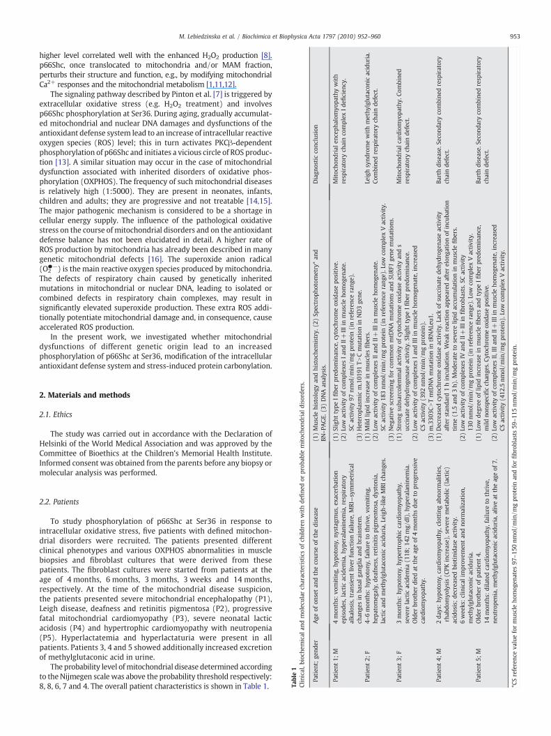
Table1
Clinical,b
ioch
emical
andmolecular
characteristicsof
child
renwithde
fine
do
Patien
t;ge
nder
Age
ofon
setan
dtheco
urse
ofthedisease
Patien
t1;
M4mon
ths:
vomiting,
hypo
tony
,nystagm
us,e
xacerbat
episod
es,lacticacidem
ia,h
yperalan
inem
ia,respirator
alka
losis,tran
sien
tliv
erfunc
tion
failu
re,M
RI—symme
chan
gesin
basalg
anglia
andbrains
tem.
Patien
t2;
F4–
6mon
ths:
hypo
tony
,failure
tothrive
,vom
iting,
hepa
tomeg
aly,
deafne
ss,retinitis
pigm
entosa,d
yston
lactic
andmethy
lglutaco
nicaciduria,L
eigh
-likeMRI
c
Patien
t3;
F3mon
ths:
hypo
tony
,hyp
ertrop
hiccardiomyo
pathy,
seve
relactic
acidem
ia(1
18;14
2mg/
dl),hy
peralanin
Older
brothe
rdied
attheag
eof
4mon
thsdu
eto
pro
cardiomyo
pathy.
Patien
t4;
M2da
ys:hy
potony
,cardiom
yopa
thy,
clotting
abno
rma
rhab
domyo
lysis(C
PKincrea
se),seve
remetab
olic
(la
acidosis;de
crea
sedbiotinidaseactivity.
6wee
ks:clinical
improv
emen
tan
dno
rmalization,
methy
lglutaco
nicaciduria.
Patien
t5;
MOlder
brothe
rof
patien
t4.
14mon
ths:
dilatedcardiomyo
pathy,
failu
reto
thrive
,ne
utrope
nia,
methy
lglutaco
nicaciduria,a
liveat
thea
*CSreferenc
eva
lueformus
cleho
mog
enates
97–15
0nm
ol/m
in/m
gprotein
2.2. Patients
To study phosphorylation of p66Shc at Ser36 in response tointracellular oxidative stress, five patients with defined mitochon-drial disorders were recruited. The patients presented differentclinical phenotypes and various OXPHOS abnormalities in musclebiopsies and fibroblast cultures that were derived from thesepatients. The fibroblast cultures were started from patients at theage of 4 months, 6 months, 3 months, 3 weeks and 14 months,respectively. At the time of the mitochondrial disease suspicion,the patients presented severe mitochondrial encephalopathy (P1),Leigh disease, deafness and retinitis pigmentosa (P2), progressivefatal mitochondrial cardiomyopathy (P3), severe neonatal lacticacidosis (P4) and hypertrophic cardiomyopathy with neutropenia(P5). Hyperlactatemia and hyperlactaturia were present in allpatients. Patients 3, 4 and 5 showed additionally increased excretionof methylglutaconic acid in urine.
The probability level ofmitochondrial disease determined accordingto the Nijmegen scale was above the probability threshold respectively:8, 8, 6, 7 and 4. The overall patient characteristics is shown in Table 1.

954 M. Lebiedzinska et al. / Biochimica et Biophysica Acta 1797 (2010) 952–960
2.3. Fibroblast cultures
Human skin fibroblasts were grown from explants of skin biopsiesof one healthy individual and five patients in Dulbecco modifiedEagle's medium with glucose (4.5 g/l), 5 mM sodium pyruvate and2mM L-glutamine (Sigma Aldrich), supplemented with 10% (v/v) fetalbovine serum (Gibco), and 1.2% antibiotic, antimycotic solution (SigmaAldrich) in an atmosphere of 5% (v/v) carbon dioxide in air at 37 °C.Additionally, NHDF neonatal dermal fibroblasts, Cat. n. CC-2509, andNHDF adult dermalfibroblasts, Cat. n. CC-2511, from Lonzawere used ascontrol healthyfibroblasts. The cellswere grown in75 cm2 cultureflasksand used within 6 days after reaching confluence.
2.4. Hispidin treatment
Dulbecco modified Eagle's medium was supplemented with 5 μMhispidin and the cells were cultured for 48 h. In the case of longtreatment (28 days), the growth medium supplemented with 3 μMhispidin was changed every two days. The passages (from fourth toeight) were also made in the presence of 3 μM hispidin.
2.5. Enzymatic measurements
Spectrophotometric assays in post-nuclear supernatants obtainedfrom skeletal muscle biopsies and fibroblast cell cultures were used forthe measurement of complex I (NADH:ubiquinone oxidoreductase,
Fig. 1. Bioenergetic parameters (mtΔΨ, respiratory chain activity) and ROS production(A)Mitochondrial membrane potential in cultured fibroblasts. Data shown aremeans±SD, *shown are means±SD, *pb0.05 (n=6) versus control. (C) Cytosolic superoxide (cO2
●−), (Dcultured fibroblasts. Data shown are means±SD, *pb0.05 (n=6) versus control for each p
rotenone-sensitive), complex II (succinate:ubiquinone oxidoreductase,malonate-sensitive), complex III (ubiquinone:cytochrome c oxidoreduc-tase, antimycin A-sensitive), complex IV (cytochrome c oxidase) andcitrate synthase as described previously [17]. Activities of the respiratorycomplexes were expressed as the ratio to the activity of citrate synthasein order to avoid the influence of different amounts of mitochondria inthe material under investigation. Protein content in post-nuclear super-natants was determined by the method of Lowry et al. [18].
2.6. Samples preparation for Western blot
Cell pellets were resuspended in cold lysis buffer (50 mM Tris pH7.5, 150 mM NaCl, 1% Triton, 0.1% SDS, 1% sodium deoxycholate)containing inhibitors of proteases (1 mM PMSF and the proteinprotease inhibitor cocktail) and phosphatases (1 mM Na3VO4, 10 mMNaF). Samples were incubated on ice for 15 min and centrifuged at14,000×g for 20 min at 4 °C to remove insoluble material. Proteinconcentration in lysates was determined using Bradford method.Samples for SDS–PAGE were denatured in reducing Laemmli loadingbuffer at 45 °C or 95 °C for 5 min, depending on the antibody.
2.7. Western blotting
Cell lysates (25 – 50 μg protein) were separated electrophoreticallyin 8% or 10% SDS–polyacrylamide gel (BioRad) and transferred ontoPVDF membrane (BioRad). Membranes were blocked using 2% non–fat
(cytosolic and mitochondrial O2●− and H2O2) in control and patients' fibroblasts.
pb0.05 (n=6) versus control. (B) Respiratory chain activity in cultured fibroblasts. Data) mitochondrial superoxide (mtO2
●−) and (E) hydrogen peroxide (H2O2) production inarameter.
955M. Lebiedzinska et al. / Biochimica et Biophysica Acta 1797 (2010) 952–960
milk in TBS buffer containing 0.01% Tween 20 (Sigma Aldrich) for 1 h.Proteins were detected with anti-Shc and anti-Ser36-P-p66Shc mono-clonal antibodies (1:1000, Abcam), anti-SOD1 rabbit polyclonal anti-body (1:5000, Santa Cruz), anti-SOD2 goat polyclonal antibody (1:500,Santa Cruz), anti-catalase monoclonal antibody (1:1000, Santa Cruz)and anti-actin antibody (1:10000, Abcam) followed by secondary HRP–conjugated antibodies (1:5000) (Santa Cruz).
2.8. Measurement of mitochondrial membrane potential (mtΔΨ)
Fibroblasts grown in 12-well plates were washed twice with PBSto remove the medium and then incubated in the presence of 10 µM5,5',6,6'-tetrachloro-1,1',3,3'-tetraethylbenzimidazolylcarbocyanineiodide (JC-1) in PBS containing 5 mM glucose for 10 min at 37 °C. Thecells were washed twice with PBS and the green and red fluorescencewas recorded in a microplate reader (Infinite M200, Tecan, Austria),respectively, at 485 nm excitation/520 nm emission and at 535 nmexcitation/635 nm emission wavelengths.
2.9. Measurement of respiratory chain activity
Fibroblasts grown in 12-well plates were washed twice with PBSand then incubated in PBS containing 5 mM glucose and 6 µMresosurine and the fluorescence was recorded immediately thereafterin a microplate reader at 510 nm excitation and 595 nm emission
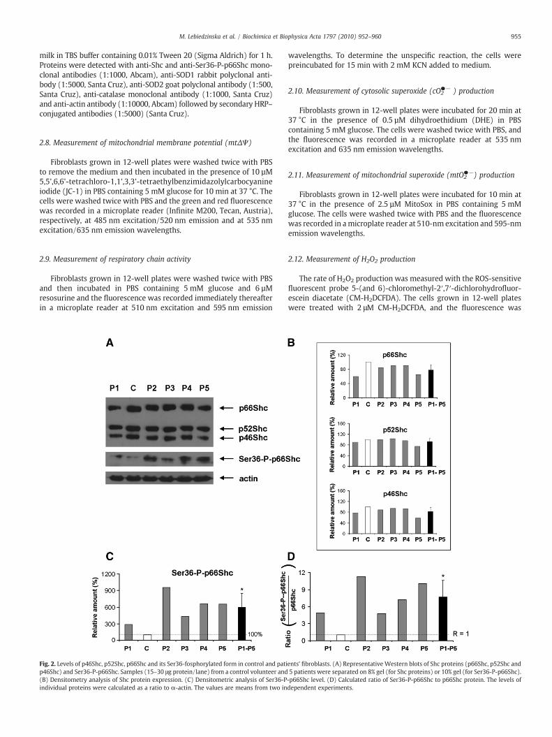
Fig. 2. Levels of p46Shc, p52Shc, p66Shc and its Ser36-fosphorylated form in control and patip46Shc) and Ser36-P-p66Shc. Samples (15–30 μg protein/lane) from a control volunteer and(B) Densitometry analysis of Shc protein expression. (C) Densitometric analysis of Ser36-Pindividual proteins were calculated as a ratio to α-actin. The values are means from two in
wavelengths. To determine the unspecific reaction, the cells werepreincubated for 15 min with 2 mM KCN added to medium.
2.10. Measurement of cytosolic superoxide (cO2●− ) production
Fibroblasts grown in 12-well plates were incubated for 20 min at37 °C in the presence of 0.5 µM dihydroethidium (DHE) in PBScontaining 5 mM glucose. The cells were washed twice with PBS, andthe fluorescence was recorded in a microplate reader at 535 nmexcitation and 635 nm emission wavelengths.
2.11. Measurement of mitochondrial superoxide (mtO2●−) production
Fibroblasts grown in 12-well plates were incubated for 10 min at37 °C in the presence of 2.5 µM MitoSox in PBS containing 5 mMglucose. The cells were washed twice with PBS and the fluorescencewas recorded in amicroplate reader at 510-nm excitation and 595-nmemission wavelengths.
2.12. Measurement of H2O2 production
The rate of H2O2 production was measured with the ROS-sensitivefluorescent probe 5-(and 6)-chloromethyl-2′,7′-dichlorohydrofluor-escein diacetate (CM-H2DCFDA). The cells grown in 12-well plateswere treated with 2 μM CM-H2DCFDA, and the fluorescence was
ents' fibroblasts. (A) Representative Western blots of Shc proteins (p66Shc, p52Shc and5 patients were separated on 8% gel (for Shc proteins) or 10% gel (for Ser36-P-p66Shc).-p66Shc level. (D) Calculated ratio of Ser36-P-p66Shc to p66Shc protein. The levels ofdependent experiments.

956 M. Lebiedzinska et al. / Biochimica et Biophysica Acta 1797 (2010) 952–960
recorded in a microplate reader for 30 minutes at 495 nm excitationand 520 nm emission wavelengths.
2.13. Detection of protein modification by oxygen free radicals
The level of oxidized proteins was estimated with the use of theOxyBlot Protein Oxidation Detection Kit (Chemicon). Aliquots of 25 μgprotein were separated on 10% SDS–polyacrylamide gel and thestandard protocol of the manufacturer was followed.
2.14. Statistical analysis
Differences in band densities were analyzed using NIH ImageJsoftware. Data obtained from the Tecan microplate reader were cal-culated using Microsoft™ Excel 2005 and analyzed for significance byStudent's t-test.
3. Results
To study phosphorylation of p66Shc at Ser36 in the situation ofintracellular oxidative stress, we used fibroblast culture from fivepatients with genetically definedmitochondrial defects. The diagnosisof mitochondrial disease depended on the clinical phenotype,biochemical and enzymatic studies of muscle biopsies and DNAanalysis. There were one severely affected infant with isolatedcomplex I deficiency due to the mitochondrial DNA (mtDNA)mutation in ND3 subunit (P1), one infant with combined OXPHOSdefect due to a tRNALeu1 rare mutation (P3) and three children withmulti-organ mitochondrial disorders and methylglutaconic aciduria,among them one girl (P2) and two brothers with Barth syndrome (P4and P5). The patients' characteristic is presented in Table 1. Theexpression of relevant OXPHOS abnormalities in some patients'
Fig. 3. Expression of SOD1, SOD2 and catalase in control and patients' fibroblasts with mitoc(B) Expression profile of mitochondrial superoxide dismutase (SOD2). (C) Expressionpolyacrylamide gel. Bar charts show the levels of antioxidant enzymes calculated as a ratio
fibroblasts was previously detected by spectrophotometric measure-ment of respiratory chain complex I-IV activities and confirmed byBN–PAGE and SDS (not shown).
3.1. Metabolic characterization of patient fibroblasts
To determine whether a mitochondrial dysfunction was present inthe studied fibroblast cultures, mitochondrial membrane potential(mtΔΨ) and respiratory chain activity were measured. The measure-mentofmtΔΨwith theuseof afluorescentprobe JC-1 showedadecreasein the proton-motive force in mitochondria of patients' fibroblasts. Inthree cases (P1, P2 and P3),mtΔΨwas decreased by 10–20%, while in P4and P5 it was decreased by as much as 65–70% (Fig. 1A). In three lines offibroblasts (P1, P2 and P3), the activity of the respiratory chain wasreduced by ∼20%. The respiratory chain activity in P4 and P5 was onlyslightly decreased (Fig. 1B). Lower mtΔΨ and decreased activity of therespiratory chain suggest that mitochondria were coupled and that theobserved decreased electron flow through the respiratory chain could bea result of OXPHOS dysfunction. Moreover, evaluation of intracellularROS production confirmed the occurrence of increased intracellularoxidative stress in the studied cells. Both cytosolic and mitochondrialROS generation by fibroblasts of patients P1, P2 and P5 was increased(Fig. 1C, D), while in fibroblasts from P4 only mitochondrial superoxidegenerationwas enhanced. The rate of H2O2 productionwas higher in P1and was even doubled in P2 and P3 (Fig. 1E). Oxidative stress in thesecells was also demonstrated by increased level of protein carbonylation(Supplementary Fig. 1). This indicated a proper selection of thematerialfor these studies. Additionally, we compared control cell line (C) withhuman neonatal dermal fibroblasts (Fn) and human adult dermalfibroblasts (Fa). These results show that the control cell line (C) andneonatal dermal fibroblasts (Fn) have similar mitochondrial bioener-getics parameters and ROS production. The adult dermal fibroblasts
hondrial dysfunction. (A) Expression profile of cytosolic superoxide dismutase (SOD1).profile of catalase. Cell lysates (40 μg protein/lane) were separated on 10% SDS–to α-actin. The values are means from two independent experiments.

957M. Lebiedzinska et al. / Biochimica et Biophysica Acta 1797 (2010) 952–960
have significantly lower mitochondrial membrane potential andrespiratory chain activity (Supplementary Fig. 2). This indicatesnecessity of the use of cell lines derived from the individuals at thesimilar age in such comparative studies.
3.2. Evaluation of p66Shc level and Ser36 phosphorylation status ofp66Shc in control and patients' fibroblasts
To estimate changes in the levels of p66Shc and Ser36-P-p66Shc,we first measured the level of three ShcA proteins (p46Shc, p52Shcand p66Shc) and determined the ratios of p66Shc/p52Shc andp66Shc/p46Shc in control and patients' fibroblasts. The level of allShcA proteins in the control and in all five patients' fibroblasts wassimilar (Fig. 2A and B) Interestingly, in all patients' cells, the level ofSer36-phosphorylated p66Shc was dramatically elevated without any
Fig. 4. Effect of long-term hispidin treatment on Ser36-phosphorylation status ofp66Shc in control and patients' fibroblasts with mitochondrial dysfunction. Fibroblastswere treated for 28 days with 3 μM hispidin as described in Materials and methods.(A) Bar charts and R values show the ratio of Ser36-P-p66Shc to the p66Shc protein. Thevalues are means from two independent experiments. (B) The ratio of Ser36-P-p66Shcto the p66Shc. The values are means from all patients±SD, *pb0.05 (n=5) versuscontrol.
external stimulus (like H2O2 treatment) (Fig. 2A and C). Additionally,the ratio of Ser36-P-p66Shc to the p66Shc confirmed a drastic shift inthe extent of p66Shc phosphorylation at Ser36 (Fig. 2D). Comparisonof control cell lines showed similar low level of Ser36 phosphorylatedp66Shc (Supplementary Fig. 3). This points to an activation of PKCβand p66Shc phosphorylation as a response to the mitochondrialdefect-related enhanced oxidative stress in these cells. Antimycin A-induced oxidative stress in neonatal dermal fibroblasts (Fn) (repre-sented as an augmented cytosolic and mitochondrial superoxideproduction) is connected with the increased phosophorylation statusof p66Shc (Supplementary Figs. 4 and 5). Mitochondria-targetedantioxidant SkQ partially protects against antimycin A effect, whatindicates direct relation between oxidative stress and Ser36 phos-phorylation of p66Shc (Supplementary Fig. 5).
3.3. Antioxidant enzyme levels in patients' fibroblasts
Intracellular oxidative stress may modify the cellular antioxidantdefense system by p66Shc phosphorylation at Ser36. We determinedthe expression profile of cytosolic (Cu/ZnSOD, SOD1) and mitochon-drial (MnSOD, SOD2) superoxide dismutases and catalase (Cat). Weobserved a significant decrease of SOD2 level and unchanged SOD1level in fibroblasts of all five patients (Fig. 3 B and A). Catalase levels inthe cells with mitochondrial dysfunction were variable. In P3fibroblasts the level seemed to be unchanged; in fibroblasts of twopatients (P1 and P2) it was lower, whereas in fibroblasts of P4 and P5it was greatly increased (Fig. 3C). Supplementary Fig. 3 showsantioxidant enzyme levels in control cell lines.
3.4. Inhibition of the PKCβ pathway and its effect on ROS production andmitochondrial parameters
To check if the inhibition of p66Shc phosphorylation at Ser36 couldattenuate intracellular oxidative stress, we incubated the fibroblasts
Fig. 5. Effect of long-term hispidin treatment on cytosolic superoxide production incontrol and patients' fibroblasts cultured conventionally (C) or treated (+H) for28 days with 3 μM hispidin as described in Materials and methods. Data shown aremeans±SD, *pb0.05 (n=6) versus control for each parameter.

Fig. 6. Effect of long-term hispidin treatment on SOD1 and SOD2 expression in control and patients' fibroblasts. (A) Expression profile of cytosolic superoxide dismutase;(B) mitochondrial superoxide dismutase in control and patients' fibroblasts. Fibroblasts were cultured conventionally (C) or treated (+H) for 28 dayswith 3 μMhispidin as describedin Materials and methods. Bar charts show the level of antioxidant enzymes calculated as a ratio to α-actin. The values are means from two independent experiments.
958 M. Lebiedzinska et al. / Biochimica et Biophysica Acta 1797 (2010) 952–960
with hispidin. Such treatment for 48 h resulted in a slight decrease ofp66Shc phosphorylation (Supplementary Fig. 2) and had no effect onROS production and onmitochondrial parameters (data not shown) inboth control and patients' fibroblasts. The long-term effect obtainedby incubation with 3 μM hispidin for 28 days resulted in the reducedpool of Ser36-P-p66Shc (Fig. 4A). Calculated mean values from allpatients show significant (∼50%) decrease in the phosphorylationstatus of p66Shc after hispidin treatment (Fig. 4B). This was correlatedwith a decrease of cytosolic superoxide production (cO2
●−) (Fig. 5). Atthe same time, the mitochondrial superoxide production (mtO2
●−)was not significantly modified. Moreover, mitochondrial bioenergeticparameters (mtΔΨ and the respiratory chain activity) in patients'fibroblasts were also changed insignificantly by hispidin treatment.Only in fibroblasts from the healthy volunteer hispidin treatment
Fig. 7. Effect of long-term hispidin treatment on the degree of protein carbonylation aconventionally (C) or treated (+H) for 28 days with 3 μMhispidin as described in Materials aand methods. (B) Hydrogen peroxide production in cultured fibroblasts. Data shown are m
increased mtΔΨ, simultaneously decreasing the respiratory chainactivity (data not shown).
3.5. Effect of the inhibition of p66Shc phosphorylation on the levels ofSOD1 and SOD2
To find out whether the increased level of p66Shc phosphorylatedat Ser36 affects the antioxidant defense system, we studied the effectof hispidin on the expression profiles of SOD1 and SOD2.We observedthat, after inhibition of Ser36-p66Shc phosphorylation by long-termhispidin treatment, SOD1was up-regulated in the control and in threelines of patients' fibroblasts (P2, P3 and P5), whereas SOD2 waselevated in P3 and P4 and slightly elevated in P2 and P5. In the controlcells, SOD1 level was considerably increased by the hispidin treatment
nd H2O2 production in control and patients' fibroblasts. Fibroblasts were culturedndmethods. (A) Level of carbonylated proteins was estimated as described in Materialseans±SD, *pb0.05 (n=6) versus control for each parameter.
959M. Lebiedzinska et al. / Biochimica et Biophysica Acta 1797 (2010) 952–960
while SOD2 remained unchanged (Fig. 6A and B). However, under thiscondition, we observed an increase in protein carbonylation (Fig. 7A)probably as a result of increased H2O2 production (Fig. 7B).
4. Discussion
This is the first report showing that mitochondrial dysfunction-related intracellular oxidative stress can trigger phosphorylation ofp66Shc at Ser36. As a model of elevated intracellular oxidative stress,we used fibroblasts derived from five patients with definedmitochondrial disorders. Our results clearly demonstrate remarkableabnormalities in all examined bioenergetic and ROS parametersregardless of the origin of genetic defects in the patients' cells. Allfibroblasts derived from the patients showed a significant decrease inmtΔΨ and an increase in ROS production. We observed somedifferences between the patients. The changes were less expressedin the patient with mtDNA tRNALeu1 mutation (P3). Possibleelimination of the pathogenic mutation load during fibroblastcultivation (below the heteroplasmy threshold value) were notassessed and might explain this finding. Patients 4 and 5, twobrothers with Barth syndrome, differed from other patients byshowing respiratory chain activity at the normal range. Mitochondrialdysfunction in Barth syndrome may be caused by abnormalcardiolipin metabolism and its influence on assembly of respiratorychain complexes and carriers [19,20]. Reduced mitochondrial mem-brane potential and unaffected mitochondrial ATP formation werereported earlier in Barth patient lymphoblasts [21].
Since p66shc protein was related to the cellular response tooxidative stress, its high level was correlated with the increased ROSproduction [1,8]. We demonstrated that, although the expression ofp66Shc was unchanged, Ser36-P-p66Shc/p66Shc ratio was highlyelevated in all studied patients' fibroblasts. The presented resultsclearly indicate that in fibroblasts with mitochondrial dysfunction,SOD2 and SOD1 levels can depend on the p66Shc phosphorylationstate. This is in agreement with the recent reports describing similarobservations in diabetes, Alzheimer disease, ethanol-induced liverdamage and hypoxia/reoxygenation damage of hepatocytes [22–25].A lower capacity of the antioxidant defense can be a reason foroxidative damage manifested for example by higher protein carbon-ylation [26]. One of the factors interacting with p66Shc andcontrolling mammalian intracellular metabolism and antioxidantdefense is FKHRL1 (FOXO3a), a transcription factor belonging to theFoxO family and relevant to energy metabolism, proliferation and cell
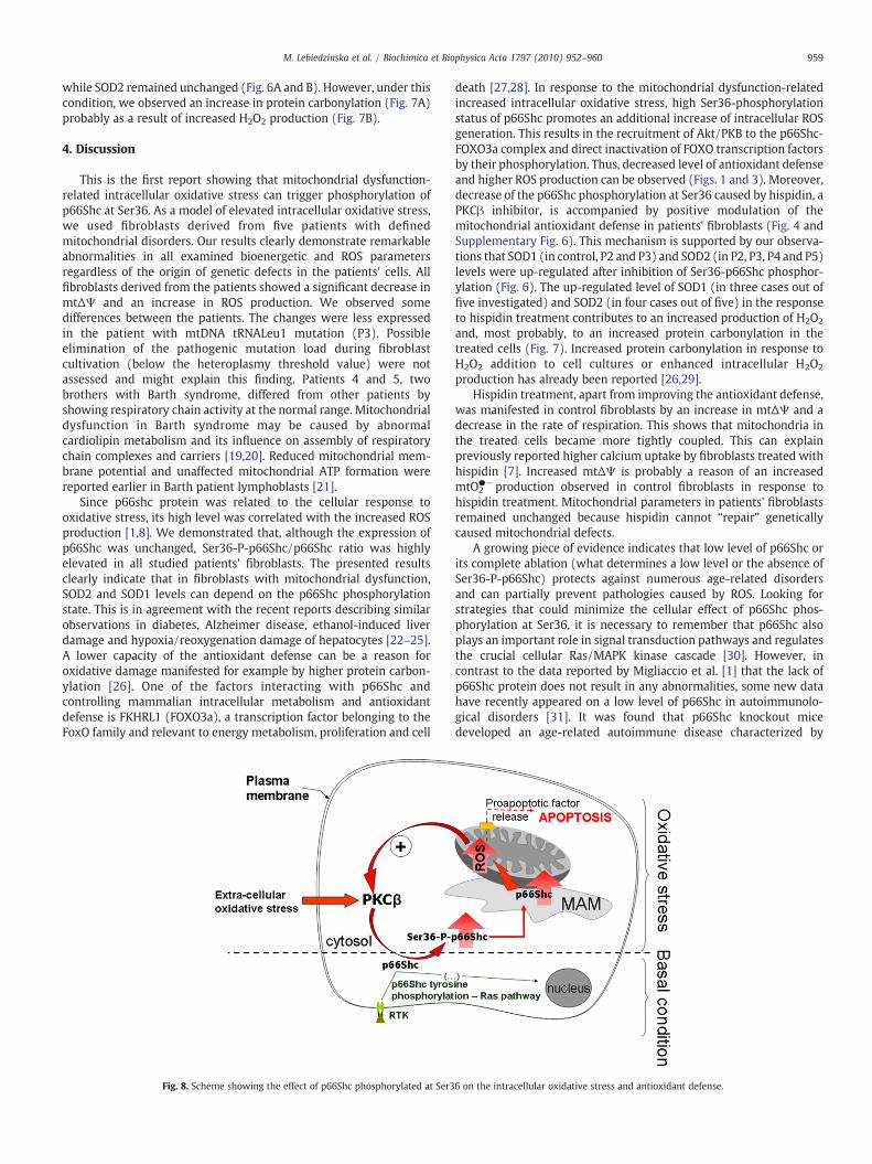
Fig. 8. Scheme showing the effect of p66Shc phosphorylated at Ser3
death [27,28]. In response to the mitochondrial dysfunction-relatedincreased intracellular oxidative stress, high Ser36-phosphorylationstatus of p66Shc promotes an additional increase of intracellular ROSgeneration. This results in the recruitment of Akt/PKB to the p66Shc-FOXO3a complex and direct inactivation of FOXO transcription factorsby their phosphorylation. Thus, decreased level of antioxidant defenseand higher ROS production can be observed (Figs. 1 and 3). Moreover,decrease of the p66Shc phosphorylation at Ser36 caused by hispidin, aPKCβ inhibitor, is accompanied by positive modulation of themitochondrial antioxidant defense in patients' fibroblasts (Fig. 4 andSupplementary Fig. 6). This mechanism is supported by our observa-tions that SOD1 (in control, P2 and P3) and SOD2 (in P2, P3, P4 and P5)levels were up-regulated after inhibition of Ser36-p66Shc phosphor-ylation (Fig. 6). The up-regulated level of SOD1 (in three cases out offive investigated) and SOD2 (in four cases out of five) in the responseto hispidin treatment contributes to an increased production of H2O2
and, most probably, to an increased protein carbonylation in thetreated cells (Fig. 7). Increased protein carbonylation in response toH2O2 addition to cell cultures or enhanced intracellular H2O2
production has already been reported [26,29].Hispidin treatment, apart from improving the antioxidant defense,
was manifested in control fibroblasts by an increase in mtΔΨ and adecrease in the rate of respiration. This shows that mitochondria inthe treated cells became more tightly coupled. This can explainpreviously reported higher calcium uptake by fibroblasts treated withhispidin [7]. Increased mtΔΨ is probably a reason of an increasedmtO2
●−production observed in control fibroblasts in response tohispidin treatment. Mitochondrial parameters in patients' fibroblastsremained unchanged because hispidin cannot “repair” geneticallycaused mitochondrial defects.
A growing piece of evidence indicates that low level of p66Shc orits complete ablation (what determines a low level or the absence ofSer36-P-p66Shc) protects against numerous age-related disordersand can partially prevent pathologies caused by ROS. Looking forstrategies that could minimize the cellular effect of p66Shc phos-phorylation at Ser36, it is necessary to remember that p66Shc alsoplays an important role in signal transduction pathways and regulatesthe crucial cellular Ras/MAPK kinase cascade [30]. However, incontrast to the data reported by Migliaccio et al. [1] that the lack ofp66Shc protein does not result in any abnormalities, some new datahave recently appeared on a low level of p66Shc in autoimmunolo-gical disorders [31]. It was found that p66Shc knockout micedeveloped an age-related autoimmune disease characterized by
6 on the intracellular oxidative stress and antioxidant defense.

960 M. Lebiedzinska et al. / Biochimica et Biophysica Acta 1797 (2010) 952–960
enhanced proliferation of B-lymphocytes that spontaneously produceantibodies and T-lymphocytes. In turn, the immunologic tolerancewas impaired and resulted in autoimmunological aggression observedin p66Shc knockout mice [31]. In the light of these data, administra-tion of mitochondrialy targeted antioxidants seems to be a promisingand safe way to stop the vicious circle of p66Shc phosphorylation atSer36, responsible for the pro-oxidant properties of p66Shc (seeFig. 8). This may open new possibilities for pharmacological inter-ventions in case of pathological intracellular oxidative stress by suchcompounds (as SkQ and mitoQ) with documented therapeutic effect[32–34].
Acknowledgements
This work was supported by the Polish Ministry of Science andHigher Education under grants N301 092 32/3407 and NN407 075 137and by the PolishMitochondrial Network, and by the Internal Project ofThe Childrens' Memorial Health Institute 119/09. P.P.'s and C.G.'s workwas supported by the Italian Association for Cancer Research (AIRC),the United Mitochondrial Disease Foundation (UMDF), the IndustrialResearch program (PRRIITT) of the Emilia Romagna region (PRRIITT),the Italian Multiple Sclerosis Foundation (FISM), Telethon (GGP09128)and local funds from the University of Ferrara.
We thank to Prof. V.P. Skulachev for providing the SkQ. Identificationof mtDNA mutations was performed in the Molecular Laboratory ofDepartment of Medical Genetics, Childrens' Memorial Health Institute(Laboratory head, Dr. Elzbieta Ciara). We are greatly indebted to Prof.Lech Wojtczak for his critical reading of the manuscript.
Appendix A. Supplementary data
Supplementary data associated with this article can be found, inthe online version, at doi:10.1016/j.bbabio.2010.03.005.
References
[1] E. Migliaccio, M. Giorgio, S. Mele, G. Pelicci, P. Reboldi, P.P. Pandolfi, L. Lanfrancone,P.G. Pelicci, The p66shc adaptor protein controls oxidative stress response and lifespan in mammals, Nature 402 (6759) (1999) 309–313.
[2] D. Harman, Aging: a theory based on free radical and radiation chemistry,J. Gerontol. 11 (1956) 298–300.
[3] M. Trinei, M. Giorgio, A. Cicalese, S. Barozzi, A. Ventura, E. Migliaccio, E. Milia, I.M.Padura, V.A. Raker, M. Maccarana, V. Petronilli, S. Minucci, P. Bernardi, L.Lanfrancone, P.G. Pelicci, A p53-p66Shc signaling pathway controls intracellularredox status, levels of oxidation-damaged DNA and oxidative stress-inducedapoptosis, Oncogene 21 (24) (2002) 3872–3878.
[4] L. Luzi, S. Confalonieri, P.P. Di Fiore, P.G. Pelicci, Evolution of Shc functions fromnematode to human, Curr. Opin. Genet. Dev. 10 (2000) 668–674.
[5] K.S. Ravichandran, Signaling via Shc family adapter proteins, Oncogene 20 (2001)6322–6330.
[6] S. Le, T.J. Connors, A.C. Maroney, c-Jun N-terminal kinase specifically phosphorylatesp66ShcA at serine 36 in response to ultraviolet irradiation, J. Biol. Chem. 276 (51)(2001) 48332–48336.
[7] P. Pinton, A. Rimessi, S. Marchi, F. Orsini, E. Migliaccio, M. Giorgio, C. Contursi, S.Minucci, F. Mantovani, M.R. Wieckowski, G. Del Sal, P.G. Pelicci, R. Rizzuto, Proteinkinase C beta and prolyl isomerase 1 regulate mitochondrial effects of the life-span determinant p66Shc, Science 315 (5812) (2007) 659–663.
[8] M. Lebiedzinska, J. Duszynski, R. Rizzuto, P. Pinton, M.R. Wieckowski, Age-relatedchanges in levels of p66Shc and serine 36-phosphorylated p66Shc in organs andmouse tissues, Arch. Biochem. Biophys. 486 (1) (2009) 73–80.
[9] M. Lebiedzinska, G. Szabadkai, A.W. Jones, J. Duszynski, M.R. Wieckowski,Interactions between the endoplasmic reticulum, mitochondria, plasma mem-brane and other subcellular organelles, Int. J. Biochem. Cell Biol. 41 (10) (2009)1805–1816.
[10] M.R. Wieckowski, C. Giorgi, M. Lebiedzinska, J. Duszynski, P. Pinton, Isolation ofmitochondria-associated membranes and mitochondria from animal tissues andcells, Nat. Protoc. 4 (11) (2009) 1582–1590.
[11] S. Nemoto, C.A. Combs, S. French, B.H. Ahn, M.M. Fergusson, R.S. Balaban, T. Finkel,The mammalian longevity-associated gene product p66shc regulates mitochon-drial metabolism, J. Biol. Chem. 281 (15) (2006) 10555–10560.
[12] F. Orsini, E. Migliaccio, M. Moroni, C. Contursi, V.A. Raker, D. Piccini, I. Martin-Padura, G. Pelliccia, M. Trinei, M. Bono, C. Puri, C. Tacchetti, M. Ferrini, R. Mannucci,I. Nicoletti, L. Lanfrancone, M. Giorgio, P.G. Pelicci, The life span determinantp66Shc localizes to mitochondria where it associates with mitochondrial heatshock protein 70 and regulates trans-membrane potential, J. Biol. Chem. 279 (24)(2004) 25689–25695.
[13] P. Pinton, R. Rizzuto, p66Shc oxidative stress and aging. Importing a lifespandeterminant into mitochondria (Perspective), Cell Cycle 7 (3) (2008) 1–5.
[14] S. Di Donato, Multisystem manifestations of mitochondrial disorders, J. Neurol.256 (2009) 693–710.
[15] F. Fernandez-Vizarra, V. Tirani, M. Zeviani, Assembly of the oxidative phosphor-ylation system in humans: what we have learned by studying its defects, Biochim.Biophys. Acta 1793 (2009) 2000–2021.
[16] G. Lenaz, A. Baracca, V. Carelli, M. D'Aurelio, G. Sgarbi, G. Solaini, Bioenergetics ofmitochondrial diseases associated with mtDNAmutations, Biochim. Biophys. Acta1658 (1-2) (2004) 89–94.
[17] E. Karczmarewicz, L. Bielecka, H. Kulczycka, R. Lorenc, E. Pronicka, Analyticalreliability of spectrophotometric analysis of the activity ofmitochondrial respiratorychain complexes in muscle homogenates, Diag. Lab. 33 (1997) 493–503.
[18] O.H. Lowry, N.J. Rosebrough, A.L. Farr, R.J. Randall, Protein measurement with theFolin phenol reagent, J. Biol. Chem. 193 (1) (1951) 265–275.
[19] S.M. Claypool, Cardiolipin, a critical determinant of mitochondrial carrier proteinassembly, Biochim. Biophys. Acta 1788 (2009) 2059–2068.
[20] T.A. Heines, A new look at cardiolipin, (Editoral), Biochim. Biophys. Acta 1788(2009) 1997–2002.
[21] Y. Xu, J.J. Sutachan, H. Plesken, R.I. Kelley, M. Schlame, Characterization oflymphoblast mitochondria from patients with Barth syndrome, Lab. Invest. 85(2005) 823–830.
[22] W. Cai, J.C. He, L. Zhu, X. Chen, G.E. Striker, H. Vlassara, AGE-receptor-1counteracts by AGEs via negative regulation of p66Shc-dependent FKHRL1phosphorylation, Am. J. Physiol. 294 (2008) 145–152.
[23] W.W. Smith, D.D. Norton, M. Gorospe, H. Jiang, S. Nemoto, N.J. Holbrook, T. Finkel,J.W. Kusiak, Phosphorylation of p66Shc and forkhead proteins mediates Abetatoxicity, J. Cell Biol. 169 (2) (2005) 331–339.
[24] O.R. Koch, S. Fusco, S.C. Ranieri, G. Maulucci, P. Palozza, L.M. Larocca, A.A. Cravero,S.M. Farre, M. De Spirito, T. Galeotti, G. Pani, Role of the life span determinantp66ShcA in ethanol-induced liver damage, Lab. Invest. 88 (7) (2008) 750–760.
[25] S. Haga, K. Terui, M. Fukai, Y. Oikawa, K. Irani, H. Furukawa, S. Todo, M. Ozaki,Preventing hypoxia/reoxygenation damage to hepatocytes by p66shc ablation:up-regulation of anti-oxidant and anti-apoptotic proteins, J. Hepatol. 48 (2008)422–432.
[26] C.M. Wong, A.K. Cheema, L. Zhang, Y.J. Suzuki, Protein carbonylation as a novelmechanism in redox signaling, Circ. Res. 102 (3) (2008) 310–318.
[27] S. Nemoto, T. Finkel, Redox regulation of forkhead proteins through a p66shc-dependent signaling pathway, Science 295 (5564) (2002) 2450–2452.
[28] S. Purdom, Q.M. Chen, Linking oxidative stress and genetics of aging wit p66Shcsignaling and forkhead transcription factors, Biogerontology 4 (2003) 181–191.
[29] I. Dalle-Donne, R. Rossi, D. Giustarini, A. Milzani, R. Colombo, Protein carbonylgroups as biomarkers of oxidative stress, Clin. Chim. Acta 329 (2003) 23–38.
[30] E. Migliaccio, S. Mele, A.E. Salcini, G. Pelicci, K.M. Lai, G. Superti-Furga, T. Pawson,P.P. Di Fiore, L. Lanfrancone, P.G. Pelicci, Opposite effects of the p52shc/p46shcand p66shc splicing isoforms on the EGF receptor-MAP kinase-fos signallingpathway, EMBO J. 16 (4) (1997) 706–716.
[31] F. Finetti, M. Pellegrini, C. Ulivieri, M.T. Savino, E. Paccagnini, C. Ginanneschi, L.Lanfrancone, P.G. Pelicci, C.T. Baldari, The proapoptotic and antimitogenic proteinp66SHC acts as a negative regulator of lymphocyte activation and autoimmunity,Blood 111 (10) (2008) 5017–5027.
[32] V.P. Skulachev, V.N. Anisimov, Y.N. Antonenko, L.E. Bakeeva, B.V. Chernyak, V.P.Erichev, O.F. Filenko, N.I. Kalinina, V.I. Kapelko, N.G. Kolosova, B.P. Kopnin, G.A.Korshunova, M.R. Lichinitser, L.A. Obukhova, E.G. Pasyukova, O.I. Pisarenko, V.A.Roginsky, E.K. Ruuge, I.I. Senin, I.I. Severina, M.V. Skulachev, I.M. Spivak, V.N.Tashlitsky, V.A. Tkachuk, M.Y. Vyssokikh, L.S. Yaguzhinsky, D.B. Zorov, An attemptto prevent senescence: amitochondrial approach, Biochim. Biophys. Acta 1787 (5)(2009) 437–461.
[33] S.S. Sheu, D. Nauduri, M.W. Anders, Targeting antioxidants to mitochondria: anew therapeutic direction, Biochim. Biophys. Acta 1762 (2) (2006) 256–265.
[34] M. Milagros Rocha, N.M. Victor, Targeting antioxidants to mitochondria andcardiovascular diseases: the effects of mitoquinone, Med. Sci. Monit. 13 (7) (2007)132–145.
Related Documents