Ovine echinococcosis I. Immunological diagnosis by enzyme immunoassay Antonio Gatti b , Angela Rosa Alvarez a , Daniel Araya b , Sergio Mancini b , Eduardo Herrero b , Graciela Santillan c , Edmundo Larrieu a, * a Veterinary Faculty, UNL Pampa, Argentina b Health Ministry, Rio Negro Province, Argentina c ANLIS/MALBRAN, Argentina Received 5 April 2006; received in revised form 25 July 2006; accepted 3 August 2006 Abstract Immunodiagnosis in sheep presents problems of sensitivity and specificity, limiting its applicability in surveillance systems. The objective of this study was to develop a sensitive, specific and accessible technique for diagnosing cystic echinococcosis in naturally infected sheep and to evaluate the validity of necropsy as a reference test. A total of 247 sheep were studied at slaughterhouses, confirming the parasitological diagnosis with histology. Serum was processed with enzyme immunoassay (EIA) using three antigen preparations: total hydatid liquid (LHT), purified fraction of LHT (S2B) and purified lipoprotein (B). Western Blot (WB) was used as a control. EIA proved effective for differentiating Echinococcus granulosus from larval stage of Taenia hydatigena and intestinal cestodes in all three antigen preparations. Serums from macroscopically negative sheep were reactive to EIA and positive with WB. In the whole flock, sensitivity was 89.2% for LHT, 80.0% for S2B and 86.4% for B. Sensitivity in lambs was 78.6% for LHT, 75.0% for S2B and 64.3% for B. Macroscopic diagnosis at the time of slaughter was found to have limitations as a reference test for immunodiagnosis of cystic equinococcosis in sheep, so it was necessary to include histology and WB as reference tests. LHT was the antigen preparation of greatest value and EIA proved to be a sensitive and specific technique, adequate for surveillance systems and for evaluating control programmes. # 2006 Elsevier B.V. All rights reserved. Keywords: Cystic equinococcosis; Sheep; Immunodiagnosis; Enzyme immunoassay 1. Introduction Cystic echinococcosis is a parasitic zoonosis caused by an internal parasite of the phylum Platyhelminthes, class Cestoda, order Cyclophyllidae, family Taeniidae, genus Echinococcus and specie granulosus. The life cycle involves two mammalian hosts. The definitive hosts are carnivores (especially dogs) in which the adult or strobilocercus form is developed; intermediate hosts are ungulates, especially sheep, in which the larvae or metacestode forms develop (Thompson and Mcmanus, 2001). Information on immunological diagnosis is limited in sheep. Indirect hemagglutination (Yong et al., 1978; Conder et al., 1980; Bakos et al., 1985), double diffusion (Yong and Heath, 1979; Conder et al., 1980) and enzyme immunoassay (EIA) (Craig and Rickard, 1981; Yong et al., 1984; Lightowlers et al., 1984; Ming, 1986; Lloyd et al., 1991) have all been tried with little success: none provided a consistent serological test for livestock (Craig, 1997). www.elsevier.com/locate/vetpar Veterinary Parasitology 143 (2007) 112–121 * Corresponding author. Tel.: +54 2920 430007; fax: +54 2920 430007. E-mail address: [email protected] (E. Larrieu). 0304-4017/$ – see front matter # 2006 Elsevier B.V. All rights reserved. doi:10.1016/j.vetpar.2006.08.022

Welcome message from author
This document is posted to help you gain knowledge. Please leave a comment to let me know what you think about it! Share it to your friends and learn new things together.
Transcript
www.elsevier.com/locate/vetpar
Veterinary Parasitology 143 (2007) 112–121
Ovine echinococcosis
I. Immunological diagnosis by enzyme immunoassay
Antonio Gatti b, Angela Rosa Alvarez a, Daniel Araya b, Sergio Mancini b,Eduardo Herrero b, Graciela Santillan c, Edmundo Larrieu a,*
a Veterinary Faculty, UNL Pampa, Argentinab Health Ministry, Rio Negro Province, Argentina
c ANLIS/MALBRAN, Argentina
Received 5 April 2006; received in revised form 25 July 2006; accepted 3 August 2006
Abstract
Immunodiagnosis in sheep presents problems of sensitivity and specificity, limiting its applicability in surveillance systems. The
objective of this study was to develop a sensitive, specific and accessible technique for diagnosing cystic echinococcosis in naturally
infected sheep and to evaluate the validity of necropsy as a reference test. A total of 247 sheep were studied at slaughterhouses,
confirming the parasitological diagnosis with histology. Serum was processed with enzyme immunoassay (EIA) using three antigen
preparations: total hydatid liquid (LHT), purified fraction of LHT (S2B) and purified lipoprotein (B). Western Blot (WB) was used
as a control. EIA proved effective for differentiating Echinococcus granulosus from larval stage of Taenia hydatigena and intestinal
cestodes in all three antigen preparations. Serums from macroscopically negative sheep were reactive to EIA and positive with WB.
In the whole flock, sensitivity was 89.2% for LHT, 80.0% for S2B and 86.4% for B. Sensitivity in lambs was 78.6% for LHT, 75.0%
for S2B and 64.3% for B. Macroscopic diagnosis at the time of slaughter was found to have limitations as a reference test for
immunodiagnosis of cystic equinococcosis in sheep, so it was necessary to include histology and WB as reference tests. LHT was
the antigen preparation of greatest value and EIA proved to be a sensitive and specific technique, adequate for surveillance systems
and for evaluating control programmes.
# 2006 Elsevier B.V. All rights reserved.
Keywords: Cystic equinococcosis; Sheep; Immunodiagnosis; Enzyme immunoassay
1. Introduction
Cystic echinococcosis is a parasitic zoonosis
caused by an internal parasite of the phylum
Platyhelminthes, class Cestoda, order Cyclophyllidae,
family Taeniidae, genus Echinococcus and specie
granulosus.
The life cycle involves two mammalian hosts. The
definitive hosts are carnivores (especially dogs) in
* Corresponding author. Tel.: +54 2920 430007;
fax: +54 2920 430007.
E-mail address: [email protected] (E. Larrieu).
0304-4017/$ – see front matter # 2006 Elsevier B.V. All rights reserved.
doi:10.1016/j.vetpar.2006.08.022
which the adult or strobilocercus form is developed;
intermediate hosts are ungulates, especially sheep,
in which the larvae or metacestode forms develop
(Thompson and Mcmanus, 2001).
Information on immunological diagnosis is limited
in sheep. Indirect hemagglutination (Yong et al., 1978;
Conder et al., 1980; Bakos et al., 1985), double
diffusion (Yong and Heath, 1979; Conder et al., 1980)
and enzyme immunoassay (EIA) (Craig and Rickard,
1981; Yong et al., 1984; Lightowlers et al., 1984; Ming,
1986; Lloyd et al., 1991) have all been tried with little
success: none provided a consistent serological test for
livestock (Craig, 1997).
A. Gatti et al. / Veterinary Parasitology 143 (2007) 112–121 113
It has been pointed out that the main limitations to
immunodiagnosis in sheep are the cross reactions
observed with other taenias (larvae of Taenia hydati-
gena and Taenia ovis), appearance of false positives and
low sensitivity (Lightowlers and Gottstein, 1995; Craig,
1997; Eckert et al., 2001).
Necropsy is chosen as the best diagnostic test (Eckert
et al., 2001) and is also used as a reference test to estimate
the sensitivity and specificity of immunodiagnostic tests.
The development of a screening test is important for the
identification of cystic hydatid carriers when animals are
imported from endemic areas to areas free of infection
(Craig, 1997; Eckert et al., 2001), and for control
programmes identifying livestock farms with transmis-
sion foci or for epidemiological surveillance to assess the
prevalence of infection (Cabrera et al., 2003).
The objective of this study was to develop a specific,
sensitive and simple immunological diagnostic screen-
ing technique and to evaluate the efficiency of necropsy
as a reference test for immunodiagnosis tests.
2. Materials and methods
2.1. Working area and population
Naturally infected sheep were studied at the time of
slaughter in five abattoirs of the Province of Rio Negro
where the prevalence of infection is 18% (Larrieu et al.,
2001).
Sheep were classified according to their age into the
following groups: rearing lambs (R), 0–6 months old;
fattening lambs (F), over 6 months and under 24 months
old; adults (A), 24 months old and over.
A total of 247 sheep were studied out of which 85
were R (34%), 60 F (24.3%) and 102 adults (41.3%).
Regarding gender, 176 (71.3%) were males and 71
(28.7%) were females.
Blood samples (10 ml) were obtained from the
jugular vein at the time of slaughter and collected in
disposable plastic tubes which were marked with the
animal’s identification number.
Serum was obtained by centrifugation, refrigerated
at 5/8 8C and sent within 48 h to the laboratory where it
was stored at �20 8C until processed.
2.2. Enzyme inmunoassay (EIA) test
The EIA was developed as the screening test with
three different types of antigen preparations: total hydatid
liquid (LHT, obtained as described by Coltorti, 1986),
purified fraction of LHT (S2B, following Coltorti et al.,
1990) and purified lipoprotein (B, obtained as described
by Oriol et al., 1971). The National Institute of Micro-
biology ‘‘Carlos G. Malbran’’ provided the antigens.
For the EIA with LHT antigen (EIA.LHT), each
antigen vial was reconstituted with sufficient sensitizer
buffer (carbonate/bicarbonate, pH 9.6) to obtain an
optimal protein concentration as determined by the
Bradford Method (1976).
The plates (micro strips) were sensitized with 1 mg/
well of antigen. Each well was seeded with 50 ml of
diluted antigen, kept for 18 h in a moist chamber and
then washed three times, each for 4 min, with washing
buffer (PBS 0.3% Tween).
In each well, 100 ml of serum diluted 1/800 in PBS–
3% milk–0.3% Tween were added and left for 45 min at
37 8C. The liquid was then aspirated and washed three
times with washing buffer. Ovine conjugated anti-
gammaglobulin marked with peroxidase diluted 1/2500
in PBS–3% milk–0.3% Tween (100 ml) was added and
left for 45 min at 37 8C. The liquid was then aspirated
and washed three times with washing buffer.
ABTS solution (Sigma1) (100 ml), incubated 45 min
at 37 8C, was used as the developer substrate with the
addition of 100 ml of braking solution (SDS 1%).
Positive control serum was obtained from sheep with
hydatidosis confirmed by histology. Negative control
serum was obtained from sheep with no hydatidosis,
cysticercosis nor intestinal cestodes. The optimal
dilution of the control and problem serum was defined
through serial dilutions of antiserum and conjugated
antigen to obtain the maximum dilution that allows
differentiation of low positive, high positive and
negative control samples, using the same protocol in
each case. Problem and control serums were seeded and
duplicates of each serum were processed. Each micro
plate included three negative serums and one positive.
A vertical plate reader with 405 nm filter was used
for reading the samples. The cut-off value was defined
with receiver operating characteristic (ROC) curves
using an Excel spreadsheet according to the design by
Grainer (1995).
For EIA with S2B antigen (EIA.S2B), each antigen
vial was reconstituted with sensitizer buffer (PBS 7.2)
and the plates (micro strips) were sensitized with an
antigen concentration of 0.15 mg/well. Serum was
diluted 1/400.
For EIAwith B antigen (EIA.B), each antigen vial was
reconstituted with sensitizer buffer (PBS 7.2) and the
plates (micro strips) were sensitized with an antigen
concentration of 0.15 mg/well. Serum was diluted 1/400.
Control serums were seeded, read and the estimation of
the cut-off value was carried out in the same way as for
LHT.
A. Gatti et al. / Veterinary Parasitology 143 (2007) 112–121114
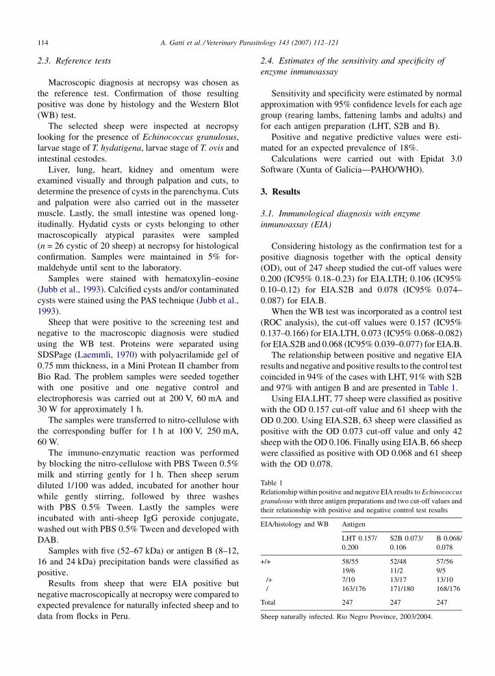
Table 1
Relationship within positive and negative EIA results to Echinococcus
granulosus with three antigen preparations and two cut-off values and
their relationship with positive and negative control test results
EIA/histology and WB Antigen
LHT 0.157/
0.200
S2B 0.073/
0.106
B 0.068/
0.078
+/+ 58/55 52/48 57/56
� 19/6 11/2 9/5
�/+ 7/10 13/17 13/10
�/� 163/176 171/180 168/176
Total 247 247 247
Sheep naturally infected. Rio Negro Province, 2003/2004.
2.3. Reference tests
Macroscopic diagnosis at necropsy was chosen as
the reference test. Confirmation of those resulting
positive was done by histology and the Western Blot
(WB) test.
The selected sheep were inspected at necropsy
looking for the presence of Echinococcus granulosus,
larvae stage of T. hydatigena, larvae stage of T. ovis and
intestinal cestodes.
Liver, lung, heart, kidney and omentum were
examined visually and through palpation and cuts, to
determine the presence of cysts in the parenchyma. Cuts
and palpation were also carried out in the masseter
muscle. Lastly, the small intestine was opened long-
itudinally. Hydatid cysts or cysts belonging to other
macroscopically atypical parasites were sampled
(n = 26 cystic of 20 sheep) at necropsy for histological
confirmation. Samples were maintained in 5% for-
maldehyde until sent to the laboratory.
Samples were stained with hematoxylin–eosine
(Jubb et al., 1993). Calcified cysts and/or contaminated
cysts were stained using the PAS technique (Jubb et al.,
1993).
Sheep that were positive to the screening test and
negative to the macroscopic diagnosis were studied
using the WB test. Proteins were separated using
SDSPage (Laemmli, 1970) with polyacrilamide gel of
0.75 mm thickness, in a Mini Protean II chamber from
Bio Rad. The problem samples were seeded together
with one positive and one negative control and
electrophoresis was carried out at 200 V, 60 mA and
30 W for approximately 1 h.
The samples were transferred to nitro-cellulose with
the corresponding buffer for 1 h at 100 V, 250 mA,
60 W.
The immuno-enzymatic reaction was performed
by blocking the nitro-cellulose with PBS Tween 0.5%
milk and stirring gently for 1 h. Then sheep serum
diluted 1/100 was added, incubated for another hour
while gently stirring, followed by three washes
with PBS 0.5% Tween. Lastly the samples were
incubated with anti-sheep IgG peroxide conjugate,
washed out with PBS 0.5% Tween and developed with
DAB.
Samples with five (52–67 kDa) or antigen B (8–12,
16 and 24 kDa) precipitation bands were classified as
positive.
Results from sheep that were EIA positive but
negative macroscopically at necropsy were compared to
expected prevalence for naturally infected sheep and to
data from flocks in Peru.
2.4. Estimates of the sensitivity and specificity of
enzyme inmunoassay
Sensitivity and specificity were estimated by normal
approximation with 95% confidence levels for each age
group (rearing lambs, fattening lambs and adults) and
for each antigen preparation (LHT, S2B and B).
Positive and negative predictive values were esti-
mated for an expected prevalence of 18%.
Calculations were carried out with Epidat 3.0
Software (Xunta of Galicia—PAHO/WHO).
3. Results
3.1. Immunological diagnosis with enzyme
inmunoassay (EIA)
Considering histology as the confirmation test for a
positive diagnosis together with the optical density
(OD), out of 247 sheep studied the cut-off values were
0.200 (IC95% 0.18–0.23) for EIA.LTH; 0.106 (IC95%
0.10–0.12) for EIA.S2B and 0.078 (IC95% 0.074–
0.087) for EIA.B.
When the WB test was incorporated as a control test
(ROC analysis), the cut-off values were 0.157 (IC95%
0.137–0.166) for EIA.LTH, 0.073 (IC95% 0.068–0.082)
for EIA.S2B and 0.068 (IC95% 0.039–0.077) for EIA.B.
The relationship between positive and negative EIA
results and negative and positive results to the control test
coincided in 94% of the cases with LHT, 91% with S2B
and 97% with antigen B and are presented in Table 1.
Using EIA.LHT, 77 sheep were classified as positive
with the OD 0.157 cut-off value and 61 sheep with the
OD 0.200. Using EIA.S2B, 63 sheep were classified as
positive with the OD 0.073 cut-off value and only 42
sheep with the OD 0.106. Finally using EIA.B, 66 sheep
were classified as positive with OD 0.068 and 61 sheep
with the OD 0.078.
A. Gatti et al. / Veterinary Parasitology 143 (2007) 112–121 115
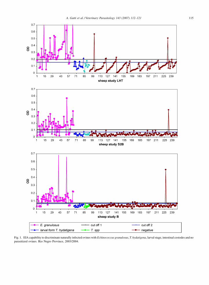
Fig. 1. EIA capability to discriminate naturally infected ovines with Echinococcus granulosus, T. hydatigena, larval stage, intestinal cestodes and no
parasitized ovines. Rio Negro Province, 2003/2004.
A. Gatti et al. / Veterinary Parasitology 143 (2007) 112–121116
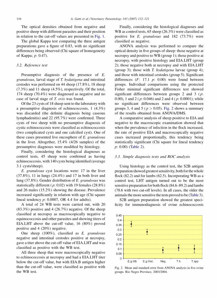
Fig. 2. Mean and standard error from ANOVA analysis in five ovine
groups. Rio Negro Province, 2003/2004.
The optical densities obtained from negative and
positive sheep with different parasites and their position
in relation to the cut-off values are presented in Fig. 1.
The global Kappa test comparing the three antigen
preparations gave a figure of 0.83, with no significant
differences being observed (Chi square of homogeneity
of Kappa; p: 0.47).
3.2. Reference test
Presumptive diagnosis of the presence of E.
granulosus, larval stage of T. hydatigena and intestinal
cestodes was performed on 44 sheep (17.8%), 18 sheep
(7.3%) and 11 sheep (4.5%), respectively. Of the total,
174 sheep (70.4%) were diagnosed as negative and no
case of larval stage of T. ovis was found.
Of the 23 cysts of 18 sheep sent to the laboratory with
a presumptive diagnosis of echinococcosis, 1 (4.3%)
was discarded (the definite diagnosis being caseous
lymphadenitis) and 22 (95.7%) were confirmed. Three
cysts of two sheep with no presumptive diagnosis of
cystic echinococcosis were classified as echinococcosis
(two complicated cysts and one calcified cyst). One of
these cases presented live oncosphere of E. granulosus
in the liver. Altogether, 15.4% (4/26 samples) of the
presumptive diagnoses were modified by histology.
Finally, considering the histological diagnoses as
control tests, 45 sheep were confirmed as having
echinococcosis, with 140 cysts being identified (average
3.1 cysts/sheep).
E. granulosus cyst locations were: 17 in the liver
(37.8%), 11 in lungs (24.4%) and 17 in both liver and
lung (37.8%). Gender distribution of E. granulosus was
statistically different ( p: 0.02) with 19 females (28.8%)
and 26 males (15.2%) showing the disease. Prevalence
increased significantly in relation with age (Chi square
lineal tendency p: 0.0007, OR 4.4 for adults).
A total of 24 WB tests were carried out, with 20
(83.3%) positive and 4 (26.7%) negative. Of the sheep
classified at necropsy as macroscopically negative to
equinococcosis and other parasites and showing titers of
EIA.LHT above the cut-off value, 16 (80%) proved
positive and 4 (20%) negative.
One sheep (100%), classified as E. granulosus
negative and intestinal cestodes positive at necropsy,
gave a titer above the cut-off value of EIA.LHT and was
classified as positive with the WB test.
All three sheep that were macroscopically negative
to echinococcosis at necropsy and had a EIA.LHT titer
below the cut-off value, but with EIA.B antigen higher
than the cut-off value, were classified as positive with
the WB test.
Finally, considering the histological diagnoses and
WB as control tests, 65 sheep (26.3%) were classified as
positive for E. granulosus and 182 (73.7%) were
classified as negative.
ANOVA analysis was performed to compare the
optical density in five groups of sheep: those negative at
necropsy and positive to WB (group 1); those positive at
necropsy, with positive histology and EIA.LHT (group
2); those negative both at necropsy and with EIA.LHT
(group 3); those with T. hydatigena larvae (group 4);
and those with intestinal cestodes (group 5). Significant
differences (F : 17.1 p: 0.00) were found between
groups. Individual comparisons using the protected
Fisher minimal significant differences test showed
significant differences between groups 2 and 3 ( p:
0.00), 1 and 2 ( p: 0.046) and 2 and 4 ( p: 0.0001); while
no significant differences were observed between
groups 3, 4 and 5 ( p > 0.05). Fig. 2 shows a summary
of the results obtained from ANOVA/DMS.
A comparative analysis of sheep positive to EIA and
negative to the macroscopic examination showed that
when the prevalence of infection in the flock increased,
the rate of positive EIA and macroscopically negative
cases increased proportionally, this tendency being
statistically significant (Chi square for lineal tendency
p: 0.00) (Table 2).
3.3. Simple diagnosis tests and ROC analysis
Using histology as the control test, the S2B antigen
preparation showed greatest sensitivity, both for thewhole
flock (82.2) and for lambs (62.5). Incorporating WB as a
control test, LHT antigen turned out to be the most
sensitive preparation for both flock (84.6–89.2) and lambs
(78.6 with two cut-off levels). In all cases, the older the
animals the more sensitive the tests proved to be (Table 3).
S2B antigen preparation showed the greatest speci-
ficity for immunodiagnosis of ovine echinococcosis
A. Gatti et al. / Veterinary Parasitology 143 (2007) 112–121 117
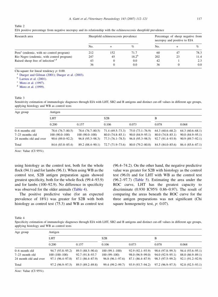
Table 2
EIA positive percentage from negative necropsy and its relationship with the echinococcosis sheepfold prevalence
Research area Sheepfold echinococcosis prevalence Percentage of sheep negative from
necropsy and positive to EIA
No. + % No. + %
Perua (endemic, with no control program) 212 152 71.7 60 47 78.3
Rio Negro (endemic, with control program) 247 45 18.2b 202 23 11.4
Raised sheep free of infectionc,d 43 0 0.0 42 1 2.3
36 0 0.0 36 0 0.0
Chi-square for lineal tendency p: 0.00.a Dueger and Gilman (2001); Dueger et al. (2003).b Larrieu et al. (2001).c Moro et al. (1997).d Moro et al. (1999).
Table 3
Sensitivity estimation of immunologic diagnoses through EIA with LHT, SB2 and B antigens and distinct cut-off values in different age groups,
applying histology and WB as control tests
Age group Antigen
LHT S2B B
0.200 0.157 0.106 0.073 0.078 0.068
0–6 months old 78.6 (76.7–80.5) 78.6 (76.7–80.5) 71.4 (69.5–73.3) 75.0 (73.1–76.9) 64.3 (60.6–68.2) 64.3 (60.6–68.1)
7–23 months old 100 (90.0–100) 100 (90.0–100) 80.0 (74.8–85.1) 90.0 (84.9–95.1) 80.0 (74.8–85.1) 90.0 (84.9–95.1)
24 months old and over 90.6 (89.0–92.2) 96.8 (95.3–98.3) 77.3 (76.1–78.5) 96.8 (95.3–98.5) 92.7 (91.4–93.9) 90.9 (89.7–92.1)
Total 84.6 (83.8–85.4) 89.2 (88.4–90.1) 72.7 (71.9–73.6) 80.0 (79.2–80.8) 84.5 (84.0–85.6) 86.4 (85.6–87.1)
Note: Value (CI 95%).
using histology as the control test, both for the whole
flock (94.1) and for lambs (96.1). When using WB as the
control test, S2B antigen preparation again showed
greatest specificity, both for the whole flock (99.4–93.9)
and for lambs (100–92.9). No difference in specificity
was observed for the older animals (Table 4).
The positive predictive value (for an expected
prevalence of 18%) was greater for S2B with both
histology as control test (75.3) and WB as control test
Table 4
Specificity estimation of immunologic diagnoses through EIA with LHT, S
applying histology and WB as control tests
Age group Antigen
LHT S2B
0.200 0.157 0.106
0–6 month old 94.7 (93.8–95.2) 89.5 (88.5–90.4) 100 (99.
7–23 month old 100 (100–100) 92.7 (91.8–93.7 100 (99–
24 month old and over 97.1 (96.4–97.8) 87.1 (86.4–87.9) 96.8 (9
Total 97.2 (96.9–97.5) 89.5 (89.2–89.8) 99.4 (9
Note: Value (CI 95%).
(96.4–74.2). On the other hand, the negative predictive
value was greater for S2B with histology as the control
test (96.0) and for LHT with WB as the control test
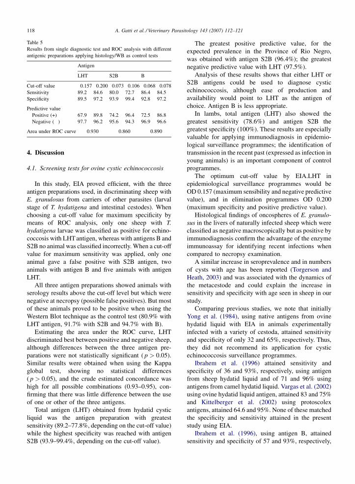
(96.2–97.7) (Table 5). Estimating the area under the
ROC curve, LHT has the greatest capacity to
discriminate (0.930 IC95% 0.86–0.97). The result of
comparing the areas beneath the ROC curve for the
three antigen preparations was not significant (Chi
square homogeneity test, p: 0.07).
B2 and B antigens and distinct cut-off values in different age groups,
B
0.073 0.078 0.068
1–100) 92.9 (92.1–93.9) 98.6 (97.8–99.3) 94.4 (93.6–95.1)
100) 98.0 (96.9–99.0) 94.0 (92.9–95.1) 88.0 (86.9–89.1)
6.1–97.6) 87.1 (86.4–87.9) 98.3 (97.5–99.2) 92.1 (91.2–92.9)
9.2–99.7) 93.9 (93.7–94.2) 97.2 (96.9–97.5) 92.8 (92.5–93.1)
A. Gatti et al. / Veterinary Parasitology 143 (2007) 112–121118
Table 5
Results from single diagnostic test and ROC analysis with different
antigenic preparations applying histology/WB as control tests
Antigen
LHT S2B B
Cut-off value 0.157 0.200 0.073 0.106 0.068 0.078
Sensitivity 89.2 84.6 80.0 72.7 86.4 84.5
Specificity 89.5 97.2 93.9 99.4 92.8 97.2
Predictive value
Positive (+) 67.9 89.8 74.2 96.4 72.5 86.8
Negative (�) 97.7 96.2 95.6 94.3 96.9 96.6
Area under ROC curve 0.930 0.860 0.890
4. Discussion
4.1. Screening tests for ovine cystic echinococcosis
In this study, EIA proved efficient, with the three
antigen preparations used, in discriminating sheep with
E. granulosus from carriers of other parasites (larval
stage of T. hydatigena and intestinal cestodes). When
choosing a cut-off value for maximum specificity by
means of ROC analysis, only one sheep with T.
hydatigena larvae was classified as positive for echino-
coccosis with LHT antigen, whereas with antigens B and
S2B no animal was classified incorrectly. When a cut-off
value for maximum sensitivity was applied, only one
animal gave a false positive with S2B antigen, two
animals with antigen B and five animals with antigen
LHT.
All three antigen preparations showed animals with
serology results above the cut-off level but which were
negative at necropsy (possible false positives). But most
of these animals proved to be positive when using the
Western Blot technique as the control test (80.9% with
LHT antigen, 91.7% with S2B and 94.7% with B).
Estimating the area under the ROC curve, LHT
discriminated best between positive and negative sheep,
although differences between the three antigen pre-
parations were not statistically significant ( p > 0.05).
Similar results were obtained when using the Kappa
global test, showing no statistical differences
( p > 0.05), and the crude estimated concordance was
high for all possible combinations (0.93–0.95), con-
firming that there was little difference between the use
of one or other of the three antigens.
Total antigen (LHT) obtained from hydatid cystic
liquid was the antigen preparation with greatest
sensitivity (89.2–77.8%, depending on the cut-off value)
while the highest specificity was reached with antigen
S2B (93.9–99.4%, depending on the cut-off value).
The greatest positive predictive value, for the
expected prevalence in the Province of Rio Negro,
was obtained with antigen S2B (96.4%); the greatest
negative predictive value with LHT (97.5%).
Analysis of these results shows that either LHT or
S2B antigens could be used to diagnose cystic
echinococcosis, although ease of production and
availability would point to LHT as the antigen of
choice. Antigen B is less appropriate.
In lambs, total antigen (LHT) also showed the
greatest sensitivity (78.6%) and antigen S2B the
greatest specificity (100%). These results are especially
valuable for applying immunodiagnosis in epidemio-
logical surveillance programmes; the identification of
transmission in the recent past (expressed as infection in
young animals) is an important component of control
programmes.
The optimum cut-off value by EIA.LHT in
epidemiological surveillance programmes would be
OD 0.157 (maximum sensibility and negative predictive
value), and in elimination programmes OD 0.200
(maximum specificity and positive predictive value).
Histological findings of oncospheres of E. granulo-
sus in the livers of naturally infected sheep which were
classified as negative macroscopically but as positive by
immunodiagnosis confirm the advantage of the enzyme
immunoassay for identifying recent infections when
compared to necropsy examination.
A similar increase in seroprevalence and in numbers
of cysts with age has been reported (Torgerson and
Heath, 2003) and was associated with the dynamics of
the metacestode and could explain the increase in
sensitivity and specificity with age seen in sheep in our
study.
Comparing previous studies, we note that initially
Yong et al. (1984), using native antigens from ovine
hydatid liquid with EIA in animals experimentally
infected with a variety of cestoda, attained sensitivity
and specificity of only 32 and 65%, respectively. Thus,
they did not recommend its application for cystic
echinococcosis surveillance programmes.
Ibrahem et al. (1996) attained sensitivity and
specificity of 36 and 93%, respectively, using antigen
from sheep hydatid liquid and of 71 and 96% using
antigens from camel hydatid liquid. Vargas et al. (2002)
using ovine hydatid liquid antigen, attained 83 and 75%
and Kittelberger et al. (2002) using protoscolex
antigens, attained 64.6 and 95%. None of these matched
the specificity and sensitivity attained in the present
study using EIA.
Ibrahem et al. (1996), using antigen B, attained
sensitivity and specificity of 57 and 93%, respectively,
A. Gatti et al. / Veterinary Parasitology 143 (2007) 112–121 119
using sheep antigen and of 90 and 99% using camel
antigen. Kittelberger et al. (2002), attained sensitivity
and specificity of 9.7 and 99.5% using antigen B of
ovine hydatid liquid; sensitivity attained in this study
using antigen B was 84.5/86.4% (depending on the
cut-off value) and specificity was 97.2/96.8%.
Thus, EIA is shown to be a specific and sensitive
technique for identifying the transmission of E.
granulosus from dogs to sheep, including lambs,
supporting its application for the diagnosis of cystic
echinococcosis in sheep. LHT would be the antigen
of choice due to its ease of production and availability.
4.2. Reference tests for serological diagnosis of
ovine echinococcosis
The present research shows that 15.4% of the
presumptive macroscopic diagnoses at necropsy were
modified by the histological confirmation test, detecting
false positive and false negative diagnoses in the
different age groups studied.
These results confirm previous studies carried out in
the Province of Rio Negro in which 37.2% of false
positive diagnoses identified at necropsy were caused
by unspecific granulomas, pseudo tuberculosis, emphy-
sema and fatty degeneration while 1.1% of false
negative diagnoses were due to small intra-parenchyma
cysts (Larrieu et al., 2001). This information coincides
with research carried out in Uruguay (Cabrera et al.,
1996) in which 26.1% of the sheep that were classified
as positive at necropsy were not hydatidosis at histology
but were mistaken with caseous lymphadenitis, larval
stage of T. hydatigena, white spot or abscesses. Small
intra-parenchyma cysts, especially in lungs, can be
unnoticeable at sanitary inspection. As a result, these
cases are falsely classified as negative at necropsy.
This study identified intra-parenchyma oncospheres
through histology and cysts of recent development
which were considered negative at the macroscopic
inspection (false negative at necropsy). This confirms
the limitations of macroscopic diagnosis at necropsy
and the validity of the histological test as a reference test
for the diagnosis of ovine cystic echinococcosis.
In order to evaluate the specificity and sensitivity
of the immunologic diagnosis in both naturally and
experimentally infected animals, previous studies
almost exclusively used serum panels where the animals
were classified as negative or positive according to the
macroscopic analysis at necropsy (Conder et al., 1980;
Craig and Rickard, 1981; Yong et al., 1984; Bakos et al.,
1985; Lightowlers et al., 1984; Ibrahem et al., 1996;
Moro et al., 1997, 1999; Kittelberger et al., 2002;
Vargas et al., 2002). Only one study used the Gram dye
to perform differential diagnosis with Corynebacterium
pseudotuberculosus (Dueger et al., 2003). This throws
some doubts on the interpretation of the above-
mentioned studies.
In contrast, the use of immunodiagnosis would make
it possible to classify as positive sheep carrying a recent
infection that would have been incorrectly classified as
negative at necropsy.
In areas with a high prevalence of the infection (e.g.
Peru) all animals can be considered to be exposed to
infection. Besides those found positive at necropsy,
71.7% of the animals negative at necropsy are detected
as serum positive (Moro et al., 1999; Dueger et al.,
2003). In areas of limited transmission (like the
Province of Rio Negro) that proportion is 11.4%; for
animals raised in areas free of infection the correspond-
ing rate is 0–2.3% (Moro et al., 1997, 1999). According
to the observations of this study, animals negative at
necropsy but positive at serology could be estimated to
be the product of different stages of the infection
process (oncospheres of a recent infection, recently
developing cysts, incipient hyaline cysts).
Yong et al. (1984) observed a positive antibody
response in three sheep experimentally infected with a
low infestation (10 eggs) of E. granulosus but failed to
find hydatid cysts at necropsy.
Serological conversion 10–14 days after inoculation
(Yong et al., 1984) confirms that animals with recent
infections will be classified as positive in serological
tests and as negative at necropsy.
There are, however, operative limitations to the use
of histological studies as reference tests. It is possible to
confirm all doubtful macroscopic lesions but it would be
laborious to check all the macroscopically negative
diagnoses in an attempt to detect recent infections,
information which would be particularly interesting for
a surveillance system, especially in lambs.
In this study, as an alternative, Western Blot was used
to check negative cases at necropsy that were positive to
the EIA screening test.
Western Blot, based on the identification of species-
specific antigen fractions of 8–12, 16 and 20–24 kDa,
was developed for immunodiagnosis of ovine echino-
coccosis and proved to have a 98.6–100% specificity
(Moro et al., 1997; Vargas et al., 2002) and no cross
reactions with other cestoda. The 8 kDa band was
present in all serum positive animals. WB sensitivity
has been reported as 91.4–97.6% (Dueger et al., 2003;
Vargas et al., 2002).
In this study, analysis of variance was carried out
comparing the optical densities of sera belonging to the
A. Gatti et al. / Veterinary Parasitology 143 (2007) 112–121120
following groups of sheep: those with macroscopically
diagnosed E. granulosus, those negative at necropsy but
positive to EIA and WB, those with other cestoda
infections, and those free of infection. This made it
possible to establish, with precision, the characteristics
common to sheep classified as positive by WB or
necropsy and the statistically significant differences
observed with sheep carriers of other cestoda and sheep
with no infection at all, confirming the specificity of the
WB diagnosis and its usefulness as a reference test.
The use of macroscopic diagnosis at necropsy as a
reference test for sensitivity and specificity estimations
has limitations, as demonstrated in this study.
Assays to be used as screening tests in sheep
populations should include confirmation of diagnosis
using either histology or highly specific serological tests
such as Western Blot.
Acknowledgements
Our acknowledgements go to Dr. Eduardo Guarnera
and Anıbal Franco for their advice on research design
and to Dr. Irma Sommerfelt for manuscript correction.
References
Bakos, E., Zurbriggen, M., Soni, C., Draghi, M., 1985. Diagnostico
serologico de la hidatidosis ovina mediante la hemaglutinacion
indirecta. Vet. Arg. 17, 624–627.
Bradford, M., 1976. Rapid and sensitive method for the quantitation of
microgram quantities of protein utilizing the principle of protein-
dye binding. A. Biochem. 72, 248–257.
Cabrera, P.A., Irabedra, P., Trindado, J., Agulla, J., Heinzen, T.,
Cardozo, M., Alvarez, M., Elola, S., Rista, L., Bondad, M., Haran,
G., Vinals, G., Mangeney, G., Sambran, Y., Valledor, S., Baralbar,
M., Morana, A., Orlando, D., 1996. Determinacion de la preva-
lencia de la hidatidosis ovina en playas de faena, Uruguay, 1994.
Hidatidosis (Uruguay) 2, 18–29.
Cabrera, P.A., Irabedra, P., Orlando, L., Rista, R., Haran, G., Vinals,
G., Blanco, M.T., Alvarez, M., Elosa, S., Morosoli, D., Morana,
A., Bondad, M., Sambran, Y., Heinzen, T., Chans, L., Pineiro, L.,
Perez, D., Pereyra, I., 2003. National prevalence larval echino-
coccosis in sheep in slaughtering plants. Ovis aries as an indicator
in control programmes in Uruguay. Acta Trop. 85, 185–281.
Coltorti, E., 1986. Standarization and evaluation of an enzymeimmu-
noassay as a screening test for the seroepidemiology of human
hydatidosis. Am. J. Trop. Med. Hyg. 35, 1000–1005.
Coltorti, E., Fernandez, E., Marguet, E., Scozzina, J., Guarnera, E.,
1990. Deteccion de portadores asintomaticos de quistes hidatıdi-
cos. Aumento de la especificidad del ensayo inmunoenzimatico.
Rev. Inst. Med. Trop. Sao Paulo 32, 275–284.
Conder, G.A., Andersen, F.L., Schantz, P.M., 1980. Immunodiagnos-
tic tests for hydatidosis in sheep: an evaluation of double diffusion,
immunoeletrophoresis, indirect hemagglutination, and intrader-
mal tests. J. Parasitol. 66, 577–584.
Craig, P.S., 1997. Immnunodiagnosis of Echinococcus granulosus and a
comparison of techniques for diagnoses of canine echinococcosis.
In: Andersen, F., Ouhelli, H., Kachani, M. (Eds.), Compendium on
Echinococcosis in Africa and in Middle Eastern Countries. Brigham
Young University Print Services, Utah, pp. 86–118.
Craig, P.S., Rickard, M., 1981. Studies on the specific immnunodiag-
nosis of larval cestode infections of cattle and sheep using antigens
purified by affinity chromatography in any enzyme-linked immu-
noabsorbent assay (ELISA). Int. J. Parasitol. 11, 441–449.
Dueger, E.L., Gilman, R.H., 2001. Prevalence, intensity and fertility of
ovine cystic echinococcosis in the central peruvian andes. Trans.
R. Soc. Trop. Med. Hyg. 95, 379–383.
Dueger, E.L., Verastegui, M., Gilman, R.H., 2003. Evaluation of the
enzyme-linked immunoelectrotransfer blot (EITB) for ovine
hydatidosis relative to age and cyst characteristics in naturally
infected sheep. Vet. Parasitol. 114, 285–293.
Eckert, J., Deplazes, P., Craig, P.S., Gemmel, M., Gottstein, B., Heath,
D.D., Jenkins, J., Kamiya, M., Lightowlers, M.H., 2001. Echino-
coccosis in animals: clinical aspects, diagnosis and treatment. In:
Eckert, J., Gemmel, M., Meslin, F., Pawlowski, Z. (Eds.), Manual
on Echinococcosis in Humans and Animals: a public health
problem of global concern, WHO/OIE, France, pp. 195–203.
Grainer, M., 1995. Two graph operating characteristic (TG-ROC): a
Microsoft Excel template for the selection of cut-off values in
diagnostic tests. J. Immunol. Methods 185, 145–146.
Ibrahem, M.M., Craig, P.S., Mcvie, A., 1996. Echinococcus granu-
losus antigen B and seroreactivity in natural ovine hydatidosis.
Res. Vet. Sci. 61, 102–106.
Jubb, K., Kennedy, P., Palmer, N., 1993. Pathology of Domestic
Animals, fourth ed., vol. II. Editorial Academic Press Inc.
Kittelberger, R., Reichel, M.P., Jenner, J., Heath, D.D., Ligthtowlers,
M.H., Moro, P., Ibrahem, M.M., Craig, P.S., Keefe, J., 2002.
Evaluation of three enzyme-linked immunoabsorbent assays
(ELISA) for the detection of serum antibodies in sheep infected
with Echinococcus granulosus. Vet. Parasitol. 110, 57–76.
Laemmli, U., 1970. Cleavage of structural proteins during the assem-
bly of the head of the bacteriophage T4. Nature 227, 680–685.
Larrieu, E., Costa, M., Cantoni, G., Alvarez, R., Cavagion, L.,
Labanchi, J., Bigatti, R., Araya, D., Herrero, E., Mancini, S.,
Cabrera, P.A., 2001. Ovine Echinococcus granulosus transmission
dynamics in the province of Rio Negro, Argentina, 1980–1999.
Vet. Parasitol. 98, 263–272.
Lightowlers, M.H., Gottstein, B., 1995. Echinococcosis/hydatidosis:
antigens, immunological and molecular diagnosis. In: Thompson,
R., Lymbery, J. (Eds.), The biology of Echinococcus granulosus
and hydatid disease. George Allen and Unwin, London, pp. 355–
396.
Lightowlers, M.H., Rickard, M., Honey, R., Obendorf, D., Mitchell,
G., 1984. Serological diagnosis of Echinococcus granulosus
infection in sheep using cyst fluid antigens processed by antibody
affinity chromatography. A. Vet. J. 61, 101–108.
Lloyd, S., Martin, S., Walters, T., Soulsby, E., 1991. Use of sentinel
lambs for early monitoring of teh South Powys hydatidosis control
scheme: prevalence of Echinococcus granulosus and some hel-
minths. Vet. Rec. 129, 73–76.
Ming, R., 1986. Application of enzyme linked immunabsorbent assay
to the diagnosis of echinococcus of sheep and cattle. A. Vet. Z. Sin.
17, 55–62.
Moro, P.L., Verastegui, M., Gilman, R.H., Falcon, N., Bernal, T.,
Gavidia, C., Malqui, V., Moro, M.H., Dueger, E.L., 1997. Enzime
linked immunoelectrotransferblot assay for diagnosis of hydati-
dosis in sheep. Vet. Rec. 140, 605–606.
Moro, P.L., Bonifacio, N., Gilman, R.H., Lopera, L., Silva, B.,
Takumoto, R., Verastegui, M., Cabrera, L., 1999. Field diagnosis
A. Gatti et al. / Veterinary Parasitology 143 (2007) 112–121 121
of Echinococcus granulosus infection among intermediate and
definitive hosts in an endemic focus of human cystic echinococ-
cosis. Trans. R. Soc. Trop. Med. Hyg. 91, 611–615.
Oriol, R., Williams, J., Perez Esandi, M., Oriol, R., 1971. Evaluation
of purified lipoprotein antigens of Echinococcus granulosus in the
immunodiagnosis of human infection. Am. J. Trop. Med. Hyg. 20,
569–574.
Thompson, R., Mcmanus, D., 2001. Aetiology: parasites and life
cycles. In: Eckert, J., Gemmel, M., Meslin, F., Pawlowski, Z.
(Eds.), Manual on Echinococcosis in Humans and Animals: a
Public Health Problem of Global Concern. WHO/OIE, France, pp.
1–19.
Torgerson, P.R., Heath, D.D., 2003. Transmission dynamics and
control options for Echinococcus granulosus. Parasitology 127,
143–158.
Vargas, D., Bonet, R., Campano, S., Chacon, T., Vidal, M., 2002.
Evaluacion epidemiologica de las tecnicas de Elisa y electroin-
muno transferencia en el diagnostico de la hidatidosis ovina en la
XI Region de Chile. Tesis Doctoral. Universidad Mayor, Chile.
Yong, W.K., Heath, D.D., Parmeter, S.N., 1978. Echinococcus gran-
ulosus, Taenia hydatigena and Taenia ovis evaluation of cyst fluids
as antigen for serodiagnosis of larval cestode in sheep. N. Z. Vet. J.
126, 231–234.
Yong, W.K., Heath, D.D., 1979. ‘Arc 5’ antibodies in sera of sheep
infected with Echinococcus granulosus, Taenia hydatigena and
Taenia ovis. Parasite Immnunol. 1, 27–38.
Yong, W.K., Heath, D.D., Van Knapen, F., 1984. Comparison of
cestode antigens in an enzyme-linked immunosorbent assay for
the diagnosis of Echinococcus granulosus, Taenia hydatigena and
Taenia ovis infections in sheep. Res. Vet. Sci. 36, 24–31.
Related Documents