REVIEW Open Access Overview of current adipose-derived stem cell (ADSCs) processing involved in therapeutic advancements: flow chart and regulation updates before and after COVID-19 Loubna Mazini 1* , Mohamed Ezzoubi 2 and Gabriel Malka 1 Abstract Adipose-derived stem cells (ADSCs) have raised big interest in therapeutic applications in regenerative medicine and appear to fulfill the criteria for a successful cell therapy. Their low immunogenicity and their ability to self- renew, to differentiate into different tissue-specific progenitors, to migrate into damaged sites, and to act through autocrine and paracrine pathways have been altogether testified as the main mechanisms whereby cell repair and regeneration occur. The absence of standardization protocols in cell management within laboratories or facilities added to the new technologies improved at patient’s bedside and the discrepancies in cell outcomes and engraftment increase the limitations on their widespread use by balancing their real benefit versus the patient safety and security. Also, comparisons across pooled patients are particularly difficult in the fact that multiple medical devices are used and there is absence of harmonized assessment assays despite meeting regulations agencies and efficient GMP protocols. Moreover, the emergence of the COVID-19 breakdown added to the complexity of implementing standardization. Cell- and tissue-based therapies are completely dependent on the biological manifestations and parameters associated to and induced by this virus where the scope is still unknown. The initial flow chart identified for stem cell therapies should be reformulated and updated to overcome patient infection and avoid significant variability, thus enabling more patient safety and therapeutic efficiency. The aim of this work is to highlight the major guidelines and differences in ADSC processing meeting the current good manufacturing practices (cGMP) and the cellular therapy-related policies. Specific insights on standardization of ADSCs proceeding at different check points are also presented as a setup for the cord blood and bone marrow. Keywords: ADSCs, HCT/P 351-361, ATMPs, cGMP, Standardization, COVID-19 © The Author(s). 2020 Open Access This article is licensed under a Creative Commons Attribution 4.0 International License, which permits use, sharing, adaptation, distribution and reproduction in any medium or format, as long as you give appropriate credit to the original author(s) and the source, provide a link to the Creative Commons licence, and indicate if changes were made. The images or other third party material in this article are included in the article's Creative Commons licence, unless indicated otherwise in a credit line to the material. If material is not included in the article's Creative Commons licence and your intended use is not permitted by statutory regulation or exceeds the permitted use, you will need to obtain permission directly from the copyright holder. To view a copy of this licence, visit http://creativecommons.org/licenses/by/4.0/. The Creative Commons Public Domain Dedication waiver (http://creativecommons.org/publicdomain/zero/1.0/) applies to the data made available in this article, unless otherwise stated in a credit line to the data. * Correspondence: [email protected] 1 Laboratoire Cellules Souches et Régénération Cellulaire et Tissulaire, Center of Biological and Medical Sciences CIAM, Mohammed VI Polytechnic University (UM6P), Lot 660, Hay Moulay Rachid, 43150 Ben Guerir, Morocco Full list of author information is available at the end of the article Mazini et al. Stem Cell Research & Therapy (2021) 12:1 https://doi.org/10.1186/s13287-020-02006-w

Welcome message from author
This document is posted to help you gain knowledge. Please leave a comment to let me know what you think about it! Share it to your friends and learn new things together.
Transcript
REVIEW Open Access
Overview of current adipose-derived stemcell (ADSCs) processing involved intherapeutic advancements: flow chart andregulation updates before and afterCOVID-19Loubna Mazini1* , Mohamed Ezzoubi2 and Gabriel Malka1
Abstract
Adipose-derived stem cells (ADSCs) have raised big interest in therapeutic applications in regenerative medicineand appear to fulfill the criteria for a successful cell therapy. Their low immunogenicity and their ability to self-renew, to differentiate into different tissue-specific progenitors, to migrate into damaged sites, and to act throughautocrine and paracrine pathways have been altogether testified as the main mechanisms whereby cell repair andregeneration occur. The absence of standardization protocols in cell management within laboratories or facilitiesadded to the new technologies improved at patient’s bedside and the discrepancies in cell outcomes andengraftment increase the limitations on their widespread use by balancing their real benefit versus the patientsafety and security. Also, comparisons across pooled patients are particularly difficult in the fact that multiplemedical devices are used and there is absence of harmonized assessment assays despite meeting regulationsagencies and efficient GMP protocols. Moreover, the emergence of the COVID-19 breakdown added to thecomplexity of implementing standardization. Cell- and tissue-based therapies are completely dependent on thebiological manifestations and parameters associated to and induced by this virus where the scope is still unknown.The initial flow chart identified for stem cell therapies should be reformulated and updated to overcome patientinfection and avoid significant variability, thus enabling more patient safety and therapeutic efficiency. The aim ofthis work is to highlight the major guidelines and differences in ADSC processing meeting the current goodmanufacturing practices (cGMP) and the cellular therapy-related policies. Specific insights on standardization ofADSCs proceeding at different check points are also presented as a setup for the cord blood and bone marrow.
Keywords: ADSCs, HCT/P 351-361, ATMPs, cGMP, Standardization, COVID-19
© The Author(s). 2020 Open Access This article is licensed under a Creative Commons Attribution 4.0 International License,which permits use, sharing, adaptation, distribution and reproduction in any medium or format, as long as you giveappropriate credit to the original author(s) and the source, provide a link to the Creative Commons licence, and indicate ifchanges were made. The images or other third party material in this article are included in the article's Creative Commonslicence, unless indicated otherwise in a credit line to the material. If material is not included in the article's Creative Commonslicence and your intended use is not permitted by statutory regulation or exceeds the permitted use, you will need to obtainpermission directly from the copyright holder. To view a copy of this licence, visit http://creativecommons.org/licenses/by/4.0/.The Creative Commons Public Domain Dedication waiver (http://creativecommons.org/publicdomain/zero/1.0/) applies to thedata made available in this article, unless otherwise stated in a credit line to the data.
* Correspondence: [email protected] Cellules Souches et Régénération Cellulaire et Tissulaire, Centerof Biological and Medical Sciences CIAM, Mohammed VI PolytechnicUniversity (UM6P), Lot 660, Hay Moulay Rachid, 43150 Ben Guerir, MoroccoFull list of author information is available at the end of the article
Mazini et al. Stem Cell Research & Therapy (2021) 12:1 https://doi.org/10.1186/s13287-020-02006-w
IntroductionAdipose tissue (AT) was first used as a grafting tool inplastic surgery. Freshly isolated from AT, stromal vascularfraction (SVF) was used for more suitable satisfactory tis-sue regeneration as it contains multipotent stem/stromalcells widely reported for their proliferative and differenti-ation behavior called adipose-derived stem cells (ADSCs).These cells are mesenchymal stem cells (MSCs) and wereused both for their ability to differentiate into cells belong-ing to mesodermic, endodermic, and exodermic cell-specific lineages and for their paracrine activity [1–3].When transplanted into damaged sites, these cells are ableto interact with their adjacent microenvironment leadingto the generation of new committed progenitors and cells.At the same way, they secrete exosomes containinggrowth factors, cytokines, chemokines, and micro-RNAinvolved in restoring tissue defects and biological func-tions [4–11]. These biomolecules play a crucial rolethrough stimulation of the molecular mechanisms in-volved in angiogenesis, immunomodulation, and cell pro-liferation/differentiation whereby repair of damaged tissueoccurs. ADSCs were reported as better immunomodula-tory actors lacking MHC class II opening the way totherapeutic investigations at allogenic setting. Increasingevidences also argue that their immunomodulatory effectis related to their regenerative ability [12]. Interestingly,MSCs produced molecules with antimicrobial activity re-ducing pain, making them a promising tool against infec-tions and cytokine storm [13–16]. Moreover, isolatedfrom different origins, MSC-derived exosomes are re-ported efficient and promising immunomodulators intreating ill COVID-19 patients [17–28]. Altogether, thesecharacteristics have emphasized ADSC use as an effectiveapproach in the treatment of patients suffering fromCOVID-19 [14, 29, 30].ADSCs are mainly separated from SVF using a mechanical
or enzymatic process, seeded facultatively in an expansionculture before being administered through autologous orallogenic transplantation. Their use in therapeutic protocolsis conditioned by high cell numbering, their low culturingpassage, and reduced time delay before processing. On theother hand, their therapeutic benefit is mandatory by theproliferative potency and ability to differentiate into cell tis-sue of interest after administration. Nevertheless, their clin-ical outcomes might be hampered by viral moleculesreleased within their exosomes complicating patient safetyand security associated to the success of this cell-free therapyin regenerative medicine [31].
ADSC therapeutic use: patient safety andregulatory frameworkADSC therapy has proven efficacy and efficiency andholds great promise in regenerative medicine. Positivebenefit-risk in restoring wound defects, bone
regeneration, and autoimmune and neurodegenerativediseases has been reported [32–41]. However, some ser-ious side effects have been shown such as blindness inSVF-treated patients presenting macular degeneration[42], challenging the justification of this cell therapy.Thus, patient safety and security have become the crit-ical parameter controlling the widespread use and thebringing of stem cell-based products to the market. Ac-tually, there are no universal guidelines for assessing abiological product, especially those classified as “non-homologous use” [43].To ensure patient safety and security, regulation agen-
cies continuously modify and reinforce their approachesto regulate cellular and tissue-based products. The “hu-man cells, tissues, or cellular or tissue-based products”(HCT/P) regulation has been set up by the Food andDrug Administration (FDA) based on two major criteria:(i) their minimal manipulation and (ii) the homologoususe for the tissue-based products. Accordingly, an HCT/P product meets the criteria of the regulatory require-ments 21 CFR 1271 section 361 called HCT/P-361.These products do not need FDA approval for releasefrom the facility and post-marketing thanks to the ab-sence of other articles excepting water, crystalloids, ster-ilizing, or preserving agents. In addition, the HCT/P-361regulation framework anticipates that the HCT/P shouldhave or not have a systemic effect and a dependenceupon the metabolic activity of the living cells for its pri-mary function. They must also comply with an autolo-gous, first- or second-degree allogenic use. Other celland tissue products are cannot be considered lawful bythe HCT/P-361 requiring thus the 21 CFR 1271 section351 regulation and the FDA approval as a Biologics Li-cense Application (BLA). Accordingly, SVF appears tofulfill all the criteria of the HCT/P-361; however, the useof collagenase to digest AT and its presence in the sepa-rated fraction has led to its recent classification underHCT/P-351. ADSCs have been similarly classified HCT/P-351.To limit the processing-associated risks, several pro-
grams such as the American Association of Blood Banks(AABB), NetCord, the Foundation for the Accreditationof Cellular Therapy (FACT), and the Joint AccreditationCommittee (JACIE) have drawn and designed specificaccreditations to better manage the stem cell facilitiesand banks. In this technical aspect, these recommenda-tions established a uniform level of practice aiming topromote high-quality products. The good tissue practice(GTP) rule proposed also under HCT/Ps rules forms theprincipal elements of their harmonization framework.These GTPs are intended to prevent HCT/Ps from con-tamination with infectious pathogens and to ensure theirintegrity and function through maintaining high qualityand safety standards. The good manufacturing practice
Mazini et al. Stem Cell Research & Therapy (2021) 12:1 Page 2 of 17
(GMP) is also designated to track and follow the level ofprocessing and manufacturing of each cell product.However, the GMP “grade” is always thought more im-portant for obtaining regulatory approval [44].In Europe, the European Medicines Agency (EMA) is
the authority in charge of evaluating and approving allregenerative medicine products. Their directive statuteson the advanced therapy medicinal products (ATMPs)are ensuring preclinical testing under good laboratorypractice (GLP) similarly to the USA. The ATMP shouldmeet the quality standards for intended use, traceabilityrequirement, risk management system, and especiallyclinical approval to be marketed under GMP conditions.Despite satisfying these requirements, discrepancies in
the therapeutic outcomes are reported, due to the vari-ability in medical devices and reagents used for process-ing and quality assessment.
State of the art of the GMP requirements forADSC useWidespread autologous and allogeneic ADSC use is un-deniably reliable to large-scale manipulation with appro-priate assurance and quality control in compliance withcGMP. High numbers of viable and functional fresh orcryopreserved ADSCs intended for clinical use are usu-ally required. Technical and medical issues relative tothe collection of tissue of origin, isolation, and storage ofthese cells have become thus paramount in devising theGMP conditions for future clinical use. According to thecurrent cellular therapy regulations, ADSC preparationsand derivatives should meet the GMP requirements in-cluding raw materials, clinical-grade reagents, and stemcell facilities to achieve and assure quality and safetyduring the entire process of preparation, banking, andmanufacturing [45, 46].On one side, basic cell processing methods appeared
unqualified to be transferred into a cGMP facility; thewhole process is considered from tissue and cell collec-tion, manipulation, storage, and releasing to the point ofcare. On the other side, clinical-grade or cGMP-compatible reagents should be used for more safety inprocessing and testing. If not available, these reagentsshould be justified to prevent the potential risk of trans-ferring immunogenic xenoproteins, infectious agents, orany other animal species leading thus the legibility of thecell product. A specific concern to highly consider is theanimal-containing products deriving from animals beingpotentially infected or hosted by the coronavirus. Add-itional and rigorous actions should be performed tocompletely secure these products’ innocuity.Some reagents are now available on the market free
from animal proteins and developed at GMP grade suchas Ficoll-Paque TM PREMIUM (GE Healthcare Life Sci-ence, USA), Collagenase-NB6 (Serva Electrophoresis
GmbH, Germany), Celase™, Adipase® or GIDzyme-2(GID Inc), TrypLE TM Select (Inviotrogen) and Tryp-Zean (Sigma-Aldrich), phosphate-buffered saline (PBS)and dimethyl sulfoxide (DMSO) (CliniMACS, MiltenyiBiotech, Germany), human platelet lysates PLT-Max(Mill Creek), Stemulate™ PL-S, and Stemulate™ PL-SP(COOK General Biotechnology).Despite the application of HCT/P guidelines and regu-
lations, manipulations of ADSCs and their derivativesshowed many differences in their reproducibility and ef-ficiency between laboratories and facilities and still needto be thoroughly well documented and listed accordingto specific standardized frameworks.
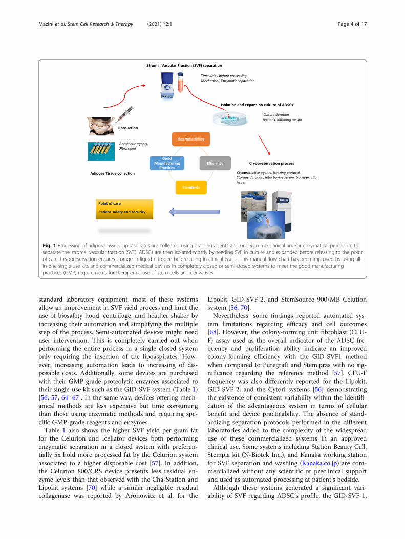
SVF processingDefined methods were shown to harvest AT with regardto lipoaspirate’s invasive manner, cell viability, and col-lection volume [47–49]. Figure 1 summarizes the basic-ally steps involved in SVF and ADSC processing. AfterColeman’s protocol, semi-closed and completely closedsystems aiming to separate fat tissue fraction from thecontaminating fluids (saline solution, adrenaline, anes-thetics) have been elaborated to completely secure thecompelling process mandatory for clinical use. Studieshave indicated that local anesthetic agents might nega-tively impact quantitatively and qualitatively preadipo-cytes [49]. Indeed, reduced ADSC viability andchondrocyte cytotoxicity were shown after lidocaine[50–52]. Ultrasound-assisted liposuction has also beenreported to compromise recovery and expansion ofADSCs [53], suggesting that the absence of a standard-ized fat harvesting process is the first parameter leadingto variability in ADSC therapeutic outcomes.To improve SVF separation and comply with the GMP
requirements, medical devices and highly technologicalequipment have been developed. These commerciallyavailable devices consisted usually of specific bags forwashing AT and permitting the enzymatic digestion andSVF separation using at the same time clinical-grade re-agent exempt from animal additives. Cell outcomes andviability added to their clonogenic potencies are reportedsignificantly efficient compared to the control separationwithout preventing the variability related to the manipu-lator [54–57].These devices provide all the supplies needed for the SVF
separation in all-in-one single-use kit having the advantageto reduce the risk of contamination during cell separationand favoring their use at patient’s bedside [58, 59]. Identicalsystem has been used for the bone marrow (BM) withadapted perfusion system leading to expansion of BM re-generative cells [60] and MSCs [61–63].Table 1 represents the main commercialized systems
used to separate SVF in semi-automated or fully auto-mated level. Compared to the manual methods requiring
Mazini et al. Stem Cell Research & Therapy (2021) 12:1 Page 3 of 17
standard laboratory equipment, most of these systemsallow an improvement in SVF yield process and limit theuse of biosafety hood, centrifuge, and heather shaker byincreasing their automation and simplifying the multiplestep of the process. Semi-automated devices might needuser intervention. This is completely carried out whenperforming the entire process in a single closed systemonly requiring the insertion of the lipoaspirates. How-ever, increasing automation leads to increasing of dis-posable costs. Additionally, some devices are purchasedwith their GMP-grade proteolytic enzymes associated totheir single-use kit such as the GID-SVF system (Table 1)[56, 57, 64–67]. In the same way, devices offering mech-anical methods are less expensive but time consumingthan those using enzymatic methods and requiring spe-cific GMP-grade reagents and enzymes.Table 1 also shows the higher SVF yield per gram fat
for the Celurion and Icellator devices both performingenzymatic separation in a closed system with preferen-tially 5x hold more processed fat by the Celurion systemassociated to a higher disposable cost [57]. In addition,the Celurion 800/CRS device presents less residual en-zyme levels than that observed with the Cha-Station andLipokit systems [70] while a similar negligible residualcollagenase was reported by Aronowitz et al. for the
Lipokit, GID-SVF-2, and StemSource 900/MB Celutionsystem [56, 70].Nevertheless, some findings reported automated sys-
tem limitations regarding efficacy and cell outcomes[68]. However, the colony-forming unit fibroblast (CFU-F) assay used as the overall indicator of the ADSC fre-quency and proliferation ability indicate an improvedcolony-forming efficiency with the GID-SVF1 methodwhen compared to Puregraft and Stem.pras with no sig-nificance regarding the reference method [57]. CFU-Ffrequency was also differently reported for the Lipokit,GID-SVF-2, and the Cytori systems [56] demonstratingthe existence of consistent variability within the identifi-cation of the advantageous system in terms of cellularbenefit and device practicability. The absence of stand-ardizing separation protocols performed in the differentlaboratories added to the complexity of the widespreaduse of these commercialized systems in an approvedclinical use. Some systems including Station Beauty Cell,Stempia kit (N-Biotek Inc.), and Kanaka working stationfor SVF separation and washing (Kanaka.co.jp) are com-mercialized without any scientific or preclinical supportand used as automated processing at patient’s bedside.Although these systems generated a significant vari-
ability of SVF regarding ADSC’s profile, the GID-SVF-1,
Fig. 1 Processing of adipose tissue. Lipoaspirates are collected using draining agents and undergo mechanical and/or enzymatical procedure toseparate the stromal vascular fraction (SVF). ADSCs are then isolated mostly by seeding SVF in culture and expanded before releasing to the pointof care. Cryopreservation ensures storage in liquid nitrogen before using in clinical issues. This manual flow chart has been improved by using all-in-one single-use kits and commercialized medical devises in completely closed or semi-closed systems to meet the good manufacturingpractices (GMP) requirements for therapeutic use of stem cells and derivatives
Mazini et al. Stem Cell Research & Therapy (2021) 12:1 Page 4 of 17

Stem.pras, and Puregraft did not impact CD73 andHLA-ABC expression level similarly to thehematopoietic markers CD14, CD45, HLADR, andCD34 when compared to the reference method. Never-theless, CD34 expression was drastically decreased inpassage 1 expanded ADSCs [57]. Aronowitz et al. re-ported no significant differences in the frequency ofCD31−/CD34+/CD45 cells in SVF separated by Lipokit,GID SVF-2, and StemSource 900/MB systems [56].When using the Unistation device, SVF presented a de-creased CD34+ expression frequency and an increasedCD45+ cell counts with highly proliferative CD271 simi-lar to the reference method [71]. On the other side, theCelution system reported significantly more endothelialcells and CD34/CD31 cells [70]. A recent report on theuse of Icellator system demonstrated a predominant ex-pression of CD90, CD29, and CD34 on cryopreservedSVF followed by CD45, CD105, CD73, and CD44; how-ever, comparative analysis with a reference method wasnot reported [69]. Inversely, isolated by the Stem.prasmethod, SVF was decreased significantly in their CD90expression level [57]. This suggests that in the absenceof a comparative analysis and a standardized assessmentmethod of the different devices, large clinical use of AT-derived products associated with patient’s benefit andsafety meeting the international requirements remains
limited. Side-by-side clinical trials will be required to es-tablish the relevance of these differences.
ADSC separation and expansionDifferent protocols were proposed in isolating ADSCs [45,72–79]. Increasing adherent ADSCs by taking SVF to 60 hadhesion followed by forceful washing was also used [80].Although sorting among specific cell markers resulted insubpopulations exhibiting distinct osteogenic and chon-drogenic differentiation potentials [81, 82]. Other reportshave proposed CD271 as a marker for tissue regenerationand showed that CD271+ ADSCs presented differenti-ation ability to mesodermic lineages [83, 84].ADSC separation is preferentially performed by pro-
longed culture of the SVF mixture over time by their ad-herence to plastic and successive culture passages [45,74, 77–79]. Lately, culture passages contained ADSCspopulations with a stromal phenotype associated withhoming properties. In contrast, early passages repre-sented heterogeneous ADSC populations with distinctsurface-cell adhesion molecules consistent with theirstemness and differentiation abilities [79, 85, 86].Despite complying with GMP conditions, culture
medium composition is a key regulator in defining cellimmunophenotype and secretome composition in pro-teins and nucleic acids [4, 11, 54, 87]. There are
Table 1 Point of care devices for bed side separation of SVF
Medicaldevise
Manufacturer Closed/semi-closed
Separationmethod
Proceeded fatquantity (g)
Processduration(min)
Cell yield/g of fatDevice/control (D/C)
CellviabilityD/C orD
References
Celution R800/CRSStemSource900/MB
CytoriTherapeutics,Inc
Closed Enzymatic 300 g 90 2.41 × 10⁶/NP NP [57, 58]
GID SVF-1,SVF-2
GID Group, Inc Closed Enzymatic 300 g 90 0.425 × 10⁶ ± 0.047 × 10⁶/0.795 × 106 ± 0.228 × 10⁶
50–84% [56, 57, 64–67]
Icellator® Tissue Genesis,Inc.
Closed Enzymatic 60 80 0.25–2.0 × 10⁶ 64.5 ±11.4
[57, 68, 69]
Puregraft™ Eurosilicone Semi-closed Mechanical 250 100 0.25 × 10⁶ ± 0,07 × 10⁶/0.79 ×106 ± 0.228 × 10⁶
77% [57]
Stem.pra® Proteal Closed Mechanical 200 110 0.535 × 10⁶ ± 0.209 × 10⁶/0.795 × 106 ± 0.228 × 10⁶
69% [57]
Cha-Station™ PNCInternationalCo., Ltd.
Semi-closed MechanicalEnzymatic
200 90 0.05 × 10⁶ NA [57, 70]
Sepax® Biosafe GropSA
Semi-closed Mechanical 300 90–120 2.6 ± 1.2 × 10⁵/ML NA [55, 57]
UNISTATION™ NeoGenesis Closed Notprovided
NP NP 1.1 × 10⁵ ± 1.1 × 10⁵/2.0 ×105 ± 1.7 × 10⁵
NA [71]
LipokitMaxStem
Medi-Khan’s Semi-closed Enzymatic NP 88–120 0.35 × 10⁶ 50–84 [56, 70]
Processing with the different devices is reported regarding the closed or semi-closed systems and their efficiency to separate SVF in terms of fat quantity, cellyield and viability, and duration. These devices provide all-in-one single-use kit for the SVF separation and might also contain their GMP-grade proteolyticenzymes. NP not provided and, NA not applicable
Mazini et al. Stem Cell Research & Therapy (2021) 12:1 Page 5 of 17

evidences that ADSCs were primed with their microen-vironment’s composition highlighting the need of stan-dardized ready-to-use culture media. Basically, culturemedium is composed of Dulbecco’s minimal essentialmedium (DMEM) supplemented with 10% of fetal bo-vine serum (FBS), fetal calf serum (FCS), human plateletlysate (hPL), or growth factors derived from platelets.One of them, the fibroblast growth factor-2 (FGF-2),specifically improves chondrogenic differentiation [88]and together with FBS induced highly expression of theCD146 antigens by cultured ADSCs [87]. Using animalproducts containing reagents remains a real concernthrough their contaminating biomolecules.These distinct properties are promising and might
have important implications for the pre-selection ofADSC subpopulations and thus sibling cell-based ther-apies. Thus, such expanded subpopulations might be re-producibly manufactured in a time point for a clinicalspecific use.Many standardized methods of isolating ADSCs have
been proposed and designed, consisting of mechanical(centrifuge) or enzymatic (collagenase) means or both ofthem in a complete closed system [57, 75, 89–92]. Theseapproaches aim to prevent the conventional methods as-sociated to contamination risks and time consuming andspecifically to reduce manufacturing time and costs. In-deed, these closed or semi-closed devices are effective,safe, and economic for a large clinical use and allowed forgreater number of isolated and expanded ADSCs whencompared to the manual procedure [76, 89, 90, 92]. TheQuantum Cell Expansion System is a hollow fiber bioreac-tor (Terumo BCT, Lakewood, CO, USA) mostly used dueto the reproducible, economical, and the large cell quan-tities seeded. This system is a GMP-compliant able to gen-erate 1 × 10⁸ cells in 2 weeks [93]. Similar efficient,however, manual protocols have been reported in compli-ance with GMP conditions [45, 76, 89].Thoroughly, expansion protocols of ADSCs have been
improved by using PL with more benefit than FBS re-garding proliferation and differentiation capacities, CFU-F frequency, and cell senescence [87, 94–101]. Using theQuantum Cell Expansion System, ADSCs yield esti-mated as population doubling is significantly more im-portant than in control conditions with FBS [90, 92].Also, when expanded with PL, ADSCs exhibited less im-munogenic potential with a preserved normal karyotypefor at least six passages of culture [102]. Likewise, evi-dences have been demonstrated for platelet-rich plasma(PRP) in inducing proliferation and motility of ADSCswithout affecting survival of mature adipocytes improv-ing thus fat engraftment outcomes [103]. Added to that,expansion protocols are set up for different passages andthe literature showed inconsistent results. We havealready reported that ADSCs should not be expanded
more than 2 passages before being used or frozen [4]due to the modified immunological profile. All these fac-tors raised the need to formulate a standard operatingprotocol complying with GMP requirements for exten-sive clinical scale.
Cryopreservation and storageFreezing mediaBasically, cryopreservation could not occur withoutusing cryoprotective agents (CPA) alone or in combin-ation with FBS thanks to their improvement of cell via-bility of frozen and thawed cells. Several hydrocolloidsand organic osmolytes are successfully used as CPA [52,104–108]. Because of storage of SVF or ADSCs is usu-ally performed with FBS, many studies aimed to developefficient cryopreservation methods without serum andanimal products to avoid immunological reactions andrisk of transmission of bacterial/viral infections andprions. Xeno- and serum-free media were thus formu-lated objectively to be used for isolation, expansion, andbanking of ADSCs. In these media, CPAs were supple-mented with polymers and anti-oxidants to mimic thebeneficial effects of serum [105, 109–111]. Consequently,the recovery, functionality, and multipotency of thawedADSCs appeared fully maintained [105, 110, 111].However, there is evidence that the cellular activity of
ADSCs after freezing and thawing was affected by CPA[112]. The most used one remains the DMSO, being re-ported the rare successful for all cell types even with itspotential toxicity and it being difficult to remove afterthawing. On the other hand, DMSO was indicated moreeffective than polyvinylpyrrolidone (PVP) [113] and isnow manufactured at GMP and clinical grade. Likewise,cryoprotectant media containing DMSO are available onthe market such as CryoStor (BioLife Solutions). Kwet al. reported an even more high cell viability and nor-mal cell phenotype and proliferation rate of ADSCscryopreserved in 5% DMSO without FBS addition [114].When adding glycerol and PL instead of FBS, high cellviability of xeno-free extracted and cryopreservedADSCs was also demonstrated [96]. Nevertheless, des-pite using DMSO at clinical grade, the DMSO-containing cryomedia are differently prepared within thelaboratories, non-standardized, adding to the variabilityof the process.
Storage durationADSC use focuses on their preservation and storage pos-sibilities; nevertheless, the optimal temperature andlength of cryopreservation are particularly relevant fortheir cell outcomes in terms of viability and differenti-ation capacity [52, 104, 115–117]. For a short time (1month), the first results did not report differences in cellviability after successive cryopreservation in − 20 °C, −
Mazini et al. Stem Cell Research & Therapy (2021) 12:1 Page 6 of 17
80 °C, and liquid nitrogen [117, 118]. Wolter et al. sug-gested better conditions in − 80 °C [115] and others fa-vored liquid nitrogen [116]. A more recent findingindicated that ADSC viability and differentiation capabil-ities after 1 or 2 months cryopreservation in − 80 °C andliquid nitrogen were comparable to fresh samples [52].Nevertheless, few investigations are conducted for thelong-term cryopreservation impact on the viability andefficiency of ADSCs. Badowski et al. have reported thatADSCs and AT lose viability and differentiation capacityafter storing as long as 44 months [119].
Cell outcomesViability is another significant parameter reflectingcryopreservation success and it appeared closely re-lated to the freezing rate and in some extent to thaw-ing protocols. Indeed, post-thaw viability assessmentwas shown to be critical in clinical applications. Cryo-preservation studies with early passages of ADSCs didnot impact the immediate post-thawing viability whenfrozen samples were thawed at 10 °C/min in con-trolled rate freezer or in water bath at 37 °C [120].Accordingly, cryopreservation has been reported toimpact ADSC functionality for longer expandedADSCs (more than 2 passages) [4].There are evidences that the freeze-thawing process
induced cell stress manifested by necrotic activity at 4–8 h post-thaw and apoptotic activity after 8–12 h thaw-ing, leading to a time-dependent decline in viability andfunction at culture temperatures [121]. The need to per-form viability control at different time points of post-thaw cultured cells became imminent for a more accur-ate assessment of cell quality and quantity. For a routineviability measurement, the trypan blue dye exclusionassay is the most commonly used. But this method isnot reproducible and remains observer dependent inaddition to the disadvantages of small number of cellsanalyzed. Many reports and consensus have suggestedthe use of fluorescent dyes such as 7-AAD and SYTO16in flow cytometry analyze, as the most accurate and reli-able indicators of cell viability [121, 122].Also, to minimize osmotic shock, controlling thawing
and transportation need to be more accurate. Some au-tomated systems for vials and bags, such as ThawSTAR(BioCision) or VIA freeze (Asymptote), might help instandardizing the thawing process. These new solutionscan be supported by the use of specific smart shippers(EVO Shipper, BioLife Solutions) in tracking positioningproducts during delivery.
ADSC banking: adopted and emerging issuesUmbilical cord blood (UCB) banking has previouslybenefited from international standards implemented byboth the AABB and the FACT together with NetCord
(NetCord-FACT) for quality management systems andtechnical requirements. These standards are incorpo-rated within their general cellular therapy standards,while FACT-NetCord CB requirements are separatedbut aligned with their cellular therapy standards. Suchgeneral guidelines and safeguards have been adopteddespite the existence of heterogeneity and differencesin ethical and legal process controlling the permissi-bility of ADSC use in therapeutics. As for thehematopoietic stem cells and CB, storing ADSCs forlong time is currently improved using cryovials orcryobags applied for manual procedures and semi-closed or closed automated systems. However, and ac-cording to GMP requirements, stem cell banks mostlyused specific cryobags to allow culture and qualitycontrol procedures such as those used for freezingand banking of CB stem cells with GMP [123]. Spe-cific containers required for storage and shipment tothe point of care are mostly identical to those used inworldwide UCB banking.Moreover, long-term banking of BM- and UC-
derived stem cells has become commonplace. A re-cent result reported that AT and ADSCs can be cryo-preserved for up to 44 months until use [119].However, the perspectives regarding cell viability,functionality, and integrity remain insufficient andfurther investigations are needed to act on the feasi-bility of the long-term banking. Likewise, in case ofbacterial/viral infections, low temperatures would notstop proliferating infectious agents, suggesting thatwith consistent agents such as the coronavirus, sus-tainable quality control at different time point is be-coming a real issue during cryopreservation.
Product release and ADSC quality controlMost attentions should now be focused on the in-process controls and final release criteria includingcontrol production tests (cell dose, viability, immunephenotype, microbial, endotoxin, and mycoplasmatesting) and functional assays (clonogenicity, trilineagedifferentiation, immunomodulation, and hematopoiesisregulation). This continuous testing process consti-tuted an adequate tool to identify any dysfunctionsleading to ADSC or derivative exclusion and preventcell and time loss. Existing methods or tests for BM-MSCs and UCB are applied according to the Inter-national Pharmacopeia and could benefit to safetyevaluation of biological preparations in terms of rap-idity and sensitivity such as in microbiological andendotoxin testing [124, 125]. Likewise, potency andfunctionality assays were largely reported in theliterature despite the absence of specific markers, butstandardization is still lacking. Finally, concomitant envir-onment controls might prevent any contamination risks
Mazini et al. Stem Cell Research & Therapy (2021) 12:1 Page 7 of 17
and allowed confirmation of the graft sterility before re-lease for transplantation.During manufacturing, ADSCs undergo different pro-
cessing conditions before being administered. As indicatedin Table 1, the type of devices used to separate SVF im-pacted directly ADSC profile. Positive cell amounts as wellas cell intensity CI were both reported different.ADSCs could be freshly expanded, cryopreserved/
thawed, and expanded or not. The expression of the clas-sical mesenchymal markers CD44, CD73, CD90, andCD105 appeared to be irrespective of these manufacturingtime points. Conversely, cryopreservation and 4 daysADSC expansion results on the increase of CD271- andCD63-positive cells. This non-classical MSC markers in-crease might correlate with increasing cell recovery duringculture and their mRNA expressions differ in proliferativeand post-proliferative ADSCs [126]. Additionally, some re-ports have indicated that ADSCs are negative for themonocyte/macrophage marker CD163 and their expres-sion of the immune regulatory markers CD274 andCD276 could be predictive for their immunomodulatorypotential [126–128]. Thus, culture conditions togetherwith cryopreservation and the patient- or donor-dependent factors would impact their therapeutic out-comes and specifically their paracrine activity. ADSCs se-crete largely the pro-inflammatory IL-6 factor, and thissecretion could be amplified during culture expansion [4].Combination of flow cytometry and q-PCR techniquesmight be more useful in characterizing clinical-gradeADSCs and complete the first criteria previously imple-mented by International Society for Cell Therapy (ISCT)
and International Federation for Adipose Therapeutics(IFATS) as release standard. Q-RT-PCR could also be use-ful for testing the apoptotic activity of thawed cells.This characterization is of interest for tracking cell be-
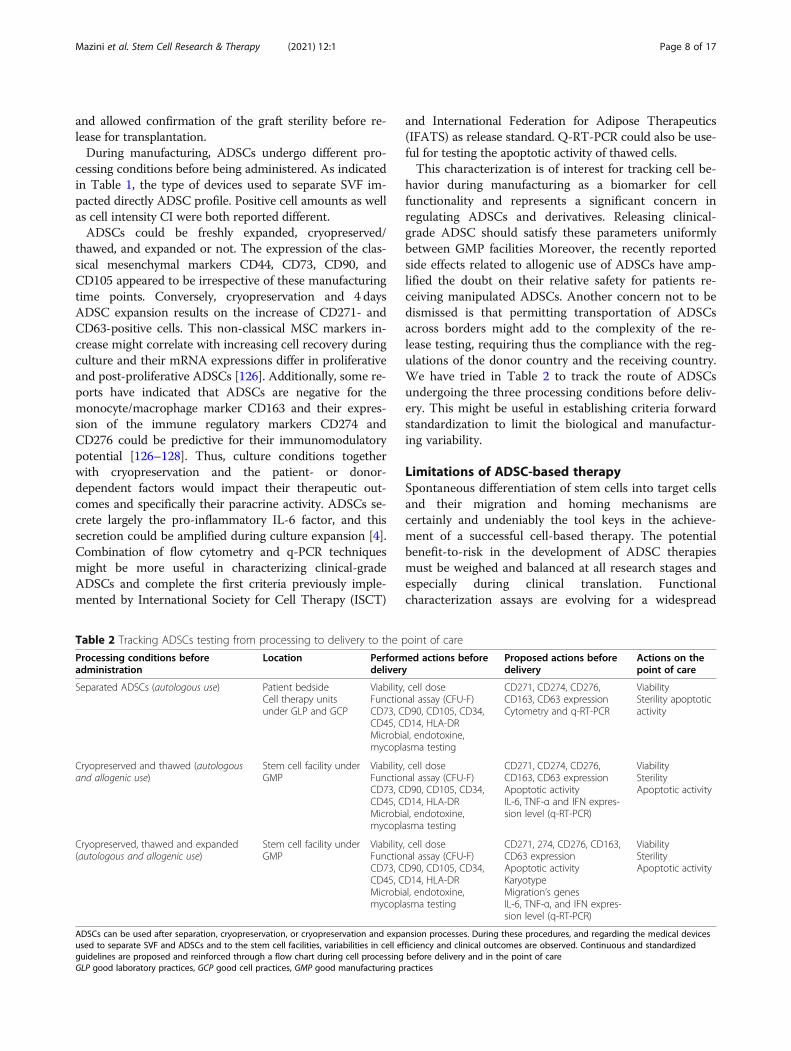
havior during manufacturing as a biomarker for cellfunctionality and represents a significant concern inregulating ADSCs and derivatives. Releasing clinical-grade ADSC should satisfy these parameters uniformlybetween GMP facilities Moreover, the recently reportedside effects related to allogenic use of ADSCs have amp-lified the doubt on their relative safety for patients re-ceiving manipulated ADSCs. Another concern not to bedismissed is that permitting transportation of ADSCsacross borders might add to the complexity of the re-lease testing, requiring thus the compliance with the reg-ulations of the donor country and the receiving country.We have tried in Table 2 to track the route of ADSCsundergoing the three processing conditions before deliv-ery. This might be useful in establishing criteria forwardstandardization to limit the biological and manufactur-ing variability.
Limitations of ADSC-based therapySpontaneous differentiation of stem cells into target cellsand their migration and homing mechanisms arecertainly and undeniably the tool keys in the achieve-ment of a successful cell-based therapy. The potentialbenefit-to-risk in the development of ADSC therapiesmust be weighed and balanced at all research stages andespecially during clinical translation. Functionalcharacterization assays are evolving for a widespread
Table 2 Tracking ADSCs testing from processing to delivery to the point of care
Processing conditions beforeadministration
Location Performed actions beforedelivery
Proposed actions beforedelivery
Actions on thepoint of care
Separated ADSCs (autologous use) Patient bedsideCell therapy unitsunder GLP and GCP
Viability, cell doseFunctional assay (CFU-F)CD73, CD90, CD105, CD34,CD45, CD14, HLA-DRMicrobial, endotoxine,mycoplasma testing
CD271, CD274, CD276,CD163, CD63 expressionCytometry and q-RT-PCR
ViabilitySterility apoptoticactivity
Cryopreserved and thawed (autologousand allogenic use)
Stem cell facility underGMP
Viability, cell doseFunctional assay (CFU-F)CD73, CD90, CD105, CD34,CD45, CD14, HLA-DRMicrobial, endotoxine,mycoplasma testing
CD271, CD274, CD276,CD163, CD63 expressionApoptotic activityIL-6, TNF-α and IFN expres-sion level (q-RT-PCR)
ViabilitySterilityApoptotic activity
Cryopreserved, thawed and expanded(autologous and allogenic use)
Stem cell facility underGMP
Viability, cell doseFunctional assay (CFU-F)CD73, CD90, CD105, CD34,CD45, CD14, HLA-DRMicrobial, endotoxine,mycoplasma testing
CD271, 274, CD276, CD163,CD63 expressionApoptotic activityKaryotypeMigration’s genesIL-6, TNF-α, and IFN expres-sion level (q-RT-PCR)
ViabilitySterilityApoptotic activity
ADSCs can be used after separation, cryopreservation, or cryopreservation and expansion processes. During these procedures, and regarding the medical devicesused to separate SVF and ADSCs and to the stem cell facilities, variabilities in cell efficiency and clinical outcomes are observed. Continuous and standardizedguidelines are proposed and reinforced through a flow chart during cell processing before delivery and in the point of careGLP good laboratory practices, GCP good cell practices, GMP good manufacturing practices
Mazini et al. Stem Cell Research & Therapy (2021) 12:1 Page 8 of 17
clinical exploitation without documenting the cell typeused regarding culture expansion and freezing (SVF, ex-panded or non-expanded ADSCs, number of expandingpassage, frozen/thawed ADSCs). However, some limita-tions are still surrounding early-phase studies usingADSCs. Safety, purity, and application dose are the mainconcerns for an efficient therapeutic application at alarge scale. In that fact, many reports have agreed forthe presence of contaminating cell populations that mayaffect the targeted biological effects and induce potentialside effects. The currently applied dose is about 1–5 ×10⁶ MSCs/kg of body weight [129–131], but their benefi-cial effect might be enhanced regarding time and admin-istration route and schedule. The heterogeneity of ADSCprofile and especially the use of SVF might also impactthese effects, as well as the overall ADSC physiologicalchanges observed during expansion culture. On theother side, the absence of a specific marker did not help incharacterizing and standardizing ADSC clinical investiga-tions. However, the combination of positive and negativemarkers should facilitate identification and selection of aclosed stromal population of CD45-CD235a-CD31-CD34+ cells within SVF excluding hematopoietic andendothelial cells [86, 132, 133]. This selection should beoptimized with other markers such as CD13, CD73,CD63, CD271, CD274, and CD276 for more reproducibleidentification or selection purposes [126, 128, 134].Otherwise, in situ use of manipulated ADSCs is some-
times hampered by the difficulty in maintaining cell con-tact with the damaged tissue. Associations have emergedwithin latest years and used ADSCs in combination withbio-engineered materials. The most representative com-bination consisted of using biomaterials, growth factors,plastic support, nanostructures, polymers, etc., as a sup-port of a tissue or organ repair based on tissue engineer-ing [135–137]. These supports were performed tofacilitate seeding of the cells and added to the difficultyin standardizing technical protocols.In addition to the limitations already discussed [12],
immunogenicity is another point of view playing a cru-cial role in clinical use of ADSCs. Long-term expandedADSCs demonstrated different immunomodulatory pro-file. ADSC secretome is also affected by their surround-ing microenvironment evidenced by differences inexosomes’ composition. The latter is mainly composedof inflammatory biomolecules, suggesting that screeningof potential inflammatory factors in such cells might bea pre-requisite for their use in clinical purposes.Genetic stability after manipulation and expansion is
another major issue of the advanced application ofADSCs in therapeutics. There is controversy on thespontaneous transformation of these cells potentially bythe formation of mesenchymal tissues at ectopic sites[138, 139] or accumulation of genetic alterations and
malignant transformations [140–142]; these transforma-tions seemed unrelated to the origin of MSCs. Moreover,the extremely rare malignant events reported derivedfrom contaminating tumor cell lines [143, 144].Consequently, the possible undesirable differentiation
of ADSCs and their interaction with tumor cells raisesgreat interest, even if the reported cases are very limited.A quantitative approach should be intended to docu-ment the functionality of ADSCs; lineage-specific geneor protein biomarkers could be used. In addition, geneticassays should be routinely integrated through conven-tional/molecular karyotyping before release to the pointof care.To be administered in the point of care, ADSCs or de-
rivatives should be transferred from the facility. Actually,no specific transportation systems are available whichmight impact stem cell viability and proliferation. Even ifliquid nitrogen is usually used for transportation, the ef-fect on frozen cell viability cannot be neglected. Also,DMSO is not washed in the released product, reducingthus the associated therapeutic outcomes. Chu DT et al.have reported that BM-MSCs undergo similar incon-venience; however, their non-frozen transportation iscompletely avoided [145]. Cell-free therapy is holdinggreat promise through using ADSC-derived exosomes.These exosomes offer crucial benefit through (i) a con-sistent and standardized composition using genome edit-ing technology, (ii) increasing their release, and (iii) newpossible formulations such as lyophilizates to overcometransportation and conservation disadvantages [146].The risks associated with the medical practice and
competence are the new issues that raise serious interestacross cell therapy agencies. The specialty accreditationand the participation of the national government andprofessional authorities in settling and standardizing pol-icies worldwide is another issue largely studied and re-ported by the working group of the USA [147]. Hence,the need to overcome and control secondary side effectsis actually challenging. A flow chart has been developedto identify control points at any process level which ap-peared different from the guidelines of finished drugs.Nevertheless, the risk tier associated to this cell therapyhas been limited only to the cell products and not to theroute of delivery and the practitioner as it is easily ap-plied for the investigated new drug [147]. These contra-dictions might raise questions on the need to refine thestem cell therapy guidance with respect to drugmanufacturing.
Refining new cell-based therapies in the contextof COVID-19Evolving new approachesThe SARS-CoV-2 (severe acute respiratory syndrome cor-onavirus 2) breakdown is now the main interest of the
Mazini et al. Stem Cell Research & Therapy (2021) 12:1 Page 9 of 17
whole world health systems. This infection is responsible ofthe coronavirus disease-19 so called COVID-19 and associ-ated with a severe acute respiratory illness and multipleorgan dysfunction leading to a significant mortality and aworldwide epidemic emergency. The pathogenesis of thevirus is due to the presence of its angiotensin I-convertingenzyme 2 receptor (ACE2), highly expressed in the lung al-veolar type II cells and capillary endothelial cells [148, 149].Other tissues also present ACE2 such as the heart, kidney,liver, and digestive organs. Additionally, alveolar cells alsoexpress the cellular transmembrane protease serine 2(TMPRSS2) which enable the virus entering the cell mem-brane through priming of its spike protein [148, 150]. Theviral infection results in an overreaction of the immune sys-tem as a cytokine storm. Overexpression and release of pro-inflammatory cytokines such as IL-1β, IL-2, IL-6, interferonα (IFNα), IFNβ, IFNγ, and monocyte chemoattractant pro-tein (MCP)-1 lead to edema, air exchange dysfunction,acute respiratory distress, secondary infection, and multipleorgan failure and even death [148, 151].As no specific drugs or vaccines are available yet,
many therapeutic plans have been proposed and most ofthem are supportive care rather than curative. An over-view of recently investigated strategies including tacklingcytokine storm, antiviral therapy, plasma from recoveredpatients, traditional Chinese medicine, blocking agentsbinding to ACE2 receptor, and vaccination have been re-ported [21, 152–154].However, interesting and encouraging approaches have
been realized through MSCs therapy intravenously orintrathecally administered to COVID-19 patients [17, 21,23, 25, 27, 28, 148, 155–157]. Effectiveness and efficacyof MSCs in disease-associated inflammation and in im-mune diseases were well documented [158]. Global ana-lysis of reduced mortality, patient safety, and absence orresolved side effects were demonstrated [21, 29]. Theimmunomodulatory effects of MSCs might be useful inattenuating or preventing the cytokine storm and out-pace the evidence in treating infected patients by target-ing immune cells including macrophages, dendritic cells,T and B cells, and natural killer. Adding to their actionon immune cells, MSCs appeared to have an anti-microbial potential and acted through secretion of anti-microbial peptides and proteins (AMPs) and expressionof indoleamine 2,3-dioxygenase (IDO) and IL-17, sug-gesting that these cells can increase the innate immuneresponse to bacterial infection [159].On October 12, more than 134 clinical trials are regis-
tered and 62 of them are using MSCs from differentsources. Nineteen clinical trials with UC-MSCs aremostly ongoing where those performed with BM-MSCsand ADSCs (9 and 10 respectively) are not yet recruiting(http://clinicaltrials.gov). Autologous and allogenic MSCtherapy has emerged as inhibiting of the immune system
and thus treating COVID-19 pneumonia [13, 14, 21].Assessing safety and efficacy of UC-MSCs in treating thepneumonia associated to the viral infection representsthe major objective of these clinical trials [160–163].UC-MSCs are also used in treating pneumonia in pa-tients infected with coronavirus [164, 165].First published results with infused umbilical cord
MSCs (UC-MSCs) to critically ill COVID-19 patientspresent an improved therapeutic outcome [156, 162, 163].Other strategies consisted of using ACE2− UC-MSCs lead-ing to an improved pulmonary functional activity and ab-sence of SARS-CoV-2 nucleic acid with more benefit toelderly patients [148]. Infusion of UC-MSCs leaded to arobust anti-inflammatory activity represented by an in-creased number of circulating lymphocytes and a decreasein overactivated cytokine-secreting immune cells and inTNF-α in contrary to an enhanced IL-10 secretion. MSCswere largely known to auto-induce and address theirmicroenvironment to ensure cell proliferation and tissueregeneration. It seems that UC-MSCs act similarlythrough paracrine effect to counteract the cytokine stormand severe inflammation, likely by protecting or rejuvenat-ing alveolar epithelial cells [155]. Interestingly, irradiatedUC-MSCs are also expected to alleviate the symptoms as-sociated to COVID-19 pneumonia thanks to the modulat-ing annexin-1 released by their exosomes [166]. Nocomparisons between MSCs sources and clinical benefitwere reported in COVID-19.Having the advantages of being in higher quantities and
easy to access, ADSCs might be further presented as apromising tool in combatting COVID-19-induced pneu-monia and be a part of future treatment option. ADSCshave proven efficiencies in treating pulmonary diseases inanimal models through a paracrine pathway promotingthus proliferation of epithelial cells and inhibiting apop-tosis [167, 168]. These cells were also reported to differen-tiate into type 2 alveolar cells [168]. One clinical trial isparticularly on ADSC exosomes to explore the safety andefficiency of aerosol inhalations in the treatment ofCOVID-19-associated pneumonia. Thirteen severe casesof COVID-19 patients under invasive mechanical ventila-tion have received doses of allogenic ADSCs and pre-sented improved clinical and biological outcomes [29].Used at autologous and allogenic state, these cells havegained interest [13, 14, 19, 23, 27, 169] provided thatMSCs and derivatives are performed in GMP conditionsand regulated by the FDA or EMA.
New cell- and tissue-based therapy limitationsIt is conceivable that ADSCs and derivatives are a greatsupport in treating COVID-19 patients by differentiatinginto adipogenic and epithelial lineages, participating inimmunomodulation, angiogenesis, and anti-inflammatory responses, and improving cell
Mazini et al. Stem Cell Research & Therapy (2021) 12:1 Page 10 of 17
regeneration. Their benefit risk still raises debate. Thefirst criticized aspect is lung trapping as for other MSCs.However, this inconvenience is a prior advantage intreating COVID-19 patients where lung cells are infectedand providing thus local immunomodulation, anti-inflammation, neo-angiogenesis, and bacterial clearance[28].The coronavirus did not infect MSC-infused cells
compared to their progeny [170]; however, many criti-cisms should be considered to fully attain the cell ther-apy efficiency and patient safety. Little is known on thebehavior of the patient immunity after the coronavirusinfection, on the possible recurrence regarding somefactor-associated patients. ACE2 receptor is widely dis-tributed on the kidney, liver, cardiovascular and gastro-intestinal organs, white and brown AT, and culturedadipocytes [171–173]. Likewise, this receptor is the keyof viral tropism in AT even if no evidences of direct in-fection of this tissue with the SARS-CoV-2 are reported[174]. However, transplanted patients with allogeneicBM and renal transplants were reported positive forCOVID-19 and died later probably due to the extremelylower amount of T cells [175]. Prior reflections andquestions rise in the case of cell therapy, but at the ac-tual knowledge, most of them remain unresolved.
i) Presence of ACE2 and TMPRSS2 or any of theirvariant in tissues or in cell transplant might be apotential problem; screening of their expressionlevels should be a pre-requisite for both the donorand receiver for the full success of cell therapy. Theimmune cells, bone marrow, thymus, and lymphnodes are negative for ACE2 [170];
ii) Inactivated virus or any virus fragment might live intissues or body fluids after healing; a control qualityshould take part at every point of cell processing;
iii) After primary infection, the virus might lie dormantin a specific tissue and reactivate in case of frailty ordecrease in immunity like the varicella zoster virus;
iv) Use of reagents containing animal components mightinteract with ACE2, leading to an increase of thereceptor affinity to the protein S of the dormant virus;
v) Even if stem cell products are performed incompliance with the approved guidelines from theFDA or other specific agencies, appropriate screeningrelative to viability, sterility, immunological profile,paracrine activity, and virus RNA testing becomescrucial for their therapeutic use.
In the proposed treating approaches, BM-MSCs arepresent in very low frequencies and could not be able torepair the whole damage and perform healing of the dif-ferent organs’ failure. Among different stem cells, UC-MSCs seem to be preferentially used in cell-based
therapies conducted for infected coronavirus patientsprobably for immediate access [157]. Nevertheless,ADSCs are another alternative to set up new therapeuticprotocols against the inflammation storm induced by theSARS-CoV-2 favoring cell repair and regeneration. Onone side, the guidelines and frameworks identified forcell- and tissue-based therapies must be reformulated toanswer the new fears induced by the COVID-19. On theother side, different works reported today should lead tolargely repeated investigations and well-controlled trialsto confirm the beneficial effect of ADSCs. These issuesare challenging and control the future cell- and tissue-based therapies.
DiscussionMany clinical studies have been designed and conductedfor a wide range of pathologies at autologous and allo-genic settings. However, the variability of their thera-peutic outcomes and lack of reproducibility are resultingfrom the absence of harmonization of their processingand their functional assessment balancing between pa-tient safety and justification of the cell therapy. Some pa-rameters play a critical role in achieving ADSC therapyand raises interest in terms of practicability and theeligibility of the products regarding the regulatoryframework.Anatomical site of AT collection is the first parameter
to underlie regarding the number of suitable viable cells.Jurgens et al. demonstrated higher yields of ADSCs iso-lated from SVF in the abdominal subcutaneous than inthe hip/thigh subcutaneous tissue [48] while these cellswere negatively correlated to body mass index and inde-pendent of patient’s age [176]. Nevertheless, there wasincreasing evidence that fat source (subcutaneous or vis-ceral) influences the proliferation and differentiationability of ADSCs and transplantation outcome [79, 177].Baglioni et al. have reported a significantly highergrowth rate and adipogenic potential in the abdominalsubcutaneous tissue [178].Once collected, ADSC separation still raises debates.
According to the FDA guidance for human cell tissueproducts (HCT/P), separation of non-adipocyte cellcomponents from fat is considered as more than “min-imal manipulation.” Exception could be made if onlyrinsing, cleansing, and sizing processing were consid-ered, suggesting a contradictory on the SVF processingposition within this regulatory. According to the EMA,ADSCs should not be cultured and isolated mechanicallyand used only in the subcutaneous tissue [179].European legislation has also decided on this new ad-
vanced therapy and classified ADSCs as ATMP. Clinicaluse of these cells was associated to their level of substan-tial manipulation as a potential indicator for their func-tional properties [180, 181]. Uncultured or expanded
Mazini et al. Stem Cell Research & Therapy (2021) 12:1 Page 11 of 17
ADSCs were not expected similar and might be dissoci-ated in terms of phenotype and functional characteristicswhich have been largely demonstrated. Moreover, cellsurface proteins involved in cell activity and the risk tierrelated to pathogen transmission raise additional consid-erations on the necessity of implementation of func-tional and viral testing during cell processing andespecially for releasing to the point of care.In contrary to basic cell processing, safety and quality
testing are considered. Additional reagents might beused in the absence of clinical-grade ones but should bejustified and controlled preventing thus transmission ofanimal infectious agents. Additionally, the animal host ofthe COVID-19 infection still remains unknown; screen-ing of medium containing animal components such asanimal serum, antibodies, or any recombinant proteinshould be a pre-requisite and certified before being onthe market. More advancements are required focusingon the development of viable techniques to remove theseanimal components without the loss of valuable bio-logical activity in the final product. Mostly, for FBS,current regulations and guidelines of biopharmaceuticalproducts such as the European Medicine Agency EMEA(1793/02 of the proprietary medical products), thePharmacopeia (Ph. 5W Current edition monograph ofBovine Serum (2262), European Regulations 2005/507for Advanced Therapies (AT), and US Code for FederalRegulation (9CFR) have recommended using of pharma-grade FBS with gamma-irradiated serum complementedwith a viral test panel [45]. FBS can be replaced by PL orhuman serum; however, the amount of autologousserum one patient can provide is limited for a large-scaleclinical expansion of ADSCs.Another point of view to highly consider is relative to
UCB collection. When pregnant women are suffering fromCOVID-19, it is tempting to postulate that the collectedcells might not be free from viral contaminating agents andthus are not eligible for transplantation, suggesting thereinforcement of the control quality of the graft.Another fact reinforcing the potential use of these cells
is that of ADSCs can survive different freezing protocolswithout losing viability opening thus the way for a futurelarge-scale cryopreservation for different therapeutic use[120, 182]. Nevertheless, viability and functionality ap-peared influenced by the long-term cryopreservation.Additionally, ADSC frequency should be improved with-out being largely and long-term expanded preventing thusany functional change or damage [4]. Elimination of CPAduring freezing and thawing could be helpful to preventany variability in cryopreserving medium preparation andpractical for widely ADSC successful clinical use.From the point of view of quality assurance programs
meeting the requirement of cGMP, a uniform cell pro-cessing protocol is of critical importance. Without
standardizing, all the variables may have significant proto-col differences that make cross comparisons difficult. In-deed, medical devices used for SVF separation influencedirectly the ADSC yield and their differentiation ability.Little reports are found on the ADSC expansion at clinicalgrade and a specific concern should be given on harmon-izing the process and to find an international consensusfor a standardizing model. The feasibility of cGMP-compliant and clinical-grade ADSC preparation and bank-ing for clinical cell transplantations should pave the wayto the harmonization of the different aspects of processingand manufacturing. Perhaps, ADSCs and derivatives in ac-cordance with GMP standards should involve several is-sues similar to drug manufacturing guidelines.In the same way, we have proposed a new flow chart
to perform high numbers of ADSCs in high safety andquality standard and preventing at least the assessmentvariability. In the case of using uniformed medical de-vices within GMP facilities, it is tempting to speculatethat the reproducibility of ADSC efficacy will result fromthese standard operating procedures. Hence, achievingtheir adequate clinical effect will remain patient’s associ-ated factors providing the presence of specialized practi-tioner. Specialty societies have followed and consideredall these questions for BM and UCB transplantationsleading to accreditations of stem cell banks which bene-fit to the manufacturing practices in different facilitiesoperating in the field. Bringing ADSCs and derivativesinto the market should take the same way.However, and with regard to patient security, a special
insight should be performed on the presence of COVID-19’s mRNA or derived proteins (protein S) even inacti-vated within the donor tissues. There are evidences ar-guing a viral tropism of AT and the presence of ACE2in adipocytes [16]. Despite the promising alternative of-fered by using ADSCs in COVID-19, the relationship be-tween adipocyte hypertrophy mediated by ACE2receptors and COVID-19 might imbalance their benefitas a potential widespread therapeutic tool. Thus, viralcompounds might reside or resist within AT leading tothe necessity to set up viral identification from fat collec-tion and might be during the whole processing of allstem cell-based products. Emerging strategies shouldtake place to investigate the extent of this virus and thedifferent ongoing vaccine testing on human tissues andon the operating procedures. Cell and tissue transplant-ation landscape is upset and is facing a new challengewith the SARS-CoV-2 virus breakdown.
ConclusionADSCs have proven efficiency in regenerating damagedtissues in vitro and in vivo. However, their self-renewaland multipotency behavior remained the focus of thesuccess of their therapeutic use as it presents multiple
Mazini et al. Stem Cell Research & Therapy (2021) 12:1 Page 12 of 17

variabilities preventing comparisons and practicability.Scientific, practitioner, and specialty societies shouldconverge efforts on the continuously optimized parame-ters such as sourcing, cell dose, cryopreservation, bank-ing, and transplantation methods altogether withfunctional assessments. Optimizing and standardizingnew guidelines relative to this process are really challen-ging as the scope of the COVID-19 remains unknown.
AbbreviationsADSCs: Adipose-derived stem cells; AT: Adipose tissue; HCT/P: Human cells,tissues, or cellular or tissue-based products; AMTPs: Advanced therapymedicinal products; BM: Bone marrow; UCB: Umbilical cord blood;GMP: Good manufacturing practices; FDA: Food and Drug Agency;CPA: Cryoprotective agent; DMSO: Dimethyl sulfoxide; FBS: Fetal bovineserum; HS: Human serum; MSCs: Mesenchymal stem cells; PBS: Phosphate-buffered saline; hPL: Human platelet lysate; SVF: Stromal vascular fraction;COVID-19: Coronavirus disease-19
AcknowledgementsThe authors wish to thank Dr. Jeffrey M. Gimble for his kind assistance inproviding banking stem cell information and the University Mohammed VIPolytechnic for supporting the publication.
Authors’ contributionsLoubna Mazini: conception and design, collection and/or assembly of data,data analysis and interpretation, manuscript writing, final approval ofmanuscript. Mohamed Ezzoubi: provision of study, data analysis andinterpretation. Gabriel Malka: administrative support, financial support, finalapproval of manuscript. The authors read and approved the final manuscript.
FundingThis work has been funded completely by the University of Mohammed VIPolytechnic, Ben Guerir, Morocco.
Availability of data and materialsNot applicable.
Ethics approval and consent to participateAll the authors have agreed to participate to this work.
Consent for publicationNot applicable
Competing interestsThe authors declare there is no conflict of interest.
Author details1Laboratoire Cellules Souches et Régénération Cellulaire et Tissulaire, Centerof Biological and Medical Sciences CIAM, Mohammed VI PolytechnicUniversity (UM6P), Lot 660, Hay Moulay Rachid, 43150 Ben Guerir, Morocco.2Centre des Brûlés et chirurgie réparatrice, Centre Hospitalier Universitaire IbnRochd Casablanca, Faculté de Médecine et de Pharmacie Casablanca,Casablanca, Morocco.
Received: 1 July 2020 Accepted: 1 November 2020
References1. Vermette M, Trottier V, Ménard V, Saint-Pierre L, Roy A, Fradette J.
Production of a new tissue-engineered adipose substitute from humanadipose-derived stromal cells. Biomaterials. 2007;28(18):2850–60.
2. Choi EW, Seo MK, Woo EY, Kim SH, Park EJ, Kim S. Exosomes from humanadipose-derived stem cells promote proliferation and migration of skinfibroblasts. Exp Dermatol. 2018;27(10):1170–2.
3. Park B-S, Jang KA, Sung J-H, Park J-S, Kwon YH, Kim KJ, et al. Adipose-derived stem cells and their secretory factors as a promising therapy forskin aging. Dermatol Surg. 2008;34(10):1323–6.
4. Othmani AE, Rouam S, Abbad A, Erraoui C, Harriba S, Boukind H, et al.Cryopreservation Impacts Cell Functionality of Long Term ExpandedAdipose-Derived Stem Cells. J Stem Cell Res Ther. 2019;09(01) Availablefrom: https://www.omicsonline.org/open-access/cryopreservation-impacts-cell-functionality-of-long-term-expanded-adiposederived-stem-cells-2157-7633-1000445-107364.html. Cited 2019 Apr 19.
5. Gimble JM, Katz AJ, Bunnell BA. Adipose-derived stem cells for regenerativemedicine. Circ Res. 2007;100(9):1249–60.
6. Nakagami H, Maeda K, Morishita R, Iguchi S, Nishikawa T, Takami Y, et al.Novel autologous cell therapy in ischemic limb disease through growthfactor secretion by cultured adipose tissue-derived stromal cells. ArteriosclerThromb Vasc Biol. 2005;25(12):2542–7.
7. Rehman J, Traktuev D, Li J, Merfeld-Clauss S, Temm-Grove CJ, Bovenkerk JE,et al. Secretion of angiogenic and antiapoptotic factors by human adiposestromal cells. Circulation. 2004;109(10):1292–8.
8. Wang L, Tu X-H, Zhao P, Song J-X, Zou Z-D. Protective effect oftransplanted bone marrow-derived mesenchymal stem cells on pancreatitis-associated lung injury in rats. Mol Med Rep. 2012;6(2):287–92.
9. Prichard HL, Reichert W, Klitzman B. IFATS collection: Adipose-derivedstromal cells improve the foreign body response. Stem Cells. 2008;26(10):2691–5.
10. Ren G, Zhang L, Zhao X, Xu G, Zhang Y, Roberts AI, et al. Mesenchymalstem cell-mediated immunosuppression occurs via concerted action ofchemokines and nitric oxide. Cell Stem Cell. 2008;2(2):141–50.
11. Kilroy GE, Foster SJ, Wu X, Ruiz J, Sherwood S, Heifetz A, et al. Cytokineprofile of human adipose-derived stem cells: expression of angiogenic,hematopoietic, and pro-inflammatory factors. J Cell Physiol. 2007;212(3):702–9.
12. Mazini L, Rochette L, Admou B, Amal S, Malka G. Hopes and Limits ofAdipose-Derived Stem Cells (ADSCs) and Mesenchymal Stem Cells (MSCs) inWound Healing. Int J Mol Sci. 2020;21(4).
13. Gentile P, Sterodimas A. Adipose Stem Cells (ASCs) and Stromal VascularFraction (SVF) as a Potential Therapy in Combating (COVID-19)-Disease.Aging Dis. 2020;11(3):465–9.
14. Gentile P, Sterodimas A. Adipose-derived stromal stem cells (ASCs) as a newregenerative immediate therapy combating coronavirus (COVID-19)-inducedpneumonia. Expert Opin Biol Ther. 2020;20(7):711–6.
15. Rogers CJ, Harman RJ, Bunnell BA, Schreiber MA, Xiang C, Wang F-S, et al.Rationale for the clinical use of adipose-derived mesenchymal stem cells forCOVID-19 patients. J Transl Med. 2020;18(1):203.
16. Copcu HE. Potential Using of Fat-derived Stromal Cells in the Treatment ofActive Disease, and also, in Both Pre- and Post-Periods in COVID-19. AgingDis. 2020;11(4):730–6.
17. Bulut Ö, GÜrsel İ. Mesenchymal stem cell derived extracellular vesicles:promising immunomodulators against autoimmune, autoinflammatorydisorders and SARS-CoV-2 infection. Turk J Biol = Turk Biyoloji Dergisi. 2020;44(3):273–82.
18. Taghavi-Farahabadi M, Mahmoudi M, Soudi S, Hashemi SM. Hypothesis forthe management and treatment of the COVID-19-induced acute respiratorydistress syndrome and lung injury using mesenchymal stem cell-derivedexosomes. Med Hypotheses. 2020;144:109865.
19. Gardin C, Ferroni L, Chachques JC, Zavan B. Could Mesenchymal Stem Cell-Derived Exosomes Be a Therapeutic Option for Critically Ill COVID-19Patients? J Clin Med. 2020;9(9).
20. Pinky null, Gupta S, Krishnakumar V, Sharma Y, Dinda AK, Mohanty S.Mesenchymal Stem Cell Derived Exosomes: a Nano Platform for Therapeuticsand Drug Delivery in Combating COVID-19. Stem Cell Rev Rep. 2020.
21. Qu W, Wang Z, Hare JM, Bu G, Mallea JM, Pascual JM, et al. Cell-basedtherapy to reduce mortality from COVID-19: Systematic review and meta-analysis of human studies on acute respiratory distress syndrome. StemCells Transl Med. 2020;9(9):1007–22.
22. Jayaramayya K, Mahalaxmi I, Subramaniam MD, Raj N, Dayem AA, Lim KM,et al. Immunomodulatory effect of mesenchymal stem cells andmesenchymal stem-cell-derived exosomes for COVID-19 treatment. BMBRep. 2020;53(8):400–12.
23. Deffune E, Prudenciatti A, Moroz A. Mesenchymal stem cell (MSc)secretome: A possible therapeutic strategy for intensive-care COVID-19patients. Med Hypotheses. 2020;142:109769.
24. Elrashdy F, Aljaddawi AA, Redwan EM, Uversky VN. On the potential role ofexosomes in the COVID-19 reinfection/reactivation opportunity. J BiomolStruct Dyn. 2020:1–12.
Mazini et al. Stem Cell Research & Therapy (2021) 12:1 Page 13 of 17

25. Tsuchiya A, Takeuchi S, Iwasawa T, Kumagai M, Sato T, Motegi S, et al.Therapeutic potential of mesenchymal stem cells and their exosomes insevere novel coronavirus disease 2019 (COVID-19) cases. Inflamm Regen.2020;40:14.
26. A B, Z P, N B, M H, A S, N R. Regenerative Medicine in COVID-19Treatment: Real Opportunities and Range of Promises. Stem Cell RevRep. 2020; Available from: https://pubmed.ncbi.nlm.nih.gov/32564256/.Cited 2020 Oct 6.
27. Gupta A, Kashte S, Gupta M, Rodriguez HC, Gautam SS, Kadam S.Mesenchymal stem cells and exosome therapy for COVID-19: current statusand future perspective. Hum Cell. 2020;33(4):907–18.
28. Bari E, Ferrarotti I, Saracino L, Perteghella S, Torre ML, Corsico AG.Mesenchymal Stromal Cell Secretome for Severe COVID-19 Infections:Premises for the Therapeutic Use. Cells. 2020;9(4).
29. Sánchez-Guijo F, García-Arranz M, López-Parra M, Monedero P, Mata-Martínez C, Santos A, et al. Adipose-derived mesenchymal stromal cells forthe treatment of patients with severe SARS-CoV-2 pneumonia requiringmechanical ventilation. A proof of concept study. EClinicalMedicine. 2020;25:100454.
30. Schäfer R, Spohn G, Bechtel M, Bojkova D, Baer PC, Kuçi S, et al. HumanMesenchymal Stromal Cells Are Resistant to SARS-CoV-2 Infection underSteady-State, Inflammatory Conditions and in the Presence of SARS-CoV-2-Infected Cells. Stem Cell Rep. 2020.
31. Gunasekaran M, Bansal S, Ravichandran R, Sharma M, Perincheri S, RodriguezF, et al. Respiratory viral infection in lung transplantation induces exosomesthat trigger chronic rejection. J Heart Lung Transplant. 2020;39(4):379–88.
32. Widgerow AD, Salibian AA, Lalezari S, Evans GRD. Neuromodulatory nerveregeneration: adipose tissue-derived stem cells and neurotrophic mediationin peripheral nerve regeneration. J Neurosci Res. 2013;91(12):1517–24.
33. Assunção-Silva RC, Mendes-Pinheiro B, Patrício P, Behie LA, Teixeira FG,Pinto L, et al. Exploiting the impact of the secretome of MSCs isolated fromdifferent tissue sources on neuronal differentiation and axonal growth.Biochimie. 2018;155:83–91.
34. Im G-I. Regeneration of articular cartilage using adipose stem cells. JBiomed Mater Res A. 2016;104(7):1830–44.
35. Gonzalez-Rey E, Gonzalez MA, Varela N, O’Valle F, Hernandez-Cortes P, RicoL, et al. Human adipose-derived mesenchymal stem cells reduceinflammatory and T cell responses and induce regulatory T cells in vitro inrheumatoid arthritis. Ann Rheum Dis. 2010;69(1):241–8.
36. Pak J, Lee JH, Pak N, Pak Y, Park KS, Jeon JH, et al. Cartilage Regeneration inHumans with Adipose Tissue-Derived Stem Cells and Adipose StromalVascular Fraction Cells: Updated Status. Int J Mol Sci. 2018;19(7).
37. Charles-de-Sá L, Gontijo-de-Amorim NF, Maeda Takiya C, Borojevic R, BenatiD, Bernardi P, et al. Antiaging treatment of the facial skin by fat graft andadipose-derived stem cells. Plast Reconstr Surg. 2015;135(4):999–1009.
38. Koh KS, Oh TS, Kim H, Chung IW, Lee KW, Lee HB, et al. Clinical applicationof human adipose tissue-derived mesenchymal stem cells in progressivehemifacial atrophy (Parry-Romberg disease) with microfat graftingtechniques using 3-dimensional computed tomography and 3-dimensionalcamera. Ann Plast Surg. 2012;69(3):331–7.
39. Rigotti G, Charles-de-Sá L, Gontijo-de-Amorim NF, Takiya CM, AmablePR, Borojevic R, et al. Expanded Stem Cells, Stromal-Vascular Fraction,and Platelet-Rich Plasma Enriched Fat: Comparing Results of DifferentFacial Rejuvenation Approaches in a Clinical Trial. Aesthet Surg J. 2016;36(3):261–70.
40. Stepien A, Dabrowska NL, Maciagowska M, Macoch RP, Zolocinska A, MazurS, et al. Clinical Application of Autologous Adipose Stem Cells in Patientswith Multiple Sclerosis: Preliminary Results. Mediat Inflamm. 2016;2016:5302120.
41. Fang B, Song Y, Liao L, Zhang Y, Zhao RC. Favorable response to humanadipose tissue-derived mesenchymal stem cells in steroid-refractory acutegraft-versus-host disease. Transplant Proc. 2007;39(10):3358–62.
42. Kuriyan AE, Albini TA, Townsend JH, Rodriguez M, Pandya HK, Leonard RE,et al. Vision Loss after Intravitreal Injection of Autologous “Stem Cells” forAMD. N Engl J Med. 2017;376(11):1047–53.
43. Sakai D, Schol J, Foldager CB, Sato M, Watanabe M. Regenerativetechnologies to bed side: Evolving the regulatory framework. J OrthopTranslat. 2017;9:1–7.
44. Who. Handbook: Good Laboratory Practice (GLP): Quality Practices forRegulated Non-clinical Research and Development. Geneva; Herndon: WorldHealth Organization Stylus Publishing, LLC [distributor; 2010. Available from:
http://public.ebookcentral.proquest.com/choice/publicfullrecord.aspx?p=579101. Cited 2020 May 4.
45. Larijani B, Aghayan H, Goodarzi P, Mohamadi-Jahani F, Norouzi-Javidan A,Dehpour AR, et al. Clinical Grade Human Adipose Tissue-DerivedMesenchymal Stem Cell Banking. Acta Med Iran. 2015;53(9):540–6.
46. Bieback K, Hecker A, Kocaömer A, Lannert H, Schallmoser K, Strunk D,et al. Human alternatives to fetal bovine serum for the expansion ofmesenchymal stromal cells from bone marrow. Stem Cells. 2009;27(9):2331–41.
47. Aguena M, Fanganiello RD, Tissiani LAL, Ishiy FAA, Atique R, Alonso N, et al.Optimization of parameters for a more efficient use of adipose-derivedstem cells in regenerative medicine therapies. Stem Cells Int. 2012;2012:303610.
48. Jurgens WJFM, Oedayrajsingh-Varma MJ, Helder MN, Zandiehdoulabi B,Schouten TE, Kuik DJ, et al. Effect of tissue-harvesting site on yield of stemcells derived from adipose tissue: implications for cell-based therapies. CellTissue Res. 2008;332(3):415–26.
49. Keck M, Zeyda M, Gollinger K, Burjak S, Kamolz L-P, Frey M, et al. Localanesthetics have a major impact on viability of preadipocytes and theirdifferentiation into adipocytes. Plast Reconstr Surg. 2010;126(5):1500–5.
50. Breu A, Eckl S, Zink W, Kujat R, Angele P. Cytotoxicity of local anesthetics onhuman mesenchymal stem cells in vitro. Arthroscopy. 2013;29(10):1676–84.
51. Girard A-C, Atlan M, Bencharif K, Gunasekaran MK, Delarue P, Hulard O, et al.New insights into lidocaine and adrenaline effects on human adipose stemcells. Aesthet Plast Surg. 2013;37(1):144–52.
52. Roato I, Alotto D, Belisario DC, Casarin S, Fumagalli M, Cambieri I, et al.Adipose Derived-Mesenchymal Stem Cells Viability and DifferentiatingFeatures for Orthopaedic Reparative Applications: Banking of AdiposeTissue. Stem Cells Int. 2016;2016:4968724.
53. Oedayrajsingh-Varma MJ, van Ham SM, Knippenberg M, Helder MN, Klein-Nulend J, Schouten TE, et al. Adipose tissue-derived mesenchymal stem cellyield and growth characteristics are affected by the tissue-harvestingprocedure. Cytotherapy. 2006;8(2):166–77.
54. Agostini F, Rossi FM, Aldinucci D, Battiston M, Lombardi E, Zanolin S, et al.Improved GMP compliant approach to manipulate lipoaspirates, tocryopreserve stromal vascular fraction, and to expand adipose stem cells inxeno-free media. Stem Cell Res Ther. 2018;9(1):130.
55. Güven S, Karagianni M, Schwalbe M, Schreiner S, Farhadi J, Bula S, et al.Validation of an automated procedure to isolate human adipose tissue-derived cells by using the Sepax® technology. Tissue Eng Part C Methods.2012;18(8):575–82.
56. Aronowitz JA, Lockhart RA, Hakakian CS, Birnbaum ZE. Adipose StromalVascular Fraction Isolation: A Head-to-Head Comparison of 4 Cell SeparationSystems #2. Ann Plast Surg. 2016;77(3):354–62.
57. Rodriguez J, Pratta A-S, Abbassi N, Fabre H, Rodriguez F, Debard C, et al.Evaluation of Three Devices for the Isolation of the Stromal VascularFraction from Adipose Tissue and for ASC Culture: A Comparative Study.Stem Cells Int. 2017;2017:9289213.
58. Duckers HJ, Pinkernell K, Milstein AM, Hedrick MH. The Bedside Celutionsystem for isolation of adipose derived regenerative cells. EuroIntervention.2006;2(3):395–8.
59. Katz AJ, Hedrick MH, Llull R, Futrell JW. A novel device for the simple andefficient refinement of liposuctioned tissue. Plast Reconstr Surg. 2001;107(2):595–7.
60. Dennis JE, Esterly K, Awadallah A, Parrish CR, Poynter GM, Goltry KL. Clinical-scale expansion of a mixed population of bone-marrow-derived stem andprogenitor cells for potential use in bone-tissue regeneration. Stem Cells.2007;25(10):2575–82.
61. Rojewski MT, Fekete N, Baila S, Nguyen K, Fürst D, Antwiler D, et al. GMP-compliant isolation and expansion of bone marrow-derived MSCs in theclosed, automated device quantum cell expansion system. Cell Transplant.2013;22(11):1981–2000.
62. Gastens MH, Goltry K, Prohaska W, Tschöpe D, Stratmann B, Lammers D, et al.Good manufacturing practice-compliant expansion of marrow-derived stemand progenitor cells for cell therapy. Cell Transplant. 2007;16(7):685–96.
63. Chen X, Xu H, Wan C, McCaigue M, Li G. Bioreactor expansion of humanadult bone marrow-derived mesenchymal stem cells. Stem Cells. 2006;24(9):2052–9.
64. Dos-Anjos Vilaboa S, Navarro-Palou M, Llull R. Age influence on stromalvascular fraction cell yield obtained from human lipoaspirates. Cytotherapy.2014;16(8):1092–7.
Mazini et al. Stem Cell Research & Therapy (2021) 12:1 Page 14 of 17
65. Brown JC, Shang H, Li Y, Yang N, Patel N, Katz AJ. Isolation of Adipose-Derived Stromal Vascular Fraction Cells Using a Novel Point-of-Care Device:Cell Characterization and Review of the Literature. Tissue Eng Part CMethods. 2017;23(3):125–35.
66. Rose LC, Kadayakkara DK, Wang G, Bar-Shir A, Helfer BM, O’Hanlon CF, et al.Fluorine-19 Labeling of Stromal Vascular Fraction Cells for Clinical ImagingApplications. Stem Cells Transl Med. 2015;4(12):1472–81.
67. Dos-Anjos Vilaboa S, Llull R, Mendel TA. Returning fat grafts to physiologicconditions using washing. Plast Reconstr Surg. 2013;132(2):323e–6e.
68. Doi K, Tanaka S, Iida H, Eto H, Kato H, Aoi N, et al. Stromal vascular fractionisolated from lipo-aspirates using an automated processing system: benchand bed analysis. J Tissue Eng Regen Med. 2013;7(11):864–70.
69. Bukowska J, Alarcon Uquillas A, Wu X, Frazier T, Walendzik K, Vanek M, et al.Safety of Human Adipose Stromal Vascular Fraction Cells Isolated with aClosed System Device in an Immunocompetent Murine Pressure UlcerModel. Stem Cells Dev. 2020;29(7):452–61.
70. Aronowitz JA, Ellenhorn JDI. Adipose stromal vascular fraction isolation: ahead-to-head comparison of four commercial cell separation systems. PlastReconstr Surg. 2013;132(6):932e–9e.
71. Hanke A, Prantl L, Wenzel C, Nerlich M, Brockhoff G, Loibl M, et al. Semi-automated extraction and characterization of Stromal Vascular Fractionusing a new medical device. Clin Hemorheol Microcirc. 2016;64(3):403–12.
72. Rada T, Gomes ME, Reis RL. A novel method for the isolation ofsubpopulations of rat adipose stem cells with different proliferation andosteogenic differentiation potentials. J Tissue Eng Regen Med. 2011;5(8):655–64.
73. Zuk PA, Zhu M, Ashjian P, De Ugarte DA, Huang JI, Mizuno H, et al. Humanadipose tissue is a source of multipotent stem cells. Mol Biol Cell. 2002;13(12):4279–95.
74. Zuk PA, Zhu M, Mizuno H, Huang J, Futrell JW, Katz AJ, et al. Multilineagecells from human adipose tissue: implications for cell-based therapies.Tissue Eng. 2001;7(2):211–28.
75. Raposio E, Bonomini S, Calderazzi F. Isolation of autologous adipose tissue-derived mesenchymal stem cells for bone repair. Orthop Traumatol SurgRes. 2016;102(7):909–12.
76. Raposio E, Simonacci F, Perrotta RE. Adipose-derived stem cells: Comparisonbetween two methods of isolation for clinical applications. Ann Med Surg(Lond). 2017;20:87–91.
77. Zhang X, Hirai M, Cantero S, Ciubotariu R, Dobrila L, Hirsh A, et al. Isolationand characterization of mesenchymal stem cells from human umbilical cordblood: reevaluation of critical factors for successful isolation and high abilityto proliferate and differentiate to chondrocytes as compared tomesenchymal stem cells from bone marrow and adipose tissue. J CellBiochem. 2011;112(4):1206–18.
78. Dominici M, Le Blanc K, Mueller I, Slaper-Cortenbach I, Marini F, Krause D, etal. Minimal criteria for defining multipotent mesenchymal stromal cells. TheInternational Society for Cellular Therapy position statement. Cytotherapy.2006;8(4):315–7.
79. Mitchell JB, McIntosh K, Zvonic S, Garrett S, Floyd ZE, Kloster A, et al.Immunophenotype of human adipose-derived cells: temporal changes instromal-associated and stem cell-associated markers. Stem Cells. 2006;24(2):376–85.
80. Griesche N, Luttmann W, Luttmann A, Stammermann T, Geiger H, Baer PC.A simple modification of the separation method reduces heterogeneity ofadipose-derived stem cells. Cells Tissues Organs (Print). 2010;192(2):106–15.
81. Rada T, Santos TC, Marques AP, Correlo VM, Frias AM, Castro AG, et al.Osteogenic differentiation of two distinct subpopulations of humanadipose-derived stem cells: an in vitro and in vivo study. J Tissue Eng RegenMed. 2012;6(1):1–11.
82. Rada T, Reis RL, Gomes ME. Distinct stem cells subpopulations isolated fromhuman adipose tissue exhibit different chondrogenic and osteogenicdifferentiation potential. Stem Cell Rev. 2011;7(1):64–76.
83. Cuevas-Diaz Duran R, González-Garza MT, Cardenas-Lopez A, Chavez-CastillaL, Cruz-Vega DE, Moreno-Cuevas JE. Age-related yield of adipose-derivedstem cells bearing the low-affinity nerve growth factor receptor. Stem CellsInt. 2013;2013:372164.
84. Calabrese G, Giuffrida R, Lo Furno D, Parrinello NL, Forte S, Gulino R, et al.Potential Effect of CD271 on Human Mesenchymal Stromal Cell Proliferationand Differentiation. Int J Mol Sci. 2015;16(7):15609–24.
85. Pachón-Peña G, Yu G, Tucker A, Wu X, Vendrell J, Bunnell BA, et al. Stromalstem cells from adipose tissue and bone marrow of age-matched female
donors display distinct immunophenotypic profiles. J Cell Physiol. 2011;226(3):843–51.
86. Bourin P, Bunnell BA, Casteilla L, Dominici M, Katz AJ, March KL, et al.Stromal cells from the adipose tissue-derived stromal vascular fraction andculture expanded adipose tissue-derived stromal/stem cells: a jointstatement of the International Federation for Adipose Therapeutics andScience (IFATS) and the International Society for Cellular Therapy (ISCT).Cytotherapy. 2013;15(6):641–8.
87. Czapla J, Matuszczak S, Kulik K, Wiśniewska E, Pilny E, Jarosz-Biej M, et al. Theeffect of culture media on large-scale expansion and characteristic of adiposetissue-derived mesenchymal stromal cells. Stem Cell Res Ther. 2019;10(1):235.
88. Hagmann S, Moradi B, Frank S, Dreher T, Kämmerer PW, Richter W, et al.FGF-2 addition during expansion of human bone marrow-derived stromalcells alters MSC surface marker distribution and chondrogenicdifferentiation potential. Cell Prolif. 2013;46(4):396–407.
89. Raposio E, Caruana G, Petrella M, Bonomini S, Grieco MP. A StandardizedMethod of Isolating Adipose-Derived Stem Cells for Clinical Applications.Ann Plast Surg. 2016;76(1):124–6.
90. Haack-Sørensen M, Juhl M, Follin B, Harary Søndergaard R, Kirchhoff M,Kastrup J, et al. Development of large-scale manufacturing of adipose-derived stromal cells for clinical applications using bioreactors and humanplatelet lysate. Scand J Clin Lab Invest. 2018;78(4):293–300.
91. Aijaz A, Li M, Smith D, Khong D, LeBlon C, Fenton OS, et al.Biomanufacturing for clinically advanced cell therapies. Nat Biomed Eng.2018;2(6):362–76.
92. Haack-Sørensen M, Follin B, Juhl M, Brorsen SK, Søndergaard RH, Kastrup J,et al. Culture expansion of adipose derived stromal cells. A closedautomated Quantum Cell Expansion System compared with manual flask-based culture. J Transl Med. 2016;14(1):319.
93. Lechanteur C, Briquet A, Giet O, Delloye O, Baudoux E, Beguin Y. Clinical-scale expansion of mesenchymal stromal cells: a large banking experience. JTransl Med. 2016;14(1):145.
94. Bieback K, Schallmoser K, Klüter H, Strunk D. Clinical Protocols for theIsolation and Expansion of Mesenchymal Stromal Cells. Transfus MedHemother. 2008;35(4):286–94.
95. Witzeneder K, Lindenmair A, Gabriel C, Höller K, Theiß D, Redl H, et al.Human-derived alternatives to fetal bovine serum in cell culture. TransfusMed Hemother. 2013;40(6):417–23.
96. Escobar CH, Chaparro O. Xeno-Free Extraction, Culture, andCryopreservation of Human Adipose-Derived Mesenchymal Stem Cells.Stem Cells Transl Med. 2016;5(3):358–65.
97. Hao L, Sun H, Wang J, Wang T, Wang M, Zou Z. Mesenchymal stromal cellsfor cell therapy: besides supporting hematopoiesis. Int J Hematol. 2012;95(1):34–46.
98. Schallmoser K, Bartmann C, Rohde E, Reinisch A, Kashofer K, Stadelmeyer E,et al. Human platelet lysate can replace fetal bovine serum for clinical-scaleexpansion of functional mesenchymal stromal cells. Transfusion. 2007;47(8):1436–46.
99. Capelli C, Gotti E, Morigi M, Rota C, Weng L, Dazzi F, et al. Minimallymanipulated whole human umbilical cord is a rich source of clinical-gradehuman mesenchymal stromal cells expanded in human platelet lysate.Cytotherapy. 2011;13(7):786–801.
100. Lange C, Cakiroglu F, Spiess A-N, Cappallo-Obermann H, Dierlamm J,Zander AR. Accelerated and safe expansion of human mesenchymalstromal cells in animal serum-free medium for transplantation andregenerative medicine. J Cell Physiol. 2007;213(1):18–26.
101. Hatlapatka T, Moretti P, Lavrentieva A, Hass R, Marquardt N, Jacobs R, et al.Optimization of culture conditions for the expansion of umbilical cord-derived mesenchymal stem or stromal cell-like cells using xeno-free cultureconditions. Tissue Eng Part C Methods. 2011;17(4):485–93.
102. Trojahn Kølle S-F, Oliveri RS, Glovinski PV, Kirchhoff M, Mathiasen AB, ElbergJJ, et al. Pooled human platelet lysate versus fetal bovine serum-investigating the proliferation rate, chromosome stability and angiogenicpotential of human adipose tissue-derived stem cells intended for clinicaluse. Cytotherapy. 2013;15(9):1086–97.
103. D’Esposito V, Passaretti F, Perruolo G, Ambrosio MR, Valentino R, Oriente F,et al. Platelet-Rich Plasma Increases Growth and Motility of Adipose Tissue-Derived Mesenchymal Stem Cells and Controls Adipocyte SecretoryFunction. J Cell Biochem. 2015;116(10):2408–18.
104. Cui XD, Gao DY, Fink BF, Vasconez HC, Pu LLQ. Cryopreservation of humanadipose tissues. Cryobiology. 2007;55(3):269–78.
Mazini et al. Stem Cell Research & Therapy (2021) 12:1 Page 15 of 17
105. Ray SS, Pramanik K, Sarangi SK, Jain N. Serum-free non-toxic freezingsolution for cryopreservation of human adipose tissue-derivedmesenchymal stem cells. Biotechnol Lett. 2016;38(8):1397–404.
106. Zhang H-T, Chen H, Zhao H, Dai Y-W, Xu R-X. Neural stem cellsdifferentiation ability of human umbilical cord mesenchymal stromal cells isnot altered by cryopreservation. Neurosci Lett. 2011;487(1):118–22.
107. Haack-Sorensen M, Bindslev L, Mortensen S, Friis T, Kastrup J. The influenceof freezing and storage on the characteristics and functions of humanmesenchymal stromal cells isolated for clinical use. Cytotherapy. 2007;9(4):328–37.
108. Casado-Díaz A, Santiago-Mora R, Jiménez R, Caballero-Villarraso J, Herrera C,Torres A, et al. Cryopreserved human bone marrow mononuclear cells as asource of mesenchymal stromal cells: application in osteoporosis research.Cytotherapy. 2008;10(5):460–8.
109. Zanicotti DG, Coates DE. Growing Adipose-Derived Stem Cells UnderSerum-Free Conditions. Methods Mol Biol. 2017;1537:439–46.
110. López M, Bollag RJ, Yu JC, Isales CM, Eroglu A. Chemically Defined andXeno-Free Cryopreservation of Human Adipose-Derived Stem Cells. PLoSOne. 2016;11(3):e0152161.
111. Minonzio G, Corazza M, Mariotta L, Gola M, Zanzi M, Gandolfi E, et al. Frozenadipose-derived mesenchymal stem cells maintain high capability to growand differentiate. Cryobiology. 2014;69(2):211–6.
112. Miyagi-Shiohira C, Kurima K, Kobayashi N, Saitoh I, Watanabe M, Noguchi Y,et al. Cryopreservation of Adipose-Derived Mesenchymal Stem Cells. CellMed. 2015;8(1–2):3–7.
113. Thirumala S, Gimble JM, Devireddy RV. Cryopreservation of stromal vascularfraction of adipose tissue in a serum-free freezing medium. J Tissue EngRegen Med. 2010;4(3):224–32.
114. Kw Y, P-M B, F X, Wa A, Jr C, Sz O, et al. Phenotypic and FunctionalCharacterization of Long-Term Cryopreserved Human Adipose-DerivedStem Cells. Sci Rep. 2015; Available from: https://pubmed.ncbi.nlm.nih.gov/25872464/?from_term=Yong+KW++%26+DMSO&from_pos=2. Cited2020 May 27.
115. Wolter TP, von Heimburg D, Stoffels I, Groeger A, Pallua N. Cryopreservationof mature human adipocytes: in vitro measurement of viability. Ann PlastSurg. 2005;55(4):408–13.
116. Moscatello DK, Dougherty M, Narins RS, Lawrence N. Cryopreservation ofhuman fat for soft tissue augmentation: viability requires use ofcryoprotectant and controlled freezing and storage. Dermatol Surg. 2005;31(11 Pt 2):1506–10.
117. Li B-W, Liao W-C, Wu S-H, Ma H. Cryopreservation of fat tissue andapplication in autologous fat graft: in vitro and in vivo study. Aesthet PlastSurg. 2012;36(3):714–22.
118. MacRae JW, Tholpady SS, Ogle RC, Morgan RF. Ex vivo fat graft preservation:effects and implications of cryopreservation. Ann Plast Surg. 2004;52(3):281–2 discussion 283.
119. Badowski MS, Muise A, Harris DT. Long-Term Biobanking of Intact Tissuefrom Lipoaspirate. J Clin Med. 2019;8(3).
120. Thirumala S, Zvonic S, Floyd E, Gimble JM, Devireddy RV. Effect of variousfreezing parameters on the immediate post-thaw membrane integrity ofadipose tissue derived adult stem cells. Biotechnol Prog. 2005;21(5):1511–24.
121. Sparrow RL, Komodromou H, Tippett E, Georgakopoulos T, Xu W. Apoptoticlymphocytes and CD34+ cells in cryopreserved cord blood detected by thefluorescent vital dye SYTO 16 and correlation with loss of L-selectin (CD62L)expression. Bone Marrow Transplant. 2006;38(1):61–7.
122. Humpe A, Beck C, Schoch R, Kneba M, Horst H-A. Establishment andoptimization of a flow cytometric method for evaluation of viability ofCD34+ cells after cryopreservation and comparison with trypan blueexclusion staining. Transfusion. 2005;45(7):1208–13.
123. Kokai LE, Traktuev DO, Zhang L, Merfeld-Clauss S, DiBernardo G, Lu H, et al.Adipose Stem Cell Function Maintained with Age: An Intra-Subject Study ofLong-Term Cryopreserved Cells. Aesthet Surg J. 2017;37(4):454–63.
124. Radrizzani M, Soncin S, Bolis S, Lo Cicero V, Andriolo G, Turchetto L. QualityControl Assays for Clinical-Grade Human Mesenchymal Stromal Cells:Validation Strategy. Methods Mol Biol. 2016;1416:339–56.
125. Radrizzani M, Soncin S, Lo Cicero V, Andriolo G, Bolis S, Turchetto L. QualityControl Assays for Clinical-Grade Human Mesenchymal Stromal Cells:Methods for ATMP Release. Methods Mol Biol. 2016;1416:313–37.
126. Camilleri ET, Gustafson MP, Dudakovic A, Riester SM, Garces CG, Paradise CR,et al. Identification and validation of multiple cell surface markers of clinical-grade adipose-derived mesenchymal stromal cells as novel release criteria
for good manufacturing practice-compliant production. Stem Cell Res Ther.2016;7(1):107.
127. Chapoval AI, Ni J, Lau JS, Wilcox RA, Flies DB, Liu D, et al. B7-H3: acostimulatory molecule for T cell activation and IFN-gamma production. NatImmunol. 2001;2(3):269–74.
128. Suh W-K, Gajewska BU, Okada H, Gronski MA, Bertram EM, Dawicki W, et al.The B7 family member B7-H3 preferentially down-regulates T helper type 1-mediated immune responses. Nat Immunol. 2003;4(9):899–906.
129. Subbanna PKT. Mesenchymal stem cells for treating GVHD: in-vivo fate andoptimal dose. Med Hypotheses. 2007;69(2):469–70.
130. Fernández O, Izquierdo G, Fernández V, Leyva L, Reyes V, Guerrero M, et al.Adipose-derived mesenchymal stem cells (AdMSC) for the treatment ofsecondary-progressive multiple sclerosis: A triple blinded, placebocontrolled, randomized phase I/II safety and feasibility study. PLoS One.2018;13(5):e0195891.
131. Álvaro-Gracia JM, Jover JA, García-Vicuña R, Carreño L, Alonso A, Marsal S, etal. Intravenous administration of expanded allogeneic adipose-derivedmesenchymal stem cells in refractory rheumatoid arthritis (Cx611): results ofa multicentre, dose escalation, randomised, single-blind, placebo-controlledphase Ib/IIa clinical trial. Ann Rheum Dis. 2017;76(1):196–202.
132. Planat-Benard V, Silvestre J-S, Cousin B, André M, Nibbelink M, TamaratR, et al. Plasticity of human adipose lineage cells toward endothelialcells: physiological and therapeutic perspectives. Circulation. 2004;109(5):656–63.
133. Schmelzer E, McKeel DT, Gerlach JC. Characterization of HumanMesenchymal Stem Cells from Different Tissues and Their MembraneEncasement for Prospective Transplantation Therapies. Biomed Res Int.2019;2019:6376271.
134. Maumus M, Peyrafitte J-A, D’Angelo R, Fournier-Wirth C, Bouloumié A,Casteilla L, et al. Native human adipose stromal cells: localization,morphology and phenotype. Int J Obes. 2011;35(9):1141–53.
135. Hokugo A, Li A, Segovia LA, Yalom A, Rezzadeh K, Zhou S, et al.Development of chemotactic smart scaffold for use in tissue regeneration.Plast Reconstr Surg. 135(5):877e–84e.
136. Laflamme K, Roberge CJ, Pouliot S, D’Orléans-Juste P, Auger FA, Germain L.Tissue-engineered human vascular media produced in vitro by the self-assembly approach present functional properties similar to those of theirnative blood vessels. Tissue Eng. 2006;12(8):2275–81.
137. Li X, Wang M, Jing X, Guo W, Hao C, Zhang Y, et al. Bone Marrow- andAdipose Tissue-Derived Mesenchymal Stem Cells: Characterization,Differentiation, and Applications in Cartilage Tissue Engineering. Crit RevEukaryot Gene Expr. 2018;28(4):285–310.
138. Prigozhina TB, Khitrin S, Elkin G, Eizik O, Morecki S, Slavin S. Mesenchymalstromal cells lose their immunosuppressive potential afterallotransplantation. Exp Hematol. 2008;36(10):1370–6.
139. Kunter U, Rong S, Boor P, Eitner F, Müller-Newen G, Djuric Z, et al.Mesenchymal stem cells prevent progressive experimental renal failure butmaldifferentiate into glomerular adipocytes. J Am Soc Nephrol. 2007;18(6):1754–64.
140. Rubio D, Garcia-Castro J, Martín MC, de la Fuente R, Cigudosa JC, Lloyd AC,et al. Spontaneous human adult stem cell transformation. Cancer Res. 2005;65(8):3035–9.
141. Bajek A, Gurtowska N, Olkowska J, Kazmierski L, Maj M, Drewa T. Adipose-Derived Stem Cells as a Tool in Cell-Based Therapies. Arch Immunol TherExp (Warsz). 2016;64(6):443–54.
142. Bernardo ME, Zaffaroni N, Novara F, Cometa AM, Avanzini MA, Moretta A, etal. Human bone marrow derived mesenchymal stem cells do not undergotransformation after long-term in vitro culture and do not exhibit telomeremaintenance mechanisms. Cancer Res. 2007;67(19):9142–9.
143. Torsvik A, Røsland GV, Svendsen A, Molven A, Immervoll H, McCormack E, etal. Spontaneous malignant transformation of human mesenchymal stemcells reflects cross-contamination: putting the research field on track - letter.Cancer Res. 2010;70(15):6393–6.
144. Lye KL, Nordin N, Vidyadaran S, Thilakavathy K. Mesenchymal stem cells:From stem cells to sarcomas. Cell Biol Int. 2016;40(6):610–8.
145. Chu D-T, Nguyen TT, Tien NLB, Tran D-K, Jeong J-H, Anh PG, et al. RecentProgress of Stem Cell Therapy in Cancer Treatment: Molecular Mechanismsand Potential Applications. Cells. 2020;9(3).
146. Aheget H, Tristán-Manzano M, Mazini L, Cortijo-Gutierrez M, Galindo-Moreno P, Herrera C, et al. Exosome: A New Player in TranslationalNanomedicine. J Clin Med. 2020;9(8).
Mazini et al. Stem Cell Research & Therapy (2021) 12:1 Page 16 of 17
147. Rodriguez RL, Frazier T, Bunnell BA, Mouton CA, March KL, Katz AJ, et al.Arguments for a Different Regulatory Categorization and Framework forStromal Vascular Fraction. Stem Cells Dev. 2020;29(5):257–62.
148. Leng Z, Zhu R, Hou W, Feng Y, Yang Y, Han Q, et al. Transplantation ofACE2- Mesenchymal Stem Cells Improves the Outcome of Patients withCOVID-19 Pneumonia. Aging Dis. 2020;11(2):216–28.
149. Hoffmann M, Kleine-Weber H, Schroeder S, Krüger N, Herrler T, Erichsen S, etal. SARS-CoV-2 Cell Entry Depends on ACE2 and TMPRSS2 and Is Blocked bya Clinically Proven Protease Inhibitor. Cell. 2020;181(2):271–280.e8.
150. Zhou P, Yang X-L, Wang X-G, Hu B, Zhang L, Zhang W, et al. A pneumoniaoutbreak associated with a new coronavirus of probable bat origin. Nature.2020;579(7798):270–3.
151. Tisoncik JR, Korth MJ, Simmons CP, Farrar J, Martin TR, Katze MG. Into theeye of the cytokine storm. Microbiol Mol Biol Rev. 2012;76(1):16–32.
152. Yi Y, Lagniton PNP, Ye S, Li E, Xu R-H. COVID-19: what has been learned andto be learned about the novel coronavirus disease. Int J Biol Sci. 2020;16(10):1753–66.
153. Elfiky AA. Ribavirin, Remdesivir, Sofosbuvir, Galidesivir, and Tenofovir againstSARS-CoV-2 RNA dependent RNA polymerase (RdRp): A molecular dockingstudy. Life Sci. 2020;253:117592.
154. Coronavirus Pandemic Update 37: The ACE-2 Receptor - The Doorway toCOVID-19 (ACE Inhibitors & ARBs). Available from: https://www.youtube.com/watch?v=1vZDVbqRhyM. Cited 2020 May 3.
155. Shetty AK. Mesenchymal Stem Cell Infusion Shows Promise for CombatingCoronavirus (COVID-19)- Induced Pneumonia. Aging Dis. 2020;11(2):462–4.
156. Liang B, Liang J-M, Ding J-N, Xu J, Xu J-G, Chai Y-M. Dimethyloxaloylglycine-stimulated human bone marrow mesenchymal stem cell-derived exosomesenhance bone regeneration through angiogenesis by targeting the AKT/mTOR pathway. Stem Cell Res Ther. 2019;10(1):335.
157. Atluri S, Manchikanti L, Hirsch JA. Expanded Umbilical Cord MesenchymalStem Cells (UC-MSCs) as a Therapeutic Strategy in Managing Critically IllCOVID-19 Patients: The Case for Compassionate Use. Pain Physician. 2020;23(2):E71–83.
158. Mazini L, Rochette L, Amine M, Malka G. Regenerative Capacity of AdiposeDerived Stem Cells (ADSCs), Comparison with Mesenchymal Stem Cells(MSCs). Int J Mol Sci. 2019;20(10).
159. Alcayaga-Miranda F, Cuenca J, Khoury M. Antimicrobial Activity ofMesenchymal Stem Cells: Current Status and New Perspectives ofAntimicrobial Peptide-Based Therapies. Front Immunol. 2017;8:339.
160. Therapy for Pneumonia Patients iInfected by 2019 Novel Coronavirus - FullText View - ClinicalTrials.gov. Available from: https://clinicaltrials.gov/ct2/show/NCT04293692. Cited 2020 May 3.
161. Current Status of Cell-Based Therapies for Respiratory Virus Infections:Applicability to COVID-19. - Abstract - Europe PMC. Available from: https://europepmc.org/article/pmc/pmc7144273. Cited 2020 May 3.
162. Shu L, Niu C, Li R, Huang T, Wang Y, Huang M, et al. Treatment of severeCOVID-19 with human umbilical cord mesenchymal stem cells. Stem CellRes Ther. 2020;11(1):361.
163. Meng F, Xu R, Wang S, Xu Z, Zhang C, Li Y, et al. Human umbilical cord-derived mesenchymal stem cell therapy in patients with COVID-19: a phase1 clinical trial. Signal Transduct Target Ther. 2020;5(1):172.
164. Mesenchymal Stem Cell Treatment for Pneumonia Patients Infected WithCOVID-19 - Full Text View - ClinicalTrials.gov. Available from: https://clinicaltrials.gov/ct2/show/NCT04252118. Cited 2020 May 3.
165. Umbilical Cord(UC)-Derived Mesenchymal Stem Cells(MSCs) Treatment forthe 2019-novel Coronavirus(nCOV) Pneumonia - Full Text View -ClinicalTrials.gov. Available from: https://clinicaltrials.gov/ct2/show/NCT04269525. Cited 2020 May 3.
166. Tovar I, Guerrero R, López-Peñalver JJ, Expósito J, Ruiz de Almodóvar JM.Rationale for the Use of Radiation-Activated Mesenchymal Stromal/StemCells in Acute Respiratory Distress Syndrome. Cells. 2020;9(9).
167. Al-Ghadban S, Bunnell BA. Adipose Tissue-Derived Stem Cells:Immunomodulatory Effects and Therapeutic Potential. Physiology(Bethesda). 2020;35(2):125–33.
168. Fukui E, Funaki S, Kimura K, Momozane T, Kimura A, Chijimatsu R, et al.Adipose Tissue-Derived Stem Cells Have the Ability to Differentiate intoAlveolar Epithelial Cells and Ameliorate Lung Injury Caused by Elastase-Induced Emphysema in Mice. Stem Cells Int. 2019;2019:5179172.
169. Shukla L, Yuan Y, Shayan R, Greening DW, Karnezis T. Fat Therapeutics: TheClinical Capacity of Adipose-Derived Stem Cells and Exosomes for HumanDisease and Tissue Regeneration. Front Pharmacol. 2020;11:158.
170. Shetty R, Ghosh A, Honavar SG, Khamar P, Sethu S. Therapeuticopportunities to manage COVID-19/SARS-CoV-2 infection: Present andfuture. Indian J Ophthalmol. 2020;68(5):693–702.
171. Slamkova M, Zorad S, Krskova K. Alternative renin-angiotensin systempathways in adipose tissue and their role in the pathogenesis of obesity.Endocr Regul. 2016;50(4):229–40.
172. Engeli S, Gorzelniak K, Kreutz R, Runkel N, Distler A, Sharma AM. Co-expression of renin-angiotensin system genes in human adipose tissue. JHypertens. 1999;17(4):555–60.
173. Pahlavani M, Kalupahana NS, Ramalingam L, Moustaid-Moussa N. Regulationand Functions of the Renin-Angiotensin System in White and BrownAdipose Tissue. Compr Physiol. 2017;7(4):1137–50.
174. Ryan PM, Caplice NM. Is Adipose Tissue a Reservoir for Viral Spread, ImmuneActivation, and Cytokine Amplification in Coronavirus Disease 2019? Obesity(Silver Spring). 2020;28(7):1191–4.
175. Huang J, Lin H, Wu Y, Fang Y, Kumar R, Chen G, et al. COVID-19 inposttransplant patients-report of 2 cases. Am J Transplant. 2020.
176. Aust L, Devlin B, Foster SJ, Halvorsen YDC, Hicok K, du Laney T, et al. Yieldof human adipose-derived adult stem cells from liposuction aspirates.Cytotherapy. 2004;6(1):7–14.
177. Schäffler A, Büchler C. Concise review: adipose tissue-derived stromal cells--basic and clinical implications for novel cell-based therapies. Stem Cells.2007;25(4):818–27.
178. Baglioni S, Cantini G, Poli G, Francalanci M, Squecco R, Di Franco A, et al.Functional differences in visceral and subcutaneous fat pads originate fromdifferences in the adipose stem cell. PLoS One. 2012;7(5):e36569.
179. Raposio E, Ciliberti R. Clinical use of adipose-derived stem cells: Europeanlegislative issues. Ann Med Surg (Lond). 2017;24:61–4.
180. Commission Directive 2009/120/EC of 14 September 2009 amendingDirective 2001/83/EC of the European Parliament and of the Council on theCommunity code relating to medicinal products for human use as regardsadvanced therapy medicinal productsText with EEA relevance. :10.
181. Smith R. Directive 2008/104/EC of the European Parliament and of theCouncil of 19 November 2008. In: Core EU Legislation. London: MacmillanEducation UK; 2015. p. 426–9. Available from: http://link.springer.com/10.1007/978-1-137-54482-7_45. Cited 2019 Apr 22.
182. Goh BC, Thirumala S, Kilroy G, Devireddy RV, Gimble JM. Cryopreservationcharacteristics of adipose-derived stem cells: maintenance of differentiationpotential and viability. J Tissue Eng Regen Med. 2007;1(4):322–4.
Publisher’s NoteSpringer Nature remains neutral with regard to jurisdictional claims inpublished maps and institutional affiliations.
Mazini et al. Stem Cell Research & Therapy (2021) 12:1 Page 17 of 17
Related Documents