Overview: Life’s Operating Instructions • In 1953, James Watson and Francis Crick introduced an elegant double- helical model for the structure of deoxyribonucleic acid, or DNA • DNA, the substance of inheritance, is the most celebrated molecule of our time • Hereditary information is encoded in DNA and reproduced in all cells of the body • This DNA program directs the Copyright © 2008 Pearson Education Inc., publishing as Pearson Benjamin Cummings

Overview: Life’s Operating Instructions In 1953, James Watson and Francis Crick introduced an elegant double-helical model for the structure of deoxyribonucleic.
Dec 21, 2015
Welcome message from author
This document is posted to help you gain knowledge. Please leave a comment to let me know what you think about it! Share it to your friends and learn new things together.
Transcript
Overview: Life’s Operating Instructions
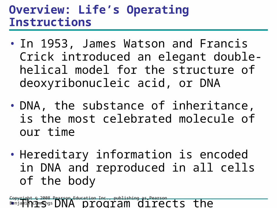
• In 1953, James Watson and Francis Crick introduced an elegant double-helical model for the structure of deoxyribonucleic acid, or DNA
• DNA, the substance of inheritance, is the most celebrated molecule of our time
• Hereditary information is encoded in DNA and reproduced in all cells of the body
• This DNA program directs the development of biochemical, anatomical, physiological, and (to some extent) behavioral traits
Copyright © 2008 Pearson Education Inc., publishing as Pearson Benjamin Cummings
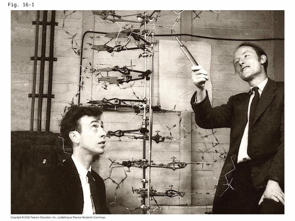
Fig. 16-1

Evidence That DNA Can Transform Bacteria
• The discovery of the genetic role of DNA began with research by Frederick Griffith in 1928
• Griffith worked with two strains of a bacterium, one pathogenic and one harmless
Copyright © 2008 Pearson Education Inc., publishing as Pearson Benjamin Cummings

• When he mixed heat-killed remains of the pathogenic strain with living cells of the harmless strain, some living cells became pathogenic
• He called this phenomenon transformation, now defined as a change in genotype and phenotype due to assimilation of foreign DNA
Copyright © 2008 Pearson Education Inc., publishing as Pearson Benjamin Cummings

Fig. 16-2
Living S cells (control)
Living R cells (control)
Heat-killed S cells (control)
Mixture of heat-killed S cells and living R cells
Mouse diesMouse dies Mouse healthy Mouse healthy
Living S cells
RESULTS
EXPERIMENT
• In 1944, Oswald Avery, Maclyn McCarty, and Colin MacLeod announced that the transforming substance was DNA
• Their conclusion was based on experimental evidence that only DNA worked in transforming harmless bacteria into pathogenic bacteria
• Many biologists remained skeptical, mainly because little was known about DNA
Copyright © 2008 Pearson Education Inc., publishing as Pearson Benjamin Cummings

Evidence That Viral DNA Can Program Cells
• More evidence for DNA as the genetic material came from studies of viruses that infect bacteria
• Such viruses, called bacteriophages (or phages), are widely used in molecular genetics research
Copyright © 2008 Pearson Education Inc., publishing as Pearson Benjamin Cummings
Animation: Phage T2 Reproductive CycleAnimation: Phage T2 Reproductive Cycle

Fig. 16-3
Bacterial cell
Phage head
Tail sheath
Tail fiber
DNA
100
nm
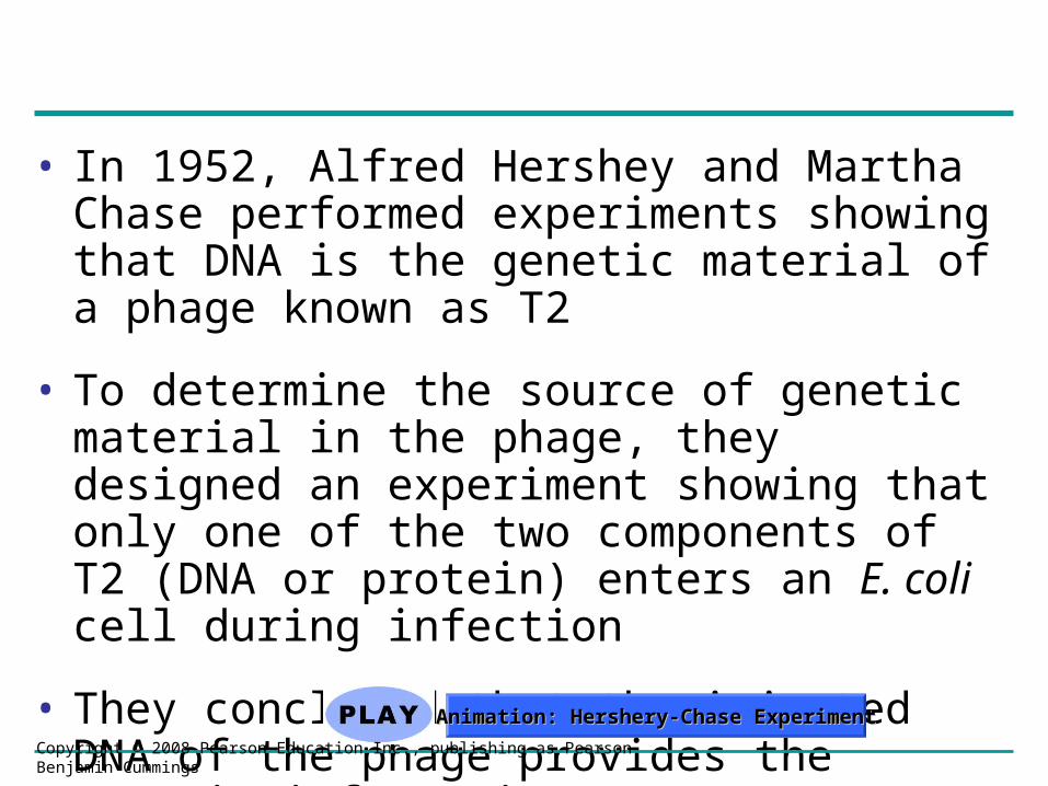
• In 1952, Alfred Hershey and Martha Chase performed experiments showing that DNA is the genetic material of a phage known as T2
• To determine the source of genetic material in the phage, they designed an experiment showing that only one of the two components of T2 (DNA or protein) enters an E. coli cell during infection
• They concluded that the injected DNA of the phage provides the genetic information
Copyright © 2008 Pearson Education Inc., publishing as Pearson Benjamin Cummings
Animation: Hershery-Chase ExperimentAnimation: Hershery-Chase Experiment
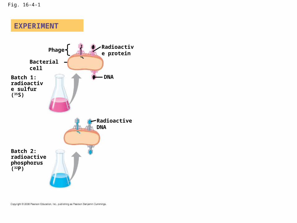
Fig. 16-4-1
EXPERIMENT
Phage
DNA
Bacterial cell
Radioactive protein
Radioactive DNA
Batch 1: radioactive sulfur (35S)
Batch 2: radioactive phosphorus (32P)

Fig. 16-4-2
EXPERIMENT
Phage
DNA
Bacterial cell
Radioactive protein
Radioactive DNA
Batch 1: radioactive sulfur (35S)
Batch 2: radioactive phosphorus (32P)
Empty protein shell
Phage DNA
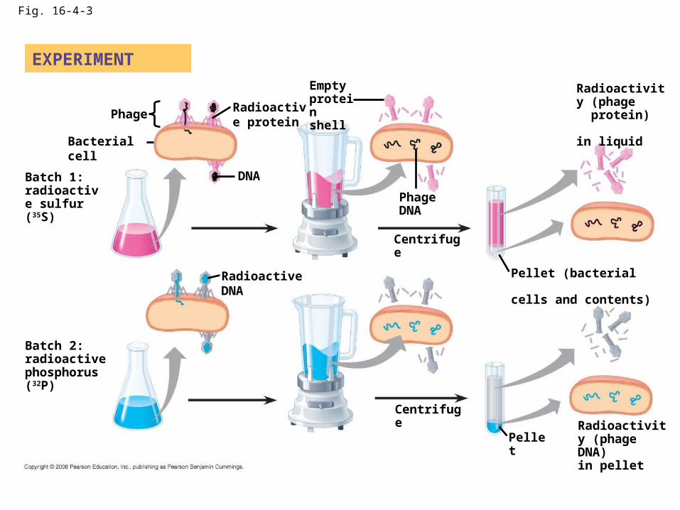
Fig. 16-4-3
EXPERIMENT
Phage
DNA
Bacterial cell
Radioactive protein
Radioactive DNA
Batch 1: radioactive sulfur (35S)
Batch 2: radioactive phosphorus (32P)
Empty protein shell
Phage DNA
Centrifuge
Centrifuge
Pellet
Pellet (bacterial cells and contents)
Radioactivity (phage protein) in liquid
Radioactivity (phage DNA) in pellet

Fig. 16-5Sugar–phosphate
backbone
5 end
Nitrogenous
bases
Thymine (T)
Adenine (A)
Cytosine (C)
Guanine (G)
DNA nucleotide
Sugar (deoxyribose)
3 end
Phosphate

Building a Structural Model of DNA: Scientific Inquiry
• After most biologists became convinced that DNA was the genetic material, the challenge was to determine how its structure accounts for its role
• Maurice Wilkins and Rosalind Franklin were using a technique called X-ray crystallography to study molecular structure
• Franklin produced a picture of the DNA molecule using this technique
Copyright © 2008 Pearson Education Inc., publishing as Pearson Benjamin Cummings
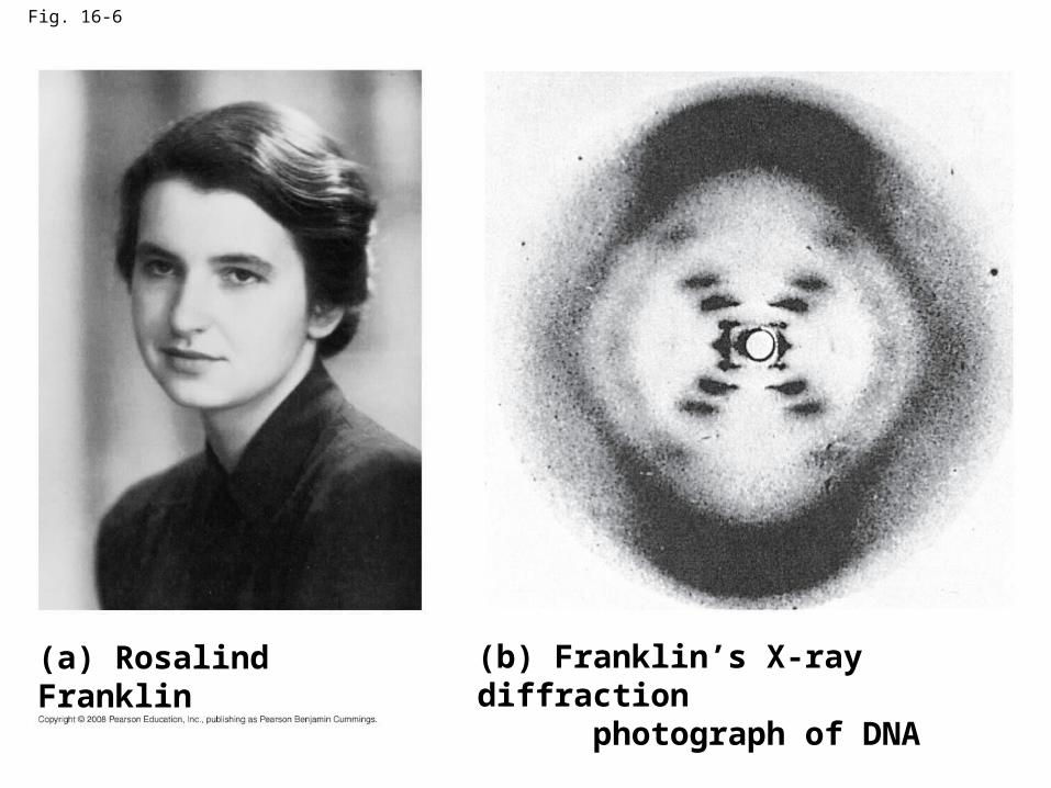
Fig. 16-6
(a) Rosalind Franklin (b) Franklin’s X-ray diffraction photograph of DNA
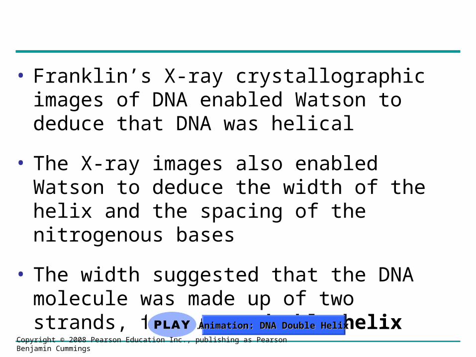
• Franklin’s X-ray crystallographic images of DNA enabled Watson to deduce that DNA was helical
• The X-ray images also enabled Watson to deduce the width of the helix and the spacing of the nitrogenous bases
• The width suggested that the DNA molecule was made up of two strands, forming a double helix
Copyright © 2008 Pearson Education Inc., publishing as Pearson Benjamin Cummings
Animation: DNA Double HelixAnimation: DNA Double Helix

Fig. 16-7
(c) Space-filling model
Hydrogen bond 3 end
5 end
3.4 nm
0.34 nm
3 end
5 end
(b) Partial chemical structure(a) Key features of DNA structure
1 nm

Fig. 16-7a
Hydrogen bond 3 end
5 end
3.4 nm
0.34 nm
3 end
5 end
(b) Partial chemical structure(a) Key features of DNA structure
1 nm
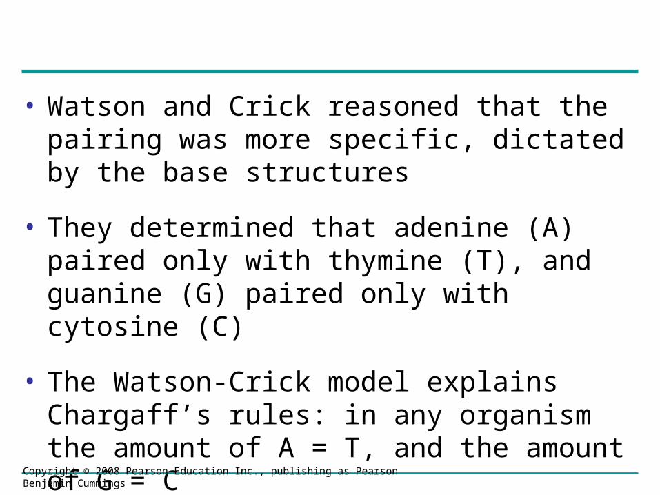
• Watson and Crick reasoned that the pairing was more specific, dictated by the base structures
• They determined that adenine (A) paired only with thymine (T), and guanine (G) paired only with cytosine (C)
• The Watson-Crick model explains Chargaff’s rules: in any organism the amount of A = T, and the amount of G = C
Copyright © 2008 Pearson Education Inc., publishing as Pearson Benjamin Cummings
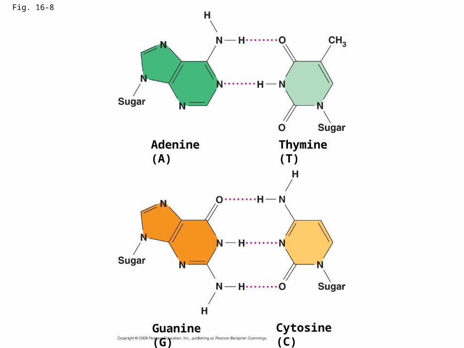
Fig. 16-8
Cytosine (C)
Adenine (A) Thymine (T)
Guanine (G)
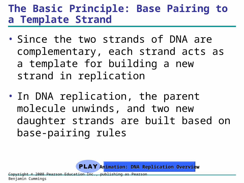
The Basic Principle: Base Pairing to a Template Strand
• Since the two strands of DNA are complementary, each strand acts as a template for building a new strand in replication
• In DNA replication, the parent molecule unwinds, and two new daughter strands are built based on base-pairing rules
Copyright © 2008 Pearson Education Inc., publishing as Pearson Benjamin Cummings
Animation: DNA Replication OverviewAnimation: DNA Replication Overview
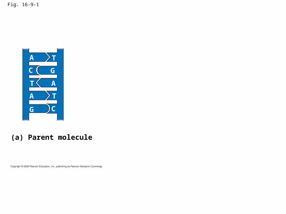
Fig. 16-9-1
A T
GC
T A
TA
G C
(a) Parent molecule

Fig. 16-9-2
A T
GC
T A
TA
G C
A T
GC
T A
TA
G C
(a) Parent molecule (b) Separation of strands
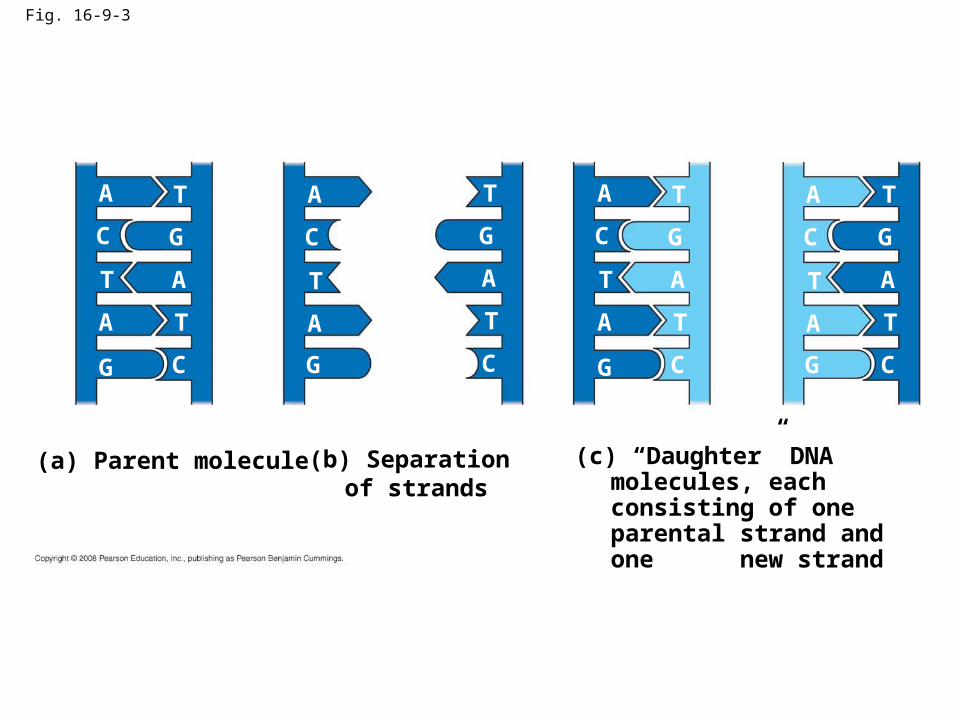
Fig. 16-9-3
A T
GC
T A
TA
G C
(a) Parent molecule
A T
GC
T A
TA
G C
(c) “Daughter” DNA molecules, each consisting of one parental strand and one new strand
(b) Separation of strands
A T
GC
T A
TA
G C
A T
GC
T A
TA
G C

• Watson and Crick’s semiconservative model of replication predicts that when a double helix replicates, each daughter molecule will have one old strand (derived or “conserved” from the parent molecule) and one newly made strand
• Competing models were the conservative model (the two parent strands rejoin) and the dispersive model (each strand is a mix of old and new)
Copyright © 2008 Pearson Education Inc., publishing as Pearson Benjamin Cummings
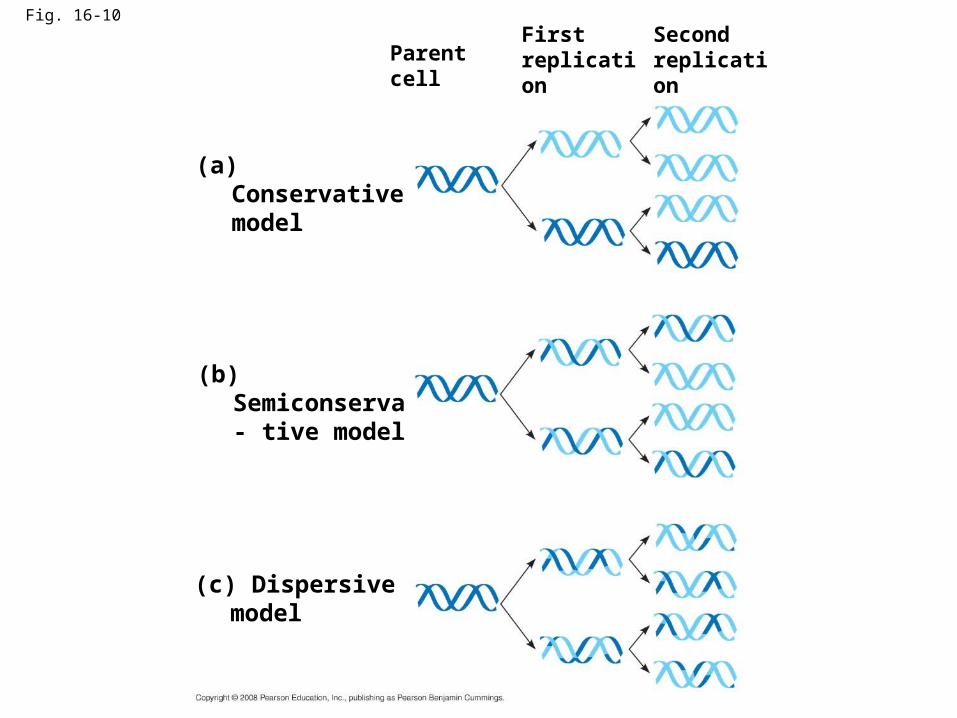
Fig. 16-10
Parent cellFirst replication
Second replication
(a) Conservative model
(b) Semiconserva- tive model
(c) Dispersive model
• Experiments by Matthew Meselson and Franklin Stahl supported the semiconservative model
• They labeled the nucleotides of the old strands with a heavy isotope of nitrogen, while any new nucleotides were labeled with a lighter isotope
Copyright © 2008 Pearson Education Inc., publishing as Pearson Benjamin Cummings

Fig. 16-11EXPERIMENT
RESULTS
CONCLUSION
1 2
43
Conservative model
Semiconservative model
Dispersive model
Bacteria cultured in medium containing 15N
Bacteria transferred to medium containing 14N
DNA sample centrifuged after 20 min (after first application)
DNA sample centrifuged after 40 min (after second replication) More
dense
Less dense
Second replicationFirst replication
DNA Replication: A Closer Look
• The copying of DNA is remarkable in its speed and accuracy
• More than a dozen enzymes and other proteins participate in DNA replication
Copyright © 2008 Pearson Education Inc., publishing as Pearson Benjamin Cummings
Getting Started
• Replication begins at special sites called origins of replication, where the two DNA strands are separated, opening up a replication “bubble”
• A eukaryotic chromosome may have hundreds or even thousands of origins of replication
• Replication proceeds in both directions from each origin, until the entire molecule is copied
Copyright © 2008 Pearson Education Inc., publishing as Pearson Benjamin Cummings
Animation: Origins of ReplicationAnimation: Origins of Replication

Fig. 16-12Origin of replication Parental (template) strand
Daughter (new) strand
Replication fork
Replication bubble
Two daughter DNA molecules
(a) Origins of replication in E. coli
Origin of replication Double-stranded DNA molecule
Parental (template) strandDaughter (new) strand
Bubble Replication fork
Two daughter DNA molecules
(b) Origins of replication in eukaryotes
0.5 µm
0.25 µm
Double-strandedDNA molecule
• At the end of each replication bubble is a replication fork, a Y-shaped region where new DNA strands are elongating
• Helicases are enzymes that untwist the double helix at the replication forks
• Single-strand binding protein binds to and stabilizes single-stranded DNA until it can be used as a template
• Topoisomerase corrects “overwinding” ahead of replication forks by breaking, swiveling, and rejoining DNA strands
Copyright © 2008 Pearson Education Inc., publishing as Pearson Benjamin Cummings

Fig. 16-13
Topoisomerase
Helicase
PrimaseSingle-strand binding proteins
RNA primer
55
5 3
3
3

• DNA polymerases cannot initiate synthesis of a polynucleotide; they can only add nucleotides to the 3 end
• The initial nucleotide strand is a short RNA primer
Copyright © 2008 Pearson Education Inc., publishing as Pearson Benjamin Cummings

• An enzyme called primase can start an RNA chain from scratch and adds RNA nucleotides one at a time using the parental DNA as a template
• The primer is short (5–10 nucleotides long), and the 3 end serves as the starting point for the new DNA strand
Copyright © 2008 Pearson Education Inc., publishing as Pearson Benjamin Cummings

Synthesizing a New DNA Strand
• Enzymes called DNA polymerases catalyze the elongation of new DNA at a replication fork
• Most DNA polymerases require a primer and a DNA template strand
• The rate of elongation is about 500 nucleotides per second in bacteria and 50 per second in human cells
Copyright © 2008 Pearson Education Inc., publishing as Pearson Benjamin Cummings
• Each nucleotide that is added to a growing DNA strand is a nucleoside triphosphate
• dATP supplies adenine to DNA and is similar to the ATP of energy metabolism
• The difference is in their sugars: dATP has deoxyribose while ATP has ribose
• As each monomer of dATP joins the DNA strand, it loses two phosphate groups as a molecule of pyrophosphate
Copyright © 2008 Pearson Education Inc., publishing as Pearson Benjamin Cummings
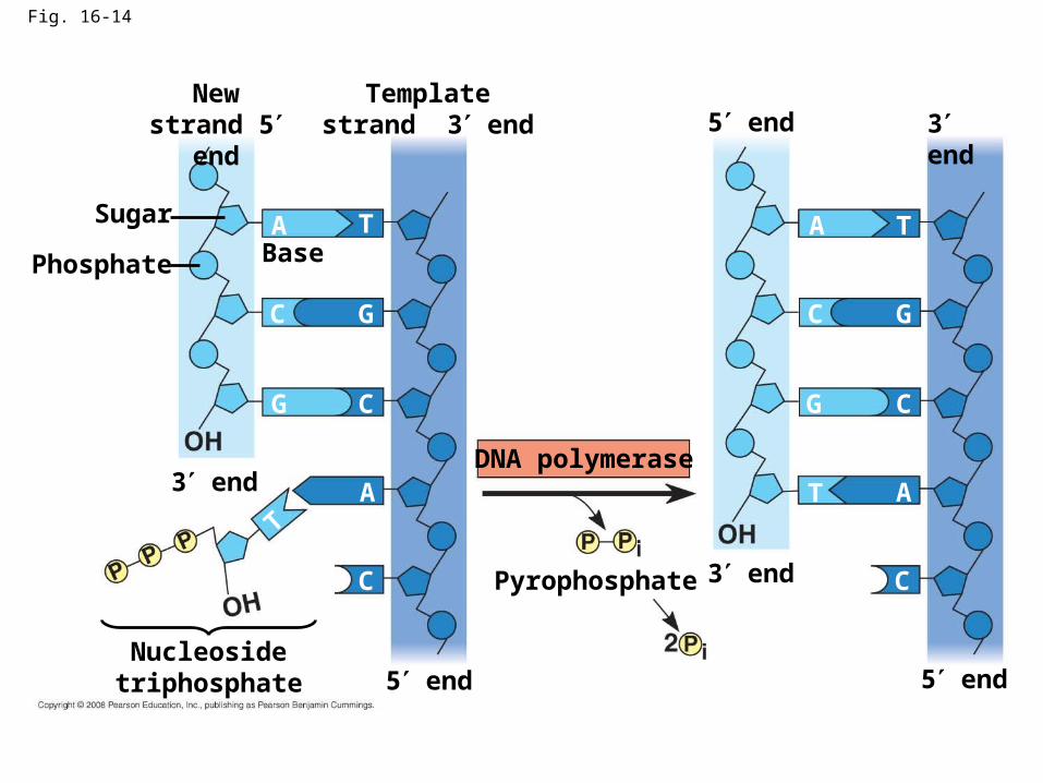
Fig. 16-14
A
C
T
G
G
G
GC
C C
C
C
A
A
AT
T
T
New strand 5 end
Template strand 3 end 5 end 3 end
3 end
5 end5 end
3 end
Base
Sugar
Phosphate
Nucleoside triphosphate
Pyrophosphate
DNA polymerase

Antiparallel Elongation
• The antiparallel structure of the double helix (two strands oriented in opposite directions) affects replication
• DNA polymerases add nucleotides only to the free 3end of a growing strand; therefore, a new DNA strand can elongate only in the 5to3direction
Copyright © 2008 Pearson Education Inc., publishing as Pearson Benjamin Cummings
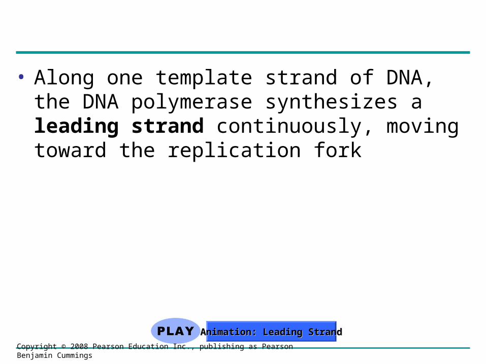
• Along one template strand of DNA, the DNA polymerase synthesizes a leading strand continuously, moving toward the replication fork
Copyright © 2008 Pearson Education Inc., publishing as Pearson Benjamin Cummings
Animation: Leading StrandAnimation: Leading Strand

Fig. 16-15
Leading strand
Overview
Origin of replicationLagging strand
Leading strandLagging strand
Primer
Overall directions of replication
Origin of replication
RNA primer
“Sliding clamp”
DNA poll IIIParental DNA
5
3
3
3
3
5
5
5
5
5

Fig. 16-15a
Overview
Leading strand
Leading strandLagging strand
Lagging strand
Origin of replication
Primer
Overall directions of replication

Fig. 16-15b
Origin of replication
RNA primer
“Sliding clamp”
DNA pol IIIParental DNA
3
5
5
5
5
5
5
3
3
3

• To elongate the other new strand, called the lagging strand, DNA polymerase must work in the direction away from the replication fork
• The lagging strand is synthesized as a series of segments called Okazaki fragments, which are joined together by DNA ligase
Copyright © 2008 Pearson Education Inc., publishing as Pearson Benjamin Cummings
Animation: Lagging StrandAnimation: Lagging Strand

Fig. 16-16Overview
Origin of replication
Leading strand
Leading strand
Lagging strand
Lagging strand
Overall directions of replication
Template strand
RNA primer
Okazaki fragment
Overall direction of replication
12
3
2
1
1
1
1
2
2
51
3
3
3
3
3
3
3
3
3
5
5
5
5
5
5
5
5
5
5
53
3

Fig. 16-16a
Overview
Origin of replication
Leading strand
Leading strand
Lagging strand
Lagging strand
Overall directions of replication
12

Fig. 16-16b1
Template strand
5
53
3

Fig. 16-16b2
Template strand
5
53
3
RNA primer 3 5
5
3
1
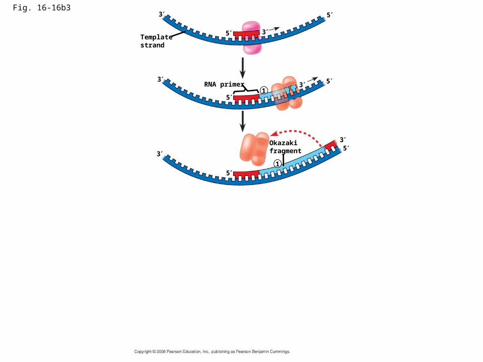
Fig. 16-16b3
Template strand
5
53
3
RNA primer 3 5
5
3
1
1
3
35
5
Okazaki fragment
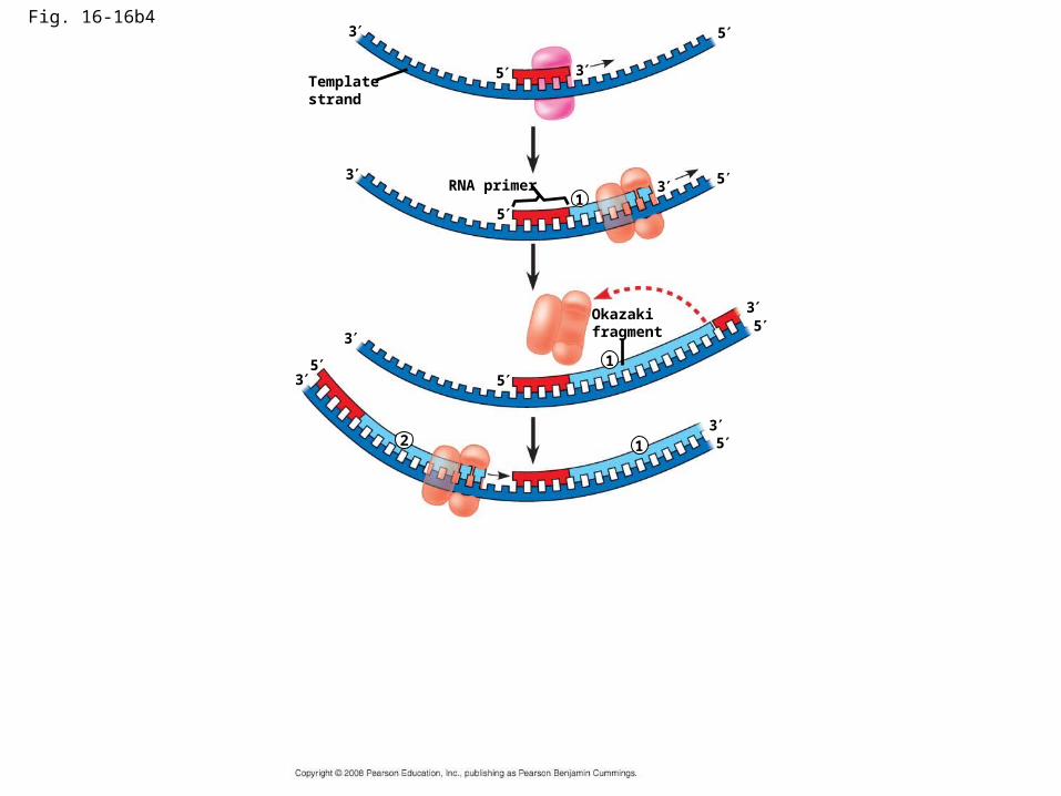
Fig. 16-16b4
Template strand
5
53
3
RNA primer 3 5
5
3
1
1
3
35
5
Okazaki fragment
12
3
3
5
5

Fig. 16-16b5
Template strand
5
53
3
RNA primer 3 5
5
3
1
1
3
35
5
Okazaki fragment
12
3
3
5
5
12
3
3
5
5
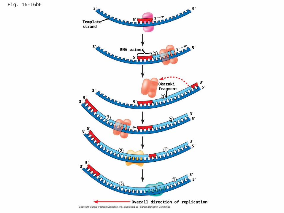
Fig. 16-16b6
Template strand
5
53
3
RNA primer 3 5
5
3
1
1
3
35
5
Okazaki fragment
12
3
3
5
5
12
3
3
5
5
12
5
5
3
3
Overall direction of replication

Table 16-1
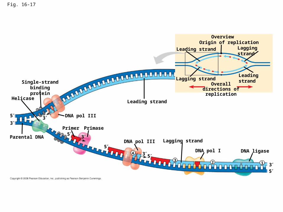
Fig. 16-17
OverviewOrigin of replication
Leading strand
Leading strand
Lagging strand
Lagging strandOverall directions
of replication
Leading strand
Lagging strand
Helicase
Parental DNA
DNA pol III
Primer Primase
DNA ligase
DNA pol III
DNA pol I
Single-strand binding protein
5
3
5
5
5
5
3
3
3
313 2
4

Proofreading and Repairing DNA
• DNA polymerases proofread newly made DNA, replacing any incorrect nucleotides
• In mismatch repair of DNA, repair enzymes correct errors in base pairing
• DNA can be damaged by chemicals, radioactive emissions, X-rays, UV light, and certain molecules (in cigarette smoke for example)
• In nucleotide excision repair, a nuclease cuts out and replaces damaged stretches of DNA
Copyright © 2008 Pearson Education Inc., publishing as Pearson Benjamin Cummings

Fig. 16-18
Nuclease
DNA polymerase
DNA ligase

Replicating the Ends of DNA Molecules
• Limitations of DNA polymerase create problems for the linear DNA of eukaryotic chromosomes
• The usual replication machinery provides no way to complete the 5 ends, so repeated rounds of replication produce shorter DNA molecules
Copyright © 2008 Pearson Education Inc., publishing as Pearson Benjamin Cummings

Fig. 16-19
Ends of parental DNA strands
Leading strand
Lagging strand
Lagging strand
Last fragment Previous fragment
Parental strand
RNA primer
Removal of primers and replacement with DNA where a 3 end is available
Second round of replication
New leading strand
New lagging strand
Further rounds of replication
Shorter and shorter daughter molecules
5
3
3
3
3
3
5
5
5
5
• Eukaryotic chromosomal DNA molecules have at their ends nucleotide sequences called telomeres
• Telomeres do not prevent the shortening of DNA molecules, but they do postpone the erosion of genes near the ends of DNA molecules
• It has been proposed that the shortening of telomeres is connected to aging
Copyright © 2008 Pearson Education Inc., publishing as Pearson Benjamin Cummings

• If chromosomes of germ cells became shorter in every cell cycle, essential genes would eventually be missing from the gametes they produce
• An enzyme called telomerase catalyzes the lengthening of telomeres in germ cells
Copyright © 2008 Pearson Education Inc., publishing as Pearson Benjamin Cummings

• The shortening of telomeres might protect cells from cancerous growth by limiting the number of cell divisions
• There is evidence of telomerase activity in cancer cells, which may allow cancer cells to persist
Copyright © 2008 Pearson Education Inc., publishing as Pearson Benjamin Cummings
• Chromatin is a complex of DNA and protein, and is found in the nucleus of eukaryotic cells
• Histones are proteins that are responsible for the first level of DNA packing in chromatin
Copyright © 2008 Pearson Education Inc., publishing as Pearson Benjamin Cummings
Animation: DNA PackingAnimation: DNA Packing
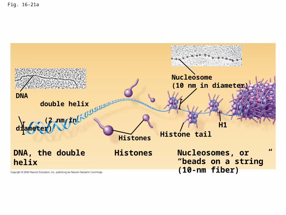
Fig. 16-21a
DNA double helix (2 nm in diameter)
Nucleosome(10 nm in diameter)
Histones Histone tailH1
DNA, the double helix Histones Nucleosomes, or “beads on a string” (10-nm fiber)

• Most chromatin is loosely packed in the nucleus during interphase and condenses prior to mitosis
• Loosely packed chromatin is called euchromatin
• During interphase a few regions of chromatin (centromeres and telomeres) are highly condensed into heterochromatin
• Dense packing of the heterochromatin makes it difficult for the cell to express genetic information coded in these regions
Copyright © 2008 Pearson Education Inc., publishing as Pearson Benjamin Cummings

• Histones can undergo chemical modifications that result in changes in chromatin organization
Copyright © 2008 Pearson Education Inc., publishing as Pearson Benjamin Cummings

Fig. 16-UN3
DNA pol III synthesizes leading strand continuously
Parental DNA DNA pol III starts DNA
synthesis at 3 end of primer, continues in 5 3 direction
Lagging strand synthesized in short Okazaki fragments, later joined by DNA ligase
Primase synthesizes a short RNA primer
53
5
5
5
3
3
You should now be able to:
1. Describe the contributions of the following people: Griffith; Avery, McCary, and MacLeod; Hershey and Chase; Chargaff; Watson and Crick; Franklin; Meselson and Stahl
2. Describe the structure of DNA
3. Describe the process of DNA replication; include the following terms: antiparallel structure, DNA polymerase, leading strand, lagging strand, Okazaki fragments, DNA ligase, primer, primase, helicase, topoisomerase, single-strand binding proteins
Copyright © 2008 Pearson Education Inc., publishing as Pearson Benjamin Cummings

4. Describe the function of telomeres
5. Compare a bacterial chromosome and a eukaryotic chromosome
Copyright © 2008 Pearson Education Inc., publishing as Pearson Benjamin Cummings
Related Documents