JOURNAL OF VIROLOGY, Dec. 2006, p. 11498–11509 Vol. 80, No. 23 0022-538X/06/$08.000 doi:10.1128/JVI.00801-06 Copyright © 2006, American Society for Microbiology. All Rights Reserved. Overexpression of the Lens Epithelium-Derived Growth Factor/p75 Integrase Binding Domain Inhibits Human Immunodeficiency Virus Replication Jan De Rijck, 1 † Linos Vandekerckhove, 1 † Rik Gijsbers, 1 Anneleen Hombrouck, 1 Jelle Hendrix, 2 Jo Vercammen, 2 Yves Engelborghs, 2 Frauke Christ, 1 ‡ and Zeger Debyser 1 ‡* Laboratory for Molecular Virology and Gene Therapy, KULeuven and IRC KULAK, Kapucijnenvoer 33, B-3000 Leuven, Flanders, Belgium, 1 and Biomolecular Dynamics, KULeuven, Celestijnenlaan 200D, B-3001 Leuven, Belgium 2 Received 19 April 2006/Accepted 30 August 2006 We initially identified lens epithelium-derived growth factor/p75 (LEDGF/p75) as a binding partner of human immunodeficiency virus type 1 (HIV-1) integrase. To investigate the role of LEDGF/p75 in HIV replication and its potential as a new antiviral target, we stably overexpressed two different fragments containing the integrase binding domain (IBD) of LEDGF/p75 fused to enhanced green fluorescent protein (eGFP). HIV-1 replication was severely inhibited by overexpression of the eGFP-IBD fusion proteins, while no inhibition was observed in cell lines overexpressing the interaction-deficient D366A mutant. Quantitative PCR pinpointed the block to the integration step, whereas nuclear import was not affected. Competition of the IBD fusion proteins with endogenous LEDGF/p75 for binding to integrase led to a potent defect in HIV-1 replication in both HeLaP4- and MT-4-derived cell lines. A previously described diketo acid-resistant HIV-1 strain remained fully susceptible to inhibition, suggesting that this strategy will also work in patients who harbor strains resistant to the current experimental integrase inhibitors. These data support LEDGF/p75 as an important cofactor for HIV replication and provide proof of concept for the LEDGF/p75-integrase interaction as a novel target for treating HIV-1 infection. Currently, more than 20 drugs that belong to four different classes are approved by the FDA for clinical treatment of human immunodeficiency virus type 1 (HIV-1) infection. How- ever, the emergence of resistant strains, especially in multi- drug-experienced patients, compromises antiretroviral regi- mens (20). To expand the antiretroviral armament, coreceptor blockers and integrase inhibitors have recently entered clinical trials. Here we investigate whether the interaction of HIV-1 integrase with its cellular binding partner, lens epithelium- derived growth factor/p75 (LEDGF/p75), could constitute a new target for antiretroviral therapy. Although lentiviruses carry the proteins required for all steps of the replication cycle, some steps require additional cellular cofactors. Several candidate cofactors of HIV integra- tion have been proposed (for recent reviews, see references 33 and 38). LEDGF/p75 was identified as an interaction partner of HIV-1 integrase by coimmunoprecipitation of nuclear ex- tracts of cells overexpressing HIV-1 integrase from a synthetic gene (integrase s ) (5) and was found to stimulate DNA binding and integration in vitro (2, 5). The interaction of LEDGF/p75 with HIV-1 integrase was subsequently confirmed in two inde- pendent reports (13, 33). LEDGF/p75 binds to the integrases of HIV-1, HIV-2, the simian immunodeficiency virus SIVmac, and feline immunodeficiency virus (FIV) but not to Moloney murine leukemia virus, respiratory syncytial virus, or human T-cell leukemia virus type 2 integrase, proving the lentiviral specificity of the interaction (2, 22). Earlier studies described LEDGF/p75 as a protein copurifying with the transcriptional coactivator PC4 (15) and as a growth factor (32). Later studies showed that LEDGF/p75 is a weak coactivator of general tran- scription but plays a protective role against cellular stress (30). LEDGF/p75 consists of 530 amino acids (aa) and contains an N-terminal PWWP motif involved in chromatin binding (Fig. 1a) (16). Accordingly, the nuclear accumulation of HIV-1 in- tegrase is apparently due to chromosomal tethering by LEDGF/p75 (24, 37). Mutations in the nuclear localization signal (NLS) of LEDGF/p75 induce its cytoplasmic accumula- tion after transient overexpression. However, stably expressed LEDGF/p75 containing a mutant NLS still accumulates in the nucleus due to chromosomal association (23, 37). To date, it is not clear whether this NLS is also responsible for targeting the HIV-1 preintegration complex to the nucleus. The minimal domain of LEDGF/p75 required for interac- tion with integrase (integrase binding domain [IBD]) was mapped to residues 347 to 429 (4, 37). In vitro experiments revealed that the IBD is necessary for interaction with inte- grase but not sufficient to stimulate strand transfer activity (4). By a yeast two-hybrid screen, the LEDGF/p75 interaction do- main of HIV-1 integrase was mapped to the catalytic core, and several mutants defective for interaction with LEDGF/p75 were identified (13). Q168A mutant integrase does not interact with LEDGF/p75 but retains in vitro 3 processing and strand transfer activity. A viral clone carrying the Q168A mutation was shown to be replication defective due to a block at the integration step. Although Llano et al. could show neither a reduction of vector transduction nor a reduction of HIV-1 * Corresponding author. Mailing address: Molecular Medicine, KULeuven and IRC KULAK, Kapucijnenvoer 33 VCTB5, B-3000 Leuven, Belgium. Phone: 32-16-332183. Fax: 32-16-336336. E-mail: [email protected]. † J.D.R. and L.V. contributed equally to this work. ‡ F.C. and Z.D. are both senior authors of this work. Published ahead of print on 20 September 2006. 11498

Welcome message from author
This document is posted to help you gain knowledge. Please leave a comment to let me know what you think about it! Share it to your friends and learn new things together.
Transcript
JOURNAL OF VIROLOGY, Dec. 2006, p. 11498–11509 Vol. 80, No. 230022-538X/06/$08.00�0 doi:10.1128/JVI.00801-06Copyright © 2006, American Society for Microbiology. All Rights Reserved.
Overexpression of the Lens Epithelium-Derived Growth Factor/p75Integrase Binding Domain Inhibits Human Immunodeficiency
Virus Replication�
Jan De Rijck,1† Linos Vandekerckhove,1† Rik Gijsbers,1 Anneleen Hombrouck,1 Jelle Hendrix,2Jo Vercammen,2 Yves Engelborghs,2 Frauke Christ,1‡ and Zeger Debyser1‡*
Laboratory for Molecular Virology and Gene Therapy, KULeuven and IRC KULAK, Kapucijnenvoer 33, B-3000 Leuven,Flanders, Belgium,1 and Biomolecular Dynamics, KULeuven, Celestijnenlaan 200D, B-3001 Leuven, Belgium2
Received 19 April 2006/Accepted 30 August 2006
We initially identified lens epithelium-derived growth factor/p75 (LEDGF/p75) as a binding partner ofhuman immunodeficiency virus type 1 (HIV-1) integrase. To investigate the role of LEDGF/p75 in HIVreplication and its potential as a new antiviral target, we stably overexpressed two different fragmentscontaining the integrase binding domain (IBD) of LEDGF/p75 fused to enhanced green fluorescent protein(eGFP). HIV-1 replication was severely inhibited by overexpression of the eGFP-IBD fusion proteins, while noinhibition was observed in cell lines overexpressing the interaction-deficient D366A mutant. Quantitative PCRpinpointed the block to the integration step, whereas nuclear import was not affected. Competition of the IBDfusion proteins with endogenous LEDGF/p75 for binding to integrase led to a potent defect in HIV-1 replicationin both HeLaP4- and MT-4-derived cell lines. A previously described diketo acid-resistant HIV-1 strainremained fully susceptible to inhibition, suggesting that this strategy will also work in patients who harborstrains resistant to the current experimental integrase inhibitors. These data support LEDGF/p75 as animportant cofactor for HIV replication and provide proof of concept for the LEDGF/p75-integrase interactionas a novel target for treating HIV-1 infection.
Currently, more than 20 drugs that belong to four differentclasses are approved by the FDA for clinical treatment ofhuman immunodeficiency virus type 1 (HIV-1) infection. How-ever, the emergence of resistant strains, especially in multi-drug-experienced patients, compromises antiretroviral regi-mens (20). To expand the antiretroviral armament, coreceptorblockers and integrase inhibitors have recently entered clinicaltrials. Here we investigate whether the interaction of HIV-1integrase with its cellular binding partner, lens epithelium-derived growth factor/p75 (LEDGF/p75), could constitute anew target for antiretroviral therapy.
Although lentiviruses carry the proteins required for allsteps of the replication cycle, some steps require additionalcellular cofactors. Several candidate cofactors of HIV integra-tion have been proposed (for recent reviews, see references 33and 38). LEDGF/p75 was identified as an interaction partnerof HIV-1 integrase by coimmunoprecipitation of nuclear ex-tracts of cells overexpressing HIV-1 integrase from a syntheticgene (integrases) (5) and was found to stimulate DNA bindingand integration in vitro (2, 5). The interaction of LEDGF/p75with HIV-1 integrase was subsequently confirmed in two inde-pendent reports (13, 33). LEDGF/p75 binds to the integrasesof HIV-1, HIV-2, the simian immunodeficiency virus SIVmac,and feline immunodeficiency virus (FIV) but not to Moloney
murine leukemia virus, respiratory syncytial virus, or humanT-cell leukemia virus type 2 integrase, proving the lentiviralspecificity of the interaction (2, 22). Earlier studies describedLEDGF/p75 as a protein copurifying with the transcriptionalcoactivator PC4 (15) and as a growth factor (32). Later studiesshowed that LEDGF/p75 is a weak coactivator of general tran-scription but plays a protective role against cellular stress (30).LEDGF/p75 consists of 530 amino acids (aa) and contains anN-terminal PWWP motif involved in chromatin binding (Fig.1a) (16). Accordingly, the nuclear accumulation of HIV-1 in-tegrase is apparently due to chromosomal tethering byLEDGF/p75 (24, 37). Mutations in the nuclear localizationsignal (NLS) of LEDGF/p75 induce its cytoplasmic accumula-tion after transient overexpression. However, stably expressedLEDGF/p75 containing a mutant NLS still accumulates in thenucleus due to chromosomal association (23, 37). To date, it isnot clear whether this NLS is also responsible for targeting theHIV-1 preintegration complex to the nucleus.
The minimal domain of LEDGF/p75 required for interac-tion with integrase (integrase binding domain [IBD]) wasmapped to residues 347 to 429 (4, 37). In vitro experimentsrevealed that the IBD is necessary for interaction with inte-grase but not sufficient to stimulate strand transfer activity (4).By a yeast two-hybrid screen, the LEDGF/p75 interaction do-main of HIV-1 integrase was mapped to the catalytic core, andseveral mutants defective for interaction with LEDGF/p75were identified (13). Q168A mutant integrase does not interactwith LEDGF/p75 but retains in vitro 3� processing and strandtransfer activity. A viral clone carrying the Q168A mutationwas shown to be replication defective due to a block at theintegration step. Although Llano et al. could show neither areduction of vector transduction nor a reduction of HIV-1
* Corresponding author. Mailing address: Molecular Medicine,KULeuven and IRC KULAK, Kapucijnenvoer 33 VCTB�5, B-3000Leuven, Belgium. Phone: 32-16-332183. Fax: 32-16-336336. E-mail:[email protected].
† J.D.R. and L.V. contributed equally to this work.‡ F.C. and Z.D. are both senior authors of this work.� Published ahead of print on 20 September 2006.
11498

replication in Jurkat cells depleted for LEDGF/p75 (22), tran-sient and stable knockdown of LEDGF/p75 in HeLaP4 cellsresulted in a two- to fivefold reduction in virus replication (36).The cause of this discrepancy is not clear but may be related tothe nature of the cell lines used or the relative potency of theknockdown.
Here we report that stable overexpression of either of twoC-terminal fragments of LEDGF/p75 containing the IBD in-hibits HIV-1 replication and lentiviral vector transduction. Aninteraction-defective mutant version of the IBD had no effecton HIV-1 replication or vector transduction. Our results sup-port a role for LEDGF/p75 in lentiviral integration and revealthe LEDGF/p75-integrase interaction as a promising new tar-get for anti-HIV therapy.
MATERIALS AND METHODS
Plasmids. To create the eukaryotic expression plasmid peGFP-IBD IRESPuro, the IRES Puro sequence was amplified by PCR with primers 5� TGCTCTAGAGCTCTAGCCCAATTCCGC and 5� TGCTCTAGATCAGGCACCGGGCTTGCGGG from pEF1 LEDGF back (22), digested with XbaI, and sub-cloned into pEGFP-C3 (Clontech) (peGFP IRES Puro). The IBD sequence (aa347 to 429) was amplified by PCR from pCP6H75 (5), using primers 5� CGGGAATTCGCCCTTGCGGCAGCCATATGTCAATGGATTCTCGA and 5� CCGGGATCCTATACCAAGAACATGTT, digested with EcoRI and BamHI, andsubcloned into peGFP-IRES Puro. To create peGFP-�325 IRES Puro, the IBDwas removed by EcoRI and BamHI digestion and replaced by the �325 fragment(aa 325 to 530), which was amplified by PCR from pCP6H75 with primers 5�GGGAATTCTAGAGCAGCAGAATAAAGATGAAG and 5� CACGAGATCTGACTCGCGATTTCAAACCTGGAGACC. To construct the lentiviral trans-
fer plasmids pCombi eGFP-IBD IRES Puro and pCombi eGFP-�325 IRESPuro, enhanced green fluorescent protein (eGFP) was removed from pCombi-eGFP (1) by XbaI-HpaI digestion and replaced by eGFP-IBD IRES Puro andeGFP-�325 IRES Puro, respectively. The D366A mutation was introduced intothe �325 fragment of peGFP-�325 IRES Puro by using the method of Kirsch andJoly (19). The eGFP-�325 D366A IRES Puro fragment was subcloned intopCombi as described previously, resulting in pCombi eGFP-�325 D366A IRESPuro. To construct a fusion between monomeric red fluorescent protein (mRFP)and integrase (pmRFP-INs), eGFP was removed from peGFP-INs (24) byEco47III-BglII digestion and replaced by mRFP amplified by PCR from CS-hrl-mRFP-ttk (kindly provided by S. S. Gambhir, Los Angeles, Calif.) (29) withprimers 5� TTCAGCGCTATGGCCTCCTCCGAGGACGTC and 5� GAATTCAGATCTGGCGCCGGTGGAGTGGCGG. To construct a lentiviral vector en-coding mRFP, eGFP was removed from pCombi-eGFP (1) by BamHI-SmaIdigestion and replaced by mRFP cDNA. To construct a retroviral vector encod-ing an eGFP-firefly luciferase fusion protein (pLNC-GFP-Fluc), the Fluc codingregion was removed from pCHMWS-Ffluc (C. Deroose et al., submitted forpublication) by BamHI-XmaI digestion and cloned into the multiple cloning siteof pEGFP-C1 (BD Biosciences, Erembodegem, Belgium) after BglII-XmaI di-gestion. The eGFP-Ffluc fragment was PCR amplified with the forward primer5� GGACCGGTGCAGTCGACGGTACCGC and the reverse primer 5� GCATCGATGGTCCCGGGTTACACGG, digested with AgeI-ClaI, and cloned intopLNC-GFP (a kind gift from G. Towers, London, United Kingdom) after eGFPwas removed by AgeI-ClaI digestion. To construct a lentiviral transfer plasmidwith the eGFP-IBD IRES Puro or eGFP-�325 IRES Puro fragment under thecontrol of a tetracycline-regulatable (Tet/off) promoter, eGFP was removed frompCCL.sin36.eGFP.PPT.Wpre.CMV.tTA-s2.tet (a kind gift from L. Naldini,Turino, Italy) by AgeI-HpaI digestion and replaced by the eGFP-IBD/�325IRES Puro cassette (peGFP-IBD IRES Puro Tet/off and peGFP-�325 IRESPuro Tet/off, respectively). The IBD and �325 fragments were expressed inEscherichia coli as maltose binding protein (MBP) fusions. For construction ofpMBP-IBD, the sequence corresponding to aa 347 to 429 was PCR amplifiedwith primers 5� TTCGGATCCATGGATTCTCGAC, containing a BamHI re-
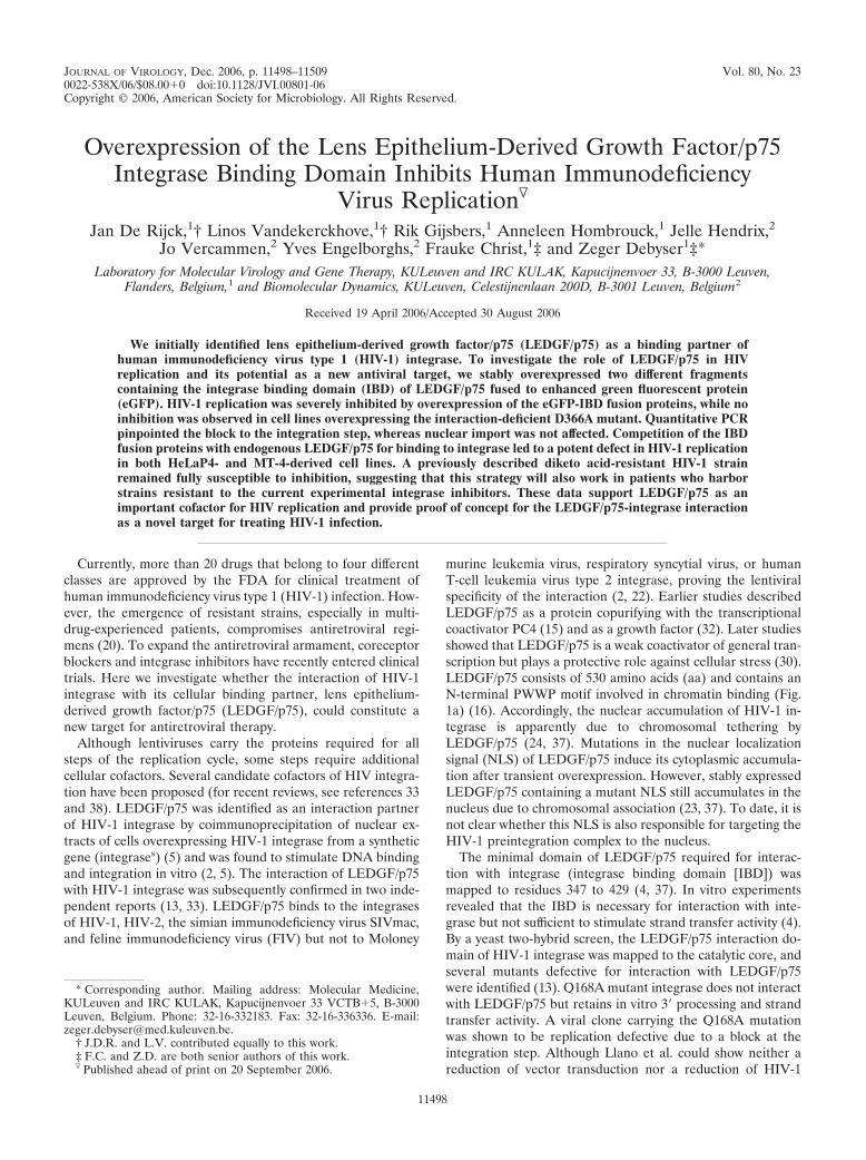
FIG. 1. Selection of eGFP-IBD- and eGFP-�325-overexpressing cells. (a) Construction of lentiviral vectors encoding eGFP-�325 and eGFP-IBD. (I) Domain structure of LEDGF/p75. (II to IV) Schematic representations of the expression cassettes of pCombi eGFP-�325 IRES Puro,pCombi eGFP-IBD IRES Puro, and pCombi eGFP IRES Puro, respectively. (b) Western blot analysis of HeLaP4-, MT-4-, and 293T-derived celllines. Equal amounts of protein extracted from each cell line were analyzed by Western blotting using an eGFP antibody. Equal loading wascontrolled by �-tubulin detection.
VOL. 80, 2006 HIV INHIBITION BY LEDGF/p75 IBD OVEREXPRESSION 11499

striction site, and 5� AGCCGTCGACTAAACCAAGAA, containing a SalI re-striction site, using pCPnat75 (24) as a template. For construction of pMBP-�325, the sequence corresponding to aa 325 to 530 was PCR amplified withprimers 5� TTCAGAATTCCAGCAGAATAAAGAT, containing an EcoRI re-striction site, and 5� GCCAAGCTTTCAGTTATCTAGTGTAGT, containing aHindIII restriction site, using pCPnat75 as a template. Subsequent to amplifica-tion, the resulting products were digested with the respective restriction endo-nucleases and ligated into pMALTM-p2X (New England Biolabs Inc.). TheD366A mutation was introduced into the �325 fragment of pMBP-�325 by usingthe method of Kirsch and Joly (19). The integrity of all plasmids was confirmedby sequencing.
Cell lines and cell culture. HeLaP4 cells, obtained from the NIH reagentprogram, were grown in Dulbecco’s modified Eagle’s medium (Gibco BRL,Belgium) containing 10% fetal calf serum (FCS; International Medical, Bel-gium), 100 �g/ml streptomycin, 100 U/ml penicillin, and 0.5 mg/ml Geneticin(Gibco BRL) (HeLaP4 medium). MT-4 cells, a human T-cell line obtained fromthe AIDS Research and Reference Reagent Program, Division of AIDS, NIAID,NIH (contributed by Douglas Richman), were grown in RPMI 1640 medium(Gibco BRL) containing 10% FCS, 100 �g/ml streptomycin, 100 U/ml penicillin,and 2 mM L-glutamine (Gibco BRL) (MT-4 medium). 293T cells were providedby O. Danos (Evry, France) and were grown in Dulbecco’s modified Eagle’smedium containing 10% FCS, 100 �g/ml streptomycin, and 100 U/ml penicillin(293T medium). All cells were cultured at 37°C in a 5% CO2 humidified atmo-sphere.
Selection of eGFP-IBD- and eGFP-�325-expressing stable cell lines. HeLaP4and 293T cells were transduced with the lentiviral vector Combi eGFP-IBDIRES Puro, Combi eGFP-�325 IRES Puro, Combi eGFP-�325 D366A IRESPuro, or Combi eGFP-IRES Puro at a multiplicity of infection (MOI) of 1 togenerate HeLaP4 eGFP-IBD, HeLaP4 eGFP-�325, HeLaP4 eGFP-�325D366A, and HeLaP4 eGFP cells, respectively, or their 293T equivalents. Poly-clonal cell lines were selected with 0.25 �g/ml puromycin. MT-4-derived cell lineswere generated in a similar way, sorting the 10% most positive cells with FACS-Vantage (BD Biosciences) prior to selection. Cells (100,000) were retained aftercell sorting to establish the polyclonal cell lines. To select HeLaP4 cell lines withthe eGFP-IBD or -�325 IRES Puro cassette under control of the Tet/off pro-moter, HeLaP4 cells were transduced with the eGFP-IBD IRES Puro Tet/off oreGFP-�325 IRES Puro Tet/off lentiviral vector, and the 10% of cells with themost eGFP expression were sorted by FACSVantage. Monoclonal cell lines wereselected with 0.25 �g/ml puromycin, and the clones with the highest eGFPexpression levels, as determined by fluorescence-activated cell sorting (FACS)(FACSCalibur; BD Biosciences), were retained.
Protein purification, in vitro pull-down assay, and Western blotting. Recom-binant MBP-IBD, MBP-�325, and MBP-�325 D366A were expressed inBL21(DE3) cells (8). After transformation, the bacteria were grown to an opticaldensity of 0.5 (MBP-IBD) or 0.6 (MBP-�325 and MBP-�325 D366A), andprotein expression was induced by the addition of a final concentration of 0.5mM (MBP-IBD) or 1 mM (MBP-�325) isopropyl-beta-D-thiogalactopyranoside.Following incubation at 37°C for 4 h, the bacteria were harvested and stored at�20°C until protein purification. For purification, the cells were resuspended inlysis buffer (50 mM Tris-HCl, pH 7.2, 500 mM NaCl, 5 mM dithiothreitol, 1 mMEDTA, 0.2 mM phenylmethylsulfonyl fluoride). After complete lysis by ultra-sonication, the supernatant was cleared by centrifugation, and recombinant pro-teins were bound to amylose resin (New England Biolabs Inc.). The resin waswashed with 20 volumes of lysis buffer, and the MBP-tagged proteins were elutedwith 10 1-ml fractions of lysis buffer supplemented with 10 mM maltose. Thefractions were analyzed by sodium dodecyl sulfate-polyacrylamide gel electro-phoresis for protein content, pooled, and concentrated by dialysis (overnight,4°C) against storage buffer (50 mM Tris-HCl, pH 7.2, 500 mM NaCl, 50%[vol/vol] glycerol). Recombinant LEDGF/p75 and HIV-1 integrase containing aC-terminal His6 tag were purified as described previously for use in the in vitropull-down assays (24).
Pulling down of LEDGF/p75 and MBP-IBD as well as MBP-�325 and MBP-�325 D366A by HIV-1 integrase was performed as described previously (24).Western blotting was performed as described previously (36).
Competition of integrase-�325 interaction by full-length LEDGF/p75. Tostudy the competition of full-length LEDGF/p75 and the �325 fragment,AlphaScreen technology (Perkin-Elmer, Benelux) was used. IN (0 or 100 nM)was preincubated in a 384-well plate for 30 min with various amounts of LEDGF/p75 at room temperature in assay buffer (25 mM Tris-HCl, pH 7.3, 150 mMNaCl, 1 mM MgCl2, 0.01% Tween 20, 0.1% bovine serum albumin). Afterpreincubation, 0, 10, 30, or 100 nM MBP-�325 was added for cross-titrationpurposes. The new mixture was incubated for another 90 min at room temper-ature. For subsequent binding of the proteins to the beads, Ni chelate-coated
acceptor beads and streptavidin-coated donor beads premixed with biotinylatedanti-MBP antibody (Vector Laboratories) were added to a final concentration of20 �g/ml. All dilutions were done in assay buffer, and if necessary, the finalvolume of the reaction was adjusted to 25 �l. In order to bind the anti-MBPantibody to the streptavidin-coated donor beads, the antibody was dialyzedovernight against assay buffer without bovine serum albumin, added to the beadsat a final concentration of 50 nM, and incubated for 1 h at room temperature.After adding the beads to the binding assay, light exposure was omitted, andincubation was carried out for another 60 min before analyzing the interaction inan EnVision plate reader (Perkin-Elmer, Benelux).
Laser scanning microscopy. HeLaP4, HeLaP4 eGFP, HeLaP4 eGFP-�325,HeLaP4 eGFP-�325 D366A, and HeLaP4 eGFP-IBD cells were transfected withpmRFP-INs and analyzed using an LSM510 unit (Carl Zeiss, Jena, Germany) asdescribed previously (36). Immunofluorescence detection of endogenousLEDGF/p75 was performed as described previously (5).
Cellular FCCS analysis. Cellular fluorescence cross-correlation spectroscopy(FCCS) measurements were performed on a commercial LSM510/ConfoCor2system (Carl Zeiss, Jena, Germany). The 488-nm line of the Ar� laser (Acousto-optical tunable filter, 0.1%; � 25 �W) was used to excite eGFP, and the 543-nmline of the HeNe laser (AOTF, 7%; �70 �W) was used to excite mRFP1. Theexcitation light was reflected by a dichroic mirror (HFT 488/543) and focusedthrough a type C Apochromat �40/1.2-W objective lens. The fluorescence emis-sion light was split by a second dichroic mirror (NFT 570) into two separate beampaths and passed through a 505- to 530-nm band-pass filter for eGFP fluores-cence and a 600- to 650-nm band-pass filter for mRFP1 fluorescence. Eachconfocal pinhole diameter was set to 70 �m. After preparation of the cells in aneight-well chambered coverglass (Nunc A/S, Roskilde, Denmark), laser scanningmicroscopy was used to search for cells suitable for FCCS. The laser beam wasfocused at a selected spot, and FCCS measurement (10 times for 20 s each) wasperformed. Auto- and cross-correlation curves were evaluated by Levenberg-Marquardt nonlinear least-square fitting to a two-component model, using Orig-inPro 7.5 software (OriginLab, Northampton, MA). The relative cross-correla-tion was calculated from the following formula: CCrel. Gcross(0)/Ggreen(0), withGcross(0) and Ggreen(0) being the amplitudes of the cross and green autocorre-lation curves, respectively.
Lentiviral and retroviral vector production. The day before transfection, fivemillion 293T cells were grown on an 8.5-cm-diameter cell culture dish in 5 mlOptimem (Gibco BRL) containing 2% FCS, 100 �g/ml streptomycin, and 100U/ml penicillin. On day 2, the medium was replaced by 5 ml Optimem with 0%FCS, and cells were cotransfected with 10 �g of the packaging plasmidpCMV�R8.91, 5 �g of the envelope plasmid pMDG (both provided by D.Trono, Geneva, Switzerland), and 20 �g of the respective transfer plasmid (11).For production of retroviral vectors, pCMVintron-gag-pol (a kind gift from G.owers, London, United Kingdom) was used as a packaging plasmid and pMDGwas used as an envelope plasmid. On day 3, the medium was again replaced by5 ml Optimem with 0% FCS, and on day 4, the supernatant was filtered througha 0.45-�m filter and concentrated 30-fold in a Vivaspin 15 50,000 MW column(Vivascience, Hannover, Germany) at 2,500 � g. Vector-containing supernatantwas stored at �80°C.
Virus strains. The molecular clone pNL4.3 was obtained from the AIDSResearch and Reference Reagent Program, Division of AIDS, NIAID, NIH.HIV-1IIIb (28) and HIV-1L-708,906 were described previously (14). To generatevesicular stomatitis virus glycoprotein (VSV-G)-pseudotyped NL4.3 virus, fivemillion 293T cells were plated 24 h prior to transfection with 20 �g pNL4.3 and5 �g pMDG and further treated as described for lentiviral vector production, butthe supernatant was not concentrated.
Lenti- and retroviral vector transduction. To analyze the effects of eGFP-IBDand eGFP-�325 overexpression on lenti- or retroviral vector transduction, cellswere transduced with serial dilutions of the Combi-mRFP or LNC-GFP FLucvector, respectively. While HeLaP4 or 293T cells were seeded in a 96-well plateat 20,000 cells/well 1 day prior to transduction, the MT-4 cells were seeded on theday of transduction at 50,000 cells/well. At 4 hours posttransduction, the mediumwas replaced with fresh HeLaP4, 293T, or MT-4 medium. mRFP expression ofthe lentiviral vector was analyzed at 72 h posttransduction by FACS analysis.Firefly luciferase expression from the retroviral vector was measured with anIVIS system (Xenogen, Alameda, Calif.) 5 min after the addition of 150 ng/mlD-luciferin. Cells were imaged for between 1 and 5 min in the IVIS system, untilthe maximum signal was reached.
HIV-1 infection. Prior to infection, HeLaP4 eGFP-IBD or -�325 Tet/off cellswere incubated with or without 5 mg/ml doxycycline for at least 7 days. Infectionof HeLaP4-derived cell lines was performed as described earlier (36). Infectionof MT-4 cells was performed with 500,000 cells in 200 �l medium at differentMOIs for 3 h. After 3 h, cells were washed twice with phosphate-buffered saline
11500 DE RIJCK ET AL. J. VIROL.

and resuspended in 5 ml MT-4 medium. HIV-1 replication was monitored byquantifying p24 antigen in the supernatant via an enzyme-linked immunosorbentassay (Alliance HIV-1 p24 ELISA kit; Perkin-Elmer, Zaventem, Belgium).
Real-time quantitative PCR analysis. One day prior to infection, 2 � 105 293Tcells were seeded into each well of a 24-well plate. Twenty-four hours later, cellswere infected with VSV-G-pseudotyped HIV-1NL4.3 corresponding to 50,000 pgp24. As a negative control, the 293T cells were treated with 0.09 �g/ml zidovu-dine (AZT) during the experiment. DNA extraction and quantification of late
reverse transcripts and two-long-terminal-repeat (2-LTR) circles were per-formed as described previously (39). The oligonucleotides used to quantify thelate reverse transcripts primed in the HIV-1 gag gene and had the followingsequences: forward primer, 5� ATCAAGCAGCCATGCAAATGTT; reverseprimer, 5� CTGAAGGGTACTAGTAGTTCCTGCTATGTC; and probe, 5� (6-carboxyfluorescein)-ACCATCAATGAGGAAGCTGCAGAATGGGA-(6-carboxytetramethylrhodamine). The primers and probe to quantify 2-LTRcircles were identical to those used previously (39). To quantify the integra-
FIG. 2. Relocalization of mRFP-integrases upon coexpression of eGFP-�325 or eGFP-IBD. HeLaP4, HeLaP4 eGFP-�325, HeLaP4 eGFP-IBD, HeLaP4 eGFP-�325 D366A, and HeLaP4 eGFP cells were transfected with pmRFP-INs 24 h before laser scanning microscopy. (A to E)eGFP expression; (F to J) mRFP-integrases expression; (K to O), merge. Bar, 10 �m. Arrows 1 represent nuclear localization of the overexpressedfragments, arrows 2 represent cytoplasmic tethering upon coexpression of integrase and eGFP-�325 or eGFP-IBD, and arrow 3 shows nuclearlocalization of coexpressed integrase and eGFP-�325 D366A.
VOL. 80, 2006 HIV INHIBITION BY LEDGF/p75 IBD OVEREXPRESSION 11501

tion events, the infected 293T cells were passaged at least four times, and thetotal amount of HIV-1 DNA was quantified using the same primers and probeas those used for detection of the late reverse transcripts.
RESULTS
Selection of eGFP-IBD- and eGFP-�325-overexpressingcells. In an effort to inhibit HIV-1 replication by preventinginteraction of HIV-1 integrase with LEDGF/p75, we generatedtwo constructs encoding the IBD of LEDGF/p75 (4, 37). First,the IBD region (aa 347 to 429) of LEDGF/p75 was fused to theC terminus of eGFP (eGFP-IBD). The second construct com-prised the C-terminal fragment (aa 325 to 530) of LEDGF/p75(eGFP-�325) (Fig. 1a). Using lentiviral vector technology,HeLaP4-derived cell lines were generated to express the fusionproteins at high levels (HeLaP4 eGFP-IBD and HeLaP4eGFP-�325). The following two control cell lines were estab-lished in parallel: HeLaP4 eGFP cells, expressing eGFP with-out a fusion, and HeLaP4 eGFP-�325 D366A cells, expressingthe D366A mutant of eGFP-�325, which is known to be de-fective for interaction with HIV-1 integrase (7). The viability ofall selected cell lines was similar to that of the parentalHeLaP4 cell line, as determined by growth kinetics (data notshown). To exclude selection bias, CD4 and CXCR4 corecep-tor expression was monitored in the HeLaP4-derived cell linesby FACS analysis. All cell lines were �99% positive for CD4and CXCR4 expression. In a similar way, 293T- and MT-4-derived cell lines were generated. For the MT-4-derived cells,the 10% most-positive cells were sorted by FACS. Expressionlevels of the fusion proteins were verified by Western blotting(Fig. 1b). Confocal microscopy with the HeLaP4-derived celllines revealed the localization of eGFP-IBD, eGFP-�325, andeGFP-�325 D366A fusion proteins to the nucleus (Fig. 2L, M,
and N, arrows 1), unlike wild-type eGFP, which was presentthroughout the cell (Fig. 2O). As expected, a transiently ex-pressed mRFP-integrases fusion protein was localized in thenuclei of wild-type HeLaP4 cells (Fig. 2F) (6, 10, 12, 21, 26,27). Upon coexpression of mRFP-integrases and eGFP-IBD/eGFP-�325, however, a dramatic relocalization of each fusionprotein from the nucleus to the cytoplasm was observed (Fig.2L and M, arrows 2). In contrast, in the HeLaP4 eGFP-�325D366A control cells expressing interaction-defective IBD, thefusion proteins retained their nuclear localization (Fig. 2N,arrow 3).
Both the IBD and �325 fragments bind to integrase in vitroand in vivo. The confocal imaging data suggested a directinteraction of the IBD and �325 fragments with HIV-1 inte-
FIG. 3. In vitro interaction of deletion mutants with integrase. (a) His6 tag-mediated pull-down of MBP-�325, MBP-�325 D366A, andMBP-IBD was carried out. Recombinant LEDGF/p75, MBP-�325, MBP-�325 D366A, and MBP-IBD were incubated with His6-tagged HIV-1integrase, and complexes were recovered using Ni2�-nitrilotriacetic acid agarose beads. Proteins were separated via 12.5% sodium dodecylsulfate-polyacrylamide gel electrophoresis and detected using Coomassie blue staining. The relative positions of the proteins are indicated on theright. Molecular size markers are indicated on the left. Lanes 1 to 4 show input quantities of each protein; lanes 5 to 8 show the recovered proteinsafter pull-down. (b) HeLaP4 eGFP-�325 cells were transfected with pmRFP-INs 24 h before fixation and permeabilization. EndogenousLEDGF/p75 was stained with a combination of monoclonal anti-LEDGF/p75 and Alexa 633-conjugated anti-mouse antibodies. Cells wereanalyzed using laser scanning microscopy.
TABLE 1. Simultaneous diffusion of HIV-1 integrase andLEDGF/p75 deletion mutantsa
Cell line � integrase or control Relativecross-correlation
HeLaP4 eGFP-�325 � mRFP 0.140 � 0.003HeLaP4 eGFP-�325 � mRFP-integrases 0.600 � 0.004HeLaP4 eGFP-IBD � mRFP-integrases 0.467 � 0.004HeLaP4 eGFP-�325 D366A � mRFP-integrases 0.184 � 0.015HeLaP4 � mRFP-eGFP 0.468 � 0.006
a HeLaP4 eGFP-�325, HeLaP4 eGFP-�325 D366A, and HeLaP4 eGFP-IBDcells were transfected with an mRFP-integrases expression plasmid. As a nega-tive control, cells were transfected with an mRFP expression plasmid. As apositive control, HeLaP4 cells were transfected with a plasmid encoding aneGFP-mRFP fusion protein. Interaction between a LEDGF/p75 deletion mutantand mRFP-integrases was detected by measuring the simultaneous diffusion ofboth proteins using fluorescence cross-correlation spectroscopy and was quanti-fied as the relative extent of cross-correlation (data are average numbers � SDfor 10 measurements).
11502 DE RIJCK ET AL. J. VIROL.

grase. To verify this observation, an in vitro pull-down assayusing His6-tagged integrase was performed as described previ-ously (Fig. 3a) (24). Full-length LEDGF/p75 was readily re-covered in complex with His6-tagged integrase (Fig. 3a, lane 5).The MBP-�325 and MBP-IBD fusions were pulled down aswell (Fig. 3a, lanes 6 and 8). No pulling down was observedwith MBP-�325 D366A (Fig. 3a, lane 7) or in the absence ofHIV-1 integrase (data not shown). To confirm this interactionin vivo, FCCS was performed with the HeLaP4-derived celllines (Table 1). This method allows the detection of interac-tions of two fluorescently labeled proteins in vivo by measuringsimultaneous diffusion. HeLaP4 eGFP-�325, HeLaP4 eGFP-
�325 D366A, and HeLaP4 eGFP-IBD cells were transfectedwith pmRFP-integrases. As a negative control, cells were trans-fected with an mRFP expression plasmid. As a positive control,HeLaP4 cells were transfected with a plasmid encoding aneGFP-mRFP fusion protein. Compared to the controls, cross-correlation of eGFP-�325 and eGFP-IBD with mRFP-inte-grases was detected, indicating that both proteins interact invivo (Table 1). Cross-correlation was not observed in theHeLaP4 eGFP-�325 D366A cells. If eGFP-�325 competeswith endogenous LEDGF/p75 for binding to integrase, thenendogenous LEDGF/p75 should remain in the nucleus due toits nuclear localization signal, whereas both eGFP-�325 andintegrase should migrate to the cytoplasm. As shown in Fig. 3b,the nuclear localization of endogenous LEDGF/p75 was in-deed confirmed for HeLaP4 eGFP-�325 cells transfected withmRFP-integrases.
To investigate in molecular detail whether LEDGF/p75 andthe deletion mutants compete directly for binding to integrase,AlphaScreen technology was used (35). In this assay, His6-tagged integrase was preincubated with various amounts ofLEDGF/p75. Different concentrations of MBP-�325 were sub-sequently added. Ni chelate-coated acceptor beads and anti-MBP-coated donor beads were added to bind integrase andMBP-�325, respectively. Binding of the molecules on thebeads leads to energy transfer from one bead to the other,ultimately producing a fluorescent signal (Fig. 4a). The fluo-rescent signal increased with increasing MBP-�325 concentra-tions (from 0.01 to 300 nM) until a plateau was reached (datanot shown), indicating saturation of the integrase–MBP-�325interaction. Representative interaction spectra are shown inFig. 4a. On the other hand, when a larger amount of LEDGF/p75 was preincubated with integrase, the fluorescent signaldecreased, from 17,000 to 9,000 counts (100 nM MBP-�325)(Fig. 4a and b). These results clearly indicate that LEDGF/p75and MBP-�325 compete for binding to HIV-1 integrase.
Lentiviral but not retroviral vector transduction is stronglyreduced by overexpression of eGFP-�325 or eGFP-IBD. Next,we transduced HeLaP4, HeLaP4 eGFP-�325, HeLaP4 eGFP-�325 D366A, and HeLaP4 eGFP-IBD cells with a second-generation VSV-G-pseudotyped lentiviral vector (42) encod-ing mRFP (Fig. 5a). Transduction efficiency was measured byFACS at 72 h posttransduction. For different vector MOIs, thetransduction efficiencies in HeLaP4 eGFP-�325 and HeLaP4eGFP-IBD cells were three times lower than those in HeLaP4and HeLaP4 eGFP-�325 D366A cells. To rule out cell typedependence, the MT-4- and 293T-derived cell lines were trans-duced in parallel. As observed for the HeLaP4-derived celllines, a potent inhibition of vector transduction was observedin the presence of eGFP-�325 or eGFP-IBD (Fig. 5b). Theinhibition was most pronounced in the MT-4-based cell lines(10- to 15-fold).
Next, the HeLaP4-derived cell lines were transduced with aMoloney murine leukemia virus-derived retroviral vector en-coding eGFP-luciferase. No reduction in luciferase expressionwas observed in HeLaP4 cells overexpressing eGFP-�325 oreGFP-IBD in comparison with control cell lines (Fig. 5c).Identical results were obtained for the 293T-derived cell lines(data not shown).
HIV-1 replication is inhibited by overexpression of eGFP-�325 or eGFP-IBD. To investigate the effects of eGFP-�325
FIG. 4. Competition of integrase-�325 interaction by full-lengthLEDGF/p75. (a) His6-tagged integrase (100 nM) was preincubatedwith various amounts of LEDGF/p75. As a negative control, 0 nMintegrase was preincubated with 1,000 nM LEDGF/p75. Increasingamounts of MBP-�325 were subsequently added for cross-titrationpurposes. The integrase–MBP-�325 interaction was measured by theAlphaScreen method. Ni chelate-coated acceptor beads and anti-MBP-coated donor beads were added to bind integrase and MBP-�325, respectively. (b) Effect of LEDGF/p75 on integrase–MBP-�325interaction. Integrase was preincubated with different amounts ofLEDGF/p75. MBP-�325 (100 nM) was subsequently added. As a neg-ative control, 100 nM MBP-�325 D366A was used. The interactionbetween integrase and MBP-�325 or MBP-�325 D366A was measuredusing the AlphaScreen method. Experiments a and b were performedthree times, and results of a representative experiment are shown.
VOL. 80, 2006 HIV INHIBITION BY LEDGF/p75 IBD OVEREXPRESSION 11503

and eGFP-IBD overexpression on multiple-round HIV-1 rep-lication, HeLaP4-derived cells were infected with HIV-1NL4.3
at an MOI of 0.01 (Fig. 6a). Viral replication was followed byharvesting of the supernatant and measurement of the p24concentration. HIV replication was readily detected inHeLaP4, HeLaP4 eGFP-�325 D366A, and HeLaP4 eGFPcells. In HeLaP4 eGFP-�325 and HeLaP4 eGFP-IBD cells,however, no viral replication could be detected for 156 h afterinfection. Even at an MOI of 0.1 or after passaging the infectedcells for more than 2 weeks, no viral breakthrough was de-tected (data not shown). To exclude any possible selection bias,we engineered monoclonal HeLaP4 cell lines expressingeGFP-IBD and eGFP-�325 from a tetracycline-dependentpromoter (Tet/off) (40). FACS analysis (data not shown) andWestern blotting revealed that high-level expression was shutoff in the presence of 5 mg/ml doxycycline (Fig. 6b). Infectionof HeLaP4 eGFP-IBD Tet/off cells with HIV-1NL4.3 at an MOIof 0.01 resulted in a clear inhibition of replication in the ab-sence of doxycycline, whereas the addition of doxycycline res-cued HIV-1 replication (Fig. 6c). Identical results were ob-tained when eGFP-�325 was transcribed from a Tet/offpromoter (Fig. 6d). Because HeLaP4 cells are not physiologi-cal host cells for HIV, the MT-4-based T-cell lines were in-fected with HIV-1NL4.3 at an MOI of 0.1, and p24 accumula-tion in the supernatant was monitored (Fig. 7a). While HIV-1replication was readily detected in the MT-4 eGFP andMT-4 eGFP-�325 D366A mutant cell lines, HIV-1 replica-tion was inhibited �20-fold at 4 days postinfection in MT-4eGFP-�325 and MT-4 eGFP-IBD cells. Viral breakthroughwas delayed 4 days in these cell lines. To exclude virus strainspecificity, the MT-4-derived cell lines were also infectedwith HIV-1IIIB and HIV-1L-708,906 at an MOI of 0.1 (Fig. 7band c). HIV-1L-708,906 is a virus strain that is resistant to theintegrase inhibitor L-708,906 (14). Again, a strong inhibitionof replication of both viral strains was observed in bothMT-4 eGFP-�325 and MT-4 eGFP-IBD cells. To furtherinvestigate viral breakthrough, MT-4 eGFP-�325 and MT-4eGFP-�325 D366A cells were infected with HIV-1NL4.3 atdifferent MOIs (Fig. 8). At an MOI of 10, viral break-through was delayed only 24 h, but viral replication was stillthreefold lower at 4 days postinfection.
HIV-1 replication in eGFP-�325 and eGFP-IBD cells isblocked at the integration step. The manifest phenotype inHIV-1 replication experiments raised the question of whetherthe block in replication occurred during early or late steps ofthe viral life cycle. First, the effects of eGFP-�325 and eGFP-IBD expression on the late steps of viral replication wereanalyzed in the 293T-derived cell lines. 293T, 293T eGFP-
FIG. 5. Effects of eGFP-�325 and eGFP-IBD overexpression onlentiviral and retroviral vector transduction. (a) Effects on lentiviralvector transduction of HeLaP4-derived cell lines. HeLaP4, HeLaP4eGFP-�325 D366A, HeLaP4 eGFP-�325, and HeLaP4 eGFP-IBDcells were transduced with a threefold dilution series of an HIV-1-derived vector encoding mRFP. After 72 h, the percentage of mRFP-positive cells was determined by FACS analysis. (b) Effects on lenti-viral vector transduction of HeLaP4-, 293T-, and MT-4-derived celllines. Wild-type HeLaP4, 293T, and MT-4 cells and their derived celllines overexpressing eGFP-�325 D366A, eGFP-�325, and eGFP-IBDwere transduced with an mRFP-expressing HIV-1-derived vector at an
MOI of 0.2, as determined in wild-type HeLaP4 cells. After 72 h, thepercentage of mRFP-expressing cells was determined by FACS anal-ysis. (c) Effects on retroviral vector transduction of HeLaP4-derivedcell lines. HeLaP4, HeLaP4 eGFP-�325 D366A, HeLaP4 eGFP-�325,and HeLaP4 eGFP-IBD cells were transduced with a Moloney murineleukemia virus-derived retroviral vector encoding firefly luciferase.After 72 h, D-luciferin was added to the cell culture medium andincubated for 5 min. Bioluminescence was measured using an IVIS 100system. In all panels, average numbers � standard deviations (SD) forduplicate experiments are shown.
11504 DE RIJCK ET AL. J. VIROL.

�325, 293T eGFP-�325 D366A, 293T eGFP-IBD, and 293TeGFP cells were transfected with the molecular clone pNL4.3,and the virus was harvested at 48 h posttransfection. Theseviral stocks were analyzed for p24 content and titrated in MT-4cells. No significant differences in p24 content or infectioustiter were observed among the different cell lines (data notshown), ruling out an effect on virus production or replicationfitness. In the next step, the formation of total HIV-1 DNA and2-LTR circles was monitored by real-time quantitative PCR(Fig. 9). To ensure single-round infections, the 293T, 293TeGFP-�325, 293T eGFP-�325 D366A, 293T eGFP-IBD, and293T eGFP cell lines were infected with a VSV-G-pseudotypedHIV-1NL4.3 virus. Plasmid contamination of DNase-treatedvector stocks was controlled by including cells treated withAZT, inhibiting de novo viral DNA synthesis (Fig. 9a). DNAwas extracted at several time points after infection, and thetotal HIV-1 DNA was measured by quantitative PCR usingprimers specific for the gag gene, not priming on to the lenti-
viral vector used to establish the cell lines. As shown in Fig. 9a,the accumulation of viral DNA at early time points was notaffected upon expression of eGFP-IBD or eGFP-�325. In con-trast, the number of 2-LTR circles increased 10-fold in the293T eGFP-IBD and 293T eGFP-�325 cells (Fig. 9b). 2-LTRcircles are formed in the nucleus and are an indirect indicationof nuclear import. As a consequence, the presence of eGFP-�325 or eGFP-IBD obviously did not inhibit reverse transcrip-tion or nuclear import. The integration events could not bemeasured by real-time quantitative Alu PCR, which makes useof an LTR primer that also anneals to the lentiviral vectorsequences present in the cell lines. Therefore, infected cellswere passaged at least four times to dilute nonintegratedDNA, ensuring that only integrated copies of viral DNA re-mained. The absence of 2-LTR circles at 72 h postinfection(Fig. 9b) is indicative of the loss of nonintegrated DNA. Mea-suring the total HIV-1 DNA after four passages revealed a20-fold reduction in the number of proviruses in the 293T
FIG. 6. Effects of eGFP-�325 and eGFP-IBD overexpression on HIV-1 replication in HeLaP4 cells. (a) HeLaP4, HeLaP4 eGFP-�325 D366A,HeLaP4 eGFP �325, HeLaP4 eGFP-IBD, and HeLaP4 eGFP cells were infected with HIV-1NL4.3 at an MOI of 0.01. HIV-1 replication wasmonitored by measurement of p24 in the supernatant at different times postinfection. The experiment was performed in quadruplicate, and averagevalues � SD are shown. (b) Western blot analysis of the monoclonal HeLaP4 eGFP-�325 Tet/off and HeLaP4 eGFP-IBD Tet/off cell lines. Oneweek prior to Western blotting, cells were grown in the presence (�) or absence (�) of 5 mg/ml doxycycline. Equal amounts of protein extractedfrom each cell line were analyzed by Western blotting using an eGFP antibody. Equal loading was controlled by �-tubulin detection. (c) HeLaP4Tet/off eGFP-IBD cells were cultured in the presence or absence of 5 mg/ml doxycycline for 7 days before infection with HIV-1NL4.3 at an MOIof 0.01. HIV-1 replication was monitored by measurement of p24 in the supernatant at different times postinfection. (d) HeLaP4 eGFP-�325Tet/off and HeLaP4 eGFP-IBD Tet/off cells were cultured in the presence or absence of 5 mg/ml doxycycline for 7 days before infection withHIV-1NL4.3 at an MOI of 0.01. At 96 h postinfection, the p24 concentration in the supernatant was measured. Average numbers of p24 � SD forduplicate experiments were determined.
VOL. 80, 2006 HIV INHIBITION BY LEDGF/p75 IBD OVEREXPRESSION 11505

eGFP-�325 and eGFP-IBD cell lines in comparison to thecontrol cell lines (Fig. 9c).
DISCUSSION
Since LEDGF/p75 was discovered as an interaction partnerof HIV-1 integrase (5), many studies have addressed its puta-
tive role in HIV-1 replication. Using RNA interference-medi-ated knockdown, a modest inhibition of HIV-1 replication wasshown in HeLaP4 cells depleted of LEDGF/p75 (36). Theseresults are at odds with the wild-type levels of HIV replicationseen in LEDGF/p75-depleted Jurkat cells (22).
We hypothesized that on the condition that LEDGF/p75plays a role as a DNA tethering factor in HIV-1 replication,expression of the C-terminal domain of LEDGF/p75 lackingthe domains known to interact with chromatin (e.g., thePWWP and AT-hook domains) (31, 34) would compete withendogenous LEDGF/p75 for its interaction with integrase andthereby inhibit the function of LEDGF/p75. Overexpression offull-length LEDGF/p75 did not result in such a phenotype (36;data not shown). Here we demonstrate unequivocally thatHIV-1 vector transduction and virus replication are indeedinhibited dramatically upon overexpression of the C-terminalfragment of LEDGF/p75 (�325) (aa 325 to 530) or the IBD (aa347 to 429) due to a block of integration. Although we cannotformally exclude an indirect effect of the fusion protein, inhi-bition of virus replication was not observed upon overexpres-sion of the eGFP-�325 D366A fragment, containing a singlemutation that abolishes the interaction with IN (7). This sug-gests that the observed inhibition in viral replication and vectortransduction is highly specific and indeed based on direct in-teraction of the overexpressed fragments (eGFP-IBD andeGFP-�325) with IN. The direct interaction of integrase andthe eGFP-�325 and eGFP-IBD fragments was confirmed by invivo fluorescence cross-correlation spectroscopy (Table 1). Inaddition to the eGFP fusions, cell lines overexpressing hem-agglutinin-tagged �325 or IBD were made as well (data notshown). Inhibition of HIV-1 replication was also observed inthese cell lines, although at a lower level, probably due to lowerexpression levels or incorrect folding of the proteins. Thisexcludes the possibility of nonspecific inhibition by the eGFPmoiety of the C-terminal fragments.
For the HeLaP4-derived cell lines, viral breakthrough couldnever be observed in the presence of eGFP-�325 or eGFP-IBD. In the T-cell line MT-4, viral breakthrough was severelydelayed (Fig. 7 and 8). To exclude artifacts due to cell lineselection, we also analyzed the inhibition of HIV-1 replicationin HeLaP4 cells expressing eGFP-IBD or eGFP-�325 from atetracycline-regulatable promoter (Fig. 6c and d). In these celllines, inhibition of viral replication was clearly dependent onthe presence of eGFP-IBD or eGFP-�325; upon doxycycline-mediated inhibition of eGFP-IBD or eGFP-�325 expression,HIV-1 replication was restored.
We previously analyzed the effect of LEDGF/p75 reductionon lentiviral vector transduction efficiency (36). At most, a 20%inhibition of transduction was observed in a monoclonalHeLaP4-derived cell line, with LEDGF/p75 levels reduced toclose to the detection limit. Most probably, knockdown of theabundant LEDGF/p75 protein was not capable of reducing thiscofactor below the threshold required for integration, althoughwe could not exclude the possibility that lentiviral vector inte-gration was less dependent on the presence of LEDGF/p75than HIV-1 replication. The results presented here prove thatLEDGF/p75 is also a cofactor for lentiviral vector transduc-tion. Overexpression of eGFP-IBD or eGFP-�325 clearly re-duced lentiviral vector transduction 3- to 15-fold, depending onthe cell line used (Fig. 5a and b), whereas transduction with a
FIG. 7. Effects of eGFP-�325 and eGFP-IBD overexpression onHIV-1 replication in MT-4 cells. (a) MT-4 eGFP-�325 D366A, MT-4eGFP �325, MT-4 eGFP-IBD, and MT-4 eGFP cells were infectedwith HIV-1NL4.3 (a) HIV-1IIIb (b), or HIV-1L-708,906 (c) at an MOI of0.1. HIV-1 replication was monitored by measurement of p24 in thesupernatant at different times postinfection. Average p24 values � SDfor triplicate experiments are shown.
11506 DE RIJCK ET AL. J. VIROL.

retroviral vector was not inhibited at all (Fig. 5c). The lentiviralspecificity of the phenotype described is in agreement with theestablished specificity of LEDGF/p75 for interactions with len-tiviral integrases (2, 22).
In the HeLaP4, 293T, and MT-4 cell lines, we overexpressed�325 or IBD fused to eGFP to increase protein stability andfor visualization by confocal microcopy. Interestingly, both fu-sion proteins localized to the nuclei of HeLaP4 cells, althoughthe NLS of LEDGF/p75 (aa 148 to 156) is not present in thesefragments (Fig. 2) (23, 37). Most probably, these fragments stillbind DNA, as proposed previously (31), or are directed to thechromatin through interactions with other cellular proteins(25). Still, this residual nuclear localization is not sufficient torescue viral replication. The D366A mutation renders the IBDdefective for interaction with HIV-1 integrase (7). Interest-ingly, introduction of the D366A mutation into eGFP-�325 didnot alter the cellular localization of this domain (Fig. 2N).
The nuclear localization of HIV-1 integrase has been thefocus of many investigations (6, 10, 12, 21, 26, 27). Accordingto Maertens et al. and Vanegas et al., transient coexpression ofHIV-1 integrase and NLS-deficient LEDGF/p75 redirects bothproteins to the cytoplasm (23, 37). However, in cell lines stablyexpressing NLS-deficient LEDGF/p75, both proteins are againdirected to the nucleus due to cell division and subsequenttethering of LEDGF/p75 to the chromatin (37). In contrast,upon stable overexpression of eGFP-IBD or eGFP-�325, thefusion protein accumulates in the cytoplasm in the presence ofmRFP-integrases (Fig. 2), while endogenous LEDGF/p75 re-tains its nuclear localization (Fig. 3b). This may be explainedby the fact that the residual chromatin binding activity of theC-terminal fragments is masked by binding to integrase. Byconfocal microscopy, a clear correlation was observed betweenthe amounts of eGFP-IBD or eGFP-�325 and mRFP-inte-grases expressed and their cellular distributions (data not
shown). At a high mRFP-integrases-to-eGFP-�325 ratio, alleGFP-�325 was found in the cytoplasm, while part of themRFP-integrases was tethered to the nucleus. It is likely thatthe truncated fragments and the endogenous LEDGF/p75compete for binding with HIV integrase. When eGFP-�325 istitrated out by an excess of integrase, part of the integraseproteins accumulate in the nucleus through interaction withendogenous LEDGF/p75. The opposite was seen when inte-grase was titrated out by eGFP-�325.
Although our results show a physical interaction of IBD and�325 proteins with integrase in the cytoplasm, one has to becareful in interpretation since overexpressed integrase doesnot always predict the behavior of integrase during HIV-1infection. Therefore, we investigated the mechanism of actionof IBD by using quantitative PCR. Quantitative PCR analysisof 293T-derived cells infected with VSV-G-pseudotypedHIV-1 pinpointed the replication block in eGFP-IBD- andeGFP-�325-overexpressing cells to the integration step (Fig.9c), whereas reverse transcription and nuclear import were notinhibited. On the contrary, a clear increase in 2-LTR circleformation was observed in the eGFP-IBD- and eGFP-�325-overexpressing cells (Fig. 9a and b), consistent with an inhibi-tion of the DNA strand transfer reaction (17, 39). The growthkinetics of cells and overall eGFP expression remained un-changed during passaging, excluding the possibility that theobserved integration defect was due to selective killing ofeGFP-IBD- or eGFP-�325-overexpressing cells. AlthougheGFP-IBD and eGFP-�325 cells are capable of relocalizingmRFP-integrases from the nucleus to the cytoplasm (Fig. 2),the nuclear import of the HIV-1 preinitiation complex (PIC)was not inhibited. The most plausible explanation for the rep-lication block is the inability of the PIC, lacking functionalLEDGF/p75, to dock the preintegration complex to the chro-matin, preventing viral integration. Nonintegrated viral DNA
FIG. 8. Effect of MOI on viral replication in MT-4 eGFP-�325 and MT-4 eGFP-�325 D366A cells. MT-4 eGFP-�325 D366A and MT-4 eGFP�325 cells were infected with HIV-1NL4.3 at an MOI of 0.01, 0.1, or 1. HIV-1 replication was monitored by measurement of p24 in the supernatantat different times postinfection. Average p24 values � SD for triplicate experiments are shown.
VOL. 80, 2006 HIV INHIBITION BY LEDGF/p75 IBD OVEREXPRESSION 11507

may then accumulate as 2-LTR (and 1-LTR) circles. In favorof a tethering role for LEDGF/p75, Ciuffi et al. recentlyshowed that HIV-1 integration occurs less frequently in tran-scription units in LEDGF/p75-depleted cells (9). Nevertheless,at this stage we cannot completely rule out the possibility of adisturbed conformation of the PIC hampering integration. In-hibition by overexpressed IBD of the interaction of anothercofactor with IN cannot be excluded either, but this wouldimply overlapping binding sites on integrase. The possibility ofblocking HIV-1 replication by overexpressing part of a cellularprotein has been described before (41). Yung and coworkersshowed that overexpression of the S6 integrase binding domainof integrase interactor 1 (INI1) efficiently inhibited the pro-duction of viral particles. INI1 is part of the SWI/SNF complexinvolved in chromatin remodeling and interacts with HIV-1integrase (18, 41). Whereas in the case of INI1 it has remainedunclear whether the inhibition of virus production by the S6fragment relates to the role of INI1 during HIV replication, inthe case of LEDGF/p75 all evidence points to a role ofLEDGF/p75 in integration.
Our data reveal that overexpression of the truncation mu-tants can compete with endogenous LEDGF/p75 and therebyinhibit virus replication. A true transdominant should not loseits phenotype when LEDGF/p75 is present in excess of theIBD. Although our Western blots suggest that the overex-pressed fragments are present in excess of endogenousLEDGF/p75, the observed potency may hint at an apparenttransdominant phenotype. Since it is possible that recruitmentof LEDGF/p75 versus the IBD already occurs in the cytoplas-mic PIC (22), cytoplasmic ratios of both proteins ought to becompared. The fact that the inhibition by overexpression of thedeletion mutants can be overcome by a single point mutation(D366A), together with the recently described crystal structureof the IBD in complex with the integrase core domain (3),supports a very defined protein-protein interface. Our datahighlight the LEDGF/p75-integrase interaction as a novel an-tiviral target for the development of small-molecule inhibitors.In this regard, it is interesting that no cross-resistance wasobserved with a diketo acid-resistant strain (Fig. 7c) (14). LikeINI1, LEDGF/p75 is a cellular protein, and as a consequence,it is very unlikely that an immunogenic response will occurupon overexpression of LEDGF/p75 fragments. IBD-basedgene therapeutic approaches against AIDS may thus be envis-aged as well. Thus, far, no effect on cell viability was seen byoverexpressing the IBD.
ACKNOWLEDGMENTS
We acknowledge M. Michiels, A. Nijs, and B. Van Remoortel forexcellent technical assistance, B. Van Maele for a critical reading ofthe manuscript, and A. Hantson for the luciferase-expressing retroviralvector. We appreciate the help of V. Van Duppen from the Hematol-ogy Division, KULeuven, with cell sorting. We thank the NIH AIDSResearch and Reference Reagent Program, Division of AIDS, NIAID,for providing different cell lines and the HIV pNL4.3 molecular clone.
Work at KULeuven was supported by the European Commission(LSHB-CT-2003-503480) (TRIoH project) and the SBO program ofthe Flemish Institute Supporting Scientific-Technological Research inIndustry (IWT). J.D.R. and L.V. are funded by grants from the IWT.
REFERENCES
1. Baekelandt, V., A. Claeys, K. Eggermont, E. Lauwers, B. De Strooper, B.Nuttin, and Z. Debyser. 2002. Characterization of lentiviral vector-mediatedgene transfer in adult mouse brain. Hum. Gene Ther. 13:841–853.
FIG. 9. eGFP-�325 and eGFP-IBD overexpression impairs HIV-1integration. 293T, 293T eGFP, 293T eGFP-IBD, 293T eGFP-�325,and 293T eGFP-�325 D366A cells were infected with VSV-G-pseudotyped HIV-1NL4.3. To exclude plasmid contamination of vectorstocks, cells were treated in a parallel experiment with 0.09 �g/mlAZT. DNAs were extracted at different times postinfection, and theamounts of total viral DNA (a) and 2-LTR circles (b) were determinedby real-time quantitative PCR. To quantify integrated DNA (c), in-fected cells were passaged four times, and the total viral DNA wasmeasured. All quantifications were performed in duplicate, and aver-ages � SD are shown.
11508 DE RIJCK ET AL. J. VIROL.

2. Busschots, K., J. Vercammen, S. Emiliani, R. Benarous, Y. Engelborghs, F.Christ, and Z. Debyser. 2005. The interaction of LEDGF/p75 with integraseis lentivirus-specific and promotes DNA binding. J. Biol. Chem. 280:17841–17847.
3. Cherepanov, P., A. L. Ambrosio, S. Rahman, T. Ellenberger, and A.Engelman. 2005. Structural basis for the recognition between HIV-1 integraseand transcriptional coactivator p75. Proc. Natl. Acad. Sci. USA 102:17308–17313.
4. Cherepanov, P., E. Devroe, P. A. Silver, and A. Engelman. 2004. Identifica-tion of an evolutionarily conserved domain in human lens epithelium-de-rived growth factor/transcriptional co-activator p75 (LEDGF/p75) that bindsHIV-1 integrase. J. Biol. Chem. 279:48883–48892.
5. Cherepanov, P., G. Maertens, P. Proost, B. Devreese, J. Van Beeumen, Y.Engelborghs, E. De Clercq, and Z. Debyser. 2003. HIV-1 integrase formsstable tetramers and associates with LEDGF/p75 protein in human cells.J. Biol. Chem. 278:372–381.
6. Cherepanov, P., W. Pluymers, A. Claeys, P. Proost, E. De Clercq, and Z.Debyser. 2000. High-level expression of active HIV-1 integrase from a syn-thetic gene in human cells. FASEB J. 14:1389–1399.
7. Cherepanov, P., Z. Y. Sun, S. Rahman, G. Maertens, G. Wagner, and A.Engelman. 2005. Solution structure of the HIV-1 integrase-binding domainin LEDGF/p75. Nat. Struct. Mol. Biol. 12:526–532.
8. Cherepanov, P., D. Surratt, J. Toelen, W. Pluymers, J. Griffith, E. De Clercq,and Z. Debyser. 1999. Activity of recombinant HIV-1 integrase on mini-HIVDNA. Nucleic Acids Res. 27:2202–2210.
9. Ciuffi, A., M. Llano, E. Poeschla, C. Hoffmann, J. Leipzig, P. Shinn, J. R.Ecker, and F. Bushman. 2005. A role for LEDGF/p75 in targeting HIVDNA integration. Nat. Med. 11:1287–1289.
10. Depienne, C., P. Roques, C. Creminon, L. Fritsch, R. Casseron, D. Dormont,C. Dargemont, and S. Benichou. 2000. Cellular distribution and karyophilicproperties of matrix, integrase, and Vpr proteins from the human and simianimmunodeficiency viruses. Exp. Cell Res. 260:387–395.
11. De Rijck, J., B. Van Maele, and Z. Debyser. 2005. Positional effects of thecentral DNA flap in HIV-1-derived lentiviral vectors. Biochem. Biophys.Res. Commun. 328:987–994.
12. Devroe, E., A. Engelman, and P. A. Silver. 2003. Intracellular transport ofhuman immunodeficiency virus type 1 integrase. J. Cell Sci. 116:4401–4408.
13. Emiliani, S., A. Mousnier, K. Busschots, M. Maroun, B. Van Maele, D.Tempe, L. Vandekerckhove, F. Moisant, L. Ben-Slama, M. Witvrouw, F.Christ, J. C. Rain, C. Dargemont, Z. Debyser, and R. Benarous. 2005.Integrase mutants defective for interaction with LEDGF/p75 are impaired inchromosome tethering and HIV-1 replication. J. Biol. Chem. 280:25517–25523.
14. Fikkert, V., B. Van Maele, J. Vercammen, A. Hantson, B. Van Remoortel, M.Michiels, C. Gurnari, C. Pannecouque, M. De Maeyer, Y. Engelborghs, E.De Clercq, Z. Debyser, and M. Witvrouw. 2003. Development of resistanceagainst diketo derivatives of human immunodeficiency virus type 1 by pro-gressive accumulation of integrase mutations. J. Virol. 77:11459–11470.
15. Ge, H., Y. Si, and R. G. Roeder. 1998. Isolation of cDNAs encoding noveltranscription coactivators p52 and p75 reveals an alternate regulatory mech-anism of transcriptional activation. EMBO J. 17:6723–6729.
16. Ge, Y. Z., M. T. Pu, H. Gowher, H. P. Wu, J. P. Ding, A. Jeltsch, and G. L.Xu. 2004. Chromatin targeting of de novo DNA methyltransferases by thePWWP domain. J. Biol. Chem. 279:25447–25454.
17. Hazuda, D. J., P. Felock, M. Witmer, A. Wolfe, K. Stillmock, J. A. Grobler,A. Espeseth, L. Gabryelski, W. Schleif, C. Blau, and M. D. Miller. 2000.Inhibitors of strand transfer that prevent integration and inhibit HIV-1replication in cells. Science 287:646–650.
18. Kalpana, G. V., S. Marmon, W. Wang, G. R. Crabtree, and S. P. Goff. 1994.Binding and stimulation of HIV-1 integrase by a human homolog of yeasttranscription factor SNF5. Science 266:2002–2006.
19. Kirsch, R. D., and E. Joly. 1998. An improved PCR-mutagenesis strategy fortwo-site mutagenesis or sequence swapping between related genes. NucleicAcids Res. 26:1848–1850.
20. Lalezari, J. P., K. Henry, M. O’Hearn, J. S. Montaner, P. J. Piliero, B.Trottier, S. Walmsley, C. Cohen, D. R. Kuritzkes, J. J. Eron, Jr., J. Chung,R. DeMasi, L. Donatacci, C. Drobnes, J. Delehanty, and M. Salgo. 2003.Enfuvirtide, an HIV-1 fusion inhibitor, for drug-resistant HIV infection inNorth and South America. N. Engl. J. Med. 348:2175–2185.
21. Limon, A., E. Devroe, R. Lu, H. Z. Ghory, P. A. Silver, and A. Engelman.2002. Nuclear localization of human immunodeficiency virus type 1 preinte-gration complexes (PICs): V165A and R166A are pleiotropic integrase mu-tants primarily defective for integration, not PIC nuclear import. J. Virol.76:10598–10607.
22. Llano, M., M. Vanegas, O. Fregoso, D. Saenz, S. Chung, M. Peretz, and
E. M. Poeschla. 2004. LEDGF/p75 determines cellular trafficking of diverselentiviral but not murine oncoretroviral integrase proteins and is a compo-nent of functional lentiviral preintegration complexes. J. Virol. 78:9524–9537.
23. Maertens, G., P. Cherepanov, Z. Debyser, Y. Engelborghs, and A. Engelman.2004. Identification and characterization of a functional nuclear localizationsignal in the HIV-1 integrase interactor LEDGF/p75. J. Biol. Chem. 279:33421–33429.
24. Maertens, G., P. Cherepanov, W. Pluymers, K. Busschots, E. De Clercq, Z.Debyser, and Y. Engelborghs. 2003. LEDGF/p75 is essential for nuclear andchromosomal targeting of HIV-1 integrase in human cells. J. Biol. Chem.278:33528–33539.
25. Maertens, G. N., P. Cherepanov, and A. Engelman. 2006. Transcriptionalco-activator p75 binds and tethers the Myc-interacting protein JPO2 tochromatin. J. Cell Sci. 119:2563–2571.
26. Petit, C., O. Schwartz, and F. Mammano. 1999. Oligomerization withinvirions and subcellular localization of human immunodeficiency virus type 1integrase. J. Virol. 73:5079–5088.
27. Pluymers, W., P. Cherepanov, D. Schols, E. De Clercq, and Z. Debyser. 1999.Nuclear localization of human immunodeficiency virus type 1 integrase ex-pressed as a fusion protein with green fluorescent protein. Virology 258:327–332.
28. Popovic, M., M. G. Sarngadharan, E. Read, and R. C. Gallo. 1984. Detec-tion, isolation, and continuous production of cytopathic retroviruses (HTLV-III) from patients with AIDS and pre-AIDS. Science 224:497–500.
29. Ray, P., A. De, J. J. Min, R. Y. Tsien, and S. S. Gambhir. 2004. Imagingtri-fusion multimodality reporter gene expression in living subjects. CancerRes. 64:1323–1330.
30. Sharma, P., D. P. Singh, N. Fatma, L. T. Chylack, Jr., and T. Shinohara.2000. Activation of LEDGF gene by thermal—and oxidative—stresses. Bio-chem. Biophys. Res. Commun. 276:1320–1324.
31. Singh, D. P., E. Kubo, Y. Takamura, T. Shinohara, A. Kumar, L. T. Chylack,Jr., and N. Fatma. 2006. DNA binding domains and nuclear localizationsignal of LEDGF: contribution of two helix-turn-helix (HTH)-like domainsand a stretch of 58 amino acids of the N-terminal to the trans-activationpotential of LEDGF. J. Mol. Biol. 355:379–394.
32. Singh, D. P., N. Ohguro, T. Kikuchi, T. Sueno, V. N. Reddy, K. Yuge, L. T.Chylack, Jr., T. Shinohara, N. Fatma, and A. Kimura. 2000. Lens epithelium-derived growth factor: effects on growth and survival of lens epithelial cells,keratinocytes, and fibroblasts. Biochem. Biophys. Res. Commun. 267:373–381.
33. Turlure, F., E. Devroe, P. A. Silver, and A. Engelman. 2004. Human cellproteins and human immunodeficiency virus DNA integration. Front. Biosci.9:3187–3208.
34. Turlure, F., G. Maertens, S. Rahman, P. Cherepanov, and A. Engelman.2006. A tripartite DNA-binding element, comprised of the nuclear localiza-tion signal and two AT-hook motifs, mediates the association of LEDGF/p75with chromatin in vivo. Nucleic Acids Res. 34:1663–1675.
35. Ullman, E. F., H. Kirakossian, S. Singh, Z. P. Wu, B. R. Irvin, J. S. Pease,A. C. Switchenko, J. D. Irvine, A. Dafforn, C. N. Skold, et al. 1994. Lumi-nescent oxygen channeling immunoassay: measurement of particle bindingkinetics by chemiluminescence. Proc. Natl. Acad. Sci. USA 91:5426–5430.
36. Vandekerckhove, L., F. Christ, B. Van Maele, J. De Rijck, R. Gijsbers, C.Van den Haute, M. Witvrouw, and Z. Debyser. 2006. Transient and stableknock-down of the integrase co-factor LEDGF/p75 reveals its role in thereplication cycle of human immunodeficiency virus. J. Virol. 80:1886–1896.
37. Vanegas, M., M. Llano, S. Delgado, D. Thompson, M. Peretz, and E. Poe-schla. 2005. Identification of the LEDGF/p75 HIV-1 integrase-interactiondomain and NLS reveals NLS-independent chromatin tethering. J. Cell Sci.118:1733–1743.
38. Van Maele, B., K. Busschots, L. Vandekerckhove, F. Christ, and Z. Debyser.2006. Cellular cofactors of HIV-1 integration. Trends Biochem. Sci. 31:98–105.
39. Van Maele, B., J. De Rijck, E. De Clercq, and Z. Debyser. 2003. Impact of thecentral polypurine tract on the kinetics of human immunodeficiency virustype 1 vector transduction. J. Virol. 77:4685–4694.
40. Vigna, E., M. Amendola, F. Benedicenti, A. D. Simmons, A. Follenzi, and L.Naldini. 2005. Efficient Tet-dependent expression of human factor IX in vivoby a new self-regulating lentiviral vector. Mol. Ther. 11:763–775.
41. Yung, E., M. Sorin, A. Pal, E. Craig, A. Morozov, O. Delattre, J. Kappes, D.Ott, and G. V. Kalpana. 2001. Inhibition of HIV-1 virion production by atransdominant mutant of integrase interactor 1. Nat. Med. 7:920–926.
42. Zufferey, R., D. Nagy, R. J. Mandel, L. Naldini, and D. Trono. 1997. Multiplyattenuated lentiviral vector achieves efficient gene delivery in vivo. Nat.Biotechnol. 15:871–875.
VOL. 80, 2006 HIV INHIBITION BY LEDGF/p75 IBD OVEREXPRESSION 11509
Related Documents