Department of Pharmaceutical Sciences, College of Pharmacy and Allied Health Professions, St. John爷s University, Queens, NY 11439, USA. Zhe鄄Sheng Chen, Department of Pharmaceutical Sciences, College of Pharmacy and Allied Health Professions, St. John爷s University, Queens, NY 11439, USA. Tel: +1鄄 718鄄 990鄄 1432; Email: chenz@ stjohns.edu. * # Current addresses: *Department of Psychiatry and Pharmacology, New York University, School of Medicine, 550 First Avenue, New York, NY 10016, USA. # Department of Pharmacology and Systems Therapeutics, Mount Sinai School of Medicine, New York, NY 10029, USA. 10.5732/cjc.011.10猿27 Chinese Anti鄄Cancer Association CACA Chinese Journal of Cancer www.cjcsysu.com Xing鄄Xiang Peng*, Amit K. Tiwari # , Hsiang鄄Chun Wu and Zhe鄄Sheng Chen Abstract Imatinib, a breakpoint cluster region (BCR)鄄Abelson murine leukemia (ABL) tyrosine kinase inhibitor (TKI), has revolutionized the treatment of chronic myelogenous leukemia (CML). However, development of multidrug resistance(MDR) limits the use of imatinib. In the present study, we aimed to investigate the mechanisms of cellular resistance to imatinib in CML. Therefore, we established an imatinib鄄resistant human CML cell line (K562鄄imatinib) through a stepwise selection process. While characterizing the phenotype of these cells, we found that K562鄄imatinib cells were 124.6鄄fold more resistant to imatinib than parental K562 cells. In addition, these cells were cross鄄resistant to second鄄 and third鄄generation BCR鄄ABL TKIs. Western blot analysis and reverse transcription鄄polymerase chain reaction(RT鄄PCR) demonstrated that P鄄glycoprotein (P鄄gp) and MDR1 mRNA levels were increased in K562鄄imatinib cells. In addition, accumulation of [ 14 C]6鄄mercaptopurine (6鄄MP) was decreased, whereas the ATP鄄dependent efflux of [ 14 C] 6鄄MP and [ 3 H]methotrexate transport were increased in K562鄄imatinib cells. These data suggest that the overexpression of P鄄gp may play a crucial role in acquired resistance to imatinib in CML K562鄄imatinib cells. Key words Human chronic myelogenous leukemia, multidrug resistance, imatinib, P鄄glycoprotein, drug transporters Chronic myelogenous leukemia (CML) is thought to arise from pluripotent hematopoietic stem cells with a clonal disorder resulting from the t (9;22) (q34;q11) reciprocal chromosome translocation [13] . Imatinib, also known as imatinib mesylate, signal transduction inhibitor571 (STI571), Gleevec, and Glivec [4,5] , is a potent selective small molecule tyrosine kinase inhibitor (TKI). As the first approved medication that targets the ATPbinding region of breakpoint cluster region (BCR) Abelson murine leukemia (ABL) tyrosine kinase [6,7] , imatinib acts by reversing the effects of the BCRABL oncoprotein in CML through inhibition of BCRABL autophosphorylation, thereby preventing subsequent substrate phosphorylation, inhibiting cell proliferation, and inducing apoptosis [810] . Imatinib has revolutionized the treatment of CML. Indeed, 95% of CML patients in chronic phase achieve remission with imatinib [5,11,12] ; however, many patients with accelerated phase or blast crisis develop resistance to imatinib [5,13] . The proposed mechanisms underlying imatinib resistance in CML include impaired drug binding due to BCRABL kinase domain point mutations, amplification of the gene, and overexpression of the multidrug resistance 1 ( ) gene in tumor cells [7,1417] . Overexpression of Pglycoprotein (Pgp), which is encoded by the gene, has been frequently implicated in resistance to chemotherapeutic drugs [17,18] . Distribution of imatinib to the brain has been identified to be limited by Pgpmediated efflux [19] . In addition, both Pgp and multidrug resistance protein (MRP)1 have been shown to directly interact with imatinib [20] , though Mukai . [21] reported that imatinib interacts with Pgp but not MRP1. Imatinib has also been shown to be a substrate of Pgp [16,22,23] . Burger . [24] , who provided the first evidence that imatinib is a substrate of breast cancer resistance protein (BCRP/ABCG2), found that chronic Original Article 110

Welcome message from author
This document is posted to help you gain knowledge. Please leave a comment to let me know what you think about it! Share it to your friends and learn new things together.
Transcript
Department of Pharmaceutical Sciences, College of Pharmacy and Allied Health Professions, St. John爷s University, Queens, NY 11439, USA.
Zhe鄄 Sheng Chen, Department of Pharmaceutical Sciences, College of Pharmacy and Allied Health Professions, St. John爷s University, Queens, NY 11439, USA. Tel: +1鄄 718鄄 990鄄 1432; Email: chenz@ stjohns.edu. * # Current addresses:
*Department of Psychiatry and Pharmacology, New York University, School of Medicine, 550 First Avenue, New York, NY 10016, USA. # Department of Pharmacology and Systems Therapeutics, Mount Sinai School of Medicine, New York, NY 10029, USA.
10.5732/cjc.011.10猿 27
Chinese Anti鄄 Cancer A ssociation CACA
Chinese Journal of Cancer
www.cjcsysu.com
Xing鄄 Xiang Peng*, Amit K. Tiwari # , Hsiang鄄 Chun Wu and Zhe鄄 Sheng Chen
Abstract Imatinib, a breakpoint cluster region (BCR)鄄 Abelson murine leukemia (ABL) tyrosine kinase inhibitor
(TKI), has revolutionized the treatment of chronic myelogenous leukemia (CML). However, development of multidrug resistance(MDR) limits the use of imatinib. In the present study, we aimed to investigate the mechanisms of cellular resistance to imatinib in CML. Therefore, we established an imatinib鄄 resistant human CML cell line (K562鄄 imatinib) through a stepwise selection process. While characterizing the phenotype of these cells, we found that K562鄄 imatinib cells were 124.6鄄 fold more resistant to imatinib than parental K562 cells. In addition, these cells were cross鄄 resistant to second鄄 and third鄄 generation BCR鄄 ABL TKIs. Western blot analysis and reverse transcription鄄 polymerase chain reaction(RT鄄 PCR) demonstrated that P鄄 glycoprotein (P鄄 gp) and MDR1 mRNA levels were increased in K562鄄 imatinib cells. In addition, accumulation of [ 14 C]6鄄 mercaptopurine (6鄄 MP) was decreased, whereas the ATP鄄 dependent efflux of [ 14 C] 6鄄 MP and [ 3 H]methotrexate transport were increased in K562鄄 imatinib cells. These data suggest that the overexpression of P鄄 gp may play a crucial role in acquired resistance to imatinib in CML K562鄄 imatinib cells.
Key words Human chronic myelogenous leukemia, multidrug resistance, imatinib, P鄄 glycoprotein, drug transporters
Chronic myelogenous leukemia (CML) is thought to arise from pluripotent hematopoietic stem cells with a clonal disorder resulting from the t (9;22) (q34;q11) reciprocal chromosome translocation [13] . Imatinib, also known as imatinib mesylate, signal transduction inhibitor571 (STI571), Gleevec, and Glivec [4,5] , is a potent selective small molecule tyrosine kinase inhibitor (TKI). As the first approved medication that targets the ATPbinding region of breakpoint cluster region (BCR) Abelson murine leukemia (ABL) tyrosine kinase [6,7] , imatinib acts by reversing the effects of the BCRABL
oncoprotein in CML through inhibition of BCRABL autophosphorylation, thereby preventing subsequent substrate phosphorylation, inhibiting cell proliferation, and inducing apoptosis [810] . Imatinib has revolutionized the treatment of CML. Indeed, 95% of CML patients in chronic phase achieve remission with imatinib [5,11,12] ; however, many patients with accelerated phase or blast crisis develop resistance to imatinib [5,13] . The proposed mechanisms underlying imatinib resistance in CML include impaired drug binding due to BCRABL kinase domain point mutations, amplification of the gene, and overexpression of the multidrug resistance 1 ( ) gene in tumor cells [7,1417] .
Overexpression of Pglycoprotein (Pgp), which is encoded by the gene, has been frequently implicated in resistance to chemotherapeutic drugs [17,18] . Distribution of imatinib to the brain has been identified to be limited by Pgpmediated efflux [19] . In addition, both Pgp and multidrug resistance protein (MRP)1 have been shown to directly interact with imatinib [20] , though Mukai . [21] reported that imatinib interacts with Pgp but not MRP1. Imatinib has also been shown to be a substrate of Pgp [16,22,23] . Burger . [24] , who provided the first evidence that imatinib is a substrate of breast cancer resistance protein (BCRP/ABCG2), found that chronic
Original Article
110
www.cjcsysu.com www.cjcsysu.com www.cjcsysu.com www.cjcsysu.com www.cjcsysu.com www.cjcsysu.com www.cjcsysu.com Chin J Cancer; 2012; Vol. 31 Issue 2
Xing鄄 Xiang Peng, et al. P鄄 glycoprotein induces acquired resistance to imatinib
imatinib exposure leads to reduced intracellular accumulation of imatinib by induction of Pgp and BCRP [25] . In contrast, Ferrao . [26] suggested that overexpression of Pgp is unlikely to be a major mechanism of imatinib resistance, and Hirayama . [27] reported that BCRP does not contribute to imatinib resistance in K562 cells.
To clarify these inconsistent and/or controversial study results, the roles of Pgp, MRP1, and BCRP in imatinib resistance need to be further explored. We have recently demonstrated that overexpressed MRP4 plays a major role in reducing the accumulation of [ 14 C] 6mercaptopurine (6MP) and/or its metabolites by functioning as an efflux pump, thereby conferring resistance to 6MP in acute lymphoblastic leukemia (ALL) [28] . In contrast, Zeng . [29] reported that overexpressed Pgp in the surface membrane of MDR1transfected murine lymphocytic leukemic L1210/MDRC.06 cells is responsible for resistance to 6MP. In a cell model obtained by passing of K562 cells in progressively increasing doses of doxorubicin, overexpression of
gene was shown to confer resistance to imatinib [16] . Using human BCR/ABLpositive CML K562 cells as the
model, we investigated the mechanisms of cellular resistance to imatinib in CML and our results show that overexpression of Pgp induces acquired resistance to imatinib in CML.
Materials and Methods
Reagents
[ 14 C]6MP (1.887 Bq/mmol) was purchased from Moravek Biochemicals (Brea, CA). Dulbecco爷s modified Eagle爷s medium (DMEM) and fetal bovine serum (FBS) were purchased from Hyclone (Logan, UT). 9 (2phos鄄 phonylmethoxyethyl)adenine (PMEA) was purchased from Gilead (Forest City, CA). Imatinib, nilotinib, dasatinib, and bosutinib were purchased from Chemie Tek (Indianapolis, IN). Coomassie brilliant blue (CBB) stain solution was purchased from BioRad (Hercules, CA). Monoclonal antibodies against Pgp were purchased from Signet Laboratories Inc. (Dedham, MA), and monoclonal antibodies against BCRP/ABCG2 were purchased from A.G. Scientific Inc. (San Diego, CA). The monoclonal antibodies against MRP1 [30] and MRP4 [31,32]
have been described previously. Cisplatin, 6MP, 6thioguanine (6TG), 2mercaptopurine (2MP), creatine phosphokinase, cytarabine (AraC), 1 (4,5dimethyl鄄 thiazol2yl)3,5diphenylformazan (MTT), EDTA, etoposide, methotrexate (MTX), and vincristine were obtained from SigmaAldrich (St. Louis, MO).
Cell culture
BCR/ABLpositive CML cell line K562 (American
Type Culture Collection, Manassas, VA), hereafter termed K562 cells, is a human cell line that was originally derived from a CML patient in blast crisis. An imatinibresistant subclone (K562imatinib) was selected from K562 cells by growth in the presence of increasing concentrations of imatinib, up to a final concentration of 30 mmol/L reached over a 3month period. K562imatinib cells were cultured in drugfree medium for at least 2 weeks before being used for the experiments. K562imatinib cells exhibited a stable phenotype as shown by the MTT assay after being cultured in the absence of drugs for 3 months. HEK293/pcDNA and HEK293/ABCG2R2 cells were kindly provided by Dr. S usan Bates and Dr. Robert Robey (NCI, NIH, Bethesda, MD) and have been described previously [33] . All cell lines were subcultured twice weekly at 37益 in a 5% CO 2 humidified atmosphere in DMEM supplemented with heatinactivated 10% FBS.
Analysis of drug sensitivity by MTT assay
Cell viability was determined by a modified MTT cytotoxicity assay as described previously [34] . In brief, cells were plated into 96well tissue culture plates (1.2 伊 10 4 cells/well) in 0.2 mL of medium. After the cells were incubated at 37益 in a 5% CO2 humidified atmosphere in DMEM supplemented with heatinactivated 10% FBS for 70 h, 20 滋 L of MTT (2 mg/mL in PBS) was added to each well and the plates were incubated for another 2 h. Cells were collected in microcentrifuge tubes and media were removed by centrifugation at 1500 伊 for 2 min. Cell pellets were washed twice with icecold PBS and 100 滋 L of dimethylsulfoxide (DMSO) was added into each tube at room temperature to solubilize formazan crystals. The dissolved formazan was then transferred into fresh 96well plates, and the absorbance was determined at 570 nm with an OPSYS Microplate Reader (DYNEX Technologies Inc., Chantilly, VA).
Analysis of accumulation and efflux of [ 14 C]6鄄 MP
Drug accumulation and efflux experiments were performed by slight modification of a method previously described [31] . In brief, for accumulation experiments, 2 伊 10 6 cells/well of K562 or K562imatinib cells were seeded in triplicate in 24well plates and incubated at 37益 with 10 滋 mol/L [ 14 C]6MP in a complete medium for 60 min. Cells were collected in microcentrifuge tubes and media were removed by centrifugation at 1500 伊 for 2 min. Cell pellets were washed 3 times with icecold PBS, and then radioactivity was measured by liquid scintillation counting. For efflux experiments, 2 伊 10 6 cells/well of K562 or K562imatinib cells were seeded in triplicate in 24well plates and were incubated at 37益 in an energy depletion medium (glucosefree, pyruvatefree DMEM containing
111
Chinese Journal of Cancer Chinese Journal of Cancer Chinese Journal of Cancer Chinese Journal of Cancer Chinese Journal of Cancer Chinese Journal of Cancer Chinese Journal of Cancer Chin J Cancer; 2012; Vol. 31 Issue 2
10% dialyzed FBS and 5 mmol/L sodium azide) containing 10 滋 mol/L [ 14 C]6MP for 60 min. The cells were then washed 3 times with icecold PBS and were incubated at 37益 for 30 and 60 min in complete medium without radiolabeled drug. Cellassociated radioactivity was determined at the end of 60min incubation in an energy depletion medium and at various subsequent time points in the complete medium.
Preparation of membrane vesicles and Western blot analysis
Membrane vesicles were prepared by the nitrogen cavitation method as described previously [31] . Briefly, cells from culture were washed twice with icecold PBS and once with vesicle buffer [10 mmol/L TrisHCl (pH 7.4), 0.25 mol/L sucrose, 0.2 mmol/L CaCl 2 ], and then equilibrated at 4益 under nitrogen pressure at 400 psi (25 kg/cm 2 ) for 15 min. The cell homogenate was added with ethylenediamine tetraacetic acid (EDTA) to a final concentration of 1 mmol/L, diluted with dilution buffer [10 mmol/L TrisHCl (pH 7.4), 0.25 mol/L sucrose], and centrifuged at 1500 伊 for 10 min to remove nuclei and unlysed cells. The supernatant was layered onto a 35% sucrose cushion [10 mmol/L TrisHCl (pH 7.4), 35% sucrose, 1 mmol/L EDTA] and centrifuged at 16 000 伊 for 30 min. The interface was collected and then centrifuged at 100 000 伊 for 45 min. The vesicle pellet was resuspended in dilution buffer by using a 26gauge needle. The protein concentrations were determined using the Bradford method [35] and vesicles were stored at 80益 until use. For Western blot analysis, 2030 mg protein was loaded per lane, resolved by 4%12% sodium dodecyl sulfate polyacrylamide gel electrophoresis (SDS PAGE), and transferred to nitrocellulose filters. Pgp, BCRP/ABCG2, MRP1, and MRP4 were detected using monoclonal antibodies (at dilutions of 1:200, 1:500, 1: 1000, 1:2000, respectively) and horseradish peroxidase conjugated secondary antibodies (at a dilution of 1: 1000). Enhanced chemiluminescence solution (Amersham Biosciences Corp., Piscataway, NJ) was used for visualization. Because actin, the normally used control, was undetectable in the samples prepared from the membrane vesicles, CBB staining was used to demonstrate approximately equal loading.
RT鄄 PCR assay
The procedures and protocols from the Perfect RNA TM kit Handbook were followed. Total cellular RNA was isolated from K562 and K562imatinib cells by using Perfect RNA TM kit from the Eppendorf Company (Westbury, NY). The total RNA concentration and purity were determined by measuring absorbance at 260 nm and 280 nm with the UV T60 spectrophotometer. Then, the integrity of total RNA was checked with agarose gel
electrophoresis and ethidium bromide staining. For reverse transcriptionpolymerase chain reaction (RT PCR), 1 mg total RNA was used for cDNA synthesis with the cMaster RTplusPCR System and cMaster RT kit. The primer sequence of MRP4 was sense 5忆 TG ATGAGCCGTATGTTTTGC3忆 and antisense 5忆 CTTCGG AACGGACTTGACAT 3忆 . The primer sequence of the internal control, glyceraldehyde 3phosphate dehydro鄄 genase (GAPDH), was sense 5忆 GCCAAAAGGGTCA TCATCTC3忆 and antisense 5忆 GTAGAGGCAGGGAT GATGTTC3忆 . The primer sequence of MDR1 (Pgp) was sense 5忆 ATATCAGCAGCCCACATCAT3忆 and antisense 5忆 GAAGCACTGGGATGTCCGGT3忆 . One step RTPCR was carried out for 35 cycles as follows: reverse transcription at 50益 for 30 min, initial denaturation at 94益 for 2 min, template denaturation at 94益 for 15 s, primer annealing at 52益 for 20 s, and primer extension/elongation at 68益 for 30 s. The PCR products were separated by denaturing agarose gel electrophoresis. The gel was stained with 1 mg/mL ethidium bromide, and the bands were visualized using the UV light.
Vesicular transport experiments
The uptake of [ 3 H]methotrexate (MTX) into membrane vesicles was studied using the rapid filtration method as previously described [33] . Membrane vesicles prepared from HEK293/pcDNA and HEK293/ABCG2R2 cells were used as negative and positive controls, respectively. Transport experiments were carried out in medium containing membrane vesicles (10 滋 g), 0.25 mol/L sucrose, 10 mmol/L TrisHCl, pH 7.4, 10 mmol/L MgCl 2 , 4 mmol/L ATP or 4 mmol/L AMP, 10 mmol/L phosphocreatine, 100 滋 g/mL creatine phosphokinase, and 0.5 滋 mol/L radiolabeled MTX, in a total volume of 50 滋 L. Reactions were carried out at 37益 for 10 min and were stopped by the addition of 3 mL of icecold stop solution (0.25 mol/L sucrose, 100 mmol/L NaCl, and 10 mmol/L TrisHCl, pH 7.4). For the rapid filtration step, samples were passed through 0.22 滋 m GVWP filters (Millipore Corporation, Billerica, MA) presoaked in the stop solution under vacuum. The filters were washed 3 times with 3 mL of icecold stop solution and dried at room temperature for 30 min. Radioactivity was measured with a liquid scintillation counter. Rates of net ATPdependent transport were determined by subtracting the values obtained in the presence of 4 mmol/L AMP from those obtained in the presence of 4 mmol/L ATP.
Statistical analysis
The data were analyzed by unpaired Student爷s test using Microsoft Office Excel 2007 and values <0.05 were considered significant.
Xing鄄 Xiang Peng, et al. P鄄 glycoprotein induces acquired resistance to imatinib
112
www.cjcsysu.com www.cjcsysu.com www.cjcsysu.com www.cjcsysu.com www.cjcsysu.com www.cjcsysu.com www.cjcsysu.com Chin J Cancer; 2012; Vol. 31 Issue 2
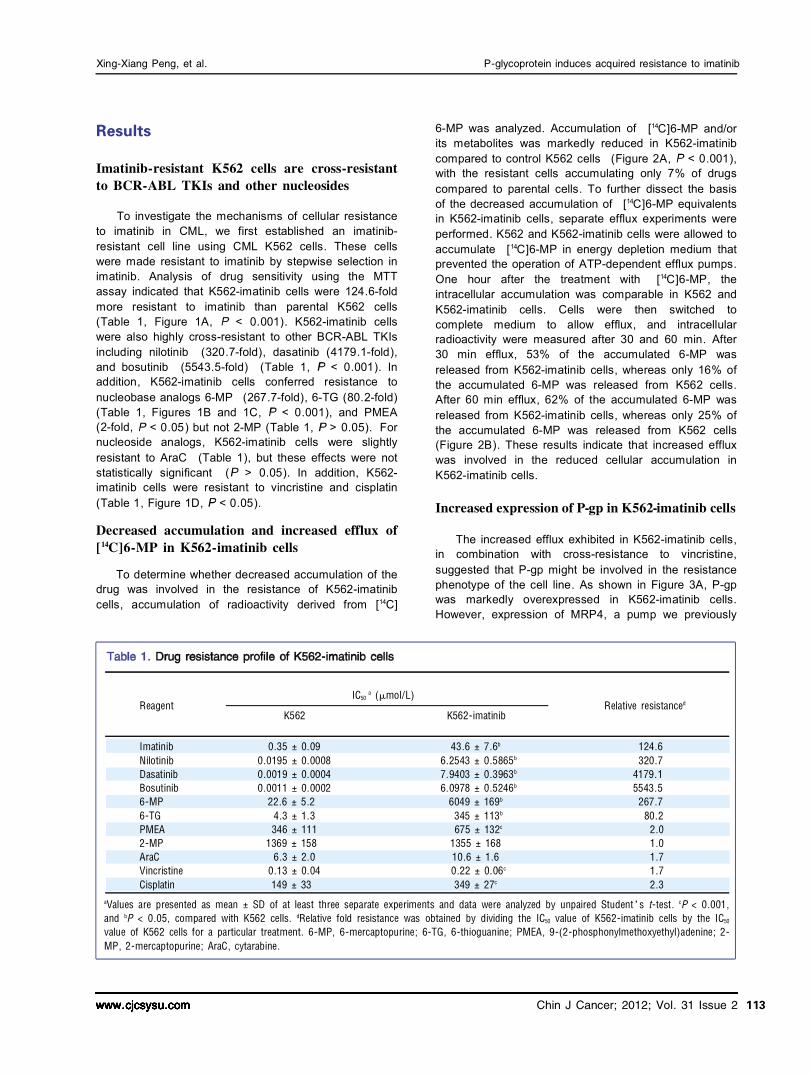
Reagent K562
Relative resistance d IC50
a (滋 mol/L)
a Values are presented as mean 依 SD of at least three separate experiments and data were analyzed by unpaired Student爷s t鄄 test. c P < 0.001, and b P < 0.05, compared with K562 cells. d Relative fold resistance was obtained by dividing the IC50 value of K562鄄 imatinib cells by the IC50
value of K562 cells for a particular treatment. 6鄄 MP, 6鄄 mercaptopurine; 6鄄 TG, 6鄄 thioguanine; PMEA, 9鄄 (2鄄 phosphonylmethoxyethyl)adenine; 2鄄 MP, 2鄄 mercaptopurine; AraC, cytarabine.
K562鄄 imatinib
Imatinib Nilotinib Dasatinib Bosutinib 6鄄 MP 6鄄 TG PMEA 2鄄 MP AraC Vincristine Cisplatin
0.35 依 0.09 0.0195 依 0.0008 0.0019 依 0.0004 0.0011 依 0.0002
22.6 依 5.2 4.3 依 1.3 346 依 111
1369 依 158 6.3 依 2.0
0.13 依 0.04 149 依 33
43.6 依 7.6 b
6.2543 依 0.5865 b
7.9403 依 0.3963 b
6.0978 依 0.5246 b
6049 依 169 b
345 依 113 b
675 依 132 c
1355 依 168 10.6 依 1.6 0.22 依 0.06 c
349 依 27 c
124.6 320.7
4179.1 5543.5
267.7 80.2
2.0 1.0 1.7 1.7 2.3
Results
Imatinib鄄 resistant K562 cells are cross鄄 resistant to BCR鄄 ABL TKIs and other nucleosides
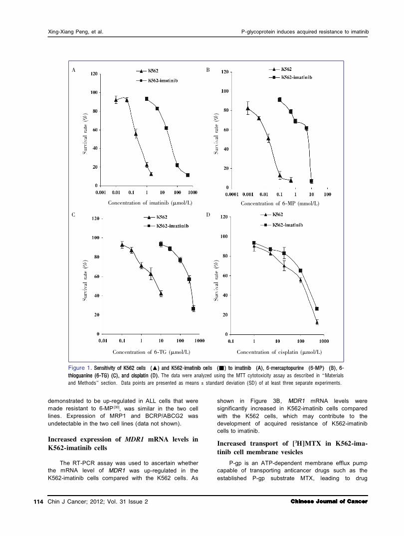
To investigate the mechanisms of cellular resistance to imatinib in CML, we first established an imatinib resistant cell line using CML K562 cells. These cells were made resistant to imatinib by stepwise selection in imatinib. Analysis of drug sensitivity using the MTT assay indicated that K562imatinib cells were 124.6fold more resistant to imatinib than parental K562 cells (Table 1, Figure 1A, < 0.001). K562imatinib cells were also highly crossresistant to other BCRABL TKIs including nilotinib (320.7fold), dasatinib (4179.1fold), and bosutinib (5543.5fold) (Table 1, < 0.001). In addition, K562imatinib cells conferred resistance to nucleobase analogs 6MP (267.7fold), 6TG (80.2fold) (Table 1, Figures 1B and 1C, < 0.001), and PMEA (2fold, < 0.05) but not 2MP (Table 1, > 0.05). For nucleoside analogs, K562imatinib cells were slightly resistant to AraC (Table 1), but these effects were not statistically significant ( > 0.05). In addition, K562 imatinib cells were resistant to vincristine and cisplatin (Table 1, Figure 1D, < 0.05).
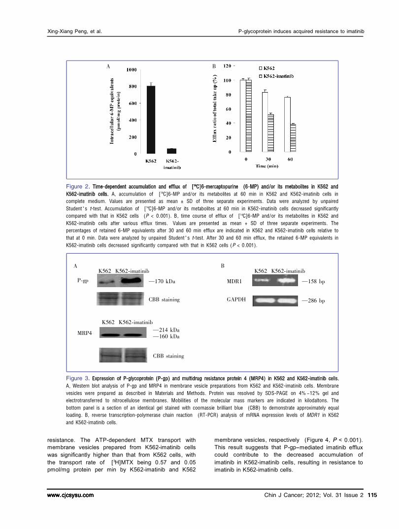
Decreased accumulation and increased efflux of [ 14 C]6鄄 MP in K562鄄 imatinib cells
To determine whether decreased accumulation of the drug was involved in the resistance of K562imatinib cells, accumulation of radioactivity derived from [ 14 C]
6MP was analyzed. Accumulation of [ 14 C]6MP and/or its metabolites was markedly reduced in K562imatinib compared to control K562 cells (Figure 2A, < 0.001), with the resistant cells accumulating only 7% of drugs compared to parental cells. To further dissect the basis of the decreased accumulation of [ 14 C]6MP equivalents in K562imatinib cells, separate efflux experiments were performed. K562 and K562imatinib cells were allowed to accumulate [ 14 C]6MP in energy depletion medium that prevented the operation of ATPdependent efflux pumps. One hour after the treatment with [ 14 C]6MP, the intracellular accumulation was comparable in K562 and K562imatinib cells. Cells were then switched to complete medium to allow efflux, and intracellular radioactivity were measured after 30 and 60 min. After 30 min efflux, 53% of the accumulated 6MP was released from K562imatinib cells, whereas only 16% of the accumulated 6MP was released from K562 cells. After 60 min efflux, 62% of the accumulated 6MP was released from K562imatinib cells, whereas only 25% of the accumulated 6MP was released from K562 cells (Figure 2B). These results indicate that increased efflux was involved in the reduced cellular accumulation in K562imatinib cells.
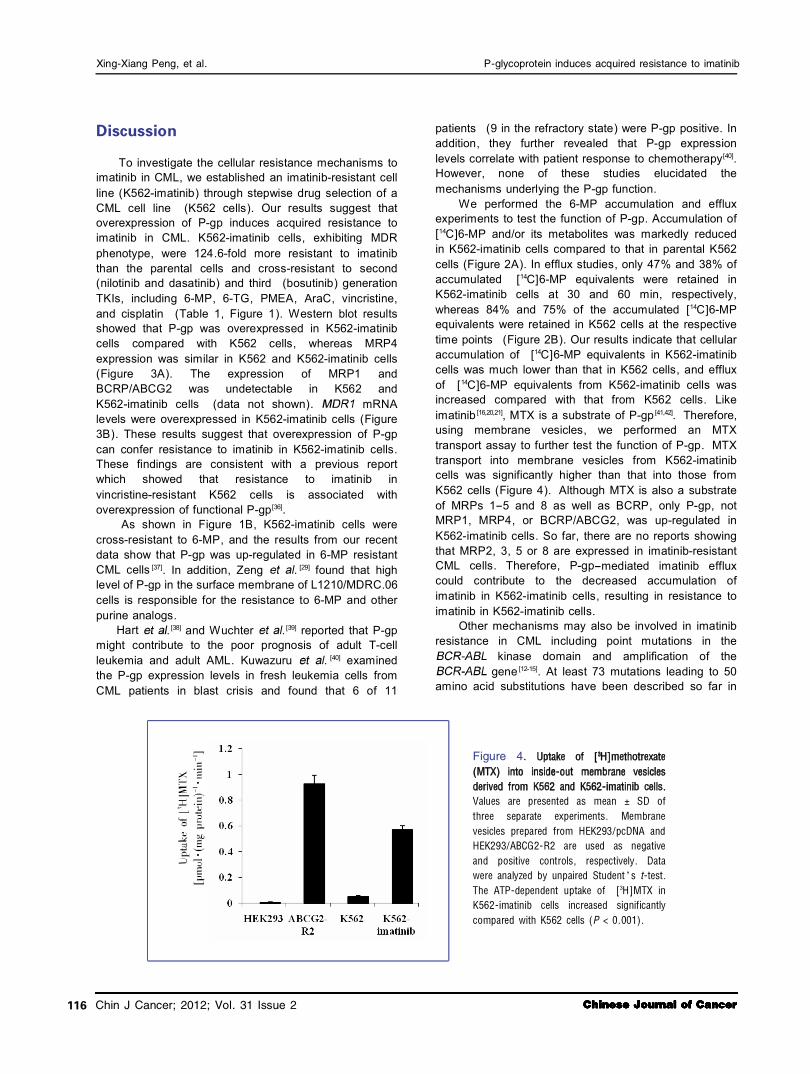
Increased expression of P鄄 gp in K562鄄 imatinib cells
The increased efflux exhibited in K562imatinib cells, in combination with crossresistance to vincristine, suggested that Pgp might be involved in the resistance phenotype of the cell line. As shown in Figure 3A, Pgp was markedly overexpressed in K562imatinib cells. However, expression of MRP4, a pump we previously
Xing鄄 Xiang Peng, et al. P鄄 glycoprotein induces acquired resistance to imatinib
113
Chinese Journal of Cancer Chinese Journal of Cancer Chinese Journal of Cancer Chinese Journal of Cancer Chinese Journal of Cancer Chinese Journal of Cancer Chinese Journal of Cancer Chin J Cancer; 2012; Vol. 31 Issue 2
Figure 1. The data were analyzed using the MTT cytotoxicity assay as described in 野Materials
and Methods冶 section. Data points are presented as means 依 standard deviation (SD) of at least three separate experiments.
Concentration of imatinib (滋 mol/L) Concentration of 6鄄 MP (mmol/L)
Concentration of 6鄄 TG (滋 mol/L) Concentration of cisplatin (滋 mol/L)
demonstrated to be upregulated in ALL cells that were made resistant to 6MP [30] , was similar in the two cell lines. Expression of MRP1 and BCRP/ABCG2 was undetectable in the two cell lines (data not shown).
Increased expression of MDR1 mRNA levels in K562鄄 imatinib cells
The RTPCR assay was used to ascertain whether the mRNA level of was upregulated in the K562imatinib cells compared with the K562 cells. As
shown in Figure 3B, mRNA levels were significantly increased in K562imatinib cells compared with the K562 cells, which may contribute to the development of acquired resistance of K562imatinib cells to imatinib.
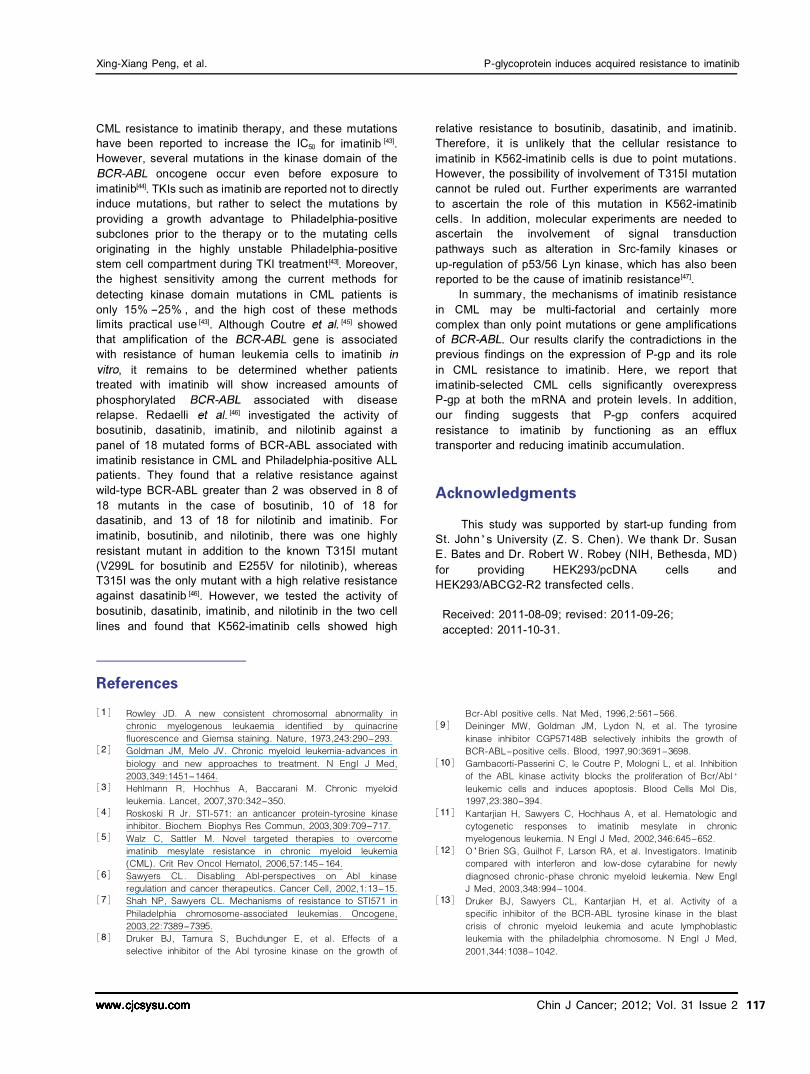
Increased transport of [ 3 H]MTX in K562鄄 ima鄄 tinib cell membrane vesicles
Pgp is an ATPdependent membrane efflux pump capable of transporting anticancer drugs such as the established Pgp substrate MTX, leading to drug
Xing鄄 Xiang Peng, et al. P鄄 glycoprotein induces acquired resistance to imatinib
A B
C D
114
www.cjcsysu.com www.cjcsysu.com www.cjcsysu.com www.cjcsysu.com www.cjcsysu.com www.cjcsysu.com www.cjcsysu.com Chin J Cancer; 2012; Vol. 31 Issue 2
resistance. The ATPdependent MTX transport with membrane vesicles prepared from K562imatinib cells was significantly higher than that from K562 cells, with the transport rate of [ 3 H]MTX being 0.57 and 0.05 pmol/mg protein per min by K562imatinib and K562
membrane vesicles, respectively (Figure 4, < 0.001). This result suggests that Pgpmediated imatinib efflux could contribute to the decreased accumulation of imatinib in K562imatinib cells, resulting in resistance to imatinib in K562imatinib cells.
Figure 2. A, accumulation of [ 14 C]6鄄 MP and/or its metabolites at 60 min in K562 and K562鄄 imatinib cells in
complete medium. Values are presented as mean 依 SD of three separate experiments. Data were analyzed by unpaired Student爷s t鄄 test. Accumulation of [ 14 C]6鄄 MP and/or its metabolites at 60 min in K562鄄 imatinib cells decreased significantly compared with that in K562 cells (P < 0.001). B, time course of efflux of [ 14 C]6鄄 MP and/or its metabolites in K562 and K562鄄 imatinib cells after various efflux times. Values are presented as mean + SD of three separate experiments. The percentages of retained 6鄄 MP equivalents after 30 and 60 min efflux are indicated in K562 and K562鄄 imatinib cells relative to that at 0 min. Data were analyzed by unpaired Student爷s t鄄 test. After 30 and 60 min efflux, the retained 6鄄 MP equivalents in K562鄄 imatinib cells decreased significantly compared with that in K562 cells (P < 0.001).
A B K562 K562鄄 imatinib K562 K562鄄 imatinib P鄄 gp 要170 kDa
CBB staining
要158 bp
要286 bp
MDR1
GAPDH
K562 K562鄄 imatinib MRP4 要214 kDa 要160 kDa
CBB staining
Figure 3. A, Western blot analysis of P鄄 gp and MRP4 in membrane vesicle preparations from K562 and K562鄄 imatinib cells. Membrane vesicles were prepared as described in Materials and Methods. Protein was resolved by SDS鄄 PAGE on 4% -12% gel and electrotransferred to nitrocellulose membranes. Mobilities of the molecular mass markers are indicated in kilodaltons. The bottom panel is a section of an identical gel stained with coomassie brilliant blue (CBB) to demonstrate approximately equal loading. B, reverse transcription鄄 polymerase chain reaction (RT鄄 PCR) analysis of mRNA expression levels of MDR1 in K562 and K562鄄 imatinib cells.
Xing鄄 Xiang Peng, et al. P鄄 glycoprotein induces acquired resistance to imatinib
A B
115
Chinese Journal of Cancer Chinese Journal of Cancer Chinese Journal of Cancer Chinese Journal of Cancer Chinese Journal of Cancer Chinese Journal of Cancer Chinese Journal of Cancer Chin J Cancer; 2012; Vol. 31 Issue 2
Discussion To investigate the cellular resistance mechanisms to
imatinib in CML, we established an imatinibresistant cell line (K562imatinib) through stepwise drug selection of a CML cell line (K562 cells). Our results suggest that overexpression of Pgp induces acquired resistance to imatinib in CML. K562imatinib cells, exhibiting MDR phenotype, were 124.6fold more resistant to imatinib than the parental cells and crossresistant to second (nilotinib and dasatinib) and third (bosutinib) generation TKIs, including 6MP, 6TG, PMEA, AraC, vincristine, and cisplatin (Table 1, Figure 1). Western blot results showed that Pgp was overexpressed in K562imatinib cells compared with K562 cells, whereas MRP4 expression was similar in K562 and K562imatinib cells (Figure 3A). The expression of MRP1 and BCRP/ABCG2 was undetectable in K562 and K562imatinib cells (data not shown). mRNA levels were overexpressed in K562imatinib cells (Figure 3B). These results suggest that overexpression of Pgp can confer resistance to imatinib in K562imatinib cells. These findings are consistent with a previous report which showed that resistance to imatinib in vincristineresistant K562 cells is associated with overexpression of functional Pgp [36] .
As shown in Figure 1B, K562imatinib cells were crossresistant to 6MP, and the results from our recent data show that Pgp was upregulated in 6MP resistant CML cells [37] . In addition, Zeng . [29] found that high level of Pgp in the surface membrane of L1210/MDRC.06 cells is responsible for the resistance to 6MP and other purine analogs.
Hart . [38] and Wuchter . [39] reported that Pgp might contribute to the poor prognosis of adult Tcell leukemia and adult AML. Kuwazuru . [40] examined the Pgp expression levels in fresh leukemia cells from CML patients in blast crisis and found that 6 of 11
patients (9 in the refractory state) were Pgp positive. In addition, they further revealed that Pgp expression levels correlate with patient response to chemotherapy [40] . However, none of these studies elucidated the mechanisms underlying the Pgp function.
We performed the 6MP accumulation and efflux experiments to test the function of Pgp. Accumulation of [ 14 C]6MP and/or its metabolites was markedly reduced in K562imatinib cells compared to that in parental K562 cells (Figure 2A). In efflux studies, only 47% and 38% of accumulated [ 14 C]6MP equivalents were retained in K562imatinib cells at 30 and 60 min, respectively, whereas 84% and 75% of the accumulated [ 14 C]6MP equivalents were retained in K562 cells at the respective time points (Figure 2B). Our results indicate that cellular accumulation of [ 14 C]6MP equivalents in K562imatinib cells was much lower than that in K562 cells, and efflux of [ 14 C]6MP equivalents from K562imatinib cells was increased compared with that from K562 cells. Like imatinib [16,20,21] , MTX is a substrate of Pgp [41,42] . Therefore, using membrane vesicles, we performed an MTX transport assay to further test the function of Pgp. MTX transport into membrane vesicles from K562imatinib cells was significantly higher than that into those from K562 cells (Figure 4). Although MTX is also a substrate of MRPs 15 and 8 as well as BCRP, only Pgp, not MRP1, MRP4, or BCRP/ABCG2, was upregulated in K562imatinib cells. So far, there are no reports showing that MRP2, 3, 5 or 8 are expressed in imatinibresistant CML cells. Therefore, Pgpmediated imatinib efflux could contribute to the decreased accumulation of imatinib in K562imatinib cells, resulting in resistance to imatinib in K562imatinib cells.
Other mechanisms may also be involved in imatinib resistance in CML including point mutations in the
kinase domain and amplification of the gene [1215] . At least 73 mutations leading to 50
amino acid substitutions have been described so far in
Xing鄄 Xiang Peng, et al. P鄄 glycoprotein induces acquired resistance to imatinib
Figure 4.
Values are presented as mean 依 SD of three separate experiments. Membrane vesicles prepared from HEK293/pcDNA and HEK293/ABCG2鄄 R2 are used as negative and positive controls, respectively. Data were analyzed by unpaired Student爷s t鄄 test. The ATP鄄 dependent uptake of [ 3 H]MTX in K562鄄 imatinib cells increased significantly compared with K562 cells (P < 0.001).
116
www.cjcsysu.com www.cjcsysu.com www.cjcsysu.com www.cjcsysu.com www.cjcsysu.com www.cjcsysu.com www.cjcsysu.com Chin J Cancer; 2012; Vol. 31 Issue 2
咱1暂
咱2暂
咱3暂 咱4暂 咱5暂
咱6暂 咱7暂
咱8暂
咱9暂
咱10暂
咱11暂
咱12暂
咱13暂
CML resistance to imatinib therapy, and these mutations have been reported to increase the IC 50 for imatinib [43] . However, several mutations in the kinase domain of the
oncogene occur even before exposure to imatinib [44] . TKIs such as imatinib are reported not to directly induce mutations, but rather to select the mutations by providing a growth advantage to Philadelphiapositive subclones prior to the therapy or to the mutating cells originating in the highly unstable Philadelphiapositive stem cell compartment during TKI treatment [43] . Moreover, the highest sensitivity among the current methods for detecting kinase domain mutations in CML patients is only 15% 25% , and the high cost of these methods limits practical use [43] . Although Coutre . [45] showed that amplification of the gene is associated with resistance of human leukemia cells to imatinib
, it remains to be determined whether patients treated with imatinib will show increased amounts of phosphorylated associated with disease relapse. Redaelli . [46] investigated the activity of bosutinib, dasatinib, imatinib, and nilotinib against a panel of 18 mutated forms of BCRABL associated with imatinib resistance in CML and Philadelphiapositive ALL patients. They found that a relative resistance against wildtype BCRABL greater than 2 was observed in 8 of 18 mutants in the case of bosutinib, 10 of 18 for dasatinib, and 13 of 18 for nilotinib and imatinib. For imatinib, bosutinib, and nilotinib, there was one highly resistant mutant in addition to the known T315I mutant (V299L for bosutinib and E255V for nilotinib), whereas T315I was the only mutant with a high relative resistance against dasatinib [46] . However, we tested the activity of bosutinib, dasatinib, imatinib, and nilotinib in the two cell lines and found that K562imatinib cells showed high
relative resistance to bosutinib, dasatinib, and imatinib. Therefore, it is unlikely that the cellular resistance to imatinib in K562imatinib cells is due to point mutations. However, the possibility of involvement of T315I mutation cannot be ruled out. Further experiments are warranted to ascertain the role of this mutation in K562imatinib cells. In addition, molecular experiments are needed to ascertain the involvement of signal transduction pathways such as alteration in Srcfamily kinases or upregulation of p53/56 Lyn kinase, which has also been reported to be the cause of imatinib resistance [47] .
In summary, the mechanisms of imatinib resistance in CML may be multifactorial and certainly more complex than only point mutations or gene amplifications of . Our results clarify the contradictions in the previous findings on the expression of Pgp and its role in CML resistance to imatinib. Here, we report that imatinibselected CML cells significantly overexpress Pgp at both the mRNA and protein levels. In addition, our finding suggests that Pgp confers acquired resistance to imatinib by functioning as an efflux transporter and reducing imatinib accumulation.
Acknowledgments This study was supported by startup funding from
St. John爷s University (Z. S. Chen). We thank Dr. Susan E. Bates and Dr. Robert W. Robey (NIH, Bethesda, MD) for providing HEK293/pcDNA cells and HEK293/ABCG2R2 transfected cells.
Received: 20110809; revised: 20110926; accepted: 20111031.
References
Rowley JD. A new consistent chromosomal abnormality in chronic myelogenous leukaemia identified by quinacrine fluorescence and Giemsa staining. Nature, 1973,243:290-293. Goldman JM, Melo JV. Chronic myeloid leukemia鄄 advances in biology and new approaches to treatment. N Engl J Med, 2003,349:1451-1464. Hehlmann R, Hochhus A, Baccarani M. Chronic myeloid leukemia. Lancet, 2007,370:342-350. Roskoski R Jr. STI鄄 571: an anticancer protein鄄 tyrosine kinase inhibitor. Biochem Biophys Res Commun, 2003,309:709-717. Walz C, Sattler M. Novel targeted therapies to overcome imatinib mesylate resistance in chronic myeloid leukemia (CML). Crit Rev Oncol Hematol, 2006,57:145-164. Sawyers CL. Disabling Abl鄄 perspectives on Abl kinase regulation and cancer therapeutics. Cancer Cell, 2002,1:13-15. Shah NP, Sawyers CL. Mechanisms of resistance to STI571 in Philadelphia chromosome鄄 associated leukemias. Oncogene, 2003,22:7389-7395. Druker BJ, Tamura S, Buchdunger E, et al. Effects of a selective inhibitor of the Abl tyrosine kinase on the growth of
Bcr鄄 Abl positive cells. Nat Med, 1996,2:561-566. Deininger MW, Goldman JM, Lydon N, et al. The tyrosine kinase inhibitor CGP57148B selectively inhibits the growth of BCR鄄 ABL-positive cells. Blood, 1997,90:3691-3698. Gambacorti鄄 Passerini C, le Coutre P, Mologni L, et al. Inhibition of the ABL kinase activity blocks the proliferation of Bcr/Abl +
leukemic cells and induces apoptosis. Blood Cells Mol Dis, 1997,23:380-394. Kantarjian H, Sawyers C, Hochhaus A, et al. Hematologic and cytogenetic responses to imatinib mesylate in chronic myelogenous leukemia. N Engl J Med, 2002,346:645-652. O爷Brien SG, Guilhot F, Larson RA, et al. Investigators. Imatinib compared with interferon and low鄄 dose cytarabine for newly diagnosed chronic鄄 phase chronic myeloid leukemia. New Engl J Med, 2003,348:994-1004. Druker BJ, Sawyers CL, Kantarjian H, et al. Activity of a specific inhibitor of the BCR鄄 ABL tyrosine kinase in the blast crisis of chronic myeloid leukemia and acute lymphoblastic leukemia with the philadelphia chromosome. N Engl J Med, 2001,344:1038-1042.
Xing鄄 Xiang Peng, et al. P鄄 glycoprotein induces acquired resistance to imatinib
117
Chinese Journal of Cancer Chinese Journal of Cancer Chinese Journal of Cancer Chinese Journal of Cancer Chinese Journal of Cancer Chinese Journal of Cancer Chinese Journal of Cancer Chin J Cancer; 2012; Vol. 31 Issue 2
咱14暂
咱15暂
咱16暂
咱17暂
咱18暂
咱19暂
咱20暂
咱21暂
咱22暂
咱23暂
咱24暂
咱25暂
咱26暂
咱27暂
咱28暂
咱29暂
咱30暂
咱31暂
咱32暂
咱33暂
咱34暂
咱35暂
咱36暂
咱37暂
咱38暂
咱39暂
咱40暂
咱41暂
咱42暂
咱43暂 咱44暂
咱45暂
咱46暂
咱47暂
Weisberg E, Griffin JD. Mechanisms of resistance imatinib (STI571) in preclinical models and in leukemia patients. Drug Resist Updat, 2001,4:22-28. Gambacorti鄄 Passerini CB, Gunby RH, Piazza R, et al. Molecular mechanisms of resistance to imatinib in Philadelphia鄄 chromosome-positive leukemias. Lancet, 2003,4:75-85. Mahon FX, Belloc F, Lagarde V, et al. MDR1 gene overexpression confers resistance to imatinib mesylate in leukemia cell line models. Blood, 2003,101:2368-2373. Widmer N, Colombo S, Buclin T, et al. Functional consequence of MDR1 expression on imatinib intracellular concentrations. Blood, 2003,102:1142. Aller SG, Yu J, Ward A, et al. Structure of P鄄 glycoprotein reveals a molecular basis for poly鄄 specific drug binding. Science, 2009,323:1718-1722. Dai H, Marbach P, Lemaire M, et al. Distribution of STI鄄 571 to the brain is limited by P鄄 glycoprotein-mediated efflux. J Pharmacol Exp Ther, 2003,304:1085-1092. Hegdus T, Orfi L, Septodi A, et al. Interaction of tyrosine kinase inhibitors with the human multidrug transporter proteins, MDR1 and MRP1. Biochim Biophys Acta, 2002,1587:318-325. Mukai M, Che XF, Furukawa T, et al. Reversal of the resistance to STI571 in human chronic myelogenous leukemia K562 cells. Cancer Sci, 2003,94:557-563. Illmer T, Schaich M, Platzbecker U, et al. P鄄 glycoprotein- mediated drug efflux is a resistance mechanism of chronic myelogenous leukemia cells to treatment with imatinib mesylate. Leukemia, 2004,18:401-408. Hamada A, Miyano H, Watanabe H, et al. Interaction of imatinib mesylate with human p鄄 glycoprotein. J Pharmacol Exp Ther, 2003,307:824-828. Burger H, van Tol H, Boersma AW, et al. Imatinib mesylate (STI571) is a substrate for the breast cancer resistance protein (BCRP)/ABCG2 drug pump. Blood, 2004,104:2940-2942. Burger H, van Tol H, Brok M, et al. Chronic imatinib mesylate exposure leads to reduced intracellular drug accumulation by induction of the ABCG2 (BCRP) and ABCB1 (MDR1) drug transport pumps. Cancer Biol Ther, 2005,4:747-752. Ferrao PT, Frost MJ, Siah SP, et al. Overexpression of P鄄 glycoprotein in K562 cells does not confer resistance to the growth inhibitory effects of imatinib (STI571) in vitro. Blood, 2003,102:4499-4503. Hirayama C, Watanabe H, Nakashima R, et al. Constitutive overexpression of P鄄 glycoprotein, rather than breast cancer resistance protein or organic cation transporter 1, contributes to acquisition of imatinib鄄 resistance in K562 cells. Pharm Res, 2008,25:827-835. Peng XX, Shi Z, Damaraju VL, et al. Up鄄 regulation of MRP4 and down鄄 regulation of influx transporters in human leukemic cells with acquired resistance to 6鄄 mercaptopurine. Leuk Res, 2008,32:799-809. Zeng H, Lin ZP, Sartorelli AC. Resistance to purine and pyrimidine nucleoside and nucleobase analogs by the human MDR1 transfected murine leukemia cell line L1210/VMDRC.06. Biochem Pharmacol, 2004,68:911-921. Breuninger LM, Paul S, Gaughan K, et al. Expression of multidrug associated protein in NIH/3T3 cells confers multidrug resistance associated with increased drug efflux and altered
intracellular drug distribution. Cancer Res, 1995,55:5342-5347. Chen ZS, Lee K, Kruh GD. Transport of cyclic nucleotides and estradiol 17鄄 beta鄄 D鄄 glucuronide by multidrug resistance protein 4: resistance to 6鄄 mercaptopurine and 6鄄 thioguanine. J Biol Chem, 2001,276:33747-33754. Lee K, Klein鄄 Szanto AJ, Kruh GD. Analysis of the MRP4 drug resistance profile in transfected NIH3T3 cells. J Natl Cancer Inst, 2000,92:1934-1940. Chen ZS, Robey RW, Belinsky MG, et al. Transport of methotrexate, methotrexate polyglutamates, and 17beta鄄 estradiol 17鄄 (beta鄄 D鄄 glucuronide) by ABCG2: effects of acquired mutations at R482 on methotrexate transport. Cancer Res, 2003,63:4048-4054. Peng XX, Li YB. Induction of cellular glutathione鄄 linked enzymes and catalase by the unique chemoprotective agent, 3 H鄄 1,2鄄 dithiole鄄 3鄄 thione in rat cardiomyocytes affords protection against oxidative cell injury. Pharmacol Res, 2002,45:491-497. Bradford MM. A rapid and sensitive method for the quantitation of microgram quantities of protein utilizing the principle of protein鄄 dye binding. Anal Biochem, 1976,72:248-254. Assef Y, Rubio F, Colo G, et al. Imatinib resistance in multidrug鄄 resistant K562 human leukemic cells. Leuk Res, 2009,33:710-716. Peng XX, Shi Z, Tiwari et al. Up鄄 regulation of P鄄 glycoprotein confers acquired resistance to 6鄄 mercaptopurine in human chronic myeloid leukemia cells. Oncol Lett, 2011,2(3):549-556. Hart SM, Ganeshaguru K, Hoffbrand AV, et al. Expression of the Multidrug resistance鄄 associated protein (MRP) in acute leukemia. Leukemia, 1994,8:2163-2168. Wuchter C, Leonid K, Ruppert V, et al. Clinical significance of P鄄 glycoprotein expression and function for response to induction chemotherapy, relapse rate and overall survival in acute leukemia. Haematologica, 2000,85:711-721. Kuwazuru Y, Yoshimura A, Hanada S, et al. Expression of multidrug transporter鄄 glycoprotein, in chronic myelogenous leukemia cells in blast crisis. Br J Haematol, 1990,74:24-29. Loscher W, Potschka H. Blood鄄 brain barrier active efflux transporters: ATP鄄 binding cassette gene family. NeuroRx, 2005,2:86-98. De Graaf D, Sharma RC, Mechetner EB, et al. P鄄 glycoprotein confers methotrexate resistance in 3T6 cells with deficient carrier鄄 mediated methotrexate uptake. Proc Natl Acad Sci USA, 1996,93:1238-1242. Baccarani M, Pane F, Saglio G. Monitoring treatment of chronic myeloid leukemia. Haematologica, 2008,93:161-169. Jiang X, Saw KM, Eaves A, et al. Instability of BCR鄄 ABL gene in primary and cultured chronic myeloid leukemia stem cells. J Natl Cancer Inst, 2007,99:680-693. le Coutre P, Tassi E, Varella鄄 Garcia M, et al. Induction of resistance to the Abelson inhibitor STI571 in human leukemic cells through gene amplification. Blood, 2000,95:1758-1766. Redaelli S, Piazza R, Rostagno R, et al. Activity of bosutinib, dasatinib, and nilotinib against 18 imatinib鄄 resistant BCR/ABL mutants. J Clin Oncol, 2009,27:469-471. Pene鄄 Dumitrescu T, Smithgall TE. Expression of a Src family kinase in chronic myelogenous leukemia cells induces resistance to imatinib in a kinase鄄 dependent manner. J Biol Chem, 2010,285:21446-21457.
Xing鄄 Xiang Peng, et al. P鄄 glycoprotein induces acquired resistance to imatinib
118
Related Documents


