J Neurosurg / November 22, 2013 DOI: 10.3171/2013.10.JNS13918 1 ©AANS, 2013 D ISRUPTION of adenosine homeostasis is a pathologi- cal hallmark of temporal lobe epilepsy (TLE), and therapeutic adenosine augmentation is a rational approach for seizure control. 5–7 Adenosine homeostasis in the brain is largely under the control of adenosine kinase (ADK), the key metabolic clearance enzyme for adeno- sine. 2 In the adult brain ADK is predominantly expressed in astrocytes, 59 where the expression levels of the enzyme determine the extent of a transmembrane gradient for adenosine, which, under baseline conditions, drives the influx of adenosine into the astrocyte through equilibra- tive nucleoside transporters. 7,9,15,19 Therefore, synaptic levels of adenosine are largely under the control of as- trocytes, which form a sink for the metabolic clearance of adenosine. 9,15,17 We have previously demonstrated that astrogliosis and associated pathological overexpression of Overexpression of adenosine kinase in cortical astrocytes and focal neocortical epilepsy in mice Laboratory investigation *HAI-YING SHEN, M.D., PH.D., 1 HAI SUN, M.D., PH.D., 2 MARISSA M. HANTHORN, B.A., 1 ZHONGWEI ZHI, B.S., 3 JING-QUAN LAN, M.D., 1 DAVID J. POULSEN, M.SC., PH.D., 4 RUIKANG K. W ANG, M.SC., PH.D., 3 AND DETLEV BOISON, PH.D. 1 1 Robert Stone Dow Neurobiology Laboratories, Legacy Research Institute, Portland, Oregon; 2 Department of Neurological Surgery, Oregon Health & Science University, Portland, Oregon; 3 Department of Bioengineering, University of Washington, Seattle, Washington; and 4 Department of Biomedical and Pharmaceutical Sciences, University of Montana, Missoula, Montana Object. New experimental models and diagnostic methods are needed to better understand the pathophysiology of focal neocortical epilepsies in a search for improved epilepsy treatment options. The authors hypothesized that a focal disruption of adenosine homeostasis in the neocortex might be sufficient to trigger electrographic seizures. They further hypothesized that a focal disruption of adenosine homeostasis might affect microcirculation and thus offer a diagnostic opportunity for the detection of a seizure focus located in the neocortex. Methods. Focal disruption of adenosine homeostasis was achieved by injecting an adeno-associated virus (AAV) engineered to overexpress adenosine kinase (ADK), the major metabolic clearance enzyme for the brain’s endog- enous anticonvulsant adenosine, into the neocortex of mice. Eight weeks following virus injection, the affected brain area was imaged via optical microangiography (OMAG) to detect changes in microcirculation. After completion of imaging, cortical electroencephalography (EEG) recordings were obtained from the imaged brain area. Results. Viral expression of the Adk cDNA in astrocytes generated a focal area (~ 2 mm in diameter) of ADK overexpression within the neocortex. OMAG scanning revealed a reduction in vessel density within the affected brain area of approximately 23% and 29% compared with control animals and the contralateral hemisphere, respectively. EEG recordings revealed electrographic seizures within the focal area of ADK overexpression at a rate of 1.3 ± 0.2 seizures per hour (mean ± SEM). Conclusions. The findings of this study suggest that focal adenosine deficiency is sufficient to generate a neocor- tical focus of hyperexcitability, which is also characterized by reduced vessel density. The authors conclude that their model constitutes a useful tool to study neocortical epilepsies and that OMAG constitutes a noninvasive diagnostic tool for the imaging of seizure foci with disrupted adenosine homeostasis. (http://thejns.org/doi/abs/10.3171/2013.10.JNS13918) KEY WORDS • adenosine kinase • adeno-associated virus • basic science • neocortical epilepsy • optical microangiography • mouse • epilepsy 1 Abbreviations used in this paper: AAV = adeno-associated virus; ADK = adenosine kinase; AP = anteroposterior; CBF = cerebral blood flow; DV = dorsoventral; EEG = electroencephalography; GFAP = glial fibrillary acidic protein; ML = mediolateral; NCE = neocortical epilepsy; OMAG = optical microangiography; SEM = standard error of the mean; TLE = temporal lobe epilepsy. * Drs. Shen and Sun contributed equally to this work.

Welcome message from author
This document is posted to help you gain knowledge. Please leave a comment to let me know what you think about it! Share it to your friends and learn new things together.
Transcript

J Neurosurg / November 22, 2013
DOI: 10.3171/2013.10.JNS13918
1
©AANS, 2013
Disruption of adenosine homeostasis is a pathologi-cal hallmark of temporal lobe epilepsy (TLE), and therapeutic adenosine augmentation is a rational
approach for seizure control.5–7 Adenosine homeostasis in
the brain is largely under the control of adenosine kinase (ADK), the key metabolic clearance enzyme for adeno-sine.2 In the adult brain ADK is predominantly expressed in astrocytes,59 where the expression levels of the enzyme determine the extent of a transmembrane gradient for adenosine, which, under baseline conditions, drives the influx of adenosine into the astrocyte through equilibra-tive nucleoside transporters.7,9,15,19 Therefore, synaptic levels of adenosine are largely under the control of as-trocytes, which form a sink for the metabolic clearance of adenosine.9,15,17 We have previously demonstrated that astrogliosis and associated pathological overexpression of
Overexpression of adenosine kinase in cortical astrocytes and focal neocortical epilepsy in mice
Laboratory investigation*Hai-Ying SHen, M.D., PH.D.,1 Hai Sun, M.D., PH.D.,2 MariSSa M. HantHorn, B.a.,1 ZHongwei ZHi, B.S.,3 Jing-Quan Lan, M.D.,1 DaviD J. PouLSen, M.Sc., PH.D.,4 ruikang k. wang, M.Sc., PH.D.,3 anD DetLev BoiSon, PH.D.1
1Robert Stone Dow Neurobiology Laboratories, Legacy Research Institute, Portland, Oregon; 2Department of Neurological Surgery, Oregon Health & Science University, Portland, Oregon; 3Department of Bioengineering, University of Washington, Seattle, Washington; and 4Department of Biomedical and Pharmaceutical Sciences, University of Montana, Missoula, Montana
Object. New experimental models and diagnostic methods are needed to better understand the pathophysiology of focal neocortical epilepsies in a search for improved epilepsy treatment options. The authors hypothesized that a focal disruption of adenosine homeostasis in the neocortex might be sufficient to trigger electrographic seizures. They further hypothesized that a focal disruption of adenosine homeostasis might affect microcirculation and thus offer a diagnostic opportunity for the detection of a seizure focus located in the neocortex.
Methods. Focal disruption of adenosine homeostasis was achieved by injecting an adeno-associated virus (AAV) engineered to overexpress adenosine kinase (ADK), the major metabolic clearance enzyme for the brain’s endog-enous anticonvulsant adenosine, into the neocortex of mice. Eight weeks following virus injection, the affected brain area was imaged via optical microangiography (OMAG) to detect changes in microcirculation. After completion of imaging, cortical electroencephalography (EEG) recordings were obtained from the imaged brain area.
Results. Viral expression of the Adk cDNA in astrocytes generated a focal area (~ 2 mm in diameter) of ADK overexpression within the neocortex. OMAG scanning revealed a reduction in vessel density within the affected brain area of approximately 23% and 29% compared with control animals and the contralateral hemisphere, respectively. EEG recordings revealed electrographic seizures within the focal area of ADK overexpression at a rate of 1.3 ± 0.2 seizures per hour (mean ± SEM).
Conclusions. The findings of this study suggest that focal adenosine deficiency is sufficient to generate a neocor-tical focus of hyperexcitability, which is also characterized by reduced vessel density. The authors conclude that their model constitutes a useful tool to study neocortical epilepsies and that OMAG constitutes a noninvasive diagnostic tool for the imaging of seizure foci with disrupted adenosine homeostasis.(http://thejns.org/doi/abs/10.3171/2013.10.JNS13918)
keY worDS • adenosine kinase • adeno-associated virus • basic science • neocortical epilepsy • optical microangiography • mouse • epilepsy
1
Abbreviations used in this paper: AAV = adeno-associated virus; ADK = adenosine kinase; AP = anteroposterior; CBF = cerebral blood flow; DV = dorsoventral; EEG = electroencephalography; GFAP = glial fibrillary acidic protein; ML = mediolateral; NCE = neocortical epilepsy; OMAG = optical microangiography; SEM = standard error of the mean; TLE = temporal lobe epilepsy.
* Drs. Shen and Sun contributed equally to this work.
H. Y. Shen et al.
2 J Neurosurg / November 22, 2013
ADK is linked to neuronal hyperexcitability and seizure activity in rodent models of TLE,3,40 whereas surgically resected specimens from human patients with TLE were likewise characterized by profound overexpression of ADK.3,43 Conversely, genetic or virus-induced reduction of ADK expression in the hippocampus was shown to sup-press seizures,40,60 while focal cell-based adenosine aug-mentation to the hippocampus was shown to protect the hippocampal formation from injury and seizures.40,41 To-gether, these findings demonstrate that adenosine homeo-stasis, controlled by astrocytic ADK, critically determines excitability of the hippocampus. However, whether this is true for other brain regions and other forms of epilepsy has not been investigated.
In addition to its direct control of neuronal func-tion, adenosine signaling regulates vascular functions, which may contribute to the brain’s susceptibility to sei-zures.1,47,50,51 Acutely, adenosine promotes hemodynamic events, such as vasodilation to increase blood flow in the brain, in response to hypoxia or ischemia.12,50,51 Adeno-sine, largely derived from the breakdown of ATP during conditions of energetic crisis, cell swelling, and acidosis, affects cerebral blood flow mostly via activation of aden-osine A2A receptors in balancing blood flow with metabo-lism. The primary effect of stimulating A2A receptors is activation of KATP and KCa channels resulting in smooth-muscle relaxation and elevated blood flow rates.50 Fur-ther, it has been shown that inhibition of ADK increases cerebral blood flow (CBF) by augmenting interstitial adenosine levels.56 In addition, chronically increased lev-els of adenosine have been shown to stimulate angiogen-esis.1,29,49 Consequently, chronic overexpression of ADK, as seen in epilepsy, may result in reduced cerebral blood flow (CBF) and reduction of local vasculature. Therefore, ADK-dependent changes in vasculature might provide a diagnostic opportunity for the detection of an epilep-togenic focus that is characterized by overexpression of ADK.
This study was designed to assess whether overex-pression of ADK links to neuronal hyperexcitability and seizure activity in the neocortex, as well as to develop a diagnostic method to identify cortical areas of neuronal hyperexcitability. Compared with TLE, the pathophysiol-ogy and mechanisms for seizure generation in neocortical epilepsy (NCE) are understudied. In particular, a scarcity of clinically relevant rodent models for NCE has limited research progress.33,45,48 In addition, NCE remains one of the most difficult forms of epilepsy to treat, and there is a need for improved diagnostic methods that account for pathophysiological mechanisms of NCE.20,54,66
Since astroglial ADK regulates neuronal excitability of the hippocampus as well as microcirculation, we hy-pothesized that a focal disruption of adenosine homeo-stasis induced by overexpression of ADK in astrocytes in the neocortex might be sufficient to induce neuronal hyperexcitability and changes in microcirculation. We used adeno-associated virus 8 (AAV8)–based viral vec-tors to induce overexpression of ADK in astrocytes of the neocortex.57 To demonstrate ADK-related changes in mi-crocirculation we used a novel imaging technology, op-tical microangiography (OMAG),30–32 which is based on
endogenous light scattering from biological tissue. This noninvasive technology allows generation of microstruc-tural and functional vascular images that can resolve the 3D distribution of dynamic blood perfusion at the cap-illary level resolution in vivo.30–32 Here, we evaluated OMAG as a potential diagnostic tool for the detection of microcirculation changes in the vicinity of focal areas with disrupted adenosine homeostasis. Our data demon-strate that focal overexpression of ADK in astrocytes of the neocortex is sufficient to elicit electrographic seizures with concomitant changes in microcirculation. Our find-ings are of significance for the understanding of mecha-nisms of seizure generation in NCE and demonstrate the diagnostic value of the noninvasive OMAG approach to identify focal neocortical areas of hyperexcitability.
MethodsAnimals
All animal procedures were performed in a facility accredited by the Association for the Assessment and Accreditation of Laboratory Animal Care and were per-formed in accordance with protocols approved by the in-stitutional animal care and use committees of the Legacy Research Institute and the Oregon Health & Science Uni-versity and according to principles outlined in the Na-tional Institutes for Health Guide for the Care and Use of Laboratory Animals. A total of 16 C57BL/6 male mice (20–30 g) were subjected to neocortical virus injection followed by OMAG and EEG (electroencephalography) (Fig. 1A). All animals were group-housed with water and food available ad libitum in temperature- and humidity-controlled rooms with a 12-hour light/dark cycle through-out the experimental period.
Adeno-Associated Virus Production and DeliveryAs described previously,57,60 the short cytoplasmic
isoform of ADK was introduced into an adeno-associ-ated virus (AAV)–based system in which ADK cDNA was placed under the control of the astrocyte-specific gfaABC1D promoter.37 This vector is designated as ADK-SS. An AAV8-pGfa-null (AAV-Null) virus containing an empty vector backbone was used as a negative con-trol vector. Virus production and titer determination was performed as described previously.57,60 For virus delivery, the ADK-SS or AAV-Null virus was unilaterally injected into the right side of the neocortex under general anesthe-sia (1.5% isoflurane, 70% N2O, and 28.5% O2), in a vol-ume of 0.5 ml of concentrated viral solution (1012 genomic particles/ml), with the following stereotactic coordinates: anteroposterior (AP) = -2.10 mm; mediolateral (ML) = –1.50 mm; dorsoventral (DV) = –0.80 mm. After the cortical injections, the incisions were sutured and treated with an antiseptic agent, and the animals were then re-turned to their home cages.
Optical MicroangiographyTo test whether focal disruption of adenosine homeo-
stasis affects cortical microcirculation, CBF was mea-sured in mice 8 weeks after viral delivery, using OMAG,
J Neurosurg / November 22, 2013
Adenosine kinase and neocortical epilepsy
3
a high-resolution optical coherence tomography technique that is capable of producing 3D images of dynamic blood perfusion within microcirculatory tissue beds at an imag-ing depth up to 2 mm below the surface.31 For example, OMAG is able to measure changes of cerebral blood per-fusion in response to systemic hypoxia and hyperoxia.31 Since OMAG requires intact cerebral cortex, this step was performed before the EEG recordings (Fig. 1A). Prior to imaging, the mouse was immobilized in a custom-made stereotactic stage under general anesthesia (1.5% isoflu-rane, 70% N2O, and 28.5% O2) and the mouse’s head was
shaved and depilated. The body temperature was main-tained between 35.5°C and 36.5°C with a thermostat-con-trolled heating pad (Harvard Apparatus). An incision of 1 cm was made along the sagittal suture, and the frontal parietal and interparietal bones were exposed by pulling the skin to the sides. The animal was then positioned un-der the OMAG scanning probe. To acquire microvascu-lar images over a large area of the cortex, scanning was performed clockwise, which resulted in 6–8 OMAG im-ages per mouse, covering both hemispheres over an area of approximately 4 × 5 mm (Fig. 1B). The total imaging
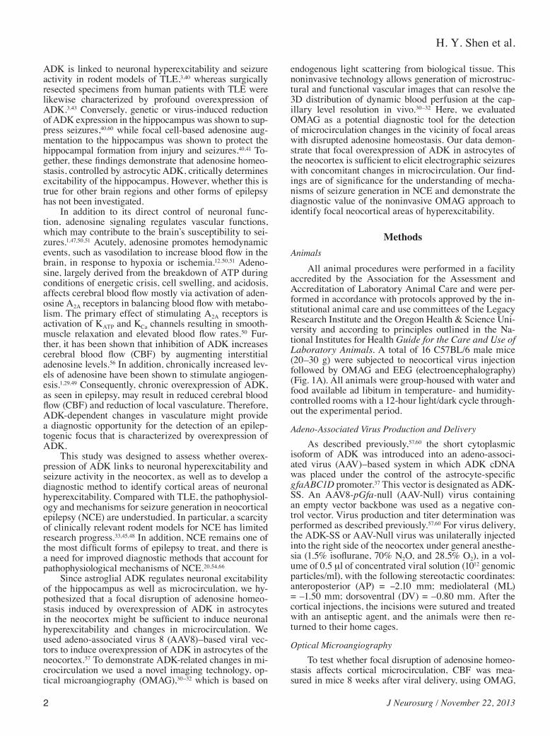
Fig. 1. Overexpression of ADK in the neocortex of mice induced by unilateral injection of ADK-SS virus. A: Schematic illus-tration of the experimental paradigm. Mice were first subjected to unilateral intracortical virus injection (Virus inj.), followed after 8 weeks by an OMAG scan and cortical EEG recordings (EEG rec.). After completion of the EEG, animals were euthanized for im-munohistochemical analysis (IHC). B: Schematic illustration of the virus-transduced area (Virus inj.) and locations of the EEG recording electrodes (red) in frontal cortex (Frontal rec.), focal area of ADK overexpression (Focal rec.), and above the cerebel-lum (reference recording [Ref. rec.]). The area scanned by OMAG is indicated by a dotted green line. C: Representative ADK immunoperoxidase (upper panel) and Nissl (lower panel) staining of brain slices showing the contralateral (contra) or ipsilateral (ipsi) injected (ADK-SS) hemisphere of an ADK-SS recipient and the virus-injected hemisphere of an AAV-Null–injected control animal (AAV-Null). Bar = 500 μm. The bar graph demonstrates relative densities of ADK immunoreactivity of DAB-stained brain sections of ADK-SS or AAV-Null virus–injected mice (2 sections per animal, 5 animals per group). D: Representative ADK staining of consecutive brain slices from an ADK-SS virus–injected animal showing the 3D extent of ADK overexpression. The notch (arrow) on the -2.15-mm slice indicates the locus of the electrode implantation. Bar = 1 mm. Data are displayed as mean ± SEM. #p < 0.01, ADK-SS versus AAV-Null group; *p < 0.01, ipsilateral versus contralateral side.

H. Y. Shen et al.
4 J Neurosurg / November 22, 2013
time per mouse was less than 10 minutes. After imaging and data acquisition, blood flow signals were isolated from each 3D OMAG data set using a volume segmentation al-gorithm.61–63 The volume segmentation algorithm was ap-plied to each OMAG data set to isolate blood flow signals within the cortex. A maximum projection method was then used to project the blood flow signals into the x-y plane to reduce the image size. The final OMAG image, including the area of the viral injection site and the corresponding area in the contralateral hemisphere, was obtained by as-sembling 6 individual images (or 8 images, if necessary). The cerebral blood perfusion was estimated by computing the functional vessel density in the area of interest in the fi-nal OMAG image.53 To achieve this, the OMAG image was first converted to a binary flow map by setting a fixed in-tensity threshold. A circular area with a diameter of 2 mm centered at the viral injection site was identified on the flow map. The percentage of pixels with a binary value of 1 ver-sus pixel numbers of the whole area was calculated as the estimation of vessel density (the quantitative equivalence of cerebral blood perfusion).53 Cerebral blood perfusion in the corresponding area in the contralateral hemisphere was estimated using the same method. Statistical compari-sons of vessel density between the ADK-SS virus–injected animals and the control animals (injected with AAV-Null virus) were then undertaken. Additional comparisons were made with the virus-injected sites and their corresponding contralateral locations. The processing and analysis on the OMAG images was carried out using MATLAB program-ming software (MathWorks, Inc.).
ElectroencephalographyThree days following the OMAG scan, bipolar, coat-
ed stainless steel electrodes (80 mm in diameter, Plastics One) were implanted into the virus-injected cortex (co-ordinates: AP = -2.1 mm; ML = -1.5 mm; DV = -1.0 mm, with bregma as reference) under general anesthesia. A cortical screw electrode (AP = 1.0 mm; ML = 1.5 mm) and a ground reference electrode (AP = 6.0 mm; ML = 0 mm) were placed over the frontal cortex and cerebellum, respectively (Fig. 1B). All electrodes were secured to the skull with dental cement. One week later, EEG monitor-ing was performed as previously described60 with modifi-cations as follows. After the mice were habituated to the EEG-recording procedure for 4 hours, EEG recording was performed for an additional 24 hours. Quantification of the EEG data was performed by an investigator who was blinded to the experimental treatment. An electro-graphic seizure was defined as high-amplitude rhythmic discharge that clearly represented a new pattern of tracing lasting for more than 5 seconds (repetitive spikes, spike-and-wave discharges, or slow waves). Epileptic events oc-curring with an interval of less than 5 seconds without the EEG returning to baseline were defined as belonging to the same seizure. Seizures were primarily electrographic in nature, although frequently accompanied by arrest or staring episodes; no concurrent convulsions were ob-served. Seizure quantification was performed exclusively on the basis of intracortical EEG recordings.
ImmunohistochemistryTo determine changes in ADK expression, virus-
injected mice were euthanized after EEG evaluation for immunohistochemical analysis as previously described.57 For the detection of ADK, a primary anti–ADK antibody (1:5000)59 was used. To detect the cell-type specificity of ADK expression,57 double immunofluorescence staining was performed using a combination of primary antibod-ies directed against ADK (polyclonal, rabbit) and glial fi-brillary acidic protein (GFAP; monoclonal, mouse 1:400, MAB360, Chemicon International), or neuronal nuclei (NeuN; monoclonal, mouse 1:750, MAB377, Chemicon). Nissl staining was performed separately on adjacent brain slices. Digital images were acquired using a Zeiss Axio-Plan inverted microscope equipped with an AxioCam 1Cc1 camera (Carl Zeiss MicroImaging). To quantitatively evaluate the expression level of ADK, the optical density of ADK-positive staining was measured using Image-Pro Plus software (version 5.1, Media Cybernetics, Inc.) as pre-viously described,40 using 1 coronal brain section at the level of the virus-injection from each animal. Data were normalized to the contralateral cortical ADK expression levels.
Statistical AnalysisData are expressed as means ± standard error of
the mean (SEM). Statistical analysis was performed by means of a 1-way ANOVA followed by Bonferroni post hoc analysis or a Student t-test. A p value < 0.05 was con-sidered statistically significant.
ResultsFocal Overexpression of ADK in Astrocytes Within the Neocortex
To determine changes in ADK expression, 8 weeks following virus injection and after completion of OMAG imaging and EEG recordings (Fig. 1A), animals were eu-thanized and their brains evaluated histologically. Nissl staining to assess general brain morphology did not reveal any gross morphological deviations in virus-injected ani-mals compared with controls; in particular, virus injec-tion was not associated with any overt signs of pyknosis, karyolysis, or shrunken cell bodies (Fig. 1C, lower panel). The lack of any obvious virus-induced cytotoxic effects is in line with the demonstrated safety profile of the AAV system for CNS applications.64,65 Immunohistochemical data demonstrated that the cortical injection of the ADK-SS virus induced a focal overexpression of ADK within the injected neocortex (Fig. 1C), whereas injection of the AAV-Null virus had no obvious effects on ADK expres-sion patterns, which were comparable to those seen in the noninjected contralateral cortex (Fig. 1C). Quantitative analysis of immunodensities demonstrated that the ADK-SS virus induced a robust focal increase in ADK expres-sion, which showed a mean fold increase of 4.8 ± 0.86 compared with ADK expression levels in the contralat-eral hemisphere (p < 0.01, 5 mice per group) and a mean fold increase of 4.4 ± 0.77 compared with the AAV-Null–

J Neurosurg / November 22, 2013
Adenosine kinase and neocortical epilepsy
5
injected hemisphere (p < 0.01, 5 mice per group) (Fig. 1C). Conversely, AAV-Null virus injection did not cause significant changes in ADK expression compared with the contralateral hemisphere (p > 0.05, 5 mice per group) (Fig. 1C). We previously demonstrated in a transgenic model system that a 63.6% increase in ADK resulted in a 50% reduction in the ambient tone of adenosine;57 we therefore conclude that the focal overexpression of ADK achieved here triggers a local deficiency in adenosine. To identify the rostral-caudal extent of ADK-SS–induced ADK overexpression, we investigated a series of brain slices spanning the region from -1.70 to -2.75 mm from bregma (Fig. 1D). Analysis of the extent of virus-induced ADK overexpression demonstrated a focal area of high levels of ADK within the neocortex with a diameter about 1.9 ± 0.4 mm (5 mice).
Higher-resolution images of ADK immunoperoxi-dase–stained sections demonstrated profound differences in the subcellular localization of ADK between experi-mental groups (Fig. 2A). Images from ADK-SS–injected cortex showed high levels of ADK expression in cell so-mata and cellular processes, consistent with overexpres-sion of the cytoplasmic isoform of ADK as expressed by the virus. ADK-positive cells had characteristic morpho-logical features of astrocytes, with a star-like appearance and several primary processes originating from the soma (Fig. 2A, left, inset). In contrast, ADK expression in con-trol samples from AAV-Null animals had a characteristic pattern of prominent nuclear ADK expression coupled with a diffuse homogenous staining of ADK throughout the tissue at much lower levels, in line with our previ-ous characterization of astrocytic ADK expression in naïve mice.22,59 To confirm the cell-type specificity of ADK-SS–induced ADK overexpression, we performed a double immunofluorescence analysis by costaining ADK with either GFAP or NeuN (Fig. 2B and 2C). As expect-ed, and in line with findings from our previous work,57,60 ADK expression was colocalized with GFAP expression and was confined to GFAP-positive cells (Fig. 2B, center, left panel) and not seen in NeuN-positive cells (Fig. 2B, center, right panel). Higher-resolution images confirmed the differential expression pattern of ADK-SS–induced overexpression of exogenous ADK (cytoplasm) versus endogenous ADK (nucleus) (Fig. 2C). Together, our data demonstrate a robust focal overexpression of the cytoplas-mic isoform of ADK in cortical astrocytes from ADK-SS–injected mice. Importantly, overexpression of ADK was confined to a distinct focal area, with a diameter of about 2 mm, close to the cortical surface, whereas other brain areas were not affected by ADK-SS or a control virus injection.
Recurrent Electrographic SeizuresTo evaluate whether focal neocortical overexpression
of ADK influenced neuronal excitability, we performed continuous EEG monitoring of neocortical electrographic activity, 8 to 9 weeks after intracortical injection of ADK-SS or AAV-Null virus. EEG recordings from the neocor-tical injection site illustrated that ADK-SS virus–injected mice developed spontaneous recurrent electrographic sei-zures within the neocortex (Fig. 3A, lower panel) where-
as AAV-Null virus–injected animals were devoid of any seizure activity (Fig. 3A, upper panel). The neocortical electrographic seizures were characterized by a gradual increase in amplitude that became rhythmic at the begin-ning of the seizure (Fig. 3A, lower panel, closed arrow) whereas the end of the seizure had an overt drop in am-plitude (Fig. 3C, lower panel, open arrow). The neocor-tical seizures, which occurred in the ADK-SS–injected animals had a seizure frequency of 1.3 ± 0.2 seizures per hour (n = 8) with an average duration of 94 ± 8 seconds. It is important to note that, even though spontaneous elec-trographic seizures were observed in all animals with ADK overexpression, those activities were not associated with any convulsive behavior. Power spectral analysis of the single event depicted in Fig. 3A indicates a predomi-nant ictal frequency of around 3 Hz (Fig. 3C), which is in contrast to interictal EEG activity from ADK-SS re-cipients or baseline activity in AAV-Null recipients (Fig. 3B). Together, the EEG data demonstrated that focal viral overexpression of ADK within astrocytes was sufficient to trigger spontaneous recurrent neocortical seizures in the absence of any epileptogenic event.
Reduced Focal Vessel DensityTo evaluate whether disruption of adenosine homeo-
stasis causes changes in microcirculation, we employed the OMAG approach to image dynamic in vivo cerebral blood perfusion and vasculature changes in the mice 8 weeks after viral injection. The high-contrast OMAG images demonstrated well-resolved cerebral vasculature indicating the cerebral perfusion status of the mice (Fig. 4A). OMAG data showed that the AAV-Null virus injec-tion did not cause focal changes in cerebral blood perfu-sion and vessel density similar to the contralateral hemi-sphere (Fig. 4A, left panel). Importantly, OMAG images showed that ADK-SS–induced overexpression of ADK resulted in an overt reduced local cerebral vessel density indicating reduced cerebral blood perfusion surrounding the viral injection site (Fig. 4A, right panel). The resulting quantitative data demonstrated a significant reduction of focal cerebral blood perfusion in the ipsilateral neocortex of ADK-SS recipients compared with the corresponding region in AAV-Null recipients (23% reduction of CBF, p < 0.05, n = 5, Fig. 4B, left panel). Likewise, the focal ves-sel density in the ipsilateral neocortex of ADK-SS–inject-ed animals was also significantly lower (29% reduction) compared with the contralateral hemisphere of the same animal (29% reduction of vessel density, p < 0.05, n = 5, Fig. 4B, right panel). Together, the analysis of the OMAG data indicated that disruption of adenosine homeostasis by overexpression of ADK led to a reduction in focal ce-rebral blood perfusion and cerebral vessel density.
DiscussionTo better understand mechanisms of ictogenesis in
NCE, we report here a novel animal model of NCE with neocortical overexpression of ADK in astrocytes, and demonstrate the feasibility of OMAG as an advanced noninvasive imaging tool to detect the epileptic focus in this model. We demonstrate the following key findings: 1)

H. Y. Shen et al.
6 J Neurosurg / November 22, 2013
Fig. 2. Cellular expression pattern of ADK. A: Results of ADK immunohistochemistry on coronal sections from mouse brain injected with ADK-SS virus or AAV-Null virus. Bar = 25 µm. B: Immunofluorescence of ADK-SS virus–induced ADK expres-sion (red) co-stained with antibodies directed against GFAP (green, in center left panel) or NeuN (green, in center right panel). Bar = 25 µm. C: Double-immunofluorescence staining of ADK (red) and GFAP (green) at higher resolution showing expression of endogenous ADK within the nucleus and cell soma of astrocytes from the neocortex of an AAV-Null–injected control animal (upper) and expression of exogenous ADK throughout the soma and processes of astrocytes from an ADK-SS recipient (lower). Bar = 10 µm.

J Neurosurg / November 22, 2013
Adenosine kinase and neocortical epilepsy
7
virus-induced overexpression of astrocytic ADK in neo-cortex is sufficient to produce robust spontaneous recur-rent focal electrographic seizures at a frequency of 1.3 ± 0.2 seizures per hour, and 2) the seizure-generating focus spatially aligns with overexpression of ADK and detect-able microcirculation changes, which can be evaluated noninvasively by OMAG.1,29,47,49–51,56
ADK is the key enzyme for the metabolic clearance of adenosine and exists in 2 isoforms, which are specific for the nucleus (ADK long [ADK-L]) or the cytoplasm (ADK short [ADK-S]).8 The specific roles of the different isoforms have not yet been fully elucidated, but mount-
ing evidence suggests that nuclear ADK-L plays a role in epigenetic functions and cell proliferation, whereas the cytoplasmic ADK-S appears to be responsible for the metabolic clearance of extracellular adenosine.8
As our current findings validate (Fig. 2A), and as pre-viously demonstrated,22,59 under control or baseline con-ditions, the majority of ADK expression is confined to the nucleus or perinuclear area of astrocytes. However, in the epileptic brain, astrocytic ADK is not only increased in quantity, but also in subcellular distribution, with a robust shift of ADK expression from the perinuclear area into the network of astrocyte processes.40 Thus quantity and
Fig. 3. Neocortical electroencephalographic activity of AAV-injected mice. A: Focal intracortical EEG traces from the neo-cortical region of mice injected with AAV-Null (upper panel) or ADK-SS (lower panel). The lower traces are at higher resolution and represent background activity (AAV-Null) and a complete representative seizure (ADK-SS), including seizure onset (closed arrow) and end (open arrow). B and C: Power spectral analysis of EEG activity from mice injected with AAV-Null (B) or ADK-SS (C). h = hours; s = seconds.

H. Y. Shen et al.
8 J Neurosurg / November 22, 2013
distribution of the cytoplasmic isoform of ADK might play a key role in ictogenesis. We therefore induced over-expression of the cytoplasmic isoform of ADK in astro-cytes, obtaining profound expression of ADK in astro-cytic processes (Fig. 2C) similar to ADK expression pat-terns in a mouse model of kainic acid induced–epilepsy.40 Our demonstration that overexpression of the cytoplasmic isoform of ADK in astrocytes is sufficient to trigger fo-cal neocortical seizures further supports a role of ADK located in astrocyte processes for ictogenesis.
While AAV-induced overexpression of ADK within astrocytes was sufficient to trigger spontaneous electro-graphic seizures in the neocortex, none of the mice in this study exhibited any signs of convulsive seizures. Pe-riods of immobility or staring episodes were occasionally associated with the electrographic seizures, indicating a phenotype of partial epilepsy without generalization. The hourly seizure rate, as well as the brief duration of the seizures (94 seconds) and the low intra-ictal spike frequency (3 Hz), is in line with electrographic seizure patterns that have been linked to deficient adenosine signaling. Focal CA3 selective seizures (4.3 seizures per hour; 17.5 seconds’ duration), linked to astrogliosis
and overexpression of ADK after intra-amygdaloid kai-nic acid injection, spontaneous seizures in Adk-tg mice with brain-wide overexpression of ADK (4.8 seizures per hour; 26.7 seconds’ duration), and spontaneous seizures in adenosine A1R knockout mice (5.3 seizures per hour; 27.8 seconds’ duration) all share the same characteristic seizure pattern with an intra-ictal spike frequency of 3–4 Hz.38 Together these findings suggest that overexpression of ADK, resulting in deficient signaling through the aden-osine A1 receptor, triggers a characteristic seizure pattern also found in focal seizure models that result from an epi-lepsy-precipitating status epilepticus. Since ADK expres-sion was found to be focally restricted to a sphere with a diameter of about 2 mm (Fig. 1), adenosine homeostasis in the remaining brain is not likely to be compromised by this manipulation. The lack of seizure spread in this model is consistent with previous findings demonstrat-ing that adenosine A1 receptor activation by endogenous adenosine is a prerequisite for the prevention of seizure spread.18,39 Thus, a local deficiency of adenosine caused by the cell-type–selective overexpression of ADK in as-trocytes, triggers focal electrographic seizures by insuffi-cient activation of the A1 receptor, which normally limits neuronal excitability by mediating presynaptic inhibition and by stabilizing the postsynaptic membrane potential. The focal nature of our manipulation is further supported by OMAG imaging suggesting a tight spatial match be-tween overexpression of ADK and changes in the micro-vasculature.
Adenosine is one of the most potent vasodilators in different kinds of tissues and organs. Under both physi-ological and pathophysiological conditions, extracellu-lar adenosine provokes acute local vasodilation within the cerebral microcirculature,47 mainly via activation of the cAMP (cyclic adenosine monophosphate) pathway through adenosine A2A and A2B receptors.49–51 Studies also suggest that adenosine exerts long-term effects on microvasculature via facilitation of angiogenesis1 through the induction of angiogenic growth factors such as vascu-lar endothelial growth factor.1 Whereas the application of adenosine or pharmacological agents that alter adenosine metabolism can stimulate vasodilation and blood vessel growth, the inhibition of ADK can increase CBF through an increase in interstitial adenosine levels.56
While not directly investigated here due to space considerations, we have demonstrated previously that the overexpression of astrocytic ADK leads to a robust reduc-tion in ambient adenosine levels,15,57 whereas downregu-lation of endogenous ADK is a physiological response to raise adenosine levels under conditions of metabolic or energetic stress.52 The reduced vessel density and reduced cerebral blood perfusion demonstrated here (Fig. 4) are consistent with a reduced concentration of adenosine in the neocortical focus of overexpressed ADK and reduced activation of the vascular A2A receptor, which plays an important role in the regulation of CBF and angiogen-esis.50 Given the lower sensitivity of the A2A receptor compared with the A1 receptor, even a minor reduction in adenosine is likely to affect the vasculature. In addition, seizures by themselves are known to trigger the release of adenosine as an endogenous mechanism contributing
Fig. 4. OMAG of cerebral blood perfusion and vessel densities in AAV-injected mice. A: Representative 2D images of cortical ves-sel density at capillary-level resolution in AAV-Null or ADK-SS virus recipients. The asterisks indicate the coordinate of virus injection and the dashed lines indicate the virus-affected area. Bar = 1 mm. B: Quantitative analysis showing reduced cortical vessel density in the hemispheres injected with ADK-SS, compared with those injected with AAV-Null (left panel) or compared with those in the contralateral hemi-sphere (right panel). Cortical vessel densities are shown as percentage relative to control. Data are displayed as mean ± SEM. #p < 0.05 paired comparisons t-test (n = 5 per group).
J Neurosurg / November 22, 2013
Adenosine kinase and neocortical epilepsy
9
to seizure termination,16,35 and seizure-induced adenosine release is consistent with an increase of CBF during ictal seizure events.21,34 Our findings of consistently reduced CBF within the ADK-related seizure focus suggest that seizure-induced elevations of adenosine are subject to en-hanced metabolic clearance through increased levels of ADK and are therefore no longer effective in influenc-ing CBF. Astrocytes play a central role in the vasomotor response, in which neurovascular coupling links massive Ca2+ elevations in the ictal phase to changes in CBF.21 However, a reduced basal adenosine level may lead to a loss of postictal cerebral vasodilation.14,47 Given the fre-quent occurrence and long duration of ADK-induced neo-cortical electrographic seizures, it is likely that the net effects of the CBF measurement obtained by OMAG are based on a combination of ictal and interictal events.
Clinically, neocortical epileptogenic brain areas may be resected following presurgical evaluation through a va-riety of techniques:20 1) computerized EEG source imaging, based on surface or invasive intracranial EEG, targets focal abnormal epileptic activity;44 2) neuroimaging approaches, such as PET, SPECT, or functional MRI are used to detect regional cerebral blood volumes;27,28,46 and 3) CT and MRI are used to detect epilepsy-related dysplasias.10 However, each of the above approaches has its own set of limitations in diagnostic sensitivity and predictive value.36 Alternative or additional diagnostic approaches might be useful to im-prove planning of a resection4,58 and are in particular need for NCE, since this condition in general shows a lower rate of concordance between diagnostic modalities as com-pared with mesial TLE.36 Changes in neuronal activities are known to alter the light scattering and absorption prop-erties of brain tissue. The intrinsic optical signal imaging technique provides 2D cortical maps of these changes and has been used to study the functional organization of visual cortex23,42 and to perform intraoperative mapping of func-tional and epileptiform activities in human subjects.11,24–26,55 These 2D techniques are only able to detect hemodynamic changes on the cortical surface with limited spatial resolu-tion. The novel imaging technology OMAG investigated here is based on endogenous light scattering from biologi-cal tissue to obtain microstructural and functional vascular images, and it can resolve the 3D distribution of dynamic blood perfusion at capillary-level resolution within the mi-crocirculatory bed in vivo.30 In the present study, we dem-onstrate that OMAG is able to detect a region with changes of microvasculature and blood perfusion in a diameter of 2 mm, which reflects the area with overexpression of vi-rus-induced ADK. This finding indicates that a potentially small neocortical epileptic focus might be detectable with OMAG, a technology that is noninvasive and reproducibly delivers high-resolution (100-μm) images without the need of exogenous contrasting agents, as usually needed for CT, MRI, or PET.61 We show here that OMAG can reliably de-tect focal overexpression of ADK. Since overexpression of ADK appears to be a common pathological hallmark found in many different forms of experimental and clini-cal epilepsy,2,3,13,22,39,40,43 OMAG could represent a novel diagnostic tool to identify focal areas of increased ADK expression as possible predictors for epileptogenic zones.
ConclusionsFindings from this mouse model suggest that focal
adenosine deficiency is sufficient to generate a neocorti-cal focus of hyperexcitability, which is also characterized by reduced vessel density. The authors conclude that their model constitutes a useful tool to study NCEs and that OMAG constitutes a noninvasive diagnostic tool for im-aging seizure foci with disrupted adenosine homeostasis.
Acknowledgments
We thank Hrebesh M. Subhash and Brian Anderson for their technical support and Shirley McCartney, Ph.D., for editorial assis-tance.
Disclosure
This work was supported by Grant R01 NS061844 (to Dr. Boison) from the National Institutes of Health. Dr. Sun is in receipt of funds from the Congress of Neurological Surgeons’ Christopher C. Getch Flagship Fellowship Award. None of the authors has any conflict of interest to disclose.
Author contributions to the study and manuscript preparation include the following. Conception and design: Boison, Shen, Sun. Acquisition of data: Boison, Shen, Sun, Zhi, Lan, Wang. Analy-sis and interpretation of data: all authors. Drafting the article: all authors. Critically revising the article: all authors. Reviewed submit-ted version of manuscript: all authors. Statistical analysis: Shen, Sun.
References
1. Adair TH: Growth regulation of the vascular system: an emerg-ing role for adenosine. Am J Physiol Regul Integr Comp Physiol 289:R283–R296, 2005
2. Aronica E, Sandau US, Iyer A, Boison D: Glial adenosine ki-nase—a neuropathological marker of the epileptic brain. Neu-rochem Int 63:688–695, 2013
3. Aronica E, Zurolo E, Iyer A, de Groot M, Anink J, Carbonell C, et al: Upregulation of adenosine kinase in astrocytes in ex-perimental and human temporal lobe epilepsy. Epilepsia 52: 1645–1655, 2011
4. Blume WT, Ganapathy GR, Munoz D, Lee DH: Indices of re-sective surgery effectiveness for intractable nonlesional focal epilepsy. Epilepsia 45:46–53, 2004
5. Boison D: Adenosine augmentation therapies (AATs) for epi-lepsy: prospect of cell and gene therapies. Epilepsy Res 85: 131–141, 2009
6. Boison D: Adenosine augmentation therapy, in Noebels JL, Avoli M, Rogawski MA, et al (eds): Jasper’s Basic Mech-anisms of the Epilepsies, ed 4. Oxford: Oxford University Press, 2012, pp 1150–1160
7. Boison D: Adenosine dysfunction in epilepsy. Glia 60:1234–1243, 2012
8. Boison D: Adenosine kinase: exploitation for therapeutic gain. Pharmacol Rev 65:906–943, 2013
9. Boison D, Chen JF, Fredholm BB: Adenosine signaling and function in glial cells. Cell Death Differ 17:1071–1082, 2010
10. Bulakbaşi N, Bozlar U, Karademir I, Kocaoğlu M, Somuncu I: CT and MRI in the evaluation of craniospinal involvement with polyostotic fibrous dysplasia in McCune-Albright syn-drome. Diagn Interv Radiol 14:177–181, 2008
11. Cannestra AF, Pouratian N, Bookheimer SY, Martin NA, Bec-kerand DP, Toga AW: Temporal spatial differences observed by functional MRI and human intraoperative optical imaging. Cereb Cortex 11:773–782, 2001
12. Coney AM, Marshall JM: Contribution of adenosine to the de-pression of sympathetically evoked vasoconstriction induced by systemic hypoxia in the rat. J Physiol 549:613–623, 2003
H. Y. Shen et al.
10 J Neurosurg / November 22, 2013
13. de Groot M, Iyer A, Zurolo E, Anink J, Heimans JJ, Boison D, et al: Overexpression of ADK in human astrocytic tumors and peritumoral tissue is related to tumor-associated epilepsy. Epilepsia 53:58–66, 2012
14. DiGeronimo RJ, Gegg CA, Zuckerman SL: Adenosine deple-tion alters postictal hypoxic cerebral vasodilation in the new-born pig. Am J Physiol 274:H1495–H1501, 1998
15. Diógenes MJ, Neves-Tomé R, Fucile S, Martinello K, Scianni M, Theofilas P, et al: Homeostatic control of synaptic activity by endogenous adenosine is mediated by adenosine kinase. Cereb Cortex [epub ahead of print], 2012
16. During MJ, Spencer DD: Adenosine: a potential mediator of seizure arrest and postictal refractoriness. Ann Neurol 32: 618–624, 1992
17. Etherington LA, Patterson GE, Meechan L, Boison D, Irving AJ, Dale N, et al: Astrocytic adenosine kinase regulates basal synaptic adenosine levels and seizure activity but not activi-ty-dependent adenosine release in the hippocampus. Neuro-pharmacology 56:429–437, 2009
18. Fedele DE, Li T, Lan JQ, Fredholm BB, Boison D: Adenosine A1 receptors are crucial in keeping an epileptic focus local-ized. Exp Neurol 200:184–190, 2006
19. Fredholm BB: Rethinking the purinergic neuron-glia connec-tion. Proc Natl Acad Sci U S A 109:5913–5914, 2012
20. Gelziniene G, Endziniene M, Vaiciene N, Magistris MR, Seeck M: Presurgical evaluation of epilepsy patients. Medici-na (Kaunas) 44:585–592, 2008
21. Gómez-Gonzalo M, Losi G, Brondi M, Uva L, Sato SS, de Curtis M, et al: Ictal but not interictal epileptic discharges ac-tivate astrocyte endfeet and elicit cerebral arteriole responses. Front Cell Neurosci 5:8, 2011
22. Gouder N, Scheurer L, Fritschy JM, Boison D: Overexpres-sion of adenosine kinase in epileptic hippocampus contributes to epileptogenesis. J Neurosci 24:692–701, 2004
23. Grinvald A: Optical imaging of architecture and function in the living brain sheds new light on cortical mechanisms un-derlying visual perception. Brain Topogr 5:71–75, 1992
24. Haglund MM, Hochman DW: Furosemide and mannitol sup-pression of epileptic activity in the human brain. J Neuro-physiol 94:907–918, 2005
25. Haglund MM, Hochman DW: Optical imaging of epileptiform activity in human neocortex. Epilepsia 45 (Suppl 4):43–47, 2004
26. Haglund MM, Ojemann GA, Hochman DW: Optical imag-ing of epileptiform and functional activity in human cerebral cortex. Nature 358:668–671, 1992
27. Haughton V, Biswal B: Clinical application of basal regional cerebral blood flow fluctuation measurements by FMRI. Adv Exp Med Biol 454:583–590, 1998
28. Henry TR: PET: cerebral blood flow and glucose metabolism—presurgical localization. Adv Neurol 83:105–120, 2000
29. Hong KW, Shin HK, Kim HH, Choi JM, Rhim BY, Lee WS: Metabolism of cAMP to adenosine: role in vasodilation of rat pial artery in response to hypotension. Am J Physiol 276: H376–H382, 1999
30. Jia Y, Li P, Dziennis S, Wang RK: Responses of peripheral blood flow to acute hypoxia and hyperoxia as measured by optical microangiography. PLoS ONE 6:e26802, 2011
31. Jia Y, Li P, Wang RK: Optical microangiography provides an ability to monitor responses of cerebral microcirculation to hy-poxia and hyperoxia in mice. J Biomed Opt 16:096019, 2011
32. Jia Y, Qin J, Zhi Z, Wang RK: Ultrahigh sensitive optical mi-croangiography reveals depth-resolved microcirculation and its longitudinal response to prolonged ischemic event within skeletal muscles in mice. J Biomed Opt 16:086004, 2011
33. Kondo S, Najm I, Kunieda T, Perryman S, Yacubova K, Lüders HO: Electroencephalographic characterization of an adult rat model of radiation-induced cortical dysplasia. Epi-lepsia 42:1221–1227, 2001
34. Kuhl DE, Engel J Jr, Phelps ME, Selin C: Epileptic patterns of local cerebral metabolism and perfusion in humans determined by emission computed tomography of 18FDG and 13NH3. Ann Neurol 8:348–360, 1980
35. Lado FA, Moshé SL: How do seizures stop? Epilepsia 49: 1651–1664, 2008
36. Lee SK, Lee SY, Kim KK, Hong KS, Lee DS, Chung CK: Surgical outcome and prognostic factors of cryptogenic neo-cortical epilepsy. Ann Neurol 58:525–532, 2005
37. Lee Y, Messing A, Su M, Brenner M: GFAP promoter ele-ments required for region-specific and astrocyte-specific ex-pression. Glia 56:481–493, 2008
38. Li T, Lan JQ, Fredholm BB, Simon RP, Boison D: Adenosine dysfunction in astrogliosis: cause for seizure generation? Neu-ron Glia Biol 3:353–366, 2007
39. Li T, Lytle N, Lan JQ, Sandau US, Boison D: Local disruption of glial adenosine homeostasis in mice associates with focal electrographic seizures: a first step in epileptogenesis? Glia 60:83–95, 2012
40. Li T, Ren G, Lusardi T, Wilz A, Lan JQ, Iwasato T, et al: Aden-osine kinase is a target for the prediction and prevention of epi-leptogenesis in mice. J Clin Invest 118:571–582, 2008
41. Li T, Steinbeck JA, Lusardi T, Koch P, Lan JQ, Wilz A, et al: Suppression of kindling epileptogenesis by adenosine releas-ing stem cell-derived brain implants. Brain 130:1276–1288, 2007
42. Martin KA: Microcircuits in visual cortex. Curr Opin Neu-robiol 12:418–425, 2002
43. Masino SA, Li T, Theofilas P, Sandau US, Ruskin DN, Fred-holm BB, et al: A ketogenic diet suppresses seizures in mice through adenosine A1 receptors. J Clin Invest 121:2679–2683, 2011
44. Michel CM, Murray MM, Lantz G, Gonzalez S, Spinelli L, Grave de Peralta R: EEG source imaging. Clin Neurophysiol 115:2195–2222, 2004
45. Najm IM, Tilelli CQ, Oghlakian R: Pathophysiological mech-anisms of focal cortical dysplasia: a critical review of human tissue studies and animal models. Epilepsia 48 (Suppl 2):21–32, 2007
46. Neirinckx RD: Evaluation of regional cerebral blood flow with 99mTc-d, 1 HM-PAO and SPECT. Neurosurg Rev 10: 181–184, 1987
47. O’Regan ME, Brown JK: Abnormalities in cardiac and respi-ratory function observed during seizures in childhood. Dev Med Child Neurol 47:4–9, 2005
48. Oghlakian RO, Tilelli CQ, Hiremath GK, Alexopoulos AV, Najm IM: Single injection of a low dose of pentylenetetrazole leads to epileptogenesis in an animal model of cortical dyspla-sia. Epilepsia 50:801–810, 2009
49. Pelligrino DA, Vetri F, Xu HL: Purinergic mechanisms in glio-vascular coupling. Semin Cell Dev Biol 22:229–236, 2011
50. Phillis JW: Adenosine and adenine nucleotides as regulators of cerebral blood flow: roles of acidosis, cell swelling, and KATP channels. Crit Rev Neurobiol 16:237–270, 2004
51. Phillis JW, Lungu CL, Barbu DE, O’Regan MH: Adenosine’s role in hypercapnia-evoked cerebral vasodilation in the rat. Neurosci Lett 365:6–9, 2004
52. Pignataro G, Maysami S, Studer FE, Wilz A, Simon RP, Boi-son D: Downregulation of hippocampal adenosine kinase after focal ischemia as potential endogenous neuroprotective mechanism. J Cereb Blood Flow Metab 28:17–23, 2008
53. Reif R, Qin J, An L, Zhi Z, Dziennis S, Wang R: Quantifying optical microangiography images obtained from a spectral domain optical coherence tomography system. Int J Biomed Imaging 2012:509783, 2012
54. Schuele SU, Lüders HO: Intractable epilepsy: management and therapeutic alternatives. Lancet Neurol 7:514–524, 2008
55. Schwartz TH, Chen LM, Friedman RM, Spencer DD, Roe AW: Intraoperative optical imaging of human face cortical topography: a case study. Neuroreport 15:1527–1531, 2004

J Neurosurg / November 22, 2013
Adenosine kinase and neocortical epilepsy
11
56. Sciotti VM, Van Wylen DG: Increases in interstitial adeno-sine and cerebral blood flow with inhibition of adenosine ki-nase and adenosine deaminase. J Cereb Blood Flow Metab 13:201–207, 1993
57. Shen HY, Lusardi TA, Williams-Karnesky RL, Lan JQ, Poulsen DJ, Boison D: Adenosine kinase determines the de-gree of brain injury after ischemic stroke in mice. J Cereb Blood Flow Metab 31:1648–1659, 2011
58. Siegel AM, Jobst BC, Thadani VM, Rhodes CH, Lewis PJ, Roberts DW, et al: Medically intractable, localization-related epilepsy with normal MRI: presurgical evaluation and surgi-cal outcome in 43 patients. Epilepsia 42:883–888, 2001
59. Studer FE, Fedele DE, Marowsky A, Schwerdel C, Wernli K, Vogt K, et al: Shift of adenosine kinase expression from neurons to astrocytes during postnatal development suggests dual functionality of the enzyme. Neuroscience 142:125–137, 2006
60. Theofilas P, Brar S, Stewart KA, Shen HY, Sandau US, Poulsen DJ, et al: Adenosine kinase as a target for therapeutic antisense strategies in epilepsy. Epilepsia 52:589–601, 2011
61. Wang RK: Three-dimensional optical micro-angiography maps directional blood perfusion deep within microcircula-tion tissue beds in vivo. Phys Med Biol 52:N531–N537, 2007
62. Wang RK, Hurst S: Mapping of cerebro-vascular blood perfu-sion in mice with skin and skull intact by Optical Micro-An-gioGraphy at 1.3 mum wavelength. Opt Express 15:11402–11412, 2007
63. Wang RK, Jacques SL, Ma Z, Hurst S, Hanson SR, Gruber A: Three dimensional optical angiography. Opt Express 15: 4083–4097, 2007
64. Weinberg MS, McCown TJ: Current prospects and challenges for epilepsy gene therapy. Exp Neurol 244:27–35, 2013
65. Weinberg MS, Samulski RJ, McCown TJ: Adeno-associated vi-rus (AAV) gene therapy for neurological disease. Neurophar-macology 69:82–88, 2013
66. Wykes RC, Heeroma JH, Mantoan L, Zheng K, MacDonald DC, Deisseroth K, et al: Optogenetic and potassium channel gene therapy in a rodent model of focal neocortical epilepsy. Sci Transl Med 4:161ra152, 2012
Manuscript submitted May 3, 2013.Accepted October 14, 2013.A portion of this work was presented at the 80th Annual Scientific
meeting of the American Association of Neurological Surgeons, April 14–18, 2012, in Miami, Florida, and was featured in the meet-ing press release.
Please include this information when citing this paper: published online November 22, 2013; DOI: 10.3171/2013.10.JNS13918.
Address correspondence to: Detlev Boison, Ph.D., RS Dow Neu-robiology Laboratories, Legacy Research Institute, 1225 NE 2nd Ave., Portland, OR 97232. email: [email protected].
Related Documents