Ovarian gene transcription and effect of cadmium pre-exposure during artificial sexual maturation of the European eel (Anguilla anguilla) Fabien Pierron Magalie Baudrimont Sylvie Dufour Pierre Elie Ange ´lique Bossy Magalie Lucia Jean-Charles Massabuau Received: 20 April 2009 / Accepted: 19 May 2009 Ó Springer Science+Business Media, LLC. 2009 Abstract European eels are dangerously threatened with extinction. Recent advances tend to show that pollution could, in addition to other already identified factors, contribute to this drama. In a previous report, cadmium (Cd) pre-exposure was found to strongly stimulate the pituitary-liver-gonad axis of maturing female silver eels, leading, lastly, to oocytes atresia and eels mortality. The present work was performed to get more insights into the effects of Cd pre- exposure on eels’ ovaries. The transcription levels of various genes involved in mitochondrial metabolism, in the cellular response to metal (metallothioneins, MTs) and oxidative stress (catalase, CAT) were investigated. Our results show that ovarian growth is associated with an up-regulation of mitochondrial genes. However, Cd pre-exposure was found to significantly impair this up-regulation. Such findings could explain, at least in part, why oocytes of Cd pre- contaminated eels could not reach final maturation. Concerning MTs, despite the end of the experiment was marked by a strong increase in their gene transcription levels in both eel groups, MTs protein content was found to increase only in the case of Cd pre-contaminated eels. Since this increase in MTs protein content was associated with a massive entry of Cd in gonads, our findings suggest that MTs mRNA, that are normally accumulated in oocytes to cope with the future needs, can be activated and translated in response to Cd exposure. Keywords Anguilla anguilla Á Gene transcription Á Metallothioneins Á Mitochondria Á Oocytes Introduction Over the last three decades, European eel populations (Anguilla anguilla, Linnaeus, 1758) have suffered a dramatic decline. Glass eel recruitment has dropped throughout their entire European distribution area and the European eel stock is now considered to be critically endangered (Stone 2003). Even if over fishing of glass eels at the mouth of many European rivers, habitat loss and physical obstacles to continen- tal migration (e.g. hydropower turbines) are recog- nized to be the most important factors of this decline, recent reports tend to show that the notoriously F. Pierron (&) Á M. Baudrimont Á A. Bossy Á M. Lucia Á J.-C. Massabuau UMR CNRS 5805 EPOC, Team GEMA, Universite ´ Bordeaux 1 et CNRS, Place du Dr Peyneau, 33120 Arcachon, France e-mail: [email protected] S. Dufour USM 0401, UMR CNRS 5178, Biologie des organismes marins et e ´cosyste `mes, DMPA, Muse ´um National d’Histoire Naturelle, 7 rue Cuvier, 75231 Paris Cedex, France P. Elie Cemagref, Unite ´ Ecosyste `mes Estuariens et Poissons Migrateurs Amphihalins, U.R. EPBX, Cemagref, 50 avenue de Verdun, 33612 Cestas, France 123 Biometals DOI 10.1007/s10534-009-9250-3

Welcome message from author
This document is posted to help you gain knowledge. Please leave a comment to let me know what you think about it! Share it to your friends and learn new things together.
Transcript
Ovarian gene transcription and effect of cadmiumpre-exposure during artificial sexual maturationof the European eel (Anguilla anguilla)
Fabien Pierron Æ Magalie Baudrimont Æ Sylvie Dufour Æ Pierre Elie ÆAngelique Bossy Æ Magalie Lucia Æ Jean-Charles Massabuau
Received: 20 April 2009 / Accepted: 19 May 2009
� Springer Science+Business Media, LLC. 2009
Abstract European eels are dangerously threatened
with extinction. Recent advances tend to show that
pollution could, in addition to other already identified
factors, contribute to this drama. In a previous report,
cadmium (Cd) pre-exposure was found to strongly
stimulate the pituitary-liver-gonad axis of maturing
female silver eels, leading, lastly, to oocytes atresia
and eels mortality. The present work was performed
to get more insights into the effects of Cd pre-
exposure on eels’ ovaries. The transcription levels of
various genes involved in mitochondrial metabolism,
in the cellular response to metal (metallothioneins,
MTs) and oxidative stress (catalase, CAT) were
investigated. Our results show that ovarian growth is
associated with an up-regulation of mitochondrial
genes. However, Cd pre-exposure was found to
significantly impair this up-regulation. Such findings
could explain, at least in part, why oocytes of Cd pre-
contaminated eels could not reach final maturation.
Concerning MTs, despite the end of the experiment
was marked by a strong increase in their gene
transcription levels in both eel groups, MTs protein
content was found to increase only in the case of Cd
pre-contaminated eels. Since this increase in MTs
protein content was associated with a massive entry
of Cd in gonads, our findings suggest that MTs
mRNA, that are normally accumulated in oocytes to
cope with the future needs, can be activated and
translated in response to Cd exposure.
Keywords Anguilla anguilla � Gene transcription �Metallothioneins � Mitochondria � Oocytes
Introduction
Over the last three decades, European eel populations
(Anguilla anguilla, Linnaeus, 1758) have suffered a
dramatic decline. Glass eel recruitment has dropped
throughout their entire European distribution area and
the European eel stock is now considered to be
critically endangered (Stone 2003). Even if over
fishing of glass eels at the mouth of many European
rivers, habitat loss and physical obstacles to continen-
tal migration (e.g. hydropower turbines) are recog-
nized to be the most important factors of this decline,
recent reports tend to show that the notoriously
F. Pierron (&) � M. Baudrimont � A. Bossy �M. Lucia � J.-C. Massabuau
UMR CNRS 5805 EPOC, Team GEMA, Universite
Bordeaux 1 et CNRS, Place du Dr Peyneau, 33120
Arcachon, France
e-mail: [email protected]
S. Dufour
USM 0401, UMR CNRS 5178, Biologie des organismes
marins et ecosystemes, DMPA, Museum National
d’Histoire Naturelle, 7 rue Cuvier, 75231 Paris Cedex,
France
P. Elie
Cemagref, Unite Ecosystemes Estuariens et Poissons
Migrateurs Amphihalins, U.R. EPBX, Cemagref, 50
avenue de Verdun, 33612 Cestas, France
123
Biometals
DOI 10.1007/s10534-009-9250-3

complex life cycle of European eels puts them at
special risk from pollution (Robinet and Feunteun
2002; Palstra et al. 2006; Pierron et al. 2007a; Pierron
et al. 2008). The biological cycle of the European eel
(see Tesch 2003 or van Ginneken and Maes 2005)
comprises four life stages, two metamorphoses and
two trans-Atlantic migrations. Reproduction of the
European eel takes place in the Sargasso Sea from
where larvae drift back towards the European coasts
following oceanic currents. After metamorphosis of
the larvae into glass eels, the organisms reach the
juvenile growth phase stage (yellow eel) in continental
habitats. This stage can last from several years to more
than 20 years, depending on the hydrosystem, and
ends with a second metamorphosis called silvering
which prepares the future genitors (silver eels) to their
transoceanic reproductive migration. However, when
silver eels leave the European coasts, their gonads are
still immature and maturation is blocked at a prepu-
bertal stage (Vidal et al. 2004). This implies that gonad
development must occur during their 5,500 km migra-
tion, i.e. a 5–6 months period marked by swimming
activity and starvation that ends with the spawning and
the death of the genitors. In natural conditions, the
prepubertal stage is the last known stage, as mature
European eels have never been caught in the wild.
From an ecotoxicological point of view, their long
somatic growth phase makes them particularly propi-
tious to accumulate large amounts of persistent
contaminants such as metals (Durrieu et al. 2005)
and organic compounds (Bordajandi et al. 2003;
Roche et al. 2002). Moreover, since silver eels undergo
a natural period of starvation at the silver stage,
previously accumulated pollutants can be remobilized
and redistributed, leading to the appearance of delayed
toxic events (Palstra et al. 2006; Pierron et al. 2008).
In a recent work, we have investigated the impact
of cadmium (Cd), a widespread, non essential and
highly toxic metal, on the reproductive capacities of
female European silver eels (Pierron et al. 2008). In
order to mimic the European eel life cycle and
especially their reproductive migration, we captured
female sliver eels during the start of their reproduc-
tive migration and exposed the half of them to Cd.
Thereafter, both unexposed (controls) and Cd pre-
contaminated eels were forced to swim in uncontam-
inated seawater and artificially matured by weekly
injections of carp pituitary extracts (CPE; Dufour
et al. 2003; Durif et al. 2006). Our results have
revealed an endocrine stimulating effect of Cd pre-
exposure on the pituitary-gonad-liver axis, an effect
mainly marked by an increase in (1) pituitary
luteinizing hormone gene transcription level (lh
subunit lh-b and gp-a), (2) vitellogenesis (at both
gene transcription and protein levels) and (3) ovarian
growth. This was followed, after 20 weeks of swim-
ming and hormonal treatment, by a strong phenom-
enon of oocytes atresia and fish mortality affecting
only Cd pre-contaminated eels. These effects
occurred before oocytes could reach full maturation
and were associated with a large entry of both
vitellogenin and Cd into the ovaries. Indeed, a
redistribution of previously stored cadmium, even
from the low Cd levels of control eels, was observed
during sexual maturation.
Here, our main goal was to get more insights into
the effects potentially triggered by Cd pre-exposure on
gonad tissue. More precisely, we have investigated the
possible effect of Cd pre-exposure on the ovarian
mitochondrial metabolism; the mitochondrion being a
well known key intracellular target of Cd toxicity
(Wang et al. 2004; Sokolova 2004; Risso-de Faverney
et al. 2004; Takaki et al. 2004; Belyaeva et al. 2008).
The transcription level of genes encoding for mito-
chondrial ribosomal 12S RNA as well as proteins
involved in the mitochondrial respiratory chain
(NADH dehydrogenase subunit 5, nd5; cytochrome
C oxidase subunit 1, cox1; ATP synthase subunit 6–8,
atp6-8 and cytochrome C, cytc) were assessed in
gonads of both control and Cd pre-contaminated
maturing eels. As mitochondria dysfunctions triggered
by Cd are often associated with the production of
reactive oxygen species (ROS), we have also moni-
tored the transcription level of the gene encoding for
catalase (cat), an enzyme involved in the fight against
oxidative stress (Scandalios 2005). The analysis was
completed by the determination of the gene transcrip-
tion level and the ovarian protein content of a protein
involved in metal sequestration and response to
oxidative stress, the metallothioneins (MTs).
Materials and methods
Experimental design
The experimental design was first described in details
in Pierron et al. (2008). Briefly, in order to mimic as
Biometals
123
well as possible the biological cycle and especially
the reproductive migration of European eels, the
experimental protocol associated two complementary
steps. First, fifty-two female silver eels were caught
in unpolluted site (Loire river, France) during their
continental downstream migration, which represents
the start of their reproductive migration. Half of
the animals was submitted to aqueous Cd exposure
over 30 days at a concentration of 14.8 ± 0.4 lg l-1
(mean ± SE, n = 12). In parallel, the other half was
maintained in the same physiological conditions but
in absence of Cd, thus constituting a control group.
Thereafter, both control and Cd pre-contaminated
eels were individually identified with colored elasto-
mer tags and randomly placed in two swim tanks
filled with natural uncontaminated seawater (salinity
30.5 ± 0.2%). In order to mimic the reproductive
migration of European eels, all organisms were
submitted to a water current to force them swimming
at a speed of about 30–40 cm s-1, representing a
distance of 5,500 km covered in 6 months. To induce
gonad maturation, both control and Cd pre-contam-
inated eels received a perivisceral injection a week of
carp pituitary extract (CPE) at a dose equivalent to 20
pituitary powder�kg body weight-1, according to a
method previously described (Durif et al. 2006).
With this protocol, five control and pre-contami-
nated eels were sacrificed for analysis just after the
first step of Cd exposure and after 8, 18 and 22 CPE
injections. However, due to excess mortality, only
four pre-exposed eels could be analysed at the last
sampling time.
During the two steps, animals were submitted to
natural photoperiod and were not fed as eels
undergo a natural period of starvation at the silver
stage.
Sampling procedure
At each sampling time, fish were weighed, measured
and killed by severing the medulla oblongata. For
ovaries, samples needed for Cd determination, histo-
logical investigations and gene transcription analyses
were collected according to a standardized method,
4 cm in front of the anus. These samples and the rest
of the ovaries were weighed to calculate the
gonadosomatic index (GSI expressed as a percentage,
(gonad weight/total body weight) 9 100). Mean GSI
data as well as mean metal burdens and results of
histological investigations have already been pub-
lished and available in Pierron et al. (2008).
Metallothionein quantification
The level of metallothionein proteins in gonads was
determined by mercury-saturation assay as previously
described, using cold inorganic mercury (Gonzalez
et al. 2006). Results are expressed in nmol of Hg-
binding sites present in the whole ovarian tissue (wet
weight, ww). Since the exact quantity of Hg-binding
sites per metallothionein molecule is unknown for
Anguilla anguilla, metallothionein burdens cannot be
expressed in nmol metallothionein in the whole
ovarian tissue.
Quantitative real time RT-PCR
For each gene, specific primers previously developed
for Anguilla anguilla were used (cf. Pierron et al.
2007b). Due to the high protein and lipid content of
gonad tissue in these maturing organisms, for each
silver eel, total RNAs were extracted from two
pieces of 60 mg of tissue. Moreover, prior to RNA
extraction, tissue homogenates were treated with
one volume of phenol/chloroform/isoamylic alcohol
(25/24/1) followed by another treatment with one
volume of pure chloroform. Thereafter, in order to
obtain a sufficient amount of RNAs for subsequent
gene transcription analyses, homogenates from the
two pieces of gonad tissue of the same individual were
mixed and total RNAs were extracted using the
Absolutely Total RNA Miniprep kit (Stratagene,
Netherlands), according to the manufacturer’s instruc-
tions. RNAs quality was evaluated by electrophoresis
on a 1% agarose gel and their concentrations were
determined by spectrophotometry. First-strand cDNA
was synthesized from 5 lg of total RNA using the
Stratascript First-Strand Synthesis System (Strata-
gene, Netherlands) according to the manufacturer’s
instructions. After extraction and reverse transcription,
amplification of cDNA was monitored using the DNA
intercaling dye SyberGreen I. Real-time PCR reac-
tions were performed in a Light-Cycler� (Roche,
Switzerland) following the manufacturer’s instruc-
tions (one cycle at 95�C for 10 min and 50 amplifi-
cation cycles at 95�C for 5 s, 60�C for 5 s and 72�C for
20 s). Each 20 ll reaction contained 2 ll reverse
transcribed product template, 1 ll of master mix
Biometals
123

including the SyberGreen I fluorescent dye (Roche,
Switzerland), and the specific primer pairs at a final
concentration of 300 nM each primer. The reaction
specificity was determined for each reaction from the
dissociation curve of the PCR product. The dissocia-
tion curve was obtained by following the SyberGreen
fluorescence level during a gradual heating of the PCR
products from 60 to 95�C. For genes encoding for 12S,
cat and mts, their gene transcription levels were
normalized according to the b-actin gene transcrip-
tion. Concerning mitochondrial genes encoding for
enzymes involved in the mitochondrial respiratory
chain (cox1, atp6-8, cytc and nd5), their gene
transcription levels were normalized according to the
mitochondrial 12S gene transcription.
Data treatment
All values are presented as mean ± S.E. Compari-
sons among groups were performed using analysis of
variance (ANOVA), after checking assumptions
(normality and homoscedasticity of the error term).
If significant effects were detected, Least Square
Deviation test was used to separate means. When
assumptions were not met, we used log and Box-Cox
data transformation (Peltier et al. 1998) or non-
parametric Kruskall-Wallis test. In this latter case, the
U-Mann-Whitney test was used to separate means.
For all the statistical results, a probability of P \ 0.05
was considered significant.
The relationship between GSI and ovarian 12S,
cox1, atp6-8, cytc or nd5 gene transcription levels as
well as the relationship between ovarian Cd and MTs
protein contents were investigated using the non-
parametric Spearman (r) rank correlation test due to
the non linearity in the data.
All computations were performed using the
STATISTICA version 6.1 software.
Results
Change in mitochondrial gene transcription
Concerning the gene transcription level of 12S, no
significant differences were observed between control
and Cd-pre-contaminated eels, and this, throughout
the maturation phase (Fig. 1a). For both control and
Cd-pre-contaminated eels, 12S gene transcription
level was found to progressively increase after 8
CPE injections. However, we must note that this
increase was more pronounced during the last
4 weeks of hormonal treatment. Indeed, 12S gene
transcription level was increased, in mean, 10- and
13-times between the 8th and the 18th (10 weeks)
CPE injection and between the 18th and the 22th
(4 weeks) CPE injection, respectively. From the
beginning to the end of the experiment, 12S gene
transcription was, in mean, significantly increased
114-times. Interestingly, for both control and Cd-pre-
contaminated eels, change in 12S gene transcription
level during the maturation phase was found to be
significantly correlated with increasing GSI of ani-
mals (Fig. 1b;Table 1).
r = 0.74 P < 0.000001
Hormonal treatment (number of weekly
CPE injections)
(A) (B)
Fig. 1 Change in basal transcription level of the gene
encoding for 12S in gonads of control (solid line, blacksquares) and Cd pre-contaminated (dotted line, white circles)
female silver eels as function of (a) the number of weekly
injection of carp pituitary extract (CPE) received by maturing
organisms (mean ± S.E., n = 5) or (b) the gonado-somatic
index (GSI) of maturing organisms (n = 40)
Table 1 Spearman correlation coefficients and levels of sig-
nificance (* P \ 0.01, ** P \ 0.001) of the relationship
between gene transcription level of 12S, cox1, atp6-8, cytc or
nd5 and the gonado-somatic index of control and Cd pre-
contaminated female silver eels treated for up to 22 weeks with
weekly injection of carp pituitary extract (n = 40)
Gene GSI
Control eels Pre-contaminated eels
12S 0.79** 0.72**
cox1 0.71** -0.11
atp6-8 0.64* 0.10
cytc 0.65* -0.02
nd5 0.71** -0.01
Biometals
123
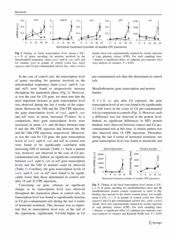
In the case of control eels, the transcription level
of genes encoding for proteins involved in the
mitochondrial respiratory chain (cox1, atp6-8, cytc
and nd5) were found to progressively increase
throughout the maturation phase (Fig. 2). However,
as was the case for 12S gene, we must note that the
most important increase in gene transcription level
was observed during the last 4 weeks of the exper-
iment. Between the 18th and the 22th CPE injection,
the gene transcription levels of cox1, atp6-8, cytc
and nd5 were, in mean, increased 57-times. As a
comparison, their gene transcription levels were
increased, in mean, 2.3- and 48-times between time
0 and the 8th CPE injection and between the 8th
and the 18th CPE injection, respectively. Moreover,
as was the case for 12S gene, the gene transcription
levels of cox1, atp6-8, cytc and nd5 in control eels
were found to be significantly correlated with
increasing GSI of animals (Table 1). Such a pattern
was, however, not observed in the case of Cd pre-
contaminated eels. Indeed, no significant correlations
between cox1, atp6-8, cytc or nd5 gene transcription
levels and the GSI of animals could be observed
(Table 1). Corollary, the gene transcription levels of
cox1, atp6-8, cytc or nd5 were found to be signifi-
cantly lower than those determined in control eels
after 18 and 22 CPE injections.
Concerning cat gene, whereas no significant
change in its transcription level was observed
throughout the maturation phase in control eels, its
transcription level was found to significantly increase
in Cd pre-contaminated eels during the last 4 weeks
of hormonal treatment. This increase was so impor-
tant that its transcription level was, at the end of
the experiment, significantly 9.4-fold higher in Cd
pre-contaminated eels than this determined in control
eels.
Metallothioneins gene transcription and protein
burden
A T = 0, i.e. just after Cd exposure, the gene
transcription level of mts was found to be significantly
2.1-fold lower in the ovary of Cd pre-contaminated
eels in comparison to controls (Fig. 3). However, such
a difference was not observed at the protein level.
Indeed, no significant differences in MTs protein
burdens were observed between control and Cd pre-
contaminated eels at this time. A similar pattern was
also observed after 18 CPE injections. Thereafter,
during the last 4 weeks of hormonal treatment, mts
gene transcription level was found to drastically and
Fig. 2 Change in basal transcription level (mean ± S.E.,
n = 5) of genes encoding for proteins involved in the
mitochondrial respiratory chain (cox1, atp6-8, cytc, nd5) and
for catalase (cat) in gonads of control (solid line, blacksquares) and Cd pre-contaminated (dotted line, white circles)
female silver eels experimentally matured by weekly injection
of carp pituitary extract (CPE). For each sampling time,
* denotes a significant effect of cadmium pre-exposure (two
ways analysis of variance, P \ 0.05)
Hormonal treatment (number of weekly CPE injections)
Fig. 3 Change in (a) basal transcription level (mean ± S.E.,
n = 5) of genes encoding for metallothioneins (mts) and (b)
metallothionein protein content (expressed as nmol of Hg-
binding sites present in the whole ovarian tissue, wet weight,
mean ± S.E., n = 3) in gonads of control (solid line, blacksquares) and Cd pre-contaminated (dotted line, white circles)
female silver eels experimentally matured by weekly injection
of carp pituitary extract (CPE). For each sampling time,
* denotes a significant effect of cadmium pre-exposure (two
ways analysis of variance and Kruskall Wallis test, P \ 0.05)
Biometals
123

significantly increase in both control and Cd pre-
contaminated eels. During this period, mts gene
transcription level was increased 54- and 345-times
in control and Cd pre-contaminated eels, respectively.
Despite mts gene transcription level was significantly
2.2-fold more important in controls in comparison to
Cd pre-contaminated eels after 18 CPE injections, no
significant differences could be observed after 22 CPE
injections. At the protein level, such a significant
increase was observed only in the case of Cd pre-
contaminated eels. Whereas MTs burden did not
significantly increased (P = 0.21) during the last
4 weeks of hormonal treatment in the case of control
eels, this value was significantly increased 4.8-times in
Cd pre-contaminated eels. Interestingly, ovarian MTs
protein burdens of both control and Cd pre-contami-
nated eels were significantly correlated with ovarian
Cd burdens (Fig. 4). Such a relationship was, however,
not observed at the transcriptional level (r = 0.02,
P = 0.91).
Discussion
In a previous work (see Pierron et al. 2008), we have
reported a stimulating effect of Cd pre-exposure on
the pituitary-gonad-liver axis of experimentally
maturing silver eels, leading to an early and enhanced
vitellogenesis. From 20 weeks of hormonal treat-
ment, this was followed by strong phenomena of
oocytes atresia and eels mortality affecting only Cd
pre-contaminated eels. Significantly, these devastat-
ing effects of Cd were observed in organisms that
presented metal concentrations in the main organs of
Cd bioaccumulation, the liver and the kidney, still
below those observed in eels from Cd contaminated
hydrosystems. Indeed, after 30 days of Cd exposure,
average Cd concentrations (data not shown) in the
liver and the kidney reached means of 1.7 ± 0.4 and
10.6 ± 2.7 lg g-1 (dry weight, dw), respectively, in
the case of Cd-exposed eels versus 0.9 ± 0.1 and
5.9 ± 0.2 lg g-1 (dw), respectively, in the controls,
i.e. in eels from the Loire river (France). For
comparison, Cd concentrations in the liver and the
kidneys of yellow eels (aged 6–14 years) inhabiting
the Gironde estuary (Durrieu et al. 2005), which is
characterized by a historic Cd pollution, reach means
of 5 ± 0.8 and 34.2 ± 5.1 lg g-1 (dw), respectively.
Here, our main goal was to get more insights into the
specific effects potentially triggered by Cd pre-
exposure on gonad tissue, mainly on the mitochon-
drial respiratory chain, a primary site of Cd toxicity
(Wang et al. 2004; Sokolova 2004; Risso-de Faver-
ney et al. 2004; Takaki et al. 2004; Belyaeva et al.
2008), by means of qRT-PCR gene transcription
analysis.
Concerning the effect of hormonal treatment on
12S gene transcription, our data show a progressive
and strong increase in its transcription level in both
control and Cd pre-contaminated eels during the
maturation phase and this, without significant differ-
ences between the two groups of animals. Moreover,
for both control and Cd pre-contaminated eels, a
significant correlation was found between 12S gene
transcription level and GSI of animals. As the GSI of
animals increased in response to hormonal treatment,
the gene transcription level of 12S increased. Such an
increase in mitochondrial gene transcription level
could reflect an increase in the number of mitochon-
dria in gonad tissue. This assumption appears indeed
consistent with previous ultrastructural investigations
carried out on gonad tissue of experimentally matur-
ing female European silver eels. Authors reported a
proliferation of mitochondria in gonad ooplasm after
hormonal treatment with CPE (Burzawa-Gerard et al.
1994). Such a report was also established from
oocytes of female Anguilla japonica artificially
matured with salmon pituitary extracts (Kayaba
et al. 2001). Whereas few and undeveloped mito-
chondria were observed in gonad of immature/
untreated animals (oocytes at the oil droplet stage),
a large number of developed mitochondria were
r = 0.72 P = 0.00065
Fig. 4 Relationship between metallothioneins (MTs) content
(expressed as lmol of Hg-binding sites present in the whole
ovarian tissue, wet weight) and Cd content in gonads of control
(black squares) and Cd pre-contaminated (white circles)
female silver eels experimentally matured by weekly injection
of carp pituitary extract (n = 40)
Biometals
123
observed in the course of vitellogenesis in oocytes of
maturing eels. As a consequence, it appears reliable
to hypothesize that the increase in 12S gene tran-
scription level that we observed in the present study
reflects an increase in the number of mitochondria in
gonads of maturing silver eels. Such an increase in
mitochondria number could be linked to the build-up
of mitochondrial activity for enhanced ATP produc-
tion; such increase in ATP production could aim to
prepare oocytes for (1) vitellogenin incorporation
(e.g. development of the zona radiata and microvil-
losities) and reshuffle, (2) steroidogenesis and (3)
final maturation (i.e., germinal vesicle migration and
breakdown; Habibi and Lessman 1986; Burzawa-
Gerard et al. 1994; Dufour et al. 2003; Kwon et al.
2005). We must note, however, that our results could
not exclude that these mRNA are stored in ooplasm
and translated at a later time, mainly during embryo-
genesis, a mechanism used by several organisms
(Vassalli and Stutz 1995; Seydoux 1996; Stebbins-
Boaz and Richter 1997).
Interestingly, for control eels, the increase in 12S
gene transcription level in response to hormonal
treatment was associated with an increase in cox1,
atp6-8, cytc and nd5 gene transcription levels. Thus,
the transcription level of genes encoding for proteins
involved in the mitochondrial respiratory chain
appears to be up-regulated by hormonal treatment.
Such a phenomenon was first described in gonads of
artificially maturing female Anguilla japonica as well
as in naturally and artificially maturing New Zealand
eels (Anguilla australis and Anguilla dieffenbachii).
The transcription level of the gene encoding for
cytochrome b (CYTB), a protein involved in the
electron transport in the mitochondrial respiratory
chain (as CYTC in the present study), was found to
significantly increase from early to late vitellogenesis
(Lokman et al. 2003). Similarly, a large-scale gene
transcription analysis by means of serial analysis of
gene expression (SAGE) revealed high levels of ATP
synthase transcripts in zebrafish ovarian fully-grown
follicules. Complementary, the proteomic analysis
carried out on the same tissue revealed high levels of
the corresponding proteins, thus supporting the fact
that ATP synthase mRNAs are effectively translated
into proteins (Knoll-Gellida et al. 2006). Concerning
the Cd effect, whereas our previous results have shown
a stimulating effect of Cd pre-exposure on the
pituitary-gonad-liver axis of European silver eels,
our present results show, at the opposite, an impair-
ment of the transcription level of genes involved in the
mitochondrial respiratory chain. Indeed, after 18 and
22 CPE injections, mean transcription levels of cox1,
atp6-8, cytc and nd5 were found to be significantly
lower than those determined in controls eels. This is,
however, consistent with a previous work carried out
on gills of European glass eels. The transcription level
of these same genes was found to be down-regulated in
response to aqueous Cd exposure (Pierron et al.
2007b). Analogously to that found here, the most
important effect of Cd exposure was observed on nd5
transcription level. Additionally, such effect of Cd on
cox1 gene transcription was also described in liver of
carp experimentally exposed to low concentrations of
a mixture of waterborne and dietary Cd (Reynders
et al. 2006). A decrease in cox2 and cox4, at both
transcriptional and protein levels, was also reported
during in vitro investigations performed on human
MDA-MB231 cells exposed to low Cd concentrations
(Cannino et al. 2008). Thus, such a down-regulation of
genes involved in the mitochondrial respiratory chain
could be the result of a direct effect of Cd on gonads,
rather than an indirect effect of the metal mediated by
changes in circulating hormone levels (see Pierron
et al. 2008). Additionally, as ovaries were not the only
organs that have significantly accumulated Cd during
the maturation phase, it is possible that similar events
occurred in other tissues of eels. This could lead to the
appearance of toxic events in various tissues of eels
(notably in kidneys, see Pierron et al. 2008). In the
particular case of ovaries, the appearance of such Cd
effects could explain, at least in part, why oocytes of
Cd pre-contaminated eels could not reach final mat-
uration and subsequently why these oocytes were
subjected to atresia at the end of the experiment.
Indeed, several studies carried out on fish, mammalian
and amphibian oocytes have highlighted the vital role
of oxidative phosphorylation during final maturation
(Brachet et al. 1975; Habibi and Lessman 1986;
Wycherley et al. 2005; Johnson et al. 2007). For
example, the use of inhibitors or uncouplers of
oxidative phosphorylation was found to abolish ger-
minal vesicle migration and breakdown in ovaries of
female goldfish (Habibi and Lessman 1986). However,
as previously described in our first article, atresia
phenomenon could have several origins. Among the
different hypotheses previously developed, we have
proposed that Cd could have directly triggered
Biometals
123

apoptosis of oocytes. Indeed, the pro-apoptotic effect
of Cd, an effect mediated by mitochondrial dysfunc-
tions and subsequent ROS production, is now well
recognized (Risso-de Faverney et al. 2004; Poliandri
et al. 2006). This hypothesis is consistent with the fact
that the end of the experiment was marked, only in the
case of Cd pre-contaminated eels, by a significant
increase in cat transcription level; the transcription
level of which being known to be up-regulated by ROS
(Scandalios 2005). Alternatively, since we observed a
strong stimulating effect of Cd pre-exposure on LH
subunits genes transcription levels, Cd could indi-
rectly, by altering circulating hormones levels, trigger
an overstimulation and subsequently atresia of oocytes
(see Pierron et al. 2008). Our present results cannot
permit to favour one hypothesis over another, certainly
because atresia phenomenon was the resultant of
multiple dysfunctions, associating direct and indirect
effects of Cd on sexual tissues.
The end of the experiment was also marked by a
strong increase in the gene transcription level of mts.
However, in the case of control eels, such an increase
in mts gene transcription level could not be observed
at the protein level. Such a discrepancy between mts
gene transcription and MTs protein content was
already observed and described in oocytes of lizard
Podarcis sicula (Riggio et al. 2003). Whereas a
significant and progressive accumulation of mts
mRNA was observed from pre-vitellogenic to vitel-
logenic oocytes and eggs, no significant accumulation
of MTs proteins could be observed. Analogously,
whereas SAGE analysis carried out on zebrafish
ovaries revealed high levels of mt2 transcripts, the
concomitant proteomic analysis failed to reveal
significant amounts of the corresponding proteins
(Knoll-Gellida et al. 2006). As already evoked, such
mRNA accumulation could aim to provide sufficient
materials needed for embryogenesis. Indeed, a num-
ber of studies indicate that translationally-inactive
mRNAs are commonly present in oocytes and eggs.
These maternal mRNAs are translated after fertiliza-
tion to cope with the needs of embryogenesis (Spirin
1994; Vassalli and Stutz 1995; Stebbins-Boaz and
Richter 1997). In the particular case of MTs,
activation of mRNAs after fertilization could aim to
cope with the needs of metalloproteins that store and
donate essential metals, notably copper and zinc
which are essential for embryogenesis (Kambe et al.
2008). Interestingly, and this, at the opposite to
control eels, the significant increase in mts transcripts
observed at the end of the experiment was effectively
marked by a concomitant and significant increase in
ovarian MTs protein content of Cd pre-contaminated
eels. Since this increase in protein burden was
associated with a massive entry of Cd in gonads of
Cd pre-contaminated eels (see Pierron et al. 2008), it
could be suggested that the significant metal accu-
mulation observed in gonads at the end of the
experiment have stimulated the translation of MTs
mRNAs. In support of this assumption, the MTs
protein content of both control and Cd pre-contam-
inated eels was found to be significantly correlated
with the Cd content of gonads. Moreover, a similar
pattern was observed in lizard oocytes. Indeed, as
described above, whereas increasing mts gene tran-
scription levels during oogenesis was not associated
with an increase in MTs protein content, a single
injection of Cd chloride in lizard female triggered an
ovarian MTs protein synthesis (Riggio et al. 2003).
Such findings suggest that Cd could activate the
translation of normally stored and untranslated MTs
mRNA. Alternatively, since the end of the experi-
ment was marked by a concomitant and significant
increase in both MTs protein content and cat gene
transcription level in gonads of Cd pre-contaminated
eels, an effect of oxidative stress on the translational
activation of MTs mRNA cannot be ruled out. In our
case, we have to note, however, that this increase in
ovarian MTs protein content was insufficient to
completely prevent the appearance of Cd-induced
toxic events in gonads of Cd pre-contaminated eels,
thus suggesting that a significant part of the accu-
mulated Cd is associated with sensitive fractions of
the cell such as mitochondria (Campbell et al. 2005).
Conclusion
This study tends to show how internal stores of Cd
could be released during fish migrations at levels high
enough to be toxic. In our experimental, Cd released
was found to seriously disrupt development of sexual
tissues of maturing female silver eels, leading to
oocytes atresia. In addition to previous reported
changes in pituitary hormonal gene transcription or
on vitellogenesis, our present results tend to show that
this Cd released could also impair ovarian mitochon-
drial metabolism and consequently, compromise full
Biometals
123

oocyte development. In addition, this work provides
new insights in ovarian mts gene transcription and
translation regulation during sexual maturation. Our
results tend to show, that albeit normally weakly
represented in eel’s ovaries, MTs protein synthesis can
be induced by Cd. However, it must be underlined that
an important limitation of our work relies not only on
the use of hormonal treatment but also from the regime
of Cd contamination. Our experimental exposure
scenario was indeed very different from what wild
eels experience during their growth in aquatic ecosys-
tems. In other words, an acute contamination by
dissolved Cd over one month cannot be, in term of
organotropism or intracellular partitioning for exam-
ple, strictly representative of a contamination that
proceed over several years, at a lower pressure of
contamination and by both the direct and the trophic
route of exposure. However, the fact that we observed
a significant remobilization and redistribution of Cd
during the course of the experiment, and also in control
individuals (see Pierron et al. 2008), reinforces the
potential occurrence of phenomena that we observed
under natural conditions.
Acknowledgments We wish to thank Bruno Etcheberria and
Henry Bouillard for their help and technical assistance in all
aspects of this study. Fabien Pierron was supported by a grant
from the French Ministry of Research.
References
Belyaeva EA, Dymkowska D, Wieckowski MR, Wojtczak L
(2008) Mitochondria as an important target in heavy metal
toxicity in rat hepatoma AS-30D cells. Toxicol Appl
Pharmacol 231(1):34–42. doi:10.1016/j.taap.2008.03.017
Bordajandi LR, Gomez G, Fernandez MA, Abad E, Rivera J,
Gonzalez MJ (2003) Study on PCBs, PCDD/Fs, organo-
chlorine pesticides, heavy metals and arsenic content in
freshwater fish species from the River Turia (Spain).
Chemosphere 53(2):163–171. doi:10.1016/S0045-6535
(03)00417-X
Brachet J, Schutter PD, Hubert E (1975) Studies on maturation
in Xenopus laevis oocytes III Energy production and
requirements for protein synthesis. Differentiation 3:3–14.
doi:10.1111/j.1432-0436.1975.tb00840.x
Burzawa-Gerard E, Baloche S, Leloup-Hatley J, Le Menn F,
Messaouri H, Nunez-Rodriguez J, Peyon P, Roger C
(1994) Ovogenese chez l’anguille (Anguilla anguilla L):
ultastructure de l’ovaire a differents stades de developp-
ement et implication des lipoproteines au cours de la
vitellogenese. Fr Peche Piscic 335:213–233. doi:10.1051/
kmae:1994014
Campbell PGC, Giguere A, Bonneris E, Hare L (2005) Cad-
mium-handling strategies in two chronically exposed
indigenous freshwater organisms–the yellow perch (Percaflavescens) and the floater mollusc (Pyganodon grandis).
Aquat Toxicol 72(1–2):83–97. doi:10.1016/j.aquatox.2004.
11.023
Cannino G, Ferruggia E, Luparello C, Rinaldi AM (2008)
Effects of cadmium chloride on some mitochondria-rela-
ted activity and gene expression of human MDA-MB231
breast tumor cells. J Inorg Biochem 102(8):1668–1676.
doi:10.1016/j.jinorgbio.2008.04.002
Dufour S, Burzawa-Gerard E, Le Belle N, Sbaihi M, Vidal B
(2003) Reproductive endocrinology of the European eel,
Anguilla Anguilla. In: Aida K, Tsukamoto K, Yamauchi K
(eds) Eel biology. Springer Verlag, Tokyo, pp 373–383
Durif C, Dufour S, Elie P (2006) Impact of silvering stage, age,
body size and condition on reproductive potential of the
European eel. Mar Ecol Prog Ser 327:171–181. doi:
10.3354/meps327171
Durrieu G, Maury-Brachet R, Girardin M, Rochard E, Boudou
A (2005) Contamination by heavy metals (Cd, Zn, Cu,
Hg) of hight fish species in the Gironde estuary (France).
Estuaries 28:581–591. doi:10.1007/BF02696069
Gonzalez P, Baudrimont M, Boudou A, Bourdineaud J-P
(2006) Comparative effects of direct cadmium contami-
nation on gene expression in gills, liver, skeletal muscle
and brain of the zebra fish (Danio rerio). Biometals
19(3):225–235. doi:10.1007/s10534-005-5670-x
Habibi H, Lessman C (1986) A study of goldfish oocyte mei-
osis in vitro: effects of 2, 4-dinitrophenol and adenosine-
5-triphosphate. Fish Physiol Biochem 1(4):197–205. doi:
10.1007/BF02311136
Johnson MT, Freeman EA, Gardner DK, Hunt PA (2007)
Oxidative metabolism of pyruvate is required for meiotic
maturation of murine oocytes in vivo. Biol Reprod 77:2–
8. doi:10.1095/biolreprod.106.059899
Kambe T, Weaver BP, Andrews GK (2008) The genetics of
essential metal homeostasis during development. Genesis
46:214–228. doi:10.1002/dvg.20382
Kayaba T, Takeda N, Adachi S, Yamauchi K (2001) Ultra-
structure of the oocytes of the Japanese eel Anguillajaponica during artificially induced sexual maturation. Fish
Sci 67:870–879. doi:10.1046/j.1444-2906.2001.00335.x
Knoll-Gellida A, Andre M, Gattegno T, Forgue J, Admon A,
Babin PJ (2006) Molecular phenotype of zebrafish ovarian
follicle by serial analysis of gene expression and proteo-
mic profiling, and comparison with the transcriptomes of
other animals. BMC Genomics 7:46. doi:10.1186/1471-
2164-7-46
Kwon HC, Choi SH, Kim YU, Son SO, Kwon JY (2005)
Androgen action on hepatic vitellogenin synthesis in the
eel, Anguilla japonica is suppressed by an androgen
receptor antagonist. J Steroid Biochem Mol Biol 96:175–
178. doi:10.1016/j.jsbmb.2005.02.010
Lokman P, Kazeto Y, Ijiri S, Young G, Miura T, Adachi S,
Yamauchi K (2003) Ovarian mitochondrial cytochrome b
mRNA levels increase with sexual maturity in freshwater
eels (Anguilla spp). J Comp Physiol B. Biochem Syst
Environ Physiol 173(1):11–19
Palstra AP, van Ginneken VJT, Murk AJ, van den Thillart
GEEJM (2006) Are dionxin-like contaminants responsible
Biometals
123

for the eel (Anguilla anguilla) drama? Naturwissens-
chaften 93:145–148. doi:10.1007/s00114-005-0080-z
Peltier MR, Wilcox CJ, Sharp DC (1998) Technical note:
application of the Box-Cox data transformation to animal
science experiments. J Anim Sci 76(3):847–849
Pierron F, Baudrimont M, Bossy A, Bourdineaud J-P, Brethes
D, Elie P, Massabuau J-C (2007a) Impairment of lipid
storage by cadmium in the European eel (Anguillaanguilla). Aquat Toxicol 81(3):304–311. doi:10.1016/j.
aquatox.2006.12.014
Pierron F, Baudrimont M, Gonzalez P, Bourdineaud J-P, Elie
P, Massabuau J-C (2007b) Common pattern of gene
expression in response to hypoxia or cadmium in the gills
of the European glass eel (Anguilla anguilla). Environ Sci
Technol 41(8):3005–3011. doi:10.1021/es062415b
Pierron F, Baudrimont M, Dufour S, Elie P, Bossy A, Baloche
S, Mesmer-Dudons N, Gonzalez P, Bourdineaud J-P,
Massabuau J-C (2008) How cadmium could compromise
the completion of the European eel’s reproductive
migration. Environ Sci Technol 42(12):4607–4612. doi:
10.1021/es703127c
Poliandri AHB, Machiavelli LI, Quinteros AF, Cabilla JP,
Duvilanski BH (2006) Nitric oxide protects the mito-
chondria of anterior pituitary cells and prevents cadmium-
induced cell death by reducing oxidative stress. Free
Radic Biol Med 40(4):679–688. doi:10.1016/j.freerad
biomed.2005.09.021
Reynders H, van der Ven K, Moens LN, van Remortel P, De
Coen WM, Blust R (2006) Patterns of gene expression in
carp liver after exposure to a mixture of waterborne and
dietary cadmium using a custom-made microarray. Aquat
Toxicol 80(2):180–193. doi:10.1016/j.aquatox.2006.08.
009
Riggio M, Trinchella F, Filosa S, Parisi E, Scudiero R (2003)
Accumulation of zinc, copper, and metallothionein
mRNA in lizard ovary proceeds without a concomitant
increase in metallothionein content. Mol Reprod Dev
66:374–382. doi:10.1002/mrd.10365
Risso-de Faverney C, Orsini N, de Sousa G, Rahmani R (2004)
Cadmium-induced apoptosis through the mitochondrial
pathway in rainbow trout hepatocytes: involvement of
oxidative stress. Aquat Toxicol 69(3):247–258. doi:
10.1016/j.aquatox.2004.05.011
Robinet TT, Feunteun EE (2002) Sublethal effects of exposure
to chemical compounds: a cause for the decline in atlantic
eels? Ecotoxicology 11(4):265–277. doi:10.1023/A:10163
52305382
Roche H, Buet A, Ramade F (2002) Relationships between
persistent organic chemicals residues and biochemical
constituents in fish from a protected area: the French
National Nature Reserve of Camargue. Comp Biochem
Physiol C Pharmacol Toxicol Endocrinol 133(3):393–410.
doi:10.1016/S1532-0456(02)00122-9
Scandalios JG (2005) Oxidative stress: molecular perception
and transduction of signals triggering antioxidant genes
defenses. Braz J Med Biol Res 38:995–1014. doi:10.1590/
S0100-879X2005000700003
Seydoux G (1996) Mechanisms of translational control in early
development. Curr Opin Genet Dev 6:555–561
Sokolova IM (2004) Cadmium effects on mitochondrial func-
tion are enhanced by elevated temperatures in a marine
poikilotherm, Crassostrea virginica Gmelin (Bivalvia:
Ostreidae). J Exp Biol 207:2639–2648. doi:10.1242/jeb.
01054
Spirin AS (1994) Storage of messenger RNA in eukaryotes:
envelopment with protein, translational barrier at 5 side,
or conformational masking by 3 side? Mol Reprod Dev
38(1):107–117. doi:10.1002/mrd.1080380117
Stebbins-Boaz B, Richter JD (1997) Translational control
during early development. Crit Rev Eukaryot Gene Expr
7(1–2):321–333
Stone R (2003) Freshwater eels are slip-sliding away. Science
302:221–222. doi:10.1126/science.302.5643.221
Takaki A, Jimi S, Segawa M, Hisano S, Takebayashi S, Iwasaki H
(2004) Long-term cadmium exposure accelerates age-
related mitochondrial changes in renal epithelial cells. Tox-
icology 203(1–3):145–154. doi:10.1016/j.tox.2004.06.005
Tesch F-W (2003) The eel Blackwell Publishing fifth edition:
408 p
van Ginneken VJT, Maes GE (2005) The European eel
(Anguilla anguilla, Linnaeus), its lifecycle, evolution and
reproduction: a literature review. Rev Fish Biol Fish
15:367–398. doi:10.1007/s11160-006-0005-8
Vassalli JD, Stutz A (1995) Translational control awakening
dormant mRNAs. Curr Biol 5(5):476–479. doi:10.1016/
S0960-9822(95)00095-9
Vidal B, Pasqualini C, Le Belle N, Holland MCH, Sbaihi M,
Vernier P, Zohar Y, Dufour S (2004) Dopamine inhibits
luteinizing hormone synthesis and release in the juvenile
European eel: a neuroendocrine lock for the onset of
puberty. Biol Reprod 71:1491–1500. doi:10.1095/biol
reprod.104.030627
Wang Y, Fang J, Leonard SS, Krishna R, Murali K (2004)
Cadmium inhibits the electron transfer chain and induces
reactive oxygen species. Free Radic Biol Med 36(11):
1434–1443. doi:10.1016/j.freeradbiomed.2004.03.010
Wycherley G, Kane MT, Hynes AC (2005) Oxidative phos-
phorylation and the tricarboxylic acid cycle are essential
for normal development of mouse ovarian follicles. Hum
Reprod 20(10):2757–2763. doi:10.1093/humrep/dei132
Biometals
123
Related Documents