1 Charles Zaloudek, MD Professor Department of Pathology UCSF Ovarian Epithelial Tumors Case 1 • 68 year old woman • Bilateral adnexal masses • Elevated CA125 (793) • Hysterectomy in 2008 • Underwent BSO and tumor debulking in 2011; 6-8 L of ascites • Extensive metastases noted, with carcinoma involving the diaphragm, peritoneum, and bowel (stage IIIC) Case 1 Gross Pathology • The ovaries were small • Right ovary: Pale tan multinodular solid mass 4 cm in maximum dimension; tumor involved the surface • Left ovary: Cystic and solid, 5 cm in maximum dimension, papillary excrescences from cyst linings • Fallopian tubes: Unremarkable • Omentum, mesentery, bowel: Riddled with mostly small nodules, largest 4.5 cm

Welcome message from author
This document is posted to help you gain knowledge. Please leave a comment to let me know what you think about it! Share it to your friends and learn new things together.
Transcript
1
Charles Zaloudek, MDProfessor
Department of PathologyUCSF
Ovarian Epithelial Tumors
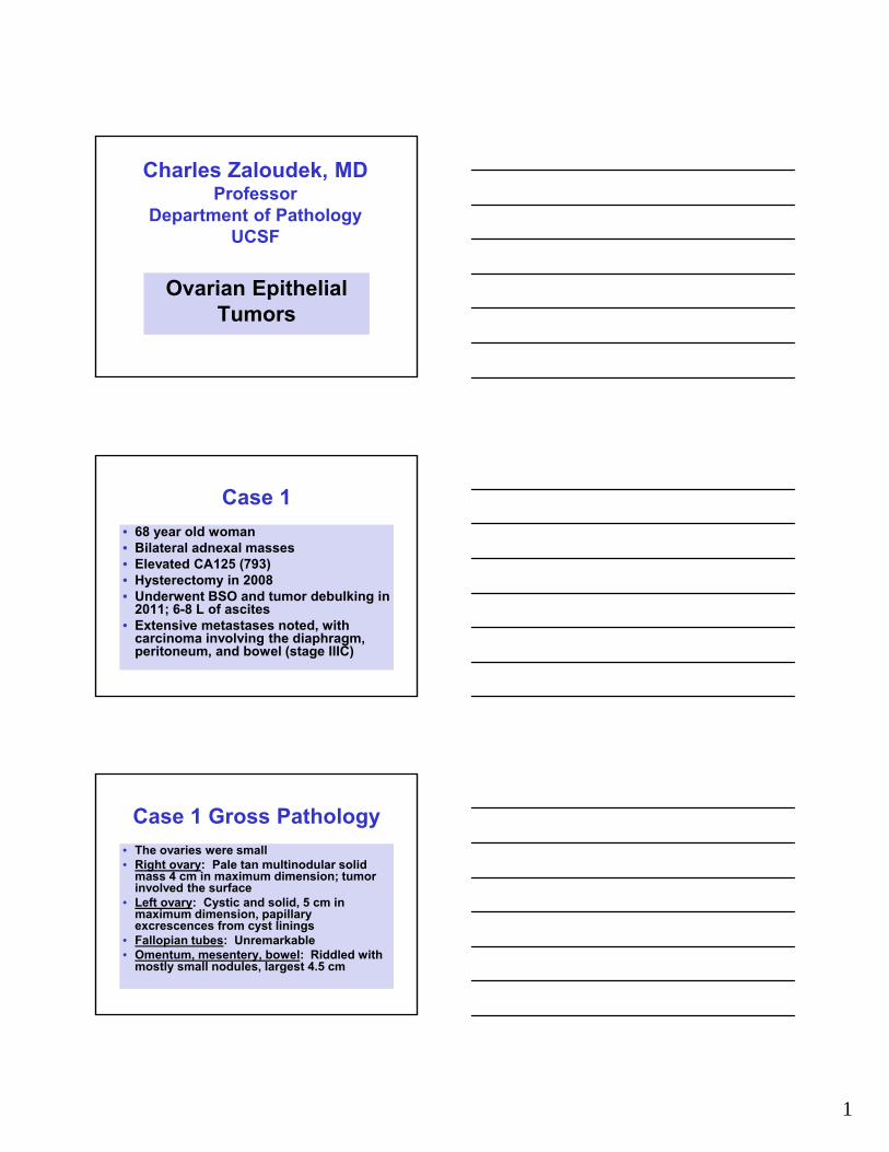
Case 1
• 68 year old woman• Bilateral adnexal masses• Elevated CA125 (793)• Hysterectomy in 2008• Underwent BSO and tumor debulking in
2011; 6-8 L of ascites• Extensive metastases noted, with
carcinoma involving the diaphragm, peritoneum, and bowel (stage IIIC)
Case 1 Gross Pathology
• The ovaries were small• Right ovary: Pale tan multinodular solid
mass 4 cm in maximum dimension; tumor involved the surface
• Left ovary: Cystic and solid, 5 cm in maximum dimension, papillary excrescences from cyst linings
• Fallopian tubes: Unremarkable• Omentum, mesentery, bowel: Riddled with
mostly small nodules, largest 4.5 cm

2
Right Ovary Left Ovary
Right ovary 4 cm maximum dimensionLeft ovary 5 cm maximum dimension
3
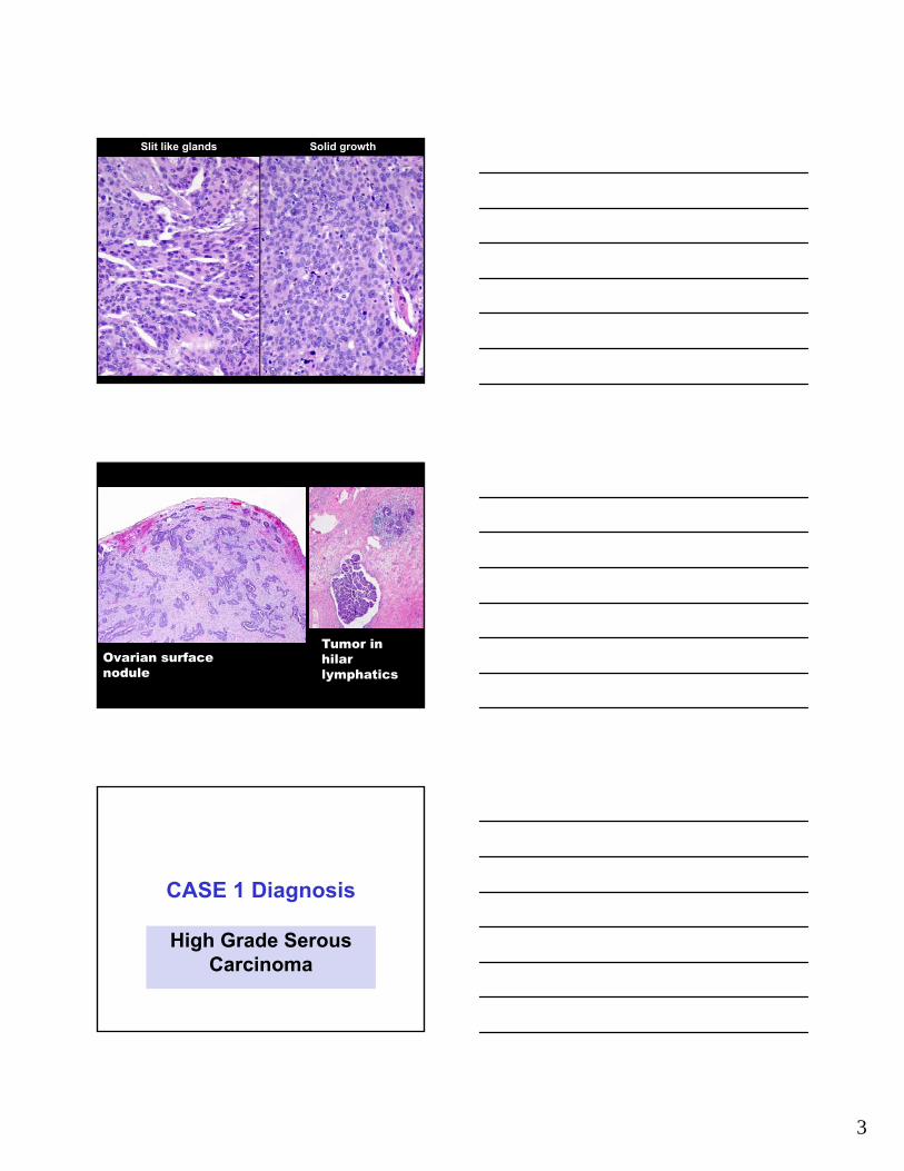
Slit like glands Solid growth
Ovarian surface nodule
Tumor in hilar lymphatics
CASE 1 Diagnosis
High Grade Serous Carcinoma
4
Discussion Points for Case 1
• Classification and general features of epithelial tumors
• Histologic features of high grade serous carcinoma
• Immunophenotype• Molecular pathology – p53• BRCA• The fallopian tube connection and
histogenesis of pelvic high grade serous carcinoma
Epithelial TumorsClinical Presentation
• Rare in patients < 20 years• Carcinoma occurs mainly > 40 years• Presenting symptoms are nonspecific:
– Pelvic or abdominal pain or discomfort– Gastrointestinal symptoms– Disturbances of menstruation– Abdominal distention
• Standard treatment is surgery and, often, platinum + taxane chemotherapy
Epithelial Ovarian Cancer• High grade serous
• Low grade serous
• Endometrioid
• Clear cell
• Mucinous
• Carcinosarcoma (MMT)
• Transitional Cell / Malignant Brenner
• Rare tumor types
• Mixed types
• Undifferentiated
• Unclassified
5
Distribution of Ovarian Tumor Types
Tumor Type %
High grade serous 68.1
Low grade serous 3.4
Clear cell 12.2
Endometrioid 11.3
Mucinous 3.4
Other 1.6
Int J Gynecol Pathol 2010; 29:203-211
Ovarian CancerFIGO Stages
• Stage I - Limited to the ovariesIA - One ovary, intracysticIB - Both ovaries, intracysticIC - Rupture; tumor on surface; + cytology
• Stage II - Spread to pelvis
• Stage III - Spread to abdomen or LN• Stage IV - Distant spread
Survival With Serous Carcinoma
Int J Gynecol Cancer 2012; 22:367-371
6
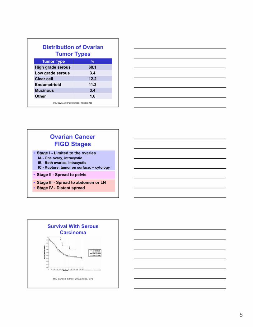
High Grade Serous Carcinoma
Stage% of all tumors
in this stage
I or II (localized) 35.5
III or IV (widespread)
87.7
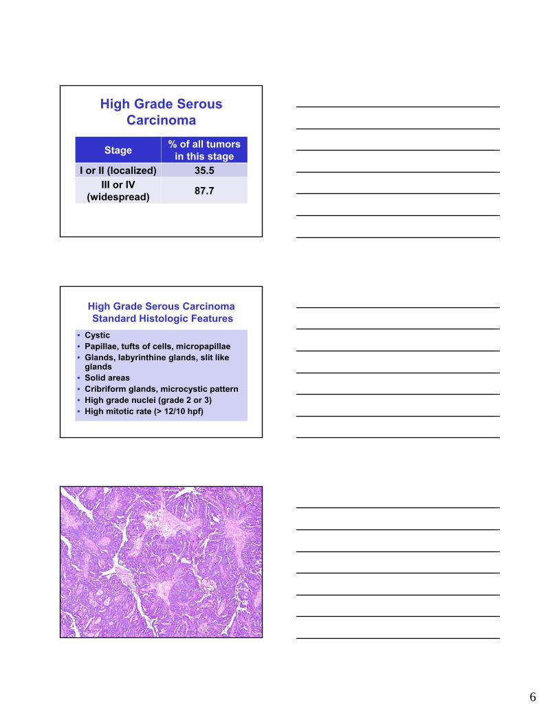
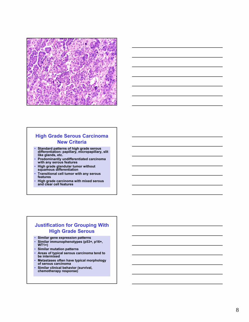
High Grade Serous CarcinomaStandard Histologic Features
• Cystic• Papillae, tufts of cells, micropapillae• Glands, labyrinthine glands, slit like
glands• Solid areas• Cribriform glands, microcystic pattern• High grade nuclei (grade 2 or 3)• High mitotic rate (> 12/10 hpf)

7
8
High Grade Serous CarcinomaNew Criteria
• Standard patterns of high grade serous differentiation: papillary, micropapillary, slit like glands, etc.
• Predominantly undifferentiated carcinoma with any serous features
• High grade glandular tumor without squamous differentiation
• Transitional cell tumor with any serous features
• High grade carcinoma with mixed serous and clear cell features
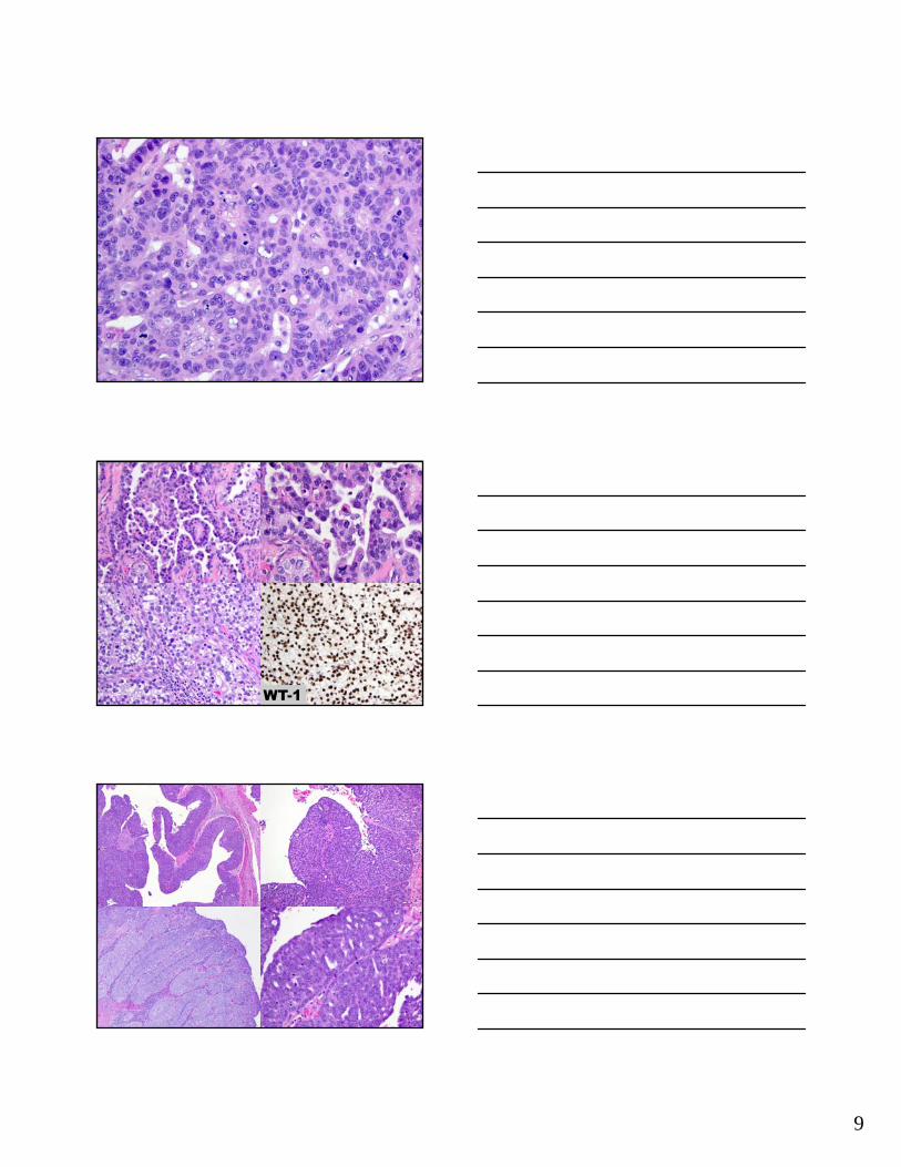
Justification for Grouping With High Grade Serous
• Similar gene expression patterns• Similar immunophenotypes (p53+, p16+,
WT1+)• Similar mutation patterns• Areas of typical serous carcinoma tend to
be intermixed• Metastases often have typical morphology
of serous carcinoma• Similar clinical behavior (survival,
chemotherapy response)
9
WT-1

10
Immunohistochemistry in the Diagnosis of High Grade Serous
CarcinomaIs This an Ovarian Tumor?
Stain Result
CK7 +
CK20 -
CDX2 -
PAX8 +
CA125 +
ER +
Stains for Tissue of Origin
PAX8
CK7
CA125
ImmunohistochemistryIs This High Grade Serous Carcinoma?
Stain Result
P53Positive test result: Strong positive staining throughout; > 50%, usually >80-90%
P53Positive test result: Completely negative; no staining in any tumor cells
P16Positive test result: Strong staining in every or nearly every tumor cell; 95-100%
WT1 Positive
HMGA2 Positive
PAX2Negative; positive suggests low grade serous or borderline
Related Documents