i OUTCOME OF SKELETAL TRACTION IN PATIENTS WITH FEMORAL SHAFT FRACTURES AT KENYATTA NATIONAL HOSPITAL DR. MUSTAFA MUSAJEE MB,ChB A dissertation submitted in partial fulfilment of the requirements of the degree of Master of Medicine in Surgery of the University of Nairobi October, 2012.

Welcome message from author
This document is posted to help you gain knowledge. Please leave a comment to let me know what you think about it! Share it to your friends and learn new things together.
Transcript
i
OUTCOME OF SKELETAL TRACTION IN PATIENTS
WITH FEMORAL SHAFT FRACTURES AT KENYATTA
NATIONAL HOSPITAL
DR. MUSTAFA MUSAJEE
MB,ChB
A dissertation submitted in partial fulfilment of the requirements of the
degree of Master of Medicine in Surgery of the University of Nairobi
October, 2012.
ii
DECLARATION:-
I hereby certify that this dissertation is my original work and has not been submitted in any other university.
Signed ..........................................................................................
DR MUSTAFA ABUTALIB ALIBHAI MUSAJEE, Principal Investigator.
MBChB (Nbi)
REG. NO. H58/7828/06
SUPERVISORS
This dissertation is submitted for examination with our approval as the university supervisors,
Prof. L. N. Gakuu
MB.ChB, MMed., FCS (ECSA), EBS
Assoc. Professor , Dept. Of Orthopaedics, University of Nairobi
Consultant Orthopaedic and Trauma Surgeon
Kenyatta National Hospital.
Sign…………………………………. Date…………………………………..
Mr. J.W. Githaiga
MBCh.B, M.Med (Surg) ,FCS(ESCA)
Lecturer, Department of surgery,University of Nairobi
Consultant General Surgeon,
KenyattaNationalHospital
Sign…………………………………. Date…………………………………..
Mr. Kirsteen Awori
MB ChB MMed (Surg), FCS(Orth) ECSA, Dip (SICOT)
Lecturer,Department of Human Anatomy,University Of Nairobi
Consultant Orthopaedic Surgeon
Kenyatta National Hospital.
Sign…………………………………. Date………………………………
iii
DEDICATION
This dissertation is dedicated to my parents, my wife and children whose patience and encouragement has
enabled me complete this study.
iv
ACKNOWLEDGEMENT
My sincere thanks and gratitude to my supervisors Prof. Gakuu , Mr. Githaiga and Mr. Awori, for their constant
encouragement, guidance and supervision throughout this study.This work would not have been possible
without their support.
I am also grateful to Mr. Ayeiko for his analytical support and contribution.
To Kenyatta National Hospital Ethics and Research Committee, for their invaluable corrections and for
approving this study.
To many more who contributed in one way or the other, I remain highly indebted.
v
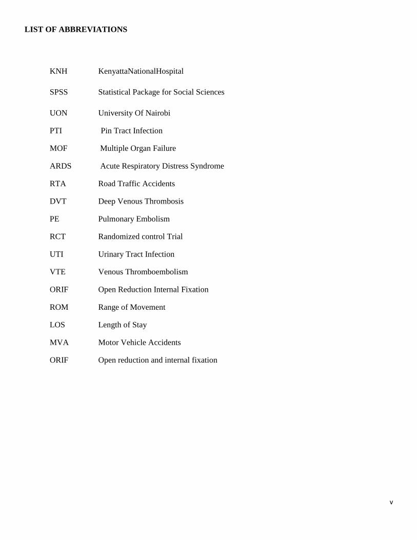
LIST OF ABBREVIATIONS
KNH KenyattaNationalHospital
SPSS Statistical Package for Social Sciences
UON University Of Nairobi
PTI Pin Tract Infection
MOF Multiple Organ Failure
ARDS Acute Respiratory Distress Syndrome
RTA Road Traffic Accidents
DVT Deep Venous Thrombosis
PE Pulmonary Embolism
RCT Randomized control Trial
UTI Urinary Tract Infection
VTE Venous Thromboembolism
ORIF Open Reduction Internal Fixation
ROM Range of Movement
LOS Length of Stay
MVA Motor Vehicle Accidents
ORIF Open reduction and internal fixation
vi
DEFINITION OF TERMS AND TERMINOLOGIES/ KEY WORDS :-
1. Outcomes :- An event or something that follows from an action, situation, result or consequence and in
the context of this study it refers to functional outcomes e.g. for range of knee motion following a period
of skeletal traction, and also to determine what proportion of patients will develop complications related
to the fracture healing (e.g. non union, mal-union), insertion of the steinmann pin or because of
immobility.
2. Skeletal Traction:- one of the two basic kinds of traction used in orthopedics for the treatment of
fractured bones and the correction of orthopedic abnormalities. Skeletal traction is applied to the
affected structure by a metal pin or wire inserted into the structure and attached to traction ropes.
Skeletal traction is often used when continuous traction is desired to immobilize, position, and align a
fractured bone properly during the healing process.
3. For purposes of this study, the shaft of the femur will be defined as: the distance between 5 cm distal to
the lesser trochanter and 6 cm proximal to the most distal point of the medial femoral condyle (dencker
1963).
4. Pin Tract Infection :- Infection of the pin tract is one of the complications that may develop with skeletal
traction, and careful scrutiny of pin sites is an important precaution. Some common signs of infection of
the pin tracts are erythema, drainage, noxious odor, pin slippage, temperature elevation, and pain
vii
TABLE OF CONTENTS
ITEM PAGE
Title page i
Declaration ii
Dedication iii
Acknowledgement iv
List of Abbreviations v
Definition of Terms and Terminologies vi
Table of Contents vii
List of Tables viii
List of Figures ix
Abstract x
Introduction 1
Literature review 2
Study Justification 8
Study Question 8
Objectives 8
Patients and Methods 9
Results 17
Discussion 33
References 42
Appendix 1 Consent Forms 48
Appendix 2 Data Sheet 52
Appendix 3 Ethical Approval 56
Appendix 4 Technique of Steinmann Pin insertion 57
viii
List of Tables Page
1. Table 1 : Basic demographic characteristics of patients 17
2. Table 2 : Duration of skeletal traction and characteristics of femoral fractures 18
3. Table 3: Fracture pattern and distribution of femoral fractures at KNH 20
4. Table 4: Characteristics of femoral fractures in patients at KNH orthopaedic unit and patient age 21
5. Table 5: PTI prevalence according to duration on traction 23
6. Table 6: PTI and fracture characteristics (risk) 24
7. Table 7: PTI complication among patients in different age groups. 26
8. Table 8: Prevalence of knee stiffness according to duration on traction 27
9. Table 9: Association between type of femoral fracture and development of knee stiffness 28
10. Table 10: Association between patient age and development of knee stiffness 28
11. Table 11: Factors associated with reduction in mid-thigh circumference. 30
12. Table 12: Duration of traction versus development of complications 32
ix
List of Figures Page
1. Figure 1 : Pie chart showing the aetiology of femoral shaft fractures. 19
2. Figure 2 : Bar graph of other injuries associated with femoral fractures. 22
3. Figure 3 : Prevalence of signs of PTI among patients on traction for femoral fractures 23
4. Figure 4 : The association of PTI with fracture aetiology. 25
5. Figure 5 : PTI occurrence in patients with and without other injuries. 25
6. Figure 6 : Prevalence of knee stiffness among patients on traction 26
7. Figure 7 : Bar charting showing fracture etiology versus knee flexion. 27
8. Figure 8 : Prevalence of reduction in mid thigh circumference following traction. 29
9. Figure 9 : Fracture healing among patients managed on skeletal traction. 31
x
ABSTRACT :-
Background: -
Road traffic accidents are a leading cause of trauma in Kenya (1). Many of the victims have multiple injuries
with a significant proportion having femoral fractures (2).
While early fracture fixation is advocated to ease pressure on costs and morbidity associated with prolonged
hospitalization, this is not possible for all patients who present at KNH due to a variety of factors. Majority have
to be on prolonged periods of skeletal traction as they await internal fixation while in others, it is the definitive
treatment.
While the spectrum of complications associated with skeletal traction is known, there are no studies to show the
burden of these complications at KNH and hence justify continued use of skeletal traction for patients awaiting
surgery.
Objective :
To determine the incidence of complications associated with skeletal traction in patients with femoral shaft
fractures at KNH and analyse their occurrence in relation to the duration of traction.
Study Design and Setting:-
This was a prospective, descriptive study of convenience sampling method of patients admitted with femoral
shaft fractures during a 3-month period in the orthopaedic wards at KNH.
Patients and methods:
Consecutive adult patients admitted with femoral shaft fractures of either sex on skeletal traction for more than
two days as they awaited internal fixation were recruited.
Data collected included the patient demographics, duration of skeletal traction, other associated injuries, cause
of the fracture, fracture geometry and occurrence and time of onset of complications directly related to skeletal
traction. The outcome measurewas the occurrence of one or more complications associated with skeletal
traction. Significant variables measured included PTI; knee stiffness; quadriceps muscle atrophy and
pneumonia.
Results:
Seventy five patients with femoral shaft fractures on skeletal traction were recruited. Transverse and
comminuted fracture patterns were the most common accounting for 41.3% and 37.3% of the fractures
xi
respectively. Ninety six percent of the patients were managed on Perkins mode of traction, whilst the mean
duration on traction was 4.7 weeks. Prevalence of PTI was 24% occurring most commonly during the 5th
and 6th
week on traction, PTI was significantly associated with comminuted fractures with 42.9% of the patients with
this fracture pattern developing PTI. Knee stiffness was the most common complication and 62.7% of the study
population had stiffness by the 4th
week on traction. Only 36% of the patients got reduction in their thigh
circumference from the time of onset of traction.
PTI was significantly associated with prolonged durations of traction as patients who developed PTI were on
traction for average of 6.7 weeks as compared to 4.25 weeks for those who did not develop any PTI. Patients
who were on traction for an average of 2.7 weeks were at a lower risk of developing knee stiffness as compared
to those on traction for 5.6 weeks.
Conclusion:
Knee stiffness and pin tract infection were the most common complications associated with skeletal traction
with the latter associated more with comminuted fractures. The incidence of these two rose significantly from
the fourth week of traction.
1
INTRODUCTION
Fracture management has evolved such that early fixation is now advocated due to a better understanding
of the metabolic response to trauma and also to cut on costs of hospitalization. In a resource-poor setting
such as Kenya, shortage of theatre space and cost of implants prevent early fixation of all trauma patients
presenting to hospitals, hence the primary management modality is skeletal traction.In many developing
countriesconservative treatment of fractures is still a viable option. However teaching of conservative
fracture methods is under – emphasized thus making it difficult for doctors and other health workers to
learn the art of conservative fracture treatment (3).Results of conservative fracture treatment have been
poor when compared to open reduction and internal fixation. Conservative fracture treatment is
mostlyleft/assigned to the junior health care personnel available, and as such it has become synonymous
with the application of plaster of Paris cast or Steinmann pin for traction then “forgetting” the patient for a
designated period.This is despite the fact that more than 90% of patients with fractures in many
developing countries will be treated by conservative methods (5).
In developing countries, shortage of surgeons, of appropriate, affordable equipment and implants, and
reliable clean surgical environments increases the risks of surgical complications of orthopaedic
procedures often to unacceptable levels. Conservative management of femur fractures with traction
remains a viable option and often the only option for cost-sensitive developing countries (5). Extended
lengths of skeletal traction are associated with several complications either as a result of traction or
prolonged immobilization. When properly applied, conservative treatment gives good and acceptable
results, and it is the “gold standard method” of care. It should always be remembered that operative
results should be compared to those of conservative treatment, not the other way around [6].
2
LITERATURE REVIEW
The use of skeletal traction at KNH for management of femoral shaft fractures has continued despite
adoption of newer techniques in the hospital in recent years, such as locked femoral nailing, in addition to
the older techniques such as plating (51).
In a study by Saidi et al the average length of hospital stay was fourteen days, this is much longer than in
reports from established trauma facilities. In a study at the Vancouver General and Teaching Hospital in
Canada, the length of hospital stay in 1997 was 9.14 days despite caring for patients with more severe
injuries (24% with ISS > 16) versus 13.4% with ISS > 15 in the KNH-based by Saidi et al(2,7).The
prolonged length of hospital stay at KNH was caused by predominance of skeletal injuries. Long bone
fracture fixation was performed late; which was occasioned by an initial period of non-operative care, that
would last up to 4 weeks, delays were also caused by time spent to raise funds for desired implants (2).
Majority of the patients pay for the services out of their pockets [8]. If a policy of early fracture treatment
incorporating a care reimbursement system that does not delay the operative intervention is introduced,
the average length of stay may improve. Early fixation prevents pulmonary complications, alleviates pain,
eases nursing care, reduces complications, and allows early rehabilitation and return to work[9].
Isotonic skeletal traction through a Steinman pin was popularized by Bohler and his students (10). Many
centres worldwide employ skeletal or skin traction temporarily before surgery (11). Traction reduces pain
at the fracture site, aligns and maintains tissue length and hence making operative reduction easier
(11,12). It however has potential disadvantages, including making nursing of the patients more difficult,
for instance use of a bed pan by the patient, pressure area care prior to surgery. Complications associated
with skeletal traction are sepsis at the pin site, knee stiffness and pulmonary complications because of the
prolonged immobilization(13).
3
In the last two decades there has been a major shift towards open and closed operative management of
femoral fractures (14).Operative management gives better results than non-operative management in
terms of anatomical; functional outcomes and complication rates (15,16). In developing nations there is an
enormous trauma burden and lack of standard, affordable equipment and implants are a hindrance to
operative management and hence skeletal traction remains a viable option (3).
Several studies have been done to determine the usefulness of pre-operative traction, in patients with
proximal femoral fractures (13,18).Traction prior to surgery is standard practise in some hospitals, a
survey of 78 hospitals in Sweden showed that a quarter of those, routinely applied skin traction to all
patients with hip fractures (12), while another survey done by Brink et al found that pre-operative traction
was standard practise in 20% of trauma departments in the Netherlands(11).
In these studies skin traction is used mainly and the patients were on traction for a maximum duration of
2.3 days(11), comparatively in our setting, patients are on skeletal traction for two to three weeks
atleastprior to internal fixation (2). In this study only 4 patients were put on skeletal traction of the total
patients put on traction, reasons for the difference are not given (11).
From the Cochrane review article on the pre-op benefits of traction, not many studies have looked at the
complications of patients put on skeletal traction. The main outcome measures in these studies were
degree of pain, analgesia use, length of surgery, ease of fracture reduction, and incidence of pressure sores
and other complications were secondary objectives (19,20).
One of the earliest accounts of complications of skeletal traction is by Kirby & Fills (21). They mainly
looked at complications associated with trans-fixation pins and wires in skeletal traction, from a series of
305 fractures of long bones, complications occurred in 12, of these only 3 were related with Steinman pin
use; one of the patients had pin tract infection (PTI), and 2 had peroneal nerve palsy, however the author
4
clearly states many patients who had a little drainage from the pin but no signs of inflammation were not
regarded as pin tract infection.
The definition and incidence of PTI in the literature is quite variable. Pin tract infection is defined as an
abnormal condition associated with skeletal traction or external fixation devices and is characterized by
infection of superficial or deeper soft tissues or by osteomyelitis. Some of the signs of pin tract infection
are erythema at the pin sites, drainage, pin loosening, elevated skin temperature, and tenderness. Factors
which predispose to pin tract infection are thermal necrosis and accumulation of fluid around the pin.
Regular pin care prevents crusting around the pins, thus minimizing fluid accumulation and hence
transmission of bacteria, within the underlying sterile tissues(22,23,24).
Patients who are put on skeletal traction are at risk of morbidities associated with prolonged bed rest. A
feature peculiar to these patients is morbidities associated with pin tract infections, which results in pain,
pin loosening and subsequently need for removal of the pin. Neglect in these cases can lead to abscess
formation and osteomyelitis (22). The prevalence of pin tract infection varies dramatically in the literature
from a 1% prevalence of major infections to an 80% prevalence of minor infections (26).Reported
incidence in the world literature on pin tract infection is averaged to be 5-10% (25). Even in the study
identified by the Cochrane review (27), the prevalence of pin tract infection varied, based on the treatment
of pin sites, from 8-25% (28).
In most studies that have looked at pin tract infection it has been noted that there is no standard
definitionof a pin tract infection,hence the possible reason for the wide variation of PTI prevalence. PTI
was reported as simply inflammation around the pin site in one series and it was noted that upto 41.6% of
patients in that study had a PTI (29). It can be additionally defined as cellulitis around the pins or as sero-
purulent discharge from the pin sites or as pin loosening.In 1962 Procter from South Africa reported his
series of 41 patients who were on Perkin’s traction for femoral shaft fractures; PTI was found in 15% of
5
the patients, while in a study by Usdin a few years later on 58 patients on Perkin’s traction, only 8.6%
developed pin tract infection (31).More recently, In Sierra Leone, Gosselin (32), in a series of 53 patients
42.6% of the patients had a pin tract infection at an average 29 days after being put on traction. Bezabeh
and Wamisho (5) in Ethiopia from a total of 68 patients diagnosedpin tract infection in11.8% of patients.
Other complications associated with skeletal traction are decubitus ulcers, venous thrombo-embolism,
knee stiffness and pneumonia. There is sparse literature on the occurrence of these complications in
association with skeletal traction.
Butt et al, (33) in a randomised control trial of operative versus non-operative treatment of distal femoral
fractures found that in the non-operative arm, a total of 26 patients developed complications. Three of
these patients had venous thrombo-embolism, 4 had chest infections, 4 had pressure sores, 4 had UTI’s
and 5 out of 26 patients developed pin tract infection.
Immobility is associated with increased risk of VTE, decubitus ulcers and pulmonary complications. Bed
rest is a highly un-physiologic form of therapy and can lead to a number of complications. Decreased
respiratory excursion and stasis of secretions leads to atelectasis and pneumonia, lesser muscle
contractions of the lower limbs results in reduced venous return, venous stasis and VTE. Pressure sores
develop because of prolonged pressure on bony prominences.Sores occur in relation to the amount of time
soft tissue iscompressed against underlying bone and the amount of pressureexerted on the patients skin
(34,35). After a femoral fracture, patient’scannot bear weight and are bedfast on their back; the same
bodysites are therefore continually subjected to pressure until aftersurgery, when the incidence of sores
diminishes (36). Therefore patients who are on extended periods of skeletal traction are at an increased
risk of developing pressure sores at the calcaneal, sacral, ischial sites.
6
Respiratory problems are common after long bone fractures, fat embolism syndrome is commonly seen in
long bone fractures(37), followed by respiratory dysfunction and insufficiency (38).
Despite the development of medical and anesthetic management, evidence indicates that early treatment
of the fractures in a multiply injured patient has a profound effect in reducing the risk of subsequent
respiratory complications (38,39, 40).There are numerous studies showing that early fixation of femoral
fractures can decrease the incidence of ARDS and multiple organ failure (MOF) (41,42,). Over the last
decade the beneficial effects of early stabilization of femoral shaft fractures by intra-medullary nailing
have been challenged. The association between early femoral fixation with reamed nailing and a higher
risk of ARDS/MOF has been suggested (30,37,).
A prospective study showed that among 178 patients, the incidence of pulmonary complications was
significantly higher in those with late stabilized fracture (42). In patients with a single fracture, the
complication rate after late fixation was 22% in comparison with 4% after early stabilization. In patients
with multiple fractures, the rates ranged between 100% and 32%, respectively (43, 44).Early fixation can
lead to the prevention of thrombosis, subsequent bed ulcers, and decreases the need for analgesics (45,
46).Furthermore, early stabilization eliminates the need for supine position for skeletal traction, it
improves pulmonary function and prevents atelectasis(41,46,47).
Severely restricted knee motion is a recognized complication of operative procedures or trauma around
the knee. This is a significant problem in underdeveloped countries where the initial management of many
of these injuries is suboptimal. The reported rate of significant knee stiffness after various injuries and
procedures around the knee is as high as 11% in well established centers, but it may be much higher in
underdeveloped countries, where trauma facilities are not adequate(48). A large percentage of these cases
present with adhesions inside as well as outside the knee, and the management of these cases then
becomes complex (49).
7
Reconditioning, loss of skeletal muscle mass and strength, is often seen because of immobilization, there
is bone demineralization due to absence of weight bearing stress on the skeleton and joint contraction
occurs because of muscle atrophy.Loss of extension is labeled more debilitating in western cultures, with
small extension deficits impeding normal walking; restricted flexion however is a serious problem in the
Asian countries, where social and religious morals make sitting on the ground a normal requirement of
everyday life. Flexion loss is mostly due to intra-articular fibrosis and scarring in the quadriceps-femoral
mechanism. Anterior adhesions involve the quadriceps expansion in the lateral and medial recesses, the
supra-patellar bursa, muscle adhesions to the femur, patella, or even shortening of the rectus femoris (49).
Procter et al studied 41 patients on Perkins traction, all the patients had full knee ROM at a period of 10
weeks(30). A few years later Usdin reported his own series of 58 patients, managed by Perkins traction,
and only 2 cases had residual knee stiffness (31).Moulton et al. reported their series of 45 consecutive
patients with femoral shaft fractures treated by straight longitudinal traction, supplemented by functional
bracing at around six weeks. All fractures healed, one with a varus mal-union and the average length of
stay (LOS) was eight weeks, and at six months, the average knee flexion was 127° (50).
From the study done in Ethiopia at the Black Lion hospital it was found that at the end of traction;
circumference of thigh was reduced only in 8 (11.8%) patients,knee range of motion was 40-60 degrees in
ten patients and between 60-90 degrees in 50 patients. It was more than 90 degrees in 7 (10.3%) patients.
At mean follow-up of eight months (range 4- 21 months). Only one patient ended up with non-union and
there was also only one mal-union. Shortening of over 2 cm was noted in 11(16.2%) patients (5).
A number of studies have described the complications of traction however no study from our setting
where patients are primarily managed on skeletal traction have looked atthe complications due to
extended periods of skeletal traction.
8
STUDY JUSTIFICATION:-
This study will provide clinicians with essential information on the pattern and frequency of the
complications that occur with skeletal traction.It will aid policy makers; in that a time frame (safety zone)
will be established within which a patient can be managed on skeletal traction without being at risk of
developing complications.
In spite of these complications, majority of our patients are on skeletal traction for prolonged periods and
to date no studies from our setup have reported on the occurrence ofadverse events associated with
extended durations of skeletal traction.Also the patient profile at KNH is different as most of our patients
are of a younger age group.
While early internal fixation has been shown to be ultimately the standard in management of femoral shaft
fractures, in the local setting this is not possible for all the patients hence the need for skeletal traction,
therefore it is important to study the occurrence of complications associated with skeletal traction.
The purpose of this study is to document the utility of skeletal traction and describe related common
morbidities.
BROAD OBJECTIVE:-
To determine the incidence of complications associated withskeletal traction in patients with femoral shaft
fractures at KNH and analyse their occurrence in relation to the duration of traction.
SPECIFIC OBJECTIVE:-
1. To determine the average duration of skeletal traction in patients with femoral shaft fractures.
2. To determine the incidence of complications resulting from skeletal traction for femoral shaft
fractures and relate these to the duration of traction.
3. To analyse the risk factors that predispose to development of complications (pin tract infection;
knee stiffness; pneumonia; disuse atrophy; non-union and mal-union )associated with skeletal
traction.
4. To determine the period within which patients with femoral shaft fractures can be managed on
skeletal traction beyond which they are at risk of developing complications.
9
PATIENTS AND METHODS
STUDY DESIGN AND PERIOD
Prospective, descriptive study on patients with femoral shaft fractures, admitted in the orthopaedic wards
at KNH between June 2012 and August 2012.
This was a study of convenience sampling of patients admitted with femoral shaft fractures during a 3-
month period in the orthopaedic units. Patients included were those who were on skeletal traction for
more than two days. They were followed up until discontinuation of skeletal traction either because the
patient was taken for ORIF, or developed a pin tract infection or the fracture had healed.
UTILITY/ VALIDITY OF THE STUDY
Majority of the patients admitted at KNH with femoral shaft fractures are managed on skeletal traction
from the onset, while awaiting ORIF. There are no protocols governing timing of operative intervention.
Despite the complications associated with skeletal traction being known, no study has been conducted
locally to document these complications. While preoperative skin traction would minimize morbidity, it’s
effectiveness in reducing femoral fractures is questionable. Therefore it is hoped that the results of this
study will assist to improve the management of femoral fractures and also it will aid in developing
guidelines on how best we can avoid complications associated with pre-operative skeletal traction.
STUDY SETTING:-
The study was carried out at the KNH orthopaedic wards. KNH is the Largest referral and teaching
hospital in Kenya. There are 3 orthopaedic firms each with its respective ward. Each firm is allocated 4
operating days. Despite this there are still a large number of patients awaiting operative fixation.
Therefore skeletal traction for femoral fractures in this setting is still a viable option in management of
femoral fractures either as a definitive form of treatment or while awaiting ORIF.
10
SELECTION CRITERIA:-
Consecutive patients with femoral shaft fractures of either sex on skeletal traction for more than two days
were recruited.
INCLUSION CRITERIA:-
All skeletally-mature patients determined by ageand imaging, with femoral shaft fractures put on
skeletal traction as a definitive or temporary treatment option
Those who consented to be recruited in to the study.
EXCLUSION CRITERIA:-
Skeletal immaturity determined radiologically.
Pre existing disease: pneumonia, VTE,pressure sores, knee stiffness
11
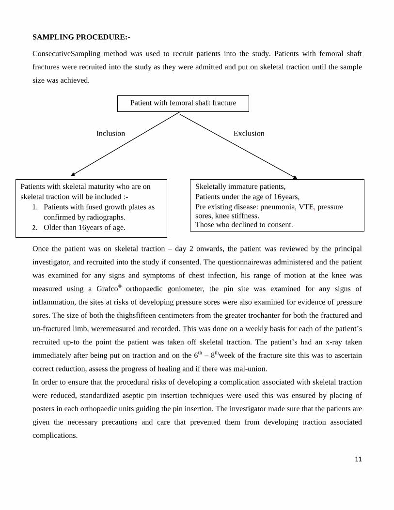
SAMPLING PROCEDURE:-
ConsecutiveSampling method was used to recruit patients into the study. Patients with femoral shaft
fractures were recruited into the study as they were admitted and put on skeletal traction until the sample
size was achieved.
Inclusion Exclusion
Once the patient was on skeletal traction – day 2 onwards, the patient was reviewed by the principal
investigator, and recruited into the study if consented. The questionnairewas administered and the patient
was examined for any signs and symptoms of chest infection, his range of motion at the knee was
measured using a Grafco®
orthopaedic goniometer, the pin site was examined for any signs of
inflammation, the sites at risks of developing pressure sores were also examined for evidence of pressure
sores. The size of both the thighsfifteen centimeters from the greater trochanter for both the fractured and
un-fractured limb, weremeasured and recorded. This was done on a weekly basis for each of the patient’s
recruited up-to the point the patient was taken off skeletal traction. The patient’s had an x-ray taken
immediately after being put on traction and on the 6th
– 8th
week of the fracture site this was to ascertain
correct reduction, assess the progress of healing and if there was mal-union.
In order to ensure that the procedural risks of developing a complication associated with skeletal traction
were reduced, standardized aseptic pin insertion techniques were used this was ensured by placing of
posters in each orthopaedic units guiding the pin insertion. The investigator made sure that the patients are
given the necessary precautions and care that prevented them from developing traction associated
complications.
Patient with femoral shaft fracture
Patients with skeletal maturity who are on
skeletal traction will be included :-
1. Patients with fused growth plates as
confirmed by radiographs.
2. Older than 16years of age.
Skeletally immature patients,
Patients under the age of 16years,
Pre existing disease: pneumonia, VTE, pressure
sores, knee stiffness.
Those who declined to consent.
12
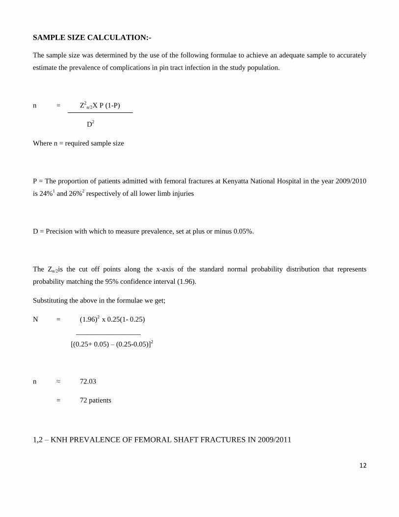
SAMPLE SIZE CALCULATION:-
The sample size was determined by the use of the following formulae to achieve an adequate sample to accurately
estimate the prevalence of complications in pin tract infection in the study population.
n = Z2
α/2X P (1-P)
D2
Where n = required sample size
P = The proportion of patients admitted with femoral fractures at Kenyatta National Hospital in the year 2009/2010
is 24%1 and 26%
2 respectively of all lower limb injuries
D = Precision with which to measure prevalence, set at plus or minus 0.05%.
The Zα/2is the cut off points along the x-axis of the standard normal probability distribution that represents
probability matching the 95% confidence interval (1.96).
Substituting the above in the formulae we get;
N = (1.96)2
x 0.25(1- 0.25)
__________________
[(0.25+ 0.05) – (0.25-0.05)]2
n ≈ 72.03
= 72 patients
1,2 – KNH PREVALENCE OF FEMORAL SHAFT FRACTURES IN 2009/2011
13
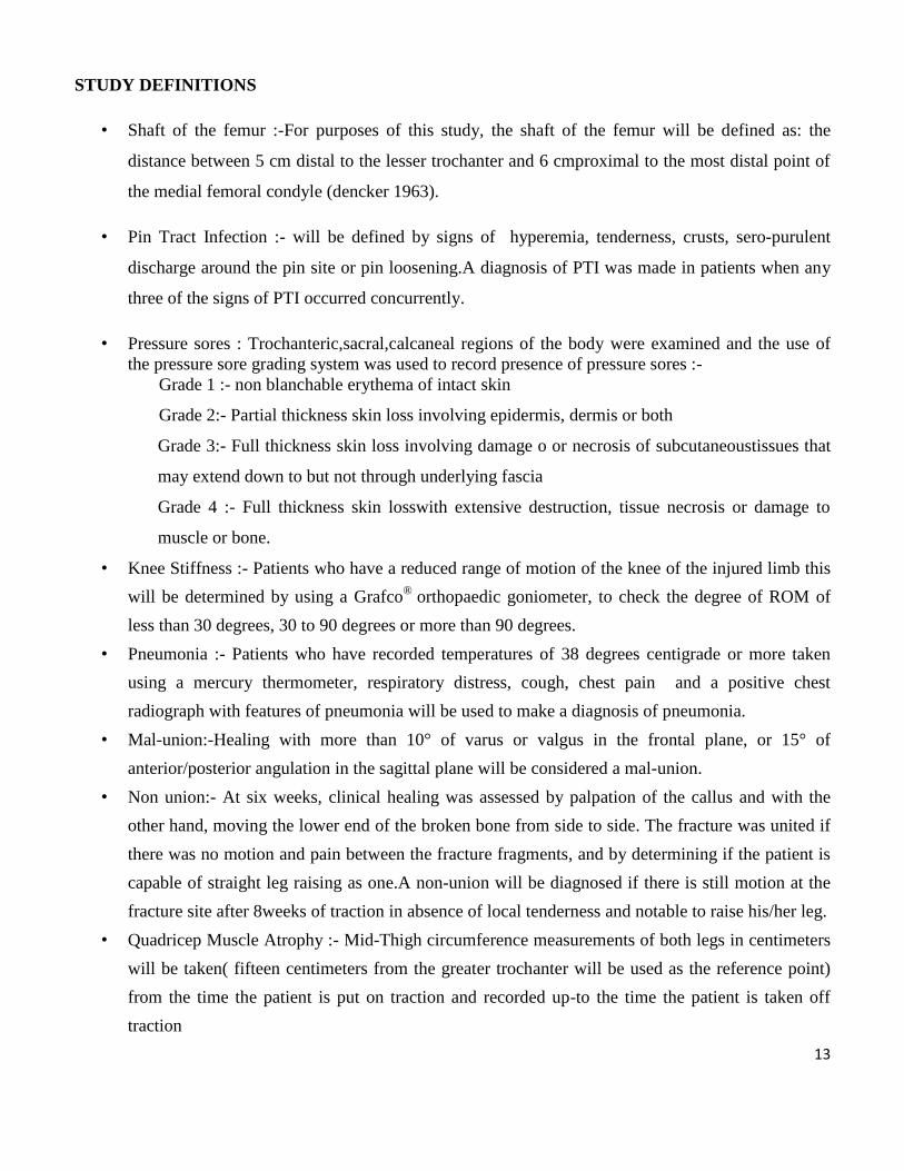
STUDY DEFINITIONS
• Shaft of the femur :-For purposes of this study, the shaft of the femur will be defined as: the
distance between 5 cm distal to the lesser trochanter and 6 cmproximal to the most distal point of
the medial femoral condyle (dencker 1963).
• Pin Tract Infection :- will be defined by signs of hyperemia, tenderness, crusts, sero-purulent
discharge around the pin site or pin loosening.A diagnosis of PTI was made in patients when any
three of the signs of PTI occurred concurrently.
• Pressure sores : Trochanteric,sacral,calcaneal regions of the body were examined and the use of
the pressure sore grading system was used to record presence of pressure sores :-
Grade 1 :- non blanchable erythema of intact skin
Grade 2:- Partial thickness skin loss involving epidermis, dermis or both
Grade 3:- Full thickness skin loss involving damage o or necrosis of subcutaneoustissues that
may extend down to but not through underlying fascia
Grade 4 :- Full thickness skin losswith extensive destruction, tissue necrosis or damage to
muscle or bone.
• Knee Stiffness :- Patients who have a reduced range of motion of the knee of the injured limb this
will be determined by using a Grafco®
orthopaedic goniometer, to check the degree of ROM of
less than 30 degrees, 30 to 90 degrees or more than 90 degrees.
• Pneumonia :- Patients who have recorded temperatures of 38 degrees centigrade or more taken
using a mercury thermometer, respiratory distress, cough, chest pain and a positive chest
radiograph with features of pneumonia will be used to make a diagnosis of pneumonia.
• Mal-union:-Healing with more than 10° of varus or valgus in the frontal plane, or 15° of
anterior/posterior angulation in the sagittal plane will be considered a mal-union.
• Non union:- At six weeks, clinical healing was assessed by palpation of the callus and with the
other hand, moving the lower end of the broken bone from side to side. The fracture was united if
there was no motion and pain between the fracture fragments, and by determining if the patient is
capable of straight leg raising as one.A non-union will be diagnosed if there is still motion at the
fracture site after 8weeks of traction in absence of local tenderness and notable to raise his/her leg.
• Quadricep Muscle Atrophy :- Mid-Thigh circumference measurements of both legs in centimeters
will be taken( fifteen centimeters from the greater trochanter will be used as the reference point)
from the time the patient is put on traction and recorded up-to the time the patient is taken off
traction
14
ETHICAL CONSIDERATIONS
Permission to conduct the study was sought from the KNH and University of Nairobi, Research and
ethics committee.
Informed voluntary consent from patients or relatives was obtained where the patient was not able to
consent for oneself.
Confidentiality was guaranteed to each patient from the data obtained.
Patients were allowed to withdraw at any point of the study should they choose to do so.
15
DATA COLLECTION:-
A pretested, structured and coded questionnaire was used. The data gathered included: patient
demographics, cause and pattern of fracture, traction type, time of commencement with the end
point being removal of the traction either for surgery or because of a complication.
Datawere collected as pertains to the date of commencement of traction up to the date patient
underwent operative management or was taken off traction.
The patients were put on skeletal traction by a trained and qualified orthopaedic technicians
assigned in each of the orthopaedic wards. Steinman pins were inserted with aseptic technique.
There was risk of injuring the peroneal nerve during insertion of the Steinman pin, however there
were no incidences reported
Patients were recruited into the study two days after they were put on traction, and were followed
upto the time they were taken off traction.
Radiographs of the fracture site were taken immediately after the patient was put on traction to
assess the alignment and reduction and subsequently at 6-8 weeks if the patient was still on
traction to assess healing of the fracture site.
The risk factors studied were age of the patient, cause of the fracture, fracture geometry and other
associated injuries.
DATA ANALYSIS:-
After cross checking the questionnaires for any missing entries a data base was designed in MS Access
which allowed the setting of controls and validation of the variables. On completion of the data entry, the
data was exported to the Statistical Package (SPSS – Version 15.0 Chicago, Illinois) for analysis.
Descriptive statistics were applied to the data collected and continuous data were summarized using
mean, standard deviation, median, mode and percentages were used for categorical or nominal data, a p
value of <0.05 was considered significant in determining the associations between the risk factors and
complications.
Data are presented in tables and figures where applicable. The t-test was used to compare duration of
traction versus the development of complications to determine any significant association between the
16
continuous variables e.g. age and duration on skeletal traction, while chi-square was used to establish the
significant associations between the fracture geometry, aetiology of fracture.Inferential statistics will be
used to analyse relative risks of developing complications as compared to the duration patients are on
traction. The incidence of complications related to period of skeletal traction will be evaluated and related
to the duration of traction using the chi square test.
Ninety five percent Confidence interval (CI) will be calculated to identify the factors that are more likely
to explain the explanatory variable (complication). P-value of less than 5% (P<0.05) will be considered
statistically significant.
LIMITATIONS TO THE STUDY:-
1. There were no clear guidelines ensuring aseptic techniques are followed during insertion of the
Steinman pin for skeletal traction before our study. Therefore there may be an increased
incidence of pin tract infections and in order to mitigate this we developed guidelines/
protocols that were put up in each orthopaedic ward giving procedural details of how the
Steinman pin should be inserted under proper aseptic technique.
2. We did not have control over how patients were selected for ORIF, therefore one of the
utilities of this study is to use the results in the development of guidelines / protocols where the
fracture patterns, cause of the fracture, or presence of other associated injuries would
determine which patient is given priority for ORIF.
17
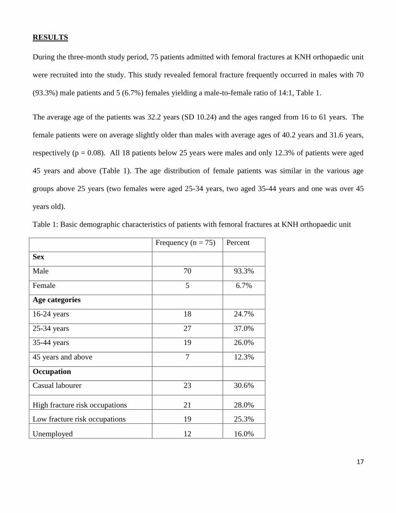
RESULTS
During the three-month study period, 75 patients admitted with femoral fractures at KNH orthopaedic unit
were recruited into the study. This study revealed femoral fracture frequently occurred in males with 70
(93.3%) male patients and 5 (6.7%) females yielding a male-to-female ratio of 14:1, Table 1.
The average age of the patients was 32.2 years (SD 10.24) and the ages ranged from 16 to 61 years. The
female patients were on average slightly older than males with average ages of 40.2 years and 31.6 years,
respectively (p = 0.08). All 18 patients below 25 years were males and only 12.3% of patients were aged
45 years and above (Table 1). The age distribution of female patients was similar in the various age
groups above 25 years (two females were aged 25-34 years, two aged 35-44 years and one was over 45
years old).
Table 1: Basic demographic characteristics of patients with femoral fractures at KNH orthopaedic unit
Frequency (n = 75) Percent
Sex
Male 70 93.3%
Female 5 6.7%
Age categories
16-24 years 18 24.7%
25-34 years 27 37.0%
35-44 years 19 26.0%
45 years and above 7 12.3%
Occupation
Casual labourer 23 30.6%
High fracture risk occupations 21 28.0%
Low fracture risk occupations 19 25.3%
Unemployed 12 16.0%
18
The most common occupation was casual laborer, and 12 (16.0%) of patients reported that they were
unemployed. The occupations considered at high risk for femoral fractures and accounting for 28.0% of
the sample were commercial or public service vehicle drivers, conductors on these vehicles, jobs in the
building industry like masonry or carpentry and security guarding duties. Patients in the low fracture risk
occupations were either in business, professional jobs or farming.
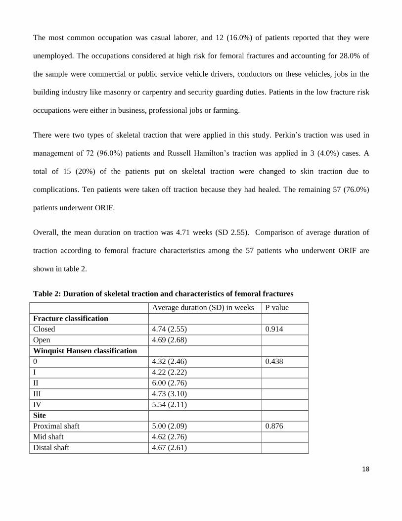
There were two types of skeletal traction that were applied in this study. Perkin’s traction was used in
management of 72 (96.0%) patients and Russell Hamilton’s traction was applied in 3 (4.0%) cases. A
total of 15 (20%) of the patients put on skeletal traction were changed to skin traction due to
complications. Ten patients were taken off traction because they had healed. The remaining 57 (76.0%)
patients underwent ORIF.
Overall, the mean duration on traction was 4.71 weeks (SD 2.55). Comparison of average duration of
traction according to femoral fracture characteristics among the 57 patients who underwent ORIF are
shown in table 2.
Table 2: Duration of skeletal traction and characteristics of femoral fractures
Average duration (SD) in weeks P value
Fracture classification
Closed 4.74 (2.55) 0.914
Open 4.69 (2.68)
Winquist Hansen classification
0 4.32 (2.46) 0.438
I 4.22 (2.22)
II 6.00 (2.76)
III 4.73 (3.10)
IV 5.54 (2.11)
Site
Proximal shaft 5.00 (2.09) 0.876
Mid shaft 4.62 (2.76)
Distal shaft 4.67 (2.61)
19
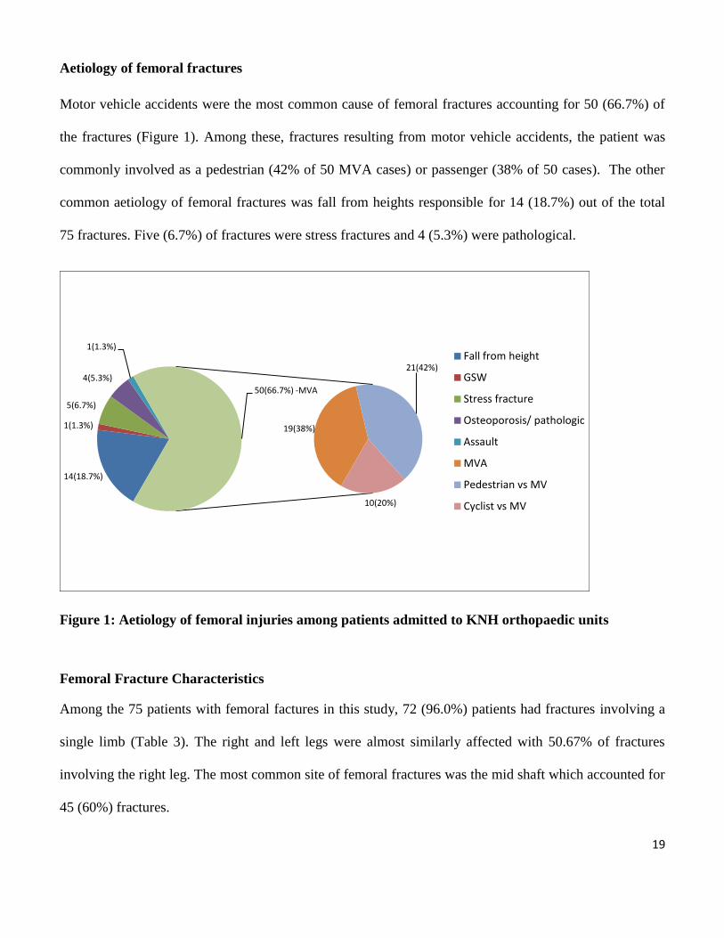
Aetiology of femoral fractures
Motor vehicle accidents were the most common cause of femoral fractures accounting for 50 (66.7%) of
the fractures (Figure 1). Among these, fractures resulting from motor vehicle accidents, the patient was
commonly involved as a pedestrian (42% of 50 MVA cases) or passenger (38% of 50 cases). The other
common aetiology of femoral fractures was fall from heights responsible for 14 (18.7%) out of the total
75 fractures. Five (6.7%) of fractures were stress fractures and 4 (5.3%) were pathological.
Figure 1: Aetiology of femoral injuries among patients admitted to KNH orthopaedic units
Femoral Fracture Characteristics
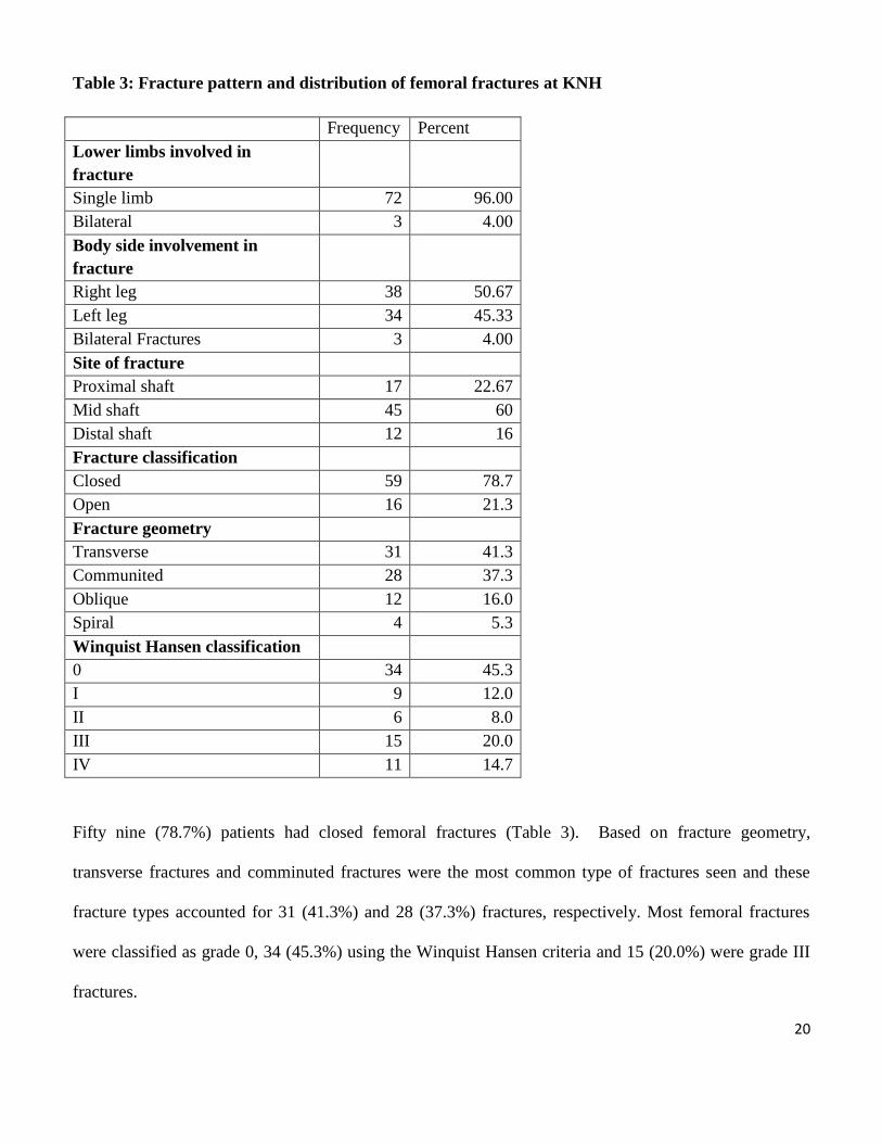
Among the 75 patients with femoral factures in this study, 72 (96.0%) patients had fractures involving a
single limb (Table 3). The right and left legs were almost similarly affected with 50.67% of fractures
involving the right leg. The most common site of femoral fractures was the mid shaft which accounted for
45 (60%) fractures.
14(18.7%)
1(1.3%)
5(6.7%)
4(5.3%)
1(1.3%)
19(38%)
21(42%)
10(20%)
50(66.7%) -MVA
Fall from height
GSW
Stress fracture
Osteoporosis/ pathologic
Assault
MVA
Pedestrian vs MV
Cyclist vs MV
20
Table 3: Fracture pattern and distribution of femoral fractures at KNH
Frequency Percent
Lower limbs involved in
fracture
Single limb 72 96.00
Bilateral 3 4.00
Body side involvement in
fracture
Right leg 38 50.67
Left leg 34 45.33
Bilateral Fractures 3 4.00
Site of fracture
Proximal shaft 17 22.67
Mid shaft 45 60
Distal shaft 12 16
Fracture classification
Closed 59 78.7
Open 16 21.3
Fracture geometry
Transverse 31 41.3
Communited 28 37.3
Oblique 12 16.0
Spiral 4 5.3
Winquist Hansen classification
0 34 45.3
I 9 12.0
II 6 8.0
III 15 20.0
IV 11 14.7
Fifty nine (78.7%) patients had closed femoral fractures (Table 3). Based on fracture geometry,
transverse fractures and comminuted fractures were the most common type of fractures seen and these
fracture types accounted for 31 (41.3%) and 28 (37.3%) fractures, respectively. Most femoral fractures
were classified as grade 0, 34 (45.3%) using the Winquist Hansen criteria and 15 (20.0%) were grade III
fractures.
21
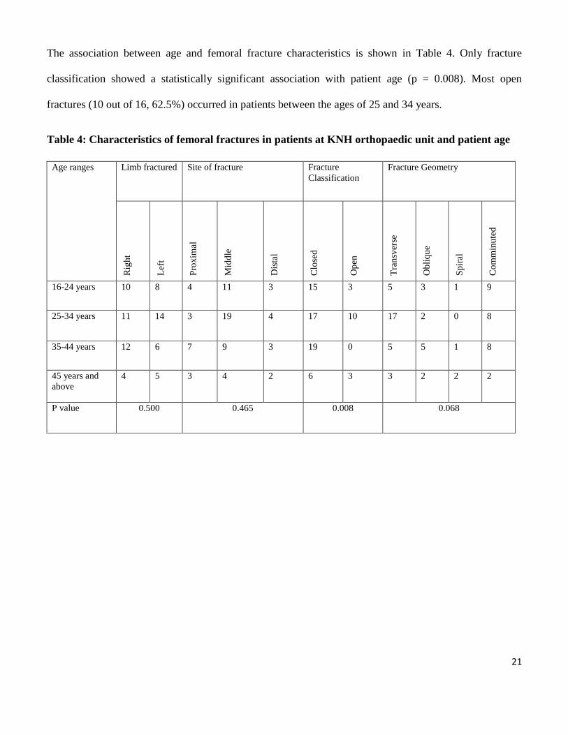
The association between age and femoral fracture characteristics is shown in Table 4. Only fracture
classification showed a statistically significant association with patient age (p = 0.008). Most open
fractures (10 out of 16, 62.5%) occurred in patients between the ages of 25 and 34 years.
Table 4: Characteristics of femoral fractures in patients at KNH orthopaedic unit and patient age
Age ranges Limb fractured Site of fracture Fracture
Classification
Fracture Geometry
Rig
ht
Lef
t
Pro
xim
al
Mid
dle
Dis
tal
Clo
sed
Op
en
Tra
nsv
erse
Ob
liq
ue
Sp
iral
Co
mm
inu
ted
16-24 years 10 8 4 11 3 15 3 5 3 1 9
25-34 years 11 14 3 19 4 17 10 17 2 0 8
35-44 years 12 6 7 9 3 19 0 5 5 1 8
45 years and
above
4 5 3 4 2 6 3 3 2 2 2
P value 0.500 0.465 0.008 0.068

22
Types of associated injuries
Out of the 75 patients with femoral fractures 51 (68%) had other associated fractures. Figure 2 shows that
the most common among the associated injuries was soft tissue injury present in 34 (45.33%) of the
patients. This was followed by skeletal injuries 26 (34.67%) and head injuries in 12 (16.0%) patients.
Figure 2:
Other injuries associated with femoral fractures among patients at KNH
Pin tract infection
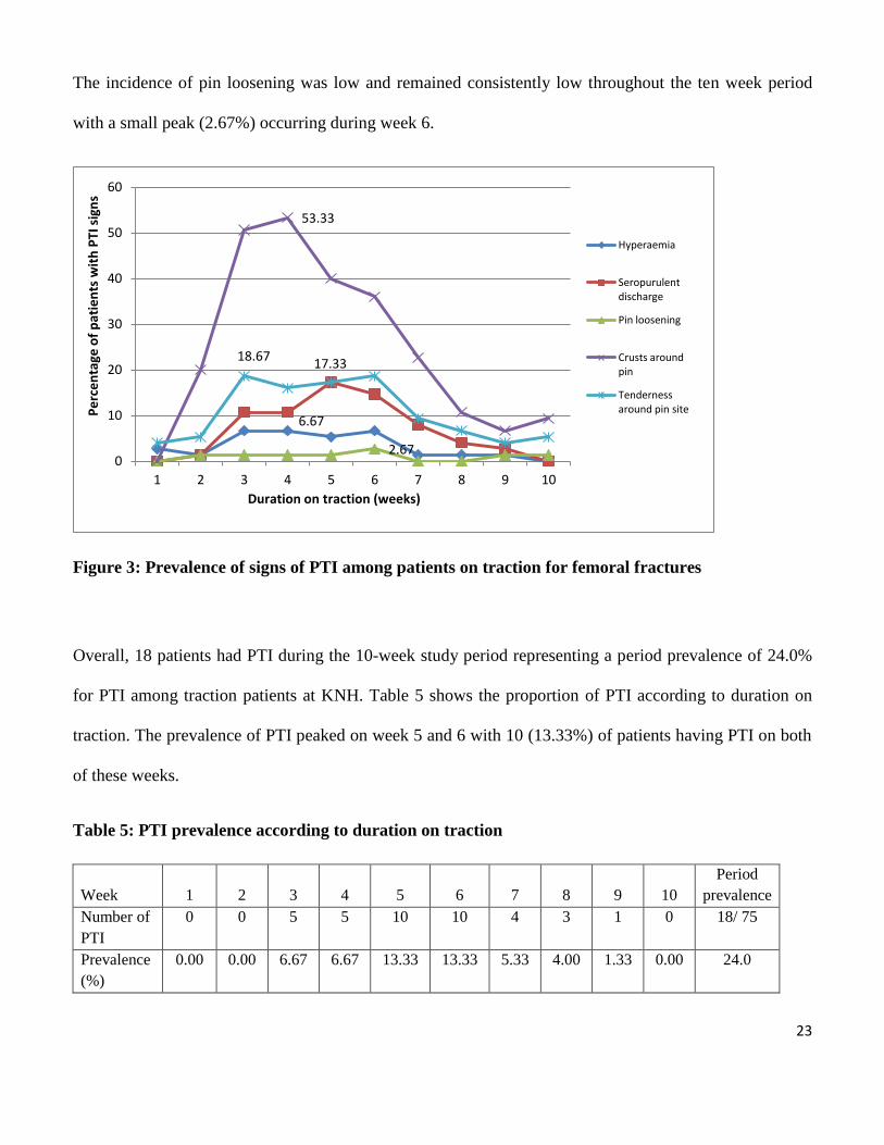
The most common sign of PTI was crust formation around the pin site (Figure 3). The prevalence of this
sign peaked at week 4 of traction with 53.33% of patient having the sign at this specific time. It was
followed by tenderness around pin site and sero-purulent discharge which had peak prevalence of 18.67%
and 17.33% occurring at week 3 and week 5, respectively.
12(16.0)
2(2.67) 3(4.0)
26(34.67)
34(45.33)
0
5
10
15
20
25
30
35
40
Head Abdomen Thoracic Skeletal STI
Nu
mb
er
of
pat
ien
ts (
%)
23
The incidence of pin loosening was low and remained consistently low throughout the ten week period
with a small peak (2.67%) occurring during week 6.
Figure 3: Prevalence of signs of PTI among patients on traction for femoral fractures
Overall, 18 patients had PTI during the 10-week study period representing a period prevalence of 24.0%
for PTI among traction patients at KNH. Table 5 shows the proportion of PTI according to duration on
traction. The prevalence of PTI peaked on week 5 and 6 with 10 (13.33%) of patients having PTI on both
of these weeks.
Table 5: PTI prevalence according to duration on traction
Week 1 2 3 4 5 6 7 8 9 10
Period
prevalence
Number of
PTI
0 0 5 5 10 10 4 3 1 0 18/ 75
Prevalence
(%)
0.00 0.00 6.67 6.67 13.33 13.33 5.33 4.00 1.33 0.00 24.0
6.67
2.67
53.33
18.67 17.33
0
10
20
30
40
50
60
1 2 3 4 5 6 7 8 9 10
Pe
rce
nta
ge o
f p
atie
nts
wit
h P
TI s
ign
s
Duration on traction (weeks)
Hyperaemia
Seropurulent discharge
Pin loosening
Crusts around pin
Tenderness around pin site
24
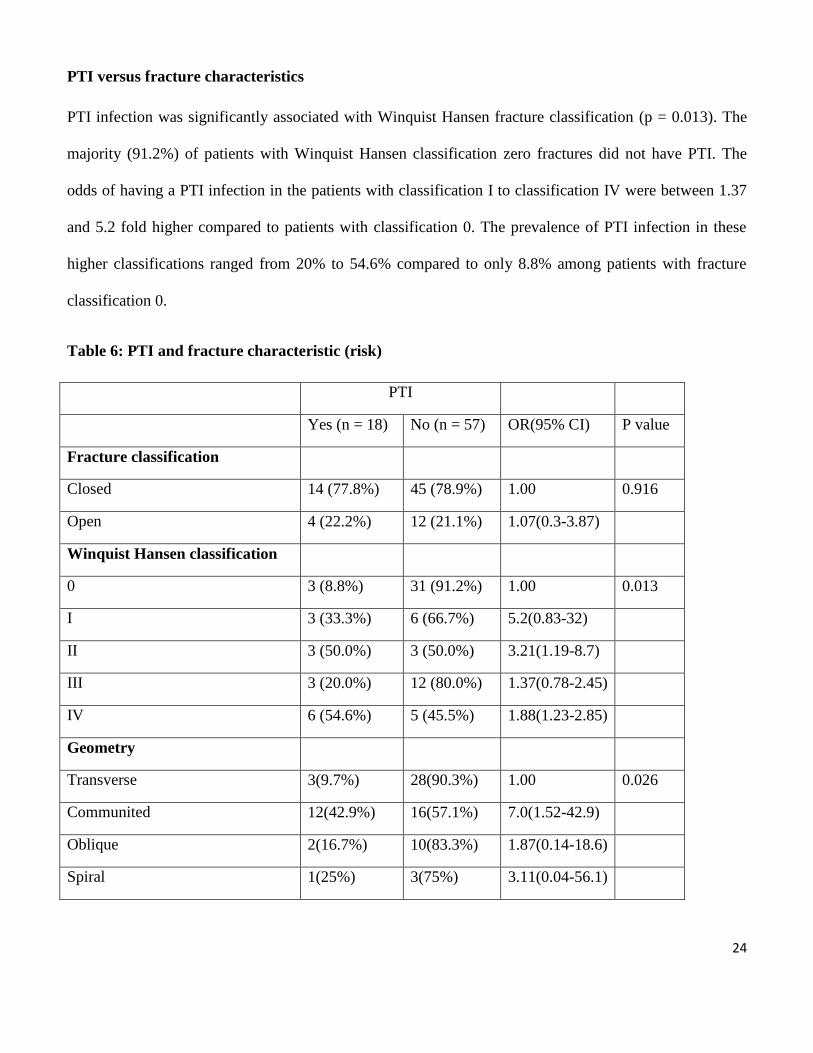
PTI versus fracture characteristics
PTI infection was significantly associated with Winquist Hansen fracture classification (p = 0.013). The
majority (91.2%) of patients with Winquist Hansen classification zero fractures did not have PTI. The
odds of having a PTI infection in the patients with classification I to classification IV were between 1.37
and 5.2 fold higher compared to patients with classification 0. The prevalence of PTI infection in these
higher classifications ranged from 20% to 54.6% compared to only 8.8% among patients with fracture
classification 0.
Table 6: PTI and fracture characteristic (risk)
PTI
Yes (n = 18) No (n = 57) OR(95% CI) P value
Fracture classification
Closed 14 (77.8%) 45 (78.9%) 1.00 0.916
Open 4 (22.2%) 12 (21.1%) 1.07(0.3-3.87)
Winquist Hansen classification
0 3 (8.8%) 31 (91.2%) 1.00 0.013
I 3 (33.3%) 6 (66.7%) 5.2(0.83-32)
II 3 (50.0%) 3 (50.0%) 3.21(1.19-8.7)
III 3 (20.0%) 12 (80.0%) 1.37(0.78-2.45)
IV 6 (54.6%) 5 (45.5%) 1.88(1.23-2.85)
Geometry
Transverse 3(9.7%) 28(90.3%) 1.00 0.026
Communited 12(42.9%) 16(57.1%) 7.0(1.52-42.9)
Oblique 2(16.7%) 10(83.3%) 1.87(0.14-18.6)
Spiral 1(25%) 3(75%) 3.11(0.04-56.1)
25
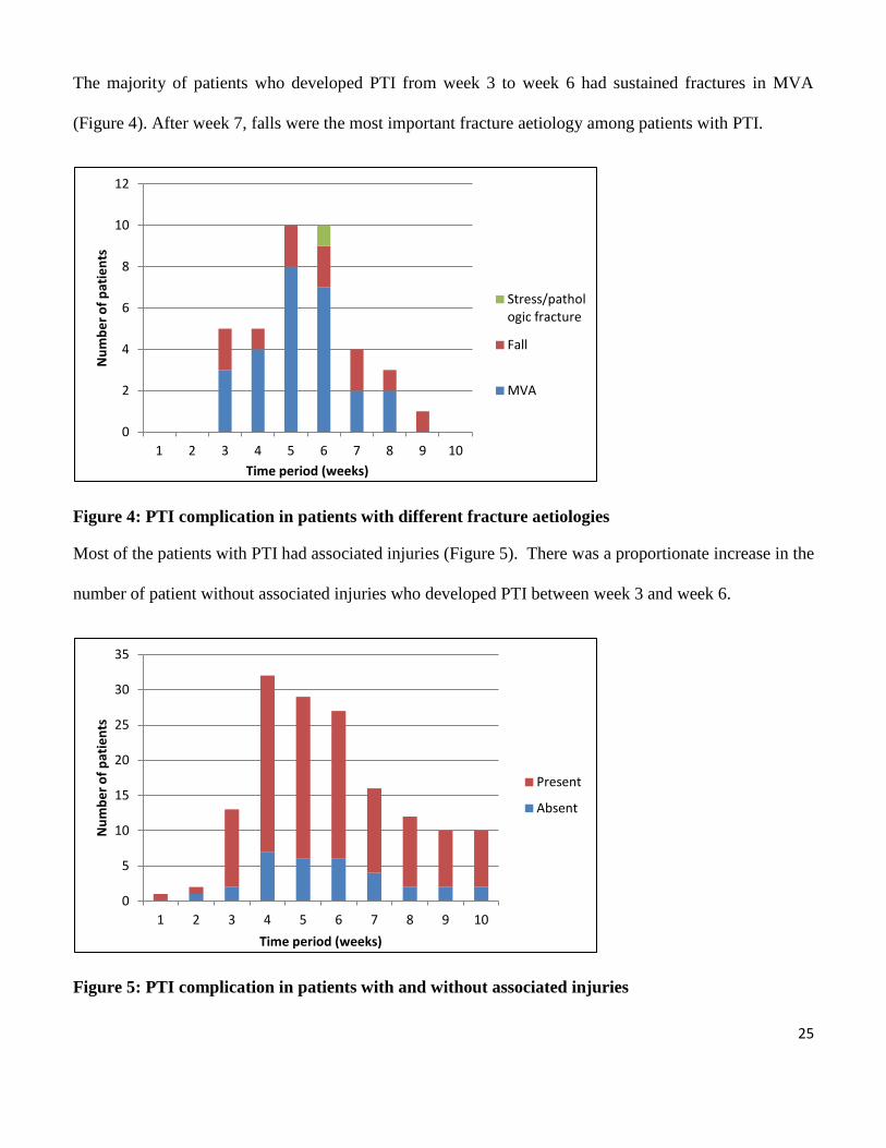
The majority of patients who developed PTI from week 3 to week 6 had sustained fractures in MVA
(Figure 4). After week 7, falls were the most important fracture aetiology among patients with PTI.
Figure 4: PTI complication in patients with different fracture aetiologies
Most of the patients with PTI had associated injuries (Figure 5). There was a proportionate increase in the
number of patient without associated injuries who developed PTI between week 3 and week 6.
Figure 5: PTI complication in patients with and without associated injuries
0
2
4
6
8
10
12
1 2 3 4 5 6 7 8 9 10
Nu
mb
er
of
pat
ien
ts
Time period (weeks)
Stress/pathologic fracture
Fall
MVA
0
5
10
15
20
25
30
35
1 2 3 4 5 6 7 8 9 10
Nu
mb
er
of
pat
ien
ts
Time period (weeks)
Present
Absent
26
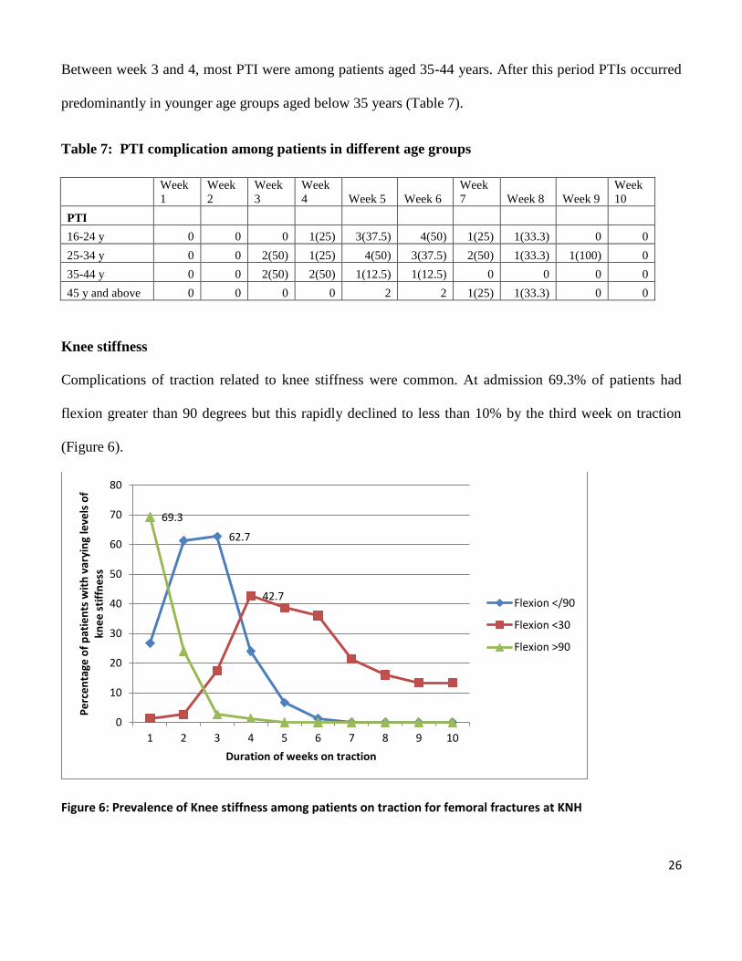
Between week 3 and 4, most PTI were among patients aged 35-44 years. After this period PTIs occurred
predominantly in younger age groups aged below 35 years (Table 7).
Table 7: PTI complication among patients in different age groups
Week
1
Week
2
Week
3
Week
4 Week 5 Week 6
Week
7 Week 8 Week 9
Week
10
PTI
16-24 y 0 0 0 1(25) 3(37.5) 4(50) 1(25) 1(33.3) 0 0
25-34 y 0 0 2(50) 1(25) 4(50) 3(37.5) 2(50) 1(33.3) 1(100) 0
35-44 y 0 0 2(50) 2(50) 1(12.5) 1(12.5) 0 0 0 0
45 y and above 0 0 0 0 2 2 1(25) 1(33.3) 0 0
Knee stiffness
Complications of traction related to knee stiffness were common. At admission 69.3% of patients had
flexion greater than 90 degrees but this rapidly declined to less than 10% by the third week on traction
(Figure 6).
Figure 6: Prevalence of Knee stiffness among patients on traction for femoral fractures at KNH
62.7
42.7
69.3
0
10
20
30
40
50
60
70
80
1 2 3 4 5 6 7 8 9 10
Pe
rce
nta
ge o
f p
atie
nts
wit
h v
aryi
ng
leve
ls o
f kn
ee
sti
ffn
ess
Duration of weeks on traction
Flexion </90
Flexion <30
Flexion >90
27
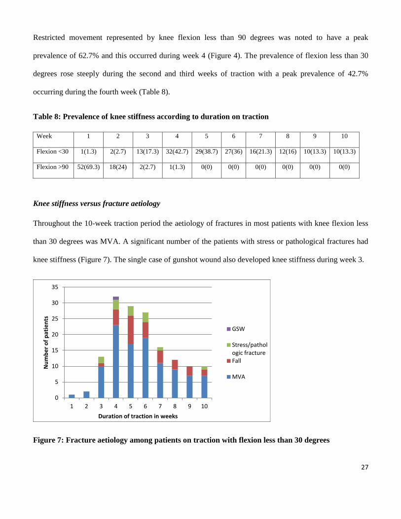
Restricted movement represented by knee flexion less than 90 degrees was noted to have a peak
prevalence of 62.7% and this occurred during week 4 (Figure 4). The prevalence of flexion less than 30
degrees rose steeply during the second and third weeks of traction with a peak prevalence of 42.7%
occurring during the fourth week (Table 8).
Table 8: Prevalence of knee stiffness according to duration on traction
Week 1 2 3 4 5 6 7 8 9 10
Flexion <30 1(1.3) 2(2.7) 13(17.3) 32(42.7) 29(38.7) 27(36) 16(21.3) 12(16) 10(13.3) 10(13.3)
Flexion >90 52(69.3) 18(24) 2(2.7) 1(1.3) 0(0) 0(0) 0(0) 0(0) 0(0) 0(0)
Knee stiffness versus fracture aetiology
Throughout the 10-week traction period the aetiology of fractures in most patients with knee flexion less
than 30 degrees was MVA. A significant number of the patients with stress or pathological fractures had
knee stiffness (Figure 7). The single case of gunshot wound also developed knee stiffness during week 3.
Figure 7: Fracture aetiology among patients on traction with flexion less than 30 degrees
0
5
10
15
20
25
30
35
1 2 3 4 5 6 7 8 9 10
Nu
mb
er
of
pat
ien
ts
Duration of traction in weeks
GSW
Stress/pathologic fracture Fall
MVA
28
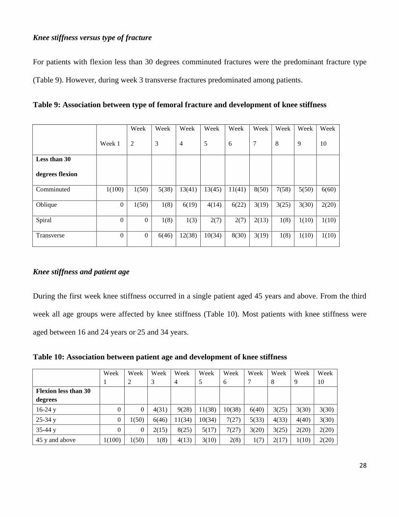
Knee stiffness versus type of fracture
For patients with flexion less than 30 degrees comminuted fractures were the predominant fracture type
(Table 9). However, during week 3 transverse fractures predominated among patients.
Table 9: Association between type of femoral fracture and development of knee stiffness
Week 1
Week
2
Week
3
Week
4
Week
5
Week
6
Week
7
Week
8
Week
9
Week
10
Less than 30
degrees flexion
Comminuted 1(100) 1(50) 5(38) 13(41) 13(45) 11(41) 8(50) 7(58) 5(50) 6(60)
Oblique 0 1(50) 1(8) 6(19) 4(14) 6(22) 3(19) 3(25) 3(30) 2(20)
Spiral 0 0 1(8) 1(3) 2(7) 2(7) 2(13) 1(8) 1(10) 1(10)
Transverse 0 0 6(46) 12(38) 10(34) 8(30) 3(19) 1(8) 1(10) 1(10)
Knee stiffness and patient age
During the first week knee stiffness occurred in a single patient aged 45 years and above. From the third
week all age groups were affected by knee stiffness (Table 10). Most patients with knee stiffness were
aged between 16 and 24 years or 25 and 34 years.
Table 10: Association between patient age and development of knee stiffness
Week
1
Week
2
Week
3
Week
4
Week
5
Week
6
Week
7
Week
8
Week
9
Week
10
Flexion less than 30
degrees
16-24 y 0 0 4(31) 9(28) 11(38) 10(38) 6(40) 3(25) 3(30) 3(30)
25-34 y 0 1(50) 6(46) 11(34) 10(34) 7(27) 5(33) 4(33) 4(40) 3(30)
35-44 y 0 0 2(15) 8(25) 5(17) 7(27) 3(20) 3(25) 2(20) 2(20)
45 y and above 1(100) 1(50) 1(8) 4(13) 3(10) 2(8) 1(7) 2(17) 1(10) 2(20)
29
Pneumonia
Pneumonia rarely occurred in the study and in cases where it was diagnosed it was noted that it occurred
relatively early. It occurred in only 3(4.0%) patients on weeks 1, 2 and 4. One of the patients with
pneumonia was changed to skin traction and a second patient with pneumonia eventually died during
week 4. The single death that occurred in this study was in a patient with signs of pneumonia.
Pressure sores
Three patients developed pressure sores during traction. All the pressure sores seen in the study were
grade I. Two patients had trochanteric or sacral region sores and the last patient developed calcaneal sores
during week 3. One of the patients with trochanteric/ sacral sores also had PTI and pneumonia and this
patient died during week 4.
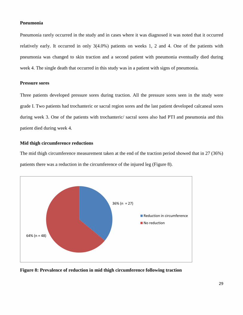
Mid thigh circumference reductions
The mid thigh circumference measurement taken at the end of the traction period showed that in 27 (36%)
patients there was a reduction in the circumference of the injured leg (Figure 8).
Figure 8: Prevalence of reduction in mid thigh circumference following traction
36% (n = 27)
64% (n = 48)
Reduction in circumference
No reduction
30
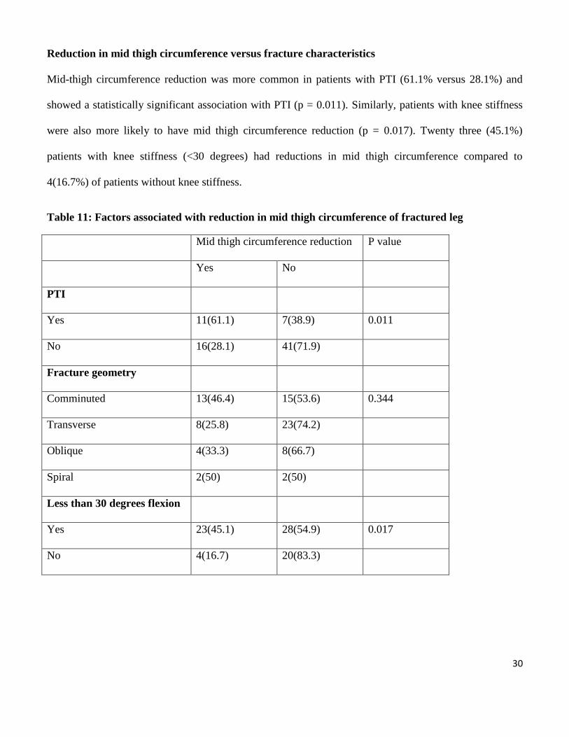
Reduction in mid thigh circumference versus fracture characteristics
Mid-thigh circumference reduction was more common in patients with PTI (61.1% versus 28.1%) and
showed a statistically significant association with PTI (p = 0.011). Similarly, patients with knee stiffness
were also more likely to have mid thigh circumference reduction (p = 0.017). Twenty three (45.1%)
patients with knee stiffness (<30 degrees) had reductions in mid thigh circumference compared to
4(16.7%) of patients without knee stiffness.
Table 11: Factors associated with reduction in mid thigh circumference of fractured leg
Mid thigh circumference reduction P value
Yes No
PTI
Yes 11(61.1) 7(38.9) 0.011
No 16(28.1) 41(71.9)
Fracture geometry
Comminuted 13(46.4) 15(53.6) 0.344
Transverse 8(25.8) 23(74.2)
Oblique 4(33.3) 8(66.7)
Spiral 2(50) 2(50)
Less than 30 degrees flexion
Yes 23(45.1) 28(54.9) 0.017
No 4(16.7) 20(83.3)
31
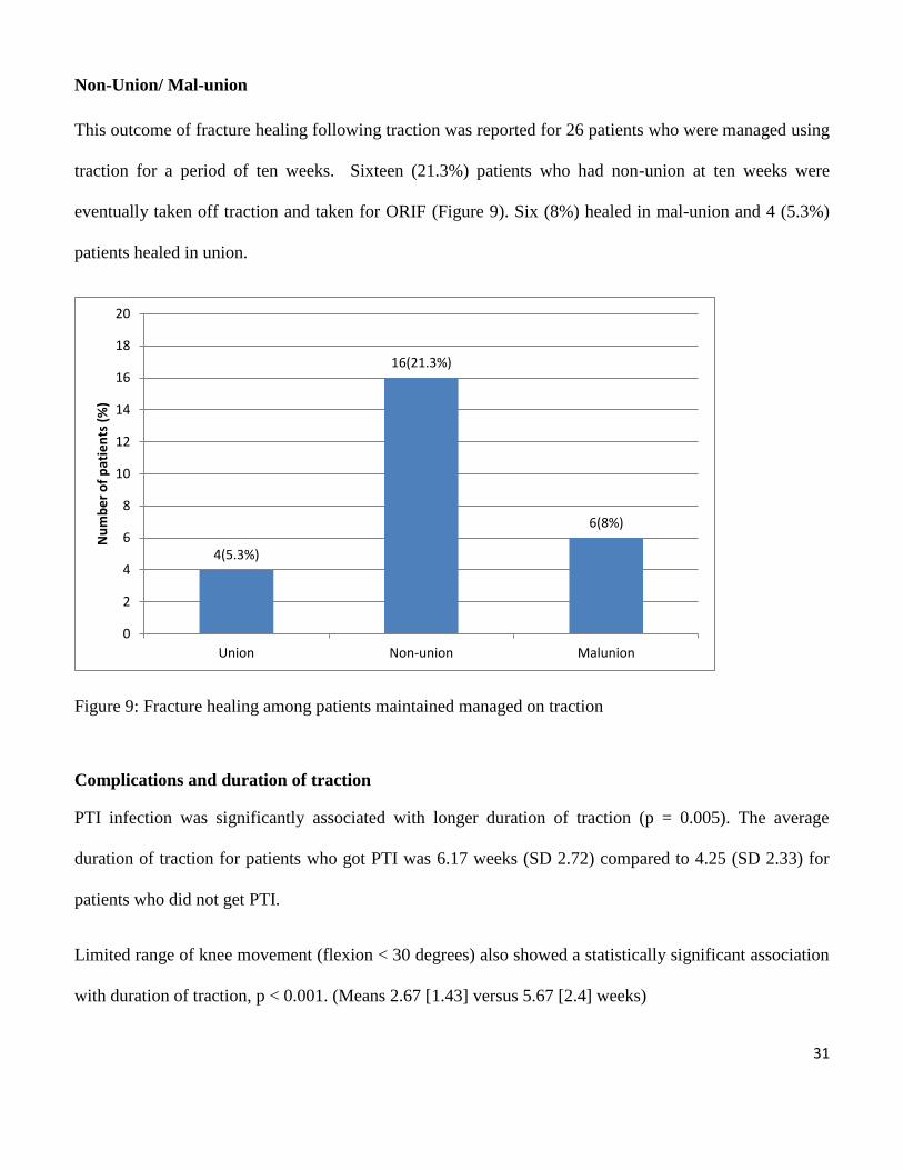
Non-Union/ Mal-union
This outcome of fracture healing following traction was reported for 26 patients who were managed using
traction for a period of ten weeks. Sixteen (21.3%) patients who had non-union at ten weeks were
eventually taken off traction and taken for ORIF (Figure 9). Six (8%) healed in mal-union and 4 (5.3%)
patients healed in union.
Figure 9: Fracture healing among patients maintained managed on traction
Complications and duration of traction
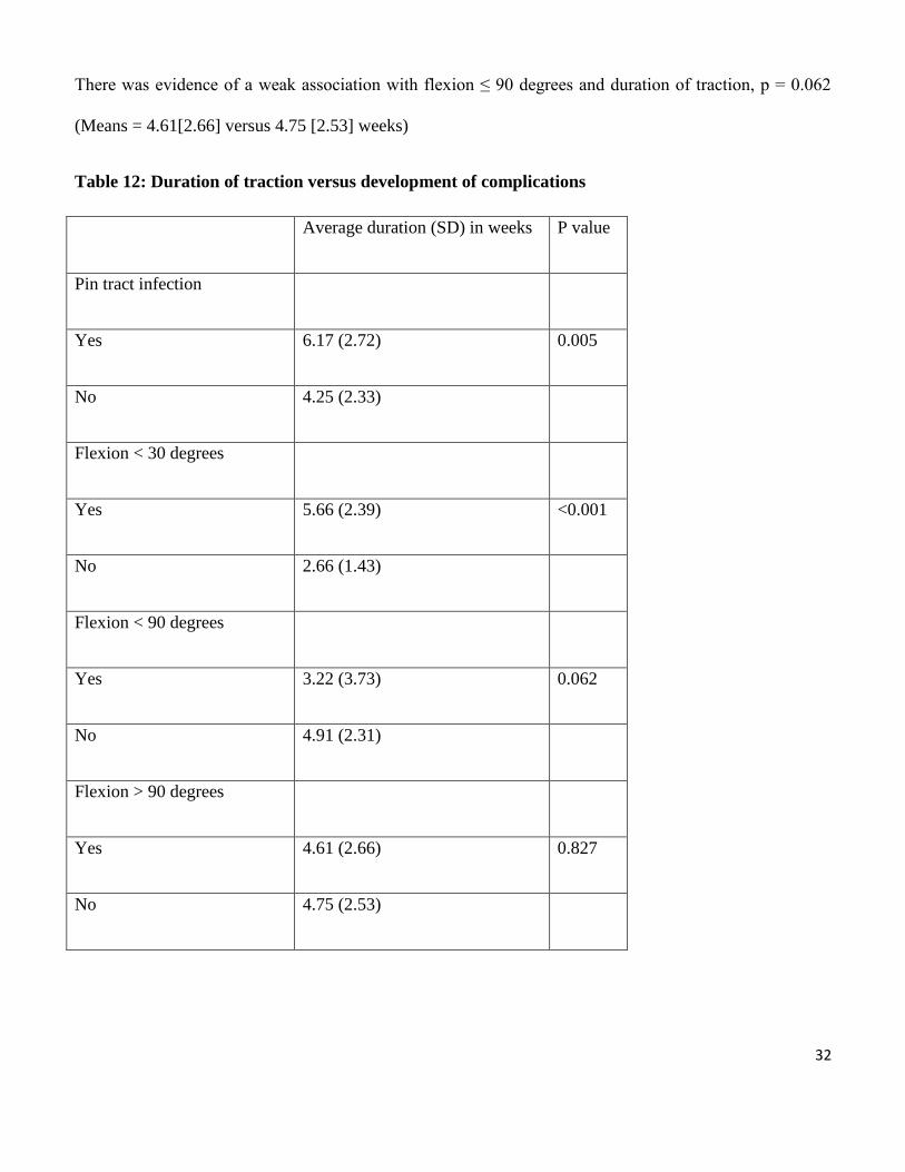
PTI infection was significantly associated with longer duration of traction (p = 0.005). The average
duration of traction for patients who got PTI was 6.17 weeks (SD 2.72) compared to 4.25 (SD 2.33) for
patients who did not get PTI.
Limited range of knee movement (flexion < 30 degrees) also showed a statistically significant association
with duration of traction, p < 0.001. (Means 2.67 [1.43] versus 5.67 [2.4] weeks)
4(5.3%)
16(21.3%)
6(8%)
0
2
4
6
8
10
12
14
16
18
20
Union Non-union Malunion
Nu
mb
er
of
pat
ien
ts (
%)
32
There was evidence of a weak association with flexion ≤ 90 degrees and duration of traction, p = 0.062
(Means = 4.61[2.66] versus 4.75 [2.53] weeks)
Table 12: Duration of traction versus development of complications
Average duration (SD) in weeks P value
Pin tract infection
Yes 6.17 (2.72) 0.005
No 4.25 (2.33)
Flexion < 30 degrees
Yes 5.66 (2.39) <0.001
No 2.66 (1.43)
Flexion < 90 degrees
Yes 3.22 (3.73) 0.062
No 4.91 (2.31)
Flexion > 90 degrees
Yes 4.61 (2.66) 0.827
No 4.75 (2.53)
33
DISCUSSION
Skeletal traction continues to play a major role in the management of femoral shaft fractures at Kenyatta
National Hospital and even further in the lower level hospitals in Kenya. This is despite evidence that
early operative intervention has better outcome and is cost effective.Its use locally is however, occasioned
by shortage of facilities, implants and surgeons; poverty and strain on government’s health budget are the
other (3). This study embarked on determining the incidence of complications associated with skeletal
traction at Kenyatta National Hospital and investigate risk factors predisposing these patients to these
treatment related morbidities.
The average age of the patients was 32.2 years, majority of the patients were in the age bracket of 25 – 34
years (Table 1). This uni-modal age distribution has been seen in other studies done from the region (5,
51). People of predominantly male gender in self employment or casual labour were mainly involved
(Table 1) as has been shown in previous studies (5,51).It has been suggested that this age category of
patients are the most economically productive and on the move in public transport as they fend for their
families (5,51,52). Most injuries were caused by road traffic accidents hence presence of associated
injuries in 68% of the patients.Developing countries are experiencing a general increase of all types of
injuries as they undergo socio-economic changes characterised by urbanisation and an increased
dependence on motor vehicles as a means of transport (3). This results in a significant increase of
complex extremity fractures (53, 54).
Both femurs were involved equally(Table 3), with majority (60%) of the fractures in the midshaft region
of the femur in a relatively similar pattern to that of Gosselin in Sierra Leone and Dim et al in Nigeria(32,
55). Transverse and comminuted pattern of fractures were the most common type as has been reported by
Bezabeh and Hansen (5, 56). These fractures are a result of direct impact to the bone or indirect forces
34
transmitted through the knee subjecting the femur to high bending loads hence motor vehicle accidents
are associated with a high rate of femoral shaft fractures (51).
The mean duration of skeletal traction was 33 days in this study. Studies in settings similar to ours have
revealed varying durations of traction, for example Bezabeh’s study in Ethiopia the average duration
patients were on traction was 35 days (5), while Gosselin’s study, revealed time in traction to be
averaging between 23-66 days with mean duration of 45 days (32). These varying durations on traction
are possibly due to differences in when the patients were taken off traction either for ORIF as was the
case in our study, or the fractures healed by being solely managed by conservative method, like in
Gosselin’s Sierra Leone study.
Complications associated with skeletal traction from this study are more in line with data reported from
other studies from the region. The most common sign of PTI was crust formation around the pin site.
Overall, 18 patients had PTI at any point during the study period representing a period prevalence of
24.0% for PTI among traction patients at KNH. The prevalence of PTI peaked on week 5 and 6 of traction
with 13.33% of patients having PTI on both of these weeks. The PTI rate in this study is slightly higher
when compared to the study done by Bezabeh, where the PTI rate was 11.8% (5). It is comparable to
other studies and was found to be: slightly lower than 36%(57) and 42.6% (32), and higher than 8.6% and
15% in studies done by Usdin (31) and Procter(30) respectively from South Africa.The high rate could
possibly be due to technical considerations: re-using many times the same smooth, blunt Steinmann pins.
Although we had put up guidelines in each orthopaedic unit and the orthopaedic technicians were advised
to adhere to the recommendations on the insertion of the Steinman pin, whether utmost sterility was
maintained during insertion of the pin with a hand drill is not known. It was noticed that many patients
did not have the traction bow properly applied, this leads to frequent re-adjustments of the traction
apparatus and this could possibly be another factor in increasing the risk of developing PTI. Host factors
35
such as chronic undernourishment or decreased immunity could have also contributed to the increased
rates of PTI. Other host factorslike the pin tract site itself could be in itself a risk for developing PTI and it
has been shown that daily pin tract site care both by the patient and nursing staff could decrease the
incidence of PTI (66). A more rigorously sterile technique, the systematic use of a hand drill to avoid
thermal necrosis, and the use of sharp, centrally threaded pins (Denham) could help to decrease this
complication to acceptable levels (32).
In this study PTI was significantly associated with prolonged durations of traction and was common in
patients with comminuted fractures (Tables 5,6). The other studies done from the region have not
described the occurrence of PTI in relation to the duration of traction or fracture patterns (5,64). We could
probably attribute the increased incidence of PTI in communited fractures to the prolonged lengths of
traction. These fractures are of a complex nature and they usually require internal fixation. The implants
are expensive and unavailable hence they are on prolonged periods of skeletal traction predisposing them
to PTI.
Knee stiffness was the most commonly occurring complication; at admission 69.3% of patients had
flexion greater than 90 degrees but this rapidly declined to less than 10% by the third week on traction.
Restricted movement represented by knee flexion less than 90 degrees was noted to have a peak
prevalence of 62.7% and this occurred during week 4. The frequency of flexion less than 30 degrees rose
steeply during the second and third weeks of traction to 42.7% in the fourth week. The rates of knee
stiffness in this study may be higher when compared to other studies. This could be due to patients having
some restriction / reluctance in flexing/extending their knees during the immediate post fracture period.
This was due to pain caused at the fracture site aggravated by movements of the knee. Oduor et al (58) in
a study at KNH had similar knee stiffness rates of 47.2%.
36
The data from Bezabeh’s study on knee stiffness is not clearly reported as they have not stated at what
point in time in respect to traction the patients develop knee stiffness they simply state that knee range of
motion was 40-60 degrees in ten patients and between 60-90 degrees in 50 patients(5). It was more than
90 degrees in 7 (10.3%) patients, similarly in the study by Gosselin, knee range of motion was recorded in
34 patients (64.2%) and reported as full in 23 and good in 11(32).This data on knee motion, although
apparently showing good results in the Perkins group, are too poor to lend themselves to critical analysis
as they are not specific as to how they categorized full knee ROM and Good knee ROM. Usdin in his
series of 58 patients from south Africa reported only 2 cases of residual knee stiffness (31), Bewes
published his series of 15 patients from Tanzania in 1974 [59], all had at least 75% of knee flexion at the
time of discharge.
Results from this study prove that knee stiffness is a significant problem even though majority of the
patients are on Perkins traction which results to minimal knee stiffness. The development of knee stiffness
was significantly associated with duration of traction (p value of < 0.001) (Table 12). However fracture
geometry, age or cause of the fracture, are not significant risks as all the patients on traction are equally
predisposed to develop knee stiffness and this is probably attributed to immobilization.
Reports on the use of the Perkins technique come mostly from developing countries.Similar results as this
study have been reported from Malawi where measurement of complications such as knee movement
restriction was only done at the time of discharge and does not reflect the true long-term outcome, they all
had knee flexion of < 90 degrees upon discharge (60).
It is important to note that despite the patients in our series being on the Perkin’s mode of traction the
beds were not broken at the foot to allow for flexion exercise. It was also observed that the only form of
physiotherapy they received was to perform isometric contraction exercises of the quadriceps and range of
motion exercises of the ankle of the injured limb this could probably explain the high incidence of knee
stiffness in the current study.
37
Purported benefits of Perkins traction over other methods is that healing rates are the same or better while
causing very little if any residual knee stiffness, thus reducing the overall recovery time [61,62].It is
recommended within 3 or 4 days from admission and almost always before the 7th day, active range of
motion exercises of the knee should be started. Pain medication may be necessary for the first few days,
but pain levels become tolerable with surprising rapidity. To be successful, the Perkins technique requires
“enthusiastic persistence” from both patient and surgeon.
Pneumonia rarely occurred in the study and in cases where it was diagnosed, it occurred relatively early.
This is possibly due to multiple injuries in these patients causing them to be lying supine most of the time
which predisposed them to develop hypostatic pneumonia. One of the patients who developed pneumonia
had suffered from chest injuries hence putting him at an increased risk. Pneumonia was diagnosed in only
4.0% of the patients on weeks 1, 2 and 4. One of the patients with pneumonia, also developed PTI and
was changed to skin traction while a second patient with pneumonia eventually died during week 4. Most
of the patients in this study were from a young age bracket and did not have multiple injuries therefore
they were not completely immobilized; they are able to sit themselves up and are more active despite
being on traction hence this could be the reason for the low incidence of pneumonia in this study. The
patients who developed pneumonia were either in elderly age group or had multiple injuries, this proves
that elderly age and multiple injuries are a significant risk factor in the development of chest infections.
However results from this study are not in keeping with other studies, Charash et al [65] reviewed early
versus delayed femoral shaft fracture stabilization in polytrauma patients with thoracic injuries. The
overall incidence of pulmonary complications (ARDS, pneumonia, fat embolism, or pulmonary
embolism) was 56% in the delayed fixation group compared with 16% in the early fixation group. The
authors noted that 48% of patients in the delayed fixation cohort (N = 25) went on to develop pneumonia
compared with 14% in the early fixation cohort (N = 56).The low incidence of chest complications are
38
possibly because most of the patients in our series did not suffer from major/severe trauma, and are of a
young age bracket.
Three patients developed pressure sores during traction. All the pressure sores seen in the study were
grade I. Two patients had trochanteric or sacral region sores and the last patient developed calcaneal sores
during week 3, one of the patients with trochanteric/ sacral sores also had PTI and pneumonia and this
patient died during week 4, this patient also had severe head injury in addition to the femoral fracture this
shows that there is an increased incidence of pressure sores with multiple injuries. The low rates of
pressure sores are could be due to the comprehensive care given by the physiotherapists; all the patients
once they were put on traction were given protective heel shoes made out of sheep hide to be worn over
the heel of the fractured limb. Patients were also encouraged on mobilization from the onset, they were
advised on turning and sitting up and this could explain why the low rates of pressure sores. An important
factor is that most of the patients were of a younger age bracket (Table 1) and although they were bed
bound they were not completely immobilized. The decreased incidence of pressure sores could also have
been due to the fact that most of the patients did not have major associated injuries. None of the studies
done from the region have looked at the occurrence of pressure sores, this is probably due to the fact that
most of the patients in these series (5, 32) were in the age ranges of 25 – 35yrs and most of the patients
were on Perkins traction and early mobilization was advised. In a study done by Versluysen it was found
that the incidence of pressure sores among her series of patients Of 100 subjects, 66 developed sores of
varying degrees of severity. The patients in this series were aged 70-94, of these 100 patients, 97 had
confirmed fractures; 88 injuries were at the proximal femur and nine in the shaft (36). With increasing age
the risks of developing pressure sores increases, if intense nursing care is not availed to the patient.
39
Fracture healing following traction was reported for 10 patients who were managed using traction only
and did not have to undergo ORIF. Out of these6 (8%) patients had fractures that healed in mal-union,
four (5.3%) patients had fractures healing in radiological union, while 16(21.3%) of the patients who were
on traction for more than 8 weeks developed non union and were eventually taken for ORIF. Procter
reported his series of 41 patients in South Africa [30], all the patients had achieved clinical union at 6
weeks. A few years later, Usdin followed up with his own series of 58 patients [31] and average LOS was
7 weeks amongst the 58 patients, 6 (10.4%) had delayed or non-unions requiring surgical treatment.
Bewes published his series of 15 patients from Tanzania in 1974 [59], they had a 100% union rate at 12
weeks though his sample size was very small. In 1977, Pearson reported on a series of 100 Nigerian
patients treated over an 18-year period with either skin (89) or skeletal (11) traction [63], mean time to
clinical consolidation was 6 weeks, and to protected ambulation 7 weeks. Two-thirds of his cases were
under 20 years of age. Buxton reported in 1981 his series of 50 consecutive patients from the UK mean
time to clinical and radiological healing was 12 weeks (57).
Most of the patients in this study were eventually taken for ORIF, therefore it is difficult to draw
conclusions about our rates of union from traction exclusively, the rates of non-union could possibly be
due to over distraction of the fragments or there was soft tissue interposition between the fracture
fragments.
Only 36% of the patients from this study developed reduction in thigh circumference, this shows that the
rates of quadriceps atrophy are not high, however during the data collection it was noted that even though
the thigh circumference was not reducing, the quadriceps muscle was significantly atrophied. Bezabeh in
his series of patients from Ethiopia also found a similar occurrence where only 12% of patients were
reported to have reductions in thigh circumference as compared to 55% who had no change(5).
Mid-thigh circumference reduction was more common in patients with PTI and showed a statistically
significant association with PTI (p = 0.011). Similarly, patients with knee stiffness were also more likely
40
to have mid thigh circumference reduction (p = 0.017). Twenty three (45.1%) patients with knee stiffness
(<30 degrees) had reductions in mid thigh circumference compared to 4(16.7%) of patients without knee
stiffness. This could be due to decreased movements of the fractured limb, which leads to muscle atrophy
and reconditioning, loss of skeletal muscle mass and strength. This is often seen because of
immobilization. Joint contraction occurs because of muscle atrophy, flexion loss is mostly due to intra-
articular fibrosis and scarring in the quadriceps-femoral mechanism. Anterior adhesions involve the
quadriceps expansion in the lateral and medial recesses, the supra-patellar bursa, muscle adhesions to the
femur, patella, or even shortening of the rectus femoris (49).
This study has revealed that patients can be managed on skeletal traction for up to 4 weeks (Table 12)
beyond which it has been found that the incidence of developing PTI and knee stiffness increases. The
other studies from the region have not revealed at what point on traction complications arose.
Developing countries are experiencing a general increase of all types of injuries as they undergo socio-
economic changes, characterized by urbanization and an increased dependence on motor vehicles as a
means of transport. This results in a significant increase of fractures of the extremities, incidence and
complexity of injuries (53).
Conservative treatment remains an acceptable mode of treatment, even in the West, for complicated
fractures it has produced favourable results in the past. Its main drawback is the prolonged hospital stay
and reduction in knee range of movement (60).
In light of limited health care facilities and doctor expertise in many developing countries, it must be
accepted that, for a long time to come, conservative methods of fracture treatment will remain not only
the method of choice but also the only practical one.
41
Conclusions
The most common complications are knee stiffness followed by pin tract infection.The risk factors
predisposing to development of these complications are, prolonged durations of traction, fractures that are
comminuted and the presence of multiple injuries.
Most of the patients in the orthopaedic units at KNH are taken for ORIF, however they are on traction for
at least four weeks prior to fixation. We conclude that patients can be safely managed on skeletal traction
for up-to four weeks beyond which they are at an increased risk of developing complications.
Recommendations
1. In order to decrease the incidence of pin tract infection twice-daily pin site care by the patient with
wool and alcohol (spirit) should be done.
2. Intensive physiotherapy from the beginning with focus on building quadriceps and knee range of
motion exercises to decrease the rates of knee stiffness, most hospitals have physiotherapists
whose help can be enlisted.
3. The use of a Perkin’s bed is preferred in patients with femoral shaft fractures. This can be made by
adapting the bed base so it can be split for knee flexion but also replaced when patient needs to
sleep.
4. Patients with multiple injuries and comminuted fractures should be considered for early ORIF, in
these patients skin traction can be considered as an initial temporary form of management as they
await definitive care.
42
References
1. H.S. Saidi. Initial injury care in Nairobi, Kenya: A call for trauma care regionalization.East Afr Med J.
2003;80 (9): 480-483.
2. H.S. Saidi, W.M. Macharia and J.E.O. Ating’a. Outcome For Hospitalized Road Trauma Patients At
A TertiaryHospital In Kenya. European Journal of Trauma. 2005;31(4):401-406.
3. L.M. Museru, C.N. Mcharo. (2002) The dilemma of fracturetreatment in developing countries. Intern
Orthop.2002; 26: 324–327.
4. A.A. White. Fracture treatment, The still unresolvedproblem. Clin.Orthop.1975;106:279–284.
5. B. Bezabeh, B.L. Wamisho. Treatment of femur shaft fractures using perkins traction at Addis
AbabaUniversity: An Ethiopian experience. East Afr Orthop J.2010; 4:12-18.
6. Charnley J. The Closed Treatment of Common Fractures. 3rd
ed. Edinburgh and London: Churchill
Livingstone; 1974; Chapter 13: 166-196.
7. Simon R, Eliopoulos V, Laflamme D, et al. Impact on process of trauma care delivery 1 year after the
introduction of a trauma program in a provincial trauma center. J Trauma. 1999;46:811–6.
8. Saidi H.S., Ating’a J.E.O., Macharia W. Risk factor profile and the cost of traffic injury in a tertiary
hospital in Kenya. Proceedings of the International Conference on Traffic Safety on Three
Continents.2001; September, Moscow Russia.
9. Gustilo RB, Corpuz V, Sherman RE. Epidemiology, mortality and morbidity in multiple trauma
patients. Orthopedics. 1985;8: 1523–8.
10. Peltier L.F. A Brief History of Traction. J Bone Joint Surg. 1968; 50-A: 1603-1617.
43
11. Brink A.C., Boonstra O., Van Der Wal B.C.H. Et Al.Is preoperative traction for proximal femoral
fractures beneficial to the patient or a comfort to the doctor? European Journal of Trauma.
2005;31(1):39–43.
12. Billsten M., Besjakov J., Hyddmark U., Et Al. Enquiry in Sweden on the use of traction
preoperatively in patients with hip fracture and a radiological study on the effect of the traction on ten
displaced cervical hip fractures [abstract]. Acta Orthopaedica Scandinavica. Supplementum.1996;
270:35.
13. Resch S, Thorngren K-G. Preoperative traction for hip fracture: A randomized comparison between
skin and skeletal traction in 78 patients. Acta Orthopaedica Scandinavica.1998;69(3):277–9.
14. C. M. Court-Brown. The Management Of Femoral And Tibial Diaphyseal Fractures. J. R. Coll. Surg.
Edinb.1998; 43(12):374-380.
15. Fakhry S.M., Rutledge R., Dahners L.E., Kessler D. Incidence, management, and outcome of femoral
shaft fracture: a statewide population-based analysis of 2805 adult patients in a rural state. J
Trauma.1994;37(2):255-60.
16. Gillquist J., Liljedahl S.O., Rieger A. Fracture of the Femur Diaphysis, Traction or Nailing. Acta Chir
Scand.;137(6): 515-520.
17. Murray, C.J.L., and Lopez, A.D. Mortality by cause for the eight regions of the world; Global Burden
of Disease Study. Lancet. 1997; 349:1269-1276.
18. Draper P, Scott F. An evaluation of Hamilton-Russell traction in the pre-operative management of
patients with hip fracture. Clinical Effectiveness in Nursing.1997;1(4):179–88.
19. Finsen V, Borset M, Buvik GE, Hauke I. Preoperative traction in patients with hip fractures.
Injury.1992;23(4):242–4.
44
20. Jerre R, Doshe A, Karlsson J. Preoperative treatment in patients with hip fractures [abstract]. Acta
Orthopaedica Scandinavica.Supplementum.1999;284:40.
21. C.K. Kirby, W. T. Fills.The incidence of complications in the use of transfixion pins and wires for
skeletal traction.Annals of Surgery.1946; 123(1):27-31.
22. Nigam V, Jaiswal A. Local antibiotics: Panacea for long term skeletal traction. Injury 2005; 36:199-
202.
23. Matthews LS, Green CA, Goldstein SA. The thermal effects of skeletal fixation: pin insertion in bone.
J Bone Joint Surg. 1984;66A:1077-83.
24. Clasper JC, Cannon LB, Stapley SA, Taylor VM, Watkins PE. Fluid accumulation and the rapid
spread of bacteria in the pathogenesis of external fixator pin track infection. Injury 2001;32:377-81.
25. Green SA, Ripley MJ. Chronic osteomyelitis in pin tracks.J Bone Joint Surg.1984;66:1092-8.
26. Clifford RP, Lyon TJ, Webb JK. Complications of external fixation of open fractures of the tibia.
Injury. 1987;18:174-6.
27. Temple J, Santy J. Pin site care for preventing infections associated with external bone fixators and
pins. Cochrane Database Syst Rev. 2004;1:CD004551.
28. Henry C. Pin sites: Do we need to clean them? Practice Nursing. 1996;7:12,15-17.
29. Mahan J, Seligson D, Henry SL, Hynes P, Dobbins J. Factors in pin tract infections. Orthopedics.
1991;14:305-8.
30. Procter D.S.C. The functional treatment of fractures of the femur. S Afr. Med J.1962; 36:598–602.
31. Usdin J. The treatment of fractures of the femur by Perkins’ traction. S Afr. Med J.1969; 43(16):480–
482.
32. Gosselin R., Lavaly D. Perkins traction for adult femoral fractures: A report on 53 patients in Sierra
Leone.Int. Orthopaedics.2007;31: 697-702.
45
33. M.S. Butt, S.J. Krikler, M.S. Ali. Displaced fractures of the distal femur in elderly patients : operative
versus non-operative treatment. J bone Joint Surg [Br].1995;77-b:110-4.
34. Kosiak M. Aetiology and pathology of ischaemic ulcers. Arch Phys Med Rehabil. 1959; 40:62-9.
35. Reswick JB, Rogers JE. Experience at Rancho Los Amigo's Hospital with devices and techniques to
prevent pressure sores. In: Kenedi RM, Scales JT, Cowden J, eds. Bedsore biomechanics. London:
Macmillan, 1976:113-22.
36. Versluysen M. Pressure sores in elderly patients: the epidemiology related to hip operations. J. Bone
JointSurg.1985;67:10-3(b).
37. Gurd AR and Wilson RI. The Fat Embolism Syndrome. J Bone Joint SurgBr.1974; 56B: 408-16.
38. Peltier LF, Adler F, Lai SP. Fat embolism: the significance of an elevated serum lipase after trauma to
bone. Am J Surg. 1960; 99: 821-6.
39. White TO, Jenkins JP, Smith RD, et al. The Epidemiology of Posttraumatic Adult Respiratory
Distress Syndrome. J Bone Joint Surg Am.2004; 86-A: 2366-76.
40. Dunham CM, Bosse MJ, Clancy TV, et al. Practice management guidelines for the optimal timing of
long-bone fracture stabilization in polytrauma patient: The east practice management guidelines work
group. J Trauma.2001; 50: 958-67.
41. Behrman S.W, Fabian TC, Kudsk KA. Improved outcome with femoral fractures. Early vs Delayed
Fixation. J Trauma.1990; 30: 792-7.
42. Bone LB, Johnson KD, Weigelt J, et al. Early Versus Delayed Stabilization of Femoral fractures. A
Prospective randomized study. J Bone Joint Surg Am.1989; 71: 336-40.
43. Johnson KD, Cadambi A, Seibert GB. Incidence of acute respiratory distress syndrome in patients
with multiple musculoskeletal injuries: effect of early operative stabilization of fractures. J
Trauma.1985; 25: 375-84.
46
44. Bone L, Bucholz R. Current concepts review. The management of fractures in the patient with
multiple traumas. J Bone JointSurg.1986; 68-A: 945-9.
45. Benoit PR, Hampson LG, Burgess JH. Value of arterial hypoxemia in the diagnosis of pulmonary fat
embolism. Ann Surg. 1972; 175:128-37.
46. Giannoudis PV, Smith RM, Bellamy MC, et al. Stimulation of the Inflammatory System by Reamed
and Un-reamed Nailing of Femoral Fractures. J Bone Joint SurgBr. 1999; 81: 356-61.
47. Anwar IA, Battistella FD, Neiman R, et al. Femur fractures and lung complications: a prospective
randomized study of reaming. Clin Orthop Relat Res.2004; 422: 71-6.
48. Chakrabarty RP, Chakrabarty S. A curative operation for extra-articular ankylosis of knee. J Indian
Med Assoc. 1990; 88 : 133-135.
49. Lindenfeld TN, Wojtys EM, Husain A. Operative treatment of arthrofibrosis of the knee. Instruc
Course Lect. 2000; 49 : 211-221.
50. Moulton A., Agunwa W.C. and Hopkins J.S. Closed femoral fractures: why not simple traction?
Injury. 1981; 13: 244–248.
51. Ombaye JA. Prospective comparison of open intramedullary, nailing and plate fixations of femoral
diaphyseal fractures at Kenyatta National Hospital. Dissertation ; 2008 :
52. Kizito DL. The value of trauma assessment scoring in predicting outcome of patients presenting with
major trauma to Kenyatta national hospital. Dissertation ; 2003 :
53. Museru LM, Leshabari MT, Grob U, Lisokotala LNM (1998) The pattern of injured patients seen in
the orthopaedic and trauma wards at Muhimbili Medical Centre – Dar es Salaam. East & Central Afr.
Journ. of Surgery 5:15–21
54. Odero WO, Kibosia JC (1995) Incidence and characteristics of injuries in Eldoret, Kenya. East Afr.
Med. J 72:706–710
47
55. DimEM.Adult traumatic femoral shaft fractures: A review of the literature. Ibom Medical Journal.
2012;5:26-38.
56. Hansen ST, Winquist RA. Closed Intramedullary Nailing of the femur- Kuntscher Technique with
reaming. Clin. Orthop Rel Research 1979; 138: 56-61.
57. Buxton RA. The use of Perkins’ traction in the treatment offemoral shaft fractures. J Bone JtSurg
Br.1981; 63-B(3):362–366.
58. Oduor PO. Distal femoral fractures: pattern and management at Kenyatta national hospital.
Dissertation; 2004.
59. Bewes PC. Fractures of the femur in a tropical context: a re-evaluation of Perkins’ traction. Trop
Doct. 1974; 4(2):64–68
60. Doorgakant A. The management of isolated closed femoral shaft fractures in a district hospital in
Malawi. Tropical Doctor. 2012; 42: 8–12
61. Perkins G. Rest and movement. J Bone Jt Surg.1953;Br 35-B(4): 521–540
62. Perkins G (1958) Fractures and dislocations. University of London, Athlone Press, London
63. Pearson CA (1977) Early protected weight bearing in African patients with fractures of the femur.
Injury 9(2):89–92
64. Mandrella B (2002) The conservative treatment of femur fractures by Perkins’ traction. Unfallchirug
105(10):923–931
65. Charash WE, Fabian TC, Croce MA. Delayed surgical fixation of femur fractures is a risk factorfor
pulmonary failure independent of thoracic trauma. J Trauma Oct 1994;37(4):667-72.
66. Sims M, Saleh M. Protocols for the care of external fixator pin sites. Prof Nurse 1996; 11 (4): 261-4.
67. Maurice K , Bewes P, Awori N. Primary Surgery Volume Two: Trauma. Low Priced Edition. Oxford;
1987; Chapter 70: 223.
48
APPENDIX 1
EXPLANATION OF INFORMED CONSENT FORM
Dear Participating Patient/ Guardian
Study No……………….
HospitalNo……………..
Title : Outcome of skeletal traction in patients with femoral shaft fractures at KenyattaNationalHospital.
My name is Dr Mustafa Musajee, a post graduate student in the department of surgery.I am going to be
carrying out a research project as part of my course work for the program.
Skeletal traction is one of the modes of management for femoral shaft fractures, however there are newer
modes of treatment where the fracture is operatively fixed using implants.
Purpose of the study
The purpose of this study is to document the utility of extended periods of skeletal traction and determine
what are some of the morbidities which are associated with prolonged skeletal traction.
Risks and benefits
This study will provide clinicians with information on some of the complications that occur with skeletal
traction and thus assist in policy making and developing protocols on how to prevent occurrence of these
complications
Voluntary participation
Participation in this study is out of your own free will. Medical care will not be denied to you in case you
decline to participate in the study. You may terminate participation at any time with no consequences
whatsoever.
Confidentiality
All information will be treated with confidentiality.
I the undersigned have been explained to and understand the above and voluntarily accept to participate in
the study.
Your participation in the study is highly appreciated and will help form future protocols in management
of femoral fractures.
Please feel free to communicate with me if you have further questions on the study.
PRINCIPAL INVESTIGATOR :- DR MUSTAFA MUSAJEE TEL : - 0734 867251
SECRETARY, KNH/UON-ERC TEL :- (254-020) 2726300 Ext. 44355
49
I have read and understood the above information. I am satisfied with the information given by the person
obtaining the consent. I have decided to participate in this study.
Signature/Thumb print: Date :………………
(Parent/Guardian) ………………
Telephone number (parent/ guardian): …………
I confirm I have adequately explained the requirements for the informed consent for the study. I have
given the patient/guardian the details of the research in layman terms.
Signature of person obtaining consent:- Date:……………….
………………………………………
50
SWAHILI CONSENT FORM
MAELEZO CHA KIBALI CHA RUHUSA
Nambari ya utafiti:………………………
Nambari ya Hospitali:………………….
Title :
MatokeoyaKuvutwamifupakwawagonjwaambayewamevunjikamifupayamiguukwenyeHospitaliKuuYa
Kenyatta.
Utafiti huu unafanywa na DrMustafa Musajee, mwanafunzi wa upasuaji katika Chuo Kikuu cha Nairobi.
Kiini cha utafiti huu nikuchunguza matokeo za kuvutwa miguu kwa wagonjwa ambaye wamevunjika
mifupa ya miguu. Utafiti huu itachunguza kama kunashida zozote zina husika na uvutaji wa miguu
Kwenye matibabu ya kuvunjika mifupa ya miguu.
Sababu ya utafiti
Utafiti huu unafanywa kuchunguza matokeo ya uvutaji ya miguu kwa matibabu yaku vunjika mifupa.
Utafiti huu utafanyika katika hospitali kuu ya Kenyatta. Taarifa zitakazo kusanywa zita tumika
kufahamisha wapasuaji na kuwapa ujuzi zaidi.
Hatari na manufaa
Utafiti huu utatoa taarifa muhimu kwa madaktari kuhusu matokeo ya Uvutaji ya miguu kwa matibabu ya
kuvunjika mifupa. Huenda ikawa itatengeneza msingi wa masomo ya baadaye ambayo wagonjwa
watakao faidika na upasuaji wa mifupa. Hakuna madhara au hatari yanayo tarajia kwa kushiriki katika
utafiti huu.Hata hivyo, kama matatizo yata tokea, matibabu zinazofaa zitapewa.
Hakuna vipimo vya ziada isipokuwa yale yakawaida kwa matibabu yatakayofanika. Hakuna gharama za
ziada zitakazostahitika na wanaoshiriki katika utafiti huu.
51
Uhusika Kwa hiari
Kuhusika kwa utafiti huu ni kwa hiari yako mwenyewe na hauwezi kushurutishwa. Utahudumiwa ata
kama ukikataa kuhusika kwa huu utafiti.Una uhuru kutamatisha kuhusika wakati wowote bila madhara
yoyote ile.
Usiri
Habari zozote utakazotoa zitawekwa kwa siri na jina lako halitachapishwa popote.
Kujiunga kwa utafiti huu ni kwa hiari yako mwenyewe na hutonyimwa matibabu yoyote usipokubali
kujiunga nao.Historia na majibu yako yote yatawekwa vizuri wala hayata sikikwa au kutangazwa kwa
mtu mwengine yeyote.
Mimi ninayeweka sahihi/kidole changu rasmi hapa chini nimeelezwa na kuelewa yaliyoandikwa hapa na
nimekubali kwa hiari yangu kujiunga kwa utafiti huu.
Sahihi/Kidole:
Tarehe __________________ (Mgonjwa/Uhusiano)
Nambari ya simu(Mgonjwa/Uhusiano)
Mimi Dr Mustafa nahakikisha kuwa nemeeleza mgonjwa sababu ya hi utafiti name kubali kujiunga
kwenye hi utafiti.
Sahihi ……………………………….. Tarehe …………………………..
Kwa Mswali yeyote tafadhali umpigie simu
1. Dk. Mustafa Musajee– Mchunguzi mkuu, SIMU: 0734867251
2. Secretary, KNH/UON-ERC - Simu :- (254-020) 2726300 Ext. 44355
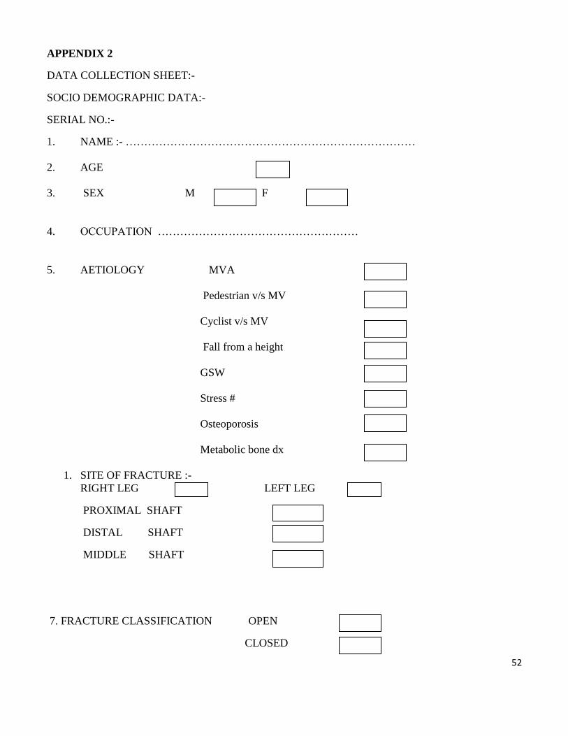
52
APPENDIX 2
DATA COLLECTION SHEET:-
SOCIO DEMOGRAPHIC DATA:-
SERIAL NO.:-
1. NAME :- ……………………………………………………………………
2. AGE
3. SEX M F F
4. OCCUPATION ………………………………………………
5. AETIOLOGY MVA
Pedestrian v/s MV
Cyclist v/s MV
Fall from a height
GSW
Stress #
Osteoporosis
Metabolic bone dx
1. SITE OF FRACTURE :-
RIGHT LEG LEFT LEG
PROXIMAL SHAFT
DISTAL SHAFT
MIDDLE SHAFT
7. FRACTURE CLASSIFICATION OPEN
CLOSED
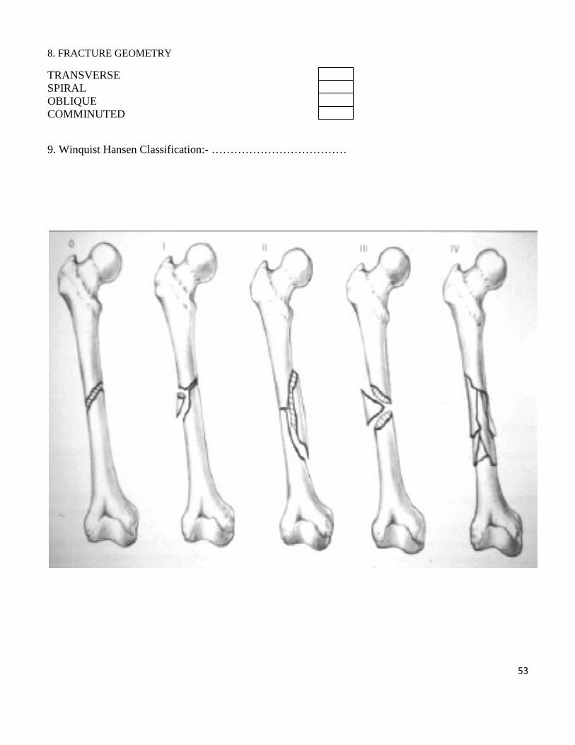
53
8. FRACTURE GEOMETRY
TRANSVERSE
SPIRAL
OBLIQUE
COMMINUTED
9. Winquist Hansen Classification:- ………………………………
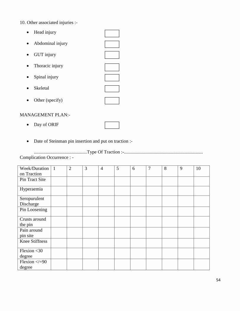
54
10. Other associated injuries :-
Head injury
Abdominal injury
GUT injury
Thoracic injury
Spinal injury
Skeletal
Other (specify)
MANAGEMENT PLAN:-
Day of ORIF
Date of Steinman pin insertion and put on traction :-
.............................................Type Of Traction :-...................................................................
Complication Occurrence : -
Week/Duration
on Traction
1 2 3 4 5 6 7 8 9 10
Pin Tract Site
Hyperaemia
Seropurulent
Discharge
Pin Loosening
Crusts around
the pin
Pain around
pin site
Knee Stiffness
Flexion <30
degree
Flexion </=90
degree
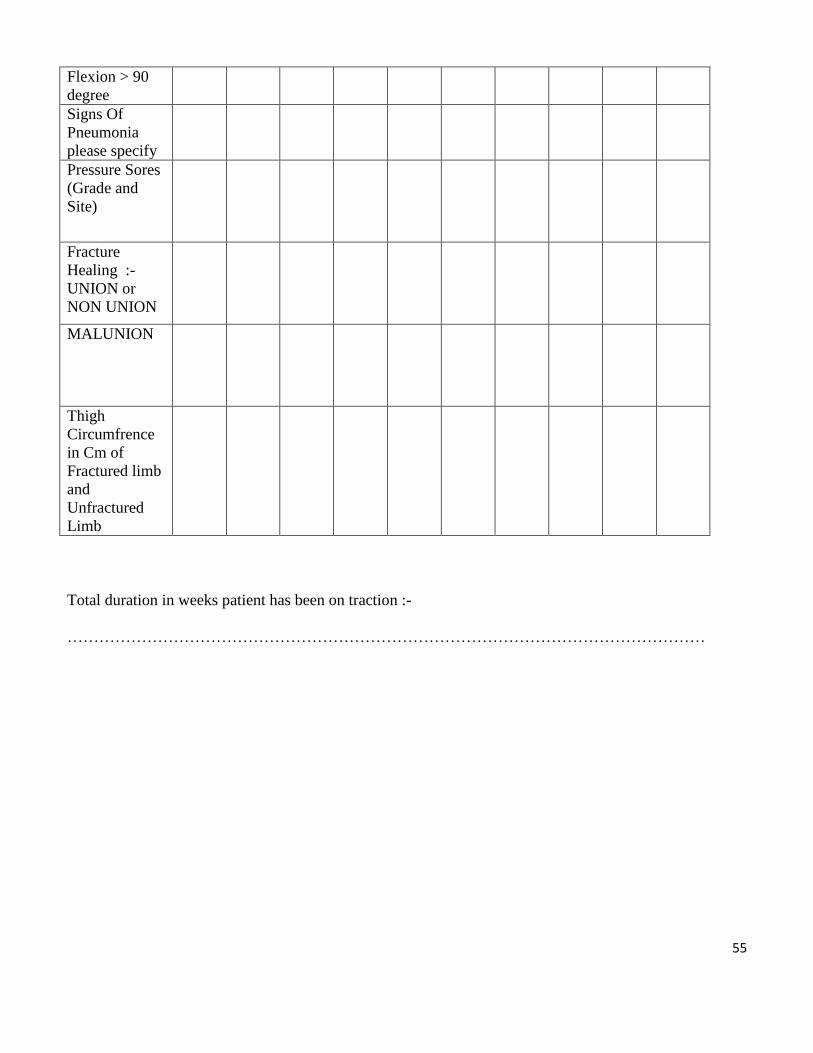
55
Flexion > 90
degree
Signs Of
Pneumonia
please specify
Pressure Sores
(Grade and
Site)
Fracture
Healing :-
UNION or
NON UNION
MALUNION
Thigh
Circumfrence
in Cm of
Fractured limb
and
Unfractured
Limb
Total duration in weeks patient has been on traction :-
…………………………………………………………………………………………………………
56
APPENDIX 3
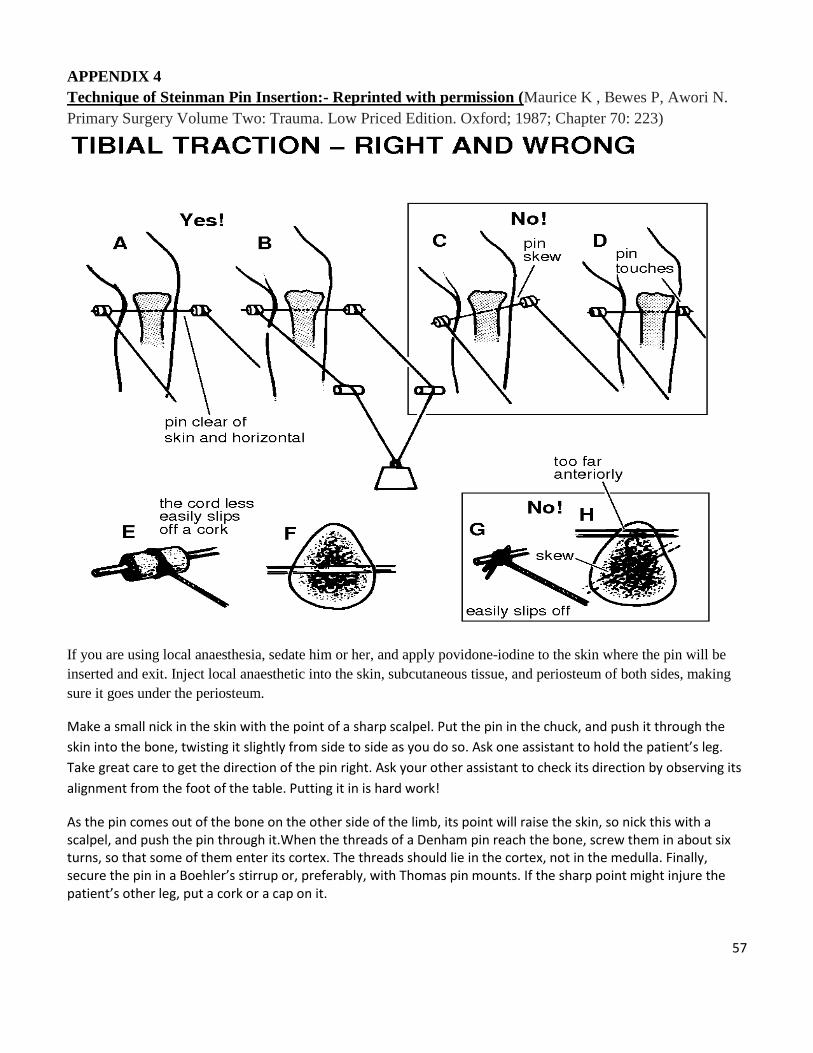
57
APPENDIX 4
Technique of Steinman Pin Insertion:- Reprinted with permission (Maurice K , Bewes P, Awori N.
Primary Surgery Volume Two: Trauma. Low Priced Edition. Oxford; 1987; Chapter 70: 223)
If you are using local anaesthesia, sedate him or her, and apply povidone-iodine to the skin where the pin will be
inserted and exit. Inject local anaesthetic into the skin, subcutaneous tissue, and periosteum of both sides, making
sure it goes under the periosteum.
Make a small nick in the skin with the point of a sharp scalpel. Put the pin in the chuck, and push it through the
skin into the bone, twisting it slightly from side to side as you do so. Ask one assistant to hold the patient’s leg.
Take great care to get the direction of the pin right. Ask your other assistant to check its direction by observing its
alignment from the foot of the table. Putting it in is hard work!
As the pin comes out of the bone on the other side of the limb, its point will raise the skin, so nick this with a scalpel, and push the pin through it.When the threads of a Denham pin reach the bone, screw them in about six turns, so that some of them enter its cortex. The threads should lie in the cortex, not in the medulla. Finally, secure the pin in a Boehler’s stirrup or, preferably, with Thomas pin mounts. If the sharp point might injure the patient’s other leg, put a cork or a cap on it.
Related Documents