OSTEOTOMIES AROUND THE HIP Presenting by:-- Dr. Sanjay kumar Post-graduate trainee Dept. Of Orthopaedics Medical College, Kolkata

Osteotomies around the hip
Jul 16, 2015
Welcome message from author
This document is posted to help you gain knowledge. Please leave a comment to let me know what you think about it! Share it to your friends and learn new things together.
Transcript
OSTEOTOMIES AROUND THE HIP
Presenting by:--Dr. Sanjay kumar
Post-graduate trainee
Dept. Of Orthopaedics
Medical College, Kolkata

BIOMECHANICS OF THE HIP JOINT • The acetabulum and femoral head form a multiaxial spheroidal
(‘ball and socket') joint
• Allows relatively unhindered motion in three degrees of freedom-flexon/extenson
-abduction/adduction
-internal/external rotation
• This articulation is innately limited in its capacity for translational motion in anteroposterior, transverse and vertical planes

• In the coronal plane, the femoral neck is inclined obliquely to the shaft at an angle of 135° (range 120–145°).
• The centre of the neck in the coronal plane is at the level of the apex of the greater trochanter.
• In the axial plane the femoral neck is anteverted, i.e. rotated anteriorly relative to the posterior surfaces of the femoral condyles: in the adult, this angle is 10–15
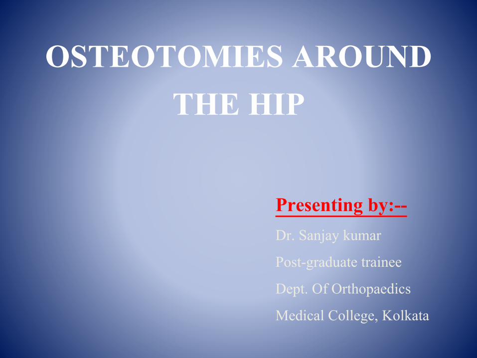
The angle (NSA) between the long axis of the femoral shaft (S) and the axis of the femoral neck (N) is on average 135° (range 125–140°). In addition, in most hips, a line perpendicular to S from the tip of the greater trochanter (B) passes through the centre of the femoral head. This approximation can be utlilized in judging the position of the femoral osteotomy in hip arthroplasty

ACETABULUM
• The acetabulum consists of the confluence of the ilium, ischium and pubis at the triradiate cartilage.
• By itself, the acetabulum covers an area slightly less than a hemisphere: it is deepened by the acetabular labrum.
• The degree of acetabular anteversion in the erect position is 14°(men) and 19° (women).
• In the coronal plane, the acetabular axis is inclined approximately 45° from the horizontal

FORCES ACTING ON THE HIP
• Under bipedal loading conditions, the femoral heads support the weight of the body minus the weight of both legs (approximately one-third body weight) and the resultant vectors are vertical.
• When viewed in the sagittal plane, minimal muscle forces are required to maintain equilibrium and balance when the weight of the upper body is directly over the femoral heads
• During normal gait the hip is subjected to->
one third of body weight-: double-leg support phase
four times body weight-: single-leg support phase

In single-leg stance, 5/6 of total body weight (W) passes just lateral to the midline, exerting a clockwise moment on the pelvis; this is counteracted by the pull of the abductors, whose lever arm (a) is approximately half that of the body centre (b). Thus the abductor force (Ab) required to maintain equilibrium is approximately twice that of body weight, resulting in a joint reaction force (JRF) approximately 3 to 4 times that of body weight.

DEFINITION
An OSTEOTOMY AROUND THE HIP is a surgical corrective procedure used to obtain a correct biomechanical alignment of the extremity so as to achieve equivocal load transmission, performed with or without removal of a portion of the bone.

OSTEOTOMY AROUND THE HIP work…. as it
• Increases the contact area / congruency.
• Improves coverage of head.
• Moves normal articular cartilage into weight bearing zone.
• Restore biomechanical advantage.
OSTEOTOMIES AROUND HIP JOINT CLASSIFIED AS –
• OSTEOTOMIES OF PROXIMAL FEMUR
• OSTEOTOMIES OF PELVIS

OSTEOTOMIES OF PROXIMAL FEMUR ARE CLASSIFIED ACCORDING TO:
• DISPLACEMENT OF DISTAL FRAGMENT.
• ANATOMICAL LOCATION OF OSTEOTOMY.
• ACCORDING TO INDICATION.

I) DISPLACEMENT OF DISTAL FRAGMENT
1.TRANSPOSITIONAL OSTEOTOMY:
• Longitudinal axis of distal fragment remains parallel to the longitudinal terminal axis of proximal fragment.
• Used in : Fracture neck of femur and OA.
• Eg: McMurray osteotomy, Pauwel’sosteotomy & Putti osteotomy.
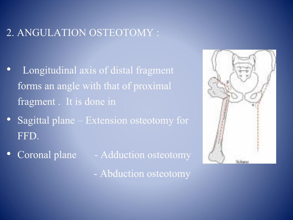
2. ANGULATION OSTEOTOMY :
• Longitudinal axis of distal fragment forms an angle with that of proximal fragment . It is done in
• Sagittal plane – Extension osteotomy for FFD.
• Coronal plane - Adduction osteotomy
- Abduction osteotomy

II) ANATOMICAL LOCATION :
• HIGH CERVICAL
• INTERTROCHANTERIC
• GREATER TROCHANTERIC
• SUBTROCHANTERIC

III) BASED ON INDICATION : • 1.NON UNION # NECK OF FEMUR
- McMurry’s osteotomy- Dickson’s osteotomy- Putti’s osteotomy- Schanz osteotomy
• 2. OSTEOARTHRITIS - Pauwel’s varus osteotomy- Pauwel’s valgus osteotomy- Mc Murrays osteotomy.
• 3. UNSTABLE INTERTROCHANTERIC #- Dimon Hughston osteotomy- Sarmiento’s osteotomy

• 4.UNREDUCED CDH- Lorenz bifurcation osteotomy- Schanz low sub trochanteric osteotomy.- Pemberten acetabuloplasty.
• 5.CONGENITAL COXA-VARA - Cuniform osteotomy by Fish- Pauwel’s Y osteotomy- Valgus osteotomy- Basilar osteotomy.
• 6. LEG-CALVE PERTHE’S DISEASE - Varus de-rotation osteotomy - Salter osteotomy- Shelf - Chiari osteotomy.
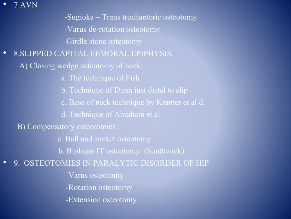
• 7.AVN -Sugioka – Trans trochanteric osteotomy-Varus de-rotation osteotomy-Girdle stone osteotomy
• 8.SLIPPED CAPITAL FEMORAL EPIPHYSIS.A) Closing wedge osteotomy of neck:
a. The technique of Fishb. Technique of Dunn just distal to slipc. Base of neck technique by Kramer et al d. d. Technique of Abraham et al
B) Compensatory osteotomies:a. Ball and socket osteotomyb. Biplanar IT osteotomy (Southwick)
• 9. OSTEOTOMIES IN PARALYTIC DISORDER OF HIP -Varus osteotomy-Rotation osteotomy-Extension osteotomy.

OSTEOTOMIES OF PELVIS DIVIDED INTO :
• a) SINGLE INNOMINATE - Salter osteotomy
• b) DOUBLE INNOMINATE - Sutherland
• c) TRIPLE INNOMINATE - Steel osteotomy - Tonnis
• d) PERI-ACETABULAR - Wagner osteotomy- Ganz osteotomy.

McMURRAY’S DISPLACEMENT OSTEOTOMY• Described as medial displacement linear oblique inter-trochantric
pelvic supporting osteotomy• INDICATIONS:
-Nonunion of femoral neck fracture-Advanced osteoarthritis .
• Extends from lateral aspect of shaft at level just below the lower border of lesser trochanter and terminates medially between lesser trochanter and lower border of neck.
• Shaft is displaced medially• AIM :
-Line of weight bearing is shifted medially-Shearing force at the nonunion is decreased, because the fracture
surface has become more horizontal


DICKSON’S HIGH GEOMETRIC OSTEOTOMY
• Line of osteotomy is changed from vertical (shearing) force to a horizontal (impacting) force. This osteotomy is done just below the greater trochanter, the distal fragment is abducted 60° and fixed with plate.
• Gives high rate of union
• Improves abductor strength
• Increases limb length

SCHANZ ANGULATION OSTEOTOMY
AIM :
• To turn the shaft from the adducted to abducted position, so that the shearing stress of weight bearing and muscle retraction becomes an impaction force.
INDICATIONS:
• Non-union fracture neck of femur
• Congenital dislocation of hip

• The femur is cut transversely at ischial tuberosity level & the proximal fragment is adducted until it rests against the side wall of the pelvis.
• This lengthens the distance of the gluteus medius and provides a fulcrum so that adequate leverage of the muscle is obtained.
• A plate is prepared and angulated sufficiently.

• This is a post op radiograph after SCHANZ OSTEOTOMY for neglected CDH…

GIRDLE STONE OSTEOTOMY In this head & neck of femur are excised at Inter trochanteric level to create pseudo arthrosis in order to improve stability. Angulations Osteotomy is added.
INDICATION
• T.B. Hip
• Pyogenic Hip
• Non union #.neck femur [in elderly pt.]
• AVN of femoral head.
Advantage :-
• Painless mobile hip joint

LORENZ (BIFURCATION OSTEOTOMY)
In this upper end of the lower fragment is abducted and inserted in to the acetabulum after making on intertrochanteric osteotomy “plane of osteotomy” below & outward to above & inward.
DISADVANTAGE :
• Increased shortening.
• Less mobility and arthritic pain.

Limb is Abducted and extended so proximal end of distal fragment directed medially and anteriorly in acetabulum
LORENZ (BIFURCATION OSTEOTOMY)

DIMON AND HUGHSTON
• Trochanteric osteotomy with valgus nailing and medial displacement to improve stability
• There techniques are occasionally useful in some extremely comminutedfractures.

SARMIENTO TECHNIQUE

OSTEOARTHRITIS OF HIP
AIM OF OSTEOTOMY:
• 1. RELIEF OF PAIN:
-Mechanical : reducing the ratio between abductor and body weight, lever, relaxing capsule.
-Haemodynamic: Also by decreasing the intra osseous pressure.
• 2. CORRECTION OF DEFORMITY: flexion, adduction, external rotation.
• 3. REVERSAL OF DEGENERATIVE PROCESS: helped by increase in joint space.

CLINICALLY THE FOLLOWING SHOULD BE RECORDED :
• Limp – antalgic or trendelenberg
• Position in which hip is least painful.
• Amount of lengthening or shortening.
• Fixed abduction/flexion and rotation deformity.
• Degree of both active and passive movement of joint.

PAUWEL’S VARUS OSTEOTOMYAIM :
Varus intertrochanteric femoral osteotomies are designed to elevate the greater trochanter and move it laterally, while moving the abductor and psoas muscles medially, to :• Restore joint congruity • Decrease the force acting on the edge of the acetabulum moves to the
middle of weight bearing surface.
INDICATIONS: • Antalgic abductor limb • Abduction deformity • Painful adduction
• Neck shaft angle > 135°
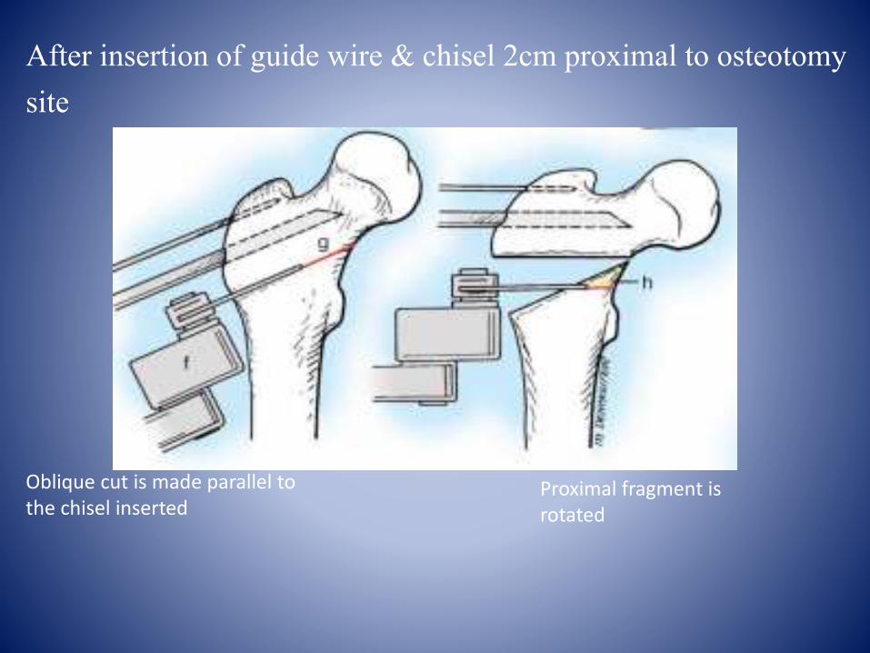
After insertion of guide wire & chisel 2cm proximal to osteotomy site
Oblique cut is made parallel to the chisel inserted
Proximal fragment is rotated

• TYPES OF WEDGE IN VARUS OSTEOTOMY:

Wedge being shifted to lateral side
Insertion of angled blade plate
FINAL RESULT

• CONTRAINDICATIONS:
-Fixed external rotation of > 25°
-Flexion of 70° or less.
• DISADVANTAGES:
-Shortens the limb to some degrees.
-Creates a trendelenberg gait.
-Increases the prominence of greater trochanter.
-Overloading of the medial compartment of knee.

PAUWEL’S VALGUS OSTEOTOMYAIM:• Valgus intertrochanteric femoral osteotomies transfer the
center of hip rotation medially from the superior aspect of the acetabulum to decrease the weight bearing area of femoral head .
• Normally 15° of correction is required.INDICATIONS: • Trendelenburg Limb • Adduction deformity• Motion in adduction beyond adduction deformity • Painful abductionCONTRAINDICATIONS:• Flexion of less than 60°• Knock knees as this will increase the deformity at knee.

• After insertion of guide wire & chisel 2cm proximal to osteotomy site similar to explained before :-

UNREDUCED CDHIN CDH, THE BASIC PATHOLOGY IS:
• A dysplastic acetabulum that is shallow and vertical. This permits the femoral head to slip out when the limb is in extension and adduction.
• A displaced head rests against the lateral wall of ilium. This constant pressure on the femoral head increases the degree of anteversion. An osteotomy in CDH is thus aimed at correcting these defects.
AIM:
• To contain the femoral head within the acetabulum.
• To improve the dynamic and static forces maintaining reduction
SALTER'S INNOMINATE OSTEOTOMY:AIM :
• In this, the entire acetabulum together with pubis and ischium is rotated as a unit.
INDICATIONS:
• CDH in children from 18 months to 6 years of age and in congenital subluxation upto early adult life.
• Before the osteotomy, femoral head should be positioned opposite the level of the acetabulum achieved by period of traction.
• Contractures of iliopsoas and adductor muscles must be released.

Technique of Salter innominate osteotomy

ADVANTAGES:
• Relatively simple procedure.
• No change in acetabular configuration.
DISADVANTAGES:
• Relatively unstable needs internal fixation.
• Second surgery for pin removal.
• Possibility of joint penetration by pins

Subluxation in 4 yrs old girl of DDH 1yrs post op after SALTERS osteotomy

PEMBERTON ACETABULOPLASTY
AIM:
• This operation redirects the inclination of the acetabular roof by an osteotomy of the ilium, superior to the acetabulum followed by levering of the roof inferiorly.
INDICATION:
• In dysplastic hips between the age of 1 year and the age when the tri-radiate cartilage became too inflexible to serve as a hinge (about 12 years in girls and 14 years in boys).

ADVANTAGES:
• Osteotomy is incomplete, therefore more stable
• Internal fixation is not required
• Greater degree of correction can be achieved with less rotation of the acetabulum.
DISADVANTAGES:
• Technically more difficult
• It alters the configuration and capacity of the acetabulum and can result in an incongruence relationship between it and femoral head.

Acetabular dysplasia in 8-year-old girl after treatment of CDH with Pemberton acetabuloplasty on Right side.

TRIPLE INNOMINATE OSTEOTOMY BY STEEL
• INDICATIONS- Adolescents & skeletally mature adults with residual dysplasia & subluxation in whom remodelling of acetabulum is no longer anticipated.
• ADVANTAGE - Better coverage of femoral head by articular cartilage [chiari- fibrous cartilage], Better hip joint stability, no need of spica cast.

Immediate post op in 16yr old girl
After 1 yr of STEEL osteotomy
DISADVANTAGES: • Difficult to perform.• Does not change the size of the acetabulum.• It distorts the pelvis so natural child birth is impossible in
adulthood.
MODIFIED BY LIPTON & BOWEN• Resecting 1-1.5 cm bone from ischial tuberosity to favor
medialization.• To resect a triangular wedge from outer part of ilium which
favors slot formation which serves as abutment.• Use 7.3mm cannulated screws instead of steinmann pins.

SHELF OSTEOTOMY BY STAHELI
• Have commonly been performed to enlarge the volume of the acetabulum.
• The objective is to create a shelf, the size of which is decided by measuring the “width of augmentation (WA)” using the CE angle of wiberg.
• Best to do after 5 years of age.
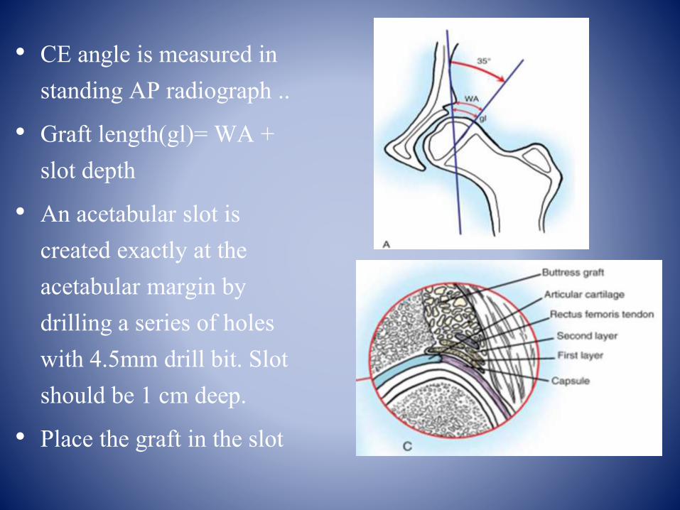
• CE angle is measured in standing AP radiograph ..
• Graft length(gl)= WA + slot depth
• An acetabular slot is created exactly at the acetabular margin by drilling a series of holes with 4.5mm drill bit. Slot should be 1 cm deep.
• Place the graft in the slot

The rectus femoris is sutured for stability of the graft .Postoperative: hip spica can be applied in 15 deg of
abduction and 20° of flexion.

CONTRA-INDICATIONS:
• DDH with spherical congruity suited for re-directional osteotomy.
• Hips requiring concurrent open reduction that must have supplementary stability.
• Patients un-suited for spica cast application

CHIARI OSTEOTOMY
This is a capsular interposition osteotomy as the capsule is interposed between the newly formed acetabular roof and femoral head.
INDICATIONS:
• Congenital subluxation in patients 4 to 6 years or older, including adults.
• Dysplastic hip with osteoarthritis
• For Coxa magna after Perthes disease or avascular necrosis after treatment of congenital dysplasia.
• For paralytic dislocation caused by muscular weakness or spasticity.

• The osteotomy is made precisely between the insertion of the capsule and reflected head of rectus femoris.
• Ending distal to the AIIS anteriorly and in sciatic notch posteriorly.
• on lateral table with plane directed 20° superiorly towards inner table.

• The distal fragment is displaced medially by forcing the limb into abduction hinging at symphysis pubis.
• It is displaced enough medially so that the proximal fragment completely covers the femoral head
• If necessary the fragments may be transfixed by screw driven obliquely.

PRE OP POST OP WITH CHIARI

GANZ OSTEOTOMY: (BERNESE) PRIACETUBULAR OSTEOTOMY
• This triplaner osteotomy is for adolescent and adult dysplastic hip that required correction of congruency & containment of the femoral head with little or no arthritis.
• If significant degenerative changes are presents a proximal femoral osteotomy can be added.
• Approach Smith Peterson approach.


ADVANTAGES :
• Only one approach is used.
• A large amount of correction can be obtained in all directions, including the medial and lateral planes.
• Blood supply to the acetabulum is preserved.
• The posterior column of the hemipelvis remains mechanically intact, allowing immediate crutch walking with minimal internal fixation.
• The shape of the true pelvis is unaltered, permitting a normal child delivery.
• Can be combined with trochanteric osteotomy if needed


LEGG CALVE PERTHES DISEASE
PATHOLOGY:
• Self limited disease of avascular necrosis of ossification center of the capital epiphysis, resulting in variable degree of deformity of femoral head.
AIM:
• To prevent or minimize residual deformity of femoral head by creating the biomechanical environment which is not detrimental to normal growth and remodeling of epiphysis.
• This is achieved by containing the femoral head within the
acetabulum.

VARUS DE-ROTATION OSTEOTOMYAIM :
• By reducing the ante-version and neck shaft angle to obtain maximum coverage of the femoral head.
• This osteotomy is done before 4 years of age, as after this age, there are less chances of Acetabular remodeling.
DISADVANTAGES:
• Excessive varus angulation that may not correct with growth
• Further shortening of already shortened extremity
• Possibility of a gluteus lurch produced by decreasing the length of the lever arm of the gluteus musculature.

• The degree of de roration is estimated with the amount of internal rotation but furthur adjustments can be made during the surgery.
• If the internal rotation is severely limited even after 4 weeks of bed rest with traction: Varus osteotomy is done along with extension by giving slight backward tilt to the proximal segment.

Using the side plate and screws firmly join the proximal and distal fragments
Insert the barrel guide into the back of the implanted lag screw.
Make the osteotomy cut & tilt the head into varus
OTHER OSTEOTOMIES IN PERTHES DISEASE
• SALTER Innominate osteotomy:
• SHELF procedure (Staheli): If the hip is congruous, it can be performed for coxa magna and lack of acetabular coverage for the femoral head.
• CHIARI Osteotomy: It is used as a salvage procedure to accomplish coverage of large flattened femoral head.
• VALGUS EXTENSION osteotomy: Indicated in malformed femoral head in residual Perthe's disease with hinge abduction.

AVASCULAR NECROSIS OF FEMORAL HEAD
AIM :
• To reposition the necrotic part of the femoral head to a non-weight bearing area.
INDICATIONS:
• Osteotomy is done in FICAT'S stage I and II of AVN.

PAUWEL'S `Y' OSTEOTOMY
• A guide pin is inserted from the greater trochanter to head of femur.
• One limb of osteotomy is made from the base of greater trochanter towards the base of neck medially and inferiorly.
• The distal limb of the Y then passes upwards and medially to reach the proximal limb and a wedge of bone with the required correction is removed
from the proximal aspect of distal fragment with its base directed laterally.

• The trochanter head segment is levered into valgus.
• The two fragments are apposed by displacing the proximal end of the shaft medially and abducting the limb.
• The nail is then attached by a plate to the shaft

SUGIOKA TRANSTROCHANTRIC ROTATIONAL OSTEOTOMY
• This is done for osteonecrosis to prevent progressive collapse of the articular surface and to improve the congruity of hip joint.
• To do this the femoral head and neck segment is rotated anteriorly around its longitudinal axis, though a trans-trochantric osteotomy.
• So that the weight bearing force is transmitted to the posterior articular surface of femoral head, which is not involved in the ischemic process
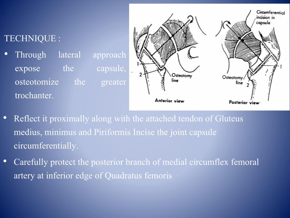
• Reflect it proximally along with the attached tendon of Gluteus medius, minimus and Piriformis Incise the joint capsule circumferentially.
• Carefully protect the posterior branch of medial circumflex femoral artery at inferior edge of Quadratus femoris
TECHNIQUE :
• Through lateral approachexpose the capsule,osteotomize the greatertrochanter.
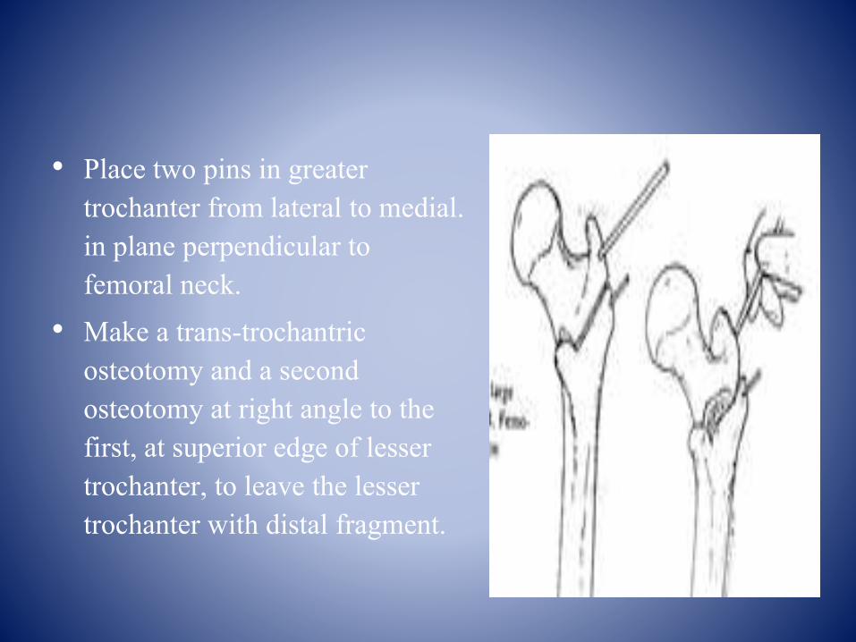
• Place two pins in greater trochanter from lateral to medial. in plane perpendicular to femoral neck.
• Make a trans-trochantricosteotomy and a second osteotomy at right angle to the first, at superior edge of lesser trochanter, to leave the lesser trochanter with distal fragment.

After completing second osteotomy use the proximal pin to rotate proximal fragment 45-90°depending on the size of
necrotic area.
Fix the osteotomy internally with large screws and washer. Re-attach the greater trochanter to proximal and distal
fragment with screws.

Post op after one yr
Postoperative: skin traction is given for 2-3 weeks active range of motion exercises of hip are begun at 10-14 days.

SLIPPED CAPITAL FEMORAL EPIPHYSIS
In this condition, the epiphysis slowly displaces inferiorly and causing adduction and external rotation deformity of the limb.
AIM:
• Osteotomy is performed here to reposition the femoral head concentrically within the acetabulum.
INDICATIONS:
• Chronic slip with moderate to severe displacement.
• Malunited slip

TWO BASIC TYPES:• CLOSING WEDGE OSTEOTOMY OF NECK: Usually associated with
serious complications of AVN and chondrolysis, therefore these osteotomies are not recommended. These are of four types.
a. The technique of Fishb. Technique of Dunn just distal to slipc. Base of neck technique by Kramer et al d. d. Technique of Abraham et al
• COMPENSATORY OSTEOTOMIES THROUGH THE TROCHANTRIC REGION: These osteotomies produce a deformity in the opposite direction. It includes
a. Ball and socket osteotomyb. Biplane intertrochanteric osteotomy (Southwick)

CUNEIFORM OSTEOTOMY OF FEMORAL NECK (FISH):
• Fish recommended this in moderate to severe slips of more than 30°.
• Watson-Jones approach
• Capsule is incised & femoral neck is exposed.
• Locate the physis.
• Determine the size of wedge to be removed by noting the degree of slip.

• Adjacent to the epiphyseal plate, a wedge shaped piece of bone is removed with its base directed anteriorly and superiorly with apex psotero-inferiorly.
• Take care that osteotome does not penetrate the intact posterior periosteum, damaging retinacularvessels.

• Reduce the epiphysis by flexion,abduction and internal rotation oflimb, taking care to put muchtension on the posteriorperiosteum, capsule and vessels.
• After reduction fix the epiphysis toneck with 2-3 pins six inches longthreaded on one half of theirlengths with a nut on the thread.
• Do not penetrate articularcartilage.

2. CUNIEFORM OSTEOTOMY OF FEMORAL NECK (DUNN):
• Dunn described an osteotomy for severe chronic slips in children with open physis.
• This procedure should not be done if the physis is closed.
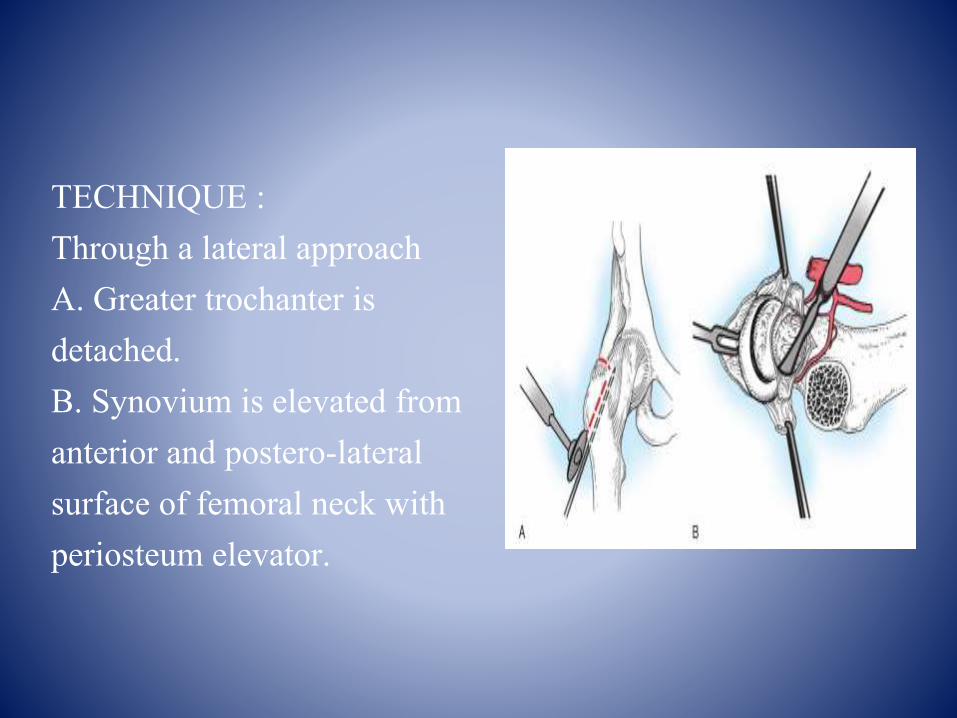
TECHNIQUE :Through a lateral approachA. Greater trochanter is detached.B. Synovium is elevated from anterior and postero-lateral surface of femoral neck with periosteum elevator.

C. Head is free of all fibrocartilage and callus.
D. Osteotomy line on upper end of femoral neck is made for excision of trapezoid segment.

E. Head of femur is replaced on femoral neck and three threaded Steinmann pins are used for fixation of shaft, head, and neck of femur.
F. Two cancellous screws are used to fix greater trochanter in normal position.

CONTRAINDICATIONS OF OSTEOTOMY
• NEUROPATHIC ARTHROPATHY
• INFLAMMATORY ARTHROPATHY
• ACTIVE INFECTIONS
• SEVERE OSTEOPENIA
• ADVANCED ARTHRITIS/ANKYLOSIS
• ADVANCED AGE
• SMOKING, OBESITY

REFERANCES• GRAY’S ANATOMY-40th edition
• CAMPBELL’S OPERATIVE ORTHOPAEDICS, 12th edition.
• TEXT BOOK OF ORTHOPAEDICS – G.S. KULKARNI.
• TUREK ORTHOPAEDICS-4th edition
• STANDARD ORTHOPAEDICS OPERATIONS– ADAMS.
• OPERATIVE ORTHOPAEDICS – CHAPMAN’S.
• INTERNET

Related Documents